Note: Descriptions are shown in the official language in which they were submitted.
CA 03107380 2021-01-22
WO 2020/031067
PCT/IB2019/056652
1
DESCRIPTION of the invention having the title:
"MODEL FOR IN-VITRO SIMULATION OF THE BEHAVIOUR OF DYSFUNCTIONAL VESSELS"
The present invention refers to a model for in-vitro simulation of the
behaviour of dysfunctional human
vessels, such as for example vessels affected by aneurysm, stenosis or
sclerosis plaques, as an
instrument for testing medical devices and drugs with the aim of verifying the
effectiveness and safety
thereof prior to use thereof on humans. Specifically, the present invention
refers to an in vitro model of a
vascular structure, preferably a substantially tubular-shaped synthetic
vascular structure having
dysfunctional anatomical and physiological characteristics, simulating the
vascular structure of a healthy
subject whose vascular structure has been damaged or deformed or deteriorated
due to a damage
selected from among the group comprising or, alternatively, consisting of
aneurysm, stenosis, sclerosis
plaques, forms of tumours or cardiomyopathies having the characteristics as
claimed in the attached
claims. Furthermore, the present invention also refers to a reliable and
reproducible industrialisation
process for eliminating air bubbles for producing an engineered vascular
tissue for the in vitro test of
medicinal products for human use and veterinarian products for animal use.
It is known that the development of a medicinal product entails a long and
sensitive process. Basically, in
the medicinal or veterinarian product development process, whether a drug or
medical device, prior to
using the product in humans or in animals it is important to be able to
determine the type of effects on the
tissue/s with which it comes into contact so as to evaluate the aspects
relating both to biological safety
and to the efficiency of the medicinal product and foretell potential problems
relating to the use thereof.
In this context, the evaluation of the biological safety and efficiency of the
medicinal product is extensive
and complex while the evaluation of the interaction with the tissue/s of only
one constituent material
cannot be considered as isolated from the overall planning of the medicinal or
veterinarian product which
must be evaluated as a whole and in a context that can reproduce the
conditions of use as faithfully as
possible.
Today, the evaluation of biological safety and of the efficiency of a
medicinal or veterinarian product are
based on in vitro, ex vivo tests and on animal models that have significant
differences with respect to the
final conditions of use. In particular, the animal models have significant
physiological differences that
complicate the transposition of the validity of the results to humans. Such
differences are observable even
further when evaluating medicinal products for human use or veterinarian
products for animal use that
provide for the use thereof in the cardiovascular and peripheral vascular
region, such as, for example,
heart valves, stents, grafts, catheters, bandages and nets.
Unfortunately, the evaluation of the interaction of medicinal products for
human use or veterinarian
products for animal use with the vascular tissues and with the blood so as to
be able to establish the
CA 03107380 2021-01-22
WO 2020/031067
PCT/IB2019/056652
2
biological safety and efficiency is crucial and the models currently in use
reveal major limits and
drawbacks both anatomical/structural and regarding the blood composition.
Furthermore, tests on animals have been at the centre of an intense and
controversial debate around the
issue whether using animals for biomedical testing can be considered morally
acceptable. This issue led
to the 3R principle already back in 1959, and it still remains a hotly
disputed current topic in debates and in
the international research programmes. The 3R principle refers to three key
concepts: replacement,
reduction and refinement. The 3R principle argues that research in the
biomedical industry should aim at,
with utmost effort possible, replacing or substituting the animal model with
an alternative model; reducing
the number of animals used in a given experimental protocol as much as
possible; refining, i.e. improving
the experimental conditions to which the animals are subjected.
The vascular tissue engineering industry shows that it is crucial to be able
to produce a continuous (i.e.
having a monolayer of confluent cells) and functional endothelium mainly
consisting of endothelial cells
(ECs).
The production of continuous and functional endothelium is a crucial factor
towards promoting scaffold
endothelisation and guaranteeing appropriate efficiency of the tissue-
engineered constructs (consisting of
scaffolds and cells), including preventing thrombosis and stenosis once said
constructs have been
implanted.
The in vitro generation of vascular endothelium or engineered vascular
construct/tissue, in short provides
for the following steps:
(1) seeding endothelial cells in the lumen of the scaffold and ensuing
adhesion thereof,
(2) cell growth/proliferation and organisation as a function of mechanical
stimuli (such as for example the
flow of a fluid) to which they are subjected, and
(3) generating one or more layers of functional endothelial cells.
Due to the crucial importance of the seeding step, many research groups at
university level have invested
in research over the last decades creating various techniques for uniformly
seeding the endothelial cells in
the lumen of the scaffold and enhancing the seeding effectiveness. However,
the results achieved have
not been entirely satisfactory.
Static seeding and dynamic seeding currently represent the main endothelial
cell seeding methods.
CA 03107380 2021-01-22
WO 2020/031067
PCT/IB2019/056652
3
The simplest static method consists in pipetting a cell suspension directly on
the luminal surface of the
scaffold followed by a short incubation step on a Petri dish. This method
strongly depends on the operator
and it is complicated by the difficulty to obtain a uniform endothelial layer.
Vastly based on rotation, vacuum, electrostatic or magnetic forces, the
dynamic methods on the contrary
increase cell seeding effectiveness, uniformity and adhesion. Some of these
dynamic principles could be
directly transferred and paired with perfusion bioreactors, allowing a
reduction in the handling of the
scaffold. In this case, the scaffolds can be immediately seeded once housed in
the bioreactor chamber.
Other research groups instead use the dynamic seeding method through a
continuous injection of a cell
suspension using a syringe pump, or through a continuous perfusion of the
lumen of the scaffold seeded
with a growth medium using a peristaltic pump, after injecting the endothelial
cell suspension. These
methods require higher volumes of cell suspension with respect to a
pipetting/rotation seeding method due
to the need of also filling the volume of the perfusion piping as well as the
volume of the injection syringe,
revealing possible limitations with reference to reducing costs (growth cells
and media).
A completely different technique with respect to the ones described previously
is based on dripping the
cell suspension in the lumen of a scaffold. In this case, the main drawback
lies in a low initial adhesion of
the cells and a low reproducibility of the method given that it strongly
depends on the operator.
Furthermore, the choice of a method for connecting a perfusion circuit
(perfusion method), required for the
perfusion of a scaffold, mainly tubular, to the bioreactor-scaffold system
must be adapted to the
experimental setup.
Considering the landscape regarding the methods for seeding and connecting a
perfusion circuit
(perfusion method) mentioned above, considerable limits and drawbacks still
exist.
Therefore, there clearly arises the need to provide a process comprising a
method for seeding and
connecting the perfusion circuit (perfusion method) to the bioreactor-scaffold
system that is reproducible,
reliable and effective, especially considering the applicability of said
process for GLP (Good Laboratory
Practice)-approved Tissue engineering laboratories whose objective focuses on
advanced preclinical tests
and clinical tests.
There arises the need to develop a process for the production of engineered
vascular tissues/constructs
having a continuous (i.e. having a monolayer of confluent cells) and
functional endothelium, wherein said
process is simple, quick, effective, independent from the operator, well
defined, highly reproducible and
reliable, comprising: (1) a method for seeding cells, mainly endothelial
cells, in the lumen of a scaffold, that
CA 03107380 2021-01-22
WO 2020/031067
PCT/IB2019/056652
4
guarantees the homogeneity and the uniform adhesion of the endothelial cells,
with the elimination of air
bubbles in the scaffold; (2) a method for connecting the perfusion circuit to
the bioreactor-scaffold system
(perfusion method) capable of guaranteeing sterility and maintaining the
absence of air bubbles in the
assembled system consisting of the perfusion circuit and bioreactor-scaffold
system.
There also arises the need of being able to develop in vitro engineered
vascular tissues/constructs
comprising a continuous and functional endothelium for conducting advanced
preclinical and clinical tests,
so as to avoid using laboratory cavies to conduct preclinical and clinical
tests. Therefore, there arises the
need for a procedure that is reliable, effective and reproducible for the
industrialisation of said in vitro
engineered vascular tissues/constructs.
As observed above, the process for developing and approving medicinal
products, such as for example
medical devices and drugs, normally requires conducting numerous preclinical
tests to be conducted in-
vitro, ex-vivo and in-vivo on animals prior to conducting clinical testing on
humans. Despite animal testing
offering an in vivo model for the test of medicinal products, the anatomical
structure, the physiology and
the blood composition considerably differ from those of humans and the results
achieved on the animal
model may considerably differ from those achieved in human testing.
Furthermore, besides being technically complex, causing diseases (such as for
example vascular
dysfunctions, aneurysms or stenosis) using animal models is widely ethically
inadmissible. Thus, the in
vivo test on animals cannot be conducted to verify whether the medicinal
product is effective at treating
given diseases. To confirm the scientific willingness and policy towards
minimising in vivo testing on
animals, since 1987 the 3R (Replace, Reduce, Refine) Research Foundation has
been promoting the
development of methods alternative to the animal model and has been creating
awareness among the
public and scientific community around animal testing only in the absence of
alternative methods so as to
meet the "technical" requirement, with the aim of reducing the number of
laboratory animals and suffering
inflicted on the animals to the minimum. Methods alternative to animal testing
represent significant
progress not only from an ethical point of view as regards treating animals
more responsibly but above all
as concerns the possibility of developing models that are reliable,
reproducible and that allow to simulate
the "human" environment to the uttermost. Computerised simulations and in
vitro cell culture for the
preliminary analysis of some characteristics of medicinal products have been
created in recent years to
this end.
In compliance with the 3R principle and with the aim of overcoming the
anatomical/physiological limitations
of animal models, it would be useful to have dysfunctional engineered vascular
models capable of
reproducing the diseases (such as for example stenosis, aneurysms) and
"special" vascular structures as
CA 03107380 2021-01-22
WO 2020/031067
PCT/IB2019/056652
testing platform for medical devices and drugs prior to the use thereof in
humans. The use of an in vitro
system capable of replicating, in a standardised manner, human anatomical
structures and the in-vivo
environment would not only allow to accelerate the research and development of
new medicinal products,
but also to obtain more reliable responses on the safety and on the
effectiveness of the tested products
5 with respect to the animal model.
The Applicant met the aforementioned needs.
Forming an object of the present invention is an in vitro model of a
substantially tubular-shaped vascular
structure having dysfunctional anatomical and physiological characteristics,
simulating the vascular
structure of a healthy subject whose vascular structure has been damaged or
deformed or deteriorated
due to a damage selected from among the group comprising or, alternatively,
consisting of aneurysm,
stenosis, sclerosis plaques, forms of tumours or cardiomyopathies having the
characteristics as claimed in
the attached claims.
Furthermore, forming an object of the present invention is an in vitro model
of a substantially tubular-
shaped vascular structure having dysfunctional anatomical and physiological
characteristics simulating the
same vascular structure of a healthy subject whose vascular structure has been
damaged or deformed or
deteriorated due to a damage selected from among the group comprising or,
alternatively, consisting of
aneurysm, stenosis, sclerosis plaques, forms of tumours or cardiomyopathies,
wherein said model
comprises or, alternatively, consists of one or more biocompatible porous
polymeric supports ("scaffolds")
capable of promoting a cell adhesion and growth, wherein said scaffold is
seeded with endothelial cells
that cover a lumen of the scaffold and constitute an endothelium having a
monolayer of confluent cells,
said scaffold being provided with deformities or defects on a tubular
structure thereof, having the
characteristics as claimed in the attached claims.
Preferably said in vitro model has a vascular structure that was selected from
among the blood vessels or
valves of the central or peripheral circulatory system; preferably arteries,
veins, capillaries, aortic or mitral
valve. Preferably, said in vitro model is a dysfunctional vascular model.
Preferably, said vascular structure
is synthetic vascular structure. Preferably, said scaffold consists of
electrospun silk fibroin, copolymers of
polyglycolic acid/polylactic acid (PGA/PLA) or copolymers of polyglycolic
acid/polycaprolactone
(PGA/PCL). Preferably, said in vitro model comprises or, alternatively,
consists of one or more scaffolds
seeded with endothelial cells, and optionally muscle cells. Preferably, said
deformities or said defects of
the tubular structure comprise bifurcations, curvatures, elbows,
constrictions, dilatations or combinations
thereof.
CA 03107380 2021-01-22
WO 2020/031067
PCT/IB2019/056652
6
Forming an object of the present invention is a method for testing a medical
device or a drug with the aim
of verifying the effectiveness and safety thereof prior to the in vivo use
thereof on humans or animals, said
method comprising the following steps:
- preparing a substantially tubular-shaped scaffold having the
dysfunctional anatomical and physiological
characteristics suitable to simulate a damage or a deformation or a
deterioration due to an aneurysm,
stenosis, sclerosis plaques, forms of tumours or cardiomyopathies;
- seeding at least part of the internal lumen of said scaffold with
endothelial cell lines so as to obtain a
continuous and homogeneous layer of seeded endothelial cells (seeding method),
optionally seeding at
least part of the outer surface of said scaffold with muscle cell lines;
- promoting the growth of said endothelial cells, and optionally said muscle
cells, up to obtaining a
continuous and uniform layer of endothelial cells, to obtain said in vitro
model;
- introducing into said in vitro model a medical device or a drug subject
of test, and
- allowing the circulation (perfusion model) in said in-vitro model
comprising said medical device or drug of
a human whole blood sample, artificial blood or derivatives thereof so as to
evaluate the behaviour and the
interaction of said medical device or drug with said human whole blood sample,
artificial blood or
derivatives thereof.
Forming an object of the present invention is a model for in-vitro simulation
of the behaviour of
dysfunctional human vessels comprising or, alternatively, consisting of
vessels affected by aneurysm,
stenosis or sclerosis plaques, as an instrument for testing medical devices
and drugs with the aim of
verifying the effectiveness and safety thereof prior to use thereof on humans,
having the characteristics as
claimed in the attached claims.
Also forming an object of the present invention is providing in vitro vascular
structure models, mainly
substantially tubular-shaped, having, depending on the need, dysfunctional
anatomical and physiological
characteristics with respect to the healthy human vascular structure, such as,
by way of non-limiting
example, aneurysms, stenosis, sclerosis plaques, having the characteristics as
claimed in the attached
claims.
Forming another object of the present invention are dysfunctional vascular
models comprising or,
alternatively, consisting of one or more scaffolds seeded with suitable
selected cells, having the
characteristics as claimed in the attached claims.
Forming another object of the present invention is a method for providing said
dysfunctional vascular
models using scaffolds seeded with suitable selected cells, having the
characteristics as claimed in the
attached claims.
CA 03107380 2021-01-22
WO 2020/031067
PCT/IB2019/056652
7
Such dysfunctional vascular structures include: (i) a scaffold consisting of
biocompatible material suitable
to be electrospun, such as for example silk fibroin, or molten in moulds or
printed using bioprinters such as
for example polylactic acid, polycaprolactone, etc. The structure of the
scaffold is suitable to allow the
seeding of endothelial vascular cells in the scaffold and to allow the through-
flow of fluids such as growth
medium for sustaining the cells, blood (artificial or whole human).
A different embodiment of the structure of the scaffold described in the
previous point provides for the
possibility of seeding, on the outer surface of the scaffold, as a function of
the characteristics to be tested
in the medicinal product subject of the test, muscle cells or nervous cells,
providing for a weft that
promotes the orientation of said cells so as to be able to simulate the
vasoconstriction or vasodilation
depending on the mechanical, pharmacological or chemical stimulus to which the
model is subjected.
In the scaffold, for example in order to simulate clots, sclerosis plaques,
thrombosis or stenosis, blood
components (such as for example platelets, cholesterol, erythrocytes, etc.).
Still with the aim of simulating a dysfunction of the vascular structure, the
scaffolds can be made having
deformities or defects on the tubular structure thereof, such as for example,
bifurcations, curvatures,
elbows, constrictions, dilatations.
The hydraulic circuit and the bioreactor into which the scaffold is inserted
is outlined hereinafter in the
present description to which reference shall be made with the help of the
attached drawings. The circuit,
already as provided, allows the perfusion of the seeded cells on the scaffold
with growth medium so as to
allow the survival and growth thereof, with the possibility of passing to a
pulsatile regime upon replacing
the growth medium with blood (whole or diluted with growth medium or with
eluate of the product to be
tested), or with artificial blood, or with growth medium diluted with eluate
of the product to be tested. The
circuit allows - by means of suitable valve upstream of the bioreactor - to
introduce the medicinal product
(drug, medical device) into the scaffold.
The dysfunctional scaffolds subject of the present invention and obtained by
means of the method
described herein were tested using:
¨ Blood analysis (blood alterations caused by the dysfunction of the vessel
and/or by the product
used for treating the dysfunction of the vessel) or hemocompatibility of the
medicinal product.
¨ Biocompatibility of the medicinal product used for treating vascular
dysfunction such as for
example stents, devices for the embolization of the aneurysms, catheters,
heart valves, drugs, etc.
¨ Verifying functionality, feasibility, or appropriateness of the design of
the medicinal product at
correcting the vascular dysfunction.
CA 03107380 2021-01-22
WO 2020/031067
PCT/IB2019/056652
8
Furthermore, after a long and intense research and development activity, the
Applicant created a process
comprising a seeding method and a method for connecting the perfusion circuit
to the bioreactor-scaffold
system, having the characteristics as claimed in the attached claims.
Preferred embodiments of the present invention will be clear from the detailed
description that follows.
Figures 1-27 are outlined hereinafter in the present description.
In the context of the present invention the expression continuous and
functional endothelium is used to
indicate an endothelium, with physiological-like behaviour, wherein the
endothelial cells are adjacent to
each other, adhered to the scaffold and expressing markers typical of the
endothelial cells, such as for
example Von Willebrand factor (VWF), cluster of differentiation 31 (CD31),
vascular cell adhesion
molecule 1(VCAM-1). In particular, the expression continuous endothelium is
used to indicate an
endothelium having a monolayer of confluent cells.
In the context of the present invention the expression scaffold is used to
indicate a biocompatible porous
polymeric medium capable of promoting the cell adhesion and growth,
endothelial cells in this case. The
polymeric scaffold can be of synthetic or natural origin and consist of only
one polymer or copolymers
(entirety of polymers), such as for example electrospun silk fibroin or
copolymers of PGA/PLA (polyglycolic
acid/polylactic acid) or PGA/PCL (polyglycolic acid/polycaprolactone).
In the context of the present invention the expression "confluence" refers to
a surface of the scaffold (in
particular an inner surface or lumen of such scaffold) which is covered by
adherent cells. In particular, so-
called "confluent cells" have a confluence equivalent to or greater than 90%,
preferably comprised
between 90% and 100%, even more preferably comprised between 95% and 100%,
hence substantially
the entire surface of the scaffold is covered by adherent cells and there is
no more surface left available
on the scaffold so that the cells can grow as a monolayer.
In the context of the present invention the cells constituting an endothelium
of a vascular tissue are
defined as endothelial cells. Examples of endothelial cells are the HAOECs
(human aortic endothelial
cells), HCAECs (human coronary artery endothelial cells), HMEVECs (human
dermal microvascular
endothelial cells), or HUVECs (human umbilical vein endothelial cells).
In the context of the present invention, a scaffold having the lumen mainly
covered by functional
endothelial cells following the in vitro endothelisation process is defined as
engineered vascular construct.
CA 03107380 2021-01-22
WO 2020/031067
PCT/IB2019/056652
9
Said endothelial cells covering the lumen of the scaffold constitute a
continuous endothelium, i.e. an
endothelium having a monolayer of confluent cells.
In the context of the present invention, the cell growth and maintenance
fluid, specific for each type of cell,
is defined as growth medium. In particular, as concerns endothelial cells used
in this specific case
(HUVECs ¨ Human Umbilical Vein Endothelial Cells, purchased from Sigma
Aldrich, code 200-05n), the
growth medium is Endothelial Growth Medium (EGM, Sigma Aldrich, 211-500). EGM
contains fetal bovine
serum (2%), adenine (0.2 pg/ml), ammonium metavanadate (0.0006 pg/ml),
amphotericin B (0.3 pg/ml),
calcium chloride 2H20(300 pg/ml), choline chloride (20 pg/ml), copper sulphate
5H20 (0.002 pg/ml),
trioptic acid DL-6,8(0.003 pg/ml), folinic acid (calcium) (0.6 pg/ml), heparin
(4 pg/ml), hydrocortisone (2
pg/ml), L-aspartic acid (15 pg/ml), L-cysteine (30 pg/ml), L-tyrosine (20
pg/ml), manganese sulphate
monohydrate (0.0002 pg/ml), ammonium molybdate 4H20 (0.004 pg/ml),
nicotinamide (8 pg/ml), nickel
chloride 6H20 (0.0001 pg/ml), penicillin (60 pg/ml), phenol red sodium salt
(15 pg/ml), potassium chloride
(300 pg/ml), putrescine dihydrochloride (0.0002 pg/ml), pyridoxine
hydrochloride (3 pg/ml), sodium
.. metasilicate 9H20 (3 pg/ml), sodium sulphate 7H20 (200 pg/ml), sodium
selenite (0.01 pg/ml),
streptomycin sulphate 100 pg/ml), thiamine hydrochloride (4 pg/ml), and zinc
sulphate 7H20 (0.0003
pg/ml). The fresh growth medium is the sterile medium not used previously,
directly supplied by the
manufacturer. The expression hot growth medium is used to indicate that the
growth medium was
previously heated at a temperature comprised in the range between 30 C and 45
C, preferably at 37 C.
The process subject of the present invention comprises a seeding method and a
method for connection
between a bioreactor and a scaffold perfusion circuit (perfusion method),
preferably tubular scaffolds, for
engineering a vascular tissue with ensuing production of vascular grafts
engineered (vascular
constructs/tissues) for testing medicinal products. Said process, comprising
the seeding method and the
method for connecting a perfusion circuit for a bioreactor-scaffold system
(perfusion method),
advantageously guarantees the accurate removal of air bubbles from the system
described hereinafter
and, thus, it guarantees maximum reproducibility of the process. Furthermore,
reducing the risk of the air
bubbles coming into contact with the endothelial cells allows to prevent
damaging the endothelial cells and
it allows to obtain a confluent monolayer of endothelial cells adhered onto
the lumen of the scaffold
(continuous and functional endothelium). In this specific case, such seeding
method and method for
connecting a scaffold perfusion circuit (perfusion method) is applied onto a
scaffold preferably tubular
electrospun silk fibroin in a bioreactor for the perfusion.
The process of the present invention allows to overcome the limitations of the
models currently available
and meeting the 3R requirements in that it offers a valid alternative to using
animal models.
CA 03107380 2021-01-22
WO 2020/031067
PCT/IB2019/056652
Forming an object of the present invention is a process for producing an
engineered vascular tissue or
construct, preferably a scaffold (Fig. 2, 21) having a lumen covered with a
functional and continuous
endothelium having a confluent cell monolayer, preferably usable for testing
medicinal or veterinarian
products wherein said process comprises applying:
5 - a method for seeding an endothelial cell culture in the lumen of a
scaffold (21) to obtain a seeded
scaffold (21); said seeded scaffold (21) being present in a bioreactor (11),
to obtain a bioreactor (Fig. 3,
11)-seeded scaffold (21) system;
wherein said seeding method comprises the steps of:
- releasing said endothelial cell culture in form of a cell suspension
comprising a fresh growth medium and
10 endothelial cells in a container (Fig. 10, 91) mounted on a T-shaped
connector (Fig. 10, 12) arranged
upstream of the bioreactor (11) by means of a rotary connector (Fig. 10, CR1);
followed by
- releasing said endothelial cell culture in the lumen of the scaffold (21)
present in the bioreactor chamber
(11) with a continuous flow such that the flow speed allows said cell
suspension to drip into the T-shaped
connector (12) without generating air bubbles and pushing the air bubbles
present in the lumen of the
scaffold (21) towards an opening of a T-shaped connector (Fig. 10, 13)
arranged downstream of the
bioreactor (11) allowing the outflow thereof;
and, subsequently,
- a method for perfusion - with a fresh growth medium having a temperature
comprised in the range
between 30 C and 45 C, preferably at 37 C - of the endothelial cells present
in the lumen of said seeded
scaffold (21); said perfusion method being obtained by connecting a perfusion
circuit (Fig. 6; 51, 52, 53,
54, 55, and 51-56 or 51-57 and BT) to said bioreactor (11)-seeded scaffold
(21) system;
wherein said perfusion method comprises a step of
- partly filling an element for removing the air bubbles (71 or BT) present
in the perfusion circuit with said
fresh growth medium, wherein said element for removing the air bubbles (71 or
BT) comprises a chamber,
a cap that closes said chamber, an access with inflow function (211) and an
access with outflow function
(212), wherein said chamber of the element for removing the air bubbles (71 or
BT) has a volume and
wherein a first part of said volume is filled with said fresh growth medium
and wherein a second part of
said volume is filled with air, said second part of said volume having the
function of trapping the air
bubbles present in said fresh growth medium which flows through said access
with inflow function (211)
and said access with outflow function (212).
With the aim of illustrating preferred embodiments, the proposed technical
solution represented by the
process subject of the present invention is, for ease of comprehension,
divided into the two methods which
are described in detail separately hereinafter: (1) method for seeding a cell
culture in the lumen of a
CA 03107380 2021-01-22
WO 2020/031067
PCT/IB2019/056652
11
scaffold, preferably a tubular scaffold; (2) method for connecting a perfusion
circuit to a bioreactor-scaffold
system (perfusion method).
(1) Method for seeding a cell culture in the lumen of a scaffold according to
a preferred embodiment.
The method for uniform seeding of endothelial cells for a bioreactor-scaffold
system, comprises a plurality
of steps carried out sequentially and under sterile conditions:
1.1 A scaffold (Fig. 2; 21), preferably a tubular scaffold, for example an
electrospun silk fibroin tubular
scaffold, mounted on the grips of the scaffold-holder (Fig. 1; 13, 13a, 13b)
is housed in the bioreactor
chamber, to obtain a bioreactor-scaffold system. Applied at both ends of the
bioreactor (upstream and
downstream) are rotary connectors (Fig. 4; CR1, CR2) and T-shaped connectors
(Fig. 4; T2, T3).
In the context of the present invention the expression bioreactor-scaffold
system is used to indicate the
assembly of the bioreactor and the scaffold (Fig. 3; 11, 21), preferably a
tubular scaffold such as for
example an electrospun silk fibroin tubular scaffold, which is housed and
fixed by means of grips of the
scaffold-holder(Fig. 1; 13, 13a, 13b) which is arranged in the bioreactor. The
scaffold, preferably tubular, is
fastened to the grips of the scaffold-holder (which is internally hollow so as
to allow the perfusion of the
scaffold) with self-fastening strips, after having protected the scaffold, an
electrospun silk fibroin in this
case, with a sterilised teflon tape.
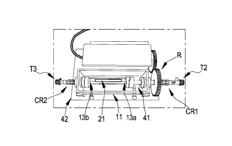
The insertion of the scaffold-holder 13 into the bioreactor 11 occurs in a
manner such that the inlet
upstream of the bioreactor coincides (Fig. 4; CR1, 41) with an end of the
scaffold (Fig. 4; 13a) and the
opening downstream of the bioreactor chamber (Fig. 4; CR2, 42) coincides with
the other end of the
scaffold (Fig. 4; 13b). In this manner, the scaffold 21 is perfectly coaxial
with respect to the perfusion path
generated by the internally hollow scaffold-holder. The larger axis according
to which the scaffold mounted
in the bioreactor is oriented is defined as the longitudinal axis.
1.2 The scaffold is preconditioned using fresh growth medium injected into the
lumen of the scaffold which
is housed in the bioreactor, using a syringe with a luer-lock connector which
is engaged to one of the two
ends of the bioreactor by means of a T-shaped connector (Fig. 9; T2).
Subsequently, the open ends of the
connectors arranged upstream and downstream of the bioreactor are closed using
caps so as to avoid the
emptying of the lumen of the scaffold. Furthermore, the fresh growth medium is
inserted into the bioreactor
chamber until the scaffold housed therein is fully covered. In this manner,
the scaffold housed in the
bioreactor chamber is preconditioned, preferably for 1 hour to 25 C, using a
fresh growth medium both
internally (in the lumen) and externally.
1.3 After preconditioning, the scaffold inside the bioreactor preconditioned
with the growth medium
injected into the lumen is emptied preferably using a sterile pipette and the
growth medium previously
introduced into the chamber is eliminated preferably using a sterile pipette.
The growth medium residues
CA 03107380 2021-01-22
WO 2020/031067
PCT/IB2019/056652
12
present in the connectors (rotated and T-shaped) arranged downstream and
upstream of the bioreactor
are eliminated with a vacuum using a pipette, without making the scaffold
collapse.
1.4 The T-shaped connectors arranged upstream (Fig. 9, T2) and downstream
(Fig. 9; T3) of the end of
the bioreactor, with the scaffold therein mounted on the grips, are directed
with the upper opening
upwards (at 90 with respect to the plane in which the bioreactor-scaffold
system lies). Subsequently, the
lateral opening of the T-shaped connectors arranged downstream (T3) and
upstream (T2) of the
bioreactor is capped. Mounted on the opening facing upwards of the T-shaped
connector (Fig. 9; T2)
arranged upstream of the bioreactor is a container, preferably a syringe (Fig.
9; 91) with a luer-lock
connector with capacity for example of 5 ml without the plunger thereof. The
opening of the T-shaped
connector (Fig. 9; T3) arranged upstream of the bioreactor remains open
instead (Figure 9).
1.5 Using a pipette, preferably a sterile plastic pipette with capacity of for
example 25 ml (Fig. 10; 101), is
drawn from a container prepared in which is a cell suspension consisting of
fresh growth medium and
endothelial cells (e.g. HUVECs). Subsequently, the drawn cell suspension is
released into the container or
into the syringe (Fig 10; 91) mounted on the T-shaped connector element (Fig.
10; T2) upstream of the
bioreactor through the element (Fig. 10; CR1). The cell suspension must be
released, using the pipette
(Fig. 10; 101), with capacity of for example 25 ml, with a continuous flow so
that the flow speed allows the
cell suspension to drip into the T-shaped connector (Fig. 10; T2) without
generating air bubbles and
pushing possible air bubbles present in the scaffold towards the opening of
the T-shaped connector T3
arranged downstream (Fig. 10; T3) of the bioreactor 11 and, hence flow out
(Figure 10).
1.6 When the cell suspension, loaded using a syringe, reaches the open end of
the T-shaped connector
T3 arranged downstream of the bioreactor without possible air bubbles, the T-
shaped connector T2
arranged upstream of the bioreactor is closed using a cap (Figure 11).
1.7 Subsequently, the syringe (91) with the cell suspension residue is rotated
by about 90 with respect to
the plane on which it lies (Figure 12); in this position the plunger of the
syringe (102) is re-inserted at the
open end of the syringe (Figure 12) by inserting the insulating black part
only so as not to create pressure
inside the scaffold. Subsequently, the syringe (91) can be unscrewed from the
T-shaped connector T2
upstream of the bioreactor without forming air bubbles (Figure 13) and the end
of the connector is closed
using a cap (Figure 14).
1.8 A hot fresh growth medium (as previously defined) is added into the
bioreactor chamber until the
seeded scaffold present in the bioreactor chamber is half-submerged in the
growth medium.
1.9 A continuous rotation is then applied along the longitudinal axis of the
scaffold, for example with a
rotation speed comprised between 1.5 and 2 rpm, for 24 hours. The rotation
allows the uniform cell
adhesion on the lumen of the scaffold, allows the scaffold to remain
continuously wet by the growth
medium present in the bioreactor chamber and it allows the through-flow of the
nutrients between the
CA 03107380 2021-01-22
WO 2020/031067
PCT/IB2019/056652
13
medium present in the chamber (outside the scaffold) and the cell suspension
one seeded in the lumen of
the scaffold.
1.10 Incubating, preferably for 24 hours at 37 C with 5% of 002, the scaffold
housed in the bioreactor
chamber (under rotation).
Advantageously, the seeding method created by the Applicant allows to operate
under sterility conditions.
Furthermore, the present seeding method subject of the present invention is
rapid, reproducible, and
advantageously allows to prepare a scaffold having the lumen surface
(internal) with endothelial cells
homogeneously and uniformly adhered along the entire length of the scaffold
(from the proximal part to the
medial part up to the distal part). The present seeding method subject of the
present invention allows to
seed the cells eliminating both the air bubbles present in the bioreactor-
scaffold system and the air
bubbles that are formed, hence avoiding to damage the cells. Basically, each
step of the present method
is standardised and reproducible and it optimises the cost and the operating
time.
Experimental evidence of the seeding method
To prove the effectiveness of the seeding method subject of the present
invention, the following analysis
were conducted.
The viability of the cells adhered to the scaffold was assessed using an assay
which uses resazurin (trade
name Alamar Blue, name IUPAC 7-hydroxy-10-oxidophenoxazin-10-ium-3-one, CAS
550-82-3) as
reagent. Such assay consists in a metabolic reaction that allows to quantify
cell viability due to the
oxidation-reduction of the indicator (resazurin) which is reduced to
resofluorine, a pink fluorescent
compound in the presence of reducing atmosphere of a vital cell. After 24
hours of adhesion, the seeded
scaffold is removed from the grips. Subsequently, the scaffold is sectioned
(cutting it) into three areas
measuring about 2cm each depending on the distance from the site of injection
of the cell suspension:
proximal, medial and distal. Subsequently, each section is divided into 4
parts measuring about 1cm2. 3
samples each representing each region (proximal, medial, distal) of the
scaffold with adhered endothelial
cells were selected for the assay with resazurin. Each sample is positioned in
a well of a 24-well dish and
incubated with 1m1 of a 0.02 mg/ml resazurin sodium salt (Sigma Aldrich,
R7017) solution with fresh
growth medium preferably for 3 hours at 37 C with 5% of 002. The reaction that
is developed between the
0.02 mg/ml resazurin sodium salt solution with fresh growth medium and the
scaffold sample (with the
adhered endothelial cells) is analysed using the A.U. (arbitrary unit of
fluorescence) detection at 590 nm
by using a spectrofluorometer.
A further analysis conducted is the assessment of the amount of genomic DNA
present in the cells
adhered on the samples of the scaffold previously used for the assay with
resazurin. The genomic DNA is
CA 03107380 2021-01-22
WO 2020/031067
PCT/IB2019/056652
14
extracted from the adhered cells by a scaffold through lysis and it is
subsequently quantified using Quant-
ifrm PicoGreenTM dsDNA Assay (P7589, Invitrogen, Molecular Probes) where the
fluorescent stain of the
nucleic acids ( PicoGreen) allows - through a standard reference curve - to
determine the concentration of
genomic DNA in solution.
In Figures 15A and 158 represented in the chart are the values obtained using
the assay with resazurin on
samples representing each region (proximal, medial, distal) of a scaffold
seeded with endothelial cells and
incubated for 24 hours in three different experiments (named DYN1, DYN2 and
DYN3). The charts in
Figures 15A and 158 show a good adhesion and viability of the endothelial
cells.
This data was confirmed by the quantification of the genomic DNA (Figures 150
and 15D) calculated
considering that the genomic DNA content of an endothelial cell is of about 7
pg.
No significant cell viability difference was observed among the various
proximal, medial and distal sections
of the scaffolds seeded with endothelial cells. In particular, cell viability
and the number thereof can be
compared along the length (main axis of the scaffold) in the proximal, medial
and distal portions thereof.
With the aim of supporting this evidence, costaining was conducted using DAPI
(4',6-Diamidino-2-
Phenylindole, Dihydrochloride, D1306, ThermoFisher scientific) and Rhodamine-
Phalloidin (R415,
ThermoFisher scientific) on samples representing each region (proximal,
medial, distal) of a scaffold
seeded with endothelial cells and incubated for 24 hours. The two DAPI and
Rhodamine-Phalloidin
reagents are specific respectively for the nuclear detection and for actin
filaments (F-actin), morphological
components of a live cell, visible after the staining using a fluorescence
microscope or a confocal
microscopy. After 24 hours of culture, these results show that the endothelial
cells are vital and distributed
on the lumen of the scaffold in a uniform fashion. In particular, these
results show a 90% cell confluence.
Furthermore, conducted on samples representing each region (proximal, medial,
distal) of a scaffold
seeded with endothelial cells and incubated for 24 hours are gene expression
analysis for markers typical
of endothelial cells: for example, the Von Willebrand factor (VWF), cluster of
differentiation 31 (CD31),
vascular cell adhesion molecule 1(VCAM-1). In order to conduct a gene
expression evaluation, the total
RNA is extracted from cells (endothelial in this case) and after reverse
transcription at cDNA is quantified
using a specific Taqman Gene Expression Assay (ThermoFisher Scientific) using
the real-time PCR
technique. Functional levels for gene expression of the markers listed
previously are indicators of good
functionality and viability of the cells adhered on the lumen of the scaffold.
Lastly, H&E "Haematoxylin and Eeosin" staining analysis is conducted on
samples of a scaffold seeded
with endothelial cells according to the present invention with the aim of
evaluating the distribution of the
cells and the morphology thereof, and an immunofluorescence assay for specific
endothelial functionality
markers.
CA 03107380 2021-01-22
WO 2020/031067
PCT/IB2019/056652
In conclusion, the present seeding method subject of the present invention
revealed to be efficient in that it
guarantees a homogeneous, uniform and reproducible seeding of vital
endothelial cells along the entire
lumen.
5 (2) Method for connecting a perfusion circuit to a bioreactor-scaffold
system (perfusion method) according
to the first embodiment (Figures 5-8, 18-21).
The connection method is based on the following sequential steps, subsequent
to seeding (method for
seeding a cell culture in the lumen of a scaffold according to the embodiment
described above under point
10 (1)) and at 24 hours from adhesion of the endothelial cells:
2.1 Place the tube or under-pump (Fig. 5; 52) of the closed perfusion circuit
under the head of the
peristaltic pump (Fig. 5; 55), which - upon activation - generates a
peristaltic force capable of suctioning
fluids, hot fresh growth medium in this case. The closed perfusion circuit is
filled due to the suctioning, by
the tube (Fig. 5; 51) of the perfusion circuit connected to the reservoir
(Fig. 5; 56), of the hot fresh growth
15 medium which is previously poured into the reservoir once closed. Fill
the tubes of the perfusion circuit
with the hot fresh growth medium until the fresh growth medium returns to the
reservoir through the tube
54 (Fig. 5; 54) of the closed perfusion circuit. Place the bioreactor-scaffold
system under the same sterility
conditions as the perfusion circuit.
2.2 Open the upper and lateral ends of the T-shaped connector T2 arranged
upstream of the bioreactor.
2.3 Upon removing the caps from the upper and lateral ends of the T-shaped
connector T2 arranged
upstream of the bioreactor, the possible creation of air bubbles is
compensated by manually adding
(preferably using a pasteur pipette) having the same volume as the hot fresh
growth medium (Fig. 16).
2.4 Occlude the tube 54 of the closed perfusion circuit, preferably using a
clamp (Fig. 17; 171) in a
position proximal to the connector between the tube 54 and the tube 53 of the
perfusion circuit (Fig. 17).
Be careful to keep the head of the pump closed so as to prevent the emptying
of the tube 53 of the closed
perfusion circuit
2.5 Keep the tube 53 of the closed perfusion circuit in vertical position,
unscrew the connector arranged
between the tube 53 and the tube 54 of the perfusion circuit and preferably
cap the tube 54 of the
perfusion circuit using a cap.
2.6 Screw the tube 53 of the perfusion circuit to the open lateral end of the
T-shaped connector T2
upstream of the bioreactor at a lateral access thereof. The connector upstream
of the bioreactor must
always be kept in vertical position (Fig. 18).
2.7 Open the T-shaped connector T3 downstream of the bioreactor by unscrewing
the cap of the lateral
opening.
CA 03107380 2021-01-22
WO 2020/031067
PCT/IB2019/056652
16
2.8 Remove the cap from the connector of the tube 54 of the perfusion circuit
(Fig. 19A) and screw it onto
the lateral opening of the T-shaped connector downstream of the bioreactor
(Fig. 198).
2.9 Remove the clamp 171 which occludes the tube 54 of the perfusion circuit
(Fig. 20).
2.10 Fill the element for the removal of the air bubbles (Fig. 21, 71),
defined with the technical expression
of bubble trap in the context of the present invention. The bubble trap
consists of an element represented
by a chamber closed using a cap and having two accesses serving as an outflow
and inflow. The bubble
trap chamber contains a volume of liquid (hot fresh growth medium in this
specific case) and a volume of
air that traps possible air bubbles present in the perfusion liquid which
flows through the two accesses of
the bubble trap chamber.
Fill the bubble trap with hot fresh growth medium so as to leave a given air
volume and close the chamber
as well as its accesses using the respective caps.
2.11 Connect the bubble trap to the perfusion circuit previously connected to
the bioreactor-scaffold
system as follows (Fig. 7):
a. close the tube 53 of the perfusion circuit using a clamp arranged
proximally to the connector which
connects the tube 53 to the lateral end of the T-shaped connector Ti (arranged
between the tube 53 and
the tube 52 of the perfusion circuit) and unscrew it (Fig. 7; 52).
b. preferably, close the tube 53 of the perfusion circuit using a cap. Such
operation prevents the emptying
of the tube 53 of the perfusion circuit.
c. cap the lateral end of the T-shaped connector Ti (arranged between the tube
53 and the tube 52 of the
perfusion circuit) and open the upper end thereof.
d. open the inflow access of the bubble trap chamber and connect it to the
access of the upper end and
arranged vertically with respect to the T-shaped connector Ti.
e. open the outflow access of the bubble trap and connect it to the tube 53 of
the perfusion circuit, being
careful not to twist the tube at all.
f. keep the bubble trap in vertical position.
g. remove the clamp 171 from the tube 53 of the perfusion circuit just
connected to the bubble trap
chamber. Start the pump and open the cap of the upper end of the T-shaped
connector T2 upstream of
the bioreactor so as to eliminate possible air bubbles formed in the tube 53
during the process preventing
them from reaching the scaffold. The peristaltic force applied by the pump to
the assembled system,
consisting of the perfusion circuit and the bioreactor-scaffold system will
allow the perfusion of the
scaffold.
h. after such verification, close the T-shaped connector T2 upstream of the
bioreactor using the cap
thereof.
CA 03107380 2021-01-22
WO 2020/031067
PCT/IB2019/056652
17
(3) Method for connecting a perfusion circuit to a bioreactor-scaffold system
(perfusion method) according
to a second preferred embodiment (Figures 22-27).
The connection method is based on the following sequential steps, subsequent
to seeding (method for
seeding a cell culture in the lumen of a scaffold according to the embodiment
described above under point
(1)) and at 24 hours from adhesion of the endothelial cells:
3.1) Connect - under sterility conditions - the tubes of the perfusion circuit
(Fig. 22; 51; 52; 53; 54; 55), the
element for removing the air bubbles (Fig. 22; BT), defined in the context of
the present invention with the
technical expression bubble trap, and the reservoir (Fig. 22; 56). The element
for removing the air bubbles
or bubble trap (Bt) consists of an element represented by a closed chamber,
preferably made of glass,
using a cap and having two asymmetric accesses: the access with the tap and
connecting nozzle serves
as an inflow (Fig. 27, 211) while access with the connecting nozzle only
serves as an outflow (Fig. 27,
221). The bubble trap chamber contains a volume of liquid (hot fresh growth
medium in this specific case)
and a volume of air that traps possible air bubbles present in the perfusion
liquid which flows through the
two accesses of the bubble trap chamber.
3.2) Place the under-pump (Fig. 22; 52) of the closed perfusion circuit under
the head of the peristaltic
pump (Fig. 22; 57), which - upon activation - generates a peristaltic force
capable of suctioning fluids, hot
fresh growth medium in this case. The closed perfusion circuit is filled due
to the suctioning, by the tube
(Fig. 22; 51) of the perfusion circuit connected to the reservoir (Fig. 22;
56), of the hot fresh growth
medium which is previously poured into the reservoir (Fig. 22; 56) in turn
closed. Fill all the tubes of the
perfusion circuit, the bubble trap (Fig. 22, BT) and the reservoir (Fig. 22;
56) with a liquid perfusion, such
as a hot fresh growth medium (as defined above), until the hot fresh growth
medium returns to the
reservoir through the tube 55 (Fig. 22; 55) of the closed perfusion circuit.
The BT and the reservoir are
filled so as to leave a given air volume. In particular, the bubble trap (Fig.
22, BT) is filled so that said
bubble trap chamber has a first part of the volume thereof filled with said
fresh growth medium and a
second part of the volume thereof filled with air, said second part of said
volume having the function of
trapping the air bubbles present in the perfusion liquid (hot fresh growth
medium) which flows through said
access serving as an inflow (211) and said access serving as an outflow (212)
of the bubble trap (BT) (Fig.
22).
3.3) Occlude the tube 54 (Fig 23), preferably using a clamp (Fig. 23; 172) in
a position proximal to the BT
and move the tap of the BT to the closing position (in perpendicular position
with respect to the tubes of
the perfusion circuit).
3.4) Place the bioreactor-scaffold system under the same sterility conditions
as the perfusion circuit.
CA 03107380 2021-01-22
WO 2020/031067
PCT/IB2019/056652
18
3.5) Occlude the tube 55 (Fig. 23), preferably using a clamp (Fig. 23; 171;
Fig. 17; 171) in a position
proximal to the connection C with the tube 54 (Fig. 23; C). Open the upper and
lateral ends of the T-
shaped connector 12 arranged upstream of the bioreactor (Fig. 4; 12).
3.6) Upon removing the caps from the upper and lateral ends of the T-shaped
connector 12 (Fig. 4)
arranged upstream of the bioreactor, the possible creation of air bubbles is
compensated by manually
adding (preferably using a pasteur pipette) having the same volume as the hot
fresh growth medium (Fig.
16).
3.7) Keep the tube 54 (Fig. 23) of the closed perfusion circuit in vertical
position, unscrew the connector
arranged between the tube 54 (Fig. 23) and the tube 55 (Fig. 23) of the
perfusion circuit and preferably
cap the tube 55 of the perfusion circuit using a cap (Fig. 24).
3.8) Screw the tube 54 (Fig. 27) of the perfusion circuit to the open lateral
end of the T-shaped connector
12 (Fig. 27) upstream of the bioreactor at a lateral access thereof. The
connector upstream of the
bioreactor must always be kept in vertical position (Fig. 18 or 27).
3.9) Open the T-shaped connector 13 (Fig. 4) downstream of the bioreactor by
unscrewing the cap of the
.. lateral opening.
3.10) Unscrew the rotary connector CR2 together with the T-shaped connector 13
(Fig. 4), holding the
toothed wheel R (Fig. 4) still and screw the connector of the tube 55 (Fig.
25) on the lateral opening of the
scaffold-holder 14a (Fig. 1) (Fig. 25).
3.11) Remove the clamp 171 which occludes the tube 55 of the perfusion circuit
(Fig. 26).
3.12) Position the bioreactor-seeded scaffold system connected to the
perfusion circuit at about 37 C and
at about 5% of 002, place the under-pump 52 (Fig. 27) in free position, open
the tap of the bubble trap BT
(Fig. 27) by positioning it parallel to the tubes of the perfusion circuit,
remove the clamp 172 (Fig. 27) and
then start the pump (Fig. 27, 57). The peristaltic force applied by the pump
to the assembled system,
consisting of the perfusion circuit and the bioreactor-scaffold system will
allow the perfusion of the
scaffold.
Advantageously, the connection method (perfusion method) described herein,
both in the first embodiment
and in the second embodiment described above, allows to connect a perfusion
circuit of a scaffold,
preferably tubular, to the bioreactor-seeded scaffold system. This procedure
allows to prevent the
formation of air bubbles and prevents the bubbles, should they be formed, from
reaching the scaffold
seeded with endothelial cells of the bioreactor-scaffold system. Furthermore,
the air bubbles possibly
already present in the perfusion circuit do not reach the scaffold due to the
presence of the bubble trap
(Fig. 21, 71; Fig. 27, BT) in the circuit. In this manner, the method created
by the Applicant is capable of
ensuring complete absence of air bubbles in the lumen of the scaffold. This
system meets the
requirements set forth by the configuration of the bioreactor. All details
indicated and described are
required to make the method independent from the operator and thus for
ensuring the reproducibility of the
CA 03107380 2021-01-22
WO 2020/031067
PCT/IB2019/056652
19
results during the in vitro generation of a construct with functional
endothelium at industrial level. In
addition, this method allows to be able to operate under sterility conditions,
being based on simple actions.
Furthermore, the quick and traceable procedure allows to reduce the risk that
the air bubbles come into
contact with the cells, thus avoiding damaging the endothelial cells adhered
on the lumen of the scaffold,
preferably tubular. Said perfusion method allows to obtain engineered vascular
constructs/tissues having a
scaffold having a lumen covered with a continuous (i.e. having a monolayer of
confluent cells) and
functional endothelium.
Experimental evidence of the connection method
The experimental analysis regarding the evaluation of the method for
connecting the perfusion circuit to
the bioreactor-scaffold system are the same ones applied for the evaluation of
the seeding method of a
cell culture in a scaffold preferably tubular.
In the context of the present invention the perfusion circuit (Fig. 5 and Fig.
22) is defined as an assembly
of: tubes (Fig. 5; 51- 54 or Fig. 22; 51-55), a reservoir (Fig. 5 or Fig. 22;
56), a peristaltic pump (Fig. 5; 55
or Fig. 22; 57) and an element for removing the air bubbles (Fig. 21, 71 or
Fig. 22, BT). Said element for
removing the air bubbles can be present in the perfusion circuit prior to the
connection of the perfusion
circuit to the bioreactor-seeded scaffold system (second embodiment of the
perfusion method) or,
alternatively, it may be inserted into the perfusion circuit after the
connection of the perfusion circuit to the
bioreactor-seeded scaffold system (first embodiment of the perfusion method).
Said tubes are made of
biocompatible material and are connected to each other so as to allow the
perfusion of the scaffold (Fig.
21 and Fig. 27), preferably tubular, housed in the bioreactor 11, by means of
the peristaltic pump (Fig. 5;
55, Fig. 22; 57) (in such case, Masterflex , L/S Digital Dispensing Pump
Drives 07551-20, Cole-Parmer)
with the Easy-Load II 77200-62 (Masterflex, Cole-Parmer) head. With reference
to then second
embodiment of the perfusion method described above and illustrated in figure
27, the perfusion circuit
mainly consists of five tubes with an inner diameter of 3/16": a first tube 51
for suctioning from the
reservoir, a second under-pump tube 52, a third tube 53 which connects the
circuit to the bubble trap BT,
a fourth tube 54 which connects the BT to the T-shaped connector T2 upstream
of the bioreactor-scaffold
system, a fifth tube 55 for return to the reservoir 56 connected to the T-
shaped connector T3 downstream
of the bioreactor-scaffold system.
With reference to the first embodiment of the perfusion method described above
and represented in figure
5, the tube 51 is connected to the under-pump tube 52, the under-pump tube 52
to the BT, the BT to the
tube 53, the tube 53 to the T-shaped connector T2 upstream of the bioreactor-
scaffold system, the tube 54
to the T-shaped connector T3 downstream of the bioreactor-scaffold system and
to the reservoir 56.
CA 03107380 2021-01-22
WO 2020/031067
PCT/IB2019/056652
The reservoir (Fig. 5 or Fig. 22, 56) is the element containing the hot fresh
growth medium (for example
Endothelial Growth Medium EGM, Sigma Aldrich) from which the tube 51 (Fig. 5
or Fig. 22) suctions and
to which the tube 54 (Fig. 5) or 55 (Fig. 22) returns, keeping the entire
bioreactor-seeded scaffold system
in a closed system. The reservoir (Fig. 5 or Fig. 22, 56) is under atmospheric
pressure, due to a 0.22 pm
5 filter present in the cap of the reservoir, which guarantees the
sterility of the air.
Also forming an object of the present invention are the following preferred
embodiments RPn, as indicated
below.
RP1. A process for producing vascular tissues, preferably scaffold (Fig. 2;
21), for testing medicinal
10 products, said process comprises applying:
- a method for seeding an endothelial cell culture in the lumen of a
scaffold (Fig. 2; 21) to obtain a seeded
scaffold; said seeded scaffold being present in a bioreactor (Fig. 4; 11), to
obtain a bioreactor-seeded
scaffold system and, subsequently,
- a method for perfusion of the endothelial cells present in the lumen of
said seeded scaffold; said
15 perfusion method being obtained by connecting a perfusion circuit (Fig.
6; 51, 52, 53, 54, 55, and 56) to
said bioreactor-seeded scaffold system;
RP2. The process according to claim RP1, wherein said method for seeding an
endothelial cell culture in
the lumen of a scaffold comprises:
- mounting a scaffold (21), preferably an electrospun silk fibroin tubular
scaffold, on the grips of a scaffold-
20 holder (Fig. 1; 13, 13a, 13b) and housing said scaffold-holder (13, 13a,
13b) with the scaffold (21) in the
bioreactor chamber (11), to obtain a bioreactor-scaffold system (Fig. 4; 11,
21);
- injecting a fresh growth medium into the lumen of said scaffold (21)
fixed on said scaffold-holder (13)
arranged inside the bioreactor chamber (11);
- adding said fresh growth medium into the bioreactor chamber (11) where
said scaffold-holder (13) with
the scaffold (21) is present already injected with said growth medium;
- leaving, preferably for a time interval comprised between 1 hour and 18
hours at a temperature
comprised between 20 C and 30 C, preferably 25 C, said growth medium in the
lumen of the scaffold
(21) and in the bioreactor chamber (11) where said scaffold-holder (13) with
the scaffold (21) is present
already injected with said growth medium;
- clearing the internal of the lumen of the scaffold (21) and of the
bioreactor chamber of the culture
medium;
- releasing a cell suspension consisting of said fresh growth medium and
endothelial cells of the HUVECs
type into a container of the syringe type (Fig. 10; 91) mounted on the
connector element (T2) arranged
upstream of the bioreactor (11) by means of the element (CR1); said cell
suspension is released into the
lumen of the scaffold (21) present in the bioreactor chamber (11) with a
continuous flow so that the flow
CA 03107380 2021-01-22
WO 2020/031067
PCT/IB2019/056652
21
speed allows said cell suspension to drip into the container (12) without
generating the air bubbles and
pushing any air bubbles present in the lumen of the scaffold (21), towards the
opening of the connector
(13) arranged downstream of the bioreactor (11) allowing the outflow thereof;
- adding said fresh growth medium into the bioreactor chamber (11) where
said scaffold-holder (13) with
the scaffold (21) is present containing said cell suspension therein;
- incubating, preferably for 24 hours at 37 C in presence of 5% of 002, the
scaffold (21) housed in the
bioreactor chamber (11).
RP3. The process according to RP1 or RP2, wherein said method for the
perfusion of the endothelial cells
present in the lumen of said seeded scaffold comprises:
- preparing a closed perfusion circuit (Fig. 5) comprising the tubes (Fig.
5; 51, 52, 53, 54, 55 and 56);
- occluding the tube (54) of the perfusion circuit using a closing element
of the clamp type (Fig. 17; 171) in
a position proximal to the connector (C);
- unscrewing the connector (C) arranged between the tube 53 and the tube 54
(Fig. 5; Fig. 17) of the
perfusion circuit;
- screwing the tube (53) of the perfusion circuit to the open lateral end
of the connector (12) upstream of
the bioreactor (Fig. 18) at a lateral access thereof;
- opening the connector (13) downstream of the bioreactor and unscrewing
the cap of the lateral opening
(Fig. 19a);
- connecting the tube (54) of the perfusion circuit to the lateral opening of
the connector (13) arranged
downstream of the bioreactor (Fig. 19b) and removing the closing element of
the clamp type (Fig. 20;
171).
RP4. The process according to RP3, wherein an element (71) represented by
chamber closed using a
cap (72) and having two accesses serving as an inflow (211) and as an outflow
(212) of the bubble-trap
type (Fig. 21) is inserted into the tube (53) of the perfusion circuit (Fig.
8; Fig. 21); said chamber has a
volume where a first part thereof is filled with a hot fresh growth medium and
where a second part thereof
is filled with air so as to trap - in the latter second part of volume -
possible air bubbles present in the
perfusion liquid which flows through said accesses (211 and 212).
RP5. The process according to any one of RP1-4, wherein the scaffold,
preferably a tubular scaffold, is
selected from among polymeric scaffolds of synthetic or natural origin formed
by only one polymer or by
copolymers, such as for example electrospun silk fibroin or copolymers of
polyglycolic acid/polylactic acid
(PGA/PLA) or polyglycolic acid/polycaprolactone (PGA/PCL).
CA 03107380 2021-01-22
WO 2020/031067
PCT/IB2019/056652
22
RP6. The process according to any one of RP1-5, wherein the endothelial cells
are selected from among
the cells that form an endothelium of a vascular tissue, such as for example
HAOECs (human aortic
endothelial cells), HCAECs (human coronary artery endothelial cells), HMEVECs
(human dermal
microvascular endothelial cells) or HUVECs (human umbilical vein endothelial
cells).
RP7. The process according to any one of RP1-6, wherein the growth medium used
is the Endothelial
Growth Medium (EGM, Sigma Aldrich, 211-500), preferably heated to 37 C.
RP8. A scaffold having a lumen covered with a continuous and functional
endothelium obtained by means
of the process according to any one of RP1-7.
RP9. Use of the scaffold according to RP8, to conduct in vitro preclinical and
clinical tests of a medicinal
product to be used in the cardiovascular and peripheral vascular region, such
as for example, heart
valves, stents, grafts, catheters.
The first phase to be carried out and optimised is the cell seeding phase,
preferably endothelial cells,
followed by a second critical phase of connecting the perfusion circuit to the
system comprising the
bioreactor and the scaffold, so as to ensure reliability, effectiveness and
reproducibility to the
industrialisation process for the in vitro generation of a continuous and
functional endothelium.
The success of the industrialisation process subject of the present invention
for the production of the
engineered vascular tissue/construct, preferably a scaffold having a lumen
covered with a functional and
continuous endothelium having a confluent cell monolayer, mainly consists in
the success relating to the
phase of seeding and connecting the perfusion circuit to the bioreactor-
scaffold system, so as to globally
guarantee the elimination of air bubbles in a reliable and reproducible
manner. The seeding method
depends on the cell source and on the density thereof, on the chemical and
porosity properties and on the
full removal of the air bubbles from the lumen of the scaffold during the
injection of the cell suspension. On
the other hand, the method for connection between the perfusion circuit and
the bioreactor-scaffold
system is based on maintaining the sterility of the assembled system and on
the guarantee of absence of
air bubbles that can come into contact with the seeded scaffold. It should be
observed that the formation
of air bubbles must be avoided given that the air bubbles can damage the
cells, jeopardising the viability
thereof with ensuing lack of endothelisation of the scaffolds.
The description of the present invention shows that the choice of the method
for connecting the perfusion
circuit to the bioreactor-scaffold system depends on the method for seeding
the previously optimised
endothelial cells due to the fact that said connection method must be suitable
to the experimental setup
and to the perfusion needs and the position chosen for this system in the
incubator.
CA 03107380 2021-01-22
WO 2020/031067
PCT/IB2019/056652
23
The process for seeding a scaffold, preferably a tubular scaffold, is one of
the factors crucial towards in
vitro generation of functional engineered vascular constructs, using confluent
endothelium, as shown in
the description of the present invention. This process is responsible for a
uniform and homogeneous
distribution of endothelial cells in the lumen same case applying to the
adhesion of the cells to the surface.
The description of the present invention shows that the choice of the most
appropriate seeding method,
adapting it to each bioreactor-scaffold system, defines the reproducibility of
the process, showing the
advantages thereof in the reproducibility of the results.
Also in preclinical tests, as well as others, there arises the need for
creating reproducible methods for the
large-scale production of functional vascular constructs.
Thus, the seeding method of the present invention, well defined and traceable,
guarantees a highly
uniform distribution in terms of adhesion of endothelial cells and good
reproducibility of the results,
required for a laboratory whose activity focuses on the production of in vitro
vascular constructs as test
models of the preclinical field as well as other fields.
After 24 hours of adhesion, the cells adhered in the lumen of the scaffold,
mainly tubular, must maintain
their morphology and viability so as to obtain a homogeneous vascular
endothelium. Thus, it is important
to avoid any cell alteration during the connection process which could alter
the state of cell adhesion, with
possible loss of the vascular layer subject of growth (being formed).
.. The known methods for connection between the bioreactor and the perfusion
circuit cause cell suffering
with ensuing detachment - even partial - of the endothelial cells from the
luminal surface, so that the
formation of a functional endothelial layer is slowed or hindered.
Furthermore, the known methods for
connection between the bioreactor and the perfusion circuit do not guarantee
the absence of air bubbles,
that may be formed due to possible torsions or compressions (full or partial)
of the connection tubes during
the perfusion. It is important to absolutely prevent the contact between said
air bubbles and the seeded
scaffold which is avoided by introducing an element, for example a bubble-trap
capable of eliminating the
air bubbles before they reach the seeded scaffold. This drawback was
successfully overcome thanks to
the process subject of the present invention which allows to obtain an inner
surface of the scaffold covered
with a uniform and functional layer of endothelial cells, in particular a
confluent cell layer).
Preferred embodiments En of the present invention are described below:
El. An in-vitro model of a substantially tubular-shaped vascular structure
having dysfunctional anatomical
and physiological characteristics simulating the same vascular structure of a
healthy subject whose
.. vascular structure has been damaged or deformed or deteriorated due to a
damage selected from among
CA 03107380 2021-01-22
WO 2020/031067
PCT/IB2019/056652
24
the group comprising or, alternatively, consisting of aneurysm, stenosis,
sclerosis plaques, forms of
tumours or cardiomyopathies;
wherein said model comprises or, alternatively, consists of one or more
biocompatible porous polymeric
supports (scaffolds) capable of promoting a cell adhesion and growth, wherein
said scaffold is seeded with
endothelial cells which cover a lumen of the scaffold and constitute an
endothelium having a single layer
of confluent cells, said scaffold being provided with deformities or defects
on a tubular structure thereof.
E2. The in vitro model according to El, wherein said vascular structure is
selected from among blood
vessels or blood ducts or central or peripheral circulatory system valves;
preferably arteries, veins,
capillaries, aortic or mitral valve.
E3. The in vitro model according to El or E2, wherein said vascular structure
is a synthetic vascular
structure, and wherein said scaffold consists of electrospun silk fibroin,
copolymers of polyglycolic
acid/polylactic acid (PGA/PLA) or copolymers of polyglycolic
acid/polycaprolactone (PGA/PCL).
E4. The in vitro according to any one of El -E3, wherein said deformities or
said defects of the tubular
structure comprise bifurcations, curvatures, elbows, constrictions,
dilatations or combinations thereof.
E5. A method for testing a medical device or a drug so as to verify the
effectiveness and safety thereof
before the in-vivo use thereof on the man or animal, said method comprising
the following steps:
- preparing a substantially tubular-shaped scaffold having the
dysfunctional anatomical and physiological
characteristics suitable to simulate a damage or a deformation or a
deterioration due to an aneurysm,
stenosis, sclerosis plaques, forms of tumours or cardiomyopathies;
- seeding at least one part of the interior lumen of said scaffold with
endothelial cell lines so as to obtain a
continuous and homogeneous layer of seeded endothelial cells (seeding method),
optionally seeding at
least one part of the outer surface of said scaffold with muscle cell lines;
- promoting the growth of said endothelial cells, and optionally said
muscle cells, up to obtaining a
continuous and uniform layer of endothelial cells, to obtain said in vitro
model;
- introducing into said in vitro model a medical device or a drug subject
of test, and
- allowing the circulation (perfusion model) in said in-vitro model comprising
said medical device or drug of
a human whole blood sample, artificial blood or derivatives thereof so as to
evaluate the behaviour and the
interaction of said medical device or drug with said human whole blood sample,
artificial blood or
derivatives thereof.
CA 03107380 2021-01-22
WO 2020/031067
PCT/IB2019/056652
E6. A method or process for the production of an engineered vascular tissue or
construct, preferably a
scaffold (21) having a lumen covered with functional and continuous
endothelium having a confluent cell
monolayer, for testing medical or veterinarian products, said process
comprising applying:
- a method for seeding an endothelial cell culture in the lumen of a
scaffold (21) to obtain a seeded
5 scaffold (21); said seeded scaffold (21) being present in a bioreactor
(11), to obtain a bioreactor (11)-
seeded scaffold (21) system;
wherein said seeding method comprises the steps of:
- releasing said endothelial cell culture in form of a cell suspension
comprising a fresh growth medium and
endothelial cells in a container (91) mounted on a T-shaped connector (12)
arranged upstream of the
10 bioreactor (11) by means of a rotary connector (CR1); followed by
- releasing said endothelial cell culture in the lumen of the scaffold (21)
present in the bioreactor chamber
(11) with a continuous flow such that the flow speed allows said cell
suspension to drip into the T-shaped
connector (12) without generating air bubbles and pushing the air bubbles
present in the lumen of the
scaffold (21) towards an opening of a T-shaped connector (13) arranged
downstream of the bioreactor
15 (11) allowing the outflow thereof;
and, subsequently,
- a method for perfusion - with a fresh growth medium having a temperature
comprised in the range
between 30 C and 45 C, preferably at 37 C - of the endothelial cells present
in the lumen of said seeded
scaffold (21); said perfusion method being obtained by connecting a perfusion
circuit (51-56) or (51-57 and
20 BT) to said bioreactor (11)-seeded scaffold (21) system;
wherein said perfusion method comprises a step of
- partly filling an element for removing the air bubbles (71) or (BT)
present in the perfusion circuit with said
fresh growth medium, wherein said element for removing the air bubbles (71) or
(BT) comprises a
chamber, a cap that closes said chamber, an access with inflow function (211)
and an access with outflow
25 function (212), wherein said chamber of the element for removing the air
bubbles (71 or BT) has a volume
and wherein a first part of said volume is filled with said fresh growth
medium and wherein a second part
of said volume is filled with air, said second part of said volume having the
function of trapping the air
bubbles present in said fresh growth medium which flows through said access
with inflow function (211)
and said access with outflow function (212).
E7. The process according to E5 or E6, wherein said method for seeding said
endothelial cell culture in
the lumen of said scaffold (21) comprises:
- mounting the scaffold (21), preferably an electrospun silk fibroin
tubular scaffold, on the grips of a
scaffold-holder (13, 13a, 13b) and housing said scaffold-holder (13, 13a, 13b)
with the scaffold (21) in the
bioreactor chamber (11), to obtain a bioreactor(11)-scaffold (21) system;
CA 03107380 2021-01-22
WO 2020/031067
PCT/IB2019/056652
26
followed by
- injecting the fresh growth medium into the lumen of said scaffold (21)
fixed on said scaffold-holder (13)
arranged inside the bioreactor chamber (11); followed by
- adding said fresh growth medium into the bioreactor chamber (11) where
said scaffold-holder (13, 13a,
13b) with the scaffold (21) is present injected with said growth medium;
followed by
- leaving for a time interval comprised between 1 hour and 18 hours at a
temperature comprised between
20 C and 30 C, preferably 25 C, said growth medium in the lumen of the
scaffold (21) and in the
bioreactor chamber (11) where said scaffold-holder (13) with the scaffold (21)
is present injected with said
growth medium; followed by
- clearing the internal of the lumen of the scaffold (21) and of the
bioreactor chamber (11) of the growth
medium; followed by
- releasing said endothelial cell culture in said container (91) according
to claim 1, preferably said
container (91) is a syringe; followed by
- releasing said cell suspension in the lumen of the scaffold (21)
according to claim 1; followed by
- adding said fresh growth medium in the bioreactor chamber (11) where said
scaffold-holder (13) with the
scaffold (21) is present seeded containing said cell suspension in the lumen;
and followed by
- incubating, preferably for 24 hours at 37 C in presence of 5% of CO2, the
scaffold (21) housed in the
bioreactor chamber (11).
E8. The process according to any one of E5-E7, wherein said method for the
perfusion of the endothelial
cells present in the lumen of said seeded scaffold (21) comprises:
- preparing said closed perfusion circuit comprising the tubes (51), (52),
(53), (54), and, optionally, (55);
- occluding the tube (54) or (55) of the perfusion circuit using a closing
element (171) in a position proximal
to a connector (C), preferably said closing element is a clamp or the like;
followed by
- unscrewing the connector (C) arranged between the tube (53) or (54) and the
tube (54) or (55)
respectively in the perfusion circuit;
- screwing the tube (53) or (54) of the perfusion circuit to an open
lateral end of the T-shaped connector
(T2) upstream of the bioreactor (11) at a lateral access thereof; followed by
- opening the T-shaped connector (T3) downstream of the bioreactor (11) and
unscrewing a cap of a
lateral opening of the T-shaped connector (T3); followed by
- connecting the tube (54) or (55) of the perfusion circuit to the lateral
opening of the T-shaped connector
(T3) arranged downstream of the bioreactor (11) and removing the closing
element (171);
followed, if need be, by
- inserting - between the tube (53) and the under-pump tube (52) of the
perfusion circuit - the element for
removing the air bubbles (71).
CA 03107380 2021-01-22
WO 2020/031067
PCT/IB2019/056652
27
E9. The process according to any one of E6-E8, wherein the element for
removing the air bubbles (71) or
(BT) is a bubble-trap or the like.
E10. The process according to any one of E5-E9, wherein the scaffold (21),
preferably a tubular scaffold,
.. is selected from among polymeric scaffolds of synthetic or natural origin,
wherein said polymeric scaffolds
are formed by only one polymer or by copolymers, preferably electrospun silk
fibroin or copolymers of
polyglycolic acid/polylactic acid (PGA/PLA) or copolymers of polyglycolic
acid/polycaprolactone
(PGA/PCL).
.. Eli. The process according to any of E5-E10, wherein the endothelial cells
are selected from among the
cells that form an endothelium of a vascular tissue, preferably HAOECs (human
aortic endothelial cells),
HCAECs (human coronary artery endothelial cells), HM EVECs (human dermal
microvascular endothelial
cells) or HUVECs (human umbilical vein endothelial cells).
.. E12. The process according to any one of E6-Ell, wherein the growth medium
used is the Endothelial
Growth Medium comprising fetal bovine serum (2%), adenine (0.2 pg/ml),
ammonium metavanadate
(0.0006 pg/ml), amphotericin B (0.3 pg/ml), calcium chloride 2H20(300 pg/ml),
choline chloride (20 pg/ml),
copper sulphate 5H20 (0.002 pg/ml), trioptic acid DL-6,8(0.003 pg/ml), folinic
acid (calcium) (0.6 pg/ml),
heparin (4 pg/ml), hydrocortisone (2 pg/ml), L-aspartic acid (15 pg/ml), L-
cysteine (30 pg/ml), L-tyrosine
(20 pg/ml), manganese sulphate monohydrate (0.0002 pg/ml), ammonium molybdate
4H20 (0.004 pg/ml),
nicotinamide (8 pg/ml), nickel chloride 6H20 (0.0001 pg/ml), penicillin (60
pg/ml), phenol red sodium salt
(15 pg/ml), potassium chloride (300 pg/ml), putrescine dihydrochloride (0.0002
pg/ml), pyridoxine
hydrochloride (3 pg/ml), sodium metasilicate 9H20 (3 pg/ml), sodium sulphate
7H20 (200 pg/ml), sodium
selenite (0.01 pg/ml), streptomycin sulphate (100 pg/ml), thiamine
hydrochloride (4 pg/ml) and zinc
.. sulphate 7H20 (0.0003 pg/ml), preferably heated to 37 C.
E13. A scaffold (21) having a lumen coated with a functional and continuous
endothelium (21) having a
confluent cell monolayer obtained by means of a process comprising the
following steps:
- preparing a substantially tubular-shaped scaffold having the
dysfunctional anatomical and physiological
characteristics suitable to simulate a damage or a deformation or a
deterioration due to an aneurysm,
stenosis, sclerosis plaques, forms of tumours or cardiomyopathies;
- seeding at least one part of the interior lumen of said scaffold with
endothelial cell lines so as to obtain a
continuous and homogeneous layer of seeded endothelial cells (seeding method),
optionally seeding at
least one part of the outer surface of said scaffold with muscle cell lines;
CA 03107380 2021-01-22
WO 2020/031067
PCT/IB2019/056652
28
- promoting the growth of said endothelial cells, and optionally said muscle
cells, up to obtaining a
continuous and uniform layer of endothelial cells, to obtain said in vitro
model;
preferably wherein said scaffold (21) can be used in an in vitro model
according to one from E1-E4; more
preferably wherein said process comprises the characteristics of any one from
E6-E12.
E14. Use of the scaffold (21) according to any one of E1-E4 or E13, for
conducting in vitro preclinical or
clinical tests of a medicinal product for human use or of a veterinarian
product for animal use to be used in
the cardiovascular and peripheral vascular region, preferably valves, heart
valves, stents, grafts,
catheters, bandages or nets.
CA 03107380 2021-01-22
WO 2020/031067
PCT/IB2019/056652
29
LIST OF REFERENCE NUMBERS
11 bioreactor
13 scaffold-holder
13a scaffold-holder grip
13b scaffold-holder grip
14a lateral opening of the scaffold-holder
21 scaffold
41 inflow of the bioreactor chamber
42 outflow of the bioreactor chamber
51 tube
52 tube or under-pump
53 tube
54 tube
55 head of the peristaltic pump
56 reservoir
57 pump
71 element for removing air bubbles (or bubble trap)
72 cap
91 container, preferably syringe
101 pipette
102 syringe plunger
171 clamp
172 clamp
211 access with inflow function
212 access with outflow function
BT bubble trap
CR1 rotary connector
CR2 rotary connector
Ti T-shaped connector
12 T-shaped connector
13 T-shaped connector