Note: Descriptions are shown in the official language in which they were submitted.
CA 03107502 2021-01-21
WO 2020/041065
PCT/US2019/046493
EPITHELIAL CELL SPHEROIDS
Related Patent Applications
This patent application claims the benefit of U.S. provisional patent
application no. 62/720,010 filed
on August 20, 2018, entitled EPITHELIAL CELL SPHEROIDS, naming Chengkang Zhang
as
inventor, and designated by attorney docket no. PPG-2006-PV. This patent
application also claims
the benefit of U.S. provisional patent application no. 62/726,580 filed on
September 4, 2018,
entitled EPITHELIAL CELL SPHEROIDS, naming Chengkang Zhang as inventor, and
designated
.. by attorney docket no. PPG-2006-PV2. The entire content of the foregoing
applications is
incorporated herein by reference, including all text, tables and drawings, for
all purposes.
Field
The technology relates in part to epithelial cell spheroids and methods of
producing epithelial cell
spheroids.
Background
Organs such as lung, kidney, liver, pancreas and skin can be characterized by,
among other
things, the presence of epithelia made of organ-specific epithelial cells.
Often, epithelial cells are
defined by one or more specific functions of each such organ. Specific
functions may include, for
example, gas exchange in the lung, filtration in the kidney, detoxification
and conjugation in the
liver, endocrine (e.g., insulin) production in the pancreatic islet cells or
protection against
hazardous conditions in the environment by the skin.
Epithelial cells may be directly attached to each other by cell-cell
junctions, where cytoskeletal
filaments are anchored, to form epithelia. Often, epithelia are anchored to
other tissue on one side
(i.e., the basal side) and generally are free of such attachment on their
opposite side (i.e., the
apical side). A basal lamina (or basement membrane) lies at the interface with
underlying tissue,
mediating the attachment. For certain tissues, the apical side of an
epithelium generally is
exposed to the environment. Access to the apical membrane of an epithelial
cell is useful for
various biological function studies, e.g., the interaction between infectious
agents such as viruses
and the epithelial cells.
1
CA 03107502 2021-01-21
WO 2020/041065
PCT/US2019/046493
An air-liquid interface (ALI) culture is an in vitro method currently used for
creating an apical-basal
polarized epithelial tissue model. Generally, epithelial cells are plated on a
porous support in cell
culture medium and allow to grow to confluence. The culture medium is then
removed from the top
side of the porous plastic support to expose the cells to air, which induces
the cells to form apical-
basal polarized epithelium. The side that is exposed to air becomes the apical
side, and the side
that is attached to the porous support becomes the basal side. Epithelial cell
ALI culture can be a
useful in vitro tool for a variety of studies, e.g., safety profile of
compound exposure, infectivity of
viruses, trans-epithelium drug delivery, and other cellular functions such as
water and solute
transport across an apical-basal polarized epithelium. A long in vitro
maturation/differentiation
process (generally a few weeks to over 1 month), a dependence on porous
membrane inserts
(e.g., 6-, 12-, 24- or 96-well) format, and a lack of suitable methods for
cryopreserying mature ALI
cultures, however, pose significant challenges for employing ALI culture in
high-throughput assays
to satisfy increasing demands for a physiologically-relevant assay model.
For certain applications, organoid culture protocols may be used, which can
support the formation
of apical-basal polarized epithelium in culture. Generally, individual
epithelial cells are inoculated
into thick extracellular matrix (e.g., MatrigelTm), and cultured in medium to
form epithelial organoids.
The organoids typically are hollow enclosures lined by epithelial cells with
the apical side facing
inwards (away from the extracellular matrix), and the basal side connected to
the extracellular
matrix. This format precludes access to the apical side of the cells without
penetrating the
organoids. Retrieving organoids out of Matrigel TM can be difficult and often
depends on the use of
a protease, and cryopreseryation of organoids is not commonly practiced.
Epithelial bodies (e.g., spheroids) made of epithelial cells having their
apical sides facing outwards,
readily obtainable from culture media, and amenable to cryopreseryation would
be useful for a
variety of functional studies (e.g., toxicology studies, drug delivery,
disease models, infectious
agent analysis, and the like), and medical applications such as cell therapy.
Summary
Provided herein in certain aspects are methods for producing a cellular
spheroid comprising (a)
aggregating a plurality of epithelial cells under aggregation conditions,
thereby forming a cellular
aggregate, where the epithelial cells comprise an apical membrane and a basal
membrane; and
2
CA 03107502 2021-01-21
WO 2020/041065
PCT/US2019/046493
(b) culturing the cellular aggregate under spheroid-inducing culture
conditions, thereby generating
a cellular spheroid where (i) the spheroid comprises an interior and an
exterior, and (ii) for some or
all of the epithelial cells in the spheroid, the basal membrane is in the
spheroid interior and the
apical membrane is on the spheroid exterior.
Also provided herein in certain aspects are methods for producing a cellular
spheroid comprising
(a) aggregating a plurality of epithelial cells under aggregation conditions,
thereby forming a
cellular aggregate, where the epithelial cells comprise an apical membrane and
a basal
membrane; and (b) culturing the cellular aggregate under spheroid-inducing
culture conditions,
thereby generating a cellular spheroid where (i) the spheroid comprises an
interior and an exterior,
and (ii) for some or all of the epithelial cells in the spheroid, the basal
membrane is in the spheroid
interior and the apical membrane is on the spheroid exterior; where the
aggregation conditions
and/or the spheroid-inducing culture conditions comprise one or more
transforming growth factor
beta (TGF-beta) inhibitors and one or more cytoskeletal structure modulators.
Also provided herein in certain aspects are methods for producing a cellular
spheroid comprising
(a) attaching one or more epithelial cells to a substrate under substrate
attachment conditions,
thereby forming a cell-substrate body, where the one or more epithelial cells
comprise an apical
membrane and a basal membrane; and (b) culturing the cell-substrate body under
spheroid-
inducing culture conditions, thereby generating a cellular spheroid where
(i) the spheroid comprises an interior and an exterior, and (ii) for some or
all of the epithelial cells in
the spheroid, the basal membrane is in the spheroid interior and the apical
membrane is on the
spheroid exterior.
Also provided herein in certain aspects are methods for producing a cellular
spheroid comprising
(a) attaching one or more epithelial cells to a substrate under substrate
attachment conditions,
thereby forming a cell-substrate body, where the one or more epithelial cells
comprise an apical
membrane and a basal membrane; and (b) culturing the cell-substrate body under
spheroid-
inducing culture conditions, thereby generating a cellular spheroid where (i)
the spheroid
comprises an interior and an exterior, and (ii) for some or all of the
epithelial cells in the spheroid,
the basal membrane is in the spheroid interior and the apical membrane is on
the spheroid
exterior; where the substrate attachment conditions and/or the spheroid-
inducing culture conditions
comprise one or more transforming growth factor beta (TGF-beta) inhibitors and
one or more
cytoskeletal structure modulators.
3
CA 03107502 2021-01-21
WO 2020/041065
PCT/US2019/046493
Also provided herein in certain aspects are artificial cellular assemblies
comprising epithelial cells
assembled into a spheroid, where the spheroid comprises an interior and an
exterior; each of the
epithelial cells comprises an apical membrane and a basal membrane; and for
some or all of the
epithelial cells in the spheroid, the basal membrane is in the spheroid
interior and the apical
membrane is on the spheroid exterior.
Also provided herein in certain aspects are cellular spheroids produced by or
obtainable by a
method comprising (a) aggregating a plurality of epithelial cells under
aggregation conditions,
.. thereby forming a cellular aggregate, where the epithelial cells comprise
an apical membrane and
a basal membrane; and (b) culturing the cellular aggregate under spheroid-
inducing culture
conditions, thereby generating a cellular spheroid where (i) the spheroid
comprises an interior and
an exterior, and (ii) for some or all of the epithelial cells in the spheroid,
the basal membrane is in
the spheroid interior and the apical membrane is on the spheroid exterior.
Also provided herein in certain aspects are cellular spheroids produced by or
obtainable by a
method comprising (a) aggregating a plurality of epithelial cells under
aggregation conditions,
thereby forming a cellular aggregate, where the epithelial cells comprise an
apical membrane and
a basal membrane; and (b) culturing the cellular aggregate under spheroid-
inducing culture
.. conditions, thereby generating a cellular spheroid where (i) the spheroid
comprises an interior and
an exterior, and (ii) for some or all of the epithelial cells in the spheroid,
the basal membrane is in
the spheroid interior and the apical membrane is on the spheroid exterior;
where the aggregation
conditions and/or the spheroid-inducing culture conditions comprise one or
more transforming
growth factor beta (TGF-beta) inhibitors and one or more cytoskeletal
structure modulators.
Also provided herein in certain aspects are cellular spheroids produced by or
obtainable by a
method comprising (a) attaching one or more epithelial cells to a substrate
under substrate
attachment conditions, thereby forming a cell-substrate body, where the one or
more epithelial
cells comprise an apical membrane and a basal membrane; and (b) culturing the
cell-substrate
body under spheroid-inducing culture conditions, thereby generating a cellular
spheroid where (i)
the spheroid comprises an interior and an exterior, and (ii) for some or all
of the epithelial cells in
the spheroid, the basal membrane is in the spheroid interior and the apical
membrane is on the
spheroid exterior.
4
CA 03107502 2021-01-21
WO 2020/041065
PCT/US2019/046493
Also provided herein in certain aspects are cellular spheroids produced by or
obtainable by a
method comprising (a) attaching one or more epithelial cells to a substrate
under substrate
attachment conditions, thereby forming a cell-substrate body, where the one or
more epithelial
cells comprise an apical membrane and a basal membrane; and (b) culturing the
cell-substrate
body under spheroid-inducing culture conditions, thereby generating a cellular
spheroid where (i)
the spheroid comprises an interior and an exterior, and (ii) for some or all
of the epithelial cells in
the spheroid, the basal membrane is in the spheroid interior and the apical
membrane is on the
spheroid exterior; where the substrate attachment conditions and/or the
spheroid-inducing culture
conditions comprise one or more transforming growth factor beta (TGF-beta)
inhibitors and one or
more cytoskeletal structure modulators.
Also provided herein in certain aspects are populations of cellular spheroids,
where each spheroid
comprises an interior and an exterior; each spheroid comprises epithelial
cells, where the epithelial
cells comprise primary epithelial cells; each of the epithelial cells
comprises an apical membrane
and a basal membrane; and for some or all of the epithelial cells in the
spheroid, the basal
membrane is in the spheroid interior and the apical membrane is on the
spheroid exterior; and the
population of cellular spheroids is a homogeneous population or a
substantially homogeneous
population.
Certain embodiments are described further in the following description,
examples, claims and
drawings.
Brief Description of the Drawings
.. The drawings illustrate certain embodiments of the technology and are not
limiting. For clarity and
ease of illustration, the drawings are not made to scale and, in some
instances, various aspects
may be shown exaggerated or enlarged to facilitate an understanding of
particular embodiments.
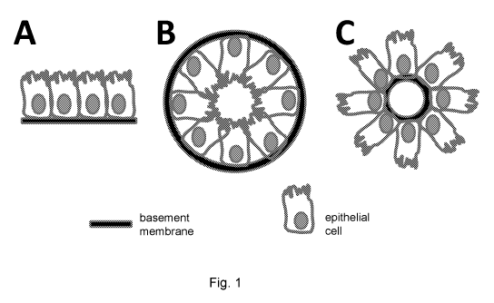
Fig. 1 shows a diagram of different methods that promote the formation of
apical-basal polarized
epithelium. Panel A shows an air-liquid interface method. Panel B shows an
organoid method, in
which the epithelial cells are inoculated into thick extracellular matrix and
allowed to grow into a
hollow spheroid with the apical side facing inwards. Panel C shows an apical
side outward-
oriented (ASO) epithelial spheroid, in which the epithelial cells are grown on
a "core" comprising
basement membrane proteins, and the apical side of the spheroid faces
outwards.
5
CA 03107502 2021-01-21
WO 2020/041065
PCT/US2019/046493
Fig. 2 shows airway epithelial cells formed a continuous epithelium sheet in
submersion in the
presence of both A 83-01 and Y-27632. Normal human bronchial epithelial cells
(HBEC; passage
11 (P11), population doubling (PD)-30) were seeded on top of 25% MatrigelTM
(BD Biosciences) in
24-well plate and cultured in submersion with different media. Panel A shows
cells cultured in
PneumaCultTm-ALI medium (P; STEMCELL Technologies). Panel B shows cells
cultured in
PneumaCultTm-ALI medium with 5 pM Y-27632 (P+Y). Panel C shows cells cultured
in
PneumaCultTm-ALI medium with 1 pM A 83-01 (P+A). Panel D shows cells cultured
in
PneumaCultTm-ALI medium with 1 pM A83-01 and 5 pM Y-27632 (P+A+Y). By day 27,
only cells
cultured in the presence of both A and Y compounds formed continuous
epithelium in the
submerged format.
Fig. 3 shows an epithelium sheet formed by airway epithelial cells under
submersion conditions
continued to survive in PneumaCultTm-ALI with A83-01 and Y-27632 (P+A+Y) for
over 2 months.
Fig. 4 shows airway epithelial cells seeded on top of Matrigel and cultured in
PneumaCultTm-ALI
supplemented with A83-01 and Y-27632 (P+A+Y) for at least 30 days. Panel A
shows spheres
(also referred to as bronchospheres) formed by individual airway epithelial
cells which were
entrapped in Matrigel TM. The apical side, where spontaneous beating of the
cilia could be seen,
faced inwards. Panel B shows multiciliated cells could also be found in the
continuous airway
epithelium sheet formed on top of Matrigel, with the apical side faced up.
This indicated that the
differentiation of multiciliated cells proceeded even in submersion when both
A 83-01 and Y-27632
(A+Y) were added to the medium.
Fig. 5 shows airway epithelial cells expressing GFP encapsulated in HyStemO-C
hydrogel and
cultured in submersion in Keratinocyte-SFM (KSFM; Gibco/Thermo Fisher 17005-
042)
supplemented with A 83-01 and Y-27632 (KSFM A+Y), PneumaCultTm-ALI (P), or
PneumaCultTm-
ALI supplemented with A83-01 and Y-27632 (P+A+Y) media. By day 7, most of the
cells cultured
in PneumaCultTm-ALI medium were dead (shown by the loss of GFP expression).
Some cells
survived (shown as GFP-positive) in KSFM A+Y or P+A+Y medium, but they
remained as single
cells, and did not grow into spheres.
Fig. 6 shows airway epithelial cells expressing GFP encapsulated in alginate
and cultured in
submersion in Keratinocyte-SFM (KSFM; Gibco/Thermo Fisher 17005-042)
supplemented with A
6
CA 03107502 2021-01-21
WO 2020/041065
PCT/US2019/046493
83-01 and Y-27632 (KSFM A+Y), PneumaCultTm-ALI (P), or PneumaCultTm-ALI
supplemented with
A83-01 and Y-27632 (P+A+Y) media. By day 7, most of the cells cultured in
PneumaCultTm-ALI
medium were dead (shown by the loss of GFP expression). Some cells survived
(shown as GFP-
positive) in KSFM A+Y or P+A+Y medium, but they remained as single cells, and
did not grow into
spheres.
Fig. 7 shows aggregated airway epithelial cells expressing GFP before
encapsulation in alginate,
HyStem0-0 hydrogel, or Matrigel TM .
.. Fig. 8 shows pre-aggregating airway epithelial cells expressing GFP before
encapsulation in
alginate, HyStem0-0 hydrogel, or Matrigel TM improved cell survival. By day
14, cells grew into
hollow spheres with the apical side facing outwards. Airway cell aggregates
cultured in liquid
suspension in an ultra-low attachment plate also grew into spheres with the
apical side facing
outwards.
Fig. 9 shows airway epithelial cells expressing GFP which were pre-aggregated
in AggreWellTm400
and cultured in suspension in an ultra-low attachment well in PneumaCultTm-ALI
supplemented
with A83-01 and Y-27632 (P+A+Y) medium. After 21 days, the aggregates grew
into spheres with
the apical side facing outwards.
Fig. 10A and Fig. 10B show H&E staining of two ASO (apical side outward-
oriented) spheroids
made of airway epithelial cells cultured for 3 months in PneumaCultTm-ALI
supplemented with A83-
01 and Y-27632 (P+A+Y) medium. The multiciliated cells are discernable with
their cilia facing
outwards. The size bar in Fig. 10A and Fig. 10B represents 50 microns.
Figs. 11A-110 show antibody staining of ASO (apical side outward-oriented)
spheroids made of
airway epithelial cells cultured in PneumaCultTm-ALI supplemented with A83-01
and Y-27632
(P+A+Y) medium. As shown in Fig. 11A, by day 14, the expanded airway
epithelial cells formed
ASO spheroids with multiciliated cells (stained with an antibody to acetylated
tubulin (Ac Tubilin))
and secretory cells (stained with an antibody to mucin SAC (MUC5AC). DAPI was
used as nuclear
counterstain. The size bar in Fig. 11A represents 50 microns. Fig. 11B shows
two different ASO
spheroids immunostained for expression of Collagen XVII (COL17) protein.
Nuclei are stained with
DAPI. Fig. 110 shows two different ASO spheroids immunostained for expression
of Keratin 5
(KRT5) protein. Nuclei are stained with DAPI.
7
CA 03107502 2021-01-21
WO 2020/041065
PCT/US2019/046493
Fig. 12 shows epithelial spheroids cultured in suspension. Top: 21-day old ASO
spheroids at 4X.
Bottom: 21-day old ASO spheroids at 20X. The size bar for the top panel
represents 1000 microns;
the size bar for bottom represents 200 microns.
Detailed Description
Provided herein are epithelial cell spheroids and methods of producing
epithelial cell spheroids.
Epithelial cell spheroids provided herein, and epithelial cell spheroids
produced by the methods
described herein, may serve as apical-basal polarized epithelial tissue
models, and may be useful,
for example, for certain in vitro tissue studies and some medical
applications. Generally, an
epithelial cell spheroid provided herein comprises epithelial cells oriented
such that the basal side
of each epithelial cell faces the inside of the spheroid and the apical side
of each epithelial cell
faces the outside of the spheroid. Such spheroids may be referred to herein as
apical side
outward-oriented (ASO) epithelial spheroids.
Epithelial cells
Provided herein are spheroids comprising epithelial cells (i.e., epithelial
cell spheroids) and
methods of producing epithelial cell spheroids. Methods of producing cell
spheroids may comprise
forming cellular aggregates and/or forming cell-substrate bodies. Accordingly,
cell spheroids,
cellular aggregates and/or cell-substrate bodies may comprise epithelial
cells. An epithelial cell, or
epithelium, typically refers to a cell or cells that line hollow organs, as
well as those that make up
glands and the outer surface of the body. Epithelial cells can comprise
squamous epithelial cells,
columnar epithelial cells, adenomatous epithelial cells or transitional
epithelial cells. Epithelial
cells can be arranged in single layers or can be arranged in multiple layers,
depending on the
organ and location.
Epithelial cells may have cell polarity. For example, certain epithelial cells
have an apical-basal
polarity. Such cells may comprise an apical membrane on one side and a basal
membrane on an
opposite side. Such cells also may comprise a lateral membrane. Generally,
epithelial cells
having an apical-basal polarity comprise an apical membrane on one side and a
basal membrane
on an opposite side, and an apical membrane located between the apical
membrane and the basal
membrane.
8
CA 03107502 2021-01-21
WO 2020/041065
PCT/US2019/046493
The basal side of an epithelial cell typically is anchored to other tissue. A
basement membrane (or
basal lamina) lies at the interface with underlying tissue, mediating the
attachment. A basement
membrane generally is a thin (e.g., about 100-nm) extracellular matrix (ECM)
that contains a
meshwork of proteins such as laminins, collagen IV, proteoglycans and nidogen.
In certain
instances, cell-matrix anchoring junctions tether the basal surface of an
epithelial cell to the
basement membrane. Cell-matrix anchoring junctions may include hemidesmosomes
and actin-
linked cell-matrix junctions. Hemidesmosomes generally anchor intermediate
filaments in an
epithelial cell to extracellular matrix (ECM). Actin-linked cell-matrix
junctions generally anchor actin
filaments in an epithelial cell to ECM. In certain instances, cells can
interact with a basement
membrane by binding basement membrane components through cell surface integrin
receptors.
These interactions allow the basement membrane to provide epithelia with
survival, proliferation
and differentiation signals, as well as directional cues to establish
polarity. An epithelial cell may
interact with the extracellular matrix (ECM) through integrin receptors.
Cell¨matrix interactions
typically are involved in creating epithelial cell polarity (see e.g., Lee et
al. 2014 J. Cell Sci.
127:3217-3225). The orientation of epithelial polarity typically requires
extrinsic signals, which
often originate within the ECM. Basal and lateral membranes share certain
common protein
markers which include, for example, Lethal Giant Larvae (Lgl), Discs Large
(Dig), and Scribble
(Scrib).
Epithelial cells having an apical-basal polarity generally are free of
attachment on the apical side.
For certain tissues, the apical side of an epithelial cell is exposed to the
environment (e.g., apical
side of an airway cell is exposed to inhaled air; apical side of an intestinal
cell is exposed to
ingested food and liquid). In certain instances, the apical side of an
epithelial cell is exposed to the
interior or lumen of a tubule or organ (e.g., interior of a renal tubule). For
certain epithelial cells,
apical membrane is characterized by the presence of cilia and/or microvilli.
Cilia generally are
found on ciliated epithelial cells, such as epithelial cells in the lungs.
Cilia may move by waving
rhythmically (e.g., to move debris and/or mucus out). Microvilli generally are
found in
tissues/organs specialized for absorption, such as the digestive tract or
kidneys. Microvilli function
by increasing the surface area of the cell membrane, thus allowing for more
materials to be
absorbed into the cell at a quicker rate. For example, microvilli are found in
the small intestine and
increase the surface area for nutrient absorption. Protein markers for apical
membrane may
include, for example, Cdc42, atypical protein kinase C (aPKC), Par6,
Par3/Bazooka/ASIP, Crumbs,
Stardust, and protein at tight junctions (PATJ).
9
CA 03107502 2021-01-21
WO 2020/041065
PCT/US2019/046493
Epithelial cells may be directly attached to each other at their lateral
membranes by cell-cell
junctions, where cytoskeletal filaments are anchored, to form epithelia. Cell-
cell junctions may
include, for example, tight junctions, cell-cell anchoring junctions (e.g.,
adherens junctions,
desmosomes), and channel forming junctions (e.g., gap junctions). Tight
junctions generally are
parts of cell membranes joined together to seal gaps between epithelial cells
and form an
impermeable or substantially impermeable barrier to fluid; adherens junctions
generally connect
actin filament bundles in one cell with that in the next cell; desmosomes
generally connect
intermediate filaments in one cell to those in the next cell; and gap
junctions allow passage of
molecules (e.g., small water-soluble molecules) from cell to cell. In the most
apical portion of the
cell, the relative positions of the junctions are the same or similar in most
vertebrate epithelia. A
tight junction typically occupies the most apical position, followed by an
adherens junction
(adhesion belt) and then by a parallel row of desmosomes. Gap junctions and
additional
desmosomes generally are less regularly organized.
In some embodiments, epithelial cells form tight junctions under certain
culture conditions. For
example, epithelial cells may form tight junctions under aggregation
conditions described herein,
under substrate attachment conditions described herein, and/or under spheroid
inducing conditions
described herein. Formation of tight junctions may be visualized, for example,
by
immunofluorescence staining of tight junction proteins (e.g., ZO-1). In some
embodiments,
epithelial cells can be induced to form tight junctions under aggregation
conditions. In some
embodiments, epithelial cells can be induced to form tight junctions under
substrate attachment
conditions. In some embodiments, epithelial cells can be induced to form tight
junctions under
spheroid inducing conditions. For example, epithelial cells can be induced to
form tight junctions
when exposed to certain concentrations of calcium. In some embodiments,
epithelial cells can be
induced to form tight junctions when exposed to calcium concentrations that
are about 0.5 mM or
higher. For example, epithelial cells can be induced to form tight junctions
when exposed to
calcium concentrations that are about 0.5 mM, 0.6 mM, 0.7 mM, 0.8 mM, 0.9 mM,
1 mM, 1.1 mM,
1.2 mM, 1.3 mM, 1.4 mM, 1.5 mM, 1.6 mM, 1.7 mM, 1.8 mM, 1.9 mM, 2.0 mM, or
higher. In some
embodiments, epithelial cells can be induced to form tight junctions when
exposed to a calcium
concentration of about 1.5 mM.
Epithelial cells can comprise keratinocyte epithelial (KE) cells or non-
keratinocyte epithelial (NKE)
cells. Keratinocytes form the squamous epithelium that is found at anatomic
sites such as the skin,
CA 03107502 2021-01-21
WO 2020/041065
PCT/US2019/046493
ocular surface, oral mucosa, esophagus and cervix. Keratinocytes terminally
differentiate into flat,
highly keratinized, non-viable cells that help protect against the environment
and infection by
forming a protective barrier. Examples of keratinocyte epithelial cells
include, but are not limited to,
dermal keratinocytes, ocular epithelial cells, corneal epithelial cells, oral
mucosal epithelial cells,
and cervical epithelial cells.
Non-keratinocyte epithelial (NKE) cells form the epithelium of the body such
as found in the breast,
prostate, liver, respiratory tract, retina and gastrointestinal tract. NKE
cells typically differentiate
into functional, viable cells which function, for example, in absorption
and/or secretion. These cells
typically do not form highly keratinized structures characteristic of squamous
epithelial cells.
NKE cells described herein can be of any type or tissue of origin. Examples of
NKE cells include,
but are not limited to, prostate epithelial cells, mammary epithelial cells,
hepatocytes, liver epithelial
cells, biliary epithelial cells, gall bladder cells, pancreatic islet cells,
pancreatic beta cells,
pancreatic ductal epithelial cells, pulmonary epithelial cells, lung
epithelial cells, airway epithelial
cells, nasal epithelial cells, tracheal epithelial cells, bronchial epithelial
cells, kidney epithelial cells,
bladder epithelial cells, urethral epithelial cells, stomach epithelial cells,
esophageal epithelial cells,
large intestinal epithelial cells, small intestinal epithelial cells,
testicular epithelial cells, ovarian
epithelial cells, fallopian tube epithelial cells, thyroid epithelial cells,
parathyroid epithelial cells,
adrenal epithelial cells, thymus epithelial cells, pituitary epithelial cells,
glandular epithelial cells,
amniotic epithelial cells, retinal pigmented epithelial cells, sweat gland
epithelial cells, sebaceous
epithelial cells, and hair follicle epithelial cells. In some embodiments,
epithelial cells comprise
airway epithelial cells. In some embodiments, epithelial cells comprise
keratinocyte epithelial cells.
In some embodiments, epithelial cells comprise prostate epithelial cells. In
some embodiments,
epithelial cells comprise mammary epithelial cells.
In some embodiments, epithelial cells comprise basal epithelial cells. Basal
epithelial cells
generally are cells in the deepest layer of stratified epithelium and
multilayered epithelium. Basal
epithelial cells may be cells whose nuclei locate close to the basal lamina in
a pseudostratified
epithelium. In some instances, basal epithelial cells may divide (e.g., by
asymmetric cell division or
symmetric cell division), giving rise to other basal cells and/or other
epithelial cell types (e.g., other
cell types in a stratified epithelium, multilayered epithelium or
pseudostratified epithelium). A
proportion of basal epithelial cells in some epithelia may have lifelong self-
renew capability and can
give rise to other epithelial cell types and basal cells, and sometimes are
considered as epithelial
11
CA 03107502 2021-01-21
WO 2020/041065
PCT/US2019/046493
stem cells. The proportion of basal epithelial cells that have lifelong self-
renew capability and are
considered as epithelial stem cells varies among different tissues.
In some embodiments, epithelial cells are isolated. The term isolated
generally refers to cells
removed from their original environment (e.g., the natural environment if they
naturally occurring, or
an in vitro cell source (e.g., embryonic stem (ES) cell culture, induced
pluripotent stem cell (iPSCs)
culture)), and thus are altered "by the hand of man" from their original
environment. Epithelial cells
may be separated from non-epithelial cells and/or extracellular components
(e.g., tissue matrix
components) present in a source sample. Isolated epithelial cells may be
provided with fewer non-
epithelial cells and/or extracellular components (e.g., tissue matrix
components) than the amount
of non-epithelial cells and/or extracellular components present in a source
sample. A composition
containing isolated epithelial cells can be substantially isolated (e.g.,
about 90%, 91%, 92%, 93%,
94%, 95%, 96%, 97%, 98%, 99% or greater than 99% free of non-epithelial cells
and/or
extracellular components). In some embodiments, a method herein comprises
isolating epithelial
cells from a subject. In some embodiments, a method herein comprises isolating
epithelial cells
from tissue from a subject. Epithelial cells isolated from tissue from a
subject generally are free of
extracellular components from the tissue. Accordingly, in some embodiments,
isolated epithelial
cells comprise no extracellular components from the tissue from the subject.
Generally,
extracellular components produced by isolated epithelial cells (e.g., after
isolation; during
aggregation; during spheroid formation) are not considered as extracellular
components from the
tissue from the subject.
Epithelial cells may be obtained or isolated from a subject and/or a cellular
source. Cells obtained
from a subject and/or a cellular source may be referred as an originating
epithelial cell population.
A cellular source may include epithelial cells from a particular tissue or
organ in a subject. A
cellular source may include epithelial cells from a sample from a subject. A
cellular source may
include a population of embryonic stem (ES) cells, induced pluripotent stem
cells (iPSCs), and the
like. In some embodiments, an originating epithelial cell population is
isolated from an embryo or a
stem cell culture derived from an embryo. In some embodiments, epithelial
cells are isolated from
an induced pluripotent stem cell (iPSC) culture. Epithelial cells may be
obtained from a subject in
a variety of manners (e.g., harvested from living tissue, such as a biopsy,
plucked hair follicles,
body fluids like urine or body-cavity fluids, or isolated from circulation). A
subject may include any
animal, including but not limited to any mammal, such as mouse, rat, canine,
feline, bovine,
equine, porcine, non-human primate and human. In certain embodiments, a
subject is a human.
12
CA 03107502 2021-01-21
WO 2020/041065
PCT/US2019/046493
In some embodiments, a subject is an embryo. In some embodiments, a subject is
an animal or
human that has gestated longer than an embryo in a uterine environment and
often is a post-natal
human or a post-natal animal (e.g., neonatal human, neonatal animal, adult
human or adult
animal). A subject sometimes is a juvenile animal, juvenile human, adult
animal or adult human.
In some embodiments, epithelial cells are isolated from a sample from a
subject. A sample can
include any specimen that is isolated or obtained from a subject or part
thereof. Non-limiting
examples of specimens include fluid or tissue from a subject, including,
without limitation, blood or
a blood product (e.g., serum, plasma, or the like), umbilical cord blood, bone
marrow, chorionic
amniotic fluid, amnion, cerebrospinal fluid, spinal fluid, lavage fluid (e.g.,
bronchoalveolar, gastric,
peritoneal, ductal, ear, arthroscopic), biopsy sample or tissue biopsy, buccal
swab, plucked hair
follicles, skin punch biopsy, nasal brushing, celocentesis sample, washings of
female reproductive
tract, urine, feces, sputum, saliva, nasal mucous, prostate fluid, lavage,
semen, lymphatic fluid,
bile, tears, sweat, breast milk, breast fluid, hard tissues (e.g., liver,
spleen, kidney, lung, or ovary),
the like or combinations thereof. The term blood encompasses whole blood,
blood product or any
fraction of blood, such as serum, plasma, buffy coat, or the like as
conventionally defined. Blood
plasma refers to the fraction of whole blood resulting from centrifugation of
blood treated with
anticoagulants. Blood serum refers to the watery portion of fluid remaining
after a blood sample
has coagulated. In some embodiments, fetal cells are isolated from a maternal
sample (e.g.,
maternal blood, amniotic fluid).
In some embodiments, epithelial cells comprise normal, healthy cells (e.g.,
cells that are not
diseased). In some embodiments, epithelial cells comprise diseased cells.
Diseased epithelial
cells may include cells from a subject carrying disease-causing mutation(s)
(e.g., epithelial cells
with genetic mutation(s) in the CFTR gene). Diseased epithelial cells may
include cells from
abnormal tissue, such as from a neoplasia, a hyperplasia, a malignant tumor or
a benign tumor. In
certain embodiments, diseased epithelial cells may include cells that are not
tumor cells. In certain
embodiments, diseased epithelial cells may include cells isolated from
circulation (e.g., circulating
tumor cells (CTCs)) of a subject. In certain embodiments, diseased epithelial
cells may include
cells isolated from bodily samples such as, for example, urine, semen, stool
(feces), and the like.
In some embodiments, epithelial cells comprise cells that are altered,
modified or engineered (e.g.,
genetically altered, genetically modified, genetically engineered). In some
embodiments, epithelial
cells are altered, modified or engineered (e.g., genetically altered,
genetically modified, genetically
13
CA 03107502 2021-01-21
WO 2020/041065
PCT/US2019/046493
engineered). The terms altered, engineered, and modified may be used
interchangeably herein in
reference to cell, and generally refer to a cell (e.g., epithelial cell) that
has been manipulated such
that it is distinct (e.g., detectably changed or physically different) from a
naturally occurring cell.
For example, the sum total of the cellular activities of a modified or
engineered cell can be distinct
.. from those of a naturally occurring cell, e.g., a modified cell may include
or lack one or more
activities relative to the activities present in an unmodified cell utilized
as a starting point (e.g., host
cell) for modification. In another example, one or more cellular activities of
a modified or
engineered cell may be altered relative to the cellular activity or activities
of the host cell. A
modified or engineered cell can be genetically modified through any alteration
in its genetic
composition. For example, a genetically modified cell can include one or more
heterologous
polynucleotides, can have one or more endogenous nucleic acid deletions and/or
can have one or
more genetic mutations. Mutations include point mutations, insertions and
deletions of a single or
multiple residues in a nucleic acid. In some embodiments, an engineered cell
includes a
heterologous polynucleotide, and in certain embodiments, an engineered cell
has been subjected
to selective conditions that alter an activity, or introduce an activity,
relative to the host cell. Thus,
a modified or engineered cell has been altered directly or indirectly by a
human being. It is
understood that the terms modified cell and engineered cell refer not only to
the particular cell but
to the progeny or potential progeny of such a cell. Because certain
modifications may occur in
succeeding generations due to either mutation or environmental influences,
such progeny may not
be identical to the parent cell, but are still included within the scope of
the term as used herein. In
some embodiments, epithelial cells comprise cells that are not genetically
altered.
In some embodiments, epithelial cells comprise primary cells. Primary
epithelial cells generally are
taken directly from living tissue, such as a biopsy, plucked hair follicles,
bodily samples such as a
stool sample, body fluids like urine, semen or body-cavity fluids, or isolated
from circulation. In
certain instances, primary cells have not been passaged. In certain instances,
primary cells have
been passaged one time. Primary cells may be isolated from differentiated
tissue (e.g., isolated
from epithelium of various organs). Typically, primary cells have been freshly
isolated, for
example, through tissue digestion and plated. Primary cells may or may not be
frozen and then
thawed at a later time. In addition, the tissue from which the primary cells
are isolated may or may
not have been frozen or preserved in some other manner immediately prior to
processing.
Typically, cells are no longer primary cells after the cells have been
passaged more than once.
Cells passaged once or more and immediately frozen after passaging are also
not considered as
primary cells when thawed. In certain embodiments, epithelial cells are
initially primary cells and
14
CA 03107502 2021-01-21
WO 2020/041065
PCT/US2019/046493
become non-primary cells after passaging. Cells passaged more than once may be
referred to as
derived from primary epithelial cells. In some embodiments, epithelial cells
are maintained or
proliferated in cell culture after the cells are isolated from tissue and
prior to forming epithelial cell
spheroids described herein. In some embodiments, epithelial cells are derived
from primary cells.
Cellular spheroids, cellular aggregates, and/or cell-substrate bodies
described herein may
comprise primary cells. For example, epithelial cells in cellular aggregates
may comprise primary
epithelial cells. In another example, epithelial cells in cell-substrate
bodies may comprise primary
epithelial cells. In another example, epithelial cells in cellular spheroids
may comprise primary
epithelial cells. In some embodiments, cellular spheroids, cellular
aggregates, and/or cell-
substrate bodies described herein may consist essentially of primary cells.
For example, epithelial
cells in cellular aggregates may consist essentially of primary epithelial
cells. In another example,
epithelial cells in cell-substrate bodies may consist essentially of primary
epithelial cells. In another
example, epithelial cells in cellular spheroids may consist essentially of
primary epithelial cells.
Cellular aggregates, cell-substrate bodies, and/or cellular spheroids
consisting essentially of
primary epithelial cells refers to cellular aggregates, cell-substrate bodies,
and/or cellular spheroids
where at least about 75% of the cells are primary epithelial cells. For
example, cellular
aggregates, cell-substrate bodies, and/or cellular spheroids consisting
essentially of primary
epithelial cells may contain at least about 80% primary epithelial cells, at
least about 85% primary
epithelial cells, at least about 90% primary epithelial cells, at least about
95% primary epithelial
cells, at least about 96% primary epithelial cells, at least about 97% primary
epithelial cells, at least
about 98% primary epithelial cells, or at least about 99% primary epithelial
cells. In some
embodiments, cellular spheroids, cellular aggregates, and/or cell-substrate
bodies described
herein may consist of primary cells. For example, epithelial cells in cellular
aggregates may consist
of primary epithelial cells. In another example, epithelial cells in cell-
substrate bodies may consist
of primary epithelial cells. In another example, epithelial cells in cellular
spheroids may consist of
primary epithelial cells.
In some embodiments, primary epithelial cells undergo one or more cell
division cycles prior to
forming epithelial cell spheroids and/or during cell spheroid formation. For
example, primary
epithelial cells may divide to form daughter cells, daughter cells may divide
to form further daughter
cells or further primary cell descendants, and so on. Accordingly, in some
embodiments, epithelial
cells (e.g., epithelial cells in cellular aggregates, epithelial cells in cell-
substrate bodies, epithelial
cells in cellular spheroids) comprise primary epithelial cells and/or primary
epithelial cell daughter
CA 03107502 2021-01-21
WO 2020/041065
PCT/US2019/046493
cells and/or primary epithelial cell further daughter cells or further primary
cell descendants. In
some embodiments, epithelial cells (e.g., epithelial cells in cellular
aggregates, epithelial cells in
cell-substrate bodies, epithelial cells in cellular spheroids) consist
essentially of primary epithelial
cells and/or primary epithelial cell daughter cells and/or primary epithelial
cell further daughter cells
or further primary cell descendants. In some embodiments, epithelial cells
(e.g., epithelial cells in
cellular aggregates, epithelial cells in cell-substrate bodies, epithelial
cells in cellular spheroids)
consist of primary epithelial cells and/or primary epithelial cell daughter
cells and/or primary
epithelial cell further daughter cells or further primary cell descendants.
In some embodiments, epithelial cells comprise non-primary cells, such as
cells from an
established cell line (e.g., Madin-Darby Canine Kidney (MDCK) cells,
immortalized cell line (HeLa
cells, HEK 293 cells, immortalized HBE cells), transformed cells, thawed cells
from a previously
frozen collection and the like. In some embodiments, epithelial cells comprise
no non-primary
cells. In some embodiments, epithelial cells comprise no cells from an
established cell line (e.g.,
no MDCK cells). In some embodiments, epithelial cells comprise no cells from
an immortalized cell
line (e.g., no HeLa cells; no HEK 293 cells; no immortalized HBE cells). Non-
primary cells may be
anchorage independent (i.e., cells that have lost the need for anchorage
dependence, which often
is essential for cell growth, division, and spreading; cells that have become
anchorage-
independent are often transformed or have become neoplastic in nature). In
some embodiments,
epithelial cells comprise no anchorage-independent cells. In some embodiments,
epithelial cells
comprise anchorage-dependent cells. In some embodiments, epithelial cells
consist of comprise
anchorage-dependent cells. Anchorage dependence generally refers to the need
for cells to be
adhered to or in contact with other cells, extracellular matrix, or tissue
culture plastic (e.g., via
proteins). Often, cells (e.g., primary cells, non-transformed cells) grown in
culture require some
sort of anchorage for survival. In certain embodiments, epithelial cells
comprise secondary cells.
In certain embodiments, epithelial cells comprise no secondary cells.
In some embodiments, epithelial cells comprise expanded epithelial cells
(e.g., ex-vivo expanded
epithelial cells). In some embodiments, epithelial cells are expanded
epithelial cells. In some
.. embodiments, epithelial cells are ex-vivo expanded epithelial cells.
Epithelial cells may be
expanded under any suitable expansion culture conditions, such as, for
example, expansion
culture conditions described herein. In some embodiments, epithelial cells
comprise expanded
primary epithelial cells. For example, primary cells may be obtained (e.g.,
harvested from a
16
CA 03107502 2021-01-21
WO 2020/041065
PCT/US2019/046493
subject), expanded, and then subjected to aggregation conditions, substrate
attachment
conditions, and/or spheroid-inducing culture conditions described herein.
In some embodiments, a culture composition, cellular aggregate, cell-substrate
body and/or
cellular spheroid comprises a heterogeneous population of epithelial cells
(e.g., comprises a
mixture of cell types and/or differentiation states such as epithelial stem
cells, epithelial
progenitors, epithelial precursor cells, lineage-committed epithelial cells,
transit-amplifying epithelial
cells, differentiating epithelial cells, differentiated epithelial cells, and
terminally differentiated
epithelial cells) derived from the same tissue or same tissue compartment. In
some embodiments,
a culture composition, cellular aggregate, cell-substrate body and/or cellular
spheroid comprises a
homogenous population of epithelial cells (e.g., does not include a mixture of
cell types and/or
differentiation states) derived from the same tissue or same tissue
compartment. In some
embodiments, a homogeneous population of epithelial cells comprises at least
about 90%
epithelial cells that are of the same cell type and/or are present at the same
differentiation state.
For example, a homogeneous population of epithelial cells may comprise at
least about 90%, 91%,
92%, 93%, 94%, 95%, 96%, 97%, 98%, 99% or 100% epithelial cells that are of
the same cell type
and/or are present at the same differentiation state. In some embodiments, a
homogeneous
population of epithelial cells comprises about 100% epithelial cells that are
of the same cell type
and/or are present at the same differentiation state. In some embodiments,
epithelial cells are a
homogenous population of basal epithelial cells. In some embodiments, an
originating epithelial
cell population may be heterogeneous or may be homogeneous. In some
embodiments, an
expanded epithelial cell population may be heterogeneous or may be
homogeneous. In some
embodiments, an epithelial cell aggregate may be heterogeneous or may be
homogeneous. In
some embodiments, a cellular aggregate may comprise a heterogeneous epithelial
cell population
or may comprise homogeneous epithelial cell population. In some embodiments, a
cell-substrate
body may comprise a heterogeneous epithelial cell population or may comprise
homogeneous
epithelial cell population. In some embodiments, a cellular spheroid may
comprise a
heterogeneous epithelial cell population or may comprise homogeneous
epithelial cell population.
In some embodiments, epithelial cells are characterized by the cell types
and/or differentiation
states that are included in, or absent from, a population of epithelial cells.
In some embodiments,
such cell characterization may be applicable to an originating epithelial cell
population. In some
embodiments, such cell characterization may be applicable to an expanded
epithelial cell
population. In some embodiments, such cell characterization may be applicable
to an originating
17
CA 03107502 2021-01-21
WO 2020/041065
PCT/US2019/046493
epithelial cell population and an expanded epithelial cell population. In some
embodiments, such
cell characterization may be applicable to epithelial cells in a cellular
aggregate. In some
embodiments, such cell characterization may be applicable to epithelial cells
in a cell-substrate
body. In some embodiments, such cell characterization may be applicable to
epithelial cells in a
cellular spheroid. In some embodiments, epithelial cells that include a
particular cell type and/or
differentiation state comprise at least about 50% epithelial cells that are of
the particular cell type
and/or differentiation state. In some embodiments, epithelial cells that
include a particular cell type
and/or differentiation state comprise at least about 90% epithelial cells that
are of the particular cell
type and/or differentiation state. For example, epithelial cells that include
a particular cell type
and/or differentiation state may comprise at least about 90%, 91%, 92%, 93%,
94%, 95%, 96%,
97%, 98%, 99% or 100% epithelial cells that are of the particular type and/or
differentiation state.
Generally, epithelial cells that do not include a particular cell type and/or
differentiation state
comprise less than about 10% cells that are of the particular cell type and/or
differentiation state.
For example, epithelial cells that do not include a particular cell type
and/or differentiation state
may comprise less than about 10%, 9%, 8%, 7%, 6%, 5%, 4%, 3%, 2%, or 1% cells
that are of the
particular cell type and/or differentiation state.
In certain embodiments, a culture composition, cellular aggregate, cell-
substrate body and/or
cellular spheroid consists essentially of a population of a particular type of
epithelial cell, referred to
hereafter as "the majority cells." Such populations can include a minor amount
of one or more
other types of epithelial cells, referred to hereafter as "the minority
cells." The minority cells
typically are from, or are derived from, the same tissue as the majority
cells, and often are from, or
are derived from, the same tissue compartment, as the majority cells. The
majority cells can be
greater than 50%, greater than 60%, greater than 70%, or greater than 80% of
the total cells in the
composition and often are about 90% or more of the total cells in the
composition, and sometimes
are about 91%, 92%, 93%, 94%, 95%, 96%, 97%, 98% or 99% or more of the total
cells in the
composition or population.
In some embodiments, a culture composition, cellular aggregate, cell-substrate
body and/or
cellular spheroid comprises a heterogeneous population of epithelial cells at
different cell cycle
phases, such as the M phase, the G1 phase, the S phase, the G2 phase, and the
GO phase which
includes senescence and quiescence. In some embodiments, an originating
epithelial cell
population comprises a heterogeneous population of epithelial cells at
different cell cycle phases,
such as the M phase, the G1 phase, the S phase, the G2 phase, and the GO phase
which includes
18
CA 03107502 2021-01-21
WO 2020/041065
PCT/US2019/046493
senescence and quiescence. In some embodiments, an expanded epithelial cell
population
comprises a heterogeneous population of epithelial cells at different cell
cycle phases, such as the
M phase, the G1 phase, the S phase, the G2 phase, and the GO phase which
includes senescence
and quiescence. In some embodiments, a cellular aggregate comprises a
heterogeneous
population of epithelial cells at different cell cycle phases, such as the M
phase, the G1 phase, the
S phase, the G2 phase, and the GO phase which includes senescence and
quiescence. In some
embodiments, a cell-substrate body comprises a heterogeneous population of
epithelial cells at
different cell cycle phases, such as the M phase, the G1 phase, the S phase,
the G2 phase, and
the GO phase which includes senescence and quiescence. In some embodiments, a
cellular
spheroid comprises a heterogeneous population of epithelial cells at different
cell cycle phases,
such as the M phase, the G1 phase, the S phase, the G2 phase, and the GO phase
which includes
senescence and quiescence. Epithelial cells at a particular cell cycle phase
can make up 1% to
100% of the population.
In some embodiments, epithelial cells comprise cells at one or more stages of
differentiation. In
some embodiments, such stages of differentiation may be described for an
originating epithelial
cell population. In some embodiments, such stages of differentiation may be
described for an
expanded epithelial cell population. In some embodiments, such stages of
differentiation may be
described for an originating epithelial cell population and an expanded
epithelial cell population. In
some embodiments, such stages of differentiation may be described for
epithelial cells in a cellular
aggregate. In some embodiments, such stages of differentiation may be
described for epithelial
cells in a cell-substrate body. In some embodiments, such stages of
differentiation may be
described for epithelial cells in a cellular spheroid. For example, epithelial
cells (or a population of
epithelial cells) may comprise epithelial stem cells, epithelial progenitor
cells, lineage-restricted
epithelial progenitor cells, epithelial precursor cells, lineage-committed
epithelial cells, transit-
amplifying epithelial cells, proliferating epithelial cells, differentiating
epithelial cells, differentiated
epithelial cells, quiescent epithelial cells, formerly quiescent epithelial
cells, non-proliferating
epithelial cells, and terminally differentiated epithelial cells (e.g., cells
that are found in tissues and
organs). Epithelial cells also may comprise lineage-committed epithelial cells
differentiated and/or
derived from pluripotent stem cells (embryonic stem (ES) cells or induced
pluripotent stem cells
(iPSCs)).
In some embodiments, epithelial cells comprise differentiated epithelial
cells. Differentiated
epithelial cells may divide, but typically do not have the capacity for
indefinite self-renewal. In
19
CA 03107502 2021-01-21
WO 2020/041065
PCT/US2019/046493
some embodiments, differentiated epithelial cells do not acquire the ability
to differentiate into
multiple tissue types. Differentiated epithelial cells cultured in conditions
described herein
generally are more differentiated than undifferentiated cells (e.g., stem
cells (embryonic or adult),
progenitor cells, precursor cells) and are less differentiated than terminally
differentiated cells.
Differentiated epithelial cells generally do not include stem cells (embryonic
or adult), progenitor
cells or precursor cells. In certain instances, differentiated epithelial
cells may be referred to as
"tissue-specific" and/or "lineage-committed" epithelial cells. In certain
instances, differentiated
epithelial cells may comprise tissue-specific and/or lineage-committed
epithelial cells. In some
embodiments, differentiated epithelial cells comprise quiescent epithelial
cells. In some
.. embodiments, differentiated epithelial cells comprise basal epithelial
cells.
In some embodiments, epithelial cells comprise quiescent or formerly quiescent
cells. Quiescent
cells generally are non-proliferating cells (i.e., non-cycling cells, cells
that have withdrawn from the
cell cycle, resting cells), and may be characterized as reversibly growth
arrested. Under certain
conditions, quiescent cells can be induced to proliferate. Quiescent cells may
be characterized as
existing in the GO phase of the cell cycle. Quiescent cells that have been
induced to proliferate
may be referred to as formerly quiescent cells.
In some embodiments, epithelial cells comprise organ-specific epithelial
cells. Organ-specific
epithelial cells sometimes are referred to as tissue-specific epithelial
cells. In some embodiments,
organ-specific epithelial cells may differentiate into more specific cell
types within a given organ,
but generally do not possess or acquire the ability to differentiate into
cells of other types of organs.
Organ-specific epithelial cells generally are more differentiated than
undifferentiated cells (e.g.,
stem cells (embryonic or adult)) and are less differentiated than terminally
differentiated cells.
.. Organ-specific epithelial cells generally do not include embryonic stem
cells. Organ-specific
epithelial cells may or may not include adult stem cells (e.g., adult
epithelial stem cells), and organ-
specific epithelial cells may or may not include progenitor cells or precursor
cells.
In some embodiments, epithelial cells comprise lineage-committed epithelial
cells. In some
embodiments, epithelial cells can comprise lineage-committed epithelial cells
differentiated from
pluripotent stem cells such as embryonic stem (ES) cells and induced
pluripotent stem cells
(iPSCs). Lineage-committed epithelial cells may divide, but typically do not
have the capacity for
indefinite self-renewal. In some embodiments, lineage-committed epithelial
cells may differentiate
into various cell types within a given cell lineage (e.g., respiratory,
digestive or integumentary
CA 03107502 2021-01-21
WO 2020/041065
PCT/US2019/046493
lineages), but generally do not possess or acquire the ability to
differentiate into cells of different
cell lineages (e.g., integumentary lineage-committed epithelial cells
generally do not differentiate
into blood cells). Lineage-committed epithelial cells generally are more
differentiated than
undifferentiated pluripotent stem cells and are less differentiated than
terminally differentiated cells.
Lineage-committed epithelial cells generally do not include pluripotent stem
cells (embryonic or
induced pluripotent). In some embodiments, lineage-committed epithelial cells
include progenitor
cells or precursor cells. In some embodiments, lineage-committed epithelial
cells comprise basal
epithelial cells.
In some embodiments, epithelial cells include terminally differentiated
epithelial cells. In some
embodiments, epithelial cells do not include terminally differentiated
epithelial cells. Terminally
differentiated epithelial cells generally do not divide and are committed to a
particular function.
Terminally differentiated epithelial cells generally are characterized by
definitive withdrawal from
the cell cycle and typically cannot be induced to proliferate. In some
embodiments, epithelial cells
do not include post-mitotic cells. Post-mitotic cells generally are incapable
of or no longer capable
of cell division. In some embodiments, epithelial cells do not include
senescent cells.
In some embodiments, epithelial cells include embryonic stem cells. In some
embodiments,
epithelial cells do not include embryonic stem cells. In some embodiments,
epithelial cells are
differentiated and/or derived from embryonic stem cells. In some embodiments,
epithelial cells are
not derived from embryonic stem cells. Generally, embryonic stem cells are
undifferentiated cells
that have the capacity to regenerate or self-renew indefinitely. Embryonic
stem cells sometimes are
considered pluripotent (i.e., can differentiate into many or all cell types of
an adult organism) and
sometimes are considered totipotent (i.e., can differentiate into all cell
types, including the placental
tissue).
In some embodiments, epithelial cells include induced pluripotent stem cells
(iPSCs). In some
embodiments, epithelial cells do not include induced pluripotent stem cells
(iPSCs). In some
embodiments, epithelial cells are differentiated and/or derived from induced
pluripotent stem cells
(iPSCs). In some embodiments, epithelial cells are not derived from induced
pluripotent stem cells
(iPSCs). Generally, induced pluripotent stem cells (iPSCs) are a type of
pluripotent stem cell that
can be generated directly from adult cells. In some embodiments, epithelial
cells include
pluripotent cells. In some embodiments, epithelial cells do not include
pluripotent cells. In some
21
CA 03107502 2021-01-21
WO 2020/041065
PCT/US2019/046493
embodiments, epithelial cells include toti potent cells. In some embodiments,
epithelial cells do not
include totipotent cells.
In some embodiments, epithelial cells include adult stem cells. Adult stem
cells typically are less
differentiated than differentiated cells, organ-specific cells or lineage-
committed cells and are more
differentiated than embryonic stem cells. Adult stem cells may be referred to
as stem cells,
undifferentiated stem cells, precursor cells and/or progenitor cells, and are
not considered
embryonic stem cells as adult stem cells are not isolated from an embryo.
Adult epithelial stem
cells may be referred to as epithelial stem cells, undifferentiated epithelial
stem cells, epithelial
precursor cells and/or epithelial progenitor cells. In some embodiments,
epithelial cells do not
include adult stem cells or cells derived from adult stem cells. In some
embodiments, epithelial
cells do not include epithelial stem cells or cells derived from epithelial
stem cells. In some
embodiments, epithelial cells do not include pluripotent epithelial stem cells
or cells derived from
pluri potent epithelial stem cells. In some embodiments, epithelial cells do
not include progenitor
cells or cells derived from progenitor cells. In some embodiments, epithelial
cells do not include
precursor cells or cells derived from precursor cells. In some embodiments,
epithelial cells do not
include continuously proliferating (e.g., continuously proliferating in vivo)
epithelial stem cells (e.g.,
intestinal crypt cells; Lgr5+ cells) or cells derived from continuously
proliferating epithelial stem
cells.
Cellular spheroids, cellular aggregates, and/or cell-substrate bodies
described herein may
comprise stem cells. Cellular spheroids, cellular aggregates, and/or cell-
substrate bodies
described herein may be derived from stem cells. Cellular spheroids, cellular
aggregates, and/or
cell-substrate bodies described herein may comprise adult stem cells. Cellular
spheroids, cellular
aggregates, and/or cell-substrate bodies described herein may be derived from
adult stem cells.
Cellular spheroids, cellular aggregates, and/or cell-substrate bodies
described herein may
comprise progenitor cells. Cellular spheroids, cellular aggregates, and/or
cell-substrate bodies
described herein may be derived from progenitor cells. In some embodiments,
cellular spheroids,
cellular aggregates, and/or cell-substrate bodies described herein are derived
from a homogenous
.. population of cultured stem cells. In some embodiments, cellular spheroids,
cellular aggregates,
and/or cell-substrate bodies described herein are derived from a homogenous
population of
cultured adult stem cells. In some embodiments, cellular spheroids, cellular
aggregates, and/or
cell-substrate bodies described herein are derived from a homogenous
population of cultured
progenitor cells. In some embodiments, cellular spheroids, cellular
aggregates, and/or cell-
22
CA 03107502 2021-01-21
WO 2020/041065
PCT/US2019/046493
substrate bodies described herein are derived from a population of epithelial
cells comprising no
terminally differentiated cells. In some embodiments, cellular spheroids,
cellular aggregates,
and/or cell-substrate bodies described herein are derived from a homogenous
population of
cultured airway stem cells and/or airway progenitor cells. In some
embodiments, cellular
spheroids, cellular aggregates, and/or cell-substrate bodies described herein
are derived from a
population of epithelial cells comprising no terminally differentiated airway
cells.
In some embodiments, epithelial cells may be characterized by whether the
cells possess one or
more markers (e.g., cell surface markers, mRNAs, proteins, epigenetic
signatures) and/or do not
possess measurable levels of, or possess low levels of, certain markers. In
some embodiments,
such marker characterization may be applicable to an originating epithelial
cell population. In some
embodiments, such marker characterization may be applicable to an expanded
epithelial cell
population. In some embodiments, such marker characterization may be
applicable to an
originating epithelial cell population and an expanded epithelial cell
population. In some
embodiments, such marker characterization may be applicable to a cellular
aggregate. In some
embodiments, such marker characterization may be applicable to a cell-
substrate body. In some
embodiments, such marker characterization may be applicable to a cellular
spheroid.
Cellular aggregates
Provided herein are methods for producing cellular spheroids. In some
embodiments, a method
includes forming cellular aggregates. Generally, cellular aggregates are
formed prior to producing
cellular spheroids. Cellular aggregates may be formed by aggregating a
plurality of cells (e.g.,
epithelial cells) under aggregation conditions. A plurality of cells may
comprise one cell capable of
dividing, or in the process of dividing, into two cells. A plurality of cells
may comprise two or more
cells. In some embodiments, a plurality of cells comprises between about 10
cells to about 10,000
cells. For example, a plurality of cells may comprise about 10 cells, about 20
cells, about 30 cells,
about 40 cells, about 50 cells, about 60 cells, about 70 cells, about 80
cells, about 90 cells, about
100 cells, about 110 cells, about 120 cells, about 130 cells, about 140 cells,
about 150 cells, about
160 cells, about 170 cells, about 180 cells, about 190 cells, about 200 cells,
about 300 cells, about
400 cells, about 500 cells, about 600 cells, about 700 cells, about 800 cells,
about 900 cells, about
1000 cells, about 2000 cells, about 3000 cells, about 4000 cells, about 5000
cells, about 6000
cells, about 7000 cells, about 8000 cells, about 9000 cells, or about 10,000
cells. In some
embodiments, a plurality of cells comprises about 200 cells. In some
embodiments, a plurality of
cells comprises about 150 cells. In some embodiments, a plurality of cells
comprises about 100
23
CA 03107502 2021-01-21
WO 2020/041065
PCT/US2019/046493
cells. In some embodiments, a plurality of cells comprises about 50 cells. A
plurality of cells may
comprise fewer than 1 million cells. For example, a plurality of cells may
comprise fewer than
500,000 cells, fewer than 400,000 cells, fewer than 300,000 cells, fewer than
200,000 cells, fewer
than 100,000 cells, fewer than 50,000 cells, fewer than 20,000 cells, fewer
than 10,000 cells, or
fewer than 1,000 cells.
In some embodiments, aggregates are formed by aggregating between about 50 to
200 cells (e.g.,
epithelial cells) under aggregation conditions. In some embodiments,
aggregates are formed by
aggregating more than 200 cells (e.g., epithelial cells) under aggregation
conditions. Cells may
.. expand (i.e., divide) under aggregation conditions, and cells may expand
after being removed from
aggregation conditions (e.g., when placed under expansion conditions and/or
when placed under
spheroid-inducing conditions, as described herein). In some embodiments,
smaller aggregates
(e.g., 50-200 cells) may require culture under expansion/spheroid inducing
conditions for a period
of time before spheroid formation. For example, smaller aggregates (e.g., 50-
200 cells) may
require culture under expansion/spheroid inducing conditions for about 10-25
days before
spheroids begin to form. In some embodiments, larger aggregates (e.g., greater
than 200 cells)
may require culture under expansion/spheroid inducing conditions for a shorter
period of time (e.g.,
fewer than 10-25 days) before spheroids begin to form, or may require little
or no time under
expansion/spheroid inducing conditions before spheroids begin to form.
In some embodiments, aggregation conditions comprise culturing cells (e.g.,
epithelial cells) in an
aggregation well or container (e.g., AggreWellTM, STEMCELL Technologies). Such
wells and
containers may be designed to bring cells into direct contact with each other
(e.g., through gravity
or centrifugation), thereby inducing the formation of aggregates. Designs may
include, for
.. example, conical shaped wells/tubes, round bottom wells/tubes, inverse
pyramid-shaped
wells/tubes, and the like.
In some embodiments, aggregation conditions comprise culturing cells (e.g.,
epithelial cells) in a
hanging drop. A hanging drop culture generally involves culturing cells in a
small drop of liquid,
such as cell culture medium, suspended from a surface (e.g., glass surface,
lid of a tissue culture
plate, and the like). An example of the hanging drop system is the InSphero
GravityPLUSTM
Hanging Drop System (PerkinElmer; cat # ISP-06-010). A hanging drop may be
suspended by
gravity and surface tension, for example. Cells in a hanging drop generally
are in direct contact
with each other.
24
CA 03107502 2021-01-21
WO 2020/041065
PCT/US2019/046493
In some embodiment, aggregation conditions comprise culturing cells by
agitating a single cell
suspension culture (e.g., in a roller bottle) for a period of time until the
single cell suspension
culture forms a cell aggregate in suspension. Cells in a single cell
suspension culture under
agitation (e.g., in a roller bottle) may undergo rotational collisions,
thereby forming aggregates.
The process is similar to what is described, for example, in U.S. Patent No.
8895300, which is
incorporated by reference herein.
In some embodiments, cells (e.g., epithelial cells) are cultured under
aggregation conditions for a
period of time. For example, cells may be cultured under aggregation
conditions for about 6 hours,
about 12 hours, about 18 hours, about 24 hours, about 36 hours, about 48
hours, about 72 hours,
or more. Generally, cells are cultured under aggregation conditions until
cells have substantially
aggregated. Cells are considered substantially aggregated when a certain
fraction of cells (e.g.,
cells in a container or hanging drop) are aggregated. In some embodiments,
cells are considered
substantially aggregated when at least about 50% of cells are aggregated. For
example, cells may
considered substantially aggregated when at least about 50%, at least about
60%, at least about
70%, at least about 80%, at least about 90%, or about 100% of cells are
aggregated. Cell
aggregation can be assessed, for example, by detecting the expression and
location of proteins
involved in tight junctions such as ZO-1, or proteins involved in adherens
junctions such as E-
Cadherin.
Epithelial cells may produce one or more basement membrane components. In some
embodiments, epithelial cells produce one or more basement membrane components
before the
cells are cultured under aggregate conditions. Epithelial cells in a cellular
aggregate may continue
to produce one or more basement membrane components. Basement membrane
components in
a cellular aggregate may be referred to as a basement membrane core. Basement
membrane
components and/or a basement membrane core may comprise one or more basement
membrane
proteins or fragments thereof. In some embodiments, basement membrane proteins
comprise one
or more of laminin, collagen (e.g., collagen IV), fibronectin, and nidogen.
In some embodiments, aggregation conditions are serum-free conditions. In some
embodiments,
aggregation conditions are feeder cell-free conditions. In some embodiments,
aggregation
conditions are defined conditions. In some embodiments, aggregation conditions
are xeno-free
conditions. In some embodiments, aggregation conditions are serum-free and
feeder cell-free
CA 03107502 2021-01-21
WO 2020/041065
PCT/US2019/046493
conditions. In some embodiments, aggregation conditions are defined, serum-
free and feeder cell-
free conditions. In some embodiments, aggregation conditions are xeno-free,
serum-free and
feeder cell-free conditions. In some embodiments, aggregation conditions are
defined, xeno-free,
serum-free and feeder cell-free conditions. Defined conditions, xeno-free
conditions, serum-free
conditions, and feeder cell-free conditions are described in further detail
herein.
In some embodiments, aggregation conditions comprise one or more transforming
growth factor
beta (TGF-beta) inhibitors (e.g., one or more TGF-beta inhibitors described
herein). In some
embodiments, aggregation conditions comprise one or more cytoskeletal
structure modulators
.. (e.g., one or more cytoskeletal structure modulators described herein). In
some embodiments,
aggregation conditions comprise one or more transforming growth factor beta
(TGF-beta) inhibitors
and one or more cytoskeletal structure modulators. TGF-beta inhibitors may
comprise one or more
ALK5 inhibitors (e.g., A83-01, GVV788388, RepSox, and SB 431542). Cytoskeletal
structure
modulators may comprise one or more agents that disrupt cytoskeletal
structure. Cytoskeletal
structure modulators may be chosen from one or more of a Rho-associated
protein kinase
inhibitor, a p21-activated kinase (PAK) inhibitor, and a myosin II inhibitor.
In some embodiments,
one or more cytoskeletal structure modulators are chosen from one or more Rho-
associated
protein kinase inhibitors. Rho-associated protein kinase inhibitors may be
chosen from Y-27632,
SR 3677, thiazovivin, HA1100 hydrochloride, HA1077 and GSK-429286. In some
embodiments,
one or more cytoskeletal structure modulators are chosen from one or more PAK
inhibitors. PAK
inhibitors may comprise IPA3. In some embodiments, one or more cytoskeletal
structure
modulators are chosen from one or more myosin II inhibitors. Myosin II
inhibitors may comprise
blebbistatin.
In some embodiments, aggregation conditions comprise calcium. In some
embodiments,
aggregation conditions comprise calcium at a concentration of at least about
0.5 mM. In some
embodiments, aggregation conditions comprise calcium at a concentration of
about 0.5 mM. In
some embodiments, aggregation conditions comprise calcium at a concentration
of at least about 1
mM. In some embodiments, aggregation conditions comprise calcium at a
concentration of about
1 mM. In some embodiments, aggregation conditions comprise calcium at a
concentration of at
least about 1.5 mM. In some embodiments, aggregation conditions comprise
calcium at a
concentration of about 1.5 mM.
26
CA 03107502 2021-01-21
WO 2020/041065
PCT/US2019/046493
Cell-substrate bodies
Provided herein are methods for producing cellular spheroids. In some
embodiments, a method
includes generating cell-substrate bodies. Generally, cell-substrate bodies
are generated prior to
producing cellular spheroids. Cell-substrate bodies may be generated by
attaching one or more
cells (e.g., epithelial cells) to a substrate under substrate attachment
conditions. One or more cells
may comprise one cell that is capable of dividing. For example, if a single
cell which is capable of
dividing attaches to a substrate, the cell may divide and grow into multiple
cells on the substrate.
In some embodiments, one or more cells comprise more than one cell that is
capable of dividing.
One or more cells may comprise two or more cells. In some embodiments, one or
more cells
comprises between about 10 cells to about 200 cells. For example, one or more
cells may
comprise about 10 cells, about 20 cells, about 30 cells, about 40 cells, about
50 cells, about 60
cells, about 70 cells, about 80 cells, about 90 cells, about 100 cells, about
110 cells, about 120
cells, about 130 cells, about 140 cells, about 150 cells, about 160 cells,
about 170 cells, about 180
cells, about 190 cells, or about 200 cells. In some embodiments, one or more
cells comprises
more than 200 cells. In some instances, cells are provided to a substrate at
an appropriate density
so that the cells substantially cover the surface of the substrate. In some
embodiments, cells
substantially cover the surface of the substrate when about 90% or more of the
substrate surface is
covered. For example, cells may substantially cover the surface of the
substrate when about 90%
or more, about 91% or more, about 92% or more, about 93% or more, about 94% or
more, about
95% or more, about 96% or more, about 97% or more, about 98% or more, about
99% or more, or
about 100% of the substrate surface is covered. For example, in the instance
of microcarriers,
cells may be seeded at an appropriate density so that the cells substantially
cover the surface of
the microcarrier. An approximate number of cells can be calculated based on
the surface area of
the microcarrier and the average size of the cells.
A cell-substrate body generally refers to one or more cells attached to a
substrate. A substrate can
be any physically separable solid to which a cell (e.g., epithelial cell) can
be directly or indirectly
attached including, but not limited to, particles such as microspheres,
microcarriers, microparticles,
and beads (e.g., paramagnetic beads, magnetic beads, microbeads, nanobeads).
Solid supports
also can include, for example, coated microspheres, coated microparticles,
coated beads, other
coated particles, chips, columns, optical fibers, wipes, filters (e.g., flat
surface filters), one or more
capillaries, glass and modified or functionalized glass (e.g., controlled-pore
glass (CPG)), quartz,
mica, diazotized membranes (paper or nylon), polyformaldehyde, cellulose,
cellulose acetate,
27
CA 03107502 2021-01-21
WO 2020/041065
PCT/US2019/046493
paper, ceramics, metals, metalloids, semiconductive materials, quantum dots,
other
chromatographic materials, magnetic particles; plastics (including acrylics,
polystyrene, copolymers
of styrene or other materials, polybutylene, polyurethanes, TEFLON TM,
polyethylene,
polypropylene, polyamide, polyester, polyvinylidenedifluoride (PVDF), and the
like),
polysaccharides, nylon or nitrocellulose, resins, silica or silica-based
materials including silicon,
silica gel, and modified silicon, Sephadex0, Sepharose0, carbon, metals (e.g.,
steel, gold, silver,
aluminum, silicon and copper), inorganic glasses, conducting polymers
(including polymers such
as polypyrole and polyindole); micro or nanostructured surfaces such as tiling
arrays, nanotube,
nanowire, or nanoparticulate decorated surfaces; or porous surfaces or gels
such as
methacrylates, acrylamides, sugar polymers, cellulose, silicates, or other
fibrous or stranded
polymers. In some embodiments, a substrate may be coated using passive or
chemically-
derivatized coatings with any number of materials, including polymers, such as
dextrans,
acrylamides, gelatins or agarose. Beads and/or particles may be free or in
connection with one
another (e.g., sintered). In some embodiments, a substrate can be a collection
of particles. In
some embodiments, the particles can comprise silica, and the silica may
comprise silica dioxide.
In some embodiments the silica can be porous, and in certain embodiments the
silica can be non-
porous. In some embodiments, the particles further comprise an agent that
confers a
paramagnetic property to the particles. In certain embodiments, the agent
comprises a metal, and
in certain embodiments the agent is a metal oxide, (e.g., iron or iron oxides,
where the iron oxide
contains a mixture of Fe2+ and Fe3+). In some embodiments, a substrate is a
microsphere. In
some embodiments, a substrate is a microcarrier.
In some embodiments, a substrate comprises a coating. In some embodiments, a
substrate is a
coated microsphere. In some embodiments, a substrate is a coated microcarrier.
In some
embodiments, a coating comprises one or more extracellular matrix components.
In some
embodiments, a coating comprises one or more basement membrane components.
Basement
membrane components may comprise one or more basement membrane proteins or
fragments
thereof. Basement membrane proteins may comprise one or more of laminin (e.g.,
LN-511),
collagen, collagen IV, fibronectin, and nidogen. In some embodiments, basement
membrane
components comprise mimetic peptides. For example, basement membrane
components
comprise fibronectin-mimetic peptides, laminin-mimetic peptides, collagen-
mimetic peptides,
collagen IV-mimetic peptides, and/or nidogen-mimetic peptides. In some
embodiments a substrate
is coated with MatrigelTM (BD Biosciences).
28
CA 03107502 2021-01-21
WO 2020/041065
PCT/US2019/046493
A substrate may be a dissolvable substrate. In some embodiments, a substrate
is a dissolvable
microsphere. In some embodiments, a substrate is a dissolvable microcarrier.
Thus, in some
embodiments, a method herein comprises dissolving a substrate (e.g.,
dissolving a substrate after
generating a cellular microsphere). Dissolving a substrate generally allows
recovery of spheroids
without the need for substrate separation. Any suitable dissolvable substrate
may be used such
as, for example, dissolvable microspheres made from gelatin, dissolvable
microspheres made from
starch, dissolvable microspheres made from water-soluble polyvinyl alcohol
(PVA), dissolvable
microcarriers made from denatured collagen, and dissolvable microcarriers made
of
polygalacturonic acid (PGA) polymer chains cross-linked via calcium ions
(e.g., Corning
Dissolvable Microcarriers, Corning 4979 or 4987). Substrates may be dissolved
according to
manufacturer's instructions (e.g., dissolved using a solution of EDTA and
pectinase). An example
method for dissolving a substrate (e.g., Corning Dissolvable Microcarriers)
comprises the steps
of: 1) a wash step with DPBS to rinse away culture media- this may be
performed by settle-
aspirate operation; and 2) dissolution with the addition of EDTA (which
chelates calcium ions and
destabilizes the polymer crosslinking), pectinase (which targets degradation
of the PGA polymer),
and a standard cell culture protease (which breaks down cell and extracellular
matrices).
Substrates may be dissolved within about 10 to 20 minutes.
A cell attached to a substrate may be referred to as a cell adhered to a
substrate, a cell tethered to
a substrate, and/or a cell anchored to a substrate. A cell may be attached to
a substrate by way of
interactions between a basal membrane of a cell (e.g., basal membrane of an
epithelial cell) and
one or more basement membrane components on the substrate (e.g., one or more
basement
membrane components in a coating on the substrate). For example, a cell may be
attached to a
substrate through interactions between one or more cell-matrix anchoring
junctions (e.g., actin-
linked cell-matric junctions, hemidesmosomes) and one or more basement
membrane proteins
(e.g., laminin, collagen, collagen IV, fibronectin, nidogen). In certain
instances, single cells are
mixed with coated substrate, and the cells can adhere to the substrate through
interactions
between integrin receptors and ECM proteins such as collagens, laminins, and
the like.
In some embodiments, substrate attachment conditions are serum-free
conditions. In some
embodiments, substrate attachment conditions are feeder cell-free conditions.
In some
embodiments, substrate attachment conditions are defined conditions. In some
embodiments,
substrate attachment conditions are xeno-free conditions. In some embodiments,
substrate
attachment conditions are serum-free and feeder cell-free conditions. In some
embodiments,
29
CA 03107502 2021-01-21
WO 2020/041065
PCT/US2019/046493
substrate attachment conditions are defined, serum-free and feeder cell-free
conditions. In some
embodiments substrate attachment conditions are xeno-free, serum-free and
feeder cell-free
conditions. In some embodiments, substrate attachment conditions are defined,
xeno-free, serum-
free and feeder cell-free conditions. Defined conditions, xeno-free
conditions, serum-free
conditions, and feeder cell-free conditions are described in further detail
herein.
In some embodiments, substrate attachment conditions comprise one or more
transforming growth
factor beta (TGF-beta) inhibitors (e.g., one or more TGF-beta inhibitors
described herein). In some
embodiments, substrate attachment conditions comprise one or more cytoskeletal
structure
modulators (e.g., one or more cytoskeletal structure modulators described
herein). In some
embodiments, substrate attachment conditions comprise one or more transforming
growth factor
beta (TGF-beta) inhibitors and one or more cytoskeletal structure modulators.
TGF-beta inhibitors
may comprise one or more ALK5 inhibitors (e.g., A83-01, GVV788388, RepSox, and
SB 431542).
Cytoskeletal structure modulators may comprise one or more agents that disrupt
cytoskeletal
.. structure. Cytoskeletal structure modulators may be chosen from one or more
of a Rho-associated
protein kinase inhibitor, a p21-activated kinase (PAK) inhibitor, and a myosin
II inhibitor. In some
embodiments, one or more cytoskeletal structure modulators are chosen from one
or more Rho-
associated protein kinase inhibitors. Rho-associated protein kinase inhibitors
may be chosen from
Y-27632, SR 3677, thiazovivin, HA1100 hydrochloride, HA1077 and GSK-429286. In
some
embodiments, one or more cytoskeletal structure modulators are chosen from one
or more PAK
inhibitors. PAK inhibitors may comprise IPA3. In some embodiments, one or more
cytoskeletal
structure modulators are chosen from one or more myosin II inhibitors. Myosin
II inhibitors may
comprise blebbistatin.
In some embodiments, substrate attachment conditions comprise calcium. In some
embodiments,
substrate attachment conditions comprise calcium at a concentration of at
least about 0.5 mM. In
some embodiments, substrate attachment conditions comprise calcium at a
concentration of about
0.5 mM. In some embodiments, substrate attachment conditions comprise calcium
at a
concentration of at least about 1 mM. In some embodiments, substrate
attachment conditions
comprise calcium at a concentration of about 1 mM. In some embodiments,
substrate attachment
conditions comprise calcium at a concentration of at least about 1.5 mM. In
some embodiments,
substrate attachment conditions comprise calcium at a concentration of about
1.5 mM.
CA 03107502 2021-01-21
WO 2020/041065
PCT/US2019/046493
Cellular spheroids
Provided herein are methods for producing cellular spheroids. Generally, a
cellular spheroid is
generated after forming a cellular aggregate described herein or after forming
a cell-substrate body
described herein. A cellular spheroid may be produced by culturing a cellular
aggregate or a cell-
substrate body under spheroid-inducing conditions.
A cellular spheroid produced herein generally has a three dimensional
structure. Cellular
spheroids herein may be spherical in shape, substantially spherical in shape,
or amorphous (e.g.,
generally lacking a definable shape). Cellular spheroids herein generally
comprise an interior and
an exterior; and may be solid, substantially solid, or hollow. Hollow
spheroids may comprise a
lumen (e.g., an interior lumen). Cellular spheroids herein may comprise a
continuous monolayer
(e.g. sheet) of cells (e.g., epithelial cells).
Provided herein are cellular spheroids comprising epithelial cells. As noted
above, epithelial cells
may have a basal membrane and an apical membrane. Generally, for some or all
of the epithelial
cells in the spheroid, the basal membrane is in the spheroid interior and the
apical membrane is on
the spheroid exterior. Such spheroids may be referred to as apical side
outward-oriented (ASO)
spheroids or apical side outward-oriented (ASO) epithelial spheroids. In some
embodiments, more
than 50% of the epithelial cells in a cellular spheroid have an apical-basal
polarity where the basal
membrane is in the spheroid interior and the apical membrane is on the
spheroid exterior. For
example, more than 50%, more than 60%, more than 70%, more than 80%, more than
90%, or
more than 95% of the epithelial cells in a cellular spheroid may have an
apical-basal polarity where
the basal membrane is in the spheroid interior and the apical membrane is on
the spheroid
exterior. In some embodiments, about 100% of the epithelial cells in a
cellular spheroid have an
apical-basal polarity where the basal membrane is in the spheroid interior and
the apical
membrane is on the spheroid exterior.
In some embodiments, the exterior of a cellular spheroid provided herein
comprises one or more
features of epithelial apical membrane. For example, the exterior of a
cellular spheroid provided
herein may comprise cilia. In certain instances, the exterior of a cellular
spheroid provided herein
comprises microvilli. In some embodiments, the exterior of a cellular spheroid
provided herein
comprises one or more markers of epithelial apical membrane. For example, the
exterior of a
cellular spheroid provided herein may comprise one or more apical membrane
protein markers
31
CA 03107502 2021-01-21
WO 2020/041065
PCT/US2019/046493
(e.g., Cdc42, atypical protein kinase C (aPKC), Par6, Par3/Bazooka/ASIP,
Crumbs, Stardust, and
protein at tight junctions (PATJ)).
In some embodiments, the interior of a cellular spheroid provided herein
comprises one or more
.. features of epithelial basal membrane and/or basal lamina (basement
membrane). For example,
the interior of a cellular spheroid provided herein may comprise cell-matrix
anchoring junctions
(e.g., hemidesmosomes; actin-linked cell-matrix junctions) or components
thereof (e.g., actin
filaments). In some embodiments, the interior of a cellular spheroid provided
herein comprises one
or more basement membrane components (e.g., one or more basement membrane
components
described herein). In some embodiments, the interior of a cellular spheroid
provided herein
comprises one or more markers of epithelial basal membrane. For example, the
interior of a
cellular spheroid provided herein may comprise one or more basal membrane
protein markers
(e.g., Lethal Giant Larvae (Lgl), Discs Large (Dig), Scribble (Scrib), and
lntegrins which connect the
cytoskeleton to extracellular matrix proteins within the basement membrane).
In some embodiments, epithelial cells in the spheroid comprise a lateral
membrane. In some
embodiments, the epithelial cells in the spheroid comprise intercellular
junctions at the lateral
membrane. In some embodiments, the epithelial cells in the spheroid comprise
intercellular tight
junctions at the lateral membrane. Intercellular adherens junctions, and
intercellular gap junctions
also may be located on the lateral membrane.
Cellular spheroids may be produced ex vivo (i.e., outside the body of a
subject). Cellular spheroids
may exist as isolated cellular spheroids. For example, cellular spheroids may
be isolated from a
physiological context (e.g., not part of a subject; not part of a human).
Generally a cellular
spheroid is an artificial cellular assembly (i.e., not existing in nature, and
produced "by the hand of
man").
A cellular spheroid may be produced by culturing a cellular aggregate or a
cell-substrate body
under spheroid-inducing conditions. In some embodiments, spheroid-inducing
culture conditions
comprise culturing a cellular aggregate in liquid suspension. In some
embodiments, spheroid-
inducing culture conditions comprise culturing a cellular aggregate in liquid
suspension in a low-
attachment container (e.g., ultra-low attachment plate). In some embodiments,
spheroid-inducing
culture conditions comprise encapsulating a cellular aggregate in a hydrogel
(i.e., water-infused
network of polymers). Examples of hydrogels include alginate, HyStemO-C
hydrogel, natural
32
CA 03107502 2021-01-21
WO 2020/041065
PCT/US2019/046493
hydrogels, synthetic hydrogels, collagen-based hydrogels (e.g., PURECOL,
FIBRICOL (Advanced
BioMatrix); THERMACOL, COLLAGEL (Vitrogen, Flexcell)), fibrin-based hydrogels
(e.g., TISSEEL,
ARTISS (Baxter); EVICEL (Johnson & Johnson)), polyacrylamide, polyethylene
glycol (PEG),
hyaluronic acid (HA), and polypeptide-based hydrogels. In some embodiments,
spheroid-inducing
culture conditions comprise encapsulating a cellular aggregate in an
extracellular matrix (e.g.,
MatrigelTM (BD Biosciences)).
In some embodiments, spheroid-inducing culture conditions comprise culturing a
cell-substrate
body in liquid suspension. In some embodiments, spheroid-inducing culture
conditions comprise
encapsulating a cell-substrate body in a hydrogel (e.g., alginate, HyStemO-C
hydrogel, or any
hydrogel described above). In some embodiments, spheroid-inducing culture
conditions comprise
encapsulating a cell-substrate body in an extracellular matrix (e.g.,
MatrigelTM (BD Biosciences)).
In some embodiments, spheroid-inducing conditions are serum-free conditions.
In some
embodiments, spheroid-inducing conditions are feeder cell-free conditions. In
some embodiments,
spheroid-inducing conditions are defined conditions. In some embodiments,
spheroid-inducing
conditions are xeno-free conditions. In some embodiments, spheroid-inducing
conditions are
serum-free and feeder cell-free conditions. In some embodiments, spheroid-
inducing conditions
are defined, serum-free and feeder cell-free conditions. In some embodiments
spheroid-inducing
conditions are xeno-free, serum-free and feeder cell-free conditions. In some
embodiments,
spheroid-inducing conditions are defined, xeno-free, serum-free and feeder
cell-free conditions.
Defined conditions, xeno-free conditions, serum-free conditions, and feeder
cell-free conditions are
described in further detail herein.
In some embodiments, spheroid-inducing conditions comprise one or more
transforming growth
factor beta (TGF-beta) inhibitors (e.g., one or more TGF-beta inhibitors
described herein). In some
embodiments, spheroid-inducing conditions comprise one or more cytoskeletal
structure
modulators (e.g., one or more cytoskeletal structure modulators described
herein). In some
embodiments, spheroid-inducing conditions comprise one or more transforming
growth factor beta
(TGF-beta) inhibitors and one or more cytoskeletal structure modulators. TGF-
beta inhibitors may
comprise one or more ALK5 inhibitors (e.g., A83-01, GVV788388, RepSox, and SB
431542).
Cytoskeletal structure modulators may comprise one or more agents that disrupt
cytoskeletal
structure. Cytoskeletal structure modulators may be chosen from one or more of
a Rho-associated
protein kinase inhibitor, a p21-activated kinase (PAK) inhibitor, and a myosin
II inhibitor. In some
33
CA 03107502 2021-01-21
WO 2020/041065
PCT/US2019/046493
embodiments, one or more cytoskeletal structure modulators are chosen from one
or more Rho-
associated protein kinase inhibitors. Rho-associated protein kinase inhibitors
may be chosen from
Y-27632, SR 3677, thiazovivin, HA1100 hydrochloride, HA1077 and GSK-429286. In
some
embodiments, one or more cytoskeletal structure modulators are chosen from one
or more PAK
inhibitors. PAK inhibitors may comprise I PA3. In some embodiments, one or
more cytoskeletal
structure modulators are chosen from one or more myosin II inhibitors. Myosin
II inhibitors may
comprise blebbistatin.
In some embodiments, spheroid-inducing conditions comprise calcium. In some
embodiments,
spheroid-inducing conditions comprise calcium at a concentration of at least
about 0.5 mM. In
some embodiments, spheroid-inducing conditions comprise calcium at a
concentration of about 0.5
mM. In some embodiments, spheroid-inducing conditions comprise calcium at a
concentration of
at least about 1 mM. In some embodiments, spheroid-inducing conditions
comprise calcium at a
concentration of about 1 mM. In some embodiments, spheroid-inducing conditions
comprise
calcium at a concentration of at least about 1.5 mM. In some embodiments,
spheroid-inducing
conditions comprise calcium at a concentration of about 1.5 mM.
In some embodiments, a population of cellular spheroids described herein is
provided. A
population of cellular spheroids may be a homogeneous population. A
homogeneous population of
cellular spheroids refers to a population where all spheroids comprise
epithelial cells having a
polarity where the basal membrane is in the spheroid interior and the apical
membrane is on the
spheroid exterior. Accordingly, a homogeneous population of cellular spheroids
refers a
homogeneous population of apical side outward-oriented (ASO) spheroids.
Generally, a
homogeneous population of ASO cellular spheroids comprises no spheroids having
an opposite
orientation (i.e., apical side inward-oriented). A population of cellular
spheroids may be a
substantially homogeneous population. A substantially homogeneous population
of cellular
spheroids refers to a population where substantially all spheroids comprise
epithelial cells having a
polarity where the basal membrane is in the spheroid interior and the apical
membrane is on the
spheroid exterior. Accordingly, a substantially homogeneous population of
cellular spheroids
refers a substantially homogeneous population of apical side outward-oriented
(ASO) spheroids.
Generally, a substantially homogeneous population of ASO cellular spheroids
comprises a small
percentage (e.g., less than about 10%) of spheroids having an opposite
orientation (i.e., apical side
inward-oriented). For example, a substantially homogeneous population of ASO
cellular spheroids
may comprise less than about 10%, less than about 9%, less than about 8%, less
than about 7%,
34
CA 03107502 2021-01-21
WO 2020/041065
PCT/US2019/046493
less than about 6%, less than about 5%, less than about 4%, less than about
3%, less than about
2%, or less than about 1% of spheroids having an opposite orientation (i.e.,
apical side inward-
oriented). In some embodiments, a substantially homogeneous population of ASO
cellular
spheroids comprises less than about 1% of spheroids having an opposite
orientation (i.e., apical
side inward-oriented).
In some embodiments, a population of uniformly sized cellular spheroids is
provided. A population
of uniformly sized cellular spheroids generally refers to a population of
spheroids with diameters
ranging within about 100 microns relative to a median diameter for the
population. In some
embodiments, a median diameter is between about 50 to 100 microns. For
example, a median
diameter may be about 50 microns, 51 microns, 52 microns, 53 microns, 54
microns, 55 microns,
56 microns, 57 microns, 58 microns, 59 microns, 60 microns, 61 microns, 62
microns, 63 microns,
64 microns, 65 microns, 66 microns, 67 microns, 68 microns, 69 microns, 70
microns, 71 microns,
72 microns, 73 microns, 74 microns, 75 microns, 76 microns, 77 microns, 78
microns, 79 microns,
80 microns, 81 microns, 82 microns, 83 microns, 84 microns, 85 microns, 86
microns, 87 microns,
88 microns, 89 microns, 90 microns, 91 microns, 92 microns, 93 microns, 94
microns, 95 microns,
96 microns, 97 microns, 98 microns, 99 microns, or 100 microns. In some
embodiments, a median
diameter is about 60 microns. In some embodiments, a median diameter is about
75 microns. In
some embodiments, a population of uniformly sized cellular spheroids comprises
spheroids with
.. diameters between about 30 microns to about 150 microns. In some
embodiments, a population of
uniformly sized cellular spheroids comprises spheroids with diameters between
about 40 microns
to about 150 microns. In some embodiments, a population of uniformly sized
cellular spheroids
comprises spheroids with diameters between about 30 microns to about 125
microns. In some
embodiments, a population of uniformly sized cellular spheroids comprises
spheroids with
diameters between about 40 microns to about 125 microns. In some embodiments,
a population of
uniformly sized cellular spheroids comprises spheroids with diameters between
about 40 microns
to about 110 microns. In some embodiments, a population of uniformly sized
cellular spheroids
comprises less than 10% spheroids with diameters larger than 150 microns. In
some
embodiments, a population of uniformly sized cellular spheroids comprises less
than 5% spheroids
with diameters larger than 150 microns. In some embodiments, a population of
uniformly sized
cellular spheroids comprises less than 1% spheroids with diameters larger than
150 microns. In
some embodiments, a population of uniformly sized cellular spheroids comprises
no spheroids with
diameters larger than 150 microns.
CA 03107502 2021-01-21
WO 2020/041065
PCT/US2019/046493
Uses of cellular spheroids
In certain embodiments, cellular spheroids provided herein may be used for
certain biomedical and
laboratory uses such as, for example, biological function studies (e.g.,
interactions between
infectious agents (e.g., viruses, bacteria) and epithelial cells; interactions
between inflammatory
agents (e.g., gluten) and epithelial cells (e.g., intestinal epithelial
cells); cellular functions of the
apical-basal polarized epithelium), drug/chemical delivery and safety
analyses, toxicology analysis,
mutational screening, biomolecule production (e.g., expression of proteins
(e.g., therapeutic
proteins, secreted proteins)), diagnostics (e.g., identifying abnormal
epithelial cells), and/or
therapeutics (e.g., screening candidate therapeutic agents; cell therapy
(e.g., genetically modified
cells for cell therapy)). In some instances, cellular spheroids may be used
for autologous
applications (e.g., autologous implant), and in certain instances, cellular
spheroids may be used for
non-autologous applications (e.g., non-autologous implant or drug screening).
In some instances,
cellular spheroids may be collected and/or isolated and/or stored (e.g., for a
cell bank). In some
instances, cellular spheroids may be frozen and thawed for later use. In some
instances, cellular
spheroids may undergo more than one freeze/thaw round.
In one example, cellular spheroids may be used for protein expression,
virus/vaccine production,
and the like. In some instances, cellular spheroids may be used for protein
expression (e.g.,
proteins secreted from the apical side of epithelial cells). In some
instances, epithelial cells in a
cellular spheroid can be genetically modified to express a protein of interest
(e.g., a therapeutic
protein; a secreted protein). In some instances, an epithelial cell or group
of cells can be
genetically modified and then induced to form a cellular spheroid under
spheroid-inducing culture
conditions described herein. Such genetic modification of the cells would be
designed to, for
example, insert a transgene (e.g., a disease-modifying transgene) that codes
for a particular
protein. A protein expressed by a transgene may act as a functional version of
a missing or a
defective protein, or may act as a suppressor or inhibitor of genes or other
proteins. Cellular
spheroids comprising cells expressing a particular protein can then be placed
in a subsequent
environment, for example, such as an autologous implant or a non-autologous
implant into a
subject, such that the cells will produce the protein in vivo. For example,
cellular spheroids
comprising cells expressing a protein can be placed in a subsequent
environment, such as alginate
encapsulation, which can protect the spheroids from being attacked by immune
cells and used as
a non-autologous implant into a subject, such that the cells will produce the
protein in vivo.
36
CA 03107502 2021-01-21
WO 2020/041065
PCT/US2019/046493
In another example, cellular spheroids may be useful for identifying one or
more candidate
treatments for a subject. For example, cellular spheroids may be assayed for
generating a
response profile. A response profile typically is a collection of one or more
data points that can
indicate the likelihood that a particular treatment will produce a desired
response, for example in
normal or abnormal epithelial cells. A response to a therapeutic agent may
include, for example,
cell death (e.g., by necrosis, toxicity, apoptosis, and the like), and/or a
reduction of growth rate for
the cells. Methods to assess a response to a therapeutic agent include, for
example, determining
a dose response curve, a cell survival curve, a therapeutic index and the
like. For example, nasal
or trachea epithelial cells may be isolated from a subject carrying
mutation(s) in the CFTR gene,
and the spheroid-inducing culture conditions herein may be utilized to obtain
cellular spheroids for
further analysis, such as, for example, assays for generating a response
profile to therapeutic
agents such as drugs, antibodies, RNAi and antisense nucleic acids, and/or
gene therapy regimes.
In another example, cellular spheroids can be useful for identifying candidate
treatments for a
.. subject having a condition marked by the presence of abnormal or diseased
epithelial cells. Such
conditions may include for example neoplasias, hyperplasias, and malignant
tumors or benign
tumors. In some instances, abnormal epithelial cells obtained from a subject
may be induced to
form a cellular spheroid under spheroid-inducing culture conditions described
herein to produce an
in vitro population of abnormal epithelial cell spheroids. For example, tumor
cells may be isolated
from a subject's primary or metastatic tumor, and the spheroid-inducing
culture conditions herein
may be utilized to obtain cellular spheroids for further analysis, such as,
for example, functional,
phenotypic and/or genetic characterization of the tumor cells.
In another example, cellular spheroids can be useful for studying the
interactions between
infectious or inflammatory agents and epithelial cells. For example,
intestinal epithelial cells
obtained from a subject having Celiac disease may be induced to form cellular
spheroids under
spheroid-inducing culture conditions described herein. Such spheroids, having
the apical side
facing outwards, would allow accessibility to the microvilli of the intestinal
epithelial cells. This
configuration would be useful for studying the interactions between gluten and
intestinal epithelial
cells, and the inflammatory effects in Celiac patients.
In another example, cellular spheroids can be useful for the vaccine
production. For example,
human rhinovirus-C (HRV-C) strains generally only infect multiciliated cells
in the respiratory tract
through the apical side, and generally cannot be propagated in conventional 2D
airway epithelial
37
CA 03107502 2021-01-21
WO 2020/041065
PCT/US2019/046493
cell culture. Airway epithelial cells may be induced to form cellular
spheroids under spheroid-
inducing culture conditions described herein. Such spheroids, having the
apical side facing
outwards, would allow HRV-C to infect and propagate in the multiciliated
cells. Viral particles
produced from the infected cells could be attenuated and used as vaccines to
prevent future
infections by the HRV-C viruses.
In another example, cellular spheroids can be useful for studying the toxicity
of a drug or chemical.
For example, cigarette smoke generally is associated with airway epithelial
mucus cell hyperplasia
and a decrease in ciliated cells. Airway epithelial cells may be induced to
form cellular spheroids
under spheroid-inducing culture conditions described herein. Such spheroids,
having the apical
side facing outwards, would allow studying the effect of cigarette smoke
extract by adding it to the
culture medium to study the effect on cilia beating, mucus production and
toxicity towards the cells
within the spheroids.
In another example, cellular spheroids can be useful for testing various drug
delivery systems. For
example, orally ingested drugs may be formulated for optimal absorption at a
particular location in
the digestive tract (e.g., small intestine, large intestine). Intestinal
epithelial cells may be induced
to form cellular spheroids under spheroid-inducing culture conditions
described herein. Such
spheroids, having the apical side facing outwards, would allow studying the
absorption properties
of various drug formulations.
Cell culture
Provided herein are methods and compositions for cell culture. In particular,
provided herein are
culture conditions for generating cellular spheroids. Cell culture, or
culture, typically refers to the
maintenance of cells in an artificial, in vitro environment, or the
maintenance of cells in an external,
ex vivo environment (i.e., outside of an organism), and can include the
cultivation of individual cells
and tissues. Certain cell culture systems described herein may be an ex vivo
environment and/or
an in vitro environment.
In some embodiments, primary cells are isolated and cultured. Primary cells
may be isolated, for
example, from a single needle biopsy, from a tissue biopsy, from a plucked
hair, from body fluids
like urine or body-cavity fluids, from the circulation of a subject, and the
like. After isolation, cellular
material may be washed (e.g., with saline and/or a PBS solution). Cellular
material may be treated
38
CA 03107502 2021-01-21
WO 2020/041065
PCT/US2019/046493
with an enzymatic solution such as, for example, collagenase, dispase and/or
trypsin, to promote
dissociation of cells from the tissue matrix. Dispase, for example, may be
used to dissociate
epithelium from underlying tissue. An intact epithelium may then be treated
with trypsin or
collagenase, for example. Such digestion steps often result in a slurry
containing dissociated cells
and tissue matrix. The slurry can then be centrifuged with sufficient force to
separate the cells from
the remainder of the slurry. A cell pellet may then be removed and washed with
buffer and/or
saline and/or cell culture medium. The centrifuging and washing can be
repeated any number of
times. After a final washing, cells can then be washed with any suitable cell
culture medium. In
certain instances, digestion and washing steps may not be performed if the
cells are sufficiently
separated from the underlying tissue upon isolation (e.g., for cells isolated
from circulation or using
needle biopsy). In some embodiments, cells are dissociated from the tissue
(e.g., epithelium) of
origin prior to aggregation, substrate attachment, and/or spheroid formation.
Thus, in certain
embodiments, spheroids are not formed directly from tissue explants. In some
embodiments, the
starting material for a method described herein for spheroid formation is a
single cell suspension.
In some embodiments, cells are substantially dissociated from the tissue
(e.g., epithelium) of origin.
Cells that are substantially dissociated from the tissue (e.g., epithelium) of
origin comprise less
than 10% (e.g., less than 9%, less than 8%, less than 7%, less than 6%, less
than 5%, less than
4%, less than 3%, less than 2%, less than 1%) cells that are attached to one
another, are attached
to other cell types (e.g., non-epithelial cells), and/or are attached to
extracellular components from
the tissue of origin. In some embodiments, cells are completely dissociated
from the tissue (e.g.,
epithelium) of origin. Cells that are completely dissociated from the tissue
(e.g., epithelium) of
origin are not attached to one another, are not attached to other cell types
(e.g., non-epithelial
cells), and are not attached to any extracellular components from the tissue
of origin.
In some embodiments, cells such as tumor cells may be isolated from the
circulation of a subject.
In certain embodiments, tumor cells may be isolated according to cell markers
specifically
expressed on certain types of tumor cells (see e.g., Lu. J., et al., Intl. J.
Cancer, 126(3):669-683
(2010) and Yu, M., et al., J. Cell Biol., 192(3): 373-382 (2011), which are
incorporated by
reference). Cells may or may not be counted using an electronic cell counter,
such as a Coulter
.. Counter, or they can be counted manually using a hemocytometer.
Cell seeding densities may be adjusted according to certain desired culture
conditions. For
example, an initial seeding density of from about 1 x 10³ to about 1-10 x 105
cells per cm2 may be
used. In some embodiments, an initial seeding density of from about 1-10 to
about 1-10 x 105 cells
39
CA 03107502 2021-01-21
WO 2020/041065
PCT/US2019/046493
per cm2 may be used. In certain instances, 1 x 10⁶ cells may be cultured in a
75 cm2 culture flask.
Cell density may be altered as needed at any passage.
Cells may be cultivated in a cell incubator at about 37 C at normal
atmospheric pressure. The
incubator atmosphere may be humidified and may contain from about 3-10% carbon
dioxide in the
air. In some instances, the incubator atmosphere may contain from about 0.1-
30% oxygen.
Temperature, pressure and carbon dioxide and oxygen concentration may be
altered as needed.
Culture medium pH may be in the range of about 7.1 to about 7.6, or from about
7.1 to about 7.4,
or from about 7.1 to about 7.3.
Cell culture medium may be replaced every 1-2 days or more or less frequently
as needed. As the
cells approach confluence in the culture vessel, they may be passaged. A cell
passage is a
splitting or dividing of the cells, and a transferring a portion of the cells
into a new culture vessel or
culture environment. Cells which are adherent to the cell culture surface may
require detachment.
Methods of detaching adherent cells from the surface of culture vessels are
well known and can
include the use of enzymes such as trypsin.
A single passage refers to a splitting or manual division of the cells one
time, and a transfer of a
smaller number of cells into a new container or environment. When passaging,
the cells can be
split into any ratio that allows the cells to attach and grow. For example, at
a single passage the
cells can be split in a 1:2 ratio, a 1:3 ratio, a 1:4 ratio, a 1:5 ratio, and
so on. In some
embodiments, cells are passaged at least about 1 time to at least about 300
times. For example,
cells may be passaged at least about 2 times, 5 times, 10 times, 20 times, 30
times, 40 times, 50
times, 60 times, 70 times, 80 times, 90 times, 100 times, 200 times or 300
times. In some
embodiments, cells are passaged at least about 15 times. In some embodiments,
cells are
passaged at least about 25 times. In some embodiments, epithelial cells (e.g.,
epithelial cells in
cellular aggregates, epithelial cells in cell-substrate bodies, epithelial
cells in cellular spheroids)
have not undergone passaging. Accordingly, in some embodiments, cellular
aggregates, cell-
substrate bodies, and/or cellular spheroids do not comprise passaged
epithelial cells.
Cell growth generally refers to cell division, such that one mother cell
divides into two daughter
cells. Cell growth may be referred to as cell expansion. Cell growth herein
generally does not
refer to an increase in the actual size (e.g., diameter, volume) of the cells.
Stimulation of cell
growth can be assessed by plotting cell populations (e.g., cell population
doublings) overtime. A
CA 03107502 2021-01-21
WO 2020/041065
PCT/US2019/046493
cell population with a steeper growth curve generally is considered as growing
faster than a cell
population with a less steep curve. Growth curves can be compared for various
treatments
between the same cell types, or growth curves can be compared for different
cell types with the
same conditions, for example.
In some embodiments, a method herein comprises expanding a population of
cells. For example,
a method herein may comprise expanding a population of epithelial cells prior
to generating cellular
spheroids. Epithelial cells may be expanded under expansion culture conditions
(e.g., expansion
culture conditions described in U.S. Patent No. 9790471, U.S. Patent No.
9963680, and U.S.
Patent Application Publication No. U520170073635, each of which are
incorporated by reference
in their entirety). For example, expansion culture conditions may be serum
free and feeder cell-
free conditions and may comprise a transforming growth factor beta (TGF-beta)
inhibitor and a
cytoskeletal structure modulator. In certain instances, expansion culture
conditions are defined,
xeno-free, serum-free and feeder cell-free conditions and comprise a
transforming growth factor
beta (TGF-beta) inhibitor and albumin. In some embodiments, expansion culture
conditions
comprise 1) a serum free medium (e.g., Keratinocyte-SFM (Gibco/Thermo Fisher
17005-042)
supplied with prequalified human recombinant Epidermal Growth Factor 1-53 (EGF
1-53, used at
about 0.5 ng/mL) and Bovine Pituitary Extract (BPE, used at about 30 pg/mL));
2) a transforming
growth factor beta (TGF-beta) inhibitor (e.g., A 83-01, used at about 1 pM);
3) a cytoskeletal
structure modulator (e.g., Y-27632, used at about 5 pM); and 4) and a beta-
adrenergic agonist
(e.g., isoproterenol, used at about 3 pM).
Expanding a population of cells may be referred to as proliferating a
population of cells. Expanding
a population of cells may be expressed as population doubling. A cell
population doubling occurs
when the cells in culture divide so that the number of cells is doubled. In
some instances, cells are
counted to determine if a population of cells has doubled, tripled or
multiplied by some other factor.
The number of population doublings may not be equivalent to the number of
times a cell culture is
passaged. For example, passaging the cells and splitting them in a 1:3 ratio
for further culturing
may not be equivalent to a tripled cell population. A formula that may be used
for the calculation of
population doublings (PD) is presented in Equation A:
n = 3.32 * (log Y - log I) + X Equation A
41
CA 03107502 2021-01-21
WO 2020/041065
PCT/US2019/046493
where n = the final PD number of the cell culture when it is harvested or
passaged, Y = the cell
yield at the time of harvesting or passaging, I = the cell number used as
inoculum to begin that cell
culture, and X = the PD number of the originating cell culture that is used to
initiate the subculture.
A population of cells may double a certain number of times over a certain
period of time. In some
embodiments, a population of cells is capable of doubling, or doubles, at
least about 1 time to at
least about 500 times over a certain period of time. For example, a population
of cells may be
capable of doubling, or double, at least about 2 times, 5 times, 10 times, 20
times, 30 times, 40
times, 50 times, 60 times, 70 times, 80 times, 90 times, 100 times, 110 times,
120 times, 130
times, 140 times, 150 times, 160 times, 170 times, 180 times, 190 times, 200
times, 250 times, 300
times, 350 times, 400 times, 450 times or 500 times. In some embodiments, a
population of cells
doubles, or is capable of doubling, a certain number of times over a period of
about 1 day to about
500 days. For example, a population of cells may double, or is capable of
doubling, a certain
number of times over a period of about 2 days, 5 days, 10 days, 20 days, 30
days, 40 days, 50
days, 60 days, 70 days, 80 days, 90 days, 100 days, 110 days, 120 days, 130
days, 140 days, 150
days, 160 days, 170 days, 180 days, 190 days, 200 days, 250 days, 300 days,
350 days, 400
days, 450 days or 500 days.
Expanding a population of cells may be expressed as fold increase in cell
numbers. A formula that
may be used for the calculation of fold increase as a function of population
doublings is presented
in Equation B:
F = 2n Equation B
where F = the fold increase in cell numbers after n population doublings. For
example, after one
(1) population doubling, the number of cells increases by 2 fold, and after
two (2) population
doublings, the number of cells increases by 4 (22 = 4) fold, and after three
(3) population doublings,
the number of cells increases by 8 (23= 8) fold, and so on. Hence, after
twenty (20) population
doublings, the number of cells increases by more than one million fold (220 =
1,048,576), and after
thirty (30) population doublings, the number of cells increases by more than
one billion fold (23 =
1,073,741,824), and after forty (40) population doublings, the number of cells
increases by more
than one trillion fold (24 = 1,099,511,627,776), and so on. In some
embodiments, a population of
cells is expanded, or is capable of being expanded, at least about 2-fold to
at least about a trillion-
fold. For example, a population of cells may be expanded at least about 5-
fold, 10-fold, 15-fold,
42
CA 03107502 2021-01-21
WO 2020/041065
PCT/US2019/046493
20-fold, 30-fold, 40-fold, 50-fold, 100-fold, 1,000-fold, 10,000-fold, 100,000-
fold, 1 million-fold, 1
billion-fold, or 1 trillion-fold. A particular fold expansion may occur over a
certain period of time in
culture such as, for example, 2 days, 3 days, 4 days, 5 days, 10 days, 20
days, 30 days, 40 days,
50 days, 100 days or more.
Cells may be continuously proliferated or continuously cultured. Continuous
proliferation or
continuous culture refers to a continuous dividing of cells, reaching or
approaching confluence in
the cell culture container such that the cells require passaging and addition
of fresh medium to
maintain their health. Continuously proliferated cells or continuously
cultured cells may possess
features that are similar to, or the same as, immortalized cells. In some
embodiments, cells
continue to grow and divide for at least about 5 passages to at least about
300 passages. For
example, cells may continue to grow and divide for at least about 10 passages,
20 passages, 30
passages, 40 passages, 50 passages, 60 passages, 70 passages, 80 passages, 90
passages,
100 passages, 200 passages or 300 passages.
In some embodiments, epithelial cells are a heterogeneous population of
epithelial cells upon initial
collection and plating and become a homogenous population of epithelial cells
after one or more
passages. For example, a heterogeneous population of epithelial cells may
become a
homogeneous population of epithelial cells after 2 passages, after 3 passages,
after 4 passages,
after 5 passages, after 10 passages, after 20 passages, after 30 passages,
after 40 passages,
after 50 passages, or after 100 or more passages.
In some embodiments, epithelial cells are characterized by the cell types
and/or differentiation
states that are included in, or absent from, a population of epithelial cells
at initial collection and
plating. In some embodiments, epithelial cells are characterized by the cell
types and/or
differentiation states that are included in, or absent from, a population of
epithelial cells after one or
more passages. For example, epithelial cells may be characterized by the cell
types and/or
differentiation states that are included in, or absent from, a population of
epithelial cells after 2
passages, after 3 passages, after 4 passages, after 5 passages, after 10
passages, after 20
passages, after 30 passages, after 40 passages, after 50 passages, or after
100 or more
passages. In some embodiments, epithelial cells are characterized by the cell
types and/or
differentiation states that are included in an originating epithelial cell
population. In some
embodiments, epithelial cells are characterized by the cell types and/or
differentiation states that
are included in an expanded epithelial cell population.
43
CA 03107502 2021-01-21
WO 2020/041065
PCT/US2019/046493
In some embodiments, cells do not undergo differentiation during expansion,
continuous
proliferation or continuous culture. For example, cells may not differentiate
into terminally
differentiated cells or other cell types during expansion, continuous
proliferation or continuous
culture. In some embodiments, cells of a particular organ or lineage do not
differentiate into cells
of a different organ or lineage. For example, airway epithelial cells may not
differentiate into
fibroblast cells, intestinal epithelial cells, intestinal goblet cells,
gastric epithelial cells, or pancreatic
epithelial cells during expansion, continuous proliferation or continuous
culture. In some
embodiments, cells undergo some degree of differentiation during expansion,
continuous
proliferation or continuous culture. For example, lineage-committed epithelial
cells may
differentiate into cell types within a given lineage and/or organ-specific
epithelial cells may
differentiate into other cell types within a given organ during expansion,
continuous proliferation or
continuous culture.
In some embodiments, a certain proportion of the epithelial cells may be at GO
resting phase
where the cells have exited cell cycle and have stopped dividing, which
includes both quiescence
and senescence states. A certain proportion of the epithelial cells may be at
G1 phase, in which
the cells increase in size and get ready for DNA synthesis. A certain
proportion of the epithelial
cells may be at S phase, in which DNA replication occurs. A certain proportion
of the epithelial
cells may be at G2 phase, in which the cells continue to grow and get ready to
enter the M
(mitosis) phase and divide. A certain proportion of the epithelial cells may
be at M (mitosis) phase
and complete cell division.
In some embodiments, cells are characterized by telomere length. In some
embodiments, cells in
an originating epithelial cell population are characterized by telomere
length. In some
embodiments, cells in an expanded epithelial cell population are characterized
by telomere length.
Typically, telomere length shortens as cells divide. A cell may normally stop
dividing when the
average length of telomeres is reduced to a certain length, for example, 4 kb.
In some
embodiments, average telomere length of cells cultured in media and/or culture
conditions
described herein may be reduced to a length of less than about 10 kb, and the
cells can continue
to divide. For example, average telomere length of cells cultured in media
and/or culture
conditions described herein may be reduced to a length of less than about 9
kb, 8 kb, 7 kb, 6 kb, 5
kb, 4 kb, 3 kb, 2 kb, or 1 kb, and the cells can continue to divide. Average
telomere length
sometimes is expressed as a mean telomere length or median telomere length.
Average telomere
44
CA 03107502 2021-01-21
WO 2020/041065
PCT/US2019/046493
length may be determined using any suitable method for determining telomere
length, and may
vary according to cell type. In some embodiments, average telomere length is
determined as
relative abundance of telomeric repeats to that of a single copy gene.
In some embodiments, cells are expanded, continuously proliferated or
continuously cultured for a
certain number of passages without altering cellular karyotype. For example,
an alteration in
cellular karyotype may include duplication or deletion of chromosomes or
portions thereof and/or
translocation of a portion of one chromosome to another. Karyotype may be
assayed for a
population of cells after a certain number of passages which may be compared
to a population of
.. cells of the same origin prior to passaging. In some embodiments, cells
have an unaltered
karyotype after at least about 5 passages to at least about 300 passages. For
example, cells may
have an unaltered karyotype after at least about 10 passages, 20 passages, 30
passages, 40
passages, 50 passages, 60 passages, 70 passages, 80 passages, 90 passages, 100
passages,
200 passages or 300 passages. In certain instances, cells that have an
unaltered karyotype after
a certain number of passages may be referred to as conditionally immortalized
cells or
conditionally reprogrammed cells. Generally, conditionally immortalized cells
or conditionally
reprogrammed cells retain a normal karyotype and remain nontumorigenic. In
some embodiments,
epithelial cells (e.g., epithelial cells in cellular aggregates, epithelial
cells in cell-substrate bodies,
epithelial cells in cellular spheroids) comprise conditionally immortalized
cells or conditionally
reprogrammed cells. In some embodiments, epithelial cells (e.g., epithelial
cells in cellular
aggregates, epithelial cells in cell-substrate bodies, epithelial cells in
cellular spheroids) do not
comprise conditionally immortalized cells or conditionally reprogrammed cells.
In some embodiments, methods herein comprise use of an extracellular matrix
(ECM) and/or ECM
components. In some embodiments, methods herein do not comprise use of an
extracellular
matrix (e.g., liquid suspension conditions described herein). For example,
methods herein may
include culturing epithelial cells, generating cellular aggregates, generating
cell-substrate bodies,
and/or generating cellular spheroids without the use of an ECM. ECM may
contain basement
membrane components such as basement membrane proteins or fragments thereof.
ECM may
contain certain polysaccharides, water, elastin, and certain glycoproteins
such as, for example,
collagen (e.g., collagen IV), entactin (nidogen), fibronectin, and laminin.
ECM may contain mimetic
peptides (e.g., fibronectin-mimetic peptides and/or laminin-mimetic peptides).
ECM may be
generated by culturing ECM-producing cells, and optionally removing these
cells, prior to the
plating of epithelial cells. Examples of ECM-producing cells include
chondrocytes, which produce
CA 03107502 2021-01-21
WO 2020/041065
PCT/US2019/046493
collagen and proteoglycans; fibroblast cells, which produce type IV collagen,
laminin, interstitial
procollagens and fibronectin; and colonic myofibroblasts, which produce
collagens (type I, Ill, and
V), chondroitin sulfate proteoglycan, hyaluronic acid, fibronectin, and
tenascin-C. ECM also may
be commercially provided. Examples of commercially available extracellular
matrices include
extracellular matrix proteins (Invitrogen), basement membrane preparations
from Engelbreth-Holm-
Swarm (EHS) mouse sarcoma cells (e.g., MatrigelTM (BD Biosciences)),
coagulated fibrin matrices,
and synthetic extracellular matrix materials, such as ProNectin (Sigma
Z378666). Mixtures of
extracellular matrix materials may be used in certain instances. Extracellular
matrices may be
homogeneous (comprise essentially a single component) or heterogeneous
(comprise a plurality of
.. components). Heterogeneous extracellular matrices generally comprise a
mixture of ECM
components including, for example, a plurality of glycoproteins and growth
factors. Example
heterogeneous extracellular matrices include basement membrane preparations
from Engelbreth-
Holm-Swarm (EHS) mouse sarcoma cells (e.g., MatrigelTm). In some embodiments,
methods
herein do not comprise use of a heterogeneous extracellular matrix. For
example, methods herein
.. may include culturing epithelial cells, generating cellular aggregates,
generating cell-substrate
bodies, and/or generating cellular spheroids without the use of a
heterogeneous extracellular
matrix. Extracellular matrices may be defined (all or substantially all
components and amounts
thereof are known) or undefined (all or substantially all components and
amounts thereof are not
known). Example undefined extracellular matrices include basement membrane
preparations from
.. Engelbreth-Holm-Swarm (EHS) mouse sarcoma cells (e.g., MatrigelTm). In some
embodiments,
methods herein do not comprise use of an undefined extracellular matrix. For
example, methods
herein may include culturing epithelial cells, generating cellular aggregates,
generating cell-
substrate bodies, and/or generating cellular spheroids without the use of an
undefined extracellular
matrix.
In some embodiments, cells are cultured in a container. A container for
culturing cells may be
referred to as a culture vessel, and may include plates, dishes, flasks,
stacking vessels, wells (e.g.,
in a 6-well, 24-well, 96-well plate; in a 384-well plate), roller bottle, WAVE
bag, bioreactor, and the
like. In some embodiments, cells are cultured in a container comprising a
coating. For example,
cells may be plated onto the surface of culture vessels containing one or more
attachment factors.
In some embodiments, cells are plated onto the surface of culture vessels
without attachment
factors. In embodiments where attachment factors are used, a culture container
can be precoated
with a natural, recombinant or synthetic attachment factor or factors or
peptide fragments thereof,
such as but not limited to collagen, fibronectin, laminin, and natural or
synthetic fragments thereof.
46
CA 03107502 2021-01-21
WO 2020/041065
PCT/US2019/046493
In some embodiments, a culture vessel is precoated with collagen. In some
embodiments, a
culture vessel is precoated with a basement membrane matrix. In some
embodiments, a culture
vessel is precoated with a homogeneous and/or defined extracellular matrix. In
some
embodiments, cells are cultured in a container having a low attachment surface
(e.g., Corning
Ultra-low attachment surface). Cells cultured in containers having a low
attachment surface are
generally inhibited from adhering or attaching to the surface of the
container, and therefore are
forced into a suspended state. A low attachment surface may contain a coating
that is hydrophilic,
non-ionic, and/or neutrally charged. The coating may be a hydrogel. The
coating may be
covalently bound to the surface of a container. In some embodiments, cells are
cultured in a
.. container comprising no coating.
Cells may maintain one or more functional characteristics throughout the
culturing process. In
some embodiments, a functional characteristic may be a native functional
characteristic. Native
functional characteristics generally include traits possessed by a given cell
type while in its natural
environment (e.g., a cell within the body of a subject before being extracted
for cell culture).
Examples of native functional characteristics include gas exchange
capabilities in pulmonary
epithelial cells, detoxification capabilities in liver epithelial cells,
filtration capabilities in kidney
epithelial cells, and endocrine production and/or metabolite responsiveness in
pancreatic islet
cells. In some embodiments, cells do not maintain one or more functional
characteristics
.. throughout the culturing process.
A characteristic of cells in culture sometimes is determined for an entire
population of cells in
culture. For example, a characteristic such as average telomere length,
doubling time, growth rate,
division rate, gene level or marker level, for example, is determined for the
population of cells in
culture. A characteristic often is representative of cells in the population,
and the characteristic
may vary for particular cells in the culture. For example, where a population
of cells in a culture
exhibits an average telomere length of 4 kb, a portion of cells in the
population can have a
telomere length of 4 kb, a portion of cells can have a telomere length greater
than 4 kb and a
portion of cells can have a telomere length less than 4 kb. In another
example, where a population
of cells is characterized as expressing a high level of a particular gene or
marker, all cells in the
population express the particular gene or marker at a high level in some
embodiments, and in
certain embodiments, a portion of cells in the population (e.g., at least 75%
of cells) express the
particular gene or marker at a high level and a smaller portion of the cells
express the particular
gene at a moderate level, low level or undetectable level. In another example,
where a population
47
CA 03107502 2021-01-21
WO 2020/041065
PCT/US2019/046493
of cells is characterized as not expressing, or expressing a low level of a
particular gene or marker,
no cells in the population express the particular gene or marker at a
detectable level in some
embodiments, and in certain embodiments, a portion of cells in the population
(e.g., less than 10%
of cells) express the particular gene or marker at a detectable level.
A characteristic of cells in culture (e.g., ability to form a spheroid,
viability, growth, population
doublings, marker expression) sometimes is compared to the same characteristic
observed for
cells cultured in control culture conditions. Often, when comparing a
characteristic observed for
cells cultured in control culture conditions, an equal or substantially equal
amount of cells from the
same source is added to certain culture conditions and to control culture
conditions. Control
culture conditions may include the same base medium (e.g., a serum-free base
medium) and
additional components minus one or more agents (e.g., one or more of a TGF-
beta inhibitor (e.g.,
one or more TGF-beta signaling inhibitors), a ROCK inhibitor, a myosin II
inhibitor, a PAK inhibitor).
In some embodiments, cell culture conditions consist essentially of certain
components necessary
to achieve one or more characteristics of cells in culture (e.g., ability to
form a spheroid, viability,
growth, population doublings, marker expression) compared to the same
characteristic(s) observed
for cells cultured in control culture conditions. When a cell culture
condition consists essentially of
certain components, additional components or features may be included that do
not have a
significant effect on the one or more characteristics of cells in culture
(e.g., ability to form a
spheroid, viability, growth, population doublings, marker expression) when
compared to control
culture conditions. Such additional components or features may be referred to
as non-essential
components and may include typical cell culture components such as salts,
vitamins, amino acids,
certain growth factors, fatty acids, and the like.
Feeder cells
Cells may be cultured with or without feeder cells. Generally, feeder cells
are cells co-cultured with
other cell types for certain cell culture systems. Feeder cells typically are
nonproliferating cells and
sometimes are treated to inhibit proliferation, and often are maintained in a
live, metabolically
active state. For example, feeder cells can be irradiated with gamma
irradiation and/or treated with
mitomycin C, which can arrest cell division while maintaining the feeder cells
in a metabolically
active state.
48
CA 03107502 2021-01-21
WO 2020/041065
PCT/US2019/046493
Feeder cells can be from any mammal and the animal source of the feeder cells
need not be the
same animal source as the cells being cultured. For example, feeder cells may
be, but are not
limited to mouse, rat, canine, feline, bovine, equine, porcine, non-human
primate and human
feeder cells. Types of feeder cells may include splenocytes, macrophages,
thymocytes, amniotic
.. cells, and/or fibroblasts. Types of feeder cells may be the same cell type
which they support.
Types of feeder cells may not be the same cell type which they support. J2
cells are used as
feeder cells for certain cell culture systems, and are a subclone of mouse
fibroblasts derived from
the established Swiss 3T3 cell line.
In some embodiments, cells are cultured in the absence of feeder cells. In
some embodiments,
cells are not cultured in media conditioned by feeder cells (i.e., not
cultured in a conditioned
medium). In some embodiments, cells are not cultured in the presence of
fractionated feeder cells,
or particulate and/or soluble fractions of feeder cells. Any one or all of the
above culture conditions
(i.e., cultured in the absence of feeder cells; not cultured in a conditioned
medium; not cultured in
the presence of fractionated feeder cells, or particulate and/or soluble
fractions of feeder cells) may
be referred to as feeder-cell free conditions or feeder-free conditions.
Culture conditions provided
herein typically are feeder-cell free culture conditions.
Media and cell culture compositions
Cells typically are cultured in the presence of a cell culture medium.
Spheroid-inducing culture
conditions provided herein typically comprise a cell culture medium.
Aggregation conditions
provided herein typically comprise a cell culture medium. Substrate attachment
conditions
provided herein typically comprise a cell culture medium. Expansion culture
conditions provided
herein typically comprise a cell culture medium. A cell culture medium may
include any type of
medium such as, for example, a serum-free medium; a serum-containing medium; a
reduced-
serum medium; a protein-free medium; a chemically defined medium; a protein-
free, chemically
defined medium; a peptide-free, protein-free, chemically defined medium; an
animal protein-free
medium; a xeno-free medium; a defined, xeno-free medium; a BPE-free medium,
and the like and
combinations thereof. A cell culture medium typically is an aqueous-based
medium and can
include any of the commercially available and/or classical media such as, for
example, Dulbecco's
Modified Essential Medium (DMEM), Knockout-DMEM (KODMEM), Ham's F12 medium,
DMEM/Ham's F12, Advanced DMEM/Ham's F12, Ham's F-10 medium, RPM! 1640, Eagle's
Basal
Medium (EBM), Eagle's Minimum Essential Medium (MEM), Glasgow Minimal
Essential Medium
49
CA 03107502 2021-01-21
WO 2020/041065
PCT/US2019/046493
(G-MEM), Medium 199, Keratinocyte-SFM (KSFM; Gibco/Thermo-Fisher) complete
medium or
base medium, prostate epithelial growth medium (PrEGM; Lonza), CHO cell
culture media,
PER.06 media, 293 media, hybridoma media, PneumaCultTm-ALI medium (STEMCELL
Technologies), and the like and combinations thereof.
In some embodiments, a cell culture medium is a serum-containing medium. Serum
may include,
for example, fetal bovine serum (FBS), fetal calf serum, goat serum or human
serum. Generally,
serum is present at between about 1% to about 30% by volume of the medium. In
some
instances, serum is present at between about 0.1% to about 30% by volume of
the medium. In
some embodiments, a medium contains a serum replacement.
In some embodiments, a cell culture medium is a serum-free medium. A serum-
free medium
generally does not contain any animal serum (e.g. fetal bovine serum (FBS),
fetal calf serum, goat
serum or human serum), but may contain certain animal-derived products such as
serum albumin
(e.g., purified from blood), growth factors, hormones, carrier proteins,
hydrolysates, and/or
attachment factors. In some embodiments, a serum-free cell culture medium
comprises
Keratinocyte-SFM (KSFM; Gibco/Thermo-Fisher; e.g., cat # 17005-042).
Keratinocyte-SFM
generally is supplied with prequalified human recombinant Epidermal Growth
Factor 1-53 (EGF 1-
53; may be used at about 0.5 ng/mL) and Bovine Pituitary Extract (BPE; may be
used at about 30
pg/mL). KSFM may include insulin, transferrin, hydrocortisone,
Triiodothyronine (T3). Complete
KSFM generally includes a KSFM base medium, EGF 1-53 and BPE. In some
embodiments, a
serum-free cell culture medium comprises Keratinocyte-SFM base medium (KSFM
base medium;
Gibco/Thermo-Fisher). KSFM base medium generally refers to a KSFM medium
without EGF 1-53
and BPE. A representative formulation of KSFM base medium is described, for
example, in U.S.
Patent No. 6692961. In some embodiments, a cell culture medium is a serum-free
and BPE-free
medium (e.g., PneumaCultTm-ALI Medium; STEMCELL Technologies).
In some embodiments, a cell culture medium is a defined serum-free medium.
Defined serum-free
media, sometimes referred to as chemically-defined serum-free media, generally
include identified
components present in known concentrations, and generally do not include
undefined components
such as animal organ extracts (e.g., pituitary extract, BPE) or other
undefined animal-derived
products (e.g., unquantified amount of serum albumin (e.g., purified from
blood), growth factors,
hormones, carrier proteins, hydrolysates, and/or attachment factors). Defined
media may include a
basal media such as, for example, DMEM, F12, or RPM! 1640, containing one or
more of amino
CA 03107502 2021-01-21
WO 2020/041065
PCT/US2019/046493
acids, vitamins, inorganic acids, inorganic salts, alkali silicates, purines,
pyrimidines, polyamines,
alpha-keto acids, organosulphur compounds, buffers (e.g., HEPES), antioxidants
and energy
sources (e.g., glucose); and may be supplemented with one or more of
recombinant albumin,
recombinant growth factors, chemically defined lipids, recombinant insulin
and/or zinc, recombinant
.. transferrin or iron, selenium and an antioxidant thiol (e.g., 2-
mercaptoethanol or 1-thioglycerol).
Recombinant albumin and/or growth factors may be derived, for example, from
non-animal sources
such as rice or E. coli, and in certain instances synthetic chemicals are
added to defined media
such as a polymer polyvinyl alcohol which can reproduce some of the functions
of bovine serum
albumin (BSA)/human serum albumin (HSA). In some embodiments, a defined serum-
free media
may be selected from MCDB 153 medium (Sigma-Aldrich M7403), Modified MCDB 153
medium
(Biological Industries, Cat. No. 01-059-1), MCDB 105 medium (Sigma-Aldrich
M6395), MCDB 110
medium (Sigma-Aldrich M6520), MCDB 131 medium (Sigma-Aldrich M8537), MCDB 201
medium
(Sigma-Aldrich M6670), and modified versions thereof. In some embodiments, a
defined serum-
free media is MCDB 153 medium (Sigma-Aldrich M7403). In some embodiments, a
defined serum-
free media is Modified MCDB 153 medium (Biological Industries, Cat. No. 01-059-
1). In some
embodiments, a defined serum-free cell culture medium comprises Keratinocyte-
SFM medium
without BPE (e.g., KSFM base medium; Gibco/Thermo-Fisher).
In some embodiments, a cell culture medium is a xeno-free serum-free medium.
Xeno-free
generally means having no components originating from animals other than the
animal from which
cells being cultured originate. For example, a xeno-free culture has no
components of non-human
animal origin when human cells are cultured. In some embodiments, a cell
culture medium is a
defined xeno-free serum-free medium. Defined xeno-free serum-free media,
sometimes referred
to as chemically-defined xeno-free serum-free media, generally include
identified components
present in known concentrations, and generally do not include undefined
components such as
animal organ extracts (e.g., pituitary extract) or other undefined animal-
derived products (e.g.,
serum albumin (e.g., purified from blood), growth factors, hormones, carrier
proteins, hydrolysates,
and/or attachment factors). Defined xeno-free serum-free media may or may not
include lipids
and/or recombinant proteins from animal sources (e.g., non-human sources) such
as, for example,
recombinant albumin, recombinant growth factors, recombinant insulin and/or
recombinant
transferrin. Recombinant proteins may be derived, for example, from non-animal
sources such as
a plant (e.g., rice) or bacterium (e.g., E. coli), and in certain instances
synthetic chemicals are
added to defined media (e.g., a polymer (e.g., polyvinyl alcohol)), which can
reproduce some of the
functions of bovine serum albumin (BSA)/human serum albumin (HSA). In some
embodiments, a
51
CA 03107502 2021-01-21
WO 2020/041065
PCT/US2019/046493
defined serum-free medium may comprise a commercially available xeno-free
serum substitute,
such as, for example, XFKOSRTM (Invitrogen). In some embodiments, a defined
serum-free
medium may comprise a commercially available xeno-free base medium such as,
for example,
mTeSR2Tm (Stem Cell Technologies), NutriStem TM (StemGent), X-Vivo 1OTM or X-
Vivo 15TM
(Lonza Biosciences), or HEScGROTM (Millipore). In some embodiments, a defined
xeno-free
serum-free cell culture medium comprises Keratinocyte-SFM base medium (KSFM
base medium;
Gibco/Thermo-Fisher).
Additional ingredients may be added to a cell culture medium herein. For
example, such additional
ingredients may include amino acids, vitamins, inorganic salts, inorganic
acids, adenine,
ethanolamine, D-glucose, heparin, N-[2-hydroxyethyl]piperazine-N'-[2-
ethanesulfonic acid]
(HEPES), hydrocortisone, insulin, lipoic acid, phenol red,
phosphoethanolamine, putrescine,
sodium pyruvate, pyruvic acid, ammonium metavanadate, molybdic acid,
silicates, alkali silicates
(e.g., sodium metasilicate), purines, pyrimidines, polyamines, alpha-keto
acids, organosulphur
compounds, buffers (e.g., HEPES), antioxidants, thioctic acid,
triiodothyronine (T3), thymidine and
transferrin. In certain instances, insulin and/or transferrin may be replaced
by ferric citrate or
ferrous sulfate chelates. Amino acid may include, for example, L-alanine, L-
arginine, L-
asparagine, L-aspartic acid, L-cysteine, L-glutamic acid, L- glutamine,
glycine, L-histidine, L-
isoleucine, L-leucine, L-lysine, L-methionine, L-phenylalanine, L-proline, L-
serine, L-threonine, L-
tryptophan, L-tyrosine and L-valine. Vitamins may include, for example,
biotin, D-biotin, choline
chloride, D-Ca+2-pantothenate, D-pantothenic acid, folic acid, i-inositol, myo-
inositol, niacinamide,
pyridoxine, riboflavin, thiamine and vitamin B12. Inorganic salts may include,
for example, calcium
salt (e.g., CaCl2), CuSO4, FeSO4, KCI, a magnesium salt (e.g., MgCl2, MgSO4),
a manganese salt
(e.g., MnCl2), sodium acetate, NaCI, NaHCO3, Na2HPO4, Na2SO4, and ions of
certain trace
elements including selenium, silicon, molybdenum, vanadium, nickel, tin and
zinc. These trace
elements may be provided in a variety of forms, including the form of salts
such as Na2Se03,
Na2SiO3, (NH4)6Mo7024, NH4V03, NiSO4, SnCI and ZnSO. Additional ingredients
may include, for
example, heparin, epidermal growth factor (EGF), at least one agent increasing
intracellular cyclic
adenosine monophosphate (cAMP) levels, at least one fibroblast growth factor
(FGF), acidic FGF,
granulocyte macrophage colony-stimulating factor (GM-CSF) (uniprot accession
number P04141),
granulocyte colony stimulating factor (G-CSF) (uniprot accession number
P09919), hepatocyte
growth factor (HGF) (uniprot accession number P14210), neuregulin 1 (NRG1)
(uniprot accession
number Q61CV5), neuregulin 2 (NRG2) (uniprot accession number Q3MI86),
neuregulin 3 (NRG3)
(uniprot accession number B9EGV5), neuregulin 4 (NRG4) (uniprot accession
number QOP6N6),
52
CA 03107502 2021-01-21
WO 2020/041065
PCT/US2019/046493
epiregulin (ERG) (uniprot accession number 014944), betacellulin (BC) (uniprot
accession number
Q86UF5), Interleukin-11 (11_11) (uniprot accession number P20809), a collagen
and heparin-
binding EGF-like growth factor (HB-EGF) (uniprot accession number Q14487).
.. In some embodiments, a cell culture medium comprises calcium. In some
embodiments, calcium
is present at a concentration of about 2 mM or more. In some embodiments,
calcium is present at
a concentration below 2 mM. In some embodiments, calcium is present at a
concentration
between about 0.5 mM and 2 mM. For example, calcium may be present at a
concentration of
about 0.5 mM, 0.6 mM, 0.7 mM, 0.8 mM, 0.9 mM, 1.0 mM, 1.2 mM, 1.3 mM, 1.4 mM,
1.5 mM, 1.6
mM, 1.7 mM, 1.8 mM, 1.9 mM, 0r2.0 mM. In some embodiments, calcium is present
at a
concentration of about 1.5 mM. In some embodiments, calcium is present at a
concentration of
about 1 mM. In some embodiments, calcium is present at a concentration of
about 0.5 mM. In
some embodiments, calcium is present at a concentration below 1 mM. For
example, calcium may
be present a concentration below 1 mM, below 900 pM, below 800 pM, below 700
pM, below 600
pM, below 500 pM, below 400 pM, below 300 pM, below 200 pM, below 100 pM,
below 90 pM,
below 80 pM, below 70 pM, below 60 pM, below 50 pM, below 40 pM, below 30 pM,
below 20 pM,
or below 10 pM. In some embodiments, calcium is present at a concentration
below 500 pM. In
some embodiments, calcium is present at a concentration below 300 pM. In some
embodiments,
calcium is present at a concentration below 100 pM. In some embodiments,
calcium is present at
a concentration below 20 pM. In some embodiments, calcium is present at a
concentration of
about 90 pM.
Certain components may be added to a cell culture to induce formation of tight
junctions. For
example, calcium or additional calcium may be added to a cell culture to
induce formation of tight
junctions. In some embodiments, calcium may be added such that the calcium
concentration in
the cell culture medium is at least about 0.5 mM to induce formation of tight
junctions. For
example, the calcium concentration in the cell culture medium can be at least
about 0.5 mM, 0.6
mM, 0.7 mM, 0.8 mM, 0.9 mM, 1 mM, 1.1 mM, 1.2 mM, 1.3 mM, 1.4 mM, 1.5 mM, 1.6
mM, 1.7
mM, 1.8 mM, 1.9 mM or 2.0 mM. In some embodiments, calcium is added to a cell
culture such
.. that the calcium concentration in the cell culture medium is about 1.5 mM
to induce formation of
tight junctions.
Certain components may be added to a cell culture to promote differentiation.
For example,
calcium or additional calcium may be added to a cell culture to promote
differentiation. In some
53
CA 03107502 2021-01-21
WO 2020/041065
PCT/US2019/046493
embodiments, calcium may be added such that the calcium concentration in the
cell culture
medium is at least about 0.5 mM to promote differentiation. For example, the
calcium
concentration in the cell culture medium can be at least about 0.5 mM, 0.6 mM,
0.7 mM, 0.8 mM,
0.9 mM, 1 mM, 1.1 mM, 1.2 mM, 1.3 mM, 1.4 mM, 1.5 mM, 1.6 mM, 1.7 mM, 1.8 mM,
1.9 mM or
2.0 mM. In some embodiments, calcium is added to a cell culture such that the
calcium
concentration in the cell culture medium is about 1.5 mM to promote
differentiation.
In some embodiments, a cell culture medium comprises albumin (e.g., serum
albumin). Albumin
is a protein generally abundant in vertebrate blood. In some embodiments, a
cell culture medium
comprises bovine serum albumin (BSA). In some embodiments, a cell culture
medium comprises
human serum albumin (HSA). Albumin may be purified (e.g., from human or bovine
serum) or may
be recombinantly produced, such as for example, in plants (e.g., rice),
bacteria (e.g., E. coli), or
yeast (e.g., Pichia pastoris, Saccharomyces cerevisiae). In some embodiments,
a cell culture
medium comprises recombinant human serum albumin (rHSA). In some embodiments,
a cell
culture medium comprises recombinant human serum albumin (rHSA) produced in
rice.
In some embodiments, culture conditions herein comprise one or more defined
xeno-free serum
replacement components. Defined xeno-free serum replacement components may
include, for
example, albumin proteins, functional fragments of albumin proteins, proteins
having amino acid
sequences that are at least 90% identical to an albumin protein, proteins
having one or more
functional traits of albumin, allelic variants of albumin, storage albumins,
albuminoids, ovalbumin,
and blood transport proteins. Blood transport proteins generally function in
blood (e.g., serum,
plasma) as carriers for molecules and elements having low solubility such as,
for example,
hormones, salts, bile salts, fatty acids (e.g., free fatty acids), calcium,
sodium, potassium, ions,
transferrin, hematin, tryptophan, bilirubin (e.g., unconjugated bilirubin),
thyroxine (T4), vitamins and
certain drugs. Blood transport proteins may include, for example, serum
albumin, alpha-
fetoprotein, vitamin D-binding protein and afamin. Defined xeno-free serum
replacement
components generally exclude undefined organ extracts and other undefined
mixtures.
In some embodiments, a defined xeno-free serum replacement component comprises
serum
albumin. Serum albumins are secreted proteins produced in the liver and
generally found in
abundance in blood. Serum albumins typically regulate blood volume (e.g., by
maintaining the
oncotic pressure (i.e., colloid osmotic pressure) of blood), and generally can
serve as carriers for
molecules and elements having low water solubility such as, for example, lipid-
soluble hormones,
54
CA 03107502 2021-01-21
WO 2020/041065
PCT/US2019/046493
bile salts, unconjugated bilirubin, free fatty acids, calcium, ions,
transferrin, hematin, tryptophan,
and certain drugs (e.g., warfarin, phenobutazone, clofibrate, phenytoin, and
the like). In certain
instances, serum albumin can act as antioxidant and/or anticoagulant and can
serve as a plasma
pH buffer. In some embodiments, a defined xeno-free serum replacement
component comprises
.. human serum albumin.
In some embodiments, a defined xeno-free serum replacement component comprises
a
recombinantly produced albumin. For example, albumin may be recombinantly
produced in plants
(e.g., rice), bacteria (e.g., E. coli), or yeast (e.g., Pichia pastoris,
Saccharomyces cerevisiae). In
some embodiments, a defined xeno-free serum replacement component comprises a
recombinant
human serum albumin (rHA). In some embodiments, a defined xeno-free serum
replacement
component comprises a recombinant human serum albumin (rHA) expressed in rice
(e.g., Sigma,
A9731).
In some embodiments, a defined xeno-free serum replacement component comprises
a functional
fragment of an albumin protein. A functional fragment generally retains one or
more functions of a
full-length albumin such as, for example, the ability to regulate blood volume
and/or serve as a
carrier protein. In some instances, a functional fragment performs a function
at a level that is at
least about 50% the level of function for a full length albumin. In some
instances, a functional
fragment performs a function at a level that is at least about 75% the level
of function for a full
length albumin. In some instances, a functional fragment performs a function
at a level that is at
least about 90% the level of function for a full length albumin. In some
instances, a functional
fragment performs a function at a level that is at least about 95% the level
of function for a full
length albumin. Levels of albumin function can be assessed, for example, using
any suitable
functional assay for albumin such as, for example, albumin binding assays
(e.g., binding of albumin
to anionic forms of colored dyes (e.g., methyl orange, HABA (2-(4'-
hydroxyazobenzene)-benzoic
acid), bromcresol green, and the like), binding and subsequent solubilization
of hydrophobic
compounds, binding of albumin to neonatal Fc receptor (FcRn), and other ligand-
albumin binding
assays (e.g., using a site-specific fluorescent probe as described, for
example, in U.S. Patent No.
8476081, the entirety of which is incorporated by reference herein).
In some embodiments, culture conditions herein comprise one or more agents
that inhibit retinoic
acid signaling. Retinoic acid is a metabolite of vitamin A (retinol) that
mediates the functions of
vitamin A required for growth and development, primarily in chordate animals.
Retinoic acid
CA 03107502 2021-01-21
WO 2020/041065
PCT/US2019/046493
signaling generally functions during early embryonic development, helping to
determine position
along the embryonic anterior/posterior axis by serving as an intercellular
signaling molecule that
guides development of the posterior portion of an embryo.
Retinoic acid generally acts by binding to the retinoic acid receptor (RAR),
which is bound to DNA
as a heterodimer with the retinoid X receptor (RXR) in regions called retinoic
acid response
elements (RAREs). Binding of the retinoic acid ligand to RAR alters the
conformation of the RAR,
which affects the binding of other proteins that either induce or repress
transcription of a nearby
gene (including Hox genes and several other target genes). Retinoic acid
receptors can mediate
transcription of different sets of genes controlling differentiation of a
variety of cell types, thus the
target genes regulated typically depend upon the target cells. In some cells,
one of the target
genes is the gene for the retinoic acid receptor itself (RAR-beta in mammals),
which amplifies the
response. Control of retinoic acid levels is maintained by a suite of proteins
that control synthesis
and degradation of retinoic acid.
In some embodiments, culture conditions herein comprise one or more retinoic
acid receptor (RAR)
antagonists. RAR antagonists may include RARa antagonists, RAR8 antagonists,
and/or RARy
antagonists; and RAR antagonists may include pan-RAR antagonists. Non-limiting
examples of
RAR antagonists include BMS-453, BMS-195614, BMS 493, AGN 193109-d7, AGN
193109, AGN
194310, AGN 194431, AGN 194301, CD 2665, ER 50891, LE 135 and MM 11253. In
some
embodiments, culture conditions herein comprise one or more agents that
inhibit retinoic acid
signaling by way of additional mechanisms (e.g., by affecting retinoic acid
production and/or
metabolism).
In some embodiments, retinoic acid signaling inhibitors (e.g., receptor (RAR)
antagonists) are used
at sub-micromolar concentrations. For example, retinoic acid signaling
inhibitors may be used at
concentrations below 1 micromolar, below 100 nanomolar, or below 10 nanomolar.
In some embodiments, a cell culture medium comprises one or more lipids.
Lipids generally refer
to oils, fats, waxes, sterols, fat-soluble vitamins (e.g., vitamins A, D, E,
and K), fatty acids,
monoglycerides, diglycerides, triglycerides, phospholipids, glycerolipids,
glycerophospholipids,
sphingolipids, saccharolipids, polyketides, prenol lipids and the like, and
may include mixtures of
lipids (e.g., chemically defined lipids mixtures). In some embodiments, lipids
may be selected from
arachidonic acid, cholesterol, DL-alpha-tocopherol acetate, linoleic acid,
linolenic acid, myristic
56
CA 03107502 2021-01-21
WO 2020/041065
PCT/US2019/046493
acid, oleic acid, palmitic acid, palmitoleic acid, pluronic F-68, stearic
acid, polysorbate 80 (TWEEN
80), TWEEN 20, cod liver oil fatty acids (methyl esters),
polyoxyethylenesorbitan monooleate, D-a-
tocopherol acetate. In some embodiments, lipids may include one or more of
linoleic acid, linolenic
acid, oleic acid, palmitic acid, and stearic acid. In some embodiments, a
lipids mix may be a
commercially available lipids mix (e.g., Chemically Defined Lipid Concentrate
(Gibco, 11905-031);
Lipid Mixture (Sigma-Aldrich L5146); Lipid Mixture 1, Chemically Defined
(Sigma-Aldrich L0288)).
In some embodiments, a lipids mix may include a mixture of lipids supplied
with a commercially
available albumin (e.g., AlbuMAX0 I Lipid-Rich BSA (Gibco, 11020-039)).
In some embodiments, a cell culture medium comprises one or more mitogenic
growth factors. For
example, a mitogenic growth factor may include epidermal growth factor (EGF),
transforming
growth factor-alpha (TGF-alpha), fibroblast growth factor (FGF), basic
fibroblast growth factor
(bFGF), acidic fibroblast growth factor (aFGF), brain-derived neurotrophic
factor (BDNF), platelet-
derived growth factor (PDGF), insulin-like growth factor I (IGF-I), insulin-
like growth factor II (IGF-
II), and/or keratinocyte growth factor (KGF). In some embodiments, a medium
does not comprise
a mitogenic growth factor.
In some embodiments, a cell culture medium comprises one or more mitogenic
supplements. For
example, a mitogenic supplement may include bovine pituitary extract (BPE;
Gibco/Thermo-
Fisher), B27 (Gibco/Thermo-Fisher), N-Acetylcysteine (Sigma), GEM21 NEUROPLEX
(Gemini Bio-
Products), and N2 NEUROPLEX (Gemini Bio-Products). In some embodiments, a cell
culture
medium does not comprise a mitogenic supplement.
In some embodiments, a cell culture medium comprises one or more agents that
increase
intracellular cyclic adenosine monophosphate (cAMP) levels. For example, a
cell culture medium
may comprise one or more beta-adrenergic agonists (e.g., one or more beta-
adrenergic receptor
agonists). Beta-adrenergic agonists (e.g., beta-adrenergic receptor agonists)
generally are a class
of sympathomimetic agents which activate beta adrenoceptors (e.g., beta-1
adrenergic receptor,
beta-2 adrenergic receptor, beta-3 adrenergic receptor). The activation of
beta adrenoceptors
activates adenylate cyclase, which leads to the activation of cyclic adenosine
monophosphate
(cAMP). Beta-adrenergic agonists (e.g., beta-adrenergic receptor agonists) may
include, for
example, epinephrine, isoproterenol, dobutamine, xamoterol, salbutamol
(ALBUTEROL),
levosalbutamol (LEVALBUTEROL), fenoterol, formoterol, metaproterenol,
salmeterol, terbutaline,
clenbuterol, isoetarine, pirbuterol, procaterol, ritodrine, arbutamine,
befunolol,
57
CA 03107502 2021-01-21
WO 2020/041065
PCT/US2019/046493
bromoacetylalprenololmenthane, broxaterol, cimaterol, cirazoline, denopamine,
dopexamine,
etilefrine, hexoprenaline, higenamine, isoxsuprine, mabuterol,
methoxyphenamine, nylidrin,
oxyfedrine, prenalterol, ractopamine, reproterol, rimiterol, tretoquinol,
tulobuterol, zilpaterol, and
zinterol. In some embodiments, a cell culture medium comprises isoproterenol.
In some
embodiments, a cell culture medium comprises isoproterenol at a concentration
of between about
0.5 pM to about 20 pM. For example, isoproterenol may be present at a
concentration of about 0.5
pM, about 0.6 pM, about 0.7 pM, about 0.8 pM, about 0.9 pM, about 1 pM, about
1.25 pM, about
1.5 pM, about 1.75 pM, about 2 pM, about 2.5 pM, about 3 pM, about 3.5 pM,
about 4 pM, about
4.5 pM, about 5 pM, about 5.5 pM, about 6 pM, about 7 pM, about 8 pM, about 9
pM, about 10 pM,
about 11 pM, about 12 pM, about 13 pM, about 14 pM, or about 15 pM.
Other agents that increase intracellular cAMP level may include agents which
induce a direct
increase in intracellular cAMP levels (e.g., dibutyryl cAMP), agents which
cause an increase in
intracellular cAMP levels by an interaction with a cellular G-protein (e.g.,
cholera toxin and
forskolin), and agents which cause an increase in intracellular cAMP levels by
inhibiting the
activities of cAMP phosphodiesterases (e.g., isobutylmethylxanthine (I BMX)
and theophylline).
In some embodiments, a cell culture medium comprises one or more inhibitors.
Inhibitors may
include, for example, one or more TGF-beta inhibitors (e.g., one or more TGF-
beta signaling
inhibitors), one or more p21-activated kinase (PAK) inhibitors, one or more
myosin II inhibitors
(e.g., non-muscle myosin II (NM II) inhibitors), and one or more Rho kinase
inhibitors (e.g., one or
more Rho-associated protein kinase inhibitors). Such classes of inhibitors are
discussed in further
detail below. Inhibitors may be in the form of small molecule inhibitors
(e.g., small organic
molecules), antibodies, RNAi molecules, antisense oligonucleotides,
recombinant proteins, natural
or modified substrates, enzymes, receptors, peptidomimetics, inorganic
molecules, peptides,
polypeptides, aptamers, and the like and structural or functional mimetics of
these. An inhibitor
may act competitively, non-competitively, uncompetitively or by mixed
inhibition. For example, in
certain embodiments, an inhibitor may be a competitive inhibitor of the ATP
binding pocket of a
target kinase (e.g., protein kinase). In some embodiments, an inhibitor
disrupts the activity of one
or more receptors. In some embodiments, an inhibitor disrupts one or more
receptor-ligand
interactions. In some embodiments, an inhibitor may bind to and reduce the
activity of its target. In
some embodiments, an inhibitor may bind to and reduce the activity of its
target by about 10% or
more compared to a control. For example, an inhibitor may bind to and reduce
the activity of its
target by about 20% or more, 30% or more, 40% or more, 50% or more, 60% or
more, 70% or
58
CA 03107502 2021-01-21
WO 2020/041065
PCT/US2019/046493
more, 80% or more, 90% or more, 95% or more, or 99% or more compared to a
control. Inhibition
can be assessed using a cellular assay, for example.
In some embodiments, an inhibitor is a kinase inhibitor (e.g., a protein
kinase inhibitor). The
effectiveness of a kinase inhibitor inhibiting its target's biological or
biochemical function may be
expressed as an ICso value. The ICso generally indicates how much of a
particular inhibitor is
required to inhibit a kinase by 50%. In some embodiments, an inhibitor has an
ICso value equal to
or less than 1000 nM, equal to or less than 500 nM, equal to or less than 400
nM, equal to or less
than 300 nM, equal to or less than 200 nM, equal to or less than 100 nM, equal
to or less than 50
nM, equal to or less than 20 nM, or equal to or less than 10 nM.
In some embodiments, an inhibitor may directly or indirectly affect one or
more cellular activities,
functions or characteristics. For example, an inhibitor may induce telomerase
reverse
transcriptase expression in cultured cells, for example through the inhibition
of the TGF-beta
signaling pathway. In certain embodiments, a TGF-beta inhibitor (e.g., a TGF-
beta signaling
inhibitor) activates telomerase reverse transcriptase expression in cultured
cells. In certain
embodiments, an ALK5 inhibitor activates telomerase reverse transcriptase
expression in cultured
cells. In certain embodiments, A83-01 activates telomerase reverse
transcriptase expression in
cultured cells. In another example, an inhibitor may modulate cytoskeletal
structure (e.g., disrupt
cytoskeletal structure) within cultured cells, for example through the
inhibition of Rho kinase (e.g.,
Rho-associated protein kinase), p21-activated kinase (PAK), and/or myosin ll
(e.g., non-muscle
myosin II (NM II)). Modulation the cytoskeletal structure may include, for
example, a modification
of, a disruption to, or a change in any aspect of cytoskeletal structure
including actin
microfilaments, tubulin microtubules, and intermediate filaments; or
interaction with any associated
proteins, such as molecular motors, crosslinkers, capping proteins and
nucleation promoting
factors. In certain embodiments, a ROCK inhibitor modulates the cytoskeletal
structure within
cultured cells. In certain embodiments, Y-27632 modulates the cytoskeletal
structure within
cultured cells. In certain embodiments, a PAK1 inhibitor modulates the
cytoskeletal structure within
cultured cells. In certain embodiments, IPA3 modulates the cytoskeletal
structure within cultured
cells. In certain embodiments, a myosin II inhibitor (e.g., a non-muscle
myosin ll (NM II) inhibitor)
modulates the cytoskeletal structure within cultured cells. In certain
embodiments, blebbistatin
modulates the cytoskeletal structure within cultured cells.
59
CA 03107502 2021-01-21
WO 2020/041065
PCT/US2019/046493
Cells may be cultured to stimulate differentiation of cells into the cells of
the organ or tissue from
which the cells were originally derived. For example, cells may be seeded onto
one side of a
permeable membrane. In some instances, cells cultured on one side of a
permeable membrane
can be exposed to air while the cells receive nutrients from the other side of
the permeable
membrane, and such culture may be referred to as an air-liquid-interface. In
some instances, cells
develop increasing transmembrane electric resistance (TEER) during air-liquid-
interface
differentiation. In another example, cells may be seeded into or onto a
natural or synthetic three-
dimensional cell culture surface. A non-limiting example of a three-
dimensional surface is a
Matrigele-coated culture surface. In some instances, cells are embedded in
Matrigele or other
hydrogels. Other three dimensional culture environments include surfaces
comprising collagen gel
and/or a synthetic biopolymeric material in any configuration, such as a
hydrogel, for example.
TGF-beta inhibitors
In some embodiments, a method herein comprises inhibiting transforming growth
factor beta (TGF-
beta) signaling in cultured epithelial cells. TGF-beta signaling generally
controls proliferation,
cellular differentiation, and other functions in a variety of cell types, and
can play a role in cell cycle
control, regulation of the immune system, and development in certain cell
types. Inhibition of TGF-
beta signaling may include inhibition of any TGF-beta signaling pathway and/or
member of the
TGF-beta superfamily including ligands such as TGF-beta1, TGF-beta2, TGF-
beta3, inhibins,
activin, anti-mullerian hormone, bone morphogenetic protein (BMP; e.g., BMP1-
7, BMP8a, BMP8b,
BMP10, BMP 11, BMP15)), decapentaplegic, nodal, activin, and Vg-1; receptors
such as TGF-beta
superfamily type I receptors, TGF-beta superfamily type II receptors, type I
serine/threonine kinase
receptors, type ll serine/threonine kinase receptors, TGF-beta type I
receptor, TGF-beta type II
receptor, activin receptor, nodal receptor, activin/nodal receptor, activin
receptor-like kinases
(ALKs; e.g., ALK1, ALK2, ALK3, ALK4, ALK5, ALK6, ALK7 and ALK8); and
downstream effectors
such as R-SMAD and other SMAD proteins (e.g., SMAD1, SMAD2, SMAD3, SMAD4,
SMAD5,
SMAD6, SMAD7, SMAD 8/9).
In some embodiments, the activity of one or more activin receptor-like kinases
is inhibited. In some
embodiments, one or more activin receptor-like kinase receptor-ligand
interactions are inhibited. In
some embodiments, the activity of one or more TGF-beta receptors is inhibited.
In some
embodiments, one or more TGF-beta receptor-ligand interactions are inhibited.
In some
embodiments, the activity of one or more TGF-beta type I receptors is
inhibited. In some
CA 03107502 2021-01-21
WO 2020/041065
PCT/US2019/046493
embodiments, one or more TGF-beta type I receptor-ligand interactions are
inhibited. In some
embodiments, the activity of one or more type I activin/nodal receptors is
inhibited. In some
embodiments, one or more type I activin/nodal receptor-ligand interactions are
inhibited. In some
embodiments, the activity of one or more type I nodal receptors is inhibited.
In some
embodiments, one or more type I nodal receptor-ligand interactions are
inhibited. In some
embodiments, one or more of ALK1, ALK2, ALK3, ALK4, ALK5, ALK6, ALK7 and ALK8
are
inhibited. In some embodiments, ALK5 is inhibited. In some embodiments, ALK4
is inhibited. In
some embodiments, ALK7 is inhibited. In some embodiments, ALK5, ALK4, and/or
ALK7 are
inhibited. In some embodiments, ALK5, ALK4, and ALK7 are inhibited.
In some embodiments, a cell culture medium comprises one or more TGF-beta
inhibitors (e.g., one
or more TGF-beta signaling inhibitors). In some embodiments, a TGF-beta
inhibitor (e.g., a TGF-
beta signaling inhibitor) binds to one or more TGF-beta receptors. In some
embodiments, a TGF-
beta inhibitor (e.g., a TGF-beta signaling inhibitor) binds to one or more TGF-
beta ligands. In
some embodiments, a TGF-beta inhibitor (e.g., a TGF-beta signaling inhibitor)
binds to one or
more SMAD proteins. In some embodiments, a TGF-beta inhibitor (e.g., a TGF-
beta signaling
inhibitor) binds to one or more TGF-beta receptors and one or more TGF-beta
ligands. In some
embodiments, a TGF-beta inhibitor (e.g., a TGF-beta signaling inhibitor) binds
to one or more TGF-
beta receptors and one or more SMAD proteins. In some embodiments, a TGF-beta
inhibitor (e.g.,
a TGF-beta signaling inhibitor) disrupts one or more TGF-beta receptor-ligand
interactions. In
some embodiments, a TGF-beta inhibitor (e.g., a TGF-beta signaling inhibitor)
disrupts one or
more TGF-beta receptor-SMAD interactions. In some embodiments, a TGF-beta
inhibitor (e.g., a
TGF-beta signaling inhibitor) blocks phosphorylation or autophosphorylation of
a TGF-beta
receptor. In some embodiments, a TGF-beta inhibitor (e.g., a TGF-beta
signaling inhibitor)
.. promotes the de-phosphorylation of one or more TGF-beta receptors. In some
embodiments, a
TGF-beta inhibitor (e.g., a TGF-beta signaling inhibitor) blocks
phosphorylation of one or more
SMAD proteins. In some embodiments, a TGF-beta inhibitor (e.g., a TGF-beta
signaling inhibitor)
promotes the de-phosphorylation of one or more SMAD proteins. In some
embodiments, a TGF-
beta inhibitor (e.g., a TGF-beta signaling inhibitor) promotes the ubiquitin-
mediated degradation of
one or more TGF-beta receptors. In some embodiments, a TGF-beta inhibitor
(e.g., a TGF-beta
signaling inhibitor) promotes the ubiquitin-mediated degradation of one or
more SMAD proteins. In
some embodiments, a TGF-beta inhibitor (e.g., a TGF-beta signaling inhibitor)
affects the nuclear
translocation of SMADs, nuclear shuffling of SMADs, interactions of SMAD with
co-activators, and
the like. In certain instances, TGF-beta signaling can be measured by SMAD
reporter assays
61
CA 03107502 2021-01-21
WO 2020/041065
PCT/US2019/046493
(e.g., SBE Reporter Kit (TGF[3/SMAD signaling pathway) BPS Bioscience, Catalog
# 60654;
TGF/SMAD Signaling Pathway SBE Reporter ¨ HEK293 Cell Line, BPS Bioscience,
Catalog #
60653). In some embodiments, one or more TGF-beta inhibitors (e.g., a TGF-beta
signaling
inhibitors) include ALK5, ALK4, and/or ALK7 inhibitors.
A TGF-beta inhibitor (e.g., a TGF-beta signaling inhibitor) may be an ALK5
inhibitor, in some
embodiments. An ALK5 inhibitor may bind to ALK5 or one or more ALK5 ligands or
both. An
ALK5 inhibitor may bind to ALK5 or one or more downstream SMAD proteins or
both. An ALK5
inhibitor may disrupt one or more ALK5-ligand interactions or may disrupt one
or more ALK5-
SMAD interactions. In some embodiments, an ALK5 inhibitor blocks
phosphorylation of SMAD2.
ALK5 inhibitors may include one or more small molecule ALK5 inhibitors. In
some embodiments,
an ALK5 inhibitor is an ATP analog. In some embodiments, an ALK5 inhibitor
comprises the
structure of Formula A:
R2
R3 zYN
X
) __________________ (
n(R6)--"R4 R5
Formula A
where:
X, Y and Z independently are chosen from N, C and 0;
R1, R2 and R3 independently are chosen from hydrogen, C1-C10 alkyl,
substituted C1-C10
alkyl, C3-C9 cycloalkyl, substituted C3-C9 cycloalkyl, C5-C10 aryl,
substituted C5-C10 aryl, C5-
C10 cycloaryl, substituted C5-C10 cycloaryl, C5-C9 heterocyclic, substituted
C5-C9 heterocyclic,
C5-C9 hetercycloaryl, substituted C5-C9 heterocycloaryl, -linker-(C3-C9
cycloalkyl), -linker-
(substituted C3-C9 cycloalkyl), -linker-(C5-C10 aryl), -linker-(substituted C5-
C10 aryl), -linker-(C5-
C10 cycloaryl), -linker-(substituted C5-C10 cycloaryl), -linker-(C5-C9
heterocyclic), -linker-
(substituted C5-C9 heterocyclic), -linker-(C5-C9 hetercycloaryl), -linker-
(substituted C5-C9
heterocycloaryl);
n is 0 or 1;
62
CA 03107502 2021-01-21
WO 2020/041065
PCT/US2019/046493
R4, R5 and R6 independently are chosen from hydrogen, C1-C10 alkyl,
substituted C1-C10
alkyl, 01-010 alkoxy, substituted 01-010 alkoxy, 01-06 alkanoyl, 01-06
alkoxycarbonyl,
substituted 01-06 alkanoyl, substituted 01-06 alkoxycarbonyl, 03-09
cycloalkyl, substituted 03-
09 cycloalkyl, 05-010 aryl, substituted 05-010 aryl, 05-010 cycloaryl,
substituted 05-010
cycloaryl, 05-09 heterocyclic, substituted 05-09 heterocyclic, 05-09
hetercycloaryl, substituted
05-09 heterocycloaryl, -linker-(C3-C9 cycloalkyl), -linker-(substituted 03-09
cycloalkyl), -linker-
(05-010 aryl), -linker-(substituted 05-010 aryl), -linker-(05-C10 cycloaryl), -
linker-(substituted 05-
010 cycloaryl), -linker-(05-09 heterocyclic), -linker-(substituted 05-09
heterocyclic), -linker-(05-09
hetercycloaryl), -linker-(substituted 05-09 heterocycloaryl); and
the substituents on the substituted alkyl, alkoxy, alkanoyl, alkoxycarbonyl
cycloalkyl, aryl,
cycloaryl, heterocyclic or heterocycloaryl groups are hydroxyl, 01-010 alkyl,
hydroxyl 01-010
alkylene, 01-06 alkoxy, 03-09 cycloalkyl, 05-09 heterocyclic, 01-6 alkoxy 01-6
alkenyl, amino,
cyano, halogen or aryl.
ALK5 inhibitors may include, for example, A83-01 (3-(6-Methy1-2-pyridiny1)-N-
phenyl-4-(4-
quinolinyI)-1H-pyrazole-1-carbothioamide), GVV788388 (4-[443-(2-Pyridiny1)-1H-
pyrazol-4-y1]-2-
pyridiny1]-N-(tetrahydro-2H-pyran-4-y1)-benzamide), RepSox (2-(3-(6-
Methylpyridine-2-y1)-1H-
pyrazol-4-y1)-1,5-naphthyridine), and SB 431542 (4-[4-(1,3-benzodioxo1-5-y1)-5-
(2-pyridiny1)-1H-
imidazol-2-yl]benzamide). In some embodiments, the ALK5 inhibitor is A83-01.
ROCK (Rho-associated protein kinase) inhibitors
In some embodiments, a method herein comprises inhibiting the activity of Rho
kinase (e.g., Rho-
associated protein kinase) in cultured epithelial cells. In some embodiments,
a method herein
does not comprise inhibiting the activity of Rho kinase (e.g., Rho-associated
protein kinase) in
cultured epithelial cells. Rho kinase (e.g., Rho-associated protein kinase)
belongs to the Rho
GTPase family of proteins, which includes Rho, Rac1 and Cdc42 kinases. An
effector molecule of
Rho is ROCK, which is a serine/threonine kinase that binds to the GTP-bound
form of Rho. The
catalytic kinase domain of ROCK, which comprises conserved motifs
characteristic of
serine/threonine kinases, is found at the N-terminus. ROCK proteins also have
a central coiled-coil
domain, which includes a Rho-binding domain (RBD). The C- terminus contains a
pleckstrin-
homology (PH) domain with an internal cysteine-rich domain. The coiled-coil
domain is thought to
interact with other alpha helical proteins. The RBD, located within the coiled-
coil domain, interacts
with activated Rho GTPases, including RhoA, RhoB, and RhoC. The PH domain is
thought to
63
CA 03107502 2021-01-21
WO 2020/041065
PCT/US2019/046493
interact with lipid mediators such as arachidonic acid and
sphingosylphosphorylcholine, and may
play a role in protein localization. Interaction of the PH domain and RBD with
the kinase domain
results in an auto-inhibitory loop. In addition, the kinase domain is involved
in binding to RhoE,
which is a negative regulator of ROCK activity.
The ROCK family includes ROCK1 (also known as ROK-beta or p160ROCK) and ROCK2
(also
known as ROK-alpha). ROCK1 is about 1354 amino acids in length and ROCK2 is
about 1388
amino acids in length. The amino acid sequences of human ROCK1 and human ROCK2
can be
found at UniProt Knowledgebase (UniProtKB) Accession Number Q13464 and 075116,
respectively. The nucleotide sequences of human ROCK1 and ROCK2 can be found
at GenBank
Accession Number NM 005406.2 and NM 004850, respectively. The nucleotide and
amino acid
sequences of ROCK1 and ROCK2 proteins from a variety of animals can be found
in both the
UniProt and GenBank databases.
Although both ROCK isoforms are ubiquitously expressed in tissues, they
exhibit differing
intensities in some tissues. For example, ROCK2 is more prevalent in brain and
skeletal muscle,
while ROCK1 is more abundant in liver, testes and kidney. Both isoforms are
expressed in
vascular smooth muscle and heart. In the resting state, both ROCK1 and ROCK2
are primarily
cytosolic, but are translocated to the membrane upon Rho activation. Rho-
dependent ROCK
activation is highly cell-type dependent, and ROCK activity is regulated by
several different
mechanisms including changes in contractility, cell permeability, migration
and proliferation to
apoptosis. Several ROCK substrates have been identified (see e.g., Hu and Lee,
Expert Opin.
Ther. Targets 9:715-736 (2005); Loirand et al, Cir. Res. 98:322-334 (2006);
and Riento and Ridley,
Nat. Rev. Mol. Cell Bioi. 4:446-456 (2003) all of which are incorporated by
reference). In some
instances, ROCK phosphorylates LIM kinase and myosin light chain (MLC)
phosphatase after
being activated through binding of GTP-bound Rho.
Inhibiting the activity of Rho kinase (e.g., Rho-associated protein kinase)
may include reducing the
activity, reducing the function, or reducing the expression of at least one of
ROCK1 or ROCK2.
The activity, function or expression may be completely suppressed (i.e., no
activity, function or
expression); or the activity, function or expression may be lower in treated
versus untreated cells.
In some embodiments, inhibiting the activity of Rho kinase (e.g., Rho-
associated protein kinase)
involves blocking an upstream effector of a ROCK1 and/or ROCK2 pathway, for
example GTP-
bound Rho, such that ROCK1 and/or ROCK2 are not activated or its activity is
reduced compared
64
CA 03107502 2021-01-21
WO 2020/041065
PCT/US2019/046493
to untreated cells. Other upstream effectors include but are not limited to,
integrins, growth factor
receptors, including but not limited to, TGF-beta and EGFR, cadherins, G
protein coupled
receptors and the like. In some embodiments, inhibiting the activity of Rho
kinase (e.g., Rho-
associated protein kinase) involves blocking the activity, function or
expression of downstream
effector molecules of activated ROCK1 and/or ROCK2 such that ROCK1 and/or
ROCK2 cannot
propagate any signal or can only propagate a reduced signal compared to
untreated cells.
Downstream effectors include but are not limited to, vimentin, LIMK, Myosin
light chain kinase,
NHEI, cofilin and the like.
In some embodiments, inhibiting the activity of Rho kinase (e.g., Rho-
associated protein kinase)
may comprise the use of one or more Rho kinase inhibitors (e.g., one or more
Rho-associated
protein kinase inhibitors). Rho kinase inhibitors (e.g., Rho-associated
protein kinase inhibitors)
may include one or more small molecule Rho kinase inhibitors (e.g., one or
more small molecule
Rho-associated protein kinase inhibitors). Examples of molecule Rho kinase
inhibitors (e.g., Rho-
associated protein kinase inhibitors) include, for example, Y-27632 ((R)-(+)-
trans-4-(1-Aminoethyl)-
N-(4-Pyridyl)cyclohexanecarboxamide dihydrochloride), SR 3677 (N-[242-
(Dimethylamino)ethoxy]-
4-(1H-pyrazol-4-yl)phenyl-2,3-dihydro-1,4-benzodioxin-2-carboxamide di
hydrochloride), thiazovivin
(N-Benzy1[2-(pyrimidin-4-Aamino]thiazole-4-carboxamide), HA1100 hydrochloride
(1-[(1,2-
Dihydro-1-oxo-5-isoquinolinyl)sulfonyl]hexahydro-1H-1,4-diazepine
hydrochloride), HA1077
(fasudil hydrochloride), and GSK-429286 (444-(Trifluoromethyl)pheny1]-N-(6-
Fluoro-1H-indazol-5-
y1)-2-methyl-6-oxo-1,4,5,6-tetrahydro-3-pyridinecarboxamide), each of which is
commercially
available. Additional small molecule Rho kinase inhibitors (e.g., small
molecule Rho-associated
protein kinase inhibitors) include those described, for example, in
International Patent Application
Publication Nos. WO 03/059913, WO 03/064397, WO 05/003101, WO 04/112719, WO
03/062225
and WO 03/062227, and described in U.S. Patent Nos. 7,217,722 and 7,199,147,
and U.S. Patent
Application Publication Nos. 2003/0220357, 2006/0241127, 2005/0182040 and
2005/0197328, the
contents of all of which are incorporated by reference.
p21-activated kinase (PAK) inhibitors
In some embodiments, a method herein comprises inhibiting the activity of p21-
activated kinase
(PAK) in cultured epithelial cells. PAK proteins, a family of serine/threonine
p21-activated kinases,
include PAK1, PAK2, PAK3 and PAK4, and generally function to link the Rho
family of GTPases to
cytoskeleton reorganization and nuclear signaling. These proteins are targets
for Cdc42 and Rac
CA 03107502 2021-01-21
WO 2020/041065
PCT/US2019/046493
and may function in various biological activities. PAK1, for example, can
regulate cell motility and
morphology. In some embodiments, a method herein comprises inhibiting the
activity of PAK1 in
cultured epithelial cells.
.. In some embodiments, a cell culture medium comprises one or more PAK1
inhibitors. In some
embodiments, a PAK1 inhibitor binds to a PAK1 protein. In some embodiments, a
PAK1 inhibitor
binds to one or more PAK1 activators (e.g., Cdc42, Rac). In some embodiments,
a PAK1 inhibitor
binds to one or more downstream effectors of PAK1. In some embodiments, a PAK1
inhibitor
binds to a PAK1 protein and one or more PAK1 activators (e.g., Cdc42, Rac). In
some
embodiments, a PAK1 inhibitor disrupts one or more PAK1-activator
interactions. In some
embodiments, a PAK1 inhibitor disrupts one or more PAK1-effector interactions.
In some
embodiments, a PAK1 inhibitor targets an autoregulatory mechanism and promotes
the inactive
conformation of PAK1.
PAK1 inhibitors may include one or more small molecule PAK1 inhibitors. PAK1
inhibitors may
include, for example, IPA3 (1,1'-Dithiodi-2-naphthtol), AG-1478 (N-(3-
ChlorophenyI)-6,7-dimethoxy-
4-quinazolinanine), FRAX597 (642-chloro-4-(1,3-thiazol-5-yl)phenyl]-8-ethyl-
244-(4-
methylpiperazin-1-yl)anilino]pyrido[2,3-d]pyrimidin-7-one), FRAX486 (6-(2,4-
DichlorophenyI)-8-
ethyl-2-[[3-fluoro-4-(1-piperazinyl)phenyl]amino]pyrido[2,3-d]pyrimidin-7(8H)-
one), and PF-3758309
((S)-N-(2-(dimethylamino)-1-phenylethyl)-6,6-dimethy1-3-((2-methylthieno[3,2-
d]pyrimidin-4-
yl)amino)-4,6-dihydropyrrolo[3,4-c]pyrazole-5(1H)-carboxamide). In some
embodiments, the PAK1
inhibitor is IPA3.
Myosin II inhibitors
In some embodiments, a method herein comprises inhibiting activity of myosin
II (e.g., non-muscle
myosin ll (NM II)) in cultured epithelial cells. Myosin II (e.g., non-muscle
myosin II (NM II)) is a
member of a family of ATP-dependent motor proteins and plays a role in muscle
contraction and
other motility processes (e.g., actin-based motility). Non-muscle myosin ll
(NM II) is an actin-
binding protein that has actin cross-linking and contractile properties and is
regulated by the
phosphorylation of its light and heavy chains. Owing to its position
downstream of convergent
signaling pathways, non-muscle myosin II (NM II) is involved in the control of
cell adhesion, cell
migration and tissue architecture. In higher eukaryotes, non-muscle myosin II
is activated by
phosphorylation of its regulatory light chain (MLC) at Ser19/Thr18. MLC
phosphorylation controls
66
CA 03107502 2021-01-21
WO 2020/041065
PCT/US2019/046493
both the assembly of the actomyosin contractile apparatus and its
contractility. Two groups of
enzymes generally control MLC phosphorylation. One group includes kinases that
phosphorylate
MLC (MLC kinases), promoting activity, and the other is a phosphatase that
dephosphorylates
MLC, inhibiting activity. Several kinases can phosphorylate MLC at Ser19/Thr18
in vitro and, in
some cases, in vivo. These include, for example, MLCK, ROCK, PAK (p21-
activated kinase),
citron kinase, ILK (integrin-linked kinase), MRCK (myotonic dystrophyprotein
kinase-related,
cdc42-binding kinase) and DAPKs (death-associated protein kinases including
ZIPK). The major
myosin phosphatase present in smooth and non-muscle cells includes three
subunits: a large
subunit of w 130 kDa (referred to as the myosin phosphatase targeting subunit
MYPT1 (also called
M130/133, M110 or MBS)), a catalytic subunit of 38 kDa (the 5 isoform of type
1 protein
phosphatase, PP1c) and a small subunit of 20 kDa. Rho-associate protein kinase
(ROCK) can
activate myosin II by inhibiting MYPT1 and by directly phosphorylating MLC.
PAK1 can activate
myosin II through the phosphorylation of atypical protein kinase C (aPKC).
In some embodiments, a cell culture medium comprises one or more myosin II
inhibitors (e.g., non-
muscle myosin ll (NM II) inhibitors). In some embodiments, a myosin ll
inhibitor binds to a myosin
II protein. In some embodiments, a myosin II inhibitor binds to a myosin head
structure. In some
embodiments, a myosin II inhibitor binds to the myosin-ADP-P, complex. In some
embodiments, a
myosin ll inhibitor disrupts myosin ll ATPase activity. In some embodiments, a
myosin ll inhibitor
competes with ATP for binding to myosin II. In some embodiments, a myosin II
inhibitor competes
with nucleotide binding to myosin subfragment-1. In some embodiments, a myosin
II inhibitor
disrupts myosin II-actin binding. In some embodiments, a myosin II inhibitor
disrupts the
interaction of the myosin head with actin and/or substrate. In some
embodiments, a myosin II
inhibitor disrupts ATP-induced actomyosin dissociation. In some embodiments, a
myosin II
inhibitor interferes with a phosphate release process. In some embodiments, a
myosin II inhibitor
prevents rigid actomyosin cross-linking.
Myosin ll inhibitors (e.g., non-muscle myosin II (NM II) inhibitors) may
include one or more small
molecule myosin ll inhibitors (e.g., small molecule non-muscle myosin ll (NM
II) inhibitors). Myosin
ll inhibitors may include, for example, blebbistatin (( )-1,2,3,3a-Tetrahydro-
3a-hydroxy-6-methyl-1-
phenyl-4H-pyrrolo[2,3-b]quinolin-4-one)and analogs thereof (e.g., para-
nitroblebbistatin, (S)-nitro-
Blebbistatin, S-(-)-7-desmethyl-8-nitro blebbistatin, and the like), BTS (N-
benzyl-p-toluene
sulphonamide), and BDM (2,3-butanedione monoxime). In some embodiments, the
myosin II
inhibitor is blebbistatin.
67
CA 03107502 2021-01-21
WO 2020/041065
PCT/US2019/046493
Subsequent environments
In some embodiments, the cells may be removed from the culture conditions
described herein after
a certain amount of time and placed into a subsequent environment. A
subsequent environment
may be an environment that promotes differentiation of the cells. A subsequent
environment may
be an in vivo environment that is similar or identical to the organ or tissue
from which the cells were
originally derived (e.g., an autologous implant). A subsequent environment may
be an in vitro or
ex vivo environment that closely resembles certain biochemical or
physiological properties of the
organ or tissue from which the cells were originally derived. A subsequent
environment may be a
synthetic environment such that factors known to promote differentiation in
vitro or ex vivo are
added to the cell culture. In some embodiments, cells are placed into a
subsequent environment
that is specific to stimulate differentiation of cells into the cells of the
organ or tissue from which the
cells were originally derived.
Any of the components described above may be present or absent in a subsequent
environment.
In some embodiments, one or more inhibitors described above is absent in a
subsequent
environment. For example, one or more of a TGF-beta inhibitor (e.g., a TGF-
beta signaling
inhibitor), a ROCK inhibitor, a PAK1 inhibitor, a myosin II inhibitor (e.g.,
non-muscle myosin ll (NM
II) inhibitor), and a retinoic acid signaling inhibitor may be absent in a
subsequent environment.
In some embodiments, the cells are placed into a subsequent environment where
TGF-beta
signaling is not inhibited. In some embodiments, the cells are placed into a
subsequent
environment where ROCK is not inhibited. In some embodiments, the cells are
placed into a
subsequent environment where PAK1 is not inhibited. In some embodiments, the
cells are placed
into a subsequent environment where myosin II (e.g., non-muscle myosin II (NM
II)) is not inhibited.
In some embodiments, the cells are placed into a subsequent environment where
retinoic acid
signaling is not inhibited. In some embodiments, the cells are placed into a
subsequent
environment where TGF-beta signaling and ROCK are not inhibited. In some
embodiments, the
cells are placed into a subsequent environment where TGF-beta signaling and
PAK1 are not
inhibited. In some embodiments, the cells are placed into a subsequent
environment where TGF-
beta signaling and myosin II (e.g., non-muscle myosin ll (NM II)) are not
inhibited. In some
embodiments, the cells are placed into a subsequent environment where TGF-beta
signaling and
retinoic acid signaling are not inhibited.
68
CA 03107502 2021-01-21
WO 2020/041065
PCT/US2019/046493
In some embodiments, the cells maintain or regain one or more native
functional characteristics
after placement into the cell culture environment where TGF-beta signaling is
not inhibited. In
some embodiments, the cells maintain or regain one or more native functional
characteristics after
placement into the cell culture environment where ROCK is not inhibited. In
some embodiments,
the cells maintain or regain one or more native functional characteristics
after placement into the
cell culture environment where PAK1 is not inhibited. In some embodiments, the
cells maintain or
regain one or more native functional characteristics after placement into the
cell culture
environment where myosin II (e.g., non-muscle myosin ll (NM II)) is not
inhibited. In some
.. embodiments, the cells maintain or regain one or more native functional
characteristics after
placement into the cell culture environment where retinoic acid signaling is
not inhibited. In some
embodiments, the cells maintain or regain one or more native functional
characteristics after
placement into the cell culture environment where TGF-beta signaling and ROCK
are not inhibited.
In some embodiments, the cells maintain or regain one or more native
functional characteristics
after placement into the cell culture environment where TGF-beta signaling and
PAK1 are not
inhibited. In some embodiments, the cells maintain or regain one or more
native functional
characteristics after placement into the cell culture environment where TGF-
beta signaling and
myosin ll (e.g., non-muscle myosin II (NM II)) are not inhibited. In some
embodiments, the cells
maintain or regain one or more native functional characteristics after
placement into the cell culture
environment where TGF-beta signaling and retinoic acid signaling are not
inhibited.
Encapsulation of spheroids in hydrogel
In some embodiments, cells or spheroids are encapsulated in hydrogel and
cultured in a container.
A hydrogel used for encapsulation may include, for example, alginate,
hyaluronic acid/collagen
hydrogel such as HyStemO-C, MatrigelTM, and the like. The hydrogel generally
allows sufficient
transport of oxygen, nutrients, metabolic wastes, and secretory products to
and from the spheroids
to the bulk media, without allowing the cells to leak out of the capsules.
Encapsulation may offer
efficient protection for the spheroids and may facilitate subsequent
downstream processes.
Encapsulation of spheroids could prevent aggregation of individual spheroids
into a bulk mass, for
example. Encapsulation also may increase spheroid density in culture
containers and could
increase the yield of biological factors secreted by the spheroids.
69
CA 03107502 2021-01-21
WO 2020/041065
PCT/US2019/046493
For in vivo use of encapsulated spheroids, encapsulation may prevent
macromolecules (e.g.,
antibodies) or immune cells from attacking the spheroids. This may allow the
use of allogeneic
spheroids in the recipient without systemic immune suppression. Encapsulation
of spheroids also
may allow for easy removal of the spheroids from the subject when necessary.
Certain methods described herein may be performed in conjunction with methods
and
compositions described, for example, in International Patent Application
Publication No.
W02016/161192; International Patent Application Publication No. W02017/044454;
U.S. Patent
Application Publication No. U520170073635; U.S. Patent Application Publication
No.
U520170029779; U.S. Patent Application Publication No. U520170029780; U.S.
Patent
Application Publication No. U520180002669; U.S. Patent Application Publication
No.
U520180051258; U.S. Patent Application Publication No. U520180208899; U.S.
Patent
Application Publication No. U520180208900; U.S. Patent No. 9,790,471; and U.S.
Patent No.
9,963,680, the entire content of each is incorporated herein by reference,
including all text, tables,
equations and drawings.
Examples
The examples set forth below illustrate certain embodiments and do not limit
the technology.
Example 1: Preliminary epithelial cell culture analysis
In this example, preliminary epithelial cell culture analysis was performed on
non-aggregated (e.g.,
single cell) airway epithelial cells and the results are described.
Epithelial cells cultured in or on top of MatrigelTM
A conventional method to induce airway basal epithelial cells to
grow/differentiate into mucociliary
lineages is to culture the cells under air-liquid-interface (ALI) conditions.
It was reported that
submersion in medium creates a hypoxic environment that represses the
differentiation of
multiciliated cells (see e.g., Gerovac et al., Submersion and Hypoxia Inhibit
Ciliated Cell
Differentiation in a Notch-Dependent Manner; Am J Respir Cell Mol Biol. (2014)
51(4):516-25).
However, airway epithelial cells did grow into mucociliary lineages under
submersion conditions in
the presence of A 83-01 and Y-27632. Specifically, airway epithelial cells
formed a continuous
CA 03107502 2021-01-21
WO 2020/041065
PCT/US2019/046493
epithelium sheet in submersion in the presence of both A 83-01 and Y-27632
(see Fig. 2). Normal
human bronchial epithelial cells (HBEC; passage 11 (P11), population doubling
(PD)-30) were
seeded on top of 25% Matrigel TM (BD Biosciences) in 24-well plate and
cultured with different
media. Panel A of Fig. 2 shows cells cultured in PneumaCultTm-ALI medium (P;
STEMCELL
Technologies). Panel B of Fig. 2 shows cells cultured in PneumaCultTm-ALI
medium with 5 pM Y-
27632 (P+Y). Panel C of Fig. 2 shows cells cultured in PneumaCultTm-ALI medium
with 1 pM A
83-01 (P+A). Panel D of Fig. 2 shows cells cultured in PneumaCultTm-ALI medium
with 1 pM A83-
01 and 5 pM Y-27632 (P+A+Y). By day 27, only cells cultured in the presence of
both A and Y
compounds formed continuous epithelium in the submerged format. The epithelium
sheet formed
by airway epithelial cells under submersion conditions continued to survive in
PneumaCultTm-ALI
with 1 pM A83-01 and 5 pM Y-27632 (P+A+Y) for over 2 months (see Fig. 3).
Multiciliated cells
could be found in the continuous airway epithelium sheet, with the apical side
faced up (see Fig. 4,
panel B). This indicated that the differentiation of multiciliated cells
proceeded in submersion when
both A 83-01 and Y-27632 (A+Y) were added to the medium.
In another experiment, individual airway epithelial cells formed
bronchospheres when they were
embedded in Matrigel TM (see Fig. 4, panel A). Airway epithelial cells were
cultured in
PneumaCultTm-ALI supplemented with 1 pM A83-01 and 5 pM Y-27632 (P+A+Y) for at
least 30
days. Panel A of Fig. 4 shows spheres (also referred to as bronchospheres)
formed by individual
.. airway epithelial cells which were embedded in MatrigelTM. The apical side,
where spontaneous
beating of the cilia could be seen, faced inwards.
Epithelial cells cultured in hydro gel
Airway epithelial cells expressing GFP (42I/GFP, 20,000 cells) were
encapsulated as single cell
suspension in HyStemO-C hydrogel (ESI BIO, HyStemO-C Hydrogel Kit, Cat #
GS313), or alginate
(1%, VWR, 200005-674) and cultured in submersion in Keratinocyte-SFM
(Gibco/Thermo Fisher
17005-042) supplied with prequalified human recombinant Epidermal Growth
Factor 1-53 (EGF 1-
53, used at 0.5 ng/mL) and Bovine Pituitary Extract (BPE, used at 30 pg/mL),
and supplemented
.. with 1 pM A 83-01, 5 pM Y-27632 and 3 pM isoproterenol (referred to herein
as KSFM A+Y);
PneumaCultTm-ALI (P); or PneumaCultTm-ALI supplemented with 1 pM A83-01 and 5
pM Y-27632
(P+A+Y) media. By day 7, most of the cells were dead in PneumaCultTm-ALI
medium as shown by
the loss of GFP expression (see Figs. 5 and 6). Some cells survived (shown as
GFP-positive) in
KSFM A+Y or P+A+Y medium, but they remained as single cells, and did not grow
into spheres.
71
CA 03107502 2021-01-21
WO 2020/041065
PCT/US2019/046493
This data indicates that unlike MatrigelTM, HyStemO-C hydrogel or alginate
lacks factors that
promote cell attachment and survival. Thus, single airway epithelial cells did
not form
bronchospheres when they were encapsulated in HyStem-C hydrogel, or alginate.
Example 2: Apical side outward-oriented (ASO) epithelial spheroids generated
from cellular
aggregates
In this example, apical side outward-oriented (ASO) epithelial spheroids were
generated from
cellular aggregates. Epithelial cells were grown on a basement membrane core,
and the basal
side of the epithelial cells attached to the basement membrane. The core was
prepared by forming
cellular aggregates. Since the epithelial cells produce most of the proteins
found in the basement
membrane, a core was produced by aggregating epithelial cells and allowing the
epithelial cells to
produce basement membrane proteins, including laminins and collagens. The cell
aggregates
were then cultured in suspension to prevent adherence to the culture vessels.
Eventually the cells
grew into spheroids with the apical side facing outwards. Thus, pre-
aggregating airway epithelial
cells and then culturing the aggregates in suspension lead to the formation of
ASO epithelial
spheroids.
Specifically, airway epithelial cells expressing GFP (42I/GFP, 100 cells or 50
cells) were induced to
form aggregates using AggreWellTm400 (STEMCELL Technologies, 34450) and
cultured overnight
in PneumaCultTm-ALI supplemented with 1 pM A83-01 and 5 pM Y-27632 (P+A+Y)
media; or
Keratinocyte-SFM (Gibco/Thermo Fisher 17005-042) supplied with prequalified
human
recombinant Epidermal Growth Factor 1-53 (EGF 1-53, used at 0.5 ng/mL) and
Bovine Pituitary
Extract (BPE, used at 30 pg/mL), and supplemented with 1 pM A 83-01, 5 pM Y-
27632 and 3 pM
isoproterenol (KSFM A+Y) + 1.5mM CaCl2. Aggregated airway epithelial cells
expressing GFP are
shown in Fig. 7.
The aggregates were then cultured as free-floating spheres in an ultra-low
attachment plate
(Corning, 3471) in P+A+Y medium. By day 14, cells grew into spheres with the
apical side facing
outwards (see Fig. 8, bottom panel). Fig. 9 shows airway epithelial cells
expressing GFP which
were pre-aggregated in AggreWellTm400 and cultured in suspension in an ultra-
low attachment well
in P+A+Y medium. After 21 days, the aggregates grew into spheres with the
apical side facing
outwards.
72
CA 03107502 2021-01-21
WO 2020/041065
PCT/US2019/046493
The ASO epithelial spheroids were maintained in suspension culture for at
least 3 months, and
maintained mature functions such as spontaneous cilia beating. Fig 10A and
Fig. 10B show H&E
staining of two ASO (apical side outward-oriented) spheroids made of airway
epithelial cells
cultured for three months in PneumaCultTm-ALI supplemented with A83-01 and Y-
27632 (P+A+Y)
medium. The multiciliated cells are discernable with their cilia facing
outwards. Figs. 11A-11C
show antibody staining of an ASO spheroid made of airway epithelial cells
cultured in
PneumaCultTm-ALI supplemented with A83-01 and Y-27632 (P+A+Y) medium. By day
14, the
expanded airway epithelial cells formed ASO spheroids with multiciliated cells
(stained with an
antibody to acetylated tubulin) and secretory cells (stained with an antibody
to mucin 5AC); DAPI
was used as nuclear counterstain (Fig. 11A). Expression of Collagen XVII
(COL17) protein is
shown in Fig. 11B, and expression of Keratin 5 (KRT5) protein is shown in Fig.
110. Nuclei were
stained with DAPI. Both Collagen XVII and Keratin 5 expression is
representative of a basal
airway epithelial phenotype. In Fig. 11B and Fig. 110, both Collagen XVII and
Keratin 5 protein
are localized on the interior side of the spheroid, which is indicative of a
basal cell layer oriented
toward the lumen of the spheroid and an apical surface oriented toward the
outside.
Fig. 12 shows a uniform size distribution for epithelial spheroids cultured in
suspension. The ASO
spheroids measured between 30-150 microns in diameter with few having
diameters outside this
range. In certain populations of spheroids, a bell curve size distribution was
observed with a
median diameter of about 75 microns. In certain populations of spheroids, a
bell curve size
distribution was observed with a median diameter of about 60 microns.
The ASO epithelial spheroids were cryopreserved in liquid nitrogen storage
with
P+A+Y+10c/oFBS+10%DMSO, and thawed according to standard protocol for regular
cell cultures.
After thawing from liquid nitrogen storage, the ASO epithelial spheroids were
cultured in
suspension in P+A+Y for at least two weeks and maintained mature function such
as spontaneous
cilia beating.
Example 3: Apical side outward-oriented (ASO) epithelial spheroids
encapsulated in hydro gel
In this example, apical side outward-oriented (ASO) epithelial spheroids were
generated from
cellular aggregates and encapsulated in hydrogel. Epithelial cells were grown
on a basement
membrane core, and the basal side of the epithelial cells attached to the
basement membrane.
The core was prepared by forming cellular aggregates. Since the epithelial
cells produce most of
73
CA 03107502 2021-01-21
WO 2020/041065
PCT/US2019/046493
the proteins found in the basement membrane, a core was produced by
aggregating epithelial cells
and allowing the epithelial cells to produce basement membrane proteins,
including laminins and
collagens. The cell aggregates were then cultured in encapsulation in HySteme-
C, alginate or
Matrigel TM . Eventually the cells grew into spheroids with the apical side
facing outwards. Thus,
pre-aggregating airway epithelial cells and then culturing the aggregates in
encapsulation in
HySteme-C, alginate or Matrigel TM under culture conditions provided herein
lead to the formation
of ASO epithelial spheroids.
Specifically, airway epithelial cells expressing GFP (42I/GFP, 100 cells or 50
cells) were induced to
form aggregates using AggreWellTm400 (STEMCELL Technologies, 34450) and
cultured overnight
in PneumaCultTm-ALI supplemented with 1 pM A83-01 and 5 pM Y-27632 (P+A+Y)
media; or
Keratinocyte-SFM (Gibco/Thermo Fisher 17005-042) supplied with prequalified
human
recombinant Epidermal Growth Factor 1-53 (EGF 1-53, used at 0.5 ng/mL) and
Bovine Pituitary
Extract (BPE, used at 30 pg/mL), and supplemented with 1 pM A 83-01, 5 pM Y-
27632 and 3 pM
.. isoproterenol (KSFM A+Y) + 1.5mM CaCl2. Aggregated airway epithelial cells
expressing GFP
before encapsulation in alginate, HySteme-C hydrogel, or MatrigelTM are shown
in Fig. 7.
The aggregates were then encapsulated in HySteme-C in P+A+Y medium, alginate
in P+A+Y
medium, or MatrigelTM in P+A+Y medium. Pre-aggregating airway epithelial cells
expressing GFP
before encapsulation in alginate, HySteme-C hydrogel, or MatrigelTM improved
cell survival. By
day 14, cells grew into hollow spheres with the apical side facing outwards
(see Fig. 8, top three
panels). The ASO epithelial spheroids were cultured in encapsulation in
HySteme-C, alginate or
MatrigelTM in P+A+Y medium for more than 1 month, and maintained mature
functions such as
spontaneous cilia beating.
Example 4: Apical side outward-oriented (ASO) epithelial spheroids generated
on microcarriers
In this example, apical side outward-oriented (ASO) epithelial spheroids are
generated on a
substrate, typically microcarriers or microspheres. Epithelial cells are grown
on microspheres or
microcarriers comprising basement membrane proteins, and the basal side of the
epithelial cells
attaches to the basement membrane proteins. The microcarriers can be prepared
by coating a
substrate (e.g., microspheres or microcarriers such as Corning Dissolvable
Microcarriers,
Corning 4979 or 4987) with basement membrane proteins, or extracellular matrix
(e.g., MatrigelTm).
74
CA 03107502 2021-01-21
WO 2020/041065
PCT/US2019/046493
Specifically, epithelial cells are cultured in the presence of Corning
Dissolvable Microcarriers
(Corning 4979 or 4987) coated with basement membrane proteins (e.g., laminins
such as LN-511,
fibronectin, collagen IV, or Nidogen) or fragments of basement membrane
proteins (such as
fibronectin-mimetic peptides or laminin-mimetic peptides), or extracellular
matrix (e.g., Matrigel Tm),
and are cultured overnight in PneumaCultTm-ALI supplemented with 1 pM A83-01
and 5 pM Y-
27632 (P+A+Y) media; or Keratinocyte-SFM (Gibco/Thermo Fisher 17005-042)
supplied with
prequalified human recombinant Epidermal Growth Factor 1-53 (EGF 1-53, used at
0.5 ng/mL) and
Bovine Pituitary Extract (BPE, used at 30 pg/mL), and supplemented with 1 pM A
83-01, 5 pM Y-
27632 and 3 pM isoproterenol (KSFM A+Y) + 1.5mM CaCl2, to allow the cells
attach to the
substrate.
The microcarriers are then grown as free-floating spheres in an ultra-low
attachment plate
(Corning, 3471) in P+A+Y medium to allow the cells grow to confluence and form
apical side
outward-oriented (ASO) epithelial spheroids. The microcarriers can then be
quickly dissolved to
release the spheroids following the instruction provided by the supplier.
The ASO epithelial spheroids are maintained in suspension culture for at least
3 months. The
epithelial spheroids also are cryopreserved in liquid nitrogen storage with
P+A+Y+10c/oFBS+10%DMSO, and thawed like regular cell cultures. After thawing
from liquid
nitrogen storage, the ASO epithelial spheroids are cultured in suspension in
P+A+Y for at least two
weeks.
Example 5: Examples of embodiments
The examples set forth below illustrate certain embodiments and do not limit
the technology.
Al. A method for producing a cellular spheroid comprising:
(a) aggregating a plurality of epithelial cells under aggregation conditions,
thereby forming a
cellular aggregate, wherein the epithelial cells comprise an apical membrane
and a basal
membrane; and
(b) culturing the cellular aggregate under spheroid-inducing culture
conditions, thereby
generating a cellular spheroid wherein:
(i) the spheroid comprises an interior and an exterior, and
CA 03107502 2021-01-21
WO 2020/041065
PCT/US2019/046493
(ii) for some or all of the epithelial cells in the spheroid, the basal
membrane is in the
spheroid interior and the apical membrane is on the spheroid exterior.
A1.1 A method for producing a cellular spheroid comprising:
(a) aggregating a plurality of epithelial cells under aggregation conditions,
thereby forming a
cellular aggregate, wherein the epithelial cells comprise an apical membrane
and a basal
membrane; and
(b) culturing the cellular aggregate under spheroid-inducing culture
conditions, thereby
generating a cellular spheroid wherein:
(i) the spheroid comprises an interior and an exterior, and
(ii) for some or all of the epithelial cells in the spheroid, the basal
membrane is in the
spheroid interior and the apical membrane is on the spheroid exterior; wherein
the aggregation conditions and/or the spheroid-inducing culture conditions
comprise one or
more transforming growth factor beta (TGF-beta) inhibitors and one or more
cytoskeletal structure
modulators.
A2. The method of embodiment Al or A1.1, wherein the aggregation conditions
comprise culturing
the epithelial cells in an aggregation well or container.
A3. The method of embodiment Al or A1.1, wherein the aggregation conditions
comprise culturing
the epithelial cells in a hanging drop.
A4. The method of any one of embodiments Al to A3, wherein the cellular
aggregate comprises
one or more basement membrane components.
A4.1 The method of any one of embodiments Al to A4, wherein the epithelial
cells in the cellular
aggregate produce one or more basement membrane components.
AS. The method of embodiment A4 or A4.1, wherein the one or more basement
membrane
components comprise basement membrane proteins or fragments thereof.
A6. The method of embodiment AS, wherein the one or more basement membrane
proteins
comprise laminin.
76
CA 03107502 2021-01-21
WO 2020/041065
PCT/US2019/046493
A7. The method of embodiment A6, wherein the one or more basement membrane
proteins
comprise collagen.
A8. The method of embodiment A7, wherein the one or more basement membrane
components
comprise collagen IV.
A9. The method of embodiment A8, wherein the one or more basement membrane
components
comprise fibronectin.
A10. The method of embodiment A9, wherein the one or more basement membrane
components
comprise nidogen.
All. The method of any one of embodiments Al to A10, wherein the aggregation
conditions are
serum-free conditions.
Al2. The method of any one of embodiments Al to All, wherein the aggregation
conditions are
feeder cell-free conditions.
A13. The method of any one of embodiments Al to Al2, wherein the aggregation
conditions are
defined conditions.
A14. The method of any one of embodiments Al to A13, wherein the aggregation
conditions are
xeno-free conditions.
A15. The method of any one of embodiments Al to A14, wherein the aggregation
conditions
comprise one or more transforming growth factor beta (TGF-beta) inhibitors.
A16. The method of any one of embodiments Al to A15, wherein the aggregation
conditions
comprise one or more cytoskeletal structure modulators.
A17. The method of any one of embodiments Al to A16, wherein the aggregation
conditions
comprise one or more transforming growth factor beta (TGF-beta) inhibitors and
one or more
cytoskeletal structure modulators.
77
CA 03107502 2021-01-21
WO 2020/041065
PCT/US2019/046493
A18. The method of any one of embodiments A15 to A17, wherein the one or more
TGF-beta
inhibitors comprise one or more ALK5 inhibitors.
A19. The method of embodiment A18, wherein the one or more ALK5 inhibitors are
chosen from
A83-01, GVV788388, RepSox, and SB 431542.
A20. The method of any one of embodiments A16 to A19, wherein the one or more
cytoskeletal
structure modulators comprise one or more agents that disrupt cytoskeletal
structure.
A21. The method any one of embodiments A16 to A20, wherein the one or more
cytoskeletal
structure modulators are chosen from one or more of a Rho-associated protein
kinase inhibitor, a
p21-activated kinase (PAK) inhibitor, and a myosin II inhibitor.
A22. The method of embodiment A21, wherein the one or more cytoskeletal
structure modulators
are chosen from one or more Rho-associated protein kinase inhibitors.
A23. The method of embodiment A22, wherein the one or more Rho-associated
protein kinase
inhibitors are chosen from Y-27632, SR 3677, thiazovivin, HA1100
hydrochloride, HA1077 and
GSK-429286.
A24. The method of embodiment A21, wherein the one or more cytoskeletal
structure modulators
are chosen from one or more PAK inhibitors.
A25. The method of embodiment A24, wherein one or more PAK inhibitors comprise
IPA3.
A26. The method of embodiment A21, wherein the one or more cytoskeletal
structure modulators
are chosen from one or more myosin II inhibitors.
A27. The method of embodiment A26, wherein the one or more myosin II
inhibitors comprise
blebbistatin.
A28. The method of any one of embodiments Al to A27, wherein the aggregation
conditions
comprise calcium at a concentration of at least 0.5 mM.
78
CA 03107502 2021-01-21
WO 2020/041065
PCT/US2019/046493
A28.1 The method of any one of embodiments Al to A27, wherein the aggregation
conditions
comprise calcium at a concentration of at least 1 mM.
A29. The method of any one of embodiments Al to A27, wherein the aggregation
conditions
comprise calcium at a concentration of at least 1.5 mM.
A30. The method of any one of embodiments Al to A29, wherein the spheroid-
inducing culture
conditions comprise culturing the cellular aggregate in liquid suspension.
A31. The method of any one of embodiments Al to A29, wherein the spheroid-
inducing culture
conditions comprise encapsulating the cellular aggregate in a hydrogel.
A32. The method of any one of embodiments Al to A29, wherein the spheroid-
inducing culture
conditions comprise encapsulating the cellular aggregate in an extracellular
matrix.
A33. The method of any one of embodiments Al to A32, wherein the spheroid-
inducing culture
conditions are serum-free conditions.
A34. The method of any one of embodiments Al to A33, wherein the spheroid-
inducing culture
conditions are feeder cell-free conditions.
A35. The method of any one of embodiments Al to A34, wherein the spheroid-
inducing culture
conditions are defined conditions.
A36. The method of any one of embodiments Al to A35, wherein the spheroid-
inducing culture
conditions are xeno-free conditions.
A37. The method of any one of embodiments Al to A36, wherein the spheroid-
inducing culture
conditions comprise one or more transforming growth factor beta (TGF-beta)
inhibitors.
A38. The method of any one of embodiments Al to A37, wherein the spheroid-
inducing culture
conditions comprise one or more cytoskeletal structure modulators.
79
CA 03107502 2021-01-21
WO 2020/041065
PCT/US2019/046493
A39. The method of any one of embodiments Al to A38, wherein the spheroid-
inducing culture
conditions comprise one or more transforming growth factor beta (TGF-beta)
inhibitors and one or
more cytoskeletal structure modulators.
A40. The method of any one of embodiments A37 to A39, wherein the one or more
TGF-beta
inhibitors comprise one or more ALK5 inhibitors.
A41. The method of embodiment A40, wherein the one or more ALK5 inhibitors are
chosen from
A83-01, GVV788388, RepSox, and SB 431542.
A42. The method of any one of embodiments A38 to A41, wherein the one or more
cytoskeletal
structure modulators comprise one or more agents that disrupt cytoskeletal
structure.
A43. The method of any one of embodiments A38 to A42, wherein the one or more
cytoskeletal
structure modulators are chosen from one or more of a Rho-associated protein
kinase inhibitor, a
p21-activated kinase (PAK) inhibitor, and a myosin II inhibitor.
A44. The method of embodiment A43, wherein the one or more cytoskeletal
structure modulators
are chosen from one or more Rho-associated protein kinase inhibitors.
A45. The method of embodiment A44, wherein the one or more Rho-associated
protein kinase
inhibitors are chosen from Y-27632, SR 3677, thiazovivin, HA1100
hydrochloride, HA1077 and
GSK-429286.
A46. The method of embodiment A43, wherein the one or more cytoskeletal
structure modulators
are chosen from one or more PAK inhibitors.
A47. The method of embodiment A46, wherein one or more PAK inhibitors comprise
IPA3.
A48. The method of embodiment A43, wherein the one or more cytoskeletal
structure modulators
are chosen from one or more myosin II inhibitors.
A49. The method of embodiment A48, wherein the one or more myosin II
inhibitors comprise
blebbistatin.
CA 03107502 2021-01-21
WO 2020/041065
PCT/US2019/046493
A50. The method of any one of embodiments Al to A49, wherein the spheroid-
inducing culture
conditions comprise calcium at a concentration of at least 0.5 mM.
A50.1 The method of any one of embodiments Al to A49, wherein the spheroid-
inducing culture
conditions comprise calcium at a concentration of at least 1 mM.
A51. The method of any one of embodiments Al to A49, wherein the spheroid-
inducing culture
conditions comprise calcium at a concentration of at least 1.5 mM.
A52. The method of any one of embodiments Al to A51, wherein the spheroid is
solid.
A53. The method of any one of embodiments Al to A51, wherein the spheroid is
hollow.
A54. The method of embodiment A53, wherein the spheroid interior comprises a
lumen.
A55. The method of any one of embodiments Al to A54, wherein each of the
epithelial cells in the
spheroid comprises a lateral membrane.
A56. The method of embodiment A55, wherein the epithelial cells in the
spheroid comprise
intercellular tight junctions at the lateral membrane.
A57. The method of any one of embodiments Al to A56, wherein the spheroid
exterior comprises
cilia and/or microvilli.
A58. The method of any one of embodiments Al to A57, wherein the spheroid
interior comprises
one or more basement membrane components.
A59. The method of any one of embodiments Al to A58, wherein the cellular
spheroid is produced
ex vivo.
A60. The method of any one of embodiments Al to A59, wherein the cellular
spheroid is an
isolated cellular spheroid.
81
CA 03107502 2021-01-21
WO 2020/041065
PCT/US2019/046493
A61. The method of any one of embodiments Al to A60, wherein the cellular
spheroid is an
artificial cellular assembly.
A62. The method of any one of embodiments Al to A61, wherein the epithelial
cells in (a)
comprise non-primary cultured epithelial cells.
A63. The method of any one of embodiments Al to A62, wherein the epithelial
cells in (a)
comprise ex-vivo expanded epithelial cells.
A64. The method of any one of embodiments Al to A63, wherein the epithelial
cells in (a)
comprise isolated epithelial cells.
A65. The method of any one of embodiments Al to A64, wherein the epithelial
cells in (a)
comprise genetically engineered epithelial cells.
A66. The method of any one of embodiments Al to A65, wherein the epithelial
cells in (a)
comprise one or more of prostate epithelial cells, mammary epithelial cells,
hepatocytes, liver
epithelial cells, biliary epithelial cells, gall bladder cells, pancreatic
islet cells, pancreatic beta cells,
pancreatic ductal epithelial cells, pulmonary epithelial cells, lung
epithelial cells, airway epithelial
cells, nasal epithelial cells, tracheal epithelial cells, bronchial epithelial
cells, kidney epithelial cells,
bladder epithelial cells, urethral epithelial cells, stomach epithelial cells,
esophageal epithelial cells,
large intestinal epithelial cells, small intestinal epithelial cells,
testicular epithelial cells, ovarian
epithelial cells, fallopian tube epithelial cells, thyroid epithelial cells,
parathyroid epithelial cells,
adrenal epithelial cells, thymus epithelial cells, pituitary epithelial cells,
glandular epithelial cells,
amniotic epithelial cells, retinal pigmented epithelial cells, sweat gland
epithelial cells, sebaceous
epithelial cells, hair follicle epithelial cells, keratinocyte epithelial
cells, dermal keratinocytes, ocular
epithelial cells, corneal epithelial cells, oral mucosal epithelial cells, and
cervical epithelial cells.
A67. The method of embodiment A66, wherein the epithelial cells in (a)
comprise airway epithelial
cells.
A68. The method of embodiment A66, wherein the epithelial cells in (a)
comprise keratinocyte
epithelial cells.
82
CA 03107502 2021-01-21
WO 2020/041065
PCT/US2019/046493
A69. The method of embodiment A66, wherein the epithelial cells in (a)
comprise prostate
epithelial cells.
A70. The method of embodiment A66, wherein the epithelial cells in (a)
comprise mammary
epithelial cells.
A71. The method of any one of embodiments Al to A70, wherein the epithelial
cells in (a)
comprise primary epithelial cells.
A72. The method of any one of embodiments Al to A71, wherein the epithelial
cells in (a)
comprise expanded primary epithelial cells.
A73. The method of any one of embodiments Al to A72, wherein the epithelial
cells in (a)
comprise isolated primary epithelial cells.
A74. The method of any one of embodiments Al to A73, wherein the epithelial
cells in (a)
comprise anchorage dependent epithelial cells.
A75. The method of any one of embodiments Al to A74, comprising prior to (a)
obtaining the
epithelial cells from a subject.
A76. The method of embodiment A75, wherein the subject is a human.
A77. The method of any one of embodiments Al to A76, comprising prior to (a)
isolating the
epithelial cells from tissue from a subject, thereby generating isolated
epithelial cells.
A78. The method of embodiment A77, wherein the isolated epithelial cells
comprise no
extracellular components from the tissue from the subject.
Bl. A method for producing a cellular spheroid comprising:
(a) attaching one or more epithelial cells to a substrate under substrate
attachment
conditions, thereby forming a cell-substrate body, wherein the one or more
epithelial cells comprise
an apical membrane and a basal membrane; and
83
CA 03107502 2021-01-21
WO 2020/041065
PCT/US2019/046493
(b) culturing the cell-substrate body under spheroid-inducing culture
conditions, thereby
generating a cellular spheroid wherein:
(i) the spheroid comprises an interior and an exterior, and
(ii) for some or all of the epithelial cells in the spheroid, the basal
membrane is in the
spheroid interior and the apical membrane is on the spheroid exterior.
B1.1 A method for producing a cellular spheroid comprising:
(a) attaching one or more epithelial cells to a substrate under substrate
attachment
conditions, thereby forming a cell-substrate body, wherein the one or more
epithelial cells comprise
an apical membrane and a basal membrane; and
(b) culturing the cell-substrate body under spheroid-inducing culture
conditions, thereby
generating a cellular spheroid wherein:
(i) the spheroid comprises an interior and an exterior, and
(ii) for some or all of the epithelial cells in the spheroid, the basal
membrane is in the
spheroid interior and the apical membrane is on the spheroid exterior; wherein
the substrate attachment conditions and/or the spheroid-inducing culture
conditions
comprise one or more transforming growth factor beta (TGF-beta) inhibitors and
one or more
cytoskeletal structure modulators.
B2. The method of embodiment B1 or B1.1, comprising after (b), dissolving the
substrate.
B3. The method of embodiment B1, B1.1, or B2, wherein the substrate is a
microsphere.
B4. The method of embodiment B1, B1.1 or B2, wherein the substrate is a
microcarrier.
B5. The method of any one of embodiments B1 to B4, wherein the substrate
comprises a coating.
B5.1 The method of embodiment B5, wherein the coating comprises one or more
basement
membrane components.
B6. The method of embodiment B5.1, wherein the one or more basement membrane
components
comprise one or more basement membrane proteins or fragments thereof.
84
CA 03107502 2021-01-21
WO 2020/041065
PCT/US2019/046493
B7. The method of embodiment B6, wherein the one or more basement membrane
proteins
comprise one or more of laminin, collagen, fibronectin, and nidogen.
B8. The method of embodiment B6, wherein the one or more basement membrane
proteins
comprise collagen IV.
B9. The method of embodiment B5, wherein the one or more basement membrane
components
comprise mimetic peptides.
B10. The method of embodiment B9, wherein the one or more basement membrane
components
comprise fibronectin-mimetic peptides and/or laminin-mimetic peptides.
B11. The method of any one of embodiments B1 to B10, wherein the substrate
attachment
conditions are serum-free conditions.
B12. The method of any one of embodiments B1 to B11, wherein the substrate
attachment
conditions are feeder cell-free conditions.
B13. The method of any one of embodiments B1 to B12, wherein the substrate
attachment
conditions are defined conditions.
B14. The method of any one of embodiments B1 to B13, wherein the substrate
attachment
conditions are xeno-free conditions.
B15. The method of any one of embodiments B1 to B14, wherein the substrate
attachment
conditions comprise one or more transforming growth factor beta (TGF-beta)
inhibitors.
B16. The method of any one of embodiments B1 to B15, wherein the substrate
attachment
conditions comprise one or more cytoskeletal structure modulators.
B17. The method of any one of embodiments B1 to B16, wherein the substrate
attachment
conditions comprise one or more transforming growth factor beta (TGF-beta)
inhibitors and one or
more cytoskeletal structure modulators.
CA 03107502 2021-01-21
WO 2020/041065
PCT/US2019/046493
B18. The method of any one of embodiments B15 to B17, wherein the one or more
TGF-beta
inhibitors comprise one or more ALK5 inhibitors.
B19. The method of embodiment B18, wherein the one or more ALK5 inhibitors are
chosen from
A83-01, GVV788388, RepSox, and SB 431542.
B20. The method of any one of embodiments B16 to B19, wherein the one or more
cytoskeletal
structure modulators comprise one or more agents that disrupt cytoskeletal
structure.
B21. The method of any one of embodiments B16 to B20, wherein the one or more
cytoskeletal
structure modulators are chosen from one or more of a Rho-associated protein
kinase inhibitor, a
p21-activated kinase (PAK) inhibitor, and a myosin II inhibitor.
B22. The method of embodiment B21, wherein the one or more cytoskeletal
structure modulators
are chosen from one or more Rho-associated protein kinase inhibitors.
B23. The method of embodiment B22, wherein the one or more Rho-associated
protein kinase
inhibitors are chosen from Y-27632, SR 3677, thiazovivin, HA1100
hydrochloride, HA1077 and
GSK-429286.
B24. The method of embodiment B21, wherein the one or more cytoskeletal
structure modulators
are chosen from one or more PAK inhibitors.
B25. The method of embodiment B24, wherein one or more PAK inhibitors comprise
IPA3.
B26. The method of embodiment B21, wherein the one or more cytoskeletal
structure modulators
are chosen from one or more myosin II inhibitors.
B27. The method of embodiment B26, wherein the one or more myosin II
inhibitors comprise
blebbistatin.
B28. The method of any one of embodiments B1 to B27, wherein the substrate
attachment
conditions comprise calcium at a concentration of at least 0.5 mM.
86
CA 03107502 2021-01-21
WO 2020/041065
PCT/US2019/046493
B28.1 The method of any one of embodiments B1 to B27, wherein the substrate
attachment
conditions comprise calcium at a concentration of at least 1 mM.
B29. The method of any one of embodiments B1 to B27, wherein the substrate
attachment
conditions comprise calcium at a concentration of at least 1.5 mM.
B30. The method of any one of embodiments B1 to B29, wherein the spheroid-
inducing culture
conditions comprise culturing the cell-substrate body in liquid suspension.
B31. The method of any one of embodiments B1 to B29, wherein the spheroid-
inducing culture
conditions comprise encapsulating the cell-substrate body in a hydrogel.
B32. The method of any one of embodiments B1 to B29, wherein the spheroid-
inducing culture
conditions comprise encapsulating the cell-substrate body in an extracellular
matrix.
B33. The method of any one of embodiments B1 to B32, wherein the spheroid-
inducing culture
conditions are serum-free conditions.
B34. The method of any one of embodiments B1 to B33, wherein the spheroid-
inducing culture
conditions are feeder cell-free conditions.
B35. The method of any one of embodiments B1 to B34, wherein the spheroid-
inducing culture
conditions are defined conditions.
B36. The method of any one of embodiments B1 to B35, wherein the spheroid-
inducing culture
conditions are xeno-free conditions.
B37. The method of any one of embodiments B1 to B36, wherein the spheroid-
inducing culture
conditions comprise one or more transforming growth factor beta (TGF-beta)
inhibitors.
B38. The method of any one of embodiments B1 to B37, wherein the spheroid-
inducing culture
conditions comprise one or more cytoskeletal structure modulators.
87
CA 03107502 2021-01-21
WO 2020/041065
PCT/US2019/046493
B39. The method of any one of embodiments B1 to B38, wherein the spheroid-
inducing culture
conditions comprise one or more transforming growth factor beta (TGF-beta)
inhibitors and one or
more cytoskeletal structure modulators.
.. B40. The method of any one of embodiments B37 to B39, wherein the one or
more TGF-beta
inhibitors comprise one or more ALK5 inhibitors.
B41. The method of embodiment B40, wherein the one or more ALK5 inhibitors are
chosen from
A83-01, GVV788388, RepSox, and SB 431542.
B42. The method of any one of embodiments B39 to B41, wherein the one or more
cytoskeletal
structure modulators comprise one or more agents that disrupt cytoskeletal
structure.
B43. The method of any one of embodiments B39 to B42, wherein the one or more
cytoskeletal
structure modulators are chosen from one or more of a Rho-associated protein
kinase inhibitor, a
p21-activated kinase (PAK) inhibitor, and a myosin II inhibitor.
B44. The method of embodiment B43, wherein the one or more cytoskeletal
structure modulators
are chosen from one or more Rho-associated protein kinase inhibitors.
B45. The method of embodiment B44, wherein the one or more Rho-associated
protein kinase
inhibitors are chosen from Y-27632, SR 3677, thiazovivin, HA1100
hydrochloride, HA1077 and
GSK-429286.
B46. The method of embodiment B43, wherein the one or more cytoskeletal
structure modulators
are chosen from one or more PAK inhibitors.
B47. The method of embodiment B46, wherein one or more PAK inhibitors comprise
IPA3.
B48. The method of embodiment B43, wherein the one or more cytoskeletal
structure modulators
are chosen from one or more myosin II inhibitors.
B49. The method of embodiment B48, wherein the one or more myosin II
inhibitors comprise
blebbistatin.
88
CA 03107502 2021-01-21
WO 2020/041065
PCT/US2019/046493
B50. The method of any one of embodiments B1 to B49, wherein the spheroid-
inducing culture
conditions comprise calcium at a concentration of at least 0.5 mM.
B50.1 The method of any one of embodiments B1 to B49, wherein the spheroid-
inducing culture
conditions comprise calcium at a concentration of at least 1 mM.
B51. The method of any one of embodiments B1 to B49, wherein the spheroid-
inducing culture
conditions comprise calcium at a concentration of at least 1.5 mM.
B52. The method of any one of embodiments B1 to B51, wherein the spheroid is
solid.
B53. The method of any one of embodiments B1 to B51, wherein the spheroid is
hollow.
B54. The method of embodiment B53, wherein the spheroid interior comprises a
lumen.
B55. The method of any one of embodiments B1 to B54, wherein each of the
epithelial cells in the
spheroid comprises a lateral membrane.
B56. The method of embodiment B55, wherein the epithelial cells in the
spheroid comprise
intercellular tight junctions at the lateral membrane.
B57. The method of any one of embodiments B1 to B56, wherein the spheroid
exterior comprises
cilia and/or microvilli.
B58. The method of any one of embodiments B1 to B57, wherein the spheroid
interior comprises
one or more basement membrane components.
B59. The method of any one of embodiments B1 to B58, wherein the cellular
spheroid is produced
.. ex vivo.
B60. The method of any one of embodiments B1 to B59, wherein the cellular
spheroid is an
isolated cellular spheroid.
89
CA 03107502 2021-01-21
WO 2020/041065
PCT/US2019/046493
B61. The method of any one of embodiments B1 to B60, wherein the cellular
spheroid is an
artificial cellular assembly.
B62. The method of any one of embodiments B1 to B61, wherein the epithelial
cells in (a)
comprise non-primary cultured epithelial cells.
B63. The method of any one of embodiments B1 to B62, wherein the epithelial
cells in (a)
comprise ex-vivo expanded epithelial cells.
B64. The method of any one of embodiments B1 to B63, wherein the epithelial
cells in (a)
comprise isolated epithelial cells.
B65. The method of any one of embodiments B1 to B64, wherein the epithelial
cells in (a)
comprise genetically engineered epithelial cells.
B66. The method of any one of embodiments B1 to B65, wherein the epithelial
cells comprise one
or more of prostate epithelial cells, mammary epithelial cells, hepatocytes,
liver epithelial cells,
biliary epithelial cells, gall bladder cells, pancreatic islet cells,
pancreatic beta cells, pancreatic
ductal epithelial cells, pulmonary epithelial cells, lung epithelial cells,
airway epithelial cells, nasal
epithelial cells, tracheal epithelial cells, bronchial epithelial cells,
kidney epithelial cells, bladder
epithelial cells, urethral epithelial cells, stomach epithelial cells,
esophageal epithelial cells, large
intestinal epithelial cells, small intestinal epithelial cells, testicular
epithelial cells, ovarian epithelial
cells, fallopian tube epithelial cells, thyroid epithelial cells, parathyroid
epithelial cells, adrenal
epithelial cells, thymus epithelial cells, pituitary epithelial cells,
glandular epithelial cells, amniotic
epithelial cells, retinal pigmented epithelial cells, sweat gland epithelial
cells, sebaceous epithelial
cells, hair follicle epithelial cells, keratinocyte epithelial cells, dermal
keratinocytes, ocular epithelial
cells, corneal epithelial cells, oral mucosal epithelial cells, and cervical
epithelial cells.
B67. The method of embodiment B66, wherein the epithelial cells comprise
airway epithelial cells.
B68. The method of embodiment B66, wherein the epithelial cells comprise
keratinocyte epithelial
cells.
CA 03107502 2021-01-21
WO 2020/041065
PCT/US2019/046493
B69. The method of embodiment B66, wherein the epithelial cells comprise
prostate epithelial
cells.
B70. The method of embodiment B66, wherein the epithelial cells comprise
mammary epithelial
cells.
B71. The method of any one of embodiments B1 to B70, wherein the epithelial
cells in (a)
comprise primary epithelial cells.
B72. The method of any one of embodiments B1 to B71, wherein the epithelial
cells in (a)
comprise expanded primary epithelial cells.
B73. The method of any one of embodiments B1 to B72, wherein the epithelial
cells in (a)
comprise isolated primary epithelial cells.
B74. The method of any one of embodiments B1 to B73, wherein the epithelial
cells in (a)
comprise anchorage dependent epithelial cells.
B75. The method of any one of embodiments B1 to B74, comprising prior to (a)
obtaining the
epithelial cells from a subject.
B76. The method of embodiment B75, wherein the subject is a human.
B77. The method of any one of embodiments B1 to B76, comprising prior to (a)
isolating the
epithelial cells from tissue from a subject, thereby generating isolated
epithelial cells.
B78. The method of embodiment B77, wherein the isolated epithelial cells
comprise no
extracellular components from the tissue from the subject.
Cl. An artificial cellular assembly comprising epithelial cells assembled into
a spheroid, wherein:
the spheroid comprises an interior and an exterior;
each of the epithelial cells comprises an apical membrane and a basal
membrane; and
for some or all of the epithelial cells in the spheroid, the basal membrane is
in the spheroid
interior and the apical membrane is on the spheroid exterior.
91
CA 03107502 2021-01-21
WO 2020/041065
PCT/US2019/046493
01.1 The artificial cellular assembly of embodiment Cl, wherein the epithelial
cells were obtained
from a subject.
01.2 The artificial cellular assembly of embodiment 02, wherein the subject is
a human subject.
02. The artificial cellular assembly of embodiment Cl, 01.1, or 01.2, wherein
the spheroid is solid.
03. The artificial cellular assembly of embodiment Cl, 01.1, or 01.2, wherein
the spheroid is
hollow.
04. The artificial cellular assembly of embodiment 03, wherein the spheroid
interior comprises a
lumen.
05. The artificial cellular assembly of any one of embodiments Cl to 04,
wherein each of the
epithelial cells comprises a lateral membrane.
06. The artificial cellular assembly of embodiment 05, wherein the epithelial
cells comprise
intercellular junctions at the lateral membrane.
07. The artificial cellular assembly of any one of embodiments Cl to 06,
wherein the spheroid
exterior comprises cilia and/or microvilli.
08. The artificial cellular assembly of any one of embodiments Cl to 07,
wherein the spheroid
interior comprises one or more basement membrane components.
09. The artificial cellular assembly of embodiment 08, wherein the one or more
basement
membrane components comprise one or more basement membrane proteins or
fragments thereof.
010. The artificial cellular assembly of embodiment 09, wherein the one or
more basement
membrane proteins comprise one or more of laminin, collagen, fibronectin, and
nidogen.
C11. The artificial cellular assembly of embodiment 09, wherein the one or
more basement
membrane proteins comprise collagen IV.
92
CA 03107502 2021-01-21
WO 2020/041065
PCT/US2019/046493
012. The artificial cellular assembly of embodiment 08, wherein the one or
more basement
membrane components comprise mimetic peptides.
013. The artificial cellular assembly of embodiment 012, wherein the one or
more basement
membrane components comprise fibronectin-mimetic peptides and/or laminin-
mimetic peptides.
014. The artificial cellular assembly of any one of embodiments 08 to 013,
wherein the one or
more basement membrane components are produced by the epithelial cells.
014.1 The artificial cellular assembly of any one of embodiments 08 to 014,
wherein the one or
more basement membrane components were not obtained from the subject.
015. The artificial cellular assembly of any one of embodiments 08 to 013,
wherein the epithelial
cells are attached to a substrate.
016. The artificial cellular assembly of embodiment 015, wherein the substrate
is a microsphere.
017. The artificial cellular assembly of embodiment 015, wherein the substrate
is a microcarrier.
018. The artificial cellular assembly of any one of embodiments 015 to 017,
wherein the one or
more basement membrane components are provided on the substrate.
019. The artificial cellular assembly of any one of embodiments Cl to 018,
wherein the spheroid
is produced ex vivo.
020. The artificial cellular assembly of any one of embodiments Cl to 019,
wherein the spheroid
is an isolated spheroid.
021. The artificial cellular assembly of any one of embodiments Cl to 020,
wherein the epithelial
cells comprise primary epithelial cells.
021.1 The artificial cellular assembly of any one of embodiments Cl to 021,
wherein the epithelial
cells comprise anchorage dependent epithelial cells.
93
CA 03107502 2021-01-21
WO 2020/041065
PCT/US2019/046493
022. The artificial cellular assembly of any one of embodiments Cl to 020,
wherein the epithelial
cells are derived from non-primary cultured epithelial cells.
022.1 The artificial cellular assembly of embodiment 022, wherein the
epithelial cells are derived
from anchorage dependent non-primary cultured epithelial cells.
023. The artificial cellular assembly of any one of embodiments Cl to 022.1,
wherein the
epithelial cells are derived from ex-vivo expanded epithelial cells.
024. The artificial cellular assembly of any one of embodiments Cl to 023,
wherein the epithelial
cells comprise isolated epithelial cells.
025. The artificial cellular assembly of any one of embodiments Cl to 023,
wherein the epithelial
cells are derived from isolated epithelial cells.
026. The artificial cellular assembly of any one of embodiments Cl to 025,
wherein the epithelial
cells are derived from genetically engineered epithelial cells.
027. The artificial cellular assembly of any one of embodiments Cl to 026,
wherein the epithelial
cells comprise one or more of prostate epithelial cells, mammary epithelial
cells, hepatocytes, liver
epithelial cells, biliary epithelial cells, gall bladder cells, pancreatic
islet cells, pancreatic beta cells,
pancreatic ductal epithelial cells, pulmonary epithelial cells, lung
epithelial cells, airway epithelial
cells, nasal epithelial cells, tracheal epithelial cells, bronchial epithelial
cells, kidney epithelial cells,
bladder epithelial cells, urethral epithelial cells, stomach epithelial cells,
esophageal epithelial cells,
large intestinal epithelial cells, small intestinal epithelial cells,
testicular epithelial cells, ovarian
epithelial cells, fallopian tube epithelial cells, thyroid epithelial cells,
parathyroid epithelial cells,
adrenal epithelial cells, thymus epithelial cells, pituitary epithelial cells,
glandular epithelial cells,
amniotic epithelial cells, retinal pigmented epithelial cells, sweat gland
epithelial cells, sebaceous
epithelial cells, hair follicle epithelial cells, keratinocyte epithelial
cells, dermal keratinocytes, ocular
epithelial cells, corneal epithelial cells, oral mucosal epithelial cells, and
cervical epithelial cells.
028. The artificial cellular assembly of embodiment 027, wherein the
epithelial cells comprise
airway epithelial cells.
94
CA 03107502 2021-01-21
WO 2020/041065
PCT/US2019/046493
029. The artificial cellular assembly of embodiment 027, wherein the
epithelial cells comprise
keratinocyte epithelial cells.
030. The artificial cellular assembly of embodiment 027, wherein the
epithelial cells comprise
prostate epithelial cells.
031. The artificial cellular assembly of embodiment 027, wherein the
epithelial cells comprise
mammary epithelial cells.
Dl. A cellular spheroid produced by or obtainable by a method comprising:
(a) aggregating a plurality of epithelial cells under aggregation conditions,
thereby forming a
cellular aggregate, wherein the epithelial cells comprise an apical membrane
and a basal
membrane; and
(b) culturing the cellular aggregate under spheroid-inducing culture
conditions, thereby
generating a cellular spheroid wherein:
(i) the spheroid comprises an interior and an exterior, and
(ii) for some or all of the epithelial cells in the spheroid, the basal
membrane is in the
spheroid interior and the apical membrane is on the spheroid exterior.
D1.1 A cellular spheroid produced by or obtainable by a method comprising:
(a) aggregating a plurality of epithelial cells under aggregation conditions,
thereby forming a
cellular aggregate, wherein the epithelial cells comprise an apical membrane
and a basal
membrane; and
(b) culturing the cellular aggregate under spheroid-inducing culture
conditions, thereby
generating a cellular spheroid wherein:
(i) the spheroid comprises an interior and an exterior, and
(ii) for some or all of the epithelial cells in the spheroid, the basal
membrane is in the
spheroid interior and the apical membrane is on the spheroid exterior; wherein
the aggregation conditions and/or the spheroid-inducing culture conditions
comprise one or
more transforming growth factor beta (TGF-beta) inhibitors and one or more
cytoskeletal structure
modulators.
CA 03107502 2021-01-21
WO 2020/041065
PCT/US2019/046493
D2. The cellular spheroid of embodiment D1 or D1.1, wherein the aggregation
conditions comprise
culturing the epithelial cells in an aggregation well or container.
D3. The cellular spheroid of embodiment D1 or D1.1, wherein the aggregation
conditions comprise
culturing the epithelial cells in a hanging drop.
D4. The cellular spheroid of any one of embodiments D1 to D3, wherein the
cellular aggregate
comprises one or more basement membrane components.
D4.1 The cellular spheroid of any one of embodiments D1 to D4, wherein the
epithelial cells in the
cellular aggregate produce one or more basement membrane components.
D5. The cellular spheroid of embodiment D4 or D4.1, wherein the one or more
basement
membrane components comprise one or more basement membrane proteins or
fragments thereof.
D6. The cellular spheroid of embodiment D5, wherein the one or more basement
membrane
proteins comprise collagen.
D7. The cellular spheroid of embodiment D6, wherein the one or more basement
membrane
.. proteins comprise collagen IV.
D8. The cellular spheroid of embodiment D7, wherein the one or more basement
membrane
components comprise laminin.
D9. The cellular spheroid of embodiment D8, wherein the one or more basement
membrane
components comprise fibronectin.
D10. The cellular spheroid of embodiment D9, wherein the one or more basement
membrane
components comprise nidogen.
D11. The cellular spheroid of any one of embodiments D1 to D10, wherein the
aggregation
conditions are serum-free conditions.
96
CA 03107502 2021-01-21
WO 2020/041065
PCT/US2019/046493
D12. The cellular spheroid of any one of embodiments D1 to D11, wherein the
aggregation
conditions are feeder cell-free conditions.
D13. The cellular spheroid of any one of embodiments D1 to D12, wherein the
aggregation
conditions are defined conditions.
D14. The cellular spheroid of any one of embodiments D1 to D13, wherein the
aggregation
conditions are xeno-free conditions.
D15. The cellular spheroid of any one of embodiments D1 to D14, wherein the
aggregation
conditions comprise one or more transforming growth factor beta (TGF-beta)
inhibitors.
D16. The cellular spheroid of any one of embodiments D1 to D15, wherein the
aggregation
conditions comprise one or more cytoskeletal structure modulators.
D17. The cellular spheroid of any one of embodiments D1 to D16, wherein the
aggregation
conditions comprise one or more transforming growth factor beta (TGF-beta)
inhibitors and one or
more cytoskeletal structure modulators.
D18. The cellular spheroid of any one of embodiments D15 to D17, wherein the
one or more TGF-
beta inhibitors comprise one or more ALK5 inhibitors.
D19. The cellular spheroid of embodiment D18, wherein the one or more ALK5
inhibitors are
chosen from A83-01, GVV788388, RepSox, and SB 431542.
D20. The cellular spheroid of method of any one of embodiments D16 to D19,
wherein the one or
more cytoskeletal structure modulators comprise one or more agents that
disrupt cytoskeletal
structure.
D21. The cellular spheroid of method of any one of embodiments D16 to D20,
wherein the one or
more cytoskeletal structure modulators are chosen from one or more of a Rho-
associated protein
kinase inhibitor, a p21-activated kinase (PAK) inhibitor, and a myosin II
inhibitor.
97
CA 03107502 2021-01-21
WO 2020/041065
PCT/US2019/046493
D22. The cellular spheroid of embodiment D21, wherein the one or more
cytoskeletal structure
modulators are chosen from one or more Rho-associated protein kinase
inhibitors.
D23. The cellular spheroid of embodiment D22, wherein the one or more Rho-
associated protein
kinase inhibitors are chosen from Y-27632, SR 3677, thiazovivin, HA1100
hydrochloride, HA1077
and GSK-429286.
D24. The cellular spheroid of embodiment D21, wherein the one or more
cytoskeletal structure
modulators are chosen from one or more PAK inhibitors.
D25. The cellular spheroid of embodiment D24, wherein one or more PAK
inhibitors comprise
IPA3.
D26. The cellular spheroid of embodiment D21, wherein the one or more
cytoskeletal structure
modulators are chosen from one or more myosin II inhibitors.
D27. The cellular spheroid of embodiment D26, wherein the one or more myosin
II inhibitors
comprise blebbistatin.
D28. The cellular spheroid of any one of embodiments D1 to D27, wherein the
aggregation
conditions comprise calcium at a concentration of at least 0.5 mM.
D28.1 The cellular spheroid of any one of embodiments D1 to D27, wherein the
aggregation
conditions comprise calcium at a concentration of at least 1 mM.
D29. The cellular spheroid of any one of embodiments D1 to D27, wherein the
aggregation
conditions comprise calcium at a concentration of at least 1.5 mM.
D30. The cellular spheroid of any one of embodiments D1 to D29, wherein the
spheroid-inducing
culture conditions comprise culturing the cellular aggregate in liquid
suspension.
D31. The cellular spheroid of any one of embodiments D1 to D29, wherein the
spheroid-inducing
culture conditions comprise encapsulating the cellular aggregate in a
hydrogel.
98
CA 03107502 2021-01-21
WO 2020/041065
PCT/US2019/046493
D32. The cellular spheroid of any one of embodiments D1 to D29, wherein the
spheroid-inducing
culture conditions comprise encapsulating the cellular aggregate in an
extracellular matrix.
D33. The cellular spheroid of any one of embodiments D1 to D32, wherein the
spheroid-inducing
culture conditions are serum-free conditions.
D34. The cellular spheroid of any one of embodiments D1 to D33, wherein the
spheroid-inducing
culture conditions are feeder cell-free conditions.
D35. The cellular spheroid of any one of embodiments D1 to D34, wherein the
spheroid-inducing
culture conditions are defined conditions.
D36. The cellular spheroid of any one of embodiments D1 to D35, wherein the
spheroid-inducing
culture conditions are xeno-free conditions.
D37. The cellular spheroid of any one of embodiments D1 to D36, wherein the
spheroid-inducing
culture conditions comprise one or more transforming growth factor beta (TGF-
beta) inhibitors.
D38. The cellular spheroid of any one of embodiments D1 to D37, wherein the
spheroid-inducing
culture conditions comprise one or more cytoskeletal structure modulators.
D39. The cellular spheroid of any one of embodiments D1 to D38, wherein the
spheroid-inducing
culture conditions comprise one or more transforming growth factor beta (TGF-
beta) inhibitors and
one or more cytoskeletal structure modulators.
D40. The cellular spheroid of any one of embodiments D37 to D39, wherein the
one or more TGF-
beta inhibitors comprise one or more ALK5 inhibitors.
D41. The cellular spheroid of embodiment D40, wherein the one or more ALK5
inhibitors are
chosen from A83-01, GVV788388, RepSox, and SB 431542.
D42. The cellular spheroid of method of any one of embodiments D38 to D41,
wherein the one or
more cytoskeletal structure modulators comprise one or more agents that
disrupt cytoskeletal
structure.
99
CA 03107502 2021-01-21
WO 2020/041065
PCT/US2019/046493
D43. The cellular spheroid of any one of embodiments D38 to D42, wherein the
one or more
cytoskeletal structure modulators are chosen from one or more of a Rho-
associated protein kinase
inhibitor, a p21-activated kinase (PAK) inhibitor, and a myosin II inhibitor.
D44. The cellular spheroid of embodiment D43, wherein the one or more
cytoskeletal structure
modulators are chosen from one or more Rho-associated protein kinase
inhibitors.
D45. The cellular spheroid of embodiment D44, wherein the one or more Rho-
associated protein
kinase inhibitors are chosen from Y-27632, SR 3677, thiazovivin, HA1100
hydrochloride, HA1077
and GSK-429286.
D46. The cellular spheroid of embodiment D43, wherein the one or more
cytoskeletal structure
modulators are chosen from one or more PAK inhibitors.
D47. The cellular spheroid of embodiment D46, wherein one or more PAK
inhibitors comprise
IPA3.
D48. The cellular spheroid of embodiment D43, wherein the one or more
cytoskeletal structure
modulators are chosen from one or more myosin II inhibitors.
D49. The cellular spheroid of embodiment D48, wherein the one or more myosin
II inhibitors
comprise blebbistatin.
D50. The cellular spheroid of any one of embodiments D1 to D49, wherein the
spheroid-inducing
culture conditions comprise calcium at a concentration of at least 0.5 mM.
D50.1 The cellular spheroid of any one of embodiments D1 to D49, wherein the
spheroid-inducing
culture conditions comprise calcium at a concentration of at least 1 mM.
D51. The cellular spheroid of any one of embodiments D1 to D49, wherein the
spheroid-inducing
culture conditions comprise calcium at a concentration of at least 1.5 mM.
D52. The cellular spheroid of any one of embodiments D1 to D51, wherein the
spheroid is solid.
100
CA 03107502 2021-01-21
WO 2020/041065
PCT/US2019/046493
D53. The cellular spheroid of any one of embodiments D1 to D51, wherein the
spheroid is hollow.
D54. The cellular spheroid of embodiment D53, wherein the spheroid interior
comprises a lumen.
D55. The cellular spheroid of any one of embodiments D1 to D54, wherein each
of the epithelial
cells in the spheroid comprises a lateral membrane.
D56. The cellular spheroid of embodiment D55, wherein the epithelial cells in
the spheroid
comprise intercellular tight junctions at the lateral membrane.
D57. The cellular spheroid of any one of embodiments D1 to D56, wherein the
spheroid exterior
comprises cilia and/or microvilli.
D58. The cellular spheroid of any one of embodiments D1 to D57, wherein the
spheroid interior
comprises one or more basement membrane components.
D59. The cellular spheroid of any one of embodiments D1 to D58, wherein the
cellular spheroid is
produced ex vivo.
D60. The cellular spheroid of any one of embodiments D1 to D59, wherein the
cellular spheroid is
an isolated cellular spheroid.
D61. The cellular spheroid of any one of embodiments D1 to D60, wherein the
cellular spheroid is
an artificial cellular assembly.
D62. The cellular spheroid of any one of embodiments D1 to D61, wherein the
epithelial cells in
(a) comprise non-primary cultured epithelial cells.
D63. The cellular spheroid of any one of embodiments D1 to D62, wherein the
epithelial cells in
(a) comprise ex-vivo expanded epithelial cells.
D64. The cellular spheroid of any one of embodiments D1 to D63, wherein the
epithelial cells in
(a) comprise isolated epithelial cells.
101
CA 03107502 2021-01-21
WO 2020/041065
PCT/US2019/046493
D65. The cellular spheroid of any one of embodiments D1 to D64, wherein the
epithelial cells in
(a) comprise genetically engineered epithelial cells.
D66. The cellular spheroid of any one of embodiments D1 to D65, wherein the
epithelial cells
comprise one or more of prostate epithelial cells, mammary epithelial cells,
hepatocytes, liver
epithelial cells, biliary epithelial cells, gall bladder cells, pancreatic
islet cells, pancreatic beta cells,
pancreatic ductal epithelial cells, pulmonary epithelial cells, lung
epithelial cells, airway epithelial
cells, nasal epithelial cells, tracheal epithelial cells, bronchial epithelial
cells, kidney epithelial cells,
bladder epithelial cells, urethral epithelial cells, stomach epithelial cells,
esophageal epithelial cells,
large intestinal epithelial cells, small intestinal epithelial cells,
testicular epithelial cells, ovarian
epithelial cells, fallopian tube epithelial cells, thyroid epithelial cells,
parathyroid epithelial cells,
adrenal epithelial cells, thymus epithelial cells, pituitary epithelial cells,
glandular epithelial cells,
amniotic epithelial cells, retinal pigmented epithelial cells, sweat gland
epithelial cells, sebaceous
epithelial cells, hair follicle epithelial cells, keratinocyte epithelial
cells, dermal keratinocytes, ocular
epithelial cells, corneal epithelial cells, oral mucosal epithelial cells, and
cervical epithelial cells.
D67. The cellular spheroid of embodiment D66, wherein the epithelial cells
comprise airway
epithelial cells.
D68. The cellular spheroid of embodiment D66, wherein the epithelial cells
comprise keratinocyte
epithelial cells.
D69. The cellular spheroid of embodiment D66, wherein the epithelial cells
comprise prostate
epithelial cells.
D70. The cellular spheroid of embodiment D66, wherein the epithelial cells
comprise mammary
epithelial cells.
D71. The cellular spheroid of any one of embodiments D1 to D70, wherein the
epithelial cells in
(a) comprise primary epithelial cells.
D72. The cellular spheroid of any one of embodiments D1 to D71, wherein the
epithelial cells in
(a) comprise expanded primary epithelial cells.
102
CA 03107502 2021-01-21
WO 2020/041065
PCT/US2019/046493
D73. The cellular spheroid of any one of embodiments D1 to D72, wherein the
epithelial cells in
(a) comprise isolated primary epithelial cells.
D74. The cellular spheroid of any one of embodiments D1 to D73, wherein the
epithelial cells in
(a) comprise anchorage dependent epithelial cells.
D75. The cellular spheroid of any one of embodiments D1 to D74, wherein the
method comprises,
prior to (a), obtaining the epithelial cells from a subject.
D76. The cellular spheroid of embodiment D75, wherein the subject is a human.
D77. The cellular spheroid of any one of embodiments D1 to D76, wherein the
method comprises,
prior to (a), isolating the epithelial cells from tissue from a subject,
thereby generating isolated
epithelial cells.
D78. The cellular spheroid of embodiment D77, wherein the isolated epithelial
cells comprise no
extracellular components from the tissue from the subject.
El. A cellular spheroid produced by or obtainable by a method comprising:
(a) attaching one or more epithelial cells to a substrate under substrate
attachment
conditions, thereby forming a cell-substrate body, wherein the one or more
epithelial cells comprise
an apical membrane and a basal membrane; and
(b) culturing the cell-substrate body under spheroid-inducing culture
conditions, thereby
.. generating a cellular spheroid wherein:
(i) the spheroid comprises an interior and an exterior, and
(ii) for some or all of the epithelial cells in the spheroid, the basal
membrane is in the
spheroid interior and the apical membrane is on the spheroid exterior.
E1.1 A cellular spheroid produced by or obtainable by a method comprising:
(a) attaching one or more epithelial cells to a substrate under substrate
attachment
conditions, thereby forming a cell-substrate body, wherein the one or more
epithelial cells comprise
an apical membrane and a basal membrane; and
103
CA 03107502 2021-01-21
WO 2020/041065
PCT/US2019/046493
(b) culturing the cell-substrate body under spheroid-inducing culture
conditions, thereby
generating a cellular spheroid wherein:
(i) the spheroid comprises an interior and an exterior, and
(ii) for some or all of the epithelial cells in the spheroid, the basal
membrane is in the
spheroid interior and the apical membrane is on the spheroid exterior; wherein
the substrate attachment conditions and/or the spheroid-inducing culture
conditions
comprise one or more transforming growth factor beta (TGF-beta) inhibitors and
one or more
cytoskeletal structure modulators.
.. E2. The cellular spheroid of embodiment El or E1.1, comprising after (b),
dissolving the substrate.
E3. The cellular spheroid of embodiment El, E1.1, or E2, wherein the substrate
is a microsphere.
E4. The cellular spheroid of embodiment El, E1.1, or E2, wherein the substrate
is a microcarrier.
E5. The cellular spheroid of any one of embodiments El to E4, wherein the
substrate comprises a
coating.
E5.1 The cellular spheroid of embodiment E5, wherein the coating comprises one
or more
basement membrane components.
E6. The cellular spheroid of embodiment E5.1, wherein the one or more basement
membrane
components comprise one or more basement membrane proteins or fragments
thereof.
.. E7. The cellular spheroid of embodiment E6, wherein the one or more
basement membrane
proteins comprise one or more of laminin, collagen, fibronectin, and nidogen.
E8. The cellular spheroid of embodiment E6, wherein the one or more basement
membrane
proteins comprise collagen IV.
E9. The cellular spheroid of embodiment E5, wherein the one or more basement
membrane
components comprise mimetic peptides.
104
CA 03107502 2021-01-21
WO 2020/041065
PCT/US2019/046493
E10. The cellular spheroid of embodiment E9, wherein the one or more basement
membrane
components comprise fibronectin-mimetic peptides and/or laminin-mimetic
peptides.
El 1. The cellular spheroid of any one of embodiments El to El 0, wherein the
substrate
attachment conditions are serum-free conditions.
E12. The cellular spheroid of any one of embodiments El to Ell, wherein the
substrate
attachment conditions are feeder cell-free conditions.
E13. The cellular spheroid of any one of embodiments El to E12, wherein the
substrate
attachment conditions are defined conditions.
E14. The cellular spheroid of any one of embodiments El to E13, wherein the
substrate
attachment conditions are xeno-free conditions.
E15. The cellular spheroid of any one of embodiments El to E14, wherein the
substrate
attachment conditions comprise one or more transforming growth factor beta
(TGF-beta) inhibitors.
E16. The cellular spheroid of any one of embodiments El to E15, wherein the
substrate
attachment conditions comprise one or more cytoskeletal structure modulators.
E17. The cellular spheroid of any one of embodiments El to E16, wherein the
substrate
attachment conditions comprise one or more transforming growth factor beta
(TGF-beta) inhibitors
and one or more cytoskeletal structure modulators.
E18. The cellular spheroid of any one of embodiments E15 to E17, wherein the
one or more TGF-
beta inhibitors comprise one or more ALK5 inhibitors.
E19. The cellular spheroid of embodiment E18, wherein the one or more ALK5
inhibitors are
chosen from A83-01, GVV788388, RepSox, and SB 431542.
E20. The cellular spheroid of any one of embodiments E16 to E19, wherein the
one or more
cytoskeletal structure modulators comprise one or more agents that disrupt
cytoskeletal structure.
105
CA 03107502 2021-01-21
WO 2020/041065
PCT/US2019/046493
E21. The cellular spheroid of any one of embodiments E16 to E20, wherein the
one or more
cytoskeletal structure modulators are chosen from one or more of a Rho-
associated protein kinase
inhibitor, a p21-activated kinase (PAK) inhibitor, and a myosin II inhibitor.
E22. The cellular spheroid of embodiment E21, wherein the one or more
cytoskeletal structure
modulators are chosen from one or more Rho-associated protein kinase
inhibitors.
E23. The cellular spheroid of embodiment E22, wherein the one or more Rho-
associated protein
kinase inhibitors are chosen from Y-27632, SR 3677, thiazovivin, HA1100
hydrochloride, HA1077
and GSK-429286.
E24. The cellular spheroid of embodiment E21, wherein the one or more
cytoskeletal structure
modulators are chosen from one or more PAK inhibitors.
E25. The cellular spheroid of embodiment E24, wherein one or more PAK
inhibitors comprise
IPA3.
E26. The cellular spheroid of embodiment E21, wherein the one or more
cytoskeletal structure
modulators are chosen from one or more myosin II inhibitors.
E27. The cellular spheroid of embodiment E26, wherein the one or more myosin
II inhibitors
comprise blebbistatin.
E28. The cellular spheroid of any one of embodiments El to E27, wherein the
substrate
attachment conditions comprise calcium at a concentration of at least 0.5 mM.
E28.1 The cellular spheroid of any one of embodiments El to E27, wherein the
substrate
attachment conditions comprise calcium at a concentration of at least 1 mM.
E29. The cellular spheroid of any one of embodiments El to E27, wherein the
substrate
attachment conditions comprise calcium at a concentration of at least 1.5 mM.
E30. The cellular spheroid of any one of embodiments El to E29, wherein the
spheroid-inducing
culture conditions comprise culturing the cell-substrate body in liquid
suspension.
106
CA 03107502 2021-01-21
WO 2020/041065
PCT/US2019/046493
E31. The cellular spheroid of any one of embodiments El to E29, wherein the
spheroid-inducing
culture conditions comprise encapsulating the cell-substrate body in a
hydrogel.
E32. The cellular spheroid of any one of embodiments El to E29, wherein the
spheroid-inducing
culture conditions comprise encapsulating the cell-substrate body in an
extracellular matrix.
E33. The cellular spheroid of any one of embodiments El to E32, wherein the
spheroid-inducing
culture conditions are serum-free conditions.
E34. The cellular spheroid of any one of embodiments El to E33, wherein the
spheroid-inducing
culture conditions are feeder cell-free conditions.
E35. The cellular spheroid of any one of embodiments El to E34, wherein the
spheroid-inducing
culture conditions are defined conditions.
E36. The cellular spheroid of any one of embodiments El to E35, wherein the
spheroid-inducing
culture conditions are xeno-free conditions.
E37. The cellular spheroid of any one of embodiments El to E36, wherein the
spheroid-inducing
culture conditions comprise one or more transforming growth factor beta (TGF-
beta) inhibitors.
E38. The cellular spheroid of any one of embodiments El to E37, wherein the
spheroid-inducing
culture conditions comprise one or more cytoskeletal structure modulators.
E39. The cellular spheroid of any one of embodiments El to E38, wherein the
spheroid-inducing
culture conditions comprise one or more transforming growth factor beta (TGF-
beta) inhibitors and
one or more cytoskeletal structure modulators.
E40. The cellular spheroid of any one of embodiments E37 to E39, wherein the
one or more TGF-
beta inhibitors comprise one or more ALK5 inhibitors.
E41. The cellular spheroid of embodiment E40, wherein the one or more ALK5
inhibitors are
chosen from A83-01, GVV788388, RepSox, and SB 431542.
107
CA 03107502 2021-01-21
WO 2020/041065
PCT/US2019/046493
E42. The cellular spheroid of any one of embodiments E39 to E41, wherein the
one or more
cytoskeletal structure modulators comprise one or more agents that disrupt
cytoskeletal structure.
.. E43. The cellular spheroid of any one of embodiments E39 to E42, wherein
the one or more
cytoskeletal structure modulators are chosen from one or more of a Rho-
associated protein kinase
inhibitor, a p21-activated kinase (PAK) inhibitor, and a myosin II inhibitor.
E44. The cellular spheroid of embodiment E43, wherein the one or more
cytoskeletal structure
modulators are chosen from one or more Rho-associated protein kinase
inhibitors.
E45. The cellular spheroid of embodiment E44, wherein the one or more Rho-
associated protein
kinase inhibitors are chosen from Y-27632, SR 3677, thiazovivin, HA1100
hydrochloride, HA1077
and GSK-429286.
E46. The cellular spheroid of embodiment E43, wherein the one or more
cytoskeletal structure
modulators are chosen from one or more PAK inhibitors.
E47. The cellular spheroid of embodiment E46, wherein one or more PAK
inhibitors comprise
IPA3.
E48. The cellular spheroid of embodiment E43, wherein the one or more
cytoskeletal structure
modulators are chosen from one or more myosin II inhibitors.
.. E49. The cellular spheroid of embodiment E48, wherein the one or more
myosin II inhibitors
comprise blebbistatin.
E50. The cellular spheroid of any one of embodiments El to E49, wherein the
spheroid-inducing
culture conditions comprise calcium at a concentration of at least 0.5 mM.
E50.1 The cellular spheroid of any one of embodiments El to E49, wherein the
spheroid-inducing
culture conditions comprise calcium at a concentration of at least 1 mM.
108
CA 03107502 2021-01-21
WO 2020/041065
PCT/US2019/046493
E51. The cellular spheroid of any one of embodiments El to E49, wherein the
spheroid-inducing
culture conditions comprise calcium at a concentration of at least 1.5 mM.
E52. The cellular spheroid of any one of embodiments El to E51, wherein the
spheroid is solid.
E53. The method of any one of embodiments El to E51, wherein the spheroid is
hollow.
E54. The cellular spheroid of embodiment E53, wherein the spheroid interior
comprises a lumen.
E55. The cellular spheroid of any one of embodiments El to E54, wherein each
of the epithelial
cells in the spheroid comprises a lateral membrane.
E56. The cellular spheroid of embodiment E55, wherein the epithelial cells in
the spheroid
comprise intercellular tight junctions at the lateral membrane.
E57. The cellular spheroid of any one of embodiments El to E56, wherein the
spheroid exterior
comprises cilia and/or microvilli.
E58. The cellular spheroid of any one of embodiments El to E57, wherein the
spheroid interior
comprises one or more basement membrane components.
E59. The cellular spheroid of any one of embodiments El to E58, wherein the
cellular spheroid is
produced ex vivo.
E60. The cellular spheroid of any one of embodiments El to E59, wherein the
cellular spheroid is
an isolated cellular spheroid.
E61. The cellular spheroid of any one of embodiments El to E60, wherein the
cellular spheroid is
an artificial cellular assembly.
E62. The cellular spheroid of any one of embodiments El to E61, wherein the
epithelial cells in (a)
are non-primary cultured epithelial cells.
109
CA 03107502 2021-01-21
WO 2020/041065
PCT/US2019/046493
E63. The cellular spheroid of any one of embodiments El to E62, wherein the
epithelial cells in (a)
are ex-vivo expanded epithelial cells.
E64. The cellular spheroid of any one of embodiments El to E63, wherein the
epithelial cells in (a)
are isolated epithelial cells.
E65. The cellular spheroid of any one of embodiments El to E64, wherein the
epithelial cells in (a)
are genetically engineered epithelial cells.
E66. The cellular spheroid of any one of embodiments El to E65, wherein the
epithelial cells
comprise one or more of prostate epithelial cells, mammary epithelial cells,
hepatocytes, liver
epithelial cells, biliary epithelial cells, gall bladder cells, pancreatic
islet cells, pancreatic beta cells,
pancreatic ductal epithelial cells, pulmonary epithelial cells, lung
epithelial cells, airway epithelial
cells, nasal epithelial cells, tracheal epithelial cells, bronchial epithelial
cells, kidney epithelial cells,
.. bladder epithelial cells, urethral epithelial cells, stomach epithelial
cells, esophageal epithelial cells,
large intestinal epithelial cells, small intestinal epithelial cells,
testicular epithelial cells, ovarian
epithelial cells, fallopian tube epithelial cells, thyroid epithelial cells,
parathyroid epithelial cells,
adrenal epithelial cells, thymus epithelial cells, pituitary epithelial cells,
glandular epithelial cells,
amniotic epithelial cells, retinal pigmented epithelial cells, sweat gland
epithelial cells, sebaceous
epithelial cells, hair follicle epithelial cells, keratinocyte epithelial
cells, dermal keratinocytes, ocular
epithelial cells, corneal epithelial cells, oral mucosal epithelial cells, and
cervical epithelial cells.
E67. The cellular spheroid of embodiment E66, wherein the epithelial cells
comprise airway
epithelial cells.
E68. The cellular spheroid of embodiment E66, wherein the epithelial cells
comprise keratinocyte
epithelial cells.
E69. The cellular spheroid of embodiment E66, wherein the epithelial cells
comprise prostate
epithelial cells.
E70. The cellular spheroid of embodiment E66, wherein the epithelial cells
comprise mammary
epithelial cells.
110
CA 03107502 2021-01-21
WO 2020/041065
PCT/US2019/046493
E71. The cellular spheroid of any one of embodiments El to E70, wherein the
epithelial cells in (a)
comprise primary epithelial cells.
E72. The cellular spheroid of any one of embodiments El to E71, wherein the
epithelial cells in (a)
.. comprise expanded primary epithelial cells.
E73. The cellular spheroid of any one of embodiments El to E72, wherein the
epithelial cells in (a)
comprise isolated primary epithelial cells.
E74. The cellular spheroid of any one of embodiments El to E73, wherein the
epithelial cells in (a)
comprise anchorage dependent epithelial cells.
E75. The cellular spheroid of any one of embodiments El to E74, wherein the
method comprises,
prior to (a), obtaining the epithelial cells from a subject.
E76. The cellular spheroid of embodiment E75, wherein the subject is a human.
E77. The cellular spheroid of any one of embodiments El to E76, wherein the
method comprises,
prior to (a), isolating the epithelial cells from tissue from a subject,
thereby generating isolated
epithelial cells.
E78. The cellular spheroid of embodiment E77, wherein the isolated epithelial
cells comprise no
extracellular components from the tissue from the subject.
Fl. A population of cellular spheroids, wherein:
each spheroid comprises an interior and an exterior;
each spheroid comprises epithelial cells, wherein
the epithelial cells comprise primary epithelial cells;
each of the epithelial cells comprises an apical membrane and a basal
membrane;
and
for some or all of the epithelial cells in the spheroid, the basal membrane is
in the
spheroid interior and the apical membrane is on the spheroid exterior; and
the population of cellular spheroids is a homogeneous population or a
substantially
homogeneous population.
111
CA 03107502 2021-01-21
WO 2020/041065
PCT/US2019/046493
F1.1 The population of cellular spheroids of embodiment F1, wherein the
epithelial cells were
obtained from a subject.
F1.2 The population of cellular spheroids of embodiment F2, wherein the
subject is a human
subject.
F2. The population of cellular spheroids of embodiment F1, F1.1 or F1.2,
wherein each spheroid is
solid.
F3. The population of cellular spheroids of embodiment F1, F1.1 or F1.2,
wherein each spheroid is
hollow.
F4. The population of cellular spheroids of embodiment F3, wherein each
spheroid interior
comprises a lumen.
F5. The population of cellular spheroids of any one of embodiments F1 to F4,
wherein each of the
epithelial cells comprises a lateral membrane.
F6. The population of cellular spheroids of embodiment F5, wherein the
epithelial cells comprise
intercellular junctions at the lateral membrane.
F7. The population of cellular spheroids of any one of embodiments F1 to F6,
wherein each
spheroid exterior comprises cilia and/or microvilli.
F8. The population of cellular spheroids of any one of embodiments F1 to F7,
wherein each
spheroid interior comprises one or more basement membrane components.
F9. The population of cellular spheroids of embodiment F8, wherein the one or
more basement
membrane components comprise one or more basement membrane proteins or
fragments thereof.
F10. The population of cellular spheroids of embodiment F9, wherein the one or
more basement
membrane proteins comprise one or more of laminin, collagen, fibronectin, and
nidogen.
112
CA 03107502 2021-01-21
WO 2020/041065
PCT/US2019/046493
F11. The population of cellular spheroids of embodiment F9, wherein the one or
more basement
membrane proteins comprise collagen IV.
F12. The population of cellular spheroids of embodiment F8, wherein the one or
more basement
membrane components comprise mimetic peptides.
F13. The population of cellular spheroids of embodiment F12, wherein the one
or more basement
membrane components comprise fibronectin-mimetic peptides and/or laminin-
mimetic peptides.
F14. The population of cellular spheroids of any one of embodiments F8 to F13,
wherein the one
or more basement membrane components are produced by the epithelial cells.
F14.1 The population of cellular spheroids of any one of embodiments F8 to
F14, wherein the one
or more basement membrane components were not obtained from the subject.
F15. The population of cellular spheroids of any one of embodiments F8 to F13,
wherein the
epithelial cells are attached to a substrate.
F16. The population of cellular spheroids of embodiment F15, wherein the
substrate is a
microsphere.
F17. The population of cellular spheroids of embodiment F15, wherein the
substrate is a
microcarrier.
F18. The population of cellular spheroids of any one of embodiments F15 to
F17, wherein the one
or more basement membrane components are provided on the substrate.
F19. The population of cellular spheroids of any one of embodiments F1 to F18,
wherein the
spheroids are produced ex vivo.
F20. The population of cellular spheroids of any one of embodiments F1 to F19,
wherein the
spheroids are isolated spheroids.
113
CA 03107502 2021-01-21
WO 2020/041065
PCT/US2019/046493
F21. The population of cellular spheroids of any one of embodiments F1 to F20,
wherein the
cellular spheroids are artificial cellular assemblies.
F22. The population of cellular spheroids of any one of embodiments F1 to F21,
wherein the
epithelial cells comprise anchorage dependent epithelial cells.
F23. The population of cellular spheroids of any one of embodiments F1 to F22,
wherein the
epithelial cells are derived from ex-vivo expanded primary epithelial cells.
F24. The population of cellular spheroids of any one of embodiments F1 to F23,
wherein the
epithelial cells comprise isolated epithelial cells.
F25. The population of cellular spheroids of any one of embodiments F1 to F24,
wherein the
epithelial cells comprise one or more of prostate epithelial cells, mammary
epithelial cells,
hepatocytes, liver epithelial cells, biliary epithelial cells, gall bladder
cells, pancreatic islet cells,
pancreatic beta cells, pancreatic ductal epithelial cells, pulmonary
epithelial cells, lung epithelial
cells, airway epithelial cells, nasal epithelial cells, tracheal epithelial
cells, bronchial epithelial cells,
kidney epithelial cells, bladder epithelial cells, urethral epithelial cells,
stomach epithelial cells,
esophageal epithelial cells, large intestinal epithelial cells, small
intestinal epithelial cells, testicular
epithelial cells, ovarian epithelial cells, fallopian tube epithelial cells,
thyroid epithelial cells,
parathyroid epithelial cells, adrenal epithelial cells, thymus epithelial
cells, pituitary epithelial cells,
glandular epithelial cells, amniotic epithelial cells, retinal pigmented
epithelial cells, sweat gland
epithelial cells, sebaceous epithelial cells, hair follicle epithelial cells,
keratinocyte epithelial cells,
dermal keratinocytes, ocular epithelial cells, corneal epithelial cells, oral
mucosal epithelial cells,
and cervical epithelial cells.
F26. The population of cellular spheroids of embodiment F25, wherein the
epithelial cells
comprise airway epithelial cells.
F27. The population of cellular spheroids of embodiment F25, wherein the
epithelial cells comprise
keratinocyte epithelial cells.
F28. The population of cellular spheroids of embodiment F25, wherein the
epithelial cells comprise
prostate epithelial cells.
114
CA 03107502 2021-01-21
WO 2020/041065
PCT/US2019/046493
F29. The population of cellular spheroids of embodiment F25, wherein the
epithelial cells comprise
mammary epithelial cells.
The entirety of each patent, patent application, publication and document
referenced herein hereby
is incorporated by reference. Citation of the above patents, patent
applications, publications and
documents is not an admission that any of the foregoing is pertinent prior
art, nor does it constitute
any admission as to the contents or date of these publications or documents.
Their citation is not
an indication of a search for relevant disclosures. All statements regarding
the date(s) or contents
of the documents is based on available information and is not an admission as
to their accuracy or
correctness.
Modifications may be made to the foregoing without departing from the basic
aspects of the
technology. Although the technology has been described in substantial detail
with reference to one
or more specific embodiments, those of ordinary skill in the art will
recognize that changes may be
made to the embodiments specifically disclosed in this application, yet these
modifications and
improvements are within the scope and spirit of the technology.
The technology illustratively described herein suitably may be practiced in
the absence of any
element(s) not specifically disclosed herein. Thus, for example, in each
instance herein any of the
terms "comprising," "consisting essentially of," and "consisting of" may be
replaced with either of
the other two terms. The terms and expressions which have been employed are
used as terms of
description and not of limitation, and use of such terms and expressions do
not exclude any
equivalents of the features shown and described or portions thereof, and
various modifications are
possible within the scope of the technology claimed. The term "a" or "an" can
refer to one of or a
plurality of the elements it modifies (e.g., "a reagent" can mean one or more
reagents) unless it is
contextually clear either one of the elements or more than one of the elements
is described. The
term "about" as used herein refers to a value within 10% of the underlying
parameter (i.e., plus or
minus 10%), and use of the term "about" at the beginning of a string of values
modifies each of the
values (i.e., "about 1, 2 and 3" refers to about 1, about 2 and about 3). For
example, a weight of
"about 100 grams" can include weights between 90 grams and 110 grams. Further,
when a listing
115
CA 03107502 2021-01-21
WO 2020/041065
PCT/US2019/046493
of values is described herein (e.g., about 50%, 60%, 70%, 80%, 85% or 86%) the
listing includes
all intermediate and fractional values thereof (e.g., 54%, 85.4%). Thus, it
should be understood
that although the present technology has been specifically disclosed by
representative
embodiments and optional features, modification and variation of the concepts
herein disclosed
may be resorted to by those skilled in the art, and such modifications and
variations are considered
within the scope of this technology.
Certain embodiments of the technology are set forth in the claim(s) that
follow(s).
116