Note: Descriptions are shown in the official language in which they were submitted.
CA 03108364 2021-02-01
- 1 -
Description
Title
Method for carrying out real-time PCR
The present invention relates to a method for performing a
real-time PCR, wherein PCR cycles are performed for
amplification of sample nucleic acids and of reference
nucleic acids. The invention further relates to a computer
program which is configured for performance of the method.
Prior art
The polymerase chain reaction (PCR) is a sensitive
bioanalysis method for detection of particular gene
segments or, in general, of nucleic acid sequences. Here,
specific DNA sequences are multiplied or amplified by
cyclic duplication. Multiplication requires the enzyme DNA
polymerase. The products of one multiplication cycle serve
as starting materials or as a model (template) for the next
multiplication cycle. One known embodiment of PCR is the
so-called real-time PCR, in which the reaction course can
be followed especially by means of fluorescent probes. A
real-time PCR allows the quantification of the starting
amount of the DNA which was present in the reaction mixture
before amplification. Quantification is done on the basis
of reference measurements which, for each reaction, are
concomitantly run and measured in separate reaction
preparations in parallel.
Polymerase chain reactions proceed in multiple
amplification cycles. The starting DNA is first denatured
and, at the same time, separated into its individual strands
(melting). In this state, primers can attach themselves to
the individual strands in the next step (annealing). In the
Date Recue/Date Received 2021-02-01
CA 03108364 2021-02-01
- 2 -
following step, the DNA polymerase attaches itself and
synthesizes the respective counterstrand of the DNA in one
direction, starting at the attached primers (elongation).
This first amplification cycle is followed by a renewed
denaturation and attachment of the primers, followed by a
further synthesis of counterstrands. The reaction
preparation must therefore contain DNA molecules as a model,
primers, nucleotides and the enzyme DNA polymerase.
Denaturation, primer hybridization and elongation are
controlled via adjustment of the temperature. The PCR
process is therefore generally performed in a thermocycler,
with generally about 20 to 50 cycles being intended and the
particular number of amplification cycles being set in
advance.
German published patent application DE 10 2010 052 524 Al
describes, for example, a PCR method for qualitative and
quantitative detection of nucleic acid sequences in real-
time, with use of a DNA probe labeled with a fluorophore.
By means of primers, what is generated under hybridization
conditions is a mixture of duplexes to which the labeled
primer is attached. By addition of a polymerase having
exonuclease activity, the labeled DNA probe is cut and
quenching is ended, thereby generating a measurable
fluorescent signal.
Disclosure of the invention
Advantages of the invention
The invention provides a method for performing a process
for amplification of nucleic acids, wherein sample nucleic
acids and reference nucleic acids are preferably amplified
in separate reaction preparations. According to the
invention, signals of the amplification are observed in
Date Recue/Date Received 2021-02-01
CA 03108364 2021-02-01
- 3 -
real-time and the number of amplification cycles and/or the
duration of the amplification process are dynamically
adjusted depending on signals of the amplification. For the
observing of the signals of the amplification, the signals
can be detected in a manner known per se, preference being
given to using fluorescent probes, in order to make
detectable the amplification of the nucleic acids that has
taken place. In this connection, the system can be
configured in such a way that the fluorescence increases
proportionally with the amount of the amplified products,
it being possible to use various fluorescent dyes. For
example, it is possible to use DNA dyes such as cyanine
dyes (e.g., SYBR Green or PicoGreen ) or the like, which
intercalate into double-stranded DNA. Another option are
so-called FRET probes (Forster resonance energy transfer),
wherein a donor fluorochrome interacts with an acceptor
fluorochrome. The detected and evaluated signals of the
amplification are set in relation to the controls, and it
is on this basis that the number of amplification cycles
and/or the duration of the amplification process are
dynamically adjusted depending on signals of the
amplification. Thus, the central point of the invention is
that the amplification signals of sample and of reference
or control are detected and evaluated in real-time or at
multiple time points during the course of the process and
predefined actions are carried out on the basis thereof,
especially by the number of amplification cycles and/or the
duration of the amplification process being dynamically
adjusted.
Preferably, this process is a real-time PCR, wherein PCR
cycles are performed for amplification of sample nucleic
acids and of reference nucleic acids. The cycles, the number
of which is dynamically adjusted, are PCR cycles in this
preferred embodiment. Preferably, the signals of the
Date Regue/Date Received 2021-02-01
CA 03108364 2021-02-01
- 4 -
amplification are related to a respectively performed PCR
cycle. Thus, what can be done for example is a detection
and evaluation of the signals after each PCR cycle. It is
thus possible on the basis of this effectively PCR-
simultaneous evaluation, for example after each cycle, to
decide whether a renewed cycle is to be started or the
entire PCR process is to be stopped. For example, if a
signal rise is established in the case of the sample
containing the sample nucleic acid and/or in the case of
the preparation containing the reference nucleic acid, the
PCR can be stopped. Therefore, the time for the PCR process
can be shortened by being able to end the process after
detection of the cycle threshold (CT value), which
represents the start of the exponential rise of the
amplification signal. In addition, it is thus possible to
stop the PCR at a point at which a defined and known amount
of PCR product has been generated. What can therefore be
achieved is that, despite fluctuating PCR conditions, for
example due to varying nature of the DNA-containing sample,
always the same product amount is generated in the
amplification.
Furthermore, the method according to the invention is also
suitable for other amplification processes using DNA-
synthesizing enzymes (amplification enzymes), for example
for a whole genome amplification (WGA) or other
amplification, especially also isothermal DNA amplification
methods in which the amplification process proceeds
essentially at a constant temperature. In the case of these
processes, what can be used for example are various
polymerases, helicases, ligases or combinations of enzymes
of the DNA replication ensemble. In these embodiments,
especially the duration of the amplification process is
dynamically adjusted depending on the amplification signals.
Date Regue/Date Received 2021-02-01
CA 03108364 2021-02-01
- 5 -
Observing the signals of the amplification in real-time is
to be understood to mean that the signals are not
necessarily detected continuously, but instead that the
signals can be detected at particular, time-discrete time
points which are, for example, assignable to individual PCR
cycles, for example after each attachment step or each
elongation step of a PCR cycle.
Nucleic acid is to be understood in this connection to mean
especially DNA, the DNA serving as a model (template) for
amplification. Both the sample nucleic acids and the
reference nucleic acids or comparative samples are
concomitantly run in separate reaction preparations. Here,
the reaction preparations contain the respective nucleic
acid as template DNA. Furthermore, the customary reagents
for, for example, a PCR preparation are present, i.e.,
especially primers which interact with the individual
strands of the DNA at particular positions owing to the
complementary nucleotide sequences and define the starting
point of DNA synthesis. Furthermore, a thermostable DNA
polymerase and deoxyribonucleoside triphosphates as
building blocks for the DNA strand to be synthesized by the
DNA polymerase are present. Furthermore, the ions necessary
for the function of the DNA polymerase and a suitable buffer
solution are present. For other amplification processes,
especially isothermal amplification processes, for which
the method is likewise advantageously usable, the reaction
preparations contain relevant components which are likewise
known per se.
In the method, what can be provided is that the observing
and/or the evaluation of the signals of the amplification
in real-time only starts when a specifiable minimum number
of amplification cycles and/or a specifiable minimum
duration of the amplification process has been performed.
Date Recue/Date Received 2021-02-01
CA 03108364 2021-02-01
- 6 -
For example, this minimum cycle number can be defined as
meaning that the cycle number is chosen such that no signal
is to be expected before said cycle number has taken place
or before the minimum duration of the amplification has
elapsed. This embodiment has the advantage that capacities
for observing and evaluating the signals for the phases in
which no relevant results are to be expected can be saved.
The minimum number of PCR cycles can, for example, lie in
the range of 10 or fewer. During these initial cycles, a
baseline, for example, can be generated for the subsequent
evaluation.
In a preferred embodiment of the method, the process is
ended when the signal intensity of the amplification in the
preparation containing the sample nucleic acid reaches
and/or exceeds the signal intensity of the amplification in
the preparation containing the reference nucleic acid. In
this case, it is to be assumed that the amount of the sample
nucleic acid corresponds to the amount of the reference
nucleic acids or the concentration thereof. With this
embodiment of the method, especially the starting
concentration of the sample nucleic acid can be ascertained,
and the process can subsequently be ended. Ending the
process before a specifiable maximum number of
amplification cycles is reached or before a specifiable
maximum duration of the process has the particular
advantage that the appearance of undesired side-products is
minimized, which side-products can form especially at high
cycle number at the end of PCR reactions (e.g., the
formation of primer dimers). This facilitates further
analysis in the optional further characterization of the
amplification products.
The amplification process can be terminated when optionally
a maximum number of amplification cycles and/or a
Date Recue/Date Received 2021-02-01
CA 03108364 2021-02-01
- 7 -
specifiable maximum duration of the process has been
performed without a significant rise in the signal of the
amplification in the preparation containing the sample
nucleic acid having been established up to this time point.
Said maximum number can, for example, be the number of PCR
cycles that is chosen in conventional PCR experiments, for
example 50 PCR cycles.
Altogether, the presently described method does not require
any new assay development, since use is made of the
customary reagents and reaction parameters for
amplification processes. Only the control of the process,
especially the dynamic intervention into the process
duration and, for example, into the number of PCR cycles
and optionally the composition of the controls, depending
on the application case, are put into the context of a new
system. At the same time, the described method allows a
controlled full automation of assay workflows without
having to interpose quantification methods, which would
require a collection of sample with a subsequent
purification of the amplification products.
The method can, for example, be carried out such that the
signals of the amplification are observed in relation to
respectively performed amplification cycles. The respective
cycle is classified as "amplification" in the event of a
significant rise in the signals. A comparison of this
classification result between the preparations containing
sample nucleic acids and containing reference nucleic acids
for the respective cycle is used for an evaluation. As an
alternative (or in addition) to individual amplification
cycles, the signals can be related to definable time points
during the process, the signals being captured at said
definable time points. For example, the signals can be
recorded at a rate between 1 s to 1 min, i.e., that, for
Date Recue/Date Received 2021-02-01
CA 03108364 2021-02-01
- 8 -
example, the signals can be captured (e.g., by recording
fluorescent images) at a cycle rate of 1 s or 30 s or 1 min
and, for example, evaluated as described above. Depending
on the application, the observation time window can, for
example, be between 1 s and 10 min, preferably between 30 s
and 5 min. In a particularly preferred embodiment of the
method, the results of the amplification process are
evaluated as an indicator vector display. For this purpose,
amplification cycles or time points classified as
"amplification" can, for example, be assigned to the
indicator value "1" and the other cycles or time points to
the indicator value "0".
Particularly advantageously, the starting amount of the
sample nucleic acids can be ascertained and/or checked
using the method. To this end, preferably at least two
comparative samples having a defined, i.e., known and
specified, starting amount of the reference nucleic acids
are concomitantly run in parallel. For example, a
comparative sample having a minimum starting amount or
minimum starting concentration and at least one comparative
sample having a maximum starting amount or maximum starting
concentration can be used. The largest starting amount
(largest standard concentration) and the smallest or
minimum starting amount (smallest standard concentration)
allow, then, the setting of a detection window. By means of
further comparative samples having concentrations within
said window, it is possible to create multiple subintervals
which allow an interval assignment for the starting
concentration in the sample and can, for example, be used
for quality control. The various concentrations of the
comparative samples or standard samples can, for example,
differ by a factor of 10. Once amplification signals are
establishable in the sample (indicator value of "1"), the
amplification process can be terminated and the starting
Date Regue/Date Received 2021-02-01
CA 03108364 2021-02-01
- 9 -
concentration or a concentration interval for the sample
can be deduced in a comparison with the respective hitherto
achieved indicator values of the preparations having the
standard concentrations. A particular advantage here is
that the time for performing the process can be shortened.
The maximum cycle number or the maximum process duration,
which has to be worked through in conventional methods,
need not be performed in order to be able to detect an
amplification and the quantity thereof; instead, the
process can be terminated after detection of the cycle
threshold (CT value), which represents the start of the
exponential rise of the amplification signal. The
associated time saving is particularly advantageous
especially in the case of use in a point-of-care (PoC)
application.
In a further preferred embodiment of the method, the method
is used as an infection detection. Here, at least one
comparative sample having a concentration of the nucleic
acid to be detected (e.g., a characteristic gene segment of
a pathogen) that represents a lower detection limit is
concomitantly run. Said detection limit can be the latest
termination criterion of the amplification reaction. If a
signal, i.e., especially the signal "amplification", is
detected earlier in the preparation containing the sample
nucleic acids, the test can be rated as positive. It is
possible here to concomitantly run yet further comparative
samples having different concentrations of the nucleic acid
to be detected, wherein, in the case of a valid test, the
chronological order of the appearance of amplification
signals for the comparative samples should correspond to
the order of the concentrations.
In a further embodiment of the method, the method is used
as a mutation detection. To this end, preferably a
Date Regue/Date Received 2021-02-01
CA 03108364 2021-02-01
- 10 -
comparative sample having a defined concentration of the
relevant nucleic acid which comprises a 100% proportion of
the mutation to be detected and preferably a further
comparative sample having a defined concentration of the
nucleic acid which contains a 0% proportion of the mutation
to be detected (wild type) are concomitantly run. Between
these two limits, it is possible to choose and use multiple
mixture ratios of mutation nucleic acid and wild-type
nucleic acid.
In a further embodiment of the method, the method can be
used for a whole genome amplification (WGA). A particular
advantage here is that the amount of amplification product
that forms can be checked by concomitantly running
appropriate comparative reactions having nucleic acid
concentrations of known concentration. Especially in the
case of whole genome amplifications, what may arise is the
problem of undesired side-products, especially in the case
of high cycle numbers or after a relatively long
amplification period, i.e., at the end of the WGA process.
In contrast, the presently described method offers the
advantage that the process can be terminated once a
particular product amount or product concentration has been
reached, meaning that the formation of undesired side-
products does not occur or the formation of undesired side-
products is minimized.
To use the method for a whole genome amplification,
preferably at least one comparative sample containing a
defined concentration of the nucleic acid (DNA) of a
reference genome is concomitantly run. This first
comparative sample is preferably specific for the species
in question. If, for example, a human genome is to be
amplified, what is used as the reference genome is the DNA
of another person or preferably a mixture from a
Date Recue/Date Received 2021-02-01
CA 03108364 2021-02-01
- 11 -
multiplicity of different persons, so that genetic
diversity can be taken into account. Preferably, the
defined concentration or amount of the reference genome
corresponds to a maximum usable amount of DNA for whole
genome amplification systems. Furthermore, a second
comparative sample that contains no nucleic acid to be
amplified (no template control) is preferably provided.
Furthermore, a so-called quantitative reference as third
comparative sample that contains a defined amount of
nucleic acid of the reference genome is preferably provided,
said defined amount corresponding to the desired target
amount of product in the whole genome amplification. Here,
this preparation of the third comparative sample contains
no amplification enzyme. This means that, for said third
comparative sample, no amplification takes place during the
process. By using fluorescent dyes which intercalate into
double-stranded DNA, which are thus independent of an
amplification taking place, what occurs in the case of said
third comparative sample is the intercalation of the
fluorescent dye into the double-stranded DNA already
present, and so the resultant fluorescent signal
corresponds to the signal which is to be achieved by the
process in the case of the actual sample for the whole
genome amplification. The appearance of amplification
signals for the comparative samples in comparison with
signals for the sample defines various checkpoints which
allow a controlled and automatable performance of the
process.
In a further embodiment of the method, the method is used
for a targeted and checked preamplification in the context
of a nested PCR for example. Here, the amount of the
amplified nucleic acid or the PCR products is checked and
controlled by concomitantly running appropriate standards.
The presently described method can also be used for a nested
Date Recue/Date Received 2021-02-01
CA 03108364 2021-02-01
- 12 -
PCR comprising a first multiplex PCR and at least one second
singleplex PCR, wherein especially the amount of the
nucleic acid amplified in the first multiplex PCR can be
checked using the method. In general, what occurs in a
nested PCR is the amplification of multiple predefined gene
segments in a first multiplex PCR. In one (or more) second
singleplex PCRs, individual genes or gene segments are then
specifically detected on the basis of the first PCR product.
For example, said method can be used for a mutation
detection, involving multiplication of the gene segments on
which the mutation to be detected or the mutations
potentially lie. The individual mutations are then
specifically detected only in the second reaction. In this
case, these second reactions in particular often have only
a limited ideal working range. This means that too little
or too much input material from the first PCR can adversely
affect the efficiency of the reaction. With the aid of the
presently described method, it is possible to measure how
much sample starting material was present in the first PCR.
Furthermore, the amount of the emerging amplification
product or the PCR product of the first reaction can be
controlled by terminating the reaction upon reaching a
particular target value. On the basis of the capturable and
controllable concentration of the PCR product in the
preamplification, an appropriate dilution of the first PCR
product can be subsequently set, and so the PCR product
from the first reaction that will be used as template DNA
in the second reaction can be adjusted to an optimal
concentration for the subsequent detection reaction.
The described method is particularly suitable for
performance in microfluidic systems, for example as a lab-
on-a-chip system, with the advantage of only very low sample
volumes being required. In this case, the advantages of the
described system become important especially also in
Date Recue/Date Received 2021-02-01
CA 03108364 2021-02-01
- 13 -
connection with possible automation. The various components
for performance of the described method can, for example,
be provided as a kit for a user. Said kit can, then, contain
especially the comparative samples, reagents, enzymes and
buffers that are necessary for the process in question.
The method can be realized as a computer program which is
configured for performance of the method. Said computer
program can be stored on a machine-readable data carrier
and/or be implemented in an appropriate controller for
performance of amplification processes.
Further features and advantages of the invention are
apparent from the following description of exemplary
embodiments in conjunction with the drawings. Here, the
individual features can each be realized separately or in
combination with one another.
In the drawings:
Fig. 1 shows a schematic representation of the steps of
a real-time PCR with implementation of the method
according to the invention;
Fig. 2 show an illustration of the evaluation of
fluorescent signals in a PCR process in the
context of the method according to the invention;
Fig. 3 show an illustration of the performance of an
amplification process as per the method according
to the invention for determination of the
starting concentration of a sample;
Fig. 4 shows an evaluation of an amplification process
as per the method according to the invention in
Date Recue/Date Received 2021-02-01
CA 03108364 2021-02-01
- 14 -
the case of performance of an infection detection
on the basis of an indicator vector display;
Fig. 5 shows an evaluation of an amplification process
as per the method according to the invention in
the case of performance of a mutation detection
on the basis of an indicator vector display;
Fig. 6 show an illustration of the course of the method
according to the invention in the case of a whole
genome amplification and
Fig. 7 shows a schematic overview of the necessary
instrument components for performance of a real-
time PCR as per the method according to the
invention.
Description of the invention
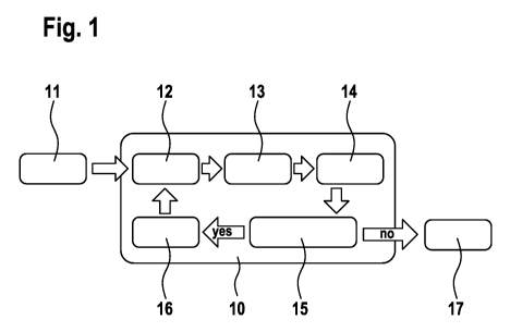
Fig. 1 schematically shows the course of a real-time PCR 10
as per the method according to the invention. After the
start 11 of the PCR process, the PCR cycles are started,
these individual steps being carried out by a control of
the temperature in a thermocycler. At regular intervals,
especially at defined time points within the PCR cycle (or
analogously at particular time points in isothermal
amplification processes), signals of the amplification are
captured and, for example, recorded and evaluated as
fluorescent images. The choice of the respective suitable
time point can, for example, depend on the probe
respectively used. In the example shown here, measurement
is, for example, carried out after each attachment step.
However, in most cases, measurement is carried out after
each elongation step. Each PCR cycle comprises the step of
denaturation 12 of the template DNA. The template DNA used
Date Recue/Date Received 2021-02-01
CA 03108364 2021-02-01
- 15 -
are sample nucleic acids and reference nucleic acids in
separate reaction preparations. The denaturation step 12 is
followed by the attachment (annealing) of the respective
primers in step 13. In this example, this is followed by
the measurement of the signals of the amplification in step
14. The measured signals are evaluated in step 15, a check
in particular being made to determine whether the measured
signal is classified as "amplification" or not. Especially
in comparison with the reference samples, a decision is
then made as to whether further PCR cycles are performed or
not. For example, if it is established in step 15 that the
measured signal should be classified as background, i.e.,
not as "amplification", the PCR cycle is continued with the
elongation step 16. Thereafter, the new PCR cycle starts
with the denaturation step 12. However, if it is established
in step 15 that the measured signal should not be rated as
background signal, but should be classified as
"amplification", the PCR process can be terminated, and
optionally further analyses and evaluations of the PCR
products formed can be carried out (step 17).
The detection of the signals in step 14 is based on
fluorescent probes, by means of which an amplification
which has taken place is made detectable in various ways
known per se, for example by incorporation in the DNA
synthesis or by attachment or intercalation into the DNA.
Especially statistical testing is then carried out to
determine whether this new data point can be classified as
background with data points already measured in previous
PCR cycles or whether the signal significantly deviates
from the hitherto determined background signal and can be
referred to as "amplification".
Expediently, a minimum and a maximum PCR cycle number are
specified as boundary conditions for the PCR process. The
Date Recue/Date Received 2021-02-01
CA 03108364 2021-02-01
- 16 -
minimum cycle number defines from when a signal can be
expected at the earliest. These data points are
automatically assigned to the background and are not tested
for amplification. Said minimum cycle number can, for
example, be set to 10 or smaller. During these initial
cycles, a base line can be generated. The maximum cycle
number can form a termination criterion for the case of no
amplification being detectable in the sample. Said number
is typically the number of cycles that is also specified in
a classic PCR process.
Fig. 2 illustrates the evaluation (step 15 in Fig. 1) of
the detected fluorescent signals. Said fluorescent signals
can be captured in relation to individual PCR cycles or
else in relation to particular time points during the
amplification process, especially in the cases of
isothermal amplification processes (e.g., in the case of
whole genome amplifications). Subfigure A shows the
background (BG) or a baseline that is formed by individual
data points (open circles) which are measured especially in
early PCR cycles and in which there can be no assumption of
an amplification. The frame around the individual data
points represents an estimated background with certain
tolerances. This thus defined background is the basis of
the tests of the subsequent data points on the basis of the
measured fluorescent signals in following PCR cycles or in
the following amplification process. Subfigure B depicts
the subsequently measured data points as closed circles,
which are based on further fluorescent signals in
subsequent PCR cycles or in the subsequent process and which
are located within the frame of the background. The most
recent data point depicted with a cross represents the
current measurement value, which is likewise located within
the frame of the background. Here, it can be assumed that
no amplification has taken place. What is thus initially
Date Recue/Date Received 2021-02-01
CA 03108364 2021-02-01
- 17 -
calculated on the basis of the data points from preceding
cycles is the old background, i.e., the background is
ascertained for all points with the exception of the current
measurement value (BG1). When the current data point is
available, a second background BG2 is calculated, the
current data point being included. It is then possible to
carry out statistical testing to determine whether the two
possible backgrounds BG1 and BG2 significantly differ. The
statistical evaluation can be done as per the following
specification:
Hypothesis Hl: BG1 = BG2
Hypothesis HO: BG1 # BG2
If, as in subfigure B, P(H1) > P(H0), there is no
significant difference and no amplification has taken place.
The amplification process is continued. By contrast, if the
background changes significantly owing to the current data
point (P(H1) < P(H0)), an amplification can be assumed, as
depicted in subfigure C. This information is the basis of
further action in the amplification process and the process
can be ended.
Fig. 3 illustrates the embodiment of the amplification
process with which a starting concentration of a sample DNA
is determined. This example is elucidated with reference to
a PCR process. This example and the following examples can
also, for example, be applied to isothermal amplification
processes, wherein the observed amplification signals are
then assigned not to individual PCR cycles, but to discrete
time points in the amplification process. Concomitantly run
in parallel with the sample 31 are various reaction
preparations containing standards 32, 33, 34 and 35 as
comparative samples. Here, the standard 35 represents the
largest standard concentration Si and the standard 32
Date Recue/Date Received 2021-02-01
CA 03108364 2021-02-01
- 18 -
represents the smallest standard concentration S4. Between
the maximum and the minimum standard concentration, as many
intermediate stages of the standard concentrations as
desired can be chosen in principle. In this example, there
are two concentrations 52 and S3. The number of different
standard concentrations Si to Sn determines the resolution
of concentration determination. All the preparations are
run in parallel after the start 30 of the PCR process and,
during the individual PCR cycles, the signals of the
amplification are captured in step 36. In step 37, what is
evaluated is whether an amplification can be deduced or not.
This can be done especially by means of the method as
described in connection with Fig. 2. The numerals 1 and 0
depicted in the field 38 stand for a classification as
amplification ("1") or no amplification ("0"). The PCR
process can be terminated when an amplification is
established for the sample 31. In comparison with the
amplification results for the standard samples 32 to 35, it
is then possible to deduce the concentration interval in
which the starting concentration of the DNA in the sample
31 was present. If no amplification could be established
for the sample 31, but an amplification already appeared
for the lowest standard concentration 35, the PCR process
can likewise be terminated, since the starting
concentration in the sample 31 is below the detection limit
which is defined by the minimum standard concentration 35.
This approach is realized by the query 39, by a check being
made between the sample 31 and the comparative sample 32 or
the standard having the lowest concentration to determine
whether an amplification was established for one of the two
preparations. In this case, the PCR process is ended (step
40). If an amplification cannot be established either for
the sample 31 or for the standard S4 having the lowest
concentration 32, the next PCR cycle is carried out in step
41. This method allows an unambiguous assignment of a
Date Regue/Date Received 2021-02-01
CA 03108364 2021-02-01
- 19 -
concentration interval. The concentration intervals are,
then, defined by the number of standards. What is to be
expected here is that the standards Si to Sn provide
amplification signals successively from the greatest
concentration up to the lowest concentration as the PCR
process advances. If an amplification is established for
the sample 31, and at the same time an amplification for
the standards Si to Si (i < n), the starting concentration
for the sample 31 lies in the interval [Si, Si+i] . If the
establishment of an amplification for the standards is not
in agreement with the order of their concentrations, the
test is not valid. Thus, if one preparation having a lower
standard concentration shows an amplification at a PCR
cycle at which a standard having a higher concentration
does not yet show any amplification, the reactions are not
equally efficient or not comparable. The choice of the
standard concentrations can, for example, be made such that
they each differ from one another by a factor of 10. This
corresponds to a quantification in the context of a classic
real-time PCR.
Fig. 4 illustrates the method by means of an indicator
vector display for an infection detection. In addition to
the actual sample 51, three standards 52, 53, 54 are
concomitantly run, wherein the standard 52 is the standard
Si having the lowest concentration of the DNA to be detected,
the standard 53 is the standard S2 having a medium
concentration of the DNA to be detected and the standard 54
is the standard S3 having a maximum amount of the DNA to be
detected. The standard Si represents the detection limit.
Said detection limit is the latest termination criterion of
the reaction. If an amplification is established earlier
for the sample 51 and if the order of the appearance of the
amplification for the standards corresponds to the order of
their concentration, the test is rated as positive. Fig. 4
Date Recue/Date Received 2021-02-01
CA 03108364 2021-02-01
- 20 -
summarizes, in an indicator vector /, the evaluations to
determine whether the reaction can be rated as
amplification or not at a particular PCR cycle. In said
vector, each reaction vessel or each PCR preparation
(samples and standards) has an entry which is re-evaluated
after each cycle. A reaction is rated as "amplification" if
a signal is detectable above the background. The indicator
value 1 (true) is assigned thereto in the indicator vector
I. If no amplification is establishable, the indicator of
the reaction is set to 0 (false). In this example, the
standard 53 is the largest standard and is listed on the
left as upper detection limit. The second standard 52, which
in terms of amount is between the largest and the smallest
standard, follows next. Following at the third position is
the smallest standard Si, which represents the detection
limit. Following as the last entry in the state vector is
the actual sample 51. The experiment is initialized with /
= [0,0,0,0]. Fig. 4 shows the four vectors which represent
a valid test. All twelve other possible cases are not
permissible, and the test would have to be reported as
invalid. In the case of / = [1,1,1,0], the signal is in the
range of the detection limit. In this case, one or more
cycles can optionally be attached owing to noise of reaction
efficiency, so that any small differences present between
the individual reaction vessels do not lead to an error in
the test decision. In the last column of the display, the
test result is displayed as positive (+) or as negative (-)
for the respective vectors. Once one of these vectors is
present, the reaction can be terminated.
Fig. 5 illustrates the embodiment of the method for
application of a mutation detection, likewise as an
indicator vector display. In the case of a mutation
detection, what is generally used is a predefined amount of
the sample DNA in the sample 61 to be tested. Since the
Date Regue/Date Received 2021-02-01
CA 03108364 2021-02-01
- 21 -
amount is predefined, the same amount of standard DNA or
reference DNA is always used in the standards 62, 63 and
64. The standard Si 64 contains an initially charged
template DNA in which 100% has the mutation (M) to be
detected. Said standard Si forms the upper limit at which
an amplification should be detected first. The lower limit
and hence the last termination criterion of the reaction is
a standard S3 62 which contains 100% wild-type template (W).
Between these two limits, it is possible, then, to choose
multiple mixture ratios of mutation DNA and wild-type DNA.
In this example, a further standard S2 63 is provided that
contains 50% mutation (M = 50%). The setting of mixture
ratios of mutation DNA and wild-type DNA allows the division
of the sample into proportion bins, analogous to histograms.
The standard S2 with M = 50% that is chosen here allows a
categorization of the proportion of mutation of greater
than 50% and less than 50%. In this approach, it is thus,
for example, possible to determine the ploidity of the gene.
Finer subdivisions are achieved by the insertion of further
standards and by a numerical estimation of the efficiency.
As elucidated in the previous example with reference to Fig.
4, the reaction is checked via the state vector / and test
decisions are accordingly made.
Fig. 6 illustrates the method in connection with a whole
genome amplification. In the case of a whole genome
amplification, all sequences which occur in a sample are
amplified, i.e., not just defined DNA sequences which are
addressed via primers. In a classic whole genome
amplification, fluorescent probes, as is customary in a
real-time PCR, are not used; instead, the amplification
product which forms is visualized and quantified by the use
of specific dyes. Said specific dyes (e.g., PicoGreen ,
SYBR Green) intercalate into double-stranded DNA and, in
doing so, emit light more intensely, meaning that a rise in
Date Regue/Date Received 2021-02-01
CA 03108364 2021-02-01
- 22 -
fluorescence indicates an amplification that has taken
place. Thus, if a fluorescent signal is detectable, this
indicates the presence of double-stranded DNA, and so this
can thus be rated as amplification. Said dye is added in a
defined amount to the reaction mixtures in the preparations
for the process. Besides the actual sample 71 (S), three
further comparative samples 72, 73 and 74 are concomitantly
run in the process for the whole genome amplification. The
comparative sample 72 contains no template DNA as so-called
no template control (NTC). In the case of said sample, no
amplification should be establishable, since DNA to be
amplified is not present. As further comparative sample 73,
the DNA of a reference genome (RG) is concomitantly run.
Said reference genome is expediently species-specific. For
example, if a human genome is to be amplified as a whole,
the genome of one or more other persons is used as reference
genome. The amount used of the reference genome corresponds,
for example, to the maximum usable amount for a suitable
whole genome amplification system. Said reference genome in
the comparative sample 73 should therefore provide an
amplification signal first of all. After the start 70, the
reaction is performed and is tested for amplification until
the reference genome 73 and the sample 71 are positive in
the state vector (state 75). Thus, in the state 75, both
reference genome 73 and sample 71 show an amplification and
the NTC control 72 shows no reaction or amplification. It
is only in this state that sufficient DNA is present in the
sample, and up to this state, no nonspecific primer
amplification (formation of primer dimers) has taken place,
as has been shown on the basis of the control 72. This state
represents a first checkpoint. If the conditions for the
first checkpoint have been met, the reaction is continued,
but now tested for a new termination criterion 76. The new
test criterion 76 is the comparison of the intensity of the
amplification for the sample 71 and the quantitative
Date Recue/Date Received 2021-02-01
CA 03108364 2021-02-01
- 23 -
reference 74. Said quantitative reference 74 contains the
desired amount of reference genome, this corresponding to
the desired amount of product in the whole genome
amplification. Here, the quantitative reference 74 contains
all the components in the WGA preparation, like the other
preparations, with the exception of the amplification
enzyme. As a result, no amplification, i.e., no DNA
synthesis, takes place in the quantitative reference 74
during the reaction. What is provided is only a reference
fluorescent signal due to the initially charged fluorescent
dye. If, then, amplified sample 71 and the quantitative
reference 74 have the same fluorescence intensity, it can
be assumed that the same amount of double-stranded DNA is
present in principle in both preparations, and so the
reaction can be terminated in step 77. As a further
termination criterion, what can be provided is that the NTC
control 72 shows an amplification signal.
This method can also be applied to a specific, targeted
preamplification in which the amount of DNA that is
synthesized in a preamplification is checked. In this case,
specific primers are used instead of the whole genome, the
result being that specific gene segments are accordingly
highly copied. What can also be used here as probe instead
of a dye which intercalates at double-stranded DNA is a
specific fluorescently labeled probe which generates a
fluorescent signal depending on synthesized DNA, for
example a TaqMan probe with fluorophore and quencher. The
quantitative reference then contains the desired target
amount of amplified material, an equivalent amount of
cleaved probe, i.e., the same amount of free fluorophores
and quenchers, and a complementary residual amount of the
probe. The basis of this is that, in the case of a real-
time PCR preparation in a TaqMan probe system, a defined
starting amount of the probes (No = coV) is specified. When
Date Recue/Date Received 2021-02-01
CA 03108364 2021-02-01
- 24 -
the amplification starts, the probe is cleaved. The amount
of probes and free fluorophores is then dependent on the
copy number NAmplicon that arises. The residual probes Ns can
be calculated using Ns=No-NAmplicon. Instead of an NTC control,
what is concomitantly run as termination criterion is a
further reference which makes a detection limit for the
amplification (LoA - limit of amplification) detectable.
Here, a minimum genome dilution to be used is used. Here,
the first checkpoint is thus the amplification time point
at which an assay-specific, predefined genome dilution,
i.e., the reference LoA, was amplified.
The methods of the whole genome amplification as per the
explanations in relation to Fig. 6 and the mutation
detection as per the explanations in relation to Fig. 5 can
be linked to one another and be configured as a monitored
workflow for a mutation detection. Here, microfluidic
systems and/or pipetting robots can be used. Such a fully
automatic workflow can, especially in connection with
microfluidic systems, offer a very advantageous possible
use of the method according to the invention which can, for
example, be used in point-of-care applications.
Fig. 7 illustrates the instrument components which can be
used for the described real-time PCR processes. Here, the
basis is formed by an instrument which makes an optofluidic
real-time PCR possible and thus allows a signal readout of
optical signals in order to be able to observe the
amplification in the individual samples or PCR reactions on
the basis of fluorescence signals. Such an instrument
comprises a heating and cooling system 101 (thermocycler)
which interacts with the various PCR reaction preparations
102. Furthermore, the instrument has an optical unit 103
which effects the readout of the amplification signals.
Furthermore, a device for fluid handling 104 can be provided,
Date Recue/Date Received 2021-02-01
CA 03108364 2021-02-01
- 25 -
for example a robot system or a corresponding microfluidic
system. Altogether, it is advantageous to configure such a
system as a microfluidic system, since a microfluidic
system can be operated with very small sample volumes and
allows semiautomation or full automation. The system is
furthermore provided with a reaction control unit 105 which
effects an in situ evaluation of the optical data. To
realize the feedback real-time PCR system of the present
invention, the reaction control unit 105 is configured such
that it can interact with all units of the system. The
reaction control unit 105 can especially control the
dynamic adjustment of the number of PCR cycles depending on
the observed signals of the amplification.
Date Regue/Date Received 2021-02-01