Note: Descriptions are shown in the official language in which they were submitted.
87735454
1
METHOD, DEVICE AND SYSTEM FOR DETERMINING THE CONCENTRATION
OF ANALYTES IN A SAMPLE
[0001] The subject application claims priority from US provisional Application
No. 62/715,026, filed August 6, 2018.
FIELD OF TECHNOLOGY
[0002] The present disclosure relates to the field of analysis of a sample and
more
particularly to the field of determining the concentration of analytes in the
sample.
BACKGROUND
[0003] Hemolysis is a phenomenon wherein the red blood cells rupture in whole
blood,
releasing their content into the blood plasma. This condition may occur due to
various
reasons such as immune reactions, infections, and medications. Hemolysis may
occur within
the body of an individual or after the blood has been drawn out of the body. A
major cause of
hemolysis is the pre-analytical steps involved in blood sample handling,
including collection
of the blood sample from an individual. Hemolysis alters the composition of
the blood
plasma due to the presence of degradation products of blood cells. If the
composition of the
blood plasma is altered beyond a certain threshold for hemoglobin and
bilirubin, the blood
sample is flagged for hemolysis. In such cases, the blood sample may become
incapable of
further usage and therefore has to be rejected. Therefore, the object of the
invention is to
provide a method to determine concentration of analytes, particularly free
hemoglobin, in a
whole blood sample. Free hemoglobin can cause interference while measuring
levels of one
or more analytes in blood. The object of the invention is achieved by a method
and a device
for determining the concentration of analytes in whole blood.
87735454
2
SUMMARY
[0004] A method of determining a concentration of one or more analytes in a
sample is
disclosed. In one aspect of the invention, the method includes introducing the
sample
through a channel. Additionally, the method includes illuminating the sample
with light
having varying wavelengths. Furthermore, the method includes obtaining an
image of the
illuminated sample at each of the wavelengths. The method also includes
analyzing the image
to determine the concentration of the one or more analytes.
[0005] In another aspect, a system for determining the concentration of one or
more analytes
in a sample includes a channel configured to carry the sample. The device
further includes a
light source configured to emit light at varying wavelengths, wherein the
sample in the
channel is illuminated at varying wavelengths using the light source.
Additionally, the system
includes a processing unit, a calibration database coupled to the processing
unit and a
memory coupled to the processing unit. The memory includes an image processing
module
configured for obtaining an image of the illuminated sample. The image
processing module is
further configured for analyzing the image to detect a cell-free plasma layer.
Additionally, the
image processing module is configured for determining the concentration of the
one or more
analytes in the cell-free plasma layer
100061 In another aspect, a device for determining the concentration of one or
more analytes
in a sample includes a channel configured to carry the sample. The device
further includes a
light source configured to emit light at varying wavelengths, wherein the
sample is
illuminated with light at varying wavelengths using the light source.
Additionally, the device
includes an imaging capturing module configured to capture an image of the
illuminated
sample.
87735454
2a
[0006a] In another aspect, there is provided a method of determining a
concentration of one or
more analytes in a sample, the method comprising: introducing the sample
through a channel,
wherein the sample is whole blood; generating a cell-free plasma layer in the
channel, wherein
the cell-free plasma layer comprises the one or more analytes; illuminating
the sample with light
having varying wavelengths; obtaining an image of the illuminated sample at
each of the
wavelengths; analyzing the images, including defining a threshold of intensity
value of pixels
associated with the cell-free plasma layer, detecting the cell-free plasma
layer in each of the
images based on the threshold and determining the concentration of one or more
analytes in the
cell-free plasma layer.
[0006b] In another aspect, there is provided a system for determining a
concentration of one or
more analytes in a sample, the system comprising: a processing unit; a
calibration database
coupled to the processing unit; a memory coupled to the processing unit, the
memory comprising
an image processing module configured for: obtaining an image of an
illuminated sample,
wherein the sample is whole blood; analyzing the image, including defining a
threshold of
intensity value of pixels associated with the cell-free plasma layer,
detecting the cell-free plasma
layer in the image based on the threshold, and determining the concentration
of one or more
analytes in the cell-free plasma layer.
[0006c1 In another aspect, there is provided a device for imaging one or more
analytes in a
sample, the device comprising: a channel configured to carry the sample,
wherein the sample is
whole blood; a light source configured to emit light at varying wavelengths,
wherein the sample
is illuminated at varying wavelengths using the light source, wherein a cell-
free plasma layer is
generated in the channel and wherein the cell-free plasma layer comprises the
one or more
analytes; and an image capturing module configured to capture an image of the
illuminated
sample and configured to transfer the captured image to a server for further
processing, the
further processing including defining a threshold of intensity value of pixels
associated with the
cell-free plasma layer, detecting the cell-free plasma layer in the image
based on the threshold
and determining the concentration of one or more analytes in the cell-free
plasma layer.
[0007] This summary is provided to introduce a selection of concepts in a
simplified folut that
are further described below in the following description. It is not intended
to identify
Date recue/Date received 2023-03-27
87735454
3
features or essential features of the claimed subject matter. Furthermore, the
claimed subject
matter is not limited to implementations that solve any or all disadvantages
noted in any part of
this disclosure.
BRIEF DESCRIPTION OF THE DRAWINGS
[0008] The present invention is further described hereinafter with reference
to illustrated
embodiments shown in the accompanying drawings, in which:
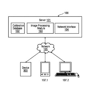
[0009] Figure 1 illustrates block diagram of a client-server architecture
which provides
geometric modeling of components representing different parts of a real world
object, according
to an embodiment.
[0010] Figure 2 illustrates a block diagram of a system in which an embodiment
of a method of
determining a concentration of one or more analytes in a sample can be
implemented.
[0011] Figure 3 illustrates an embodiment of a device for determination of a
concentration of
one or more analytes in the sample.
[0012] Figure 4 illustrates a flowchart of an embodiment of a method of
determining a
concentration of one or more analytes in a sample.
[0013] Figure 5 illustrates a flowchart of an embodiment of a method of
analyzing an image to
determine the concentration of one or more analytes.
[0014] Figure 6 illustrates a flowchart of an embodiment of a method of
determining a cell-free
plasma layer in an image.
[0015] Figure 7 illustrates an embodiment of an absorption spectrum of free-
hemoglobin,
bilirubin and lipids.
[0016] Figures 8a-8c illustrate an embodiment of a set of images obtained for
each analyte of
known concentrations, at varying wavelengths.
Date recue/Date received 2023-03-27
CA 031.08774 2021-02-04
WO 2020/033194
PCT/US2019/044307
4
[0017] Figure 9 illustrates an embodiment of a graphical representation
obtained for the
optical densities of free hemoglobin, bilirubin and lipids at known
concentrations and at
varying wavelengths.
[0018] Figure 10 illustrates an embodiment of a set of images obtained for an
unknown
sample at varying wavelengths.
[0019] Figure 11 illustrates an embodiment of graphical representations 1100
depicting the
consistency of the invention in determining concentrations of one or more
analytes in known
samples.
DETAILED DESCRIPTION
[0020] Hereinafter, embodiments for carrying out the present invention are
described in
detail. The various embodiments are described with reference to the drawings,
wherein like
reference numerals are used to refer to like elements throughout. In the
following description,
for purpose of explanation, numerous specific details are set forth in order
to provide a
thorough understanding of one or more embodiments. It may be evident that such
embodiments may be practiced without these specific details. In other
instances, well known
materials or methods have not been described in detail in order to avoid
unnecessarily
obscuring embodiments of the present disclosure. While the disclosure is
susceptible to
various modifications and alternative forms, specific embodiments thereof are
shown by way
of example in the drawings and will herein be described in detail. It should
be understood,
however, that there is no intent to limit the disclosure to the particular
forms disclosed, but on
the contrary, the disclosure is to cover all modifications, equivalents, and
alternatives falling
within the spirit and scope of the present disclosure.
[0021] FIG 1 provides an illustration of a block diagram of a client-server
architecture that is
a geometric modelling of components representing different parts of real-world
objects,
CA 03108774 2021-02-04
WO 2020/033194
PCT/US2019/044307
according to an embodiment. The client-server architecture 100 includes a
server 101 and a
plurality of client devices 107.1-107.2. Each of the client devices 107.1-
107.2 is connected to
the server 101 via a network 106, for example, local area network (LAN), wide
area network
(WAN), WiFi, etc. In one embodiment, the server 101 is deployed in a cloud
computing
environment. As used herein, "cloud computing environment" refers to a
processing
environment comprising configurable computing physical and logical resources,
for example,
networks, servers, storage, applications, services, etc., and data distributed
over the network
106, for example, the internet. The cloud computing environment provides on-
demand
network access to a shared pool of the configurable computing physical and
logical resources.
The server 101 may include a calibration database 102 that comprises captured
images of a
channel comprising whole blood. The server 101 may include an image processing
module
103 that analyzes the image of the whole blood to determine a concentration of
one or more
analytes. Additionally, the server 101 may include a network interface 104 for
communicating with the client devices 107.1-107.2 via the network 105.
100221 The client devices 107.1-107.n include a device 107.1 to determine the
concentration
of one or more analytes in the whole blood sample. The device 107.1 may be
configured to
capture an image of a processed whole blood sample. Such image may be sent to
the server
101 via a network interface. The client devices 1017.1-107.n also include a
user device 107.2,
used by a user. In an embodiment, the user device 107.2 may be used by the
user, to receive
the concentration values of the one or more analytes present in the sample.
The concentration
values can be accessed by the user via a graphical user interface of an end
user web
application on the user device 107.n. In another embodiment, a request may be
sent to the
server 101 to access the concentration values via the network 106.
100231 FIG 2 is a block diagram of a system 101 in which an embodiment can be
implemented, for example, as a system to determine the concentration of one or
more
CA 03108774 2021-02-04
WO 2020/033194
PCT/US2019/044307
6
analytes, configured to perform the processes as described therein. It is
appreciated that the
server 101 is an exemplary implementation of the system in FIG 2. In FIG 2,
the system 101
comprises a processing unit 201, a memory 202, a storage unit 203, an input
unit 204, an
output unit 205 a network interface 105 and a standard interface or bus 206.
The system 101
can be a (personal) computer, a workstation, a virtual machine running on host
hardware, a
tnicrocontroller, or an integrated circuit. As an alternative, the system 101
can be a real or a
virtual group of computers (the technical term for a real group of computers
is "cluster", the
technical term for a virtual group of computers is "cloud").
[0024] The processing unit 201, as used herein, means any type of
computational circuit,
such as, but not limited to, a microprocessor, microcontroller, complex
instruction set
computing microprocessor, reduced instruction set computing microprocessor,
very long
instruction word microprocessor, explicitly parallel instruction computing
microprocessor,
graphics processor, digital signal processor, or any other type of processing
circuit. The
processing unit 201 may also include embedded controllers, such as generic or
programmable
logic devices or arrays, application specific integrated circuits, single-chip
computers, and the
like. In general, a processing unit 201 can comprise hardware elements and
software
elements. The processing unit 201 can be configured for multithreading, i.e.
the processing
unit 201 can host different calculation processes at the same time, executing
the either in
parallel or switching between active and passive calculation processes.
[0025] The memory 202 may be volatile memory and non-volatile memory. The
memory
202 may be coupled for communication with the processing unit 201. The
processing unit
201 may execute instructions and/or code stored in the memory 202. A variety
of computer-
readable storage media may be stored in and accessed from the memory 202. The
memory
202 may include any suitable elements for storing data and machine-readable
instructions,
such as read only memory, random access memory, erasable programmable read
only
CA 03108774 2021-02-04
WO 2020/033194
PCT/US2019/044307
7
memory, electrically erasable programmable read only memory, a hard drive, a
removable
media drive for handling compact disks, digital video disks, diskettes,
magnetic tape
cartridges, memory cards, and the like. In the present embodiment, the memory
202 includes
an image processing module 103 stored in the form of machine-readable
instructions on any
of the above-mentioned storage media and may be in communication to and
executed by
processing unit 201. When executed by the processing unit 201, the image
processing module
103 causes the processing unit 201 to analyze the image of the sample to
determine the
concentration of one or more analytes. Method steps executed by the processing
unit 201 to
achieve the abovementioned functionality are elaborated upon in detail in
Figure 4, 5, and 6.
[00261 The storage unit 203 may be a non-transitory storage medium which
stores a
calibration database 102. The calibration database 102 is a repository of
images associated
with the whole blood in a channel 306. The input unit 204 may include input
means such as
keypad, touch-sensitive display, camera, etc. capable of receiving input
signal. The bus 207
acts as interconnect between the processing unit 201, the memory 202, the
storage unit 203,
the communication interface 107 the input unit 204 and the output unit 205.
100271 Those of ordinary skilled in the art will appreciate that the hardware
depicted in FIG 2
may vary for particular implementations. For example, other peripheral devices
such as an
optical disk drive and the like, Local Area Network (LAN)/ Wide Area Network
(WAN)/
Wireless (e.g., Wi-Fi) adapter, graphics adapter, disk controller,
input/output (I/O) adapter,
network connectivity devices also may be used in addition or in place of the
hardware
depicted. The depicted example is provided for the purpose of explanation only
and is not
meant to imply architectural limitations with respect to the present
disclosure.
100281 A system in accordance with an embodiment of the present disclosure
includes an
operating system employing a graphical user interface. The operating system
permits multiple
display windows to be presented in the graphical user interface simultaneously
with each
CA 03108774 2021-02-04
WO 2020/033194
PCT/US2019/044307
8
display window providing an interface to a different application or to a
different instance of
the same application. A cursor in the graphical user interface may be
manipulated by a user
through the pointing device. The position of the cursor may be changed and/or
an event such
as clicking a mouse button, generated to actuate a desired response.
[0029] One of various commercial operating systems, such as a version of
Microsoft
WindowsTM, a product of Microsoft Corporation located in Redmond, Washington
may be
employed if suitably modified. The operating system is modified or created in
accordance
with the present disclosure as described.
[0030] The present invention is not limited to a particular computer system
platform,
processing unit, operating system, or network. One or more aspects of the
present invention
may be distributed among one or more computer systems, for example, servers
configured to
provide one or more services to one or more client computers, or to perform a
complete task
in a distributed system. For example, one or more aspects of the present
invention may be
performed on a client-server system that comprises components distributed
among one or
more server systems that perform multiple functions according to various
embodiments.
These components comprise, for example, executable, intermediate, or
interpreted code,
which communicate over a network using a communication protocol. The present
invention
is not limited to be executable on any particular system or group of systems,
and is not
limited to any particular distributed architecture, network, or communication
protocol.
[0031] Disclosed embodiments provide systems and methods for analyzing a
sample. In
particular, the systems and methods may determine a concentration of one or
more analytes
in a whole blood sample.
[0032] Figure 3 illustrates an embodiment of a device 300 for determining the
concentration
of one or more analytes in the whole blood. The device 300 includes a light
source 301. The
light source 301 may be a multi-wavelength light source, i.e. capable of
emitting light of
87735454
9
varying wavelengths. In an embodiment, the light source 301 is configured to
emit light of at
least three different wavelength ranges. The wavelength ranges of the light
source 301 may
be, for example, between 400 nm and 420 nm; 440 nm and 460 nm; and 520 nm and
650 nm.
The wavelength ranges may be defined based on an absorption peak for each
analyte to be
determined. In an embodiment, the light emitted 305 from the light source 301
may be
homogenized using a diffuser 302. The device 301 further includes a channel
306 configured
to carry the whole blood sample. The channel 306 may be, for example, a
microfluidic
channel 306 or a microfluidic chip. The microfluidic channel 306 may have a
depth in the
range between 100 and 200 gm. Therefore, the path length of the light in the
channel 306 is
low. The channel 306 may be transparent so as to allow light from the light
source 301 to
interact with the whole blood and is transmitted out 307. The light 305 from
the light source
301 radiates on to the microfluidic channel 306 after passing through an iris
303 and a
collimating lens 304. The device 300 additionally includes an imaging
capturing module. The
image capturing module may include imaging lenses 308 and an imaging sensor
309,
configured to capture an image of the illuminated microfluidic channel 306.
The imaging
sensor 309 may be, for example a charge-coupled device (CCD) or a
complementary metal
oxide semiconductor (CMOS). In an embodiment, the image capturing module is
also
configured to transfer the captured image to the server 101 for further
processing. In another
embodiment, the image capturing module is an exemplary embodiment of the input
unit 204
in Figure 2.
[0033] Figure 4 illustrates a flowchart of an embodiment of a method 400 of
determining
the concentration of one or more analytes in the whole blood sample. At step
401, the whole
blood sample is introduced through the channel 306. The whole blood sample may
be
introduced into the microfluidic channel 306 from one end of the channel 306.
The whole
blood sample may form a uniform layer in the channel 306. At step 402, a cell-
free plasma
CA 03108774 2021-02-04
WO 2020/033194
PCT/US2019/044307
layer is generated in the microfluidic channel 306. The cell-free plasma layer
may be
generated, for example, using acoustophoresis. Acoustophoresis is a method of
causing
particles exposed to an acoustic standing wave field to move in the sound
field. Therefore,
when the whole blood sample is exposed to an acoustic standing wave field, the
blood cells
migrate towards the sound field, thereby generating a cell-free plasma layer.
Alternatively,
the cell-free plasma layer may be generated by differential wetting in
capillaries in the
microfluidic channel 306. At step 403, the cell-free plasma layer may be
illuminated with
light having varying wavelengths. The light from the light source 301 may be
directed to the
microfluidic channel 306 such that the cell-free plasma layer is illuminated
with the light.
The light source 301 may be capable of emitting light at varying wavelengths.
Therefore,
based on the type of analyte to be determined, the cell-free plasma layer may
be illuminated
with light of varying wavelengths. In an embodiment, the cell-free plasma
layer may be
illuminated with light at wavelengths chosen from a range between 400 nm and
420 nm; 440
nm and 460 nm; and/or 520 nm and 650 nm. The wavelength of the light may be
determined
based on the absorption peak value associated with the one or more analytes to
be
determined. Figure 7 illustrates an embodiment of an absorption spectrum 700
associated
with free-hemoglobin, bilirubin and lipids. According to the absorption
spectrum 700,
maximum absorbance for free hemoglobin is achieved at a wavelength range of
400 nm to
420 nm. Similarly, the maximum absorbance for bilirubin is achieved at
wavelength range of
440 nm to 460 nm. For lipids, the wavelength range of 520 nm to 650 nm is
chosen such that
there is minimum spectral interference from the other two analytes. Therefore,
the
wavelength range of 400 nm and 420 nm is associated with the analyte free
hemoglobin; the
wavelength range of 440 nm and 460 nm is associated with the analyte bilirubin
and the
wavelength range of 520 nm and 650 nm is associated with scattering of the
analyte lipid.
CA 03108774 2021-02-04
WO 2020/033194
PCT/US2019/044307
11
[0034] At step 404 of the method 400, an image of the illuminated cell-free
plasma layer in
the channel 306 is obtained. In an embodiment, the image of the cell-free
plasma layer may
be captured using the image capturing module 303, 304. The image may therefore
be
received from the image capturing module 303, 304. Alternatively, the captured
image may
be stored in the calibration database 102 and may be obtained from the
calibration database
102 for further analysis. Such image of the cell-free plasma layer may be
obtained each time
the plasma layer is illuminated with the chosen wavelength. Therefore, for
example, if the
cell-free plasma layer is illuminated with light having three different
wavelengths, one image
for each of the three wavelengths is obtained. At step 405, the obtained image
is analyzed by
the image processing module to determine the concentration of one or more
analytes in the
whole blood sample.
[0035] Figure 5 illustrates a flowchart of an embodiment of a method 500 of
analyzing the
image to determine the concentration of one or more analytes in the whole
blood sample. At
step 501, a cell-free plasma layer is detected in the image. The cell-free
plasma layer may be
detected in the image, for example, based on the pixel intensities. The method
steps involved
in detecting the cell-free plasma layer in the image is described in detail in
Figure 6.
Referring to Figure 6, a chart of an embodiment of a method 600 of determining
a cell-free
plasma layer in the image is illustrated. At step 601, a threshold associated
with an intensity
value of pixels of the cell-free plasma layer is determined. The pixels
associated with the cell-
free plasma layer may have a higher intensity pixel value in comparison to
pixel value
associated with the blood cells (predominantly red blood cells). Therefore, a
threshold may
be defined such that at step 602, the cell-free plasma layer may be detected
in the image
based on the threshold.
[0036] At step 502 of the method 500, an optical density associated with the
plasma is
determined at each of the chosen wavelengths. In an embodiment, the image
processing
CA 03108774 2021-02-04
WO 2020/033194
PCT/US2019/044307
12
module 105 may be calibrated with known standard samples of the analytes to be
determined,
before an unknown sample is tested. The calibration enables determination of
absorption
coefficient associated with each of the analytes to be determined. Therefore,
known samples
may be of free hemoglobin, bilirubin and lipid taken individually. Absorption
coefficients for
each analyte are constant and may depend on the material property of the
analytes and the
wavelength of illuminated light. In order to calibrate the image processing
module 105,
known standard samples of free hemoglobin, bilirubin and lipid are used at
defined
concentrations. The concentrations for free hemoglobin may be, for example, in
the range
between 0 mg/dL and 600 mg/dL.,. An image is obtained for concentrations of,
for example,
50 mg/dL; 100 mg/dL; 200 mg/dL; and 400 mg/dL of free hemoglobin at each of
the chosen
wavelengths. Similarly, the concentrations for bilirubin may be, for example,
in the range of
0 mg/dL to 50 mg/dL. An image is obtained for concentrations of, for example,
1.25 mg/dL;
2.5 mg/dL; 5mg/dL; 10 mg/dL; 20 mg/dL; and 40 mg/dL of bilirubin at each of
the chosen
wavelengths. Known standard concentrations of lipid may range from 0 mg/dL to
800
mg/dL. An image is obtained for concentrations of, for example, 75 mg/dL; 150
mg/dL; 300
mg/dL and 600 mg/dL.
100371 An optical density is calculated for each analyte, at each
concentration. Optical
density is a logarithmic ratio of falling radiation to the transmitted
radiation through the
sample. Optical density is a fraction of absorbed radiation at a particular
wavelength. Optical
density may be calculated using the following mathematical expression:
Optical density = - log -110
where /refers to mean pixel value of the sample and /0 refers to mean pixel
value of blank.
Optical density may also be referred to as a product of absorption coefficient
and
concentration. Therefore, for a given analyte, the optical density may be
depicted as:
Optical density = [Cl
87735454
13
where E is the absorption coefficient of the analyte and C is the
concentration of the analyte.
Therefore, for pure and known samples of free hemoglobin, bilirubin and
lipids, the optical
density may be calculated.
[0038]
Figures 8a, 8b, and 8c illustrate an embodiment of a set of images 801, 802,
803,
respectively, obtained for each analyte of known concentrations, at varying
wavelengths. The set
of images 801, 802, 803 may be the calibration dataset. The first set of
images 801 is associated
with free-hemoglobin. The images are obtained for free hemoglobin
concentrations of 50 mg/dL;
100 mg/dL; 200 mg/dL and 400 mg/dL. The free hemoglobin sample at each of
these
concentrations is illuminated with light having a wavelength in the ranges of
400 nm to 420 nm;
and/or 440 nm to 460 nm; and/or 520 nm to 650 nm. From the image set 801, it
is observed that
the absorption peak for free hemoglobin at each concentration is achieved at
wavelength range of
400 nm to 420 nm. The second set of images 802 is associated with bilirubin.
The images are
obtained for bilirubin concentrations of 1.25 mg/dL; 2.5 mg/dL; 5mg/dL; 10
mg/dL; 20 mg/dL;
and 40 mg/dL. The bilirubin sample at each of these concentrations is
illuminated with light
having a wavelength in the ranges of 400 nm to 420 nm; and/or 440 nm to 460
nm; and/or 520
I1M to 650 nm. From the image set 802, it is observed that the absorption peak
for bilirubin at
each concentration is achieved at wavelength range of 440 nm to 460 nm. The
third set of images
803 is associated with lipids. The images are obtained for lipid
concentrations of 75 mg/dL; 150
mg/dL; 300 mg/dL and 600 mg/dL. The lipid sample at each of these
concentrations is
illuminated with light having a wavelength in the ranges of 400 nm to 420 nm;
and/or 440 nm to
460 nm; and/or 520 nm to 650 nm. From the image set 803, it is observed that
lipids scattering of
illuminated light at each concentration is achieved at wavelength range of 520
nm to 650 nm.
[0039] Figure 9 illustrates a set of graphical representations 901, 902, 903
obtained for the
optical densities of free hemoglobin, bilirubin and lipids at known
concentrations and at
Date recue/Date received 2023-03-27
CA 03108774 2021-02-04
WO 2020/033194
PCT/US2019/044307
14
varying wavelengths. The graphical representation set 901 depicts that a
gradient for
absorption of light with respect to free hemoglobin concentration is steeper
for wavelength
range of 400 nm to 420 nm with respect to the other wavelength ranges.
Similarly, the
graphical representation set 902 for bilirubin depicts maximum optical density
achievement
at wavelength range of 440 nm to 460 nm. Additionally, the graphical
representation set 903
depicts scattering of light due to lipids. The absorption coefficients for
each of the analytes at
each wavelength range may be derived from the graphical representations 901,
902, 903 and
an absorption coefficient matrix may be computed.
CHb lElib(4) EBU(AV)
ELip(Av) -1 0 D (4)
CB11 = EHb(11b) EBil(Ab) ELip(a-b) OD (Ab)
C LiP _ Ellb (AO EBu(g) ELip(Ag) OD(A9)
[0040] In an embodiment, the image processing module 105 may be trained based
on the
images obtained for samples with known concentrations and the absorption
coefficient matrix
to accurately determine the concentration in an unknown sample. Therefore,
when the whole
blood sample, containing the analytes in unknown concentrations is analyzed,
at step 502 of
method 500, the obtained images are analyzed to determine the optical density
of the
analytes. Figure 10 illustrates an embodiment of images 1000 obtained for the
cell-free
plasma layer at wavelength ranges of 400 nm to 420 nm; and/or 440 nm to 460
nm; and/or
520 nm to 650 nm. At step 503, the absorption coefficient of each analyte is
determined at
each of the varying wavelengths. The absorption coefficient matrix derived
from the known
samples is used to determine the concentration of the analytes in the whole
blood sample, at
step 504.
[0041] Figure 11 illustrates an embodiment of graphical representations 1100
depicting the
consistency of the invention in determining concentrations of one or more
analytes in known
samples. The graphical representation 1101 refers to analysis results of free
hemoglobin
CA 03108774 2021-02-04
WO 2020/033194
PCT/US2019/044307
sample at known concentrations of 50 mg/dL, 100 mg/dL and 200 mg/dL. The
standard
deviation of test results is as below:
Desired concentration (mg/dL) 50 100 200
Mean of determined
47.0 100.3 198.0
concentration (mg/dL)
Standard deviation 3,3 10.2 14.7
Coefficient of variation (%) 7.0 10.2 7.4
The graphical representation 1102 refers to analysis results of bilirubin
sample at known
concentrations of 5 mg/dL, 10 mg/dL and 20 mg/dL. The standard deviation of
test results as
below:
Desired concentration
5 10 20
(mg/dL)
Mean of determined
5 9.8 19.8
concentration (mg/dL)
Standard deviation 0.8 1.4 1.1
Coefficient of variation (%) 7.0 10.2 7.4
The graphical representation 1103 refers to analysis results of lipid sample
at known
concentrations of 200 mg/dL, 400 mg/dL and 600 mg/dL. The standard deviation
of test
results as below:
Desired concentration
200 400 600
(mg/dL)
Mean of determined 243.7 470.4 665.9
CA 03108774 2021-02-04
WO 2020/033194
PCT/US2019/044307
16
concentration (mg/dL)
Standard deviation 22.5 16.0 45.4
Coefficient of variation (%) 9.2 3.4 6.8
[0042] Since the image data set 1000 have several thousand pixels, the mean
values of pixels
do not significantly affect the results. Even in the presence of stray red
blood cells, the
invention provides desired results. As only a small area of the obtained image
is analyzed to
determine the concentration of analytes, low sample volumes of < 1 microliter
is sufficient
for optical analysis. Furthermore, the invention is cost effective as the
hardware components
are limited. Additionally, the channel 306 is reusable. The invention also
enables detection of
bilirubin and lipids in the sample along with hemolysis measurement.
[0043] The foregoing examples have been provided merely for the purpose of
explanation
and are in no way to be construed as limiting of the present invention
disclosed herein. While
the invention has been described with reference to various embodiments, it is
understood that
the words, which have been used herein, are words of description and
illustration, rather than
words of limitation. Further, although the invention has been described herein
with reference
to particular means, materials, and embodiments, the invention is not intended
to be limited
to the particulars disclosed herein; rather, the invention extends to all
functionally equivalent
structures, methods and uses, such as are within the scope of the appended
claims. Those
skilled in the art, having the benefit of the teachings of this specification,
may effect
numerous modifications thereto and changes may be made without departing from
the scope
and spirit of the invention in its aspects.