Note: Descriptions are shown in the official language in which they were submitted.
CA 03108813 2021-02-05
WO 2020/061690 PCT/CA2019/051362
1
A PASSIVE MIXING MICROFLUIDIC URINARY ALBUMIN CHIP (UAL-CHIP) FOR
CHRONIC KIDNEY DISEASE ASSESSMENT
PRIOR APPLICATION INFORMATION
The instant application claims the benefit of US Provisional Application
Serial
Number 62/735,295, filed September 24, 2018, and titled "A passive mixing
microfluidic
urinary albumin chip (UAL-Chip) for chronic kidney disease assessment", the
entire
contents of which are incorporated herein by reference for all purposes.
BACKGROUND OF THE INVENTION
Around 8-16% of the population suffer from chronic kidney disease (CKD). [1]
The cause of CKD varies but some of the most common factors include diabetes,
high
blood pressure and cardiovascular disease. There are few signs or symptoms in
the
early stage of CKD, which makes early diagnosis difficult. CKD can progress to
fatal
end-stage kidney failure if it is not treated properly. Optimal detection and
risk
assessment of CKD requires simultaneous estimation of both kidney function
(e.g.
glomerular filtration rate [GFR]) and kidney damage (e.g. albuminuria or
proteinuria).
[2]
Albuminuria is a pathological condition wherein the protein albumin is
abnormally
presented in the urine. Thirty micrograms per millilitre (30 g/ml) or higher
of albumin
in urine is considered an indicator of kidney damage. The measurement of urine
albumin level is necessary for early diagnosis and monitoring of kidney
disease. Urine
collection is non-invasive, which makes it an ideal sample for point-of-care
(POC)
detection.
There are a variety of methods for assessing urinary albumin excretion,
ranging
from the colorimetric dipstick method to immunoassays to high-performance
liquid
chromatography (HPLC)-based methods. [3] While the dipstick test is
inexpensive and
easy to perform, its accuracy is limited. Immunoassays and HPLC methods are
more
accurate but suffer from complicated test procedures and require specialized
facilities.
The traditional dipstick tests involve wetting a colorimetric dye-impregnated
test
strip with a sample of urine. The albumin concentration is determined by
either visually
CA 03108813 2021-02-05
WO 2020/061690 PCT/CA2019/051362
2
comparing the reaction colors with the color scales on the label or reading
the reaction
colors with for example an analyzer. Readings are only reported in terms of
negative,
trace, 1+, 2+, 3+ and 4+ or the semi-quantitative values of 30, 100, 300 or
2000 mg/dL
corresponding to each color change. The limit of detection (LOD) and accuracy
of the
dipstick tests is usually not good.For example, the Chemstrip can produce a
color
change only when the albumin concentration is higher than 60 pg/ml, which is 3
times
higher than the recommended 20 pg/ml threshold to determine microalbuminuria.
Furthermore, the emersion time of the strip in the urine and the standby time
after taking
it out before reading the signal are critical, raising the potential for
operator errors, (e.g.,
color changes that occur after 2 minutes are usually of no diagnostic value).
To improve
the detection limit, the Micral-Test strip was developed based on an
immunological
reaction. In this test, urine first passes through a conjugate fleece where
albumin binds
to specific, gold-labeled antibodies and then flows to a detection pad. A
chemical
reaction in the detection pad produces a color that is compared visually to
color blocks,
with colors representing albumin concentrations of 0, 20, 50, and 100 pg/ml.
Although
the Mical-Test strip has better accuracy, the -$10 cost per chip (vs <$1 per
standard
dipstick strip) makes it cost prohibitive for large-scale screening.
Traditionally, different immunologically-based laboratory methods such as
immunonephelometry, immunoturbidimetry, and radioimmunoassay, have been used
for the confirmation and measurement of microalbuminuria. These tests usually
have
higher accuracy than the dipstick strips and the radioimmunoassay was reported
to
have a LOD as low as 16 pg/L. However, some studies have suggested that
immunological methods cannot detect all intact albumin in the urine, which
raises the
potential for false negative errors in detecting albuminuria. In contrast,
HPLC-based
laboratory tests can detect both immunoreactive and immuno-unreactive intact
albumin. However, both the immunologically-based and HPLC-based laboratory
tests
are complicated to use and have high facility requirements. A POC method that
optimally balances accuracy, cost, simplicity and low facility requirement,
would be a
highly desirable tool to effectively address the epidemic of CKD in poor,
remote,
underserviced regions of the world.
CA 03108813 2021-02-05
WO 2020/061690 PCT/CA2019/051362
3
The calorimetric test for albumin typically uses bromocresol green (BOG) or
bromocresol purple (BOP), which is the same dye used in the dipstick tests. It
has been
reported that calorimetric albumin tests based on BOG or BOP suffer from
inaccuracy
at low albumin concentrations..
One method that balances accuracy, cost, simplicity and facility requirement
is
the nonimmunological fluorescent assay, which has been reported by Kessler and
colleagues. The principle of the method is based on a protein-dye complex
resulting
from the specific binding of a fluorescent dye to human albumin, which
generates a
strong fluorescent signal. Specifically, this method has higher accuracy than
the dipstick
method and lower cost and facility requirement than immunoassays and HPLC,
suggesting it a suitable method for POC albumin detection.
The conventional method to perform the fluorescent test is to mix the reacting
dye and the test sample at a fixed ratio in a well-plate; wait for several
minutes for the
mixture to react; and measure the fluorescent intensity from the reaction
product to
evaluate the albumin concentration. However, such a method requires precise
solution
metering equipment such as a pipette to reach the mixing ratio. In addition,
the time
window for detection is short, usually requiring reading of the signal within
5 minutes of
the reaction starting, as after 5 minutes, the signal will change due to
overreaction and
evaporation, which affects the accuracy of the measurement. Furthermore, the
well-
plate method requires relatively large volumes of reagents (for example, tens
of
microliters per well).
Microfluidics enables advanced sample processing, manipulation and analysis
in miniaturized fluidic devices. The low sample and reagent consumption, high-
throughput, low-cost, integration and portability make microfluidics suitable
for disease
bio marker detection.
SUMMARY OF THE INVENTION
According to a first aspect of the invention, there is provided a method for
mixing
unequal amounts of two reagents to produce a detectable reaction in a
microfluidic chip
comprising
CA 03108813 2021-02-05
WO 2020/061690 PCT/CA2019/051362
4
providing a microfluidic chip comprising:
a first reagent inlet in fluid communication with a first reagent channel,
said first reagent channel having a height, a width and a length, said first
reagent
channel having a first reagent flow rate defined by the height, the width and
the length
of the first reagent channel; and
a second reagent inlet in fluid communication with a second reagent
channel, said second reagent channel having a height, a width and a length,
said
second reaction channel having a second reagent flow rate defined by the
height, the
width and the length of the second reagent channel;
said first reagent channel and said second reagent channel meeting at a
junction point, said junction point in fluid communication with a reaction
channel, and
said reaction channel in fluid communication with an outlet;
applying a quantity of a first reagent solution to the first reagent inlet;
applying a quantity of a second reagent solution to the second reagent outlet;
applying a quantity of oil to the first reagent inlet and the second reagent
inlet,
said oil having a density slightly lower than a density of the first reagent
solution and a
density slightly lower than a density of the second reagent solution so that
said oil floats
on top of the first reagent solution and the second reagent solution;
said first reagent flowing along the first reagent channel at the first
reagent
channel flow rate and said second reagent flowing along the second reagent
channel
at the second reagent channel flow rate until said first reagent and said
second reagent
begin mixing at the junction point, thereby producing a detectable reaction;
and
detecting the detectable reaction within the reaction channel.
According to another aspect of the invention, there is provided a method for
detecting albumin in urine using a microfluidic chip comprising
providing a microfluidic chip comprising:
a first reagent inlet in fluid communication with a first reagent channel,
said first reagent channel having a height, a width and a length, said first
reagent
channel having a first reagent flow rate defined by the height, the width and
the length
of the first reagent channel;
CA 03108813 2021-02-05
WO 2020/061690 PCT/CA2019/051362
a second reagent inlet in fluid communication with a second reagent
channel, said second reagent channel having a height, a width and a length,
said
second reaction channel having a second reagent flow rate defined by the
height, the
width and the length of the second reagent channel;
5
said first reagent channel and said second reagent channel meeting at a
junction point, said junction point in fluid communication with a reaction
channel, and
said reaction channel in fluid communication with an outlet;
applying a quantity of a urine to the first reagent inlet;
applying a quantity of albumin-detecting dye to the second reagent outlet;
applying a quantity of oil to the first reagent inlet and the second reagent
inlet,
said oil having a density slightly lower than a density of the first reagent
solution and a
density slightly lower than a density of the second reagent solution so that
said oil floats
on top of the urine and the albumin detecting reagent;
said urine flowing along the first reagent channel at the first reagent
channel flow
rate and said second reagent flowing along the second reagent channel at the
second
reagent channel flow rate until said first reagent and said second reagent
begin mixing
at the junction point, thereby producing a detectable reaction, wherein said
first reagent
channel flow rate is approximately one sixth of the flow rate of the second
reagent
channel; and
detecting the detectable reaction within the reaction channel.
BRIEF DESCRIPTION OF THE DRAWINGS
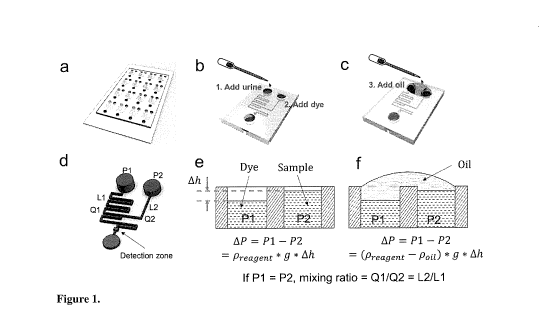
Figure 1. Illustration of working principle of the UAL-Chip. (a) A complete
UAL-
Chip with 16 mixing units; (b-c) The operation procedure for single mixing
unit; (d) The
fluidic network in single mixing unit. P1 and P2 are the hydraulic pressure in
the two
inlets. L1 and L2 are the lengths of the two branch channels. 01 and 02 and
the flow
rate of the two branch channels; (d-f) Explanation of the pressure-balancing
strategy.
Ah indicates the liquid height difference between the two inlets, which causes
the
pressure difference. Adding oil to connect these two inlets balances this
pressure
CA 03108813 2021-02-05
WO 2020/061690 PCT/CA2019/051362
6
difference, making the mixing ratio only dependent on the ratio of lengths of
the two
branch channels, which is identical for all the 16 units.
Figure 2. Validation of pressure-balancing strategy. (a) Four identical mixing
units were used to perform this test; (b) the fluorescent image just after the
converging area. The white dash box indicates the place used to plot the
intensity
profile; (c) The intensity profile in the four channels before pressure-
balancing; (d) The
intensity profile in the four channels after pressure-balancing.
Figure 3. Validation of UAL-Chip using albumin standards and CKD urine
samples. (a) The fluorescent signals depending on the albumin concentrations;
(b)The calibration curve of the intensity against the albumin standard
concentration.
The solid line is the linear fit of the data (R2 = 0.99); (c) Photostability
test between
UAL-Chip and well-plate; (d) Comparison between the UAL-Chip measurements and
well-plate measurements (Pearson correlation coefficient = 0.99; slope =
0.95); (e)
Positive correlation between albumin level and UACR value (Pearson correlation
coefficient = 0.73); (f) No correlation is found between albumin level and
eGFR value
(Pearson correlation coefficient = -0.17).
Figure 4. Schematic diagram of chip arrangement for measurement of both
albumin and creatinine from application of a single urine sample.
Figure 5. (A) Illustration of another embodiment of the UACR detection system;
(B) schematic diagram of one possible design of the fluorescent imaging
system.
Figure 6. One embodiment of a microfluidic chip for simultaneous detection of
creatinine and albumin. (A) Visualization of the channel pattern in the chip
using food
dye; (B) The detection of creatinine and albumin standard sample using a
colorimetric
dye and fluorescent dye. The creatinine detection dye is InfinityTM Creatinine
Reagent.
The albumin detection dye is albumin blue 580.
DESCRIPTION OF THE PREFERRED EMBODIMENTS
Unless defined otherwise, all technical and scientific terms used herein have
the
same meaning as commonly understood by one of ordinary skill in the art to
which the
invention belongs. Although any methods and materials similar or equivalent to
those
CA 03108813 2021-02-05
WO 2020/061690 PCT/CA2019/051362
7
described herein can be used in the practice or testing of the present
invention, the
preferred methods and materials are now described. All publications mentioned
hereunder are incorporated herein by reference.
Urinary albumin level is an important indicator of kidney damage in chronic
kidney disease (CKD) diagnosis but an effective, routine albumin detection
tool is
lacking.
A microfluidics-based fluorescent test of urinary albumin must be able to
precisely control the mixing ratio of the reacting dye and the test sample.
One possible
solution is to design mechanical structures for on-chip volume metering.
However, the
main drawback of this approach is the requirement for complex device designs
and/or
sample manipulation. [4] Alternatively, the mixing ratio can be controlled by
continuous
mixing of the reacting dye and the test sample driven by pressure flows at
defined flow
rate ratios. An obvious way is to use two external pumps to control the
injection rate of
the dye and sample. [5] However, this will increase the cost and complexity of
the
system. A standalone device that can still maintain a precise mixing ratio is
desirable
to enable the POC detection of albumin. Furthermore, a method that allows for
simple
sample preparation without the need of a precise pipette is preferable.
To accomplish these goals, we developed the fluorescent microfluidic urinary
albumin chip (UAL-Chip), which exploits the nonimmunological fluorescent
assay. In
.. this chip, we constructed a passive and continuous mixing module, in which
the loading
process requires only an inexpensive dropper, and the signal is stable over
time, as
discussed below. We applied a pressure-balancing strategy based on the
immiscible
oil coverage which highly improves the precision in controlling the mixing
ratio of sample
and dye. The UAL-Chip has achieved an estimated limit of detection (LOD) of
5.2 pg/m1
using albumin standards, which is below the 30 pg albumin per ml of urine
level
considered to be indicative of kidney damage. We also assessed the albumin
level in
12 CKD patients' urine samples. As discussed below, the results produced from
these
samples with the UAL-chip are consistent with the traditional well-plate
measurements
and clinical results.
CA 03108813 2021-02-05
WO 2020/061690 PCT/CA2019/051362
8
According to an aspect of the invention, there is provided a method for mixing
two reagents to produce a detectable reaction in a microfluidic chip
comprising
providing a microfluidic chip comprising:
a first reagent inlet in fluid communication with a first reagent channel,
said first reagent channel having a height, a width and a length, said first
reagent
channel having a first reagent flow rate defined by the height, the width and
the length
of the first reagent channel;
a second reagent inlet in fluid communication with a second reagent
channel, said second reagent channel having a height, a width and a length,
said
second reaction channel having a second reagent flow rate defined by the
height, the
width and the length of the second reagent channel;
said first reagent channel and said second reagent channel meeting at a
junction point, said junction point in fluid communication with a reaction
channel, and
said reaction channel in fluid communication with an outlet;
applying a quantity of a first reagent solution to the first reagent inlet;
applying a quantity of a second reagent solution to the second reagent outlet;
applying a quantity of oil to the first reagent inlet and the second reagent
inlet,
said oil having a density slightly lower than a density of the first reagent
solution and a
density slightly lower than a density of the second reagent solution so that
said oil floats
on top of the first reagent solution and the second reagent solution;
said first reagent flowing along the first reagent channel at the first
reagent
channel flow rate and said second reagent flowing along the second reagent
channel
at the second reagent channel flow rate until said first reagent and said
second reagent
begin mixing at the junction point, thereby producing a detectable reaction;
and
detecting the detectable reaction within the reaction channel.
Once the two reagents are added to the inlets, gravity will drive the
solutions to
flow toward the outlet by virtue of the outlet being empty. Because of the
small
dimensions of the channels, the flow can last for longer than 1 hour even
though the
volume added at the inlet(s) is small. This provides continuous and stable
mixing.
Furthermore, the reagents do not need to be added simultaneously because the
signal
CA 03108813 2021-02-05
WO 2020/061690 PCT/CA2019/051362
9
doesn't decay because of the continuous mixing. That is, the reagents in the
two inlets
don't need to be added simultaneously but the time gap should be smaller than
the time
required for one reagent to travel from one inlet to another inlet. Depending
on the
length of the channel, the time may be a few minutes.
In some embodiments, the flow can reach the junction point in about 2-3
minutes.
The flow is continuous until the pressure difference between the inlets and
outlet is
balanced, but this could take a relatively long period of time, for example,
longer than
1 hour, due to the small flow rate in microfluidic channels.
As will be appreciated by one of skill in the art, one of the reagents may be
a
bodily fluid, such as, for example, urine, serum or saliva, or may be another
suitable
fluid or solution that is being tested.
As will be appreciated by one of skill in the art, for reagents and samples
such
as the bodily fluids, most of these have a density that is close to water. For
example,
the density of urine is typically between 1.002 g/ml and 1.030 g/ml, serum is
typically
.. 1.025 g/ml, and saliva is typically around 1.0 g/ml.
As will be appreciated by one of skill in the art, a sample of interest can be
diluted
to a suitable concentration so as to fall within the detection range.
The detectable reaction may be for example a fluorescent reaction or a
colorimetric reaction.
For example, one of the reagents may be the dye reagent from the Albumin
Fluorescent Assay KitTM, FITC-dextran, rhodamine, or Texas red.
Other suitable reagents for use as part of a detectable reaction will be
readily
apparent to one of skill in the art. For example, any suitable reagent used in
a
commercially available kit for detection of a substrate of interest may be
used within the
invention. That is, there are a large number of assays known in the art which
produce
a detectable reaction, all of which can be used in the microfluidic chip of
the invention,
with the advantage that by adjustment of the flow rates of each channel as
discussed
herein, reagents for the reaction can be mixed together at the desired ratio
without
measuring the amount of each reagent applied. Similarly, the conditions under
which
CA 03108813 2021-02-05
WO 2020/061690 PCT/CA2019/051362
these reactions can be detected are also well-known in the art and can be used
with
the device and the method of the invention.
The oil may be for example but by no means limited to silicone oil (density of
0.971 g/m1), mineral oil (density of 0.85 g/m1) or FluorinetTM oil (density of
1.85 g/m1).
5 Other suitable oils will be readily apparent to one of skill in the art.
In some embodiments of the invention, the first reagent inlet and the second
reagent inlet are positioned on the chip such that the first inlet and the
second inlet can
be covered by a single drop of oil after the first reagent solution and the
second reagent
solution have been applied to the first reagent inlet and the second reagent
inlet
10 respectively. That is, the respective inlets are arranged such that they
can be covered
by a contiguous drop of oil or a single drop of oil once the reagents have
been applied
to the inlets, as discussed herein.
In some embodiments of the invention, the first reagent and the second reagent
are mixed at unequal amounts. That is, the detectable reaction does not
require 1:1
mixing of the two reagents by virtue of the engineered difference in the flow
rates, as
discussed herein.
As discussed herein, the quantity of the first reagent or the quantity of the
second
reagent may be an unmeasured quantity. That is, the reagents may be applied
without
measurement of the amount being applied to the inlets. As will be appreciated
by one
of skill in the art, this removes a considerable source of variability in
reactions as with
the device and method of the invention, there are no concerns regarding the
accuracy
of the amount of reagents used in the reaction.
For example, as discussed herein, each reagent may be applied to their
respective inlet as a single drop, which is traditionally considered to be
approximately
15 I to approximately 30 I.
As will be appreciated by one of skill in the art, in addition to depending on
channel dimensions, the flow rate of a given reagent solution will also depend
on the
fluidic viscosity, which one of skill in the art will understand needs to be
taken into
account when determining flow rate.
Each of the reagent channels may have a length of between 5 mm to 10 cm.
CA 03108813 2021-02-05
WO 2020/061690 PCT/CA2019/051362
11
The reaction channel must be long enough to allow for thorough mixing, which
will of course depend on the flow rate, and may have a length between 10 mm to
10
cm. In addition, the reaction channel may be configured so as to promote
mixing. For
example, the reaction channel may have a "zig zag" configuration, with many
turns, so
as to promote mixing.
The reaction channel and the reagent channels may have a width of between 50
prn to 1 mm.
As discussed in greater detail below, the mixing ratio can be represented as
01/02 = L2/L1, where 01 and 02 are the flow rates of the first reaction
channel and
the second reaction channel respectively, and L2 and L1 are the lengths of the
second
reaction channel and the first reaction channel, respectively.
For example, in one embodiment, the first reagent is urine and the second
reagent is a suitable dye reagent for the detection of albumin.
As discussed herein, these reagents can be combined to detect the presence of
.. albumin in urine. For the detection of albumin with the dye reagent from
the Albumin
Fluorescent Assay KitTM, the suggested mixing ratio is sample:dye = 1:6. In
some
embodiments, this reaction is detected at 620 nm, although other suitable
wavelengths
may be used and are within the scope of the invention, as discussed herein
In some embodiments, the width and depth of both reagent channels are the
same but the length of the urine reagent channel may be 6 times that of the
dye reagent
channel, for example, 36 mm for the urine channel and 6 mm for the dye
channel.
As discussed herein, in use, a single drop of urine and a single drop of the
fluorescent dye for detection of albumin may be applied to the first reagent
inlet and the
second reagent inlet respectively. That is, the reagents may be applied
without prior
.. measurement, without applying a measured or metered amount. Despite this,
the two
reagents will mix at a 6:1 ratio because of the difference in the flow rates
between the
two reagent channels, or in some embodiments, because of the difference in the
lengths of the two reagent channels.
In some embodiments, the width of the reagent channels may be about 60 pm.
CA 03108813 2021-02-05
WO 2020/061690 PCT/CA2019/051362
12
In some embodiments, the reaction channel may be about 14 mm long and about
100 j_im wide.
It is noted that other suitable dimensions can be readily determined by one of
skill in the art using routine experimentation.
As will be appreciated by one of skill in the art, such a method can be used
to
mix any two reagents that need to be added in unequal amounts. Furthermore,
the
amount of each reagent being added does not need to be accurately measured or
measured at all for the proper reaction to take place, that is, for the
reaction to take
place at the appropriate ratio.
While the examples above describe the use of two reagent channels, it is
important to note that in some embodiments the detectable reaction could be
produced
by more than two reagent channels, for example, channels for three reagents
that meet
at a single junction point.
Alternatively, two reagent channels may meet at a first junction point to form
a
first reaction channel and that first reaction channel may meet a third
reagent channel
at a second junction point downstream of the first junction point.
Furthermore, as shown in Figure la, one chip may include multiple sets of
reagent channels. As discussed below, the embodiment shown in Figure 1
includes 16
sets of identical channels. As discussed below, this may be used for example
to test
samples from 16 different individuals or may be used to test samples from 8
different
individuals twice etcetera.
Alternatively, a single chip may include different reagent/reaction channel
combinations, that is, wherein one set of reaction channels is arranged to
carry out a
specific detectable reaction while a second set of reaction channels is
arranged to carry
out a second detectable reaction. As will be appreciated by one of skill in
the art, in
these embodiments, the sample for each reaction may be the same so that for
example
a urine sample of an individual could be subjected to two different tests, for
example,
measurement of albumin levels and measurement of creatinine levels.
Furthermore, shown in Figures 4, 5A and 6 are chip designs that can be used
to detect urine albumin and creatinine at the same time. As discussed herein,
the ratio
CA 03108813 2021-02-05
WO 2020/061690 PCT/CA2019/051362
13
of L1 : L2 : L3 or the flow rate of C1: 02: 03 to achieve the desirable mixing
ratio
between creatinine dye : Urine and Albumin dye : Urine. It is of note that the
protocol
for use of this device would be similar to the albumin detection, that is, add
the dyes
and sample to the corresponding inlets, cover all the inlets with the same
drop of oil to
balance the pressure and detect the fluorescent signals in the detection
areas.
In the embodiment shown in Figure 6, Panel (A) shows the channel pattern in
the chip using food dye while Panel (B) shows the detection of creatinine and
albumin
standard sample using a colorimetric dye and fluorescent dye. In the example
shown
in Figure 6, the creatinine detection dye is InfinityTM Creatinine Reagent.
The albumin
-- detection dye is albumin blue 580. As can be seen in Panel (B), in this
embodiment,
the mixture containing creatinine and albumin to be tested is applied to a
central inlet.
As shown in Panel (A), this mixture, for example, a urine sample, flows along
a
channel that intersects with channels from the creatinine reporting agent, in
this case,
a colorimetric creatinine dye, and the albumin reporting agent, in this case,
a
fluorescent albumin dye. As can be seen in Panel (A), following the
intersection point
of the respective channels, the sample will mix with the colorimetric
creatinine dye
and the fluorescent albumin dye respectively. These channels continue to a
detection
zone which as can be seen in Panels (A) and (B) are of a greater volume than
the
channel, for easy detection of the colorimetric and fluorescent signal.
Accordingly, in
some embodiments of the invention, there may be one or more detection zones
integrated into the mixing channel that are of a larger volume, for example,
wider
and/or deeper, than the rest of the channel.
As will be appreciated by one of skill in the art, the ratio of urine to
creatinine
dye is dependent on the creatinine dye used. While each dye will have its
optimum
mixing ratio with the sample, determination of this mixing ratio is of course
routine
experimentation. Once this ratio is known, the lengths of the branch channels
can be
adjusted accordingly. As can be seen, the chip design is flexible to
adjustments to the
mixing ratio.
While the albumin dye used herein is albumin blue 580 fluorescent dye, there
-- are other colorimetric dyes for albumin, such as bromocresol green (BOG)
and
CA 03108813 2021-02-05
WO 2020/061690
PCT/CA2019/051362
14
bromocresol purple (BCP). For creatinine, picric acid is a calorimetric dye
used in the
famous Jaffe reaction. There are other reagents such as metal nanoclusters,
whose
fluorescent signal will be quenched after reacting with creatinine.
According to another aspect of the invention, there is provided a method for
detecting albumin in urine using a microfluidic chip comprising
providing a microfluidic chip comprising:
a first reagent inlet in fluid communication with a first reagent
channel, said first reagent channel having a height, a width and a length,
said first
reagent channel having a first reagent flow rate defined by the height, the
width and
the length of the first reagent channel;
a second reagent inlet in fluid communication with a second
reagent channel, said second reagent channel having a height, a width and a
length,
said second reaction channel having a second reagent flow rate defined by the
height,
the width and the length of the second reagent channel;
said first reagent channel and said second reagent channel
meeting at a junction point, said junction point in fluid communication with a
reaction
channel, and said reaction channel in fluid communication with an outlet;
applying a quantity of a urine to the first reagent inlet;
applying a quantity of albumin-detecting dye to the second reagent
outlet;
applying a quantity of oil to the first reagent inlet and the second reagent
inlet, said oil having a density slightly lower than a density of the first
reagent solution
and a density slightly lower than a density of the second reagent solution so
that said
oil floats on top of the urine and the albumin detecting dye;
said urine flowing along the first reagent channel at the first reagent
channel flow rate and said albumin detecting dye flowing along the second
reagent
channel at the second reagent channel flow rate until the urine and the
albumin
detecting dye begin mixing at the junction point, thereby producing a
detectable
reaction, wherein said first reagent channel flow rate is approximately one
sixth of the
.. flow rate of the second reagent channel; and
CA 03108813 2021-02-05
WO 2020/061690 PCT/CA2019/051362
detecting the detectable reaction within the reaction channel.
In some embodiments of the invention, the microfluidic chip further comprises
a
third reagent inlet in fluid communication with a third reagent channel, said
third
reagent channel having a height, a width and a length, said third reagent
channel
5 having a third reagent flow rate defined by the height, the width and the
length of the
third reagent channel, wherein said first reagent channel and said third
reagent
channel meet at a second junction point, said junction point in fluid
communication
with a second reaction channel and said second reaction channel in fluid
communication with a second outlet, said second junction point being distal to
and
10 separate from the junction point; and the method further comprises
applying a
creatinine detecting dye to the third inlet; said urine flowing along the
first reagent
channel at the first reagent channel flow rate and said creatinine detecting
dye flowing
along the third reagent channel at the third reagent channel flow rate until
the urine
and the creatinine detecting dye begin mixing at the second junction point,
thereby
15 producing a second detectable reaction.
In some embodiments, the first reagent channel is about six times as long as
the second reagent channel.
According to another aspect of the invention, there is provided a method for
detecting albumin and creatinine in urine using a microfluidic chip comprising
providing a microfluidic chip comprising:
a first reagent inlet in fluid communication with a first reagent
channel, said first reagent channel having a height, a width and a length,
said first
reagent channel having a first reagent flow rate defined by the height, the
width and
the length of the first reagent channel;
a second reagent inlet in fluid communication with a second
reagent channel, said second reagent channel having a height, a width and a
length,
said second reaction channel having a second reagent flow rate defined by the
height,
the width and the length of the second reagent channel; and
a third reagent inlet in fluid communication with a third reagent
.. channel having a height, a width and a length, said third reagent channel
having a
CA 03108813 2021-02-05
WO 2020/061690 PCT/CA2019/051362
16
third reagent flow rate defined by the height, the width and the length of the
third
reagent channel,
said first reagent channel and said second reagent channel
meeting at a first junction point, said first junction point in fluid
communication with a
first reaction channel, and said first reaction channel in fluid communication
with a first
outlet; and
said first reagent channel and said third reagent channel meeting
at a second junction point, said second junction point in fluid communication
with a
second reaction channel and said second reaction channel in fluid
communication
with a second outlet, said second junction point being distal to and separate
from the
first junction point.
applying a quantity of urine to the first reagent inlet;
applying a quantity of an albumin-detecting dye to the second reagent
outlet;
applying a quantity of a creatinine-detecting dye to the second reagent
outlet;
applying a quantity of oil to the first reagent inlet and the second reagent
inlet, said oil having a density slightly lower than a density of the first
reagent solution
and a density slightly lower than a density of the second reagent solution so
that said
oil floats on top of the urine and the albumin detecting reagent;
said urine flowing along the first reagent channel at the first reagent
channel flow rate;
said albumin-detecting dye flowing along the second reagent channel at the
second reagent channel flow rate and mixing with the urine at the first
junction point,
thereby producing a first detectable reaction, wherein said first reagent
channel flow
rate is approximately one sixth of the flow rate of the second reagent
channel;
said creatinine detecting dye flowing along the third reagent channel at the
third reagent channel flow rate and mixing with the urine, thereby producing a
second
detectable reaction;
CA 03108813 2021-02-05
WO 2020/061690 PCT/CA2019/051362
17
detecting the first detectable reaction to determine albumin
concentration; and
detecting the second detectable reaction to determine creatinine
concentration.
As discussed herein, we have developed a low-cost and high accuracy
microfluidic urinary albumin chip (UAL-Chip) for rapid detection of albumin in
urine.
There are three major advantages in the design of the UAL-Chip: (1) we
incorporated a fluorescent reaction assay into the chip to improve the
detection
accuracy; (2) we constructed a passive and continuous mixing module in the
chip that
provides user friendly operation and signal stability; (3) we applied a
pressure-
balancing strategy based on the use of immiscible oil coverage that achieves
precise
control of the mixing ratio of sample and dye.
We validated the UAL-Chip using both albumin standards and urine samples
from 12 CKD patients and achieved an estimated limit of detection of 8.4
pg/mIwhich
is below the 30 pg/m1 level that is indicative of kidney damage. The albumin
levels in
CKD patients' urine samples measured by UAL-chip is consistent with the
traditional
well-plate measurements and clinical results, as discussed below.
Specifically, the combination of a passive microfluidic mixer and a pressure-
balancing strategy to enable precise chemical mixing for albumin detection has
several
advantages including: 1) the operation is easy; 2) precise volume metering
equipment
is not necessary; and 3) the signal is stable over time.
Although this method is based on continuous flow, the consumption of reagent
is very small at any given time during the assay due to the low flow rate in
the
microfluidic device. Specifically, we have verified that 10 I of sample and
reagent could
maintain signal stability for more than 1 hour.
The method used in the UAL-Chip demonstrates a general method for the
detection of other target markers which require similar mixing strategies
between the
test sample and a reacting chemical. The mixing ratio can be easily tuned or
optimized
by changing the length of the branch channels.
CA 03108813 2021-02-05
WO 2020/061690 PCT/CA2019/051362
18
As discussed above, shown in Figure 1 is a UAL-Chip that includes 16 parallel
mixing units in a single device but more mixing units could be integrated
together to
improve the throughput. Additionally, one sample may be run multiple times,
thereby
providing greater accuracy in the results, as discussed above.
In this study, the signal was read by a fluorescent microscope. However, a
portable imaging system can be incorporated to make the system suitable for
POC test,
such as for example shown in Figure 5B. This could be achieved using
photodiode
detector as described in a previous report. [5] Furthermore, using the
smartphone to
read the fluorescent signal is becoming popular [6] and there are many good
examples
demonstrating the integration of smartphone and microfluidic technologies for
biomedical applications. [7, 8]
Compared with dipstick strips, UAL-Chip shows comparable low-cost (<$1) and
fast detection speed (<5 mins) but has lower LOD and better signal stability.
The low
LOD of 5.2 g/ml makes this method suitable for diagnosing microalbuminuria
and
monitoring the progression of kidney disease. The method used in the UAL-Chip
could
become a general method for the detection of other target markers, which rely
on a
similar mixing strategy between the test sample and a reagent. As suggested,
UACR
instead of albumin alone is a better indicator for kidney damage. Development
of a chip
that can measure both creatinine and albumin is easily achievable given the
development of simple fluorescent dyes for creatinine detection.
In some embodiments, passive mixing microstructures may be integrated into
the channel to achieve a thorough and rapid mixing in a short mixing channel.
In conclusion, the UAL-Chip represents a portable and disposable microfluidic
based tool for determining urinary albumin. The microchip is easy to fabricate
at low
cost and the operation is simple for end-users.
As will be appreciated by one of skill in the art, the method of the invention
may
be used to monitor kidney damage, for example, albumin levels in urine, of an
individual, as a means of monitoring disease progression.
CA 03108813 2021-02-05
WO 2020/061690 PCT/CA2019/051362
19
The method of the invention may also be used for screening at-risk
individuals,
for example, individuals with a familial history of chronic kidney disease,
with diabetes
mellitus, high blood pressure or glomerulonephritis.
Individuals diagnosed with chronic kidney disease and who show signs of the
disease worsening or anyone who is being monitored regularly and shows signs
of
the disease worsening may be prescribed medication to reduce blood pressure or
may be assigned to a low protein, low salt diet. Such individuals may also be
prescribed erythropoietin and/or calcitriol.
Accordingly, some embodiments of the method may be used to monitor kidney
damage on an ongoing basis and if the results indicate that the kidney damage
is
worsening, the patient is assigned a treatment, either a low protein, low salt
diet, or
medication to reduce blood pressure or specifically prescribed erythropoietin
and/or
calcitriol.
The invention will now be further explained and elucidated by way of examples;
however, the invention is not necessarily limited to or by the examples.
Results
EXAMPLE 1 - Working principle of the UAL-Chip
In the embodiment shown in Figure la, the UAL-chip has 16 identical mixing
units; however, we will discuss one unit to explain the working principle. As
illustrated
in Figures lb-d, the single mixing unit has two inlets and one outlet.
Specifically, each
inlet is connected to a channel and the two channels converge upstream of the
outlet.
The detection zone is located after the two streams converge and proceed along
an
extended zigzag mixing channel to allow the two input solutions to mix
thoroughly.
According to the design, the mixing ratio depends on the ratio of volumetric
flow
rates in the two branches (01/02). Due to the small dimension of the
microfluidic
channel, the flow inside the channel is considered as laminar flow. According
to
Poiseuille's Law, in the case of laminar flow, the volumetric flow rate is
given by the
pressure difference between the two ends of pipeline divided by the viscous
resistance:
CA 03108813 2021-02-05
WO 2020/061690 PCT/CA2019/051362
= (1)
Q2 = 1:313ne (2)
¨2
PO is the pressure at the converging point of the two branches. Ri and R2 are
the flow
resistances in the two branch channels. Pi and P2 are the pressures in the two
input
5 reservoirs. Both the urine and dye solution are water based solutions
with very low
concentration of solutes, it's reasonable to assume they have the same density
Preagent
and viscosity .
Pi and P2 can be estimated using the hydrostatic pressure equation:
= Preagent x g x h1 (3)
10 P2 = Preagent X g x h2 (4)
Where hi and h2are the liquid level heights in the two reservoirs.
In one embodiment of the UAL-Chip, the two branches have a rectangular shape
with the same width w and height h. The flow resistance Ri and R2 can be
expressed
as:
15 R1 12pL1 (5)
w^ h3(1-0.63x&vz)
1242
R2 (6)
w^ h3(1-0.6341)
We can see that Rioc Li and R2 OC L2, where Li and L2 are the lengths of the
branch channels. As will be apparent to one of skill in the art, other
arrangements,
wherein the width or depth, either alone or in combination with one another
and/or the
20 length, are within the scope of the invention.
According to Equation 1-6, if the pressures in the two inlets are same
(Pi=P2),
the mixing ratio can be easily calculated using the following equation:
Q1 R2 L2
Mixing ratio = ¨ = ¨ = ¨ (7)
Q2 R1 L1
In UAL-Chip, all the 16 units have the identical design, so the L2/Li is
constant.
As long as each unit can meet the requirement of Pi=P2, all the 16 units will
obtain the
same mixing ratio. On the other hand, if Pi is not equal to P2, the mixing
ratio will be
variable, which is proportional to the pressure difference AP = P1 ¨ P2. So
the critical
issue becomes how to make the AP as small as possible.
CA 03108813 2021-02-05
WO 2020/061690 PCT/CA2019/051362
21
According to Equation 3-4, Pi and P2 are dependent on the liquid levels hi and
h2 in the two reservoirs. However, in the actual experiment, it's difficult to
make hi = h2
because of variations in the loading volumes and dimensions of the reservoirs.
To
address this issue, we used a pressure balancing strategy by covering and
connecting
the two inlets with oil. We chose the oil which is immiscible with the
reagents and whose
density is a little bit smaller than the reagent; so the oil won't mix with
the reagent and
will float on the top. The principle of the pressure balancing is illustrated
in Figure lc-f.
Before adding the oil, the pressure difference between the two inlets is
AP = P1 ¨ P2 = Preagent X g x Ah (8)
After adding the oil on the top and connecting the two inlets, the pressure
difference becomes
AP = P1 ¨ P2 = (Preagent Poil) X g x Ah (9)
If the density of the oil is very close to the density of the reagent, the
Preagent ¨
pou becomes very small and thus the pressure difference AP becomes negligible.
To
give an example, we assume the height difference between the two loading ports
is
lmm; the density of the reagent and oil is 1 g/cm3 and 0.963 g/cm3 ,
respectively. The
pressure difference AP between the two ports will be 10 Pa before balancing
and 0.37
Pa after balancing. The difference could be further decreased if we use an oil
that has
a density closer to the reagent.
To validate this method, we fabricated a device with 16 parallel mixing units
and
we used 4 of them to test the flow-balancing strategy (Figure 2a). We added
FITC-
dextran dye and water into the two inlets respectively. Then we measured the
fluorescent profile in the area just after the two streams converged (Figure
2b). The
duty ratio in the profile can be used to indirectly represent the mixing ratio
of the dye
and water. In this experiment, we loaded different volumes in the four units
on purpose
to make the pressures imbalanced. As shown in Figure 2c, the intensity
profiles in the
four units are quite different. After we added oil to cover the inlets, the
intensity profiles
become almost identical (Figure 2d), suggesting that the pressures are
balanced and
thus the mixing ratio became identical in all the four mixing units.
CA 03108813 2021-02-05
WO 2020/061690 PCT/CA2019/051362
22
To summarize, the UAL-Chip is easy to operate by using passive pumping
method to infuse the reagents. The oil based pressure balancing strategy can
significantly decrease the variation of mixing ratios between different mixing
units, thus
improving the detection accuracy of the fluorescent assay.
EXAMPLE 2 - Validation of the UAL-Chip using the albumin standard
To get the calibration curve for the albumin detection, an HSA standard was
serially diluted to different concentrations (200, 100, 50, 25, 12.5, 6.25
g/mland blank).
The fluorescent signal was captured after loading the standard HSA and dye
solution
to the different mixing units of the microfluidic chip (Figure 3a). The
calibration curve
was plotted after subtracting the blank signal (Figure 3b). The R2 value of
the linear fit
is 0.99. The LOD calculated from regression line is 5.2 g/ml, which is > 3
times lower
than the normal range (clinical cut-off level: -20 g/ml).
Compared to the traditional well-plate method, UAL-Chip also showed great
advantage in signal stability due to its continuous mixing property. In the
well-plate
method, the sample and dye signal is premixed and loaded into a well. The
fluorescent
signal decays slowly due to concentration changes caused by solvent
evaporation. In
the UAL-Chip, fresh reagents flow into the detection zone continuously; the
sealed
channel and the oil that covers the reagent wells prevent evaporation as well.
As
illustrated in Figure 3c, the signal of the UAL-Chip maintained stable in the
one-hour
stability test while the signal using well-plate decayed significantly. The
standard
deviation of the signal intensity in one hour is 0.64 for the UAL-Chip and
8.11 for the
well-plate method. This indicates that the signal decays about 48% after 1
hour in well-
plate.
The UAL-Chip demonstrates a linear relationship between concentration and
fluorescent intensity in 0-200 g/m1 HSA standards and the LOD is below the
normal
range of albumin level in urine. In addition, the significant signal stability
of the UAL-
Chip decreases detection inaccuracy caused by variations in measurement time
points.
As will be appreciated by one of skill in the art, using the conventional
test, the
signal can decay by almost 50% within one hour whereas using the method
described
CA 03108813 2021-02-05
WO 2020/061690 PCT/CA2019/051362
23
herein, the signal is stable for over an hour. This decay in the prior art is
significant
because a signal that is read too late may miss an individual who has albumin
levels
of 30-40 g/ml (which is an indication of kidney disease).
EXAMPLE 3 - Validation of the UAL-Chip using CKD samples
The developed method was further validated using real clinical urine samples
from patients who have been diagnosed with CKD. The collected patient
information
includes disease stage, gender, and some biomarker measurements such as eGFR,
UACR, serum creatinine and albumin, HgA1C and low density lipoprotein (LDL).
We
measured the albumin concentration in the urine samples from 12 CKD patients
using
the UAL-Chip and the well-plated method. Because the dynamic range of the
albumin
concentration is large, a series of dilutions of the urine samples were
prepared and
measured. The concentrations were calculated based on the calibration curve.
The
albumin test results using the microfluidic mixer were in agreement with the
traditional
well-plate-based method (Figure 3d). The Pearson correlation coefficient of
these two
measurements is 0.99.
We further compared the albumin results measured by the UAL-Chip with the
clinical results (measured by Roche Cobas c501 using an immunoturbidimetric
assay).
Although clear positive correlation was observed between these two
measurements
(the Pearson correlation coefficient is 0.73; Figure 3e), they are not in
perfect
agreement, especially for the samples that have high albumin levels. There are
several
reasons for these differences. First, the tests were performed on different
samples
collected on different dates. Urinary albumin excretion rates in CKD vary
significantly
from day to day, diminishing the correlation between samples collected on
different
days. Moreover, this variability is higher at higher levels of albumin
excretion. In
addition, the multiple dilutions required to fit the measurement into the
linear range of
the calibration curve will have further weakened correlation at high albumin
levels.
We then used the recommended cutoffs of the urine albumin levels for
albuminuria (normal: <20 g/ml; microalbuminuria: 20-200 g/ml; clinical
albuminuria:
>200 g/ml) [5] to classify the patients into different groups and compared
the
CA 03108813 2021-02-05
WO 2020/061690 PCT/CA2019/051362
24
classification accuracy between the UAL-Chip results and clinical albumin
results
(Table 1). Only two patients were misclassified in the normal group and the
microalbuminuria group. The classification accuracy was 10/12 = 83%.
In clinical practice, UACR instead of albumin alone is considered a better
indicator of kidney damage as it can control for variations in urine
concentration. [4]
Three albuminuria categories (Al: UACR <3 mg/mmol, normal to mildly increased;
A2:
UACR 3-30 mg/mmol, moderately increased; A3: UACR >30 mg/mmol, severely
increased) are usually employed to indicate the kidney damage levels. [4] To
evaluate
the sensitivity, specificity, and positive and negative predictive values of
using the
albumin levels by the UAL-Chip results to determine albuminuria, we used cross-
tabulation tables for pairs of albumin positivity level
20 g/ml (or 200 g/ml) and
the reference standard of an UACR 3 mg/mmol (or 30 mg/mmol). The accuracy
values are as shown in Table 2. The sensitivity, specificity, PPV, NPV, and
accuracy of
the UAL-Chip results are 100% for both the detection of UACR 3 mg/mmol and
UACR
30 mg/mmol from the test results of 12 patients.
Although the sample size of 12 patients is relatively small, these results at
least
in part suggest that UAL-Chip is reliable in testing clinical urine samples
and its
measurement is comparable with traditional methods for measurement in a
clinical
setting.
Clinical test usually measure the UACR instead of albumin level to control for
variations in urine flow rate. Although UACR is a normalized ratio, the
changes in
albumin excretion will reflect change in the ratio. Indeed, clear positive
correlation was
observed between albumin concentration and UACR (Figure 3e). The Pearson
correlation coefficient is 0.73. We also compared the albumin level to the
eGFR value.
However, no correlation relationship is observed (Figure 3f). This is
reasonable
considering that eGFR and UACR are two independent markers for renal
assessment
and no direct association has been reported between them. These results
suggest that
UAL-Chip is reliable in testing clinical urine samples and its measurement is
comparable with the traditional method and measurement in a clinical setting.
CA 03108813 2021-02-05
WO 2020/061690 PCT/CA2019/051362
EXAMPLE 4 - Comparison of the UAL-Chip with the dipstick strips and other
laboratory methods.
Table 3 shows the comparison of our method with some widely-used dipstick
strips and laboratory methods. Compared with the immunologically-based and
HPLC-
5 based laboratory methods, our method has higher LOD but much lower cost.
Com-
pared with dipstick methods, our chip has comparable costs and detection speed
but
lower LOD, which can distinguish the microalbuminuria from normal level
(cutoff of 20
mg/L). In addition, the signal stability of our method is much better,
reducing the
potential for measurement error.
Methods
Microfluidic device design and fabrication
The device pattern was designed using AUTOCAD and printed on a transparent
film with high resolution. A SU-8 device master was fabricated on a 3-inch
silicon wafer
using photolithography process. Polydimethylsiloxane (PDMS) replica devices
were
made from the SU-8 master mold. 3mm diameter holes were punched in the PDMS
slab as the inlet and outlet reservoirs. The PDMS slab was bonded to a glass
slide to
seal the channel.
Clinical samples and reagents
The urine samples from CKD patients were collected at the Seven Oaks General
Hospital through an approved ethical protocol. The clinical descriptors of the
patients
were documented by the hospital (See SI for the information). The Albumin Blue
Fluorescent Assay Kit (Active Motif, 15002) was used to measure albumin levels
in
urine. The silicone oil (Alfa Aesar, A1272822) was used to cover and connect
the
solution loading ports to balance the pressure.
Albumin detection assay
The dye reagent and standard human serum albumin (HSA) was prepared
according to the product datasheet. Raw urine samples or diluted urine samples
by
CA 03108813 2021-02-05
WO 2020/061690 PCT/CA2019/051362
26
DPBS were used as the test samples. The operation procedure was illustrated in
Figure
la. The dye solution and test samples were added to the corresponding inlets
in the
microfluidic device using a dropper. The two inlets were then covered and
connected
by adding one drop of the oil on the top. Wait for 2-3 minutes until the
reagents enter
the detection zone. The fluorescent signal was recorded by an inverted
fluorescent
microscope. The signals of standard HSA were used to construct the calibration
curve.
The concentrations of the unknown samples were calculated according to the
calibration curve. The LOD is calculated from the regression line of the
calibration curve
(3*standard error of regression/slope). Each test has two repeats. We also
tested the
albumin level using the well-plate method as the control. The sample and dye
were
loaded and mixed in the wells using a pipette at a fixed ratio. The signal was
recorded
by a multi-plate reader (Synergy 4 HT). Each test sample was assayed in
duplicate.
Photostability test
The stability of the of the fluorescence intensity was compared between the
microfluidic method and well-plate method. In the microfluidic device, the
sample and
dye were loaded into the inlets and covered by oil. In the well-plate, the
sample and dye
were loaded into a well and mixed thoroughly using the pipette. The
fluorescence
emission was imaged by fluorescent time-lapse imaging for 1 hour with an
interval of 3
minute. The intensity versus time plots were used to evaluate the
photostability.
While the preferred embodiments of the invention have been described above,
it will be recognized and understood that various modifications may be made
therein,
and the appended claims are intended to cover all such modifications which may
fall
within the spirit and scope of the invention.
CA 03108813 2021-02-05
WO 2020/061690
PCT/CA2019/051362
27
Table 1. Classification accuracy according to the albumin levels from the UAL-
Chip results and the clinical results (Cobas c501, immunoturbidimetry). The
numbers in the table indicate different patients (totally 12 patients).
Albumin level Wimp UAL-Chip Clinical test
Normal (<20) 2 7
Microalbuminuria (20-200) 4, 7, 8 2, 4, 8
Clinical albuminuria (>200) 1, 3, 5, 6, 9, 10, 11, 12 1, 3, 5, 6, 9, 10, 11,
12
Table 2. Diagnostic accuracy of the UAL-Chip results for detections of UACR n
mg/mmol and UACRn mg/mmol (from total 12 patients).
Sensitivity Specificity PPV NPV Accuracy
Albumin 20 g/ml (UAL-Chip)
for detection of UACR 100% 100%
100% 100% 100%
mg/mmol
Albumin 200 g/ml (UAL-Chip)
for detection of UACR 30 100% 100%
100% 100% 100%
mg/mmol
CA 03108813 2021-02-05
WO 2020/061690 PCT/CA2019/051362
28
Table 3. Comparison of the UAL-Chip with the dipstick strips and other
laboratory
methods. Most of the data is from the manual books of each product. The data
of
radioimmunoassay arid HPLC method is from ref 9. ND: not determined.
LOD for Detection Signal
Assays Cost
albumin speed stability
Poor
150 Fast (<5
Multistix 10 SG $0.481s1rip (<2
mg/L minutes)
mins)
Poor
Fast (<5
Chemstrip 60 mg/L $0.65/strip (<2
minutes)
mins)
Dipstick strips
Poor
Fast (<5
Clinitek Microalburnin 30 mg/L $4/strip (<2
minutes)
mins)
Poor
Fast (<5
Micral-Test strip 20 mg/L $10/strip (<5
minutes)
mins)
Immunonephelometry
Fair (10
(Beckman Array 2 mg/L ND
minutes)
Analyzer)
Immunologically-
High (high-
I mmunoturbidimetry Fair (10 cost
based laboratory 3 mg/L ND
methods (Cobas c501) minutes) reagents
Extremely and
Radioimmunoassay 16 pg/L slow (3-4 equprnent ND
days) requirement)
Slaw (10-
HPLC laboratory method 2 mg/L ND
CA 03108813 2021-02-05
WO 2020/061690
PCT/CA2019/051362
29
minutes)
$0.1 &lest
(include the
Fast (<5 Good
UAL-Chip 5.2 mg/L coal of
nules) (>1 hr)
device and
reagent)
CA 03108813 2021-02-05
WO 2020/061690 PCT/CA2019/051362
REFERENCES
[1] Jha, V.; Garcia-Garcia, G.; Iseki, K.; Li, Z.; Naicker, S.;
Plattner, B.; Saran,
R.; Wang, A. Y.-M.; Yang, C.-W., Lancet 2013, 382 (9888), 260-272. DOI
http://dx.doi.org/10.1016/50140-6736(13)60687-X.
5 [2] ToneIli, M.; Muntner, P.; Lloyd, A.; Manns, B. J.; James, M. T.;
Klarenbach, S.; Quinn, R. R.; Wiebe, N.; Hemmelgarn, B. R., Annals of internal
medicine 2011, 154 (1), 12-21.
[3] Busby, D. E.; Bakris, G. L., The Journal of Clinical
Hypertension 2004, 6
(s11), 8-12.
10 [4] Du, W.; Li, L.; Nichols, K. P.; Ismagilov, R. F., Lab on a Chip
2009,9 (16),
2286-2292.
[5] Hofmann, 0.; Wang, X.; Bradley, D. D., Lab on a Chip 2005, 5 (8), 863-
868.
[6] Coskun, A. F.; Nagi, R.; Sadeghi, K.; Phillips, S.; Ozcan, A., Lab on a
Chip
15 2013, 13 (21), 4231-4238.
[7] Yang, K.; Wu, J.; Peretz-Soroka, H.; Zhu, L.; Li, Z.; Sang, Y.;
Hipolito, J.;
Zhang, M.; Santos, S.; Hillier, C., Biosensors and Bioelectronics 2018, 99,
259-267.
[8] Yang, K.; Peretz-Soroka, H.; Liu, Y.; Lin, F., Lab on a Chip 2016, 16
(6),
943-958.