Note: Descriptions are shown in the official language in which they were submitted.
CA 03109426 2021-02-10
WO 2020/037214
PCT/US2019/046835
PARTICLE-CONTAINING DROPLET SYSTEMS
WITH MONODISPERSE FLUID VOLUMES
Related Application
[0001] This Application claims priority to U.S. Provisional Patent
Application No.
62/719,476 filed on August 17, 2018, which is hereby incorporated by reference
in its
entirety. Priority is claimed pursuant to 35 U.S.C. 119 and any other
applicable statute.
Statement Re2ardin2 Federally Sponsored
Research and Development
[0002] This invention was made with government support under Grant Number
GM126414, awarded by the National Institutes of Health. The government has
certain rights
in the invention.
Technical Field
[0003] The technical field generally relates to small, sub-millimeter
particles having a
defined void or cavity formed therein that holds a fluid and is suspended in a
separate
immiscible fluid. More specifically, the technical field relates to dropicle
structures that are
formed from drop-carrier particles that hold fluid within the void or cavity
formed therein. In
one preferred embodiment, the drop-carrier particle is formed from a
hydrophilic hydrogel
material and holds an aqueous solution within the void or cavity.
Back2round
[0004] Microfluidics is the gold standard approach to form monodisperse
emulsions
(suspensions of dispersed drops of immiscible fluid in another continuous
phase of fluid).
Although microfluidics technologies have become more accessible, devices and
equipment
are expensive and require expertise. In some applications creating droplets
that contain
particles is also advantageous (e.g., for performing solid-phase reactions or
growing cells that
adhere to surfaces). Creating droplets that contain a single particle per
droplet is currently
challenging because of the stochastic processes of particle encapsulation in
droplets. An
alternative approach to create aqueous drops with increased monodispersity
compared to a
randomly mixed emulsion was described by Novak et al., Single-Cell Multiplex
Gene
Detection and Sequencing with Microfluidically Generated Agarose Emulsions,
Angewandte
Chemie International Edition, 50(2), 390-395 (2011).
1
CA 03109426 2021-02-10
WO 2020/037214
PCT/US2019/046835
[0005] In Novak et al., droplets were formed using simple agitation
(vortexing/pipetting)
of an aqueous phase containing dispersed particles into an oil water
suspension. Novak et al.
utilized re-emulsification of cell-encapsulated agarose beads to create
isolated compartments
for PCR amplification. This work relied on microfluidic devices for the
encapsulation of
cells and primer-functionalized beads in the agarose-gel droplets. Downstream
analysis after
PCR was performed by cytometry on the released primer beads, but not the
larger agarose
bead.
[0006] The particles contained in the droplets can act as templates to
define a minimum
droplet size. However, since the fluid surrounds the particle only in a thin
layer there are
significant disadvantages of this approach. The droplets formed by this
approach have large
variations in the "thin" volume formed around hydrogel particles, provide no
space for
encapsulation of microscale objects (e.g., cells, beads), and are not ideal
for reactions with
large molecules that cannot freely diffuse in the particle matrix.
Summary
[0007] In one embodiment, the use of microparticles which contain a void or
cavity region
connected to or in communication with the particle surface can act as
significantly improved
particle templates to generate a uniform distribution of droplets while also
containing an open
space to perform reactions or encapsulate cells, beads, and other small micro-
objects. These
cavity-containing particles also enhance the ability to encapsulate larger
volumes of an
aqueous fluid sample (per droplet and total sample volume for a plurality of
particles)
compared to non-cavity containing particles, which is important for cell
culture, cell secretion
analysis, and diagnostic analysis of large volumes of sample.
[0008] Microparticle shape and void design are important parameters to
control to achieve
uniform volume emulsions templated by these particles. Particle shapes that
can assemble
and nest/interlock with each other can lead to aggregated particles that
decrease the
uniformity of the drops formed upon mixing with a two-fluid-phase system. In a
preferred
embodiment microparticle shape is defined by a spherical envelope with an
inscribed
subtracted void volume within the spherical envelope. The subtracted void or
cavity may
take the shape of a sphere that interfaces, communicates with, or opens to the
outer surface of
the particle, creating a final particle with a crescent-shaped cross-section.
The inscribed void
intersects the spherical envelope at its surface in order to create a pathway
for fluid filling. In
one preferred aspect, the void intersects the spherical envelope at a narrow
opening (i.e., a
low fraction of the surface area of the spherical envelope). In some
embodiments this
2
CA 03109426 2021-02-10
WO 2020/037214
PCT/US2019/046835
fractional area defined by the opening is <33% of the overall spherical
envelope of the
particle, in others <10%, and in further embodiments the fractional area is
<5%.
Alternatively, in some embodiments the subtracted void does not intersect the
spherical
envelope's surface. In such an embodiment, cells or large molecules cannot be
isolated by a
drop templated by the particle, however, diffusion of water and small
molecules is possible
for a microparticle formed from a hydrogel or other porous material enabling
filling of the
void and molecular analyses or other downstream assays. In related
embodiments, the void
volume includes a polymer material with higher porosity/molecular diffusivity
compared to
the microparticle material. Note that in other embodiments the envelope shape
of the particle
may be ellipsoid or other shape that does not pack with large surface areas of
contact. The
void volume may also comprise one or more void regions subtracted from the
particle
volume.
[0009] Microparticle surface properties and materials should also be
controlled to support
the formation of drops. For example, microparticles with a hydrophilic surface
(e.g., low
interfacial tension with an aqueous phase compared to an oil phase) can be
used to template
aqueous-based droplets. Alternatively, a hydrophobic/fluorophilic particle
(e.g., low
interfacial tension with an oil/fluorinated oil phase compared to aqueous
phase) may be used
to template oil-based droplets in a separate immiscible continuous phase. The
oil phase in the
oil-based droplets may include fluorinated oils, mineral oils, silicone oils,
plant-derived oils,
animal-derived oils, crude oils, hydrocarbons or fuels, organic solvents, and
the like. In one
preferred embodiment, the microparticle is formed from a hydrophilic hydrogel
material that
templates the formation of an aqueous droplet of uniform volume based on an
inscribed void
volume that is contained in a fluorinated oil continuous phase.
[0010] In one exemplary embodiment, cavity or void-containing hydrogel
particles are
fabricated to create uniform cavities using an aqueous two-phase system
combined with
droplet microfluidics. In the exemplary embodiment, a PEG/dextran aqueous two-
phase
system is disclosed, although other aqueous two-phase systems could be used
such as
PEG/poly vinyl alcohol or PEG/high ionic strength salt systems, (or even three-
immiscible
phase systems). In one specific embodiment, PEG and dextran are co-flowed in a
flow-
focusing droplet generator to generate emulsions from the mixed materials that
phase-
separate to create two distinct regions in each drop. The outer PEG region is
crosslinked
(e.g., via UV excitation and presence of a photoinitiator and crosslinker) and
the inner
dextran layer is washed away to leave a large void space within the
microparticle. By tuning
the relative concentrations of both the PEG and dextran one can tune the
morphology and/or
3
CA 03109426 2021-02-10
WO 2020/037214
PCT/US2019/046835
volume of the void space or cavity within the microgel particle. Initial
testing of the
emulsification of the cavity-containing particles into oil show a uniform
range of droplet
sizes. Furthermore, the void space or cavity within the particles is shown to
be freely
accessible to large molecules such as high molecular weight FITC dextran
solution (500 kDa)
which enters the void or cavity.
[0011] In one embodiment, a droplet-based system that employs volumes
associated with
solid-phase particles suspended in an immiscible fluid includes a plurality of
three-
dimensional hydrophilic drop-carrier particles formed from a crosslinked
hydrogel, wherein
each hydrophilic drop-carrier particle has a void or cavity formed therein. An
aqueous fluid
is associated with the three-dimensional hydrophilic drop-carrier particles
and is disposed in
the void or cavity of the plurality of three-dimensional hydrophilic drop-
carrier particles. The
plurality of three-dimensional hydrophilic drop-carrier particles associated
with the aqueous
fluid are further disposed in an oil phase. In some embodiments, the aqueous
fluid disposed
in the void or cavity of the three-dimensional hydrophilic drop-carrier
particles have
substantially the same volumes (e.g., substantially monodisperse volumes). The
system thus
includes a plurality of solid particles having a defined void or cavity formed
therein that
holds a first fluid therein and is suspended in a second, separate immiscible
fluid. In some
embodiments, the first fluid may be an aqueous fluid while the second,
separate immiscible
fluid is an oil-based fluid. In other embodiments, the first fluid is an oil-
based fluid while the
second, separate immiscible fluid is an aqueous fluid.
[0012] In another embodiment, a droplet-based system that employs volumes
associated
with solid-phase particles suspended in an immiscible fluid includes a
plurality of three-
dimensional hydrophilic drop-carrier particles formed from a crosslinked
hydrogel, each
hydrophilic drop-carrier particle having a void or cavity formed therein. An
aqueous fluid is
associated with the three-dimensional hydrophilic drop-carrier particles and
disposed in the
void or cavity of the plurality of three-dimensional hydrophilic drop-carrier
particles. The
plurality of three-dimensional hydrophilic drop-carrier particles associated
with the aqueous
fluid are further disposed or suspended in an oil phase to form dropicle
emulsions.
Preferably, substantially all of the dropicle emulsions that are formed
contain a single drop-
carrier particle therein.
[0013] In another embodiment, a method of manufacturing hydrophilic drop-
carrier
particles includes providing a microfluidic droplet generator device having a
plurality of
inlets. A fluid solution containing a crosslinkable first component of an
aqueous two-phase
system containing a photoinitiator is flowed in one of the inlets, wherein the
crosslinkable
4
CA 03109426 2021-02-10
WO 2020/037214
PCT/US2019/046835
first component is poly(ethylene glycol) PEG, or a PEG-derivative. A solution
of a second
component of the aqueous two-phase system containing a crosslinker is flowed
in another of
the inlets. An oil phase is flowed into the device in another of the inlets,
whereby droplets
are formed in the microfluidic device, each droplet separating into separate
regions within the
respective droplet, the separate regions containing an enriched phase of the
crosslinkable first
component (e.g., PEG or PEG-derivative) and an enriched phase of the second
component of
the aqueous two-phase system (e.g., polymer such as dextran). The enriched
phase of the
crosslinkable first component is then crosslinked by exposure to light to form
hydrophilic
drop-carrier particles.
[0014] In another embodiment, a method of manufacturing hydrophilic drop-
carrier
particles includes providing a microfluidic droplet generator device having a
plurality of
inlets. A solution of a crosslinkable first component of an aqueous two-phase
system
containing a crosslinker is flowed into the droplet generator device in one of
the inlets,
wherein the crosslinkable first component is poly(ethylene glycol) PEG, or a
PEG-derivative.
A solution of a second component of the aqueous two-phase system is flowed
into the droplet
generator device in another of the inlets. An oil phase is flowed into the
droplet generator
device in another of the inlets, whereby droplets are formed in the
microfluidic device, each
droplet separating into separate regions within the respective droplet
containing an enriched
phase of the crosslinkable first component (e.g., PEG or PEG-derivative) and
an enriched
phase of the second component of the aqueous two-phase system. The enriched
phase of the
crosslinkable first component is then crosslinked by increasing the pH to form
hydrophilic
drop-carrier particles.
[0015] In another embodiment, a method of performing a cell secretion assay
using drop-
carrier particles includes the operations of: providing a plurality of three-
dimensional
hydrophilic drop-carrier particles, each particle having a void or cavity
formed therein;
loading cells into the voids or cavities of the plurality of three-dimensional
hydrophilic drop-
carrier particles; adding an affinity agent to the plurality of three-
dimensional hydrophilic
drop-carrier particles specific to a cell secretion of interest; emulsifying
the plurality of three-
dimensional hydrophilic drop-carrier particles containing the cells and
affinity agent to form
a plurality of dropicles; incubating the plurality of dropicles; breaking the
emulsion of the
dropicles to recover the three-dimensional hydrophilic drop-carrier particles
containing the
cells in an aqueous solution and adding a stain, dye, or other secondary
affinity reagent
specific to the secretion of interest on one or more of the plurality of three-
dimensional
hydrophilic drop-carrier particles; analyzing the plurality of three-
dimensional hydrophilic
CA 03109426 2021-02-10
WO 2020/037214
PCT/US2019/046835
drop-carrier particles of the prior operation for a signal formed or generated
by the stain, dye,
or other secondary affinity reagent specific to the cell secretion of interest
on one or more of
the plurality of three-dimensional hydrophilic drop-carrier particles. The
plurality of three-
dimensional hydrophilic drop-carrier particles may optionally be washed prior
to adding the
affinity agent.
Brief Description of the Drawin2s
[0016] FIG. 1A illustrates one embodiment of a dropicle.
[0017] FIG. 1B illustrates another embodiment of a dropicle.
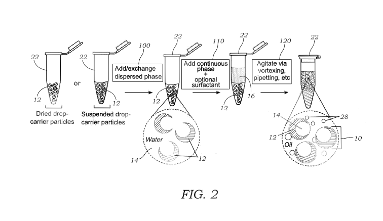
[0018] FIG. 2 illustrates a general schematic overview of dropicle
formation. Dried or
suspended drop-carrier particles are taken and dispersed phase with optional
surfactant is
added or exchanged. A continuous phase with optional surfactant is added, and
the
suspension is agitated (e.g., via vortexing, pipetting, etc.) to generate
emulsions of decreasing
size. After sufficient agitation dropicles of uniform size are formed along
with satellite
droplets.
[0019] FIG. 3 illustrates the formation of "dropicles" with a 5 uM FITC
dextran (500
kDa) solution as the dispersed phase, and NovecTM 7500 fluorinated oil with
0.5% Pico-
Surfrm as the continuous phase. Fluorescent images show distinct signal within
the cavity of
the dropicles.
[0020] FIG. 4A illustrates images of the emulsions formed with a microgel
particle
suspension is emulsified into NovecTM 7500 oil + 0.5% Pico-Surfrm with
spherical drop-
carrier particles shown in the TRITC channel (right).
[0021] FIG. 4B illustrates a graph of number of droplets as a function of
particles/droplet
showing that nearly all droplets contain either 0 or 1 particles.
[0022] FIG. 4C illustrates a graph showing droplet size distribution
showing a range of
non-uniform satellite droplets along with a uniform region of droplets formed
with
encapsulated spherical particles.
[0023] FIG. 4D illustrates a graph showing droplet size distribution
showing a range of
non-uniform satellite droplets along with a uniform region of dropicles formed
with
encapsulated crescent-shaped drop-carrier particles.
[0024] FIG. 4E illustrates a graph of the fraction of droplets as a
function of the number of
drop-carrier particles (crescent-shaped) per droplet (n=1207). Nearly all
droplets contain a
single drop-carrier particle.
6
CA 03109426 2021-02-10
WO 2020/037214
PCT/US2019/046835
[0025] FIG. 5 schematically illustrates the separation of dropicles from
satellite droplets.
In some embodiments an external force, or combination of external forces is
applied (e.g.,
magnetic, gravitational, buoyant, drag, centripetal, etc.) such that dropicles
and satellite
droplets experience a different force (magnitude and/or direction).
[0026] FIGS. 6A and 6B illustrate images of PEG-Vinyl Sulfone microgel
particles that
were gelled in the presence of thiolated magnetic particles (1 um) and
subsequently
emulsified in fluorinated oil. FIG. 6A shows the initial emulsification
resulted in aqueous
droplets encapsulating magnetic microgel particles in a large background of
empty satellite
droplets. FIG. 6B is taken after application of a magnetic field that enables
emulsified
magnetic microgel particles to be separated from the background of empty
droplets.
[0027] FIG. 7A illustrates one embodiment of the process used to fabricate
the cavity-
containing microparticles via an aqueous two-phase system. In this embodiment,
PEG (e.g.,
4-arm 10 kDa PEG-norbornene) and dextran (e.g., 40 kDa) phases are co-flowed
in a
microfluidic droplet generator device (FIG. 7B) to generate monodisperse
emulsions in oil
(e.g., NovecTM 7500 + 0.25% Pico-SurfTm). The PEG and dextran phase separate
once drops
are formed and the PEG phase is then crosslinked. In one embodiment UV
excitation is used
to crosslink the gels. More specifically, photoinitiator (2% w/v Lithium
pheny1-2,4,6-
trimethylbenzoylphosphinate (LAP)) is pre-dispersed in the PEG phase, and a
crosslinker
(DTT) is pre-dispersed in the dextran phase. After phase separation UV light
is used to
generate radicals which induce thiol-ene reaction between the PEG-norbornene
and DTT
crosslinkers to create a gel matrix. After crosslinking the particles are
washed to remove the
oil and dextran phases. Respective microscopic images of droplet generation,
phase
separation, and washing are illustrated below respective regions of the
microfluidic droplet
generation device.
[0028] FIG. 7B illustrates a microfluidic droplet generation device that is
used to form the
drop-carrier particles using the method of FIG. 7A.
[0029] FIG. 8A illustrates a phase diagram of example PEG-dextran aqueous
two-phase
system. In this embodiment, 20 kDa 8-arm PEG vinyl sulfone and 40 kDa dextran
solutions
were used. Emulsions were formed using the flow focusing/droplet generating
microfluidic
device illustrated in FIG. 7B. The morphology of the PEG and dextran regions
can be tuned
by changing their relative concentrations. At dilute concentrations the
dextran and PEG
phases remain mixed. At less dilute concentrations the PEG and dextran undergo
phase
separation. As the dextran to PEG concentration ratio is increased in the
final droplet, the
volume fraction of the inner dextran region is increased. As total
concentration of PEG and
7
CA 03109426 2021-02-10
WO 2020/037214
PCT/US2019/046835
dextran is increased, interfacial tension between the two phases causes
protrusion of the
dextran region (upper right-hand images). Droplets shown are approximately 100
micrometers in diameter.
[0030] FIG. 8B illustrates a phase diagram of example PEG-dextran aqueous
two-phase
system. In this embodiment, 10 kDa 4-arm PEG-norbomene and 40 kDa dextran
solutions
were used. By adjusting the concentrations of PEG and Dextran both the
morphology of the
droplets and the resulting UV crosslinked particles can be tuned.
[0031] FIG. 9 illustrates a range of drop-carrier particles fabricated
using the aqueous two-
phase system approach. After crosslinking and washing steps, drop-carrier
particles swell to
approximately 130% their original diameter. For conditions shown here 20% w/v
40 kDa
Dextran and 15% w/v 4-arm PEG-norbomene (10 kDa) were used. The 100 micrometer
and
80 micrometer diameter drop-carrier particles 12 were fabricated using a
microfluidic droplet
generation device with a channel height of 70 micrometers, PEG flow rate of 4
microliter/min, dextran flow rate of 1 microliter/min, and oil flow rate of 10
and 20
microliter/min, respectively. The 55, 45, and 40 micrometer diameter drop-
carrier particles
12 were fabricated using a microfluidic droplet generation device with a
channel height of 18
micrometers, PEG flow rate of 2 microliter/min, dextran flow rate of 0.5
microliter/min, and
oil flow rate of 5, 10, and 20 microliter/min, respectively.
[0032] FIG. 10A illustrates PEG-dextran emulsions formed in fluorinated
oil. Image
analysis (right) shows high-uniformity of both the PEG (CV=0.75%) and dextran
phase
(CV=1.45%). CV is coefficient of variation.
[0033] FIG. 10B illustrates crosslinked drop-carrier particles dispersed in
water and
imaged using fluorescence microscopy (left two images). Particles were
conjugated with a
TRITC-maleimide dye to view them in a fluorescent channel - a similar
fluorophore
conjugation step can be used for fluorescent barcoding. Distribution of
particle size is
uniform (CV<5%) as seen in graph of number of drop-carrier particles vs.
diameter which is
located on the right side of FIG. 10B.
[0034] FIGS. 10C and 10D illustrates drop-carrier particles dispersed in
water and imaged
using fluorescence microscopy. FIG. 10D shows the enlarged region of FIG. 10C.
[0035] FIG. 10E illustrates histograms of opening diameter and particle
diameter as
function of particle count. High uniformity was achieved by UV crosslinking of
phase
separated droplets while they remained in the microfluidic chip (Outer
diameter CV=1.5%,
opening diameter CV=2.1%).
8
CA 03109426 2021-02-10
WO 2020/037214
PCT/US2019/046835
[0036] FIG. 11A illustrates theoretical calculations of dropicle volume
variation. Here, a
dropicle is considered that is templated by a spherical particle and a
crescent or hollow drop-
carrier particle (i.e., void or cavity-containing particle), and compared to a
droplet formed by
a microfluidic device (without any particle). The drop-carrier particle is
constrained to a
fixed size, and the outer droplet diameter is varied relative to the particle
diameter. The
graphs demonstrate that the variance of the dispersed phase volume compared to
the total
volume is decreased with increased diameter by using the particle with a
cavity. More
specifically, variation in dispersed phase volume decreases as the ratio of
the internal cavity
diameter to the outer particle diameter increases, which is advantageous to
performing
uniform reactions in the dropicles.
[0037] FIGS. 11B and 11C illustrates theoretical calculations of reagent
encapsulation
efficiency in dropicles. Here, a dropicle is considered that is templated by a
spherical particle
and a crescent or hollow drop-carrier particle (i.e., void or cavity-
containing particle). FIG.
11B demonstrates increased encapsulation efficiency for particles with
relatively larger
cavities as well as for dropicles with thicker outer water layers. FIG. 11C
demonstrates that
increasing the concentration of drop-carrier particles before formation of
dropicles (reducing
void fraction) increases encapsulation efficiency.
[0038] FIG. 12 schematically illustrates how drop-carrier particles can be
decorated with
many different reactive moieties to enable particle functionalization and
dropicle
compatibility with many standard assays. Three methods of particle conjugation
include
through orthogonal reactive chemistries, biotin-streptavidin coupling, or cell
adhesive
peptides.
[0039] FIGS. 13A and 13B illustrates how digital nucleic acid amplification
assays such
as PCR (FIG. 13A) and digital ELISA (FIG. 13B) can be carried out in
dropicles. Drop-
carrier particles functionalized with nucleic acids or antibodies are mixed in
aqueous solution
with the appropriate assay reagents. Analytes of interest preferentially self-
associate with the
surface of drop-carrier particles allowing subsequent washing to remove
background signals.
Particles are then emulsified through mechanical agitation to form dropicles
isolating
reactions and accumulated signals within the droplet volume or attached to the
drop-carrier
particles themselves. Dropicles can then be analyzed through standard
microscopy, flow
cytometry, or via plate readers.
[0040] FIGS. 14A-14C illustrate dropicle emulsification does not affect
cell viability.
Jurkat cells stained with hoescht and calcein were suspended in aqueous
solution along with
drop-carrier particles. Drop-carrier particles were emulsified through
mechanical agitation in
9
CA 03109426 2021-02-10
WO 2020/037214
PCT/US2019/046835
fluorinated oil to form dropicles containing cells. Dropicles were imaged via
fluorescence
microscopy in brightfield (cell morphology) (FIG. 14A), DAPI (hoescht ¨
nuclear stain)
(FIG. 14B), and FITC (calcein ¨ cell viability) channels (FIG. 14C).
[0041] FIG. 15A-15D illustrate an overview of representative embodiments of
barcoding.
FIG. 15A illustrates how the size of internal cavity is maintained while
varying the outer
dimension of the drop-carrier particles. Dropicles are identified by size
range. In FIG. 15B, a
drop-carrier particle is modified with one or more dyes of varying intensity.
Unique particle
types are defined by intensity or ratio of multiple dye intensities. In FIG.
15C, magnetic
particles of varying number or magnetic content are embedded/crosslinked
on/into the
particles allowing for separation by relative magnetic force. In FIG. 15D,
varying number
and size of light scattering particles are embedded/crosslinked onto or into
the drop-carrier
particles allowing for separation based on relative amount/intensity of light
scattering or
scattered angle.
[0042] FIG. 16 illustrates an illustrative general workflow for cell
secretion analysis and
optional sorting. Drop-carrier particles are seeded into a well plate, flask,
etc. and cells are
then seeded into the drop-carrier particle cavities or voids. After cells
attach, drop-carrier
particles and associated cells are formed into dropicles to compartmentalize
by pipetting or
other mixing action with an oil-based continuous phase. Cells are incubated
and secreted
molecules are captured onto associated drop-carrier particles. Drop-carrier
particles with
associated cells are transferred back into the water phase and captured
secretions are labeled
with fluorescent molecules through, for example, a second affinity
interaction. Drop-carrier
particles and associated cells can then be analyzed and sorted using a flow
cytometer.
[0043] FIG. 17A schematically illustrates an embodiment in which antibodies
secreted
from a cell are captured on a drop-carrier particle associated with a cell. In
this example,
Anti-IL-8 secreting CHO cells are incubated in dropicles, and secreted
antibodies are
captured by protein A conjugated to drop-carrier particles. After transferring
back to an
aqueous phase, the capture antibodies are labeled with fluorescent Anti-IgG
for visualization.
[0044] FIG. 17B illustrates representative microscopy images showing high
fluorescent
signal above a threshold on drop-carrier particles associated with cells and
no fluorescent
signal or low fluorescent signal for drop-carrier particles without cells
attached. A brightfield
image (left) shows the drop-carrier particles and a cell in the cavity of one
drop-carrier
particle. The middle image shows a fluorescence microscopy image of the cell
using a
fluorescent stain that indicates a live cell. The right image shows a
fluorescence microscopy
image of the Anti-IgG staining of the secreted antibody covering the drop-
carrier particle.
CA 03109426 2021-02-10
WO 2020/037214
PCT/US2019/046835
[0045] FIG. 18 illustrates high-throughput sorting of drop-carrier
particles with associated
secreting cells using a fluorescence activated cell sorter. The top row of
images shows the
drop-carrier particles and associated cells prior to sorting. Brightfield
imaging is shown on
the left, fluorescence imaging showing live cells stained with calcein AM is
shown in the
middle, and fluorescence imaging in a separate channel showing captured
antibody secretions
labeled with Cy5 conjugated secondary antibodies specific to IgG are shown on
the right. All
three (upper panel) images are for the same field of view. These drop-carrier
particles and
associated cells are passed through a flow sorter. Forward scatter (FSC) and
side scatter
(SSC) for drop-carrier particle events in the cytometer are shown in a 2D
plot. The upper
right quadrant of events are gated and fluorescence intensity in the far-red
channel for these
events is shown in a histogram (right-side graph showing fluorescent
intensity). Microscopy
imaging demonstrates accumulation of drop-carrier particles with cells and
high
concentrations of captured proteins by sorting off high fluorescent signal
events above a
specified threshold shown in the image as the 'Sorting Gate'. All scale bars
are 200 microns.
[0046] FIG. 19 illustrates an example workflow for performing
sorting/analysis of single
cells or single cell colonies based on total secretion. Following previously
mentioned
approaches, single cells can be isolated into drop-carrier particles, and
emulsified into
dropicles where secretions accumulate without crosstalk and are captured onto
drop-carrier
particles. The drop-carrier particles can then be transferred back into water,
stained to
indicate the quantity of secretions, and analyzed/sorted along with the
attached cells. Sorted
sub populations of cells can then be expanded to perform repeated selection
steps. Screening
can be performed over multiple cycles to improve selection of desired sub-
populations. In a
related embodiment single cells seeded in the drop-carrier particles can be
grown to create a
clonal colony attached to a drop-carrier particle prior to emulsification.
This enables
combined analysis and sorting based on growth and secretion of a clone.
Detailed Description of the Illustrated Embodiments
[0047] FIG. 1A illustrates one embodiment of a dropicle 10. The term
"dropicle" as used
herein refers to a solid-phase particle or drop-carrier particle 12 that is
contained in, contains
or is associated with discrete volumes of dispersed (e.g., water) phase
solution or fluid 14
suspended in an immiscible phase 16 (e.g., oil). The drop-carrier particles 12
(or sometimes
also referred to as particles 12) are small, sub-millimeter scale (in their
longest dimension)
and spherically or ellipsoidal-shaped particles that are formed, in one
preferred embodiment,
from a cross-linked hydrogel material that is hydrophilic in one preferred
embodiment. A
11
CA 03109426 2021-02-10
WO 2020/037214
PCT/US2019/046835
typical range of dimensions (diameter or longest dimension) for the drop-
carrier particles 12
is between about 10 p.m and about 500 p.m, more preferably between 20 p.m and
200 p.m and
even more preferably between 40 and 120 p.m. Each drop-carrier particle 12 has
a void
volume or cavity 18 formed therein. The void or cavity 18 defines a three-
dimensional
volume that holds at least a portion of the dispersed phase solution or fluid
14 (e.g., aqueous
phase). Typical fluid volumes held within the void or cavity 18 include
volumes in the range
of about 100 fL and about 10 nL. A length dimension (e.g., diameter for
spherical void) of
the void or cavity 18 within a drop-carrier particle 12 is several microns,
typically more than
about 5 p.m and less than about 250 p.m. In some embodiments, the drop-carrier
particles 12
are contained within complete droplets of aqueous phase solution or fluid 14
with a portion of
the solution or fluid 14 also being located in the void or cavity 18. This is
illustrated in FIG.
1A.
[0048] In one embodiment, a droplet-based emulsion system is provided that
employs
discrete volumes associated with solid-phase drop-carrier particles 12
suspended in an
immiscible fluid. In this system, according to one embodiment, a plurality of
aqueous
dropicles 10 form an emulsion contained in an oil 16. The oil 16 acts as the
continuous phase
(i.e., the external phase of emulsions) while the aqueous-based dropicles 10
acts as the
dispersed phase (i.e., the internal phase of the emulsions, or the phase to be
encapsulated).
The oil 16 surrounds the dropicles 10 to create a monodisperse dropicle 10
emulsion.
Monodisperse refers to the ability of the dropicles 10 to retain substantially
the same volume
of fluid in each of the dropicles 10.
[0049] In another embodiment, a plurality of oil-based dropicles 10 form an
emulsion
contained in aqueous fluid 16. The aqueous fluid 16 acts as the continuous
phase (i.e., the
external phase of emulsions) while the oil-based dropicles 10 acts as the
dispersed phase (i.e.,
the internal phase of the emulsions, or the phase to be encapsulated). The
aqueous fluid 16
surrounds the dropicles 10 to create a monodisperse dropicle 10 emulsion. This
embodiment
is the reverse of the prior embodiment where the drop-carrier particle 12 is
hydrophobic and
the surrounding continuous phase is aqueous-based.
[0050] FIG. 1A illustrates an embodiment of the drop-carrier particle 12
that includes a
void or cavity 18 that interfaces, communicates with, or opens to the outer
surface of the
drop-carrier particle 12. As explained herein, the void or cavity 18 may be
formed as a
subtracted void or cavity that takes the shape of a sphere, creating a final
drop-carrier particle
12 with a crescent-shaped cross-section such as that illustrated in FIG. 1A.
The inscribed
void or cavity 18 intersects the spherical or elliptical envelope at its
surface in order to create
12
CA 03109426 2021-02-10
WO 2020/037214
PCT/US2019/046835
a pathway for fluid filling (and also access for cells, beads, and other micro-
objects). In one
preferred aspect, the void or cavity 18 intersects the spherical or elliptical
envelope at a
narrow opening (i.e., a low fraction of the actual surface area of the
spherical or elliptical
envelope of the drop-carrier particle 12). In some embodiments this fractional
area defined
by the opening is <33% of the overall spherical/elliptical envelope or surface
area of the
drop-carrier particle 12, in others <10%, and in further embodiments the
fractional area is
<5%.
[0051] Alternatively, in some embodiments the subtracted void or cavity 18
does not
intersect the spherical envelope's surface. For example, FIG. 1B illustrates
one such
embodiment of a drop-carrier particle 12 that includes a void or cavity 18
that is located
completely internal to the three-dimensional hydrophilic drop-carrier particle
12 and does not
intersect with a surface of the three-dimensional hydrophilic drop-carrier
particle 12. In this
embodiment, the material that forms the drop-carrier particle 12 may be made
permeable or
semi-permeable so that fluids, reagents, and/or sample may diffuse through the
drop-carrier
particle 12 and into the void or cavity 18. Likewise, reaction products in
some embodiments,
may be able to diffuse out of the drop-carrier particle 12 depending on size.
The particular
size cut-off for diffusion may be tuned by adjusting the porosity of the
underlying material
that forms the drop-carrier particle 12. Typically, larger species such as
cells, beads, or other
micro-objects would not be able to diffuse through the drop-carrier particle
12 while fluids
and small molecules and other species are able to diffuse through the drop-
carrier particle 12.
In a related embodiment, the void or cavity 18 holds or carries a porous
polymer material that
allows for molecular diffusion of sample molecules and reagents from the
surrounding fluid
(e.g., uncrosslinked dextran). Preferably, in this embodiment, the porous
polymer material
located in the void or cavity 18 is more porous than the underlying material
that forms the
drop-carrier particle 12.
[0052] In some embodiments, the surface of the drop-carrier particles 12
may be
decorated with one or more reactive or binding moieties 20 as seen in FIGS.
1A, 12, 13A,
13B, 16, 17A to enable dropicle 10 functionalization and dropicle 10
compatibility with
many standard assays. For example, reactive or binding moieties 20 may be
formed on the
surface of the drop-carrier particles 12 within the void or cavity 18. Binding
or reactive
moieties 20 may include, by way of example, nucleic acids, peptides, cell
adhesion peptides,
catalysts, enzymes, antibodies, primers, antigens, aptamers, biotin, or
biotin/streptavidin
complexes. Orthogonal reactive chemistries known to those skilled in the art
may also be
used for conjugation of reactive or binding moieties 20 to drop-carrier
particles 12.
13
CA 03109426 2021-02-10
WO 2020/037214
PCT/US2019/046835
[0053] As explained herein, in embodiments making use of hydrophilic drop-
carrier
particles 12, the formation of dropicles 10 is achieved by combining a
suspension of drop-
carrier particles 12 in an aqueous phase with oil (and optional surfactant)
and mixing (e.g., by
vortexing, pipetting, etc.) such as that illustrated in FIG. 2. Agitation and
fluid dynamic
shearing from mixing generate the emulsions of decreasing size. After
continued agitation,
drop-carrier particles 12 contained within the droplets act as a size
restraint that prevents
further shrinking of the droplet. Solid-templated droplets in which the drop-
carrier particles
12 contain voids or cavities 18 can also be thermodynamically stabilized. With
increasing
temperature or time, dropicles 10 do not coalesce in the same manner that
unsupported drops
of a dispersed phase in an aqueous phase will coalesce due to a decrease in
interfacial energy
of the system upon coalescence. Both pipetting and vortexing may be used to
form dropicles
10. Using mixing by pipetting and/or vortexing one can achieve uniform
emulsions of
dropicles 10 along with smaller satellite droplets containing no drop-carrier
particles 12. In
one embodiment of a uniform particle-templated emulsion substantially all of
the particle-
containing drops (e.g. > 95%, > 99%, or > 99.9%) are each associated with a
single drop-
carrier particle 12. Due to their unique size range, dropicles 10 can easily
be identified using
image analysis or filtered from the surrounding smaller satellite drops (i.e.,
background
droplets generated during dropicle 10 formation) using standard filtration
techniques.
Satellite drops also will not contain reactive moieties 20 that are attached
to drop-carrier
particles 12 and therefore do not proceed with reactions as occurs within
dropicles 10. When
the total aqueous volume of the sample is less than or equal to the sum of the
volumes that
can be supported by each of the drop-carrier particles 12 mixed with the
sample, satellite
droplets can be significantly reduced or eliminated.
[0054] In general, it was found that for the formation of dropicles 10
pipetting performed
better then vortexing for forming monodisperse dropicles 10 with a large
fraction of aqueous
emulsion containing only a single drop-carrier particle 12 (see, e.g., FIGS.
4B and 4E).
Dropicle formation can be improved by first breaking up an initial suspension
of drop-carrier
particles 12 in oil either by mechanically disturbing / tapping the reagent
tube containing the
samples, or by using a pipette with a larger diameter opening (e.g., 1000 uL
pipette), and then
breaking up into finer emulsions by vortexing or pipetting with a pipette with
a smaller
diameter e.g., 100, 200 uL pipette).
[0055] The concentration of surfactant present in the organic phase also
affects the
monodispersity of the formed dropicles 10 and the fraction of volumes that
contain a single
drop-carrier particle 12. As a general trend, increasing the concentration of
surfactant in the
14
CA 03109426 2021-02-10
WO 2020/037214
PCT/US2019/046835
oil phase led to better dispersion of drop-carrier particles 12 into single-
particle-containing
dropicles 10. Pico-Surf concentrations above 0.5% v/v in Novec 7500 led to
high
monodispersity. In a preferred embodiment 2% v/v Pico-Surf in Novec 7500 is
used as the
continuous phase, resulting in almost complete monodispersity of the formed
dropicles 10.
Further improvements to dropicle monodispersity can be attained through
addition of
aqueous surfactants within the aqueous phase (e.g., Pluronic F-127, Triton X-
100). In one
embodiment, 0.1% Pluronic F-127 is included in the aqueous phase to reduce
particle
aggregation and reduce interfacial tension, resulting in more uniform
dropicles 10. In another
embodiment, addition of 1% w/v PEG-5000 in the aqueous dispersed phase reduced
particle
12 aggregation, yielding more uniform dropicles 10.
[0056] It should additionally be noted that factors such as the polymer
composition of
drop-carrier particles 12, or the salt concentration of the dispersed phase,
influences the
affinity of drop-carrier particles 12 for one another in solution. For
example, spherical drop-
carrier particles 12 formed from higher wt% PEG were preferred. 6 wt% PEG was
more
preferable than 3 wt%. 12 wt% PEG drop-carrier particles 12 were even more
preferable.
Therefore, the polymer backbone formulation can affect the surfactant
concentrations needed
to properly disperse drop-carrier particles 12 in solution to form
monodisperse dropicles 10.
Additionally, dropicle monodispersity was increased using low salt solutions,
such as DI
water.
[0057] The choice of oil phase can also influence the function of the
resulting dropicles
10. For example, Novec-7500 infused with Pico-Surf surfactant was preferred in
forming
more uniform dropicles 10 than alternative oil phases such as FluorinertTM FC-
40 oil with
RAN surfactant, although this condition also was capable of forming dropicles
10. However,
FluorinertTM FC-40 with RAN surfactant displayed improved thermostability when
compared
to Novec-7500 with Pico-Surf. Thus, certain applications can benefit from an
exchange of oil
phase. For example, dropicles 10 may be formed in Novec-7500 to yield
monodisperse
emulsions, after-which Novec oil can be removed and replaced with FluorinertTM
FC-40 for
high temperature applications, such as thermocycling for DNA amplification.
[0058] FIG. 2 illustrates one method of forming dropicles 10. First drop-
carrier particles
12 that are either dried or suspended in a dispersed phase are provided. These
may be
contained in a holder or vessel 22 (e.g., Eppendorf tube is illustrated
although other holders
or vessels can also be used). If no dispersed phase is present, then the
dispersed phase (e.g.,
aqueous phase) is added to the holder or vessel 22. In another embodiment, the
initial
dispersed phase may be added to or exchanged with another dispersed phase as
seen in
CA 03109426 2021-02-10
WO 2020/037214
PCT/US2019/046835
operation 100 in FIG. 2. FIG. 2 shows the drop-carrier particles 12 contained
in the dispersed
phase 14. Next, as seen in operation 110, the continuous phase 26 is added
(e.g., oil phase)
along with an optional surfactant. The holder or vessel 22 is then subject to
an agitation
operation 120 which may include vortexing, pipetting, and the like. The
agitation operation
120 generates the dropicles 10 and, in some instances, satellite droplets 28.
The satellite
droplets 28 may be removed using, for example, filtration of the mixture. The
formation of
satellite droplets 28 may be minimized by controlling the volume of disperse
phase that is
added during formation of the dropicles 10.
[0059] FIG. 3 illustrates the formation of dropicles 10 with a 5 [tM FITC
dextran (500
kDa) solution as the dispersed phase 14, and NovecTM 7500 fluorinated oil with
0.5% Pico-
Surfrm surfactant added as the continuous phase 16. FIG. 3 illustrates the
drop-carrier
particles 12 contained in the dispersed phase 14 prior to emulsification
(left) and after
emulsification where dropicles 10 are formed. Corresponding images are
provided below
before emulsification (left image) and after emulsification (right image). The
fluorescent
images show distinct signal within the void or cavity 18 of the dropicles 10
(right image).
[0060] FIG. 4A illustrates images of the resulting emulsions formed (using
spherical
particles 12 to template water droplets in a continuous phase that is composed
of NovecTM
7500 fluorinated oil with 0.5% Pico-Surfrm surfactant). The dropicles 10 are
shown in the
brightfield image (left image) and the spherical drop-carrier particles 12 are
shown in the
fluorescent channel (right image). Both highly variably sized satellite
droplets 28 are shown
as well as uniform dropicles 10 which overlay with the fluorescent-stained
drop-carrier
particles 12. FIG. 4B illustrates a graph showing a count of droplets as a
function of drop-
carrier particles 12 per droplet. Of the droplets containing drop-carrier
particles 12, nearly all
contain only a single drop-carrier particle 12. FIG. 4C illustrates a graph
showing droplet
size distribution showing a range of non-uniform satellite droplets 28 along
with a uniform
region of droplets formed with encapsulated drop-carrier particles 12 (i.e.,
dropicles 10).
[0061] FIG. 4D illustrates a graph showing droplet size distribution
showing a range of
non-uniform satellite droplets 28 along with a uniform region of droplets
formed with
encapsulated crescent shaped drop-carrier particles 12 (i.e., dropicles 10).
Crescent drop-
carrier particles 12 were used to template water droplets in a continuous
phase comprised of
NovecTM 7500 fluorinated oil with 0.5% Pico-Surfrm surfactant. FIG. 4E
illustrates a graph
showing a count of droplets as a function of drop-carrier particles 12
(crescent-shaped) per
droplet. Of the droplets containing drop-carrier particles 12, nearly all
contain only a single
drop-carrier particle 12.
16
CA 03109426 2021-02-10
WO 2020/037214
PCT/US2019/046835
[0062] FIG. 5 illustrates the separation of dropicles 10 from satellite
droplets 28 according
to alternative embodiments. In these embodiments an external force, or
combination of
external forces is applied (e.g., magnetic, gravitational, buoyant, drag,
centripetal, etc.) such
that dropicles 10 and satellite droplets 28 experience a different force
(magnitude and/or
direction). For example, the drop-carrier particles 12 may contain a magnetic
material along
with an externally applied magnetic field that is used to separate the
dropicles 10 from the
satellite droplets 28 that do not respond to an applied magnetic field.
[0063] For example, FIGS. 6A and 6B illustrate images of PEG-Vinyl Sulfone
microgel
particles that were gelled in the presence of thiolated magnetic particles (1
um) and
subsequently emulsified in fluorinated oil and then separated. FIG. 6A
illustrates that initial
emulsification resulted in aqueous droplets (AD) encapsulating magnetic
microgel particles
in a large background of empty satellite droplets 28. FIG. 6B illustrates that
after application
of a magnetic field, emulsified magnetic microgel particles can be separated
from the
background of empty satellite droplets 28.
[0064] To manufacture the drop-carrier particles 12 with the void or cavity
18, a
microfluidic droplet generator device 30 is provided which is used to form a
monodisperse
emulsion in an oil phase whereby the internal dispersed phase comprises an
aqueous two-
phase system. One part of the aqueous two-phase system is a crosslinkable
hydrogel
precursor such as poly(ethylene glycol) (PEG) or a derivative thereof The
other part of the
aqueous two-phase system is a polymer such as dextran. The two phases of the
aqueous
two-phase system then separates into distinct regions within the formed
droplets. Then, one
component of the two-phase system (namely, a crosslinkable component) is
crosslinked to
form the drop-carrier particle 12. FIG. 7A schematically illustrates the
process of creating
drop-carrier particles 12 having a void or cavity 18 formed therein. As seen
in FIG. 7A, an
aqueous two-phase system is used to form the drop-carrier particles 12. In
this specific
embodiment the aqueous two-phase system includes PEG or a PEG-derivative
(e.g., 10 kDa
4-arm PEG-norbornene) which is the crosslinkable component (using a
crosslinker) and
dextran (e.g., 40 kDa) which is not crosslinked. The microfluidic droplet
generator device 30
is used to generate an emulsion of the aqueous two-phase system within an oil
phase. The
droplet that contains the two aqueous phase components (e.g., PEG and dextran)
separate
after droplet formation in a phase separation operation. After phase
separation, the PEG or
PEG-derivative component is crosslinked into a gel. For example, a crosslinker
such as
diothiothreitol (DTT) in the presence of a photoinitiator (e.g., Irgacure0
2959, LAP, etc.)
within the PEG or PEG-derivative component is then subject to light exposure
(e.g., UV
17
CA 03109426 2021-02-10
WO 2020/037214
PCT/US2019/046835
excitation) to initiate crosslinking. Of course, other crosslinkers such as
cysteine containing
peptides or other dithiols or multi-arm crosslinkers may also be used. In
another
embodiment, a thiol-ene reaction between multi arm PEG-Norbornene + dithiol
crosslinkers
is performed initiated via UV light and photoinitiator. In related
embodiments, either the
PEG and/or polymer phases can contain a combination of one or both the
photoinitiator and
crosslinker. After crosslinking the drop-carrier particles 12 can be washed to
remove the oil
phase and the dextran phases.
[0065] FIG. 7B illustrates the layout of the microfluidic droplet generator
device 30 which
is used to create the drop-carrier particles 12 having the voids or cavities
18 using flow
focusing. The microfluidic droplet generator device 30 includes a number of
microfluidic
channels 32, 34, 36 that converge/intersect into a droplet generation zone or
region 38 where
emulsions (droplets) are generated. The generated droplets travel down another
microfluidic
channel 40 that may lead to a chamber or region 42 where the droplets may
accumulate and
be temporarily stored. An outlet 44 in the device 30 may be used to remove the
droplets/drop-carrier particles 12. Each microfluidic channel 32, 34, 36
includes a respective
inlet 46, 48, 50 for inputting the various components. In this example, PEG
(e.g., 4-arm 10
kDa peg-norbornene) and dextran (e.g., 40 kDa) phases are co-flowed in the
microfluidic
device 30 to generate monodisperse emulsions in oil (e.g., NovecTM 7500 +
0.25% Pico-
Surfrm). In one embodiment, a first inlet 46 that leads to microfluidic
channels 32 is used to
deliver the oil phase along with the surfactant. A second inlet 48 that leads
to microfluidic
channel 34 is used to deliver the dextran. A third inlet 50 that leads to the
microfluidic
channel 36 is used to deliver the PEG as well as the crosslinker and the
photoinitiator. In
another embodiment a first inlet 46 that leads to microfluidic channels 32 is
used to deliver
the oil phase along with the surfactant. A second inlet 48 that leads to
microfluidic channel
34 is used to deliver dextran along with crosslinker. A third inlet 50 that
leads to the
microfluidic channel 36 is used to deliver the PEG as well as the
photoinitiator. Separation
of polymer precursor and crosslinker prior to droplet generation is especially
useful when
using highly reactive chemistries which may begin to crosslink within the
sample syringe or
channel inlet, halting flow. In another embodiment crosslinker is not included
in the PEG or
dextran phase, but is instead injected in its own additional inlet and
connecting channel. This
is advantageous when the crosslinker does not easily dissolve into the dextran
phase, for
example larger thiolated PEG crosslinkers.
[0066] The PEG and dextran phases separate once droplets are formed and the
PEG phase
is then crosslinked. As noted above, in one embodiment UV excitation is used
to crosslink
18
CA 03109426 2021-02-10
WO 2020/037214
PCT/US2019/046835
the gels. More specifically, photoinitiator (2% w/v Lithium pheny1-2,4,6-
trimethylbenzoylphosphinate (LAP)) is pre-dispersed in the PEG phase, and a
crosslinker
(DTT) is pre-dispersed in the dextran phase. After phase separation UV light
is used to
generate radicals which induce thiol-ene reaction between the PEG-norbomene
and DTT
crosslinkers to create a gel matrix that forms the drop-carrier particles 12.
Crosslinking may
occur in the microfluidic droplet generator device 30 or "off chip," after
droplets exit through
an outlet of 44 the microfluidic droplet generator device 30. After
crosslinking the drop-
carrier particles 12 are washed to remove the oil and dextran phases.
[0067] In another embodiment, a change in pH levels is used to crosslink
the drop-carrier
particle 12. For example, PEG-Vinyl sulfone/maleimide + dithiol crosslinker is
used at
lowered pH to prevent gelation prior to drop formation in a microfluidic
droplet generator
device 30. The pH is then increased in the dispersed phase by addition of an
organic base
(e.g., triethylamine) in the oil phase downstream to initiate the crosslinking
reaction. In a
related embodiment a parallelized step emulsification droplet generator
microfluidic device
30, as described in, de Rutte, J. M., Koh, J., Di Carlo, D., Scalable High-
Throughput
Production of Modular Microgels for In Situ Assembly of Microporous Tissue
Scaffolds. Adv. Funct Mater. 2019, 29, 1900071, which is incorporated herein
by reference,
operates on a pre-polymer phase mixed with the dextran phase in a single
solution to generate
monodisperse drops prior to phase separation and polymerization (e.g., via UV
light or pH
change). In all of the above embodiments, following crosslinking of the PEG,
the dextran
phase can be removed and the desired aqueous phase added or substituted to
form the
dropicles 10. Note that in some embodiments, such as when forming hollow drop-
carrier
particles 12, as exemplified in FIG. 1B, the internal dextran phase can be
maintained within
the void or cavity 18.
[0068] FIG. 8A illustrates a phase diagram of example PEG-dextran aqueous
two-phase
system. By tuning the relative concentrations of both the PEG and dextran one
can tune the
morphology and/or volume of the void space or cavity 18 within the drop-
carrier particle 12.
Here, 20 kDa 8-arm PEG vinyl sulfone and 40 kDa dextran solutions were used.
Emulsions
were formed using microfluidic droplet generator device 30 of FIG. 7B. The
morphology of
the PEG and dextran regions can be tuned by changing their relative
concentrations. At
dilute concentrations the dextran and PEG phases remain mixed. At less dilute
concentrations the PEG and dextran undergo phase separation. As the dextran to
PEG
concentration ratio is increased in the final droplet, the volume fraction of
the inner dextran
region is increased. As total concentration of PEG and dextran is increased,
interfacial
19
CA 03109426 2021-02-10
WO 2020/037214
PCT/US2019/046835
tension between the two phases causes protrusion of the dextran region (upper
right-hand
images). Droplets shown are approximately 100 micrometers in diameter. For
conditions
with high concentrations of PEG and dextran, phase separation was found to be
rapid (<1
second). At conditions near the binodal it was found that phase separation
would take longer
(>1 second).
[0069] FIG. 8B illustrates a phase diagram of example PEG-dextran aqueous
two-phase
system. In this embodiment, 10 kDa 4-arm PEG-norbornene and 40 kDa dextran
solutions
were used. By adjusting the concentrations of PEG and dextran both the
morphology of the
droplets and the resulting UV crosslinked drop-carrier particles 12 can be
tuned. At dilute
concentrations the PEG and dextran phases remain mixed as a single phase
(below the
binodal line). At higher concentrations (above the binodal), PEG and dextran
phase separate
into distinct PEG-rich and dextran-rich phases within the droplet. As the
total concentration
of PEG and dextran is increased the interfacial tension between the two phases
increases
causing the dextran phase to be in contact with a larger portion of the oil
interface (Top right
images). After crosslinking with UV light, the resulting drop-carrier
particles 12 (excluding
the one example that did not undergo phase separation) maintain similar cavity
volumes
(relative to total size of drop-carrier particle 12), but have increasing
cavity opening size. As
the relative ratio of dextran to PEG concentration is increased (bottom right)
the volume of
the dextran rich phase is also increased. After UV crosslinking, drop-carrier
particles 12 with
larger volume of dextran rich phase had proportionally larger cavities or
voids 18.
Additionally, it was found that for conditions near the binodal curve,
especially with higher
amounts of dextran, after crosslinking the cavities remain fully enclosed (the
void does not
touch the envelope of the outer particle 12).
[0070] FIG. 9 illustrates a range of drop-carrier particles 12 fabricated
using the aqueous
two-phase system approach using the microfluidic droplet generator device 30.
After
crosslinking and washing steps, drop-carrier particles 12 swell to
approximately 130% their
original diameter. For conditions shown here 20% w/v 40 kDa dextran and 15%
w/v 4-arm
PEG-norbornene (10 kDa) were used. The 100 micrometer and 80 micrometer
diameter
drop-carrier particles 12 were fabricated using a microfluidic droplet
generator device 30
with a microfluidic channel height of 70 micrometers and width of 60
micrometers at the
junction, PEG flow rate of 4 microliter/min, dextran flow rate of 1
microliter/min, and oil
flow rate of 10 and 20 microliter/min, respectively. The 55, 45, and 40
micrometer diameter
drop-carrier particles 12 were fabricated using a microfluidic droplet
generator device 30
with a microfluidic channel height of 18 micrometers and width of 35
micrometers at the
CA 03109426 2021-02-10
WO 2020/037214
PCT/US2019/046835
junction, PEG flow rate of 2 microliter/min, dextran flow rate of 0.5
microliter/min, and oil
flow rate of 5, 10, and 20 microliter/min, respectively.
[0071] FIG. 10A illustrates an image of non-crosslinked emulsions as well
as a graph of
drop count as a function of diameter for the outer PEG envelope and dextran
internal
component of PEG-dextran emulsions formed in fluorinated oil. Image analysis
shows high-
uniformity for the diameters of both the PEG (CV=0.75%) and dextran phase
(CV=1.45%).
CV is coefficient of variation. FIG. 10B illustrates images of crosslinked
drop-carrier
particles 12 dispersed in water and imaged using fluorescence microscopy. Drop-
carrier
particles 12 were conjugated with a TRITC-maleimide dye to view them in a
fluorescent
channel - a similar fluorophore conjugation step can be used for fluorescent
barcoding of
drop-carrier particles 12. FIG. 10B also illustrates a graph of particle count
as a function of
diameter for the crosslinked drop-carrier particles 12. Distribution of
particle size is uniform
(CV<5%).
[0072] FIGS. 10C and 10D illustrates drop-carrier particles 12 dispersed in
water and
imaged using fluorescence microscopy. Drop-carrier particles 12 were
crosslinked with UV
light while passing through the outlet region of the droplet generator device
30. Drop-carrier
particles 12 were modified to have biotin groups through the addition of
biotin-PEG-Thiol
during the fabrication process. Drop-carrier particles 12 were then labeled
with Alexa
FluorTM 568 streptavidin, which bound to biotin on the surface of drop-carrier
particles 12, in
order to visualize them fluorescently. Analysis of fluorescent images showed
high
uniformity (outer diameter CV=1.5%, opening diameter CV=2.1%) as seen in FIG.
10E.
[0073] FIG. 11A illustrates theoretical calculations of dropicle 10 volume
variation. Here,
a dropicle 10 templated by a spherical drop-carrier particle 12 is considered
along with a
crescent drop-carrier particle 12 (i.e., void or cavity-containing particle)
and is compared to a
droplet formed by a microfluidic device 30 without any particle 12 contained
therein. This
analysis builds on the results that the volume within the cavity 18 can be
well-controlled
while the outer diameter of fluid around the drop-carrier particle 12 is less
controlled during
the emulsification process. The drop-carrier particle 12 is constrained to a
fixed size, and the
outer droplet diameter is varied relative to the particle diameter. The
variance of the
dispersed phase volume compared to the total volume is decreased with
increased diameter
by using the drop-carrier particle 12 with a cavity or void 18. More
specifically, variation in
dispersed phase volume decreases as the ratio of the internal cavity 18
diameter to the outer
particle diameter increases, which is advantageous to performing uniform
reactions in the
dropicles 10. As the void fraction for the crescent particle 12 increases,
e.g. for D11/Dot =
21
CA 03109426 2021-02-10
WO 2020/037214
PCT/US2019/046835
0.50 and 0.75, the behavior approaches the ideal situation of a spherical drop
without an
internal particle.
[0074] FIGS. 11B and 11C illustrates theoretical calculations of reagent
encapsulation
efficiency in dropicles 10. Here, a dropicle 10 is considered that is
templated by a spherical
drop-carrier particle 12 and a crescent drop-carrier particle 12 (i.e., void
or cavity-containing
particle). The encapsulated reagent or sample is assumed to not freely diffuse
into the
particle matrix. The encapsulation efficiency is defined as the fraction of
sample volume in
the initial concentrated suspension of drop-carrier particles 12 that is
associated with the
dropicles 10 after dropicle formation. Reagent that is not encapsulated is
expected to form
satellite droplets 28. The void fraction of the initial particle suspension is
defined as the
fraction of the total volume (sample + particles) that occupies the regions
outside of the drop-
carrier particle 12 envelope. In order to normalize calculations between
crescent particles 12
and spherical particles 12 the inner cavity 18 volume is not considered as
part of the initial
void fraction. FIG. 11B demonstrates increased encapsulation efficiency for
particles 12 with
relatively larger cavities as well as for dropicles 10 with thicker outer
water layers. In both
cases the particle 12 takes up a smaller fraction of the final dropicle 10
volume allowing
encapsulation of relatively more sample volume. FIG. 11C demonstrates that
increasing the
concentration of drop-carrier particles 12 before formation of dropicles 10
(reducing void
fraction) increases encapsulation efficiency. In the case where the void
fraction is increased,
there is relatively more reagent or sample in the system and dropicles 10
quickly saturated
resulting in formation of a larger volume of satellite droplets 28 and thus
lower encapsulation
efficiency.
[0075] FIG. 12 illustrates how drop-carrier particles 12 can be decorated
with many
different reactive moieties to enable particle functionalization and dropicle
10 compatibility
with many standard assays. For example, reactive or binding moieties 20 may be
formed on
the surface of the drop-carrier particles 12 within the void or cavity 18.
Binding or reactive
moieties 20 may include, by way of example, nucleic acids, peptides, cell
adhesion peptides,
catalysts, enzymes, antibodies, primers, antigens, aptamers, biotin, or
biotin/streptavidin
complexes. Orthogonal reactive chemistries known to those skilled in the art
may also be
used for conjugation of reactive or binding moieties 20 to drop-carrier
particles 12.
[0076] In one embodiment peptides or larger proteins containing free
cysteine groups can
be added with thiol-based crosslinkers within the dextran phase. As these
peptides merge
with the hydrogel phase they can covalently link to thiol reactive sites on
the polymer
backbone including norbornenes. In order to promote adhesion of CHO cells to
drop-carrier
22
CA 03109426 2021-02-10
WO 2020/037214
PCT/US2019/046835
particles 12 this approach has been used to include cell adhesive peptides
during drop-carrier
particle 12 manufacture. A concentration of at least 4 mg/mL of the integrin
binding peptide
RGD was used within the dextran phase and demonstrated enough adhesive
strength to
maintain cell association with drop-carrier particles 12 even in the presence
of vigorous
mechanical agitation, centrifugation and drop-carrier particle sorting. In
another
embodiment, the peptides or larger proteins containing free cysteine groups
can be added into
the PEG phase and crosslinked before or after mixing with the dextran phase.
[0077] In a second exemplary embodiment, the drop-carrier particles 12 were
modified
directly with biotin by incorporating anywhere between 0.5 mg/mL ¨ 5 mg/mL of
5kDa
biotin-PEG-thiol within the PEG phase during drop-carrier particle 12
manufacture. Upon
exposure to UV-light and in the presence of photoinitiator, as described in
the various
embodiments disclosed herein, the extra thiol groups on these molecules bind
to thiol reactive
moieties on the hydrogel backbone, yielding drop-carrier particles 12
uniformly decorated
with biotin groups. Biotinylated sites are usually also modified with
streptavidin groups to
enable conjugation to secondary biotinylated molecules. The amount of
streptavidin used can
be adjusted based off the desired number of binding sites per particle. It was
experimentally
determined that in many embodiments centered around capture of secreted
proteins from
individual cells, 8-10 [tg/mL of streptavidin is sufficient to coat the
exterior of drop-carrier
particles 12 and yield secretion signal over a wide dynamic range. However, in
other
embodiments in which cells are attached to particles 12 through biotin-
streptavidin reactions
and secretions are simultaneously captured on drop-carrier particle 12
surfaces using
biotinylated capture antibodies, higher concentrations of streptavidin have
been used ranging
from 10 [tg/mL ¨250 [tg/mL. Of particular note, streptavidin is normally added
to drop-
carrier particles 12 in PBS, as exogenous biotin is a common additive in cell
culture media
and may pre-saturate many of the available binding sites.
[0078] FIGS. 13A and 13B illustrates how digital assays such as PCR (FIG.
13A) and
ELISA (FIG. 13B) can be carried out in dropicles 10. Drop-carrier particles 12
functionalized with nucleic acids or antibodies are mixed in aqueous solution
with the
appropriate assay reagents (e.g., target DNA, analytes, reporter dyes,
secondary antibodies,
primers, DNTPs, polymerases). Analytes of interest preferentially self-
associate with the
surface of drop-carrier particles 12 allowing subsequent washing to remove
background
signals. The drop-carrier particles 12 are then emulsified through mechanical
agitation to
form dropicles 10 isolating analytes and generated signals (e.g., through
enzyme
amplification) within the droplet volume or on the drop-carrier particle 12
themselves. In
23
CA 03109426 2021-02-10
WO 2020/037214
PCT/US2019/046835
embodiments where signals are captured directly on the drop-carrier particles
12, the
emulsion may be broken and readouts can be conducted directly by observing
fluorometric or
colorimetric signals on drop-carrier particles 12 through fluorescence
microscopy or flow
cytometry (e.g., using flow cytometer 150). In embodiments where signal
accumulates
within the dropicle 10 but is not associated directly with the drop-carrier
particle 12, dropicle
emulsions can be analyzed directly by observing fluorescent or colorimetric
signal
changes through fluorescence microscopy or other fluorescence imaging
modalities, or some
commercial flow cytometry systems which are compatible with an oil continuous
phase 16.
[0079] FIGS. 14A-14C illustrate that dropicle emulsification does not
affect cell viability.
Jurkat cells stained with hoescht and calcein were suspended in aqueous
solution along with
drop-carrier particles 12. Drop-carrier particles 12 were emulsified through
mechanical
agitation in fluorinated oil to form dropicles 10 containing cells. Dropicles
10 were imaged
via fluorescence microscopy in several channels including brightfield (FIG.
14A) to visualize
cell morphology, DAPI to visualize the hoescht-stained nucleus (FIG. 14B), and
FITC to
visualize the calcein-labeled live cells (FIG. 14C).
[0080] FIGS. 15A-15D illustrate an overview of representative embodiments
of barcoding
incorporated into drop-carrier particles 12. FIG. 15A illustrates how the size
of the internal
void or cavity 18 is maintained while varying the outer dimension of the drop-
carrier particles
12. Dropicles 10 with unique chemistry or properties are identified by size
range. In FIG.
15B, a drop-carrier particle 12 is modified with one or more dyes of varying
intensity (in this
embodiment two dyes are illustrated of varying intensity). Unique drop-carrier
particle types
are defined by intensity or ratio of multiple dye intensities (e.g., Dye I:Dye
2 or Dye 2:Dye
1). In FIG. 15C, magnetic particles 52 of varying number or magnetic content
are
embedded/crosslinked on/into the drop-carrier particles 12 allowing for
separation by relative
magnetic force. In FIG. 15D, varying number and size of light scattering
particles 54 are
embedded/crosslinked onto or into the drop-carrier particles 12 allowing for
separation or
separate analysis of unique drop-carrier particle type 12 based on relative
amount/intensity of
light scattering or scattered angle.
[0081] Advantages of Dropicle System
[0082] A distinct advantage of the system that uses dropicles 10 is the
ability to create a
large number of uniform droplets in a short period of time (¨I min). For
example, consider
the case where a sample of 10 million cells is to be encapsulated for single
cell analysis.
Single Poisson distribution loading into microfluidically-generated droplets
requires
production of 108 droplets (assuming 10% encapsulation efficiency). A typical
microfluidic
24
CA 03109426 2021-02-10
WO 2020/037214
PCT/US2019/046835
droplet generator device 30 produces droplets at a rate of 1000 Hz, requiring
28 hours to
encapsulate the entire cell population. In certain cases, encapsulation of a
secondary
bead/particle is necessary (e.g., drop-seq). In this situation double Poisson
loading would
require production of 109 drops to ensure encapsulation of a single bead and
single cell (1%
bead+cell encapsulation efficiency). In this case, time for encapsulation
would be
approximately 11.7 days. Drop-carrier particles 12, however, can be produced
in large
quantities and at short time scales ahead of time, stored, and shipped to a
user. The user can
then perform the emulsification step to form dropicles 10 massively in
parallel using simple
mixing steps in a bulk solution allowing for encapsulation of large numbers of
cells.
[0083] Use of surfactants, surfactant mixtures, oils
[0084] The amount of surfactant in the continuous phase can be tuned to
adjust interfacial
tension between the immiscible phases to optimize droplet break up and
dropicle 10
formation. Surfactants (non-ionic, e.g., Pluronic, or ionic) in the dispersed
phase may also be
added to further adjust the interfacial tension as well as reduce aggregation
of the drop-carrier
particles 12 in the dispersed phase. Oils used for dropicle 10 formation can
be adjusted
depending on intended use. For example, fluorinated oils (NovecTM 7500 3M, FC-
40 3M,
etc.) may be used with comparable surfactants (Pico-Surfrm, RAN Surfactant,
etc.) for
dropicle formation. These oils are particularly suited for analysis of
biological analytes due
to their high degree of biocompatibility. In other embodiments different oils
(e.g., silicone
oils, mineral oils, plant-derived oils, animal-derived oils, long-chain
hydrocarbons, organic
solvents) may be desired to allow transfer of reagents between dropicles 10,
change of pH
within dropicles 10, etc. In other embodiments the drop-carrier particles 12
are fabricated out
of a hydrophobic material (e.g., Poly(propylene glycol) diacrylate,
Trimethylolpropane
ethoxylate triacrylate, Hexanediol diacrylate, Hexanediol diacrylate + Lauryl
acrylate,
1H,1H,6H,6H-Perfluoro-1,6-hexanediol diacrylate, or methacryloxypropyl
terminated
polydimethylsiloxane) and are dispersed into an oil phase which then is mixed
with an
aqueous phase to create oil-in-water dropicle 10 emulsions.
[0085] Drop-carrier particle to dispersed volume ratio
[0086] The relative number of drop-carrier particles 12 in the dispersed
phase 14 can be
optimized to achieve efficient and consistent dropicle 10 formation as well as
to prevent
excess formation of satellite droplets 28. For higher dispersed volume ratios
there is a lower
fraction of dispersed volume associated with the dropicles, leading to higher
satellite droplet
formation. At lower volume ratios there is an increase in capture efficiency
of reagents and
particles (fraction of microscale objects from a sample associated with
dropicles 10) into
CA 03109426 2021-02-10
WO 2020/037214
PCT/US2019/046835
dropicles 10. Note that unlike microfluidic droplet encapsulation in which the
fraction of
analyte analyzed from a volume depends on the fraction of volume encapsulated,
the
presence of the solid-phase in a dropicle 10 enables the concentration of
analyte on the
surface (or within the matrix) of the solid-phase decoupling the volume
analyzed from the
fraction of analyte that can be analyzed.
[0087] Transfer of dropicles back into dispersed phase
[0088] In certain instances, it is desirable to return the drop-carrier
particles 12 back into a
large volume of the dispersed phase 14 (e.g., aqueous phase) in order to
perform additional
washing steps, secondary conjugations, run drop-carrier particles 12 through a
flow
cytometer 150 (FIGS. 13A, 13B, 16, and 18) for analysis/sorting, etc. For a
system that uses
hydrogel-based drop-carrier particles 12, water, and fluorinated oil, a second
surfactant
(perfluoro-octanol) can be added into the oil phase 16 to destabilize the
emulsions and collect
the drop-carrier particles 12 in the aqueous phase 14. More specifically,
excess oil and
surfactant are removed from the emulsion suspension via pipetting without
removing
dropicles 10. A 20% solution of perfluoro-octanol in NovecTM 7500 fluorinated
oil (3M) is
then added to the suspension at a volume approximately equal to the remaining
suspension
volume. Additional aqueous phase 14 is added to dilute the sample to more
easily handle the
suspension and/or prevent crosstalk between drop-carrier particles 12. The
suspension is
gently mixed and droplets coalesce after several minutes. The particle
suspension can be
directly removed from the top of immiscible phases that develop. If desired,
additional
washing steps can be performed with low density organic phase miscible with
the fluorinated
oil. The addition of hexane allows for fluorinated oil to be separated above
the water phase
and can be removed via pipetting. Other methods such as centrifugation and
destabilization
via electric fields have been utilized to coalesce emulsions and could be
potential alternative
methods.
[0089] Washing drop-carrier particles for biological assays
[0090] Efficient labeling or detection of biological samples requires
incubation in the
presence of reagents such as antibodies or fluorescent small molecules.
Furthermore, assays
requiring sequential addition of reagents such as sandwich ELISA rely upon
intermediate
washing steps to remove residual reagents from previous steps. Here, various
methods are
described to wash/isolate dropicles 10, as well as sorting in both oil and
water phases.
[0091] After transfer of drop-carrier particles 12 back into a water phase
washing steps
may be performed to remove free analytes in solution and prevent cross-talk.
In one
embodiment the particle suspension is diluted in an aqueous phase and
centrifuged to
26
CA 03109426 2021-02-10
WO 2020/037214
PCT/US2019/046835
concentrate drop-carrier particles 12 at the bottom of a vessel 22 (for
particles that have
higher density than water). In one embodiment drop-carrier particles 12 are
centrifuged at
2000g for 1 min. In another embodiment drop-carrier particles 12 are
centrifuged at 300g for
3 -5 min. Lower centrifugation speeds are advantageous when sensitive cells
are used.
Supernatant is removed and replaced with a new solution and drop-carrier
particles 12 are
mixed back into this new solution. This process of centrifugation and removal
can be
repeated multiple times to increase the level of washing. The new solution may
comprise a
wash solution or new reagent solution. In another embodiment drop-carrier
particles 12 are
modified with magnetic particles (e.g., embedded within the gel matrix of the
drop-carrier
particle 12) to impart magnetic properties and allow for magnetic
manipulation/accumulation
of drop-carrier particles 12 in a location in a vessel 22 to allow for washing
or solution
exchanging procedures. In another embodiment, drop-carrier particles 12 can be
modified
with microbubbles or hollow glass particles to reduce their density and
washed/separated
using buoyancy force controlled using for example, conjugated microbubbles
(BACS).
[0092] Isolation of dropicles from satellite droplets
[0093] In order to reduce background signal from satellite droplets 28,
dropicles 10 can be
separated from satellite droplets 28 using various methods as described
herein. In one
embodiment, magnetic fields are applied to separate dropicles 10 with
magnetically modified
drop-carrier particles 12 from satellite droplets 28 which are non-magnetic
(See e.g., FIG. 5).
Magnetic particles can be embedded in the matrix of the drop-carrier particles
12 by loading
them into the solution during microfluidic fabrication. An external magnetic
field can be
used to attract the magnetically-labeled drop-carrier particles 12 to a
location in a vessel 22 or
slide separate from the satellite droplets 28, where they can be collected
with high purity,
imaged, or otherwise analyzed. In another embodiment, well-known
size/deformability-
based filtration schemes known in the art may be used to separate larger
dropicles 10 from
smaller background satellite droplets 28. This may include use of well-known
microfluidic
particle separation technologies that allow for size-based separation of
particles and droplets.
Example approaches include: inertial microfluidic separation devices,
deterministic lateral
displacement devices, tangential flow filtration devices, and acoustic or
sedimentation-based
separation devices that make use of size and/or density differences of
particles. Buoyancy
differences can also be exploited for separation. For example, dropicles 10
tend to
accumulate at top of an Eppendorf tube due to increased ratio of buoyancy to
drag, while
satellite droplets 28 accumulate below and can be removed partially by
pipetting.
[0094] Aqueous two-phase system approach to fabricating drop-carrier
particles
27
CA 03109426 2021-02-10
WO 2020/037214
PCT/US2019/046835
[0095] Microgel drop-carrier particles 12 containing cavities may be
fabricated utilizing
aqueous two-phase systems (ATPS) combined with droplet microfluidics (FIGS. 7A
and 7B).
These microgel drop-carrier particles 12 have internal voids or cavities 18.
Generally, one
phase of the ATPS should comprise a crosslinkable component, while the other
phase should
not contain the crosslinkable component. In one embodiment a crosslinkable PEG
phase and
dextran phase are co-flowed in a microfluidic droplet generator device 30
along with a third
oil phase containing surfactant to generate mixed aqueous emulsions in which a
uniform
fraction of PEG phase and dextran phase is present in each droplet suspended
in an oil phase.
In certain concentration regimes of PEG and dextran, the two phases separate
into distinct
spatial locations within a droplet with a distinct morphology (FIG. 8). One
spatial location
contains an enriched phase containing the PEG component while another spatial
location
contains the second component of the aqueous two-phase system (e.g., polymer
such as
dextran). Cross-linking is then induced in the enriched phase containing the
PEG component
and the formed drop-carrier particles 12 are washed to remove oil and dextran,
producing
spherical or ellipsoidal PEG particles with voids or cavities 18 inscribed
within the PEG
drop-carrier particle 12. The shape of the void or cavity 18 corresponds to
the shape of the
previously dextran-rich region of each droplet.
[0096] Crosslinking of the PEG phase can be achieved using a range of
techniques. In
exemplary approaches both UV and pH-initiated crosslinking of the PEG phase
have been
demonstrated. For example, PEGDA was cross-linked via chain growth radical
polymerization triggered by UV light and photoinitiator (Irgacure0 2959 (144-
(2-
Hydroxyethoxy)-pheny11-2-hydroxy-2-methyl-1-propane-l-one), LAP (Lithium
pheny1-2,4,6-
trimethylbenzoylphosphinate), etc.). In another embodiment, a thiol-ene
reaction between
multi arm PEG-Norbornene + dithiol or multi-arm thiol crosslinkers is
performed initiated via
UV light and photoinitiator. In a related embodiment, PEG-Vinyl
sulfone/maleimide +
dithiol or multi-arm thiol crosslinker is used at lowered pH to prevent
gelation prior to drop
formation. The pH is increased in the dispersed phase by addition of an
organic base (e.g.,
triethylamine) in the oil phase downstream to initiate the crosslinking
reaction.
[0097] In one exemplary embodiment drop-carrier particles 12 were
fabricated using the
following conditions: dextran phase comprising 20% w/v 40 kDa dextran and
¨1.3% DTT
crosslinker in phosphate buffered saline solution (PBS), pH 7 injected at 2.67
[tL/min, a PEG
phase comprising 17.5% w/v 4-arm PEG-Norbornene, 2% w/v LAP (Lithium pheny1-
2,4,6-
trimethylbenzoylphosphinate), and 0.5 mg/ml Biotin-PEG-thiol in PBS injected
at 8 [tL/min,
and oil phase composed of 0.25% v/v Pico-Surfrm in Novec 7500 oil injected at
44 [tL/min
28
CA 03109426 2021-02-10
WO 2020/037214
PCT/US2019/046835
into a microfluidic droplet generator device 30 with channel height of 70
microns and
junction width of 60 microns. Drop-carrier particles 12 were crosslinked with
focused UV
light through a DAPI filter set and 4X objective at a power of 50-200 mW/cm2
over an
approximate duration of 1-3 seconds near the outlet region of the microfluidic
device (height
130 microns). Using this approach drop-carrier particles 12 were fabricated at
a rate of
¨1000 per second with high geometric uniformity. To characterize particle 12
uniformity
biotin groups on particles were labeled with a solution of 8 microgram/ml
Alexa Fluori'm
modified streptavidin. Drop-carrier particles 12 were then fluorescently
imaged in the
TRITC channel as shown in FIGS. 10C and 10D. Image analysis showed that the
outer
diameter of the drop-carrier particles 12 had a mean of 82.5 microns and CV of
1.5%, while
the opening diameter had a mean of 51.0 microns and CV of 2.1%.
[0098] Drop-carrier particles 12 are collected off-chip as an emulsion
within a continuous
phase of oil and surfactant. In embodiments where purified drop-carrier
particles 12 are
desired in an aqueous phase or in embodiments where purified drop-carrier
particles 12 in an
aqueous phase serve as the starting point of an assay, drop-carrier particles
12 must undergo
several washing steps to remove excess surfactant, unreacted biomolecules, and
associated
organic phase from drop-carrier particles 12. In one embodiment, purified drop-
carrier
particles 12 were isolated through successive washing steps of Novec-7500 oil,
hexane,
ethanol, and PBS with 0.1% Pluronic F-127. More specifically, the particle
emulsion and
associated oil phase are collected off-chip and the majority of the oil phase
is removed. For a
one mL collection of drop-carrier particles 12, three to five milliliters of
PBS and one
milliliter of 20% perfluoro-octanol in Novec-7500 is added to the particle
emulsion. The
solution is gently agitated for a few minutes to break the emulsion and the
excess oil removed
once more. Next, 3 mL of Novec-7500 oil is added once more and the solution is
agitated
and centrifuged for 2 mins at 2000 G, after which the excess Novec oil is
removed. This
process is repeated for a total of 3 washes. Next, 3 mL of hexane is added to
the particle
suspension agitated, centrifuged, and removed in the same manner. This process
is repeated
for a total of three additional washes. Next 3, mL of ethanol is added to the
particle
suspension, agitated, centrifuged and removed in the same manner. This process
is repeated
for a total of three additional washes. Finally, 3 mL of PBS with 0.1%
Pluronic F-127 is
added to the particle suspension, agitated, centrifuged and removed in the
same manner. This
process is repeated for a total of three additional washes. After the last of
these washes, drop-
carrier particles 12 should be clear of remaining organic solvent, surfactant
and unreacted
components and can be used in assays. For applications involving cells, drop-
carrier particles
29
CA 03109426 2021-02-10
WO 2020/037214
PCT/US2019/046835
12 should be stored for an extended period of time, at least 8-10 hours in a
solution of ethanol
for sterilization and can be washed additional times to return to an aqueous
phase before cell
seeding.
[0099] Modifying size and shape
[00100] Drop-carrier particle 12 size and the volume of the void or cavity 18
can vary in
size and shape. Using the microfluidic droplet generator device 30, the outer
particle
diameter can be tuned by adjusting the relative flow rate of the oil phase to
aqueous phase
and/or changing channel geometry. Drop-carrier particles 12 having greater
than 100
micrometers in diameter and <40 micrometers in diameter have been fabricated
using
different channel heights/flow rates. For example, 100 micrometer diameter
drop-carrier
particles 12 were produced using a device with 70 micrometer channel height
with the
following flowrates: oil = 10 microliter/min (in one embodiment), PEG =
4pt/min, Dextran
= 1 pL/min. 40 micrometer diameter drop-carrier particles 12 were generated
using a device
with 18 micrometer channel height with the following flow rates: oil = 20
Wmin, PEG =
2A/min, Dextran = 0.5 pL/min (FIG. 9).
[00101] Relative cavity volume and morphology can be tuned by adjusting the
concentration and therefore size of the PEG and dextran regions (FIGS. 8A and
8B). At
critical concentrations defined by the binodal curve, PEG and dextran will
separate into two
phases. Interfacial tension between the two phases changes depending on the
concentration of
PEG and dextran, changing the contact angle between the PEG, dextran, and oil
phases. For
example, increasing the PEG and dextran concentration increases the
interfacial tension
between the two phases, leading to a more protruded geometry. By adjusting the
relative
ratio of PEG and dextran the relative volume of the two regions can be
manipulated while
keeping morphology consistent.
[00102] Uniformity of production
[00103] Generation of monodisperse drops using microfluidics (microfluidic
droplet
generator device 30) enables high uniformity of the formed drop-carrier
particles 12
containing void regions. Specifically, in embodiments described above, the non-
crosslinked
PEG-dextran emulsions maintain a CV <1% for outer diameter, and CV<2% for
equivalent
diameter of internal dextran phase (FIG. 10A). The formed drop-carrier
particles 12 with
voids or cavities 18 defined by the dextran phase-separated region maintain a
high uniformity
with CV <5% (FIG. 10B).
[00104] Advantage of crescent shaped particles for drop-carrier particles
CA 03109426 2021-02-10
WO 2020/037214
PCT/US2019/046835
[00105] A distinct advantage of the described geometries over a spherical
template is the
ability to create dropicles 10 with more uniform reagent volume external of
the material
space used for the drop-carrier particle 12. This is of particular importance
in the
encapsulation/quantification of large molecules that cannot freely diffuse
into the particle
material matrix. Droplets templated by spherical particles contain a small
volume fraction
external to the particle. This volume fraction is highly sensitive to the
relative diameter
between the dispersed phase of the emulsion and the drop-carrier particle 12.
Creating drop-
carrier particles 12 with an internal cavity or void 18, however,
significantly improves the
uniformity of the volume external to the drop-carrier particle 12 by having a
predefined
geometry that contains a large portion of the encapsulated fluid volume. In
certain
embodiments a 3-fold reduction in volume variation of formed drops using drop-
carrier
particles 12 containing cavities or voids 18 in comparison to solid spherical
particles is
achieved (see FIG. 11A). Increased uniformity in volume leads to increased
uniformity of
reactions performed within dropicles 10. Another advantage of the cavity or
void 18 is
providing space to encapsulate cells, beads, and other microscale objects of
interest.
Furthermore, manipulation of void or cavity 18 morphology and size can allow
for selective
capture of objects of specific size, shape, stiffness, etc.
[00106] In one example, the size of the void or cavity 18 can be tuned such
that there is
only space for a single cell, bead, or other microscale object to fit. In this
way a higher
fraction of dropicles 10 with single cells (or other microscale object of
interest) can be
achieved when compared to standard Poisson loading where multiple targets may
be
encapsulated at higher concentrations. In another example, if multiple cell
types are present
with different size characteristics (e.g. cell type A with mean diameter of 30
microns, and cell
type B with mean diameter of 15 microns), different cell types can be selected
using drop-
carrier particles 12 with void or cavity 18 sizes tuned to the cell sizes. For
example, cell type
B can first be selectively captured by drop-carrier particles 12 with cavity
size opening of 20
microns, and then isolated from the remainder of the cell population.
Optionally, a drop-
carrier particle 12 with void or cavity 18 size opening of 40 microns can be
used in a second
step to capture and encapsulate cell type A.
[00107] Other Approaches to fabricate drop-carrier particles
[00108] Particles containing cavities suitable to act as drop-carrier
particles 12 can be
fabricated through use of other sacrificial encapsulated materials. In one
embodiment, a
single sacrificial particle (size less than a droplet) is encapsulated in a
droplet containing a
crosslinkable material. The crosslinkable material of the droplet is then
crosslinked using
31
CA 03109426 2021-02-10
WO 2020/037214
PCT/US2019/046835
various methods known in the art (e.g., those described previously as well as
other
crosslinking methods known to those skilled in the art), and the embedded
sacrificial particle
is washed and/or degraded away. Alternatively, in some embodiments, multiple
sacrificial
particles/materials are encapsulated into the crosslinkable emulsion to create
a micro/nano
porous cavity network that includes the void volume within each created drop-
carrier particle
12. A single sacrificial particle can be encapsulated in each droplet of a
microfluidically-
generated emulsion through the use of microfluidic inertial ordering prior to
droplet
encapsulation for example. Example sacrificial particle materials include:
alginate, gelatin,
MMP cleavable PEG, polystyrene microbeads, etc. For tuning nanoporosity, in
one
embodiment consisting of porous drop-carrier particles 12 non crosslinkable
PEG is added to
the crosslinkable PEG phase prior to crosslinking. After crosslinking the gel
is washed to
remove non-crosslinked PEG and reveal void spaces.
[00109] Other embodiments utilize three phase systems to create voids in
polymerizable
fluids. For example, a three immiscible phase emulsion: e.g., water,
fluorinated oil,
trimethylolpropane ethoxylate triacrylate (ETPTA) can be used to create a void
within an
ETPTA polymerized particle. Other embodiments utilize a mask + light-initiated
crosslinking of precursor solutions to generate 2D/3D projections into the
polymerizable
material with an included void space (e.g., transient liquid molding, stop-
flow lithography,
continuous-flow lithography).
[00110] Storage of drop-carrier particles
[00111] In one embodiment, drop-carrier particles 12 are stored while
suspended in a
dispersed phase 14. Optionally, glycerol or other cryoprotectants can be used
to store drop-
carrier particles 12 at reduced temperatures (-20 or -80 C) without particle
damage. In other
embodiments drop-carrier particles 14 may be dried or lyophilized and then re-
dispersed into
dispersed phase 14 or a sample fluid comprising the dispersed phase 14 prior
to use. This can
enable control of drop-carrier particle 12 density in a given dispersed phase
volume and
allows for dispersion of drop-carrier particles 12 into oil continuous phase
with a high
efficiency of encapsulation of the disperse phase / sample fluid.
Lyophilization is one method
to enable long term storage of drop-carrier particles 12. Methods of
lyophilization of
hydrogel particles without damage to the structure of the hydrogel particle
can be found in
Sheikhi et al., Microengineered emulsion-to-powder (MEtoP) technology for the
high-fidelity
preservation of molecular, colloidal, and bulk properties of hydrogel
suspensions, ACS Appl.
Polym. Mater., 1, 8, 1935-1941 (2019), which is incorporated by reference
herein.
32
CA 03109426 2021-02-10
WO 2020/037214
PCT/US2019/046835
[00112] Chemical Modification of Dropicles
[00113] In a number of embodiments, the drop-carrier particles 12 described
herein are
manufactured from polymeric precursors which are widely available and used in
many
biotechnological applications. As such, there are numerous, well-defined
methods of
chemical modification that can be applied to the drop-carrier particles 12
prior to
emulsification, in order to render them compatible with or useful for various
bio-assays.
Examples include thiol-ene/thiol-yne click chemistry, Michael addition, NHS
ester reactions,
azide-alkyne chemistry, biotin-streptavidin binding, antigen-antibody
interactions, etc. (FIG.
12). Several embodiments describing these modifications and their applications
are presented
below.
[00114] Biotinylation of Particles
[00115] Biotin is a small molecule that has found tremendous utility in the
biotechnology
industry because it binds strongly and specifically with the proteins avidin
and streptavidin.
Additionally, biotin is easily conjugated to larger bio-molecules without
affecting their
function, enabling their rapid and efficient conjugation to streptavidin
coated substrates or
other biomolecules of interest. For conjugation to polymers there are numerous
commercially available biotins linked to short polymer chains containing
reactive groups
which are readily crosslinked with a multitude of hydrogel precursors. One
preferred method
of biotinylating the drop-carrier particles 12 is through the incorporation of
biotin PEG-thiol
within the PEG solution in the microfluidic droplet generation device 30. More
specifically,
biotin functionalized hydrogel drop-carrier particles 12 having a void
structure has been
accomplished by co-flowing 15 wt%, 10 kDa PEG-norbomene in PBS and 5 mg/mL
biotin
PEG-thiol with 20 wt% dextran containing 1.3% DTT in PBS. Conjugation was
verified
after removal of surfactant and reconstitution of drop-carrier particles 12 in
an aqueous phase
by incubation of gels with streptavidin-FITC and the subsequent observation of
fluorescence
signal. Biotinylation of drop-carrier particles 12 enables conjugation of many
common
reagents, such as antibodies, proteins, nucleic acids, cells, nanoparticles,
and primers, and
allows for their use in a wide range of assays including ELISA, PCR, other
nucleic
amplification assays and flow cytometry.
[00116] Cell Adhesive Peptides
[00117] Cell adhesive peptides can also be bound to drop-carrier particles 12
during or after
particle manufacture. In one embodiment, RGD peptide (Ac-RGDSPGERCG-NH2) [SEQ
ID NO: 11 or another integrin binding peptide or derivative peptide can be
incorporated into
the precursor solution and covalently crosslinked into the polymer backbone
through reaction
33
CA 03109426 2021-02-10
WO 2020/037214
PCT/US2019/046835
with a cysteine group present within the peptide. Integrin binding peptides
promote cellular
adhesion onto underlying substrates and are critical for maintaining viability
in adherent cell
populations. Currently droplet assays that require long term incubation or
cell culture, are
limited to suspension cells due to the absence of a solid surface in standard
water in oil
emulsions on which cells can adhere. This platform extends the utility of
droplet assays by
providing a uniformly sized pocket for attachment and quantification of single
cell signals
(the space of the inner void or cavity 18), without limiting the total volume
of media
available to cells (the total droplet volume) as liquids and small molecules
can easily diffuse
through the solid gel support. Furthermore, a unique capability of dropicles
10 is the ability
to control the dimensions of the internal void or cavity 18, both to serve as
a sieve to limit the
size of encapsulated cells (e.g., capturing leukocytes, red blood cells or
bacteria from a
background of large cells), to control the volume and number of cells that can
be
encapsulated, or to encapsulate and maintain both adherent and suspension
cells concurrently.
This type of co-culture proves difficult with current technologies such as
probing interactions
between pairs of cellular populations. For example, enabling detection and
subsequent
expansion of antigen specific T-cells towards patient-derived tumor cells.
[00118] Cellular Attachment through FSL Modification
[00119] Biotinylated drop-carrier particles 12 can be used to bind to non-
adherent cell
types as well by either pretreating particles with 10-250 [tg/mL of
streptavidin while
simultaneously modifying cells with biotin conjugated to free lipids or
cholesterols,
(commercially available as FSL-biotin, 1,2-distearoyl-sn-glycero-3-
phosphoethanolamine-
PEG-biotin (DSPE-PEG-Biotin), Cholesterol-PEG-biotin, 1,2-Dimyristoyl-sn-
Glycero-3-
Phosphoethanolamine ¨ PEG- biotin (DMPE-PEG-biotin), etc.), or by modifying
cells with
biotinylated lipids and streptavidin prior to mixing with biotinylated
particles 12. In
preferred embodiments 10 ¨ 100 [tg/mL of biotinylated lipid are added to a
suspension of 1
million cells in PBS and incubated for 60-90 minutes at 37 C. Affinity and
binding of non-
adherent cells from an aqueous solution enables increased concentrations of
cells
encapsulated in dropicles 10 rather than in satellite droplets 28 which is
critical for the
analysis of highly dilute samples. Additionally, successful sorting of high
performing
members of cell populations using standard techniques such as flow cytometry
(using flow
cytometer 150) is more easily achieved in the aqueous phase and thus is highly
reliant, both,
on the ability to keep cells within drop-carrier particles 12 after breaking
the water-in-oil
emulsions, and on subsequent release of cells from drop-carrier particles 12
post sorting.
Because, FSL-biotin is not permanently bound to the cell membrane it will
degrade with time
34
CA 03109426 2021-02-10
WO 2020/037214
PCT/US2019/046835
and eventually release the cell from its substrate. Degradable peptides can
also be
incorporated into the pegylated backbone during drop-carrier particle 12
formation, and can
be cleaved upon completion of the assay by exposure to the appropriate
protease enzyme.
For example, the peptide Ac-GCRDGPQGIWGQDRCG-N}{2 [SEQ ID NO: 21 can be used
during drop-carrier particle 12 fabrication as a crosslinker due to the
presence of two cysteine
groups at its ends and imparts degradability due to a peptide sequence that is
the substrate of
matrix metalloproteinases.
[00120] Separation of Cells by Selective Adhesion
[00121] Conjugation or covalent binding of modified antibodies to the surface
of drop-
carrier particles 12 is also easily achieved through co-incubation in aqueous
solution. Surface
conjugated antibodies enable selective binding of subsets of cellular
populations based on
expression of particular proteins on their surface. This enables capture and
enrichment of
single cells of interest directly through antibody interactions with surface
antigens conserved
across all cells in a population, or with rarer surface antigens to enrich a
subset of cells within
a large background of extraneous cells in a mixed population. Such enrichment
is a critical
feature in assays on many biological fluid samples where data from small
subpopulations of
interest is often confounded by noise stemming from measurements on off target
cells. In
embodiments where capture of all cells through a conserved protein marker is
desired,
biotinylated antibodies are usually pre-bound to cells in order to decorate
all available surface
moieties, while conserving reagents. Up to 10 million T cells have been
decorated in 1 mL
volume with 10 [tg/mL of biotin-Anti-CD3 antibody and adhered to free
streptavidin sites on
drop-carrier particles 12. In other embodiments, in which capture of a select
group of cells is
required, drop-carrier particles 12 must be modified with antibody directly to
eliminate
adhesion of cells not containing the surface marker of interest. Due to the
larger total surface
area of drop-carrier particles 12 this requires a higher concentration of
antibodies, e.g., 10-
250 [tg/mL per sample depending on antibody affinity for target antigen.
[00122] In one embodiment, the selective binding and accumulation of cells is
achieved by
incubating and actively mixing an aqueous solution containing drop-carrier
particles 12
conjugated with antibodies with a mixed cell solution. Antibodies targeting a
cell surface
antigen (e.g., CD8, CD4) leads to selective enrichment of a cell type of
interest (e.g., T-cell
sub-populations). During mixing, target cells enter into the void or cavity 18
of the drop-
carrier particle 12 and bind to antibodies and remain attached within the void
or cavity region
18. Non-target cells can enter the void region but do not selectively adhere
and therefore are
dislodged by subsequent mixing of the solution. Target cells can also bind to
the outer
CA 03109426 2021-02-10
WO 2020/037214
PCT/US2019/046835
surface of the drop-carrier particles 12 however the shear during mixing of
the solution can
also dislodge these cells not maintained in the void or cavity region 18. In
one embodiment,
a concentrated suspension of T-lymphocytes (at least 10 million cells/mL)
modified as
described above with 10 pg/mL of biotin-anti-CD3 is added to a concentrated
streptavidin
modified drop-carrier particle pellet at a ratio of 5 cells/particle and
pipetted at a rate of 2-4
repetitions per second for 2 minutes using a 100 pi, 200 pi, or 1000 pi
pipette tips. Drop-
carrier particles 12 are then filtered using a Fisherbrand 40 p.m cell
strainer and recovered,
resulting in a drop-carrier particle 12 population highly enriched with cells
attached within
the central cavity 18. Adjustment of the average number of cells bound in the
"pocket" 18 of
the drop-carrier pocket 12 is possible by adjusting the initial cell to drop-
carrier particle 12
ratio during seeding. Notably, the cavity or void region 18 can be sized (-10-
20 microns) to
only enable the entry and maintenance of a single cell, two cells, or a target
number of cells
on average. Adhered cells can then be encapsulated in an oil phase as
described herein and in
later sections to perform single-cell assays. Adhered cells could include
mammalian cells,
but alternatively can include bacteria, algal, fungal, or other cells or cell
fragments, such as
microvesicles or exosomes.
[00123] Micro/nano object (Cell/bead/proteins/etc.) entrapment via
crosslinkable dispersed
phase
[00124] In a separate embodiment, a crosslinkable material can be added to the
sample to
be encapsulated as the dispersed phase 14 in dropicles 10. Gelation of the
dispersed phase
can be triggered prior to transferring of the drop-carrier particles 12 back
into a water phase
in order to bind or immobilize cells, beads, or other samples in their
respective drop-carrier
particle 12 for downstream analysis/sorting. In one example, the use of
temperature sensitive
gels (Agarose, gelatin, etc.) can be used to gel and encapsulate cells in a
gel matrix prior to
transfer to an aqueous phase (e.g., through cooling to 4 C). The encapsulated
cells can then
be run through downstream analysis and sorting instruments (e.g., flow
cytometry). In some
embodiments, a reversible gelling material is used such that cells can then be
released from
the drop-carrier particle 12 and the gelled region via a temperature change to
melt agarose (or
other gel material), or the use of an enzyme, e.g., agarase, or a combination
thereof
[00125] In other embodiments UV or pH triggerable crosslinking mechanisms are
used to
initiate gelation and entrapment (radical polymerization, Thiol-ene, Michael
addition, etc.).
If necessary, reversible crosslinking can be performed using a reversible
crosslinker. In some
embodiments, crosslinkers with disulfide bonds can be broken down using
reducing agents,
using enzyme cleavable crosslinkers (e.g., through specific peptide
sequences), or using
36
CA 03109426 2021-02-10
WO 2020/037214
PCT/US2019/046835
crosslinking chemistry susceptible to hydrolysis by addition of base (e.g.,
molecules
conjugated with cross-linking agents containing a sulfone bond, acrylamide,
ester, thioester,
etc.)
[00126] Varying Porosity for Changes in Droplet Reactivity
[00127] The porosity of each drop-carrier particle 12 can also be manipulated
during
crosslinking either by incorporation of porogens, variation in the length of
precursor chains,
or via the addition of a secondary chemistry enabling partial degradation on
demand. This
control provides an extra layer of versatility, whereby drop-carrier particles
12 can be
specifically designed based on the requirements for the specific assay. Drop-
carrier particles
12 with smaller pores may be impermeable to proteins and other large bio-
molecules,
enabling more sensitive detection of such species, since the apparent volume
for such assays
will be the particle void space rather than the entire particle volume. In
contrast, larger pore
spacing will enable diffusion of larger bio-molecules and provides more sites
for attachment
before signal saturation, widening the quantifiable range.
[00128] Use of Dropicles in Assays
[00129] Dropicles 10 can be rendered compatible with many widely used droplet
or other
digital assays that rely on compartmentalizing volumes. Through careful
choices in material
chemistry and particle design, dropicles 10 can be directly applied to many
assay workflows,
and can enhance the collected data through the enrichment of desired cell
populations, or by
providing a smaller apparent volume for analyte detection.
[00130] Digital Nucleic Acid Amplification Assays
[00131] Nucleic acid amplification is used widely in biology for the
identification and
characterization of nucleic acid sequences in order to monitor gene
expression, identify
hereditary diseases, and validate appropriate transfection in genetic
modification workflows.
Polymerase chain reaction (PCR), the most well-known of these amplification
schemes, has
been adapted for use in droplets, and has even been commercialized by Bio-Rad
for use as a
digital assay. Droplet digital PCR (ddPCR) uses a microfluidic droplet
generator to segment
aqueous volumes containing nucleic acids, primers, dNTPs, fluorescent
intercalating dyes,
and DNA synthesis enzymes into nanoliter volume droplets. As the sample is
thermocycled,
target DNA is amplified, resulting in an increased fluorescent signal (e.g.,
due to an
intercalating fluorescent dye) within droplets containing the target sequence
that is easily
distinguished from background fluorescence in droplets in which amplification
did not
proceed because of the lack of a target sequence. This technique has the added
advantage of
37
CA 03109426 2021-02-10
WO 2020/037214
PCT/US2019/046835
providing absolute quantitation of the concentration of a target sequence
within a biological
sample.
[00132] Digital nucleic acid amplification approaches (such as digital PCR,
digital loop-
mediated isothermal amplification (LAMP) or other isothermal amplification
approaches) are
easily translated to the dropicle 10 system, as the hydrogel particles can
simply be
reconstituted in the aqueous PCR or other amplification cocktail mixed with
the sample
containing target nucleic acid sequences (FIG. 13A). Subsequent sample
emulsification via
vortexing or pipetting enables distribution of target sequences and cocktail
mix analogously
to standard ddPCR approaches. The uniform volumes within dropicles 10 can then
be
thermocycled or heated to generate fluorescent signal when target is present
to be analyzed
for absolute quantification. Importantly, the presence of the drop-carrier
particle 12 that is
supporting the dropicle 10 can enhance the thermostability of the emulsion and
reduce
coalescence. Emulsification through this approach can produces a number of
satellite
droplets 28 as well. However, the relative ratio of these satellite droplets
28 to those filled
with a particle can be tuned based off of the surfactants used, the degree of
drying of the
initial microgel sample (void fraction), and the sample volume compared to the
available
volume within drop-carrier particles mixed with the sample. Therefore, one can
limit the
total volume of sample encapsulated in empty satellite droplets 28, or
calculate the fraction of
total aqueous volume partitioned into dropicles 10, and apply a correction
factor to quantify
the number of target nucleic acid sequences present in the total volume. For
example,
encapsulation efficiency of targets can be approximated for a given initial
concentration of
drop-carrier particles 12, drop-carrier particle geometry (outer particle
diameter, and inner
cavity volume), and expected thickness of water layer surrounding the drop-
carrier particle
12 after dropicle 10 formation (see FIGS. 11A-11C and associated text
regarding
encapsulation efficiency theory).
[00133] In a related embodiment, drop-carrier particles 12 can be precoated
with primers
and/or capture nucleic acid sequences specific for the target sequence (or
sequences) to
promote target nucleic acid binding to the drop-carrier particle 12 in the
aqueous phase prior
to emulsification. This would enable analyte concentration locally on drop-
carrier particles
12 and eliminate amplification in satellite droplets 28 during thermocycling.
By utilizing a
large excess of the number of drop-carrier particles 12 to expected sample
target sequences
these systems will be operating in a digital counting regime, although at
higher
concentrations analog fluorescence signal in each droplet can also provide
information on
nucleic acid concentration. The absolute fluorescence value (not just an on-
off threshold) can
38
CA 03109426 2021-02-10
WO 2020/037214
PCT/US2019/046835
be used to back calculate the average number of bound sequences per dropicle
10 and yields
an absolute value for the total number of target sequences in the sample. The
ability to
concentrate the target sequences on drop-carrier particles 12 and then perform
downstream
amplification and readout is a unique advantage of the dropicle system. In
addition, the
ability to capture nucleic acid sequences from a sample allows washing and
replacement with
a different solution (e.g., the nucleic acid amplification mix solution, see
washing section
above), enabling the ability to remove background contaminants from a sample
solution
which may interfere with the amplification reaction. Lastly, the dropicle
approach has the
added advantage that the digital assay becomes completely device free,
providing users with
no knowledge of microfluidics easy access to these assays that may otherwise
be
unobtainable or require large equipment and/or expertise.
[00134] Further modification of dropicles 10 enable them to, carry out even
the most
complex embodiments of digital PCR assays such as BEAMing (beads, emulsion,
amplification, magnetics) digital PCR. In BEAMing, bio-fluid samples are again
partitioned
into droplets along with primer functionalized beads. When the target nucleic
acid is present
in a droplet with a bead, the nucleic acid is amplified by PCR leading to the
attachment of
amplicons to the primers on the encapsulated bead. The bead-attached amplicons
can contain
information about mutations in the target sequence. Note that not all droplets
contain both
target and one bead in previous implementations of BEAMing since the
emulsification of
beads is random, which is a shortcoming of the technique. Upon completion of
amplification, the emulsion is broken and each bead is magnetically recovered
and incubated
with one or more types of fluorescently labeled nucleic acid reporter probes
which are
complementary to sequences containing different mutations. A particular
mutation of interest
amplified and attached to a bead will give rise a unique fluorescent signal on
that bead.
These fluorescently labeled beads can then be run through a flow cytometer 150
to
characterize the prevalence of a mutation in a target sequence present in a
sample.
[00135] An assay similar to BEAMing can be performed using dropicles 10,
however, with
additional advantages. In one embodiment the dropicle system acts to create
more
monodisperse emulsions for BEAMing and the opening size or volume of the
cavity or void
region 18 in a drop-carrier particle 12 is dimensioned to allow only a single
bead to enter into
each dropicle 10. In another preferred embodiment, the drop-carrier particle
12 itself is
conjugated with primers and acts as the solid-phase bead in a BEAMing
workflow. This has
significant advantages in that each drop will have a single attached solid-
phase (i.e., the drop-
carrier particle 12). The drop-carrier particle 12 can then be re-suspended in
an aqueous
39
CA 03109426 2021-02-10
WO 2020/037214
PCT/US2019/046835
phase 14 for fluorescent probe binding and analysis in a flow cytometer 150.
Each drop-
carrier particle 12 can also be barcoded in a number of ways to provide unique
indicia to the
drop-carrier particle 12. For example, in one embodiment, the drop-carrier
particle may
contain separate primers/nucleic acid capture sequences associated with each
barcode that
target different sequences to enable multiplexing of the nucleic acid
detection assay. Primers
on one or both side of a target sequence can be immobilized on the drop-
carrier particle 12 to
also create attached target nucleic acid sequences.
[00136] As noted herein, one can also magnetically separate dropicles 10 from
a
background of satellite droplets 28 by covalently linking or embedding
magnetic
nanoparticles within the drop-carrier particle 12. In the presence of an
externally applied
magnetic field drop-carrier particles 12 can be accumulated or moved and
washed. By
manufacturing magnetic drop-carrier particles 12 below the critical size
cutoff for standard
flow cytometry (<50 micrometers) one can quantify mutational heterogeneity in
the sample,
directly from the fluorescence signal above a threshold associated with each
drop-carrier
particle 12.
[00137] In a related embodiment, amplified nucleic acid sequences can also be
trapped on a
drop-carrier particle 12 for analysis by including a pre-gel solution in the
aqueous phase and
gelling of the aqueous dispersed phase after amplification to prevent the loss
of amplicons
once returning the drop-carrier particle 12 to an aqueous phase. This approach
is more
preferred for the case of long amplicons (e.g., those produced using a LAMP
reaction).
[00138] In another embodiment a drop-carrier particle 12 with a fully-enclosed
cavity 18 is
used. The particle matrix porosity can be tuned such that DNA target and
reaction mixture
can diffuse through the matrix. After dropicle 10 formation and amplification,
amplified
products that are too large to diffuse through the particle matrix can be
retained. This is
beneficial in that drop-carrier particles 12 can be transferred back into an
aqueous phase for
downstream analysis and quantification (e.g., flow cytometer) without loss of
the amplified
signal which remains retained within the fully-enclosed cavity.
[00139] Barcoding of drop-carrier particles 12 can be used to simultaneously
capture and
analyze different target nucleic acid sequences in a mixed sample. A mixture
of separate
types of barcoded drop-carrier particles 12 with separate nucleic acid capture
reagents can be
introduced into a sample to collect separate targets. The signal for a
particular drop-carrier
particle 12 can be linked to the barcode for that drop-carrier particle 12 to
identify the target
type and when analyzing a plurality of drop-carrier particles 12 a multiplex
assay can be
performed. Approaches to barcode drop-carrier particles 12 are described
further herein.
CA 03109426 2021-02-10
WO 2020/037214
PCT/US2019/046835
[00140] Digital ELISA
[00141] Digital ELISA is a conceptually similar technique to ddPCR which is
used to
quantify low concentrations of proteins or other analytes within small volume
aqueous
partitions using a sandwich immuno-recognition reaction. To perform digital
ELISA in
dropicles 10, capture antibodies specific to the target protein or other
analyte are first
conjugated to the drop-carrier particle 12 surface (see FIG. 13B). Antibody
coated drop-
carrier particles 12 are then placed in a solution containing the target
analyte such that the
number of drop-carrier particles 12 is in large excess to the number of
analytes and incubated
to allow complete binding. Following binding the sample solution is washed and
replaced
with a wash solution. Upon completion of this initial binding reaction and
washing step a
secondary antibody with affinity to a second region of the target analyte and
containing a
signal amplification component is added to the solution and allowed to bind to
analytes
associated with drop-carrier particles 12. Excess secondary antibody is again
washed from
the drop-carrier particles 12 using techniques described herein and the
solution is replaced
with a signal development solution which contains a substrate that reacts with
the
amplification component of the antibody to generate signal (fluorescent,
colorimetric, pH
change). Examples of signal amplification components include enzymes such as
horseradish
peroxidase (HRP), 0-galactosidase, esterases, etc. Examples of substrates
include
Fluorescein di-O-D-galactopyranoside (FDG) or other fluorogenic substrates of
(3-
galactosidase, as well as fluorogenic substrates of HRP such as Amplex Red,
QuantaRed,
QuantaBlu, etc. Immediately following the addition of the signal development
solution drop-
carrier particles 12 are emulsified in an oil phase 16 to form dropicles 10
and the entire
sample is incubated to accumulate fluorescent or other signal with the
dropicles 10.
Following signal development, dropicles 10 can be placed in a large reservoir
where all
dropicles 10 can be imaged concurrently using microscopy or wide-field low
cost imagers,
and quantified to determine the fraction of dropicles 10 with high signal
relative to the total
number of dropicles 10. As specified earlier, this allows computation of the
expected
concentration of analyte in the sample. Moreover, after analyte is bound to
the drop-carrier
particles 12, which are washed and resuspended in the ELISA process, and the
sample is
emulsified, all satellite droplets 28 will be free of signal generating
elements and can be
discarded without issue. In another embodiment, tyramide can be used to link
the surface of
the drop-carrier particle 12 itself as the product of HRP enzymatic turnover
allowing for
amplified local signals to be captured on the drop-carrier particle 12. These
signals, such as a
fluorescent signal can then provide a direct measure of whether analyte was
bound on each
41
CA 03109426 2021-02-10
WO 2020/037214
PCT/US2019/046835
drop-carrier particle 12 using downstream techniques such as flow cytometry
(using a flow
cytometer 150) after dropicle 10 emulsions are broken. In one embodiment Alexa
FluorTM
488-tyramide is used as the substrate which is activated by HRP acting as an
amplification
component on the secondary antibody and the activated fluorescent tyramide
conjugate
covalently binds to nearby proteins, peptides, or other phenol groups
incorporated on or in the
drop-carrier particle 12. In a related embodiment, biotin-tyramide is used as
the substrate
leading to amplification of signal by covalent linking of multiple biotin
molecules to the
drop-carrier particle 12. Biotin can then be labeled in a number of ways
fluorescently using
streptavidin conjugated fluorophores.
[00142] As for nucleic acid analysis, barcoding of drop-carrier particles 12
can be used to
simultaneously capture and analyze different target analytes in a mixed
sample. A mixture of
separate types of barcoded drop-carrier particles 12 with separate antibody
capture reagents
can be introduced into a sample to collect separate targets. The signal for a
particular drop-
carrier particle 12 can be linked to the barcode for that drop-carrier
particle 12 because they
are co-located. This enables analyzing target types for a plurality of drop-
carrier particles 12
to perform a multiplex assay for a number of proteins/analytes. Approaches to
barcode drop-
carrier particles 12 are described further herein.
[00143] Single Cell Sequencing
[00144] The push for increased resolution in cellular gene expression
measurements
combined with the reduction in cost of the aforementioned sequencing
approaches has
fostered an interest in droplet mediated single cell sequencing of RNA and
DNA. In single-
cell RNA sequencing approaches, individual cells are co-encapsulated with a
barcoded bead
that is able to capture mRNA with bound poly-T capture nucleic acids that also
contain a
unique cellular barcode and molecular identifier. These barcoded capture
nucleic acids also
act as primers for reverse transcription. Cells are lysed in droplets, mRNA is
released and
binds to the poly-T capture nucleic acids on the barcoded bead. The emulsion
is then broken
and the captured RNA on each bead is reverse transcribed. The pooled cDNA
contains
barcodes from each individual cell to obtain unique gene expression reads that
correspond to
that cell member of the population. When there is a single barcoding bead and
cell in each
droplet this process attributes a unique molecular signature to reads from
each individual cell
and allows pooling and bulk gene sequencing and other analysis of signatures
while
maintaining sample differentiation. Several popular variations of these
approaches available
both commercially and open source include Drop-Seq, InDrop, and CytoSeq.
42
CA 03109426 2021-02-10
WO 2020/037214
PCT/US2019/046835
[00145] Single cell nucleic acid sequencing is adapted using the dropicle
system with the
notable advantage of being device-free, again enabling quantification of gene
expression
from unique cells without the need for complex microfluidic devices or
instruments for the
end user. In one embodiment using dropicles 10 for single-cell RNA sequencing,
the cavity
or void region 18 of the drop-carrier particles 12 is dimensioned to hold a
single cell and
prevent more than one cell from entering. Drop-carrier particles 12 are
further functionalized
to contain mRNA capture moieties on their surface which are uniquely barcoded
as described
herein. Drop-carrier particles 12 may optionally also be functionalized to
contain adhesion
ligands or antibodies for cells as described herein to enable selective cell
isolation prior to
single-cell RNA-sequencing. Lytic reagents can also be incorporated in the
matrix of the
drop-carrier particle 12 prior to cell encapsulation and formation of a
dropicle 10 emulsion.
The lytic reagent can be released slowly to lyse captured cells or triggered
to be released or
activated as discussed further below. Alternatively, lytic reagent can be
added by mixing of
dropicles 10 with additional drops containing lytic agent which interact with
dropicles 10
without dislodging adhered cells. Bulk emulsification of dropicles 10 in the
presence of cells
can encapsulate millions of single cells on a timescale of minutes, rather
than hours to days as
required by a single microfluidic device. In another embodiment a lytic agent
that is miscible
in both the oil 16 and water phase 14 can be used. For this case, after the
formation of the
dropicle 10 the lytic agent can be added to oil 16 and some fraction will
partition into the
dropicle 10 to cause cell lysis. In another embodiment an inactive lytic
reagent can be added
into the dispersed phase, and then after dropicle formation, or right before
dropicle formation,
can be activated by an external stimuli. For example, ionic surfactants such
as sarkosyl are
active in certain pH ranges. In this example the pH of the solution can be
lowered to keep
sarkosyl inactivated, and then after dropicle formation can be activated by
increasing pH
through proton acceptors (e.g., triethylamine) dissolved in the oil phase that
then partition
into the aqueous phase of the dropicle 10.
[00146]
[00147] Furthermore, the standard methods to barcode nucleic acid capture
sequences on
microparticles in the existing methods are through split and pool synthesis,
where
microparticles are split into four different solutions each containing one
nucleotide, collected,
and randomly divided once more a total of n times to form random oligomers of
length n.
This type of addition can be done directly on the drop-carrier particles 12
used to make
dropicles 10, obviating the need for a second solid phase and limiting DNA
amplification
solely to droplets containing functionalized particles. That is, using
dropicles 10 can provide
43
CA 03109426 2021-02-10
WO 2020/037214
PCT/US2019/046835
a significant advantage by overcoming the stochastic process of bead and cell
loading which
is wasteful of both beads and cells. Drops with a single-cell but no beads
lead to no signal,
and drops with a bead without cell also lead to no signal. Dropicles 10
contain a single bead
(the drop-carrier particle 12) and the void region can be dimensioned to
isolate a single-cell.
[00148] Controlled release of molecules/reagents from drop-carrier matrix
[00149] In some embodiments, reagents are trapped within the matrix of the
drop-carrier
particle 12 are released over time. For example, lytic reagents (i.e.,
surfactants, detergents,
and/or enzymes) can be stored in dried drop-carrier particles 12. Upon
hydration, lytic
reagents are slowly released to lyse cells encapsulated in dropicles 10. In
another
embodiment, lytic reagent is incubated in the drop-carrier matrix material
until near
saturation, dispersed phase is exchanged with non-lytic fluid containing cells
to be
encapsulated, and dropicles 10 are formed. The lytic reagent stored in the
drop-carrier matrix
is released slowly by diffusion to lyse encapsulated cells. The hydrogel
matrix porosity of
the drop-carrier particle 12 can be adjusted, as described herein, to tune the
time course of
reagent release, with increased porosity leading to more rapid release and
reduced porosity
leading to prolonged release.
[00150] In other embodiments, the matrix of the drop-carrier particle 12 is
formed from a
material that swells upon a change in environmental conditions (e.g., pH,
temperature, light,
etc.), enabling triggered release of encapsulated reagents stored in the
matrix. For example,
light triggered degradation of o-nitrophenyl containing backbone polymers in
the drop-carrier
particle 12 matrix can lead to decreased crosslinking density, swelling of the
drop-carrier
matrix and reagent release. In another embodiment, the matrix of the drop-
carrier particle 12
is charged and oppositely charged reagents are associated via charge-charge
interactions.
Release of the charged reagents can be induced by a change in pH which changes
the charge
of the reagent, the drop-carrier particle 12, or both. In one embodiment the
pH can be tuned
externally through the addition of organic acids/bases in the oil phase (e.g.,
acetic acid,
triethylamine). In other embodiments, the aforementioned approaches may be
used to release
drugs/molecules over a range of time periods to probe cell response.
[00151] Cell Viability During Encapsulation
[00152] Several assays require the maintenance of cellular viability even
after single cell
measurements. For example, in order to sort out high performers for production
of valuable
bio-products, or for selection of cells with optimal phenotype to treat
certain diseases. These
assays can also be carried out in the dropicle 10 emulsion systems by using
oils and
surfactants known to be highly cytocompatible. In a preferred embodiment, a
fluorocarbon
44
CA 03109426 2021-02-10
WO 2020/037214
PCT/US2019/046835
continuous phase containing a non-ionic cytocompatible surfactant is used. To
demonstrate
this, Jurkat cells stained with both Hoechst and calcein were mixed, with
dried PEG microgel
drop-carrier particles 12 manufactured as described herein and emulsified via
pipetting in
NovecTM 7500 with 0.5% Pico-SurfTm. Upon emulsification dropicles 10 were
placed on a
microscope slide and imaged in brightfield, DAPI, and FITC channels via
fluorescent
microscopy to determine viability (see FIGS. 14A-14C). The vast majority of
cells
emulsified in dropicles 10 stained positive for calcein both pre and post
emulsification,
indicating compatibility with the proposed encapsulation approaches.
[00153] In another example, Chinese Hamster Ovary (CHO) cells were
encapsulated in
dropicles 10 in media, with Novec"7500 and 2% v/v Pico-Surf' used as the oil
phase.
Cells were retrieved at various time points using 20% v/v PFO in NovecTm and
stained with
Calcein AM as a live stain and propidium iodide as a dead stain. It was found
that cells
maintained high viability through this process and for over 24 hours (>80%
viability).
[00154] Single-Cell Secretion Isolation
[00155] Cell populations that are traditionally considered homogeneous may in
fact exhibit
a tremendous heterogeneity in phenotype which is simply masked by limited
resolution in
many common assays. In order to glean useful metrics of cellular population
dynamics these
assays must be carried out at the single cell level. For example, large scale
immunological
responses to perturbations of the local microenvironment in vivo are
orchestrated through
secretion of signaling molecules between leukocytes. If one simply probes the
body fluid in
bulk to determine protein composition in this scenario, a total sample
concentration will be
obtained, however no information can be gleaned with regard to which
subpopulations are
secreting, limiting the degree to which such coordinated responses can be
understood.
[00156] With dropicles 10 numerous schemes can be applied to capture,
quantify, and sort
cells based off secreted protein signals. FIG. 16 illustrates one example of
an illustrative
workflow for performing secretion capture and analysis using dropicles 10.
Cells can be
bound to the surface of the drop-carrier particle 12 as described above using
either
biotinylated lipids, RGD, and/or surface marker specific antibodies depending
on the desired
makeup of the encapsulated cell populations. To capture secretions, capture
reagents (e.g.,
antibodies targeting secreted molecules) as discussed above for ELISA systems
can be
immobilized on and/or in the matrix of the drop-carrier particle 12. Once
encapsulated, cells
are incubated while secreted proteins accumulate on the surface of the
particle bound to
capture reagents. Secondary reporter antibodies, for example with attached
fluorophores,
which are also present in the emulsified solution, or can be introduced once
breaking the
CA 03109426 2021-02-10
WO 2020/037214
PCT/US2019/046835
emulsion will bind with captured proteins. Localization of signal onto the
drop-carrier
particle 12 itself can enable sorting through standard flow cytometry/FACS
systems 150 after
breaking emulsions and reconstituting drop-carrier particles 12 into the
aqueous phase.
Furthermore, reversible binding of cells to the particle surface, e.g.,
through biotinylated lipid
modification, enables cells to be sorted along with the drop-carrier particle
12, resulting in
not only quantitative secretion analysis at the single-cell level, but also
sorting out particular
cells with desired phenotypes.
[00157] General Workflow
[00158] (1) Loading cells on the drop-carrier particles 12 in an aqueous
solution.
[00159] In one example embodiment, drop-carrier particles 12 are first loaded,
e.g., by
pipetting, into a well plate, well, flask, or other vessel with a flat bottom
surface (See FIG. 16
¨ Particle Seeding and Cell Loading). Due to the asymmetry of the drop-carrier
particle 12
shape in some embodiments, drop-carrier particles 12 settle with their
cavities in an upright
orientation (e.g., the cavity 18 opens to the surface opposite the direction
of acceleration due
to gravity). This is advantageous in that the open cavity 18 can then be
seeded with cells.
The amount of drop-carrier particles 12 to seed can be approximated for a
given particle
diameter and well surface area by assuming closed packing of spheres. For
example, for a
particle 12 diameter of 85 microns and a twelve well plate (surface area 2 cm2
per well) it
was found that 30 microliters of concentrated particles 12 covered a large
fraction of the
bottom of the well surface.
[00160] After drop-carrier particles 12 settle (typically 5-10 mins), cells
can then be
carefully seeded into the wells (e.g., using a pipette) and allowed to settle,
with a fraction of
the cells settling in the cavities 18 of the drop-carrier particles 12. The
fraction of cells that
fall into the cavities 18 vs external to the cavities 18 can be increased by
increasing the ratio
of the cavity opening width to the drop-carrier particle diameter. In general,
the fraction of
drop-carrier particles 12 containing a given number of cells (i.e., 0, 1, 2,
3, 4, 5, 6, 7, 8, 9, 10,
or more cells) can be calculated from Poisson statistics, and can be
controlled by adjusting
the cell density during seeding stages. In one example experiment, 15
microliters of
concentrated drop-carrier particles 12 (85 micron outer diameter, 50 micron
inner diameter)
were seeded in each well of a twenty-four well plate. Various amounts of cells
were added to
different wells and allowed to settle into the drop-carrier particles 12.
Cells and particles 12
were imaged to determine the number of cells per particle 12. Addition of
10,000 cells per
well resulted in Poisson loading of approximately 0.09 cells per particle 12.
More
specifically, 93.1% of particles 12 were empty, 6.65% contained one (1) cell,
and 0.25%
46
CA 03109426 2021-02-10
WO 2020/037214
PCT/US2019/046835
contained two (2) or more cells. Of particles 12 with cells, 96% contained
only one cell.
Addition of 30,000 cells per well resulted in Poisson loading of approximately
0.2 cells per
particle 12. More specifically, 82.2% of particles 12 were empty, 15.9%
contained one (1)
cell, and 1.9% contained two (2) or more cells. Of particles 12 with cells,
89% contained
only one cell. Addition of 100,000 cells per well resulted in Poisson loading
of approximately
0.8 cells per particle 12. More specifically, 42.6% of particles 12 were
empty, 32.6%
contained one (1) cell, and 24.8% contained two (2) or more cells. Of
particles 12 with cells,
56.8% contained only one cell.
[00161] Different cell seeding amounts are ideal for different applications.
For
embodiments in which capturing no more than one cell per particle 12 is
critical for assay
validity, lower seeding densities are ideal (e.g., 10,000 cells per twenty-
four well plate well
(surface area of 2 cm2)). In contrast, for embodiments in which multiple cells
per particle 12
are desirable, such as for evaluation of cell-cell interactions a higher
seeding density is
preferred (e.g., 100,000 cells per twenty-four well plate well (surface area
of 2 cm2) in which
15 ul of concentrated drop-carrier particles 12 were seeded in each well). In
other
embodiments, where loss of cells is detrimental and should be limited,
multiple layers of
drop-carrier particles 12 can be arrayed such that cells that do not settle
into the cavities 18 of
the first layer of drop-carrier particles 12 can settle into cavities 18 in
second or subsequent
layers of drop-carrier particles 12.
[00162] A second method of associating cells with drop-carrier particles 12
leverages the
difference in shear forces experienced by cells bound within sheltered drop-
carrier particle
cavities 18 versus those bound on outer surfaces of drop-carrier particles 12.
In brief, a
concentrated suspension of cells with affinity to the surface of the drop-
carrier particle 12
(e.g., through adhesive ligands) can be mixed into a concentrated suspension
of drop-carrier
particles 12 and agitated vigorously, at 2-4 pipette repetitions per second 50-
100x, using a
100 uL, 200 uL, or 1 mL pipette. Cells will distribute throughout the
suspension and rapidly
adhere to the surfaces of the drop-carrier particles 12. Those that bind to
the exterior of the
drop-carrier particle 12 will be sheared off rapidly and return back into
suspension, whereas
cells which become entrapped within the void or cavity 18 are more sheltered
from much of
the external fluid shearing force, leaving them adhered to the surface of the
pocket formed by
the void or cavity 18 of the drop-carrier particle 12. Once particle
suspensions have been
sufficiently agitated, they can be filtered as described below. This offers a
method to rapidly
enrich the fraction of cells found within the drop-carrier particle cavity 18.
47
CA 03109426 2021-02-10
WO 2020/037214
PCT/US2019/046835
[00163] Modification of the surfaces of drop-carrier particles 12 enables
adhesion and
subsequent culture of seeded cells within drop-carrier particle cavities 18.
For example,
commonly used integrin binding peptides, such as RGD, incorporated into the
surface of the
drop-carrier particle 12 enables adhesion of cells for example based on the
presence of
integrins, maintaining the attachment of cells to the drop-carrier particles
12 even in the
presence of vigorous mechanical agitation from pipetting, centrifugation, and
flow sorting
procedures. In a preferred embodiment, RGD is added at a concentration of at
least 4 mg/mL
in the dextran phase during drop-carrier particle 12 manufacture. In this
approach, radicals
generated from photoinitiators in the PEG phase induce covalent bonding
between free thiols
on peptide cysteine groups and unbound norbomenes on the polymer backbone of
the drop-
carrier particle 12 precursor. CHO cells seeded on such RGD-modified drop-
carrier particles
12 remained associated and spread on the surface of the drop-carrier particle
12 for several
days.
[00164] Cell lines which are typically non-adherent can also be associated
with surfaces of
the drop-carrier particle 12. In one embodiment, biotin-streptavidin
interactions are used to
link cells to drop-carrier particles 12. More specifically, biotinylated drop-
carrier particles 12
are pre-modified with streptavidin and target cells are pre-modified with
either biotinylated
lipids/cholesterols or biotinylated antibodies generating affinity between
drop-carrier
particles 12 and cell populations or subsets of cell populations. In one
preferred workflow,
primary T-cells can be bound to biotinylated drop-carrier particles 12 by
first pre-modifying
biotinylated drop-carrier particles 12 with 10 pg/mL of streptavidin in PBS.
Concurrently T-
cells are modified by mixing 10 pg/mL of biotin-anti-CD3 antibody to fewer
than 10 million
cells, and incubated at 37 C for 20 minutes. Both drop-carrier particles 12
and cells are
washed several times with PBS to ensure removal of unbound materials. Drop-
carrier
particles 12 are then spun down for 5 minutes at 2000 G to form a tight pellet
to which a
concentrated anti-CD3 modified cell suspension is added. The cell and
suspension of drop-
carrier particles 12 is then continuously agitated by manually pipetting for
at least 2 minutes.
The sample is then filtered using a cell strainer, as described below, to
collect only drop-
carrier particles 12 and any cells that were bound to their surface. An
alternative embodiment
using biotinylated lipids proceeds in much the same way, with the important
caveat that any
cell can be modified using this approach, regardless of surface protein
composition. Here,
cells are incubated at 37 C with 10-100 pg/mL of biotinylated lipids for a
total period of 60-
90 minutes before washing and attaching to drop-carrier particles 12 by
pipetting.
48
CA 03109426 2021-02-10
WO 2020/037214
PCT/US2019/046835
[00165] (2) Washing away unbound cells and/or background secretions and adding
an
affinity reagent to bind to the drop-carrier particles 12 that captures a
specific cell secretion
of interest.
[00166] In certain applications, cells that remain unassociated with drop-
carrier particles 12
are undesirable and may even become a source of noise. In order to reduce
background,
drop-carrier particles 12 can be washed prior to formation of dropicles 10,
eliminating
unbound cells from the solution before assays are conducted. In one approach,
the
suspension of drop-carrier particles 12, drop-carrier particles 12 with
attached cells, and
unassociated cells are added to a cell strainer with a mesh size larger than
the cell diameter
but smaller than the drop-carrier particle 12 diameter. This allows drop-
carrier particles 12
with attached cells to be retained by the mesh, while unassociated cells pass
through. While
the drop-carrier particles 12 are retained, they can be continuously washed by
sequential
additions of buffer, eliminating any cells not tightly adhered to the surface
of the drop-carrier
particles 12. Drop-carrier particles 12 and their associated cells are
subsequently isolated
through simple inversion of the cell strainer, addition of buffer from the
underside of the
mesh, and collection of the resulting solution containing buffer and eluted
drop-carrier
particles 12 with attached cells. One preferred cell strainer for this
application is the
Fisherbrand 40 pm sterile cell strainer from Fisher Scientific.
[00167] In applications with rare cells, it may be desired to recover any
cells not associated
with the drop-carrier particle cavities 18. In this case, the above strategy
can be used, with
cells not associated with drop-carrier particles 12 collected for later
seeding into a new
sample of drop-carrier particles 12.
[00168] In some applications it is desired to capture secretions onto drop-
carrier particles
12. In such embodiments it is beneficial to modify the surface of drop-carrier
particles 12
with an affinity reagent such as an antibody or immunoglobulin binding
proteins to act as a
molecular capture site for secretions (FIG. 16 ¨ Secretion Capture and
Sorting). In
exemplary embodiments in which the rate of secretion is slow and the binding
of cells to
drop-carrier particles 12 is rapid, as in biotin-streptavidin reactions, drop-
carrier particles 12
can be pre-modified with molecular capture sites internal or on the surface of
the drop-carrier
particle 12 and rapidly partitioned into dropicles 10 without contaminating
signals stemming
from the associated cells. In other embodiments, where secretion rates are
rapid and cells
may take several hours to strongly adhere to the surfaces of drop-carrier
particles 12, as in the
case where adherent cells (e.g., CHO cells) are adhered to an RGD binding
peptide, cells are
first allowed to adhere to surfaces of the drop-carrier particles 12,
preferably for between 2-
49
CA 03109426 2021-02-10
WO 2020/037214
PCT/US2019/046835
12 hours. Once cells have attached to the drop-carrier particles 12, samples
can be washed
via centrifugation and solution exchange to remove background secretions and
unbound cells.
Drop-carrier particles 12 can then be modified with affinity reagents such as
biotinylated
protein A or biotinylated antibodies that bind to secretions of interest to
form molecular
capture sites attached to the drop-carrier particles 12, and then quickly
emulsified into
dropicles 10. A detailed protocol is disclosed herein. In one preferred
embodiment, drop-
carrier particles 12 are functionalized with both RGD peptides and biotin
groups by
incorporating both 4 mg/mL RGD in the dextran phase of the drop-carrier
particle precursor
and 0.5-5 mg/mL biotin-PEG-thiol in the PEG phase of the drop-carrier particle
precursor
during manufacture as described in the detailed description for FIG. 12. Cells
are seeded on
these peptide and biotin modified drop-carrier particles 12. After attachment,
each drop-
carrier particle 12 sample (30 pL of drop-carrier particles 12 in a twelve
well plate) is treated
with 0.02 mg/mL streptavidin in PBS, which binds to biotin groups on surfaces
of the drop-
carrier particle 12. Samples are incubated for 10 minutes, and washed several
times with
PBS with 0.5% BSA. Next, each concentrated drop-carrier particle 12 sample is
modified
with 10 L, of a 0.5 mg/mL stock of biotinylated-protein A and incubated for
ten minutes, as
described above. After appropriate modification, drop-carrier particles 12 can
be directly
applied for secretion measurement applications and can be emulsified as
described below.
[00169] (3) Emulsifying the drop-carrier particles 12 with attached cells and
bound affinity
reagent in an oil continuous phase to form dropicles 10.
[00170] The optimal formation and maintenance of dropicles 10 is dependent on
several
factors including the type and concentration of surfactant used, and the
method of agitation.
In one exemplary embodiment, 2% Pico-Surfrm (Sphere Fluidics) in NovecTM 7500
engineered fluid as the continuous phase was used, with drop-carrier particles
12 in PBS or
cell culture media as the dispersed phase in a 2:1 volume ratio respectively.
Under these
circumstances it was found that vigorous agitation via manual pipetting (2-4
pipette
repetitions per second 50-100X using a 100 pt, 200 pL or 1 mL pipette)
reliably and
reproducibly forms monodisperse dropicles 10 with a high fraction containing
only a single
drop-carrier particle 12.
[00171] (4) Incubating the dropicles 10 for a time period to accumulate
secretions that bind
with the affinity reagent.
[00172] In order to preserve cell viability during incubation, dropicles 10
were formed
using both cell culture media (RPMI-1640/DMEM, etc.), and PBS enriched with 2%
FBS.
As most small molecules can readily diffuse through the pores of hydrogel drop-
carrier
CA 03109426 2021-02-10
WO 2020/037214
PCT/US2019/046835
particles 12, the entire dropicle 10 volume serves as a nutrient reservoir.
Additionally,
dropicles 12 can be maintained at around 37 C within a standard cell
incubator. It was found
that adding a layer of light or heavy mineral oil on top of the dropicle 10
layer improves the
stability by reducing evaporation and coalescence.
[00173] All analytes produced by single cells encapsulated within dropicles 10
are retained
within their associated aqueous phase volumes or compartments. The relatively
small
volume of these dropicles 10 leads to local accumulation of secreted signals
which aids the
rapid binding of detectable amounts of secretion on the surfaces of drop-
carrier particles 12.
Depending on expected secretion levels, incubation times can be adjusted
depending on cell
type and secretion targets of interest. For example, if low levels of
secretions are expected
longer incubation times can be used (>12 hours); in the case of high secretion
rates shorter
incubation times can be used (-2-3 hours).
[00174] Depending on the expected secretion levels, the number of secretion
binding sites
and or spatial location of binding sites can be adjusted. In one example,
available binding
sites can be increased by fabricating drop-carrier particles 12 with a matrix
porosity with pore
sizes that allow secretions to freely diffuse through the gel matrix. In this
embodiment, the
full 3D geometry of the drop-carrier particle 12 can be used to capture
secreted molecules,
increasing the total number of binding sites, which is beneficial for high
secretion levels as
binding sites are not easily saturated enabling better dynamic range of
detection. In some
cases, e.g. low secretion levels, it is advantageous to spatially localize the
binding sites in
order to create a more concentrated signal. For example, by using solid drop-
carrier particles
12, or particles with porosity such that secretions cannot freely diffuse
through the matrix of
the drop-carrier particle 12, accessible binding sites are localized to the
surface of the drop-
carrier particles 12. In further embodiments, binding sites can be localized
to the surface of
the inner cavity or void 18 to further localize the secretion capture and
resulting signal. In the
detailed example description of CHO cells secreting anti-IL8 antibodies
described herein, the
drop-carrier particle pore size is small enough to prevent free diffusion of
the target secretion,
anti-IL-8. This causes secreted anti-IL-8 to bind to the exterior of the drop-
carrier particle 12
(i.e., within the cavity 18 as well as on the periphery of the outer edge),
and strengthens the
visibility of the accumulated fluorescent signal.
[00175] (5) Breaking the dropicle 10 emulsion to return the drop-carrier
particles 12 with
attached cells into an aqueous solution.
[00176] Once dropicles 10 have been incubated long enough for sufficient
secretions to
have accumulated and bind on the drop-carrier particles 12 for visualization,
the emulsions
51
CA 03109426 2021-02-10
WO 2020/037214
PCT/US2019/046835
are destabilized to retrieve drop-carrier particles 12 and their associated
cells. This is
accomplished through the addition of secondary surfactants such as
perfluorooctanol (PF0)
or PicoBreakTM. In one embodiment, all excess oil is removed from the dropicle
10
emulsion and it is replaced by an equal volume of 20% PF0 in NovecTm-7500 oil.
The
solution is weakly agitated by gentle tapping on the tube surface as the
droplets destabilize,
after ¨2 minutes a clear boundary is visible between the aqueous and organic
phases. Any
remaining small organic satellite droplets 28 can be removed from the aqueous
phase through
a rapid ¨5 second centrifugation at low speeds (-100 g). The aqueous solution
and drop-
carrier particles 12 are then readily removed from the dissociated oil phase
(e.g. by pipetting)
prior to downstream analysis.
[00177] (6) Staining the drop-carrier particles 12 with attached cells for
captured secretions
using a second affinity reagent specific to the secretion.
[00178] Several different reporting schemes can be used to analyze the
secreted molecules
bound to drop-carrier particles 12. In one preferred embodiment a secondary
antibody
conjugated to a fluorophore which is specific against a second epitope on the
secreted
molecule can be added to form a fluorescent sandwich immunocomplex, reporting
the
presence of the bound secreted molecule. This method enables quantification
through many
commonly used analytical tools such as flow cytometers 150 (illustrated in
FIGS. 13A, 13B,
16, and 18), plate readers, and fluorescent microscopes.
[00179] For secreted molecules present in particularly low concentrations,
amplification
schemes wherein reporter antibodies are conjugated to enzymes such as
horseradish
peroxidase and cleave fluorescent dyes that bind to free sites on particles
can amplify signal,
as described herein, such as through the use of tyramide chemistry. In a
related embodiment,
magnetic nanoparticles or magnetic microparticles can be used to label
captured secreted
molecules of interest. The addition of magnetic properties can be used in
numerous ways.
For example, to enrich drop-carrier particles 12 of interest or to sort
samples of interest using
magnetic forces. It should be appreciated that staining is not limited to
these two modalities
(e.g., fluorescence and magnetic) and can include a combination of multiple
modalities.
Other modalities could include colorimetric, phosphorescence, light scattering
particles,
plasmonic nanoparticles, among others known in the art.
[00180] (7) Analyzing (e.g., with a flow cytometer 150) the stained drop-
carrier particles
12 with attached cells and optionally sorting cells of interest attached to
the drop-carrier
particles 12 based on a threshold of intensity based on staining corresponding
to secretion
amount/affinity.
52
CA 03109426 2021-02-10
WO 2020/037214
PCT/US2019/046835
[00181] Once stained, drop-carrier particles 12 and their associated cells can
be analyzed,
and also sorted, in high throughput using commercially available flow sorters
150 (illustrated
in FIGS. 13A, 13B, 16, and 18). In preferred embodiments, drop-carrier
particles 12 are
suspended within nutrient enriched PBS such as PBS + 2% FBS to preserve cell
viability
over the sorting process. The relative size of drop-carrier particles 12
enables clear
distinction of particles from contaminating dust, cell debris, or small
diameter oil droplets in
the solution during sorting allowing easy identification using forward scatter
and side scatter
signals. Adhered cells stained with cytoplasmic tracking dyes, nuclear stains,
viability stains,
or reporter antibodies are also easily detected on drop-carrier particles 12,
enabling direct
analysis of cell containing drop-carrier particles 12. Lastly, reporter
antibodies added to the
surfaces of drop-carrier particles 12 upon disruption of emulsions allows
direct quantification
of relative protein production from individual cell clones within particle
cavities 18 from the
relative fluorescence intensity of the drop-carrier particle 12 surface.
Sorted cells of interest,
for example clones secreting high levels of a desired protein are readily
isolated and remain
viable for subsequent expansions, enabling enrichment of beneficial cell
phenotypes.
[00182] In other examples screening of single cells based on total secretion
can be
performed over multiple cycles to improve selection of desired subpopulations.
This general
workflow is illustrated in FIG. 19. Following previously mentioned approaches,
single cells
can be isolated into drop-carrier particles 12, emulsified into dropicles 10
where secretions
accumulate without crosstalk and are captured onto drop-carrier particles 12.
The drop-
carrier particles 12 can then be transferred back into water, stained to
indicate the quantity of
secretions, and analyzed/sorted along with the attached cells. Sorted sub-
populations of cells
can then be expanded to perform repeated selection steps. In one embodiment,
sorted cells
attached to drop-carrier particles 12 can be seeded into a well plate or flask
and directly
expanded from the drop-carrier particles 12. For example, after several days
of culture it was
found that CHO cells expanded across the surface of the drop-carrier particle
12 eventually
expanding on the surface of the well plate they were seeded in. This has the
advantage of
reducing processing steps and limiting trypsinization of cells. If desired,
adherent cells can
be removed from the drop-carrier particles 12 using standard trypsinization
and passaging
steps. After expansion of cells, the single cell secretion and sorting cycle
can be performed
again by seeding recovered cells on drop-carrier particles 12 again and
repeating the
previously outlined steps. In a related embodiment, single cells seeded in the
drop-carrier
particles 12 can be grown to create a clonal colony attached to a drop-carrier
particle 12 prior
to emulsification. This enables the combined analysis/sorting based on growth
and secretion
53
CA 03109426 2021-02-10
WO 2020/037214
PCT/US2019/046835
of a clone. For example, cells can be seeded into drop-carrier particles 12 at
a concentration
such that most contain only a single cell. If desired, an initial population
can be screened to
remove drop-carrier particles 12 with multiple cells (e.g., using flow-sorter
150). After
seeding cells can be expanded directly on the drop-carrier particles 12 such
that a single cell
colony is formed. This can be done over various times (e.g., <24 hours, 24
hours - 1 week, >
lweek) depending on the application. After colony formation, background signal
can be
washed away and drop-carrier particles 12 can be modified with secretion
binding sites. A
secretion screen is then performed as previously outlined for single cells.
The dropicle 10
system is uniquely suited to this workflow as cells can be grown on drop-
carrier particles 12
prior to encapsulation, giving the opportunity to remove any unwanted
background signal
that accumulates during cell growth. In other approaches where single cells
are encapsulated
immediately and expanded, signal is accumulated during the entire growth
period which may
be undesired or lead to saturation of signal.
[00183] Example Workflow
[00184] An example workflow for selecting out high secreting CHO cells is
detailed as
follows. The example cell-line used is CHO-DP12 clone #1934 (ATCC). Cell media
was
prepared as specified by ATCC. The CHO-DP12 cell line produces human anti-IL-8
antibodies which is the targeted secretion for this example experiment.
[00185] (1) loading cells to attach to the drop-carrier particles 12 in an
aqueous solution.
[00186] In this example, drop-carrier particles 12 with an outer diameter of
82.5 microns,
inner diameter of 50 microns were used. Drop-carrier particles 12 were
modified with 0.5
mg/ml of biotin-PEG-thiol (5000 MW, nanocs) and 4 mg/ml of RGD (added to
dextran phase
during fabrication as previously described). 30 [IL of concentrated drop-
carrier particles 12
were diluted with 1 mL of cell media and added into one well of a 12 well
plate. Drop-carrier
particles 12 were then allowed to settle for 10 min. CHO DP-12 cells were
concentrated to 4
million cells per ml. For a target encapsulation of ¨ 0.3 cells per particle
18 [IL of
concentrated cell stock was taken and diluted to 50 [IL with media, and then
carefully
transferred into the well pre-seeded with drop-carrier particles 12. For a
target encapsulation
of ¨0.1 cells per particle, 6 [IL of concentrated cell stock was diluted to 50
[IL and then
carefully transferred into the well pre-seeded with drop-carrier particles 12.
Cells were
allowed to seed for 10 min before moving the well plate into an incubator. It
was found that
a range of 4-12 hours was needed for cells to attach strongly to the drop-
carrier particles 12.
54
CA 03109426 2021-02-10
WO 2020/037214
PCT/US2019/046835
[00187] (2) washing away unbound cells and/or background secretions and adding
an
affinity reagent to bind to the drop-carrier particles 12 that captures a
specific cell secretion
of interest.
[00188] After cells were incubated for a sufficient amount of time to attach
to the drop-
carrier particles 12, drop-carrier particles 12 were transferred from the well
plate to a 15 mL
conical tube. This was done by tilting the well plate at approximately a 30
angle and
pipetting excess media from the top down to shear off drop-carrier particles
12 sticking to the
surface, and pipetting the dislodged drop-carrier particles 12 and associated
cells to the 15 ml
conical tube. To limit adhesion of drop-carrier particles 12 to the walls of
conical tubes tube,
the tubes can be pretreated with a solution of PBS with 0.1% Pluronic F-127,
PBS with 0.5%
BSA (Bovine serum albumin), or PBS with 2% FBS (fetal bovine serum). Drop-
carrier
particles 12 and associated cells were then washed 2-3 times with PBS (with
calcium and
magnesium ions) supplemented with 0.5% BSA. This wash removes any biotin that
might be
present in the media and any proteins from the cells that may be present in
the background
media. Note for all washing steps samples were centrifuged at 300g for 3 min.
Drop-carrier
particles 12 are then modified with a 0.02 mg/ml solution of streptavidin
which binds to
available biotin groups on the drop-carrier particles 12. After incubating for
10 min., drop-
carrier particles 12 and associated cells were washed 2-3 times with PBS +
0.5% BSA. Next,
the drop-carrier particles 12 were modified with biotinylated protein A which
is used as an
example capture site for the secreted anti-IL-8 proteins. In this example, 10
uL of a 0.5
mg/ml stock solution of biotinylated protein A (Thermo Fisher Scientific) was
added to each
sample and then incubated for 10 min. Alternatively, biotinylated IgGl, FC
Mouse Anti-
Human can be used. Finally, the samples were washed 2-3 times with PBS + 0.5%
BSA. On
the final wash the PBS was replaced with cell culture media.
[00189] (3) emulsifying the drop-carrier particles 12 with attached cells and
bound affinity
reagent in an oil continuous phase to form dropicles 10.
[00190] Drop-carrier particles 12 and associated cells were concentrated by
spinning the
samples down (300g, 2 min). Supernatant was then removed until approximated 50
uL of
sample remained. 100 IA of NovecTM oil + 2% w/v Pico-Surf' was then added to
the
sample. The Eppendorf tube was then flicked 2-5x to help break the sample up
into large
droplets. Then using either a 100 IA or 200 uL pipette, samples were pipetted
up and down
at a rate of 2-4 pipettes per second, 50-100X to generate monodisperse
dropicles 10. After
dropicle 10 formation mineral oil was then added on top of the sample (-100-
150 L) to
prevent evaporation of sample and to reduce coalescence while samples
incubate.
CA 03109426 2021-02-10
WO 2020/037214
PCT/US2019/046835
[00191] (4) incubating the dropicles 10 for a time period to accumulate
secretions that bind
with the affinity reagent.
[00192] Samples were allowed to incubate for 2-24 hours in a cell incubator
(37 C, 5%
CO2). During this step secreted Anti-IL-8 proteins attach to Protein A binding
sites on the
drop-carrier particles 12 (see FIG. 17A).
[00193] (5) breaking the dropicle 10 emulsion to return the drop-carrier
particles 12 with
attached cells into an aqueous solution.
[00194] To transfer the drop-carrier particles 12 and cells back into water
phase, ¨4 -6 mL
of PBS + 2% FBS was added on top of the sample. The added mineral oil is then
removed by
pipetting. 50 uL of 20% v/v PFO in NovecTM oil was then added to the sample to
aid in
coalescing the droplet. Coalescing is aided by gently tapping the Eppendorf
tube. After 2-5
min any remaining droplets can be coalesced by centrifuging the sample at 100g
for 5-10
seconds. Once phase transfer is complete drop-carrier particles 12 and
associated cells are
transferred to anew 15 ml conical tube (pretreated with 2% FBS in PBS, or 0.5%
BSA, or
0.1% Pluronic F-127).
[00195] (6) staining the drop-carrier particles 12 with attached cells for
captured secretions
using a second affinity reagent specific to the secretion.
[00196] Drop-carrier particle 12 samples in aqueous phase 14 are washed 2-3
times with
PBS + 2% FBS. On the last wash samples are spun down and supernatant is
aspirated until
¨100 IA remains. 10 uL of 0.5 mg/ml Goat Anti-Human IgG H&L (Cy5) pre-adsorbed
(Abcam, ab97172) was added to the sample to stain captured Anti-IL-8 proteins
captured on
the drop-carrier particles 12 (FIG. 17A). After incubating for 30 min at 37 C
the samples
were then washed 2-3X with PBS + 2%FBS to remove any un-conjugated stain.
Samples
were then put on ice during transfer to the flow cytometer 150.
[00197] Optionally, during this staining process, cells associated with the
drop-carrier
particles 12 can be stained with Calcein AM. FIG. 17B shows example microscope
images
taken of drop-carrier particles 12 with an associated cell stained with
Calcein AM.
Fluorescent imaging clearly shows a strong signal in the Cy5 channel
(secretion stain
channel) on the drop-carrier particle 12 with the Calcein AM-stained live cell
present. Very
little background signal is shown on the other drop-carrier particles 12 that
do not have
associated cells. This indicates successful secretion capture and labeling, as
well as limited
crosstalk between dropicles 10 during the incubation step.
[00198] (7) analyzing (e.g., with a flow cytometer 150) the stained drop-
carrier particles 12
with attached cells and optionally sorting cells of interest attached to the
drop-carrier particles
56
CA 03109426 2021-02-10
WO 2020/037214
PCT/US2019/046835
12 based on a threshold of intensity based on staining corresponding to
secretion
amount/affinity.
[00199] Samples were analyzed in high-throughput using the On-Chip Sort flow
cytometer
150 from On-Chip Biotechnologies Co., Ltd., Tokyo, Japan. Samples were diluted
to 200 [IL
with PBS + 2% FBS and added to the sample inlet. Here, the 150 p.m flow chip
from On-
Chip biotechnologies was used. PBS + 2% FBS was also used as the sheath fluid.
Gating on
the forward scatter height (FSC (H)) and side scatter height (SSC(H)) was used
to select out
drop-carrier particles 12 from other background events/noise. Other event may
be associated
with small amounts of NovecTM 7500 oil droplets still present in solution,
cells dissociated
from drop-carrier particles 12, or cell debris. Example gating plots are shown
in FIG. 18.
After this first gating, sorting events were then selected from relative far-
red fluorescence
signal. For example, in FIG.18 the top 10% of far red signal was selected as a
sorting gate.
Samples containing drop-carrier particles 12 with attached cells were then
sorted and
collected. Using the On-Chip Sorter flow cytometer 150, event rates were
typically around
50-300 events/s for the samples. Collected samples were stained with Calcein
AM and
visualized under fluorescence microscopy as shown FIG. 18. After the sort, a
large fraction
of the cells maintained viability as shown by the calcein AM live stain
signal. Further images
of the Cy5 channel (captured secretion stain channel) show clear accumulation
of drop-carrier
particles 12 and associated cells with high levels of captured secretions
after the sort. This
demonstrates ability to sort cells based off of relative secretion levels
using the dropicle 10
system.
[00200] Barcoding Dropicles
[00201] Many schemes can be used to barcode drop-carrier particles 12 batch
wise with
unique identifiers such that multiple targets (e.g., nucleic acids, proteins,
cells, or other
analytes) can be probed in the same experimental procedure. These include
through split and
pool synthesis for generation of DNA oligos as described above, or through
fluorescence
intensity with varying ratios of multiple fluorophores (FIG. 15B), magnetic
affinity via
varying concentrations of pre-encapsulated magnetic nano-particles 52 (FIG.
15C), unique
flow cytometric side scatter signatures from nanoparticles 54 of varying sizes
and amounts
(FIG. 15D), or changes in particle morphology which allow diameters or other
dimensions of
the drop-carrier particle 12 to serve as a unique identifier while maintaining
uniformly sized
cavities or voids (FIG. 15A). Different barcoding approaches can also be
combined to further
increase the number of different types of drop-carrier particles 12.
57
CA 03109426 2021-02-10
WO 2020/037214
PCT/US2019/046835
[00202] Covalent attachment of a small number of differently colored
fluorophores
provides a tremendous amount of discriminative potential simply through
variations in
relative concentrations of each molecule. For example, attachment or otherwise
embedding
of only three different fluorophores at 8 different concentrations yields over
500 distinct
barcodes which can be uniquely detected and attributed to a specific drop-
carrier particle.
Fluorometric barcoding of drop-carrier particles 12 is easily achieved through
attachment of
fluorophores conjugated to antibodies, reactive groups, or any other moiety
which can
normally be bound to the surface of the drop-carrier particle 12 or embedded
within or
covalently linked to the matrix of the drop-carrier particle 12. For example,
drop-carrier
particles 12 fabricated using either PEG-Norb or PEG-VS can be labeled during
or after
fabrication by conjugating maleimide modified fluorescent dyes (e.g., Alexa
Fluor Tm 488
Maleimide, Alexa Fluor rm 568 Maleimide, etc.) to the thiolated crosslinkers.
Different ratios
of these fluorophores can be mixed in order to create unique barcodes. In
another
embodiment, biotin-PEG-thiol (Nanocs) can be conjugated to PEG-Norb, PEG-VS,
or any
other thiol reactive PEG based polymers before or after drop-carrier particle
12 crosslinking.
Biotin groups can then be modified with fluorescent streptavidin molecules
(e.g., Alexa Fluor
I'm 488, 568, 647 streptavidin among others) with various colors,
concentrations, and ratios.
Modifying with fluorescent streptavidin after particle fabrication allows for
efficient
barcoding processes. For example, pre-mixtures of different labeled
streptavidin molecules
can be premixed at different concentrations/ratios and fabricated drop-carrier
particles 12 can
be subsequently added to create many different barcodes efficiently. In
another example,
fluorescent nanoparticles of varying concentrations and ratios can be mixed
with the hydrogel
precursor and embedded within the hydrogel matrix of drop-carrier particles 12
during
manufacture in one embodiment. Fluorescent readout of barcoded drop-carrier
particles 12 in
an aqueous phase or within dropicles 10 can be performed using standard
microscopy, flow
cytometry 150, plate readers, or many other standard pieces of laboratory
equipment.
[00203] Magnetism can be used to barcode drop-carrier particles 12 and the
resultant
dropicles 10 through variations in magnetic attractive force within an applied
magnetic field.
Drop-carrier particles 12 can be manufactured with different levels of
magnetic content,
yielding different magnetic barcodes. Different level of magnetic content can
be achieved by
introducing water soluble magnetic nanoparticle or microparticles 52 within
drop-carrier
particle precursor solutions prior to polymerization. One embodiment in which
magnetic
barcoding is used involves placing dropicles 10 or drop-carrier particles 12
on a surface over
an array of magnetic pillars in the presence of an oscillating magnetic field.
This oscillation
58
CA 03109426 2021-02-10
WO 2020/037214
PCT/US2019/046835
results in a net unidirectional movement across the plane of pillars which are
spaced further
and further apart along the plane of movement. The total distance covered by
dropicles 10 or
drop-carrier particles 12 in an aqueous phase is then proportional to the
degree of magnetic
functionalization as drop-carrier particles 12 with less magnetic content will
eventually stop,
unable to traverse the distance to the next pillar sequence, whereas those
with a larger degree
of magnetization will continue moving due to the stronger attractive force
such as that
disclosed in U.S. Patent No. 10,144,911, which is incorporated herein by
reference.
Magnetic functionalization of drop-carrier particles 12 also enables rapid
movement of
emulsions throughout the continuous phase. Magnetic particles 42 encapsulated
within
emulsified microparticles provide enough force in the presence of an applied
magnetic field
to separate drop-carrier particle 12 containing droplets from a large
background of satellite
droplets 28 (see FIG. 5). Therefore, application of this barcoding scheme can
not only
separate subpopulations spatially through variations in applied force, but can
also separate
drop-carrier particles 12 from unwanted satellite droplets 28 to improve
signal in many
assays.
[00204] Granularity is a commonly used metric in flow cytometry to separate
distinct
subsets of cells from one another. Drop-carrier particles 12 can be barcoded
through the
addition of light scattering nanoparticles 54 of varying size and/or
concentration that scatter
light to different angles and/or with different intensities. Nanoparticles 54
(e.g., polystyrene
nanoparticles) can be loaded into the precursor hydrogel solution in one
embodiment prior to
polymerization to create drop-carrier particles 12 with embedded scatter
barcodes. This
alternative approach allows for analysis of the drop-carrier particle 12 type
in flow
cytometers 150, e.g., by analyzing the side-scatter signal without the need
for extra
fluorescence signals which require compensation and could interfere with
analysis of analytes
that are fluorescently labeled and attached to the drop-carrier particle 12.
[00205] Another method of barcoding drop-carrier particles includes through
the size of the
drop-carrier particle 12 itself This is illustrated in FIG. 15A. Different
sizes of drop-carrier
particles 12 will lead to different forward scatter signals in a flow
cytometer 150. Differences
in particle diameter also allows for visual differentiation between particle
classes and can be
easily classified through wide field imaging and microscopy. Additionally,
microfluidic
techniques such as inertial focusing or deterministic lateral displacement can
be used to sort
drop-carrier particles 12 of different sizes from one another through
variations in cumulative
force applied on each drop-carrier particle 12 within the flow field. As
discussed with other
non-fluorescent barcodes, such size-based barcoding enables fluorescent
signals across
59
CA 03109426 2021-02-10
WO 2020/037214
PCT/US2019/046835
multiple wavelengths to be used for analyte detection without interference
from the barcode.
In addition, for imaging flow cytometers 150, the shape/size of the drop-
carrier particle 12
and void or cavity region 18 can be imaged providing a method of barcoding as
well.
[00206] Methods of reading and sorting dropicles/drop-carrier particles
[00207] In order to translate signals produced in dropicle assays into
quantifiable measures,
well-defined methods of reading assay outputs must be compatible with the
dropicle system.
Furthermore, developing methods to process signals and sort subsets of
dropicles 10 into
different populations is critical for the isolation of high performers from
single cell studies.
Notably, the drop-carrier particles 12 and the formed dropicles 10 are
compatible with many
commonly used laboratory systems, making both reading and sorting relatively
straightforward with appropriate equipment.
[00208] In certain embodiments, signals corresponding to reactions conducted
in the
volume encapsulated within a dropicle 10 become attached to the surface of the
drop-carrier
particle 12, or accumulate in the aqueous phase 14 within and surrounding the
drop-carrier
particle 12. This enables detection of fluorometric or colorimetric signal
variations through
signal detectors (e.g., photomultiplier tubes (PMTs)) in both custom built
microfluidic
devices, which can be compatible with dropicles 10 in an oil continuous phase,
and
commercial flow cytometers 150 and FACS systems which are compatible with drop-
carrier
particles 12 once brought back into an aqueous phase. Some commercial flow
cytometers
150 are also compatible with oil continuous phases such as the On-Chip Flow
and On-Chip
Sort from On-Chip Biotechnologies Co., Ltd., Tokyo, Japan. The diameter of the
drop-
carrier particle 12 should be tuned to be < ¨50 micrometers for use in
standard commercial
flow cytometry instruments 150, however, larger sizes up to ¨500 micrometers
can be used in
other commercial cytometry systems (e.g., from On-Chip Biotechnologies or
Biosorter from
Union Biometrica). Barcoding signatures can also be read simultaneously using
fluorescence
and/or scatter signals as discussed herein. The number of positive signal drop-
carrier
particles 12 for each barcode can then be counted from the flow cytometry
scatter plots when
a threshold or gate(s) are used to identify specific sub-populations, leading
to a multiplexed
assay by counting fractions of drop-carrier particles 12 within particular
barcode gates and
with high vs. low assay signal. Sorting based on these assay signals can also
be performed to
isolate high vs. low drop-carrier particles 12 for further cellular analysis
and growth, or
nucleic acid analysis and downstream sequencing. In other embodiments
dropicles 10
containing captured analytes (e.g., using antibodies) can be further exposed
with magnetic
CA 03109426 2021-02-10
WO 2020/037214
PCT/US2019/046835
particles/micro bubble particles that also bind to captured analytes to form a
sandwich.
Positive signal particles can then be separated via magnetic field or buoyancy
respectively.
[00209] Standard fluorescent microscopy is also an effective method of probing
assay
outcomes. Here wide-field imaging protocols can be applied to measure all
dropicles 10 in a
sample batch and develop a better understanding of variability within discrete
assay
measurements. In some embodiments a portable imaging system may be used to
allow for
on-site/point of care analysis (e.g., wide-field fluorescent imaging on a
mobile device) like
that disclosed in U.S. Patent Application Publication No. 2013-0157351 or U.S.
Patent No.
9,007,433 which are incorporated by reference herein. Additionally, the non-
uniformity in
drop-carrier particle 12 geometry implies that magnetic fields can be utilized
to align
populations of magnetic dropicles 10 in the same orientation to obtain uniform
signals from
the particle void or cavity 18 during imaging or during analysis in flow.
[00210] The asymmetry of the crescent shaped drop-carrier particles 12 can be
exploited to
align drop-carrier particles 12 based off buoyancy forces. For example, it was
found that
crescent-shaped drop-carrier particles 12 that are denser than the water phase
would tend to
settle and orient with their cavities 18 exposed upright. This typically
occurred on a time
scale of 5 ¨ 10 min. This preferred alignment can be used to make it easier to
load
microscale objects into exposed cavities 18 (e.g., loading cells). The shape
of the drop-
carrier particles 12 can also be exploited to create a preferred orientation
during flow in a
confined channel. For example, the drop-carrier particle 12 shape can be tuned
such that
particles orient in a preferred direction such that scatter signatures are
more uniform.
[00211] While embodiments of the present invention have been shown and
described,
various modifications may be made without departing from the scope of the
present
invention. The invention, therefore, should not be limited except to the
following claims and
their equivalents.
61