Note: Descriptions are shown in the official language in which they were submitted.
CA 03109668 2021-02-15
WO 2020/037420
PCT/CA2019/051149
1
SURGICAL KIT FOR KNEE OSTEOTOMIES AND CORRESPONDING
PREOPERATIVE PLANNING METHOD
TECHNICAL FIELD
The technical field generally relates to tools used in knee osteotomy
procedures,
and more particularly in high tibial osteotomies.
BACKGROUND
Knee osteotomies are orthopedic procedures which aim to correct the alignment
of knee joints to adjust pressure distribution. A high tibial osteotomy is a
type of
knee osteotomy which involves correcting the alignment of a knee joint by
reconfiguring the mechanical axis of the tibia. Depending on the required
correction angle, the high tibial osteotomy can be an open wedge osteotomy or
a
closed wedge osteotomy. In an open wedge osteotomy, a planar cut is made in
the tibia below the knee, and the tibia bone is opened along the planar cut to
form
a wedge-shaped opening with a specified angle. In a closed wedge osteotomy, a
wedge of bone having a specified angle is removed from the tibia bone below
the
knee, and the tibia bone is closed along the wedge. After the bone is opened
or
closed, it is retained in place by installing a fixation plate. The opening or
closing
effectively adjusts the angle of the tibia relative to the femur, thereby
reconfiguring
how pressure between the tibia and the femur is distributed in the knee.
Existing tools and procedures are limited in the accuracy and precision with
which
the alignment of the knee can be corrected. There is therefore much room for
improvement.
SUMMARY
According to an aspect, a preoperative planning method for a high-tibial knee
osteotomy procedure is provided. The method includes the steps of: a)
constructing a 3D model of a patient's bones; b) analyzing the 3D model to
select
a desired correction angle to apply to the patient's tibia bone to adjust a
mechanical
axis thereof; c) determining surgical steps required to apply the desired
correction
CA 03109668 2021-02-15
WO 2020/037420
PCT/CA2019/051149
2
angle to the patient's tibia bone; d) designing a patient-specific guide to
guide
generic surgical tools in performing the surgical steps, the patient-specific
guide
being designed to conform to the anatomy of the patient's bones using the 3D
model; and e) manufacturing the patient-specific guide designed in step d).
According to an aspect, a computer system is provided. The computer system is
configured to: a) receive a 3D model of a patient's bones; b) analyze the 3D
model
to select a desired correction angle to apply to the patient's tibia bone to
adjust a
mechanical axis thereof; c) determine surgical steps required to apply the
desired
correction angle to the patient's tibia bone; d) design a patient-specific
guide to
guide generic surgical tools in performing the surgical steps, the patient-
specific
guide being designed to conform to the anatomy of the patient's bones using
the
3D model; and e) transmit instructions to a manufacturing device to
manufacture
the patient-specific guide designed in step d).
According to an aspect, a non-transitory computer-readable medium is provided.
The non-transitory computer-readable medium has instructions stored thereon
which, when executed by the computer, cause the computer to perform the steps
of: a) receiving a 3D model of a patient's bones; b) analyzing the 3D model to
select a desired correction angle to apply to the patient's tibia bone to
adjust a
mechanical axis thereof; c) determining surgical steps required to apply the
desired
correction angle to the patient's tibia bone; d) designing a patient-specific
guide to
guide generic surgical tools in performing the surgical steps, the patient-
specific
guide being designed to conform to the anatomy of the patient's bones using
the
3D model; and e) transmitting instructions to a manufacturing device to
manufacture the patient-specific guide designed in step d).
According to an aspect, a surgical kit for performing a high-tibial knee
osteotomy
is provided. The surgical kit includes a plurality of generic tools, and at
least one
patient-specific guide configured to cooperate with the generic tools to guide
the
same in performing steps of the high-tibial knee osteotomy procedure as
determined according to a preoperative plan.
CA 03109668 2021-02-15
WO 2020/037420
PCT/CA2019/051149
3
According to an aspect, a fixation plate for securing an opening formed in a
bone
is provided. The fixation plate includes: a body securable to the bone, the
body
having a bone interface side and an outward facing side; and a wedge element
extending from the bone interface side of the body for inserting into the
opening
formed in the bone; wherein the wedge element is shaped to conform to contours
of the opening formed in the bone.
In an embodiment, the wedge element includes a proximal abutment for abutting
against a proximal internal surface of the bone in the opening, and a distal
abutment for abutting against a distal internal surface of the bone in the
opening.
In an embodiment, the proximal and distal abutments have respective bearing
surfaces sized to abut against cortical sections of the proximal and distal
internal
surfaces of the bone.
In an embodiment the wedge element extends along a width between an anterior
side and a posterior side of body, further wherein at least one of the bearing
surfaces is tapered along the width.
In an embodiment, the wedge element extends from the body along a depth,
further wherein at least one of the bearing surfaces is tapered along the
depth.
In an embodiment, the bearing surfaces extend between anterior and posterior
side edges, further wherein at least one of the anterior and posterior side
edges
are tapered.
In an embodiment, the bearing surfaces of the proximal and distal abutments
have
respective surface areas which are different from one another.
In an embodiment, the bearing surfaces of the proximal and distal abutments
are
offset from one another.
In an embodiment, the proximal and distal abutments are spaced apart from one
another via a canal.
CA 03109668 2021-02-15
WO 2020/037420
PCT/CA2019/051149
4
In an embodiment, the canal is an evolutive canal having a shape which
progressively changes along a width of the wedge element.
In an embodiment the canal is shaped with a curved depth profile.
In an embodiment, the wedge element comprises an anterior wedge member
positioned proximate to an anterior side of plate body, and a posterior wedge
member positioned proximate to a posterior side of plate body.
In an embodiment, the anterior and posterior wedge members are space apart
from one another via an opening in the plate body.
In an embodiment, the wedge element comprises an anterior section extending
from a posterior side of the plate body along a width, and a posterior section
extending from the anterior section along a width.
In an embodiment, the anterior and posterior sections of wedge element
together
define an extended wedge element having a curved profile following a contour
of
the bone.
In an embodiment, the extended wedge element is shaped to extend along at
least
a first face of the bone, and a second face of the bone posterior to the first
face.
According to an aspect, a fixation plate for securing an opening formed in a
bone
is provided. The fixation plate includes: a body securable to the bone, the
body
having a bone interface side and an outward facing side; and a wedge element
extending from the bone interface side of the body for inserting into the
opening
formed in the bone; wherein the wedge element comprises a proximal abutment
for abutting against a proximal internal surface of the bone in the opening,
and a
distal abutment for abutting against a distal internal surface of the bone in
the
opening, said proximal and distal abutments being spaced apart from one
another
via a canal.
In an embodiment, the canal is an evolutive canal, having a shape which
progressively changes along a width of the wedge element.
CA 03109668 2021-02-15
WO 2020/037420
PCT/CA2019/051149
According to an aspect, a method for designing a patient-specific fixation
plate is
provided. The method includes the steps of: a) obtaining 3D model of the
patient's
bone; b) determining an expected shape of an opening to be formed in the
patient's
bone using the 3D model; c) designing a fixation plate having a body and a
wedge
5 element extending therefrom, and configuring the wedge element to conform to
the expected shape of the opening; and d) manufacturing the fixation plate
according to the design.
In an embodiment, the method further includes the steps of determining a
desired
amount of flexure to allow in the wedge element and configuring the wedge
element with an evolutive canal to allow the desired amount of flexure subject
to a
load applied thereacross.
According to an aspect, a spacing element for spacing a fixation plate away
from
a bone to which the fixation plate is secured is provided. The spacing element
has
a body with a bone interface side and a plate interface side and sidewalls
extending
thereinbetween, said bone interface side having a bone contacting surface
having
contours conforming to surface contours of the bone.
In an embodiment, the sidewalls define a central aperture extending through
the
body for receiving a fastener therethrough, the central aperture opening on
the
bone interface side and on the plate interface side.
In an embodiment, the plate interface side has a plate contacting surface
which is
substantially planar.
In an embodiment, the plate interface side has a plate contacting surface
having
contours conforming to surface contours of the plate.
In an embodiment, the plate interface side is configured to engage with the
plate
in a predetermined orientation.
In an embodiment, the body is substantially cylindrical in shape.
In an embodiment, the body is made from a rigid, biocompatible material.
CA 03109668 2021-02-15
WO 2020/037420
PCT/CA2019/051149
6
In an embodiment, body is made from metal.
According to an aspect, a fixation plate kit is provided. The fixation plate
kit
includes: a fixation plate having a body with a bone interface side and an
outward
facing side, the body having a plurality of fastener apertures defined therein
for
receiving fasteners to secure the fixation plate to a bone; and a plurality of
spacing
elements for positioning between the fixation plate and the bone when the
fixation
plate is secured to the bone, each of the spacing elements having a body with
a
bone interface side for contacting the bone, a plate interface side for
contacting
the plate, and sidewalls extending between the bone interface side and the
plate
interface side, the bone interface side of the spacing elements having a bone
contacting surface with contours conforming to surface contours of the bone.
In an embodiment, the fixation plate is configured to secure to a
predetermined
position on the bone, further wherein the bone interface side of the fixation
plate
has contours following surface contours of the bone at the predetermined
position.
In an embodiment, each of the plurality of spacing elements is configured to
interface with the bone at predetermined positions relative thereto, further
wherein
the bone contacting surfaces of the plurality of spacing elements have surface
contours conforming to the surface contours of the bone at the predetermine
positions.
In an embodiment, each of the plurality of spacing elements is configured to
interface with the fixation plate at predetermine positions relative thereto,
further
wherein the bone contacting surfaces of the plurality of spacing elements have
surface contours conforming to the surface contours of the bone at the
predetermined positions.
In an embodiment, each of the plurality of spacing elements is configured to
interface with the fixation plate in alignment with a corresponding one of the
fastener apertures.
CA 03109668 2021-02-15
WO 2020/037420
PCT/CA2019/051149
7
In an embodiment, the sidewalls of the spacing elements define thicknesses
thereof, further wherein each of the plurality of spacing elements is
configured with
a thickness to provide a uniform spacing between the bone and the bone
interface
side of the fixation plate body.
In an embodiment, the body has recesses defined on the bone interface side
thereof configured to engage with corresponding spacing elements.
According to an aspect, a fixation plate for securing to a bone is provided.
The
fixation plate includes: a body having a bone interface side and an outward
facing
side, the bone interface side having surface contours conforming to surface
contours of a predetermined position of the bone; and a plurality of spacing
elements extending from the bone interface side for spacing the bone interface
side of the body away from the bone when the fixation plate is secured
thereto.
In an embodiment, each of the plurality of spacing elements has a bone
contacting
surface with contours conforming to the surface contours of the predetermined
position of the bone.
In an embodiment, the fixation plate body has a plurality of fastener
apertures
defined therein for receiving fasteners to secure the fixation plate to the
bone,
further wherein the spacing elements are positioned in alignment with the
fastener
apertures.
In an embodiment, the spacing element comprises annular bumps extending from
the fixation plate body around the fastener apertures on the bone interface
side of
the body.
In an embodiment, the spacing element is integrally formed as part of the
fixation
plate body.
According to an aspect, a surgical guide assembly for performing a knee
osteotomy procedure is provided. The assembly includes: a body for securing to
a
patient's tibia bone; and a plurality of guide modules removably attached to
the
body, each guiding module being adapted to receive a corresponding surgical
tool
CA 03109668 2021-02-15
WO 2020/037420
PCT/CA2019/051149
8
and to guide the corresponding surgical tool along a predetermined path during
the knee osteotomy procedure.
In an embodiment, the plurality of guide modules includes at least one
drilling
module removably secured to the body, each drilling module including a
plurality
of drill guides for cooperating with a plurality of corresponding drill bits
to guide a
position, depth, and angle thereof to form drill holes in the patient's tibia
bone in a
predetermined configuration to weaken the patient's tibia bone in preparation
for
forming a cut therein.
In an embodiment, the drill guides are positioned and oriented in a co-planar,
parallel arrangement to define parallel drill holes in the patient's bone in a
common
plane.
In an embodiment, the drill guides include a first group of parallel drill
guides for
creating drill holes in a first plane, and a second group of parallel drill
guides for
creating drill holes in a second plane.
In an embodiment, the body has a drill module interface adapted for
selectively
connecting one of the at least one drilling module.
In an embodiment, the at least one drilling module includes a first drilling
module
for guiding drill bits to form drill holes in a first parallel orientation in
a common
plane and a second drilling module for guiding drill bits to form drill holes
in a
second parallel orientation different from the first parallel orientation, and
in the
same common plane.
In an embodiment, the plurality of guide modules further includes a cutting
module
secured to the body, the cutting module including a slot sized and shaped to
receive a corresponding osteotome therein, and to guide the osteotome to cut
the
patient's tibia bone at a position, angle, and depth corresponding to an area
of the
patient's tibia bone weakened by the drilling module.
In an embodiment, the cutting module is positioned adjacent the patient's
tibia
bone, and the drilling module is positioned adjacent the cutting module.
CA 03109668 2021-02-15
WO 2020/037420
PCT/CA2019/051149
9
In an embodiment, the body includes an anchor module for anchoring removable
modules relative to the patient's bone, the anchor module including a
removable
module interface for selectively interfacing with one of the guiding modules.
In an embodiment, the removable module interface includes at least one
aperture
for receiving corresponding protrusions extending from a removable module.
In an embodiment, the body includes a first section and a second section
detachably connected to the first section.
In an embodiment, the second section is configured to be secured to an
anterior
surface of the patient's tibia bone, and the first section is configured to be
secured
to the patient's tibia bone lateral relative to the second section, the anchor
module
being provided in the first section.
In an embodiment, the first and second sections are independently securable
relative to the patient's tibia bone to allow one of the first and second
sections to
be removed from the patient's tibia bone while the other one of the first and
second
sections remains secured to the patient's tibia bone.
In an embodiment, the anchor module includes a proximal section positioned
proximate the joint between the patient's femur and tibia bones, and a distal
section
spaced further away from the joint between the femur and tibia.
In an embodiment, the proximal and distal sections are separable from one
another
to allow them to move independently while being secured to different sections
of
the patient's tibia bone.
In an embodiment, the plurality of guide modules further includes a
predrilling
module for predrilling holes in the patient's tibia bone for receiving
fasteners to
secure at least one of a plate and an implant to the patient's tibia bone.
In an embodiment, the predrilling module includes a predrilling module body
having
a bone interface side for abutting against the patient's tibia bone, an
operative side
CA 03109668 2021-02-15
WO 2020/037420
PCT/CA2019/051149
opposite the bone interface side and a plurality of drill guides extending
from the
operative side for guiding corresponding drill bits.
In an embodiment, the predrilling module further includes an attachment
mechanism for at least one of securing the predrilling module relative to the
5 patient's tibia bone and assuring proper alignment of the predrilling
module relative
to the patient's tibia bone.
In an embodiment, the attachment mechanism includes an attachment interface
for interfacing with the removable module interface of the anchor module to
attach
the predrilling module to the anchor module, the attachment mechanism allowing
10 the predrilling module to be positioned in only one position when
attached to the
anchor module.
In an embodiment, the attachment interface includes two protrusions sized and
shaped to engage in corresponding apertures of the anchor module.
In an embodiment, the protrusions are positioned to align with the anchor
module
while the patient's tibia bone is in a closed configuration to allow the
predrilling
module to engage with the patient's tibia bone and predrill holes prior to
opening
the bone.
In an embodiment, the assembly further includes a spreader module configured
to
operate in cooperation with the anchor module for opening the patient's tibia
bone
.. along a planar cut formed therein.
In an embodiment, the spreader module includes an upper arm and a lower arm
pivotally connected to one another via a hinge, each one of the upper and
lower
arms having a load end and an effort end, the upper and lower arms being
pivotable such that movement of the effort ends of the upper and lower arms
towards one another moves the load ends of the upper and lower arms away from
each other.
In an embodiment, the anchor module includes a proximal section and a distal
section positioned on the patient's tibia bone on opposite sides of the planar
cut,
CA 03109668 2021-02-15
WO 2020/037420
PCT/CA2019/051149
11
the upper arm including a protrusion for engaging with the proximal section
and
the lower arm including a protrusion for engaging with the distal section.
In an embodiment, at least some of the plurality of guide modules are
removably
and interchangeably attachable to the body.
In an embodiment, the body includes a bone interface side for abutting against
the
patient's tibia bone, the bone interface side including a surface having
contours
complementary in shape to the surface contours of a predetermined area of the
patient's tibia bone.
According to an aspect, a tool for spreading and/or contracting a bone along a
cut
formed therein as part of a knee osteotomy procedure is provided. The
spreading
tool includes: an upper arm and a lower arm respectively extending along a
length
between an effort end and a load end, the upper and lower arms being pivotally
connected to one another via a hinge positioned between the effort and load
ends;
and an anchor interface proximate the load ends for respectively anchoring the
load ends of the upper and lower arms relative to respective first and second
fixed
positions on the bone; the tool being operable, via rotation of the upper
and/or
lower arms about the hinge, between a closed configuration in which the load
ends
of the upper and lower arms are proximate one another and an open
configuration
in which the load ends of the upper and lower arms are spaced apart from one
another.
In an embodiment, the upper and lower arms extend opposite one another
between the effort and load ends.
In an embodiment, the upper and lower arms are substantially arcuated, and
extend away from one another between the hinge and the load ends and/or
between the hinge and the effort ends.
In an embodiment, the anchor interface is adapted to engage in an anchor
module
secured on a surface of the bone.
CA 03109668 2021-02-15
WO 2020/037420
PCT/CA2019/051149
12
In an embodiment, the anchor interface includes protrusions extending from the
load ends of the upper and lower arms, said protrusions being adapted tor
respectively engage in first and second anchoring points of the anchor module
positioned on the bone on opposite sides of the cut.
In an embodiment, the protrusions extend substantially perpendicularly from
the
arms.
In an embodiment, the protrusions are cylindrical and have respective
cylindrical
axes.
In an embodiment, the protrusions are adapted to rotate about their respective
cylindrical axis relative to the anchoring points in which they are
respectively
engaged.
In an embodiment, the tool further includes an actuating assembly operatively
connected to the effort ends of the upper and lower arms, operable to pivot
the
upper and/or lower arms about the hinge.
In an embodiment, the upper and lower arms respectively have a threaded bore
extending therethrough proximate the effort ends, and wherein the actuating
assembly comprises a screw mechanism extending through the threaded bores
and being adapted to pivot the arms about the hinge upon rotation of the screw
mechanism.
In an embodiment, the screw mechanism is adapted to retain the spacing of
effort
ends when the actuating assembly is not operated.
In an embodiment, the actuating assembly further comprises a hand wheel
connected to the screw mechanism for facilitating rotation of the screw
mechanism
by hand.
In an embodiment, the tool further includes a gauge extending between the
upper
and lower arms for indicating a magnitude of an opening angle defined between
the load ends.
CA 03109668 2021-02-15
WO 2020/037420
PCT/CA2019/051149
13
In an embodiment, the gauge includes a scale connected to the upper arm, and
movable through an aperture provided in the lower arm.
In an embodiment, the lower arm includes a window communicating with the
aperture to allow reading the scale through the window.
In an embodiment, the upper and lower arms are made from a rigid material.
In an embodiment, the upper and lower arms are made from 3D-printable
material.
According to an aspect, a tool for spreading and/or contracting a bone along a
cut
formed therein as part of a knee osteotomy procedure is provided. The tool
includes: an upper arm and a lower arm respectively extending along a length
between an effort end and a load end, the upper and lower arms being pivotally
connected to one another via a hinge positioned between the effort and load
ends;
an anchor interface proximate the load ends for respectiverly anchoring the
load
ends of the upper and lower arms relative to respective first and second
anchoring
points on opposite sides of the cut in the bone; the tool being operable
towards an
open configuration in which a spreading force is applied across the first and
second
anchoring points via the load ends, and towards a closed configuration in
which a
contracting force is applied across the first and second anchoring points via
the
load ends.
In an embodiment, the upper and lower arms extend opposite one another
between the effort and load ends.
In an embodiment, the upper and lower arms are substantially arcuated, and
extend away from one another between the hinge and the load ends and/or
between the hinge and the effort ends.
In an embodiment, the anchor interface comprises protrusions extending from
the
load ends for interfacing with the anchoring points.
In an embodiment, the protrusions extend substantially perpendicularly from
the
arms.
CA 03109668 2021-02-15
WO 2020/037420
PCT/CA2019/051149
14
In an embodiment, the protrusions are cylindrical and have respective
cylindrical
axes.
In an embodiment, the protrusions are adapted to rotate about their respective
cylindrical axis relative to the anchoring points in which they are
respectively
engaged.
In an embodiment, the tool further includes an actuating assembly operatively
connected to the effort ends and being operable to apply the force thereto.
In an embodiment, the upper and lower arms respectively have a threaded bore
extending therethrough proximate the effort end, and wherein the actuating
assembly comprises a screw mechanism extending through the threaded bores
for pivoting the arms about the hinge upon rotation of the screw mechanism.
In an embodiment, the screw mechanism is adapted to retain the spacing of
effort
ends when the actuating assembly is not operated.
In an embodiment, the actuating assembly further comprises a hand wheel
connected to the screw mechanism, for facilitating rotation of the screw
mechanism by hand.
In an embodiment, the tool further includes a gauge extending between the
upper
and lower arms for indicating a magnitude of an opening angle defined between
the load ends.
In an embodiment, the gauge comprises a scale connected to the upper arm, and
movable through an aperture provided in the lower arm.
In an embodiment, the lower arm comprises a window communicating with the
aperture to allow reading the scale through the window.
In an embodiment, the upper and lower arms are made from a rigid material.
In an embodiment, the upper and lower arms are made from 3D-printable
material.
CA 03109668 2021-02-15
WO 2020/037420
PCT/CA2019/051149
According to an aspect, a patient-specific tool is provided for performing a
knee
osteotomy procedure on a patient's tibia bone having a wedge opening having a
top interior surface and a bottom interior surface. The tool includes: a body
including a wedge element sized and shaped to fit in the wedge opening, the
5 wedge element having at least one bone contacting surface having contours
complementary in shape to the surface contours of the top and bottom interior
surfaces of the patient's tibia bone.
In an embodiment, the body includes a handle end to facilitate manipulation of
the
tool during the knee osteotomy procedure and an operative end comprising the
10 wedge element, the wedge element being shaped and configured to fit
snugly in
the wedge opening in the patient's tibia bone based on the expected shape
thereof
as determined according to a pre-operative plan.
In an embodiment, the wedge element includes a top surface shaped to conform
to the contour of the top interior surface of the patient's tibia bone and a
bottom
15 surface shaped to conform to the contour of the bottom interior surface
of the
patient's tibia bone.
In an embodiment, the operative end of the body further includes a tab element
to
limit the insertion depth of the wedge element into the wedge opening.
In an embodiment, the tab element is shaped to conform to the exterior
contours
of the patient's tibia bone.
In an embodiment, the tab element includes a top surface shaped to conform to
the exterior contour of the patient's tibia bone above the wedge opening, and
a
bottom surface shaped to conform to the exterior contour of the patient's
tibia bone
below the wedge opening.
In an embodiment, the handle end includes a handle to allow the tool to be
easily
grasped and manipulated by hand.
CA 03109668 2021-02-15
WO 2020/037420
PCT/CA2019/051149
16
In an embodiment, the handle has a rectangular-shaped profile and includes an
anterior side and a lateral side, the anterior and lateral sides being marked
to
indicate proper orientation during the procedure.
In an embodiment, the body includes a bone interface side configured to be
positioned against the patient's tibia bone and an operative side comprising a
plurality of drill guides extending therefrom for guiding corresponding drill
bits for
predrilling holes in the patient's tibia bone for receiving fasteners to
secure one of
a plate and an implant to the patient's tibia bone.
In an embodiment, the bone interface side comprises a bone-contacting surface
having contours complementary in shape to the surface contours of the
patient's
tibia bone, the wedge element extending from the bone interface side.
In an embodiment, the body includes a proximal section for positioning
adjacent a
surface of the patient's bone above the wedge opening, a distal section for
positioning adjacent a surface of the patient's bone below the wedge opening
and
an intermediate section for spanning the wedge opening, the wedge element
being
located on the intermediate section.
According to an aspect, a patient-specific opening validating tool is provided
for
validating a wedge opening of a patient's tibia bone during a knee osteotomy
procedure. The tool includes: a body having a handle end to facilitate
manipulation
of the tool during the knee osteotomy procedure and an operative end
comprising
a wedge element shaped and configured to fit snugly in the wedge opening in
the
patient's tibia bone based on the expected shape thereof as determined
according
to a pre-operative plan.
In an embodiment, the wedge element includes a top surface shaped to conform
to the contour of the top interior surface of the patient's tibia bone and a
bottom
surface shaped to conform to the contour of the bottom interior surface of the
patient's tibia bone.
CA 03109668 2021-02-15
WO 2020/037420
PCT/CA2019/051149
17
In an embodiment, the operative end of the body further comprises a tab
element
to limit the insertion depth of the wedge element into the wedge opening.
In an embodiment, the tab element is shaped to conform to the exterior
contours
of the patient's tibia bone.
In an embodiment, the tab element comprises a top surface shaped to conform to
the exterior contour of the patient's tibia bone above the wedge opening, and
a
bottom surface shaped to conform to the exterior contour of the patient's
tibia bone
below the wedge opening.
In an embodiment, the handle end includes a handle to allow the tool to be
easily
grasped and manipulated by hand.
In an embodiment, the handle has a rectangular-shaped profile and includes an
anterior side and a lateral side, the anterior and lateral sides being marked
to
indicate proper orientation during the procedure.
According to an aspect, a method is provided for validating a wedge opening of
a
patient's tibia bone during a knee osteotomy procedure, the wedge opening
having
top and bottom interior surfaces, the method including the steps of: providing
an
opening validating tool including a body having a handle end and an operative
end
comprising a wedge element shaped and configured to fit snugly in the wedge
opening in the patient's tibia bone based on the expected shape thereof as
determined according to a pre-operative plan; inserting the opening validating
tool
into the wedge opening using the handle end such that the wedge element
conforms to the contour of interior surfaces of the wedge opening, wherein a
snug
fit of the wedge element confirms that the correct opening has been formed and
an incorrect fit of the wedge element indicates that an adjustment of the
wedge
opening is necessary.
According to an aspect, a patient-specific predrilling guide is provided for
performing a knee osteotomy procedure on a patient's tibia bone, the patient's
tibia
bone having a wedge opening having a top interior surface and a bottom
interior
CA 03109668 2021-02-15
WO 2020/037420
PCT/CA2019/051149
18
surface. The guide includes: a body for securing to the patient's tibia bone,
the
body having a bone interface side configured to be positioned against the
patient's
tibia bone and an operative side comprising a plurality of drill guides
extending
therefrom for guiding corresponding drill bits for predrilling holes in the
patient's
tibia bone for receiving fasteners to secure one of a plate and an implant to
the
patient's tibia bone; and a wedge element extending from bone interface side,
the
wedge element having at least one bone contacting surface having contours
complementary in shape to the surface contours of the top and bottom interior
surfaces of the patient's tibia bone to allow the guide to be secured at a
predetermined position relative to the wedge opening.
In an embodiment, the bone interface side has contours complementary in shape
to the surface contours of the patient's tibia bone.
In an embodiment, the body includes a proximal section for positioning
adjacent a
surface of the patient's bone above the wedge opening, a distal section for
positioning adjacent a surface of the patient's bone below the wedge opening
and
an intermediate section for spanning the wedge opening, the wedge element
being
located on the intermediate section.
According to an aspect, a guide for guiding drill bits to form holes in a bone
in a
predetermined pattern for receiving fasteners to secure an implant to the
bone, the
guide including: a guide body having a bone interface side opposite an
operative
side, the bone interface side including a bone contacting surface engageable
with
a surface of the bone; and a plurality of drill guides extending from the
operative
side of the guide body for guiding corresponding drill bits; wherein the bone
contacting surface of the guide body is configured to substantially conform to
surface contours of the bone at a predetermined position on the bone.
In an embodiment, each drill guide includes a guide barrel extending from the
operative side along a lengthwise axis and terminating at a terminal end.
CA 03109668 2021-02-15
WO 2020/037420
PCT/CA2019/051149
19
In an embodiment, the guide barrels extend from the operative side at
predetermined angles and are positioned on the operative side according to the
predetermined pattern.
In an embodiment, the guide barrels are adapted to limit insertion depth of
the drill
bits for forming holes in the bone having a predetermined depth.
In an embodiment, each guide barrel includes sidewalls defining a guide tunnel
extending through the guide barrel along the lengthwise axis, the guide tunnel
having openings on the bone interface side and operative side of the guide
body
configured to receive a corresponding drill bit therethrough.
In an embodiment, the sidewalls are adapted to constrain movement of the drill
bit
to a predetermined depth, position and/or orientation relative to the bone.
In an embodiment, the guide further includes a handle member connected to the
guide body adapted to facilitate manipulation and positioning of the guide
body.
In an embodiment, the handle member is a rigid elongated member extending from
the operative side of the guide body.
In an embodiment, the guide body further comprises fastener apertures for
receiving fasteners to secure the guide body to the bone.
In an embodiment, the guide barrels are positioned to assist in forming holes
on
either side of a planar cut formed in the bone.
In an embodiment, the guide body includes an alignment mechanism configured
to engage with an anchor module secured on a surface of the bone and spanning
transversely across the planar cut.
In an embodiment, the alignment mechanism includes an attachment interface for
respectively interfacing with anchoring points of the anchoring module
positioned
on either side of the planar cut.
CA 03109668 2021-02-15
WO 2020/037420
PCT/CA2019/051149
In an embodiment, the attachment interface is configured to interface with the
anchoring points in only one orientation.
In an embodiment, the anchoring points include apertures, and wherein the
attachment interface comprises protrusions configured to respectively engage
in
5 the apertures.
In an embodiment, the guide is configured to assist in forming holes in the
bone
prior to altering a geometry of the bone.
In an embodiment, the guide body is adapted to span across an opening formed
along the planar cut, and includes a proximal section positioned above the
opening
10 and a distal section positioned below the opening.
In an embodiment, the guide body further includes an intermediate section
spanning the opening between the proximal and distal sections, and an
alignment
mechanism extending from the intermediate section for engaging the bone to
secure the guide body in a predetermined position relative to the bone.
15 In an embodiment, the alignment mechanism includes a wedge extending
from the
intermediate section adapted to be inserted within the opening.
In an embodiment, the wedge includes contours configured to match inner
surface
contours of the opening.
In an embodiment, the guide is made from a rigid material.
20 In an embodiment, the guide is made from 3D-printable material.
According to an aspect, a method is provided for designing a guide for guiding
drill
bits to form holes in a bone in a predetermined pattern for securing a knee
osteotomy implant on the bone prior to altering a geometry of the bone. The
method includes the steps of: creating a digital 3D model of the bone;
virtually
cutting the 3D model of the bone to form a planar cut therein; virtually
opening the
3D model of the bone along the planar cut to a desired opening angle;
virtually
positioning an implant and corresponding fasteners on the 3D model of the bone
CA 03109668 2021-02-15
WO 2020/037420
PCT/CA2019/051149
21
to set final positions of drill holes; virtually closing the 3D model of the
bone to
determine corresponding initial positions of the drill holes; and designing
the guide
with drill guides positioned according to the initial positions of the drill
holes.
According to an aspect, a guide is provided for assisting in forming holes in
a bone
according to a predetermined pattern for receiving fasteners to secure an
implant
on the bone. The guide includes: a guide body having a bone interface side
opposite an operative side, the bone interface side comprising a bone
contacting
surface engageable with a surface of the bone; and a plurality of drill guides
connected to the operative side of the guide body for guiding corresponding
drill
bits adapted to form the holes, wherein the drill guides are positioned to
guide drill
bits to form holes in the bone in initial positions prior to a planned
alteration of a
geometry of the bone which will cause the drill holes to move into final
positions in
alignment with fastener apertures in the implant.
In an embodiment, the guide is custom made according to the anatomy of the
bone
such that the bone contacting surface substantially conforms to surface
contours
of the bone at a predetermined position on the bone.
In an embodiment, each drill guide comprises a guide barrel extending from the
operative side along a lengthwise axis and terminating at a terminal end.
In an embodiment, the guide barrels extend from the operative side at
predetermined angles and are positioned on the operative side according to the
predetermined pattern.
In an embodiment, the guide barrels are adapted to limit insertion depth of
the drill
bits for forming holes in the bone having a predetermined depth.
In an embodiment, each guide barrel includes sidewalls defining a guide tunnel
extending through the guide barrel along the lengthwise axis, the guide tunnel
having openings on the bone interface side and operative side of the guide
body
configured to receive a corresponding drill bit therethrough.
CA 03109668 2021-02-15
WO 2020/037420
PCT/CA2019/051149
22
In an embodiment, the sidewalls are adapted to constrain movement of the drill
bit
to a predetermined depth, position and/or orientation relative to the bone.
In an embodiment, the guide further includes a handle member connected to the
guide body adapted to facilitate manipulation and positioning of the guide
body.
In an embodiment, the handle member is a rigid elongated member extending from
the operative side of the guide body.
In an embodiment, the guide body further includes fastener apertures for
receiving
fasteners to secure the guide body to the bone.
In an embodiment, the guide barrels are positioned to assist in forming holes
on
either side of a planar cut formed in the bone.
In an embodiment, the guide body comprises an alignment mechanism configured
to engage with an anchor module secured on a surface of the bone and spanning
transversely across the planar cut.
In an embodiment, the alignment mechanism includes an attachment interface for
respectively interfacing with anchoring points of the anchoring module
positioned
on either side of the planar cut.
In an embodiment, the attachment interface is configured to interface with the
anchoring points in only one orientation.
In an embodiment, the anchoring points include apertures, and the attachment
interface includes protrusions configured torespectively engage in the
apertures.
In an embodiment, the guide is made from a rigid material.
In an embodiment, guide is made from 3D-printable material.
According to an aspect, a guide is provided for guiding drill bits to form
holes in a
bone in a predetermined pattern for receiving fasteners to secure an implant
to the
bone. The guide includes: a guide body having a bone interface side opposite
an
operative side, the bone interface side comprising a bone contacting surface
CA 03109668 2021-02-15
WO 2020/037420
PCT/CA2019/051149
23
engageable with a surface of the bone; a plurality of drill guides extending
from the
operative side of the guide body for guiding corresponding drill bits; and an
alignment mechanism connected to the guide body for engaging with anchoring
points on the bone to secure the guide body in a predetermined position
relative to
the bone, wherein the bone contacting surface of the guide body is configured
to
substantially conform to surface contours of the bone at a predetermined
position
on the bone.
In an embodiment, each drill guide comprises a guide barrel extending from the
operative side along a lengthwise axis and terminating at a terminal end.
In an embodiment, the guide barrels extend from the operative side at
predetermined angles and are positioned on the operative side according to the
predetermined pattern.
In an embodiment, the guide barrels are adapted to limit insertion depth of
the drill
bits for forming holes in the bone having a predetermined depth.
.. In an embodiment, each guide barrel includes sidewalls defining a guide
tunnel
extending through the guide barrel along the lengthwise axis, the guide tunnel
having openings on the bone interface side and operative side of the guide
body
configured to receive a corresponding drill bit therethrough.
In an embodiment, the sidewalls are adapted to constrain movement of the drill
bit
to a predetermined depth, position and/or orientation relative to the bone.
In an embodiment, the guide further includes a handle member connected to the
guide body adapted to facilitate manipulation and positioning of the guide
body.
In an embodiment, the handle member is a rigid elongated member extending from
the operative side of the guide body.
In an embodiment, the guide body further includes fastener apertures for
receiving
fasteners to secure the guide body to the bone.
CA 03109668 2021-02-15
WO 2020/037420
PCT/CA2019/051149
24
In an embodiment, the guide barrels are positioned to assist in forming holes
on
either side of a planar cut formed in the bone.
In an embodiment, the alignment mechanism is configured to engage with
anchoring points on the surface of the bone on either sides of the planar cut.
In an embodiment, the anchoring points comprise apertures, and the alignment
mechanism includes protrusions configured to respectively engage in the
apertures.
In an embodiment, the alignment mechanism is configured to engage the
anchoring points in only one orientation.
In an embodiment, the guide is configured to assist in forming holes in the
bone
prior to a altering a geometry of the bone.
In an embodiment, the guide body is adapted to span across an opening formed
along the planar cut, and comprises a proximal section positioned above the
opening and a distal section positioned below the opening.
In an embodiment, the guide body further includes an intermediate section
spanning the opening between the proximal and distal sections, and an
alignment
mechanism extending from the intermediate section for engaging the bone to
secure the guide body in a predetermined position relative to the bone.
In an embodiment, the alignment mechanism includes a wedge extending from the
intermediate section adapted to be inserted within the opening.
In an embodiment, the wedge includes contours configured to match inner
surface
contours of the opening.
CA 03109668 2021-02-15
WO 2020/037420
PCT/CA2019/051149
BRIEF DESCRIPTION OF THE DRAWINGS
Figure 1A is a perspective view of a surgical guide secured to a patient's
tibia bone,
according to an embodiment; Figure 1B is a top view of the surgical guide of
Figure
1A, showing drill holes formed through a cross section of the patient's tibia
bone.
5 Figure
2 is a side view of a drill bit configured to cooperate with corresponding
drill
guides in the surgical guide of Figure 1A, according to an embodiment; Figure
2A
is a side view of drill bits according to alternate embodiments having depth
guides
permanently secured relative to their cutting ends.
Figures 3A and 3B are respectively medial and anterior perspective views of a
10
predrilling module secured to an anchor module on the patient's tibia bone,
according to an embodiment.
Figure 4 is a perspective view of a spreading module, according to an
embodiment;
Figures 4A and 4B are side views showing operation of a spreading module
respectively in a closed configuration and an open configuration.
15 Figure
5 is a perspective view of an opening validator, according to an
embodiment; Figure 5A is a cross sectional view showing the opening validator
of
Figure 5 inserted into an open wedge formed in the patient's tibia bone.
Figure 6A is a perspective view of a fixation plate securing an open wedge
formed
in the patient's tibia bone, according to an embodiment; Figure 6B is a
partial-cross
20
section detail view of the fixation plate secured directly to the patient's
tibia bone
via a fastener.
Figure 7 is a perspective view of a spacing element, according to an
embodiment;
Figure 7A is a cross sectional view of the spacing element of Figure 7 taken
along
7A-7A.
25 Figure
8 is a cross sectional view of a fixation plate secured to a patient's tibia
bone via fasteners using spacing elements, according to an embodiment; Figures
8A, 8B and 8C are partial-cross section detail views of the fixation plate
spaced
CA 03109668 2021-02-15
WO 2020/037420
PCT/CA2019/051149
26
apart from the patient's bone at different distances via different sizes of
spacing
elements.
Figure 9 is a perspective view of a surgical guide secured to the patient's
tibia
bone, according to an alternate embodiment in which the osteotome guide acts
as
an interface for connecting a removable drilling module.
Figure 10A is a perspective view of the surgical guide of Figure 9, including
a first
removable drilling module secured thereto via the osteotome guide; Figure 10B
is
a top view of the surgical guide and drilling module of Figure 10A, showing
drill bits
forming drill holes through a cross section of the patient's tibia bone.
Figure 11A is a perspective view of the surgical guide of Figure 9, including
a
second removable drilling module secured thereto via the osteotome guide;
Figure
11B is a top view of the surgical guide and drilling module of Figure 11A,
showing
drill bits forming drill holes through a cross section of the patient's tibia
bone.
Figure 12A is a perspective view of a surgical guide secured to the patient's
tibia
bone, according to an alternate embodiment in which the osteotome guide is
configured to form a biplanar cut in the patient's bone; Figure 12B is a
perspective
view of the surgical guide of Figure 12A, including a first removable drilling
module
secured thereto via the osteotome guide, the first removable drilling module
being
configured to drill along a first plane; Figure 12C is a perspective view of
the
surgical guide of Figure 12A, including a second removable drilling module
secured thereto via the osteotome guide, the second removable drilling module
being configured to drill along the first plane and a second plane; and Figure
12D
is a perspective view of the patient's tibia bone with the anterior section of
surgical
guide of Figure 12A removed, showing the biplanar cut formed in the patient's
tibia
bone.
Figure 13 is a perspective view of a predrilling module, according to an
alternate
embodiment in which the predrilling module is configured to drill holes for
the
fixation plate after an open wedge has been formed in the patient's bone;
Figures
CA 03109668 2021-02-15
WO 2020/037420
PCT/CA2019/051149
27
13A and 13B are perspective views showing positioning of the predrilling
module
of Figure 13 and validating of the opening formed in the patient's bone.
Figure 14 is a perspective view of a fixation plate securing an open wedge
formed
in a patient's tibia bone, according to an embodiment in which the fixation
plate is
provided with a wedge element; Figures 14A, 14B and 14C are respectively front
perspective, rear perspective and side views of the fixation plate of Figure
14.
Figure 15 is a side view of a portion of a fixation plate having a wedge
element
with an evolutive canal and patient-specific bone conforming surfaces,
according
to an embodiment; Figure 15A is a rear view of the fixation plate of Figure
15; and
Figure 15B is a cross sectional view of the fixation plate of Figure 15 taken
along
line 15B-15B.
Figure 16 is a side view of section of a fixation plate having a straight
wedge
element, according to an embodiment; Figure 16A is a rear view thereof.
Figure 17 is a perspective view of a fixation plate securing an open wedge
formed
in a patient's tibia bone, according to an embodiment in which the fixation
plate is
provided with two wedge elements; Figures 17A and 17B are respective front and
rear views of the fixation plate of Figure 17.
Figure 18A is a side view of a portion of a fixation plate having two wedge
elements,
according to an embodiment; Figure 18B is a rear view of the fixation plate of
Figure 18A.
Figure 19 is a perspective view of a fixation plate securing an open wedge
formed
in a patient's tibia bone, according to an embodiment in which the fixation
plate is
provided with a C-shaped wedge element.
Figure 20 is a perspective view showing an open wedge formed in a patient's
tibia
bone supported by a straight wedge, according to an embodiment; Figure 20A is
a detail view of the wedge of Figure 20; Figure 20B is a partial cross section
of the
bone and wedge of Figure 20, showing stress distribution at an interface
between
the wedge and the bone.
CA 03109668 2021-02-15
WO 2020/037420
PCT/CA2019/051149
28
Figure 21 is a perspective view showing an open wedge formed in a patient's
tibia
bone supported by a patient-specific, bone conforming wedge, according to an
embodiment; Figure 21A is a detail view of the wedge of Figure 21; Figure 21B
is
a partial cross section of the bone and wedge of Figure 21, showing stress
distribution at an interface between the wedge and the bone.
Figure 22 is a side perspective view of an open wedge formed in a patient's
tibia
bone supported by a fixation plate with a bone conforming wedge having tapered
bearing surfaces, according to an embodiment; Figure 22A is a side view of the
fixation plate of Figure 22.
Figure 23 is a front perspective view of an open wedge formed in a patient's
tibia
bone supported by a fixation plate with a bone confirming wedge having offset
bearing surfaces, according to an embodiment; Figure 23A is a detail view of
the
wedge element of the fixation plate of Figure 23.
Figure 24A is a cross sectional view showing a fixation plate secured to a
patient's
tibia bone, according to an embodiment in which the fixation plate is provided
with
a single wedge element conforming to the patient's cortical bone; Figure 24B
is a
cross sectional view showing a fixation plate secured to a patient's tibia
bone,
according to an embodiment in which the fixation plate is provided with two
wedge
elements conforming to the patient's cortical bone; Figure 24C is a cross
sectional
view showing a fixation plate secured to a patient's tibia bone, according to
an
embodiment in which the fixation plate is provided with C-shaped wedge element
conforming to the patient's cortical bone.
Figures 25A, 25B, 25C and 25D are respective front perspective, rear
perspective,
front and side views of a full contact plate, according to an embodiment;
Figure
25E is a detail view of a portion of Figure 25D showing the contact surface
and
chamfered edge of the plate.
Figures 26A, 26B, 26C, 26D and 26E are respective front perspective, rear
perspective, front, rear and side views of a low contact plate, according to
an
CA 03109668 2021-02-15
WO 2020/037420
PCT/CA2019/051149
29
embodiment; Figure 26F is a detail view of a portion of Figure 25E showing the
contact surface and chamfered edge of the plate.
Figure 27 is a perspective view of a fastener for a fixation plate, according
to an
embodiment; Figure 27A is a detail view of the head of the fastener of Figure
27.
Figure 28 is a perspective view of the fastener of Figure 27 installed in a
fixation
plate having a chamfered edge, according to an embodiment; Figure 28A is a
partial cross-sectional view of the plate of Figure 28 showing a seat for
minimizing
protrusion of the fastener head from the plate.
Figure 29 is a partially transparent, perspective view of the fastener and
plate for
Figure 28, showing permitted angulation of the fastener relative to the plate.
Figure 30 is a flow chart illustrating a preoperative planning method,
according to
an embodiment.
Figures 31A and 31B illustrate generic and patient-specific components in a
surgical kit, according to an embodiment.
DETAILED DESCRIPTION
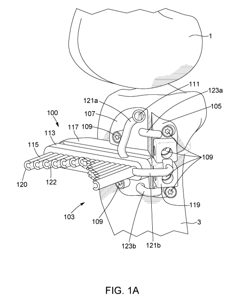
With reference to Figures 1A and 1B a surgical guide 100 is provided according
to
an embodiment. The surgical guide 100 is configured to be mounted to a
patient's
tibia bone 3 and includes a plurality of modules to guide various surgical
tools used
throughout the osteotomy procedure. The surgical guide 100 is patient-specific
in
that it is designed and manufactured according to the specific anatomy of a
patient.
In this fashion, the surgical guide 100 can be shaped and configured such that
it
can fit precisely on a predetermined position on the patient's bone 3 and be
secured thereto to assure proper alignment of guides for various surgical
tools. In
the present embodiment, the surgical guide 100 has a body made from 3D printed
plastic, although it is appreciated that other biocompatible materials
compatible
with other custom manufacturing methods are also possible.
CA 03109668 2021-02-15
WO 2020/037420
PCT/CA2019/051149
The body of surgical guide 100 comprises a bone interface side 101 for facing
the
patient's bone 3, and an operative side 103 for facing away from the patient's
bone
3. In the present embodiment, bone interface side 101 is configured to be
positioned directly on the patient's bone, and comprises a surface having
contours
5
complementary is shape to the surface contours of a predetermined area of the
patient's bone 3. In this configuration, bone interface side 101 can abut
against the
patient's bone, and key into a specific position thereon. In the present
embodiment,
bone interface side 101 comprises a solid surface, however it is appreciated
that
other configurations are possible. For example, the surface can be defined by
an
10 open
lattice, and can comprise edges conforming to the contours of the patient's
bone 3. Operative side 103 is provided opposite interface side 101 and
includes a
variety of components for interacting with surgical tools, as will be
described in
more detail hereinafter.
In the present embodiment, the body of surgical guide 100 is subdivided into
two
15
separable sections, including a lateral section 105 for securing relative to a
lateral
or medial surface of the patient's bone 3 and an anterior section 107 for
securing
relative to an anterior surface of the patient's bone 3. It is appreciated,
however,
that in other embodiments, more or fewer sections are possible to secure
relative
to different surfaces of the patient's bone 3 depending on surgical
requirements.
20 In the
present embodiment, lateral section 105 and anterior section 107 are
independently securable relative to the patient's bone 3. In this fashion, the
lateral
105 or anterior 107 section can be removed from the patient's bone 3 when no
longer needed, while the other section can remain secured in place. In the
present
embodiment, lateral 105 and anterior 107 sections are secured directly to the
25
patient's bone, however it is appreciated that in some embodiments, only one
of
the lateral 105 and anterior 107 need be affixed directly to the bone. For
example,
lateral section 105 can be affixed directly to the bone 3, whereas anterior
section
107 can be removably attached to lateral section 105 such that it is secured
relative
the patient's bone 3 without being directly affixed thereto.
CA 03109668 2021-02-15
WO 2020/037420
PCT/CA2019/051149
31
In the present embodiment, lateral 105 and anterior 107 sections comprise bone-
conforming plates secured to the patient's bone 3 via fasteners. The fasteners
comprise surgical screws 109 although it is appreciated that other types of
fastening mechanisms are also possible. The screws 109 engage in the patient's
bone 3 through canals 110 opening on the bone interface 101 and operative 103
sides of the surgical guide 100. The canals 110 comprise sidewalls extending
along a length for guiding insertion of screws 109 through canals 110 at a
specified
angle and depth. In this fashion, screws 109 drilled into the patient's bone 3
through canals 110 can be guided into a predetermined position, orientation
and
depth such that they can secure patient-specific surgical guide 100 to the
patient's
bone 3 in an optimal fashion, and such that the screws 109 will not interfere
with
tools used during subsequent steps during the osteotomy procedure. The
sidewalls of canals 110 can further be configured to abut against a head of
screw
109 to block the screw 109 from being inserted too deep into the patient's
bone 3.
In the present embodiment, a plurality of canals 110 are provided for securing
the
surgical guide 100 to the patient's bone 3 via a plurality of screws 109 at
strategic
locations. It is appreciated, however, that in other embodiments, a different
number
of screws 109 and canals 110 can be provided, and that they can be positioned
and oriented differently depending on the patient's specific anatomy and
according
to the planned procedure. Moreover, in the present embodiment, each of screws
109 is the same size, but it is appreciated that in other embodiments,
different
sized screws can be used to secure different parts of the surgical guide 100,
and
that the canals 110 can be sized and shaped accordingly. Finally, although the
screws 109 are guided by canals 110 in the present embodiment, it is
appreciated
that other screw-guiding mechanisms are possible in other embodiments.
As mentioned above, lateral 105 and anterior 107 sections are separable from
one
another. In the present embodiment, lateral 105 and anterior 107 sections are
generally disjointed from one another and are connected via connecting
members.
In other words, lateral 105 and anterior 107 sections are not directly fused
together,
and instead comprise separate spaced-apart sections removably secured to one
CA 03109668 2021-02-15
WO 2020/037420
PCT/CA2019/051149
32
another at a finite number of fixed points. In this configuration, each of
lateral 105
and anterior 107 sections define two separate bone-contacting surfaces
including
two bone-conforming plates on bone interface side 101 of surgical guide 100.
It is
appreciated, however, that in other embodiments, lateral 105 and anterior 107
sections can together form a single coherent surface or plate for contacting
the
bone 3.
Connecting members 121, 123, can be provided to removably connect different
sections of the surgical guide 100. In the present embodiment, the lateral 105
and
anterior 107 sections are connected to one another at three fixed points via
connecting members 121b, 123a and 123b. The connecting members 121b, 123a,
123b are stems comprising narrow strands of rigid material connected at a
first end
to the lateral section 105 and at a second end to the anterior section 107.
The
connecting members 121b, 123a, 123b are fused to lateral 105 and anterior 107
sections and/or are formed as integral parts thereof. In this fashion, lateral
105 and
anterior 107 sections can be rigidly connected to one another and can be
disconnected by respectively severing each of connecting members 121b, 123a,
123b. Connecting members 121, 123 are configured such that an intermediate
portion thereof is spaced away from surgical guide 100 and/or the patient's
bone
3, thereby allowing the connecting members 121, 123 to be readily severed
using
a severing tool (such as cutting pliers, a saw, or scissors, for example)
while
minimizing a risk of damaging surgical guide 100 or bone 3. In the present
configuration, connecting members 121b, 123a, 123b loop away from the surgical
guide 100 and comprise a rounded intermediate section spaced away from
surgical guide 100. Although a particular configuration of connecting members
121, 123 has been shown, it is appreciated that other configurations are
possible.
In other embodiments, connecting members 121, 123 can have different shapes,
and can include different connecting elements. For example, in some
embodiments, instead of being fused and/or an integral part of lateral 105
and/or
anterior 107 sections, connecting members 121, 123 can be separate pieces
removably engageable in lateral 105 and/or anterior 107 sections. As can be
CA 03109668 2021-02-15
WO 2020/037420
PCT/CA2019/051149
33
further appreciated, in other embodiments, a different number of connecting
members 121, 123 can be provided, and they can be positioned differently.
As mentioned above, the surgical guide 100 comprises a plurality of modules to
guide various surgical tools used throughout the osteotomy procedure. Each
module can perform a different function for assisting with various tasks
throughout
an osteotomy procedure. Some modules can form integral parts of the lateral
105
and/or anterior 107 sections secured directly to the patient's bone 3, whereas
other
modules can be independent elements which can be secured to relative to the
patient's bone 3 by attaching to lateral 105 and/or anterior 107 sections.
Although
a particular set of modules will be described in detail hereinafter, it is
appreciated
that other modules and combinations thereof are possible depending on the
requirements of the surgical procedure. Moreover, although some modules are
described as performing particular functions, it is appreciated that some
modules
can perform two or more functions and/or have other advantages or uses not
explicitly described herein, but that would be readily understood by a person
of skill
in the art upon reading the present disclosure.
Security Pin Guide Module
In the present embodiment, a security pin guide module is provided for guiding
insertion of a corresponding security pin or rod 111 into the patient's bone
3.
Security pin guide module is an integral part of body of surgical guide, and
comprises a security pin guide 112 formed therein. More specifically, security
pin
guide 112 is provided on anterior section 107 of surgical guide 100, although
it is
appreciated that other configurations are possible. In the present embodiment,
security pin guide 112 is positioned proximate a top portion of anterior
section 107
and comprises a canal to guide an angle of security pin 111 as it is inserted
into
the patient's bone 3. The pin guide 112 is angled such that when the security
pin
111 is inserted into the patient's bone 3 it runs parallel to the tibial
plateau. The
security pin 111 is made from a rigid, biocompatible material, such as
stainless
steel or titanium, and can be screwed into the patient's bone 3. Once inserted
into
the patient's bone 3, the security pin 111 can remain in place for the
remainder of
CA 03109668 2021-02-15
WO 2020/037420
PCT/CA2019/051149
34
the osteotomy procedure to protect the tibial plateau from fracturing.
Accordingly,
the security pin guide module can be configured to be removable from security
pin
111 once the security pin 111 is installed. For example, pin guide 112 can be
configured such that security pin 111 can slide therethrough unobstructed,
allowing
pin 111 to slide out from pin guide 112 when the security pin guide module is
removed, for example when the anterior section 107 is removed from the
patient's
bone 3. Other configurations of pin 111 and pin guide 112 are also possible.
Drilling Module
A drilling module 113 is provided to assist in creating drill holes 116 in the
patient's
bone 3 in preparation for forming a cut therein. In the present embodiment,
the
drilling module 113 is removably secured to the body of surgical guide 100 via
connecting members 121. More specifically, a plurality of connecting members
121a, 121b, and 121c extend between the drilling module 113 and the body of
surgical guide 100, securing the drilling module 113 to lateral 105 and
anterior 107
sections of surgical guide 100. The connecting members 121 comprise stems of
rigid material forming integral parts of both surgical guide 100 and drilling
module
113, and drilling module 113 can be removed from surgical guide 100 by
severing
stems of connecting members 121.
Although in the present embodiment the drilling module 113 is secured to the
body
of surgical guide 100 via severable stems, it is appreciated that other
connection
mechanisms are possible to secure and position drilling module 113 relative to
the
patient's bone. For example, drilling module can engage with body of surgical
guide 100 via fasteners, and/or can engage directly to the patient's bone. In
an
embodiment, for example as shown in Figure 9, the drilling module 113 can clip
onto a predetermined position on surgical guide 100. In the embodiment of
Figure
9, surgical guide 100a comprises a drill module interface 131 in the form of a
tongue element. A corresponding removable drill guide module, such as drill
guide
modules 113a and 113b shown in Figures 10A and 10B, can comprise a slot or
groove sized and shaped to receive tongue 131 therein. In this configuration,
drill
guide module 113a, 113b can clip onto a fixed position on surgical guide 100
by
CA 03109668 2021-02-15
WO 2020/037420
PCT/CA2019/051149
sliding over tongue 131. It is appreciated that in alternate embodiments,
drill guide
113 can comprise a tongue for fitting in a corresponding groove in surgical
guide
100 and/or a combination of tongue and grooves for fitting with corresponding
tongue and groves in surgical guide 100.
5
Referring back to Figures 1A and 1B, the drilling module 113 comprises a
plurality
of drill guides 115 for cooperating with corresponding drill bits to guide a
position,
depth, and angle thereof to form drill holes 116 in the patient's bone 3 in a
predetermined configuration. In the present embodiment, the drill guides 115
each
comprise a guiding element accessible from the operative side 103 of surgical
10 guide
100. The guiding element comprises a guide barrel 120 extending from the
operative side 103 of surgical guide 100, although it is appreciated that
other types
of guide elements are also possible. The guide barrel 120 extends along a
lengthwise axis, between a proximal end proximate the bone interface side 101
of
guide 100, and a terminal end 124 on the operative side 103 of guide 100. The
15 guide
barrel 120 comprises sidewalls defining a hollow interior in the form of a
guide tunnel 122 extending through the guide barrel 120 along the lengthwise
axis
thereof, and opening on the bone interface side 101 and operative side 103 of
guide 100. The guide tunnels 122 are sized and shaped to receive a
corresponding
drill bit therein, allowing the drill bit to slide in and out of barrel 120,
while sidewalls
20 of
barrel 120 constrain movement of the drill bit to a predetermined depth,
position,
and orientation relative to the patient's bone.
With reference to Figure 2, a drill bit 200 configured to cooperate with drill
guide is
shown according to an embodiment. The drill bit 200 comprises a drill bit body
201
extending along a length, and terminating at a cutting end 202. A depth guide
203
25 is
provided on the drill bit body 201 and spaced away from the cutting end 202,
effectively defining an operative length 205 of drill bit 200. In the present
embodiment, depth guide 203 is removably secured to drill bit body 201 via
fastener 204, allowing operative length 205 of drill bit 200 to be adjusted by
loosening fastener 204 and sliding depth guide 203 to a desired location along
the
30 length
of the drill bit body 201. It is appreciated, however, that in other
CA 03109668 2021-02-15
WO 2020/037420
PCT/CA2019/051149
36
embodiments, depth guide 203 can be permanently affixed to, and/or form an
integral part of, drill bit body 201, effectively defining a fixed
predetermined
operative length 205. For example, as shown in Figure 2A, drill bits 200a,
200b,
200c and 200d having respective fixed lengths of 80mm, 90mm, 100mm and
110MM are shown. Each drill bit comprises a depth guide 203 permanently
secured relative to cutting end 202. As can be appreciated, a collection of
fixed
drill bits can be provided as part of a kit, and each bit can be identified
via markings,
and/or via color coding. In some embodiments, the markings and/or color coding
can match with corresponding marking and/or color coding on the drill guides
115
in the drilling module 113.
With reference now to Figures 1A, 1B, 2 and 2A, depth guide 203 comprises an
abutment member 206 for limiting an insertion depth of drill bit 200 in guide
barrel
120. When operative length 205 of drill bit 200 is fully inserted into guide
barrel
120, the abutment member 206 abuts against terminal 124, effectively
preventing
.. further insertion of drill bit 200. As can be appreciated, in this
configuration, drill bit
200 can only be inserted into guide barrel 120 at a fixed insertion depth 118
relative
to the terminal end 124. The position of terminal end 124 relative to the
patient's
bone 3 thus defines the penetration depth of drill bit 200 into the patient's
bone 3.
Accordingly, the length of guide barrel 120 determines the bone penetration
depth
of drill bit 200: a longer guide barrel 120 results in a shallower bone
penetration
depth of drill bit 200, and a shorter guide barrel results in a deeper bone
penetration
depth. Similarly, the position and orientation of the guide barrel 120 defines
the
position and orientation at which the drill bit 200 penetrates the patient's
bone 3.
In the present embodiment, a plurality of drill guides 115 are provided for
.. cooperating with a calibrated drill bit 200 having a fixed operative length
205. The
drill guides 115 comprise guide barrels 120 positioned and arranged to create
drill
holes 116 in a predefined pattern to weaken the patient's bone 3 in
preparation for
a planar cut. More specifically, the drill guides 115 are positioned and
oriented in
a co-planar, parallel arrangement to define parallel drill holes 116 in the
patient's
bone 3 in a common plane 133. The guide barrels 120 of drill guides 115 are
sized
CA 03109668 2021-02-15
WO 2020/037420
PCT/CA2019/051149
37
based on the specific geometry of the patient's bone 3, such that the drill
holes 116
cover a majority of a cross section of the patient's bone 3, while leaving a
non-
weakened section to eventually form a hinge along which the patient's bone 3
can
be opened. More specifically, the guide barrels 120 are positioned such that
drill
holes define a hinge axis 9 at a border between weakened and non-weakened
areas of the patient's bone 3 in the common plane 133. As can be appreciated,
hinge axis 9 can be oriented depending on the type and position of opening to
be
formed in the patient's bone 3 as determined according to a preoperative plan,
to
correct the mechanical axis of the patient's bone 3 as needed. In the present
embodiment, hinge axis 9 is a straight line, but it is appreciated that other
shapes
are also possible.
Although in the present embodiment the drilling module 113 is configured to
create
drill holes 116 in a parallel orientation, it is appreciated that in other
embodiments,
the drilling module 113 can be configured such that some or all drill holes do
not
run parallel to one another. For example, the drill holes 116 can be grouped
into
two or more arrangements which intersect with one another. Although different
groups of drill holes can be guided by the same drilling module 113, it is
appreciated that in some embodiments, two or more drilling modules 113 can be
provided, for example to create drill holes 116 in different arrangements, to
weaken
the patient's bone 3 in different steps/stages, and/or to allow drill bits to
be inserted
at different angles of approach. Where a plurality of drilling modules 113 are
provided, they can be positioned and/or attached on the same section of the
guide
100, or can be positioned on different sections of the guide 100, for example
to drill
on different faces of the patient's bone 3 and/or allow drill bits to be
inserted at
different orientations, for example to facilitate drilling holes in a position
which
would otherwise be more difficult to access.
For example, as shown in Figures 9, 10A, 10B, 11A, and 11B, surgical guide
100a
can be configured with an anterior section 107 having a drill module interface
131
for connecting one or more removable drill modules 113 thereto. A first
drilling
module 113a can be attached thereto to guide drill bits 200 to form drill
holes 116
CA 03109668 2021-02-15
WO 2020/037420
PCT/CA2019/051149
38
in a first parallel orientation 133a in the common plane 133 in the patient's
bone.
The first drilling module 113a can subsequently be removed, and in its place a
second drilling module 113b can be attached to the same position on anterior
section 107 via drill module interface 131. The second drilling module 113b
can
then guide drill bits 200 to form drill holes 116 in a second parallel
orientation 133b
different from the first parallel orientation 133a, and in the same plane 133.
As can
be appreciated, the two drilling modules 113a, 113b can allow for weakening
the
patient's bone 3 along the plane 133 in two phases and by inserting drill bits
200
at different orientations. This can, for example, allow a complete area of the
patient's bone 3 to be weakened in preparation for cutting the patients bone,
while
reducing the size of the tissue incision required to access the patient's bone
3 to
perform the procedure.
Finally, although in the presently described embodiments the drilling module
113
is configured to guide drill holes 116 in a common plane 133, it is
appreciated that
in other embodiments, the drilling module can be configured to guide drill
holes
116 into two or more planes depending on the requirements of the surgical
procedure. For example, with reference to Figures 12A, 12C, and 12D drilling
module 113 can comprise a first group of parallel drill guides 115a for
creating drill
holes 116 in a first plane 133, and a second group of parallel drill guides
115b for
creating drill holes 116 in a second plane 135. As can be appreciated, the
first
plane 133 is not parallel to second plane 135 and is substantially
perpendicular
thereto, allowing to weaken the bone 3 to eventually form a biplanar cut 5a,
5b
therein.
As can be appreciated, in some embodiments, a single drilling module 113 can
be
configured to create all the necessary drill holes to weaken the bone 3 in
planes
133, 135 in preparation for forming biplanar cuts 5a, 5b. However, in other
embodiments, two or more drilling modules 113 can be provided to create the
necessary drill holes in planes 133, 135 in phases. For example, in the
embodiment shown in Figures 12A, 12B, 12C, and 12D, two drilling modules 113a
and 113b are provided. A first drilling module 113a can be secured to drilling
CA 03109668 2021-02-15
WO 2020/037420
PCT/CA2019/051149
39
module interface 131 to create drill holes 116 in the first plane 133 in a
first parallel
orientation 133a. The drilling module 113a includes a cover element 137 for
covering openings in the drilling module interface 131 extending along the
direction
of the second plane 135. Once the drill holes have been formed in the first
plane
133, the drilling module 113a can be removed, and a second drilling module
113b
can be secured to the drilling module interface 131. The second drilling
module
113b is provided with a first group of drill guides 115a for drilling holes
116 in the
first plane 133 in a second parallel orientation 133b different from the first
parallel
orientation 133a, thereby completing the required weakening of the bone in the
first plane 133. The second drilling module 113b is further provided with a
second
group of drill guides 115b for drilling holes 116 in the second plane 135. In
the
present embodiment, the second group of drill guides 115b in the second
drilling
module 113b are sufficient to weaken the bone to form the second planar cut
5b.
It is appreciated, however, that in other embodiments, further drill guides
can be
provided to cut in the second plane 135 in different parallel orientations.
Although in the embodiment described above, modules 113a and 113b are
described as "first" and "second" modules, it is appreciated that their order
of use
can be inversed depending on the requirements of the surgical procedure.
Moreover, although two modules were described, it is appreciated that in other
embodiments, subsequent modules can be provided to further weaken the bone
via drill holes 116 in different parallel orientations and/or in different
planes as
required. Moreover, in some embodiments, a cover element can be provided to
cover opening in the drilling module interface 131 extending along the
direction of
the first plane 133, for example in a drilling module configured to drill
holes only in
the second plane 135.
Cutting Module
Referring back to Figures 1A and 1 B, a cutting module 117 is provided to
assist in
cutting the patient's bone 3. In the present embodiment, the cutting module
117
comprises an osteotome guide 127 for guiding a corresponding osteotome to cut
the patient's bone 3 at predetermined position, orientation and depth. The
guide
CA 03109668 2021-02-15
WO 2020/037420
PCT/CA2019/051149
127 is configured to guide osteotome to create a planar cut in the patient's
bone 3
in the area weakened by the drill holes 116 formed using the drilling module
113.
The cutting module 117 is provided in anterior section 107 of guide 100, and
is
affixed directly to the patient's bone via fasteners 109. It is appreciated,
however,
5 that
in other embodiments, the cutting module 117 can be removably attached to
the lateral 105 and/or anterior 107 sections of the surgical guide 100.
Although in the present embodiment a single cutting module 117 is shown, it is
appreciated that two or more cutting modules can be provided in other
embodiments. For example, in some embodiments, two or more cutting modules
10 can be
provided to help create a single planar cut in two or more stages. In some
embodiments, a first cutting module can be configured to create a first planar
cut
in a first direction, and a second cutting module can be configured to create
a
second planar cut in a second direction. The cutting modules can be
permanently
or removably affixed relative to the same area of the patient's bone 3, and/or
can
15 be
removably or permanently affixed relative to different areas of the patient's
bone
3, for example to access the bone 3 from different positions.
The osteotome guide 127 comprises a body extending between a bone-contacting
end on the bone interface side 101 of surgical guide 100, and a terminal end
on
operative side 103 of surgical guide 100. The body has a planar aperture or
slot
20 129
extending therethrough and opening on the bone-contacting end and the
terminal end. The slot 129 is sized and shaped to receive a corresponding
osteotome therein, and to guide the osteotome to cut the patient's bone 3 at a
position, angle, and depth corresponding to the area of the patient's bone 3
weakened by the drilling module 113. More specifically, osteotome can slide in
and
25 out of
slot 129, while sidewalls around the aperture constrict the movement of
osteotome to the correct position and angle to form the desired cut.
Similarly, an
abutting member of osteotome is configured to abut against terminal end of the
osteotome guide 127 to limit an insertion depth of the osteotome. As can be
appreciated, osteotome guide 127 can have visual indications provided thereon
to
CA 03109668 2021-02-15
WO 2020/037420
PCT/CA2019/051149
41
further help guide osteotome visually and/or to indicate a type of osteotome
to be
used with guide 127.
In the present embodiment, in order to guide the osteotome to cut the area of
the
patient's bone 3 weakened by drilling module 113, the osteotome guide 127 is
positioned in alignment with the drill guides 115. More specifically, the
cutting
module 117 is positioned adjacent the patient's bone 3, and the drilling
module 113
is positioned adjacent the cutting module 117, such that the drill guides 115
open
in alignment with the slot 129 in the osteotome guide 127. In this
configuration, drill
guides 115 guide drill bits 200 through the slot 129 in osteotome guide 127
before
entering the patient's bone 3, thereby assuring that drill bits 200 and
osteotome
operate in the same plane 133. In the present configuration, cutting module
117 is
affixed directly to patient's bone 3, while drilling module 113 is removably
attached
to cutting module 117. Drilling module 113 can thus be removed after drill
holes
116 have been formed, providing the osteotome with direct access to cutting
module 117. It is appreciated that other configuration are possible which can
still
allow brill bits 200 and osteotome to operate in the same plane. For example,
in
some embodiments, both drilling module 113 and cutting module 117 can be
removably attachable to surgical guide 100. Drilling module 113 can be
attached
first to created drill holes 116. Drilling module 113 can be subsequently
removed,
and cutting module 117 can be attached to the same are of guide 100 as
drilling
module 113, allowing cutting module 117 to guide the osteotome in the same
plane
as the drill holes 116.
In the present embodiment, the cutting module 117 is configured to guide
osteotome to create a single planar cut 5 in the patient's bone 3, however it
is
appreciated that in other embodiment, the guide can be configured to create
two
or more cuts and/or cuts having a contour or curve. For example, with
reference to
Figures 12A and 12C, surgical guide 100b comprises an osteotome guide 127
configured with first 129a and second 129b slots for guiding osteotome to cut
the
patient's bone to create two planar cuts 5a and 5b along two different planes
133,
135. As can be appreciated, although two slots 129a and 129b are provided, use
CA 03109668 2021-02-15
WO 2020/037420
PCT/CA2019/051149
42
of the second slot 129b can be optional, allowing the same guide 100b to be
compatible with both procedures involving single planar cuts 5 and biplanar
cuts
5a, 5b. For example, as shown in Figure 12D, a drilling module 113a can be
provided which includes drill guides 115 in only the first plane 133, whereas
a cover
element 137 covers the slots 129b in the second plane 135. In this fashion,
the
bone is only weakened along the first plane 133, and cut 5a can be formed in
said
plane. Additionally or alternatively, when manufactured for such procedures,
the
slot 129b in guide 100b can be covered to prevent an osteotome from being
inserted therein. As can be appreciated, the guide 100b can still include the
section
of tongue of drill module interface 131 which extends along the plane where
slot
129b once extended. In this fashion, the shape of drilling module interface
131 can
be the same regardless of whether or not a second plane is to be cut. This can
allow for the same general shape/configuration of surgical guide 100b to be
used
for different types of surgical procedures involving single or biplanar cuts,
and
similarly allows for the same general shape/configuration of drill modules 113
to
be used. This can simplify the manufacturing and design of surgical guide 100
and
corresponding modules, as the same shape can be used for all procedures types,
yet simply adapted to conform the anatomy of the patient's bone 3.
Anchor Module
With reference now to Figure 3A, an anchor module 119 is provided to anchor
removable modules relative to the patient's bone 3. In the present embodiment,
anchor module 119 is provided in the lateral section 105 of the surgical guide
100,
but it is appreciated that in other embodiments, anchor module 119 can be
provided in a different section of guide 100. Moreover, in some embodiments, a
plurality of anchor modules can be provided. The anchor module 119 is affixed
directly to the patient's bone 3 via fasteners 109 and comprises a removable
module interface 128 for interfacing with removable modules. The anchor module
can thus act as a secure base to which other modules can be removably
attached,
allowing the removable modules to be properly aligned relative to the
patient's
bone 3 at relevant steps during the surgical procedure. In the present
embodiment,
CA 03109668 2021-02-15
WO 2020/037420
PCT/CA2019/051149
43
the removable module interface 128 comprises apertures for receiving
corresponding protrusions extending from a removable module, although it is
appreciated that other removable connection interfaces are possible.
In the present embodiment, the anchor module 119 comprises two sections for
providing two distinct anchoring points. More specifically, the anchor module
119
comprises a proximal section 125a positioned proximate the joint between the
patient's femur 1 and tibia 3 bones, and a distal section 125b spaced further
away
from the joint between the femur 1 and tibia 3. The proximal 125a and distal
125b
sections are separable from one another, allowing them to move independently
while being secured to different sections of the patient's bone 3. In the
present
embodiment, proximal 125a and distal 125b sections are secured to one another
via connecting member 126. The connecting member 126 can be severed to
separate proximal 125a and distal 125b sections and allow them to move
independently with different sections of bone. For example, in the present
embodiment, proximal 125a and distal 125b sections are positioned on the
patient's bone 3 on opposite sides of the planar cut formed by drilling module
113
and cutting module 117. After the planar cut is formed, connecting member 126
can be severed to separate proximal 125a and distal 125b sections. The bone 3
can be opened along the planar cut, with the proximal 125a and distal 125b
sections moving away from one another while being respectively connected to
the
bone 3 above and below the opening formed in the bone 3. In this fashion, the
proximal section 125a can provide an anchoring point above or proximal the
opening in the bone 3, while the distal section 125b provides an anchoring
point
below or distal the opening in the bone 3. It is appreciated that other
positions and
configurations of anchor module 119 and corresponding sections are possible,
depending on the surgical procedure. It is further appreciated that the
separable
sections of anchor module 119 can be connected to one another via different
removable connection mechanisms.
Predrilling Module
CA 03109668 2021-02-15
WO 2020/037420
PCT/CA2019/051149
44
With reference to Figures 3A and 3B, a predrilling module 300 is provided for
predrilling holes in the patient's bone 3 for eventually receiving fasteners
to secure
a plate or other implant to the patient's bone 3. The predrilling module 300
is
patient-specific in that it is custom made according to the anatomy of the
patient's
bone 3 and according to a preoperative plan. In this fashion, the predrilling
module
300 can be configured to precisely fit on a predetermined position of the
patient's
bone 3 to assure proper alignment, and to assist in drilling holes in the
patient's
bone 3 in predetermined positions, orientations and depths.
In the illustrated embodiment, the predrilling module 300 comprises a body 302
having a bone interface side 301 and an operative side 303. The bone interface
side 301 comprises a bone-contacting surface having contours complementary in
shape to the surface contours of the patient's bone 3. In this configuration,
bone
interface side 301 can abut against the patient's bone 3, and key into a
specific
position thereon. In the present embodiment, bone interface side 301 comprises
a
solid surface, however it is appreciated that other configurations are
possible. For
example, the surface can be defined by an open lattice, and can comprise edges
conforming to the contours of the patient's bone 3.
The operative side 303 is provided opposite the bone interface side 301 and
comprises a plurality of drill guides 307 extending therefrom for guiding
corresponding drill bits. In the present embodiment, the drill guides 307 each
comprise a guide barrel 309 extending from the body of the predrilling module
303
at a predetermined angle along a lengthwise axis and terminating at a terminal
end
314. The guide barrel 309 comprises sidewalls defining a hollow interior in
the form
of a guide tunnel 311 extending through the guide barrel 309 along the
lengthwise
axis thereof and opening on the bone interface side 301 and operative side 303
of
predrilling module 303. The guide tunnels 311 are sized and shaped to receive
a
corresponding drill bit therein, allowing the drill bit to slide in and out of
barrel 309,
while sidewalls of barrel 309 constrain movement of the drill bit to a
predetermined
depth, position, and orientation relative to the patient's bone 3. An abutting
member
on the drill bit can limit an insertion depth of an operative end of the drill
bit into the
CA 03109668 2021-02-15
WO 2020/037420
PCT/CA2019/051149
barrel 309 as it abuts with terminal end 314 of guide barrel 309. As can be
appreciated, in this configuration, the length of barrel 309 can limit
insertion depth
of a drill bit and assure the depth of drill holes formed therewith.
The plurality of drill guides 307 are configured to cooperate with a
calibrated drill
5 bit having a fixed operative length. The guide barrels 309 of the drill
guides 307
are sized, positioned and oriented to create drill holes 308 in a predefined
pattern
for receiving fasteners to secure an implant, such as plate, to the patient's
bone 3.
As will be described in more detail hereinafter, the implant to be secured can
be
patient-specific and can be designed to be affixed using different types of
10 fasteners. Based on the anatomy of the patient's bone 3, a preoperative
plan can
define a configuration of fasteners, including size, depth, orientation, and
position,
such that the implant can be affixed optimally. The drill guides 307 can thus
be
configured to guide drill bits to form drill holes 308 in preparation for
receiving the
configuration of fasteners defined in the preoperative plan. For example, the
length
15 of each guide barrel 309 can be adjusted to limit the insertion depth of
the drill bit,
creating drill holes 308 with different predetermined depths. Similarly, the
position
an orientation of guide barrels 309 can be adjusted to define drill holes 308
which
extend at different angles and positions. Finally, diameters of guide tunnels
311
can be adjusted to accommodate drill bits of different diameters to create
drill holes
20 of different sized for accommodating different sizes of fasteners.
In the present embodiment, the predrilling module 300 is configured to
predrill
holes 308 in the patient's bone 3 prior to a surgical alteration of the bone's
geometry. The predrilling module 300 is thus configured to account for the
drill
holes 308 moving as the geometry of the bone is altered during surgery, such
that
25 the drill holes 308 will be in alignment with the fasteners of an
implant once the
bone alterations are complete. For example, in the context of a high-tibial
open-
wedge osteotomy procedure, the predrilling module 300 can be configured to
predrill holes while the patient's bone 3 is in a closed configuration (i.e.
before the
patient's bone 3 is opened along the planar cut formed using the drilling 113
and
30 cutting 117 modules). In this configuration, the guide barrels 309 are
positioned to
CA 03109668 2021-02-15
WO 2020/037420
PCT/CA2019/051149
46
form drill holes 308 which will eventually align with the location of
fasteners for
affixing an implant once the patient's bone 3 is opened along the planar cut
to an
opened configuration. As can be appreciated, the required position of drill
holes
308 can be determined by modelling the patient's bone 3, virtually opening the
bone model to a desired opening angle, and virtually positioning an implant
and
corresponding fasteners on the bone model to set final positions of the drill
holes
308. The bone model can be subsequently closed virtually to determine
corresponding initial positions of the drill holes 308. The predrilling module
300 can
then be designed according to the initial positions of the drill holes 308.
As shown in Figures 3A and 3B, predrilling module 300 comprises an
attachment/alignment mechanism 305 for securing the predrilling module 300
relative to the patient's bone 3 and/or for assuring proper alignment of the
predrilling module 300 relative to the patient's bone 3. In the present
embodiment,
the attachment/alignment mechanism 305 comprises an attachment interface for
interfacing with removable module interface 128 in anchor module 119. The
attachment/alignment mechanisms 305 is configured such that the predrilling
module 300 can attach to anchor module 119 in only one position/orientation,
thus
assuring that predrilling module 300 is properly aligned once it is attached
to
anchor module 119. For example, in the present embodiment, the attachment
interface comprises two protrusions or pins 306 sized and shaped to engage in
corresponding apertures in anchor module 119. The protrusions 306 provide two
fixed attachment points which must be respectively align with two fixed
anchoring
points in the anchor module 119 for the predrilling module 300 to engage with
anchor module 119. In the present embodiment, the protrusions 306 are
positioned
to align with anchor module 119 while the patient's bone 3 is in a closed
configuration, thereby allowing the predrilling module 300 to engage with the
patient's bone 3 and predrill holes 308 prior to opening the bone 3 (i.e. the
protrusions 306 respectively align with the proximal 125a and distal 125b
sections
while they are adjacent one another). It is appreciated that in other
embodiments,
the protrusions 306 can be positioned to align with the anchor module 119 when
the patient's bone is in the opened configurations (i.e. when the proximal
125a and
CA 03109668 2021-02-15
WO 2020/037420
PCT/CA2019/051149
47
distal 125b sections are space apart from one another across the opening in
the
patient's bone 3).
Although in the present embodiment a single mechanism 305 provides both the
functions of securing and aligning predrilling module 300 relative to the
patient's
bone 3, it is appreciated that in other embodiments, different mechanisms can
be
provided to align and/or to secure predrilling module 300, and that separate
mechanisms can be provided to respectively perform the alignment or attachment
functions. For example, in some embodiments, predrilling module 300 can be
secured to the patient's bone directly via fasteners. In some embodiments, the
bone interface side 301 of predrilling module 300 can be shaped to have
contours
complementary in shape to the contours of a specific area of the patient's
bone 3.
In some embodiments, mechanism 305 can comprise a member configured to
interface and/or insert into a hole or other feature formed in the patient's
bone 3,
for example in the opening formed along the planar cut.
The predrilling module 300 further comprises a handle member 313 which allows
the module 300 to be more easily manipulated and positioned. In the present
embodiment, the handle member 313 is a rigid elongated member extending from
the body of the predrilling module 300 along a lengthwise axis and facilitates
manipulation of the module 300 by hand. It is appreciated that in other
embodiments, different types of handle members can be provided. For example,
handle member can be removable and/or can comprise an interface for a
positioning tool or guide. In the present embodiment, the handle member 313
has
inscriptions provided thereon to identify the predrilling module 300 and/or to
indicate the type of drill bits with which the predrilling module 300 is
designed to
cooperate.
Although in the illustrated embodiment the predrilling module 300 is
configured to
drill holes 308 prior to a change in the geometry of the patient's bone 3, it
is
appreciated that the predrilling module 300 can be configured differently
according
to the requirements of the surgical procedure. For example, as shown in Figure
13, an embodiment of a predrilling module 300a is shown in which the module
CA 03109668 2021-02-15
WO 2020/037420
PCT/CA2019/051149
48
300a is configured to drill holes 308 after the geometry of the patient's bone
3 has
been surgically altered. In this embodiment, the predrilling module 300a is
configured to span across opening 7 formed in the patient's bone 3, and
position
drill guides 307 to define drill holes 308 directly in their final position.
More
specifically, the predrilling module 300a has a body 302 substantially similar
to a
fixation plate which will ultimately be used to secure the opening 7 in the
patient's
bone 3. The bone 3 can thus be opened along planar cut 5 to form opening 7,
and
once the opening 7 is formed, the predrilling module 300 can be secured to the
bone at the same position where the fixation plate will eventually be
attached. The
predrilling module 300 will thus have its drill guides 307 positioned exactly
where
the fastener apertures of fixation plate will eventually be positioned.
Therefore,
after drill holes 308 are formed, predrilling module 300 can be removed and
replaced with fixation plate. Fixation plate can be positioned to align with
the holes
308 and then secured in place via fasteners.
In the present embodiment, the body 302 of predrilling module 300 has a bone
interface side 301 having a bone-contacting surface substantially conforming
to a
surface contour of the patient's bone 3 at a predetermined position. The body
302
is configured with a proximal section 302a for positioning adjacent a surface
of the
patient's bone 3 above opening 7, a distal section 302b for positioning
adjacent a
surface of the patient's bone 3 below opening 7, and an intermediate section
302c
for spanning the opening 7. The attachment/alignment mechanism 305 comprises
a wedge extending from bone interface side 301 on the intermediate section
302c
of body 302, and configured to be inserted into the opening 7. As can be
appreciated, wedge 305 can be sized and shaped according to the expected
dimensions of the desired opening 7 according to a preoperative plan. It can
further
comprise contours matching inner surface contours of the opening 7, as will be
described in more detail below in connection with the opening validator. The
wedge
305 can thus allow predrilling module 300 to secure at a predetermined
position
relative to opening 7, while also validating that the bone 3 has been opened
to the
correct angle. Once module 300 has been correctly positioned, it can be
secured
in place relative to the patient's bone 3 before drilling is performed through
drill
CA 03109668 2021-02-15
WO 2020/037420
PCT/CA2019/051149
49
guides 307. In the present embodiment, the body 302 comprises fastener
apertures 312a, 312b in the proximal 302a and distal 302b sections to allow
the
body 302 to be secured directly to the patient's bone 3 via fasteners. It is
appreciated, however, that other attachment mechanism are possible. For
example, the module 300 could secure to an anchor module already attached to
the patient's bone 3 at the correct position.
Spreader Module
With reference now to Figures 4, 4A and 4B, a spreader module 400 (or
spreading
tool) to assist in spreading the patient's bone 3 is shown according to an
embodiment. In the present embodiment, the spreader module 400 is configured
to open the patient's bone 3 along a planar cut 5 formed therein. The planar
cut 5
is opened at an angle about a hinge 9, thereby defining an open wedge 7 in the
patient's bone. The spreader module 400 is configured to operate in
cooperation
with anchor module 119 secured to the patient's bone 3, but it is appreciated
that
other configurations are possible. As can be appreciated, the spreader module
400
can be a generic tool, and need not be custom made according to the patient.
Instead, the surgical guide 100 can be designed to cooperate with generic
spreader module 400. Accordingly, spreader module 400 can be made out of any
rigid material, according to any manufacturing process. However, it is
appreciated
that in some embodiments, the spreader module 400 can be custom designed for
the patient and to conform to a specific geometry of the guide 100. In such
embodiments, the spreader module 400 can be made from materials suitable for
custom manufacturing, for example from the same 3D printed plastic from which
the surgical guide 100 and corresponding modules are made.
In the present, spreader module 400 comprises an upper arm 402a and a lower
arm 402b pivotally connected to one another via a hinge 407. As can be
appreciated, spreader module 400 is generally configured as a double lever,
with
an effort end 401 and a load end 403, and hinge 407 acting as a fulcrum
therebetween. More specifically, as effort ends 401a, 401b of upper and lower
arms 402a, 402b are moved towards one another, upper and lower arms 402a,
CA 03109668 2021-02-15
WO 2020/037420
PCT/CA2019/051149
402b pivot about hinge 407 causing load ends 403a, 403b to move away from one
another. In other words, a force applied at effort end 401 causing ends 401a
and
401b to converge is transferred to load end 403, causing load ends 403a and
403b
to separate. It is appreciated that other configurations of spreader module
400 are
5
possible, so long as it permits a separating force to be applied to load ends
403a
and 403b. For example, in some embodiments, the spreader module 400 can be
configured such that a spreading of effort ends 401a, 401b transfers a
spreading
force to load ends 403a, 403b. In other embodiments, different types of
spreading
mechanisms are possible.
10 In the
present embodiment, force on effort end 401 is applied via a hand wheel
409. As wheel 409 is operated, screw mechanism 411 rotates and engages in
threaded bores in effort ends 401a, 401b, thereby drawing effort ends 401a,
401b
together or spreading them apart depending on the rotating direction of screw
411.
As can be appreciated, in this configuration, a rotational force applied to
wheel 409
15 is
converted into a linear force which draws effort ends 401a, 401b together or
spaces them apart. Moreover, the rotational force applied to and wheel 409
merely
causes a change in spacing of effort ends 401a, 401b. A constant force does
not
need to be applied to wheel 409 to retain effort ends 401, 401b at a fixed
spacing;
instead, when no force is applied, the engagement of screw mechanism 411
20
retains arms 402 of spreader module 400 at their current angle, retaining
effort
ends 401a, 401b at a fixed spacing until force is applied to wheel 409.
Spacing of
effort ends 401a, 401b can thus be precisely controlled by hand, via small
and/or
measured rotational movements of hand wheel 409. It is appreciated, however,
that a force controlling spacing of effort ends 401a, 401b can be applied via
25 different mechanisms, and that such mechanisms need not necessarily be
operated by hand. For example, in some embodiments, force can be applied via
hydraulics or motors, and/or can be controlled electronically.
As mentioned above, spreader module 400 is configured to cooperate with anchor
module 119 secured to the patient's bone 3. Spreading module 400 comprises an
30 anchor
interface 405 at load end 403 for interfacing with anchor 119 and
CA 03109668 2021-02-15
WO 2020/037420
PCT/CA2019/051149
51
transferring spreading force thereto. More specifically, in the present
embodiment,
the anchor interface 405 comprises protrusions or pins sized and shaped to
engage in corresponding apertures in anchor module 119. A protrusion or pin at
load end 403a of upper arm 402a is positioned to engage with proximal section
125a of anchor module 119, whereas a protrusion or pin at load end 403b or
lower
arm 402b is positioned to engage with distal section 125b of anchor module. In
this
configuration, arms 402a, 402b of spreader module independently engage in the
distinct anchoring points 125a, 125b, allowing arms 402a, 402b to apply a
spreading force thereon in opposite directions, and move anchoring points
125a,
125b away from one another.
In the present embodiment, the protrusions or pins extend from arms 402a, 402b
substantially perpendicular therefrom, and along an axis substantially
parallel to
the pivot axis of hinge 407. As can be appreciated, in this configuration,
spreader
module 400 can engage with anchor 119 by sliding protrusions or pins of anchor
interface 405 laterally into the corresponding apertures of anchor 119. A
vertical
spreading force can be subsequently applied to arms 402a, 402b without causing
interface 405 to disengage. In the same manner, spreader module 400 can be
easily disengaged from anchor 119 by sliding the protrusions or pins out along
the
lateral direction. As can be further appreciated, in this configuration,
spreader
module 400 can engage with anchor module 119 and operate along the lateral
section of the patient's bone 3, leaving anterior section of the bone 3 clear
so as
to not interfere with subsequent steps in the surgical procedure. Apertures in
anchor module 119 open on both anterior and lateral sides thereof, allowing
the
spreader module 400 to engage on either the anterior or lateral side of anchor
module 119 depending on the requirements of the surgical procedure. It is
appreciated, however, that in other embodiments, spreader module 400 can
engage on other sides of anchor module 119, such as on its front side, and/or
on
top/bottom sides.
In the present embodiment, pins or protrusions of anchor interface 405 are
substantially cylindrical and engage in substantially circular apertures in
anchor
CA 03109668 2021-02-15
WO 2020/037420
PCT/CA2019/051149
52
module 119. As can be appreciated, in this configuration, pins or protrusions
can
rotate freely inside apertures of anchor module 119, allowing relative angular
displacement of ends 403a, 403b relative to anchoring points 125a, 125b while
engaged therein. It is appreciated, that in other embodiments, anchor
interface 405
and/or anchor module 119 can comprise different engagement mechanisms. For
example, in some embodiments, anchor interface 405 can be secured to anchor
module 119 via fasteners. In some embodiments, ends 403a, 403b can key into
anchoring points 125a, 125b at specific relative orientations, and/or pins or
protrusions can be pivotally secured to ends 403a, 403b of arms 402a, 402b.
Spreader module 400 is operable to move between a closed configuration 400a
and an opened configuration 400b. In the closed configuration 400a, anchor
interface 405 on load ends 403a, 403b are substantially proximate one another
and aligned with anchoring points 125a, 125b prior to spreading the patient's
bone
3. In the opened configuration 400b, anchor interface 405 on load ends 403a,
403b
are spaced apart from one another, and load end 403a, 403b are angled relative
to one another at an opening angle. In the present embodiment, a gauge 413 is
provided to indicate the magnitude of opening angle. The gauge 413 comprises a
scale affixed to upper arm 402a, and movable through a corresponding aperture
in lower arm 402b. A window 415 in lower arm 402b provides a visual indicator
for
reading scale. It is appreciated, however, that other gauge mechanisms are
possible to indicate the magnitude of opening angle. In the present
embodiment,
gauge 413 is calibrated such that scale is zeroed when the spreader module 400
is in the closed configuration 400a. The opening angle indicated by gauge 413
can
thus provide an accurate and precise indication of the opening angle of
spreader
module 400. In some embodiments, the gauge 413 can be further calibrated such
that it corresponds to the opening angle about hinge 9 in patient's bone 3. In
this
configuration, the gauge can provide a precise and accurate indicate of
opening
angle of the open wedge 7 formed in the patient's bone, as the bone is opened
along cut 5 using spreader module 400.
CA 03109668 2021-02-15
WO 2020/037420
PCT/CA2019/051149
53
Although the module 400 is referred to herein as a "spreader" module, it is
appreciated that it can be used not only to spread the patient's bone 3, but
also to
contract the patient's bone 3, for example as part of a closed-wedge
osteotomy. In
such procedures, the spreader module 400 can be operated to draw anchoring
points 125a, 125b closer together, for example to close an open wedge 7 cut
into
the patient's bone 3. More particularly, spreader module 400 can engage with
anchoring points 125a, 125b while in the opened configuration 400b, with the
anchoring points 125a, 125b being positioned on opposite sides of an open
wedge
7. The spreader module 400 can be subsequently operated towards the closed
configuration 400a by turning hand wheel 409, thereby drawing anchoring points
125a, 125b together and closing the wedge 7.
Opening Validator
With reference now to Figures 5 and 5A, an opening validator 500 for
validating
the open wedge 7 formed in the patient's bone 3 is shown according to an
embodiment. As can be appreciated, a desired opening angle of open wedge 7
can be predetermined according to a preoperative plan. Although the gauge in
spreader module 400 can provide an indication of the opening angle during the
procedure, opening validator 500 can provide a more precise confirmation as to
whether the patient's bone 3 has been opened the right amount to attain the
desired angle of open wedge 7. Accordingly, opening validator 500 is provided
to
directly measure the open wedge 7 formed in the patient's bone 3.
In the present embodiment, opening validator 500 is a patient-specific tool
designed to match the anatomy of the patient's bone 3. More specifically, the
opening validator 500 is shaped and configured to fit snugly in the opening 7
in the
patient's bone 3 based on the expected shape thereof as determined according
to
a pre-operative plan. During the surgical procedure, as the patient's bone 3
is being
spread to form opening 7, the opening validator 500 can be inserted into the
opening 7. A snug fit of opening validator 500 can confirm that the correct
opening
7 has been formed, whereas an incorrect fit can indicate that an adjustment of
CA 03109668 2021-02-15
WO 2020/037420
PCT/CA2019/051149
54
opening 7 is necessary. It is appreciated that other mechanisms for validating
the
opening are also possible.
As shown in Figure 5, the opening validator 500 comprises a unitary body 501,
made from a rigid, biocompatible material. In the present embodiment, the body
501 is made from a 3D printed plastic, although it is appreciated that other
materials are possible, and that the validator 500 can be made using other
custom
manufacturing processes. The body 501 includes a handle end 503 and an
operative end 505.
Handle end 503 is configured to facilitate manipulation of opening validator
500
during the surgical procedure. In the illustrated embodiment, handle end 503
comprises a handle 507 to allow the validator 500 to be easily grasped and/or
manipulated by hand. It is appreciated, however, that other interfaces for
manipulating the validator 500 are also possible. In the present embodiment,
the
handle 507 has a substantially rectangular-shaped profile, including an
anterior
side 509a and a lateral side 509b. The anterior 509a and lateral 509b are
marked
to indicate proper orientation during the surgical procedure. It is
appreciated,
however, that other shapes of handle 507 are also possible.
Operative end 505 is configured to engage with the opening 7 formed in the
patient's bone 3 at a predetermined position and orientation. More
specifically, the
operative end 505 comprises a wedge element 511 sized and shaped to fit in the
opening 7, and a tab element 515 to limit the insertion depth of wedge 511.
Wedge
element 511 is shaped to conform to the contour of interior surfaces 5a, 5b of
the
patient's bone 3 formed by planar cut 5 and confirm the height of opening 7
proximate the exterior surface of bone 3, and thus confirm opening angle 7a.
More
specifically, wedge elements 511 comprises a top surface 513a shaped to
conform
to the contour of top or proximal interior surface 5a, and a bottom surface
513b
shaped to conform to the contour of bottom or distal interior surface 5b.
Similarly,
tab element 515 is shaped to conform to the exterior contours of the patient's
bone
3. More specifically, tab element 515 comprises a top surface 517a shaped to
conform to the exterior contour of the patient's bone 3 above the cut 5, and a
CA 03109668 2021-02-15
WO 2020/037420
PCT/CA2019/051149
bottom surface 517b shaped to conform to the exterior contour of the patient's
bone 3 below the cut 5. As show in Figure 5A, when opening 7 in the patient's
bone 3 is opened to the right angle, and when validator 500 is correctly
positioned
therein, top 513a and bottom 513b surfaces of wedge element 511, and top 517a
5 and bottom 517b surfaces of tab element 515 will simultaneously conform and
engage with the corresponding surfaces of the patient's bone 3, thereby
locking
opening validator 500 in place and confirming that configuration of opening 7
matches the pre-operative plan. Any mismatch between the surfaces of the
validator 500 elements and the surfaces of the patient's bone 3 can indicate
that
10 ad adjustment is required.
As can be appreciated, opening validator 500 can be used to assure that
opening 7
in patient's bone 3 is formed correctly prior to proceeding with subsequent
steps
of the procedure. For example, it can confirm opening 7 prior to attaching a
fixation
plate, as will be described below, to secure and retain opening. As another
15 example, as illustrated in Figures 13A and 13B, the opening validator
500 can
confirm opening 7 prior to attaching predrilling module 300a, and thus help
position
the same, such that fastener holes can be drilled in the patient's bone 3
after
opening 7 has been formed.
Fixation Plate
20 With reference now to Figures 6A and 6B, a fixation plate 600 is shown.
Fixation
plate 600 comprises a body 601 made from a rigid, biocompatible and
degradation-
resistant material, such as stainless steel or titanium, although it is
appreciated
that other materials are possible, including different metals and/or plastics
and/or
a combination thereof. In the present embodiment, fixation plate 600 is an
25 osteotomy plate for securing to an antero-medial side of the patient's
bone 3 and
retaining the opening 7 formed therein during an open-wedge osteotomy
procedure. It is appreciated that in other embodiments, fixation plate 600 can
be
configured for securing to another side of the patient's bone 3 depending on
surgical requirements. In the present embodiment, body 601 comprises a
proximal
30 section 601a for securing to the patient's bone 3 above opening 7, a
distal section
CA 03109668 2021-02-15
WO 2020/037420
PCT/CA2019/051149
56
601b for securing to the patient's bone 3 below opening 7, and an intermediate
section 601c for spanning the opening 7. As will be described in more detail
hereinafter, the present fixation plate 600 is patient-specific in that it has
been
designed based on the specific anatomy of the patient's bone 3 and based on
the
specific needs of the patient determined during a preoperative plan. The shape
and configuration of fixation plate 600 can therefore vary from one procedure
to
another based upon the bone anatomy of different patients and based on their
different needs.
The body 601 of fixation plate 600 is sized, shaped, and configured to fit
snugly on
the patient's bone 3 while also providing the required support and being
minimally
noticeable under the patient's skin. In the present embodiment, body 601 is
thin
and substantially flat, and is configured to follow the contours of the
patient's bone
3. In this configuration, for example, when the fixation plate 600 is secured
to the
patient's bone 3, it can protrude from the surface of the patient's bone 3 at
a
uniform height along the entire body 601. Moreover, in some embodiments, body
601 can be designed to have a thickness which varies in different locations,
allowing body 601 to have increased or reduced strength or rigidity where
required
and/or allow body 601 to protrude less noticeably from the patient's bone at
certain
areas.
The body 601 of fixation plate 600 comprises a bone interface side 603 and an
outward-facing side 605. Bone interface side 603 comprises an inner surface
for
positioning adjacent the patient's bone 3. The contours of inner surface of
bone
interface side 603 are complementary in shape to surface contours of a
predetermined position on the patient's bone 3. In this fashion, fixation
plate 600
can fit snugly on a position of the patient's bone 3 determined
preoperatively.
Outward-facing side 605 is substantially smooth and/or flat to make it
minimally
noticeable under the patient's skin. In the present embodiment, the outward-
facing
side 605 comprises sloped and/or chamfered edges 607 which provide a smoother
transition between the body 601 of fixation plate 600 and the patient's bone
3.
CA 03109668 2021-02-15
WO 2020/037420
PCT/CA2019/051149
57
The fixation plate 600 is secured to the patient's bone 3 via fasteners 609.
In the
present embodiment, fasteners 609 comprise surgical screws which are drilled
into
the patient's bone 3, although it is appreciated that other type of fasteners
are
possible. The fasteners 609 engage with plate 600 via apertures or canals 610
opening on the bone interface side 603 and the outward facing side 605 of the
plate 600. As can be appreciated, canals 610 can be sized and shaped to
receive
different sizes of fasteners 609. Moreover, canals 610 can be configured to
guide
fastener 609 at a predetermined angle or orientation as it is inserted into
the
patient's bone 3. For example, in the present embodiment, canals 610 comprise
sidewalls extending through the thickness of the body 601 of plate 600 at a
predetermined angle to guide the fasteners 609 as they are drilled through the
canals 610. In some embodiments, the sidewalls of canals 610 can be threaded,
for example to engage with corresponding threads of fasteners 609 as the
fasteners 609 are being drill through canals 610, and/or to engage or lock
with a
head of the fasteners 609 once fully inserted. The sidewalls of canals 610 can
further be configured to abut against a head of fastener 609 to block the
fastener
609 from being inserted too deep into the patient's bone 3.
As can be appreciated, based on a preoperative plan, fixation plate 600 can be
designed with a different number and configuration of canals 610 for receiving
a
different number and configuration of fasteners 609 based on the specific
needs
of the patient to promote optimal securing of the plate 600. Moreover, the
fixation
plate 600 can be configured such that it can accommodate combinations of
different sizes of fasteners 609 (both diameter and length) and different
orientation
of fasteners 609, for example based on the position of the patient's bone 3 to
which
a particular fastener 609 is to be secured. In the illustrated embodiment, the
plate
600 is configured to accommodate two large laterally-spaced fasteners 609 in
the
proximal section of body 601a, and two smaller vertically-spaced fasteners 609
in
the distal section of body 601b. As will be explained in more detail
hereinafter,
many other configurations of plate 600 are possible.
CA 03109668 2021-02-15
WO 2020/037420
PCT/CA2019/051149
58
In some embodiments, additional support members can be provided to further
assist fixation plate 600 in retaining the opening 7 formed in the patient's
bone 3
and/or to assist in correctly positioning fixation plate 600 relative to
opening 7. By
way of example, and with reference to the embodiment of Figures 14, 14A, 14B,
.. and 14C, a wedge element 611 can be provided to abut against internal
surfaces
5a, 5b on opposite sides of opening 7 when fixation plate 600 is positioned on
the
patient's bone 3. As can be appreciated, as a load is applied across opening
7, the
wedge element 611 can exert an opposing force on the patient's bone 3 via
internal
surfaces 5a, 5b. In this configuration, a load across the opening 7 can be
borne by
the wedge element 611 and dissipated through the patient's bone 3, rather than
being borne by the fasteners 609 holding the plate 600 in place. In the
illustrated
embodiment, wedge element 611 is formed as an integral part of body 601 of
fixation plate 600, and is made from the same rigid, biocompatible material,
i.e.
stainless steel or titanium. It is appreciated, however, that in other
embodiments,
wedge element 611 can be a separate piece which can be fastened or secured to
the fixation plate 600 and/or directly to the patient's bone 3. It is further
appreciated
that wedge element 611 can be made of a different material, such as a rigid
plastic
or the like, depending on the required structural properties.
In the present embodiment, wedge element 611 extends from the bone interface
side 603 of fixation plate 600, and is positioned on intermediate section 601c
of
fixation plate body 601. In this configuration, wedge element 611 extends
inside
opening 7 when the fixation plate 600 is secured to the patient's bone 3. The
wedge
element 611 comprises a proximal abutment 613 for abutting against a proximal
internal surface 5a of bone 3, and a distal abutment 615 for abutting against
a
distal internal surface 5b of bone 3. Proximal 613 and distal 615 abutments
are
spaced apart from one another via a concave canal 617. In this configuration,
a
certain amount of flexure is permitted in the rigid body 601 of fixation plate
600 as
a load is applied across abutments 613, 615. It is appreciated, however, that
in
other embodiments, wedge element 611 can be a solid block having abutments
613, 615 defined on opposite sides thereof.
CA 03109668 2021-02-15
WO 2020/037420
PCT/CA2019/051149
59
As with the other components of fixation plate 600, the wedge element 611 can
be
configured according to patient-specific needs. For example, based on a
preoperative plan and 3D models of the patient's bone 3, various components,
surfaces, contours, etc. of the wedge element 611 and be shaped and configured
to conform to the specific anatomy of the patient's bone 3 and/or opening 7
formed
therein. Wedge element 611 can further be configured to provide varying levels
of
structural support as required based on patient-specific needs.
More specifically, and with reference to Figures 16 and 16A, an exemplary
embodiment of a wedge element 611 is shown. In the illustrated embodiment, the
body 601 of fixation plate 600 has a nominal thickness 602 in intermediate
section
601c, and wedge element 611 extends therefrom. The wedge element 611
comprises proximal 613 and distal 615 abutments extending from body 601 and
extends along a width 612a between anterior 619 and posterior 621 sides. The
proximal 613 and distal 615 abutments have respective bone contacting bearing
surfaces 613a and 615a spaced apart from one another by a spanning distance
612b, for respectively abutting against proximal 5a and distal 5b internal
surfaces
on opposite sides of opening 7 in the patient's bone 3. As can be appreciated,
the
spanning distance 612b can be adjusted according to the expected size of
opening
7 as determined in a preoperative plan, to extend precisely between proximal
5a
and distal 5b internal surfaces and abut against the same. By precisely
spanning
the distance between proximal 5a and distal 5b internal surfaces, wedge
element
611 can provide the necessary support to retain the internal surfaces 5a, 5b a
fixed
distance from one another, and retain opening 7 at the desired opening angle.
In
this configuration, wedge element 611 can further assist in correctly
positioning
fixation plate 600 on the patient's bone 3. As can be appreciated, the wedge
611
will only be able to fit inside the opening 7 at a position where the opening
is wide
enough to accommodate the spanning distance 612b of abutments 613, 615.
Accordingly, the wedge element 611 can be designed with a spanning distance
612b such that it fits inside opening 7 at a predetermined position and
orientation
relative to the patient's bone 3, as determined in a preoperative plan,
thereby
positioning the fixation plate 600 to which the wedge element 611 is secured.
CA 03109668 2021-02-15
WO 2020/037420
PCT/CA2019/051149
In the present embodiment, and as shown in Figure 24A, wedge 611 is configured
to abut against the patient's cortical bone 3a, i.e. the hard outer layer of
patient's
bone 3, as opposed to the soft trabecular bone 3b. Accordingly, and referring
back
to Figures 16 and 16A, proximal 613 and distal 615 abutments can be sized and
5 shaped
to interface with the patient's cortical bone 3a while avoiding contact with
the patient's trabecular bone 3b. More particularly, in the present
embodiment,
bone contacting surfaces 613a and 615a are substantially planar and extend
substantially perpendicular relative to body 601 through respective depths
614a
and 616a. As can be appreciated, depths 614a and 616a can be adjusted based
10 on the
thickness of the patient's cortical bone 3a, such that the abutments 613 and
615 extend into opening 7 to a depth corresponding to the thickness of the
cortical
bone 3a, for example to approximately 4mm. In the present embodiment, bone
contacting surfaces 613a and 615a have the same depths 614a, 616a, but it is
appreciated that in other embodiments, the depths can be different, for
example
15
depending on the expected position and orientation of wedge 611, and/or
variances in the thickness of the patient's cortical bone 3a. As can be
appreciated,
the surface areas of bearing surfaces 613a and 615a are defined by depths
614a,
616a, and width 612a of wedge 611. Accordingly, width 612a can be adjusted, in
addition to depths 614a, 616a, according to the required surface area of
bearing
20
surfaces 613a, 615a. In the present embodiments, width 612a is approximately
8mm, but other sizes are also possible depending on patient-specific
requirements.
As mentioned above, the wedge element 611 can be configured to provide
different
levels of support based on patient-specific needs. For example, for some
patients,
25 it may
be desirable to have more rigidity in the fixation plate 600, whereas for
other
patients, it may be desirable to allow a certain amount of micromovements via
flexure or deformation of the fixation plate 600 across the opening 7.
Accordingly,
respective thicknesses 614b and 616b of proximal 613 and distal 615 abutments
can be adjusted based on a desired level of rigidity. For example, in some
30
embodiments, such as the one illustrated in Figure 16 and 16A, abutments 613,
615 can be relatively thin members extending from body (for example with
CA 03109668 2021-02-15
WO 2020/037420
PCT/CA2019/051149
61
thicknesses 614b and 616b of approximately lmm), thus allowing a certain
amount
of deformation as loads are applied to their respective bearing surfaces 613a,
615b. In other embodiments, abutments 613, 615 can be relatively thick and/or
can
have a thickness corresponding to the spanning distance 612b of wedge 611
(i.e.
the wedge being formed from a solid block of material, with abutments 613, 615
defined on opposite sides thereof), thereby providing increased rigidity and
allowing little to no deformation of wedge 611 under typical loads. In the
embodiment illustrated in Figures 16 and 16A, the respective thicknesses 614b,
616b of abutments 613 and 615 are the same, however it is appreciated that in
other embodiments they can be different, for example to provide different
levels of
rigidity in proximal and distal sections of plate 600 and/or to control the
distribution
of forces in wedge 611 as a load is applied to abutments 613, 615.
As can be appreciated, abutments 613 and 615 can be designed with different
shapes and configurations which can further affect the rigidity and/or the
distribution of forces in wedge 611. For example, in the embodiment shown in
Figures 16 and 16A, abutments 613 and 615 are configured as curved members
with a progressive reduction of their depths 614a, 616a towards a central area
of
wedge 611. In other words, a canal 617 extends along a height 617a between
respective interior edges 613b, 615b of abutments 613 and 615. The canal 617
has a depth 617b which increases towards the central area of wedge 611,
thereby
subtracting from the depths of abutments 613, 615. In the present embodiment,
the depth 617a of canal 617 follows a polynomial curve (i.e. A)(2+BX+C),
reaching
a maximum depth 617b midway along its height 617a. Thus, when viewed from
posterior 219 or anterior 221 sides, the canal 617 has a parabolic or C-shaped
profile. In this configuration, when a load is applied across abutments 613,
615,
stresses can be focused towards the central area of the wedge 611. Although in
the present embodiment the canal 617 is substantially C-shaped, it is
appreciated
that other configurations are also possible, including different shapes having
progressive and/or abrupt changes in depth 617b. For example, in some
embodiments, the canal 617 can have a substantially V-shaped profile, a
substantially rectangular-shaped profile, etc. In the present embodiment the
CA 03109668 2021-02-15
WO 2020/037420
PCT/CA2019/051149
62
maximum depth 617a of canal 617 corresponds to the depths 614a, 616a of
abutments 613, 615. In this configuration, the canal 617 does not extend past
the
thickness 602 of plate 600. It is appreciated, however, that other
configurations are
possible. For example, the canal 617 can be shallower than depths 614a, 616a,
such that a minimum or base thickness 618 of plate 600 between abutments 613,
615 is thicker than a nominal thickness 602 of plate 600 adjacent to the wedge
611.
In the illustrated embodiment, the wedge 611 can be referred to as a straight
wedge in that the bearing surfaces 613a, 615a are substantially straight and
uniform. For example, bearing surfaces 613a, 615a are substantially
rectangular,
and are substantially parallel to one another. Similarly, the canal 617 is
straight
and uniform along the width 612a of wedge 611. It is appreciated, however,
that
the shape and orientation of bearing surfaces 613a, 615a, and/or canal 617 can
be adjusted to better conform to the specific needs of a patient. For example,
as
illustrated in Figures 20, 20A and 20B the surface area of bearing surfaces
613a,
615a of a straight wedge 611 may not be in full contact with interior surfaces
5a,
5b of opening 7, and can thus create areas of increased pressure. However, if
wedge 611 is configured to follow the shape of interior surfaces 5a, 5b as
shown
in Figures 21, 21A and 21B, a superior interface between wedge 611 and the
patient's bone 3 can be achieved (i.e. increased surface area of contact),
allowing
for better stress distribution through the bone.
With reference now to Figures 15, 15A and 15B, a wedge element 611 is shown
according to an alternate embodiment in which abutments 613, 615 are shaped to
follow the specific shape and contours of opening 7. More specifically, in the
illustrated embodiment, bearing surfaces 613a, 615a are sloped or tapered
along
the direction of width 612a. Similarly, bearing surfaces 613a, 615b are sloped
or
tapered along the direction of their depths 614a, 616a. In this configuration,
when
the wedge 611 is positioned inside opening 7, bearing surfaces 613a, 615a can
follow the slope of interior surfaces 5a, 5b, and increase the contact surface
area
therewith. This configuration of wedge 611 can further allow for the
correction of
CA 03109668 2021-02-15
WO 2020/037420
PCT/CA2019/051149
63
varus/valgus deformity in the frontal plane as well as the correction of the
tibial
slope in the sagittal plane. In the present embodiment, bearing surfaces 613a,
615b are tapered inwards along width 612a towards the posterior side 621, to
follow a corresponding narrowing of opening 7 towards a posterior side of the
patient's bone 3. It is appreciated, however, that the tapering direction and
magnitude can differ according to the expected shape of the opening 7 as
determined in a preoperative plan. It is also appreciated that the proximal
613a
and distal 615a bearing surfaces can be tapered at different angles. In the
present
embodiment, bearing surfaces 613a, 615a are also tapered inward along the
direction of their depths 614a, 616a. In this configuration, the bearing
surfaces
613a, 615a can follow the slope of interior surfaces 5a, 5b as they converge
towards hinge 9 at opening angle 7a, as shown in Figures 22 and 22A. Again, it
is
appreciated that the tapering angle of bearing surfaces 613a, 615a can be
different.
In the present embodiment, the width 612a of the wedge 611 is uniform along
the
wedge span 612b. In other words, bearing surfaces 613a, 615a are aligned with
one another, and have the same width 612a. It is appreciated, however, that in
other embodiments, bearing surfaces 613a, 615a can have different widths
and/or
can be offset from one another. For example, as illustrated in Figures 23 and
23A,
load distribution in the patient's bone 3 can be physiologically more
important in
the medial compartment. Accordingly, when plate is secured to patient's bone 3
across opening 7, the plate 600 can undergo a rotation effort in the antero-
medial
plane. To equilibrate the stress induced in the wedge 611 and bone 3, bearing
surfaces 613a, 615a can have different widths and/or can be offset, for
example
by being configured with tapered side edges 613c, 615c. It is appreciated that
other
relative size and positions of bearing surfaces 613a, 615a are also possible
in
different embodiments, according to patent specific requirements.
In the embodiment illustrated in Figures 15, 15A and 15B, the wedge element
611
is further configured with bearing surfaces 613a, 615a which conform to a
shape
of the patient's cortical bone 3a to ensure better contact therewith, and
avoid
CA 03109668 2021-02-15
WO 2020/037420
PCT/CA2019/051149
64
contact with the trabecular bone 3b. As can be appreciated, the thickness of
the
patient's cortical bone 3a can vary at different points along the
circumference of
the patient's bone 3. Accordingly, the respective depths 614a, 616a of
abutments
613, 615 can in direction of wedge width 612a. In the present embodiment, and
as
best illustrated in Figure 15B, depths 614a of proximal abutment 613 decreases
from anterior side 619 to posterior side 621, thus defining a bearing surface
613a
having a sloped or tapered interior edge shaped to match a thinning of the
patient's
cortical bone 3a towards posterior side 621. Although in the present
embodiment
the interior edge of bearing surface 613a has a sloped, linear shape, it is
appreciated that other shapes are also possible depending on the specific
shape
of the patient's cortical bone 3a. Moreover, although only the proximal
abutment
613 is shown in Figure 15B, it is appreciated that distal bearing surface 615a
can
be configured with a similar or different shape.
In the present embodiment, the wedge element 611 is further configured with an
evolutive canal 617, i.e. a canal having a shape which changes or evolves
along
width 612a of wedge 611. As shown in Figures 15, 15A and 15B, the height 617a
of canal 617 varies along width 612a wedge. More specifically, interior edges
613b,
615b of abutments 613, 615 are tapered inwards from posterior side 621 to
anterior
side 619, resulting in the canal height 617a decreasing from posterior side
621 to
anterior side 619. In the present embodiment, edges 613b, 615b are angled
inward
towards one another at substantially equal and opposite angles, although it is
appreciated that in other embodiments, angles of edges 613b, 615b can differ,
or
edges 613b, 615b can be angled and parallel to one another. It is further
appreciated that in other embodiments, edges 613b, 615b can follow curved
paths.
As can be appreciated, the present configuration of canal 617 can also allow
abutments 613, 615 to have evolutive thicknesses 614b, 616b along width 612a.
More particularly, in the present embodiment, respective thicknesses 614b,
616b
of abutments 613, 615 increase from posterior side 621 to anterior side 619.
In the present embodiment, the wedge element 611 is further configured with a
minimum or base thickness 618 of plate 600 which varies along width 612a of
CA 03109668 2021-02-15
WO 2020/037420
PCT/CA2019/051149
wedge 611. As best shown in Figure 15B, the base thickness 618 increases from
posterior side 621 to anterior side 619. For example, on posterior side 621,
the
base thickness 618 can correspond to the nominal thickness 602 of plate 600,
whereas on anterior side 619, the base thickness 618 can be greater than the
5
nominal thickness 602. Although in the present embodiment the base thickness
618 increases linearly along width 612a, it is appreciated that in other
embodiments, the change in thickness 618 can be nonlinear. As can be
appreciated, variances in base thickness, along with the variances in the
canal
configuration and/or abutment thicknesses can allow for the rigidity and/or
10
permitted amount of micromovements between abutments 613, 615 to vary across
the width 612a of wedge 611.
In the embodiments described above, plate 600 is provided with a single wedge
611 was shown for engaging in opening 7 along an antero-medial side of the
patient's bone 3. It is appreciated, however, that in other embodiments, other
15 wedge
configurations are possible. For example, with reference to Figures 17,
17A, 17B, 18A and 18B, a double wedge plate 600 is shown according to an
embodiment. In the illustrated embodiment, plate 600 is provided with a first
anterior wedge 611a, and a second posterior wedge 611b spaced apart from one
another in intermediate section 601 of plate body 601. In the present
configuration,
20 wedges
611a and 611b are spaced apart from one another via an opening 623 in
plate body 601. As can be appreciated, opening 623 can help reduce the weight
of plate and/or to encourage flexure in the intermediate section 601c. It is
appreciated that in other embodiments, opening 623 need not be provided, and
plate body 601 can be closed between wedges 611a and 611b.
25 When
plate 600 is engaged with patient's bone 3, wedges 611a and 611b engage
in opening 7 on an antero-medial side of the patient's bone 3, providing
support at
anterior and posterior positions. As with the embodiments of the patient-
specific
wedges described above, each of wedges 611a and 611b can be configured
according to patient-specific needs, and based on patient-specific anatomy.
For
30
example, as illustrated in Figure 24B, each of wedges 611a and 611b can be
CA 03109668 2021-02-15
WO 2020/037420
PCT/CA2019/051149
66
shaped and configured to follow and abut the patient's cortical bone 3a. The
other
size and shape parameters of wedges 611a and 611b, as described above, can
also be customized based on the expected position of wedge 611a, 611b as
determined preoperatively, and the dimensions of wedges 611a, 611b can differ
from one another. For example, in the present embodiment, posterior wedge 611b
has a spanning distance greater than the spanning distance of anterior wedge
611a to account for a widening of opening 7 towards the posterior. The size,
shape
and configuration of wedges 611a, 611b can further be configured such that
wedges 611a and 611b work together to provide the necessary level of support,
and/or account for stress distribution in the plate 600 and/or the patient's
bone 3
based on patient-specific requirements as determined preoperatively.
In the embodiments described above, the wedge 611 is configured to engage in,
and provide support to, opening 7 on an antero-medial side of the patient's
bone
3. It is appreciated, however, that in some embodiments, further support may
be
desired towards the anterior and/or posterior of the patient's bone 3.
Accordingly,
in some embodiments, the wedge 611 can be configured as an extended wedge
with a section which extends away from the plate body 601 in the anterior
and/or
posterior direction. With reference to Figure 19, an exemplary fixation plate
600
with an extended wedge 611 is provided. In the illustrated embodiment, the
wedge
611 is a double wedge and comprises an anterior wedge element 611a and a
posterior wedge element 611b. The posterior wedge element 611b is configured
as an extended wedge which comprises an anterior section 625 extending from
plate body 601 along the antero-medial section of the patient's bone 3, and a
posterior section 627 which extends from anterior section 625 towards the
posterior of the patient's bone 3. As can be appreciated, and as shown in
Figure 24C, the extended wedge element 611b is configured to follow the
contour
of the patient's bone 3 as it wraps around towards the posterior, and
therefore
defines a C-shape. It is appreciated, however, that other shapes are possible.
It is
further appreciated that in other embodiments, the extended wedge can comprise
an anterior-extending section which can wrap around an anterior surface of the
bone. It should be appreciated that although the posterior wedge 611b is
CA 03109668 2021-02-15
WO 2020/037420
PCT/CA2019/051149
67
configured as an extended wedge in the present embodiment, in other
embodiments the anterior wedge 611a can be configured as an extended wedge
in place of, or in addition to the posterior wedge 611b. Finally, it should be
appreciated that an extended wedge can be provided as part of a plate having a
single wedge.
As can be appreciated, as with the other embodiments of wedges describes
above,
the extended wedge 611 can have contours and surfaces that conform to the
specific shape of the patient's bone 3. For example, as shown in Figure 24C,
the
extended wedge 611b can be sized and shaped to follow and abut against the
patient's cortical bone 3a, while avoiding the trabecular bone 3b. The
extended
wedge 611b can further be configured with tapered and/or offset abutments
surfaces as described above and can be provided with a straight or evolutive
channel to provide flexure if desired.
Spacing Element
In the embodiment illustrated in Figure 6B, the fixation plate 600 is in
direct contact
with the patient's bone 3. In other words, the inner surface of bone interface
side
603 of fixation plate 600 abuts directly against the surface of the patient's
bone 3.
It is appreciated, however, that in other embodiments, the fixation plate (or
section
thereof) can be spaced apart from the patient's bone 3 and not be in direct
contact
therewith. Accordingly, bone interface side 603 can be configured to conform
to
surface contours of the patient's bone 3 at a predetermined spacing therefrom,
and
spacing elements can be provided to create a spacing between inner surface of
bone interface side 603 and the surface of the patient's bone 3 when the
fixation
plate 600 is secured to the patient's bone 3.
With reference to Figure 7, a spacing element 700 for spacing a fixation plate
from
a patient's bone is shown according to an embodiment. Spacing element 700
comprises a body 701 made from a rigid, biocompatible material, such as metal,
which can be the same or different material than fixation plate. Body 701 has
a
bone interface side 703 for contacting the patient's bone, and a plate
interface side
CA 03109668 2021-02-15
WO 2020/037420
PCT/CA2019/051149
68
705 for contacting the fixation plate. Sidewalls 707 extend between the bone
interface side 703 and the plate interface side 705, defining a thickness 709
of the
spacing element. The body 701 further defines a central aperture 710 for
allowing
a corresponding fastener to pass there-through. The central aperture extends
through the thickness 709 of the body 701, and opens on the bone interface
side
703 and the plate interface side 705. In the present embodiment, the body 701
is
substantially cylindrical in shape, with a radius 711. It is appreciated,
however, that
other shapes are also possible. For example, in some embodiments, body 701 can
be frustoconical in shape, and can have a radius 711 which varies along
thickness
709.
In the present embodiment, the spacing element 700 is custom made to conform
to the specific anatomy of a patient's bone. More specifically, the bone
interface
side 703 comprises a surface having contours conforming to the surface
contours
of the patient's bone. As can be appreciated, the position of spacing element
700
can be determined during pre-operative planning using a 3D model of the
patient's
bone, and the surface of bone interface side 703 can be configured to conform
to
the patient's bone at the determined position, such that the spacing element
700
fits snugly against the patient's bone at a specific position and orientation.
The
thickness 709 and radius 711 of spacing element 700 can further be adjusted
based on patient-specific requirements. For example, as will be discussed in
more
detail below, thickness 709 can be adjusted to create a larger or smaller
spacing
distance, and radius 711 can be adjusted to increase or decrease the surface
area
of spacing element 700 in contact with the patient's bone and/or the fixation
plate.
In the present embodiment, the surface of plate interface side 705 is
substantially
flat and planar, however it is appreciated that in other embodiments, it can
be
configured to conform to a particular contour of the plate. Moreover, in some
embodiments, plate interface side 705 and/or sidewalls 707 can be shaped and
configured to key into fixation plate, for example to assure proper alignment
and
relative orientation of spacing element 700 and fixation plate. In some
embodiments, interface side 705 can be configured to removably adhere or
secure
to fixation plate.
CA 03109668 2021-02-15
WO 2020/037420
PCT/CA2019/051149
69
With reference to Figure 8, spacing element 700 is positioned between the
fixation
plate 600 and the patient's bone 3 to create a spacing 633 there-between. In
the
present embodiment, a plurality of spacing elements 700 is provided. Each of
the
spacing elements 700 is aligned with a corresponding fastener 609 and is
specifically configured to conform to a particular position on the patient's
bone 3.
Each fastener 609 extends through the fixation plate 600 and through a
corresponding spacing element 700 before securing in the bone 3. In the
present
embodiment, a spacing element 700 is provided for each fastener 609, although
it
is appreciated that in other embodiments, spacing elements 700 can be provided
for only some of the fasteners 609. In the present embodiment, the spacing
elements 700 are positioned relative to the fixation plate 600 during the
surgical
procedure, although it is appreciated that in other embodiments, spacing
elements
700 can be pre-adhered to fixation plate 600.
As can be appreciated, the number and configuration of the spacing elements
700
can be selected based on patient-specific spacing requirements. For example,
in
the present embodiment, spacing elements 700 are configured to provide a
spacing 633 of approximately 2mm. However, as illustrated in Figures 8A, 8B
and
8C, other embodiments of spacing elements 700a, 700b, 700c can have different
thicknesses 709 to provide different spacing distances 633a, 633b, 633c, for
example within the range of approximately 1.8mm to 2.2mm. In the embodiment
illustrated in Figure 8, the spacing elements 700 are configured to provide a
consistent or uniform spacing along the entire area of fixation plate 600.
However,
it is appreciated that in other embodiments, plate 600 and spacing elements
700
can be configured such that some sections of spacing plate 600 are spaced
further
apart from the patient's bone 3 than other sections. For example, proximal
section
601a can be spaced away from bone 3 at a first spacing distance 633a, whereas
distal section 601b can be spaced away from bone 3 at a second spacing
distance
633b. Accordingly, a single plate 600 can be secured to bone 3 using a
plurality of
spacing elements 700 having different thicknesses. Moreover, in some
embodiments, the spacing elements 700 used for the same plate 600 can have
CA 03109668 2021-02-15
WO 2020/037420
PCT/CA2019/051149
different radii 711, such that some spacing elements 700 have larger bone-
contacting surfaces than others.
In the above-described embodiments, spacing elements 700 are independent from
plate 600 in that they are not integrally formed as part of plate body 601.
Instead,
5 the described spacing elements 700 can be removable and/or repositionable
relative to plate 600 and/or can be made from different materials than plate
600. It
is appreciated, however, that in other embodiments, spacing elements 700 can
be
integrally formed as part of plate 600. Accordingly, a plate with integrally
formed
spacing elements 700 can be referred to a low contact plate, in that the plate
is
10 configured to have a bone interface side with reduced contact surface area
with
the patient's bone 3 by way of spacing elements 700. In contrast, a plate
without
spacing elements can be referred to as a full contact plate, in that the bone
interface side will be in full contact with the patient's bone 3.
With reference to Figures 25A, 25B, 25C, 25D and 25E, an exemplary full
contact
15 plate 600 is shown according to an embodiment. In the illustrated
embodiment, the
plate 600 comprises a body 601 with a bone interface side 603 opposite an
outward facing side 605. Fastener aperture 610 extend through body 601 and
open
on the bone interface 603 and outward facing 605 sides. As can be appreciated,
the bone interface side 603 is substantially flat and featureless (i.e.
without bumps,
20 protrusions, etc.), defining a continuous or unbroken bone
contacting surface 604
extending substantially throughout the entirety of the bone interface side
603.
Although in the present embodiment the bone interface side 603 is
substantially
planar, it is appreciated that this is for illustrative purposes only, and
that in other
embodiments the bone interface side 603 can follow the contours of the surface
of
25 a patient's bone 3 while having a flat and featureless surface to
allow full and direct
contact with the surface of the patient's bone 3.
In the present embodiment, outward facing side 605 is provided with surface
features to allow for a smooth transition between the surface of the patient's
bone
3 and the plate 600. A sloped or chamfered edge 607 extends around the
perimeter
30 of
body 601 on outward facing side 605, providing a gradual transition between
the
CA 03109668 2021-02-15
WO 2020/037420
PCT/CA2019/051149
71
bone interface side 603 and a highest point on the outward facing side 605.
The
plate 600 is further configured with annular recesses 608a and/or annular
bumps
or protrusions 608b around fastener apertures 610 on outward facing side 605.
The recesses 608a and/or bumps 608b can allow for a fastener to be seated in
plate 600 when engaged in aperture 610 and prevent the fastener from
protruding
from a highest point of outward facing side 605. As can be appreciated, this
configuration can allow for a smooth transition between fastener head and
plate
600.
An exemplary low contact plate 600 is shown according to an embodiment in
Figures 26A, 26B, 26C, 26D, 26E and 26F. As can be appreciated the structure
of
low contact plate 600 is substantially similar to the full contact plate
described
above, including similar surface features on outward facing side 605. However,
as
best seen in Figures 26B, 26D, 26E and 26F, bone interface side 603 is
provided
with surface features in the form of annular bumps or protrusions around
apertures
610, defining spacing elements 700. In the present embodiment, spacing
elements
700 are integrally formed as part of plate body 601 and are formed from the
same
material. It is appreciated, however, that in other embodiments, spacing
elements
700 can be fused to body 601 and/or can be made of a different material. As
can
be appreciated, spacing elements 700 define a plurality of bone contacting
surfaces 604 on bone interface side 603. This can reduce the overall area of
plate
body 601 in contact with the patient's bone 3, as the plate will only contact
the
bone along the surface 604 of spacing elements 700, rather than along the
entirety
of the bone interface side 603. It should be appreciated that in the present
embodiment, bone contacting surfaces 604 on spacing elements 700 are
substantially planar for illustrative purposes only. In other embodiments, the
bone
contacting surfaces 604 on spacing elements 700 can be shaped to conform to
the
surface contours of the patient's bone 3 to assure full contact between
surface 604
and the surface of the patient's bone 3. Finally, although in the present
embodiment spacing elements 700 are provided as annular surface features
around apertures 610, it should be appreciated that in other embodiments, the
CA 03109668 2021-02-15
WO 2020/037420
PCT/CA2019/051149
72
surface features defining spacing elements 700 can be provided elsewhere on
bone interface side 603 of plate 600.
Fasteners
As discussed above, fixation plate 600 can be secured to a patient's bone 3
via
fasteners 609. The fixation plate 600 can be configured with different numbers
of
apertures 610 to accommodate different numbers of fasteners 609, and apertures
610 can be sized to accommodate different sizes of fasteners 609 and oriented
to
guide fasteners 609 at predetermined angles into the patient's bone 3.
Accordingly,
the surgeon can select the desired number, size, position, and orientation of
fasteners 609 during a preoperative plan, and fixation plate 600 can be
configured
to accommodate the same. The surgeon can further select a desired length of
fastener, for example depending on the desired depth that fastener should
extend
into the patient's bone 3.
As can be appreciated, different types of fasteners 609 can be provided to
secure
fixation plate 600 to the patient's bone 3. With reference to Figures 27 and
27A,
an exemplary embodiment of a fastener 609 in the form of a flat-headed
surgical
screw is provided. The fastener 609 comprises a body 629 with a head 629a and
a threaded section 629b. The fastener 609 is a flat-headed fastener in that
head
629a has a substantially planar surface, although it is appreciated that in
other
embodiments, other shapes are possible.
As illustrated in Figures 28 and 28A, the illustrated fastener 609 is sized
and
shaped to cooperate with aperture 610 in plate 600. More specifically,
fastener 609
is sized to engage in a fastener seat 608 defined via annular recess 608a and
annular bump 608b around aperture 610. In this configuration, when fastener
609
is engaged in aperture 610, head 629a is flush with annular bump 608b,
defining
a smooth contour therewith, and preventing fastener 609 from protruding from
plate 600.
CA 03109668 2021-02-15
WO 2020/037420
PCT/CA2019/051149
73
In the present embodiment, fastener 609 is configured to allow supplementary
angulation while it is inserted during a surgical procedure. As can be
appreciated,
sidewalls of aperture 610 and/or fastener seat 608 can be angulated such that
fastener 609 will be oriented at a predetermined angle when it is inserted
into
aperture 610, as defined according to the preoperative plan. However, with
further
reference to Figure 29, in the present embodiment, the fastener head 629a is
provided with an undersurface 631 configured to cooperate with fastener seat
608
to allow supplemental fastener angulation (I). More specifically, in the
present
embodiment, undersurface 631 and/or annular recess 608a are curved, thereby
allowing head 629a to pivot slightly in seat 608, thereby allowing a
supplemental
angulation (I) of fastener 609. In this configuration, when the fastener 609
is
inserted, it will penetrate the patient's bone 3 generally at an angle as
determined
preoperatively. However, a surgeon will have the freedom of adjusting the
angle of
fastener slightly by supplemental angulation (I), for example by up to 3
degrees. In
some embodiments, a locking element can be provided to lock the angle of
fastener 609 after is has been inserted and avoid subsequent movement thereof.
For example, a cap element can be screwed over and/or engage with head 629a
to prevent subsequent angulation of fastener 609.
Although a particular type of fastener 609 was described herein, it is
appreciated
that other types of fasteners are also possible. For example, in some
embodiments, the fastener 609 can be self locking. In such configurations, the
fastener head 629a can be provided with threads for engaging with
corresponding
threads seat 608 of plate 600, thereby locking fastener head 629a in seat at a
predetermined orientation and preventing supplementary angulation thereof.
Preoperative Planning and Surgical Toolkit
As can be appreciated, the tools and guides described above can be provided as
part of a surgical toolkit comprising generic and patient-specific components.
In
other word, the toolkit includes components designed specifically for a
patient, and
which can only be used to carry out a specific planned surgery (i.e. single
use
CA 03109668 2021-02-15
WO 2020/037420
PCT/CA2019/051149
74
components), and non-specific components which can be re-used during
subsequent surgical procedures (i.e. multi-use components). The patient-
specific
components can be designed and fabricated to assist in performing steps of a
high-
tibial osteotomy procedure as determined according to a preoperative plan.
With reference to Figure 30, a preoperative planning method 800 is shown
according to an embodiment. The method 800 comprises a first step 801 of
constructing a 3D model of a patient's bones. The 3D model can be constructed,
for example, by using different types of medical imaging techniques, such as a
CT
scan, to acquire images of the patient's bones, and assembling said images to
form a 3D model which describes the structure of the patient's bones,
including
their shapes, surfaces, and/or volumes, among other parameters. The 3D model
can subsequently be used to preoperatively simulate the effect of surgical
interventions on the patient's bones.
A second step 803 of the method can comprise selecting a desired correction
angle
to apply to the patient's tibia bone via surgical intervention. In an
embodiment, a
computer program can calculate the mechanical axis of the patient's knee
and/or
the distribution of stresses within the patient's knee, using the 3D model.
The
computer program can allow modifying the 3D model to adjust the orientation of
the patient's tibia bone relative to the patient's femur. The mechanical axis
and/or
distribution of stresses in the knee can be recalculated following the
adjustment,
and a correction angle can be selected once a desired knee alignment has been
attained.
As can be appreciated, in some embodiments, the correction angle can be
selected automatically by the computer program. In such embodiments, the
computer program can determine an optimal correction angle which would result
in a mechanical axis which evenly distributes stresses throughout the
patient's
knee, or which reduces stresses on a worn part of the patient's knee. In other
embodiments, the optimal correction angle can be selected by a user. For
example, the 3D model, mechanical axis and/or stress distributions can be
presented on a display of a computing device, and the user can be provided
with
CA 03109668 2021-02-15
WO 2020/037420
PCT/CA2019/051149
controls to adjust the correction angle. As the user adjusts the correction
angle,
the mechanical axis and the distributions of stresses can be recalculated in
real
time and shown on the display to help the user visualize the effect of
changing the
correction angle. The user can then select a desired correction angle once the
5 mechanical axis and/or stress distributions are as desired. In some
embodiments,
a computer can automatically recommend an optimal correction angle based on
predetermined parameters, and the user can adjust the 3D model as necessary to
select a final desired correction angle.
Once the correction angle has been selected, a third step 805 can comprise
10 designing a patient-specific fixation plate to retain the patient's
tibia bone at the
selected correction angle. As can be appreciated, the 3D model can be used to
determine the expected shape and form of the patient's bone caused by surgical
intervention. More specifically, the steps of the surgical procedure can be
simulated using the 3D model, allowing the 3D model to describe the expected
15 shape and form of the patient's bone during and after the surgical
procedure. For
example, the 3D model of the patient's tibia bone can be virtually cut and
opened
or closed to attain the selected correction angle. A patient-specific fixation
plate
can then be designed to conform to the final expected shape and contours of
the
patient's bone and the open or closed wedge formed therein, based on the shape
20 and form described by the 3D model.
In an embodiment, the fixation plate can be designed from scratch and
completely
custom made for the patient. Rather than starting from a model or template and
modifying the same to conform to the patient (ex: providing a model of a
standard
T-shaped plate, and modifying the contours of the standard plate to match the
25 .. contours of the patient's bones), the fixation plate can be designed
from scratch
based on patient-specific needs, such as a desired number, position,
configuration,
of fasteners, among others. Accordingly, the fixation plate can be designed
with
non-standard, complex and/or freeform shapes to conform to patient-specific
needs.
CA 03109668 2021-02-15
WO 2020/037420
PCT/CA2019/051149
76
For example, in some embodiments, the computer program can provide a user
interface which allows the user to design and visualize the fixation plate on
the 3D
model of the patient's bone. The interface can include controls which allow
the user
to position fasteners on the patient's bone and customize parameters of each
fastener. For example, the user can select a desired fastener from a library
including a plurality of fasteners of different types, shapes, lengths,
diameters, etc.,
and select a desired position and orientation of said fastener. The user can
continue to position any number of fasteners on the bone as desired. When the
fasteners are positioned, the computer program can design a fixation plate
which
accommodates all the fasteners positioned by the user. For example, the
computer
program can determine an optimal shape which joins all the fasteners, while
providing required structural integrity and support, and reducing weight.
In some embodiments, the program can provide controls which allow the user to
further adjust the shape of the plate, while also allowing the user to select
other
plate parameters, such as wedge types and positions, spacing distance, spacer
types and positions, etc. Once the shape and configuration of the plate have
been
finalized, the computer program can generate a 3D model of the plate. As can
be
appreciated, the contours of the plate, wedge and/or spacing elements (if
applicable) in the generated 3D model can be configured to conform to the
contours of the surfaces of the patient's bone (and internal surfaces of the
open
wedge formed therein, if applicable) as described in the 3D model of the
patient's
bone. For example, the computer program can determine an expected position of
wedge and/or spacing element relative to the bone using the 3D model,
determine
the expected surface contours at that position, and configure the surface
contours
of the wedge and/or spacing element to conform to the bone contours at that
position.
A fourth step 807 of the preoperative planning method can comprise designing a
custom surgical guide for assisting in opening the patient's bone to the
selected
opening angle and installing the fixation plate. As described above, the 3D
model
of the patient's bone can be used to simulate different steps of the surgical
CA 03109668 2021-02-15
WO 2020/037420
PCT/CA2019/051149
77
procedure and determined the expected shape of the patient's bones at the
different steps. Accordingly, the computer program can be configured to use
the
3D model to design a surgical guide with modules configured to conform to the
shape of the patient's bone at the different steps, and guide surgical tools
as
needed at each step.
For example, the computer program can determine a shape, position,
orientation,
depth, etc. of a single or biplanar cut to be formed in the patient's bone to
attain
the required opening angle. The program can then design a drilling module
and/or
a cutting module configured to conform to the patient's unaltered bone, and
cooperate with standard surgical tools such as osteotomes and drill bits to
form
the cut as planned. Depending on specified surgical requirements, a plurality
of
drilling and/or cutting modules can be provided, for example to drill and/or
cut the
patient's bone in different steps, as described above. The guide can further
be
configured with a guide to position a security pin to protect the tibial
plateau
throughout the procedure.
The program can further determine the shape, position, orientation, depth,
etc. of
fasteners for the fixation plate, and design a corresponding predrilling
module
configured to cooperate with drill bits to predrill holes in the patient's
bone for
receiving the fasteners in the planned configuration. As described above, in
some
embodiments, the predrilling module can be configured to drill holes after the
opening has been formed in the patient's bone. Accordingly, the predrilling
module
can be designed to conform to the patient's bone after the opening has been
formed in the bone, and can be configured with a positioning element, such as
a
wedge, for engaging in the opening. In other embodiments, the predrilling
module
can be configured to drill holes for the plate fasteners before the patient's
bone is
opened. Accordingly, the program can use the 3D model of the patient's bone
with
the opening and fastener positions defined therein, and virtually close the
patient's
bone using the model to determine the corresponding position of fasteners on
the
unopened bone. The program and subsequently design a predrilling module
configured to drill holes at positions and orientations in the patient's
unopened
CA 03109668 2021-02-15
WO 2020/037420
PCT/CA2019/051149
78
bone that will correspond with the selected final positions and orientations
of the
fasteners after the bone is opened at the selected opening angle.
As can be appreciated, the computer program can design further modules to
assist
in the surgical procedure, including anchor modules, opening validators, etc.
as
described above. Each of the modules can be configured to conform to the
patient's bone based on the 3D model. As can be further appreciated, the
modules
can be based off premade templates, and customized to conform to patient-
specific geometry and to guide surgical tools based on the preoperative plan.
Once
the guide and its modules have been designed, the computer program can
generate 3D models of the same.
Once the custom fixation plate, surgical guide and modules have been designed
using the computer program, a fifth step 809 of the preoperative planning
method
can comprise manufacturing the plate, surgical guide and modules. The plate,
guide and modules can be manufactured using the 3D models generated in the
previous steps. For example, the 3D models can be used to direct additive
manufacturing techniques, such as 3D printing, to physically create the plate,
guide
and modules as designed. In some embodiments, the pieces can be printed, and
subsequently refined using machining techniques and tools. In some
embodiments, the plate and spacers (if applicable) can be manufactured using
metal, whereas the guide and modules can be manufactured using plastic.
After the various components have been manufactured, a final step 811 of the
preoperative planning method can comprise providing the components as part of
a surgical kit. As shown in Figures 31A and 31B, the surgical kit can include
a
combination of patient-specific components 900b and generic surgical tools
900a
for use therewith. For example, the surgical kit can include patient-specific
components 900b such as the fixation plate 600 and the surgical guide 100,
including the drilling module, cutting module, anchor module, predrilling
module
300, opening validator 500, drill depth guided 203, etc., as described above.
The
surgical kit can further include a collection or container 901 of the
plurality of
fasteners 609 chosen to secure the fixation plate in addition to fasteners 109
for
CA 03109668 2021-02-15
WO 2020/037420
PCT/CA2019/051149
79
securing the surgical guide 100 and modules. In some embodiments, physical
models 903, 905 of the patient's bone can be provided, representing the shape
of
the patient's bone before and/or after the opening is formed therein. Finally,
the
generic components 900a in the surgical kit can include surgical tools for
cooperating with the patient-specific components, such as cutting pliers 907,
a
spreader module 400, a screwdriver and/or screw bit 909, 911, a security pin
111,
calibrated drill bits 200, osteotomes 913, an explorer tool 915, etc., as
described
above. As can be appreciated, the generic components and/or the patient-
specific
components can be appropriately labelled to assure the correct tools are used
in
cooperation with the correct patient-specific components. Although a
particular set
of components has been described, it is appreciated that the kit can include
fewer
or more components, depending on the requirements of the surgical procedure.
Surgical Procedure
As can be appreciated, the surgical kit described above can be used to assist
in a
high-tibial osteotomy procedure to correct the alignment of a patient's knee
in
accordance with the preoperative plan.
As shown in Figures 1A and 1B, a first step of the surgical procedure can
comprise
positioning the surgical guide 100 on the patient's tibia bone 3. As can be
appreciated, the bone interface side 101 of guide is configured to conform to
the
surface of the patient's bone 3 at a predetermined position. Therefore, an
explorer
tool can be used to help position the surgical guide 100 correctly, for
example by
verifying that there are no gaps between the bone interface side 101 and the
surface of the patient's bone 3.
Once the guide 100 has been positioned, fasteners 109 can be screwed into the
anterior section 107, and then into the lateral section 105 to secure the
guide 100
to the patient's bone 3. As can be appreciated, drill bits can be used to
predrill
holes to prepare for receiving the fasteners 109, if necessary.
After the guide 100 has been secured, security pin 111 can be inserted into
the
patient's bone 3 through security pin guide 112. The patient's bone 3 can then
be
CA 03109668 2021-02-15
WO 2020/037420
PCT/CA2019/051149
weakened in preparation for forming planar cut 5. In this step, calibrated
drill bits
200 (as shown in Figures 2 and 2A) are inserted through guide cylinders 120 in
drilling module 113. The drilling module 113 is then removed by severing
connecting members 121a, 121b and 121c, thereby exposing cutting module 117.
5 An osteotome is inserted through osteotome guide 127 of cutting module
117, and
the patient's bone 3 can be cut along the area weekend by drill holes to
define
planar cut 5 with a hinge axis 9. The cutting module 117 can subsequently be
removed by severing connecting members 123a and 123b, and removing
fasteners 109 in anterior section 107, leaving only lateral section 105
attached to
10 the bone, along with anchor module 119.
With reference now to Figures 3A and 3B, after the cutting module 117 has been
removed, pre-drilling module 300 can be positioned relative to the patient's
bone
3 by engaging with anchor module 119. Drill bits 200 can subsequently be
inserted
into drill guides 307 to create the drill holes 308 for eventually receiving
fasteners
15 for securing the fixation plate.
As shown in Figures 4, 4A and 4B, after drill holes 308 have been formed,
spreader
module 400 can engage with anchor module 119, and connecting member 126
can be severed, allowing proximal 125a and distal 125b sections of anchor
module
119 to be spread apart from one another. The spreader module 400 can be
20 operated towards its open configuration 400b until the desired opening
angle in
indicated through window 415 of the spreader module's 400 angle scale. As
shown
in Figures 5 and 5a, the opening validator 500 can be inserted into the
opening 7
formed in the patient's bone to confirm that it has been opened to the exact
desired
opening angle. If opening validator 500 does not fit snugly, spreader module
400
25 can be adjusted to increase or decrease the angle of opening 7, until
opening
validator 500 fits properly.
As shown in Figure 6A, once the bone 3 has been opened to the desired angle,
fixation plate 600 can secure the bone 3 to retain the opening at the desired
angle.
In the present embodiment, the drill holes 308 have already been created for
CA 03109668 2021-02-15
WO 2020/037420
PCT/CA2019/051149
81
receiving plate-securing fasteners 609. Accordingly, plate 600 can be
positioned
on the patient's bone 3 such that drill holes 308 line up with fastener
apertures 610
of plate 600. Positioning of the plate 600 can be further confirmed, if
necessary,
using an explorer tool to confirm that there is no spacing between cone
interface
side 603 of plate 600 and the surface of the patient's bone 3. Once the
position of
plate 600 is confirmed, it can be secured by inserting fasteners 609 into the
corresponding fastener apertures 610. After the plate is secured, the surgical
procedure is complete, and spreader module 400 can be removed from anchor
module 119, anchor module 119 can be removed from the patient's bone 3 by
removing its fasteners 109, and security pin 111 can be removed, leaving only
plate 600 attached to patient's bone 3.
Although the exemplary procedure described above was in connection with a high-
tibial open-wedge osteotomy, it is appreciated that similar steps can apply in
connection with a closed-wedge osteotomy. Moreover, although the surgical
procedure was described with a particular set and configuration of tools, it
is
appreciated that a similar procedure can be applied using a different set and
configuration of tools. For example, similar steps can apply mutatis mutandis
when
a plurality of drilling modules are provided (ex: when connected to cutting
module
via a clip mechanism), when predrilling module is configured to drill holes
for plate-
securing apertures after the bone is opened, when plate is provided with a
wedge,
etc.
While the above description provides examples of the embodiments, it will be
appreciated that some features and/or functions of the described embodiments
are
susceptible to modification without departing from the spirit and principles
of
operation of the described embodiments. Accordingly, what has been described
above has been intended to be illustrative and non-limiting and it will be
understood
by persons skilled in the art that other variants and modifications may be
made
without departing from the scope of the invention as defined in the claims
appended hereto.