Note: Descriptions are shown in the official language in which they were submitted.
CA 03109810 2021-02-16
WO 2020/047152
PCT/US2019/048647
IMPLANTABLE CLOSED-LOOP NEUROMODULA'TION DEVICE, SYSTEMS, AND
METHODS OF USE
CROSS-REFERENCE TO RELATED APPLICATION
100011 This application claims the priority benefit of U.S. Provisional
Application No.
62/724,253, filed August 29, 2018, the entire disclosure of which is
incorporated herein by
reference for all purposes.
TECHNICAL FIELD
(0002) The present invention relates to an implantable closed-loop
neuromodulation device,
and methods of using the implantable device.
BACKGROUND
[00031 The peripheral nervous system of an individual operates activity of
vital organs and
physiological homeostasis with tight control. Electrical pulses transmitted
through nerves
can alter, for example, heart rates, inflammation, and bladder or bowel
control. Certain
medical conditions can arise when these neural signals fail to properly
control the body,
either by over-stimulating or under-stimulating target organs.
[00041 Invasive methods have been developed for treating abnormal
physiological activity by
controlling the electrical signals of the peripheral nervous system. Such
methods can include
implanting electrodes into the body of a patient, with the tips of the
electrodes contacting
target nerves. These electrodes generally have long leads that attach to an
external device or
a bulky implanted device, which subject the patient to substantial risk of
infection or
displacement of the electrodes. Additionally, because many of the methods are
so invasive,
certain treatments are limited to clinical settings, and cannot be used as an
at-home remedy.
Wholly implantable devices have been developed for less invasive treatment,
but such
devices are too large to be placed in many locations of the body. Therefore,
the implanted
devices require the use of long leads, which can be displaced or break.
100051 Closed-loop neuromodulation devices can emit a neuromodulating
electrical pulse in
response to receiving a signal, such as an action potential transmitted by a
nerve. However,
signals transmitted by nerves can be compounded (i.e., compound action
potentials). and can
transmitted by one of several fascicles located within a nerve bundle.
Therefore, many
closed-loop devices detecting signals from a nerve are not sufficiently
precise to distinguish
between benign action potentials and action potentials originating form
targeted downstream
1
CA 03109810 2021-02-16
WO 2020/047152
PCT/US2019/048647
nerves. Additionally, neural stimulation of many neuromodulation devices emit
a broad
electrical pulse to a nerve, which results in stimulation of off-target
downstream nerves.
There continues to be a need for implantable closed-loop devices that can
stimulate specific
nerves in a controlled manner and with limited risks and side effects.
[0006] The disclosures of all publications, patents, and patent applications
referred to herein
are each hereby incorporated by reference in their entireties. To the extent
that any reference
incorporated by reference conflicts with the instant disclosure, the instant
disclosure shall
control.
SUMMARY OF THE INVENTION
[0007] Described herein are implantable closed-loop neuromodulation device and
methods of
using the implantable device.
[0008] For example, in one embodiment an implantable closed-loop
neuromodulation device,
comprises: one or more curved members extending from a body, the curved
members
configured to at least partially circumscribe a nerve, wherein the curved
members comprise
one or more electrode pads; the body comprising: an ultrasonic transducer
configured to
receive ultrasonic waves and convert energy from the ultrasonic waves into an
electrical
energy; and a computational circuit electrically connected to the one or more
electrode pads,
configured to: receive a detection signal based on a detected
electrophysiological signal,
generate a stimulation signal based on the detection signal, and operate the
one or more
electrode pads of the one or more curved members to emit an electrical pulse
to the nerve
based on the stimulation signal.
[0009] In some embodiments, the one or more curved members comprises a
plurality of
electrode pads positioned along the curved member.
[0010] In some embodiments, the one or more curved members comprises a curved
electrode
pad that at least partially circumscribes the nerve. In some embodiments, at
least one of the
one or more curved members comprises two or more curved electrode pads that
each at least
partially circumscribes the nerve on the same curved member.
[0011] In some embodiments, the one or more electrode pads or the plurality of
electrode
pads comprises three or more electrode pads.
[0012] In some embodiments, an implantable closed-loop neuromodulation device
comprises
one or more curved members extending from a body, each curved member
comprising a
plurality of electrode pads configured to be radially positioned around an
axis parallel to the
length of a nerve; the body comprising: an ultrasonic transducer configured to
receive
2
CA 03109810 2021-02-16
WO 2020/047152
PCT/US2019/048647
ultrasonic waves and convert energy from the ultrasonic waves into an
electrical energy; and
a computational circuit electrically connected to the plurality' of electrode
pads, configured to:
receive a detection signal based on a detected electrophysiological signal,
generate a
stimulation signal based on the detection signal, and operate the plurality of
electrode pads of
at least one of the one or more curved members to emit an electrical pulse to
the nerve based
on the stimulation signal.
[0013] In some embodiments, the plurality of electrode pads comprises three or
more
electrode pads. In some embodiments, the electrode pads within the plurality
of electrode
pads are radially positioned in a common plane of the nerve. In some
embodiments, the
device is configured to detect the electrophysiological signal from a targeted
subset of nerve
fibers within the nerve. In some embodiments, the device is configured to
detect the
electrophysiological signal from one or more targeted fascicles within the
nerve, one or more
targeted afferent nerve fibers within the nerve, or one or more targeted
efferent nerve fibers
within the nerve. In some embodiments, the device is configured to detect the
electrophysiological signal from two or more different targeted fascicles
within the nerve. In
some embodiments, the device is configured to emit the electrical pulse to a
targeted subset
of nerve fibers within the nerve. In some embodiments, the device is
configured to emit the
electrical pulse to one or more targeted fascicles within the nerve, one or
more targeted
afferent nerve fibers within the nerve, or one or more targeted efferent nerve
fibers within the
nerve. In some embodiments, the device is configured to emit the electrical
pulse to two or
more different targeted fascicles within the nerve.
[0014] In some embodiments, the device is configured to detect the
electrophysiological
signal from a first targeted subset of nerve fibers within the nerve, and to
emit the electrical
pulse to a second targeted subset of nerve fibers within the nerve, wherein
the first targeted
subset of nerve fibers and the second targeted subset of nerve fibers are the
same or different.
[0015] In some embodiments, the body further comprises a battery configured to
receive the
electrical energy from the ultrasonic transducer and power the computational
circuit.
[0016] In some embodiments, the device comprises a non-transitory memory. In
some
embodiments, the non-transitory memory is configured to store data comprising
data based
on the detected electrophysiological signal, data based on the emitted
electrical pulse, or data
based on a detected or measured physiological condition. In some embodiments,
the non-
transitory memory is configured to store data received from an interrogator.
In some
embodiments, the ultrasonic transducer is configured to emit ultrasonic
backscatter waves
that encode at least a portion of the data. In some embodiments, the data
comprises a time
3
CA 03109810 2021-02-16
WO 2020/047152
PCT/US2019/048647
stamp, a velocity, a direction, an amplitude, a frequency, or a waveform of
the detected
electrophysiological signal or the emitted electrical pulse. In some
embodiments, the non-
transitory memory is configured to store data acquired over a period of time.
In some
embodiments, the non-transitory memory stores one or more template detection
signals or
one or more template pulses. In some embodiments, the computational circuit is
configured
to generate the stimulation signal by comparing the detection signal to the
one or more
template detection signals. In some embodiments, generating the stimulation
signal comprises
retrieving a template pulse from the non-transitory memory, and generating the
stimulation
signal based on the retrieved template pulse.
[0017] In some embodiments, the stimulation signal is generated using a
mathematical
relationship between the detection single and the stimulation signal.
[0018] In some embodiments, the device further comprises a sensor configured
to detect or
measure a physiological condition. In some embodiments, the physiological
condition is
temperature, pH, pressure, heart rate, strain, or presence or amount of an
analyte. In some
embodiments, the detection signal comprises a detected electrophysiological
pulse
component and an additional detected physiological condition component.
[0019] In some embodiments, the device comprises a first curved member
comprising a first
set of one or more electrode pads and a second curved member comprising a
second set of
one or more electrode pads, wherein the first curved member and the second
curved member
are each configured at least partially circumscribe the nerve at different
positions along the
length of the nerve. In some embodiments, the first set of one or more
electrode pads
comprises a plurality of electrode pads positioned along the first curved
member, the second
set of one or more electrode pads comprises a plurality of electrode pads
positioned along the
second curved member, or both. In some embodiments, the first set of one or
more electrode
pads comprises a curved electrode pad that at least partially circumscribes
the nerve, the
second set of one or more electrode pads comprises a curved electrode pad that
at least
partially circumscribes the nerve, or both. In some embodiments, the first set
of electrode
pads and the second set of electrode pads are configured to detect the
cicctrophysiological
signal transmitted by the nerve. In some embodiments, the device further
comprises a third
curved member comprising a third plurality of electrode pads, wherein the
third curved
member is configured to be at least partially circumscribe the nerve at a
position between the
first curved member and the second curved member along the length of the
nerve. In some
embodiments, the third set of electrode pads comprises a plurality of
electrode pads
positioned along the third curved member. In some embodiments, the third set
of electrode
4
CA 03109810 2021-02-16
WO 2020/047152
PCT/US2019/048647
pads comprises a curved electrode pad that at least partially circumscribes
the nerve. In some
embodiments, the computational circuit is configured to determine a subset of
nerve fibers
that transmits the electrophysiological signal based on the
electrophysiological signal
detected by one or more of the first plurality of electrode pads, the second
plurality of
electrode pads, or the third plurality of electrode pads. In some embodiments,
the subset of
nerve fibers that transmits the electrophysiological signal is further
determined based on data
received from an interrogator. In some embodiments, the first plurality of
electrode pads, the
second plurality of electrode pads, or the third plurality of electrode pads
are configured to
emit the electrical pulse to the nerve. In some embodiments, the electrode
pads within the
first plurality of electrode pads, the second plurality of electrode pads, or
the third plurality of
electrode pads are configured to be selectively activated to emit the
electrical pulse to a
targeted subset of nerve fibers within the nerve.
100201 In some embodiments, the device comprises: a first curved member
comprising a first
plurality of electrode pads, and a second curved member comprising a second
plurality of
electrode pads, the first plurality of electrode pads and the second plurality
of electrode pads
configured to detect the electrophysiological signal transmitted by the nerve;
and a third
curved member comprising a third plurality of electrode pads, and a fourth
curved member
comprising a fourth plurality of electrode pads, the third plurality of
electrode pads and the
fourth plurality of electrode pads configured to emit the electrical pulse;
wherein the first
plurality of electrodes, the second plurality of electrodes, the third
plurality of electrodes, and
the fourth plurality of electrodes are each configured to be radially
positioned around the axis
parallel to the nerve at different positions along the length of the nerve. In
some
embodiments, the third curved member and the fourth curved member are
positioned between
the first curved member and the second curved member along the length of the
nerve. In
some embodiments, the device further comprises a fifth curved member
comprising a fifth
plurality of electrode pads configured to detect the electrophysiological
signal. In some
embodiments, the fifth curved member is positioned between the third curved
member and
the fourth curved member along the length of the nerve. In some embodiments,
the
computational circuit is configured to determine a subset of nerve fibers that
transmits the
electrophysiological signal based on the electrophysiological signal detected
by one or more
of the first plurality of electrode pads, the second plurality of electrode
pads, or the fifth
plurality of electrode pads. In some embodiments, the subset of nerve fibers
that transmits the
electrophysiological signal is further determined based on data received from
an interrogator.
In some embodiments, the electrode pads within the third plurality of
electrode pads or the
CA 03109810 2021-02-16
WO 2020/047152
PCT/US2019/048647
fourth plurality of electrode pads are configured to be selectively activated
to emit the
electrical pulse to a targeted subset of nerve fibers within the nerve.
[0021] In some embodiments, the device comprises a first curved member
comprising a first
electrode pad and a second curved member, wherein the first of electrode pad
and the second
electrode pad are each configured to at least partially surround the axis
parallel to the length
of the nerve at different positions along the length of the nerve. in some
embodiments, the
first electrode pad and the second electrode pad are configured to detect the
electrophysiological signal transmitted by the nerve. In some embodiments, the
device further
comprises a third curved member comprising a third electrode pad configured to
at least
partially surround the axis parallel to the length of the nerve at a position
between the first
curved member and the second curved member along the length of the nerve. In
some
embodiments, the computational circuit is configured to determine a subset of
nerve fibers
that transmits the electrophysiological signal based on the
electrophysiological signal
detected by one or more of the first electrode pad, the second electrode pad,
or the third
electrode pad. In some embodiments, the subset of nerve fibers that transmits
the
electrophysiological signal is further determined based on data received from
an interrogator.
In some embodiments, the first electrode pad, the second electrode pad, or the
third electrode
pad is configured to emit the electrical pulse to the nerve. In some
embodiments, the first
electrode pad, the second electrode pad, or the third electrode pad is
configured to be
selectively activated to emit the electrical pulse to a targeted subset of
nerve fibers within the
nerve.
[00221 In some embodiments, the device comprises: a first curved member
comprising a first
of electrode pad and a second curved member comprising a second electrode pad,
the first
electrode pad and the second electrode pad configured to detect the
electrophysiological
signal transmitted by the nerve; and a third curved member comprising a third
electrode pad,
and a fourth curved member comprising a fourth electrode pad, the third
electrode pas and the
fourth electrode pad configured to emit the electrical pulse; wherein the
first electrode pad,
the second electrode pad, the third electrode pad, and the fourth electrode
pad are configured
to at least partially surround an axis parallel to the length of a nerve at
different positions
along the length of the nerve. In some embodiments, the third curved member
and the fourth
curved member are positioned between the first curved member and the second
curved
member along the length of the nerve. In some embodiments, the device further
comprises a
fifth curved member comprising a fifth electrode pad configured to detect the
electrophysiological signal. In some embodiments, the fifth curved member is
positioned
6
CA 03109810 2021-02-16
WO 2020/047152
PCT/US2019/048647
between the third curved member and the fourth curved member along the length
of the
nerve. In some embodiments, the computational circuit is configured to
determine a subset of
nerve fibers that transmits the electrophysiological signal based on the
electrophysiological
signal detected by one or more of the first electrode pad, the second
electrode pad, or the fifth
electrode pad. In some embodiments, the subset of nerve fibers that transmits
the
electrophysiological signal is further determined based on data received from
an interrogator.
In some embodiments, the third electrode pads or the fourth electrode pad is
configured to be
selectively activated to emit the electrical pulse to a targeted subset of
nerve fibers within the
nerve.
[0023] In some embodiments, the computational circuit is configured to
determine a direction
or a velocity of the electrophysiological signal.
[0024] In some embodiments, the one or more electrode pads or the plurality of
electrode
pads is configured to be positioned outside of the nerve and in electrical
communication with
the nerve.
[0025] In some embodiments, the one or more electrode pads or the plurality of
electrode
pads is configured to be in contact with the epinernium of the nerve. In some
embodiments,
the one or more electrode pads or the plurality of electrode pads is
configured to penetrate the
epineurium of the nerve at one or more locations.
[0026] In some embodiments, the computational circuit is configured to
downsample the
detection signal or a component of the detection signal. In some embodiments,
the
computational circuit is configured to generate the stimulation signal based
on a direction, a
velocity, a frequency, an amplitude, or a waveform of a compound action
potential or a
subset of the compound action potential transmitted by the nerve or a subset
of nerve fibers
within the nerve.
100271 In some embodiments, the stimulation signal comprises a timing,
amplitude,
frequency, or waveform of the electrical pulse emitted by the device.
[0028] Further described herein is a system comprising any one of the above
devices and an
interrogator configured to emit ultrasonic waves that power the device. In
some
embodiments, the interrogator is an external device. In some embodiments, the
device
comprises a non-transitory memory configured to store data based on the
detected
electrophysiological signal or the emitted electrical pulse, the ultrasonic
transducer is
configured to emit ultrasonic backscatter waves that encode at least a portion
of the data, and
the interrogator is configured to receive the ultrasonic backscatter waves. In
some
embodiments, the interrogator is further configured to decode the data.
7
CA 03109810 2021-02-16
WO 2020/047152
PCT/US2019/048647
[0029] Also described herein is a method of modulating neural activity,
comprising:
receiving ultrasonic waves at an ultrasonic transducer on a fully implanted
closed-loop
neuromodulation device; converting the ultrasonic waves into an electrical
energy that
powers the device; detecting, using the device, an electrophysiological signal
transmitted by a
targeted subset of nerve fibers within a nerve; generating, using the device,
a stimulation
signal based on the detected electrophysiological signal; emitting, using the
device, an
electrical pulse to the nerve based on the generated stimulation signal. In
some embodiments,
the electrical pulse is emitted to a second targeted subset of nerve fibers
within the nerve.
[0030] Further described herein is a method of modulating neural activity,
comprising:
receiving ultrasonic waves at an ultrasonic transducer on a fully implanted
closed-loop
neuromodulation device; converting the ultrasonic waves into an electrical
energy that
powers the device; detecting, using the device, an electrophysiological signal
transmitted by a
nerve; generating, using the device, a stimulation signal based on the
detected
electrophysiological signal; emitting, using the device, an electrical pulse
to a targeted subset
of nerve fibers within the nerve based on the generated stimulation signal.
[0031] In some embodiments of the described methods, the method comprises
storing the
electrical energy on a battery within the device. In some embodiments, the
method comprises
storing data based on the detected electrophysiological signal or the emitted
electrical pulse
on a non-transitory memory within the device. In some embodiments, the data
comprise a
time stamp, a frequency, an amplitude, a waveform, a velocity, or a direction
of the detected
electrophysiological signal or the emitted electrical pulse.
[0032] In some embodiments of the described methods, the method comprises
receiving data
from an interrogator. In some embodiments, the data is encoded in ultrasonic
waves
transmitted by the interrogator. In some embodiments, the data received from
the interrogator
is stored on a non-transitory memory within the device.
[0033] In some embodiments of the described methods, the method comprises
emitting an
ultrasonic backscatter encoding at least a portion of the data stored on the
non-transitory
medium.
[0034] In some embodiments of the described methods, the method comprises
determining a
direction or a velocity of the detected electrophysiological signal.
[0035] In some embodiments of the described methods, the method comprises
detecting or
measuring a physiological condition. In some embodiments, the physiological
condition
comprises temperature, pH, pressure, heart rate, strain, and/or presence or
amount of an
analyte.
8
CA 03109810 2021-02-16
WO 2020/047152
PCT/US2019/048647
[0036] In some embodiments of the described methods, the method comprises
downsampling
the detected electrophysiological signal prior to generating the stimulation
signal.
[0037] In some embodiments of the described methods, the stimulation signal is
generated
based on a frequency, amplitude, or waveform of the detected
electrophysiological signal.
BRIEF DESCRIPTION OF THE DRAWINGS
[0038] FIG. 1 illustrates a schematic of an exemplary body for the implantable
closed-loop
neuromodulation device described herein.
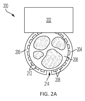
[0039] FIG. 2A illustrates an exemplary implantable neuromodulation device
with two
curved members extending from a body and implanted on a nerve (shown as a
cross-sectional
plane). The curved members partially circumscribe the nerve, and include a
plurality of
electrode pads positioned along the curved member.
[0040] FIG. 2B illustrates an exemplary implantable neuromodulation device
with a curve
member that substantially circumscribes a nerve (shown as a cross-sectional
plane). The
curve member includes an electrode pad that partially circumscribes the nerve,
although not
to the same extent as the curved member.
[0041] FIG. 3A illustrates a front view of an exemplary implantable closed-
loop
neuromodulation device with five curved members extending from a body and
implanted on
a nerve. FIG. 3B illustrates a side view of the device illustrated in FIG. 3A,
and FIG. 3C
illustrates a top view of the device illustrated in FIG. 3A.
100421 FIG. 4 shows an interrogator in communication with an implantable
device through
an ultrasonic transducer. The interrogator emits carrier waves, which are
received by the
implantable device. The implantable device then emits an ultrasonic
backscatter, which can
be received by the interrogator. Optionally, the ultrasonic backscatter
encodes data or
infonnation about the implantable device.
[0043] FIG. 5 illustrates an exemplary interrogator that can be used in a
system including the
implantable device described herein.
DETAILED DESCRIPTION
[0044] Described herein is an implantable closed-loop neuromodulation device
that includes
one or more curved members that at least partially circumscribe a nerve or
other filamentous
tissue, and include one or more electrode pads. The one or more electrode pads
may be, for
example, a plurality of electrode positioned along the curved member, or may
be a curved
electrode pad that at least partially circumscribes the nerve. The one or more
curved member
9
CA 03109810 2021-02-16
WO 2020/047152
PCT/US2019/048647
extends from a device body, which houses one or more ultrasonic transducers
and a
computational circuit for on-board computing of a stimulation pulse in
response to the device
detecting an electrophysiological signal. The one or more ultrasonic
transducers can receive
ultrasonic waves and convert energy from the ultrasonic waves into an
electrical energy that
can power the device. In some embodiments of the device, the electrical energy
is stored in a
battery, which is housed in the body of the device. The electrical energy
powers the
computational circuit, which is electrically connected to the electrode pads.
100451 The computational circuit allows for on-board computing so that the
device can emit
an electrical pulse in response to an electrophysiological signal detected by
the device. For
example, an electrophysiological signal transmitted by the nerve can be
detected by one or
more (e.g., a plurality of) electrode pads on at least one of the one or more
curved members
of the device. The detection signal from the electrophysiological signal
(which may be
filtered, digitized, compressed, or otherwise processed) is received by the
computational
circuit, which generates a stimulation signal using the detection signal. The
computational
circuit can further operate the one or more electrode pads on at least one of
the one or more
curved members (which may be the same as or different from the one or more
electrodes
and/or curved member that detected the electrophysiological signal) to emit an
electrical
pulse based on the generated stimulation signal.
100461 The curved members include one or more electrode pads, and are
configured to at
least partially circumscribe a nerve. For example, in some embodiments, the
one or more
curved members comprises a plurality of electrode pads positioned along the
curved member,
or the one or more curved members comprises a curved electrode pad that at
least partially
circumscribes the nerve. This configuration allows for targeted detection or
stimulation of
nerve activity. For example, a subset of electrode pads can be activated to
target an electrical
pulse to a subset of nerve fibers. Additionally, the device can detect an
electrophysiological
signal transmitted by a subset of nerve fibers by detecting the
electrophysiological signal
using the plurality of electrode pads and deciphering signals detected by the
electrode pads to
determine the transmitting subset. Therefore, the device can be configured to
detect an
electrophysiological signal from a targeted fascicle within the nerve or emit
an electrical
pulse to a targeted fascicle within the nerve.
100471 Data related to the detected electrophysiological signal or the emitted
electrical pulse
can be stored on a non-transitory memory within the body of the device. The
data can be
transmitted to an external device, for example by encoding the data in
ultrasonic backscatter
waves emitted by the one or more ultrasonic transducers. The interrogator can
transmit the
CA 03109810 2021-02-16
WO 2020/047152
PCT/US2019/048647
ultrasonic waves to the device, for example the ultrasonic waves that are
converted into the
electrical energy by the one or more ultrasonic transducers of the device, and
ultrasonic
backscatter waves are emitted. The current flowing through the one or more
ultrasonic
transducers can be modulated to encode the data, which causes the ultrasonic
backscatter
waves emitted by the one or more ultrasonic transducers to encode the data.
[0048] Further described herein are methods of modulating neural activity. The
method can
include receiving ultrasonic waves at one or more ultrasonic transducers of an
implanted
closed-loop neuromodulation device and converting the ultrasonic waves into an
electrical
energy that powers the device. The device is used to detect an
electrophysiological signal
transmitted by a targeted signaling fascicle within a nerve. The device is
then used to
automatically generate a stimulation signals using the detected
electrophysiological signal,
and to emit an electrical pulse to the nerve based on the generated
stimulation signal. The
electrical pulse can be targeted to a targeted receiving fascicle within the
nerve, which may
be the same or different as the targeted signaling fascicle.
[0049] In another example, a method of modulating neural activity includes
receiving
ultrasonic waves at one or more ultrasonic transducers on a fully implanted
closed-loop
neuromodulation device, and converting the ultrasonic waves into an electrical
energy that
powers the device. The device is used to detect an electrophysiological signal
transmitted by
a nerve. The device is then used to generate a stimulation signal based on the
detected
electrophysiological signal, and emit an electrical pulse to a targeted
receiving fascicle within
the nerve based on the generated stimulation signal.
Definitions
100501 As used herein, the singular forms "a," "an," and "the" include the
plural reference
unless the context clearly dictates otherwise.
[0051] Reference to "about" or "approximately" a value or parameter herein
includes (and
describes) variations that are directed to that value or parameter per se. For
example,
description referring to "about X" includes description of "X."
[0052] It is understood that aspects and variations of the invention described
herein include
"consisting" and/or "consisting essentially of' aspects and variations.
100531 The terms "implantable" and "implanted" refer to an object being fully
implantable or
fully implanted in a subject such that no portion of the object breaches the
surface of the
subject.
11
CA 03109810 2021-02-16
WO 2020/047152
PCT/US2019/048647
[0054] The term "substantially" refers to 90% or more. For example, a curved
member that
substantially surrounds a cross-section of a nerve refers to a curved member
that, surrounds
90% or more of the cross-section of the nerve.
[0055] The term "subject" and "patient" are used interchangeably herein to
refer to a
vertebrate animal.
[0056] The terms "treat," "treating," and "treatment" are used synonymously
herein to refer
to any action providing a benefit to a subject afflicted with a disease state
or condition,
including improvement in the condition through lessening, inhibition,
suppression, or
elimination of at least one symptom, delay in progression of the disease or
condition, delay in
recurrence of the disease or condition, or inhibition of the disease or
condition.
[0057] Where a range of values is provided, it is to be understood that each
intervening value
between the upper and lower limit of that range, and any other stated or
intervening value in
that stated range, is encompassed within the scope of the present disclosure.
Where the stated
range includes upper or lower limits, ranges excluding either of those
included limits are also
included in the present disclosure.
[0058] It is to be understood that one, some or all of the properties of the
various
embodiments described herein may be combined to form other embodiments of the
present
invention. The section headings used herein are for organizational purposes
only and are not
to be construed as limiting the subject matter described.
[0059] Features and preferences described above in relation to "embodiments"
are distinct
preferences and are not limited only to that particular embodiment; they may
be freely
combined with features from other embodiments, where technically feasible, and
may form
preferred combinations of features. The description is presented to enable one
of ordinary
skill in the art to make and use the invention and is provided in the context
of a patent
application and its requirements. Various modifications to the described
embodiments will be
readily apparent to those persons skilled in the art and the generic
principles herein may be
applied to other embodiments. Thus. the present invention is not intended to
be limited to the
embodiment shown but is to be accorded the widest scope consistent with the
principles and
features described herein.
Implantable Closed-Loop Neuromodulation Device
[0060] The implantable neuromodulation device is a closed-loop device that can
detect an
electrophysiological signal from a nerve or a subset of nerve fibers, and emit
an electrical
pulse to the nerve or a subset of nerve fibers of the nerve (which may be the
same subset or a
12
CA 03109810 2021-02-16
WO 2020/047152
PCT/US2019/048647
different subset of nerve fibers from which the electrophysiological signal
was detect) in
response to the detected electrophysiological signal. In some embodiments, the
implantable
device detects a compound action potential (or a subset of the compound action
potential) or
other modulation of the electrophysiological signal, and the electrical pulse
is emitted in
response to the detected compound action potential (or subset thereof) or
other modulation of
the electrophysiological signal. Processing for the generation of the
stimulation signal in
response to the detected electrophysiological signal is performed by on-board
computing
using the computational circuit. Therefore, no external communication is
needed to emit the
electrical pulse in response to the detected electrophysiological signal.
[0061] The implantable closed-loop neuromodulation device includes one or more
curved
members that are configured to surround a nerve, and includes one or more
(e.g., a plurality
of) electrode pads that can detect an electrophysiological signal transmitted
by the nerve
and/or stimulate the nerve by emitting an electrical pulse. The device can
include a plurality
of curved members, with a first portion configured to detect the
electrophysiological signal
and a second portion configured to emit the electrical pulse. The curved
members can
include one or more (e.g., a plurality of) electrode pads on the inner surface
of the curved
members so that the electrode pads can be place in electrical communication
with the never
when implanted. For example, the curved members may include a plurality of
electrode pads
positioned along the curved member, which at least partially encompasses the
nerve, or the
curve members may include a curved electrode pad that at least partially
circumscribes the
nerve.
[0062] In some embodiments, the curved member substantially surrounds a cross-
section of
the nerve, with the electrode pads on an inner surface of the curved member
and radially
positioned around an axis along the length of the nerve. In this
configuration, the electrode
pads are circularly aligned with the cross-section of the nerve.
[0063] In some embodiments, the curved members include a plurality of
electrode pads,
which are radially positioned around an axis parallel to the length of the
nerve, and are in
electrical communication with the nerve when the implantable device is
implanted. The
curved members extend from a body, which include one or more ultrasonic
transducers
configured to receive ultrasonic waves and convert energy from the ultrasonic
waves into an
electrical energy, and a computational circuit electrically connected to the
plurality of
electrode pads. In some embodiments, the implantable device includes one, two,
three, or
more ultrasonic transducers.
13
CA 03109810 2021-02-16
WO 2020/047152
PCT/US2019/048647
[00641 The body of the device can house an integrated circuit, which includes
the
computational circuit, a modulation circuit, a detection circuit, and a
stimulation circuit. The
computational circuit is electrically connected to the plurality of electrode
pads on the one or
more curved members, and is configured to operate the electrode pads to emit
an electrical
pulse or detect an electrophysiological signal through the electrode pad. For
example, the
computational circuit is configured to receive a detection signal, generate a
stimulation signal
using the detection signal, and operate the plurality of electrode pads of at
least one of the one
or more curved members to emit an electrical pulse to the nerve based on the
stimulation
signal. The detection signal is based on the detected electrophysiological
signal. Optionally,
the detection signal may be further based on an additional physiological
condition, for
example temperature, pressure, heart rate, pH, or detection or concentration
of an analyte.
That is, the detection signal may optionally include a detected
electrophysiological signal
component and a detected physiological condition component. In some
embodiments, the
physiological condition is detected or measured using a sensor, which may be
on the device,
as further described herein.
[00651 The computational circuit can be a digital circuit, an analog circuit,
or a mixed-signal
integrated circuit. Exemplary computational circuits include a microprocessor,
a finite state
machine (FSM), a field programmable gate array (FPGA), and a microcontroller.
In some
embodiments, the integrated circuit includes a volatile memory, which can be
accessed by the
computational circuit.
[00661 In some embodiments, the computational circuit is configured to
selectively activate
the electrode pads within the plurality of electrode pads for targeted
emission of the electrical
pulse, as further described herein.
[00671 When the electrode pads signal are in electrical communication with the
nerve, an
electrophysiological signal transmitted by the nerve is detected by the
electrode pads. The
electrophysiological signal can include a baseline signal, and an action
potential or compound
action potential transmitted by the nerve results in modulation of the
electrophysiological
signal. A detection signal based on the electrophysiological signal detected
by the electrode
pads of the device is received by the computational circuit. The detection
signal received by
the computational circuit may be a raw electrophysiological signal detected by
the device, or
the electrophysiological signal may be processed (for example, amplified,
digitized, and/or
filtered) before being received by the computational circuit. In some
embodiments, the
detection signal includes a detected electrophysiological signal component and
a
physiological condition component, which can be together analyzed by the
computational
14
CA 03109810 2021-02-16
WO 2020/047152
PCT/US2019/048647
circuit to generate the stimulation signal. In some embodiments, the detection
signal (or the
detected electrophysiological signal component of the detection signal) is
compressed by the
computational circuit or other suitable circuitry within the device.
Compression of the
detection signal allows for faster and more energy efficient processing by the
computational
circuit, which allows for a more efficient closed-loop device. For example,
the battery life of
the on-board batter is longer with less data processing, and the time delay
between receiving
the detection signal and generating the stimulation signal is decreased. By
way of example,
compression of the detection signal can include down sampling the detection
signal by
retaining a portion of the data points in the detection signal. In another
example, in some
embodiments, the digital signal is compressed by identifying an
clectrophysiological signal
spike above a baseline threshold, and using a timestamp associated with the
electrophysiological signal spike as an input for the computational circuit.
In some
embodiments, the detection signal can be compared to a baseline signal, which
may be an
average signal (either clectrophysioloeical signal, physiological condition,
or both) detected
for a period of time. The period of time can be, for example about 1 minute or
more (such as
about 2 minutes or more, about 5 minutes or more, about 10 minutes or more,
about 15
minutes or more, about 30 minutes or more, or about 45 minutes or more). In
some
embodiments, the period of time is about 1 hour or less (such as about 45
minutes or less,
about 30 minutes or less, about 15 minutes or less, about 10 minutes or less,
about 5 minutes
or less, or about 2 minutes or less. A detected deviation of the detection
signal from the
baseline signal can be used to trigger generation of the stimulation signal.
For example, in
some embodiments, if the amplitude of the modulated electrophysiological
signal is above a
baseline electrophysiological signal or above a predetermined amplitude
threshold, the
detected modulation is a signal input, which can be associated with one or
more additional
detected modulation in a temporal dimension. In some embodiments, the
computational
signal analyzes the non-compressed (e.g., raw) signal.
100681 In some embodiments, the detected electrophysiological signal component
of the
detection signal includes, for example, a velocity, a direction, a frequency,
an amplitude, a
waveform of a compound action potential or a subset of the compound action
potential (such
as one or more action potential) transmitted by the nerve or a subset of nerve
fibers within the
nerve. The detected electrophysiological signal component may additionally or
alternatively
include information related to the subset of nerve fibers from which the
electrophysiological
signal was detected (that is, a location of the subset of nerve fibers within
the nerve). This
CA 03109810 2021-02-16
WO 2020/047152
PCT/US2019/048647
information can be used by the computational circuit, for example, to select a
template
detection signal and/or generate the stimulation signal.
[0069] A detection circuit can be included in the integrated circuit, and
electrically connected
to the plurality of electrode pads configured to detect the
electrophysiological signal. The
detection circuit can also optionally include an analog to digital converter
(ADC), one or
more filters, and/or one or more amplifiers.
[0070] Optionally, the implantable device further includes one or more sensors
configured to
measure or detect a physiological condition, such as an analyte, a pH, a
temperature, a strain,
a pulse rate, or a pressure (e.g., a blood pressure). The physiological
condition detected by
the implantable device can optionally be a component, in addition to the
detected
electrophysiological signal, of the detection signal received by the
computational circuit.
Therefore, the detection signal including the detected electrophysiological
signal component
and the additionally detected physiological condition component is used by the
computational
circuit to generate the simulation signal.
[0071] The detection signal can include one or more detected signals
(electrophysiological
signal and/or physiological condition), which may be detected at different
time points. A
time stamp for the signals can be associated with the detected signal, and can
be included in
the detection signal for analysis by the computational circuit. For example, a
detection signal
that includes a predetermined number of detected electrophysiological signal
spikes within a
period of time can result in the generation of a stimulation signal by the
computational
circuit.
[0072] The computational circuit can analyze the detection signal to generate
a stimulation
signal using the detection signal. The analysis can include, for example,
identifying a
modulation of the detection signal (such as a modulation of the detected
electrophysiological
signal, the detected physiological condition, or both), which can act as a
trigger for
generation of the stimulation signal. The modulation of the
electrophysiological signal can
indicate, for example, a compound action potential or a component of the
compound action
potential (e.g., one or more action potentials) that is being transmitted by
the nerve. The
stimulation signal can be generated using a mathematical relationship between
the detection
signal and the stimulation signal. Thus, the computational circuit can input
the detection
signal into the mathematical relationship to generate the stimulation signal.
The
mathematical relationship can be determine, for example, using machine
learning or can be a
pre-selected mathematical relationship. In some embodiments, the computational
circuit uses
a digital logic, an analog logic, an artificial neural network, a
convolutional neural network
16
CA 03109810 2021-02-16
WO 2020/047152
PCT/US2019/048647
(CNN), or neuromorphic computing to detect deviation of the detection signal
from a
baseline signal.
[0073] In some embodiments, generating the stimulation signal can include
comparing the
detection signal (which may include a detected electrophysiological signal
component and/or
a detected physiological signal component) to a template detection signal, and
the stimulation
signal is generated based on the variance or similarity between the detection
signal and the
template detection signal. One or more template detection signals can be
stored, for example,
on a non-transitory memory in the body of the device. The computational
circuit can use, for
example, a digital logic, an analog logic, an artificial neural network, a
convolutional neural
network (CNN), or neuromorphic computing to detect the variance or similarity
between the
detected electrophysiological signal and the template electrophysiological
signal.
[0074] The stimulation signal generated by the computational circuit can
include information
about the electrical pulse to be emitted by the device, such as amplitude,
frequency,
waveform, or targeted location (i.e., subset of nerve fibers) within the
nerve. In some
embodiments, one or more template pulses are stored on a non-transitory memory
within the
device (e.g., within the body of the device). The computational circuit can
generate the
stimulation signal by retrieving a template pulse from the non-transitory
memory using the
detection signal. For example, generating the stimulation signal can include
analyzing the
detection signal, retrieving a template pulse from the non-transitory memory
based on the
analyzed detection signal, and generating the stimulation signal based on the
retrieved
template pulse. Depending on whether or how the detection signal is modulated
from a
baseline or compares to a template detection signal can determine which
template pulse is
retrieved or stimulation signal generated.
[0075] The integrated circuit can include a stimulation circuit, which is
operated by the
computational circuit and is electrically connected to electrode pads that
emit the
electrophysiological pulse. The stimulation circuit can include a stimulating
capacitor, which
can be charged by the battery or electrical energy converted from the
ultrasonic waves by the
one or more ultrasonic transducers. The status of the stimulating capacitor,
for example
capacitor charge, can be determined by the computational circuit. Optionally,
the status of
the stimulating capacitor is recorded on the non-transitory memory or encoded
in ultrasonic
backscatter waves through the modulation circuit operated by the computational
circuit.
[0076] The computational circuit operates the electrode pads of at least one
of the one or
more curved members to emit an electrical pulse to the nerve based on the
stimulation signal.
For example, the stimulation signal can include a pulse amplitude, frequency,
and/or
17
CA 03109810 2021-02-16
WO 2020/047152
PCT/US2019/048647
waveform, and the computational circuit controls the electrode pads to emit
the pulse in
accordance with the stimulation signal. The device can include a capacitor
(i.e., a stimulating
capacitor), such as within the body of the device, which stores an electrical
charge and is
controlled by the computational circuit. The computational circuit controls
the capacitor to
emit the electrical pulse through the electrode pads. In some embodiments, the
computational circuit is configured to determine a stimulating capacitor
status, such as a
charge of the capacitor. The capacitor status can be stored in the non-
transitory memory
and/or encoded in ultrasonic backscatter waves.
100771 In some embodiments, the implantable device further includes a battery
configured to
receive the electrical energy from the one or more ultrasonic transducers and
power the
computational circuit. Inclusion of the battery allows the computational
circuit to function
without an external power source, including detecting an electrophysiological
signal or
emitting an electrical pulse to the nerve. The battery can be contained within
the body of the
implantable device. The battery can be, for example, a rechargeable
electrochemical battery.
The energy stored by the battery can power the device, for example when the
one or more
ultrasonic transducers are not receiving ultrasonic waves. The battery can be
charged by
transmitting ultrasonic waves to the device using an interrogator which are
received by the
one or more ultrasonic transducers. The one or more ultrasonic transducers
convert the
ultrasonic waves into an electrical energy, and are electrically connected to
the battery. In
this manner, the electrical energy charges the battery of the device.
100781 The implantable closed-loop neuromodulation device can also include a
non-
transitory memory configured to store data based on an electrophysiological
signal detected
by the device or an electrical pulse emitted by the device. The data can
include, for example,
a time stamp, a velocity, a direction, an amplitude, a frequency, or a
waveform of a detected
action potential or compound action potential; and/or a time stamp, an
amplitude, a
frequency, or a waveform of an electrical pulse emitted by implantable device.
In some
embodiments, the non-transitory memory can store data related to a detected
physiological
condition (such as temperature, pH, pressure, heart rate, strain, and/or
presence or amount of
an analyte). The data stored on the non-transitory memory may be acquired over
a period of
time (such as about 1 minute or more, about 5 minutes or more, about 10
minutes or more,
about 15 minutes or more, about 30 minutes or more, about 45 minutes or more,
about 1 hour
or more, about 2 hours or more, about 4 hours our more, about 6 hours or more,
about 8 hours
or more, about 12 hours or more, or about 24 hours or more).
18
CA 03109810 2021-02-16
WO 2020/047152
PCT/US2019/048647
[0079] In some embodiments, the device is configured to encode at least a
portion of the data
stored on the non-transitory memory in ultrasonic backscatter waves. This
allows the data to
be wirelessly transmitted to an interrogator, which may be implanted or
external to the
subject. Data encoded in the ultrasonic backscatter waves can be compressed.
Compression
may be used, for example, for efficient transmission of the data due to
bandwidth limits
between the implantable device and the interrogator. By way of example, data
compression
can include transmitting down sampled data from the detection signal,
processed data, or one
or more features in the signal (such as a time stamp of a detected
electrophysiological signal
spike). The implantable device can include a modulation circuit electrically
connected to the
one or more ultrasonic transducers. Upon receiving ultrasonic waves from an
interrogator, a
current is generated that flows through the one or more ultrasonic transducers
and the
modulation circuit. The computational circuit can operate the modulation
circuit to encode
data stored on the non-transitory memory onto the current. The one or more
ultrasonic
transducers of the device emit ultrasonic backscatter waves, which can encode
the data
encoded into the current. The ultrasonic backscatter waves can be received by
an
interrogator, which may be the same or different as the interrogator
transmitting the
ultrasonic waves to the implantable device, and the data encoded on the
ultrasonic backscatter
waves can be deciphered.
[0080] The non-transitory memory can also be used to store data transmitted to
the device
from an interrogator. The interrogator can transmit data (such as temperature
data, or data
related to some other physiological condition, such as an analyte
concentration in the blood
or interstitial fluid of a subject), which is received by the implantable
device and can be
stored on the non-transitory memory. The data can be transmitted, for example,
through
ultrasonic waves that encode the data. The interrogator can transmit the
ultrasonic waves,
which are received by the ultrasonic transducer of the device and deciphered
by the
computational circuit.
[0081] The non-transitory memory can store one or more instructions for
operating the
device, which can be executed using the computational circuit. For example,
the non-
transitory memory can include instructions for receiving a detection signal
based on detected
electrophysiological signal; generating a stimulation signal using the
detection signal; and
operating the plurality of electrode pads of at least one of the one or more
curved members to
emit an electrical pulse to the nerve based on the stimulation signal. In some
embodiments,
the non-transitory memory includes instructions for selectively activating one
or more
electrodes with the plurality of electrodes for targeted emission of the
electrical pulse. In
19
CA 03109810 2021-02-16
WO 2020/047152
PCT/US2019/048647
some embodiments, the non-transitory memory comprises instructions for
analyzing a
detected electrophysiological signal (and, optionally, a measured
physiological condition), for
example by determining a variance in the detected electrophysiological signal
(and/or
physiological condition) compared to a baseline electrophysiological signal
(and/or
physiological condition). In some embodiments, the non-transitory memory
comprises
instructions for comparing the detected electrophysiological signal (and/or
physiological
condition) to a template electrophysiological signal (and/or physiological
condition).
[0082] FIG. I illustrates a schematic of an exemplary body for the implantable
closed-loop
neuromodulation device described herein. The body includes an ultrasonic
transducer
electrically connected to a battery and a modulation circuit. The battery is
electrically
connected to and powers a computational circuit, which is electrically
connected to a non-
transitory memory and the modulation circuit. The computational circuit is
also electrically
connected and is configured to operate the electrodes on the curved member or
curved
members of the device through a stimulation circuit or a detection circuit.
Ultrasonic waves
are received by the ultrasonic transducer, which converts the energy from the
ultrasonic
waves into an electrical energy that charges the battery. The electrodes on
the device are
configured to detect an electrophysiological signal, and a detection signal
based on the
electrophysiological signal is received by the computational circuit. The
detection signal
received by the computational circuit may be processed (for example,
amplified, digitized,
and/or filtered) by the detection circuit before being received by the
computational circuit.
Optionally, the computational circuit accesses the non-transitory memory to
store data related
to the detected electrophysiological signal. The computational circuit can
generate a
stimulation signal based on the detection signal, and operate the electrodes
to emit an
electrical pulse to the nerve based on the stimulation signal. Optionally, the
computational
circuit accesses the non-transitory memory to store data related to the
stimulation signal or
electrical pulse emitted to the nerve. Data stored on the non-transitory
memory can be
wirelessly transmitted through ultrasonic backscatter waves emitted by the
ultrasonic
transducer. The ultrasonic transducer receives ultrasonic waves, and generates
a current that
flows through the modulation circuit. The computational circuit accesses the
memory and
operates the modulation circuit to modulate the current flowing through the
modulation
circuit to encode the data. The ultrasonic backscatter waves emitted by the
ultrasonic
transducer thereby encode the data.
[0083] In some embodiments, the body includes a housing, which can include a
base, one or
more sidewalls, and a top. The housing can enclose the one or more ultrasonic
transducers
CA 03109810 2021-02-16
WO 2020/047152
PCT/US2019/048647
and the integrated circuit (which includes the computational circuit, the non-
transitory
memory, the battery, the modulation circuit, a detection circuit, and/or a
stimulation circuit
(which can include a stimulating capacitor)). The hosing may be sealed closed
(for example
by soldering or laser welding) to prevent interstitial fluid from coming in
contact with the
ultrasonic transducer(s) and/or the integrated circuit. The housing is
preferably made from a
bioinert material, such as a bioinert metal (e.g., steel or titanium) or a
bioinert ceramic (e.g.,
titania or alumina). The housing (or the top of the housing) may be thin to
allow ultrasonic
waves to penetrate through the housing. In some embodiments, the thickness of
the housing
is about 100 micormeters (gm) or less in thickness, such as about 75 pm or
less, about 50 p.m
or less, about 25 gin or less, or about 10 gin or less. In some embodiments,
the thickness of
the housing is about 5 pm to about 10 pm, about 10 pm to about 25 pm, about 25
gm to about
50 pm, about 50 gm to about 75 gm, or about 75 gm to about 100 gm in
thickness.
[0084] The body of the implantable device is relatively small, which allows
for comfortable
and lone-term implantation while limiting tissue inflammation that is often
associated with
implantable devices. In some embodiments, the longest dimension of the body of
the device
is about 10 mm or less, such as about 5 mm to about 9 mm, or about 6 mm to
about 8 mm.
[0085] In some embodiments, the body comprises a material, such as a polymer,
within the
housing. The material can fill empty space within the housing to reduce
acoustic impedance
mismatch between the tissue outside of the housing and within the housing.
Accordingly, the
body of the device is preferably void of air or vacuum.
[0086] One or more ultrasonic transducers of the implantable device can be a
micro-machined ultrasonic transducer, such as a capacitive micro-machined
ultrasonic
transducer (CMUT) or a piezoelectric micro-machined ultrasonic transducer
(PMUT), or can
be a bulk piezoelectric transducer. Bulk piezoelectric transducers can be any
natural or
synthetic material, such as a crystal, ceramic, or polymer. Exemplary bulk
piezoelectric
transducer materials include barium titanate (BaTiO3), lead zirconate titanate
(PZT), zinc
oxide (ZO), aluminum nitride (AIN), quartz, berlinite (A1PO4), topaz,
langasite
(La3Ga5Si014), gallium orthophosphate (GaPO4), lithium niobate (LiNb03),
lithium tantalite
(LiTa03), potassium niobate (KNb03), sodium tungstate (Na2W03), bismuth
ferrite
(BiFe03), polyvinylidene (di)fluoride (PVDF), and lead magnesium niobate-lead
titanate
(PMN-PT).
[0087] In some embodiments, the bulk piezoelectric transducer is approximately
cubic (i.e.,
an aspect ratio of about 1:1:1 (length:width:height). In some embodiments, the
piezoelectric
transducer is plate-like, with an aspect ratio of about 5:5:1 or greater in
either the length or
21
CA 03109810 2021-02-16
WO 2020/047152
PCT/US2019/048647
width aspect, such as about 7:5:1 or greater, or about 10:10:1 or greater. In
some
embodiments, the bulk piezoelectric transducer is long and narrow, with an
aspect ratio of
about 3:1:1 or greater, and where the longest dimension is aligned to the
direction of the
ultrasonic backscatter waves (i.e., the polarization axis). In some
embodiments, one
dimension of the bulk piezoelectric transducer is equal to one half of the
wavelength (X)
corresponding to the drive frequency or resonant frequency of the transducer.
At the resonant
frequency, the ultrasound wave impinging on either the face of the transducer
will undergo a
180 phase shift to reach the opposite phase, causing the largest displacement
between the
two faces. In some embodiments, the height of the piezoelectric transducer is
about 10 p.m to
about 1000 p.m (such as about 40 pm to about 400 pm, about 100 pm to about 250
gm, about
250 gm to about 500 pm, or about 500 p.m to about 1000 pm). In some
embodiments, the
height of the piezoelectric transducer is about 5 mm or less (such as about 4
min or less,
about 3 mm or less, about 2 mm or less, about 1 mm or less, about 500 p.m or
less, about 400
p.m or less, 250 p.m or less, about 100 p.m or less, or about 40 p.m or less).
In some
embodiments, the height of the piezoelectric transducer is about 20 pm or more
(such as
about 40 pm or more, about 100 p.m or more. about 250 p.m or more, about 400
p.m or more,
about 500 pm or more, about 1 mm or more. about 2 mm or more, about 3 mm or
more, or
about 4 mm or more) in length.
[0088] In some embodiments, the one or more ultrasonic transducers have a
length of about 5
mm or less such as about 4 mm or less, about 3 mm or less, about 2 mm or less,
about 1 mm
or less, about 500 pm or less, about 400 p.m or less, 250 p.m or less, about
100 pm or less, or
about 40 pm or less) in the longest dimension. In some embodiments, the
ultrasonic
transducer has a length of about 20 pm or more (such as about 40 pm or more,
about 100 pm
or more, about 250 pm or more, about 400 p.m or more, about 500 p.m or more,
about 1 mm
or more, about 2 mm or more, about 3 mm or more, or about 4 mm or more) in the
longest
dimension.
[0089] The ultrasonic transducer is connected two electrodes to allow
electrical
communication with the computational circuit. The first electrode is attached
to a first face of
the transducer and the second electrode is attached to a second face of the
transducer, wherein
the first face and the second face are opposite sides of the transducer along
one dimension. In
some embodiments, the electrodes comprise silver, gold, platinum, platinum-
black, poly(3,4-
ethylenedioxythiophene (PEDO'T), a conductive polymer (such as conductive PDMS
or
22
CA 03109810 2021-02-16
WO 2020/047152
PCT/US2019/048647
polyimide), or nickel. In some embodiments, the axis between the electrodes of
the
transducer is orthogonal to the motion of the transducer.
[0090] The curved members of the device extend from the body of the device to
at least
partially circumscribe a nerve, and one or more electrode pads are included on
the curved
members. The electrode pads can be configured to be in electrical
communication with the
nerve, for example to detect an electrophysiological signal transmitted by the
nerve and/or
emit one or more electrical pulses to the nerve. For example, the one or more
electrode pads
may be on an inner surface of the curved members, and the one or more curved
members may
engage the nerve or filamentous tissue that includes the nerve (such as a
blood vessel
connected to the nerve) to secure the device to the nerve or other filamentous
tissue and
position the electrode pads.
[0091] The curved members may be flexible, which allows for deformation of the
curved
members during implantation of the device. For example, the cured members may
be flexed
outwardly while the device is being positioned on the nerve. Release of the
curved members
allows the curved members to wrap around the nerve or filamentous tissue
containing the
nerve. Optionally, the curved member includes two portions that are bridged by
the body of
the device.
100921 The electrode pad (or pads) may, for example, be configured to at least
partially
surround an axis parallel to the length of a nerve, or a plurality of
electrode pads may be
configured to be radially positioned around the axis parallel to the length of
the nerve. The
device may include curved members with different electrode pad configurations.
For
example, in some embodiments, a device may include one or more curved members
with a
plurality of electrode pads positioned along the curved member, and one or
more curved
members with a curved electrode pad that at least partially circumscribes the
nerve.
[0093] In some embodiments, the curved members that extend from the body of
the device
each include a plurality of electrode pads configured to be radially
positioned around the
nerve (i.e., around an axis that runs parallel through the center of and along
the length of the
nerve) and in electrical communication with the nerve. The curved members
extend away
from the body before curving toward the body as the curved members extend
below the body,
resulting in a ring-like structure that results in the curved members
sustainably
circumscribing a cross-section of the nerve or filamentous tissue that
includes the nerve (such
as a blood vessel connected to the nerve). In some embodiments, the curved
members make
a single loop around the cross-section of the nerve. Once in position, the
electrode pads of a
given curved member are within the same cross-sectional location relative to
the nerve. A
23
CA 03109810 2021-02-16
WO 2020/047152
PCT/US2019/048647
space within the curved member can be included to allow the device to be
implanted on the
nerve. The curved members can be flexible, which allows for deformation of the
curved
members during implantation of the device. The cured members can be flexed
outwardly
while the device is being positioned on the nerve. Release of the curved
members allows the
curved members to wrap around the nerve or filamentous tissue containing the
nerve.
Optionally, the curved member includes two portions that are bridged by the
body of the
device.
[0094] FIG. 2A illustrates an exemplary embodiment of a device with a first
curved member
and a second curved member that each partially circumscribe a nerve to engages
the nerve.
The device 200 includes a body 202 attached to a first curved member 204 and a
second
curved member 206. A plurality of electrodes 208 on the inner surface of the
first curved
member 204 is positioned along the first curved member, 204, and plurality of
electrodes 212
is positioned along the second curved member 206. In the illustrated example,
the first curved
member 204 and the second curved member 206 are flexible members that are
separated by a
gap (i.e., a separation) 214. In this configuration, the first curved member
204 and the second
curved member 206 can be flexed outwardly (thereby widening the gap 214) to
allow the
nerve 208 to be positioned within the space between the curved members, and
the curved
members can be released so that the curved members wrap around the nerve.
[0095] FIG. 2B illustrates another exemplary embodiment of a device with a
curved member
that engages a nerve. The device 216 includes a body 218 and a curved member
220 that
substantially circumscribes a nerve 222. The inner surface of the curved
member 220
includes a curved electrode pad 224 that circumscribes the nerve 222. The
curved member
220 may be flexible, and a space 226 may be present between the body 218 and
the end 228
of the curved member 220 (or between a first curved member a second curved
member). The
curved member may be flexed outwardly to allow the nerve 222 to be positioned
within the
space formed by the curved member, and the curved member may be released so
that the
curved member wraps around the nerve 222.
100961 The configurations of the curved members and electrode pads shown in
FIG. 2A and
FIG. 2B may be combined. For example, a device may include a curved member as
shown in
FIG. 2A and a curved member as shown in FIG. 2B. In another embodiment, the
device may
include first and second curved members (as shown in FIG. 2A) and a curved
electrode (as
shown in FIG. 2B). In another embodiment, the device may include a curved
member that
substantially surrounds the nerve (e.g., as shown in FIG. 2B) with a plurality
of electrode
positioned along the curved member (e.g., as shown in FIG. 2A).
24
CA 03109810 2021-02-16
WO 2020/047152
PCT/US2019/048647
[00971 The size. shape. and spacing of the one or more curved members on the
device can
depend on the type and size of tissue that device engages. In some
embodiments, the two or
more curved members are spaced by about 0.25 mm or more (such as about 0.5 mm
or more,
about 1 mm or more, about 2 mm or more, about 3 mm or more, about 4 mm or
more, about
turn or more, about 6 rum or more, or about 7 mm or more). In some
embodiments, the two
or more curved members are space by about 8 mm or less (such as about 7 mm or
less, about
6 mm or less, about 5 mm or less, about 4 mm or less, about 3 mm or less.
about 2 mm or
less, about 1 mm or less, or about 0.5 mm or less). By way of example, the two
or more
curved members can be spaced by about 0.25 mm to about 0.5 mm, about 0.5 mm to
about 1
mm, about 1 mm to about 2 mm, about 2 mm to about 3 mm, about 3 mm to about 4
mm,
about 4 mm to about 5 min. about 5 mm to about 6 mm, about 5 mm to about 7 mm,
or about
7 turn to about 8 mm apart. The width of the curved members can also vary
depending on the
application of the device or the tissue engaged by the device. In some
embodiments, the
width of the curved member is about 100 gm or more (such as about 150 gm or
more, about
250 pm or more, about 500 pm or more, about 1 mm or more. or about 1.5 mm or
more). In
some embodiments, the width of the curved member is about 2 mm or less (such
as about 1.5
mm or less, about 1 mm or less, about 500 gm or less, about 250 tun or less,
or about 150 tun
or less. In some embodiments, the width of the curved members is about 100 pm
to about 2
mm (such as about 100 pm to about 150 pm, about 150 gm to about 250 pm, about
250 pm to
about 500 pm, about 500 gm to about 1 mm, about 1 mm to about 1.5 mm, or about
1.5 mm
to about 2 mm). The inner surface of the curved members form a cylindrical
space through
which the nerve and/or filamentous tissue passes. The diameter of the
cylindrical space
formed by the curved members depends on the target nerve and/or filamentous
tissue that the
implantable device will engage. In some embodiments, the one or more curved
members of
the device form a cylindrical space with a diameter of about 50 pm to about 15
mm (for
example, about 50 pm to about 100 pm. about 100 p.m to about 250 pm, about 250
pm to
about 500 pm, about 500 gm to about 1 mm, about 1 mm to about 1.5 mm, about
1.5 mm to
about 2.5 mm, about 2.5 mm to about 5 mm, about 5 mm to about 10 mm, or about
10 mm to
about 15 mm).
[00981 The one or more curved members may be configured to at least partially
circumscribe
the nerve or other filamentous tissue. For example, the curved member may be
configured to
circumscribe at least 25%, at least 33%, at least 50%, at least 66%, at least
75%, at least 90%,
or at least 100% (for example, the curve member may completely surround the
nerve or
filamentous tissue, or may include more than one complete loop around the
nerve or
CA 03109810 2021-02-16
WO 2020/047152
PCT/US2019/048647
filamentous tissue) of the nerve or filamentous tissue. Similarly, the one or
more curved
electrode pads, may circumscribe at least 25%, at least 33%, at least 50%, at
least 66%, at
least 75%, at least 90%, or at least 100% of the nerve, and the portion of the
nerve
circumscribed by the curved electrode pad may be the same or less than the
portion of the
nerve circumscribed by the curved member. The plurality of electrode pads
positioned on the
curved member may be positioned along the full length of the curved member or
a portion of
the length of the curved member.
[0099] In some embodiments, the implantable device includes one or more
additional
securing members configured to secure the implantable device to the
filamentous tissue.
Such securing members can include, for example, loops for suturing the
implantable device
to anatomical structure (such as the filamentous tissue or nerve, or other
tissue surrounding
the filamentous tissue or nerve), pins, or clamps. For example, the
implantable device can be
sutured to the filamentous tissue or nerve, or tissue surrounding the
filamentous tissue or
nerve, to limit movement of the implantable device once implanted.
[0100] The curved members of the implantable device can comprise a metal,
metal alloy,
ceramic, silicon, or a non-polymeric material. The curved members may be
flexible, and are
preferably sprung such that the curved members can be positioned around the
nerve and/or
filamentous tissue. In some embodiments, the one or more curved members or a
portion of
the one or more curved members are coated with an elastomeric coating or a non-
elastomeric
coating, which is preferably bioinert, such as polydimethylsioloxane (PDMS), a
silicone, a
urethane polymer, a poly(p-xylylene)polymer (such as a poly(p-xylylene)
polymer sold under
the tradename PARYLENE0), or a polyimide. The one or more curved members each
include a plurality of electrode pads on an inner surface of the curved
members. In some
embodiments, the electrode pads on the inner surface of the curved members are
not coated
with the elastomeric coating or the non-elastomeric polymer coating, although
may be coated
with a conductive material (e.g., electroplated with a PEDOT polymer or a
metal to improve
electrical characteristics of the electrode pad). Accordingly, in some
embodiments, only the
outer surface of the curved member is coated with the coating. Optionally, the
coating
further coats the housing of the body.
[0101] The one or more curved members can hold the implantable device in place
on the
nerve and/or filamentous tissue. In some embodiments, the one or more curved
members
allow for some rotational movement of the implantable device on the nerve
and/or
filamentous tissue. In some embodiments, the one or more curved members grip
the nerve
and/or filamentous tissue by exerting an inward pressure on the nerve and/or
filamentous
26
CA 03109810 2021-02-16
WO 2020/047152
PCT/US2019/048647
tissue. The amount of inward pressure exerted by the one or more curved
members can be
determined based on the size and curvature of the curved members, as well as
by the spring
constant of the curved members. The inward pressure should be sufficient to
hold the
implantable device in place while the tissue heals after insertion, but not so
high that the
epineurium or vascular walls that contact the curved members are damaged. In
some
embodiments, the inward pressure on the nerve or filamentous tissue is about 1
MPa or less
(such as about 0.7 MPa or less, about 0.5 MPa or less, or about 0.3 MPa or
less). In some
embodiments, the inward pressure on the nerve or filamentous tissue is about
0.1 MPa to
about 1 MPa (such as about 0.1 MPa to about 0.3 MPa, about 0.3 MPa to about
0.5 MPa,
about 0.5 MPa to about 0.7 MPa, or about 0.7 MPa to about 1 MPa).
[0102] The plurality of electrode pads on each curved member is positioned on
an inner
surface of the curved member (that is, the surface of the curved surface that
is configured to
interface with the nerve and/or filamentous tissue). In some embodiments, the
plurality of
electrode pads include 2, 3, 4, 5, 6, 7, 8,9, 10, 11, 12, 13, 14, 15, 16, 17,
18, 19, 20, or more
electrode pads, such as between about 3 and about 50 electrode pads, between
about 3 and
about 5 electrode pads, between about 5 and about 10 electrode pads, between
about 10 and
about 25 electrode pads, or between about 25 and about 50 electrode pads. In
some
embodiments, the electrode pads within the plurality of electrode pads can be
selectively
activated by the computational circuit, which allows for targeted electrical
pulse emission, as
further described herein.
[0103] The electrode pads can include any suitable conductive material, such
as one or more
of (or an alloy of one or more of) tungsten, platinum, palladium, gold,
iridium, niobium,
tantalum, or titanium. The material of the detecting electrode pads and the
stimulating
electrode pads may be the same or different. The size and shape of the
electrode pads may
also be the same or different. For example, electrode pads on a given curved
members may
be of the same or different size, and electrode pads on different curved
members may be of
the same or different size.
101041 The electrode pads of the implantable device are positioned by the
curved members to
be in electrical communication with the nerve. In some embodiments, the
electrode pads are
not in direct contact with the nerve (for example outside and not indirect
contact with the
nerve), but are in electrical communication with the nerve. In some
embodiments, the
electrode pads are positioned within about 2 mm (within about 1.8 mm, within
about 1.6 mm,
within about 1.4 mm, within about 1.2 mm, within about 1.0 mm, within about
0.8 mm,
within about 0.6 mm, within about 0.4 mm, or within about 0.2 mm of the nerve.
In some
27
CA 03109810 2021-02-16
WO 2020/047152
PCT/US2019/048647
embodiments, the plurality of electrode pads is configured to penetrate the
epineurium of the
nerve at one or more locations. For example, the electrode pads can be needle-
shaped, which
allows for penetration of the epineurium. In some embodiments, the electrode
pads directly
contact the nerve, for example the epineurium of the nerve.
101051 In some embodiments, one or more of the curved members on the device is
configured to detect the electrophysiological signal transmitted by the nerve.
In some
embodiments, one or more or more curved members on the device are configured
to emit the
electrical pulse. The one or more curved members that are configured to detect
the
electrophysiological signal transmitted by the nerve may be the same or
different from the
one or more curved members that are configured to emit the electrical pulse.
For example, in
some embodiments the device includes a first curved member that includes a
first plurality of
electrode pads configured to detect the electrophysiological signal
transmitted by the nerve,
and a second curved member that includes a second plurality of electrode pads
configured to
emit the electrical pulse to the nerve. In some embodiments, the device
includes two, three,
four, five, six, seven, eight or more curved members. In some embodiments, the
device
includes one, two, three, four, five, six, seven, eight or more curved members
configured to
detect the electrophysiological signal transmitted by the nerve, and one, two,
three, four, five,
six, seven, eight or more curved members configured to emit the electrical
pulse. In some
embodiments, the curved members having electrode pads configured to detect the
electrophysiological signal and the curved members having electrode pads
configured to emit
the electrical pulse are separate curved members to allow for concurrent
detection of the
electrophysiological signal and emission of the electrical pulse.
101061 Multiple curved members can be positioned along the length of the
nerve. This
configuration allows for electrophysiological signal detection and/or emission
of an electrical
pulse, which may be target to a subset of nerve fibers within the nerve, at
different points
along the length of the nerve. By detecting an electrophysiological signal at
two or more
positions along the length of the nerve, the computational circuit can
determine a direction
and/or velocity of the electrophysiological signal transmitted by the nerve.
To determine
direction of the electrophysiological signal (e.g., an efferent signal or an
afferent signal), the
computational circuit can use on a first time stamp of the
electrophysiological signal detected
by a first curved member and a second time stamp of the electrophysiological
signal detected
by the second curved member. To determine the velocity of the
electrophysiological signal,
the computational circuit can further use the known distance between the first
curved member
and the second curved member. In some embodiments, the identity of the
28
CA 03109810 2021-02-16
WO 2020/047152
PCT/US2019/048647
electrophysiological signal detected by the electrode pads of the first curved
member and the
second curved member is confirmed by comparing one or more
electrophysiological signal
features (e.g., amplitude, frequency, or waveform) detected by the electrode
pads on the first
curved member and the electrode pads of the second curved member.
[0107] The implantable device can also include two or more curved members
comprising a
plurality of electrodes configured to emit an electrical pulse or an
electrical pulse train. The
two or more curved members can be positioned at different locations along the
length of the
nerve, and are configured to emit an electrical pulse at the different
positions. The electrical
pulse emitted by the two or more different curved members may be the same or
different, and
may be targeted to the same or different subset of curved members within the
nerve. For
example, a first electrode pad (or a first plurality of electrode pads) on a
first curved member
can emit an electrical pulse configured to block transmission of an
electrophysiological signal
by a first subset of nerve subset of nerve fibers, and a second electrode pad
(or second
plurality of electrode pads) on a second curved member can be configured to
emit an
electrical pulse that stimulates a second subset of nerve fibers. In some
embodiments, the first
subset of nerve fibers can be, for example, efferent nerve fibers, while the
second subset of
nerve fibers is afferent nerve fibers. In other embodiments the first subset
of nerve fibers are
afferent nerve fibers, and the second subset of nerve fibers are efferent
nerve fibers. By
blocking transmission of an electrophysiological signal in a first subset of
nerve fibers and
stimulating a second subset of nerve fibers, off-target effects of the
stimulation are
minimized. In another example, the one or more electrode pads within the first
plurality of
electrode pads on the first curved member and one or more electrode pads
within the second
plurality of electrode pads on the second curved members can be operated for
bipolar
stimulation along the length of the nerve. In a further example, the plurality
of electrodes on
the first curved member and the plurality of electrodes on the second curved
member can
each emit a coordinated electrical pulse (that is, the electrical pulses
emitted by the separate
pluralities of electrodes are coordinated with each other), which can be used
for specific focal
stimulation.
[0108] By way of example, in some embodiments the device comprises a first
curved
member comprising a first plurality of electrode pads and a second curved
member
comprising a second plurality of electrode pads, wherein the first plurality
of electrodes and
the second plurality of electrodes are each configured to be radially
positioned around the
axis parallel to the length of the nerve at different positions along the
length of the nerve. In
some embodiments, the first plurality of electrode pads and the second
plurality of electrode
29
CA 03109810 2021-02-16
WO 2020/047152
PCT/US2019/048647
pads are configured to detect the electrophysiological signal transmitted by
the nerve. In
some embodiments, the device optionally further comprises a third curved
member
comprising a third plurality of electrode pads, wherein the third plurality of
electrode pads is
configured to be radially positioned around the axis parallel to the length of
the nerve at a
position between the first curved member and the second curved member along
the length of
the nerve.
101091 As another example, in some embodiments, the device includes a first
curved member
comprising a first plurality of electrode pads and a second curved member
comprising a
second plurality of electrode pads, wherein the first plurality of electrodes
and the second
plurality of electrodes are each configured to be radially positioned around
the axis parallel to
the length of the nerve at different positions along the length of the nerve,
wherein the first
plurality of electrode pads or the second plurality of electrode pads are
configured to emit the
electrical pulse to the nerve. In some embodiments, the electrode pads within
the first
plurality of electrode pads and/or the second plurality of electrode pads are
configured to be
selectively activated to emit the electrical pulse to the nerve for example by
targeting a subset
of nerve fibers within the nerve.
101101 In another example, in some embodiments, the device includes a first
curved member
comprising a first plurality of electrode pads, and a second curved member
comprising a
second plurality of electrode pads, the first plurality of electrode pads and
the second
plurality of electrode pads configured to detect the electrophysiological
signal transmitted by
the nerve; and a third curved member comprising a third plurality of electrode
pads, and a
fourth curved member comprising a fourth plurality of electrode pads, the
third plurality of
electrode pads and the fourth plurality of electrode pads configured to emit
the electrical
pulse; wherein the first plurality of electrodes, the second plurality of
electrodes, the third
plurality of electrodes, and the fourth plurality of electrodes are each
configured to be radially
positioned around the axis parallel to the nerve at different positions along
the length of the
nerve. Optionally, the third curved member and the fourth curved member are
positioned
between the first curved member and the second curved member along the length
of the
nerve. In some embodiments, the device further includes a fifth curved member
comprising a
fifth plurality of electrode pads configured to detect the
electrophysiological signal or emit
the electrical pulse. The fifth curved member is optionally positioned between
the third
curved member and the fourth curved member along the length of the nerve. In
some
embodiments, the first plurality of electrode pads, the second plurality of
electrode pads, the
third plurality of electrode pads, or the fourth plurality of electrode pads
and/or the fifth
CA 03109810 2021-02-16
WO 2020/047152
PCT/US2019/048647
plurality of electrode pads are configured to be selectively activated to emit
the electrical
pulse.
[0111] FIGS. 3A-3C illustrate a front (3A), side (3B), and top (3C) view of an
exemplary
implantable closed-loop neuromodulation device with five curved members
extending (302,
304, 306, 308, and 310) from a body 312, implanted on a nerve 314. The body of
the device
includes the integrated circuit 324, a non-transitory memory 326, a battery
328, and an
ultrasonic transducer 330 (such as a piezoelectric transducer). As seen in
FIG. 3A, the curved
members can include a first portion 302a and a second portion 302b bridged by
the body 312
at point 316. In some embodiments, the first portion 302a and the second
portion 302b are
directly connected, and the curved member is attached to the body through a
connecting
member. The curved members include a plurality of electrode pads 318 on the
inner surface
of the curved members, and the electrode pads 318 are radially positioned
around an axis
parallel to the length of the nerve. A separation 320 between the first
portion 302a and the
302b is present along the curved member (which may be similarly present in
other curved
members of the device). The implantable device can be implanted by flexing the
first portion
and the second portion of the curved member outwardly, thereby expanding the
size of the
separation and allowing the nerve or other filamentous tissue to pass through
the separation
and fit within the cylindrical space formed by the curved members. The first
portion and the
second portion of the curved member can be released, which allows the curved
member to
wrap around the nerve or other filamentous tissue.
[0112] The plurality of electrode pads of the exemplary device shown in FIG.
3A are outside
of the nerve, but in direct contact with the epineurium of the nerve. The
nerve includes
several fascicles 322 within the nerve. The electrode pads 318 within a curved
member can
be operated for targeted emission of an electrical pulse to one or more of the
fascicles 322 or
other subset of nerve fibers, and/or operated for targeted detection of an
electrophysiological
signal transmitted by one or more of the fascicles 322 or other subset of
nerve fibers. For
example, the electrode pads 318 can be selectively activated by the
computational circuit
within the integrated circuit 324, which is housed within the body 212, to
emit an electric
pulse targeted to one or more fascicles 322. In another example, the electrode
pads 218 are
operated by the computational circuit to detect an electrophysiological signal
transmitted by
one or more of the fascicles 322 within the nerve 314. The curved members can
be
configured to detect the electrophysiological signal transmitted by the nerve
or a subset of
nerve fibers, emit an electrical pulse to the nerve or targeted to a subset of
nerve fibers, or
both detect the electrophysiological signal transmitted by the nerve or a
subset of nerve fibers
31
CA 03109810 2021-02-16
WO 2020/047152
PCT/US2019/048647
and emit an electrical pulse to the nerve or targeted to a subset of nerve
fibers. By way of
example, curved members 302, 306, and 310 can be configured to detect the
electrophysiological signal transmitted by the nerve or a subset of nerve
fibers, and curved
members 304 and 308 can be configured to emit an electrical pulse to the nerve
or targeted to
a subset of nerve fibers.
[0113] The one or more curved members of the implantable device are sized to
engage a
selected nerve or fibrous tissue containing a nerve. The nerve can be the
spinal cord or a
peripheral nerve. In some embodiments, the nerve is an autonomic nerve or a
somatic nerve.
In some embodiments, the nerve is a sympathetic nerve or a parasympathetic
nerve. In some
embodiments, the nerve is a vagus nerve, a mesenteric nerve, a splenic nerve,
a sciatic nerve,
a tibial nerve, a pudendal nerve, a celiac ganglion, a sacral nerve, or any
branch thereof.
Targeted Detection of an Elect rophysiological Signal
[0114] One or more of the curved members of the implantable device can be
configured to
detect the electrophysiological signal from a targeted subset of nerve fibers
within the nerve.
The subset of fibers can be, for example, one or more (e.g., 2, 3, 4, or more)
fascicles, or a
portion of one or more (e.g., 2, 3, 4, or more) fascicles within the nerve. In
some
embodiments, the subset of nerve fibers comprises or consists of afferent
nerve fibers within
the nerve, or a subset of afferent nerve fibers within the nerve. In some
embodiments, the
subset of nerve fibers comprises or consists of efferent nerve fibers within
the nerve, or a
subset of efferent nerve fibers within the nerve. In some embodiments, the
subset of nerve
fibers comprises or consists of efferent nerve fibers within two or more
fascicles within the
nerve or afferent nerve fibers within two or more fascicles within the nerve.
[0115] One or more techniques such as computational modeling (e.g., finite
element models),
inverse source estimation, multipole (e.g., tripole) neural recording,
velocity-selective
recording, or beamfonning can be used to selectively target the subset of
nerve fibers. See,
for example, Taylor et al., Multiple-electrode nerve cuffs Ar low-velocity and
velocity
selective neural recording, Medical & Biological Engineering & Computing, vol.
42, pp.
634- 643 (2004): and Wodlinger et al., Localization and Recovery of Peripheral
Neural
Sources with Beamfbrming Algorithms, IEEE Transactions on Neural Systems and
Rehabilitation Engineering, vol. 17, no. 5, pp. 461-468 (2009).
[0116] The computational circuit of the implantable device operates the
plurality of
electrodes on one or more curved members of the device for targeted detection
of the
electrophysiological signal. The computational circuit can analyze the
electrophysiological
32
CA 03109810 2021-02-16
WO 2020/047152
PCT/US2019/048647
signal detected by all or a subset of the electrode pads to determine the
subset of nerve fibers
within the nerve that are transmitting the electrophysiological signal.
Certain nerves may
transmit compound electrophysiological signal (or compound action potentials),
which is the
sum of the electrophysiological signals (or action potentials) simultaneously
transmitted by
two or more different subsets of nerve fibers. Based on the
electrophysiological signal
detected by the plurality of electrode pads, the computational circuit is able
to determine
which subset of nerve fibers transmits which electrophysiological signal. In
some
embodiments data received from the interrogator (such as temperature data, or
data related to
an analyte concentration or other physiological condition) is further used to
determine which
subset of nerve fibers transmits the electrophysiological signal.
[0117] For example, in some embodiments, the computational circuit is
configured to
selectively detect an electrophysiological signal from a targeted subset of
nerve fibers using
velocity-selective recording, which may be combined with multipolar (e.g.,
tripolar)
recording (which can include any number of tripoles within the plurality of
electrodes on one
or more curved members).
[0118] Beamforming can additionally or alternatively be used to detect the
electrophysiological signals from the targeted subset of nerve fibers. A
portion of or all of the
electrode pads of one or more curved members can detect the
electrophysiological signal
from the nerve, and the computational circuit can determine the cross-
sectional location of
the transmitted signal within the nerve based on the differences in
electrophysiological signal
detected by a portion or all of the electrode pads of the one or more curved
members.
[0119] Stimulation of one or more nerves at a location separate from the
location of the
implanted device can result in a modulation of the electrophysiological signal
at the location
of the implanted device. The modulation of the electrophysiological signal
detected at
different subsets of nerve fibers within the nerve in electrical communication
with the
electrode pads of the device can be the result of stimulation in different
distant nerves. For
example, stimulation of the splenic nerve can result in modulation of an
electrophysiological
signal detected from first subset of nerve fibers within the vagus nerve, and
stimulation of a
renal nerve can result in modulation of an electrophysiological signal
detected from a second
subset of nerve fibers within the vagus nerve. Therefore, an implantable
device positioned on
the vagus nerve can detect an electrophysiological signal from the first
subset of nerve fibers
to monitor stimulation of the splenic nerve, and a second subset of nerve
fibers to monitor
stimulation of the renal nerve.
33
CA 03109810 2021-02-16
WO 2020/047152
PCT/US2019/048647
[0120] In some embodiments, the implantable device is positioned at a first
nerve locus and
is configured to detect stimulation of a second nerve locus by selectively
detecting an
electrophysiological signal from a subset of nerve fibers within the first
nerve locus that is
associated with the second nerve locus. In some embodiments, the first nerve
locus and the
second nerve locus are separated by one or more nerve branch points or one or
more
synapses. In some embodiments, the second nerve locus is proximal to the brain
relative to
the first nerve locus, and in some embodiment the second nerve locus is distal
from the brain
relative to the first nerve locus. In some embodiments, the targeted subset of
nerve fibers
comprises or consists of afferent nerve fibers. In some embodiments, the
targeted subset of
nerve fibers comprises or consists of efferent nerve fibers.
Targeted Stimulation of a Nerve
101211 One or more of the curved members of the device can be configured to
emit a targeted
electrical pulse to a subset of nerve fibers within the nerve by selectively
activating one or
more electrode pads within the plurality of electrode pads on the curved
member. The
computational circuit of the device can operate the electrode pads to
selectively activate the
electrode pads. Selective activation can include, for example, activating a
portion of the
electrode pads within the plurality of electrode pads of one or more curved
members and/or
differentially activating all or a portion of the electrode pads within the
plurality of electrode
pads of the one or more curved members. The plurality of electrodes can
therefore be
operated to steer the electrical pulse emitted by the plurality of electrode
pads to the target
subset of nerve fibers. Techniques such as electrical field interference
and/or multipolar
stimulation (e.g., tripolar stimulation) can be used to target the electrical
pulse to the subset of
nerve fibers within the nerve. See, for example, Grossman, et al., Noninvasive
Deep Brain
Stimulation via Temporally Inteifering Electrical Fields, Cell, vol. 169, pp.
1029-1041
(2017). The electrode pads with one or more curved members can be selectively
activated by
the computational circuit to target the emitted electrical pulse to the subset
of nerve fibers.
101221 The subset of nerve fibers targeted by the emitted electrical pulse can
be the same or
different as the subset of nerve fibers from which the electrophysiological
signal is detected.
The one or more curved member configured to emit the targeted electrical pulse
can be the
same or different as the one or more curved members on the device configured
to detect the
electrophysiological signal. The emitted targeted electrical pulse can
stimulate the nerve at
the position of the implantable device. The subset of nerve fibers targeted by
the electrical
34
CA 03109810 2021-02-16
WO 2020/047152
PCT/US2019/048647
pulse can be the same or a different subset of nerve fibers for which the
electrophysiological
signal is selectively detected.
101231 The subset of nerve fibers targeted by the electrical pulse emitted by
the device can
be, for example, one or more (e.g., 2, 3, 4, or more) fascicles, or a portion
of one or more
(e.g., 2, 3, 4, or more) fascicles within the nerve. In some embodiments, the
subset of nerve
fibers comprises or consists of afferent nerve fibers within the nerve, or a
subset of afferent
nerve fibers within the nerve. In some embodiments, the subset of nerve fibers
comprises or
consists of efferent nerve fibers within the nerve, or a subset of efferent
nerve fibers within
the nerve. In some embodiments, the subset of nerve fibers comprises or
consists of efferent
nerve fibers within two or more fascicles within the nerve or afferent nerve
fibers within two
or more fascicles within the nerve.
101241 Targeted stimulation of a subset of nerve fibers by emitting a targeted
electrical pulse
to the subset of nerve fibers can result in stimulation of a nerve at a
location distant from the
position of the nerve. The distant nerve stimulated by the implantable device
depends on the
subset of nerves at the position of the implantable device targeted by the
electrical pulse
emitted by the device. In some embodiments, the implantable device is
positioned at a first
nerve locus and is configured to stimulate a second nerve locus by emitting a
targeted
electrical pulse to a subset of nerve fibers within the first nerve locus that
is associated with
the second nerve locus. In some embodiments, the first nerve locus and the
second nerve
locus are separated by one or more nerve branch points or one or more
synapses. In some
embodiments, the second nerve locus is proximal to the brain relative to the
first nerve locus,
and in some embodiment the second nerve locus is distal from the brain
relative to the first
nerve locus. In some embodiments, the targeted subset of nerve fibers
comprises or consists
of afferent nerve fibers. In some embodiments, the targeted subset of nerve
fibers comprises
or consists of efferent nerve fibers.
Wireless Communication
101251 Although the implantable device can emit an electrical pulse in
response to a detected
electrophysiological signal through a closed-loop, on-board computational
circuit, in some
embodiments there is a system that includes a closed-loop implantable device
described
herein and an interrogator configured to emit ultrasonic waves that power the
device. The
interrogator may be an external (i.e., non-implanted) device or a separate but
fully implanted
device. If implanted, the interrogator can wirelessly communicate with an
external device,
for example using ultrasonic communication or radiofrequency (RF). The
ultrasonic
CA 03109810 2021-02-16
WO 2020/047152
PCT/US2019/048647
transducer in the implantable device is configured to receive the ultrasonic
waves emitted by
the interrogator, which converts the energy from the ultrasonic waves into an
electrical
energy. The electrical energy can be stored in a battery in the device, if
present, and can be
used to power device components such as the computational circuit.
101261 In some embodiments, the interrogator is configured to wirelessly
communicate with
the closed-loop implantable device through ultrasonic communication. The
implantable
device receives ultrasonic waves from the interrogator through the ultrasonic
transducer of
the implantable device. Optionally, the ultrasonic waves transmitted by the
interrogator to
the implantable device can encode instructions for operating the implantable
device, which
can be received by the computational circuit. Vibrations of the ultrasonic
transducer on the
implantable device caused by the ultrasonic waves generate a voltage across
the electric
terminals of the transducer, and current flows through the device, including
the integrated
circuit. The ultrasonic waves encoding the information or instructions emitted
by the
interrogator are received by the ultrasonic transducer in the implantable
device. The
information or instructions are then encoded in an electrical current flowing
through the
ultrasonic transducer resulting from receiving the ultrasonic waves, and the
encoded
information or instructions can be deciphered by the computational circuit. In
some
embodiments, the computational circuit stores the instructions or information
on the non-
transitory memory of the device.
101271 Information encoded in the ultrasonic waves emitted by the interrogator
and received
by the closed-loop implantable device can include, for example, instructions
for starting or
stopping closed-loop neuromodulation, one or more calibration instructions,
one or more
updates to the operation software, and/or or one or more templates (such as
template
electrophysiological signals, one or more template electrophysiological
signals, and/or one or
more template stimulation signals). The information encoded in the ultrasonic
waves can be
processed by the computational circuit and/or stored on the non-transitory
memory, if
present. Calibration instructions for the device can include, for example, an
association
between (1) a subset of nerve fibers within the nerve, and (2) a nerve or a
subset of nerve
fibers, at a different nerve locus within the subject. In some embodiments,
the association
includes instructions for stimulating a second nerve locus by emitting
targeted electrical pulse
to a subset of nerve fibers within a first nerve locus in electrical
communication with the
device. In some embodiments, the association includes instructions for
detecting stimulation
of a second nerve locus by selectively detecting an electrophysiological
signal from a subset
of nerve fibers at a first nerve locus in electrical communication with the
device.
36
CA 03109810 2021-02-16
WO 2020/047152
PCT/US2019/048647
[0128] In some embodiments, the closed-loop implantable device transmits
information to
the interrogator through ultrasonic backscatter. FIG. 4 shows an interrogator
in
communication with an implantable device through an ultrasonic transducer. The
external
ultrasonic transceiver emits ultrasonic waves ("carrier waves"), which can
pass through
tissue. The carrier waves cause mechanical vibrations on the ultrasonic
transducer (e.g., a
bulk piezoelectric transducer, a PUMT, or a CMUT). A voltage across the
ultrasonic
transducer is generated, which imparts a current flowing through an integrated
circuit on the
implantable device. The current flowing through to the ultrasonic transducer
causes the
transducer on the implantable device to emit backscatter ultrasonic waves. In
some
embodiments, the integrated circuit modulates the current flowing through the
ultrasonic
transducer to encode information, and the resulting ultrasonic backscatter
waves encode the
information. The backscatter waves can be detected by the interrogator, and
can be analyzed
to interpret information encoded in the ultrasonic backscatter.
[0129] Ultrasonic backscatter emitted from the implantable device can encode
information
relating to the implantable device. The ultrasonic backscatter can be received
by the
interrogator device (which may be the same or different from the interrogator
that transmitted
the ultrasonic waves received by the ultrasonic transducer in the implantable
device), and
deciphered to determine information encoded in the ultrasonic backscatter
waves. The
information can be encoded using a modulation circuit within the integrated
circuit of the
implantable device. The modulation circuit can modulate the current flowing
through the
ultrasonic transducer to encode the information. The modulated current flows
through the
ultrasonic transducer to modulate the ultrasonic backscatter, thereby encoding
the
information in the ultrasonic backscatter waves. The modulation circuit
includes one or more
switches, such as an on/off switch or a field-effect transistor (FET). An
exemplary FET that
can be used with some embodiments of the implantable device is a metal-oxide-
semiconductor field-effect transistor (MOSFE'T). The modulation circuit can
alter the
impedance of a current flowing through the ultrasonic transducer, and
variation in current
flowing through the transducer encodes the information.
[0130] Information encoded in the ultrasonic backscatter emitted from the
device can include
information related to a detected electrophysiological signal, a detected
physiological
condition (e.g., temperature, pH, oxygen levels, pressure, etc.), information
related to the
device status (for example whether the device is in operation, or a
stimulating capacitor
status, such as stimulating capacitor charge), or information related to the
emitted electrical
pulse. For example, the information related to the detected
electrophysiological signal can
37
CA 03109810 2021-02-16
WO 2020/047152
PCT/US2019/048647
include information related to the subset of nerve fibers from which the
electrophysiological
signal is detected (e.g., location information), amplitude or frequency of the
detected
electrophysiological signal, similarity to a template electrophysiological
pulse, and/or a time
stamp of the detected electrophysiological signal. The information related to
the detected
physiological condition can include, for example, a temperature, a pH, an
oxygen level, or a
pressure; and/or a time stamp for the detected physiological condition.
Information related to
the emitted electrical pulse can include, for example, information related to
the subset of
nerve fibers (e.g., a targeted location of the emitted electrical pulse); an
amplitude, frequency,
or waveform of the emitted electrical pulse, and/or a time stamp of the
emitted electrical
pulse.
[0131] In some embodiments, information encoded in the ultrasonic backscatter
includes a
unique identifier for the implantable device, which is optionally a digitized
unique identifier.
This can be useful, for example, to ensure the interrogator is in
communication with the
correct implantable device when a plurality of implantable devices is
implanted in the
subject. In some embodiments, the information encoded in the ultrasonic
backscatter
includes a verification signal that verifies an electrical pulse was emitted
by the implantable
device. In some embodiments, the information encoded in the ultrasonic
backscatter includes
an amount of energy stored or a voltage in the energy storage circuit (or one
or more
capacitors in the energy storage circuit).
[0132] In some embodiments, the backscattered ultrasound is digitized by the
implantable
device. For example, the implantable device can include an oscilloscope or
analog-to-digital
converter (ADC) and/or a memory, which can digitally encode information in
current (or
impedance) fluctuations. The digitized current fluctuations, which can encode
information,
are received by the ultrasonic transducer, which then transmits digitized
acoustic waves. The
digitized data can compress the analog data, for example by using singular
value
decomposition (SVD) and least squares-based compression. In some embodiments,
the
compression is performed by a correlator or pattern detection algorithm. The
backscatter
signal may go through a series of non-linear transformation, such as 4th order
Butterworth
bandpass filter rectification integration of backscatter regions to generate a
reconstruction
data point at a single time instance. Such transformations can be done either
in hardware
(i.e., hard-coded) or in software.
[0133] In some embodiments, the digitized signal compresses the size of the
analog signal.
The decreased size of the digitized signal can allow for more efficient
reporting of
information encoded in the ultrasonic backscatter. By compressing the size of
the transmitted
38
CA 03109810 2021-02-16
WO 2020/047152
PCT/US2019/048647
information through digitization, potentially overlapping signals can be
accurately
transmitted.
[0134] Communication between the interrogator and the implantable device can
use a pulse-
echo method of transmitting and receiving ultrasonic waves. In the pulse-echo
method, the
interrogator transmits a series of interrogation pulses at a predetermined
frequency, and then
receives backscatter echoes from the implanted device. In some embodiments,
the pulses are
square, rectangular, triangular, sawtooth, or sinusoidal. In some embodiments,
the pulses
output can be two-level (GND and POS), three-level (GND, NEG, POS), 5-level,
or any
other multiple-level (for example, if using 24-bit DAC). In some embodiments,
the pulses
are continuously transmitted by the interrogator during operation. In some
embodiments,
when the pulses are continuously transmitted by the interrogator a portion of
the transducers
on the interrogator are configured to receive ultrasonic waves and a portion
of the transducers
on the interrogator are configured to transmit ultrasonic waves. Transducers
configured to
receive ultrasonic waves and transducers configured to transmit ultrasonic
waves can be on
the same transducer array or on different transducer arrays of the
interrogator. In some
embodiments, a transducer on the interrogator can be configured to
alternatively transmit or
receive the ultrasonic waves. For example, a transducer can cycle between
transmitting one
or more pulses and a pause period. The transducer is configured to transmit
the ultrasonic
waves when transmitting the one or more pulses, and can then switch to a
receiving mode
during the pause period.
[0135] The interrogator includes one or more ultrasonic transducers, which can
operate as an
ultrasonic transmitter and/or an ultrasonic receiver (or as a transceiver,
which can be
configured to alternatively transmit or receive the ultrasonic waves). The one
or more
transducers can be arranged as a transducer array, and the interrogator can
optionally include
one or more transducer arrays. In some embodiments, the ultrasound
transmitting function is
separated from the ultrasound receiving function on separate devices. That is,
optionally, the
interrogator comprises a first device that transmits ultrasonic waves to the
implantable
device, and a second device that receives ultrasonic backscatter from the
implantable device.
In some embodiments, the transducers in the array can have regular spacing,
irregular
spacing, or be sparsely placed. In some embodiments the array is flexible. In
some
embodiments the array is planar, and in some embodiments the array is non-
planar.
[0136] An exemplary interrogator is shown in FIG. 5. The illustrated
interrogator shows a
transducer array with a plurality of ultrasonic transducers. In some
embodiments, the
transducer array includes 1 or more, 2 or more, 3 or more, 5 or more, 7 or
more, 10 or more,
39
CA 03109810 2021-02-16
WO 2020/047152
PCT/US2019/048647
15 or more, 20 or more, 25 or more, 50 or more, 100 or more 250 or more, 500
or more, 1000
or more, 2500 or more, 5000 or more, or 10,000 or more transducers. In some
embodiments,
the transducer array includes 100,000 or fewer, 50,000 or fewer, 25,000 or
fewer, 10,000 or
fewer, 5000 or fewer, 2500 or fewer, 1000 or fewer, 500 or fewer, 200 or
fewer, 150 or
fewer, 100 or fewer, 90 or fewer, 80 or fewer, 70 or fewer, 60 or fewer, 50 or
fewer, 40 or
fewer, 30 or fewer, 25 or fewer, 20 or fewer, 15 or fewer, 10 or fewer, 7 or
fewer or 5 or
fewer transducers. The transducer array can be, for example a chip comprising
50 or more
ultrasonic transducer pixels.
101371 The interrogator shown in FIG. 5 illustrates a single transducer array;
however the
interrogator can include 1 or more, 2 or more, or 3 or more separate arrays.
In some
embodiments, the interrogator includes 10 or fewer transducer arrays (such as
9, 8, 7, 6, 5, 4,
3, 2, or 1 transducer arrays). The separate arrays, for example, can be placed
at different
points of a subject, and can communicate to the same or different implantable
devices. In
some embodiments, the arrays are located on opposite sides of an implantable
device. The
interrogator can include an application specific integrated circuit (ARC),
which includes a
channel for each transducer in the transducer array. In some embodiments, the
channel
includes a switch (indicated in FIG. 5 by "T/Rx"). The switch can
alternatively configure the
transducer connected to the channel to transmit ultrasonic waves or receive
ultrasonic waves.
The switch can isolate the ultrasound receiving circuit from the higher
voltage ultrasound
transmitting circuit.
101381 In some embodiments, the transducer connected to the channel is
configured only to
receive or only to transmit ultrasonic waves, and the switch is optionally
omitted from the
channel. The channel can include a delay control, which operates to control
the transmitted
ultrasonic waves. The delay control can control, for example, the phase shift,
time delay,
pulse frequency and/or wave shape (including amplitude and wavelength). The
delay control
can be connected to a level shifter, which shifts input pulses from the delay
control to a
higher voltage used by the transducer to transmit the ultrasonic waves. In
some
embodiments, the data representing the wave shape and frequency for each
channel can be
stored in a 'wave table'. This allows the transmit waveform on each channel to
be different.
Then, delay control and level shifters can be used to stream data to the
actual transmit signals
to the transducer array. In some embodiments, the transmit waveform for each
channel can
be produced directly by a high-speed serial output of a microcontroller or
other digital system
and sent to the transducer element through a level shifter or high-voltage
amplifier. In some
embodiments, the ASIC includes a charge pump (illustrated in FIG. 5) to
convert a first
CA 03109810 2021-02-16
WO 2020/047152
PCT/US2019/048647
voltage supplied to the ASIC to a higher second voltage, which is applied to
the channel. The
channels can be controlled by a controller, such as a digital controller,
which operates the
delay control.
[0139] In the ultrasound receiving circuit, the received ultrasonic waves are
converted to
current by the transducers (set in a receiving mode), which is transmitted to
a data capture
circuit. In some embodiments, an amplifier, an analog-to-digital converter
(ADC), a
variable-gain-amplifier, or a time-gain-controlled variable-gain-amplifier
which compensates
for tissue loss, and/or a band pass filter is included in the receiving
circuit. The ASIC can
draw power from a power supply, such as a battery (which is preferred for a
wearable
embodiment of the interrogator). In the embodiment illustrated in FIG. 5, a
1.8V supply is
provided to the ASIC, which is increased by the charge pump to 32V, although
any suitable
voltage can be used. In some embodiments, the interrogator includes a
processor and or a
non-transitory computer readable memory. in some embodiments, the channel
described
above does not include a T/Rx switch but instead contains independent Tx
(transmit) and Rx
(receive) with a high-voltage Rx (receiver circuit) in the form of a low noise
amplifier with
good saturation recovery. In some embodiments, the T/Rx circuit includes a
circulator. In
some embodiments, the transducer array contains more transducer elements than
processing
channels in the interrogator transmit /receive circuity, with a multiplexer
choosing different
sets of transmitting elements for each pulse. For example, 64 transmit receive
channels
connected via a 3:1 multiplexer to 192 physical transducer elements - with
only 64
transducer elements active on a given pulse.
[0140] By way of example, in some embodiments the interrogator, or the
transducer(s) of the
interrogator, is wearable. For example, the interrogator, or the transducer(s)
of the
interrogator, may be fixed to the body by a strap or adhesive. In another
example, the
interrogator can be a wand, which may be held by a user (such as a healthcare
professional).
In some embodiments, the interrogator can be held to the body via suture,
simple surface
tension, a clothing-based fixation device such as a cloth wrap, a sleeve, an
elastic band, or by
sub-cutaneous fixation. The transducer or transducer array of the interrogator
may be
positioned separately from the rest of the transducer. For example, the
transducer array can
be fixed to the skin of a subject at a first location (such as proximal to one
or more implanted
devices), and the rest of the interrogator may be located at a second
location, with a wire
tethering the transducer or transducer array to the rest of the interrogator.
41
CA 03109810 2021-02-16
WO 2020/047152
PCT/US2019/048647
[0141] The specific design of the transducer array of the interrogator depends
on the desired
penetration depth, aperture size, and size of the individual transducers
within the array. The
Rayleigh distance, R, of the transducer array is computed as:
D2 A2 D2
R= D2 >> A2
4A 4A.'
where D is the size of the aperture and ). is the wavelength of ultrasound in
the propagation
medium (i.e., the tissue). As understood in the art, the Rayleigh distance is
the distance at
which the beam radiated by the array is fully formed. That is, the pressure
filed converges to
a natural focus at the Rayleigh distance in order to maximize the received
power. Therefore,
in some embodiments, the implantable device is approximately the same distance
from the
transducer array as the Rayleigh distance.
[0142] The individual transducers in a transducer array can be modulated to
control the
Raleigh distance and the position of the beam of ultrasonic waves emitted by
the transducer
array through a process of beamforming or beam steering. Techniques such as
linearly
constrained minimum variance (LCMV) beamforming can be used to communicate a
plurality of implantable devices with an external ultrasonic transceiver. See,
for example,
Bertrand et al., Beamfirming Approaches for UMethered, Ultrasonic Neural Dust
Motes for
Cortical Recording: a Simulation Study, IEEE EMBC (Aug. 2014). In some
embodiments,
beam steering is performed by adjusting the power or phase of the ultrasonic
waves emitted
by the transducers in an array.
[0143] In some embodiments, the interrogator includes one or more of
instructions for beam
steering ultrasonic waves using one or more transducers, instructions for
determining the
relative location of one or more implantable devices, instructions for
monitoring the relative
movement of one or more implantable devices, instructions for recording the
relative
movement of one or more implantable devices, and instructions for
deconvoluting backscatter
from a plurality of implantable devices.
101441 Optionally, the interrogator is controlled using a separate computer
system, such as a
mobile device (e.g., a smartphone or a table). The computer system can
wirelessly
communicate to the interrogator, for example through a network connection, a
radiofrequency (RF) connection, or Bluetooth. The computer system may, for
example, turn
on or off the interrogator or analyze information encoded in ultrasonic waves
received by the
interrogator.
42
CA 03109810 2021-02-16
WO 2020/047152
PCT/US2019/048647
[0145] In some embodiments, an interrogator communicates with a plurality of
implantable
devices. This can be performed, for example, using multiple-input, multiple
output (MIMO)
system theory. For example, communication between the interrogator and the
plurality of
implantable devices using time division multiplexing, spatial multiplexing, or
frequency
multiplexing. The interrogator can receive a combined backscatter from the
plurality of the
implantable devices, which can be deconvoluted, thereby extracting information
from each
implantable device. In some embodiments, interrogator focuses the ultrasonic
waves
transmitted from a transducer array to a particular implantable device through
beam steering.
The interrogator focuses the transmitted ultrasonic waves to a first
implantable device,
receives backscatter from the first implantable device, focuses transmitted
ultrasonic waves to
a second implantable device, and receives backscatter from the second
implantable device.
In some embodiments, the interrogator transmits ultrasonic waves to a
plurality of
implantable devices, and then receives ultrasonic waves from the plurality of
implantable
devices.
EXEMPLARY EMBODIMENTS
[0146] The foregoing description has been described with reference to
specific
embodiments. Additional exemplary embodiments are provided below. However, the
illustrative discussions and exemplary embodiments above are not intended to
be exhaustive
or to limit the disclosure to the precise forms disclosed. Many modifications
and variations
are possible in view of the above teachings. The embodiments were chosen and
described in
order to best explain the principles of the techniques and their practical
applications. Others
skilled in the art are thereby enabled to best utilize the techniques and
various embodiments
with various modifications as are suited to the particular use contemplated.
101471 Although the disclosure has been fully described with reference to
the
accompanying figures, it is to be noted that various changes and modifications
will become
apparent to those skilled in the art. Such changes and modifications are to be
understood as
being included within the scope of the disclosure and examples as defined by
the claims.
[0148] The following embodiments are exemplary and are not intended to limit
the claimed
invention.
[0149] Embodiment 1. An implantable closed-loop neuromodulation device,
comprising:
one or more curved members extending from a body, the curved members
comprising
one or more electrode pads configured to at least partially surround an axis
parallel to the
43
CA 03109810 2021-02-16
WO 2020/047152
PCT/US2019/048647
length of a nerve, or a plurality of electrode pads configured to be radially
positioned around
the axis parallel to the length of the nerve;
the body comprising:
an ultrasonic transducer configured to receive ultrasonic waves and convert
energy from the ultrasonic waves into an electrical energy; and
a computational circuit electrically connected to the one or more electrode
pads or the plurality of electrode pads, configured to:
receive a detection signal based on a detected electrophysiological
signal,
generate a stimulation signal based on the detection signal, and
operate the one or more electrode pads or the plurality of electrode
pads of at least one of the one or more curved members to emit an electrical
pulse to the
nerve based on the stimulation signal.
101501 Embodiment 2. The device of embodiment 1, wherein the curved members
comprise the one or more electrode pads configured to at least partially
surround the axis
parallel to the length of the nerve.
[01511 Embodiment 3. The device of embodiment 2, wherein the one or more
electrode
pads surrounds the axis parallel to the length of the nerve by at least 50%.
[01521 Embodiment 4. The device of embodiment 2, wherein the one or more
electrode
pads surrounds the axis parallel to the length of the nerve by at least 100%.
101531 Embodiment 5. The device of any one of embodiments 1-4, wherein the
one or
more electrode pads configured to at least partially surround the axis
parallel to the length of
a nerve comprise two or more electrode pads on the same curved member.
[01541 Embodiment 6. The device of any one embodiments 1-5, wherein the
electrode
pads configured to at least partially surround the axis parallel to the length
of the nerve
partially surround the axis parallel to the length of the nerve in a cross-
sectional plane of the
nerve.
101551 Embodiment 7. The device of any one of embodiments 1-6, wherein the
curved
members comprise the plurality of electrode pads configured to be radially
positioned around
the axis parallel to the length of the nerve.
[01561 Embodiment 8. An implantable closed-loop neuromodulation device,
comprising:
one or more curved members extending from a body, each curved member
comprising a plurality of electrode pads configured to be radially positioned
around an axis
parallel to the length of a nerve;
44
CA 03109810 2021-02-16
WO 2020/047152
PCT/US2019/048647
the body comprising:
an ultrasonic transducer configured to receive ultrasonic waves and convert
energy from the ultrasonic waves into an electrical energy: and
a computational circuit electrically connected to the plurality of electrode
pads, configured to:
receive a detection signal based on a detected electrophysiological
signal,
generate a stimulation signal based on the detection signal, and
operate the plurality of electrode pads of at least one of the one or more
curved members to emit an electrical pulse to the nerve based on the
stimulation signal.
[0157] Embodiment 9. The device of any one of embodiments 1-8, wherein the
plurality
of electrode pads comprises three or more electrode pads.
[0158] Embodiment 10. The device of any one of embodiments 1-9, wherein the
electrode pads within the plurality of electrode pads are radially positioned
in a common
plane of the nerve
[0159] Embodiment 11. The device of any one of embodiments 1-10, wherein
the device
is configured to detect the electrophysiological signal from a targeted subset
of nerve fibers
within the nerve.
[0160] Embodiment 12. The device of embodiment 11, wherein the device is
configured
to detect the electrophysiological signal from one or more targeted fascicles
within the nerve,
one or more targeted afferent nerve fibers within the nerve, or one or more
targeted efferent
nerve fibers within the nerve.
[0161] Embodiment 13. The device of embodiment 11, wherein the device is
configured
to detect the electrophysiological signal from two or more different targeted
fascicles within
the nerve.
[0162] Embodiment 14. The device of any one of embodiments 1-13, wherein
the device
is configured to emit the electrical pulse to a targeted subset of nerve
fibers within the nerve.
[0163] Embodiment 15. The device of embodiment 14, wherein the device is
configured
to emit the electrical pulse to one or more targeted fascicles within the
nerve, one or more
targeted afferent nerve fibers within the nerve, or one or more targeted
efferent nerve fibers
within the nerve.
[0164] Embodiment 16. The device of embodiment 14, wherein the device is
configured
to emit the electrical pulse to two or more different targeted fascicles
within the nerve.
CA 03109810 2021-02-16
WO 2020/047152
PCT/US2019/048647
[0165] Embodiment 17. The device of any one of embodiments 1-16, wherein
the device
is configured to detect the electrophysiological signal from a first targeted
subset of nerve
fibers within the nerve, and to emit the electrical pulse to a second targeted
subset of nerve
fibers within the nerve, wherein the first targeted subset of nerve fibers and
the second
targeted subset of nerve fibers are the same or different.
[0166] Embodiment 18. The device of any one of embodiments 1-17, wherein
the body
further comprises a battery configured to receive the electrical energy from
the ultrasonic
transducer and power the computational circuit.
[0167] Embodiment 19. The device of any one of embodiments 1-18, wherein
the device
comprises a non-transitory memory.
[0168] Embodiment 20. The device of embodiment 19, wherein the non-
transitory
memory is configured to store data comprising data based on the detected
electrophysiological signal, data based on the emitted electrical pulse, or
data based on a
detected or measured physiological condition.
[0169] Embodiment 21. The device of embodiment 19 or 20, wherein the non-
transitory
memory is configured to store data received from an interrogator.
[0170] Embodiment 22. The device of embodiment 20 or 21, wherein the
ultrasonic
transducer is configured to emit ultrasonic backscalter waves that encode at
least a portion of
the data.
[0171] Embodiment 23. The device of any one of embodiments 20-22, wherein
the data
comprises a time stamp, a velocity, a direction, an amplitude, a frequency, or
a waveform of
the detected electrophysiological signal or the emitted electrical pulse.
[0172] Embodiment 24. The device of any one of embodiments 19-23, wherein
the non-
transitory memory is configured to store data acquired over a period of time.
[0173] Embodiment 25. The device of any one of embodiments 19-24, wherein
the non-
transitory memory stores one or more template detection signals or one or more
template
pulses.
[0174] Embodiment 26. The device of embodiment 25, wherein the
computational
circuit is configured to generate the stimulation signal by comparing the
detection signal to
the one or more template detection signals.
[0175] Embodiment 27. The device of embodiment 25 or 26, wherein generating
the
stimulation signal comprises retrieving a template pulse from the non-
transitory memory, and
generating the stimulation signal based on the retrieved template pulse.
46
CA 03109810 2021-02-16
WO 2020/047152
PCT/US2019/048647
[0176] Embodiment 28. The device of any one of embodiments 1-24, wherein
the
stimulation signal is generated using a mathematical relationship between the
detection single
and the stimulation signal.
[0177] Embodiment 29. The device of any one of embodiments 1-28, wherein
the device
further comprises a sensor configured to detect or measure a physiological
condition.
[0178] Embodiment 30. The device of embodiment 29, wherein the
physiological
condition is temperature, pH, pressure, heart rate, strain, or presence or
amount of an analyte.
[0179] Embodiment 31. The device of embodiment 29 or 30, wherein the
detection
signal comprises a detected electrophysiological pulse component and an
additional detected
physiological condition component.
[0180] Embodiment 32. The device of any one of embodiments 1-31, wherein
the device
comprises a first curved member comprising a first plurality of electrode pads
and a second
curved member comprising a second plurality of electrode pads, wherein the
first plurality of
electrode pads and the second plurality of electrode pads are each configured
to be radially
positioned around the axis parallel to the length of the nerve at different
positions along the
length of the nerve.
[0181] Embodiment 33. The device of embodiment 32, wherein the first
plurality of
electrode pads and the second plurality of electrode pads are configured to
detect the
electrophysiological signal transmitted by the nerve.
[0182] Embodiment 34. The device of embodiment 32 or 33, wherein the device
further
comprises a third curved member comprising a third plurality of electrode
pads, wherein the
third plurality of electrode pads is configured to be radially positioned
around the axis
parallel to the length of the naive at a position between the first curved
member and the
second curved member along the length of the nerve.
[0183] Embodiment 35. The device of any one of embodiments 32-34, wherein
the
computational circuit is configured to determine a subset of nerve fibers that
transmits the
electrophysiological signal based on the electrophysiological signal detected
by one or more
of the first plurality of electrode pads, the second plurality of electrode
pads, or the third
plurality of electrode pads.
[0184] Embodiment 36. The device of embodiment 35, wherein the subset of
nerve
fibers that transmits the electrophysiological signal is further determined
based on data
received from an interrogator.
47
CA 03109810 2021-02-16
WO 2020/047152
PCT/US2019/048647
[0185] Embodiment 37. The device of any one of embodiments 32-36, wherein
the first
plurality of electrode pads, the second plurality of electrode pads, or the
third plurality of
electrode pads is configured to emit the electrical pulse to the nerve.
[0186] Embodiment 38. The device of embodiment 37, wherein the electrode
pads
within the first plurality of electrode pads, the second plurality of
electrode pads, or the third
plurality of electrode pads is configured to be selectively activated to emit
the electrical pulse
to a targeted subset of nerve fibers within the nerve.
[0187] Embodiment 39. The device of any one of embodiments 1-38, wherein
the device
comprises:
a first curved member comprising a first plurality of electrode pads, and a
second
curved member comprising a second plurality of electrode pads, the first
plurality of
electrode pads and the second plurality of electrode pads configured to detect
the
electrophysiological signal transmitted by the nerve; and
a third curved member comprising a third plurality of electrode pads, and a
fourth
curved member comprising a fourth plurality of electrode pads, the third
plurality of electrode
pads and the fourth plurality of electrode pads configured to emit the
electrical pulse;
wherein the first plurality of electrode pads, the second plurality of
electrode pads, the
third plurality of electrode pads, and the fourth plurality of electrode pads
are each configured
to be radially positioned around the axis parallel to the nerve at different
positions along the
length of the nerve.
[0188] Embodiment 40. The device of embodiment 39, wherein the third curved
member
and the fourth curved member are positioned between the first curved member
and the second
curved member along the length of the nerve.
[0189] Embodiment 41. The device of embodiment 39 or 40, further comprising
a fifth
curved member comprising a fifth plurality of electrode pads configured to
detect the
electrophysiological signal.
[0190] Embodiment 42. The device of embodiment 41, wherein the fifth curved
member
is positioned between the third curved member and the fourth curved member
along the
length of the nerve.
[0191] Embodiment 43. The device of embodiment 41 or 42, wherein the
computational
circuit is configured to determine a subset of nerve fibers that transmits the
electrophysiological signal based on the electrophysiological signal detected
by one or more
of the first plurality of electrode pads, the second plurality of electrode
pads, or the fifth
plurality of electrode pads.
48
CA 03109810 2021-02-16
WO 2020/047152
PCT/US2019/048647
[0192] Embodiment 44. The device of embodiment 43, wherein the subset of
nerve
fibers that transmits the electrophysiological signal is further determined
based on data
received from an interrogator.
[0193] Embodiment 45. The device of any one of embodiments 39-44, wherein
the
electrode pads within the third plurality of electrode pads or the fourth
plurality of electrode
pads is configured to be selectively activated to emit the electrical pulse to
a targeted subset
of nerve fibers within the nerve.
[0194] Embodiment 46. The device of any one of embodiments 1-31, wherein
the device
comprises a first curved member comprising a first electrode pad and a second
curved
member, wherein the first of electrode pad and the second electrode pad are
each configured
to at least partially surround the axis parallel to the length of the nerve at
different positions
along the length of the nerve.
[0195] Embodiment 47. The device of embodiment 46, wherein the first
electrode pad
and the second electrode pad are configured to detect the electrophysiological
signal
transmitted by the nerve.
[0196] Embodiment 48. The device of embodiment 46 or 47, wherein the device
further
comprises a third curved member comprising a third electrode pad configured to
at least
partially surround the axis parallel to the length of the nerve at a position
between the first
curved member and the second curved member along the length of the nerve.
[0197] Embodiment 49. The device of any one of embodiments 46-48, wherein
the
computational circuit is configured to determine a subset of nerve fibers that
transmits the
electrophysiological signal based on the electrophysiological signal detected
by one or more
of the first electrode pad, the second electrode pad, or the third electrode
pad.
[0198] Embodiment 50. The device of embodiment 49, wherein the subset of
nerve
fibers that transmits the electrophysiological signal is further determined
based on data
received from an interrogator.
[0199] Embodiment 51. The device of any one of embodiments 46-50, wherein
the first
electrode pad, the second electrode pad, or the third electrode pad is
configured to emit the
electrical pulse to the nerve.
[0200] Embodiment 52. The device of embodiment 51, wherein the first
electrode pad,
the second electrode pad, or the third electrode pad is configured to be
selectively activated to
emit the electrical pulse to a targeted subset of nerve fibers within the
nerve.
[0201] Embodiment 53. The device of any one of embodiments 1-31, wherein
the device
comprises:
49
CA 03109810 2021-02-16
WO 2020/047152
PCT/US2019/048647
a first curved member comprising a first of electrode pad and a second curved
member comprising a second electrode pad, the first electrode pad and the
second electrode
pad configured to detect the electrophysiological signal transmitted by the
nerve; and
a third curved member comprising a third electrode pad, and a fourth curved
member
comprising a fourth electrode pad, the third electrode pas and the fourth
electrode pad
configured to emit the electrical pulse;
wherein the first electrode pad, the second electrode pad, the third electrode
pad, and
the fourth electrode pad are configured to at least partially surround an axis
parallel to the
length of a nerve at different positions along the length of the nerve.
[0202] Embodiment 54. The device of embodiment 53, wherein the third curved
member
and the fourth curved member are positioned between the first curved member
and the second
curved member along the length of the nerve.
[0203] Embodiment 55. The device of embodiment 53 or 54, further comprising
a fifth
curved member comprising a fifth electrode pad configured to detect the
electrophysiological
signal.
[0204] Embodiment 56. The device of embodiment 55, wherein the fifth curved
member
is positioned between the third curved member and the fourth curved member
along the
length of the nerve.
102051 Embodiment 57. The device of embodiment 55 or 56, wherein the
computational
circuit is configured to determine a subset of nerve fibers that transmits the
electrophysiological signal based on the electrophysiological signal detected
by one or more
of the first electrode pad, the second electrode pad, or the fifth electrode
pad.
[0206] Embodiment 58. The device of embodiment 57, wherein the subset of
nerve
fibers that transmits the electrophysiological signal is further determined
based on data
received from an interrogator.
[0207] Embodiment 59. The device of any one of embodiments 53-58, wherein
the third
electrode pads or the fourth electrode pad is configured to be selectively
activated to emit the
electrical pulse to a targeted subset of nerve fibers within the nerve.
[0208] Embodiment 60. The device of any one of embodiments 1-59, wherein
the
computational circuit is configured to determine a direction or a velocity of
the
clectrophysiological signal.
[0209] Embodiment 61. The device of any one of embodiments 1-60, wherein
the one or
more electrode pads or the plurality of electrode pads is configured to be
positioned outside
of the nerve and in electrical communication with the nerve.
CA 03109810 2021-02-16
WO 2020/047152
PCT/US2019/048647
[0210] Embodiment 62. The device of embodiment 61, wherein the one or more
electrode pads or the plurality of electrode pads is configured to be in
contact with the
epineurium of the nerve.
[0211] Embodiment 63. The device of any one of embodiments 1-62, wherein
the one or
more electrode pads or the plurality of electrode pads is configured to
penetrate the
epineurium of the nerve at one or more locations.
[0212] Embodiment 64. The device of any one of embodiments 1-63, wherein
the
computational circuit is configured to down sample the detection signal or a
component of the
detection signal.
[0213] Embodiment 65. The device of any one of embodiments 1-64, wherein
the
computational circuit is configured to generate the stimulation signal based
on a direction, a
velocity, a frequency, an amplitude, or a waveform of a compound action
potential or a
subset of the compound action potential transmitted by the nerve or a subset
of nerve fibers
within the nerve.
[0214] Embodiment 66. The device of any one of embodiments 1-65, wherein
the
stimulation signal comprises a timing, amplitude, frequency, or waveform of
the electrical
pulse emitted by the device.
[0215] Embodiment 67. A system, comprising the device of any one of
embodiments 1-
66 and an interrogator configured to emit ultrasonic waves that power the
device.
[0216] Embodiment 68. The system of embodiment 67, wherein the interrogator
is an
external device.
[0217] Embodiment 69. The system of embodiment 67 or 68, wherein:
the device comprises a non-transitory memory configured to store data based on
the
detected electrophysiological signal or the emitted electrical pulse,
the ultrasonic transducer is configured to emit ultrasonic backscatter waves
that
encode at least a portion of the data, and
the interrogator is configured to receive the ultrasonic backscatter waves.
[0218] Embodiment 70. The system of embodiment 69, wherein the interrogator
is
further configured to decode the data.
[0219] Embodiment 71. A method of modulating neural activity, comprising:
receiving ultrasonic waves at an ultrasonic transducer on a fully implanted
closed-
loop neuromodulation device;
converting the ultrasonic waves into an electrical energy that powers the
device;
51
CA 03109810 2021-02-16
WO 2020/047152
PCT/US2019/048647
detecting, using the device, an electrophysiological signal transmitted by a
targeted
subset of nerve fibers within a nerve;
generating, using the device, a stimulation signal based on the detected
electrophysiological signal;
emitting, using the device, an electrical pulse to the nerve based on the
generated
stimulation signal.
[0220] Embodiment 72. The method of embodiment 71, wherein the electrical
pulse is
emitted to a second targeted subset of nerve fibers within the nerve.
102211 Embodiment 73. A method of modulating neural activity, comprising:
receiving ultrasonic waves at an ultrasonic transducer on a fully implanted
closed-
loop neuromodulation device;
converting the ultrasonic waves into an electrical energy that powers the
device;
detecting, using the device, an electrophysiological signal transmitted by a
nerve;
generating, using the device, a stimulation signal based on the detected
electrophysiological signal;
emitting, using the device, an electrical pulse to a targeted subset of nerve
fibers
within the nerve based on the generated stimulation signal.
102221 Embodiment 74. The method of any one of embodiments 71-73,
comprising
storing the electrical energy on a battery within the device.
[0223] Embodiment 75. The method of any one of embodiments 71-74,
comprising
storing data based on the detected electrophysiological signal or the emitted
electrical pulse
on a non-transitory memory within the device.
102241 Embodiment 76. The method of embodiment 75, wherein the data
comprise a
time stamp, a frequency, an amplitude, a waveform, a velocity, or a direction
of the detected
electrophysiological signal or the emitted electrical pulse.
[0225] Embodiment 77. The method of any one of embodiments 71-76,
comprising
receiving data from an interrogator.
[0226] Embodiment 78. The method of embodiment 77, wherein the data is
encoded in
ultrasonic waves transmitted by the interrogator.
102271 Embodiment 79. The method of embodiment 77 or 78, wherein the data
received
from the interrogator is stored on a non-transitory memory within the device.
[0228] Embodiment 80. The method of any one of embodiments 71-79,
comprising
emitting an ultrasonic backscatter encoding at least a portion of the data
stored on the non-
transitory medium.
52
CA 03109810 2021-02-16
WO 2020/047152
PCT/US2019/048647
[0229] Embodiment 81. The method of any one of embodiments 71-79,
comprising
determining a direction or a velocity of the detected electrophysiological
signal.
[0230] Embodiment 82. The method of any one of embodiments 71-81,
comprising
detecting or measuring a physiological condition.
[0231] Embodiment 83. The method of embodiment 82, wherein the
physiological
condition comprises temperature, pH, pressure, heart rate, strain, and/or
presence or amount
of an analyte.
[0232] Embodiment 84. The method of any one of embodiments 71-83,
comprising
downsampling the detected electrophysiological signal prior to generating the
stimulation
signal.
[0233] Embodiment 85. The method of any one of embodiments 71-84, wherein
the
stimulation signal is generated based on a frequency, amplitude, or waveform
of the detected
electrophysiological signal.
53