Note: Descriptions are shown in the official language in which they were submitted.
PROSTHETIC CAPSULAR DEVICES
[0001] (intentionally left blank)
BACKGROUND
Field
[0002] The present application relates to prosthetic capsular devices,
systems,
and methods for insertion into the eye.
Description
[0003] Cataract surgery is one of the most successfully and most frequently
performed surgical procedures in the United States. Each year, millions of
people
achieve a dramatic improvement in their visual function thanks to this
procedure. With
the increasing proportion of the U.S. population reaching their retirement
years, there is
expected to be an almost doubling of the demand for cataract surgery over the
next
twenty years from 3.3 million to over 6 million annually. In response to the
increased
demand, more ophthalmologists may be trained and certified to perform cataract
surgery, and each trained and certified ophthalmologist may perform more
cataract
surgeries each year.
SUMMARY
[0004] Various embodiments described herein relate to prosthetic capsular
devices, systems, and methods for insertion into the eye. In some embodiments,
a
prosthetic
2056199.1
1
Date Recue/Date Received 2023-01-30
capsular device that is configured to be inserted in an eye after removal of a
lens comprises a
housing structure capable of containing an intraocular device. In certain
embodiments, the
housing structure comprises an anterior portion, wherein the anterior portion
comprises an
anterior opening, wherein the anterior opening is capable of allowing at least
one of insertion,
removal, or replacement of the intraocular device, and wherein the anterior
opening is further
configured to be coupled to a refractive surface to cover the anterior
opening; a posterior
portion, wherein the posterior portion comprises a posterior opening wherein
the posterior
opening is capable of allowing at least one of insertion, removal, or
replacement of the
intraocular device, and wherein the posterior opening is further configured to
be coupled to a
refractive surface to cover the posterior opening; and a continuous lateral
portion interposed
between the anterior portion and the posterior portion, wherein the continuous
lateral portion
protrudes radially beyond the anterior portion and the posterior portion,
wherein the
continuous lateral portion fully encloses a lateral side of the housing
structure, wherein an
internal cavity of the continuous lateral portion forms a groove for
containing the intraocular
device, wherein the housing structure is symmetrical over a plane at a
midpoint of the
continuous lateral portion between the anterior portion and the posterior
portion.
[0005]
In certain embodiments, the prosthetic capsular device can be capable of
holding a refractive surface and at least one additional intraocular device.
In certain
embodiments, the groove is configured to contain haptics of the intraocular
device or a
capsular tension ring potentially attached to another intraocular device.
In certain
embodiments, the intraocular device is at least one of an intraocular lens,
intraocular pressure
sensor, electronic intraocular pressure sensor, photovoltaic cells, solar
cells, battery,
computer, antennae, sensor, fixation device, capsular tension ring, electronic
device,
electronic accommodating intraocular lens, liquid crystal display optic,
input/output device,
or one or more components thereof. In certain embodiments, the prosthetic
capsular device
comprises at least one of silicone, hydrogel, collamer, acrylic, or an acrylic
derivative. In
certain embodiments, the prosthetic capsular device is self-expandable upon
insertion in the
natural capsular bag. In certain embodiments, the prosthetic capsular device
is deformable
for insertion in the natural capsular bag.
[0006]
In certain embodiments, the continuous lateral portion comprises a
straight-walled portion, a first curved portion, and a second curved portion.
In certain
-2-
Date Recue/Date Received 2021-02-22
embodiments, the straight-walled portion is substantially perpendicular to the
anterior
opening and the posterior opening. In certain embodiments, the first curved
portion extends
from the anterior portion, and wherein the second curved portion extends from
the posterior
portion. In certain embodiments, the intraocular device comprises at least one
of a Galilean
telescope or microscope. In certain embodiments, the intraocular device
comprises an
electronic accommodating intraocular lens.
[0007] In certain embodiments, the prosthetic capsular device
further comprises
an equiconvex refractive surface, wherein the refractive surface comprises a
plurality of tabs
for affixing the refractive surface to at least one of the circular anterior
opening or the
circular posterior opening, and wherein the plurality of tabs protrudes from
the refractive
surface in alternating posterior and anterior directions. In certain
embodiments, the tabs are
configured to be affixed to the interior of the device and the exterior of the
device in
alternating order. In certain embodiments, each of the plurality of tabs
comprises an eyelet
opening for affixing the tab to the device or to hold suture for scleral
fixation. In certain
embodiments, the refractive surface is capable of being inserted separately
from the housing
structure into the natural capsular bag without being attached to the housing
structure.
[0008] In certain embodiments, the refractive surface comprises a
refractive
power between -35D and +35D. In certain embodiments, the refractive surface is
affixed to
at least one of the circular anterior opening or the circular posterior
opening using a friction
fit. In certain embodiments, the refractive surface is affixed to at least one
of the circular
anterior opening or the circular posterior opening using sutures. In certain
embodiments, the
refractive surface is usable as a reference point for selection of an
intraocular lens for
placement in the internal cavity of the continuous lateral portion. In certain
embodiments,
the refractive surface comprises a refractive power less than -35D. In certain
embodiments,
the refractive surface comprises a refractive power greater than +35D.
[0009] The methods summarized above and set forth in further detail
below may
describe certain actions taken by a practitioner; however, it should be
understood that these
steps can also include the instruction of those actions by another party.
Thus, actions such as
"inserting an intraocular lens into a prosthetic capsular device" include
"instructing the
insertion of an intraocular lens into a prosthetic capsular device."
-3-
Date Recue/Date Received 2021-02-22
[0009a] According to an aspect of the invention is a prosthetic
capsular device
configured to be inserted in a natural capsular bag of an eye after removal of
a lens, the device
comprising:
a housing structure capable of containing an intraocular device, the housing
structure
comprising:
an anterior portion, wherein the anterior portion comprises an anterior
opening, wherein
the anterior opening is capable of allowing at least one of insertion,
removal, or replacement of the
intraocular device, and wherein the anterior opening is further configured to
be coupled to a
refractive surface to cover the anterior opening;
a posterior portion, wherein the posterior portion comprises a posterior
opening wherein
the posterior opening is capable of allowing at least one of insertion,
removal, or replacement of
the intraocular device, and wherein the posterior opening is further
configured to be coupled to a
refractive surface to cover the posterior opening;
a continuous lateral portion interposed between the anterior portion and the
posterior
portion, wherein the continuous lateral portion protrudes radially beyond the
anterior portion and
the posterior portion, wherein the continuous lateral portion fully encloses a
lateral side of the
housing structure, wherein an internal cavity of the continuous lateral
portion forms a groove for
containing the intraocular device, and wherein the continuous lateral portion
comprises an exterior
surface comprising a rounded bulge, the rounded bulge extending radially
beyond the anterior
portion and the posterior portion; and
a drug eluting device located within the housing structure and configured for
releasing a
drug into the eye.
10009b1 According to further aspects are:
1. A prosthetic capsular device configured to be inserted in a natural
capsular bag of
an eye after removal of a lens, the device comprising:
a housing structure capable of containing an intraocular device, the housing
structure
comprising:
an anterior portion, wherein the anterior portion comprises an anterior
opening, wherein
the anterior opening is capable of allowing at least one of insertion,
removal, or replacement of the
3a
Date Recue/Date Received 2023-01-30
intraocular device, and wherein the anterior opening is further configured to
be coupled to
a refractive surface to cover the anterior opening;
a posterior portion, wherein the posterior portion comprises a posterior
opening wherein
the posterior opening is capable of allowing at least one of insertion,
removal, or replacement of
the intraocular device, and wherein the posterior opening is further
configured to be coupled to a
refractive surface to cover the posterior opening; and
a continuous lateral portion interposed between the anterior portion and the
posterior
portion, wherein the continuous lateral portion protrudes radially beyond the
anterior portion and
the posterior portion, wherein the continuous lateral portion fully encloses a
lateral side of the
housing structure, wherein an internal cavity of the continuous lateral
portion forms a groove for
containing the intraocular device.
2. The prosthetic capsular device of Embodiment 1, wherein the continuous
lateral
portion comprises a straight-walled portion, a first curved portion, and a
second curved portion.
3. The prosthetic capsular device of Embodiment 1, wherein the straight-
wailed
portion is substantially perpendicular to the anterior opening and the
posterior opening.
4. The prosthetic capsular device of Embodiment 2 or 3, wherein the first
curved
portion extends from the anterior portion, and wherein the second curved
portion extends from the
posterior portion.
5. The prosthetic capsular device of any one of Embodiments 1 to 4, wherein
the
refractive surface is equiconvex.
6. The prosthetic capsular device of any one of Embodiments 1 to 5, wherein
the
refractive surface is capable of being inserted separately from the housing
structure into the natural
capsular bag without being attached to the housing structure.
7. The prosthetic capsular device of any one of Embodiments 1 to 6, wherein
the
refractive surface comprises a refractive power between -35D and +35D.
8. The prosthetic capsular device of Embodiment 1, wherein the refractive
surface is
affixed to at least one of the anterior opening or the posterior opening using
a friction fit.
9. The prosthetic capsular device of Embodiment 1, wherein the refractive
surface is
affixed to at least one of the anterior opening or the posterior opening using
sutures.
3b
Date Recue/Date Received 2023-01-30
10. The prosthetic capsular device of any one of Embodiments 1 to 9,
wherein the
refractive surface is usable as a reference point for selection of an
intraocular lens for placement
in the internal cavity of the continuous lateral portion.
11. The prosthetic capsular device of any one of Embodiments 1 to 10,
wherein the
groove is configured to contain haptics or a capsular tension ring of the
intraocular device.
12. The prosthetic capsular device of any one of Embodiments 1 to 11,
wherein the
intraocular device is at least one of an intraocular lens, intraocular
pressure sensor, electronic
intraocular pressure sensor, photovoltaic cells, solar cells, battery,
computer, antennae, sensor,
fixation device, capsular tension ring, electronic device, electronic
accommodating intraocular
lens, liquid crystal display optic, input/output device, or one or more
components thereof.
13. The prosthetic capsular device of any one of Embodiments 1 to 12,
wherein the
prosthetic capsular device comprises at least one of silicone, hydrogel,
collamer, acrylic, or an
acrylic derivative.
14. The prosthetic capsular device of any one of Embodiments 1 to 13,
wherein the
prosthetic capsular device is self-expandable upon insertion in the natural
capsular bag.
15. The prosthetic capsular device of any one of Embodiments 1 to 14,
wherein the
prosthetic capsular device is deformable for insertion in the natural capsular
bag.
16. The prosthetic capsular device of Embodiment 1, wherein the intraocular
device
comprises at least one of a Galilean telescope or microscope.
17. The prosthetic capsular device of Embodiment 1, wherein the intraocular
device
comprises an electronic accommodating intraocular lens.
18. The prosthetic capsular device of Embodiment 1, wherein the refractive
surface
comprises a refractive power of less than -35D.
19. A prosthetic capsular device configured to be inserted in a natural
capsular bag of
an eye after removal of a lens, the device comprising:
a housing structure capable of containing an intraocular device, the housing
structure
comprising:
an anterior portion, wherein the anterior portion comprises an anterior
opening, wherein
the anterior opening is capable of allowing at least one of insertion,
removal, or replacement of the
intraocular device, wherein the anterior opening is surrounded by a continuous
anterior wall
3c
Date Recue/Date Received 2023-01-30
defining a first capsular-engaging surface, and wherein anterior opening is
further
configured to be coupled to a refractive surface to substantially enclose the
anterior opening upon
securement therein;
a posterior portion, wherein the posterior portion comprises a posterior
opening, wherein
the posterior opening is capable of allowing at least one of insertion,
removal, or replacement of
the intraocular device, and wherein the posterior opening is surrounded by a
continuous posterior
wall defining a second capsular-engaging surface;
a continuous lateral portion interposed between the anterior portion and the
posterior
portion, wherein the continuous lateral portion protrudes radially beyond the
anterior portion and
the posterior portion, wherein the continuous lateral portion fully encloses a
lateral side of the
housing structure, wherein an internal cavity of the continuous lateral
portion forms a groove for
containing the intraocular device;
an anterior transition point dividing the continuous anterior wall into an
anterior straight-
walled portion and a radially inner anterior portion, wherein the anterior
straight-walled portion is
substantially orthogonal to the radially inner anterior portion; and
a posterior transition point dividing the continuous posterior wall into a
posterior straight-
walled portion and a radially inner posterior portion, wherein the posterior
straight-walled portion
is substantially orthogonal to the radially inner posterior portion.
20. The prosthetic capsular device of Embodiment 19, wherein the continuous
lateral
portion comprises a central straight-walled portion and a radially outer
portion.
21. The prosthetic capsular device of Embodiment 19 or 20, wherein the
central
straight-walled portion is substantially perpendicular to the anterior opening
and the posterior
opening.
22. The prosthetic capsular device of Embodiment 19 or 20, wherein the
continuous
lateral portion comprises a first curved portion adjacent to the anterior
straight-walled portion and
a second curved portion adjacent to the posterior straight-walled portion.
23. The prosthetic capsular device of Embodiment 22, wherein the first
curved portion
extends from the anterior portion, and wherein the second curved portion
extends from the
posterior portion.
24. The prosthetic capsular device of any one of Embodiments 19 to 23,
wherein the
housing structure further comprises a continuous central cavity, the
continuous central cavity at
3d
Date Recue/Date Received 2023-01-30
least partially defined by the continuous anterior wall, the continuous
posterior wall, and
the continuous lateral portion.
25. The prosthetic capsular device of Embodiment 19, wherein the radially
inner
anterior portion tapers radially inwardly toward the anterior opening and the
radially inner
posterior portion tapers radially inwardly toward the posterior opening.
26. The prosthetic capsular device of any one of Embodiments 19 to 25,
wherein
dimensions of the prosthetic capsular device closely matches dimensions of the
natural capsular
bag of the eye in which the cataract has been removed.
27. The prosthetic capsular device of any one of Embodiments 19 to 26,
wherein the
refractive surface is capable of being inserted separately from the housing
structure into the natural
capsular bag without being attached to the housing structure.
28. The prosthetic capsular device of any one of Embodiments 19 to 27,
wherein the
refractive surface comprises a refractive power between -35D and +35D.
29. The prosthetic capsular device of Embodiment 19, wherein the refractive
surface is
affixed to the anterior opening using a friction fit.
30. The prosthetic capsular device of Embodiment 19, wherein the refractive
surface is
affixed to the anterior opening using sutures.
31. The prosthetic capsular device of Embodiment 19, wherein the groove is
configured
to contain haptics or a capsular tension ring of the intraocular device.
32. The prosthetic capsular device of Embodiment 19, wherein the
intraocular device
is at least one of an intraocular lens, intraocular pressure sensor,
electronic intraocular pressure
sensor, photovoltaic cells, solar cells, battery, computer, antennae, sensor,
fixation device, capsular
tension ring, electronic device, electronic accommodating intraocular lens,
liquid crystal display
optic, input/output device, or one or more components thereof.
33. The prosthetic capsular device of Embodiment 19, wherein the prosthetic
capsular
device comprises at least one of silicone, hydrogel, collamer, acrylic, or an
acrylic derivative.
34. The prosthetic capsular device of any one of Embodiments 19 to 33,
wherein the
prosthetic capsular device is self-expandable upon insertion in the natural
capsular bag.
35. The prosthetic capsular device of any one of Embodiments 19 to 34,
wherein the
prosthetic capsular device is deformable for insertion in the natural capsular
bag.
3e
Date Recue/Date Received 2023-01-30
36. The prosthetic capsular device of Embodiment 19, wherein the
intraocular device
comprises at least one of a Galilean telescope or microscope.
37. The prosthetic capsular device of Embodiment 19, wherein the
intraocular device
comprises an electronic accommodating intraocular lens.
38. The prosthetic capsular device of Embodiment 19, wherein the refractive
surface
comprises a refractive power of less than -35D.
2056032.1
3f
Date Recue/Date Received 2023-01-30
BRIEF DESCRIPTION OF THE DRAWINGS
100101 A better understanding of the devices and methods described
herein will
be appreciated upon reference to the following description in conjunction with
the
accompanying drawings, wherein:
[0011] Figure lA is an anterior side perspective view of an example
prosthetic
capsular device;
[0012] Figure 1B is another anterior side perspective view of the
example
prosthetic capsular device of Figure 1A;
[0013] Figure 1C is a posterior side perspective view of the example
prosthetic
capsular device of Figure 1A;
[0014] Figure 1D is a side plan view of the example prosthetic
capsular device of
Figure 1A;
[0015] Figure lE is an anterior plan view of the example prosthetic
capsular
device of Figure 1A;
[0016] Figure 1F is a cross-sectional view of the example prosthetic
capsular
device of Figure lA along the line 1F-1F of Figure 1E;
[0017] Figure 1G is a cross-sectional view of the example prosthetic
capsular
device of Figure lA along the line 1G-1G of Figure 1E;
[0018] Figure 2A is an anterior side perspective view of another
example
prosthetic capsular device;
[0019] Figure 2B is another anterior side perspective view of the
example
prosthetic capsular device of Figure 2A;
[0020] Figure 2C is a posterior side perspective view of the example
prosthetic
capsular device of Figure 2A;
[0021] Figure 2D is a side plan view of the example prosthetic
capsular device of
Figure 2A;
[0022] Figure 2E is an anterior plan view of the example prosthetic
capsular
device of Figure 2A;
[0023] Figure 2F is a cross-sectional view of the example prosthetic
capsular
device of Figure 2A along the line 2F-2F of Figure 2E;
-4-
Date Recue/Date Received 2021-02-22
[0024] Figure 2G is a cross-sectional view of the example prosthetic
capsular
device of Figure 2A along the line 2G-2G of Figure 2E;
[0025] Figure 3A is an anterior side perspective view of another
example
prosthetic capsular device;
[0026] Figure 3B is an anterior plan view of the example prosthetic
capsular
device of Figure 3A;
[0027] Figure 3C is a side plan view of the example prosthetic
capsular device of
Figure 3A;
[0028] Figure 3D is a cross-sectional view of the example prosthetic
capsular
device of Figure 3A along the line 3D-3D of Figure 3B;
[0029] Figure 4A is an anterior side perspective view of two (2)
example
prosthetic capsular devices of Figure 3A coupled together;
[0030] Figure 4B is a posterior side perspective view of two (2)
example
prosthetic capsular devices of Figure 3A coupled together;
[0031] Figure 4C is an anterior plan view of two (2) example
prosthetic capsular
devices of Figure 3A coupled together;
[0032] Figure 4D is a side plan view of two (2) example prosthetic
capsular
devices of Figure 3A coupled together;
[0033] Figure 4E is a cross-sectional view along the line 4E-4E of
Figure 4C of
two (2) example prosthetic capsular devices of Figure 3A coupled together;
[0034] Figure 5A is an anterior side perspective view of another
example
prosthetic capsular device;
[0035] Figure 5B is a posterior side perspective view of the example
prosthetic
capsular device of Figure 5A;
[0036] Figure 5C is an anterior plan view of the example prosthetic
capsular
device of Figure 5A;
[0037] Figure 5D is a side plan view of the example prosthetic
capsular device of
Figure 5A;
[0038] Figure 5E is a cross-sectional view of the example prosthetic
capsular
device of Figure 5A along the line 5E-5E of Figure 5C;
-5-
Date Recue/Date Received 2021-02-22
[0039] Figure 5F is another side plan view of the example prosthetic
capsular
device of Figure 5A;
[0040] Figure 5G is a cross-sectional view of the example prosthetic
capsular
device of Figure 5A along the line 5G-5G of Figure 5F;
[0041] Figure 6A is an anterior side perspective view of another
example
prosthetic capsular device;
[0042] Figure 6B is an anterior plan view of the example prosthetic
capsular
device of Figure 6A;
[0043] Figure 6C is a cross-sectional view of the example prosthetic
capsular
device of Figure 6A along the line 6C-6C of Figure 6B;
[0044] Figure 6D is a cross-sectional view of the example prosthetic
capsular
device of Figure 6A along the line 6D-6D of Figure 6B;
[0045] Figure 7A is an anterior side perspective view of another
example
prosthetic capsular device;
[0046] Figure 7B is an anterior plan view of the example prosthetic
capsular
device of Figure 7A;
[0047] Figure 7C is a cross-sectional view of the example prosthetic
capsular
device of Figure 7A along the line 7C-7C of Figure 7B;
[0048] Figure 7D is a cross-sectional view of the example prosthetic
capsular
device of Figure 7A along the line 7D-7D of Figure 7B;
[0049] Figure 8A is an anterior side perspective view of another
example
prosthetic capsular device;
[0050] Figure 8B is an anterior plan view of the example prosthetic
capsular
device of Figure 8A;
[0051] Figure 8C is a cross-sectional view of the example prosthetic
capsular
device of Figure 8A along the line 8C-8C of Figure 8B;
[0052] Figure 8D is a cross-sectional view of the example prosthetic
capsular
device of Figure 8A along the line 8D-8D of Figure 8B;
[0053] Figure 9A is an anterior side perspective view of another
example
prosthetic capsular device;
-6-
Date Recue/Date Received 2021-02-22
[0054] Figure 9B is an anterior plan view of the example prosthetic
capsular
device of Figure 9A;
[0055] Figure 9C is a cross-sectional view of the example prosthetic
capsular
device of Figure 9A along the line 9C-9C of Figure 9B;
[0056] Figure 9D is a cross-sectional view of the example prosthetic
capsular
device of Figure 9A along the line 9D-9D of Figure 9B;
[0057] Figure 10A is an anterior side perspective view of another
example
prosthetic capsular device;
[0058] Figure 10B is an anterior plan view of the example prosthetic
capsular
device of Figure 10A;
[0059] Figure 10C is a cross-sectional view of the example
prosthetic capsular
device of Figure 10A along the line 10C-10C of Figure 10B;
[0060] Figure 10D is a side plan view of the example prosthetic
capsular device
of Figure 10A;
[0061] Figure 11A is an anterior side perspective view of another
example
prosthetic capsular device;
[0062] Figure 11B is an anterior plan view of the example prosthetic
capsular
device of Figure 11A;
[0063] Figure 11C is a cross-sectional view of the example
prosthetic capsular
device of Figure 11A along the line 11C-11C of Figure 11B;
[0064] Figure 11D is a cross-sectional view of the example
prosthetic capsular
device of Figure 11A along the line 11D-11D of Figure 11B;
[0065] Figure 12A is another anterior plan view of the example
prosthetic
capsular device of Figure 11A;
[0066] Figure 12B is a cross-sectional view of the example
prosthetic capsular
device of Figure 11A along the line 12B-12B of Figure 12A;
[0067] Figure 12C is a cross-sectional view of the example
prosthetic capsular
device of Figure 11A along the line 12C-12C of Figure 12A;
[0068] Figure 12D is a cross-sectional view of the example
prosthetic capsular
device of Figure 11A along the line 12D-12D of Fig= 12A;
-7-
Date Recue/Date Received 2021-02-22
[0069] Figure 12E is a cross-sectional view of the example
prosthetic capsular
device of Figure 11A along the line 12E-12E of Figure 12A;
[0070] Figure 12F is a cross-sectional view of the example
prosthetic capsular
device of Figure 11A along the line 12F-12F of Figure 12A;
[0071] Figure 12G is a cross-sectional view of the example
prosthetic capsular
device of Figure 11A along the line 12G-12G of Figure 12A;
[0072] Figure 13A is an anterior side perspective view of the
example prosthetic
capsular device of Figure 11A with a secondary device inserted therein;
[0073] Figure 13B is an anterior plan view of the example prosthetic
capsular
device of Figure 11A with a secondary device inserted therein;
[0074] Figure 13C is a cross-sectional view of the example
prosthetic capsular
device of Figure 11A with a secondary device inserted therein along the line
13C-13C of
Figure 13B;
[0075] Figure 13D is a cross-sectional view of the example
prosthetic capsular
device of Figure 11A with a secondary device inserted therein along the line
13D-13D of
Figure 13B;
[0076] Figure 14A is an anterior side perspective view of another
example
prosthetic capsular device;
[0077] Figure 14B is an anterior plan view of the example prosthetic
capsular
device of Figure 14A;
[0078] Figure 14C is a cross-sectional view of the example
prosthetic capsular
device of Figure 14A along the line 14C-14C of Figure 14B;
[0079] Figure 14D is a cross-sectional view of the example
prosthetic capsular
device of Figure 14A along the line 14D-14D of Figure 14B;
[0080] Figure 15A is another anterior side perspective view of the
example
prosthetic capsular device of Figure 14A;
[0081] Figure 15B is another anterior plan view of the example
prosthetic
capsular device of Figure 14A;
[0082] Figure 15C is another cross-sectional view of the example
prosthetic
capsular device of Figure 14A along the line 15C-15C of Figure 15B;
-8-
Date Recue/Date Received 2021-02-22
[0083] Figure 15D is another cross-sectional view of the example
prosthetic
capsular device of Figure 14A along the line 15D-15D of Figure 15B;
[0084] Figure 16A is another anterior plan view of the example
prosthetic
capsular device of Figure 14A;
[0085] Figure 16B is a cross-sectional view of the example
prosthetic capsular
device of Figure 14A along the line 16B-16B of Figure 16A;
[0086] Figure 16C is a cross-sectional view of the example
prosthetic capsular
device of Figure 14A along the line 16C-16C of Figure 16A;
[0087] Figure 16D is a cross-sectional view of the example
prosthetic capsular
device of Figure 14A along the line 16D-16D of Figure 16A;
[0088] Figure 16E is a cross-sectional view of the example
prosthetic capsular
device of Figure 14A along the line 16E-16E of Figure 16A;
[0089] Figure 16F is a cross-sectional view of the example
prosthetic capsular
device of Figure 14A along the line 16F-16F of Figure 16A;
[0090] Figure 16G is a cross-sectional view of the example
prosthetic capsular
device of Figure 14A along the line 16G-16G of Figure 16A;
[0091] Figure 16H is a cross-sectional view of the example
prosthetic capsular
device of Figure 14A along the line 16G-16G of Figure 16A;
[0092] Figure 17A is an anterior side perspective view of an example
haptics
configured to be used in conjunction with a prosthetic capsular device;
[0093] Figure 17B is an anterior plan view of the example haptics of
Figure 17A;
[0094] Figure 17C is a side view of the example haptics of Figure
17A;
[0095] Figure 18A is an anterior side perspective view of the
example prosthetic
capsular device of Figure 14A with a secondary device inserted therein;
[0096] Figure 18B is an anterior plan view of the example prosthetic
capsular
device of Figure 14A with a secondary device inserted therein;
[0097] Figure 18C is a cross-sectional view of the example
prosthetic capsular
device of Figure 14A with a secondary device inserted therein along the line
18C-18C of
Figure 18B;
-9-
Date Recue/Date Received 2021-02-22
[0098] Figure 18D is a cross-sectional view of the example
prosthetic capsular
device of Figure 14A with a secondary device inserted therein along the line
18D-18D of
Figure 18B;
[0099] Figure 18E is an anterior plan view of a portion of the
example prosthetic
capsular device of Figure 14A;
[0100] Figure 19A is an anterior side perspective view of another
example
prosthetic capsular device;
[0101] Figure 19B is an anterior plan view of the example prosthetic
capsular
device of Figure 19A;
[0102] Figure 19C is a cross-sectional view of the example
prosthetic capsular
device of Figure 19A along the line 19C-19C of Figure 19B;
[0103] Figure 19D is a cross-sectional view of the example
prosthetic capsular
device of Figure 19A along the line 19D-19D of Figure 19B;
[0104] Figure 19E is a side plan view of the example prosthetic
capsular device
of Figure 19A;
[0105] Figure 19F is a cross-sectional view of the example
prosthetic capsular
device of Figure 19A along the line 19F-19F of Figure 19D;
[0106] Figure 20A is an anterior side perspective view of an example
optic
configured to be used in conjunction with a prosthetic capsular device;
[0107] Figure 20B is an anterior plan view of the example optic of
Figure 20A;
[0108] Figure 20C is a side plan view of the example optic of Figure
20A along a
major axis of the anterior plan view illustrated in Figure 20B;
[0109] Figure 20D is a side plan view of the example optic of Figure
20A along a
minor axis of the anterior plan view illustrated in Figure 20B;
[0110] Figure 21A is an anterior side perspective view of another
example
prosthetic capsular device;
101111 Figure 21B is an anterior plan view of the example prosthetic
capsular
device of Figure 21A;
[0112] Figure 21C is a cross-sectional view of the example
prosthetic capsular
device of Figure 21A along the line 21C-21C of Figure 21B;
-10-
Date Recue/Date Received 2021-02-22
[0113] Figure 21D is a cross-sectional view of the example
prosthetic capsular
device of Figure 21A along the line 21D-21D of Figure 21B;
[0114] Figure 22A is an anterior side perspective view of an example
refractive
surface or intraocular lens that can be configured to be used in conjunction
with a prosthetic
capsular device;
[0115] Figure 22B is an anterior plan view of the example refractive
surface or
intraocular lens of Figure 22A;
[0116] Figure 22C is a side plan view of the example refractive
surface or
intraocular lens of Figure 22A;
[0117] Figure 22D is another side plan view of the example
refractive surface or
intraocular lens of Figure 22A;
[0118] Figure 23A is an anterior plan view of an example
accommodating optic
device configured to be used in conjunction with a prosthetic capsular device;
[0119] Figure 23B is an anterior plan view of an example
accommodating optic
system comprising the example accommodating optic device of Figure 23A used in
conjunction with a prosthetic capsular device;
[0120] Figure 23C is a cross-sectional view of the example
accommodating optic
system of Figure 23B along a short axis of the prosthetic capsular device;
[0121] Figure 23D is a block diagram depicting an example control
process for an
accommodating optic system;
[0122] Figure 23E is a block diagram depicting another example
control process
for an accommodating optic system;
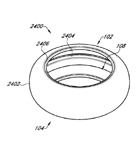
[0123] Figure 24A is an anterior side perspective view of another
example
prosthetic capsular device;
[0124] Figure 24B is an anterior plan view of the example prosthetic
capsular
device of Figure 24A;
[0125] Figure 24C is a cross-sectional view of the example
prosthetic capsular
device of Figure 24A along the line 24C-24C of Figure 24B;
[0126] Figure 24D is a cross-sectional view of the example
prosthetic capsular
device of Figure 24A along the line 24D-24D of Figure 24B;
-11 -
Date Recue/Date Received 2021-02-22
[0127] Figure 24E is a side plan view of the example prosthetic
capsular device
of Figure 24A;
[0128] Figure 24F is a cross-sectional view of the example
prosthetic capsular
device of Figure 24A along the line 24F-24F of Figure 24D;
[0129] Figure 25A is an anterior side perspective view of another
example
prosthetic capsular device;
[0130] Figure 25B is an anterior plan view of the example prosthetic
capsular
device of Figure 25A;
[0131] Figure 25C is a cross-sectional view of the example
prosthetic capsular
device of Figure 25A along the line 25C-25C of Figure 25B;
[0132] Figure 25D is a cross-sectional view of the example
prosthetic capsular
device of Figure 25A along the line 25D-25D of Figure 25B;
[0133] Figure 26A is an anterior side perspective view of another
example
refractive surface or intraocular lens that can be configured to be used in
conjunction with a
prosthetic capsular device;
[0134] Figure 26B is an anterior plan view of the example refractive
surface or
intraocular lens of Figure 26A;
[0135] Figure 26C is a cross-sectional view of the example
refractive surface or
intraocular lens of Figure 26A along the line 26C-26C of Figure 26B;
[0136] Figure 26D is a side plan view of the example refractive
surface or
intraocular lens of Figure 26A;
[0137] Figure 27A is an anterior side perspective view of another
example
prosthetic capsular device;
[0138] Figure 27B is an anterior plan view of the example prosthetic
capsular
device of Figure 25A;
[0139] Figure 27C is a cross-sectional view of the example
prosthetic capsular
device of Figure 27A along the line 27C-27C of Figure 27B;
[0140] Figure 27D is a side plan view of the example prosthetic
capsular device
of Figure 27A;
[0141] Figure 28A is an anterior side perspective view of another
example
prosthetic capsular device;
-12-
Date Recue/Date Received 2021-02-22
[0142] Figure 28B is an anterior plan view of the example prosthetic
capsular
device of Figure 28A;
[0143] Figure 28C is a cross-sectional view of the example
prosthetic capsular
device of Figure 28A along the line 28C-28C of Figure 28B;
[0144] Figure 28D is a side plan view of the example prosthetic
capsular device
of Figure 28A;
[0145] Figure 29A is an anterior side perspective view of another
example
prosthetic capsular device;
[0146] Figure 29B is an anterior plan view of the example prosthetic
capsular
device of Figure 29A;
[0147] Figure 29C is a cross-sectional view of the example
prosthetic capsular
device of Figure 29A along the line 29C-29C of Figure 29B;
[0148] Figure 29D is a side plan view of the example prosthetic
capsular device
of Figure 29A;
[0149] Figure 30A is an anterior plan view of another example
prosthetic capsular
device;
[0150] Figure 30B is a cross-sectional view of the example
prosthetic capsular
device of Figure 30A along the line 30B-30B of Figure 30A;
[0151] Figure 31A is an anterior side perspective view of another
example
prosthetic capsular device;
[0152] Figure 31B is an anterior plan view of the example prosthetic
capsular
device of Figure 31A;
[0153] Figure 31C is a cross-sectional view of the example
prosthetic capsular
device of Figure 31A along the line 31C-31C of Figure 31B;
[0154] Figure 31D is a side plan view of the example prosthetic
capsular device
of Figure 31A;
[0155] Figure 32A is an anterior side perspective view of another
example
refractive surface or intraocular lens that can be configured to be used in
conjunction with a
prosthetic capsular device;
[0156] Figure 32B is an anterior plan view of the example refractive
surface or
intraocular lens of Figure 32A;
-13-
Date Recue/Date Received 2021-02-22
[0157] Figure 32C is a cross-sectional view of the example
refractive surface or
intraocular lens of Figure 32A along the line 32C-32C of Figure 32B;
[0158] Figure 32D is a side plan view of the example refractive
surface or
intraocular lens of Figure 32A;
[0159] Figure 33A is an anterior side perspective view of an example
prosthetic
capsular device;
[0160] Figure 33B is an anterior plan view of the example prosthetic
capsular
device of Figure 33A;
[0161] Figure 33C is a cross-sectional view of the example
prosthetic capsular
device of Figure 33A along the line 33C-33C of Figure 33B;
[0162] Figure 34 is an anterior side perspective view of another
example
prosthetic capsular device;
[0163] Figure 35A is a side perspective view of an example tubular
device;
[0164] Figure 35B is a side perspective view of another example
tubular device;
[0165] Figure 35C is a side perspective view of another example
tubular device;
[0166] Figure 35D is a side perspective view of another example
tubular device;
[0167] Figure 35E is a side perspective view of another example
tubular device;
[0168] Figure 36 is an anterior side perspective view of an example
prosthetic
capsular system comprising an example prosthetic capsular device and an
example tubular
device;
[0169] Figure 37 is an anterior side perspective view of the example
prosthetic
capsular system of Figure 36 in an eye;
[0170] Figure 38A is an anterior side perspective partially-exploded
view of an
example prosthetic capsular system comprising an example prosthetic capsular
device, an
example tubular device, and an example containment structure;
[0171] Figure 38B is an anterior side perspective view of the
example prosthetic
capsular system of Figure 38A;
[0172] Figure 39 is an anterior side perspective view of another
example
prosthetic capsular system in an eye;
[0173] Figure 40 is a block diagram depicting an example control
process for a
prosthetic capsular system comprising a tubular device;
-14-
Date Recue/Date Received 2021-02-22
[0174] Figure 41 is a block diagram depicting another example
control process
for a prosthetic capsular system comprising a tubular device; and
[0175] Figure 42 is an anterior side perspective view of another
example
prosthetic capsular system comprising a tubular device in an eye;
[0176] Figure 43 is a perspective view of an example AR/VR
projection device or
system configured to be placed over a nose bridge of a user;
[0177] Figure 44 is a perspective view of an example prosthetic
capsular device
comprising a prism or prism bar;
[0178] Figure 45 is a perspective view of an example prism or prism
bar
configured to be used in conjunction with a prosthetic capsular device and/or
AR/VR
projection device or system;
[0179] Figure 46 is a block diagram depicting an example of a
computer
hardware system configured to run software for implementing one or more
embodiments of a
prosthetic capsular device system;
[0180] Figure 47 is a block diagram depicting another example of a
computer
hardware system configured to run software for implementing one or more
embodiments of a
prosthetic capsular device system; and
[0181] Figure 48 is a block diagram depicting another example of a
computer
hardware system configured to run software for implementing one or more
embodiments of a
prosthetic capsular device system.
DETAILED DESCRIPTION
[0182] In addition to the increase in demand for cataract surgery,
technological
advances have increased patient expectations for the surgery. The procedure
takes a short
amount of time to perform, and patients expect quick recovery of visual
function. Patients
are also asking their ophthalmologist to give them the restoration of more
youthful vision
without glasses through the use multifocal intraocular lenses, presbyopia
correcting lenses,
toric lenses, and monovision, to name a few. Despite accurate preoperative
measurements
and excellent surgical technique, the desired refractive outcome requires a
dose of good
-15-
Date Recue/Date Received 2021-02-22
fortune as there are numerous uncontrolled variables involved. As many as 20-
50% of post-
operative cataract patients may benefit from glasses or follow-up refractive
surgical
enhancements to achieve their desired refractive endpoint. One of the main
reasons for this
high amount of refractive unpredictability is believed to be the final resting
position of the
lens implant in the eye, mathematically expressed as the effective lens
position (ELP), which
can be quite variable and unpredictable in the current state of cataract
surgery. Recently,
hundreds of millions of dollars have been invested into developing highly
sophisticated
femtosecond laser systems that are able to more precisely control the size and
shape of the
capsulotomy and corneal incisions with the stated goal of lessening the
variability of the ELP
and thus aiding in better refractive outcomes. Unfortunately, the increased
precision of the
femtosecond laser systems has not been able to account for the major problem
plaguing the
variability of the ELP, which is the volumetric difference between the
cataract, natural
capsular bag, and intraocular lens implant (IOL).
101831 Devices and methods that help provide the desired refractive
endpoint in
cataract surgery are described in U.S. Patent No. 8,900,300, U.S. Patent No.
9,414,907, and
U.S. Patent No. 9,358,103.
101841 Figure lA illustrates an anterior side perspective view of an
example of a
prosthetic capsular device 100. Figure 1B illustrates another anterior side
perspective view
of the example prosthetic capsular device 100 for Figure 1A.
101851 In some embodiments, the device 100 includes features
described with
respect to the devices described in U.S. Patent No. 9,358,103 or modifications
thereof. For
example, the device 100 can comprise an anterior side 102, a posterior side
104, and one or
more sidewalls 106 extending between the anterior side 102 and the posterior
side 104; a
cavity or opening 108 defined by the anterior side 102, posterior side 104,
and the one or
more sidewalls 106, and the posterior side 104 optionally comprises a
refractive surface 110.
As such, the device 100 can be configured to comprise both a refractive
surface 110 and a
secondary or additional intraocular lens, electronic device, or other
intraocular device held
within the cavity 108.
101861 At least a portion of the posterior side 104 can comprise a
refractive
surface, which may, for example, allow a pseudophakic refraction to be
performed
intraoperatively with a known lens already inside the eye. The refractive
surface 110 can
-16-
Date Recue/Date Received 2021-02-22
comprise a refractive power of about +1 diopter. In other embodiments, the
refractive
surface 110 may comprise any and all lens powers and designs that are
currently known in
the art of intraocular lenses, including, but not limited to: spherical,
aspheric, wavefront,
convex, concave, multifocal (diffractive, refractive, zonal), toric,
accommodative, ultraviolet
(UV) filtering, diffractive chromatic aberration reducing lenses, light
adjustable lenses
(ultraviolet light adjustable, femtosecond phase wrapping), and optical powers
ranging from
any positive diopter value (e.g., including +35 D and above) to any negative
diopter value
(e.g., including -35 D and below).
[0187] The refractive surface 110 may advantageously reduce the
refractive
power of an IOL to be placed in the device 100. For example, if the device did
not include a
refractive surface 110 (e.g., comprised a simple or modified ring), then one
or more IOL
devices would need to provide all of the refractive power, which could
increase the volume
of the IOL, leading to a larger incision and associated complications. A
device 100
comprising a refractive surface 110 implanted in the eye can advantageously
allow for a
second refractive device or IOL to be coupled with (e.g., placed within, next
to, and/or on top
of) the refractive surface 110. The posterior refractive surface 110 can allow
the ELP of the
eye to be determined along with any residual refractive error. If any further
refractive error
is discovered, a second refractive device or IOL can be added to the
refractive surface 110
(e.g., immediately), which can neutralize the deficit and help ensure that the
desired outcome
is achieved. The posterior refractive surface 110 can be accurately placed and
anchored
and/or can inhibit or prevent shifting of lateral and/or posterior-anterior
position, rotation,
tilt, etc. of the posterior refractive surface 110 that could lead to
degradation of vision.
[0188] Further, in certain embodiments, the device 100 includes one
or more
additional features. For example, the device 100 can comprise a generally
lenticular or lens-
like shape as opposed to a box-like design. In other words, the generally
shape of the device
100 can be more like the shape of a natural lens. Risks of negative and/or
positive
dysphotopsia can be reduced due to the generally lenticular shape of the
device 100.
Negative dysphotopsia is a common problem in cataract surgery, generally
described by
patients as a temporal dark crescent in their vision and is believed to occur
either due to the
optical phenomenon known as total internal reflection or by obstruction of
light. This can
occur either at the junction of the optic edge and the empty collapsed
surrounding capsule
-17-
Date Recue/Date Received 2021-02-22
forming a relatively planar surface, or due to the capsule overlapping a
portion of the optic,
most commonly the nasal aspect. In embodiments in which the implantable device
100
comprises an overall lens-like configuration, the capsule can be held open,
preventing a
relatively planar surface from being formed by fusion of the posterior and
anterior capsule.
More specifically, when light hits a curvilinear slice of the device 100,
which can be made
from silicone for example, it may travel through the curvilinear slice instead
of bouncing off
and causing a negative shadow as it generally would for flat surfaces. This
may be especially
true in the horizontal meridian across the 180-degree plane. As such, in some
embodiments,
the device 100 does not comprise any flat edges or surfaces. In other words,
every surface of
the device 100 can be curvilinear. Flat optical surfaces can promote total
internal reflection,
and are not found in the natural human lens or lens capsule in the native
state. One goal of
some of the embodiments described herein is to reduce negative dysphotopsias
by not having
any flat optical surfaces.
101891 In certain embodiments, one or more sidewalls 106 of the
device 100 can
extend from only a portion of the posterior 104 and/or anterior sides 102
instead of extending
from the whole circumference of the posterior 104 and/or anterior sides 102.
The outer
periphery of a sidewall 106 can comprise an arc of a circle. For example, in
the illustrated
embodiment, the device 100 comprises two sidewalls 106A, 106B each of which
extend from
only a portion of the circumference of the posterior side 104 and/or
refractive surface 110. In
other words, certain portions of the anterior side 102 and posterior side 104
are not connected
by a sidewall.
101901 There can be a number of advantages for having only a portion
of the
sidewall present instead of having a sidewall encompass the whole
circumference of the
device 100. For example, by not having a sidewall at some portions, the area
behind the
refractive surface 110 can be more accessible. This can be important during
surgical
implantation of the device 100 to facilitate removal of viscoelastic material
from behind the
lens or refractive surface 110 immediately or shortly after the device 100 is
implanted. In
devices in which a sidewall encompasses the whole device 100, it can be
difficult to
maneuver between that space of the natural capsule and the sidewall capsular
bag to get
behind the lens or refractive surface 110 to vacuum out the viscoelastic
material. Without
having a sidewall present at least along some portions of the posterior side
104, it can be
-18-
Date Recue/Date Received 2021-02-22
substantially easier to reach the area behind the lens or refractive surface
110 for removal of
viscoelastic material and substantially reduce risks of posterior capsular
distension syndrome
due to remaining viscoelastic material.
[0191] In addition, by not having a sidewall present at least along
some portions
of the posterior side 104, the overall bulk of the device 100 can be reduced.
As such, the
device 100 can be compressed to fit into a smaller injector and incision in
the eye compared
to a device with sidewalls surrounding the whole device. In other words, the
device 100 can
be folded, rolled, or otherwise compressed over the longitudinal axis of the
device, or line
1G-1G of Figure 1E, such that line 1-F-1F of the device 100 is compressed to
allow the
device 100 to be inserted into a small injector and/or incision in the eye for
implantation. For
example, in some embodiments, the device 100 can be inserted into the eye
through an
incision of about 2.2 mm. In other embodiments, the device 100 can be inserted
into the eye
through an incision of about 1.5 mm, about 1.6 mm, about 1.7 mm, about 1.8 mm,
about 1.9
mm, about 2.0 mm, about 2.1 mm, about 2.2 mm, about 2.3 mm, about 2.4 mm,
about 2.5
mm, about 2.6 mm, about 2.7 mm, about 2.8 mm, about 2.9 mm, about 3.0 mm,
about 3.1
mm, about 3.2 mm, about 3.3 mm, about 3.4 mm, about 3.5 mm, and/or within a
range
defined by two of the aforementioned values.
[0192] Also, the reduced size of the device 100 can allow for use of
a larger optic
or lens, for example for use on the anterior side 104 and/or for placement
within the cavity
108. More specifically, a larger lens or refractive surface 110 can be used
with the device
100 due to the reduced bulk of the device 100 itself by removal of some of the
sidewalls.
Use of a larger lens or refractive surface 110 can be advantageous to reduce
halos and/or
glare post-surgery. For example, when the pupil dilates more than 5 mm, such
as at night,
light that reaches the outer portions of the refractive surface 110 may not be
focused. A
larger lens or refractive surface 110 can be generally better to address such
issues,
specifically to prevent nighttime symptoms when the pupil dilates to 6 or 7 mm
for example.
[0193] In some embodiments, substantially the whole device 100,
other than the
lens or refractive surface 110 and/or one or more haptics 112, can comprise
silicone and/or a
soft silicone polymer. In addition, in certain embodiments, substantially the
whole device
100, other than the lens or refractive surface 110 and/or one or more haptics
112, can
comprise a flexible and/or elastic material. As such, the device 100 can be
foldable or
-19-
Date Recue/Date Received 2021-02-22
collapsible for implantation into the eye through a small incision. Once
inserted into the eye,
the device 100 can naturally unfold and self-expand into its expanded
configuration as
illustrated in Figure JA within the natural capsular bag. In certain
embodiments, the device
100 without having sidewalls encompassing the whole device 100 is collapsible
to a point
where the size of the optic or refractive surface 110 is the rate limiting
factor for the incision
size for surgical implantation of the device 100.
101941 The device 100 can comprise one or more capsular areas. The
one or
more capsular areas can be adapted to receive and/or hold a lens or a
secondary lens in
addition to a refractive surface 110 on the posterior side. By inserting a
secondary lens, IOL,
or other optical device into the device 100, a Galilean and/or reverse
Galilean telescope can
be provided. For example, a portion of the posterior side 104, a portion of
the anterior side
102, and a portion of the side wall 106A, 106B can define a capsular area. In
the
embodiment shown in Figures 1A-1G, the device 100 comprises two capsular
areas. The
first capsular area is defined by a portion of the posterior side 104, a
portion of the anterior
side 102, and a portion of the side wall 106A. Similarly, a second capsular
area is defined by
another portion of the posterior side 104, another portion of the anterior
side 102, and another
portion of the side wall 106B. In other embodiments, the device 100 can
comprise one, three,
four, five, six, seven, eight, nine, or ten separate capsular areas.
101951 Similarly, the device 100 can comprise one, two, three, four,
five, six,
seven, eight, nine, or ten sidewalls 106, each of which extend from only a
portion of the
circumference of the posterior side 104 and/or refractive surface 110. In some
embodiments,
one or more sidewalls 106 of the device 100 can extend from about 120 of the
circumference of the posterior side 104 and/or refractive surface 110. In
other embodiments,
one or more sidewalls 106 of the device 100 can extend from about 15 , about
30 , about 45 ,
about 60 , about 75 , about 90 , about 105 , about 135 , about 150 , about 165
, about 180 ,
about 195 , about 210 , about 225 , about 240 , about 255 , about 270 , about
285 , about
300 , about 315 , about 330 , about 345 , and/or about 360 of the
circumference of the
posterior side 104 and/or refractive surface 110. In certain embodiments, one
or more
sidewalls 106 of the device 100 can extend from a portion of the circumference
of the
posterior side 104 and/or refractive surface within a circumferential range
defined by two of
the aforementioned values.
-20-
Date Recue/Date Received 2021-02-22
[0196] In some embodiments, the one or more sidewalls 106 can
comprise a
concave shape. For example, an interior surface of the one or more sidewalls
106 and/or
interior surface of the refractive surface 110 or posterior side 104 can form
a cavity 108. The
cavity can be configured to hold an IOL, for example.
[0197] In some embodiments, the device 100 comprises one or more
haptics 112.
The one or more haptics 112 can be made of a rigid or semi-rigid material,
such as
polyimide, PMMA, polypropylene, and nylon. The one or more haptics 112 can
also or
alternatively be made of a biocompatible material, such as silicone, silicone
polymers, SIBS
(poly(styrene-block-isobutylene-block-styrene)), acrylic, acrylic polymers,
polypropylene,
polycarbonate, and Gore-Tex. One or more haptics 112 of the device 100 can
provide a
place for surrounding epithelial cells to grow and latch on to provide support
for the device
100 within the natural capsular bag.
[0198] In the illustrated embodiment, the device 100 comprises two
haptics 112,
made of polyimide for example. In other embodiments, the device 100 can
comprise one,
three, four, five, six, seven, eight, nine, or ten haptics 112. Further, in
the illustrated
embodiment, the one or more haptics 112 comprise the general shape of an outer
periphery of
a rectangular or substantially rectangular shape, which can be attached to the
anterior side of
a sidewall extension. As shown, the one or more haptics 112 can be positioned
close and/or
generally parallel to the posterior side 102 of the device 100 and do not
extend radially
outward of the device 100. This can present advantages during surgical
implantation as
radially extending haptics can potentially get hung up on the iris and/or
anterior portion of
the natural capsular bag, which can present complications during surgical
implantation. In
other embodiments, one or more haptics 112 can comprise a different shape
while being
positioned close to and/or generally parallel to the posterior side 102 and/or
anterior side 104
of the device 100, such as circular, elliptical, round, square, triangular, or
the like.
[0199] In some embodiments, a portion of a haptic 112 can be over-
molded into
the device 100 for maintaining the position of the haptic 112 and not exposing
that portion of
the haptic 112. Another portion of the haptic 112 can be exposed to the
underside of the
anterior natural capsular bag. For example, a peripheral portion of the haptic
can be over-
molded while the central portion is exposed. The portion of the device 100,
for example
made of silicone, underneath the central portion of the haptic 112 can be
indented or recessed
-21-
Date Recue/Date Received 2021-02-22
in some embodiments. As such, fibrotic bands can be formed over time to act as
an anchor
point and hold the whole device 100 in place, for example if a Yag capsulotomy
is to be
performed. More specifically, epithelial cells coating the anterior and/or
posterior natural
capsular bag can replicate and grow into the recessed area of the silicone
device 100
underneath the haptic 112 and grow around the haptic 112.
102001 In certain embodiments, one or more haptics 112 of the device
100 can
comprise a "monkey bar" type configuration. More specifically, a portion of
the device 100,
for example a portion of a sidewall, can be recessed and/or indented. A haptic
can extend
across the recessed or indented portion. For example, one end of the haptic
can be over-
molded by silicone or other material of the device 100 at one end of the
recessed or indented
portion and the other end of the haptic can be over-molded by silicone or
other material of
the device 100 at the other end of the recessed or indented portion. As such,
a haptic, for
example made of polyimide, can be formed without radially extending out of the
exterior
surface of the device 100 while having void space all around the haptic. This
can provide
strands of exposed haptic or polyimide in some embodiments, while the haptic
is stabilized
as part of the overall device. Epithelial cells can grow around the haptic and
latch on to
provide lateral support along the monkey bar-type portion. One or more such
haptics can be
provided on each side of the device 100 in a symmetric manner.
102011 In some embodiments, the device 100 comprises a single-molded
design.
In other words, the whole device 100, or substantially the whole device 100
other than the
lens or refractive surface 110 and/or one or more haptics 112, can be molded
from a single
piece of material. For example, in some embodiments, substantially the whole
device 100
can be molded of silicone using a silicone compression mold. In certain
embodiments, one
or more haptics 112, made of polyimide for example, are placed in the mold
before silicone
or other material of the device 100 is poured into the mold and compressed. In
other
embodiments, the device 100 or any portion thereof can be manufactured by 3D
laser cutting,
two photon lithography, additive manufacturing, 3D printing, compression
molding, and/or
any combination of the aforementioned manufacturing processes or others.
102021 Figure 1C illustrates a posterior side perspective view of
the example
prosthetic capsular device of Figure 1A. Figure 1D illustrates a side plan
view of the
example prosthetic capsular device of Figure 1A.
-22-
Date Recue/Date Received 2021-02-22
102031 The device 100 optionally comprises one or more posterior
fins 114. The
device 100 shown includes two posterior fins 114A, 114B. The posterior fins
114 can be
aligned along a diameter of the refractive surface 110. In some
implementations, a plurality
of posterior fins 114 (e.g., 2, 3, 4, 5, 6, or more fins 124) may be
circumferentially offset
(e.g., by about 180 , by about 120 , by about 90 , by about 72 , by about 60 ,
and the like).
In some implementations, at least some or all of a plurality of posterior fins
114 (e.g., 2, 3, 4,
5, 6, or more fins 114) may be unaligned.
[0204] In the illustrated embodiment, a line between the two
posterior fins 114
forms an angle with a major axis of the device 100. For example, the angle
between a line
connecting the posterior fins 114 and a major axis of the device 100 can be
about 10 , about
20 , about 30 , about 40 , about 50 , about 60 , about 70 , about 80 , about
90 , about 100 ,
about 110 , about 120 , about 130 , about 140 , about 150 , about 160 , about
170 , about
180 , and/or within a range between two of the aforementioned values. In
certain
embodiments, the posterior fins 114 are aligned along a major axis of the
device 100. In
other implementations, the posterior fins 114 may be aligned along a minor
axis of the device
100.
[0205] The posterior fin 114 may comprise the same material as the
device 100 or
a different material than the device 100. The posterior fin 114 may help to
space a posterior
surface of a natural capsular bag from the posterior end 104 of the device 100
radially
outward of the refractive surface 110. Spacing the posterior surface of the
natural capsular
bag from the posterior end 104 of the device 100 radially outward of the
refractive surface
110 may allow fluid to flow radially outward of the refractive surface 110,
which may help to
reduce opacification. Spacing the posterior surface of the natural capsular
bag from the
posterior end 104 of the device 100 radially outward of the refractive surface
110 may reduce
the chance of retaining viscoelastic that has some residual trapped fibrin or
inflammatory
precipitate contained within it. In some embodiments, the posterior fin 114
may extend
anterior from the posterior of the device 100 into the cavity of the device
100. In some
embodiments, the posterior fin comprises a roughened or opacified interior
and/or exterior
surface of the device 100 (e.g., having the same thickness and material as the
posterior wall
radially outward of the refractive surface 110 but treated to provide an
alignment mark).
-23-
Date Recue/Date Received 2021-02-22
102061 The device 100 can be strategically aligned in an eye with
use of the fins
114. For example, if an eye has astigmatism, a device 100 in which the
refractive surface
110 comprises a toric lens can be used to at least partially correct the
astigmatism if the
device 100 is properly oriented (e.g., with the steep axis of a cornea). In
some
implementations, at least one of the fins 114 can be different (e.g.,
different shape,
dimensions, etc.) to indicate a top or bottom of the device 100. In devices
allowing any
rotational orientation of an IOL inserted therein, a toric IOL can be rotated.
Aligning the
device 100 for alignment of a toric refractive surface 110 and/or a toric IOL
contained in the
device 100 can advantageously provide the advantages of limited IOL rotation,
reduced
volume, and astigmatism correction. For example, the optic haptic junction of
a secondary
IOL can be aligned or otherwise correlated with one or more fins 114 and allow
a surgeon to
align the device 100 in an optimal position for a secondary toric IOL to be
placed. In some
embodiments, the one or more fins 114 extending radially posterior or outward
of the
posterior of the device 100 can still be visualized from the interior of the
refractive surface
110 to facilitate alignment of a secondary IOL or device, for example due to
the transparent
and/or semi-transparent nature of the posterior of the device 100. In other
embodiments, the
one or more fins 114 extend radially anterior or inward of the posterior of
the device 100
such that it the fins 114 are viewable for facilitating alignment of a
secondary IOL or device.
102071 Figure lE illustrates an anterior plan view of the example
prosthetic
capsular device of Figure 1A. Figure 1F illustrates a cross-sectional view of
the example
prosthetic capsular device of Figure lA along the line 1F-1F of Figure 1E.
Figure 1G
illustrates a cross-sectional view of the example prosthetic capsular device
of Figure lA
along the line 1G-1G of Figure 1E.
102081 In the illustrated embodiment, the device 100 comprises a
refractive
surface 110 with a diameter of about 5.5 mm. In other embodiments, the device
100 can
comprise a refractive surface 110 with a diameter of about 5.0 mm. The
refractive surface
110 110 may have a diameter between about 4 mm and about 9 mm (e.g., about 4
mm, about
mm, about 6 mm, about 7 mm, about 8 mm, about 9 mm, ranges between such
values, etc.).
102091 In such embodiments, the device 100 can be configured to be
inserted
through a small incision of about 2.2 mm or about 2.4 mm. In certain
embodiments, the
device 100 can be inserted through an incision between about 1.5 mm and about
3 mm (e.g.,
-24-
Date Recue/Date Received 2021-02-22
about 1.6 mm, about 1.7 mm, about 1.8 mm, about 1.9 mm, about 2.0 mm, about
2.1 mm,
about 2.2 mm, about 2.3 mm, about 2.4 mm, about 2.5 mm, about 2.6 mm, about
2.7 mm,
about 2.8 mm, about 2.9 mm, about 3.0 mm, ranges between such values, etc.).
[0210] Further, in the illustrated embodiment, a length of a major
axis of the
device 100 or a length measured from the outermost end of one sidewall 106A to
the
outermost end of another sidewall 106B along a major axis of the device 100
can be about
10.00 mm. In other embodiments, the length of the major axis of the device 100
can be
about 5.00 mm, about 6.00 mm, about 7.00 mm, about 8.00 mm, about 9.00 mm,
about 10.00
mm, about 11.00 mm, about 12.00 mm, about 13.00 mm, about 14.00 mm, about
15.00 mm,
and/or within a range defined by two of the aforementioned values.
[0211] Furthermore, in the illustrated embodiment, a length of a
minor axis of the
device 100 or a length measured from one end of a sidewall 106 to the other
end of the same
sidewall 106 along a minor axis of the device 100 can be about 6.57 mm. In
other
embodiments, the length of a minor axis of the device 100 can be about 4.0 mm,
about 4.5
mm, about 5.0 mm, about 5.5 mm, about 6.0 mm, about 6.5 mm, about 7.0 mm,
about 7.5
mm, about 8.0 mm, about 8.5 mm, about 9.0 mm, and/or within a range defined by
two of the
aforementioned values.
[0212] As illustrated in Figure 1G, in some embodiments, a thickness
of a haptic
112, made from polyimide for example, can be about 0.13 mm. In other
embodiments, the
thickness of the haptic 112 can be about 0.05 mm, about 0.06 mm, about 0.07
mm, about
0.08 mm, about 0.09 mm, about 0.10 mm, about 0.11 mm, about 0.12 mm, about
0.13 mm,
about 0.14 mm, about 0.15 mm, about 0.16 mm, about 0.17 mm, about 0.18 mm,
about 0.19
mm, about 0.20 mm, and/or within a range defined by two of the aforementioned
values.
[0213] In certain embodiments, a length of the haptic 112 across the
cross section
formed by line 1G-1G or along a major axis of the device 100 can be about 1.4
mm. In other
embodiments, a length of the haptic as seen in a cross section along a major
axis of the
device 100 can be about 0.05 mm, about 0.06 mm, about 0.07 mm, about 0.08 mm,
about
0.09 mm, about 0.10 mm, about 0.11 mm, about 0.12 mm, about 0.13 mm, about
0.14 mm,
about 0.15 mm, about 0.16 mm, about 0.17 mm, about 0.18 mm, about 0.19 mm,
about 0.20
mm, and/or within a range defined by two of the aforementioned values.
-25-
Date Recue/Date Received 2021-02-22
[0214] In some embodiments, the thickness of silicone or other
material of the
device 100 can be about 0.2 mm. In certain embodiments, the thickness of
silicone or other
material of the device 100 can be about 0.1 mm, about 0.2 mm, about 0.3 mm,
about 0.4 mm,
about 0.5 mm, about 0.6 mm, about 0.7 mm, about 0.8 mm, about 0.9 mm, about
1.0 mm,
and/or within a range defined by two of the aforementioned values.
[0215] In some embodiments, the thickness of the silicone or other
material of the
device 100 varies depending on the portion of the device 100. In other words,
some portions
of the device 100 can be made of thinner materials while other portions of the
device 100 can
be made of thicker materials. For example, certain portions of the device that
provide
support to the anterior portion of the device 100 may be made with thicker
materials for
added support.
[0216] In some embodiments, a thickness of silicone or other
material of the
device 100 molded over the haptic 112 can be about 0.01 mm, about 0.02 mm,
about 0.03
mm, about 0.04 mm, about 0.05 mm, about 0.06 mm, about 0.07 mm, about 0.08 mm,
about
0.09 mm, about 0.10 mm, and/or within a range defined by two of the
aforementioned values.
[0217] In certain embodiments, the width of an opening of the cavity
formed by
each end of the two sidewalls 106 can be about 5.82 mm. In some embodiments,
the width
of the opening of the cavity formed by each end of the two sidewalls can be
about 4.0 mm,
about 4.2 mm, about 4.4 mm, about 4.6 mm, about 4.8 mm, about 5.0 mm, about
5.2 mm,
about 5.4 mm, about 5.6 mm, about 5.8 mm, about 6.0 mm, about 6.2 mm, about
6.4 mm,
about 6.6 mm, about 6.8 mm, about 7.0 mm, about 7.2 mm, about 7.4 mm, about
7.6 mm,
about 7.8 mm, about 8.0 mm, and/or within a range defined by two of the
aforementioned
values.
[0218] Also, the height of the cavity as measured from a midpoint of
the posterior
refractive surface 110 to the top of the sidewall 106 opening can be about
3.21 mm in some
embodiments. In certain embodiments, the height of the cavity as measured from
a midpoint
of the posterior refractive surface 110 to the top of the sidewall 106 opening
can be about 2.0
mm, 2.2 mm, 2.4 mm, 2.6 mm, 2.8 mm, 3.0 mm, 3.2 mm, 3.4 mm, 3.6 mm, 3.8 mm,
4.0 mm,
4.2 mm, 4.4 mm, 4.6 mm, 4.8 mm, 5.0 mm, and/or within a range defined by two
of the
aforementioned values.
-26-
Date Recue/Date Received 2021-02-22
[0219] Figure 2A illustrates an anterior side perspective view of
another example
prosthetic capsular device. Figure 2B illustrates another anterior side
perspective view of the
example prosthetic capsular device of Figure 2A. The prosthetic capsular
device of Figure
2A includes some or all of the features of the prosthetic capsular device of
Figure 1A, and
like reference numerals include like features. In particular, in some
embodiments, the
prosthetic capsular device of Figure 2A can be similar to the prosthetic
capsular device of
Figure 1A, except for the configuration of the haptics 202. All other features
of the device
200 or haptics 202, such as material, flexibility, function, or the like, can
be similar to such
features of the device 100 or haptics 112 described above in relation to
Figures 1A-1G.
[0220] In some embodiments, the device 200 does not comprise haptics
with a
license plate or rectangular configuration as in Figures 1A-1G. Rather, the
device 200 can
comprise one or more haptics that connect the sidewalls 106 and expand
radially to form a
generally circular shape. For example, in the illustrated embodiment, one end
of a haptic
202A can be anchored or over-molded on one sidewall 106A and the other end of
the same
haptic 202A can be anchored or over-molded on another sidewall 106B.
Similarly, one end
of a second haptic 202B can be anchored or over-molded on one sidewall 106A
and the other
end of the same haptic 202B can be anchored or over-molded on another sidewall
106B. The
haptic 202 can form a radially outward shape or a substantially outwardly
circular shape or
loop. The haptic 202 202 can extend radially outward from a cavity between two
or more
sidewalls 106A, 106B. Such configuration of the haptic 202 can provide for
stability of the
device 200 within the natural capsular bag.
[0221] Figure 2C illustrates a posterior side perspective view of
the example
prosthetic capsular device of Figure 2A. Figure 2D illustrates a side plan
view of the
example prosthetic capsular device of Figure 2A. Figure 2E illustrates an
anterior plan view
of the example prosthetic capsular device of Figure 2A. Figure 2F illustrates
a cross-
sectional view of the example prosthetic capsular device of Figure 2A along
the line 2F-2F of
Figure 2E. Figure 2G illustrates a cross-sectional view of the example
prosthetic capsular
device of Figure 2A along the line 2G-2G of Figure 2E.
[0222] As shown in Figure 2E, in some embodiments, an outer or under
certain
circumstances maximum diameter of the device 200, for example accounting for
extension of
the haptics 202, may be about 10 mm. In certain embodiments, the outer or
maximum
-27-
Date Recue/Date Received 2021-02-22
diameter of the device 200 can be about 7 mm, about 8 mm, about 9 mm, about 10
mm,
about 11 mm, about 12 mm, about 13 mm, and/or within a range defined by two of
the
aforementioned values.
[0223] As shown in Figure 2F, in some embodiments, an outer or under
certain
circumstances maximum thickness of the device 200, for example accounting for
the
thickness of the refractive surface 110, may be about 3.65 mm. In certain
embodiments, the
outer or maximum thickness of the device 200 can be about 3.0 mm, about 3.1
mm, about 3.2
mm, about 3.3 mm, about 3.4 mm, about 3.5 mm, about 3.6 mm, about 3.7 mm,
about 3.8
mm, about 3.9 mm, about 4.0 mm, about 4.1 mm, and/or within a range defined by
two of the
aforementioned values.
[0224] Figure 3A illustrates an anterior side perspective view of
another example
prosthetic capsular device. Figure 3B illustrates an anterior plan view of the
example
prosthetic capsular device of Figure 3A. Figure 3C illustrates a side plan
view of the
example prosthetic capsular device of Figure 3A. Figure 3D illustrates a cross-
sectional view
of the example prosthetic capsular device of Figure 3A along the line 3D-3D of
Figure 3B.
[0225] The prosthetic capsular device of Figure 3A includes some or
all of the
features of the prosthetic capsular devices of Figures 1A and/or 2A, and like
reference
numerals include like features. In particular, in some embodiments, the
prosthetic capsular
device of Figure 3A can be similar to the prosthetic capsular devices of
Figures IA and/or
2A, except for the haptics 112, 202 and sidewalls 302.
[0226] More specifically, in the illustrated embodiment, the device
300 does not
comprise any haptics, such as haptics 112, 202 described above in relation to
Figures lA and
2A. In other embodiments, the device 300 can comprise one or more haptics 112,
202
described above in relation to Figures 1A and 2A.
[0227] Further, in certain embodiments, one or more sidewalls 302 of
the device
300 can extend from only about 90 of the circumference of the posterior side
104 and/or
refractive surface 110. In other words, a single capsular area defined by a
portion of the
anterior side 102, a portion of the posterior side 104, and a sidewall 302A,
302B, can cover
about 90 of the circumference of the device 300.
[0228] The sidewalls 302 can include any and all other features of
sidewalls 106
described above in relation to Figures 1A-1G. In some embodiments, all of the
sidewalls
-28-
Date Recue/Date Received 2021-02-22
302A, 302B each extend from a substantially equal portion of the circumference
of the
posterior side 104 and/or refractive surface 110, for example each at about 90
. In other
embodiments, some of the sidewalls 302 can extend from different amounts of
portions of
the circumference of the posterior side 104 and/or refractive surface 110. For
example, one
of a plurality of sidewalls 302 can extend from about 45 while another of the
plurality of
sidewalls 302 extends from about 90 of the circumference of the posterior
side 104 and/or
refractive surface 110.
[0229] In some embodiments, a single device 300 can be configured to
be
implanted into the eye with or without a second lens being placed inside the
cavity 108. In
certain embodiments, two devices 300 are configured to be coupled together
prior to and/or
during surgical implantation. More specifically, a first device 300 can be
coupled with a
second device 300 that is placed upside down to form a lens assembly with
itself. In certain
patients, this combination of lenses may move relative to one another creating
a variable
effective power of the lens system, enhancing the range of vision provided. A
lens may be
placed inside the empty cavity formed by the two devices 300.
[0230] Figure 4A illustrates an anterior side perspective view of
two (2) example
prosthetic capsular devices of Figure 3A coupled together. Figure 4B
illustrates a posterior
side perspective view of two (2) example prosthetic capsular devices of Figure
3A coupled
together. Figure 4C illustrates an anterior plan view of two (2) example
prosthetic capsular
devices of Figure 3A coupled together. Figure 4D illustrates a side plan view
of two (2)
example prosthetic capsular devices of Figure 3A coupled together. Figure 4E
illustrates a
cross-sectional view along the line 4E-4E of Figure 4C of two (2) example
prosthetic
capsular devices of Figure 3A coupled together.
[0231] In some embodiments, one device 300 can be coupled with
another device
300 to form a closed cavity 108 inside an assembly 400 the two devices 300. To
do so, one
device 300 can coupled with another device 300 that is placed upside down. In
certain
embodiments, each device 300 can comprise two sidewalls that each extend from
roughly
90 of the circumference of the posterior side 104 and/or refractive surface
110. As such,
when coupled together, sidewalls of the two devices 300 can, in combination,
form a
sidewall that substantially covers all 360 .
-29-
Date Recue/Date Received 2021-02-22
[0232] In certain embodiments, a gap may be present between the end
of a
sidewall 302 of one device 300 and the refractive surface 110 of a second
device 300.
Instead of forming a complete seal, a gap between the two devices 300 when
coupled
together to form an assembly 400 can be advantageous to allow for fluid to
pass to and from
the cavity.
[0233] In some embodiments, this gap between the two devices 300
when
coupled to form an assembly 400 (or more specifically, the gap between an end
of a sidewall
302 of a first device 300 and the refractive surface 110 of a second device
300 when the first
device 300 and second device 300 are coupled together) can be about 0.25 mm.
In certain
embodiments, this gap can be about 0.05 mm, about 0.10 mm, about 0.15 mm,
about 0.20
mm, about 0.25 mm, about 0.30 mm, about 0.35 mm, about 0.40 mm, about 0.45 mm,
about
0.50 mm, and/or within a range defined by two of the aforementioned values.
The precise
thickness of the gap can depend on the shape and/or volume of the natural
capsular bag in
some embodiments.
[0234] In certain embodiments, one device 300 is implanted into the
eye first,
followed by optional implantation and positioning of a refractive lens inside
the cavity 108,
and then the second device 300 is implanted into the eye to form a closure of
the assembly
400. In some embodiments, the two devices 300 are coupled together first
before
implantation into the eye.
[0235] Figure 5A illustrates an anterior side perspective view of
another example
prosthetic capsular device. Figure 5B illustrates a posterior side perspective
view of the
example prosthetic capsular device of Figure 5A. The prosthetic capsular
device of Figure
5A includes some or all of the features of the prosthetic capsular devices of
Figures 1A-4A,
and like reference numerals include like features.
[0236] More specifically, in some embodiments, the device 500 can
comprise one
or more sidewalls 302 which can include some or all of the features of the
sidewalls 302 of
device 300. For example, in certain embodiments, one or more sidewalls 302 of
the device
500 can extend from only about 90 of the circumference of the posterior side
104 and/or
refractive surface 110. The sidewalls 302 can also include any and all other
features of
sidewalls 106 described above in relation to Figures 1A-4A.
-30-
Date Recue/Date Received 2021-02-22
[0237] Similarly, the device 500 can comprise one or more haptics
502 which can
include some or all of the features of the haptics 202 of device 200. For
example, one or
more haptics 502 of the device 500 can connect two sidewalls 302A, 302B.
Moreover, one
or both ends of a haptic 502 can be anchored or over-molded on a sidewall
302A, 302B of
the device 500.
[0238] Figure 5C illustrates an anterior plan view of the example
prosthetic
capsular device of Figure 5A. Figure 5D illustrates a side plan view of the
example
prosthetic capsular device of Figure 5A. Figure 5E illustrates a cross-
sectional view of the
example prosthetic capsular device of Figure 5A along the line 5E-5E of Figure
5C. Figure
5F illustrates another side plan view of the example prosthetic capsular
device of Figure 5A.
Figure 5G illustrates a cross-sectional view of the example prosthetic
capsular device of
Figure 5A along the line 5G-5G of Figure 5F.
[0239] In certain embodiments, only one end of a haptic 502 is
anchored or over-
molded on a sidewall 302A, 302B of the device 500. For example, in the
embodiment
illustrated in Figure 5A, one of a haptic 502A can be over-molded onto a
sidewall 302A,
while the other end of the haptic 502A is not molded or rigidly anchored to
the other sidewall
302B. Likewise, only one end of haptic 502B can be molded or rigidly anchored
to the same
sidewall 302A that haptic 502A is anchored to, while the other end of the
haptic 502B is not
rigidly anchored to the other sidewall 302B. The other end of the haptic 502B
can be
configured to be tucked into the interior of the other sidewall 302B similar
to a safety-pin-
like configuration.
[0240] In some embodiments, the device 500 comprises a ridge 504 on
one or
more sidewalls 302A, 302B for receiving and/or embedding the haptics 502
without rigidly
anchoring the haptic 502. For example, in certain embodiments, only one of two
sidewalls
302B comprises said ridge 504. The other sidewall 302A does not comprise a
ridge 504 in
some embodiments. Within the ridge 504, the haptics 502A, 502B can be free to
move along
the ridge 504. For example, the end of a haptic 502 can be allowed to move up
and down
along the length of the ridge 504 as the exposed portion of the haptic 502 is
compressed or
allowed to expand.
[0241] In certain embodiments, a device 500 that comprises a ridge
504 on only
one of two sidewalls 302B can be configured to be injected into the eye in a
general direction
-31-
Date Recue/Date Received 2021-02-22
from the other sidewall 302A without a ridge towards the sidewall 302B with
the ridge 504.
Insertion into the eye in this general direction will allow the exposed
portion of the haptics
502 to compress more closely towards the refractive surface 110 as the ends of
the haptics
502 will be allowed to move more into the ridge 504.
[0242] Once implanted within the eye, the device 500 can be allowed
to unfold
naturally. The haptics 502 can be allowed to naturally decompress as well,
moving the ends
of the haptics 502 more towards the openings of the ridge 504. Accordingly,
the device 500
can comprise radially extending haptics 502 to maintain the shape and/or size
of the natural
capsular bag without the ends thereof adding complications to the surgical
procedure. In
some embodiments, when in an expanded or relaxed state, the outermost
perimeter or portion
of the sidewalls 302A, 302B and the haptics 502A, 502B can form a perfect or
substantially
perfect circle with a constant radius or diameter. For example, in some
embodiments, an
outer or maximum diameter of the device 500, accounting for the haptics 502,
may be about
mm. In certain embodiments, the outer or maximum diameter of the device 500
can be
about 7 mm, about 8 mm, about 9 mm, about 10 mm, about 11 mm, about 12 mm,
about 13
mm, and/or within a range defined by two of the aforementioned values.
[0243] In certain embodiments, because portions of the haptics 502
can be
squeezed and/or hidden during the surgical implantation, the device 500 can be
injected in a
manner substantially similar to those used for devices without such radially
extending
haptics, such as the device 100 illustrated in Figure 1A. At the same time,
because the
haptics 502 are allowed to radially expand once the device 500 is implanted,
the haptics 502,
made of polyimide for example, can provide sufficient points of attachment for
epithelial
cells to anchor the device 500.
[0244] Figure 6A illustrates an anterior side perspective view of
another example
prosthetic capsular device. Figure 6B illustrates an anterior plan view of the
example
prosthetic capsular device of Figure 6A. Figure 6C illustrates a cross-
sectional view of the
example prosthetic capsular device of Figure 6A along the line 6C-6C of Figure
6B. Figure
6D illustrates a cross-sectional view of the example prosthetic capsular
device of Figure 6A
along the line 6D-6D of Figure 6B.
[0245] The prosthetic capsular device 600 of Figure 6A includes some
or all of
the features of the prosthetic capsular devices of Figures 1A-5A, and like
reference numerals
-32-
Date Recue/Date Received 2021-02-22
include like features. In particular, the prosthetic capsular device 600 of
Figure 6A can be
similar to the prosthetic capsular devices of Figures IA and 2A, except for
the haptics 112,
202 and/or other additional features.
[0246] More specifically, the device 600 can comprise one or more
haptics 602
that extend radially outward from and to a single or same sidewall 106. For
example, one
end of a haptic 602 can be over-molded or otherwise be anchored to a portion
of one sidewall
106, and the other end of the same haptic 602 can be over-molded or otherwise
be anchored
to another portion of the same sidewall 106, the portion of the haptic 602 in
between the two
ends forming a loop extending out of the sidewall 106. Each of the haptics 602
can form a
closed loop. As a result, epithelial cells can be promoted to grow around the
haptics 602 to
substantially affix the device 600 within the eye.
[0247] In certain embodiments, one or more haptics 602 can be made
of Gore-
Tex or other soft material, and the rest of the device 600 can be made of
silicone. The whole
device 600 can be made exclusively of soft material in some embodiments, which
can
resolve concerns with implanting sharp or rigid materials. Also, cellular
ingrowth can be
facilitated, for example due to Gore-Tex's high biocompatibility in some
embodiments.
Accordingly, in some embodiments, a haptic comprises a single Gore-Tex string
or tether, for
example extending in a loop-like configuration out of a sidewall. Such Gore-
Tex string or
tether can provide a natural place for a fibrotic anchor to attach and also
prevent the device
600 from slipping. As such, in certain embodiments, the natural capsular bag
can be
maintained in an open position due to the structural integrity of the device
600 and the Gore-
Tex without need of a sharp or rigid material such as polyimide.
[0248] The device 600 can comprise a major axis, for example from a
horizontal
outermost portion of one haptic 602A to a horizontal outermost portion of
another haptic
602B. The distance between horizontal outermost portions of the two haptics
602A, 602B
can be about 11.15 mm in some embodiments. In other embodiments, the distance
between
horizontal outermost portions of the two haptics 602A, 602B can be about 5 mm,
about 6
mm, about 7 mm, about 8 mm, about 9 mm, about 10 mm, about 11 mm, about 12 mm,
about
13 mm, about 14mm, about 15 mm, and/or within a range defined by two of the
aforementioned values.
-33-
Date Recue/Date Received 2021-02-22
[0249] The device 600 can comprise a minor axis, for example from a
vertical
outermost portion of one haptic 602A, 602B to a vertical outermost portion of
the same
haptic 602A, 602B. The distance between vertical outermost portions of a
single haptics
602A, 602B can be about 7.95 mm in some embodiments. In other embodiments, the
distance between vertical outermost portions of a single haptic 602A, 602B can
be about 5
mm, about 5.5 mm, about 6 mm, about 6.5 mm, about 7 mm, about 7.5 mm, about 8
mm,
about 8.5 mm, about 9 mm, about 9.5 mm, about 10 mm, about 10.5 mm, about 11
mm,
about 11.5 mm, about 12 mm, about 13 mm, about 14mm, about 15 mm, and/or
within a
range defined by two of the aforementioned values.[0246] The device 600 can
also
comprise one or more notches 604. For example, in the illustrated embodiment,
each
capsular area comprises a notch 604A, 604B along the interior of each capsular
area or
sidewall 106A, 106B. The notch 604 can comprise one or more recessed areas or
slots for
insertion of one or more additional devices. For example, in some embodiments,
the notch
604 can comprise one or more slots configured for insertion of a secondary
IOL, an
electronic device, and/or haptics of the secondary IOL electronic device or
other secondary
device. By providing a slot or recessed area, a secondary device can be
inserted into the
device 600 at a precise location within the device 600 and be stabilized at
that location by
preventing movement of the secondary device laterally, anteriorly and/or
posteriorly within
the device 600. For example, a secondary IOL can be inserted into the device
600 such that a
distance between the secondary IOL and the refractive surface 110 is known
and/or
predetermined. Accordingly, one can determine an optimal or particular power
of a
secondary IOL based on the known refractive power of the refractive surface
110 and the
known distance between the secondary IOL and the refractive surface 110. One
or more
functional aspects of an electronic device to be inserted into the device 600
may also depend
on the particular location of the electronic device within the device 600
and/or particular
distance from the refractive surface 110, which can be predetermined and/or
controlled
utilizing the one or more notches 604.
[0250] The device 600 can comprise a plurality of notches or slots
604 on the
interior surface of each capsular area or sidewall 106A, 106B. Referring to
the cross-section
view along line 6D-6D as illustrated in Figure 6D for example, a plurality of
vertical notches
or slots 604 can be formed generally parallel to one another. In other words,
in addition to
-34-
Date Recue/Date Received 2021-02-22
the vertical notch or slot 604 shown in Figure 6D, one or more additional
vertical notches or
slots can be provided to the left and/or right of the illustrated notch or
slot 604. This can
allow for one or more secondary IOLs, electronic devices, or other devices to
be inserted into
the device 600 at varying locations or distances from the refractive surface
110. By doing so,
one can control the particular location of insertion of a secondary device in
the device 600 by
selecting one of the plurality of notches or slots to hold the secondary
device. In other words,
the secondary device can be adjusted anteriorly and/or posteriorly within the
device 600
when being inserted.
[0251] In some embodiments, a width of a notch or slot 604 can be
about 0.142
mm wide. In certain embodiments, the width of a notch or slot 604 can be about
0.05 mm,
about 0.1 mm, about 0.11 mm, about 0.12 mm, about 0.13 mm, about 0.14 mm,
about 0.15
mm, about 0.16 mm, about 0.17 mm, about 0.18 mm, about 0.19 mm, about 0.2 mm,
about
0.21 mm, about 0.22 mm, about 0.23 mm, about 0.24 mm, about 0.25 mm, and/or
within a
range defined by two of the aforementioned values.
[0252] In some embodiments, a length of a notch or slot 604 can be
about 3.77
mm. In certain embodiments, the length of a notch or slot 604 can be about 0.5
mm, about 1
mm, about 1.5 mm, about 2 mm, about 2.5 mm, about 3 mm, about 3.5 mm, about 4
mm,
about 4.5 mm, about 5 mm, about 5.5 mm, about 6 mm, about 6.5 mm, about 7 mm,
and/or
within a range defined by two of the aforementioned values.
[0253] The sidewalls 106A, 106B when viewed in the direction
illustrated in
Figure 6C can be separated by about 42 in some embodiments. In certain
embodiments, the
angle formed between the sidewalls 106A, 106B when viewed in the direction
illustrated in
Figure 6C can be about 36 , about 37 , about 38 , about 39 , about 40 , about
41 , about 42 ,
about 43 , about 44 , about 45 , about 46 , about 47 , and/or within a range
defined by two
of the aforementioned values.
[0254] Figure 7A illustrates an anterior side perspective view of
another example
prosthetic capsular device. Figure 7B illustrates an anterior plan view of the
example
prosthetic capsular device of Figure 7A. Figure 7C illustrates a cross-
sectional view of the
example prosthetic capsular device of Figure 7A along the line 7C-7C of Figure
7B. Figure
7D illustrates a cross-sectional view of the example prosthetic capsular
device of Figure 7A
along the line 7D-7D of Figure 7B.
-35-
Date Recue/Date Received 2021-02-22
[0255] The prosthetic capsular device 700 of Figure 7A includes some
or all of
the features of the prosthetic capsular devices of Figures 1A-6A, and like
reference numerals
include like features. In particular, the prosthetic capsular device 700 of
Figure 7A can be
similar to the prosthetic capsular device of Figures 1A, 2A, and 6A, except
for the haptics
112, 202, 602.
[0256] As with device 600 of Figure 6A, the device 700 can comprise
one or
more haptics 702 that extend radially outward from and to a single or same
sidewall 106.
However, unlike the device 600 of Figure 6A, each sidewall 106 of the device
700 can
comprise more than one such haptics 702. For example, in the illustrated
embodiment, each
sidewall 106 or capsular area comprises two haptics 702 in a closed loop
configuration. By
having more than one haptics extending from each sidewall 106 or capsular
area, epithelial
cells can attach to the more than one haptics and prevent or substantially
prevent the device
700 from sliding into a disadvantageous position, which may be a higher risk
for the device
600 of Figure 6A.
[0257] More specifically, a sidewall 106A can comprise a first
haptic 702A that
extends radially outward from one end of the sidewall 106A towards a center of
the sidewall.
The same sidewall 106A can also comprise a second haptic 702B that extends
radially
outward from another end of the sidewall 106A towards the center of the
sidewall. Similarly,
a second sidewall 106B can comprise a third haptic 702C that extends radially
outward from
one end of the sidewall 106B towards a center of the sidewall. The sidewall
106B can also
comprise a fourth haptic 702D that extends radially outward from another end
of the sidewall
106B towards the center of the sidewall. In other embodiments, a single
sidewall 106 can
comprise three, four, five, six, seven, eight, nine, or ten haptics 702. Any
one or more
feature of the haptics 702, such as material, flexibility, rigidity,
attachment to the device 700,
or the like, can be similar to the haptics 602 of the device 600 in Figure 6A.
[0258] When viewed in the direction illustrated in Figure 7B, a
distance between
a bottom end of one haptic 702A, 702D and a top end of another haptic 702B,
702C can be
about 1 mm. In certain embodiments, the distance between a bottom end of one
haptic 702A,
702D and a top end of another haptic 702B, 702C when viewed in the direction
illustrated in
Figure 7B can about 0.5 mm, about 0.6 mm, about 0.7 mm, about 0.8 mm, about
0.9 mm,
-36-
Date Recue/Date Received 2021-02-22
about 1 mm, about 1.1 mm, about 1.2 mm, about 1.3 mm, about 1.4 mm, about 1.5
mm,
and/or within a range defined by two of the aforementioned values.
[0259] Figure 8A illustrates an anterior side perspective view of
another example
prosthetic capsular device. Figure 8B illustrates an anterior plan view of the
example
prosthetic capsular device of Figure 8A. Figure 8C illustrates a cross-
sectional view of the
example prosthetic capsular device of Figure 8A along the line 8C-8C of Figure
8B. Figure
8D illustrates a cross-sectional view of the example prosthetic capsular
device of Figure 8A
along the line 8D-8D of Figure 8B.
[0260] The prosthetic capsular device 800 of Figure 8A includes some
or all of
the features of the prosthetic capsular devices of Figures 1A-7A, and like
reference numerals
include like features. In particular, the prosthetic capsular device 800 of
Figure 8A can be
similar to the prosthetic capsular device of Figure 2A, except for the haptics
202 and shape or
configuration of the one or more sidewalls 106.
[0261] More specifically, the one or more sidewalls 802 of the
device 800 can be
larger than those of the device 200 of Figure 2A. For example, the one or more
sidewalls
802 can extend vertically upwards and/or downwards when viewed in an anterior
side plan
view as illustrated in Figure 8B. As a result, the curvature of the outer
periphery of the one
or more sidewalls 802 can be larger than sidewalls 106 of the device 200 of
Figure 2A for
example. The general shape of the outer periphery of the device 800 can be
substantially
circular when viewed from an anterior or posterior plan view, compared to the
lenticular
shape of some of the other devices described above in relation to Figure IA
for example.
[0262] Similar to sidewalls 106A, 106B illustrated in other
embodiments, the
sidewalls 802A, 802B, when viewed in the direction illustrated in Figure 8C
can be separated
by about 410 in some embodiments. In certain embodiments, the angle formed
between the
sidewalls 802A, 802B, when viewed in the direction illustrated in Figure 8C,
can be about
36 , about 370, about 38 , about 39 , about 40 , about 41 , about 42 , about
43 , about 440
,
about 45 , about 46 , about 47 , about 48 , about 49 , about 50 , and/or
within a range
defined by two of the aforementioned values.
[0263] In some embodiments, a substantially circular outermost
periphery of the
device 800 can comprise a diameter of about 9.68 mm. In certain embodiments,
the
outermost periphery of the device 800 can comprise a substantially circular
shape with a
-37-
Date Recue/Date Received 2021-02-22
diameter of about 6 mm, about 6.5 mm, about 7 mm, about 7.5 mm, about 8 mm,
about 8.5
mm, about 9 mm, about 9.5 mm, about 10 mm, about 10.5 mm, about 11 mm, about
11.5
mm, about 12 mm, about 12.5 mm, about 13 mm, about 13.5 mm, about 14 mm, about
14.5
mm, about 15 mm, and/or within a range defined by two of the aforementioned
values.
[0264] The device 800 can comprise a thickness between an anterior
side 102 and
a posterior side 104 of about 3.707 mm when viewed from the side as
illustrated in Figure
8D. In certain embodiments, the device 800 can comprise a thickness between an
anterior
side 102 and a posterior side 104 of about 2.5 mm, about 2.6 mm, about 2.7 mm,
about 2.8
mm, about 2.9 mm, about 3.0 mm, about 3.1 mm, about 3.2 mm, about 3.3 mm,
about 3.4
mm, about 3.5 mm, about 3.6 mm, about 3.7 mm, about 3.8 mm, about 3.9 mm,
about 4 mm,
about 4.1 mm, about 4.2 mm, about 4.3 mm, about 4.4 mm, about 4.5 mm, about
4.6 mm,
about 4.7 mm, about 4.8 mm, about 4.9 mm, about 5 mm, and/or within a range
defined by
two of the aforementioned values.
[0265] In addition, due to the larger curvature of the one or more
sidewalls 802,
the one or more haptics 804 can comprise an arc of a substantially circular
configuration
from one end to the other end. In contrast, the one or more haptics 202 of the
device 200 of
Figure 2A can comprise different curvatures along the haptic 202. More
specifically, the
curvature of the haptic 202 can be relatively flat on one or both ends of the
haptic 202 located
inside or behind a sidewall 106 or capsular area compared to the curvature of
the central
portion of the haptic 202.
[0266] Similar to the device 500 of Figure 5A, the one or more
haptics 804 can be
over-molded or otherwise anchored to only one sidewall. For example, in the
illustrated
embodiment, a first end of the haptics 804A, 804B can be over-molded or
otherwise
anchored to one sidewall 802B. A second end of the haptics 804A, 804B can be
configured
to be tucked into the interior of the other sidewall 802A without being
rigidly anchored to the
sidewall 802A. As such, the second end of the haptics 804A, 804B can be
inserted freely
more or less into the other sidewall 802A as the exposed central portion of
the haptics 804A,
804B is compressed or allowed to expand.
[0267] Also similar to the device 500 of Figure 5A, the device 800
can be
configured to be injected into the eye in a general direction from the
sidewall 802B to which
the haptics 804 is anchored towards the other sidewall 802B to which the
haptics 804 is
-38-
Date Recue/Date Received 2021-02-22
configured to be tucked into. Insertion into the eye in this general direction
will allow the
exposed portion of the haptics 804 to compress more closely towards the
refractive surface
110 during insertion.
102681 Once implanted within the eye, the device 800 can be allowed
to unfold
naturally, allowing the haptics 804 to naturally decompress. Because portions
of the haptics
804 can be squeezed and/or hidden during the surgical implantation, the device
500 can be
injected in a manner substantially similar to those used for devices without
such radially
extending haptics, such as the device 100 illustrated in Figure IA. At the
same time, because
the haptics 804 are allowed to radially expand once the device 800 is
implanted, the haptics
804, made of polyimide for example, can provide sufficient points of
attachment for
epithelial cells to anchor the device 800. Any one or more other feature of
the haptics 804,
such as material, flexibility, rigidity or the like, can be similar to the
haptics 602 of the device
600 in Figure 6A.
102691 Figure 9A illustrates an anterior side perspective view of
another example
prosthetic capsular device. Figure 9B illustrates an anterior plan view of the
example
prosthetic capsular device of Figure 9A. Figure 9C illustrates a cross-
sectional view of the
example prosthetic capsular device of Figure 9A along the line 9C-9C of Figure
9B. Figure
9D illustrates a cross-sectional view of the example prosthetic capsular
device of Figure 9A
along the line 9D-9D of Figure 9B.
[0270] The prosthetic capsular device 900 of Figure 9A includes some
or all of
the features of the prosthetic capsular devices of Figures IA-8A, and like
reference numerals
include like features. In particular, the prosthetic capsular device 900 of
Figure 9A can be
similar to the prosthetic capsular device of Figure 8A, except for the haptics
804. More
specifically, the haptics 902 of the device 900 can comprise a substantially
vertical arm that
extends radially inward towards the refractive surface 110 from a midpoint or
a portion in
between the two ends of the haptics 902 that is exposed. A first end of the
vertical arm can
be connected to the exposed portion of the haptics 902, while a second end of
the vertical
arm can be connected to one or more holes or openings 904.
102711 The one or more holes or openings 904 can allow a surgical
instrument,
such as a Sinskey Hook, a Lester Hook or the like, to hook on and engage the
device 900.
For example, a surgical instrument can be coupled to one or more holes 904 to
adjust the
-39-
Date Recue/Date Received 2021-02-22
positioning of the device 900 in the eye. This can be advantageous during
surgery because of
the limited visual field, which can be for example about 5-6 mm. By coupling a
surgical
instrument to the one or more holes 904, the positioning of the device 900 can
be adjusted so
that it is viewable without risking damaging or tearing the capsule. Any other
one or more
features of the device 900 and/or haptics 902, such as size, material,
flexibility, rigidity,
attachment to the device 900 or the like, can be similar to the device 800
and/or haptics 804
of the device 800 in Figure 8A.
[0272] Figure 10A illustrates an anterior side perspective view of
another
example prosthetic capsular device. Figure 10B illustrates an anterior plan
view of the
example prosthetic capsular device of Figure 10A. Figure 10C illustrates a
cross-sectional
view of the example prosthetic capsular device of Figure 10A along the line
10C-10C of
Figure 10B. Figure 10D illustrates a side plan view of the example prosthetic
capsular
device of Figure 10A. The prosthetic capsular device 1000 of Figure 10A
includes some or
all of the features of the prosthetic capsular devices of Figure 1A-9A, and
like reference
numerals include like features.
[0273] Unlike some of the devices of Figures 1A-9A, the device 1000
can
comprise a sidewall 1002 that covers substantially or almost the entire side
circumference of
the device 1000. The sidewall 1000 can continuously cover almost the entire
side of the
device 1000 except for a small opening or gap 1010. In the illustrated
embodiment, the small
opening or gap 1010 can comprise a width of about 1.00 mm. In other
embodiments, this
gap 1010 in the sidewall 1002 can be about 0.50 mm, about 1.50 mm, about 2.00
mm, about
2.50 mm, about 3.00 mm, about 3.50 mm, about 4.00 mm, about 4.50 mm, about
5.00 mm,
and/or within a range defined by two of the aforementioned values. One or more
other
features of the sidewall, such as material, flexibility, rigidity, or the like
can be similar to
those of one or more devices of Figures 1A-9A. In other embodiments, the
sidewall 1002
may not comprise a gap 1010. For example, additional portions of silicone may
extend over
the gap 1010 in the illustrated embodiment.
[0274] The device 1000 can comprise a capsular tension ring 1004
coupled to the
sidewall. For example, the capsular tension ring 1004 can be over-molded into
the sidewall
1002. The capsular tension ring 1004 can comprise a rigid or semi-rigid
material, such as
polyimide, PMMA, polypropylene, and/or nylon. The capsular tension ring 1004
can
-40-
Date Recue/Date Received 2021-02-22
provide rigidity and maintain the structure and/or position of the device 1000
inside the eye
after implantation. The capsular tension ring 1004 can generally follow the
shape of the
circumference of the sidewall 1002 as in the illustrated embodiment. Each end
of the
capsular tension ring 1004 can extend from each end of the sidewall 1002 where
the gap
1010 in the sidewall 1002 is present. Each or one end of the capsular tension
ring 1004 can
comprise an opening or a hole 1008, similar to the holes 904 in the device 900
of Figure 9A
and be used in a similar manner to position the device 1000 after implantation
in the eye.
[0275] The device 1000 can comprise one or more recessed areas 1006
along the
exterior of the sidewall 1002. The one or more recessed areas can expose
portions of the
capsular tension ring 1004. The exposed portions of the capsular tension ring
1004 can
provide areas for epithelial cells to attach to. Accordingly, with the
attachment or growth of
epithelial cells around the exposed capsular tension ring 1004, the device
1000 can be
substantially fixed and stabilized in a particular position within the eye.
The recessed areas
1006 can also be used to suture the device as necessary. In the illustrated
embodiment, the
device 1000 or exterior sidewall 1002 thereof comprises seven recessed areas
1006. In other
embodiments, the device 1000 can comprise one, two, three, four, five, six,
eight, nine, or ten
recessed areas 1006. The number of recessed areas 1006 in the device 1000 can
also be
between a range defined two of the aforementioned values.
[0276] As illustrated in the view of Figure 10B, the length of each
recessed area
1006 along the circumference of the sidewall 1002 or the length of each
exposed portion
1006 of the capsular tension ring 1004 can be about 1.00 mm. In certain
embodiments, the
length of each recessed area 1006 along the circumference of the sidewall 1002
or the length
of each exposed portion 1006 of the capsular tension ring 1004 can be about
0.50 mm, about
1.50 mm, about 2.00 mm, about 2.50 mm, about 3.00 mm, about 3.50 mm, about
4.00 mm,
about 4.50 mm, about 5.00 mm, and/or within a range defined by two of the
aforementioned
values.
[0277] As illustrated in the view of Figure 10D, the width of each
recessed area
1006, when viewed from a side plan view, can be about 0.49 mm. In certain
embodiments,
the width of each recessed area 1006, when viewed from a side plan view, can
be about 0.35
mm, about 0.40 mm, about 0.45 mm, about 0.50 mm, about 0.55 mm, about 0.60 mm,
about
0.65 mm, about 0.70 mm and/or within a range defined by two of the
aforementioned values.
-41-
Date Recue/Date Received 2021-02-22
102781 The refractive surface 110 can be connected to the sidewall
1002 at only a
portion of the sidewall. For example, in the illustrated embodiment, the
refractive surface
100 comprises a hinge portion 1010 which is connected to the sidewall 1002. A
gap 1012
can exist between all other portions of the refractive surface 110 other than
the hinge portion
1010 and the sidewall 1002. As such, the entire device 1000 can comprise a
single piece,
rather than a multi-piece assembly. Alternatively, in other embodiments, the
device 1000 can
be a multi-piece assembly comprising multiple pieces that are coupled together
after the
initial manufacturing.
102791 The sidewall 1002 and the capsular tension ring 1004 can be
configured to
be twisted without breaking. Also, the sidewall 1002 can be foldable or
capable of being
rolled into a more compact configuration. The refractive surface 1010 and the
hinge portion
1010 can also be foldable or capable of being rolled into a more compact
configuration.
[0280] As discussed above, one advantage of removing portions of the
sidewall,
for example in the device 100 of Figure 1A, can be to allow the device to be
folded or rolled
in a more compact configuration for insertion through a small incision during
surgery. Even
though the device 1000 of Figure 10A comprises a near continuous sidewall, it
can still be
configured to be inserted through a small incision, for example no larger than
required for
insertion of the device 100 of Figure 1A, without removing portions of the
sidewall 1002,
due to the structure and method of insertion as described herein.
[0281] More specifically, instead of squeezing the device 1000 for
insertion, the
device 1000 or the sidewall 1002 and/or capsular tension ring 1004 of the
device 1000 can be
inserted into the eye in a rotational fashion segment by segment through a
standard injector.
For example, the sidewall 1002 can be folded or rolled around the length of
the capsular
tension ring 1004 into a tube-like configuration. The sidewall 1002 and
capsular tension ring
1004 can be optionally twisted or otherwise partially straightened. A portion
of the sidewall
1002 and/or capsular tension ring 1004 can be fed into a small incision in the
eye, advancing
one portion at a time rotationally, for example as each portion is
substantially straightened at
the point of insertion, allowing the capsular tension ring 1004 to retain its
memory and curl
around upon insertion.
[0282] The end of the sidewall 1002 and capsular tension ring 1004
away from
the hinge portion 1010 can be inserted first towards the other end where the
hinge portion
-42-
Date Recue/Date Received 2021-02-22
1010 is attached to. Upon reaching the portion of the sidewall 1002 and
capsular tension ring
1004 where the hinge portion 1010 is attached, the refractive surface 110 can
be slid into the
injector and into the eye in a linear fashion. In other words, the sidewall
1002 and capsular
tension ring 1004 can be inserted through a small incision into the eye in a
rotational manner
first and the lens or refractive surface 110 can be subsequently inserted in a
longitudinal
manner.
[0283] Once completely inserted into the eye, the device 1000 can
return to its
substantially circular configuration. By doing so, the device 1000 can be
inserted through a
small incision, while maintaining the structural integrity necessary for the
device 1000 to
remain intact and centered in the eye for a substantial period of time.
[0284] The device 1000 can be configured to protect the entire
capsule and
preserve the entire capsular space. More specifically, all or substantially
the entire the
circular or substantially circular sidewall 1002 or outer circumference of the
device 1000 can
be configured to contact the natural capsular bag and maintain the general
space of the
natural capsular bag without collapsing in the vitreous. Also, the device 1000
eliminates any
trail in the haptics, with the capsular tension ring 1004 embedded inside the
device. The
generally circular shape of the device 1000 can also follow the physiological
shape of the
capsule and preserve the volume of the capsule unlike certain devices that
decrease the open
volume inside after implantation. Also, the device and/or secondary lens to be
placed inside
the device 1000 may be freely rotated, which may not be possible with certain
devices.
[0285] The refractive surface 110 can also comprise one or more tabs
extending
radially outward from the outer circumference of the refractive surface 110.
The one or more
tabs can be configured to be placed or tucked underneath the sidewall 1002
after insertion to
prevent the refractive surface 110 from being tilted over. The one or more
tabs can comprise
the same material as the sidewall 1002, for example silicone.
[0286] Alternatively, the refractive surface 110 can be
circumferentially
surrounded by a flange of soft material, such as silicone. The width of the
flange can be
about 0.25 mm, about 0.50 mm, 0.75 mm, 1.00 mm, or between a range defined by
two of
the aforementioned values. The outer flange of the refractive surface 110 or
some portion
thereof can be configured to be tucked underneath the bottom of the sidewall
1002 upon
insertion into the eye.
-43-
Date Recue/Date Received 2021-02-22
[0287] Both the refractive surface 110 and the flange can be made of
the same
material, such as silicone. In other embodiments, the refractive surface 110
can be acrylic
while the flange can be made of silicone. Acrylic can provide a higher index
of refraction
while allowing the refractive surface 110 to be thinner than when made from
silicone. Also,
the optical properties and power of an acrylic lens or refractive surface 110
can be altered
using one or more laser treatments, such as phase wrapping to alter the
hydrophilicity or
hydrophobicity of the acrylic and causing the lens to either swell and
increase in power or
shrink and decrease in power. The lens or refractive surface 110 can also be
made from any
other biocompatible and optically clear materials known in the art. The
refractive surface
110 may have a refractive power between -35 D and +35D.
[0288] Figure 11A illustrates an anterior side perspective view of
another
example prosthetic capsular device. Figure 11B illustrates an anterior plan
view of the
example prosthetic capsular device of Figure 11A. Figure 11C illustrates a
cross-sectional
view of the example prosthetic capsular device of Figure 11A along the line
11C-11C of
Figure 11B. Figure 11D illustrates a cross-sectional view of the example
prosthetic capsular
device of Figure 11A along the line 11D-11D of Figure 11B.
[0289] The prosthetic capsular device of Figure 11A includes some or
all of the
features of the prosthetic capsular device of Figure 1A-10A, and like
reference numerals
include like features. In particular, the prosthetic capsular device of Figure
11A can be
similar to the prosthetic capsular device of Figure 1A and Figure 6A, except
for the
configuration of notches 1104 and/or sidewalls 1106A, 1106B. All or some other
features of
the device 1100, notches 1104, and/or sidewalls 1106A, 1106B, such as
material, flexibility,
function, or the like, can be similar to such features described above in
relation to Figures
1A-10A.
[0290] More specifically, the device 1100 can comprise one or more
notches
1104 along the interior of each capsular area or sidewall 1106A, 1106B,
similar to notches
604. A notch 1104 can comprise one or more recessed areas or slots to
facilitate insertion of
one or more additional devices, such as a secondary IOL, an electronic device,
and/or a
haptics thereof. Similar to the notch 604 and device 600 of Figure 6A, a
secondary device
can be inserted into the device 1100 at a precise location within the device
1100 and be
stabilized at that location by insertion of the secondary device or a portion
thereof into the
-44-
Date Recue/Date Received 2021-02-22
notch 1104. By doing so, a secondary device can be prevented from moving
laterally,
anteriorly and/or posteriorly within the device 1100. The notches 1104 can be
molded
together with the device 1100 at the same time as a single piece assembly. In
other
embodiments, the notches 1104 can be formed separately from the device 1100
and be
subsequently attached to the device 1100.
102911 When viewed in the cross section depicted in Figure 11D, the
one or more
notches 1104 can comprise a generally elongated shape along the short axis, or
the axis
parallel to line 11F-11F of Figure 11B. The one or more notches 1104 can be
located at a
predetermined and/or known distance from the anterior of the device 110. For
example, in
the embodiment illustrated in Figure 11C, a distance between a center of the
one or more
notches 1104 and a top end of the interior anterior or refractive surface 110
can be about 1.23
mm, for example for a 20D lens. This distance can be different depending on
the power
and/or thickness of the lens or the refractive surface 110. For example,
depending on the
power of the lens or refractive surface 110, this distance can be about 0.50
mm, about 0.60
mm, about 0.70mm, about 0.80 mm, about 0.90 mm, about 1.00 mm, about 1.10 mm,
about
1.20 mm, about 1.30 mm, about 1.40 mm, about 1.50 mm, about 1.60 mm, about
1.70 mm,
about 1.80 mm, about 1.90 mm, about 2.00 mm, and/or within a range defined by
two of the
aforementioned values.
102921 The particular location of the one or more notches 1104 with
respect to the
overall device 1100 can also be measured in terms of a distance between the
center of the one
or more notches 1104 and the split point of the optic 110. For example, in
certain
embodiments, the two thirds of the power of the optic 110 can be configured to
be placed
inside the device 1100 while one third of the power of the optic 110 is
located external to the
device 1100. The split point, for example the 1/3, 2/3 split point, can be
configured to be
constant in the device 1100 regardless of the power of the lens 110. In other
embodiments,
the split point of the optic 110 may be 1/4, 3/4 or 1/2, 1/2. In other words,
1/4, 1/2, or 3/4 of
the refractive power of the optic 110 can be configured to be located external
to the device
1100 while 3/4, 1/2, or 1/4 of the refractive power of the optic 110 can be
configured to be
located internal to the device 1100.
102931 The distance measured from the center of the notches 1104 to
a split point
of the optic 110, when viewed in the cross-sectional view illustrated in
Figure 11C, can be
-45-
Date Recue/Date Received 2021-02-22
about 1.38 mm. This distance from the center of the notches 1104 to a split
point of the optic
110 can be constant regardless of the power or thickness of the refractive
surface 110. In
certain embodiments, this distance can be about 0.50 mm, about 0.60 mm, about
0.70mm,
about 0.80 mm, about 0.90 mm, about 1.00 mm, about 1.10 mm, about 1.20 mm,
about 1.30
mm, about 1.40 mm, about 1.50 mm, about 1.60 mm, about 1.70 mm, about 1.80 mm,
about
1.90 mm, about 2.00 mm, and/or within a range defined by two of the
aforementioned values.
[0294] In some embodiments, each of the notches 1104A, 1104B can
comprise a
vertical width of about 0.15 mm when viewed in the direction of Figure 11C. In
certain
embodiments, each of the notches 1104A, 1106B, when viewed in the direction of
Figure
11C, can comprise a width of about 0.05 mm, about 0.1 mm, about 0.11 mm, about
0.12 mm,
about 0.13 mm, about 0.14 mm, about 0.15 mm, about 0.16 mm, about 0.17 mm,
about 0.18
mm, about 0.19 mm, about 0.2 mm, about 0.21 mm, about 0.22 mm, about 0.23 mm,
about
0.24 mm, about 0.25 mm, and/or within a range defined by two of the
aforementioned values.
[0295] In some embodiments, each of the notches 1104 can comprise an
angular
length of about 60 when measured from the center of the refractive 110 in an
anterior plan
view as illustrated in Figure 11B. In certain embodiments, the angular length
of each of the
notches 1104, when measured from the center of the refractive 110 in an
anterior plan view
as illustrated in Figure 11B, can be about 10 , about 20 , about 30 , about 40
, about 50 ,
about 60 , about 70 , about 80 , about 90 , about 100 , about 110 , about 120
, about 130 ,
about 140 , about 150 , about 160 , about 170 , and/or within a range defined
by two of the
aforementioned values.
[0296] The sidewalls 1106A, 1106B, when viewed in the direction
illustrated in
Figure 11C, can be separated by about 25 in some embodiments. In certain
embodiments,
the angle formed between the sidewalls 1106A, 1106B, when viewed in the
direction
illustrated in Figure 11C, can be about 10 , about 15 , about 20 , about 25 ,
about 30 , about
35 , about 40 , about 45 , about 50 , about 55 , about 60 , about 65 , about
70 , about 75 ,
about 80 , and/or within a range defined by two of the aforementioned values.
[0297] The device 1100 can be configured to be folded, rolled, or
otherwise
compressed and injected into the eye through a small incision and/or small
injector as device
100 described above in relation to Figure 1A. For example, in some
embodiments, the
device 1100 can be inserted through an incision of about 2.75 mm or less. In
other
-46-
Date Recue/Date Received 2021-02-22
embodiments, the device 1100 can be inserted into the eye through an incision
of about 1.5
mm, about 1.6 mm, about 1.7 mm, about 1.8 mm, about 1.9 mm, about 2.0 mm,
about 2.1
mm, about 2.2 mm, about 2.3 mm, about 2.4 mm, about 2.5 mm, about 2.6 mm,
about 2.7
mm, about 2.8 mm, about 2.9 mm, about 3.0 mm, about 3.1 mm, about 3.2 mm,
about 3.3
mm, about 3.4 mm, about 3.5 mm, and/or within a range defined by two of the
aforementioned values.
[0298] Figure 12A illustrates another anterior plan view of the
example prosthetic
capsular device of Figure 11A. Figure 12B illustrates a cross-sectional view
of the example
prosthetic capsular device of Figure 11A along the line 12B-12B of Figure 12A.
Figure 12C
illustrates a cross-sectional view of the example prosthetic capsular device
of Figure 11A
along the line 12C-12C of Figure 12A. Figure 12D illustrates a cross-sectional
view of the
example prosthetic capsular device of Figure 11A along the line 12D-12D of
Figure 12A.
Figure 12E illustrates a cross-sectional view of the example prosthetic
capsular device of
Figure 11A along the line 12E-12E of Figure 12A. Figure 12F illustrates a
cross-sectional
view of the example prosthetic capsular device of Figure 11A along the line
12F-12F of
Figure 12A. Figure 12G illustrates a cross-sectional view of the example
prosthetic capsular
device of Figure 11A along the line 12G-12G of Figure 12A.
[0299] In some embodiments, the cross-sectional area of the device
1100 along
the line 12B-12B can be about 2.67 mm2. Similarly, in some embodiments, the
cross-
sectional area of the device 1100 along the line 12C-12C can be about 4.83
mm2. In some
embodiments, the cross-sectional area of the device 1100 along the line 12D-
12D can be
about 4.24 mm2. In some embodiments, the cross-sectional area of the device
1100 along the
line 12E-12E can be about 3.65 mm2. In some embodiments, the cross-sectional
area of the
device 1100 along the line 12F-12F can be about 2.42 mm2. In some embodiments,
the
cross-sectional area of the device 1100 along the line 12G-12G can be about
4.34 mm2. As
such, the amount of material of the device 1100 may not necessarily depend on
the size of the
total outermost periphery of a cross section of the device 1100.
[0300] Figure 13A illustrates an anterior side perspective view of
the example
prosthetic capsular device of Figure 11A with a secondary device inserted
therein. Figure
13B illustrates an anterior plan view of the example prosthetic capsular
device of Figure 11A
with a secondary device inserted therein. Figure 13C illustrates a cross-
sectional view of the
-47-
Date Recue/Date Received 2021-02-22
example prosthetic capsular device of Figure 11A with a secondary device
inserted therein
along the line 13C-13C of Figure 13B. Figure 13D illustrates a cross-sectional
view of the
example prosthetic capsular device of Figure 11A with a secondary device
inserted therein
along the line 13D-13D of Figure 13B. The secondary device can comprise
acrylic or other
haptics that are configured to be inserted into the one or more notches 1104.
103011 In some embodiments, the device 1100 can be configured to be
used in
conjunction with a secondary device 1302, such as a secondary IOL, electronic
device,
and/or other device. The secondary device 1302 can be any device that is
configured to take
advantage of the notches 1104. For example, the secondary device 1302 can
comprise one or
more haptics and/or other features that are configured to be inserted into the
notches 1104.
The secondary device 1302 can be inserted into the device 1100 prior to
implantation of the
device 1100. Alternatively, the secondary device 1302 can be inserted into the
device 1100
after the device 1100 has been implanted into the eye. As illustrated and as
discussed above,
the secondary device 1302 can be inserted and stabilized at a particular
location within the
device 1100 by attaching, inserting, or otherwise fixating the secondary
device 1302 or a
feature thereof into the one or more notches 1104.
103021 Figure 14A illustrates an anterior side perspective view of
another
example prosthetic capsular device. Figure 14B illustrates an anterior plan
view of the
example prosthetic capsular device of Figure 14A. Figure 14C illustrates a
cross-sectional
view of the example prosthetic capsular device of Figure 14A along the line
14C-14C of
Figure 14B. Figure 14D illustrates a cross-sectional view of the example
prosthetic capsular
device of Figure 14A along the line 14D-14D of Figure 14B. Figure 15A
illustrates another
anterior side perspective view of the example prosthetic capsular device of
Figure 14A.
Figure 15B illustrates another anterior plan view of the example prosthetic
capsular device of
Figure 14A. Figure 15C illustrates another cross-sectional view of the example
prosthetic
capsular device of Figure 14A along the line 15C-15C of Figure 15B. Figure 15D
illustrates
another cross-sectional view of the example prosthetic capsular device of
Figure 14A along
the line 15D-15D of Figure 15B.
103031 The prosthetic capsular device of Figure 14A includes some or
all of the
features of the prosthetic capsular device of Figure 1A-11A, and like
reference numerals
include like features. In particular, the prosthetic capsular device of Figure
14A can be
-48-
Date Recue/Date Received 2021-02-22
similar to the prosthetic capsular device of Figure IA and/or Figure 11A,
except for the
configuration of notches 1404 and/or haptics 1402. All or some other features
of the notches
1404, such as material, flexibility, function, or the like, can be similar to
such features of the
notches 604 described above in relation to Figures 6A-6D and/or the notches
1104 described
above in relation to Figures 11A-11D. All or some other features of the
haptics 1402, such
as material, flexibility, function, or the like, can be similar to such
features of the haptics 112
described above in relation to Figures 1A-1G and/or the haptics 1102 described
above in
relation to Figures 11A-11D.
[0304] In particular, the device 1400 can comprise notches 1404 with
alternating
tabs instead of continuous notches 1104 as described above in relation to
Figures 11A-11D.
For example, each of the notches 1404 located on the interior of each capsular
area or
sidewall 106A, 106B can comprise a set of large tabs 1404B and a set of small
tabs 1404A to
provide an anterior ridge and a posterior ridge. The set of small tabs 1404A
can be located
further away from the refractive surface 110 compared to the set of large tabs
1404B as
illustrated in Figure 14C. In other words, the set of small tabs 1404A can be
positioned
closer to the posterior 102 of the device 1400 than the set of larger tabs
1404B. The
particular location of the set of small tabs 1404A and/or the set of large
tabs 1404B in
relation to the device 1400 can be similar to the location of notches 1104 of
the device 1100
as described above in relation to Figures 11A-11D. The set of small tabs 1404A
and/or the
set of large tabs 1404B can prevent movement of a secondary device laterally,
anteriorly
and/or posteriorly within the device 1400. The set of small tabs 1404A and/or
the set of large
tabs 1404B can be molded together with the device 1400 at the same time as a
single piece
assembly. In other embodiments, the set of small tabs 1404A and/or the set of
large tabs
1404B can be formed separately from the device 1400 and be subsequently
attached to the
device 1400.
[0305] The two sets of tabs 1404A, 1404B can provide two distinct
shelves, such
as a posterior ridge and an anterior ridge, for supporting the insertion and
positioning of a
secondary device or a portion thereof such as haptics of the secondary device.
For example,
in the embodiment illustrated in Figure 14D, a first shelf or ridge can be
formed between a
lower end of the set of small tabs 1404A closer to the posterior 104 of the
device and an
upper end of the set of large tabs 1404B closer to the anterior 102 of the
device. This first
-49-
Date Recue/Date Received 2021-02-22
shelf or ridge can be about 0.16 mm in width along a posterior-anterior axis
of the device
1400. This first shelf or ridge can be configured to fit a proline haptic, for
example, from a
three piece secondary IOL such as a Bausch and Lomb Li61A0. In certain
embodiments, this
first shelf or ridge can comprise a width of about 0.10 mm, about 0.11 mm,
about 0.12 mm,
about 0.13 mm, about 0.14 mm, about 0.15 mm, about 0.16 mm, about 0.17 mm,
about 0.18
mm, about 0.19 mm, about 0.20 mm, about 0.21 mm, about 0.22 mm, about 0.23 mm,
about
0.24 mm, about 0.25 mm, and/or within a range defined by two of the
aforementioned values.
[0306] In addition, in the embodiment illustrated in Figure 14D, a
second shelf or
ridge can be formed between an interior portion of the set of small tabs 1404A
and an interior
portion of the set of large tabs 1404B. This second shelf or ridge can be
about 0.50 mm in
width along a posterior-anterior axis of the device 1400. This second shelf or
ridge can be
configured to fit an acrylic or other haptic of a secondary device for
example. In certain
embodiments, this second shelf or ridge can comprise a width of about 0.10 mm,
about 0.15
mm, about 0.20 mm, about 0.25 mm, about 0.30 mm, about 0.35 mm, about 0.40 mm,
about
0.45 mm, about 0.50 mm, about 0.55 mm, about 0.60 mm, about 0.65 mm, about
0.70 mm,
about 0.75 mm, about 0.80 mm, about 0.85 mm, about 0.90 mm, about 0.95 mm,
about 1.00
mm, and/or within a range defined by two of the aforementioned values.
[0307] Notches 1404 with alternating tabs 1404A, 1404B can comprise
less
material compared to continuous notches 1104 as described above in relation to
Figures 11A-
11D. For example, notches 1404 with alternating tabs 1404A, 1404B can require
only about
50 percent of the material required for continuous notches 1104. In certain
embodiments, the
amount of material necessary to provide notches 1404 with alternating tabs
1404A, 1404B,
when compared to the amount of material necessary to provide continuous
notches 1104, can
be about 10 percent, about 15 percent, about 20 percent, about 25 percent,
about 30 percent,
about 35 percent, about 40 percent, about 45 percent, about 50 percent, about
55 percent,
about 60 percent, about 65 percent, about 70 percent, about 75 percent, about
80 percent,
about 85 percent, about 90 percent, about 95 percent, and/or between a range
defined by two
of the aforementioned values.
[0308] As such, the device 1400 comprising notches 1404 with
alternating tabs
1404A, 1404B can comprise less mass and volume compared to the device 1100
comprising
continuous notches 1104 while providing the same or similar functionality.
When the device
-50-
Date Recue/Date Received 2021-02-22
1400 is compressed for insertion, the alternative tabs 1404A, 1404B can be
configured to
fold into a void space between two tabs, thereby decreasing the volume.
Accordingly, the
device 1400 comprising notches 1404 with alternating tabs 1404A, 1404B can be
inserted
through a smaller injector and incision in the eye compared to the device 1100
comprising
continuous notches 1104. For example, in some embodiments, the device 1400 can
be
inserted through an incision of about 2.20 mm or less. In certain embodiments,
the device
1400 can be inserted into the eye through an incision of about 1.0 mm, about
1.1 mm, about
1.2 mm, about 1.3 mm, about 1.4 mm, about 1.5 mm, about 1.6 mm, about 1.7 mm,
about 1.8
mm, about 1.9 mm, about 2.0 mm, about 2.1 mm, about 2.2 mm, about 2.3 mm,
about 2.4
mm, about 2.5 mm, about 2.6 mm, about 2.7 mm, about 2.8 mm, about 2.9 mm,
about 3.0
mm, and/or within a range defined by two of the aforementioned values.
[0309] The device 1400 can also comprise one or more haptics 1402.
The one or
more haptics 1402 can comprise a general shape similar to the Greek alphabet
omega or Q.
All or some other features of the haptics 1402 can be similar to those of the
haptics 112
described above in relation to Figures 1A-1G.
[0310] In the embodiment illustrated in Figures 14A-14D, the device
1400
comprises two haptics 1402A, 1402B each attached to the exterior surface of
each capsular
area or side wall 106A, 106B. Both ends of the omega-shaped haptic 1402A,
1402B can be
over-molded or otherwise affixed to the exterior surface of each capsular area
or side wall
106A, 106B. The central portion of each omega-shaped haptic 1402A, 1402B can
be
surrounded by void space, for example due to a recessed area of the device
1400 underneath
the central portion, to facilitate cellular growth as discussed above.
[0311] In comparison to the haptics 112A, 112B of Figure 1, the
continuously
curved configuration of the haptics 1402A, 1402B can reduce kinking and may
also better
accommodate stretching that may occur when the device 1400 is compressed
through an
injection cartridge for implantation into the eye. In contrast, haptics 112A,
112B with
generally straight segments may be more likely to tear away from the body of
the lens when
stretched. Also, the curved configuration of the haptics 1402A, 1402B can
allow for the
length of the haptics to be longer than that of a generally rectangular haptic
112 while
covering a similar or substantially similar amount of space. In other words, a
curved haptics
of a device, such as the omega-shaped haptics 1402, can provide redundancy in
the material
-51 -
Date Recue/Date Received 2021-02-22
for the haptics. Accordingly, cellular growth may be better facilitated due to
the additional
length of the haptics 1402A, 1402B.
[0312] Figure 16A illustrates another anterior plan view of the
example prosthetic
capsular device of Figure 14A. Figure 16B illustrates a cross-sectional view
of the example
prosthetic capsular device of Figure 14A along the line 16B-16B of Figure 16A.
Figure 16C
illustrates a cross-sectional view of the example prosthetic capsular device
of Figure 14A
along the line 16C-16C of Figure 16A. Figure 16D illustrates a cross-sectional
view of the
example prosthetic capsular device of Figure 14A along the line 16D-16D of
Figure 16A.
Figure 16E illustrates a cross-sectional view of the example prosthetic
capsular device of
Figure 14A along the line 16E-16E of Figure 16A. Figure 16F illustrates a
cross-sectional
view of the example prosthetic capsular device of Figure 14A along the line
16F-16F of
Figure 16A. Figure 16G illustrates a cross-sectional view of the example
prosthetic capsular
device of Figure 14A along the line 16G-16G of Figure 16A. Figure 16H
illustrates a cross-
sectional view of the example prosthetic capsular device of Figure 14A along
the line 16H-
16H of Figure 16A.
[0313] In some embodiments, the cross-sectional area of the device
1400 along
the line 16B-16B can be about 3.39 mm2. Similarly, in some embodiments, the
cross-
sectional area of the device 1400 along the line 16C-16C can be about 4.03
mm2. In some
embodiments, the cross-sectional area of the device 1400 along the line 16D-
16D can be
about 4.26 mm2. In some embodiments, the cross-sectional area of the device
1400 along the
line 16E-16E can be about 4.10 mm2. In some embodiments, the cross-sectional
area of the
device 1400 along the line 16F-16F can be about 3.50 mm2. In some embodiments,
the
cross-sectional area of the device 1400 along the line 16G-16G can be about
2.42 mm2. In
some embodiments, the cross-sectional area of the device 1400 along the line
16H-16H can
be about 4.43 mm2. As such, the amount of material of the device 1400 may not
necessarily
depend on the size of the total outermost periphery of a cross section of the
device 1400.
[0314] Figure 17A illustrates an anterior side perspective view of
an example
haptics configured to be used in conjunction with a prosthetic capsular
device, such as for
example the example prosthetic capsular device 1400 of Figure 14A. Figure 17B
illustrates
an anterior plan view of the example haptics of Figure 17A. Figure 17C
illustrates a side
view of the example haptics of Figure 17A.
-52-
Date Recue/Date Received 2021-02-22
[0315] As illustrated in Figures 17A-17C, the generally omega-shaped
haptics
1402 can comprise a continuously curved configuration. A central portion
and/or a
substantially large portion of the haptics 1402 configured for cellular
ingrowth can comprise
a curvature in a first general direction. The central portion can extend
generally at both ends
along a curvature in a second general direction that is flipped or opposite to
the first general
direction terminating at two ends of the haptics 1402. The two ends of the
haptics 1402 can
be configured to be over-molded or otherwise attached to the device 1400 and
sealed off.
[0316] In some embodiments, the haptics 1402 can comprise a
thickness of about
0.08 mm when viewed from a side view as illustrated in Figure 17C. In certain
embodiments, when viewed from the side, the haptics 1402 can comprise a
thickness of
about 0.03 mm, about 0.04 mm, about 0.05 mm, about 0.06 mm, about 0.07 mm,
about 0.08
mm, about 0.09 mm, about 0.10 mm, about 0.11 mm, about 0.12 mm, about 0.13 mm,
about
0.14 mm, about 0.15 mm, and/or within a range defined by two of the
aforementioned values.
[0317] In certain embodiments, when viewed in an anterior plan view
as
illustrated in Figure 17B, the haptics 1402 can comprise a total height of
about 2.41 mm
when measured from the top of the haptics 1402 to the bottom. In some
embodiments, the
total height of the haptics 1402 when viewed in an anterior plan view can be
about 1.50 mm,
about 1.60 mm, about 1.70 mm, about 1.80 mm, about 1.90 mm, about 2.00 mm,
about 2.10
mm, about 2.20 mm, about 2.30 mm, about 2.40 mm, about 2.50 mm, about 2.60 mm,
about
2.70 mm, about 2.80 mm, about 2.90 mm, about 3.00 mm, about 3.10 mm, about
3.20 mm,
about 3.30 mm, about 3.40 mm, about 3.50 mm, and/or within a range defined by
two of the
aforementioned values.
[0318] Further, when viewed in an anterior plan view, the haptics
1402 can
comprise a total width of about 1.65 mm. In certain embodiments, when viewed
in an
anterior plan view, the total width of the haptics 1402 can be about 1.00 mm,
about 1.10 mm,
about 1.20 mm, about 1.30 mm, about 1.40 mm, about 1.50 mm, about 1.60 mm,
about 1.70
mm, about 1.80 mm, about 1.90 mm, about 2.00 mm, about 2.10 mm, about 2.20 mm,
about
2.30 mm, about 2.40 mm, about 2.50 mm, and/or within a range defined by two of
the
aforementioned values.
[0319] In addition, when viewed in an anterior plan view, a vertical
distance
between the two terminal ends of the haptics 1402 can be about 1.66 mm. In
certain
-53-
Date Recue/Date Received 2021-02-22
embodiments, when viewed in an anterior plan view, the vertical distance
between the
terminal ends of the haptics 1402 can be about 1.00 mm, about 1.10 mm, about
1.20 mm,
about 1.30 mm, about 1.40 mm, about 1.50 mm, about 1.60 mm, about 1.70 mm,
about 1.80
mm, about 1.90 mm, about 2.00 mm, about 2.10 mm, about 2.20 mm, about 2.30 mm,
about
2.40 mm, about 2.50 mm, and/or within a range defined by two of the
aforementioned values.
[0320] Moreover, when viewed in an anterior plan view, a thickness
of the
haptics 1402 can be about 0.12 mm. In certain embodiments, when viewed from
the top or in
an anterior plan view, the haptics 1402 can comprise a thickness of about 0.03
mm, about
0.04 mm, about 0.05 mm, about 0.06 mm, about 0.07 mm, about 0.08 mm, about
0.09 mm,
about 0.10 mm, about 0.11 mm, about 0.12 mm, about 0.13 mm, about 0.14 mm,
about 0.15
mm, and/or within a range defined by two of the aforementioned values.
[0321] Figure 18A illustrates an anterior side perspective view of
the example
prosthetic capsular device of Figure 14A with a secondary device inserted
therein. Figure
18B illustrates an anterior plan view of the example prosthetic capsular
device of Figure 14A
with a secondary device inserted therein. Figure 18C illustrates a cross-
sectional view of the
example prosthetic capsular device of Figure 14A with a secondary device
inserted therein
along the line 18C-18C of Figure 18B. Figure 18D illustrates a cross-sectional
view of the
example prosthetic capsular device of Figure 14A with a secondary device
inserted therein
along the line 18D-18D of Figure 18B. Figure 18E illustrates an anterior plan
view of a
portion of the example prosthetic capsular device of Figure 14A.
[0322] As previously discussed, the device 1400 can be configured to
be used in
conjunction with a secondary device 1802, such as a secondary IOL, electronic
device,
and/or other device. The secondary device 1802 can be any device that is
configured to take
advantage of the set of small tabs 1404A and/or the set of large tabs 1404B of
the notches
1404. For example, the secondary device 1802 can comprise one or more haptics
and/or
other features that are configured to be inserted into one or more ridges or
shelves formed by
the set of small tabs 1404A and/or the set of large tabs 1404B of the notches
1404. The
secondary device 1802 can be inserted into the device 1400 prior to and/or
after implantation
of the device 1400 in the eye.
[0323] Figure 19A illustrates an anterior side perspective view of
another
example prosthetic capsular device. Figure 19B illustrates an anterior plan
view of the
-54-
Date Recue/Date Received 2021-02-22
example prosthetic capsular device of Figure 19A. Figure 19C illustrates a
cross-sectional
view of the example prosthetic capsular device of Figure 19A along the line
19C-19C of
Figure 19B. Figure 19D illustrates a cross-sectional view of the example
prosthetic capsular
device of Figure 19A along the line 19D-19D of Figure 19B. Figure 19E
illustrates a side
plan view of the example prosthetic capsular device of Figure 19A. Figure 19F
illustrates a
cross-sectional view of the example prosthetic capsular device of Figure 19A
along the line
19F-19F of Figure 19D.
[0324] The prosthetic capsular device 1900 of Figure 19A includes
some or all of
the features of the prosthetic capsular device of Figure 10A and/or 14A, and
like reference
numerals include like features. Some or all features of the prosthetic
capsular device 1900
can be similar to those of other prosthetic capsular devices disclosed herein.
For example,
the prosthetic capsular device 1900 can comprise one or more notches 1404 with
alternating
tabs. Some or all features of the one or more notches 1404, alternating tabs,
and/or functions,
characteristics and/or materials thereof can be similar to those discussed
above in relation to
Figure 14A. In certain embodiments, the one or more alternating tabs 1404 can
all comprise
the same or similar size and/or shape.
[0325] The prosthetic capsular device 1900 can comprise a continuous
sidewall
portion 1902 that encompasses the whole perimeter of the device 1900. The
overall general
shape or configuration of the prosthetic capsular device 1900 can be similar
to the overall
general shape of the prosthetic capsular device 1000 of Figure 10A. However,
in contrast to
the prosthetic capsular device of Figure 10A, the sidewall 1902 of the
prosthetic capsular
device 1900 of Figure 19A may not comprise a break or void space.
[0326] By providing a continuous sidewall 1902, the prosthetic
capsular device
1900 can be more effective than certain other embodiments in keeping the
natural capsular
bag of the eye open upon insertion. That is, because there is no void space
along the side
wall, the tendency of the prosthetic capsular device 1900 to fold or collapse
within the
natural capsular bag can be lower than certain other embodiments. However, at
the same
time, the continuous configuration of the sidewall 1902 can present technical
difficulties in
inserting the device 1900 through a small incision.
[0327] Accordingly, to address this potential shortcoming, some
embodiments of
the example prosthetic capsular device 1900 do not comprise a pre-existing
posterior surface.
-55-
Date Recue/Date Received 2021-02-22
Rather, some embodiments of the example prosthetic capsular device 1900 can
comprise an
empty or void posterior and/or anterior side. As such, the device 1900 can be
configured to
be coupled with a posterior and/or anterior refractive surface or optic after
insertion in the
eye. In other words, rather than comprising a single piece assembly that
includes both a
framework and a posterior refractive surface, the prosthetic capsular device
1900 may
comprise a two-piece assembly, in which the framework and posterior refractive
surface are
provided and/or inserted separately into the natural capsular bag or eye.
[0328] More specifically, upon implantation, the framework or
prosthetic
capsular device 1900 can be inserted into the eye first, which can keep the
entire natural
capsule stinted open. An optic or refractive surface can be subsequently
inserted into the eye
and be placed or coupled with the framework or prosthetic capsular device
1900, for example
near or at the posterior and/or anterior side of the device. By separating the
framework 1900
from the posterior refractive surface, the volume of a single insertion, for
example the
framework or device 1900, can be smaller.
[0329] In addition, because the posterior optic is inserted
separately, the posterior
optic can be rather easily replaced in the future. At the same time, by
placing this optic near
or at the posterior end of the prosthetic capsular device 1900, an additional
lens, technology
device, and/or other component can be placed in the interior and/or anterior
side of the device
1900 as well.
[0330] An optic can be attached or coupled to the device 1900 in a
number of
ways. For example, an optic can be sutured to a posterior side or other
portion of the device
1900 or can be attached or coupled via a friction fit, chemical adhesive,
mechanical locking,
and/or a combination of the above. In particular, in some embodiments, the
void posterior
and/or anterior end or opening of the device 1900 can comprise a lip 1904. In
other words,
the posterior or anterior opening or end of the device 1900 can comprise two
layers of
extended material 1904 that create a groove in between the two layers. This
groove formed
by the extended material 1904 can extend throughout the posterior and/or
anterior opening of
the device 1900, for example to create a circular annulus. Each of the
extended material
1904 can comprise one or more triangular fixations configured to maintain a
position of the
optic. The optic or periphery or portion thereof, such as a tongue portion,
can be configured
-56-
Date Recue/Date Received 2021-02-22
to be inserted into the groove formed by the two layers of extended material
1904, for
example made of silicone.
[0331] As illustrated in Figure 19F, in some embodiments, the
posterior and/or
anterior opening of the device 1900 can comprise a diameter of about 6.150 mm
or 6.250 mm
in some embodiments. In certain embodiments, the posterior and/or anterior
opening of the
device 1900 can comprise a diameter of about 5 mm, about 5.1 mm, about 5.2 mm,
about 5.3
mm, about 5.4 mm, about 5.5 mm, about 5.6 mm, about 5.7 mm, about 5.8 mm,
about 5.9
mm, about 6.0 mm, about 6.1 mm, about 6.2 mm, about 6.3 mm, about 6.4 mm,
about 6.5
mm, about 6.6 mm, about 6.7 mm, about 6.8 mm, about 6.9 mm, about 7.0 mm,
about 7.1
mm, about 7.2 mm, about 7.3 mm, about 7.4 mm, about 7.5 mm, about 8.0 mm,
about 8.5
mm, about 9.0 mm, about 9.5 mm, about 10 mm, and/or within a range defined by
two of the
aforementioned values.
[0332] In certain embodiments, the lip portion 1904 surrounding the
posterior
and/or anterior opening can comprise a certain thickness when viewed from an
anterior plan
view as illustrated in Figure 19F. As such, the diameter of a circular portion
formed around
the interior circumference of the anterior and/or posterior opening of the
device 1900,
excluding the lip portion 1904, can be about 7.00 mm and/or larger than the
posterior and/or
anterior opening. In certain embodiments, the diameter of a circular portion
formed around
the interior circumference of the anterior and/or posterior opening of the
device 1900,
excluding the lip portion 1904, can be about 5.5 mm, about 5.6 mm, about 5.7
mm, about 5.8
mm, about 5.9 mm, about 6.0 mm, about 6.1 mm, about 6.2 mm, about 6.3 mm,
about 6.4
mm, about 6.5 mm, about 6.6 mm, about 6.7 mm, about 6.8 mm, about 6.9 mm,
about 7.0
mm, about 7.1 mm, about 7.2 mm, about 7.3 mm, about 7.4 mm, about 7.5 mm,
about 8.0
mm, about 8.1 mm, about 8.2 mm, about 8.3 mm, about 8.4 mm, about 8.5 mm,
about 9.0
mm, about 9.5 mm, about 10 mm, about 10.5 mm and/or within a range defined by
two of the
aforementioned values.
[0333] In some embodiments, the device 1900 can comprise a plurality
of notches
1404 placed circumferentially throughout the interior of the sidewall 1902.
Each or some of
the plurality of notches 1404 can comprise an angular width of about 8 when
viewed in an
anterior plan view as illustrated in Figure 19B. In certain embodiments, when
viewed in an
anterior plan view, each or some of the plurality of notches 1404 can comprise
an angular
-57-
Date Recue/Date Received 2021-02-22
width of about 10, about 2 , about 3 , about 40, about 5 , about 6 , about 70,
about 8 , about
90, about 100, about 110, about 12 , about 13 , about 14 , about 15 , about 20
, about 25 ,
about 30 , about 40 , about 45 , about 60 , about 75 , about 90 , and/or
within a range
defined by two of the aforementioned values. [0331]
Figure 20A illustrates an anterior
side perspective view of an example optic configured to be used in conjunction
with a
prosthetic capsular device, such as the example prosthetic capsular device of
Figure 19A or
any other example prosthetic capsular device described herein. Figure 20B
illustrates an
anterior plan view of the example optic of Figure 20A. Figure 20C illustrates
a side plan
view of the example optic of Figure 20A along a major axis of the anterior
plan view
illustrated in Figure 20B. Figure 20D illustrates a side plan view of the
example optic of
Figure 20A along a minor axis of the anterior plan view illustrated in Figure
20B.
[0334]
In some embodiments, the optic or refractive surface 2000 can comprise a
diameter of about 6.00 mm. In certain embodiments, the optic of refractive
surface 2000 can
comprise a diameter of about 5.00 mm, about 5.50 mm, about 6.00 mm, about 6.50
mm,
about 7.00 mm, about 7.50 mm, about 8.00 mm, about 8.50 mm, about 9.00 mm,
about 9.50
mm, about 10.00 mm, and/or within a range defined by two of the aforementioned
values.
[0335]
An optic or refractive surface 2000 can comprise one or more tongue
portions 2002. The one or more tongue portions 2002 can extend outwardly from
the
refractive portion of the optic 2000. The one or more tongue portions 2002 can
be
configured to be inserted into a groove of a prosthetic capsular device. For
example, the one
or more tongue portions 2002 can be inserted into the groove formed by the two
layers of
extended material 1904 in device 1900.
[0336]
An optic 2000 can comprise one, two, three, four, five, six, seven, eight,
nine, or ten tongue portions 2002. Each of the one or more tongue portions
2002 of an optic
2000 can extend radially from about 20 , about 40 , about 60 , about 80 ,
about 100 , about
120 , about 140 , about 160 , about 180 , about 200 , about 220 , about 240 ,
about 260 ,
about 280 , about 300 , about 320 , about 340 , about 360 of the
circumference of the
refractive portion of the optic 2000 and/or within a range defined by two of
the
aforementioned values.
[0337] A
tongue portion 2002 of an optic 2000 can comprise one or more eyelets
2004. The one or more eyelets 2004 can be used to fasten or fixate the optic
2000 in a
-58-
Date Recue/Date Received 2021-02-22
particular location or configuration within a prosthetic capsular device, such
as device 1900.
In some embodiments, each of the eyelets 2004 can comprise a diameter and/or
thickness of
about 0.25 mm. In certain embodiments, each of the eyelets 2004 can comprise a
diameter
and/or thickness of about 0.05 mm, about 0.10 mm, about 0.15 mm, about 0.20
mm, about
0.25 mm, about 0.30 mm, about 0.35 mm, about 0.40 mm, about 0.45 mm, about
0.50 mm,
and/or within a range defined by two of the aforementioned values.
[0338] Figure 21A illustrates an anterior side perspective view of
another
example prosthetic capsular device. Figure 21B illustrates an anterior plan
view of the
example prosthetic capsular device of Figure 21A. Figure 21C illustrates a
cross-sectional
view of the example prosthetic capsular device of Figure 21A along the line
21C-21C of
Figure 21B. Figure 21D illustrates a cross-sectional view of the example
prosthetic capsular
device of Figure 21A along the line 21D-21D of Figure 21B.
[0339] The example prosthetic device of Figure 21A can comprise one
or more
similar features as the example prosthetic device of Figure 19A. The example
prosthetic
device of Figure 21A can be configured to be used in conjunction with a
refractive surface or
an IOL 2200 as depicted in Figure 21A. For example, an example prosthetic
device 2100 can
comprise a posterior refractive surface 2200, similar to one or more other
embodiments
described herein. The posterior refractive surface 2200 can be configured to
be attachable or
selectively removable from a prosthetic device 2100.
[0340] In some embodiments, the device 2100 can comprise an overall
diameter
of about 9.650 mm when viewed in an anterior plan view as illustrated in
Figure 21B. In
certain embodiments, when viewed in an anterior plan view, the device 2100 can
comprise an
overall diameter of about 7.0 mm, about 7.5 mm, about 8.0 mm, about 8.5 mm,
about 9.0
mm, about 9.5 mm, about 10.0 mm, about 10.5 mm, about 11.0 mm, about 11.5 mm,
about
12.0 mm, and/or within a range defined by two of the aforementioned values.
[0341] In some embodiments, the device 2100, from a cross-sectional
view along
the line 21D-21D as illustrated in Figure 21D, can comprise a thickness of
about 3.5 mm
excluding the refractive surface 2200. Depending on the thickness of the
refractive surface
2200, the total thickness of the device 2100 including the refractive surface
2200 can be
about 3.980 mm. In certain embodiments, the thickness of the device 2100, from
a cross-
sectional view along the line 21D-21D and/or from a side view and including
and/or
-59-
Date Recue/Date Received 2021-02-22
excluding the refractive surface 2200, can be about 2.5 mm, about 3 mm, about
3.5 mm,
about 4 mm, about 4.5 mm, about 5 mm, about 5.5 mm, about 6 mm, and/or within
a range
defined by two of the aforementioned values.
[0342] As illustrated in Figure 21C, in some embodiments, the
posterior and/or
anterior opening of the device 2100 can comprise a diameter of about 6.250 mm,
6.250 mm,
and/or 6.350 mm. In certain embodiments, the posterior and/or anterior opening
of the
device 2100 can comprise a diameter of about 5 mm, about 5.1 mm, about 5.2 mm,
about 5.3
mm, about 5.4 mm, about 5.5 mm, about 5.6 mm, about 5.7 mm, about 5.8 mm,
about 5.9
mm, about 6.0 mm, about 6.1 mm, about 6.2 mm, about 6.3 mm, about 6.4 mm,
about 6.5
mm, about 6.6 mm, about 6.7 mm, about 6.8 mm, about 6.9 mm, about 7.0 mm,
about 7.1
mm, about 7.2 mm, about 7.3 mm, about 7.4 mm, about 7.5 mm, about 8.0 mm,
about 8.5
mm, about 9.0 mm, about 9.5 mm, about 10 mm, and/or within a range defined by
two of the
aforementioned values.
[0343] In certain embodiments, a lip portion can surround the
posterior and/or
anterior opening with a certain thickness when viewed from an anterior plan
view as
illustrated in Figure 21B. The diameter of a circular portion foimed around
the interior
circumference of the anterior and/or posterior opening of the device 2100,
excluding the lip
portion, can be about 7.00 mm and/or larger than the posterior and/or anterior
opening. In
certain embodiments, the diameter of a circular portion formed around the
interior
circumference of the anterior and/or posterior opening of the device 2100,
excluding the lip
portion can be about 5.5 mm, about 5.6 mm, about 5.7 mm, about 5.8 mm, about
5.9 mm,
about 6.0 mm, about 6.1 mm, about 6.2 mm, about 6.3 mm, about 6.4 mm, about
6.5 mm,
about 6.6 mm, about 6.7 mm, about 6.8 mm, about 6.9 mm, about 7.0 mm, about
7.1 mm,
about 7.2 mm, about 7.3 mm, about 7.4 mm, about 7.5 mm, about 8.0 mm, about
8.1 mm,
about 8.2 mm, about 8.3 mm, about 8.4 mm, about 8.5 mm, about 9.0 mm, about
9.5 mm,
about 10 mm, about 10.5 mm, and/or within a range defined by two of the
aforementioned
values.
[0344] In some embodiments, the device 2100 can comprise a plurality
of notches
1404 placed circumferentially throughout the interior of the sidewall 1902.
Each or some of
the plurality of notches 1404 can comprise an angular width of about 8 when
viewed in an
anterior plan view as illustrated in Figure 21B. In certain embodiments, when
viewed in an
-60-
Date Recue/Date Received 2021-02-22
anterior plan view, each or some of the plurality of notches 1404 can comprise
an angular
width of about 1 , about 2 , about 3 , about 4 , about 5 , about 6 , about 7 ,
about 8 , about
90, about 100, about 110, about 12 , about 13 , about 14 , about 15 , about 20
, about 25 ,
about 30 , about 40 , about 45 , about 60 , about 75 , about 90 , and/or
within a range
defined by two of the aforementioned values.
103451 Figure 22A illustrates an anterior side perspective view of
an example
refractive surface or intraocular lens that can be configured to be used in
conjunction with a
prosthetic capsular device, such as the prosthetic capsular device of Figure
21A or any other
example prosthetic capsular device described herein. Figure 22B illustrates an
anterior plan
view of the example refractive surface or intraocular lens of Figure 22A.
Figure 22C
illustrates a side plan view of the example refractive surface or intraocular
lens of Figure
22A. Figure 22D illustrates another side plan view of the example refractive
surface or
intraocular lens of Figure 22A.
103461 The example refractive surface or intraocular lens 2200 of
Figure 22A can
be configured to be used in conjunction with one or more example prosthetic
devices
disclosed herein. For example, the example refractive surface or intraocular
lens 2200 of
Figure 22A can be attached to and/or selectively removed from the prosthetic
capsular device
of Figure 21A.
103471 In some embodiments, the optic or refractive surface 2200 can
comprise a
diameter of about 6.250 mm. In certain embodiments, the optic of refractive
surface 2200
can comprise a diameter of about 5.00 mm, about 5.50 mm, about 6.00 mm, about
6.50 mm,
about 7.00 mm, about 7.50 mm, about 8.00 mm, about 8.50 mm, about 9.00 mm,
about 9.50
mm, about 10.00 mm, and/or within a range defined by two of the aforementioned
values.
103481 An example refractive surface or intraocular lens 2200 can
comprise one
or more tabs 2206 to facilitate attachment of the refractive surface or
intraocular lens 2200 to
a prosthetic capsular device and/or to fixate the two. For example, in some
embodiments, a
refractive surface or intraocular lens 2200 can comprise four tabs 2206. Each
of the tabs
2206 can comprise a curvature when viewed from a side plan view as illustrated
in Figure
22D. For example, in certain embodiments, a refractive surface or intraocular
lens 2200 can
comprise two upwardly curved tabs 2206A and two downwardly curved tabs 2206B.
As
such, two of the four tabs 2206 can be configured to be placed in the interior
of a posterior or
-61 -
Date Recue/Date Received 2021-02-22
anterior end of a prosthetic capsular device and the other two tabs 2206 can
be configured to
be placed exterior to the posterior or anterior end of the prosthetic capsular
device. This way,
the refractive surface or intraocular lens 2200 can be held substantially in
place with respect
to the posterior end of a prosthetic capsular device.
[0349] Each of the plurality of tabs 2206 can extend from the
refractive surface
2200 at an angle when viewed from a side plan view as illustrated in Figure
22D. For
example, in some embodiments, each or some of the plurality of tabs 2206 can
initially
extend from the refractive surface 2200 at an angle of about 45 in either
direction. In
certain embodiments, each or some of the plurality of tabs 2206 can initially
extend from the
refractive surface 2200 at an angle of about +/- 10 , about +/- 20 , about +/-
25 , about +/-
30 , about +/- 35 , about +/- 40 , about +/- 450, about +/- 50 , about +/-
550, about +/- 60 ,
about +/- 70 , about +/- 80 , about +/- 90 , and/or within a range defined by
two of the
aforementioned values.
[0350] In some embodiments, each or some of the tabs 2206, when
viewed from a
side plan view as illustrated in Figure 22D, can comprise a height of about
0.50 mm. In
certain embodiments, each or some of the tabs 2206, when viewed from a side
plan view as
illustrated in Figure 22D, can comprise a height of about 0.10 mm, about 0.20
mm, about
0.30 mm, about 0.40 mm, about 0.50 mm, about 0.60 mm, about 0.70 mm, about
0.80 mm,
about 0.90 mm, about 1.0 mm, and/or within a range defined by two of the
aforementioned
values.
[0351] In some embodiments, the optic 2200 can comprise one, two,
three, four,
five, six, seven, eight, nine, or ten tabs 2206. In certain embodiments, each
or some of the
one or more tabs 2206 can extend radially from about 30 of the circumference
of the
refractive portion of the optic 2200. In some embodiments, each of the one or
more tabs
2206 of an optic 2000 can extend radially from about 20 , about 40 , about 60
, about 80 ,
about 100 , about 120 , about 140 , about 160 , about 180 , about 200 , about
220 , about
240 , about 260 , about 280 , about 300 , about 320 , about 340 , about 360
of the
circumference of the refractive portion of the optic 2000, and/or within a
range defined by
two of the aforementioned values.
103521 In some embodiments, each or some of the tabs 2206, when
viewed from
an anterior plan view as illustrated in Figure 22B, can comprise a width of
about 2.0 mm. In
-62-
Date Recue/Date Received 2021-02-22
certain embodiments, each or some of the tabs 2206, when viewed from an
anterior plan
view as illustrated in Figure 22B, can comprise a width of about 0.5 mm, about
1.0 mm,
about 1.5 mm, about 2.0 mm, about 2.5 mm, about 3.0 mm, about 3.5 mm, about
4.0 mm,
about 4.5 mm, about 5.0 mm, and/or within a range defined by two of the
aforementioned
values.
[0353] A refractive surface or intraocular lens 2200 can comprise
two convex
portions 2200A, 2200B. One of the two convex portions 2200A can be configured
to be
placed in the interior of a prosthetic capsular device and the other convex
portion 2200B can
be configured to be placed exterior to the prosthetic capsular device upon
attachment thereto.
In some embodiments, the two convex portions 2200A, 2200B can comprise
substantially the
same shape, area, and/or refractive power. This way, a refractive surface or
intraocular lens
2200 can be configured such that the posterior-anterior configuration thereof
does not matter
when attaching to a prosthetic capsular device. In other words, the refractive
surface or
intraocular lens 2200 can be flipped when attaching to a prosthetic capsular
device and still
obtain substantially the same function.
[0354] Some embodiments described herein are directed to and/or can
be used in
conjunction with an accommodating optic system, device, and/or method for
controlling the
same. An accommodating optic or lens can generally refer to an optic or lens
that helps a
user view clearly at varying distances. In other words, an accommodating optic
or lens can
provide varying refractive or optical powers to correct the vision of the user
to varying
degrees as the visual needs of the user changes. Accommodating optics or
lenses can
comprise a number of different forms and/or designs. One example is an
electronic or
electro-accommodating lens, which is also known as an electroactive
accommodating lens,
electroactive lens, or electroactive intraocular lens. An electroactive
accommodating lens,
for example, can comprise liquid crystals that are configured to change in
configuration
according to an electrical signal or input to alter the optical or focal power
of the lens. An
electroactive accommodating lens can be configured to be implanted into the
eye as an
intraocular lens (104
[0355] One common problem that arises in connection with
electroactive
accommodating IOLs relates to the size and overall configuration of the
electroactive
accommodating IOL. For example, electroactive accommodating lenses or LCD
power-
-63-
Date Recue/Date Received 2021-02-22
changing lenses generally comprises liquid crystals placed between two wafers
of Plexiglas,
which is not foldable. At the same time, an IOL generally requires an optic
comprising a
diameter or width of at least 5 mm in order to provide a lens that functions
in most
environments, for example to avoid the halo effect and/or mismatch when the
pupil is larger
than the optic when in darker or other environments. In addition, it is
generally
advantageous to insert an IOL through a small incision, for example smaller
than 3 mm. As
such, in order to address and balance such criteria, certain electroactive
accommodating IOLs
comprise a generally rectangular or elongated bar shape to allow a rigid or
semi-rigid
electroactive accommodating IOL with a length or width of about 5 mm or
larger, or larger
than at least 3 mm, to be inserted through a small incision in the eye. This
is contrast to most
IOLs, which generally comprise a round or circular shape.
[0356] Certain accommodating optic systems, devices, and methods
herein
address these shortcomings. Figure 23A illustrates an anterior plan view of an
example
accommodating optic device configured to be used in conjunction with a
prosthetic capsular
device. Figure 23B illustrates an anterior plan view of an example
accommodating optic
system comprising the example accommodating optic device of Figure 23A used in
conjunction with a prosthetic capsular device. Figure 23C illustrates a cross-
sectional view
of the example accommodating optic system of Figure 23B along a short axis of
the
prosthetic capsular device.
[0357] In particular, the example accommodating optic 2300 is
configured to be
used in conjunction with any of the prosthetic capsular devices described
herein. For
example, the accommodating optic 2300 can be configured to be placed or
inserted inside the
prosthetic capsular device 1400. The accommodating optic 2300 can be
configured to be
placed anterior to the posterior refractive surface of a prosthetic capsular
device, in which the
posterior refractive surface can act as a base lens that can be supplemented
by the
accommodating optic 2300 to effectively change the focal point of a human
optical system.
[0358] The accommodating optic 2300 can be configured to provide
varying
refractive or optical power in a similar manner as electroactive accommodating
optics. A
key difference is that the accommodating optic 2300 is configured to be used
in conjunction
with one or more of the prosthetic capsular devices described herein, which
comprises a
posterior refractive surface. In other words, because the accommodating optic
2300 is
-64-
Date Recue/Date Received 2021-02-22
configured to be used in conjunction with a separate refractive surface or
lens, the
accommodating optic 2300 does not need to comprise an optic with a diameter or
width of
about 5 mm or larger as in certain electroactive accommodating optics.
[0359] The accommodating optic 2300 can comprise an optic 2302 that
is only
about 3 mm or smaller in diameter while being able to mitigate the halo effect
or mismatch
when the pupil is larger than the optic by use with a separate base lens or
posterior refractive
surface. As such, the accommodating optic 2300 can comprise a generally round
or circular
optic 2302 due to the smaller size. In certain embodiments, the optic or
refractive portion
2302 of the accommodating optic 2300 can comprise a diameter of about 4.5 mm,
about 4.0
mm, about 3.5 mm, about 3.0 mm, about 2.5 mm, about 2.0 mm, about 1.5 mm,
about 1.0
mm, about 0.5 mm, and/or within a range defined by two of the aforementioned
values.
[0360] Due to the smaller size, a substantially circular or round
accommodating
optic 2300 can be inserted through a small incision that is about the same or
slightly larger
than the diameter of the optics portion 2302. For example, an accommodating
optic 2300
with an optics 2302 diameter of about 3 mm can be inserted through an incision
of about 3
mm in the eye. As another example, an accommodating optic 2300 with an optics
2302
diameter of about 1 mm can be inserted through an incision of about 1 mm in
the eye.
[0361] As discussed above, the accommodating optic 2300 can be
configured to
be placed anterior to the posterior refractive surface of a prosthetic
capsular device. In other
words, the posterior refractive surface of the prosthetic capsular device can
act as a base lens
that can be supplemented by the accommodating optic 2300. The accommodating
optic 2300
can be capable of providing a variety of optical or refractive power. For
example, the
accommodating optic 2300 can be configured to provide an optical or refractive
power of
about 0 diopters, about 0.25 diopters, about 0.50 diopters, about 0.75
diopters, about 1.00
diopters, about 1.25 diopters, about 1.50 diopters, about 1.75 diopters, about
2.00 diopters,
about 2.25 diopters, about 2.50 diopters, about 2.75 diopters, about 3.00
diopters, about 3.25
diopters, about 3.50 diopters, about 3.75 diopters, about 4.00 diopters, about
4.25 diopters,
about 4.50 diopters, about 4.75 diopters, about 5.00 diopters, and/or within a
range defined
by any two of the aforementioned values. The accommodating optic 2300 can also
be
configured to correct wavefront higher order aberrations and/or correct or
induce
astigmatism.
-65-
Date Recue/Date Received 2021-02-22
[0362] As an illustrative example, the accommodating optic 2300 can
be clear
when in its non-powered or resting state so it would have an effective power
of 0 diopters.
However, based on input for example, the refractive power of the accommodating
optic 2300
may be changed from 0 diopters through about 1, about 2, about 3, about 4, or
about 5
diopters to provide an accommodated shift. As the accommodating optic 2300 is
placed
anterior to a posterior refractive surface of base optic, the vision of the
user would effectively
be corrected according to the power of the accommodating optic 2300.
[0363] The refractive or optical power of the accommodating optic
2300 can be
changed based on user input in some embodiments. For example, the
accommodating optic
2300 can be configured to change or alter its power based on user input
received from a
smartphone or other electronic device. The user input could be a particular
value or range of
optical power. The user input can be received through a dial or representation
of a dial, in
which the user can make a gradual selection from lower power and higher power
and vice
versa. If the user has two accommodating optics 2300 implanted, one in each
eye, the user
can control the power of just one or both of the accommodating optics 2300 at
once. For
instance, a user may control an accommodating optic 2300 of one eye to
accommodate for
far vision, while the accommodating optic 2300 in the other eye is controlled
to
accommodate for near vision, creating a monovision effect.
[0364] In addition, in certain embodiments, the accommodating optic
2300 may
comprise or be configured to be used in conjunction with one or more other
sensors, eye
tracking software, and/or artificial intelligence. For example, one or more
sensors or
electrodes may detect muscle contracting, pupil retracting, head tilt or
position tracking, or
the like to control or contribute to automatic controlling the focal power of
the
accommodating optic 2300. However, there is a general risk that the one or
more sensor may
be imperfect and/or a user is not satisfied with the automatically determined
power of the
accommodating lens 2300. In such situations, a user may manually override the
automated
system by controlling the refractive or focal power of the accommodating optic
2300 using a
user input device to fine tune the user's vision. The user input device can be
a smartphone,
smartwatch, electronic ring, electronic bracelet, or the like or other
electronic device capable
of communicating with the accommodating optic 2300, for example through
wireless
communication.
-66-
Date Recue/Date Received 2021-02-22
[0365] By using the accommodating lens in conjunction with a
separate base lens,
halo effects can also be mitigated despite the smaller size of the optics
portion 2302 of the
accommodating lens 2300. Generally speaking, the size of a human pupil in
ambient lighting
conditions can be said to be around 3 mm or less. In most functional states,
the human pupil
will likely be smaller than 3 mm. In dark environments, however, the pupil can
become
larger than 3 mm. In embodiments in which the optics portion 2302 of an
accommodating
optic 2300 has a diameter of 3 mm, some unfocused light may come in around the
periphery
of the optics 2302 of the accommodating optic 2300. This light will still be
focused by the
base lens or posterior refractive surface of the prosthetic capsular device.
As such, similar to
a multi-focal lens, light coming into the central portion through the
accommodating lens
2300 will be focused at a different point than light coming in around the
accommodating lens
2300 and going through just the posterior refractive surface or base lens. In
darker
environments, and in situations where the user does not require a near focus,
for example
while driving at nighttime or watching a concert, the user can tune the
refractive power of the
accommodating lens to adapt their needs. In other words, a user can easily
eliminate halos
by turning the accommodating lens 2300 into its resting state, thereby
obtaining essentially a
single focus distance lens.
[0366] To attach or otherwise couple the accommodating lens 2300 to
a
prosthetic capsular device, the accommodating lens 2300 can comprise one or
more arm
portions 2304 and/or haptics 2306 configured to be attached to the prosthetic
capsular device.
For example, one or more arm portions 2304 can extend radially outward from an
optics
portion 2302 of the accommodating lens 2300. Each of the arm portions 2304 can
also
comprise one or more haptics 2306 at the end, which can be configured to be
inserted or
attached to a groove or other locking mechanism or feature of the prosthetic
capsular device.
[0367] In the embodiment illustrated in Figures 23A-23C, the
accommodating
lens 2300 can comprise two arm portions 2304 extending from the optics portion
2302,
wherein each of the two arm portions 2304 comprises a curved anchor-shaped
haptics 2306
that is configured to be inserted into a slot or groove located along the
interior of the sidewall
of a prosthetic capsular device. The optics portion 2302 can be configured to
be centrally
placed anterior to the posterior refractive surface of the prosthetic capsular
device upon
fixation of the haptics 2306. For example, a 3 mm optics portion 2302 can be
placed
-67-
Date Recue/Date Received 2021-02-22
substantially in the center anterior to a posterior 5.5 mm refractive surface.
An
accommodating lens 2300 can comprise one, two, three, four, five, six, seven,
eight, nine, or
ten arm portions 2304. Each of the arm portions 2304 can extend radially
outward from the
optics portion 2302, for example separated from each other by a similar angle.
Each of the
arm portions 2304 can comprise one, two, three, four, five, six, seven, eight,
nine, or ten
haptics 2306.
[0368] The length of accommodating lens 2300 along a longitudinal
axis can be
about 9.5 mm, including a 3 mm diameter of the optics portion 2302 for
example. In certain
embodiments, the length of the accommodating lens 2300 along a longitudinal
axis can be
about 8.0 mm, about 8.5 mm, about 9.0 mm, about 10.0 mm, about 10.5 mm, about
11.0 mm,
about 11.5 mm, about 12.0 mm, and/or within a range defined by two of the
aforementioned
values.
[0369] One or more other components, such as electronic components
can be
placed within the haptics. For example, in addition to the optics portion
2302, the
accommodating lens 2300 can also comprise one or more batteries or other power
sources,
one or more induction coils, one or more capacitors, one or more wireless
antennas, wireless
receivers, and/or one or more microprocessors. The one or more wireless
antennas and/or
receivers can be one or more of a radiofrequency antenna, Bluetooth antenna,
Wi-Fi antenna,
or the like that is configured to wirelessly communicate with a user input
device or other
electronic device.
[0370] Once user input or other electronic signal is received by the
wireless
antenna and/or receiver, a microprocessor or microchip can be configured to
receive the input
and determine an input/output decision for controlling a state of the LCD
optics portion to
control the focal power. The determined output can be transmitted to a
capacitor that is
configured to output an electric charge to appropriately change the refractive
index of the
optics portion as desired.
[0371] Figure 23D is a block diagram depicting an example control
process for an
accommodating optic system. As illustrated in Figure 23D, in some embodiments,
the
system can be configured to receive one or more inputs at block 2312. The
input can be a
user input or an automated input. For example, the input received by the
system may be
from a user-initiated input through a user access point system. In addition or
alternatively,
-68-
Date Recue/Date Received 2021-02-22
the input received by the system can be from one or more sensors, such as an
intraocular
sensor and/or external light sensor that automatically determine a desired
refractive power for
the accommodating lens at a particular time and/or situation.
[0372] Once the input is received, the system can be configured to
further process
the input at block 2314. In certain embodiments, the system can be configured
to combine or
otherwise process a plurality of inputs, for example an automated input and a
user input. In
some embodiments, the system can be configured to process a single input,
whether a user
input or an automated input.
[0373] Processing one or more inputs by the system can involve one
or more
processes. In some embodiments, the system can be configured to process one or
more
inputs to determine whether to initiate one or more additional processes
configured to
increase and/or decrease the refractive power or other characteristic of an
accommodating
optic system or device. For example, if an input received by the system
comprises data that
corresponds to instructions and/or a determined need to increase the
refractive power, the
system can be configured to initiate one or more processes that are expected
to increase the
refractive power. Conversely, if an input received by the system comprises
data that
corresponds to instructions and/or a determined need to decrease the
refractive power, the
system can be configured to initiate one or more processes that are expected
to decrease the
refractive power.
[0374] If an input received by the system comprises data showing
that the current
refractive power and/or other characteristic of the accommodating optic system
or device is
optimal or operable, the system can be configured not to initiate any
processes to change the
refractive power and/or other characteristic of the accommodating optic device
or system.
[0375] Based on such determination, the system can be further
configured to
generate one or more instruction commands for transmission to one or more
electronic device
components of the system implanted in the eye at block 2316. Each electronic
device
component that received an instruction command can be further configured to
perform one or
more processes according to the received instruction command. Optionally, in
some
embodiments, the system can be further configured to determine whether the one
or more
electronic device components that received an instruction command in fact
performed the
corresponding one or more processes at block 2318. If confirmation and/or a
current status
-69-
Date Recue/Date Received 2021-02-22
input are received by the system that the one or more corresponding processes
were
performed, the process can end at block 2320 in some embodiments. However, if
such
confirmation and/or a current status input is not received, the system can be
configured to
repeat one or more processes from blocks 2312 to 2318.
[0376] Further, in some embodiments, the system can be configured to
repeat one
or more processes described in relation to Figure 8 periodically, in real-
time, or in near real-
time. For example, the system can be configured to repeat processes 2312
through 2316
and/or processes 2312 through 2318 periodically, in real-time, or in near real-
time. The one
or more processes can be repeated every about 1 second, about 2 seconds, about
3 seconds,
about 4 seconds, about 5 seconds, about 6 seconds, about 7 seconds, about 8
seconds, about 9
seconds, about 10 seconds, about 20 seconds, about 30 seconds, about 40
seconds, about 50
seconds, about 1 minute, about 2 minutes, about 3 minutes, about 4 minutes,
about 5 minutes,
and/or within a range defined by two of the aforementioned values.
103771 Figure 23E is a block diagram depicting another example
control process
for an accommodating optic system. In some embodiments, an electronic system
component
of the accommodating lens system, for example a control unit, can receive one
or more
inputs at block 2324. The one or more inputs can comprise a user input or data
relating to the
strain on the eye, external lighting conditions, muscular contractions, or any
other data that
can be indicative of a need or desire to increase or decrease the refractive
power of the
accommodating optic system or device. The user input can be achieved by a user
through a
user access point system, such as a smartphone or other handheld electronic
device. Other
data can be collected and/or received from one or more inuaocular and/or
external sensors
for use in conjunction with the accommodating optic system.
103781 The system component can be configured to further process the
received
input at block 2326. The system may determine that the received input
corresponds to
increasing, decreasing, and/or maintaining the refractive power and/or other
characteristic of
the accommodating optic system or device. If the system determines that the
received input
requires or corresponds to changing the state and/or power of the
accommodating optic
system or device, the system can be configured to generate an instruction
command to
appropriately change the state and/or power of the accommodating optic system
or device at
block 2328A. If the system determines that the received input requires or
corresponds to
-70-
Date Recue/Date Received 2021-02-22
maintain a current state and/or power of the accommodating optic system or
device, the
system can be configured to generate an instruction command to maintain state
and/or power
of the accommodating optic system or device at block 2328B.
[0379]
The system component can be further configured to electronically transmit
the generated instruction command to the same or another electronic device
component of
the accommodating lens or optic system at block 2330. In some embodiments, the
generated
instruction command can be transmitted through a wired connection.
In certain
embodiments, the generated instruction command can be transmitted through a
wireless
connection.
[0380]
In some embodiments, the system component can be further configured to
receive confirmation and/or a current status input from the accommodating
optic system at
block 2332. At block 2334, the accommodating lens or optic system or device
can increase,
decrease, and/or maintain a refractive power and/or other characteristic of
the system based
on the system instructions.
[0381]
Figure 24A illustrates an anterior side perspective view of another
example prosthetic capsular device. Figure 24B illustrates an anterior plan
view of the
example prosthetic capsular device of Figure 24A. Figure 24C illustrates a
cross-sectional
view of the example prosthetic capsular device of Figure 24A along the line
24C-24C of
Figure 24B. Figure 24D illustrates a cross-sectional view of the example
prosthetic capsular
device of Figure 24A along the line 24D-24D of Figure 24B. Figure 24E
illustrates a side
plan view of the example prosthetic capsular device of Figure 24A. Figure 24F
illustrates a
cross-sectional view of the example prosthetic capsular device of Figure 24A
along the line
24F-24F of Figure 24D.
[0382]
The example prosthetic capsular device 2400 illustrated in Figure 24A
includes some or all of the features of the example prosthetic capsular
devices illustrated in
Figures 1A-21A, and like reference numerals include like features. For
example, similar to
the example prosthetic capsular device 600 of Figure 6A, the example
prosthetic capsular
device 2400 of Figure 24A can include one or more ridges 2404. Also, similar
to the
example prosthetic capsular device 1000 of Figure 10A, the example prosthetic
capsular
device 2400 of Figure 24A can include a single, continuous sidewall 2402.
-71-
Date Recue/Date Received 2021-02-22
103831 In particular, the example prosthetic capsular device 2400
can comprise a
single continuous sidewall 2402 without any breaks or void spaces. The
sidewall 2402 can
be made of silicone. The device 2400 can comprise an anterior opening and a
posterior
opening. A void space or cavity 108 can be formed through the device 2400
connecting the
anterior opening and the posterior opening. Accordingly, the device 2400 can
comprise a
substantially tire or doughnut-like shape or configuration.
103841 The device 2400 can be configured such that the anterior side
102 and the
posterior side 104 are substantially the same. As such, it may not matter
whether the anterior
side 102 and the posterior side 104 are flipped. In other words, an anterior
half of the device
2400 can substantially be a minor image of the posterior half of the device
2400. The device
2400 can be configured to be used in conjunction with one or more refractive
surfaces or
IOLs. For example, a refractive surface or IOL can be configured to be placed
to cover the
anterior opening 102 and/or posterior opening 104. A refractive surface or IOL
configured to
be affixed to the anterior opening 102 and/or posterior opening 104 can also
be symmetrical
along the posterior-anterior axis. In other words, in some embodiments, a
refractive surface
or IOL configured to be affixed to the anterior opening 102 and/or posterior
opening 104 can
comprise the same power on both sides of the lens or refractive surface. As
such, both the
refractive surface or IOL and the device 2400 can be fully reversible over a
plane that divides
the anterior and posterior portions of the device and lens, for example for
ease of use during
surgery and to decrease risk related to the configuration of the device and/or
lens. A
refractive surface, IOL, electronic device, and/or other intraocular device
can also be placed
inside the cavity 108 of the device in between the anterior opening 102 and
the posterior
opening 104.
103851 Further, the device 2400 can comprise one or more ridges
2404. The one
or more ridges 2404 can be configured to provide mechanical support or
otherwise affix an
additional IOL, electronic device, or the like to be placed inside the device
2400. For
example, haptics or other anchoring mechanisms of an IOL, electronic device,
or the like can
be configured to be slid into the one or more ridges 2404. The one or more
ridges 2404 can
be located in between the anterior opening 102 and the posterior opening 104.
For example,
the one or more ridges 2404 can be located at a substantially midpoint
location between the
anterior opening 1202 and the posterior opening 104.
-72-
Date Recue/Date Received 2021-02-22
[0386] As such, the device 2400 can comprise three or more planes or
positions
within the device 2400 for affixing or placing an intraocular device, such as
an IOL,
electronic device, or the like. For example, a first intraocular device can be
placed or affixed
at the anterior end or opening 102, a second intraocular device can be placed
or affixed at the
posterior end or opening 104, and a third intraocular device can be placed or
affixed at the
one or more ridges 2404 and/or in the cavity 108 of the device. In certain
embodiments, the
device 2400 can be configured to hold more than one intraocular device inside
the cavity 108
of the device, for example by providing more than one ridges 2404. As such, in
some
embodiments, the device 2400 can be configured to hold three or more IOLs,
refractive
surfaces, other intraocular devices, and/or combination thereof within a
single device 2400.
[0387] In some embodiments, the anterior end 102 and/or posterior
end 104 can
be configured to affix a refractive surface 110, intraocular lens, or other
intraocular device
specifically designed for use with the device 2400. In contrast, the cavity
108 of the device
2400 can be configured to hold any generic and/or third-party designed or
manufactured
intraocular device and/or IOL.
[0388] In some embodiments, the device 2400, when viewed from an
anterior
plan view as illustrated in Figure 24B, can comprise a generally circular
shape with an outer
diameter of about 9.650 mm. In certain embodiments, the device 2400, when
viewed from
an anterior plan view, can comprise a substantially circular shape with an
outer diameter of
about 6.0 mm, about 6.5 mm, about 7.0 mm, about 7.5 mm, about 8.0 mm, about
8.5 mm,
about 9.0 mm, about 9.5 mm, about 10.0 mm, about 10.5 mm, about 11.0 mm, about
11.5
mm, about 12.0 mm, and/or within a range defined by two of the aforementioned
values.
[0389] In certain embodiments, the device 2400, when viewed from a
side view,
can comprise a thickness, excluding any refractive surface or IOL attached, of
about 3.50
mm. In some embodiments, the device 2400, when viewed from a side view and
excluding
any refractive surface or IOL, can comprise a thickness of about 0.50 mm,
about 1.00 mm,
about 1.50 mm, about 2.00 mm, about 2.50 mm, about 3.00 mm, about 3.50 mm,
about 4.00
mm, about 4.50 mm, about 5.00 mm, about 5.50 mm, about 6.00 mm, about 6.50 mm,
about
7.00 mm, and/or within a range defined by two of the aforementioned values.
[0390] In some embodiments, the device 2400 can comprise an anterior
opening
102 and/or posterior opening, for example to receive a refractive surface or
IOL, comprising
-73-
Date Recue/Date Received 2021-02-22
a diameter of about 6.350 mm. In certain embodiments, the device 2400 can
comprise an
anterior opening 102 and/or posterior opening, for example to receive a
refractive surface or
IOL, comprising a diameter of about 3.00 mm, about 3.50 mm, about 4.00 mm,
about 4.50
mm, about 5.00 mm, about 5.50 mm, about 6.00 mm, about 6.50 mm, about 7.00 mm,
about
7.50 mm, about 8.00 mm, about 8.50 mm, about 9.00 mm, and/or within a range
defined by
two of the aforementioned values.
[0391] In some embodiments, the one or more ridges 2404, when viewed
from an
anterior plan view as illustrated in Figure 24B, can comprise an outer
diameter of about
9.150 mm and an inner diameter of about 8.60 mm. In certain embodiments, the
one or more
ridges 2404, when viewed from an anterior plan view, can comprise an outer
diameter and/or
inner diameter of about 6.0 mm, about 6.5 mm, about 7.0 mm, about 7.5 mm,
about 8.0 mm,
about 8.5 mm, about 9.0 mm, about 9.5 mm, about 10.0 mm, about 10.5 mm, about
11.0 mm,
about 11.5 mm, about 12.0 mm, and/or within a range defined by two of the
aforementioned
values. In certain embodiments, the one or more ridges 2404, when viewed from
a side view,
can comprise a thickness of about 0.1 mm, about 0.2 mm, about 0.3 mm, about
0.4 mm,
about 0.5 mm, about 0.6 mm, about 0.7 mm, about 0.8 mm, about 0.9 mm, about
1.0 mm,
about 1.1 mm, about 1.2 mm, about 1.3 mm, about 1.4 mm, about 1.5 mm, about
2.0 mm,
about 2.5 mm, about 3.0 mm, about 3.5 mm, about 4.0 mm, about 4.5 mm, about
5.0 mm,
and/or within a range defined by two of the aforementioned values.
[0392] Similar to the device illustrated in Figure 19A, the device
2400 can also
comprise a lip portion 2406 surrounding the posterior and/or anterior opening
102, 104 to
receive one or more tongue portions, one or more tabs, and/or one or more
haptics of a
refractive surface or IOL. The lip portion 2406 can comprise a certain
thickness when
viewed from an anterior plan view as illustrated in Figure 24F. As such, the
diameter of a
circular portion formed around the interior circumference of the anterior
and/or posterior
opening 102, 104 of the device 2400, excluding the lip portion 2406, can be
about 7.00 mm
and/or larger than the posterior and/or anterior opening. In certain
embodiments, the
diameter of a circular portion formed around the interior circumference of the
anterior and/or
posterior opening 102, 104 of the device 2400, excluding the lip portion 2406
can be about
5.5 mm, about 5.6 mm, about 5.7 mm, about 5.8 mm, about 5.9 mm, about 6.0 mm,
about 6.1
mm, about 6.2 mm, about 6.3 mm, about 6.4 mm, about 6.5 mm, about 6.6 mm,
about 6.7
-74-
Date Recue/Date Received 2021-02-22
mm, about 6.8 mm, about 6.9 mm, about 7.0 mm, about 7.1 mm, about 7.2 mm,
about 7.3
mm, about 7.4 mm, about 7.5 mm, about 8.0 mm, about 8.1 mm, about 8.2 mm,
about 8.3
mm, about 8.4 mm, about 8.5 mm, about 9.0 mm, about 9.5 mm, about 10 mm, about
10.5
mm and/or within a range defined by two of the aforementioned values.
[0393] Figure 25A illustrates an anterior side perspective view of
another
example prosthetic capsular device. Figure 25B illustrates an anterior plan
view of the
example prosthetic capsular device of Figure 25A. Figure 25C illustrates a
cross-sectional
view of the example prosthetic capsular device of Figure 25A along the line
25C-25C of
Figure 25B. Figure 25D illustrates a cross-sectional view of the example
prosthetic capsular
device of Figure 25A along the line 25D-25D of Figure 25B.
[0394] The example prosthetic capsular device 2500 of Figure 25A
includes some
or all of the features of the example prosthetic capsular device 2400
illustrated in Figure 24A,
and like reference numerals include like features. For example, similar to the
example
prosthetic capsular device 2400 of Figure 24A, the example prosthetic capsular
device 2500
of Figure 25A can include one or more ridges 2404, a single continuous
sidewall 2402, a
posterior opening or end 104, and an anterior opening or end 102.
[0395] The example prosthetic capsular device 2500 shown in Figure
25A further
comprises a refractive surface or IOL 2600 attached thereto. The refractive
surface or IOL
2600 can be attached to the posterior end 104 and/or substantially cover the
posterior opening
104. Similarly, the refractive surface or IOL 2600 can be attached to the
anterior end 102
and/or substantially cover the anterior opening 102. Due to the fact that the
device 2600,
when separated from the refractive surface or IOL 2600, comprises an anterior
half that is
substantially equal to the posterior half, it may not matter functionally
whether the refractive
surface or IOL 2600 is attached to the posterior end 104 or the anterior end
102. In other
words, the device 2500 can be said to comprise a posterior refractive surface
or an anterior
refractive surface. As discussed above in relation to Figure 24A, one or more
additional
refractive surfaces or IOLs, electronic devices, or other intraocular devices
can further be
attached to the device, for example at the posterior or anterior end and/or
along one or more
ridges.
[0396] Figure 26A illustrates an anterior side perspective view of
an example
refractive surface or intraocular lens that can be configured to be used in
conjunction with a
-75-
Date Recue/Date Received 2021-02-22
prosthetic capsular device, such as the prosthetic capsular device of Figure
25A and/or any
other prosthetic capsular device described herein. Figure 26B illustrates an
anterior plan
view of the example refractive surface or intraocular lens of Figure 26A.
Figure 26C
illustrates a cross-sectional view of the example refractive surface or
intraocular lens of
Figure 26A along the line 26C-26C of Figure 26B. Figure 26D is a side plan
view of the
example refractive surface or intraocular lens of Figure 26A.
[0397] The refractive surface or IOL 2600 can comprise one or more
similar
features as those described in relation to the refractive surface 2200 in
relation to Figure 22A.
The refractive surface or IOL 2600 can be configured to be attached to any one
of the
example prosthetic capsular devices disclosed herein. In particular, the
refractive surface or
IOL 2600 can be configured to be attached to the anterior and/or posterior end
of the
prosthetic capsular devices 2400, 2500.
[0398] In some embodiments, the optic or refractive surface 2600 can
comprise a
diameter of about 6.250 mm. In certain embodiments, the optic of refractive
surface 2600
can comprise a diameter of about 5.00 mm, about 5.50 mm, about 6.00 mm, about
6.50 mm,
about 7.00 mm, about 7.50 mm, about 8.00 mm, about 8.50 mm, about 9.00 mm,
about 9.50
mm, about 10.00 mm, and/or within a range defined by two of the aforementioned
values.
[0399] The refractive surface or IOL 2600 can comprise an anterior
side or end
2602 and a posterior side or end 2604. In some embodiments, the anterior side
2602 can be
substantially equal to the posterior side 2604, such that the anterior-
posterior configuration of
the refractive surface of IOL 2600 does not affect the operability or
functionalities when
affixing to a prosthetic capsular device. In other embodiments, the anterior
side 2602 and the
posterior side 2604 can have one or more different features, such as
thickness, curvature,
refractive power, or the like.
[0400] The refractive surface or intraocular lens 2600 can comprise
two convex
portions 2600A, 2600B. One of the two convex portions 2600A can be configured
to be
placed in the interior of a prosthetic capsular device and the other convex
portion 2600B can
be configured to be placed exterior to the prosthetic capsular device upon
attachment thereto.
In some embodiments, the two convex portions 2600A, 2600B can comprise
substantially the
same shape, area, and/or refractive power. This way, a refractive surface or
intraocular lens
2600 can be configured such that the posterior-anterior configuration does not
matter when
-76-
Date Recue/Date Received 2021-02-22
attaching to a prosthetic capsular device. In other words, the refractive
surface or intraocular
lens 2600 can be flipped when attaching to a prosthetic capsular device and
still obtain
substantially the same function.
[0401] In some embodiments, the refractive surface or IOL 2600
comprises one
or more tabs 2600 to facilitate attachment of the refractive surface or IOL
2600 to a
prosthetic capsular device. For example, in the embodiment illustrated in the
Figure 26A, the
refractive surface or IOL 2600 comprises four tabs 2606. In other embodiments,
a refractive
surface or IOL 2600 can comprise one, two, three, five, six, seven, eight,
nine, or ten tabs
2606.
[0402] Each of the tabs 2606 can comprise a flap that is curved.
Each of the tabs
2606 can comprise a flap that is curved in the same direction. Alternatively,
some of the tabs
2606 can be curved in one direction and certain other tabs 2606 can be curved
in another
direction. For example, in the illustrated embodiment, two tabs 2606A can
extend towards
the anterior end 2602 curving towards the posterior end 2604, and the other
two tabs 2606B
can extend towards the posterior end 2604 curving towards the anterior end
2602. In other
embodiments, the tabs 2606 can be substantially flat or planar.
[0403] In attaching a refractive surface or IOL 2600 to a prosthetic
capsular
device, one or more of the tabs can be configured to be placed through to the
anterior end
102 or posterior end 104 of the device. Accordingly, as shown in Figure 25A,
two of four
tabs 2606 can be placed in the interior of the device 2500, while the other
two tabs 2606are
placed exterior to a posterior end 104 of the device. Similarly, one tab
2606can be placed in
the interior of the device 2500, while other tabs 2606are placed exterior to
the device.
[0404] Each of the plurality of tabs 2606 can extend from the
refractive surface
2600 at an angle when viewed from a side plan view as illustrated in Figure
26D. For
example, in some embodiments, each or some of the plurality of tabs 2606 can
initially
extend from the refractive surface 2600 at an angle of about 45 in either
direction. In
certain embodiments, each or some of the plurality of tabs 2606 can initially
extend from the
refractive surface 2600 at an angle of about +/- 10 , about +/- 20 , about +1-
25 , about +/-
30 , about +/- 35 , about +/- 40 , about +/- 45 , about +/- 50 , about +/- 55
, about +/- 60 ,
about +/- 70 , about +/- 80 , about +/- 90 , and/or within a range defined by
two of the
aforementioned values.
-77-
Date Recue/Date Received 2021-02-22
[0405] In some embodiments, each or some of the tabs 2606, when
viewed from a
side plan view as illustrated in Figure 26D, can comprise a height of about
0.50 mm. In
certain embodiments, each or some of the tabs 2606, when viewed from a side
plan view as
illustrated in Figure 22D, can comprise a height of about 0.10 mm, about 0.20
mm, about
0.30 mm, about 0.40 mm, about 0.50 mm, about 0.60 mm, about 0.70 mm, about
0.80 mm,
about 0.90 mm, about 1. 0 mm, and/or within a range defined by two of the
aforementioned
values.
[0406] In certain embodiments, each or some of the one or more tabs
2606 can
extend radially from about 30 of the circumference of the refractive portion
of the optic
2600. In some embodiments, each of the one or more tabs 2606 of an optic 2600
can extend
radially from about 20 , about 40 , about 60 , about 80 , about 100 , about
120 , about 140 ,
about 160 , about 180 , about 200 , about 220 , about 240 , about 260 , about
280 , about
300 , about 320 , about 340 , about 360 of the circumference of the
refractive portion of the
optic 2600, and/or within a range defined by two of the aforementioned values.
[0407] In some embodiments, each or some of the tabs 2606, when
viewed from
an anterior plan view as illustrated in Figure 26B, can comprise a width of
about 2.0 mm. In
certain embodiments, each or some of the tabs 2606, when viewed from an
anterior plan
view as illustrated in Figure 26B, can comprise a width of about 0.5 mm, about
1.0 mm,
about 1.5 mm, about 2.0 mm, about 2.5 mm, about 3.0 mm, about 3.5 mm, about
4.0 mm,
about 4.5 mm, about 5.0 mm, and/or within a range defined by two of the
aforementioned
values.
[0408] The refractive surface or IOL 2600 can comprise equal
refractive power
on each of the anterior and posterior halves. In other words, the refractive
surface or IOL
2600 can be an equi-convex lens. As such, the orientation or direction in
which the lens
2600 is inserted into the device can be disregarded as the lens can be
reversible and
symmetric along the anterior-posterior axis.
[0409] Figure 27A illustrates an anterior side perspective view of
another
example prosthetic capsular device. Figure 27B illustrates an anterior plan
view of the
example prosthetic capsular device of Figure 25A. Figure 27C illustrates a
cross-sectional
view of the example prosthetic capsular device of Figure 27A along the line
27C-27C of
-78-
Date Recue/Date Received 2021-02-22
Figure 27B. Figure 27D illustrates a side plan view of the example prosthetic
capsular
device of Figure 27A.
[0410] Figure 28A illustrates an anterior side perspective view of
another
example prosthetic capsular device. Figure 28B illustrates an anterior plan
view of the
example prosthetic capsular device of Figure 28A. Figure 28C is a cross-
sectional view of
the example prosthetic capsular device of Figure 28A along the line 28C-28C of
Figure 28B.
Figure 28D illustrates a side plan view of the example prosthetic capsular
device of Figure
28A.
[0411] The devices 2700, 2800 can include some or all of the
features of the
example prosthetic capsular device 2400 illustrated in Figure 24A, and like
reference
numerals include like features. The devices 2700, 2800 can be self-expandable
to keep the
capsule fully open. The devices 2700, 2800 can comprise three different
planes. For
example, a first plane can correspond with the posterior end 104 of the
device, where a
refractive surface or IOL can be attached. A second plane can correspond with
the anterior
end 102 of the device, where another refractive surface or IOL can be
attached. A third plane
can be positioned in between the posterior end and the anterior end, for
example along ridges
2704, 2804.
[0412] The ridges 2704, 2804 can be formed by the shape or curvature
of the
device 2700, 2800. In other words, instead of adding material to form the
ridges, material
can be removed from the device 2700, 2800 to form ridges 2704, 2804. For
example, a
central portion of the device 2700, 2800 when viewed from the view in Figure
27D, can
comprise a vertical portion that extends substantially perpendicular to
anterior and posterior
portions. The thickness of this vertical portion can be controlled to provide
a slot or ridge of
varying thickness.
[0413] In some embodiments, a prosthetic capsular device configured
to be
inserted in a natural capsular bag of an eye after removal of a lens can
comprise a housing
structure 2700, 2800 capable of containing an intraocular device and/or an
equiconvex
refractive surface. In particular, the housing structure can comprise an
anterior portion,
wherein the anterior portion comprises a circular anterior opening, wherein
the circular
anterior opening is capable of allowing at least one of insertion, removal, or
replacement of
the intraocular device, and wherein the anterior opening is further configured
to be coupled
-79-
Date Recue/Date Received 2021-02-22
to a refractive surface to cover the circular anterior opening; a posterior
portion, wherein the
posterior portion comprises a circular posterior opening wherein the circular
posterior
opening is capable of allowing at least one of insertion, removal, or
replacement of the
intraocular device, and wherein the posterior opening is further configured to
be coupled to a
refractive surface to cover the circular posterior opening; and a continuous
lateral portion
interposed between the anterior portion and the posterior portion, wherein the
continuous
lateral portion protrudes radially beyond the anterior portion and the
posterior portion,
wherein the continuous lateral portion fully encloses a lateral side of the
housing structure,
wherein an internal cavity of the continuous lateral portion forms a groove
for containing the
intraocular device. The continuous lateral portion may not have any openings,
for example
along the lateral portion of the device in some embodiments. The housing
structure 2700,
2800 can be symmetrical over a plane at a midpoint of the continuous lateral
portion between
the anterior portion and the posterior portion. In certain embodiments, the
equiconvex
refractive surface can comprise a plurality of tabs for affixing the
refractive surface to at least
one of the circular anterior opening or the circular posterior opening,
wherein the plurality of
tabs protrudes from the refractive surface in alternating posterior and
anterior directions.
[0414] As discussed above, one or more refractive surfaces, IOLs,
lenses, optics,
and/or other intraocular devices can be placed in the device 2700, 2800 at the
posterior
opening 104 and/or anterior opening 102. For example, a surgeon may initially
insert a
device with a posterior refractive surface into an eye of a patient. Depending
on the
outcome, the surgeon may insert a secondary IOL on the anterior opening of the
device 2700,
2800 to obtain better results. In other words, a secondary IOL can be placed
on the anterior
opening for fine tuning. Moreover, a diametric sensor and/or another IOL can
be placed in
the interior of the device 2700, 2800 as well, for example along the ridges on
the third plane.
[0415] The devices 2700, 2800 can be symmetric and/or reversible so
that they
are the same right side up as upside down along the anterior-posterior axis.
This can be
advantageous in that the devices 2700, 2800 can have a tendency to want to
flip around as
they are being inserted and a surgeon would not need to worry about the device
flipping way
or the other. In other words, the anterior half and the posterior half of the
device 2700, 2800
can be mirror images of each other. The device 2700, 2800 can be made of
silicone, while a
refractive surface or IOL can be made of acrylic, and cut with a lathe such as
CNC Lathing
-80-
Date Recue/Date Received 2021-02-22
for example. It can be advantageous for the device 2700, 2800 to be made of a
material that
can accommodate for stretching without tearing, but also has a sufficiently
high durometer
rating so that it maintains sufficient rigidity and stiffness inside the eye.
For example, Med
6210 silicone can be used in some embodiments. In some embodiments, the device
2700,
2800 can be substantially clear. In other embodiments, the device 2700, 2800
can be made
of opaque silicone and/or may comprise different colors, for example to
accommodate for
dysphotopsias from angles and/or ridges of the device 2700, 2800. A mold for
the device
2700, 2800 can be sandblasted so that the silicone forming the device 2700,
2800 can
comprise some texture in certain embodiments. It can be advantageous for the
device to
comprise a texturized surface to reduce glare and to diffuse light. In other
embodiments, the
device 2700, 2800 can comprise a smooth surface.
[0416] The refractive surface or IOL of Figure 26A, for example, can
be attached
to the devices 2700, 2800. For example, a refractive surface or IOL can have
four tabs, two
of which can be placed in the interior of the device and two of which can be
placed exterior
to the device to lock the refractive surface or IOL in place. To secure the
refractive surface
or IOL with respect to the device 2700, 2800, two tabs can be pushed down to
the exterior of
the device 2700, 2800 using an irrigation-aspiration (IA) device tip for
example while the
other two tabs remain inside the device 2700, 2800. In some embodiments, the
tabs of the
refractive surface or IOL, as shown in Figure 26A, can be curved. The
curvature of the
refractive surface or IOL and/or the rigidity of the device 2700, 2800 and
tabs can
substantially keep the lens in place with respect to the device 2700, 2800.
[0417] The tabs can comprise one or more eyelet openings in some
embodiments.
The one or more eyelet openings of each tab can be used for dialing or
rotating the lens to a
specific meridian. In addition, or alternatively, a surgeon may use the one or
more eyelet
openings to suture the optic to the device as necessary.
[0418] As discussed above, the device 2700, 2800 and a lens for
insertion into the
device can both be symmetric and reversible along the posterior-anterior axis.
Because the
lens or refractive surface, for example shown in Figure 26A, can comprise the
equal
refractive power on the anterior and posterior portions, there is no
refractive surprise.
Accordingly, the orientation or direction in which the device 2700, 2800
and/or lens 2600 is
inserted will not matter in some embodiments. A surgeon would not need to flip
the device
-81 -
Date Recue/Date Received 2021-02-22
2700, 2800 or lens 2600 over too obtain the correct orientation, as either
orientation, whether
anterior-posterior or posterior-anterior, will be the same.
[0419] In some embodiments, the device 2700, 2800 can be made in a
number of
different sizes or scales to accommodate for different patient biometry. For
example, there
can be a large, medium, and small sized device 2700, 2800 (or any other
combination of
sizes) to accommodate for patients with different sized cataracts. By
providing a number of
devices 2700, 2800 of varying sizes, surgeons can be able to select a
particular device and/or
optic for insertion in a particular patient.
[0420] In some embodiments, the devices 2700, 2800 can comprise an
anterior
portion 2750, a central portion 2760, and a posterior portion 2770. The
anterior portion 2750
and the central portion 2760 can be mirror images of each other. The central
portion 2760
can comprise a midline along which one-half of the central portion 2760 can be
a mirror
image of the other half of the central portion 2760. The central portion 2760
can extend
radially outward from the anterior portion 2750 and/or posterior portion 2770.
The central
portion 2760 can extend from the anterior portion 2750 and/or posterior
portion 2770 at an
angle of substantially 90 , for example to prevent or substantially prevent
post-operative
capsular pacification (PCO). In certain embodiments, the central portion 2760
can extend
from the anterior portion 2750 and/or posterior portion 2770 at an angle of
about 10 , about
20 , about 30 , about 40 , about 50 , about 60 , about 70 , about 80 , about
90 , and/or
within a range defined by two of the aforementioned values.
[0421] The anterior portion 2750 and the posterior portion 2770 can
be
configured to hold a refractive surface, IOL, or another intraocular device.
For example, a
refractive surface and/or IOL can be configured to be placed in and/or over
the anterior
portion 2750 and/or posterior portion 2770. The central portion 2760 can be
configured to
hold one or more intraocular devices, such as an IOL, refractive surface,
intraocular pressure
sensor, electronic device, and/or any other intraocular device, for example by
use of one or
more grooves. As such, the device 2700, 2800 can comprise one or more shelves,
for
example three or more shelves, to hold intraocular devices. The anterior
portion 2750 and/or
posterior portion 2770 can be configured to hold an intraocular device(s)
specifically
designed for use with the device 2700, 2800, for example comprising one or
more features
that allow fixation of the intraocular device(s) at the posterior portion 2770
and/or anterior
-82-
Date Recue/Date Received 2021-02-22
portion 2750. The central portion 2760 can be configured to hold any generic
intraocular
device, refractive surface, IOL, or the like.
[0422] As such, as a non-limiting example, the device 2700, 2800 can
allow
implantation of three or more lenses to obtain an optimal refractive power
and/or a refractive
power that is desired. Also, due to the symmetrical nature and/or
configuration of the device
2700, 2800 across a horizontal line, a surgeon can easily implant the device
2700, 2800
without risk of inserting the device 2700, 2800 in the wrong anterior-
posterior orientation.
Further, the optics or lens to be used in conjunction with the device 2700,
2800 can also
comprise a symmetrical configuration to allow for ease of implantation as
discussed herein.
Further, tabs on the lens or IOL can also be fully reversible.
[0423] As discussed herein, by providing one or more grooves and/or
a central
portion 2760, it can be possible to exactly the pinpoint the location of an
IOL or other
intraocular device to be placed in the central portion 2760 and/or elsewhere
in the device
2700, 2800. Further, the device 2700, 2800 can also be used in conjunction
with drug release
devices, which can be placed inside the device 2700, 2800 for example, to
release drugs
within the eye. As previously discussed, the device 2700, 2800 can also
provide a stable
device for housing lenses and easy removal and/or insertion of lenses and/or
other intraocular
devices. Moreover, by use of lenses with positive and/or negative refractive
powers, for
example greater than +35D and/or less than -35D, a Galilean and/or reverse
Galilean
telescope can be provided within the eye by utilizing the space between the
lenses within the
device 2700, 2800. In other words, by using high powered plus and/or minus
lenses,
Galilean telescopes and/or microscopes can be created, for example for the
purpose of object
magnification and/or minimization. As non-limiting examples, such embodiments
can have
applications for certain conditions, such as macular degeneration and/or other
conditions that
cause loss of central vision. In certain embodiments, complex optical systems
as such can be
obtained by utilizing the ability of the device to separate lens optics within
the capsule of the
device. Such complex optical system can also be further fine-tuned over time
by adjusting
one or more optics placed inside the device through exchange.
[0424] In some embodiments, the anterior portion 2750 and/or
posterior portion
2770 can comprise an outer diameter of about 8 mm and an inner diameter within
the device
2700, 2800 of about 7.50 mm. The opening(s) of the anterior portion 2750
and/or posterior
-83-
Date Recue/Date Received 2021-02-22
portion 2770 can comprise a diameter of about 6.35 mm. In some embodiments,
the central
portion 2760 can comprise an outer diameter of about 10.0 mm and an inner
diameter within
the interior of the device 2700, 2800 of about 9.50 mm. In certain
embodiments, the outer
diameter of the anterior portion 2750 and/or posterior portion 2770, the inner
diameter of the
anterior portion 2750 and/or posterior portion 2770 within the device 2700,
2800, the
opening(s) of the anterior portion 2750 and/or posterior portion 2770, the
outer diameter of
the central portion 2760, and/or the inner diameter of the central portion
2760 within the
interior of the device 2700, 2800 can be about 3.00mm, about 4.00 mm, about
5.00 mm,
about 5.50 mm, about 6.00 mm, about 6.50 mm, about 7.00 mm, about 7.50 mm,
about 8.00
mm, about 8.50 mm, about 9.00 mm, about 9.50 mm, about 10.00 mm, about 10.50
mm,
about 11.00 mm, about 11.50 mm, about 12.00 mm, about 12.50 mm, about 13.00
mm, about
14.00 mm, about 15.00 mm, and/or within a range defined by two of the
aforementioned
values.
[0425] In some embodiments, a thickness of the device 2700, 2800
when viewed
from a side view and measured from an outer end of the anterior portion 2750
to an outer end
of the posterior portion 2770 can be about 3.50 mm. In other embodiments, a
thickness of
the device 2700, 2800, when viewed from a side view and measured from an outer
end of the
anterior portion 2750 to an outer end of the posterior portion 2770, can be
about 3.00 mm. In
certain embodiments, a thickness of the device 2700, 2800, when viewed from a
side view
and measured from an outer end of the anterior portion 2750 to an outer end of
the posterior
portion 2770, can be about 0.50 mm, about 1.00 mm, about 1.50 mm, about 2.00
mm, about
2.50 mm, about 3.00 mm, about 3.50 mm, about 4.00 mm, about 4.50 mm, about
5.00 mm,
about 5.50 mm, about 6.00 mm, about 6.50 mm, about 7.00 mm, about 8.00 mm,
about 9.00
mm, about 10.00 mm, and/or within a range defined by two of the aforementioned
values.
[0426] In some embodiments, when viewed from a side view, the
anterior portion
1750, central portion 2760, and/or posterior portion 1770 can comprise an
inner thickness, as
measured between two internal surfaces of the device 2700, 2800, of about 1.25
mm. In
certain embodiments, the anterior portion 1750, central portion 2760, and/or
posterior portion
1770, when viewed from a side view, can comprise an inner thickness, as
measured between
two internal surfaces of the device 2700, 2800, of about 0.25 mm, about 0.50
mm, about 0.75
mm, about 1.00 mm, about 1.25 mm, about 1.50 mm, about 1.75 mm, about 2.00 mm,
about
-84-
Date Recue/Date Received 2021-02-22
2.25 mm, about 2.50 mm, about 2.75 mm, about 3.00 mm, and/or within a range
defined by
two of the aforementioned values.
[0427] Figure 29A illustrates an anterior side perspective view of
another
example prosthetic capsular device. Figure 29B illustrates an anterior plan
view of the
example prosthetic capsular device of Figure 29A. Figure 29C illustrates a
cross-sectional
view of the example prosthetic capsular device of Figure 29A along the line
29C-29C of
Figure 29B. Figure 29D illustrates a side plan view of the example prosthetic
capsular
device of Figure 29A.
[0428] The device 2900 can include some or all of the features of
the example
prosthetic capsular devices 2700, 2800 illustrated in Figures 27A and 28A, and
like reference
numerals include like features. The device 2900 is shown with an IOL 2901
placed in the
interior of the device 2900 and/or a central portion thereof, for example
along the ridges
therein. As illustrated, one or more haptics of the IOL 2901 can be configured
to be placed
within the ridge of the device 2900.
[0429] Figure 30A illustrates an anterior plan view of another
example prosthetic
capsular device. Figure 30B illustrates a cross-sectional view of the example
prosthetic
capsular device of Figure 30A along the line 30B-30B of Figure 30A.
[0430] Figure 31A illustrates an anterior side perspective view of
another
example prosthetic capsular device. Figure 31B illustrates an anterior plan
view of the
example prosthetic capsular device of Figure 31A. Figure 31C illustrates a
cross-sectional
view of the example prosthetic capsular device of Figure 31A along the line
31C-31C of
Figure 31B. Figure 31D illustrates a side plan view of the example prosthetic
capsular
device of Figure 31A.
[0431] The device 3100 can include some or all of the features of
the example
prosthetic capsular devices 2700, 2800 illustrated in Figures 27A and 28A, and
like reference
numerals include like features. In contrast to the devices 2700, 2800, a
central portion of the
device 3100 that extends upwards and downwards, when viewed in the orientation
of Figure
31D, may not be perpendicular to the posterior portion and/or the anterior
portion. Rather,
this central portion or the outer surface thereof can be angled, for example
at 70 degrees.
This can be advantageous for providing additional rigidity and structure to
the device;
however, this configuration may add to the amount of material to the device.
All other
-85-
Date Recue/Date Received 2021-02-22
features of the device 3100 can be similar to those described in conjunction
with devices
2700, 2800.
[0432] Figure 32A illustrates an anterior side perspective view of
another
example refractive surface or intraocular lens that can be configured to be
used in
conjunction with a prosthetic capsular device. Figure 32B illustrates an
anterior plan view of
the example refractive surface or intraocular lens of Figure 32A. Figure 32C
illustrates a
cross-sectional view of the example refractive surface or intraocular lens of
Figure 32A along
the line 32C-32C of Figure 32B. Figure 32D illustrates a side plan view of the
example
refractive surface or intraocular lens of Figure 32A.
[0433] Figure 32 illustrates an anterior plan view of another
example refractive
surface or intraocular lens. The refractive surface, IOL, lens, or optic 3200
shown in Figure
32 can be configured to be attached to any prosthetic capsular device
disclosed herein, such
as the devices 2400, 2500, 2700, 2800, 3100 illustrated in Figures 24, 25, 27,
28, and 31
among others. In particular, the refractive surface or IOL 3200 can be
configured to be
attached to the anterior and/or posterior end of a prosthetic capsular device
2400, 2500, 2700,
2800, 3100.
[0434] The optic 3200 can include one or more features as the optic
2600 of
Figure 26A. For example, in some embodiments, the refractive portion of the
optic 3200 can
comprise a diameter of about 6.250 mm. In certain embodiments, the refractive
portion of
the optic 3200 can comprise a diameter of about 5.00 mm, about 5.50 mm, about
6.00 mm,
about 6.50 mm, about 7.00 mm, about 7.50 mm, about 8.00 mm, about 8.50 mm,
about 9.00
mm, about 9.50 mm, about 10.00 mm, and/or within a range defined by two of the
aforementioned values.
[0435] Similar to the optic 2600, the refractive surface or IOL 3200
can comprise
an anterior side or end 3202 and a posterior side or end 3204. In some
embodiments, the
anterior side 3202 can be substantially equal to the posterior side 3204, such
that the anterior-
posterior configuration of the refractive surface of IOL 3200 does not affect
the operability or
functionality when affixing to a prosthetic capsular device. In other
embodiments, the
anterior side 3202 and the posterior side 3204 can have one or more different
features, such
as thickness, curvature, refractive power, or the like.
-86-
Date Recue/Date Received 2021-02-22
[0436] The refractive surface or intraocular lens 3200 can comprise
two convex
portions 3200A, 3200B. One of the two convex portions 3200A can be configured
to be
placed in the interior of a prosthetic capsular device and the other convex
portion 3200B can
be configured to be placed exterior to the prosthetic capsular device upon
attachment thereto.
In some embodiments, the two convex portions 3200A, 3200B can comprise
substantially the
same shape, area, and/or refractive power. In other words, the optic 3200 can
be an
equiconvex lens and/or be symmetrical along the anterior-posterior axis. This
way, a
refractive surface or intraocular lens 3200 can be configured such that the
posterior-anterior
configuration thereof does not matter when attaching to a prosthetic capsular
device. In other
words, the refractive surface or intraocular lens 3200 can be flipped when
attaching to a
prosthetic capsular device and still obtain substantially the same function.
[0437] In contrast to the optic 2600 of Figure 26A, the optic 3200
can include six
tabs 3206 in some embodiments. For example, three of the six tabs 3206A can be
curved
towards the posterior end of the lens, and the other three tabs 3206B can be
curved towards
the anterior end of the lens. One or more tabs 3206 can facilitate attachment
of the refractive
surface or IOL 3200 to a prosthetic capsular device. In certain embodiments, a
refractive
surface or IOL 3200 can comprise one, two, three, four, five, six, seven,
eight, nine, or ten
tabs 2606.
[0438] Each of the tabs 3206 can comprise a flap that is curved in
the same or
alternating direction. For example, in the illustrated embodiment, three tabs
3206B can
extend from the anterior side 3202, and the other three tabs 3606A can extend
from the
posterior end 3204. In other embodiments, the tabs 2606 can be substantially
flat or planar.
[0439] In attaching a refractive surface or IOL 3200 to a prosthetic
capsular
device, one or more of the tabs can be configured to be placed through the
anterior end 102
or posterior end 104 of the device. For example, three of the six tabs 3206
can be placed in
the interior of the device, while the other tabs can be placed exterior to the
device.
[0440] In certain embodiments, each or some of the one or more tabs
3206 can
extend radially from about 30 of the circumference of the refractive portion
of the optic
3200. In some embodiments, each or some of the one or more tabs 3206 of an
optic 3200
can extend radially from about 20 , about 40 , about 60 , about 80 , about 100
, about 120 ,
about 140 , about 160 , about 180 , about 200 , about 220 , about 240 , about
260 , about
-87-
Date Recue/Date Received 2021-02-22
280 , about 300 , about 320 , about 340 , about 360 of the circumference of
the refractive
portion of the optic 3200, and/or within a range defined by two of the
aforementioned values.
[0441] In some embodiments, each or some of the tabs 3206, when
viewed from
an anterior plan view can comprise a width of about 2.0 mm. In certain
embodiments, each
or some of the tabs 3206, when viewed from an anterior plan view, can comprise
a width of
about 0.5 mm, about 1.0 mm, about 1.5 mm, about 2.0 mm, about 2.5 mm, about
3.0 mm,
about 3.5 mm, about 4.0 mm, about 4.5 mm, about 5.0 mm, and/or within a range
defined by
two of the aforementioned values.
[0442] Each of the tabs can further comprise one or more eyelet
openings 3204.
The one or more eyelets 3204 can be used to fasten or fixate the optic 3200 in
a particular
location or configuration relative to a prosthetic capsular device. In some
embodiments, an
angle between the center points of two eyelet openings 3204 can be about 60 .
In certain
embodiments, an angle between the center points of two eyelet openings 3204
can be about
, about 20 , about 30 , about 40 , about 50 , about 60 , about 70 , about 80 ,
about 90 ,
about 100 , about 110 , about 120 , about 130 , about 140 , about 150 , about
160 , about
170 , about 180 , and/or within a range defined by two of the aforementioned
values.
-88-
Date Recue/Date Received 2021-02-22
Tubular devices, systems, and methods
[0443] Figure 33A illustrates an anterior side perspective view of
an example
prosthetic capsular device. Figure 33B illustrates an anterior plan view of
the example
prosthetic capsular device of Figure 33A. Figure 33C illustrates a cross-
sectional view of the
example prosthetic capsular device of Figure 33A along the line 33C-33C of
Figure 33B.
[0444] In some embodiments, the device 3300 includes features
described with
respect to the devices described in U.S. Patent No. 9,358,103 or modifications
thereof. For
example, the device 3300 can comprise an anterior side 3302, a posterior side
3304, and
sidewalls 3306 extending between the anterior side 3302 and the posterior side
3304; the
anterior side 3302 comprises an opening 3308; the posterior side 3304
optionally comprises a
refractive surface 3310; the prosthetic device 3300 comprises a ring structure
3320 (e.g.,
comprising ring structure portions 3320A, 3320B, 3320C, 3320D) coupled to a
housing
structure 3312 comprising the anterior side 3302, posterior side 3304, and
sidewalls 3306;
and the ring portions 3320A,
3320B, 3320C, 3320D comprising aperture sections 3327 comprising openings
3328,
which may also or alternatively be slits.
[0445] The device 3300 comprises openings 3326A, 3326B in the
posterior side
3304 of the housing structure 3312. Each of the openings 3326A, 3326B may be
the same as
the others of the openings 3326A, 3326B. At least one of the openings 3326A,
3326B may
be different than at least one of the other openings 3326A, 3326B. The
openings 3326A,
3326B may inhibit or prevent entrapment of fluid or potentially residual
viscoelastic material
after implantation of the device 3300, for example by allowing anterior-
posterior fluid flow
along with the anterior opening 3308.
104461 The openings 3326A, 3326B may be formed during formation of
the
housing structure 3312 (e.g., as part of a molding process) and/or formed
after formation of
the housing structure 3312 (e.g., by a laser, chemical, or mechanical removal
process). In
some implementations, the housing structure 3312 may comprise a different
material around
the openings 3326A, 3326B (e.g., the housing structure 3312 comprising
silicone and the
opening surrounding material comprising polyimide). In some implementations,
the housing
structure 3312 may comprise thicker material around the openings 3326A, 3326B
(e.g., to
buttress the openings 3326A, 3326B, for example if another device is to be
anchored to the
-89-
Date Recue/Date Received 2021-02-22
openings 3326A, 3326B). In some implementations, the housing structure 3312
may
comprise thinner material around the openings 3326A, 3326B (e.g., for easier
removal of
material and/or opening formation).
[0447] The openings 3326A, 3326B can allow evacuation of prosthetic
capsular
device 3300 viscoelastic material from behind the refractive surface 3310
and/or the posterior
wall of the housing structure 3312. The openings 3326A, 3326B can provide
access to the
posterior capsule. For example, if a primary posterior capsulotomy was created
(e.g., using a
femtosecond laser after implantation of the device 3300), the openings 3326A,
3326B could
allow use of forceps to grab a cut posterior capsulorhexis and remove it from
the eye.
Openings 3326A, 3326B on each side of the refractive surface 3310 may allow
the refractive
surface 3310 to tilt (e.g., along the major axis if the openings 3326A, 3326B
are on opposite
sides of the major axis), which may allow greater access to an area posterior
to the refractive
surface 3310.
[0448] The openings 3326A, 3326B can hold or otherwise interact with
a drug
eluting device. The openings 3326A, 3326B can allow a medicament access to the
posterior
capsule (e.g., for treatment of retinal and/or uveal diseases). The openings
3326A, 3326B
may allow a drug contained in the device 3300 to reach a posterior segment of
the eye (e.g.,
vitreous, retina, choroid). The openings 3326A, 3326B may allow a slow release
anti-VEGF
injectable (e.g., ranibizuman (e.g., Lucentis from Genentech), aflibercept
(e.g., Eylea from
Regemeron Pharmacueticals) or anti-VEGF produced from cells (e.g., from
Neurotech)
contained in the device 3300 to reach a posterior segment of the eye (e.g.,
vitreous, retina,
choroid) for treatment of macular degeneration.
[0449] The refractive surface 3310 may have a diameter between about
4 mm and
about 9 mm (e.g., about 4 mm, about 5 mm, about 6 mm, about 7 mm, about 8 mm,
about 9
mm, ranges between such values, etc.). In some embodiments, the openings
3326A, 3326B
are spaced from the outer circumference of the refractive surface 3310 by
between about 0.2
mm and about 1 mm (e.g., about 0.2 mm, about 0.3 mm, about 0.4 mm, about 0.5
mm, about
0.6 mm, about 0.7 mm, about 0.8 mm, about 0.9 mm, about 1 mm, ranges between
such
values, etc.). In some embodiments, the openings 3326A, 3326B comprise arcs of
a circle
having a diameter 3330 between about 4.5 mm and about 9.5 mm (e.g., about 4.5
mm, about
5.5 mm, about 4.5 mm, about 4.5 mm, about 4.5 mm, about 9.5 mm, ranges between
such
-90-
Date Recue/Date Received 2021-02-22
values, etc.). For example, if the refractive surface 3310 has a diameter of 5
mm and the
openings 3326A, 3326B are spaced from the outer circumference of the
refractive surface
3310 by 0.5 mm, the openings 3326A, 3326B would have a diameter 3330 of 5.5
mm.
[0450] The outer or under certain circumstances maximum diameter
3332 of the
device 3300, for example accounting for extension of the ring structure 3320,
may be
between about 9 mm and about 12 mm (e.g., about 9 mm, about 9.5 mm, about 10
mm, about
10.3 mm, about 10.5 mm, about 11 mm, about 12 mm, ranges between such values,
etc.).
[0451] The openings 3326A, 3326B may have a thickness or width 3334
between
about 0 mm (e.g., being slits as described with to Figure 33F) and about 0.5
mm (e.g., about
0 mm, about 0.1 mm, about 0.2 mm, about 0.3 mm, about 0.4 mm, about 0.5 mm,
ranges
between such values, etc.). In some embodiments, the openings 3326A, 3326B are
sized
such that there is little or no pressure gradient from posterior to anterior,
for example during
anterior decompression. The openings 3326A, 3326B may be small enough in size
that there
is a low likelihood of vitreous prolapse through the openings 3326A, 3326B.
[0452] In some embodiments, the openings 3326A, 3326B comprise arcs
of a
circle. The openings 3326A, 3326B may comprise a circumferential angle between
about
30 and about 120 (e.g., about 30 , about 45 , about 60 , about 75 , about 90
, about 105 ,
about 120 , ranges between such values, etc.). The openings 3326A, 3326B are
illustrated as
being mirror-image circular arc openings, but other shapes are also possible
(e.g., polygonal
(e.g., rectangular), arcuate (e.g., circular, ellipsoid, oval), slits,
combinations thereof, and the
like). The openings 3326A, 3326B are illustrated as being on opposite sides of
the major
axis, but openings can also or alternatively be on opposite sides of the minor
axis, on one
side of an axis, crossing one or more axes, etc.
[0453] In some embodiments, the device 3300 comprises a bulge 3316.
In some
embodiments, the bulge 3316 extends radially outward of the sidewalls 3306
(e.g., as shown
in Figures 33A and 33B). In some embodiments, the bulge 3316 extends radially
inward of
the sidewalls 3306. In some embodiments, the bulge 3316 extends radially
inward and
radially outward of the sidewalls 3306. The device 3300 includes a bulge 3316
on each end
portion. In some embodiments, the bulge 3316 can be limited to portions around
ring
structure portion anchors. The housing structure 3312 may comprise the bulge
3316 (e.g.,
the bulge 3316 being integral with the housing structure 3312). In some
implementations,
-91 -
Date Recue/Date Received 2021-02-22
the ring structure 3320 is placed in a mold and the housing structure 3312 is
over-molded
around the ring structure 3320. The bulge 3316 may be coupled to the housing
structure
3312. The bulge 3316 may comprise the same material as the housing structure
3312 or a
different material than the housing structure 3312. The bulge 3316 may allow
the anchors to
be substantially radially aligned with, radially outward of, or radially
inward of the sidewalls
3306. The bulge 3316 may provide extra material in which the ring structure
3320 may
anchor, for example maintaining a wall thickness (e.g., about 0.2 mm) on one
or both sides of
the ring structure 3320 with or without the use of a primer. The bulge 3316
may allow the
material of the housing structure 3312 to surround (e.g., completely surround)
the anchoring
portions of the ring structure portion 3320, which can avoid an area of
weakness and/or
discontinuity of the housing structure 3312. The device 3300 includes bulges
3316 that
extend along the entire edge portions of the housing structure 3312, even
beyond the
termination of the anchor portions. In some implementations, the device
includes bulges
3316 that extend slightly beyond the termination of the anchor portions.
[0454]
The device 3300 optionally comprises a posterior fin 3324. The device
3300 shown includes two posterior fins 3324. The posterior fins 3324 are
aligned along a
diameter of the refractive surface 3310 and in line with the major axis of the
prosthetic
device 3300. In some implementations, a plurality of posterior fins 3324
(e.g., 2, 3, 4, 5, 6,
or more fins 3324) may be circumferentially offset (e.g., by about 180 , by
about 120 , by
about 90 , by about 72 , by about 60 , and the like). In some implementations,
at least some
or all of a plurality of posterior fins 3324 (e.g., 2, 3, 4, 5, 6, or more
fins 3324) may be
unaligned. The posterior fins 3324 are aligned along a major axis of the
device 3300. In
some implementations, the posterior fins 3324 may be aligned along a minor
axis of the
device 3300. In some implementations, the posterior fins 3324 may be unaligned
along an
axis of the device 3300 (e.g., at an angle with respect to the major axis
and/or the minor
axis). The housing structure 3312 may comprise the posterior fin 3324 (e.g.,
the posterior fin
3324 being integral with the housing structure 3312). The posterior fin 3324
may be coupled
to the housing structure 3312. The posterior fin 3324 may comprise the same
material as the
housing structure 3312 or a different material than the housing structure
3312. The posterior
fin 3324 may help to space a posterior surface of a natural capsular bag from
the posterior
end 3304 of the housing structure 3312 radially outward of the refractive
surface 3310.
-92-
Date Recue/Date Received 2021-02-22
Spacing the posterior surface of the natural capsular bag from the posterior
end 3304 of the
housing structure 3312 radially outward of the refractive surface 3310 may
allow fluid flow
radially outward of the refractive surface 3310, which may help to reduce
pacification.
Spacing the posterior surface of the natural capsular bag from the posterior
end 3304 of the
housing structure 3312 radially outward of the refractive surface 3310 may
reduce the chance
of retaining viscoelastic that has some residual trapped fibrin or
inflammatory precipitate
contained within it. In some embodiments, the posterior fin 3324 may extend
anterior from
the posterior of the housing structure 3312 into the cavity of the housing
structure 3312. In
some embodiments, the posterior fin comprises a roughened or opacified
interior and/or
exterior surface of the housing structure 3312 (e.g., having the same
thickness and material
as the posterior wall radially outward of the refractive surface 3310 but
treated to provide an
alignment mark).
[0455] In embodiments in which the fins 3324 are aligned with the
major axis of
the device 3300, the device 3300 can be strategically aligned in an eye. For
example, if an
eye has astigmatism, a device 3300 in which the refractive surface 3310
comprises a toric
lens can be used to at least partially correct the astigmatism if the device
3300 is properly
oriented (e.g., with the steep axis of a cornea). In some implementations, at
least one of the
fins 3324 can be different (e.g., different shape, dimensions, etc.) to
indicate a top or bottom
of the device 3300. In devices allowing any rotational orientation of an IOL
inserted therein,
a toric IOL can be rotated. The device 3300 includes truncated sides, reducing
volume and in
some cases advantageously limiting rotation of an IOL inserted therein.
Aligning the device
3300 for alignment of a toric refractive surface 3310 and/or a toric IOL
contained in the
device 3300 can advantageously provide the advantages of limited IOL rotation,
reduced
volume, and astigmatism correction.
[0456] Figure 34 illustrates an anterior side perspective view of
another example
prosthetic capsular device 3400. The device 3400 includes some or all of the
features of the
device 3300, and like reference numerals include like features. The device
3400 additionally
comprises a first side aperture 3330A and a second side aperture 3330B. The
side apertures
3330A, 3330B are configured to couple a tubular device to the housing
structure 3312 of the
capsular device 3400.
-93-
Date Recue/Date Received 2021-02-22
[0457] In some embodiments, the device 3400 may comprise a single
side
aperture 3330. In some embodiments, the device 3400 may comprise more than two
side
apertures 3330. The side apertures 3330A, 3330B are shown on flat sides of the
housing
structure 3312, although other locations (e.g., including towards ends of flat
sides, on arcuate
sidewalls, on the anterior side 3302, on the posterior side 3304, and
combinations thereof)
are also possible. The side apertures 3330A, 3330B are show as through-holes.
In some
embodiments, the side apertures 3330A, 3330B may also or alternatively
comprise slits.
[0458] Any of the devices and systems described herein, such as the
devices and
systems shown in Figures 1A, 2A, 3A, 4A, 5A, 6A, 7A, 8A, 9A, 10A, 11A, 12A,
13A, 14A,
15A, 16A, 18A, 19A, 21A, 23B, 24A, 25A, 27A, 28A, 29A, 31A, 33A, and
modifications
and combinations thereof can comprise a side aperture configured to be coupled
to a tubular
device like side apertures 3330A, 3330B. In addition, any of the devices and
systems
described in U.S. Patent No. 9,358,103, which may be modified in accordance
with the
present disclosure. For example, the devices and systems shown in Figures 2,
4H, 6, 8, 9A,
10A, 11A, 11D, 12A, 13, 14, 16, 17, 18, 19, 20, 21, 22A, 22B, 23, 24, 25, 26,
27, 28, 29, 30,
31, 32, 33, 34, 35, 36, 37A, 38A, 39, 40, 41, 42, 43A, 43E, 57A, 58A, 58E,
58F, 58G, 58H,
581, 58J, 58K, 58L, 59A, 61A, 61D, 62A, 63A, 64A, 65A, 66A, 67A, 68A, 69A,
70A, 72A,
73A, 74B, 74C, 74D, 74E, 75A, 75E, 76A, 76B, 76C, 76D, 76E, 76F, 77C, 77D,
771, and
modifications and combinations thereof could comprise a side aperture
configured to be
coupled to a tubular device like the side apertures 230A, 230B. Modifications
to other
prosthetic capsular devices or systems in accordance are also possible.
[0459] Figure 35A is a side perspective view of an example tubular
device 3500.
The tubular device 3500 is configured to be coupled to a side aperture 3330 of
the device
3400. The tubular device 3500 provides a fluid flow pathway from inside the
cavity 3306 of
the device 3400 to a second location. In some embodiments, the second location
is through
the pars plana and on top of the sclera, which can be beneath the Tenon's
capsule and
conjunctiva.
[0460] The tubular device 3500 comprises a tubular portion 3532. The
tubular
portion 3532 has a generally cylindrical shape that is flexible enough to bend
and rigid
enough to resist collapsing and kinking. The tubular portion 3532 can be made
of a
-94-
Date Recue/Date Received 2021-02-22
biologically compatible material including but not limited to silicone,
silicone polymers,
SIBS (poly(styrene- block -isobutylene- block -styrene)), acrylic, acrylic
polymers,
polypropylene, polycarbonate, and Gore-Tex'.
[0461] The tubular portion 3532 at least partially defines a lumen 3536
configured to allow fluid flow. The lumen 3536 and/or tubular portion 3532 may
have
an internal diameter between about 30 and about 100 microns. In certain
embodiments,
the lumen 3536 and/or tubular portion 3532 may have an internal diameter
between
about 1 micron and about 200 microns. The lumen 3536 and/or tubular portion
3532
may also have a length between 3mm and 10mm. In certain embodiments, the lumen
3536 and/or tubular portion 3532 may have a length between about lmm and about
20mm. The lumen 3536 and/or tubular portion 3532 may also be longer with the
ability
for the implanting surgeon to trim the length to the appropriate size for a
given patient.
[0462] In some embodiments, the tubular device 3500 is a "dumb" or passive
tubular device in that the lumen 3536 is not restricted and can allow fluid
flow
therethrough at all times. The tubular portion 3532 can comprise an inflow end
and an
outflow end. The inflow end can be located at or near the device 3400 to allow
inflow of
fluid from inside the device 3400 or the eye. The outflow end can be located
at or near
the second location to allow outflow of fluid to the second location.
[0463] The tubular device 3500 is also illustrated as comprising an optional
flange 3534. The optional flange 3534 can have a generally cylindrical shape
with a
diameter larger than the diameter of the tubular portion 3532. The flange 3534
can be
configured to be inserted into a side aperture 3330 to couple the tubular
device 3500 to a
housing structure 3312 of a capsular device 3400. The circumference of the
flange 3534
can be substantially the same or slightly smaller than the circumference of a
side
aperture 3330 of a housing structure 3312.
[0464] In some embodiments, the flange 3534 is made of the same material as
the tubular portion 3532. In certain embodiments, however the flange 3534 may
also or
alternatively be made of a combination of biocompatible materials including
but not
limited to silicone, silicone polymers, SIBS (poly(styrene-block-isobutylene-
block -
styrene)), acrylic, acrylic polymers, polypropylene, polycarbonate, and Gore-
Tex'. A
diameter of the flange
Date Recue/Date Received 2023-01-30
3534 can be between approximately lmm and 3mm. In certain embodiments, the
diameter of
the flange 3534 can be between about 0.1mm and about lOmm.
[0465] The flange 3534 can be configured to be substantially
anchored in place in
a side aperture 3330 by friction or chemical glue to substantially fixate the
tubular device
3500. In some embodiments, the flange 3534 can comprise a deformable material
that can be
compressed to fit the flange 3534 in a side aperture 3330. Once fit inside a
side aperture
3330, the flange can expand to substantially anchor the flange 3534 in place
inside the side
aperture 3330.
[0466] In certain embodiments, the circumference of the flange 3534
can be
larger than the circumference of a side aperture 3330 of a housing structure
312. As such,
only the tubular portion 3532 can be configured to be inserted into a side
aperture 3330,
while the flange 3534 remains inside the cavity of the housing structure 3312.
A flange 3534
with a circumference that is larger than a circumference of a side aperture
3330 can
substantially prevent the tubular device 3500 from being pushed out of the
side aperture 3330
in a general direction away from the cavity of the housing structure 3312. The
larger
circumference of the flange 3534 can provide a stopping mechanism to prevent
the tubular
device 3500 from falling out of the side aperture 3330 with a smaller
circumference.
[0467] Figure 35B is a side perspective view of another example
tubular device
3502. Similar to the tubular device 3500 illustrated in Figure 35A, the
tubular device 3502 is
configured to be coupled to a side aperture 3330 of the device 3400. The
tubular device 3502
includes some or all of the features of the tubular device 3500, and like
reference numerals
include like features. The tubular device 3502 can be similar to the tubular
device 3500
except for the flange 3538 and fluid control 3540.
[0468] In some embodiments, the shape of the flange 3538 can
comprise a
trapezoidal cylinder shape. For example, the flange 3538 can comprise a top
surface and a
bottom surface, in which the top surface, and/or a diameter or circumference
thereof, is larger
than the bottom surface, and/or a diameter or circumference thereof. In other
embodiments,
the top surface, and/or a diameter or circumference thereof, can be smaller
than the bottom
surface and/or a diameter or circumference thereof. Both the top and bottom
surfaces, and/or
diameters or circumferences thereof, can be larger than the tubular portion
3532 and/or a
diameter or circumference thereof.
-96-
Date Recue/Date Received 2021-02-22
[0469] The flange 3538 can be configured to be inserted into a side
aperture 3330
to couple the tubular device 3500 to a housing structure 3312 of a capsular
device 3400. The
side aperture 3330, and/or a diameter or circumference thereof, can be larger
than a bottom
surface of the flange 3538, and/or a diameter or circumference thereof, and
smaller than a top
surface of the flange 3538 and/or a diameter or circumference thereof.
Similarly, in other
embodiments, a side aperture 3330 can be smaller than a bottom surface of the
flange 3538,
and/or a diameter or circumference thereof, and larger than a top surface of
the flange 3538
and/or a diameter or circumference thereof. In some embodiments, the size of a
side aperture
3330, and/or a diameter or circumference thereof, can be substantially equal
to an average of
a top surface and a bottom surface of the flange 3538, and/or diameters or
circumferences
thereof.
[0470] In embodiments in which the top surface of the flange 3538 is
larger than
the bottom surface of the flange 3538, the tubular device 3502 can be
configured to be
inserted into a side aperture 3330 starting with the bottom surface of the
flange 3538 towards
the top surface. As the tubular device 3502 is being inserted into a side
aperture 3330, the
flange 3538 may become stuck in the side aperture 3330 at a point between the
bottom
surface and the top surface of the flange 3538, for example where the diameter
or
circumference of the side aperture 3330 is substantially equal to that of the
flange 3538.
Accordingly, the tubular device 3502 can be substantially anchored or fixated
in place in a
side aperture 3330 by friction and/or mechanical fitting.
[0471] In some embodiments, the tubular device 3502 is a "smart"
tubular device
comprising a fluid control 3540. The fluid control 3540 can be configured to
alter the lumen
3536 between an open configuration and a restricted configuration to allow or
disallow fluid
flow therethrough. In addition or alternatively, the fluid control 3540 can be
configured to
alter between a configuration that actively facilitates fluid flow through the
lumen 3536 and a
configuration that does not.
[0472] More specifically, the fluid control 3540 can be a valve that
is configured
to open or close to allow or disallow fluid flow through the lumen 3536. The
valve can be
located anywhere along the lumen 3536. For example, the valve can be located
at or near an
inflow end of the lumen 3536, at or near an outflow end of the lumen 3536, in
between the
-97-
Date Recue/Date Received 2021-02-22
inflow end and outflow end of the lumen 3536, or a substantially midpoint of
the lumen 3536
between the inflow end and outflow end thereof.
[0473] The valve can be configured to be open and close based on an
intraocular
pressure setting. For example, if the intraocular pressure is too high or is
above a
predetermined level, the valve can be configured to open to allow fluid flow
from the inside
of the eye to the outside of the eye to decrease the intraocular pressure.
Conversely, if the
intraocular pressure is too low or is below a predetermined level, the valve
can be configured
to close to prevent fluid flow. In some embodiments, one or more intraocular
pressure
sensors of the device 3400 and/or tubular device 3500 can be configured to
detect the
intraocular pressure and electronically transmit the detected pressure to a
processor
configured to open and/or close the valve.
[0474] In some embodiments, the valve can be configured to open when
the
intraocular physiologic pressure is at or above about 20 mmHg. In certain
embodiments, the
valve can be configured to open when the intraocular physiologic pressure is
at or above
about 10 mmHg, 11 mmHg, 12 mmHg, 13 mmHg, 14 mmHg, 15 mmHg, about 16 mmHg,
about 17 mmHg, about 18 mmHg, about 19 mmHg, about 20 mmHg, about 21 mmHg,
about
22 mmHg, about 23 mmHg, about 24 mmHg, about 25 mmHg, about 26 mmHg, about 27
mmHg, about 28 mmHg, about 29 mmHg, about 30 mmHg, and/or within a range
defined by
two of the above-identified values.
[0475] In some embodiments, the valve can be configured to close
when the
intraocular physiologic pressure is at or below about 6 mmHg. In certain
embodiments, the
valve can be configured to open when the intraocular physiologic pressure is
at or below
about 1 mmHg, about 2 mmHg, about 3 mmHg, about 4 mmHg, about 5 mmHg, about 6
mmHg, about 7 mmHg, about 8 mmHg, about 9 mmHg, about 10 mmHg, about 11 mmHg,
about 12 mmHg, about 13 mmHg, about 14 mmHg, about 15 mmHg, about 16 mmHg,
about
17 mmHg, about 18 mmHg, about 19 mmHg, about 20 mmHg, and/or within a range
defined
by two of the above-identified values. [0473] The fluid control 3540 can also
or alternatively
be a pump or micro pump. The pump or micro pump can be located at or near an
inflow end
of the lumen 3536, at or near an outflow end of the lumen 3536, in between the
inflow end
and outflow end of the lumen 3536, or a substantially midpoint of the lumen
3536 between
the inflow end and outflow end thereof. The pump or micro pump can be
configured to
-98-
Date Recue/Date Received 2021-02-22
actively force fluid from inside of the eye to the outside of the eye. For
example, if the
intraocular pressure is too high or is above a predetermined level, the pump
or micro pump
can be configured to actively force fluid to flow from the inside of the eye
to the outside of
the eye to decrease the intraocular pressure. Conversely, if the intraocular
pressure is too low
or is below a predetermined level, the pump or micro pump can be configured to
stop. In
some embodiments, one or more intraocular pressure sensors of the device 3400
and/or
tubular device 3500 can be configured to detect the intraocular pressure and
electronically
transmit the detected pressure to a processor configured to turn the pump or
micro pump on
or off.
[0476] In some embodiments, the pump or micro pump can be configured
to
actively facilitate fluid removal when the intraocular physiologic pressure is
at or above
about 20 mmHg. In certain embodiments, the pump or micro pump can be
configured to
actively facilitate fluid removal when the intraocular physiologic pressure is
at or above
about 10 mmHg, 11 mmHg, 12 mmHg, 13 mmHg, 14 mmHg, 15 mmHg, about 16 mmHg,
about 17 mmHg, about 18 mmHg, about 19 mmHg, about 20 mmHg, about 21 mmHg,
about
22 mmHg, about 23 mmHg, about 24 mmHg, about 25 mmHg, about 26 mmHg, about 27
mmHg, about 28 mmHg, about 29 mmHg, about 30 mmHg, and/or within a range
defined by
two of the above-identified values.
[0477] In some embodiments, the pump or micro pump can be configured
to stop
facilitating fluid removal when the intraocular physiologic pressure is at or
below about 6
mmHg. In certain embodiments, the pump or micro pump can be configured to stop
facilitating fluid removal when the intraocular physiologic pressure is at or
below about 1
mmHg, about 2 mmHg, about 3 mmHg, about 4 mmHg, about 5 mmHg, about 6 mmHg,
about 7 mmHg, about 8 mmHg, about 9 mmHg, about 10 mmHg, about 11 mmHg, about
12
mmHg, about 13 mmHg, about 14 mmHg, about 15 mmHg, about 16 mmHg, about 17
mmHg, about 18 mmHg, about 19 mmHg, about 20 mmHg, and/or within a range
defined by
two of the above-identified values.
[0478] Figure 35C is a side perspective view of another example
tubular device
3504. Similar to the tubular devices 3500, 3502 illustrated in Figures 35A and
35B, the
tubular device 3504 is configured to be coupled to a side aperture 3330 of the
device 3400.
The tubular device 3504 includes some or all of the features of the tubular
devices 3500,
-99-
Date Recue/Date Received 2021-02-22
3502, and like reference numerals include like features. The tubular device
3504 can be
similar to the tubular devices 3500, 3502 except for the tubular portion 3540,
through holes
3544, and tab or plate 3546.
[0479] In some embodiments, the tubular portion 3540 and/or lumen
3536 is
tapered towards the outflow end, for example to prevent conjunctival erosion.
The tubular
device 3504 can also comprise one or more tabs or plates 3544. The one or more
tabs 3544
can be coupled to an outflow end of the tubular portion 3532. The one or more
tabs 3544 can
be configured to prevent encapsulation of the outflow end of the tubular
portion 3532, for
example in the pars plana. In some embodiments, the tubular device 3504 can
comprise only
one tab 3544. In certain embodiments, the tubular device 3504 can comprise two
tabs 3544
in a substantially flat or planar configuration, in which an angle between the
two tabs 3544 is
about 180 . In other embodiments, the tubular device 3504 can comprise three
tabs 3544, in
which an angle between any two of the three tabs can be about 120 . In certain
embodiments, the tubular device 3504 can comprise four, five, six, seven,
eight, nine, or ten
tabs, in which the angle between any two tabs can be substantially equal or
different.
[0480] The one or more tabs 3544 may comprise one or more eyelets
3548. For
example, one tab 3544 can comprise one, two, three, four, or five eyelets
3548. In some
embodiments, each tab 3544 can comprise one eyelet 3548. The eyelet 3548 can
be
configured to fixate the outflow end of the tubular portion 3532. For example,
the eyelet
3548 can be configured to fixate the outflow end of a sub-conjunctival tube to
the sclera.
The one or more eyelets 3548 can allow for sutures for fixating the outflow
end of the tubular
portion 3532.
[0481] The flange 3542 can comprise one or more through holes 3544.
For
example, the flange 3542 can comprise one, two, three, four, or five through
holes 3544. The
one or more through holes 3544 can be configured to fixate the inflow end of
the tubular
device 3504. For example, one or more screws, nuts, sutures, or the like can
be inserted
through the one or more through holes 3544 to fixate the tubular device 3504
to the housing
structure 3312.
104821 Figure 35D is a side perspective view of another example
tubular device
3506. Similar to the tubular devices 3500, 3502, 3504 illustrated in Figures
35A, 35B, and
35C, the tubular device 3506 is configured to be coupled to a side aperture
3330 of the device
-100-
Date Recue/Date Received 2021-02-22
3400. The tubular device 3506 includes some or all of the features of the
tubular devices
3500, 3502, 3504, and like reference numerals include like features. The
tubular device 3506
can be similar to the tubular devices 3500, 3502, 3504 except for comprising a
plurality of
flanges 3534, 3538.
[0483] In some embodiments, the tubular device 3506 comprises a
plurality of
flanges 3534, 3538. For example, the tubular device 3506 can comprise two,
three, four, or
five flanges. In some embodiments, the plurality of flanges can have the same
or
substantially same shape. In other embodiments, one or more of the plurality
of flanges can
have a different shape.
[0484] In the depicted embodiment, the tubular device 3506 comprises
a first
flange 3534 and a second flange 3538. The first flange 3534 can be similar to
the flange
described above in connection with Figure 35A. The second flange 3538 can be
similar to
the flange described above in connection with Figure 35B.
[0485] The tubular device 3506 can be inserted through a side
aperture 3330 of
the device 3400 in a general direction starting with the second flange 3538
towards the first
flange 3534. The first flange 3534, the second flange 3538, and/or both can be
made of a
deformable or compressible material. For example, as the tubular device 3506
is being
inserted through a side aperture 3330, the second flange 3538 can be
configured to be
compressed. The tapered configuration or trapezoidal cylinder shape of the
second flange
3538 can allow the second flange 3538 to be inserted completely through the
side aperture
3330. The first flange 3354, however, can be configured not to be inserted
through the side
aperture 3330 due its cylindrical shape and/or non-compressible material.
Accordingly, the
periphery of the side aperture 3330 can be configured to be located between
the first flange
3534 and the second flange 3538 when the tubular device 3536 is coupled to the
housing
structure 3312, thereby preventing the tubular device 3506 from moving in
either direction.
[0486] Figure 35E is a side perspective view of another example
tubular device
3508. Similar to the tubular devices 3500, 3502, 3504, 3506 illustrated in
Figures 35A, 35B,
35C, and 35D, the tubular device 3508 is configured to be coupled to a side
aperture 3330 of
the device 3400. The tubular device 3508 includes some or all of the features
of the tubular
devices 3500, 3502, 3504, and 3506, and like reference numerals include like
features. The
tubular device 3508 can be similar to the tubular devices 3500, 3502, 3504,
3506 except that
-101-
Date Recue/Date Received 2021-02-22
the tubular device 3508 does not comprise a flange and that the tubular device
3508
comprises one or more tabs 3546a, 3546b at each end of the tubular device
3508.
[0487] In some embodiments, the tubular device 3508 does not
comprise a flange.
Instead, the tubular device 3508 can comprise one or more other structures for
fixating the
tubular device 3508 with respect to the housing structure 3312 and/or eye. For
example, the
tubular device 3508 can comprise one or more tabs or plates 3546a, 3546b.
[0488] In the depicted embodiment, the tubular device 3508 comprises
one tab or
plate 3546a, 3546b at each end of the tubular portion 3536. In other words,
the inflow end of
the tubular portion can comprise a tab or plate 3546a, and the outflow end of
the tubular
portion can comprise a tab or plate 3546b. In certain embodiments, the inflow
end and/or
outflow end of the tubular portion can each comprise one, two, three, four, or
five tabs or
plates.
[0489] Each tab or plate 3546a, 3546b can comprise one or more
eyelets 3548.
For example, one tab can comprise one, two, three, four, or five eyelets 3548.
In the depicted
embodiment, each tab 3546a, 3546b comprises one eyelet 3548. The eyelet 3548
can be
configured to fixate the inflow end and/or outflow end of the tubular portion
3532. For
example, one or more screws, nuts, sutures, or the like can be inserted
through an eyelet 3548
of a tab 3546a located at or near the inflow end to fixate the inflow end to
the housing
structure 3312, side aperture 3330, and/or natural capsular bag. Similarly,
one or more
screws, nuts, sutures, or the like can be inserted through an eyelet 3548 of a
tab 3546b
located at or near the outflow end to fixate the outflow end to the second
location, such as the
sub-Tenon' s space.
[0490] Figure 36 is an anterior side perspective view of an example
prosthetic
capsular device system 3600 including the device 3400 of Figure 34A and the
tubular device
3500 of Figure 35A. As illustrated, the tubular device 3500 is coupled to the
device 3400
through a side aperture 3330B of the device 3400. More specifically, a flange
3534 of the
tubular device 3500 can be fixated in the side aperture 3330B, providing a
first opening of
the tubular portion 3532 to be in fluid connection with inside of the device
3400 and
providing a second opening of the tubular portion 3532 in a second location.
[0491] Figure 37 is an anterior side perspective view of the example
prosthetic
capsular device system of Figure 36 in an eye. As illustrated, a flange 3534
of the tubular
-102-
Date Recue/Date Received 2021-02-22
device 3500 can be fixated in the side aperture 3330B, providing a first
opening of the
tubular portion 3532 to be in fluid connection with inside of the device 3400.
The
tubular portion 3532 can be configured to extend away from the device 3400
implanted
in the natural capsular bag of the eye. The tubular portion 3532 can extend
through a
puncture in a sidewall of the natural capsular bag 3700 and inserted through
the pars
plana. As such, a second opening or end of the tubular portion 3532 can be
located in
the sub-Tenon's space, for example 2-4 mm posterior to the limbus, but without
reaching the conjunctiva 3702. Through the first and second openings, fluid
can be
configured to flow from inside of the device 3400 to the sub-Tenon's space
through the
lumen 3536.
[0492] After cataract surgery and implantation of the prosthetic device into
the
natural capsular bag, a fornix based limbal conjunctival peritomy can be
perfonned in
the quadrant that was planned to receive the tubular device 3500. The Tenon's
capsule
can be dissected from the sclera, and limited cautery can be performed for
hemostasis.
Mitomycin at variable concentrations can be placed on the sclera, for example
using
soaked sponges for a variable amount of time (ranging from 10 seconds to a
five
minutes), and can then copiously be washed away using balanced salt solution
(BSS). A
pars plana sclerostomy can be created with a sharp device such as a
myringovitreoretinal (MVR) blade. In some cases, a trochar can be inserted
through the
sclera.
[0493] Other sclerostomies can be made through the conjunctiva in other
quadrants for light and/or BSS infusion. Typically, a limited pars plana
vitrectomy can
be performed to clear vitreous away from the sclerostomy site, preventing
retinal
traction during the surgical intervention. In some cases, a vitrectomy would
not need to
be performed. A sharp instrument, possibly an MVR type blade, with the tubular
device
3500 loaded overtop and downshaft can be inserted through the sclerostomy, and
can
sharply incise the natural capsule, docking with the prosthetic device. Using
grasping
forceps, the end of the tubular device 3500 can be held in place inside the
prosthetic
device, while the sharp instrument can be removed using a modified Seldinger
technique. The internal end of the tubular portion can be seated within the
prosthetic
device 3400, and the external end of the tubular portion can be trimmed and/or
fixated
to the sclera using a suture (such as an 8-0 vicryl) or glue (such as
TisseelTm). The
Tennon's capsule and conjunctive can be sutured back to the limbus using
suture (such
as 8-0 vicryl) or glue (such as Tisseelm4).
103
Date Recue/Date Received 2023-01-30
104941 Figure 38A is an anterior side perspective partially-exploded
view of an
example prosthetic capsular device system 3800 including the device 3400 of
Figure 34A,
the tubular device 3500 of Figure 35A, and a containment structure 3802.
Figure 38B is an
anterior side perspective view of the example prosthetic capsular device
system 3800 of
Figure 38A.
104951 The containment structure 3802 can be configured to be
coupled or
attached to the device 3400. In some embodiments, the containment structure
3802 can
comprise a foldable or otherwise deformable structure that can be inserted
through an
opening and into the interior of the device 3400. For example, the containment
structure
3802 or a portion thereof can comprise a foldable or collapsible wire
structure that allows for
easy insertion of the containment structure 3802 through an opening of the
device 3400.
Once inserted, the containment structure 3802 can expand into an expanded
state. The
expanded state of the containment structure 3802 can be configured to fixate
or anchor the
containment structure 3802 within the interior of the device 3400. For
example, a wire frame
of the containment structure 3802 can be expanded in some embodiments to a
configuration
that substantially matches the shape of the interior of the device 3400. In
certain
embodiments, the containment structure 3802 in its expanded state can comprise
two
substantially straight portions and two arcuate portions to match the shape of
the interior of
the device 3400. The containment structure 3802 can be made of a semi-rigid
material, such
as PMMA, polyimide, polypropylene, and nylon. The containment structure can
also or
alternatively be made of a biocompatible material, such as silicone, silicone
polymers, SIBS
(poly(styrene-block-isobutylene-block-styrene)), acrylic, acrylic polymers,
polypropylene,
polycarbonate, and Gore-Tex.
104961 The containment structure 3802 can comprise one or more fluid
controls
3804. The one or more fluid controls 3804 can be located on one or more sides
of the
containment structure 3802. The one or more fluid controls 3804 can be
configured to be
coupled to the tubular device 3500 once the containment structure 3802 is
coupled to the
device 3400. For example, a fluid control 3804 of the containment structure
3804 can be
located on the containment structure 3804 such that it covers a side aperture
3330A, 3330B
of the device 3400, which can be coupled to a tubular device 3500, when the
containment
structure 3804 is installed. The number of fluid controls 3804 located on a
containment
-104-
Date Recue/Date Received 2021-02-22
structure 3804 can be equal to the number of side apertures 3330A, 3330B
and/or number of
tubular devices 3500 coupled to the device 3400. For example, if one tubular
device 3500 is
coupled to the device 3400, the containment structure 3802 can comprise one
fluid control
3802. If device 3400 is coupled to two tubular devices 3500, for example to
each of two side
apertures 3330A, 3330B, a containment structure 3802 with two fluid controls
3804 can be
implanted.
[0497] By providing a fluid control 3804 for the system 3800 through
implantation of the containment structure 3802, fluid flow through the tubular
device 3500
can be controlled even if the tubular device 3500 itself is a "dumb" or
passive tubular device
in that the lumen 3536 is not restricted and can allow fluid flow therethrough
at all times.
[0498] The fluid control 3804 can be a valve that is configured to
open or close to
allow or disallow fluid flow through the tubular device 3500. The valve can be
configured to
be open and close based on an intraocular pressure setting. For example, if
the intraocular
pressure is too high or is above a predetermined level, the valve can be
configured to open to
allow fluid flow from the inside of the eye to the outside of the eye to
decrease the
intraocular pressure. Conversely, if the intraocular pressure is too low or is
below a
predetermined level, the valve can be configured to close to prevent fluid
flow. The fluid
control 3804 can also comprise an intraocular pressure sensor configured to
detect the
intraocular pressure and electronically transmit the detected pressure to a
processor
configured to open or close the valve.
[0499] In some embodiments, the valve can be configured to open when
the
intraocular physiologic pressure is at or above about 20 mmHg. In certain
embodiments, the
valve can be configured to open when the intraocular physiologic pressure is
at or above
about 10 mmHg, 11 mmHg, 12 mmHg, 13 mmHg, 14 mmHg, 15 mmHg, about 16 mmHg,
about 17 mmHg, about 18 mmHg, about 19 mmHg, about 20 mmHg, about 21 mmHg,
about
22 mmHg, about 23 mmHg, about 24 mmHg, about 25 mmHg, about 26 mmHg, about 27
mmHg, about 28 mmHg, about 29 mmHg, about 30 mmHg, and/or within a range
defined by
two of the above-identified values.
[0500] In some embodiments, the valve can be configured to close
when the
intraocular physiologic pressure is at or below about 6 mmHg. In certain
embodiments, the
valve can be configured to open when the intraocular physiologic pressure is
at or below
-105-
Date Recue/Date Received 2021-02-22
about 1 mmHg, about 2 mmHg, about 3 mmHg, about 4 mmHg, about 5 mmHg, about 6
mmHg, about 7 mmHg, about 8 mmHg, about 9 mmHg, about 10 mmHg, about 11 mmHg,
about 12 mmHg, about 13 mmHg, about 14 mmHg, about 15 mmHg, about 16 mmHg,
about
17 mmHg, about 18 mmHg, about 19 mmHg, about 20 mmHg, and/or within a range
defined
by two of the above-identified values.
[0501] The fluid control 3804 can also or alternatively be a pump or
micro pump.
The pump or micro pump can be configured to actively force fluid from inside
of the eye to
the outside of the eye. For example, if the intraocular pressure is too high
or is above a
predetermined level, the pump or micro pump can be configured to actively
force fluid to
flow from the inside of the eye to the outside of the eye to decrease the
intraocular pressure.
Conversely, if the intraocular pressure is too low or is below a predetermined
level, the pump
or micro pump can be configured to stop. The fluid control 3804 can also
comprise an
intraocular pressure sensor configured to detect the intraocular pressure and
electronically
transmit the detected pressure to a processor configured to turn the pump or
micro pump on
or off.
[0502] In some embodiments, the pump or micro pump can be configured
to
actively facilitate fluid removal when the intraocular physiologic pressure is
at or above
about 20 mmHg. In certain embodiments, the pump or micro pump can be
configured to
actively facilitate fluid removal when the intraocular physiologic pressure is
at or above
about 10 mmHg, 11 mmHg, 12 mmHg, 13 mmHg, 14 mmHg, 15 mmHg, about 16 mmHg,
about 17 mmHg, about 18 mmHg, about 19 mmHg, about 20 mmHg, about 21 mmHg,
about
22 mmHg, about 23 mmHg, about 24 mmHg, about 25 mmHg, about 26 mmHg, about 27
mmHg, about 28 mmHg, about 29 mmHg, about 30 mmHg, and/or within a range
defined by
two of the above-identified values.
[0503] In some embodiments, the pump or micro pump can be configured
to stop
facilitating fluid removal when the intraocular physiologic pressure is at or
below about 6
mmHg. In certain embodiments, the pump or micro pump can be configured to stop
facilitating fluid removal when the intraocular physiologic pressure is at or
below about 1
mmHg, about 2 mmHg, about 3 mmHg, about 4 mmHg, about 5 mmHg, about 6 mmHg,
about 7 mmHg, about 8 mmHg, about 9 mmHg, about 10 mmHg, about 11 mmHg, about
12
mmHg, about 13 mmHg, about 14 mmHg, about 15 mmHg, about 16 mmHg, about 17
-106-
Date Recue/Date Received 2021-02-22
mmHg, about 18 mmHg, about 19 mmHg, about 20 mmHg, and/or within a range
defined by
two of the above-identified values.
[0504] Figure 39 is an anterior side perspective view of another
example
prosthetic capsular device system in an eye. The prosthetic capsular device
system illustrated
in Figure 39 includes some or all of the features of the prosthetic capsular
device system
illustrated in Figure 37, and like reference numerals include like features.
The prosthetic
capsular device system of Figure 39 can be similar to that of Figure 37 except
for including a
control unit 3902, an intraocular pressure sensor 3904, and a fluid control
3540.
105051 In some embodiments, the prosthetic capsular device system
can comprise
a control unit 3902. The control unit 3902 can be configured to receive one or
more inputs
and control a fluid control 3540. The system can also comprise one or more
intraocular
pressure sensors 3904 configured to detect the intraocular pressure. The one
or more
intraocular pressure sensors 3904 can be built into the housing structure 3312
and/or a
containment structure 3802 coupled to the housing structure 3312.
[0506] The one or more intraocular pressure sensors 3904 can be
configured to
detect and electronically transmit the detected intraocular pressure to the
control unit 3902
repeatedly, periodically, and/or in real-time or near real-time. For example,
the one or more
intraocular pressure sensors 3904 can be configured to detect and/or transmit
the detected
intraocular pressure to the control unit 3902 every about 1 second, about 2
seconds, about 3
seconds, about 4 seconds, about 5 seconds, about 6 seconds, about 7 seconds,
about 8
seconds, about 9 seconds, about 10 seconds, about 20 seconds, about 30
seconds, about 40
seconds, about 50 seconds, about 1 minute, about 2 minutes, about 3 minutes,
about 4
minutes, about 5 minutes, and/or within a range defined by two of the
aforementioned values.
[0507] The intraocular pressure detected by the one or more sensors
3904 can be
electronically transmitted to the control 3902 through a wired connection 3906
and/or a
wireless connection. For example, in some embodiments, the one or more
pressure sensors
3904 can comprise a wireless transceiver configured to wirelessly transmit
detected pressure
data to the control unit 3902. Similarly, the control unit 3902 can comprise a
wireless
receiver configured to receive detected pressure data from the pressure sensor
3904.
[0508] The control unit 3902 can also or alternatively be configured
to receive a
user input, for example through wireless communication. In some embodiments,
the user can
-107-
Date Recue/Date Received 2021-02-22
input instructions to remove fluid from the eye, for example through a user
input device such
as a smartphone or other user access point system. Not to be limited to
theory, glaucoma, a
condition that causes loss of vision over time, can be treated by lowering eye
pressure. As
such, in some embodiments, patients suffering from glaucoma may control and/or
lower
intraocular pressure to prevent vision loss from glaucoma by inputting
instructions to a user
access point system to facilitate removal of fluid from the eye. The control
unit 3902 can
also or alternatively be configured to receive input from one or more other
physiological
sensors, for example through wireless communication.
[0509] Based on the received user input and/or detected intraocular
pressure data,
the control unit 3902 can be configured to instruct a fluid control 3540 to
allow, disallow,
actively facilitate, and/or not actively facilitate removal of fluid through
the tubular device
3500. For example, if the intraocular pressure is above or at a predetermined
level and/or the
control unit 3902 receives corresponding user input, the control unit can be
configured to
instruct the fluid control 3540 to allow and/or actively facilitate fluid
removal. Conversely, if
the intraocular pressure is below or at a predetermined level and/or the
control unit 3902 does
not receive corresponding user input, the control unit can be configured to
instruct the fluid
control 3540 to disallow and/or not to actively facilitate fluid removal.
[0510] The control unit 3902 can be configured to electronically
transmit
instructions to allow and/or disallow fluid removal to the fluid control 3540
through a wired
connection 3908 and/or a wireless connection. For example, in some
embodiments, the
control unit 3902 can comprise a wireless transceiver configured to transmit
instructions to
the fluid control 3540. Similarly, the fluid control 3540 can comprise a
wireless receiver
configured to receive instructions from the control unit 3902. The fluid
control 3540 can be
a valve and/or pump or micro-pump as described above.
[0511] Figure 40 is a block diagram depicting an example control
process for a
prosthetic capsular device system. As illustrated in Figure 40, in some
embodiments, the
system can be configured to receive one or more inputs at block 4004. The
input can be a
user input or an automated input. For example, the input received by the
system may be
from a user-initiated input through a user access point system. In addition or
alternatively,
the input received by the system can be from one or more sensors, such as an
intraocular
-108-
Date Recue/Date Received 2021-02-22
pressure sensor configured to detect the intraocular pressure and/or other
physiological
sensors.
[0512] Once the input is received, the system can be configured to
further process
the input at block 4004. In certain embodiments, the system can be configured
to combine or
otherwise process a plurality of inputs, for example an automated input and a
user input. In
some embodiments, the system can be configured to process a single input,
whether a user
input or an automated input.
[0513] Processing one or more inputs by the system can involve one
or more
processes at block 4006. In some embodiments, the system can be configured to
process one
or more inputs to determine whether to initiate one or more additional
processes configured
to lower intraocular pressure. For example, if an input received by the system
comprises data
that corresponds to intraocular pressure at or above a predetermined level,
the system can be
configured to initiate one or more additional processes configured to remove
fluid from the
eye, thereby lowering the intraocular pressure. Similarly, in an input
received by the system
comprises a user input corresponding to removal of fluid from the eye and/or
lowering
intraocular pressure, the system can be configured to initiate one or more
additional
processes configured to remove fluid from the eye, thereby lowering the
intraocular pressure.
[0514] Conversely, if an input received by the system comprises data
that
corresponds to intraocular pressure at or below a predetermined level, the
system can be
configured not to initiate any additional processes and/or stop one or more
currently
operating processes that are configured to remove fluid from the eye and/or
lower intraocular
pressure. Similarly, in an input received by the system comprises a user input
corresponding
to stopping removal of fluid from the eye and/or lowering intraocular
pressure, the system
can be configured to stop one or more currently operating processes that are
configured to
remove fluid from the eye and/or lower intraocular pressure.
[0515] The system can be further configured to generate one or more
instruction
commands for transmission to one or more electronic device components of the
system
implanted in the eye at block 4008. If the system determined that one or more
processes to
lower intraocular pressure should be initiated based on the processed
input(s), the system can
be further configured to generate one or more specific instruction commands
and transmit the
same to one or more electronic device components implanted in the eye. In such
-109-
Date Recue/Date Received 2021-02-22
circumstances, the system can be configured to generate and transmit
instructions to an
electronically controlled pump or micro pump to initiate and/or increase the
rate of fluid
removal from the eye through the tubular device. In addition or alternatively,
in such
circumstances, the system can be configured to generate and transmit
instructions to an
electronically controlled valve to open and/or widen an opening of the valve
to increase the
rate of fluid removal from the eye through the tubular device.
[0516] Conversely, if the system determined that one or more
processes to lower
intraocular pressure should not be initiated or that one or more currently
operating processes
to lower intraocular pressure should be stopped based on the processed
input(s), the system
can also be further configured to generate one or more specific instruction
commands and
transmit the same to one or more electronic device components implanted in the
eye. In such
circumstances, the system can be configured to generate and transmit
instructions to an
electronically controlled pump or micro pump to stop and/or decrease the rate
of fluid
removal from the eye through the tubular device. In addition or alternatively,
in such
circumstances, the system can be configured to generate and transmit
instructions to an
electronically controlled valve to close and/or narrow an opening of the valve
to decrease the
rate of fluid removal from the eye through the tubular device.
[0517] Each electronic device component that received an instruction
command
can be further configured to perform one or more processes according to the
received
instruction command. Optionally, in some embodiments, the system can be
further
configured to determine whether the one or more electronic device components
that received
an instruction command in fact performed the corresponding one or more
processes at block
4010. If confirmation and/or a current status input is received by the system
that the one or
more corresponding processes were performed, the process can end at block 4012
in some
embodiments. However, if such confirmation and/or a current status input is
not received,
the system can be configured to repeat one or more processes from blocks 4004
to block
4010.
[0518] Further, in some embodiments, the system can be configured to
repeat one
or more processes described in relation to Figure 40 periodically, in real-
time, or in near real-
time. For example, the system can be configured to repeat processes 4004
through 4008
and/or processes 4004 through 4010 periodically, in real-time, or in near real-
time. The one
-110-
Date Recue/Date Received 2021-02-22
or more processes can be repeated every about 1 second, about 2 seconds, about
3 seconds,
about 4 seconds, about 5 seconds, about 6 seconds, about 7 seconds, about 8
seconds, about 9
seconds, about 10 seconds, about 20 seconds, about 30 seconds, about 40
seconds, about 50
seconds, about 1 minute, about 2 minutes, about 3 minutes, about 4 minutes,
about 5 minutes,
and/or within a range defined by two of the aforementioned values.
[0519] Figure 41 is a block diagram depicting another example
control process
for a prosthetic capsular device system. In some embodiments, an electronic
device in the
capsular device, for example a control unit, can receive one or more inputs at
block 4104.
The one or more inputs can comprise a user input or data relating to
intraocular pressure
(TOP). The user input can be achieved by a user through a user access point
system, such as
a smartphone or other handheld electronic device. The I0P-related data can be
detected
and/or received from one or more pressure sensors implanted in the eye.
[0520] The electronic device in the capsular device can be
configured to further
determine the received input at block 4106. The electronic device may
determine that the
received input corresponds to lowering the IOP and/or removal of fluid from
the eye. For
example, the received input may be a user input indicating discomfort in the
eye or other
input corresponding to lowering the TOP and/or removal of fluid from the eye.
The received
input may also be IOP data that is at or above a certain level.
[0521] Conversely, the electronic device in the capsular device may
determine
that the received input corresponds to maintaining the TOP and/or preventing
or stopping
removal of fluid from the eye. For example, the received input may be a user
input
indicating alleviation of discomfort in the eye or other input corresponding
to maintaining
current IOP and/or preventing or stopping removal of fluid from the eye. The
received input
may also be TOP data that is at or below a certain level.
[0522] If the electronic device in the capsular device determines
that the received
input corresponds to lowering the IOP and/or removal of fluid from the eye,
the electronic
device can be further configured to generate an instruction command to cause
fluid flow
through a tubular device at block 4108a. Conversely, if the electronic device
in the capsular
device determines that the received input corresponds to maintaining the IOP
and/or
preventing or stopping removal of fluid from the eye, the electronic device
can be further
-111-
Date Recue/Date Received 2021-02-22
configured to generate an instruction command to prevent and/or stop fluid
flow through a
tubular device at block 4108b.
[0523] The electronic device in the capsular device can be further
configured to
electronically transmit the generated instruction command to an electronic
device component
of the tubular device at block 4110. In some embodiments, the generated
instruction
command can be transmitted through a wire connection between the electronic
device in the
capsular device and the electronic device in the tubular device. In certain
embodiments, the
generated instruction command can be transmitted through a wireless connection
between a
wireless transceiver of the electronic device in the capsular device and a
wireless transceiver
and/or receiver of the electronic device in the tubular device.
[0524] In some embodiments, the electronic device in the capsular
device can be
further configured to receive confirmation and/or a current status input from
an electronic
device of the tubular device at block 4112. At block 4114, the electronic
device of the
tubular device can further be configured to initiate a change in the state of
the tubular device
in accordance with the instruction. For example, the electronic device of the
tubular device
can cause a valve to open or close and/or cause a pump to cause or prevent
fluid flow from
inside of the eye to a second location.
[0525] Figure 42 is an anterior side perspective view of another
example
prosthetic capsular device system in an eye. As illustrated in Figure 42, a
prosthetic capsular
device 4200 can be implanted in the eye. The prosthetic capsular device 4200
can comprise
a housing structure 4202 and one or more rings or haptics 4204. The one or
more rings or
haptics 4204 can be configured to be in contact with the natural capsular bag
3710 of the eye.
[0526] The prosthetic capsular device 4200 can further comprise an
aperture 4206
that is configured to allow fluid connection between the interior and exterior
of the housing
structure 4202. A tubular device can be coupled to the aperture 4206. More
specifically, a
tubular portion 3532 of the tubular device can be configured to provide fluid
connection
between the interior of the housing structure 4202 and a second location. For
example, a first
opening of the tubular portion 3532 can be connected to the interior of the
housing structure
4202 to provide the fluid connection. A second opening of the tubular portion
3532 can be
located at the second location. The tubular portion 3532 can be configured to
extend away
from the device 4200 implanted in the natural capsular bag of the eye 3710.
-112-
Date Recue/Date Received 2021-02-22
[0527] In some embodiments, a first puncture or incision 3712 can be
made in a
sidewall of the natural capsular bag 3712 of the eye and the tubular portion
3532 can be
inserted through the first puncture 3712. A second puncture or incision 3704
can also be
made in the sclera 3700 of the eye. The tubular portion 3532 can further be
inserted through
the second puncture or incision 3704. By inserting the tubular portion through
the first
puncture or incision 3712 and the second puncture or incision 3704, the second
opening of
the tubular portion 3532 can be located in the sub-Tenon's space, thereby
allowing fluid
connection between inside of the housing structure 4202 of the device 4200
implanted in the
eye and the sub-Tenon's space. For example, the second opening of the tubular
portion 3532
can be located 2-4 mm posterior to the limbus, but without reaching the
conjunctiva. As
such, fluid from inside of the eye can enter through the first opening of the
tubular portion
3532 inside the housing structure 4202, flow through the tubular portion 3532,
and exit
through the second opening of the tubular portion 3532 and into the Sub-
Tenon's space.
AR/VR systems, methods, and devices
[0528] With the development of technology, augmented reality (AR)
and virtual
reality (VR) devices are able to provide users with AR and VR. For example, AR
devices
can provide a user with a multitude of information, such as for example
directions, locations
of particular areas of interest, data, instructions, messages, entertainment,
images, videos,
content, and the like, based on the current location of the user and the
visual range of the user.
Some AR devices are in the form of glasses that allow a user to view
directions, locations of
convenience stores, restaurants, gas stations, or the like, as imposed on the
user's normal
visual field. Some other uses of AR devices may include providing a head-up
display (HUD)
of any information, such as directions, GPS, email, notes, presentations,
video, graphics, text
messages, or the like.
[0529] However, one shortcoming of existing technologies is that the
AR must be
viewed through or from a device or display means located between the eyes of
the user and
the location of interest. In certain existing AR devices, information or other
graphics are
projected onto or otherwise displayed on an intermediary display which must be
positioned
between the user's eyes and the location or object that the user is viewing.
For example,
some AR devices display the AR images on glasses or goggles to be worn by the
user.
-113-
Date Recue/Date Received 2021-02-22
Similarly, for certain AR devices, a user may be required to hold and view a
smartphone or
other device in order to view the information or other graphics. Otherwise,
the AR
information and/or graphics must be projected directly onto the macula of the
user, but this
would generally require a projector to be positioned generally within the
central visual field
of the user in order for the device to directly project the image onto the
retina of the user to
provide a clear image. Existing VR devices share similar shortcomings. In
either case, the
user's visual field is occluded or blocked, either partially or entirely, in
one way or another
by such AR or VR devices.
[0530] Such technical limitations lie in the fact that some device
must be located
directly within a central portion of the visual field of a user in order for
that device to display
or project an image that is clearly viewable by the user. A projector or other
source of
display must generally be located within the central visual field of the user,
which will
necessarily occlude the user's visual field. As a result, many technical or
design limitations
exist for AR and VR devices and certain safety concerns may arise as well from
obstructing
the user's visual field. As such, it can be advantageous for a user to be able
to view AR
and/or VR without the use of a device that occludes or obstructs the user's
direct visual field.
Accordingly, some embodiments of the devices, systems, and methods described
herein are
configured to provide AR and/or VR to a user without occluding or obstructing
the direct or
central visual field of the user.
[0531] In some embodiments, the information or other graphics to be
projected or
displayed must be viewable by the user without occluding or obstructing the
user's direct
visual field. In other words, in some embodiments, the projector that projects
the
information or other graphics, whether in AR or VR, is not located generally
along the direct
line of vision of the user. Rather, the projector can be located elsewhere,
for example near
the peripheral visual field of the user. However, if the projector is not
located along the
user's direct line of vision and is located near the user's peripheral view,
the projected
information will likely reach the peripheral retina and not the macula of the
user. As a result,
the user may not be able to view a clear image.
[0532] To remedy such technical problem, some embodiments of the
devices,
methods, and systems disclosed herein comprise one or more prisms or prism
bars that are
configured to be implanted within the user's eye(s). The implanted one or more
prisms or
-114-
Date Recue/Date Received 2021-02-22
prism bars can be strategically located within the user's eye(s) to bend or
redirect information
or other graphics projected from a peripherally located projector or projector
or other display
means that is not located at a substantially central position within the
user's visual field. The
bended or redirected information or other graphics can then reach the macula
of the user after
traveling through the one or prisms or prism bars. By doing so, a clear image
of augmented
or virtual information, text, graphics, or other display can be viewable by a
user without the
need of a device being placed along the direct line of sight of the user or at
a central location
within the user's visual field.
105331 Any of the devices and systems described herein, such as the
devices and
systems shown in Figures 1A, 2A, 3A, 4A, 5A, 6A, 7A, 8A, 9A, 10A, 11A, 12A,
13A, 14A,
15A, 16A, 18A, 19A, 21A, 23B, 24A, 25A, 27A, 28A, 29A, 31A, 33A, and
modifications
and combinations thereof can be configured to hold one or more prisms for use
in
conjunction with an AR/VR system, device, or method as described herein. In
addition, any
of the devices and systems described in U.S. Patent No. 9,358,103 which may be
modified in
accordance with the present disclosure. For example, the devices and systems
shown in
Figures 2, 4H, 6, 8, 9A, 10A, 11A, 11D, 12A, 13, 14, 16, 17, 18, 19, 20, 21,
22A, 22B, 23,
24, 25, 26, 27, 28, 29, 30, 31, 32, 33, 34, 35, 36, 37A, 38A, 39, 40, 41, 42,
43A, 43E, 57A,
58A, 58E, 58F, 58G, 58H, 581,
58J, 58K, 58L, 59A, 61A, 61D, 62A, 63A, 64A, 65A, 66A, 67A, 68A, 69A, 70A,
72A, 73A, 74B, 74C, 74D, 74E, 75A, 75E, 76A, 76B, 76C, 76D, 76E, 76F, 77C,
77D, 771,
and modifications and combinations thereof can be configured to hold one or
more prisms for
use in conjunction with an ARNR system, device, or method as described herein.
Modifications to other prosthetic capsular devices or systems in accordance
are also possible.
105341 In some embodiments, the system comprises one projection
device located
or placed near the peripheral vision field of the user and one prism device or
prism bar
implanted inside the user's eye. The implanted prism device or prism bar can
effectively
bend or redirect light or image projected by the projection device onto the
macula of the user
to provide a clear display without occluding the central visual field of the
user. In certain
embodiments, the system comprises one or more projection devices and/or one or
more prism
devices or prism bars. For example, the system can comprise one or more prism
devices or
prism bars implanted within both eyes of the user and one or more projection
devices
-115-
Date Recue/Date Received 2021-02-22
configured to project light or images through the one or more prism devices or
prism bars
onto the macula of both eyes of the user. In such embodiments, the system can
be configured
impose certain light and/or images bilaterally in both eyes to create three-
dimensional effects
viewable by the user.
System/Device Components
[0535] In some embodiments, the system or devices disclosed herein
can
comprise one or more projection devices and one or more prisms or prism bars.
The one or
more prisms or prism bars can be configured to be implanted into the user's
eye(s). The one
or more projection devices can be configured to be placed not along the direct
line of sight of
the user or a central portion of the user's visual field. Rather, the one or
more projection
devices can be configured to be placed at a location near or along the
peripheral visual field
of the user.
[0536] The one or more projection devices can comprise a device
housing. The
device housing can be configured to comprise one or more electronic and/or
computer
components for processing the information or other graphics to be displayed to
the user and
for projecting such information or other graphics into the user's eye and
through the one or
more prisms or prism bars to cause the information or other graphics to reach
the macula or
substantially near the macula of the user.
[0537] In some embodiments, the device housing can comprise one or
more
different materials. For example, the device housing or a portion thereof can
be made of wire,
plastic, deformable rubber, deformable foam, silicone, silicone elastomers,
polymers,
polypropylene, Styrofoam, acrylics, heat deformable laminates, thermoplastics,
one or more
corrugated forms of plastic, polyimide, propylene, shape memory alloys (SMA),
or the like.
It can be advantageous for the device housing to comprise moldable and/or
flexible material
in some embodiments. For example, moldable and/or flexible materials can allow
the device
housing to be adapted and placed along curved or movable surfaces, such as
over the user's
nose bridge, cheekbones, eyebrows, forehead, or the like. Moldable and/or
flexible materials
can also be advantageous for placing the device housing on or at locations
that can differ in
shape or configuration among different users. In certain embodiments, the
device housing or
a portion thereof can comprise a material that provides thermal and/or
electrical insulation.
In some embodiments, the device housing can comprise one or more flexible
circuits.
-116-
Date Recue/Date Received 2021-02-22
[0538] In certain embodiments, the device housing or a portion
thereof can
comprise a rigid material. For example, the device housing can comprise a
rigid plastic,
metal, alloy, wood, polymers, acrylics, resins, polysiloxane, polymethyl
methacrylate
(PMMA), or the like. In some embodiments, the device housing or a portion
thereof can
comprise one or more materials that are oxygen-permeable, rigid gas permeable,
and/or
chemically inert. Such rigid material can be advantageous for embodiments in
which the
device housing is configured to be placed on or at locations that allow for
the device housing
to generally retain its configuration. For example, in embodiments where the
device housing
is to be placed on peripheral areas of glasses, such as on the stems of a pair
of glasses, the
device housing can comprise a rigid material.
[0539] In certain embodiments, the device housing can comprise one
or more
components of the system. For example, the system can comprise one or more
projectors,
cameras, power sources or battery sources, CPUs, communication modules,
sensors,
gyroscopes, GPS modules, accelerometers, or the like. The one or more
projectors can be a
DLP type projector, LED type projector, LCD type projector, laser projector
and/or any other
type of projector. The one or more projectors can comprise a light source,
wherein the light
source can be an LED or standard lamp. The power source or battery source can
comprise a
stretchable battery for flexible circuits. The system can also comprise one or
more computer
components as described herein. The system can also be configured to
communicate with
one or more computer components of other computer systems to implement one or
more
embodiments. In some embodiments, the device housing comprises a subset of the
components of the system. In certain embodiments, a subset of the components
of the
system can be located elsewhere, for example as part of another device or as a
standalone
device, such as a smartphone, computer, laptop computer, personal electronic
device, or the
like.
[0540] Figure 43 illustrates an embodiment in which the projection
device 4300
comprises one or more battery power sources 4308, CPUs 4304, communication
modules
4306 such as Wi-Fi or Bluetooth receivers, cameras 4302, and/or AR projectors
4310.
[0541] In some embodiments, the system or projection device 4300 can
comprise
one or more cameras 4302. The one or more cameras 4302 can be configured to
scan and/or
view the surroundings of a user. For example, one or more cameras 4302 can be
configured
-117-
Date Recue/Date Received 2021-02-22
to view objects and/or points of reference generally viewable by the user and
within the
visual field of the user. In certain embodiments, the one or more cameras 4302
can be
moved to point in different directions as desired by the user. For example, in
some
embodiments, the one or more cameras 4302 can comprise and/or be configured to
be moved
by one or more motors or actuators to be pointed in different directions in
response to an
input by a user via a user device. In other embodiments, the one or more
cameras 4302 can
be moved by mechanical input by a user, such as by physically altering the
direction in which
the one or more cameras 4302 is pointing.
105421 The objects, locations, and/or points of reference captured
by the one or
more cameras 4302 can be identified by the one or more CPUs 4304. The one or
more CPUs
4304 can be configured to process the objects, locations, and/or points of
reference or
portion(s) thereof captured by the one or more cameras 4302. In some
embodiments, the one
or more CPUs 4304 can be configured to process additional information provided
by one or
more other electronic and/or computer components described herein.
105431 In certain embodiments, the system or projection device 4300
can
comprise one or more GPS modules. The one or more GPS modules can be
configured to
detect the current location of the user in substantially real-time, near real-
time and/or
periodically. In some embodiments, the system or projection device 4300 can
comprise one
or more gyroscopes and/or accelerometers. The one or more gyroscopes and/or
accelerometers can be configured to detect the current positioning of a user
in substantially
real-time, near real-time, and/or periodically.
105441 In some embodiments, information or data collected by the one
or more
cameras 4302, GPS modules, gyroscopes, and/or accelerometers can be combined
by the
system 4300 to enhance accuracy. For example, in certain embodiments, one or
more CPUs
4304 of the system or projection device 4300 can be configured to receive
and/or combine
the information or data collected by the one or more cameras 4302, GPS
modules,
gyroscopes, and/or accelerometers to determine and/or provide more accurate
data to be
displayed and/or imposed onto the user's visual field.
105451 In certain embodiments, the system or projection device 4300
can
comprise one or more communication modules 4306. For example, the one or more
communication modules 4306 can comprise Bluetooth, Wi-Fi, LIE, NFC, or other
receivers
-118-
Date Recue/Date Received 2021-02-22
and/or transceivers for electronic communication. In some embodiments, the
information or
data collected by the one or more cameras 4302, GPS modules, gyroscopes,
and/or
accelerometers can be electronically communicated to the one or more CPUs 4304
by the one
or more communication means 4306. For example, in embodiments where the GPS
module
is not within the projection device 4300 but is located as part of a separate
device, such a
smartphone, the location detected by the GPS module can be electronically
received by a
communication module 4306 of the projection device 4300. In turn, the location
information
can be transmitted to a CPU module 4304 within the projection device 4300.
[0546] Based on the information or data collected by the one or more
cameras
4302, GPS modules, gyroscopes, and/or accelerometers, the system, projection
device, 4300
and/or CPU module 4304 can be configured to determine the particular
information or
graphics to be displayed to the user. Once determined, data relating to the
determined
information or graphics can be transmitted to the one or more projectors 4310,
which can
then project such into the user's eye(s).
[0547] In some embodiments, the system and/or device 4300 can
comprise one or
more infrared light sources, radar/sonar transceivers, and/or one or more
cameras for night
vision. By use of one or more infrared light sources and/or radar/sonar
transceivers, the
system 4300 can be configured to process a more robust environmental mapping
system, for
example in combination with the information gathered from the one or more
cameras 4302.
For instance, the system 4300 can be configured to map areas and generate a
three-
dimensional AR of the physical surroundings, even in total darkness, by use of
one or more
infrared light sources, radar/sonar transceivers, and/or one or more cameras
for night vision.
[0548] More specifically, in some embodiments, the radar and/or
sonar
transceivers can be configured to transmit signals of various frequencies
and/or receive
signals in response. The response signals can then be transmitted to a CPU,
which could then
process the data to generate a map. The generated map can be overlaid with
GPS, gyroscope,
and/or accelerometer data, in certain embodiments, to produce a more robust
map. In some
embodiments, the generated map can be overlaid without GPS data when used in
an
unknown indoor or outdoor environment.
[0549] One or more systems and/or devices described herein can also
be used to
measure and/or estimate distances, identify moving and/or non-moving objects,
such as other
-119-
Date Recue/Date Received 2021-02-22
people, animals, cars, or the like, map obstacles, and/or assist with covert
operations in total
or near darkness. In certain embodiments, one or more systems and/or devices
described
herein can be configured to be used to assist with aiming a weapon at a
target. For example,
some embodiments are configured to determine an estimated and/or exact
trajectory, type
and position of the weapon, type of weapon ammunition, and/or distance to and
speed of an
object of target and relay one or more such information to be viewable by a
user.
Positioning
[0550] In some embodiments, the system can comprise one or more non-
occluding projection devices and one or more prisms or prism bars. The
particular location
or positioning of the one or more projection devices and one or more prisms or
prism bars, as
well as their locations relative to one another, can be important to provide a
clear image or
projection viewable by a user while ensuring that the one or more projection
devices
themselves do not occlude the direct or central visual field of the user. As
such, the user can
be allowed to maintain his or her entire visual field, even while utilizing
one or more AR or
VR devices, methods, or systems disclosed herein.
[0551] In certain embodiments, one or more projection devices can be
located
near or along the peripheral field of vision of the user and not along the
user's general direct
line of vision or near the central portion of the user's visual field. For
example, in certain
embodiments, the one or more projection devices can be configured to be
located or placed
near or on the user's nose, nose bridge, cheekbone, forehead, eyebrows, lips
or the like.
[0552] In certain embodiments, the system can comprise one or more
projection
devices located near or along the nasal periphery of the user's visual field.
For example, one
or more projection devices can be configured to be placed or located on the
nose and/or one
or both sides of a user's nose. Similarly, one or more projection devices can
be configured to
be placed over a user's nose or nose bridge. In such embodiments, one or more
prisms or
prism bars can be placed vertically within a user's eye. For example, in
embodiments in
which one or more projection devices are configured to be placed on the user's
nose or
generally along the nasal periphery of the user's visual field, one or more
prisms or prism
bars can be configured to be placed temporally in a vertical manner within one
or both eyes
of the user. More specifically, in certain embodiments, the one or more prisms
or prism bars
-120-
Date Recue/Date Received 2021-02-22
can be placed vertically at the right end within the user's right eye and/or
vertically at the left
end within the user's left eye.
[0553] Figure 43 illustrates an embodiment in which a projection
device 4300 is
configured to be placed over the nose bridge of a user. As depicted in Figure
43, in some
embodiments, the projection device 4300 comprises two projectors 4310 or AR
projectors
that are configured to project light or image(s) to the eye(s) of a user from
a nasal location.
[0554] In other embodiments, the system can comprise one or more
projection
devices located near or generally within the temporal periphery of the user's
visual field. For
example, one or more projection devices can be configured to placed or located
temporally.
One or more projection devices can be configured to be placed near or
generally near the
user's temporal field of vision, such as on one or more legs of a pair of
standard or specially
produced glasses. In such embodiments, one or more prisms or prism bars can be
placed
nasally in a vertical manner within a user's eye(s). In certain embodiments,
the system can
comprise one or more prisms or prism bars located vertically at the left end
within the user's
right eye and/or one or more prisms or prism bars located vertically at the
right end within
the user's left eye.
[0555] In some embodiments, the system can comprise one or more
projection
devices located near or generally along the lower periphery of the user's
field of vision. For
example, one or more projection devices can be configured to be placed near or
generally
near the user's lower field of vision, such as on or generally near the user's
cheekbone and/or
along the bottom of the frame of glasses. In such embodiments, one or more
prisms or prism
bars can be placed horizontally within a user's eye(s). In certain
embodiments, the system
can comprise one or more prisms or prism bars located horizontally at or near
the top end
within the user's eye(s).
[0556] In certain embodiments, the system can comprise one or more
projection
devices located near or generally along the upper periphery of the user's
field of vision. In
other words, one or more projection devices can be configured to placed or
located generally
above a user's eye(s). For example, one or more projection devices can be
configured to be
placed near or generally near the user's upper peripheral field of vision,
such as on or
generally near the user's eyebrow(s) or forehead or along the top of the frame
of glasses. In
-121-
Date Recue/Date Received 2021-02-22
such embodiments, one or more prisms or prism bars can be placed horizontally
within a
user's eye(s) at or near the bottom end within the user's eye(s).
[0557] However, in some embodiments, an issue of double vision may
arise. The
probabilities and/or risks related to double vision may be higher in certain
embodiments than
others, such as due to the relative location or placement of the one or more
projection devices
and one or more prisms or prism bars. For example, in embodiments in which the
one or
more projection devices are to be placed on or near the temporal periphery of
a user's field of
vision, the one or more prisms or prism bars can generally be placed inside
the user's eye(s)
in a vertical configuration near the nasal end. In such case, when the one or
more projection
devices are not projecting any light and/or the one or more projection devices
are not
installed, for example onto the stem(s) of a pair of glasses, the prism or
prism bar may still
bend natural light entering from the temporal periphery of the user's visual
field. Such light
can then reach the macula after exiting through the one or more prisms or
prism bars and
produce a double vision effect to the user. Similar effects or risks relating
to double vision
can also be present or more attenuated in embodiments in which the one or more
projection
devices are to be placed near or at the top or bottom of the periphery of the
user's visual field.
[0558] In contrast, embodiments in which the one or more projection
devices are
to be placed nasally or near or at the nasal periphery of the user's field of
vision, such risks
relating to double vision may be mitigated. More specifically, in such
embodiments, the one
or more prisms or prism bars can generally be located or placed in a vertical
configuration
near or at the temporal end(s) within the user's eye(s). The one or more
prisms located in
such manner may substantially only bend light that is entered from the nasal
periphery of the
user's field of vision. In such case, however, the user's nose can effectively
block most or a
substantial amount of the user's nasal periphery view. Accordingly, the
probability or risk
arising from double vision may be mitigated.
Movement of Projection Device(s) and/or Prism(s)
[0559] As described herein, in some embodiments, the projection
device(s) and
prism(s) or prism bar(s) can be strategically placed in locations relative to
one another in
order to effectively redirect light and/or an image projected by the
projector(s) onto the
macula or near the macula of the user. By doing so, the system can be
configured to provide
an overlay of information to a user's vision without requiring a projection
device to occlude
-122-
Date Recue/Date Received 2021-02-22
the user's visual field. However, in certain situations, it may not be
desirable for the light
and/or image projected by the one or more projection devices to end up at or
substantially at
the center of the user's macula. Also, in some situations, the angle of the
projected light
and/or image may be altered unexpectedly, for example due to movement of the
user and/or
movement of the projection device or a portion thereof relative to the
location of the one or
more prisms or prism bars. In such circumstances, the projected image and/or
light may not
be redirected or bent in an ideal angle through the one or prisms or prism
bars and may not
be clearly viewable by the user. As such, it can be advantageous for the one
or more
projection devices and/or one or more prisms or prism bars and/or angles
thereof to be
movable or altered as desired by the user.
[0560] As such, in some embodiments, the particular location(s)
and/or angle(s)
the one or more prisms or prism bars and/or one or more projection devices can
be
manipulated by the user. In certain embodiments, the one or more prisms or
prism bars
and/or one or more projection devices comprise a motor and/or actuator. The
motor and/or
actuator can be configured to alter the location(s) and/or angle(s) of the one
or more prisms
or prism bars and/or the one or more projection devices as desired by the
user. As such, the
motor and/or actuator can allow the user to change the location of the imposed
image or light
within the user's visual field and/or allow the user to alter the definition
of the projected
image or light viewable by the user.
[0561] In certain embodiments, the motor and/or actuator for moving
the one or
more prisms or prism bars and/or one or more projection devices can be
configured to be
manipulated electronically by a user, for example by inputting certain
instructions into a user
device. In some embodiments, the angle and/or location of the one or more
prisms or prism
bars and/or one or more projection devices can be altered mechanically by the
user, for
example via a manual slider.
[0562] In certain embodiments, the one or more projection devices
and/or one or
more prisms or prism bars can be configured to alter its location and/or angle
automatically
relative to one another. In other words, the one or more projection devices
and/or one or
more prisms or prism bars can be configured to automatically track the
location, angle, or
other configuration of the other and/or location, angle, or other
configuration of itself relative
to the other and alter its location, angle, or other configuration
accordingly.
-123-
Date Recue/Date Received 2021-02-22
Prism/Prism Bar(s)
[0563] In some embodiments, one or more prisms or prism bars can be
configured to be implanted into a user's eye(s). For example, in certain
embodiments, one or
more prisms or prism bars can be configured to be implanted directly into the
natural
capsular bag of a user's eye(s). In some embodiments, one or more prisms or
prism bars can
be configured to be implanted into the capsular bag of a user's eye
indirectly, for example by
being placed inside an implant housing structure or device. The implant
housing structure or
device can be configured to be implanted into a user's capsular bag and
substantially hold the
one or more prisms or prism bars in place. The implant housing structure can
provide
stability to the placement of the one or more prisms. In some embodiments, the
one or more
prisms or prism bars can be configured to be implanted into a user's eyes
after the implant
housing structure or device is first implanted.
[0564] Figure 44 depicts an embodiment of an implantable housing
4402
configured to be implanted into the eye of a user with a prism or prism bar
4400 placed
inside the implantable housing 4402. As illustrated, a prism bar 4400 can be
configured to be
placed vertically within an implantable device housing 4402, which itself is
configured to be
implanted into the capsular bag of a user. Light or images projected by one or
more
projectors can be bent or redirected by the prism or prism bar 4400 inside the
implantable
housing 4402 to reach the retina or macula of the user.
[0565] In some embodiments, the prism and/or prism bar 4400 itself
is foldable
for implanting the prism and/or prism bar 4400 into the eye(s) of a user. In
certain
embodiments, the prism and/or prism bar 4400 is rigid. The prism and/or prism
bar 4400 can
comprise one or more haptics or closed loops. For example, the one or more
haptics or
closed loops can be self-expanding or otherwise configured to expand once the
prism and/or
prism bar 4400 is implanted into the natural capsular bag to anchor and/or
hold the prism
and/or prism bar 4400 in place within the capsular bag. The one or more
haptics or closed
loops can be foldable.
[0566] In certain embodiments, the prism and/or prism bar 4400 is
configured to
be injected into the eye(s) of a user through an injector. The injector can
comprise one or
more similar aspects of a standard intraocular lens injector. In some
embodiments, the
prosthetic device or implantable housing 4402 comprises one or more openings.
For
-124-
Date Recue/Date Received 2021-02-22
example, the prosthetic device or implantable housing 4402 can comprise an
opening on a
sidewall of the prosthetic device or implantable housing 4402. In some
embodiments, the
prism and/or prism bar 4400 can be configured to be injected into the opening
of the
prosthetic device or implantable housing 4402. The size and/or configuration
of an opening
of the prosthetic device or implantable housing 4402 and the size and/or
configuration of the
prism or prism bar 4400 can be substantially the same as to hold or anchor the
prism or prism
bar 4400 in place without the need for a haptic. In other words, in certain
embodiments, a
prism or prism bar 4400 can be configured to be inserted or slid into an
opening on a sidewall
of the prosthetic device 4402, extending into the recess of the natural
capsular bag.
[0567] In some embodiments, a prism and/or prism bar 4400 do not
comprise any
haptics or closed loops. For example, the prism or prism bar 4400 can be
configured to be
inserted into an opening of a prosthetic device 4402, which is configured to
anchor or hold
the prism or prism bar 4400 in place. In certain embodiments, the prism or
prism bar 4400
can be self-expanding and/or self-retaining, either by use of a haptic system
or by fitting into
an opening.
[0568] The one or more prisms or prism bars 4400 can be a Fresnel
prism or a
regular prism. It can be advantageous for the one or more prisms or prism bars
4400 to
comprise a Fresnel prism to save space within the user's eye or implant
device. In some
embodiments, one or more Fresnel prisms can be used in conjunction with one or
more
regular prisms. In some embodiments, one or more prisms 4400 can be stacked
before
and/or after implanting into the eye(s) of a user. For example, one or more
prisms 4400 can
be stacked to form a prism bar or other configuration. In certain embodiments,
one or more
prisms 4400 can be configured to be binocular, monocular, or both.
[0569] In some embodiments, the one or more prisms or prism bars
4400 can be
configured to bend or redirect the projected image by about 100, about 20 ,
about 30 , about
40 , about 50 , about 60 , about 70 , about 80 , about 90 , about 100 , about
1100, about
120 , about 130 , about 140 , about 150 , about 160 , and/or about 170 . In
certain
embodiments, the one or more prisms 4400 can be configured to bend or redirect
the
projected image by an angle within a range defined by two of the angles
identified above.
[0570] In certain embodiments, the one or more prisms 4400 can
comprise a
general configuration of a bar, cube, rectangle, square, or the like. In some
embodiments, the
-125-
Date Recue/Date Received 2021-02-22
one or more prisms or prism bars 4400 can comprise a width of about 1 mm,
about 1.5 mm,
about 2 mm, about 2.5 mm, about 3 mm, about 3.5 mm, about 4 mm, about 4.5 mm,
about 5
mm, about 5.5 mm, about 6 mm, about 6.5 mm, about 7 mm, about 7.5 mm, about 8
mm,
about 8.5 mm, about 9 mm, about 9.5 mm, and/or about 10 mm. In some
embodiments, the
width of the one or more prisms 4400 can be within a range defined by two of
the
aforementioned values.
[0571] In some embodiments, the one or more prisms or prism bars
4400 can
comprise a length of about 1 mm, about 2 mm, about 3 mm, about 4 mm, about 5
mm, about
6 mm, about 7 mm, about 8 mm, about 9 mm, about 10 mm, about 11 mm, about 12
mm,
about 13 mm, about 14 mm, about 15 mm, about 16 mm, about 17 mm, about 18 mm,
about
19 mm, about 20 mm, about 21 mm, about 22 mm, about 23 mm, about 24 mm, about
25 mm,
about 26 mm, about 27 mm, about 28 mm, about 29 mm, and/or about 30 mm. In
certain
embodiments, the length of the one or more prisms or prism bars 4400 can be
within a range
defined by two of the aforementioned values.
[0572] In certain embodiments, the particular size and/or shape of
one or more
prisms or prism bars 4400 to be implanted into the eye(s) of a user can vary.
For example,
the particular size and/or shape of one or more prisms or prism bars 4400 to
be implanted
into the eye of a user can depend on the particulars of the user's eyes, such
as the size of the
user's natural capsular bag, size of an implantable housing 4402, and the
like.
[0573] In some embodiments, the projected image from the one or more
projectors can be configured to enter one of the hypotenuse sides of the one
or more prisms
or prism bars 4400 and exit through the other hypotenuse side of the prism or
prism bar 4400.
Figure 45 depicts an embodiment of a prism or prism bar 4400 configured to be
implanted
into the eye of a user. As illustrated in Figure 45, in certain embodiments, a
self-retained
prism lens or bar implant 4400 is configured to receive an image(s) or light
projected from a
projector and bends or redirects the image or light onto the retina of a user.
Prism Performance Characteristics
[0574] As discussed above, with one or more prisms 4400 being
implanted inside
a user's capsular bag, the user may in some situations encounter double vision
when the one
or more projectors is not projected an image into the prism 4400. For example,
even if the
one or more projectors is not currently generating and projecting an image,
the one or more
-126-
Date Recue/Date Received 2021-02-22
prisms or prism bars 4400 may still function to bend or redirect light
entering into the prism
or prism bar 4400. As a result, the user may encounter double vision to
various degrees
depending on the location or placement of the one or more prisms 4400. As
such, it can be
advantageous to provide a way for the one or prisms or prism bars 4400 not to
bend or
redirect light when one or more projectors is not in operation.
[0575] Accordingly, in some embodiments, the one or more prisms or
prism bars
4400 can comprise a particular color and/or coating configured to allow only
particular light
to enter through the prism or prism bars 4400 and be bended or redirected onto
the macula of
the user. In other words, in certain embodiments, the one or more prisms or
prism bars 4400
can be configured to allow only light generated by the one or more projection
devices to
enter into the prism 4400 and/or be redirected onto the macula of the user.
For example, the
angle, color, opacity, polarity, certain photo gray characteristics of the one
or more prisms
4400 can be utilized to prevent the one or more prisms from bending or
redirecting light
when the one or more projection devices are not projecting an image or light.
Safety Features
[0576] Some safety concerns may be present if the projection device
continuously
projects light onto the macula of the user at damaging levels. Accordingly, in
some
embodiments, the one or more projection devices does not continuously project
light, but
projects light at a flicker rate to protect against continuously damaging the
retina. For
example, the flicker rate can be about 10 Hz, about 20 Hz, about 30 Hz, about
40 Hz, about
50 Hz, about 60 Hz, about 70 Hz, about 80 Hz, about 90 Hz, about 100 Hz, about
110 Hz,
about 120 Hz, and/or within a range defined by two or more aforementioned
rates. Although
the projected light or image is at a particular flicker rate, this may not be
perceptible by the
user.
[0577] In certain embodiments, the flicker rate can be selectable by
a user. In
some embodiments, the brightness of the projected light or image can be
selected by the user.
For example, in certain embodiments, the projection device and/or other user
device can
comprise a flicker rate, dimmer switch, timer, and/or on/off switch that
allows the user the
control the manner and/or amount of time in which the light and/or image is
projected by the
projection device(s).
Projected Image
-127-
Date Recue/Date Received 2021-02-22
[0578]
In some embodiments, the projected light or image can be imposed onto
the visual field of the user to generate AR or VR effects. In certain
embodiments, the
projected image or light can be a single color or light. In some embodiments,
the type of
image and/or color to be projected into the prism or prism bar can be user-
selectable. For
example, the user can in some embodiments select the mode or particular light
or image to be
projected via a user input device. The CPU can be configured process and
electronically
transmit the user selection to the one or more projectors. In certain
embodiments, the user
input device can be a separate device, handheld or otherwise. The selection
made by a user
can be communicated via Wi-Fi, Bluetooth or other means of communication via a
communications means that is receivable by the projection device(s).
[0579]
In certain embodiments, the projection device can be configured to project
a single color, light, or other image to be portrayed and visible on the user
retina via the one
or more prisms. For example, in some embodiments, the projection device can be
configured
to project a single color of light within the visible spectrum or any other
color. In certain
embodiments, the projector can be configured to project a striped pattern,
checkered pattern,
concentric circles, and/or any other shape. In other configurations, the
projector may be
configured to project images such as pictures, text, diagrams, or any other
graphical display
of data.
[0580]
The projected image or color can be portrayed onto the user's iris and be
viewable by a third person. For example, if a particular color is projected
onto the user's iris
directly, the projected color may be viewable by others and have a cosmetic
effect. As such,
in certain embodiments, the user can be able to change the color of his or her
irises to be
viewable by others, for example for cosmetic purposes.
Medical Signals
[0581]
In some embodiments, the system can be configured to detect the medical
or health status of a user and display such on the user's eyes. For example,
in certain
embodiments, the system can comprise one or more sensors and/or be configured
to
communicate with one or more sensors that detect the medical, health, and/or
distress status
of a user. Some sensors can include intraocular pressure sensors, drug
delivery sensors,
SpO2 sensors, heart rate monitors, blood pressure monitors, glucose sensors,
blood alcohol
concentration sensors, temperature sensors (thermometers), or the like.
In some
-128-
Date Recue/Date Received 2021-02-22
embodiments, one or more sensors can be embedded into a contact lens(es) to be
worn by the
user and configured to detect glucose levels or the like.
[0582] In certain embodiments, the medical or health status of a
user can be
detected by one or more sensors and be transmitted electronically to the CPU
of the
projection device. For example, the communications module of the projection
device can
receive detected medical or health status signals and transmit such to the
CPU.
[0583] In certain embodiments, the CPU can be configured to process
the
detected health or medical status of the user and assign one or more colors or
other visual
signals. For example, a mapping table or process can assign a color red if the
medical or
health status of a user is within a particular predetermined range that
corresponds to a serious
health risk of the user. Similarly, a yellow or orange color can be assigned
if the medical or
health status of a user is within a particular predetermined range that
corresponds to an
intermediate status. If the health or medical status of the user is within a
predetermined
range that corresponds to a normal state, no color can be assigned.
[0584] The assigned color or other visual signal can be projected by
one or more
projectors onto the retina or macula of a user via one or more implanted
prisms. As a result,
the assigned color or other visual signal can be recognizable by a third party
immediately by
viewing the eye(s) of the user. From the particular color or other visual
signal that is
projected onto and viewable from the eye(s) of the user, a medical personnel
or other person
may be able to easily and immediately assess the health or medical status of
the user. For
example, the system can be configured to make the user's eye(s) appear red
when the
temperature of the user is above a predetermined level or when the user is
overheated.
Similarly, the system can be configured to make the user's eye(s) appear blue
when the user
is showing low blood oxygen concentration. The system can also be configured
to make the
user's eye(s) appear yellow when the user's blood sugar levels are above a
predetermined
level.
Computer System
[0585] In some embodiments, the systems, processes, and methods
described
herein are implemented using a computing system, such as the ones illustrated
in Figures 46,
47, and 48. The computing systems illustrated in Figures 46, 47, and 48
include certain
similar features, and like reference numerals include like features.
-129-
Date Recue/Date Received 2021-02-22
[0586] Each of the example computer systems 4602, 4702, 4802 are in
communication with one or more computing systems 4620 and/or one or more data
sources
4622 via one or more networks 4618. While Figures 46-48 illustrate embodiments
of
computing systems 4602, 4702, 4802, it is recognized that the functionality
provided for in
the components and modules of a computer system may be combined into fewer
components
and modules, or further separated into additional components and modules.
[0587] The computer system 4602 includes a tubular device control
module 4614
that carries out one or more functions, methods, acts, and/or processes
described herein
relating to tubular devices, systems, and methods, for example described in
connection with
Figures 33A-42. The computer system 4702 includes an accommodating lens
control
module 4714 that carries out one or more functions, methods, acts, and/or
processes
described herein relating to accommodating lens devices, systems, and methods,
for example
described in connection with Figures 23A-23E. The computer system 4802
includes an
AR/VR control module 4814 that carries out one or more functions, methods,
acts, and/or
processes described herein relating to AR/VR devices, systems, and methods,
for example
described in connection with Figures 43-45. Each of the tubular device control
module 4614,
accommodating lens control module 4714, and AR/VR control module 4814 is
executed on a
computer system 4602, 4702, 4802 by a central processing unit 4610 discussed
further
below.
[0588] In general the word "module," as used herein, refers to logic
embodied in
hardware or firmware or to a collection of software instructions, having entry
and exit points.
Modules are written in a program language, such as JAVA, C or C++, or the
like. Software
modules may be compiled or linked into an executable program, installed in a
dynamic link
library, or may be written in an interpreted language such as BASIC, PERL,
LUA, or Python.
Software modules may be called from other modules or from themselves, and/or
may be
invoked in response to detected events or interruptions. Modules implemented
in hardware
include connected logic units such as gates and flip-flops, and/or may include
programmable
units, such as programmable gate arrays or processors.
[0589] Generally, the modules described herein refer to logical
modules that may
be combined with other modules or divided into sub-modules despite their
physical
organization or storage. The modules are executed by one or more computing
systems, and
-130-
Date Recue/Date Received 2021-02-22
may be stored on or within any suitable computer readable medium, or
implemented in-
whole or in-part within special designed hardware or firmware. Not all
calculations,
analysis, and/or optimization require the use of computer systems, though any
of the
above-described methods, calculations, processes, or analyses may be
facilitated
through the use of computers. Further, in some embodiments, process blocks
described
herein may be altered, rearranged, combined, and/or omitted.
[0590] Each of the computer systems 4602, 4702, 4802 include one or more
processing units (CPU) 4606, which may include a microprocessor. Each of the
computer systems 4602, 4702, 4802 further include a physical memory 4610, such
as
random access memory (RAM) for temporary storage of information, a read only
memory (ROM) for permanent storage of information, and a mass storage device
4604,
such as a backing store, hard drive, rotating magnetic disks, solid state
disks (SSD),
flash memory, phase-change memory (PCM), 3D XPoint memory, diskette, or
optical
media storage device. Alternatively, the mass storage device may be
implemented in an
array of servers. Typically, the components of a computer system 4602, 4702,
4802 are
connected to the computer using a standards based bus system. The bus system
can be
implemented using various protocols, such as Peripheral Component Interconnect
(PCI), Micro Channel, SCSI, Industrial Standard Architecture (ISA) and
Extended ISA
(EISA) architectures.
[0591] A computer system 4602, 4702, 4802 can include one or more
input/output (I/0) devices and interfaces 4612, such as a keyboard, mouse,
touch pad,
and printer. The I/O devices and interfaces 4612 can include one or more
display
devices, such as a monitor, that allows the visual presentation of data to a
user. More
particularly, a display device provides for the presentation of GUIs as
application
software data, and multi-media presentations, for example. The I/0 devices and
interfaces 4612 can also provide a communications interface to various
external devices.
A computer system 4602, 4702, 4802 may include one or more multi-media devices
4608, such as speakers, video cards, graphics accelerators, and microphones,
for
example.
[0592] A computer system 4602, 4702, 4802 may run on a variety of computing
devices, such as a server, a Windows server, a Structure Query Language
server, a
Unix Server, a personal computer, a laptop computer, and so forth. A computing
system
4602, 4702, 4802 is generally controlled and coordinated by an operating
system
software, such as z/OS, Windows", Linux", UNIX1m, BSD", SunOS", Solaris",
131
Date Recue/Date Received 2023-01-30
MacOS', or other compatible operating systems, including proprietary operating
systems. Operating systems control and schedule computer processes for
execution,
perfoun memory management, provide file system, networking, and I/O services,
and
provide a user interface, such as a graphical user interface (GUI), among
other things.
[0593] Each of the computer systems 4602, 4702, 4802 is coupled to a network
4618, such as a LAN', WAN, or the Internet via a communication link 4616
(wired,
wireless, or a combination thereof). Network 4618 communicates with various
computing devices and/or other electronic devices. Network 4618 is
communicating
with one or more computing systems 4620 and one or more data sources 4622.
Each of
the tubular device control module 4614, accommodating lens control module
4714, and
AR/VR control module 4814 may access or may be accessed by computing systems
4620 and/or data sources 4622 through a web-enabled user access point.
Connections
may be a direct physical connection, a virtual connection, and other
connection type.
The web-enabled user access point may include a browser module that uses text,
graphics, audio, video, and other media to present data and to allow
interaction with
data via the network 4618.
[0594] The output module may be implemented as a combination of an all-
points addressable display such as a cathode ray tube (CRT), a liquid crystal
display
(LCD), a plasma display, or other types and/or combinations of displays. The
output
module may be implemented to communicate with input devices 4612 and they also
include software with the appropriate interfaces which allow a user to access
data
through the use of stylized screen elements, such as menus, windows, dialogue
boxes,
tool bars, and controls (for example, radio buttons, check boxes, sliding
scales, and so
forth). Furthermore, the output module may communicate with a set of input and
output
devices to receive signals from the user.
[0595] A computing system 4602, 4702, 4802 may include one or more internal
and/or external data sources (for example, data sources 4622). In some
embodiments,
one or more of the data repositories and the data sources described above may
be
implemented using a relational database, such as DB2Tm, Sybaselm, Oracle,
CodeBasem, and Microsoft SQL Server as well as other types of databases such
as a
flat-file database, an entity relationship database, and object-oriented
database, and/or a
record-based database.
132
Date Recue/Date Received 2023-01-30
[0596] A computer system 4602, 4702, 4802 may also access one or
more
databases 4622. The databases 4622 may be stored in a database or data
repository. A
computer system 4602, 4702, 4802 may access the one or more databases 4622
through a
network 4618 or may directly access the database or data repository through
1/0 devices and
interfaces 4612. The data repository storing the one or more databases 4622
may reside
within a computer system 4602, 4702, 4802.
[0597] Although this invention has been disclosed in the context of
certain
embodiments and examples, it will be understood by those skilled in the art
that the invention
extends beyond the specifically disclosed embodiments to other alternative
embodiments
and/or uses of the invention and obvious modifications and equivalents
thereof. In addition,
while several variations of the embodiments of the invention have been shown
and described
in detail, other modifications, which are within the scope of this invention,
will be readily
apparent to those of skill in the art based upon this disclosure. It is also
contemplated that
various combinations or sub-combinations of the specific features and aspects
of the
embodiments may be made and still fall within the scope of the invention. It
should be
understood that various features and aspects of the disclosed embodiments can
be combined
with, or substituted for, one another in order to form varying modes of the
embodiments of
the disclosed invention. Any methods disclosed herein need not be performed in
the order
recited. Thus, it is intended that the scope of the invention herein disclosed
should not be
limited by the particular embodiments described above.
[0598] Conditional language, such as, among others, "can," "could,"
"might," or
"may," unless specifically stated otherwise, or otherwise understood within
the context as
used, is generally intended to convey that certain embodiments include, while
other
embodiments do not include, certain features, elements and/or steps. Thus,
such conditional
language is not generally intended to imply that features, elements and/or
steps are in any
way required for one or more embodiments or that one or more embodiments
necessarily
include logic for deciding, with or without user input or prompting, whether
these features,
elements and/or steps are included or are to be performed in any particular
embodiment. The
headings used herein are for the convenience of the reader only and are not
meant to limit the
scope of the inventions or claims.
-133-
Date Recue/Date Received 2021-02-22
105991
Further, while the methods and devices described herein may be
susceptible to various modifications and alternative forms, specific examples
thereof have
been shown in the drawings and are herein described in detail. It should be
understood,
however, that the invention is not to be limited to the particular forms or
methods disclosed,
but, to the contrary, the invention is to cover all modifications,
equivalents, and alternatives
falling within the spirit and scope of the various implementations described
and the appended
claims. Further, the disclosure herein of any particular feature, aspect,
method, property,
characteristic, quality, attribute, element, or the like in connection with an
implementation or
embodiment can be used in all other implementations or embodiments set forth
herein. Any
methods disclosed herein need not be performed in the order recited. The
methods disclosed
herein may include certain actions taken by a practitioner; however, the
methods can also
include any third-party instruction of those actions, either expressly or by
implication. For
example, actions such as "inserting an intraocular lens into a prosthetic
capsular device"
include "instructing the insertion of an intraocular lens into a prosthetic
capsular device."
The ranges disclosed herein also encompass any and all overlap, sub-ranges,
and
combinations thereof. Language such as "up to," "at least," "greater than,"
"less than,"
"between," and the like includes the number recited. Numbers preceded by a
term such as
"about" or "approximately" include the recited numbers and should be
interpreted based on
the circumstances (e.g., as accurate as reasonably possible under the
circumstances, for
example 5%, 10%, 15%, etc.). For example, "about 3.5 mm" includes "3.5 mm."
Phrases preceded by a term such as "substantially" include the recited phrase
and should be
interpreted based on the circumstances (e.g., as much as reasonably possible
under the
circumstances). For example, "substantially constant" includes "constant."
Unless stated
otherwise, all measurements are at standard conditions including temperature
and pressure.
-134-
Date Recue/Date Received 2021-02-22