Note: Descriptions are shown in the official language in which they were submitted.
CA 03110189 2021-02-19
WO 2020/041502
PCT/US2019/047551
TRANSCUTANEOUS ELECTRICAL AND/OR MAGNETIC SPINAL
STIMULATION FOR BLADDER OR BOWEL CONTROL IN
SUBJECTS WITHOUT CNS INJURY
CROSS-REFERENCE TO RELATED APPLICATIONS
[0001] This application claims priority to and benefit of USSN 62/827,782,
filed on
April 1, 2019, and to USSN 62/720,835, filed on August 21, 2018, both of which
are
incorporated herein by reference in their entirety.
STATEMENT OF GOVERNMENTAL SUPPORT
[0002] This invention was made with government support under Grant
Number
.. W81WH-14-2-0129, awarded by the U.S. Army, Medical Research and Materiel
Command. The government has certain rights in the invention.
BACKGROUND
[0003] There are numerous instances where subjects have impaired
bladder and/or
bowel function where the subject does not have a spinal cord or brain injury.
For example,
constipation is very common during pregnancy and occurs in about 50% of all
pregnant
women. Typically, for a pregnant woman, constipation is related to an increase
in the
hormone progesterone which slows the digestive process resulting in
constipation, gas and
heartburn. In addition the colon absorbs more water which makes stools harder.
Worry,
anxiety, minimal physical exercise, and a low-fiber diet may also cause
constipation.
.. Sometimes iron tablets may contribute to constipation.
[0004] Constipation is also common after surgery. Numerous factors
may contribute
to constipation after surgery and such factors may include, but are not
limited to the use of
narcotic pain relievers, such as opioids, general anesthesia, an inflammatory
stimulus, such as
trauma or infection, an electrolyte, fluid, or glucose imbalance, prolonged
inactivity, and
.. changes to diet, especially insufficient fiber.
[0005] Incontinence is also common. Seven major types of incontinence
are: 1)
Stress Incontinence; 2) Overflow incontinence; 3) Urge Incontinence or
overactive bladder;
4) Functional incontinence; 5) Mixed incontinence; 6) Total Incontinence; and
7) Bedwetting.
[0006] Stress incontinence is related to pressure to urinary bladder
such as
.. overweight, pregnancy, sneezing, lifting heavy objects, exercise and some
medical
-1-
CA 03110189 2021-02-19
WO 2020/041502
PCT/US2019/047551
conditions. Stress incontinence is associated with increases in intrabdominal
pressure (e.g.,
during a cough) that causes the involuntary release of urine through the
urethra. Most cases
are due to pelvic relaxation or insufficient support from the pelvic fascia
and muscles with a
hypermobile bladder neck causing unequal pressures between the bladder and the
urethra.
Risk factors include vaginal births, age, genetic predisposition, conditions
causing chronic
increased abdominal pressure, and conditions causing urethral weakening.
[0007] People with overflow incontinence usually have difficulties
emptying their
urinary bladder. Overflow incontinence most often affects men. Overflow
incontinence can
be due to decreased or no tone in the detrusor bladder muscle and may result
in weak
contractions and cause urinary retention. This, in turn, will cause the
bladder to become
overdistended and, once full, incontinence may occur. Obstruction may also
cause similar
symptoms.
[0008] Urge incontinence (e.g., detrusor instability) is
characterized by an urge to
urinate that is so strong that the patient has problems reaching to the toilet
in time. Urge
incontinence occurs in about 10-15% of the population and is due to
involuntary contractions
of the muscle within the bladder wall. The cause is often unknown but may be
caused by any
stimulus to receptors in the bladder wall (Infections, Stones, Foreign bodies,
Bladder cancer,
Suburethral diverticula) or neurologic disease (stroke, Alzheimer's,
Parkinson's, Multiple
Sclerosis, Diabetes).
[0009] Urine leaking associated with functional incontinence most often
affects the
elderly suffering from physical or/and mental diseases such as Alzheimer's
disease and
arthritis preventing them from reaching the toilet in time.
[0010] Mixed incontinence refers to urine leakage due to two or more
types of
incontinence simultaneously, most often due to overactive bladder and stress
incontinence.
Mixed incontinence typically affects women.
[0011] Total incontinence is the severest type of incontinence and is
marked by
complete loss of control over urinary bladder resulting in a constant urine
leakage. Total
incontinence can be caused when a urinary fistula forms between the bladder
and the vagina,
permitting urine to leak out continuously at all times. This is often due to
previous radiation
or surgery, but can be due to childbirth complications.
[0012] Bedwetting is a type of incontinence typically seen in
children and is most
often a result of the immaturity of the urinary bladder. Bedwetting in young
children (by
about the age of 5 years) is normal, while occasional "night accidents" in
older children
-2-
CA 03110189 2021-02-19
WO 2020/041502
PCT/US2019/047551
usually are not a cause of concern either. But if bedwetting persists, it is
necessary to seek
medical attention because in rare cases, it can be a sign of an underlying
medical condition.
[0013] Without being bound to a particular theory, it is believed the
methods and
devices can be used to treat any of these forms of incontinence and/or
constipation.
SUMMARY
[0014] Recently, epidural spinal cord stimulation (SCS) was used to
enhance motor
function in individuals with chronic SCI (see, e.g., Harkema et at. (2011)
Lancet, 377: 1938-
1947; Angeli et at. (2014) Brain: I Neurol. 137: 1394-1409; Lu et at. (2016)
Neurorehabil.
Neural Repair, 30: 951-962. We believe that spinal networks have the capacity
to execute a
range of complicated movements requiring detailed coordination among motor
pools within
the spine with minimal or even no input from the brain Lu et at. (2016)
Neurorehabil. Neural
Repair, 30: 951-962), and electrical or magnetic stimulation of the spine
restores or permits
coordinated activation of these spinal circuits. We hypothesized that a
similar mechanism of
SCS to the restoration of reaching and grasping function may be at play with
respect to
bladder function whereby co-contraction of agonist-antagonist muscles is
abolished and
voluntary motor control of micturition may be restored (Alam et at. (2017)
Exp. Neurol., 291:
141-150). Thus, SCS can be used to address detrusor-sphincter dyssnergia
(DSD), where
there is agonist/antagonist muscle co-contraction, and disinhibit or enable
volitional control
of the spinal micturition circuit that coordinates detrusor constriction with
sphincter
relaxation.
[0015] Magnetic stimulation can be used to modulate neural circuits,
and with figure-
eight coils, the energy can be targeted to some extent. Moreover,
transcutaneous magnetic
stimulation is non-invasive and painless. Transcranial magnetic stimulation
(TMS) has been
used to modulate neuronal function in a variety of settings from migraine
treatment (Zhu &
Marmura (2016) Curr. Neurol. Neurosci. Rep. 16: 11) to depression (Perera et
at. (2016)
Brain. 9: 336-346) to restoration of motor function after ischemic stroke (Kim
et at. (2016)
Stroke, 18: 220-226). We used transcutaneous magnetic spinal cord stimulation
(TMSCS)
to stimulate the lumbar spine to try to improve bladder function in five
patients with SCI who
were unable to urinate voluntarily. We hypothesized that neuromodulation of
the spine using
TMSCS would allow these patients to achieve voluntary micturition and reduce
or eliminate
the need for bladder self-catheterization.
-3-
CA 03110189 2021-02-19
WO 2020/041502
PCT/US2019/047551
[0016] In view of the success with restoration of bladder and/or
bowel function in
subjects with a spinal cord injury, it is believe the same approach can be
taken in subject that
do not have a spinal cord or brain injury.
[0017] Accordingly, in various embodiments methods and devices are
provided to
restore the function of bladder or bowel in functions where voluntary control
over bladder
and/or bowel is impaired.
[0018] Various embodiments contemplated herein may include, but need
not be
limited to, one or more of the following:
[0019] Embodiment 1: A method of facilitating voiding or control of
bladder and/or
bowel in a subject with dysfunctional bladder and/or bowel function where said
subject does
not have a spinal cord or brain injury, said method comprising:
[0020] providing magnetic stimulation of the spinal cord at a
location,
frequency and intensity sufficient to facilitate voiding or control of bladder
and/or bowel.
[0021] Embodiment 2: The method of embodiment 1, wherein said
dysfunctional
bladder and/or bowel comprises neurogenic bladder dysfunction.
[0022] Embodiment 3: The method of embodiment 1, wherein said
dysfunctional
bladder and/or bowel comprises post-surgical constipation.
[0023] Embodiment 4: The method of embodiment 1, wherein said
dysfunctional
bladder and/or bowel comprises narcotic-induced constipation.
[0024] Embodiment 5: The method of embodiment 4, wherein said dysfunctional
bladder and/or bowel comprises opioid constipation.
[0025] Embodiment 6: The method of embodiment 1, wherein said
dysfunctional
bladder and/or bowel comprises dysfunction induced by an inflammatory
stimulus, such as
trauma or infection.
[0026] Embodiment 7: The method of embodiment 1, wherein said dysfunctional
bladder and/or bowel comprises pregnancy associated bladder and/or bowel
dysfunction.
[0027] Embodiment 8: The method of embodiment 1, wherein said
dysfunctional
bladder and/or bowel is associated with a condition selected from the group
consisting of
Meningomyelocele, Diabetes, AIDS, Alcohol abuse, Vitamin B12 deficiency
neuropathies,
Herniated disc, damage due to pelvic surgery, Syphilis, and a tumor.
-4-
CA 03110189 2021-02-19
WO 2020/041502
PCT/US2019/047551
[0028] Embodiment 9: The method according to any one of embodiments 1-
8,
wherein said method comprises facilitating voiding or control of bladder
and/or bowel by
providing magnetic stimulation of the spinal cord at a location, frequency and
intensity
sufficient to facilitate voiding or control of the bladder and/or bowel.
[0029] Embodiment 10: The method according to any one of embodiments 1-9,
wherein said magnetic stimulation comprises stimulation at a frequency ranging
from about
0.5 Hz up to about 15 Hz to induce micturition.
[0030] Embodiment 11: The method of embodiment 10, wherein said
magnetic
stimulation is at a frequency of about 1 Hz.
[0031] Embodiment 12: The method according to any one of embodiments 1-9,
wherein said magnetic stimulation comprises stimulation at a frequency from
about 20 Hz up
to about 100 Hz to stop or prevent micturition.
[0032] Embodiment 13: The method of embodiment 12, wherein said
magnetic
stimulation is at a frequency of about 30 Hz.
[0033] Embodiment 14: The method according to any one of embodiments 1-13,
wherein said magnetic stimulation comprises magnetic pulses ranging in
duration from about
5 us, or from about 10 us, or from about 15 us, or from about 20 us up to
about 500 us, or up
to about 400 us, or up to about 300 us, or up to about 200 us, or up to about
100 us. or up to
about 50 us.
[0034] Embodiment 15: The method of embodiment 14, wherein said magnetic
pulses are about 25 .is in duration.
[0035] Embodiment 16: The method according to any one of embodiments
1-15,
wherein said magnetic stimulation is monophasic.
[0036] Embodiment 17: The method according to any one of embodiments
1-16,
wherein a single treatment of said magnetic stimulation comprises 1, or 2, or
3, or 4, or 5, or
6, or 7, or 8, or 9, or 10 or more continuous stimulation periods.
[0037] Embodiment 18: The method of embodiment 17, wherein a single
treatment
of said magnetic stimulation comprises about 3 continuous stimulation periods.
[0038] Embodiment 19: The method according to any one of embodiments
17-18,
wherein said continuous stimulation periods range in duration from about 10
sec, or from
about 20 sec, or from about 3 sec or from about 40 sec, or from about 50 sec,
or from about 1
-5-
CA 03110189 2021-02-19
WO 2020/041502
PCT/US2019/047551
min, or from about 2 minutes up to about 10 minutes, or up to about 8 minutes,
or up to about
6 minutes.
[0039] Embodiment 20: The method of embodiment 19, wherein said
continues
stimulation periods are about 4 minutes in duration.
[0040] Embodiment 21: The method according to any one of embodiments 17-20,
wherein a delay between continuous stimulation periods ranges from about 5
sec, or from
about 10 sec, or from about 15 sec, or from about 20 sec up to about 5
minutes, or up to about
4 minutes, or up to about 3 minutes, or up to about 2 minutes, or up to about
1 min, or up to
about 45 sec, or up to about 30 sec.
[0041] Embodiment 22: The method of embodiment 21, wherein a delay between
continuous stimulation periods is about 30 sec.
[0042] Embodiment 23: The method according to any one of embodiments
17-22,
wherein said treatment is repeated.
[0043] Embodiment 24: The method of embodiment 23, wherein said
treatment is
repeated daily, or every 2 days, or every 3 days, or every 4 days, or every 5
days, or every 6
days, or every 7 days, or every 8 days, or every 9 days, or every 10 days, or
every 11 days, or
every 12 days, or every 13 days, or every 14 days.
[0044] Embodiment 25: The method according to any one of embodiments
23-24,
wherein the treatment is repeated over a period of at least 1 week, or at
least two weeks, or at
least 3 weeks, or at least 4 weeks, or at least 5 weeks, or at least 6 weeks,
or at least 7 weeks,
or at least 8 weeks, or at least 9 weeks, or at least 10 weeks, or at least 11
weeks, or at least
12 weeks, or at least 4 months, or at least 5 months, or at least 6 months, or
at least 7 months,
or at least 8 months, or at least 9 months, or at least 10 months, or at least
11 months, or at
least 12 months.
[0045] Embodiment 26: The method according to any one of embodiments 1-25,
wherein treatment of said subject with said magnetic stimulation facilitates
volitional voiding
at a later time without magnetic stimulation.
[0046] Embodiment 27: The method according to any one of embodiments
23-26,
wherein said treatment is repeated daily, or every 2 days, or every 3 days, or
every 4 days, or
every 5 days, or every 6 days, or every 7 days, or every 8 days, or every 9
days, or every 10
days, or every 11 days, or every 12 days, or every 13 days, or every 14 days
until the subject
obtains volitional control of micturation.
-6-
CA 03110189 2021-02-19
WO 2020/041502
PCT/US2019/047551
[0047] Embodiment 28: The method of embodiment 27, wherein said
treatment is
repeated daily, or every 2 days, or every 3 days, or every 4 days, or every 5
days, or every 6
days, or every 7 days, or every 8 days, or every 9 days, or every 10 days, or
every 11 days, or
every 12 days, or every 13 days, or every 14 days until the subject obtains
their maximal
.. volitional control of micturation.
[0048] Embodiment 29: The method of embodiment 27, wherein the
frequency of
treatment is reduced after the subject obtains volitional control of
micturition.
[0049] Embodiment 30: The method of embodiment 28, wherein the
frequency of
treatment is reduced after the subject obtains maximal volitional control of
micturition.
[0050] Embodiment 31: The method according to any one of embodiments 29-30,
wherein the frequency of treatment is reduced to a level sufficient to
maintain volitional
control of micturition.
[0051] Embodiment 32: The method of embodiment 31, wherein the
frequency of
treatment is reduced to every three days, or to a weekly treatment, or to
about every 10 days,
.. or to about every 2 weeks.
[0052] Embodiment 33: The method according to any one of embodiments
1-32,
wherein said magnetic stimulation is applied over the thoracic and/or
lumbosacral spinal
cord.
[0053] Embodiment 34: The method of embodiment 33, wherein said
magnetic
stimulation is applied over one or more regions selected from the group
consisting of Ti-Ti,
Ti-T2, Ti-T3, Ti-T4, Ti-T5, Ti-T6, Ti-T7, Ti-T8, Ti-T9, Ti-T10, Ti-T11, Ti-
T12, T2-
T2, T2-T3, T2-T4, T2-T5, T2-T6, T2-T7, T2-T8, T2-T9, T2-T10, T2-T11, T2-T12,
T3-T3,
T3-T4, T3-T5, T3-T6, T3-T7, T3-T8, T3-T9, T3-T10, T3-T11, T3-T12, T4-T4, T4-
T5, T4-
T6, T4-T7, T4-T8, T4-T9, T4-T10, T4-T11, T4-T12, T5-T5, T5-T6, T5-T7, T5-T8,
T5-T9,
T5-T10, T5-T11, T5-T12, T6-T6, T6-T7, T6-T8, T6-T9, T6-T10, T6-T11, T6-T12, T7-
T7,
T7-T8, T7-T9, T7-T10, T7-T11, T7-T12, T8-T8, T8-T9, T8-T10, T8-T11, T8-T12, T9-
T9,
T9-T10, T9-T11, T9-T12, T10-T10, T10-T11, T10-T12, T11-T11, T11-T12, T12-T12,
Li-
Li, Li-L2 , Li-L3, Li-L4, Li-L5, Lb-S1, Ll-52, Ll-53, Ll-54, L1-55, L2-L2 , L2-
L3, L2-
L4, L2-L5, L2-S1, L2-52, L2-53, L2-54, L2-55, L3-L3, L3-L4, L3-L5, L3-S1, L3-
52, L3-53,
L3-54, L3-55, L4-L4, L4-L5, L4-S1, L4-52, L4-53, L4-54, L4-55, L5-L5 , L5-S1,
L5-52,
L5-53, L5-54, L5-55, Sl-S1, Sl-S2, Sl-S3, Sl-S4, Sl-S5, S2-S2, S2-S3, S2-S4,
S2-S5, S3-
S3, S3-S4, S3-S5, S4-S4, S4-S5, and S5-S6.
-7-
CA 03110189 2021-02-19
WO 2020/041502
PCT/US2019/047551
[0054] Embodiment 35: The method of embodiment 33, wherein said
magnetic
stimulation is applied over a region between T11 and L4.
[0055] Embodiment 36: The method of embodiment 35, wherein said
magnetic
stimulation is applied over one or more regions selected from the group
consisting of T11-
.. T12, Li-L2, and L2-L3.
[0056] Embodiment 37: The method of embodiment 35, wherein said
magnetic
stimulation is applied over Ll-L2 and/or over T11-T12.
[0057] Embodiment 38: The method of embodiment 35, wherein said
magnetic
stimulation is applied over Ll.
[0058] Embodiment 39: The method according to any one of embodiments 1-38,
wherein said magnetic stimulation is applied at the midline of spinal cord.
[0059] Embodiment 40: The method according to any one of embodiments
1-39,
wherein said magnetic stimulation produces a magnetic field of at least about
1 tesla, or at
least about 2 tesla, or at least about 3 tesla, or at least about 4 tesla, or
at least about 5 tesla.
[0060] Embodiment 41: The method according to any one of embodiments 1-9,
or
17-40, wherein said magnetic stimulation is at a frequency of at least about
0.5 Hz, 1 Hz, or
at least about 2 Hz, or at least about 3 Hz, or at least about 4 Hz, or at
least about 5 Hz, or at
least about 10 Hz, or at least about 20 Hz or at least about 30 Hz or at least
about 40 Hz or at
least about 50 Hz or at least about 60 Hz or at least about 70 Hz or at least
about 80 Hz or at
least about 90 Hz or at least about 100 Hz, or at least about 200 Hz, or at
least about 300 Hz,
or at least about 400 Hz, or at least about 500 Hz.
[0061] Embodiment 42: A method of facilitating voiding or control of
bladder and/or
bowel in a subject with a dysfunctional bladder and/or bowel function where
said subject
does not have a spinal cord or brain injury, said method comprising:
[0062] providing transcutaneous electrical stimulation of the spinal cord
at a
location, frequency and intensity sufficient to facilitate voiding or control
of bladder and/or
bowel.
[0063] Embodiment 43: The method of embodiment 42, wherein said
dysfunctional
bladder and/or bowel comprises neurogenic bladder dysfunction.
[0064] Embodiment 44: The method of embodiment 42, wherein said
dysfunctional
bladder and/or bowel comprises post-surgical constipation.
-8-
CA 03110189 2021-02-19
WO 2020/041502
PCT/US2019/047551
[0065] Embodiment 45: The method of embodiment 42, wherein said
dysfunctional
bladder and/or bowel comprises narcotic-induced constipation.
[0066] Embodiment 46: The method of embodiment 45, wherein said
dysfunctional
bladder and/or bowel comprises opioid constipation.
[0067] Embodiment 47: The method of embodiment 42, wherein said
dysfunctional
bladder and/or bowel comprises dysfunction induced by an inflammatory
stimulus, such as
trauma or infection.
[0068] Embodiment 48: The method of embodiment 42, wherein said
dysfunctional
bladder and/or bowel comprises pregnancy associated bladder and/or bowel
dysfunction.
[0069] Embodiment 49: The method of embodiment 42, wherein said
dysfunctional
bladder and/or bowel is associated with a condition selected from the group
consisting of
Meningomyelocele, Diabetes, AIDS, Alcohol abuse, Vitamin B12 deficiency
neuropathies,
Herniated disc, damage due to pelvic surgery, Syphilis, and a tumor.
[0070] Embodiment 50: The method according to any one of embodiments
42-49,
wherein said method comprises facilitating voiding or control of bladder
and/or bowel by
providing transcutaneous electrical stimulation of the spinal cord at a
location, frequency and
intensity sufficient to facilitate voiding or control of the bladder and/or
bowel.
[0071] Embodiment 51: The method according to any one of embodiments
42-50,
wherein said transcutaneous electrical stimulation comprises stimulation at a
frequency of at
least about 1 Hz, or at least about 2 Hz, or at least about 3 Hz, or at least
about 4 Hz, or at
least about 5 Hz, or at least about 10 Hz, or at least about 20 Hz or at least
about 30 Hz or at
least about 40 Hz or at least about 50 Hz or at least about 60 Hz or at least
about 70 Hz or at
least about 80 Hz or at least about 90 Hz or at least about 100 Hz, or at
least about 200 Hz, or
at least about 300 Hz, or at least about 400 Hz, or at least about 500 Hz,
and/or at a frequency
ranging from about 1 Hz, or from about 2 Hz, or from about 3 Hz, or from about
4 Hz, or
from about 5 Hz, or from about 10 Hz, or from about 10 Hz, or from about 10
Hz, up to about
500 Hz, or up to about 400 Hz, or up to about 300 Hz, or up to about 200 Hz up
to about 100
Hz, or up to about 90 Hz, or up to about 80 Hz, or up to about 60 Hz, or up to
about 40 Hz, or
from about 3 Hz or from about 5 Hz up to about 80 Hz, or from about 5 Hz to
about 60 Hz, or
up to about 30 Hz. In certain embodiments the transcutaneous stimulation is at
a frequency
ranging from about 20 Hz or about 30 Hz to about 90 Hz or to about 100 Hz.
-9-
CA 03110189 2021-02-19
WO 2020/041502
PCT/US2019/047551
[0072] Embodiment 52: The method according to any one of embodiments
42-51,
wherein the transcutaneous electrical stimulation is provided on a high
frequency carrier
signal.
[0073] Embodiment 53: The method of embodiment 52, wherein the high
frequency
carrier signal ranges from about 3 kHz, or about 5 kHz, or about 8 kHz up to
about 30 kHz,
or up to about 20 kHz, or up to about 15 kHz.
[0074] Embodiment 54: The method according to any one of embodiments
52-53,
wherein the carrier frequency amplitude ranges from about 30 mA, or about 40
mA, or about
50 mA, or about 60 mA, or about 70 mA, or about 80 mA up to about 300 mA, or
up to about
200 mA, or up to about 150 mA.
[0075] Embodiment 55: The method according to any one of embodiments
52- 54,
wherein said transcutaneous electrical stimulus is a high frequency stimulus
at a duration
ranging from about 0.1 up to about 2 ms, or from about 0.1 up to about 1 ms,
or from about
0.5 ms up to about 1 ms, or for about 0.5 ms.
[0076] Embodiment 56: The method according to any one of embodiments 52-55,
wherein the transcutaneous electrical stimulation comprises a 10 kHz stimulus
repeated at 1-
40 times per second.
[0077] Embodiment 57: The method according to any one of embodiments
42-56,
wherein said transcutaneous electrical stimulus is applied for 1 to 30 s, or
for about 5 to 30 s,
or for about 10 to about 30 s.
[0078] Embodiment 58: The method according to any one of embodiments
42-57,
wherein said transcutaneous electrical stimulus is about 30 to about 100 mA.
[0079] Embodiment 59: The method according to any one of embodiments
52-58,
wherein said transcutaneous electrical stimulus comprises a 10 kHz signal
applied at 1 Hz.
[0080] Embodiment 60: The method according to any one of embodiments 42-59,
wherein said transcutaneous electrical stimulus comprises a constant-current
bipolar
rectangular stimulus.
[0081] Embodiment 61: The method according to any one of embodiments
42-60,
wherein said transcutaneous electrical stimulation comprises pulses ranging in
duration from
about 5 [ts, or from about 10 [ts, or from about 15 [ts, or from about 20 [ts
up to about 2 ms,
or up to about 1 ms, or up to about 2 ms, or up to about 500 [ts, or up to
about 400 [ts, or up
to about 300 [ts, or up to about 200 [ts, or up to about 100 [ts. or up to
about 50 [ts.
-10-
CA 03110189 2021-02-19
WO 2020/041502
PCT/US2019/047551
[0082] Embodiment 62: The method of embodiment 61, wherein said
pulses are
about 1 ms in duration.
[0083] Embodiment 63: The method according to any one of embodiments
42-62,
wherein a single treatment of said transcutaneous electrical stimulation
comprises 1, or 2, or
3, or 4, or 5, or 6, or 7, or 8, or 9, or 10 or more continuous stimulation
periods.
[0084] Embodiment 64: The method of embodiment 63, wherein said
treatment is
repeated.
[0085] Embodiment 65: The method of embodiment 64, wherein said
treatment is
repeated daily, or every 2 days, or every 3 days, or every 4 days, or every 5
days, or every 6
days, or every 7 days, or every 8 days, or every 9 days, or every 10 days, or
every 11 days, or
every 12 days, or every 13 days, or every 14 days.
[0086] Embodiment 66: The method according to any one of embodiments
64-65,
wherein the treatment is repeated over a period of at least 1 week, or at
least two weeks, or at
least 3 weeks, or at least 4 weeks, or at least 5 weeks, or at least 6 weeks,
or at least 7 weeks,
or at least 8 weeks, or at least 9 weeks, or at least 10 weeks, or at least 11
weeks, or at least
12 weeks, or at least 4 months, or at least 5 months, or at least 6 months, or
at least 7 months,
or at least 8 months, or at least 9 months, or at least 10 months, or at least
11 months, or at
least 12 months.
[0087] Embodiment 67: The method according to any one of embodiments
42-66,
wherein treatment of said subject with said transcutaneous electrical
stimulation facilitates
volitional voiding at a later time without transcutaneous electrical
stimulation.
[0088] Embodiment 68: The method according to any one of embodiments
64-67,
wherein said treatment is repeated daily, or every 2 days, or every 3 days, or
every 4 days, or
every 5 days, or every 6 days, or every 7 days, or every 8 days, or every 9
days, or every 10
days, or every 11 days, or every 12 days, or every 13 days, or every 14 days
until the subject
obtains volitional control of micturation.
[0089] Embodiment 69: The method according to any one of embodiments
64-67,
wherein said treatment is repeated daily, or every 2 days, or every 3 days, or
every 4 days, or
every 5 days, or every 6 days, or every 7 days, or every 8 days, or every 9
days, or every 10
days, or every 11 days, or every 12 days, or every 13 days, or every 14 days
until the subject
obtains their maximal volitional control of micturation.
-11-
CA 03110189 2021-02-19
WO 2020/041502
PCT/US2019/047551
[0090] Embodiment 70: The method according to any one of embodiments
64-67,
wherein the frequency of treatment is reduced after the subject obtains
volitional control of
micturition.
[0091] Embodiment 71: The method according to any one of embodiments
64-67,
wherein the frequency of treatment is reduced after the subject obtains
maximal volitional
control of micturition.
[0092] Embodiment 72: The method according to any one of embodiments
70-71,
wherein the frequency of treatment is reduced to a level sufficient to
maintain volitional
control of micturition.
[0093] Embodiment 73: The method according to any one of embodiments 42-72,
wherein said transcutaneous electrical stimulation is applied over one or more
regions
selected from the group consisting of Ti-Ti, T1-T2, T1-T3, T1-T4, T1-T5, T1-
T6, T1-T7,
T1-T8, Ti-T9, Ti-T10, Ti-T11, Ti-T12, T2-T2, T2-T3, T2-T4, T2-T5, T2-T6, T2-
T7, T2-
T8, T2-T9, T2-T10, T2-T11, T2-T12, T3-T3, T3-T4, T3-T5, T3-T6, T3-T7, T3-T8,
T3-T9,
T3-T10, T3-T11, T3-T12, T4-T4, T4-T5, T4-T6, T4-T7, T4-T8, T4-T9, T4-T10, T4-
T11, T4-
T12, T5-T5, T5-T6, T5-T7, T5-T8, T5-T9, T5-T10, T5-T11, T5-T12, T6-T6, T6-T7,
T6-T8,
T6-T9, T6-T10, T6-T11, T6-T12, T7-T7, T7-T8, T7-T9, T7-T10, T7-T11, T7-T12, T8-
T8,
T8-T9, T8-T10, T8-T11, T8-T12, T9-T9, T9-T10, T9-T11, T9-T12, T10-T10, T10-
T11, T10-
T12, T11-T11, T11-T12, T12-T12, Li-Li, Li-L2 , Li-L3, Li-L4, Li-L5, Li-Si, Ll-
52, Li-
S3, Ll-54, L1-55, L2-L2 , L2-L3, L2-L4, L2-L5, L2-S1, L2-52, L2-53, L2-54, L2-
55, L3-
L3, L3-L4, L3-L5, L3-S1, L3-52, L3-53, L3-54, L3-55, L4-L4, L4-L5, L4-S1, L4-
52, L4-53,
L4-54, L4-55, L5-L5 , L5-S1, L5-52, L5-53, L5-54, L5-55, Si-Si, Si-S2, Si-S3,
Si-S4, Si-
S5, S2-S2, S2-S3, S2-S4, S2-S5, S3-S3, S3-S4, S3-S5, S4-S4, S4-S5, and S5-S6.
[0094] Embodiment 74: The method of embodiment 73, wherein said
transcutaneous
electrical stimulation is applied over a region between T11 and L4.
[0095] Embodiment 75: The method of embodiment 74, wherein said
transcutaneous
electrical stimulation is applied over one or more regions selected from the
group consisting
of T11-T12, Li-L2, and L2-L3.
[0096] Embodiment 76: The method of embodiment 74, wherein said
transcutaneous
electrical stimulation is applied over Ll-L2 and/or over T11-T12.
[0097] Embodiment 77: The method of embodiment 74, wherein said
transcutaneous
electrical stimulation is applied over Ll.
-12-
CA 03110189 2021-02-19
WO 2020/041502
PCT/US2019/047551
[0098] Embodiment 78: The method according to any one of embodiments
42-77,
wherein said transcutaneous electrical stimulation is applied at the midline
of spinal cord.
[0099] Embodiment 79: The method according to any one of embodiments
1-78,
wherein said subject is a subject without a neurodegenerative pathology.
[0100] Embodiment 80: The method of embodiment 79, wherein said subject
does not
have Parkinson's disease, Huntington's disease, Alzheimer's disease,
amyotrophic lateral
sclerosis (ALS), primary lateral sclerosis (PLS), and/or cerebral palsy.
[0101] Embodiment 81: A method of facilitating voiding or control of
bladder and/or
bowel in a subject with dysfunctional bladder and/or bowel function where said
subject does
not have a spinal cord or brain injury, said method comprising: providing
magnetic
stimulation in combination with transcutaneous electrical stimulation at one
or more
locations, frequencies, and intensities sufficient to facilitate voiding or
control of bladder
and/or bowel.
[0102] Embodiment 82: The method of embodiment 81, wherein said
method
comprises providing magnetic stimulation to said subject using a method
according to any
one of embodiments 1-41 in combination with electrical stimulation using a
method
according to any one of embodiments 42-79.
DEFINITIONS
[0103] As used herein "electrical stimulation" or "stimulation" means
application of
an electrical signal that may be either excitatory or inhibitory to a muscle
or neuron and/or to
groups of neurons and/or interneurons. It will be understood that an
electrical signal may be
applied to one or more electrodes with one or more return electrodes.
[0104] As used herein "magnetic stimulation" or means use of a
varying magnetic
field to induce an electrical signal, e.g., in a neuron, that may be either
excitatory or
inhibitory to a muscle or neuron and/or to groups of neurons and/or
interneurons.
[0105] As used herein "epidural" means situated upon the dura or in
very close
proximity to the dura. The term "epidural stimulation" refers to electrical
epidural
stimulation. In certain embodiments epidural stimulation is referred to as
"electrical enabling
motor control" (eEmc).
[0106] The term "transcutaneous stimulation" or "transcutaneous electrical
stimulation" or "cutaneous electrical stimulation" refers to electrical
stimulation applied to
the skin, and, as typically used herein refers to electrical stimulation
applied to the skin in
-13-
CA 03110189 2021-02-19
WO 2020/041502
PCT/US2019/047551
order to effect stimulation of the spinal cord or a region thereof. The term
"transcutaneous
electrical spinal cord stimulation" may also be referred to as "tSC S". The
term "pcEmc"
refers to painless cutaneous electrical stimulation.
[0107] The term "motor complete" when used with respect to a spinal
cord injury
indicates that there is no motor function below the lesion, (e.g., no movement
can be
voluntarily induced in muscles innervated by spinal segments below the spinal
lesion.
[0108] The term "monopolar stimulation" refers to stimulation between
a local
electrode and a common distant return electrode.
[0109] The term "co-administering", "concurrent administration",
"administering in
conjunction with" or "administering in combination" when used, for example
with respect to
transcutaneous electrical stimulation, epidural electrical stimulation, and
pharmaceutical
administration, refers to administration of the transcutaneous electrical
stimulation and/or
epidural electrical stimulation and/or pharmaceutical such that various
modalities can
simultaneously achieve a physiological effect on the subject. The administered
modalities
need not be administered together, either temporally or at the same site. In
some
embodiments, the various "treatment" modalities are administered at different
times. In some
embodiments, administration of one can precede administration of the other
(e.g., drug before
electrical and/or magnetic stimulation or vice versa). Simultaneous
physiological effect need
not necessarily require presence of drug and the electrical and/or magnetic
stimulation at the
same time or the presence of both stimulation modalities at the same time. In
some
embodiments, all the modalities are administered essentially simultaneously.
[0110] The phrase "spinal cord stimulation" as used herein includes
stimulation of
any spinal nervous tissue, including spinal neurons, accessory neuronal cells,
nerves, nerve
roots, nerve fibers, or tissues, that are associated with the spinal cord. It
is contemplated that
spinal cord stimulation may comprise stimulation of one or more areas
associated with a
cervical vertebral segment.
[0111] As used herein, "spinal nervous tissue" refers to nerves,
neurons, neuroglial
cells, glial cells, neuronal accessory cells, nerve roots, nerve fibers, nerve
rootlets, parts of
nerves, nerve bundles, mixed nerves, sensory fibers, motor fibers, dorsal
root, ventral root,
dorsal root ganglion, spinal ganglion, ventral motor root, general somatic
afferent fibers,
general visceral afferent fibers, general somatic efferent fibers, general
visceral efferent
fibers, grey matter, white matter, the dorsal column, the lateral column,
and/or the ventral
column associated with the spinal cord. Spinal nervous tissue includes "spinal
nerve roots,"
-14-
CA 03110189 2021-02-19
WO 2020/041502
PCT/US2019/047551
that comprise any one or more of the 31 pairs of nerves that emerge from the
spinal cord.
Spinal nerve roots may be cervical nerve roots, thoracic nerve roots, and
lumbar nerve roots.
BRIEF DESCRIPTION OF THE DRAWINGS
[0112] Figure 1 shows a schematic illustration of one illustrative
embodiment of a
magnetic nerve stimulator.
[0113] Figure 2. Overview of the study. There were three phases of
the study:
assessment, treatment and follow-up. The time frame for each is shown in the
flow chart.
During the assessment phase, each subject received stimulation with both 1 Hz
and 30 Hz,
each stimulation frequency delivered for one week, and underwent urodynamic
testing (UPS)
with video recording at the end of the assessment phase to determine the
optimal frequency
based on the changes in urethral and detrusor pressures during micturition
attempts with
either stimulating frequency. The 1 Hz stimulation frequency reduced urethral
pressure and
increased detrusor pressure in all subjects more effectively than 30 Hz
stimulation. Therefore,
each subject received 1 Hz stimulation during the treatment phase and received
weekly
stimulation treatment for 16 weeks. During the follow-up phase, the subject
received "sham"
stimulation at <5% intensity in order to blind each subject to the change in
stimulation
treatment. The follow-up phase lasted 6 weeks or until each subject's
urological
improvements completely dissipated.
[0114] Figure 3, panels A-E, shows T2-weighted MM imaging showing the
degree of
SCI in all five subjects enrolled in the study. The MRIs were obtained to
ensure there was no
spinal cord transection and to assess the anatomical level of injury
(cervical/thoracic/lumbar).
Authors reviewed all the MRIs prior to enrolling each the subject in the
study. A synopsis of
the formal neuroradiology report was reviewed and included here for reference.
(A)
Prominent metallic artifact from fusion hardware in the superior to
midthoracic spine
significantly obscures evaluation at these levels. The small segment in which
the cord can be
visualized at the T4-T5 demonstrates prominent cord myelomalacia. Stable
compression
deformity of T5 without retropulsion. Scattered discogenic changes are seen in
the thoracic
spine from T8 through T12 without significant foraminal or canal stenosis. The
cord is
unremarkable at these levels. (B) Metallic artifact from instrumentation
hardware in upper
thoracic spine makes the evaluation of the spinal cord difficult at high
thoracic spine levels.
On axial images, significant myelomalacia is noted at T3-4 level. Below T5,
the spinal cord
appears to have normal caliber. No significant canal or foraminal stenosis.
(C) Severe spinal
cord myelomalacia at C5-C6. No evidence of spinal cord edema. Grossly stable
anterior and
-15-
CA 03110189 2021-02-19
WO 2020/041502
PCT/US2019/047551
posterior fusion from C4 to C6. Left vertebral artery occlusion, possibly
related to chronic
traumatic dissection. (D) Status post anterior fusion from C5 to C7 and
posterior fusion.
Metallic distortion artefact is noted through the fused C5 to C7 levels and
significant
myelomalacia or cord edema is noted at these levels. Visualized upper thoracic
spinal cord
appears to be in normal caliber with no compression. (E) Status post ACDF from
C6 to Ti
for repair of C7 burst fracture. Spinal cord edema and swelling spans from C4-
T1.
[0115] Figure 4, panels A-C, shows an example of the BCR amplitude
(A), which is
measured from the perineal muscle EMG activity, obtained from subject C at
baseline and
during low frequency (1 Hz) and high frequency (30 Hz) TCSMS of the lumbar
spine at the
end of the assessment phase of the study. The BCR was elicited serially >100
times, and the
mean (solid black line) 2 times the SD (cyan shading) are shown for each
stimulation
condition. The average and standard deviation of BCR responses to 1 and 30 Hz
TCSMS
(B), expressed as a percent of the baseline value in each subject, are shown
to illustrate that
the BCR amplitude was significantly reduced during 1 Hz TCSMS compared to 30
Hz
TCSMS. Student's t-test: *p < 0.0001, n.s. = non-significant, N = 100 BCR
cycles.
BCR = bulbocavernosus reflex. Examples of evoked EMG activity from a single
subject in
selected muscles are shown in C. Lumbar TCSMS at 1 Hz elicited significant EMG
activity,
but 30 Hz TCSMS did not alter EMG activity. Ensemble averages of EMG activity
(solid
black line) 2 times the SD (cyan shading) were derived from greater than 100
cycles of
stimulation. The stimulus artifacts are shown in the 30 Hz stimulation
sequences since
stimulation occurred multiple times within the recording window (large black
spikes). The
left (L) perineum, left vastus lateralis, right (R) vastus lateralis and left
quadriceps femoris
muscles were recorded. The arrows in panel A represent the peak and the nadir
of the BCR.
[0116] Figure 5, panels A-D. Examples of video urodynamics are shown
from patient
A (panel A ¨ before the 16- week TMSCS treatment and panel D ¨ after the 16
week TMSCS
treatment). The first video images in each sequence show the pre-voiding
bladder capacity,
which increased after TMSCS. The second images show the initiation of
volitional voiding
and opening the bladder neck (white arrows), and the final images show the
post-void
residuals. In panel B, examples of urine flow (red line); urethral pressure
(black line) and
detrusor pressure (blue line) are shown before (upper graph) and after the 16-
week TCSMS
treatment (lower graph). Note that detrusor pressure remained below urethral
pressure before
TMSCS, and no urine flow was generated; whereas detrusor pressure exceeded
urethral
pressure and urine flow was generated after 16 weeks of TMSCS. The average
urethral and
detrusor pressures SD obtained during efforts to void at the end of the
assessment phase are
-16-
CA 03110189 2021-02-19
WO 2020/041502
PCT/US2019/047551
shown in panel C during baseline and 1 and 30 Hz TMSCS. The detrusor pressure
rose
significantly and the urethral pressure fell significantly only during 1 Hz
TMSCS compared
to the non-stimulated condition and the 30 Hz condition (**p < 0.0001), but
the baseline,
unstimulated state and the 30 Hz condition did not differ from each other
based on an
ANOVA and specific comparisons using Tukey's HSD.
[0117] Figure 6 shows a summary of urological functions for all five
subjects and
average daily volitional micturition volume for all five subjects during
follow-up phase; all
changes were statistical significant when tested with paired t-tests (p <
0.05; see Results for
details). The top panel shows the timing of recovery and loss of voluntary
control of
micturition and the volume of urine produced each day as a function of time.
All five
subjects recovered the capacity to urinate voluntarily, and about 2-3 weeks
after the
termination of TMSCS, the capacity to urinate voluntarily declined rapidly
back to the
baseline (unable to void voluntarily). The remaining panels indicate the
initial value of each
variable before the start of TMSCS and after 16 weeks of TMSCS. The urine
stream velocity
and the bladder capacity (both measured during urodynamic studies) increased
significantly
(p <0.05) after 16 weeks of TMSCS. The residual volume and the number of self-
catheterizations diminished significantly (p < 0.05, for both variables), and
the SHIM Score
and the iQ0L, both quality of life measures, increased significantly (p <0.05)
after 16 weeks
of TMSCS.
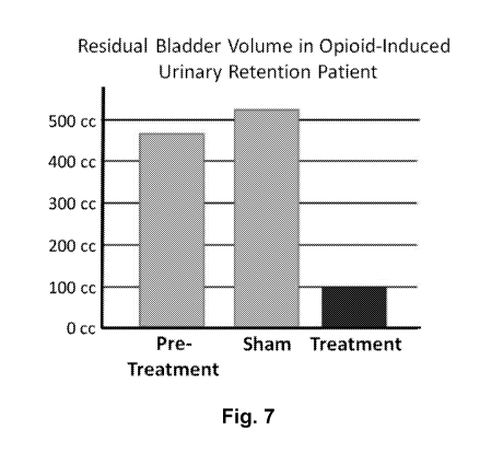
[0118] Figure 7 illustrates bladder volume of post-operative opioid-induced
urinary
retention patient treated with non-invasive magnetic spinal cord stimulation.
[0119] Figure 8 illustrates voiding efficiency in 4 patients with
opioid-induced
urinary retention treated with non-invasive magnetic spinal cord stimulation.
[0120] Figure 9 illustrates the results of an assessment of
incontinence in subjects
treated with non-invasive magnetic spinal cord stimulation.
[0121] Figure 10 illustrates time to bowel sounds and bowel movement
in post-
operative patients treated with magnetic stimulation at conus compared to sham
treated
patients.
[0122] Figure 11 shows that magnetic stimulation decreases the length
of post-
operative hospitalization.
-17-
CA 03110189 2021-02-19
WO 2020/041502
PCT/US2019/047551
DETAILED DESCRIPTION
[0123] In various embodiments methods and devices are provided to
facilitate bladder
and/or bowel control in subjects that have dysfunctional bladder or bowel
control where there
is no brain or spinal cord injury. In certain embodiments the dysfunctional
bladder and/or
bowel comprises neurogenic bladder dysfunction. In certain embodiments the
dysfunctional
bladder and/or bowel comprises dysfunction induced by an inflammatory
stimulus, such as
trauma or infection. In certain embodiments the dysfunctional bladder and/or
bowel
comprises pregnancy associated bladder and/or bowel dysfunction. In certain
embodiments
the dysfunctional bladder and/or bowel is associated with a condition selected
from the group
consisting of Meningomyelocele, Diabetes, AIDS, alcohol abuse, Vitamin B12
deficiency
neuropathies, herniated disc, damage due to pelvic surgery, syphilis, a tumor,
and the like. It
will be recognized that these examples are illustrative and the methods and
devices described
herein can be used to facilitate bladder and/or bowel function associated with
essentially any
dysfunctional state.
[0124] In certain embodiments, the dysfunctional bowel comprises
constipation
induced by one or more medical procedures, one or more drugs, one or more
disorders, etc.
For example, in certain embodiments the dysfunctional bowel may comprise post-
surgical
constipation. As another example, in certain embodiments the dysfunctional
bowel may be
induced by one or more medications (e.g., opiates (e.g., morphine) or other
narcotics,
anticholinergic agents, tricyclic antidepressants (amitriptyline),
antispasmodics (dicyclomine,
mebeverine, peppermint oil), calcium channel blockers (verapamil, nifedipine),
antiparkinsonian drugs, anticonvulsants (carbamazepine), sympathomimetics
(ephedrine),
antipsychotics (chloropromazine, clozapine, haloperidol, risperidone),
diuretics (furosemide),
antihypertensives (clonidine), antiarrhythmics (amiodarone), beta-adrenoceptor
antagonists
(atenolol), antihistamines, calcium or aluminum containing antacids, calcium
supplements,
iron supplements, antidiarrheal (loperamide), 5-HT3-receptor antagonists
(ondansetron), bile
acid sequestrants (cholestyramine), non-steroidal anti-inflammatory drugs
(ibuprofen), etc.).
As yet another example, in certain embodiments, the dysfunctional bowel may
comprise a
condition that is secondary to a primary disease or disorder such as organic
stenosis (e.g.,
colorectal cancer or other intra- or extra-intestinal masses, inflammatory
stenosis, ischemic
stenosis, surgical stenosis, etc.), an endocrine or metabolic disorder (e.g.,
hypothyroidism,
hypercalcemia, hyperparathyroidism, diabetes, porphyria, chronic renal
insufficiency, pan-
hypopituitarism, pregnancy, etc.), neurological disorders (e.g., Parkinson's
disease,
cerebrovasular disease, paraplegia, multiple sclerosis, autonomic neuropathy,
spina bifida,
-18-
CA 03110189 2021-02-19
WO 2020/041502
PCT/US2019/047551
etc.), an enteric neuropathy (e.g., Hirschsprung's disease, chronic intestinal
pseudo-
obstruction, etc.), a myogenic disorder (e.g., myotonic dystrophy,
dermatomyositis,
scleroderma, amyloidosis, chronic intestinal pseudo-obstruction, etc.), an
anorectal disorder
(e.g., anal fissures, anal strictures, etc.), and the like.
[0125] In certain embodiments, the dysfunctional bowel comprises one or
more
diarrheal conditions. For example, in certain embodiments, the dysfunctional
bowel may
comprise an acute diarrheal condition or a chronic diarrheal condition. In
certain
embodiments, diarrheal condition may be caused by a microbe (e.g., viral
gastroenteritis,
such as caused by rotavirus, norovirus, etc., or bacteria). In certain
embodiments, the
dysfunctional bowel may comprise fatty or malabsorptive diarrhea, which may,
for example,
be due to chronic pancreatitis or other chronic injury to the pancreas (e.g.,
alcohol damage,
cystic fibrosis, hereditary pancreatitis, pancreatic cancer, other trauma to
the pancreas, etc.)
and/or small bowel disease (e.g., celiac disease, Crohn's disease, Whipple's
disease, tropical
sprue, eosinophilic gastroenteritis, etc.). In certain embodiments, the
dysfunctional bowel
may comprise a watery diarrheal condition, such that caused by carbohydrate
malabsorption
(e.g., intolerance to lactose, sorbitol, fructose, etc.). In certain
embodiments, the
dysfunctional bowel may comprise medication-induced diarrhea such as induced
by
antibiotics, NSAIDs, antacids, antihypertensives, antiarrhythmics, etc. In
certain
embodiments, the dysfunctional bowel may comprise diarrhea due to inflammatory
bowel
disease (MD), ulcerative colitis, Crohn's disease ischemia of the gut,
infections, a medical
procedure (e.g., radiation therapy), colon cancer, polyps, irritable bowel
syndrome (IBS),
diabetes mellitus, other chronic medical conditions, diet, etc.
[0126] It was discovered that stimulation with devices that impart a
magnetic field
(e.g., at a frequency range from about 0.5 Hz up to about 100 Hz) can regulate
bladder
function. In particular, low frequency magnetic stimulation (e.g., 0.5Hz up to
about 20 Hz)
can induce micturition, while hither frequency magnetic stimulation (e.g. 20
Hz or 30 Hz up
to about 10 Hz or 100 Hz) can suppress micturition. More surprisingly it was
discovered that
repeated treatments with magnetic stimulation can over time increase
volitional control of
bladder function. Once volitional control of bladder function is realized,
repeated periodic
treatments (e.g., weekly, every 10 days, biweekly, etc.) can maintain this
volitional bladder
control.
[0127] It was also discovered that stimulation with devices that
impart an electrical or
magnetic field (e.g., at a frequency range from 5-100 Hz) of the cervical,
and/or thoracic,
-19-
CA 03110189 2021-02-19
WO 2020/041502
PCT/US2019/047551
and/or lumbar spinal cord, nerve roots, or combinations thereof can restore
arm and leg
movement (e.g., in subjects with a partial or full spinal cord injury). It was
also discovered
that, with training and repetition, the gains with stimulation can be
hardwired and present
even without stimulation. Additionally, it was discovered that serotonin
agonists such as
buspirone and the like can be used to further activate the spinal network to
improve motor
function.
[0128] Stimulation of the cervical, and/or thoracic, and/or lumbar
spinal cord, nerve
roots, or combinations thereof can be induced by epidural stimulation
electrodes, non-
invasive transcutaneous electrical stimulation, or magnetic stimulation.
[0129] Additionally, it was discovered that the stimulation methods
described herein
can be leveraged to regain motor function in subjects with injury to the
central nervous
system or degenerative neuromotor conditions, including, but not limited to
stroke, TBI, MS,
ALS, Parkinson's disease, Alzheimer's disease, and the like.
[0130] Without being bound to a particular theory, it is believed
that enabling the
spinal circuitry can produce a coordinated behavior that is more complete and
physiologic
than stimulation of individual nerve roots or the peripheral nerves. Moreover,
the existing
devices have the disadvantages of being invasive, producing a subset of the
desired
locomotor or micturition behavior, and do not result in enduring plastic
changes to the
circuitry that allow patients to become device independent.
[0131] By way of illustration, it is noted that Medtronic markets the
INTERSTIM
device for sacral neuromodulation with overactive bladder or fecal
incontinence. This device
can be effective, but there is a fundamental difference in the mechanism of
action compared
to the methods described herein. Neuromodulation of the sacral nerve roots, as
with the
Medtronic InterStim, attempts to produce appropriate behavior by altering the
activity of the
sacral nerves.
[0132] In contrast, the methods described herein alter the activity
of the spinal
circuitry. It is believed that enabling the spinal circuitry produces a
coordinated behavior that
is more complete and physiologically normative than stimulation of the
peripheral nerves.
Moreover, the existing devices have the disadvantages of being invasive,
producing a subset
of the micturition behavior, and do not result in enduring plastic changes to
the circuitry that
allow patients to become device independent.
-20-
CA 03110189 2021-02-19
WO 2020/041502
PCT/US2019/047551
Voiding of bladder and/or bowel.
[0133] As explained above, the orchestrated neuromuscular control of
urinary bladder
function by the sensory, motor and autonomic nervous systems can be impaired
by
degenerative or traumatic changes, such as multiple sclerosis, spinal cord
injury, stroke. It
was discovered that stimulation of the spinal cord and, optionally, associated
nerve roots can
restore voluntary control of bladder and/or bowel function.
[0134] In particular, it was discovered that non-invasive (e.g.,
magnetic or
transcutaneous electrical) stimulation of the cervical, thoracic, lumbar
(vertebral body
designation) spinal cord and associated nerve roots and combination thereof,
results in
micturition and/or restoration of bowel function. In particular it was
observed that electrical
stimulation with (10 kHz constant-current bipolar rectangular stimulus) from a
range of 1 Hz
to 100 Hz enabled micturition and restoration of bowel function. It was also
observed that
stimulation with a magnetic stimulator, generating a magnetic field, within a
range of 1 Hz to
100 Hz enabled micturition and restoration of bowel function.
Magnetic stimulation to restore bladder/bowel function.
[0135] More generally, it was discovered that that stimulation of the
spinal cord with
devices that impart a magnetic field (e.g., at a frequency range from about
0.5 Hz up to about
100 Hz) can regulate bladder function. In particular, low frequency magnetic
stimulation
(e.g., 0.5Hz up to about 15 Hz) can induce micturition, while higher frequency
magnetic
stimulation (e.g. 20 Hz or 30 Hz up to about 100 Hz) can suppress micturition.
Thus, for
example, it was observed that at a low frequency (e.g., 1Hz) the detrusor
pressure increased
with minimal or small change in urethral pressure so micturition seemed to be
enhanced
(which can be used to treat underactive and neurogenic bladder). At high
frequency (e.g., 30
Hz) urethreal pressure increased with no modification of detrusor pressure so
urine can be
retained (which can be used to treat overactive bladder or stress
incontinence).
[0136] More surprisingly it was discovered that repeated treatments
with magnetic
stimulation can over time increase volitional control of bladder function.
Once volitional
control of bladder function is realized, repeated periodic treatments (e.g.,
weekly, every 10
days, biweekly, etc.) can maintain this volitional bladder control.
[0137] Accordingly, in various embodiments methods of facilitating voiding
or
control of bladder and/or bowel in a subject with a neuromotor disorder are
provided where
the methods involve providing magnetic stimulation of the spinal cord at a
location,
frequency and intensity sufficient to facilitate voiding or control of bladder
and/or bowel. In
-21-
CA 03110189 2021-02-19
WO 2020/041502
PCT/US2019/047551
certain embodiments the magnetic stimulation comprises stimulation at a
frequency ranging
from about 0.5 Hz up to about 15 Hz to induce micturition and in certain
embodiments the
magnetic stimulation is at a frequency of about 1 Hz. In certain embodiments
the magnetic
stimulation comprises stimulation at a frequency from about 20 Hz up to about
100 Hz to
stop or prevent micturition and in certain embodiments, the magnetic
stimulation is at a
frequency of about 30 Hz.
[0138] In certain embodiments the magnetic stimulation comprises
magnetic pulses
ranging in duration from about 5 [ts, or from about 10 [ts, or from about 15
[ts, or from about
20 [ts up to about 1 ms, or up to about 750 [ts, or up to about 500 [ts, or up
to about 400 [ts, or
up to about 300 [ts, or up to about 200 [ts, or up to about 100 [ts. or up to
about 50 [ts. In
certain embodiments the magnetic pulses are about 25 .is in duration.
[0139] In certain embodiments the magnetic stimulation is monophasic,
while in
other embodiments, the magnetic stimulation is biphasic.
[0140] In certain embodiments a single treatment of magnetic
stimulation comprises
1, or 2, or 3, or 4, or 5, or 6, or 7, or 8, or 9, or 10 or more continuous
stimulation periods. In
various embodiments the continuous stimulation periods range in duration from
about 10 sec,
or from about 20 sec, or from about 3 sec or from about 40 sec, or from about
50 sec, or from
about 1 min, or from about 2 minutes up to about 10 minutes, or up to about 8
minutes, or up
to about 6 minutes. In certain embodiments the continuous stimulation periods
are about 4
minutes in duration. In certain embodiments the delay between continuous
stimulation
periods ranges from about 2 sec, or from about 5 sec, or from about 10 sec, or
from about 15
sec, or from about 20 sec up to about 5 minutes, or up to about 4 minutes, or
up to about 3
minutes, or up to about 2 minutes, or up to about 1 min, or up to about 45
sec, or up to about
sec. In certain embodiments the delay between continuous stimulation periods
is about 30
25 sec.
[0141] It was discovered that repeating the treatment can
progressively increase
subsequent volitional control of bladder function (e.g., permits volitional
voiding at a later
time without magnetic (or electrical) stimulation). Conversely removal of
repetitive
treatments can result in progressive loss of volitional control. Accordingly,
in certain
30 embodiments the treatment is repeated (e.g., repeated daily, or every 2
days, or every 3 days,
or every 4 days, or every 5 days, or every 6 days, or every 7 days, or every 8
days, or every 9
days, or every 10 days, or every 11 days, or every 12 days, or every 13 days,
or every 14
days). In certain embodiments the treatment is repeated over a period of at
least 1 week, or at
-22-
CA 03110189 2021-02-19
WO 2020/041502
PCT/US2019/047551
least two weeks, or at least 3 weeks, or at least 4 weeks, or at least 5
weeks, or at least 6
weeks, or at least 7 weeks, or at least 8 weeks, or at least 9 weeks, or at
least 10 weeks, or at
least 11 weeks, or at least 12 weeks, or at least 4 months, or at least 5
months, or at least 6
months, or at least 7 months, or at least 8 months, or at least 9 months, or
at least 10 months,
or at least 11 months, or at least 12 months. In certain embodiments the
treatment is repeated
daily, or every 2 days, or every 3 days, or every 4 days, or every 5 days, or
every 6 days, or
every 7 days, or every 8 days, or every 9 days, or every 10 days, or every 11
days, or every
12 days, or every 13 days, or every 14 days until the subject obtains
volitional control of
micturation. In certain embodiments the treatment is repeated daily, or every
2 days, or every
3 days, or every 4 days, or every 5 days, or every 6 days, or every 7 days, or
every 8 days, or
every 9 days, or every 10 days, or every 11 days, or every 12 days, or every
13 days, or every
14 days until the subject obtains their maximal volitional control of
micturation.
[0142] In certain embodiments, once volitional control is achieved,
the frequency of
treatment can be reduced to a "maintenance" level. Typically, the frequency of
treatment is is
reduced to a level sufficient to maintain volitional control (e.g., a desired
level of volitional
control) of micturition. In certain embodiments the frequency of treatment is
reduced to
every three days, or to a weekly treatment, or to about every 10 days, or to
about every 2
weeks.
[0143] In certain embodiments the magnetic stimulation is applied
over the thoracic
and/or lumbosacral spinal cord (e.g., over one or more regions selected from
the group
consisting of Ti-Ti, Ti-T2, Ti-T3, Ti-T4, Ti-T5, Ti-T6, Ti-T7, Ti-T8, Ti-T9,
Ti-T10,
Tl-T11, Ti-T12, T2-T2, T2-T3, T2-T4, T2-T5, T2-T6, T2-T7, T2-T8, T2-T9, T2-
T10, T2-
T11, T2-T12, T3-T3, T3-T4, T3-T5, T3-T6, T3-T7, T3-T8, T3-T9, T3-T10, T3-T11,
T3-T12,
T4-T4, T4-T5, T4-T6, T4-T7, T4-T8, T4-T9, T4-T10, T4-T11, T4-T12, T5-T5, T5-
T6, T5-
T7, T5-T8, T5-T9, T5-T10, T5-T11, T5-T12, T6-T6, T6-T7, T6-T8, T6-T9, T6-T10,
T6-T11,
T6-T12, T7-T7, T7-T8, T7-T9, T7-T10, T7-T11, T7-T12, T8-T8, T8-T9, T8-T10, T8-
T11,
T8-T12, T9-T9, T9-T10, T9-T11, T9-T12, T10-T10, T10-T11, T10-T12, T11-T11, T11-
T12,
T12-T12, Li-Li, Li-L2 , Li-L3, Li-L4, Li-L5, Lb-S1, Ll-52, Ll-53, Ll-54, L1-
55, L2-L2
, L2-L3, L2-L4, L2-L5, L2-S1, L2-52, L2-53, L2-54, L2-55, L3-L3, L3-L4, L3-L5,
L3-S1,
L3-52, L3-53, L3-54, L3-55, L4-L4, L4-L5, L4-S1, L4-52, L4-53, L4-54, L4-55,
L5-L5,
L5-S1, L5-52, L5-53, L5-54, L5-55, Si-Si, Sl-S2, Sl-S3, Sl-S4, Sl-S5, S2-S2,
S2-S3, S2-
S4, S2-S5, S3-S3, S3-S4, S3-S5, S4-S4, S4-S5, and S5-S6). In certain
embodiments the
magnetic stimulation is applied over a region between T11 and L4. In certain
embodiments
the magnetic stimulation is applied over one or more regions selected from the
group
-23-
CA 03110189 2021-02-19
WO 2020/041502
PCT/US2019/047551
consisting of T11-T12, Li-L2, and L2-L3. In certain embodiments the magnetic
stimulation
is applied over Ll-L2 and/or over T11-T12. In certain embodiments the magnetic
stimulation
is applied over Ll. In certain embodiments the magnetic stimulation is applied
at the midline
of spinal cord. In various embodiments the magnetic stimulation produces a
magnetic field
of at least about 0.5 tesla, or at least about 0.6 tesla, or at least about
0.7 tesla, or at least
about 0.8 tesla, or at least about 0.9 tesla, or at least about 1 tesla, or at
least about 2 tesla, or
at least about 3 tesla, or at least about 4 tesla, or at least about 5 tesla.
In certain
embodiments the magnetic stimulation is at a frequency of at least about 0.5
Hz, 1 Hz, or at
least about 2 Hz, or at least about 3 Hz, or at least about 4 Hz, or at least
about 5 Hz, or at
least about 10 Hz, or at least about 20 Hz or at least about 30 Hz or at least
about 40 Hz or at
least about 50 Hz or at least about 60 Hz or at least about 70 Hz or at least
about 80 Hz or at
least about 90 Hz or at least about 100 Hz, or at least about 200 Hz, or at
least about 300 Hz,
or at least about 400 Hz, or at least about 500 Hz.
[0144] Accordingly, in certain embodiments, methods of facilitating
voiding of the
bladder or bowel are provided where the methods involve providing magnetic
stimulation of
the spinal cord at a location, frequency and intensity sufficient to
facilitate voiding of the
bladder and/or bowel. In certain embodiments the spinal cord stimulation
facilitates initiation
of voiding of the bowel and/or bladder. In certain embodiments the spinal cord
stimulation
improves the efficacy of voiding of the bladder and/or bowel. In certain
embodiments the
spinal cord stimulation suppresses micturition. Also, in certain embodiments
the magnetic
stimulation is of a frequency and magnitude sufficient to restore volitional
control of the
bladder in the absence of stimulation.
[0145] Similarly, it was also observed that transcutaneous electrical
stimulation can
facilitate bladder and/or bowel control (see, e.g. Example 2). Transcutaneous
electrical
stimulation can readily be applied using an electrical stimulator coupled to
electrodes that are
applied to the surface of the subjects body (e.g., over the spinal cord at the
regions described
herein).
[0146] Suitable parameters for electrical stimulation and locations
of such stimulation
are discussed below and illustrated in Example 2.
Regions of stimulation.
[0147] As noted above, in various embodiments one or more regions of
the spinal
cord are stimulated to facilitate locomotor function (e.g., standing,
stepping, postural
changes, arm and/or hand control, etc.), or to facilitate voiding of bowel
and/or bladder.
-24-
CA 03110189 2021-02-19
WO 2020/041502
PCT/US2019/047551
Depending on the desired function, in certain embodiments stimulation is
applied to, or over,
one or more regions of cervical spinal cord, and/or to or over one or more
regions of the
thoracic spinal cord, and/or to or over or one or more regions of the
lumbosacral spinal cord.
[0148] For example, in certain embodiments, to facilitate locomotor
activity such as
standing, stepping, postural control, and the like, the methods may involve
stimulating one or
more regions of the thoracic and/or lumbosacral spinal cord.
[0149] In certain embodiments to facilitate locomotor activity such
as control of the
hand and/or arm and/or grasping, and the like, the methods may involve
stimulating one or
more regions of the cervical and/or thoracic spinal cord. Thus, for example,
as demonstrated
herein cervical spinal cord stimulation improves hand strength and hand and
arm locomotor
control.
[0150] In certain embodiments, to facilitate voiding of the bowel
and/or bladderõ the
methods may involve stimulating one or more regions of the thoracic and/or
lumbosacral
spinal cord. For example, in certain embodiments, stimulation (e.g., magnetic
stimulation)
may be applied to or over one or more regions selected from the group
consisting of T11-
T12, L1-L2, and L2-L3. In certain embodiments stimulation (e.g., magnetic
stimulation) may
be applied to or over Ll-L2 and/or T11-T12.
[0151] With respect to application of stimulation to the cervical
spinal cord,
illustrative regions include, but are not limited to one or more regions
straddling or spanning
a region selected from the group consisting of Cl-C1, C1-C2, C1-C3, C1-C4, C1-
C7, C1-C6,
C1-C7, Cl-T1, C2-C2, C2-C3, C2-C4, C2-05, C2-C6, C2-C7, C2-T1, C3-C3, C3-C4,
C3-
05, C3-C6, C3-C7, C3-T1, C4-C4, C4-05, C4-C6, C4-C7, C4-T1, C5-05, C5-C6, C5-
C7,
C5-T1, C6-C6, C6-C7, C6-T1, C7-C7, and C7-T1.
[0152] With respect to application of stimulation to the thoracic
spinal cord,
illustrative regions include, but are not limited to one or more regions
straddling or spanning
a region selected from the group consisting of Tl-T1, Tl-T2, Tl-T3, Tl-T4, Tl-
T5, Tl-T6,
Tl-T7, Tl-T8, Tl-T9, Tl-T10, Tl-T11, Tl-T12, T2-T2, T2-T3, T2-T4, T2-T5, T2-
T6, T2-
T7, T2-T8, T2-T9, T2-T10, T2-T11, T2-T12, T3-T3, T3-T4, T3-T5, T3-T6, T3-T7,
T3-T8,
T3-T9, T3-T10, T3-T11, T3-T12, T4-T4, T4-T5, T4-T6, T4-T7, T4-T8, T4-T9, T4-
T10, T4-
T11, T4-T12, T5-T5, T5-T6, T5-T7, T5-T8, T5-T9, T5-T10, T5-T11, T5-T12, T6-T6,
T6-T7,
T6-T8, T6-T9, T6-T10, T6-T11, T6-T12, T7-T7, T7-T8, T7-T9, T7-T10, T7-T11, T7-
T12,
T8-T8, T8-T9, T8-T10, T8-T11, T8-T12, T9-T9, T9-T10, T9-T11, T9-T12, T10-T10,
T10-
T11, T10-T12, T11-T11, T11-T12, and T12-T12.
-25-
CA 03110189 2021-02-19
WO 2020/041502
PCT/US2019/047551
[0153] With respect to application of stimulation to the lumbosacral
spinal cord,
illustrative regions include, but are not limited to one or more regions
straddling or spanning
a region selected from the group consisting of Li-Li, Li-L2 , Li-L3, Li-L4, Li-
L5, Li-Si,
Ll-52, Ll-53, Ll-54, L1-55, L2-L2 , L2-L3, L2-L4, L2-L5, L2-S1, L2-52, L2-53,
L2-54,
L2-55, L3-L3, L3-L4, L3-L5, L3-S1, L3-52, L3-53, L3-54, L3-55, L4-L4, L4-L5,
L4-S1,
L4-52, L4-53, L4-54, L4-55, L5-L5 , L5-S1, L5-52, L5-53, L5-54, L5-55, Si-Si,
Sl-52, Si-
S3, Sl-54, S1-55, S2-S2, S2-S3, S2-S4, S2-S5, S3-S3, S3-S4, S3-S5, S4-S4, S4-
S5, and S5-
S6.
Methods of stimulation.
Magnetic stimulation.
[0154] In certain embodiments the methods described herein utilize
magnetic
stimulators for stimulation of the spinal cord (e.g., spinal circuits) to
facilitate locomotor
activity (e.g., standing, stepping, sitting, laying down, stabilizing sitting
posture, stabilizing
standing posture, arm motion, hand motion, griping, hand strength, and the
like) and/or to
induce or improve voiding of the bowel and/or bladder. Magnetic spinal cord
stimulation is
achieved by generating a rapidly changing magnetic field to induce a current
at the region(s)
of interest. In certain embodiments effective spinal cord stimulation
typically utilizes a
current transient of about 108 A/s or greater discharged through a stimulating
coil. The
discharge current flowing through the stimulating coil generates magnetic
lines of force. As
the lines of force cut through tissue (e.g., the spinal cord or brain stem), a
current is generated
in that tissue. If the induced current is of sufficient amplitude and duration
such that the cell
membrane is depolarized, neural/neuromuscular tissue will be stimulated.
[0155] Since the magnetic field strength falls off with the square of
the distance from
the stimulating coil, the stimulus strength is at its highest close to the
coil surface. The
stimulation characteristics of the magnetic pulse, such as depth of
penetration, strength and
accuracy, depend on the rise time, peak electrical energy transferred to the
coil and the spatial
distribution of the field. The rise time and peak coil energy are governed by
the electrical
characteristics of the magnetic stimulator and stimulating coil, whereas the
spatial
distribution of the induced electric field depends on the coil geometry and
the anatomy of the
region of induced current flow.
[0156] In various embodiments the magnetic nerve stimulator will
produce a field
strength up to about 10 tesla, or up to about 8 tesla, or up to about 6 tesla,
or up to about 5
-26-
CA 03110189 2021-02-19
WO 2020/041502
PCT/US2019/047551
tesla, or up to about 4 tesla, or up to about 3 tesla, or up to about 2 tesla,
or up to about 1
tesla, or up to about 0.8 tesla, or up to about 0.6 tesla, or up to about 0.5
tesla. In certain
embodiments the nerve stimulator produces pulses with a duration from about 5
[ts, or from
about 10[ts, or from about 15 [ts, or from about 20 [ts up to about 10 ms, or
from about 25 [ts
up to about 500 [ts, or from about 25 [ts or to about 100 [ts, or from about
100 [ts up to about
1 ms.
[0157] In certain embodiments the magnetic stimulation is at a
frequency of at least
about 1 Hz, or at least about 2 Hz, or at least about 3 Hz, or at least about
4 Hz, or at least
about 5 Hz, or at least about 10 Hz, or at least about 20 Hz or at least about
30 Hz or at least
about 40 Hz or at least about 50 Hz or at least about 60 Hz or at least about
70 Hz or at least
about 80 Hz or at least about 90 Hz or at least about 100 Hz, or at least
about 200 Hz, or at
least about 300 Hz, or at least about 400 Hz, or at least about 500 Hz.
[0158] In certain embodiments the magnetic stimulation is at a
frequency ranging
from about 0.5 Hz, or from about 1 Hz, or from about 2 Hz, or from about 3 Hz,
or from
about 4 Hz, or from about 5 Hz, or from about 10 Hz, or from about 10 Hz, or
from about 10
Hz, up to about 500 Hz, or up to about 400 Hz, or up to about 300 Hz, or up to
about 200 Hz
up to about 100 Hz, or up to about 90 Hz, or up to about 80 Hz, or up to about
60 Hz, or up to
about 40 Hz, or from about 3 Hz or from about 5 Hz up to about 80 Hz, or from
about 5 Hz to
about 60 Hz, or up to about 30 Hz.
[0159] In certain embodiments the magnetic stimulation is at a frequency
ranging
from about 20 Hz or about 30 Hz to about 90 Hz or to about 100 Hz.
[0160] In certain embodiments the magnetic stimulation is at a
frequency, pulse
width, and amplitude sufficient to initiate and/or improve standing, stepping,
sitting, laying
down, stabilizing sitting posture, stabilizing standing posture, arm motion,
hand motion,
stimulate gripping, improve hand strength, and the like, and/or to induce or
improve voiding
of the bowel and/or bladder. In certain embodiments the stimulation is at a
frequency, pulse
width, and amplitude sufficient to provide at least 30% emptying or at least
40% emptying, or
at least 50% emptying, or at least 60% emptying, or at least 70% emptying, or
at least 80%
emptying, or at least 90% emptying, or at least 95% emptying, or at least 98%
emptying of
the bladder and/or bowel e.g., upon application of electrical stimulation as
described herein.
-27-
CA 03110189 2021-02-19
WO 2020/041502
PCT/US2019/047551
Transcutaneous electrical stimulation.
[0161] In certain embodiments the methods described herein utilize
transcutaneous
electrical stimulation for stimulation of the spinal cord (e.g., spinal
circuits) to facilitate
locomotor activity (e.g., standing, stepping, sitting, laying down,
stabilizing sitting posture,
stabilizing standing posture, arm motion, hand motion, griping, hand strength,
and the like)
and/or to induce or improve voiding of the bowel and/or bladder. The use of
surface
electrode(s), can facilitates selection or alteration of particular
stimulation sites as well as the
application of a wide variety of stimulation parameters. Additionally surface
stimulation can
be used to optimize location for an implantable electrode or electrode array
for epidural
stimulation.
[0162] In various embodiments, the methods described herein involve
transcutaneous
electrical stimulation of the cervical spine or a region of the cervical spine
and/or the thoracic
spinal cord or a region of the thoracic spinal cord, and/or a region of the
lumbosacral spinal
cord as described herein to facilitate locomotor activity and/or voiding of
the bowel and/or
bladder (e.g., as described above).
[0163] In certain embodiments the transcutaneous stimulation is at a
frequency of at
least about 1 Hz, or at least about 2 Hz, or at least about 3 Hz, or at least
about 4 Hz, or at
least about 5 Hz, or at least about 10 Hz, or at least about 20 Hz or at least
about 30 Hz or at
least about 40 Hz or at least about 50 Hz or at least about 60 Hz or at least
about 70 Hz or at
least about 80 Hz or at least about 90 Hz or at least about 100 Hz, or at
least about 200 Hz, or
at least about 300 Hz, or at least about 400 Hz, or at least about 500 Hz.
[0164] In certain embodiments the transcutaneous stimulation is at a
frequency
ranging from about 1 Hz, or from about 2 Hz, or from about 3 Hz, or from about
4 Hz, or
from about 5 Hz, or from about 10 Hz, or from about 10 Hz, or from about 10
Hz, up to about
500 Hz, or up to about 400 Hz, or up to about 300 Hz, or up to about 200 Hz up
to about 100
Hz, or up to about 90 Hz, or up to about 80 Hz, or up to about 60 Hz, or up to
about 40 Hz, or
from about 3 Hz or from about 5 Hz up to about 80 Hz, or from about 5 Hz to
about 60 Hz, or
up to about 30 Hz. In certain embodiments the transcutaneous stimulation is at
a frequency
ranging from about 20 Hz or about 30 Hz to about 90 Hz or to about 100 Hz.
[0165] In certain embodiments the transcutaneous stimulation is applied at
an
intensity ranging from about 5 mA or about 10 mA up to about 500 mA, or from
about 5 mA
or about 10 mA up to about 400 mA, or from about 5 mA or about 10 mA up to
about 300
mA, or from about 5 mA or about 10 mA up to about 200 mA, or from about 5 mA
or about
-28-
CA 03110189 2021-02-19
WO 2020/041502
PCT/US2019/047551
mA to up about 150 mA, or from about 5 mA or about 10 mA up to about 50 mA, or
from
about 5 mA or about 10 mA up to about 100 mA, or from about 5 mA or about 10
mA up to
about 80 mA, or from about 5 mA or about 10 mA up to about 60 mA, or from
about 5 mA or
about 10 mA up to about 50 mA.
5 [0166] In certain embodiments the transcutaneous stimulation is
applied stimulation
comprises pulses having a width that ranges from about 100 [ts up to about 1
ms or up to
about 800 [ts, or up to about 600 [ts, or up to about 500 [ts, or up to about
400 [ts, or up to
about 300 [ts, or up to about 200 [ts, or up to about 100 [ts, or from about
150 [ts up to about
600 [ts, or from about 200 [ts up to about 500 [ts, or from about 200 [ts up
to about 400 [ts.
10 [0167] In certain embodiments the transcutaneous stimulation is
at a frequency, pulse
width, and amplitude sufficient to initiate and/or improve standing, stepping,
sitting, laying
down, stabilizing sitting posture, stabilizing standing posture, arm motion,
hand motion,
griping, hand strength, and the like) and/or to induce or improve voiding of
the bowel and/or
bladder. In certain embodiments the stimulation is at a frequency, pulse
width, and amplitude
sufficient to provide at least 30% emptying or at least 40% emptying, or at
least 50%
emptying, or at least 60% emptying, or at least 70% emptying, or at least 80%
emptying, or at
least 90% emptying, or at least 95% emptying, or at least 98% emptying of the
bladder and/or
bowel e.g., upon application of electrical stimulation as described herein.
[0168] In certain embodiments the transcutaneous stimulation is
superimposed on a
high frequency carrier signal. In certain embodiments the high frequency
carrier signal
ranges from about 3 kHz, or about 5 kHz, or about 8 kHz up to about 30 kHz, or
up to about
20 kHz, or up to about 15 kHz. In certain embodiments the carrier signal is
about 10 kHz. In
certain embodiments the carrier frequency amplitude ranges from about 30 mA,
or about 40
mA, or about 50 mA, or about 60 mA, or about 70 mA, or about 80 mA up to about
300 mA,
or up to about 200 mA, or up to about 150 mA.
[0169] Accordingly, in certain embodiments, the transcutaneous
stimulation is
applied as a high frequency signal that is pulsed at a frequency ranging from
about 1 Hz up to
about 100 Hz as described above. In one illustrative but non-limiting
embodiment, the
stimulation is a 1 Hz transcutaneous electrical stimulation evoked with a 10
kHz constant-
current bipolar rectangular stimulus for 0.5 ms at 30 to 100 mA repeated at 1-
40 times per
second for 10 to 30 s. This results in a low (2% or less) duty cycle that is
well tolerated. In
certain embodiments the voltage is approximately 30 V at 100 mA. In certain
embodiments
-29-
CA 03110189 2021-02-19
WO 2020/041502
PCT/US2019/047551
each stimulation epoch is repeated 1-10, or 1-5 times per session, once per
week for, e.g., 6-
12 weeks.
Epidural stimulation.
[0170] In various embodiments, the methods described herein can
involve epidural
electrical stimulation for stimulation of the spinal cord (e.g., spinal
circuits) to facilitate
locomotor activity (e.g., standing, stepping, sitting, laying down,
stabilizing sitting posture,
stabilizing standing posture, arm motion, hand motion, griping, hand strength,
and the like)
and/or to induce or improve voiding of the bowel and/or bladder.
[0171] In certain embodiments, the epidural stimulation is at a
frequency of at least
about 1 Hz, or at least about 2 Hz, or at least about 3 Hz, or at least about
4 Hz, or at least
about 5 Hz, or at least about 10 Hz, or at least about 20 Hz or at least about
30 Hz or at least
about 40 Hz or at least about 50 Hz or at least about 60 Hz or at least about
70 Hz or at least
about 80 Hz or at least about 90 Hz or at least about 100 Hz, or at least
about 200 Hz, or at
least about 300 Hz, or at least about 400 Hz, or at least about 500 Hz.
[0172] In certain embodiments, the epidural stimulation is at a frequency
ranging
from about 1 Hz, or from about 2 Hz, or from about 3 Hz, or from about 4 Hz,
or from about
5 Hz, or from about 10 Hz, or from about 10 Hz, or from about 10 Hz, up to
about 500 Hz, or
up to about 400 Hz, or up to about 300 Hz, or up to about 200 Hz up to about
100 Hz, or up
to about 90 Hz, or up to about 80 Hz, or up to about 60 Hz, or up to about 40
Hz, or from
about 3 Hz or from about 5 Hz up to about 80 Hz, or from about 5 Hz to about
60 Hz, or up
to about 30 Hz.
[0173] In certain embodiments, the epidural stimulation is at a
frequency ranging
from about 20 Hz or about 30 Hz to about 90 Hz or to about 100 Hz.
[0174] In certain embodiments the epidural stimulation is at a
frequency, pulse width,
and amplitude sufficient to initiate and/or improve standing, stepping,
sitting, laying down,
stabilizing sitting posture, stabilizing standing posture, arm motion, hand
motion, stimulate
gripping, improve hand strength, and the like, and/or to induce or improve
voiding of the
bowel and/or bladder. In certain embodiments the stimulation is at a
frequency, pulse width,
and amplitude sufficient to provide at least 30% emptying or at least 40%
emptying, or at
least 50% emptying, or at least 60% emptying, or at least 70% emptying, or at
least 80%
emptying, or at least 90% emptying, or at least 95% emptying, or at least 98%
emptying of
the bladder and/or bowel e.g., upon application of electrical stimulation as
described herein.
-30-
CA 03110189 2021-02-19
WO 2020/041502
PCT/US2019/047551
[0175] In certain embodiments, the epidural stimulation is at an
amplitude ranging
from 0.5 mA, or from about 1 mA, or from about 2 mA, or from about 3 mA, or
from about 4
mA, or from about 5 mA up to about 50 mA, or up to about 30 mA, or up to about
20 mA, or
up to about 15 mA, or from about 5 mA to about 20 mA, or from about 5 mA up to
about 15
mA.
[0176] In certain embodiments, the epidural stimulation is with
pulses having a pulse
width ranging from about 100 [ts up to about 1 ms or up to about 800 [ts, or
up to about 600
[ts, or up to about 500 [ts, or up to about 400 [ts, or up to about 300 [ts,
or up to about 200 [ts,
or up to about 100 [ts, or from about 150 [ts up to about 600 [ts, or from
about 200 [ts up to
about 500 [ts, or from about 200 [ts up to about 400 [ts.
[0177] In certain embodiments the epidural stimulation is applied
paraspinally over a
cervical region identified above (e.g., over vertebrae spanning CO to C8 or a
region thereof,
e.g., over a region spanning C3 to C4).
[0178] In certain embodiments, the epidural stimulation is applied
via a permanently
implanted electrode array (e.g., a typical density electrode array, a high
density electrode
array, etc.).
[0179] In certain embodiments, the epidural electrical stimulation is
administered via
a high density epidural stimulating array (e.g., as described in PCT
Publication No:
WO/2012/094346 (PCT/US2012/020112). In certain embodiments, the high density
electrode arrays are prepared using microfabrication technology to place
numerous electrodes
in an array configuration on a flexible substrate. In some embodiments,
epidural array
fabrication methods for retinal stimulating arrays can be used in the methods
described herein
(see, e.g., Maynard (2001) Annu. Rev. Biomed. Eng., 3: 145-168; Weiland and
Humayun
(2005) IEEE Eng. Med. Biol. Mag., 24(5): 14-21, and U.S. Patent Publications
2006/0003090
and 2007/0142878). In various embodiments, the stimulating arrays comprise one
or more
biocompatible metals (e.g., gold, platinum, chromium, titanium, iridium,
tungsten, and/or
oxides and/or alloys thereof) disposed on a flexible material. Flexible
materials can be
selected from parylene A, parylene C, parylene AM, parylene F, parylene N,
parylene D,
other flexible substrate materials, or combinations thereof. Parylene has the
lowest water
permeability of available microfabrication polymers, is deposited in a
uniquely conformal
and uniform manner, has previously been classified by the FDA as a United
States
Pharmacopeia (USP) Class VI biocompatible material (enabling its use in
chronic implants)
(Wolgemuth, Medical Device and Diagnostic Industry, 22(8): 42-49 (2000)), and
has
-31-
CA 03110189 2021-02-19
WO 2020/041502
PCT/US2019/047551
flexibility characteristics (Young's modulus ¨4 GPa (Rodger and Tai (2005)
IEEE Eng. Med.
Biology, 24(5): 52-57)), lying in between those of PDMS (often considered too
flexible) and
most polyimides (often considered too stiff). Finally, the tear resistance and
elongation at
break of parylene are both large, minimizing damage to electrode arrays under
surgical
manipulation. The preparation and parylene microelectrode arrays suitable for
use in the
epidural stimulation methods described herein is described in PCT Publication
No:
WO/2012/100260 (PCT/US2012/022257).
[0180] The electrode array may be implanted using any of a number of
methods (e.g.,
a laminectomy procedure) well known to those of skill in the art. For example,
in some
embodiments, electrical energy is delivered through electrodes positioned
external to the dura
layer surrounding the spinal cord. Stimulation on the surface of the cord
(subdurally) is also
contemplated, for example, stimulation may be applied to the dorsal columns as
well as to the
dorsal root entry zone. In certain embodiments the electrodes are carried by
two primary
vehicles: a percutaneous lead and a laminotomy lead. Percutaneous leads can
typically
comprise two or more, spaced electrodes (e.g., equally spaced electrodes),
that are placed
above the dura layer, e.g., through the use of a Touhy-like needle. For
insertion, the Touhy-
like needle can be passed through the skin, between desired vertebrae, to open
above the dura
layer. An example of an eight-electrode percutaneous lead is an OCTRODE lead
manufactured by Advanced Neuromodulation Systems, Inc.
[0181] Laminotomy leads typically have a paddle configuration and typically
possess
a plurality of electrodes (for example, two, four, eight, sixteen. 24, or 32)
arranged in one or
more columns. An example of an eight-electrode, two column laminotomy lead is
a
LAMITRODE 44 lead manufactured by Advanced Neuromodulation Systems, Inc. In
certain embodiments the implanted laminotomy leads are transversely centered
over the
physiological midline of a subject. In such position, multiple columns of
electrodes are well
suited to administer electrical energy on either side of the midline to create
an electric field
that traverses the midline. A multi-column laminotomy lead enables reliable
positioning of a
plurality of electrodes, and in particular, a plurality of electrode rows that
do not readily
deviate from an initial implantation position.
[0182] Laminotomy are typically implanted in a surgical procedure. The
surgical
procedure, or partial laminectomy, typically involves the resection and
removal of certain
vertebral tissue to allow both access to the dura and proper positioning of a
laminotomy lead.
The laminotomy lead offers a stable platform that is further capable of being
sutured in place.
-32-
CA 03110189 2021-02-19
WO 2020/041502
PCT/US2019/047551
[0183] In the context of conventional spinal cord stimulation, the
surgical procedure,
or partial laminectomy, typically involves the resection and removal of
certain vertebral
tissue to allow both access to the dura and proper positioning of a laminotomy
lead.
Depending on the position of insertion, however, access to the dura may only
require a partial
removal of the ligamentum flavum at the insertion site. In certain
embodiments, two or more
laminotomy leads are positioned within the epidural space of Cl-C7 as
identified above. The
leads may assume any relative position to one another.
[0184] In various embodiments, the arrays are operably linked to
control circuitry that
permits selection of electrode(s) to activate/stimulate and/or that controls
frequency, and/or
pulse width, and/or amplitude of stimulation. In various embodiments, the
electrode
selection, frequency, amplitude, and pulse width are independently selectable,
e.g., at
different times, different electrodes can be selected. At any time, different
electrodes can
provide different stimulation frequencies and/or amplitudes. In various
embodiments,
different electrodes or all electrodes can be operated in a monopolar mode
and/or a bipolar
mode, using constant current or constant voltage delivery of the stimulation.
In certain
embodiments time-varying current and/or time-varying voltage may be utilized.
[0185] In certain embodiments, the electrodes can also be provided
with implantable
control circuitry and/or an implantable power source. In various embodiments,
the
implantable control circuitry can be programmed/reprogrammed by use of an
external device
.. (e.g., using a handheld device that communicates with the control circuitry
through the skin).
The programming can be repeated as often as necessary.
[0186] The epidural electrode stimulation systems described herein
are intended to be
illustrative and non-limiting. Using the teachings provided herein,
alternative epidural
stimulation systems and methods will be available to one of skill in the art.
Stimulators and Stimulation Systems.
Magnetic stimulators.
[0187] Magnetic nerve stimulators are well known to those of skill in
the art.
Stimulation is achieved by generating a rapidly changing magnetic field to
induce a current at
the nerve of interest. Effective nerve stimulation typically requires a
current transient of
about 108 A/s. In certain embodiments this current is obtained by switching
the current
through an electronic switching component (e.g., a thyristor or an insulated
gate bipolar
transistor (IGBT)).
-33-
CA 03110189 2021-02-19
WO 2020/041502
PCT/US2019/047551
[0188] Figure 1 schematically shows one illustrative, but non-
limiting embodiment of
a magnetic stimulator. As shown therein, magnetic nerve stimulator 100
comprises two
parts: a high current pulse generator producing discharge currents of, e.g.,
5,000 amps or
more; and a stimulating coil 110 producing magnetic pulses (e.g., with field
strengths up to 4,
6, 8, or even 10 tesla) and with a pulse duration typically ranging from about
100[ts to lms or
more, depending on the stimulator type. As illustrated in Figure 1, a voltage
(power) source
102 (e.g., a battery) charges a capacitor 106 via charging circuitry 104 under
the control of
control circuitry 114 (e.g., a microprocessor) that accepts information such
as the capacitor
voltage, power set by the user, and various safety interlocks 112 within the
equipment to
ensure proper operation, and the capacitor is then connected to the coil via
an electronic
switching component 108 when the stimulus is to be applied. The control
circuitry is
operated via a controller interface 116 that can receive user input
and/optionally signals from
external sources such as internet monitors, health care professionals, and the
like.
[0189] When activated, the discharge current flows through the coils
inducing a
magnetic flux. It is the rate of change of the magnetic field that causes the
electrical current
within tissue to be generated, and therefore a fast discharge time is
important to stimulator
efficiency.
[0190] As noted earlier the magnetic field is simply the means by
which an electrical
current is generated within the tissue, and that it is the electrical current,
and not the magnetic
field, that causes the depolarization of the cell membrane and thus the
stimulation of the
target nerve.
[0191] Since the magnetic field strength falls off with the square of
the distance from
the stimulating coil, the stimulus strength is at its highest close to the
coil surface. The
stimulation characteristics of the magnetic pulse, such as depth of
penetration, strength and
accuracy, depend on the rise time, peak electrical energy transferred to the
coil and the spatial
distribution of the field. The rise time and peak coil energy are governed by
the electrical
characteristics of the magnetic stimulator and stimulating coil, whereas the
spatial
distribution of the induced electric field depends on the coil geometry and
the anatomy of the
region of induced current flow.
[0192] The stimulating coils typically consist of one or more well-
insulated copper
windings, together with temperature sensors and safety switches.
[0193] In certain embodiments the use of single coils is
contemplated. Single coils
are effective in stimulating the human motor cortex and spinal nerve roots. To
date, circular
-34-
CA 03110189 2021-02-19
WO 2020/041502
PCT/US2019/047551
coils with a mean diameter of 80-100mm have remained the most widely used
magnetic stimulation. In the case of circular coils the induced tissue current
is near z on the
central axis of the coil and increases to a maximum in a ring under the mean
diameter of coil.
[0194] A notable improvement in coil design has been that of the
double coil (also
.. termed butterfly or figure eight coil). Double coils utilize two windings,
normally placed side
by side. Typically double coils range from very small flat coils to large
contoured versions.
The main advantage of double coils over circular coils is that the induced
tissue current is at
its maximum directly under the center where the two windings meet, giving a
more
accurately defined area of stimulation. In certain embodiments, the use of an
angled butterfly
coil may provide improved effects of stimulation.
[0195] The stimulating pulse may be monophasic, symmetrical biphasic
(with or
without an interphase gap), asymmetric biphasic (with or without an interphase
gal), or
symmetric or asymmetric polyphasic (e.g., burst stimulation having a
particular burst
duration and carrier frequency). Each of these has its own properties and so
may be useful in
particular circumstances. For neurology, single pulse, monophasic systems are
generally
employed; for rapid rate stimulators, biphasic systems are used as energy must
be recovered
from each pulse in order to help fund the next. Polyphasic stimulators are
believed to have a
role in a number of therapeutic applications.
[0196] Descriptions of magnetic nerve stimulators can be found, inter
al/a, in U.S.
patent publications US 2009/0108969 Al, US 2013/0131753 Al, US 2012/0101326
Al, IN
U.S. Patent Nos: US 8,172,742, US 6,086,525, US 5,066,272, US 6,500,110, US
8,676,324,
and the like. Magnetic stimulators are also commercially availed from a number
of vendors,
e.g., MAGVENTURE , MAGSTIM , and the like.
Electrical stimulators.
[0197] Any present or future developed stimulation system capable of
providing an
electrical signal to one or more regions of the cervical spinal cord may be
used in accordance
with the teachings provided herein. Electrical stimulation systems (e.g.,
pulse generator(s))
can be used with both transcutaneous stimulation and epidural stimulation.
[0198] In various embodiments, the system may comprise an external
pulse generator
for use with either a transcutaneous stimulation system or an epidural system.
In other
embodiments the system may comprise an implantable pulse generator to produce
a number
of stimulation pulses that are sent to the a region in proximity to the
cervical spinal cord by
-35-
CA 03110189 2021-02-19
WO 2020/041502
PCT/US2019/047551
insulated leads coupled to the spinal cord by one or more electrodes and/or an
electrode array
to provide epidural stimulation. In certain embodiments the one or more
electrodes or one or
more electrodes comprising the electrode array may be attached to separate
conductors
included within a single lead. Any known or future developed lead useful for
applying an
electrical stimulation signal in proximity to a subject's spinal cord may be
used. For
example, the leads may be conventional percutaneous leads, such as PISCES
model 3487A
sold by Medtronic, Inc. In some embodiments, it may be desirable to employ a
paddle-type
lead.
[0199] Any known or future developed external or implantable pulse
generator may
be used in accordance with the teachings provided herein. For example, one
internal pulse
generator may be an ITREL II or Synergy pulse generator available from
Medtronic, Inc,
Advanced Neuromodulation Systems, Inc.'s GENESISTM pulse generator, or
Advanced
Bionics Corporation's PRECISIONTM pulse generator. One of skill in the art
will recognize
that the above-mentioned pulse generators may be advantageously modified to
modulate
.. locomotor function and/or bladder and/or bowel control in accordance with
the teachings
provided herein.
[0200] In certain embodiments systems can employ a programmer coupled
via a
conductor to a radio frequency antenna. This system permits attending medical
personnel to
select the various pulse output options after implant using radio frequency
communications.
While, in certain embodiments, the system employs fully implanted elements,
systems
employing partially implanted elements may also be used in accordance with the
teachings
provided herein.
[0201] In one illustrative, but non-limiting system, a control module
is operably
coupled to a signal generation module and instructs the signal generation
module regarding
the signal to be generated. For example, at any given time or period of time,
the control
module may instruct the signal generation module to generate an electrical
signal having a
specified pulse width, frequency, intensity (current or voltage), etc. The
control module may
be preprogrammed prior to implantation or receive instructions from a
programmer (or
another source) through any known or future developed mechanism, such as
telemetry. The
control module may include or be operably coupled to memory to store
instructions for
controlling the signal generation module and may contain a processor for
controlling which
instructions to send to signal generation module and the timing of the
instructions to be sent
to signal generation module.
-36-
CA 03110189 2021-02-19
WO 2020/041502
PCT/US2019/047551
[0202] In certain embodiments, the controller alters and/or locomotor
function and/or
initiates or facilitates voiding of the bladder and/or bowel on demand.
[0203] In various embodiments, leads are operably coupled to signal
generation
module such that a stimulation pulse generated by signal generation module may
be delivered
via electrodes.
[0204] While in certain embodiments, two leads are utilized, it will
be understood that
any number of one or more leads may be employed. In addition, it will be
understood that
any number of one or more electrodes per lead may be employed. Stimulation
pulses are
applied to electrodes (which typically are cathodes) with respect to a return
electrode (which
typically is an anode) to induce a desired area of excitation of electrically
excitable tissue in a
region of the cervical spine. A return electrode such as a ground or other
reference electrode
can be located on same lead as a stimulation electrode. However, it will be
understood that a
return electrode may be located at nearly any location, whether in proximity
to the
stimulation electrode or at a more remote part of the body, such as at a
metallic case of a
pulse generator. It will be further understood that any number of one or more
return
electrodes may be employed. For example, there can be a respective return
electrode for each
cathode such that a distinct cathode/anode pair is formed for each cathode.
[0205] In various embodiments, the independent electrodes or
electrodes of electrode
arrays are operably linked to control circuitry that permits selection of
electrode(s) to
activate/stimulate and/or controls frequency, and/or pulse width, and/or
amplitude of
stimulation. In various embodiments, the electrode selection, frequency,
amplitude, and
pulse width are independently selectable, e.g., at different times, different
electrodes can be
selected. At any time, different electrodes can provide different stimulation
frequencies
and/or amplitudes. In various embodiments, different electrodes or all
electrodes can be
operated in a monopolar mode and/or a bipolar mode, using, e.g., constant
current or constant
voltage delivery of the stimulation.
[0206] In one illustrative but non-limiting system a control module
is operably
coupled to a signal generation module and instructs the signal generation
module regarding
the signal to be generated. For example, at any given time or period of time,
the control
module may instruct the signal generation module to generate an electrical
signal having a
specified pulse width, frequency, intensity (current or voltage), etc. The
control module may
be preprogrammed prior to use or receive instructions from a programmer (or
another
source). Thus, in certain embodiments the pulse generator/controller is
configurable by
-37-
CA 03110189 2021-02-19
WO 2020/041502
PCT/US2019/047551
software and the control parameters may be programmed/entered locally, or
downloaded as
appropriate/necessary from a remote site.
[0207] In certain embodiments the pulse generator/controller may
include or be
operably coupled to memory to store instructions for controlling the
stimulation signal(s) and
may contain a processor for controlling which instructions to send for signal
generation and
the timing of the instructions to be sent.
[0208] While in certain embodiments, two leads are utilized to
provide
transcutaneous or epidural stimulation, it will be understood that any number
of one or more
leads may be employed. In addition, it will be understood that any number of
one or more
electrodes per lead may be employed. Stimulation pulses are applied to
electrodes (which
typically are cathodes) with respect to a return electrode (which typically is
an anode) to
induce a desired area of excitation of electrically excitable tissue in one or
more regions of
the spine. A return electrode such as a ground or other reference electrode
can be located on
same lead as a stimulation electrode. However, it will be understood that a
return electrode
may be located at nearly any location, whether in proximity to the stimulation
electrode or at
a more remote part of the body, such as at a metallic case of a pulse
generator. It will be
further understood that any number of one or more return electrodes may be
employed. For
example, there can be a respective return electrode for each cathode such that
a distinct
cathode/anode pair is formed for each cathode.
Use of neuromodulatory agents.
[0209] In certain embodiments, the transcutaneous and/or
epidural and/or
magnetic stimulation methods described herein are used in conjunction with
various
pharmacological agents, particularly pharmacological agents that have
neuromodulatory
activity (e.g., are monoamergic). In certain embodiments, the use of various
serotonergic,
and/or dopaminergic, and/or noradrenergic, and/or GABAergic, and/or
glycinergic drugs is
contemplated. These agents can be used in conjunction with epidural
stimulation and/or
transcutaneous stimulation and/or magnetic stimulation as described above.
This combined
approach can help to put the spinal cord in an optimal physiological state for
neuromodulation utilizing the methods described herein.
[0210] In certain embodiments, the drugs are administered systemically,
while
in other embodiments, the drugs are administered locally, e.g., to particular
regions of the
spinal cord. Drugs that modulate the excitability of the spinal neuromotor
networks include,
-38-
CA 03110189 2021-02-19
WO 2020/041502
PCT/US2019/047551
but are not limited to combinations of noradrenergic, serotonergic, GABAergic,
and
glycinergic receptor agonists and antagonists.
[0211] Dosages of at least one drug or agent can be between
about 0.001
mg/kg and about 10 mg/kg, between about 0.01 mg/kg and about 10 mg/kg, between
about
0.01 mg/kg and about 1 mg/kg, between about 0.1 mg/kg and about 10 mg/kg,
between about
5 mg/kg and about 10 mg/kg, between about 0.01 mg/kg and about 5 mg/kg,
between about
0.001 mg/kg and about 5 mg/kg, or between about 0.05 mg/kg and about 10 mg/kg.
[0212] Drugs or agents can be delivery by injection (e.g.,
subcutaneously,
intravenously, intramuscularly), orally, rectally, or inhaled.
[0213] Illustrative pharmacological agents include, but are not limited to,
agonists and
antagonists to one or more combinations of serotonergic: 5-HT IA, 5-HT2A, 5-
HT3, and
5HT7 receptors; to noradrenergic alphal and 2 receptors; and to dopaminergic
DI and D2
receptors (see, e.g., Table 1). In certain embodiments, suitable
pharmacological agents may
include selective serotonin reuptake inhibitors (S SRI) such as fluoxetine,
etc.
Table 1. Illustrative pharmacological agents.
Name Target Action Route
Typical Typical
Dose Range
(mg/Kg) (mg/kg)
Serotonergic receptor systems
8-0HDPAT 5-HT 1A7 Agonist S.C. 0.05 0.045-0.3
Way 100.635 5-HT 1A Antagonist I.P. 0.5 0.4-1.5
Quipazine 5-HT2A/C Agonist I.P. 0.2 0.18-0.6
Ketanserin 5-HT2A/C Antagonist I.P. 3 1.5-6.0
SR 57227A 5-HT3 Agonist I.P. 1.5 1.3-1.7
Ondanesetron 5-HT3 Antagonist I.P. 3 1.4-7.0
5B269970 5-HT7 Antagonist I.P. 7 2.0-10.0
Noradrenergic receptor systems
Methoxamine Alphal Agonist I.P. 2.5 1.5-4.5
Prazosin Alpha I Antagonist I.P. 3 1.8-3.0
Clonidine Alpha2 Agonist I.P. 0.5 0.2-1.5
Yohimbine Alpha2 Antagonist I.P. 0.4 0.3-0.6
Dopaminergic receptor systems
SKF-81297 Dl-like Agonist I.P. 0.2 0.15-0.6
SCH-23390 Dl-like Antagonist I.P. 0.15 0.1-0.75
Quinipirole D2-like Agonist I.P. 0.3 0.15-0.3
Eticlopride D2-like Antagonist I.P. 1.8 0.9-1.8
-39-
CA 03110189 2021-02-19
WO 2020/041502
PCT/US2019/047551
[0214] The foregoing methods are intended to be illustrative and non-
limiting. Using
the teachings provided herein, other methods involving transcutaneous
electrical stimulation
and/or epidural electrical stimulation and/or magnetic stimulation and/or the
use of
neuromodulatory agents to facilitate voiding or control of bladder and/or
bowel in a subject
with dysfunctional bladder and/or bowel function will be available to one of
skill in the
art.EXAMPLES
[0215] The following examples are offered to illustrate, but not to
limit the claimed
invention.
Example 1
A Proof-of-Concept Study of Transcutaneous Magnetic Spinal Cord Stimulation
for
Neurogenic Bladder
[0216] Patients with chronic spinal cord injury (SCI) cannot urinate
at will and must
empty the bladder by self-catheterization. We tested the hypothesis that non-
invasive,
transcutaneous magnetic spinal cord stimulation (TMSCS) would improve bladder
function
in individuals with SCI. Five individuals with American Spinal Injury
Association
Impairment Scale A/B, chronic SCI and detrusor sphincter dyssynergia enrolled
in this
prospective, interventional study.
[0217] After a two-week assessment to determine effective stimulation
characteristics, each patient received sixteen weekly TMSCS treatments and
then received
"sham" weekly stimulation for six weeks while bladder function was monitored.
Bladder
function improved in all five subjects, but only during and after repeated
weekly sessions of
1 Hz TMSCS. All subjects achieved volitional urination. The volume of urine
produced
voluntarily increased from 0 cc/day to 1120 cc/day (p = 0.03); self-
catheterization frequency
decreased from 6.6/day to 2.4/day (p = 0.04); the capacity of the bladder
increased from
244 ml to 404 ml (p = 0.02); and the average quality of life ranking increased
significantly
(p = 0.007). Volitional bladder function was re-enabled in five individuals
with SCI following
intermittent, non-invasive TMSCS. We conclude that neuromodulation of spinal
micturition
circuitry by TMSCS may be used to ameliorate bladder function.
Results.
[0218] Subjects underwent 3 study phases as illustrated in Fig. 2.
Demographic
information and indices of bladder function for all five subjects are shown in
Table 2. The
magnetic resonance images (MRI) indicating the level and extent of SCI for
each subject are
-40-
CA 03110189 2021-02-19
WO 2020/041502
PCT/US2019/047551
shown in Fig. 3. The average duration of SCI was 8.8 7.5 years. None of the
subjects had
been able to void voluntarily since the time of injury as shown in at least
three prior
urodynamic studies in each subject.
Table 2. Demographic information, the origin and nature of the SCI and urinary
indices.
Length of
Decal of
. stimulation
Disease Mechams the
Injury ASIA until
# Sex Age Duration m of
effect
Level Grade volitional
(years) Injury . duration
micturition
(weeks)
(weeks)
A M 42 T4 A 13 MVA 4 4
B M 43 T4 A 5 Wrestling 6 3
C M 22 C5 B 8 Football 5 3
D M 25 C6 B 8 MVA 5 4
E M 23 C7 A 8 MVA 8 2
Avg -- -- -- -- -- -- 5.6 3.2
SD -- -- -- -- -- -- 1.5 0.8
Volitional Stream Bladder Bladder Daily
CIC/day CIC/day Voiding
P Post Void Velocity Capacity Capacity
re (Y/N) (ml/s) Pre (m1) Post
(m1) post
(m1)
A 9 0 Y 10 141 431
2000
B 6 3 Y 10 238 462 700
C 6 3 Y 10 270 351 800
D 6 1 Y 8 215 325
1800
E 6 5 Y 8 354 452 300
Avg 6,6 2.4 -- 9,3 244 404
1120
SD 1.3 1.9 -- 1.1 78 62 740
Ais = American Spinal Injury Association Impairment Scale. MVA=motor vehicle
accident. CIC= clean intermittent catheterization.
Determining the optimal frequency: spinal function
[0219] The bulbocavernosus reflex (BCR) is disinhibited and
pathologically
hyperactive after SCI (Fig. 4). The BCR amplitude was significantly reduced
during 1 Hz
TMSCS in all five subjects (p < 0.001). In contrast, high frequency
stimulation either
increased the BCR amplitude or had no significant effect. The average BCR
latency was
35.2 5.3 ms during both 1 Hz and 30 Hz TMSCS, which is similar to the
latency of the BCR
in normal individuals (Granata et at. (2013) Func. Neural., 28: 293-295).
-41-
CA 03110189 2021-02-19
WO 2020/041502
PCT/US2019/047551
[0220] During 1 Hz TMSCS, spinal cord evoked potentials could be
elicited in
selected lower extremity muscle groups (perineal, vastus lateralis and
quadriceps femoris);
whereas we were unable to detect any spinal evoked potentials at 30 Hz
stimulation (Fig. 4)
Determining the optimal frequency: bladder function
[0221] During the assessment phase, the urethral (P urethra) and detrusor
pressures (P
detrusor) obtained during urodynamic testing during attempted volitional
micturition were
significantly different during high and low frequency TMSCS (Fig. 5). The
urethral and
detrusor pressures are shown in Tables 3 and 4, respectively. The average
urethral pressure
was significantly lower than the baseline, unstimulated value during 1 Hz
stimulation
(p < 0.05) and the average urethral pressure was greater than the
unstimulated, baseline value
during 30 Hz stimulation (though this was not statistically significant P <
0.10). On the other
hand, the average detrusor pressure was significantly elevated during 1 Hz
stimulation
compared to both the baseline, unstimulated condition and 30 Hz stimulation (P
< 0.01 for
both comparisons), and the detrusor pressure was not different from baseline
conditions
during 30 Hz stimulation (p = 0.5). Thus, stimulation at low frequency allowed
each subject
to elevate the bladder pressure and reduce the urethral pressure (conditions
conducive to
urine flow), and 30 Hz stimulation had the opposite effect: urethral pressure
increased
significantly, but detrusor pressure was not modified by 30 Hz stimulation
(Tables 3 and 4).
Not surprisingly, increasing detrusor contraction and bladder pressure while
simultaneously
decreasing urethral pressure allowed voluntary micturition (Fig. 5).
Table 3. At the end of the assessment phase, changes in urethral pressure in
five subjects
during micturition attempts compared to the pre-attempt baseline. Positive
numbers indicate
an increase in the pressure during the attempts while negative numbers
indicate a decrease in
the urethral pressure during the attempts. Notice that high frequency
stimulation (30 Hz)
resulted in increased/unchanged urethral pressure during attempted micturition
when
compared to the no-stimulation/baseline; low frequency stimulation (1 Hz), on
the other
hand, significant decreased urethral pressure compared to both unstimulated
and 30 Hz
stimulation conditions.
A P ure No Low Frequency High Frequency
Stimulation
(mmHg) oS) (1 Hz) (30 Hz)
(N
Subject A 28.4 5.0 -25.3 2.7 36.2 11.1
Subject B 2.5 4.8 0.4 5.2 50.3
17.0
Subject C 19.8 3.4 08.3 6.3 20.0
10.0
-42-
CA 03110189 2021-02-19
WO 2020/041502
PCT/US2019/047551
Subject D 18.9 1.5 5.6 7.3 27.6 4.1
Subject E 15.3 5.2 -0.9 9.6 46.6 11.1
Average 17.0 9.4 -5.7 12.0 1Hz vs NoS 36.1 12.7
40 Hz vs NoS p
p<0.05 a Hz vs 30 Hz <0.10
p<0.001
Table 4. At the end of the assessment phase, the change in detrusor pressure
in five subjects
during micturition attempts compared to the pre-attempt baseline. Positive
numbers indicate
an increase in the pressure during the attempts while negative numbers
indicate a decrease in
the pressure during the attempts. Notice that high frequency stimulation (30
Hz) did not
change in detrusor pressure during attempted micturition when compared to the
no-
stimulation/baseline; low frequency stimulation (1 Hz), on the other hand,
significantly
increased detrusor pressure compared to the non-stimulated and 30 Hz
conditions.
A P det Low Frequency High Frequency
No Stimulation
(mmHg) (1 Hz) (30 Hz)
Subject A 19.4 2.2 38.8 0.9 24.5 4.5
Subject B 26.1 1.5 28.2 3.7 0.6 1.3
Subject C 13. 0.6 30.4 4.3 1.3 0.9
Subject D 3.0 1.5 49.4 19.5 1.6 0.8
Subject E -1.1 0.7 58.5 9.4 -0.1 0.7
Average 9.7 12.2 41.1 12.81 Hz vs Nos 5.6 10.6 30 Hv vs
NoS
p<0.001 1 Hz vs 30 Hz p=0.50
p<0.001
[0222] Based on the BCR response, the evoked EMG activity and the
responses of
urethral and detrusor pressures, only 1 Hz TMSCS was used for weekly TMSCS
during the
treatment period.
[0223] Bladder function before, during and after TMSCS All five
subjects achieved at
least some volitional urination following 16 weeks of bladder rehabilitation
with TMSCS
(Fig. 6). No subject achieved volitional urination until at least 4 weekly
TMSCS treatments
had been given, and the capacity to urinate voluntarily was restored in all 5
subjects on
average 5.6 1.5 weeks after TMSCS was begun. The capacity to urinate
voluntarily was
maintained throughout the 16-week treatment period.
[0224] Daily self-catheterization decreased from 6.6 times per day at
baseline to 2.4
times per day at the conclusion of the 16-week bladder rehabilitation (p =
0.04). Based on
urodynamic studies conducted at the end of the TMSCS treatment, the average
volume of
urine generated voluntarily increased from 0 cc/day to 1120 cc/day (p = 0.03),
and the
-43-
CA 03110189 2021-02-19
WO 2020/041502
PCT/US2019/047551
subjects were able to generate significant urine stream velocities, which rose
on average from
0 cc/sec to 9.3 cc/sec (p < 0.001). The bladder capacity increased from 244 ml
to 404 ml
(p = 0.02). Sexual function also improved from 9 to 20 as measured by Sexual
Health
Inventory for Men (SHIM) (p = 0.0003). The subjects enjoyed a much higher
quality of life;
the average i-QOL score rose from 47 to 82 (p = 0.007, Fig. 5). While all five
subjects had
improved bladder function and were able to achieve volitional micturition,
their responses to
TMSCS varied (the responsiveness order was A>D>B=C> E). This variation did not
appear to be the result of differences in their AIS. (Table 2).
[0225] The average time that volitional micturition was maintained
after the sham
stimulation began was 3.2 0.8 weeks. Follow-up diary entries confirmed that
the ability to
void voluntarily rapidly decayed in all subjects after the cessation of
effective TMSCS, and
no subject maintained the capacity for voluntary micturition five weeks after
the last effective
stimulation.
Discussion.
[0226] Voluntary micturition requires complex, orchestrated neuromuscular
control
of the urinary bladder by sensory, motor and autonomic systems. During
voluntary
micturition, sympathetic inhibition of bladder contraction is withdrawn,
parasympathetic
activation of the detrusor contraction emerges to increase vesicular pressure,
and contraction
of the urethral sphincter is inhibited to allow urine to flow out of the
bladder. This control is
achieved through fronto-pontine-spinal cord projections to parasympathetic
ganglia in the
abdomen and to sympathetic and somatic neurons in the caudal spine. In
individuals with
SCI, coordination among parasympathetic, sympathetic and somatic nerve
activities is lost:
bladder pressure is elevated, but the bladder cannot be completely emptied
because
contraction of the external sphincter is not inhibited. Patients with SCI must
perform multiple
bladder self-catheterizations each day to evacuate urine and to prevent kidney
injury due to
high pressure; which increase the risk and frequency of infection and
traumatic injury to the
urethra. Any decrease in catheterization frequency, which was achieved in all
study subjects,
represents a potential decrease in complications associated with
catheterization.
[0227] Isolated regions of lumbosacral spinal cord contain circuits
that are capable of
carrying out complex motor activities (Lu et at. (2015)Front. Molecular
Neurosci. 8, 25,
(2015); Sugaya & De Groat (1994)Am. I Physiol.,_266: R658-667). Furthermore,
spinal
cord injury in most motor complete, AIS A and B SCI subjects is not
anatomically complete,
and many spinal circuits remain intact, especially those below the level of
the spinal cord
-44-
CA 03110189 2021-02-19
WO 2020/041502
PCT/US2019/047551
injury (Heald et at. (2017) Neurorehabil. Neural Repair, 31: 583-591). In both
animal and
human subjects with chronic paralysis from SCI, motor movements have improved
after
invasive, epidural, electrical stimulations (Harkema et at. (2011) Lancet,
377: 1938-1947;
Angeli et at. (2014) Brain: I Neurol. 137: 1394-1409; Lu et at. (2016)
Neurorehabil. Neural
Repair, 30: 951-962). In this study, we hypothesized that the spinal
micturition circuit
remains intact in subjects with SCI, and since this circuit is semiautonomous,
we should be
able to enhance activation of patterned muscle activities controlled by these
circuits and
activate or modulate them using TMSCS over the thoracolumbar spine. The
mechanism of
action appears to be similar to the use of stimulation to improve upper
extremity function in
which the threshold of motor circuit activation is diminished to enable
volitional, coordinated
agonist- antagonist muscle activity (Alam et al. (2017) Exp. Neurol., 291: 141-
150).
Voluntary bladder control was restored to some extent by TMSCS in all five
individuals with
chronic SCI. Four out of five subjects (80%) were able to decrease the
frequency of self-
catheterization by at least 50%. One subject was able to void normally without
any self-
catheterization while another subject only needed one catheterization each day
(Table 2).
[0228] Other attempts to restore urination in SCI patients by
stimulating multiple
peripheral nerves, specifically the pudendal, pelvic, hypogastric and tibial
nerves (Schneider
et al. (2015) Europ. Urol. 68: 859-867; Kennelly et al. (2011)1 Spinal Cord.
Med. 34: 315-
321; Spinelli et al. (2005) Neurology & Urodynam. 24: 305-309), did not
consistently
improve bladder function. Furthermore, sacral nerve modulation requires
electrode
implantation, which is invasive and risky (Zeiton et at. (2016) Int. I
Colorect. Dis. 31: 1005-
1010; Eldabe et at. (2015) Complications of Spinal Cord Stimulation and
Peripheral Nerve
Stimulation Techniques: A Review of the Literature. Pain medicine (Malden,
Mass.)).
TMSCS differs in that it is non-invasive and painless in patients with SCI. In
addition,
TMSCS provides more consistent and effective bladder emptying than existing
epidural
stimulation of selected peripheral nerves (Bartley et at. (2013) Nat. Rev.
Urology 10, 513-
521; Brindley, (1974)1 Physiol., 237: 15p-16p; Van Kerrebroeck et at. (1996)1
Urol. 155:
1378-1381).
[0229] We believe that TMSCS allowed volitional activation of a
coordinated pattern
of parasympathetic withdrawal and sympathetic activation and somatic muscle
inhibition as
demonstrated in urodynamic studies. While the precise mechanism of TMSCS
remains
unknown, the coordinated activity of detrusor and sphincter muscles suggests
that TMSCS
works by activating or enhancing activation of central pattern generating
circuits within the
lumbosacral spinal cord and does not rely solely on activation of motor
neurons or peripheral
-45-
CA 03110189 2021-02-19
WO 2020/041502
PCT/US2019/047551
nerves. This hypothesis receives further support from the divergent responses
to TMSCS at
1 Hz and 30 Hz: 1 Hz TMSCS resulted in decreased urethral pressure, increased
detrusor
pressure and micturition, as opposed to 30 Hz TMSCS, which increased urethral
pressure,
decreased detrusor pressure and enhanced urine storage within the bladder. The
different
stimulation frequencies elicited different bladder behaviors as if different
central pattern
generators (CPGs) or different aspects of a micturition CPG were activated.
These divergent
responses suggest that TMSCS may be applicable to a broader range of
conditions such as
hyperactive bladder, which may benefit from higher frequency stimulation.
[0230] We selected patients with detrusor-sphincter dyssynergia
specifically because
this is the group of SCI patients most recalcitrant to treatment. A regular
schedule of self-
catheterization prevents ureteral reflux and the development of obstructive
uropathy and
chronic renal failure, but frequent catheterization has risks of its own:
infection, creation of
false passages, urethral stricture (Prieto et at. (2015) Neurology & Urodynam.
34: 648-653;
Bolinger et at. (2013)1 Wound, Ostomy, Continence, Nursing, 40: 83-89), a
reduced quality
of life, and a loss of independence. Improving quality of life is our ultimate
goal using
TMSCS, but this cannot be achieved if the risk of ureteral reflux and chronic
renal failure
increases. Therefore, any benefits of TMSCS, such as a more physiological
voiding sequence
with low storage pressures and increased bladder capacity and a better
coordination of
increased detrusor compliance and reduced external sphincter pressures that
enable
unobstructed voiding in a low pressure system, will be beneficial in the long
term only if
ureteral reflux is not increased. Video urodynamics performed at the
initiation and
termination of our study demonstrated no evidence of reflux. While this was a
proof of
concept, pilot study with patients followed for 16 weeks, additional studies
are needed in an
expanded cohort with extended follow-up to ensure that stable bladder and
renal function are
maintained when TMSCS is used to increase voluntary micturition and reduce the
frequency
of self-catheterization.
[0231] The BCR is a polysynaptic reflex, and BCR amplitudes in our
subjects were
10 to 100 times larger at baseline than in normal individuals. Hyperactivity
of the BCR may
be analogous to the hyperactivity of tendon reflexes following SCI and
suggests that subjects
with SCI have decreased supraspinal inhibition of the BCR. During low
frequency TMSCS,
the amplitude of the BCR decreased, from which we infer that TMSCS induced
greater
inhibition of the BCR. Magnetic stimulation may achieve these effects by
modulation of
spinal interneurons via dorsal root ganglion or dorsal column stimulation,
which is a putative
mechanism of action for epidural spinal cord stimulation (Ramasubbu & Flagg
(2013) Curr.
-46-
CA 03110189 2021-02-19
WO 2020/041502
PCT/US2019/047551
Pain Headache Rep. 17: 315), or TMSCS may modulate responses within the
sympathetic
chain and sacral parasympathetic centers and facilitate the process of
micturition.
[0232] Improvements in urinary function were not instantaneous;
progressive
improvement became apparent over the course of the study. Initially,
simultaneous
measurements of urethral and bladder pressures during volitional urination
attempts revealed
little (if any) sustained bladder contraction and persistently elevated
urethral pressures, but
after completion of at least 4 weeks of effective TMSCS, subjects became
better able to
generate sustained bladder contractions although detrusor-sphincter
dyssynergia persisted
(increased bladder pressures, but also increased urethral pressures, which
prevented bladder
emptying). At the end of the 16-week rehabilitation period, subjects were able
to produce
voluntary, coordinated bladder contractions with high detrusor pressures and
reduced urethral
pressures. Since bladder pressure exceed urethral pressure, urine flow
velocity was increased
and significantly higher urine volumes were achieved (Fig. 3).
[0233] Our subjects were able to urinate voluntarily in between
treatment sessions
when magnetic stimulation was not present. We believe that TMSCS persistently
raised the
activation state (or reduced inhibition) of the micturition circuit so that
residual neural
pathways between the supraspinal micturition centers and lumbosacral
micturition central
pattern generators were re-invigorated, which is consistent with previous
findings using
epidural stimulation to enhance recovery of motor function (Lu et at. (2016)
Neurorehabil.
Neural Repair, 30: 951-962). Restoration of voluntary micturition required
repetitive
TMSCS over at least 4 weeks. The benefits of epidural electric stimulations on
motor
function also required 3-5 sessions/weeks before improvements in motor
functions were seen
(Id.). Once supraspinal to spinal communication had been restored or re-
enabled by
TMSCS, it remained enabled so long as the subject received some minimal amount
of
TMSCS during each weekly treatment session, but the benefits of TMSCS were not
permanent. All subjects lost the ability to control micturition soon after the
termination of
effective TMSCS (Fig. 6). The temporal dynamics of the onset and offset of
benefit of
TMSCS are consistent with remodeling of the spinal circuitry in which some
relatively slow
neuronal or circuit remodeling is required to re-establish effective synaptic
or supraspinal
communication (Boulis et at. (2013) Neurosurgery, 72: 653-661; Vallejo et at.
(2016)
Neuromodulation, 19: 576-586; Ryge et at. (2010) BMC Genomics, 11: 365), and
some
aspect of TMSCS was necessary between periods of volitional bladder emptying
to maintain
the integrity of communication between supraspinal and lumbar micturition
circuits. The
once weekly treatment interval and stimulation protocol represent a
surprisingly small
-47-
CA 03110189 2021-02-19
WO 2020/041502
PCT/US2019/047551
recurrent input to maintain volitional micturition, but this schedule is
feasible for patients,
and TMSCS could be administered in weekly physical therapy sessions at low
cost. In any
event, neuronal plasticity or remodeling are well recognized in TMS studies,
specifically with
low frequency (1 Hz) stimulation (O'Shea et al. (2007) Neuron, 54: 479-490;
Lee et al.
(2003) J Neurosci. 23: 5308-5318). These results and our study of hand
function (Lu et al.
(2016) Neurorehabil. Neural Repair, 30: 951-962) provide two examples of the
capacity of
neuromodulation of spinal circuits to enable volitional control of motor
functions below the
level of SCI.
[0234] The responses to TMSCS varied among our five subjects. While
we do not
have a precise explanation for this, we know that the variation was not a
result of differences
among the AIS (Subject A, B, E were all category A, but subject A improved
much more
than the other two). The reasons for the variation are likely multifactorial,
but perhaps most
importantly, our subjects have variable amounts of residual spinal function.
The current AIS
is not sensitive to the subtleties of residual spinal functions among
subjects.
[0235] The main limitations of our study are its small size and the lack of
proof of the
actual mechanism of action. As this is a pilot study, we plan to continue to
expand the study
and enroll additional subjects. Further studies will focus on the molecular
and cellular
processes that follow magnetic stimulation to investigate the precise
mechanism of action of
magnetic stimulation.
Methods.
Subject selection.
[0236] We conducted a pilot, prospective, interventional study in
five subjects. All
aspects of the study were approved by the UCLA IRB (IRB# 14-000932) and filed
with
ClinicalTrials.gov (registration number: NCT02331979, date of registration:
06/01/2015).
All methods were performed in accordance with the relevant guidelines and
regulations as
stipulated by UCLA IRB. Informed consent was obtained prior to subject
participation. The
inclusion criteria for the study were male age 18-75, a stable American Spinal
Injury
Association Impairment Scale (AIS) A/B, motor complete spinal cord injury
between spinal
levels C2-T8 present for greater than 1 year, and a documented history of
neurogenic bladder
.. requiring intermittent catheterization. Each subject was required to have
at least three prior
urodynamic studies to confirm the diagnosis of neurogenic bladder with
detrusor sphincter
dyssynergia (DSD), which was diagnosed with urodynamic study in which a rise
in detrusor
-48-
CA 03110189 2021-02-19
WO 2020/041502
PCT/US2019/047551
pressure and concomitant needle EMG activity and rise in urethral pressure
were
demonstrated (see Tables 3 and 4). Patients with a history of autonomic
dysreflexia were
excluded from the study. Any patient who was ventilator dependent, abusing
drugs, had
musculoskeletal dysfunction (i.e., unstable fractures), cardiopulmonary
diseases, active
infections or ongoing depression requiring treatment, or had previous exposure
to and use of
spinal cord stimulation was excluded from the study. Patients with a history
of bladder botox
injection or bladder/sphincter surgeries were excluded. Five subjects were
recruited and
completed the study. There was no subject attrition.
Intervention.
[0237] Each study subject underwent baseline urodynamic testing (UPS) at
the
beginning of the study to confirm the diagnosis of a neurogenic bladder with
DSD and
establish baseline bladder functions. The study was divided into three phases:
an Assessment
phase (2 weeks), a Treatment phase (16 weeks) and a Follow-up phase (6 weeks).
During the
Assessment phase, each subject underwent once/week transcutaneous magnetic
spinal cord
stimulation (TMSCS) at both 1 Hz (low) and 30 Hz (high) frequency (40-60%
intensity) over
the lumbar spine (described below). 1 Hz and 30 Hz were both administered
during the
assessment phase because the optimal stimulation frequency in human subjects
was unknown
to us prior to this study. The two frequencies were administered in random
order. These
frequencies was chosen based on previous results in animals in which low
frequency
.. stimulation promoted and high frequency stimulation inhibited micturition
in animals with
SCI (Alam et al. (2017) Exp. Neurol., 291: 141-150). At the conclusion of the
Assessment
phase, each subject underwent another UPS to determine the better stimulation
frequency
(the characteristics of optimal stimulation are defined below). Once the
better frequency was
established (and it turned out that 1 Hz was better than 30 Hz stimulation in
all five subjects),
each subject entered the treatment phase of the study and received weekly
transcutaneous
lumbar spinal cord magnetic stimulation for a total of 16 weeks (described
below). This 16-
week period of TMSCS constituted bladder rehabilitation. Each subject received
non-video
urodynamic testing once every four weeks during the treatment phase to monitor
progress
and insure that bladder function was not further impaired. After the initial
four-week
stimulation period, each subject was asked to attempt volitional urination for
5-10 minutes
prior to bladder catheterization. The subjects were instructed to keep the
environment quiet,
relax and focus on voiding. Specifically, they were instructed to perform no
straining/Valsalva maneuver, external compression by Crede maneuver, reflex
triggering by
tapping, anal stretch, or pushing. Attempts were limited to 10 minutes. Each
subject was
-49-
CA 03110189 2021-02-19
WO 2020/041502
PCT/US2019/047551
given a urine/stool specimen collection pan (Medline DYND36600H, Mundelein,
IL) to
collect any volitional urinary output. In order to prevent potential urinary
retention, the
subjects were asked to self-catheterize after the volitional attempt and to
record the
catheterization output. The urinary output and the volume of the residual
urine in the bladder
(collected after attempted void by each patient's routine bladder
catheterization) were
recorded in a diary after every attempt to urinate voluntarily. Each subject
was also asked to
record any other changes that he may have noticed in the diary throughout the
study period.
During the follow-up period, sham transcutaneous magnetic stimulation (sham)
was
employed at reduced intensity (5%), which replicated the auditory, partial
sensory and
mechanical cues of real stimulation. Each subject was instructed to continue
to attempt to
urinate voluntarily as he had during the treatment phase, and each subject
continued to keep a
detailed urological lifestyle diary until the end of the follow-up phase (Fig.
2).
[0238] Each subject was also given an incontinence quality of life
(iQ0L)
questionnaire to complete prior to the start of the study and at the end of
the 16-week
treatment stimulation. iQOL has been validated in multiple urological quality
of life studies
in patients with SCI (Patrick et at. (1999) Eur. Urol. 36: 427-435; Jo et at.
(3026) Pain
Physician, 19: 373-380). Sexual functions were assessed by Sexual Health
Inventory for
Men (SHIM) questionnaire (Barbonetti et at. (2012)1 Sex. Med. 9: 830-836) at
the beginning
of the study and at the end of 16-week treatment phase.
Blinding.
[0239] The state of knowledge at the start of the study, in which we
did not know the
effective parameters of stimulation to effect micturition, precluded a
randomized trial.
Therefore, we conducted a single arm study in which each subject acted as his
own control.
Additionally, a sham phase was conducted at the end after stimulation because
there was
exposure to stimulation during initial Assessment Phase (we had no initial,
baseline, non-
stimulated collection period), and we were unsure of the wash-out period for
this exposure
due to the pilot nature of this study. However, subjects and experimenters
were blinded
throughout the process in the assessment, treatment and follow-up phases.
Given that these
spinal cord injury patients have diminished/no sensation due to their
injuries, The subjects did
not feel any sensations at 1 Hz or 30 Hz at the level of stimulation used
during treatment as
they have muted sensory capacity due to their spinal cord injury. We
purposefully selected a
relatively low intensity stimulus to avoid any painful sensations, and the
stimulation level
was below the sensory and motor threshold. The subjects did hear a "click"
during each
-50-
CA 03110189 2021-02-19
WO 2020/041502
PCT/US2019/047551
stimulation (especially at 1 Hz when the click was very predicable). This
auditory cue was
re-created during sham stimulation as well. Approximately from T11-L3 level.
The coil
dimensions are 172 x 92 x 51 mm in a figure-of-eight formation with two rings
each 75 mm
in diameter. The target (focality) is the center of the figure of eight, which
in our case is T12-
.. Li area, which overlies the conus medullaris in humans. We used a research
coil (with
identical sham and treatment faces), which allowed blinding of both
experimenter and
subject; thereby double blinding the follow-up phase of the study. The staff
member
responsible for controlling of the stimulator and the dose of stimulation was
not blinded as
the stimulation parameters were manipulated during the various phases of the
study;
however, this person did not interact with the subject (he sat behind a
curtain), and each staff
member was instructed to follow the same script when administering the various
tasks
regardless of the particular stimulation values used. To assess integrity of
blinding, we asked
subjects at the conclusion of the study what each study phases consisted of;
their responses
were no better than chance.
Urodynamic testing.
[0240] We employed a commercially available urodynamic machine
(Laborie
Aquarius XT, Laborie International, Mississauga, ON, Canada). Prior to the
urodynamic
testing, each subject emptied his bladder by direct catheterization. The
volume of urine was
recorded. The patient was then placed in a supine position and a triple lumen
catheter (TLC-
7M, Laborie International, Mississauga, ON, Canada) was inserted. Two needle
recording
electrodes (1512A-M, Laborie International, Mississauga, ON, Canada) were
inserted
bilaterally into the perineal muscles approximately halfway between the base
of the scrotum
and anus and 1 cm lateral to the midline. An EMG grounding pad was placed on
the knee
joint. A rectal catheter (RPC-9, Laborie International, Mississauga, ON,
Canada) was
inserted to record abdominal pressure. The subject was next placed in a left
decubitus
position. A condom catheter was used to collect any urine output, which was
directed
through a funnel into a graduated cylinder (DIS173, Laborie International,
Mississauga, ON,
Canada) on a scale (UROCAP IV, Laborie International, Mississauga, ON, Canada)
to record
the volume of urine produced and the stream velocity.
Transcutaneous magnetic stimulation.
[0241] A Mag Venture Magnetic Stimulator (MagPro R30, Atlanta, GA)
with an
active/placebo figure-8 research coil (Cool-B65 A/P Coil) was used for all
transcutaneous
magnetic stimulation sessions. The spinous processes of the lower vertebrae in
each subject
-51-
CA 03110189 2021-02-19
WO 2020/041502
PCT/US2019/047551
were palpated, and thoracic 11 to lumbar 4 vertebrae were marked. The coil was
centered
along the midline at the Li vertebral level during the stimulation and
oriented such that the
magnetic field generated was parallel to the spinal cord (rostral-caudal). We
used trains of
biphasic, single pulse (duration 250 i.ts), continuous, magnetic stimulation.
Each stimulation
session consisted of three 4-min continuous stimulation periods with a 30
second break
between each stimulation period for a total of 13 minutes (a total of 12
minutes of stimulation
plus 1 minute of breaks). For the first two weeks, each subject underwent
stimulation at 1 Hz
and 30 Hz frequencies (week one: 1 Hz/30 Hz/1 Hz, and week two: 30 Hz/1 Hz/30
Hz) until
the better frequency was determined for the patient at the first follow-up UDS
after the 2
week of stimulation. The frequency of 1 Hz and 30 Hz was selected based on our
previous
work in animals and humans (Lu et al. (2016) Neurorehabil. Neural Repair, 30:
951-962;
Gad et at. (2014) PloS one 9: el08184). Changes in urethral (directly
measured) and detrusor
(vesicular ¨ abdominal) pressures during micturition attempts were measured
during both low
frequency stimulation (1 Hz) and high frequency stimulation (30 Hz). The
stimulation
frequency that resulted in the combination of increased detrusor pressure and
decreased
urethral pressure during attempted micturition (hence, promoting bladder
emptying) was
selected as optimal. The intensity of stimulation was set 20% below the
intensity that elicited
local paraspinal muscular contraction for each subject (since muscle
contractions would have
unmasked the double blinding). This stimulation intensity was usually around
40-50% of
the maximal field strength of 2 Tesla. Once the optimal frequency was
determined, all
subjects received the optimal stimulation frequency only at a constant
intensity for the
remaining 16, weekly bladder rehabilitation sessions.
Electrophysiology
[0242] At the end of the assessment phase, the following
electrophysiological data
were obtained on each subject before, during and after low frequency (1 Hz)
and high
frequency (30 Hz) transcutaneous magnetic stimulation: bulbocavernosus reflex
(BCR),
electromyography (EMG) and spinal evoked potentials (SEP) bilaterally in the
pelvic floor
and in the vastus lateralis, gastrocnemius, gluteus and quadriceps femoris
muscles.
[0243] Pelvic floor EMGs were obtained using needle electrodes
(Laborie 1512A-M,
Laborie International, Mississauga, ON, Canada). All other muscle EMGs were
obtained
with 1 inch surface pad electrodes (MultiBioSensors, El Paso, TX).
[0244] The BCR was obtained by using ring stimulating electrodes
(Cadwell 302243-
200, Cadwell Industries, Kennewick, WA) that were stimulated with a monophasic
electric
-52-
CA 03110189 2021-02-19
WO 2020/041502
PCT/US2019/047551
pulse at 1.5 Hz, pulse width 0.2 ms, and intensity at three times the sensory
threshold (or
35 mA if the subject had no sensation). At least 100 pulses were given in each
BCR test
session. Recording, amplification and digitization of all data were done using
an RZ2
amplifier and a PZ5-32 TDT digitizer (Tucker Davis Technologies, Alachua, FL)
with a
60 Hz notch filter and band pass filtering to exclude frequencies <3 Hz and >
200 Hz.
Data analysis.
[0245] The primary outcome was voluntary urination volume per day.
Pre-specified
secondary outcomes included urine stream flow rate, bladder capacity,
catheterizations per
day, sexual health inventory for men (SHIM), and urinary incontinence quality
of life scale
(iQ0L). All electrophysiological data from the TDT system (Tucker Davis
Technologies,
Alachua, FL) were exported to a computer and analyzed using MatLab
(Matlab2015b,
MathWorks, Natick, MA).
[0246] The BCR amplitudes and latency were calculated for every
single electrical
pudendal stimulation. Spinal evoked potentials (if present) were identified in
the continuous
recording of lower extremity EMGs.
[0247] Urodynamic data were exported from the Laborie system and
analyzed using
Microsoft Excel (Exce12010, Microsoft, Redmond, WA). The changes in urethral
pressure (P
urethra) and detrusor pressure (P detrusor) were measured and compared during
baseline and
during attempted micturition. Statistical significance was assessed with
Analysis of Variance
(ANOVA) and paired Student's T-tests and the Bonferroni correction for
multiple preplanned
comparisons, when appropriate, using R 3.25 (www.r-project.org) and Graphpad
Prism
(Graphpad Software, La Jolla, CA), respectively.
Example 2
Transcutaneous Magnetic Spinal Cord Stimulation for Neurogenic Bladder
[0248] Figure 7 shows the residual bladder volume in a post-operative
opioid-induced
urinary retention patient treated with non-invasive magnetic spinal cord
stimulation. A 68
year-old male patient with urinary retention after surgery reported inability
to urinate. A pre-
treatment bladder ultrasound demonstrated 420+ cc of residual bladder volume.
Over the
course of 2 hours of attempted unsuccessful voluntary urination, bladder
volume increased to
500+ cc. The subject was treated with transcutaneous magnetic stimulation at
L1/2 spinal
vertebral body region (conus medullaris) for 15 minutes (60% intensity of 2
Tesla field
strength; biphasic, single pulse, 250 s). After conclusion of the treatment
session, patient
-53-
CA 03110189 2021-02-19
WO 2020/041502
PCT/US2019/047551
was able to void voluntarily with approximately 100 cc of residual bladder
volume.
Subsequently, the patient demonstrated another volitional attempt to void
voluntarily prior to
discharge.
[0249] Figure 8 shows the bladder voiding efficiency in four patients
with opioid-
induced urinary retention that were treated with non-invasive magnetic spinal
cord
stimulation. Four male patients with urinary retention after surgery presented
with inability
to urinate. Bladder ultrasound was used to document bladder volume and
characterize
urinary efficiency, where urinary efficiency was defined as voided
volume/(voided volume +
residual volume). Treatment was performed with transcutaneous magnetic
stimulation at
L1/2 spinal vertebral body region (conus medullaris) for 15 minutes and
repeated up to three
times (1 Hz, 50-70% intensity of 2 Tesla field strength; biphasic, single
pulse, 250 us).
Bladder voiding efficiency improved to over 60% for all patients following
treatment,
including nearly 90% bladder voiding efficiency for one patient. *P = 0.006,
by paired t-test.
[0250] Figure 9 shows the results of an assessment of incontinence in
patients
without brain or spinal cord injury, treated with non-invasive magnetic spinal
cord
stimulation. Three 3 female patients with stress incontinence were treated
with non-invasive
spinal cord stimulation to L1/2 (weekly 15 minute treatment, 30 Hz, 60-75%
intensity 60-
70% intensity of 2 Tesla field strength; biphasic, single pulse, 250 s) and
assessment of
urinary function was made at 4 and 8 weeks. Assessment was made by Urinary
Distress
Inventory, Short Form (UDI-6). **P = 0.004 by one-way ANOVA; **, P < 0.01 by
post-hoc
Tukey). As illustrated in Figure 9, non-invasive magnetic stimulation provided
a significant
improvement (lower UDI-6 score) in treated patients.
Example 3
Transcutaneous Magnetic Spinal Cord Stimulation for Post-Operative Ileus
[0251] Figure 10 shows the results for seven patients that underwent
anterior lumbar
interbody fusion (ALIF) surgery for the treatment of spinal degeneration, a
procedure which
required abdominal retroperitoneal surgical approach that induced post-
operative ileus (e.g.,
constipation). Out of these patients, three patients were treated with
magnetic stimulation
(Treatment) and four patients were treated with sham stimulation (Sham), then
assessed for
post-operative bowel sounds and bowel movement. Specifically, after surgery,
assessment of
bowel sounds was performed with abdominal auscultation performed by nurses at
hourly
intervals. Stimulation was applied to the three treated patients at the conus
medullaris of the
spinal cord every 2 hours, with each treatment session having a duration of 15
minutes of
-54-
CA 03110189 2021-02-19
WO 2020/041502
PCT/US2019/047551
stimulation at 1 Hz, 60-75% intensity of 2 Tesla field strength; biphasic,
single pulse, 250 .is.
The conus medullaris was identified in each patient by pre-operative MM, and
localization
was identified by AP X-ray correlated to surface landmark of spinous process
interspace and
localized to L1-L3 vertebral body levels among this cohort. As shown in Figure
10, magnetic
stimulation significantly reduced the time to bowel sounds and bowel movement
for the
treated patients relative to the patients treated with sham stimulation,
thereby resolving the
post-operative ileus. **P <0.05 by paired t-test.
[0252] Post-operative ileus generally is associated with increased
duration of
hospitalization, due to lack of bowel movement. Subjects from Figure 10 were
analyzed for
length of stay. As shown in Figure 10, patients treated with magnetic
stimulation
significantly decreased length of stay as compared to sham treated patients.
**P <0.05 by
paired t-test.
[0253] It is understood that the examples and embodiments described
herein are for
illustrative purposes only and that various modifications or changes in light
thereof will be
suggested to persons skilled in the art and are to be included within the
spirit and purview of
this application and scope of the appended claims. All publications, patents,
and patent
applications cited herein are hereby incorporated by reference in their
entirety for all
purposes.
-55-