Note: Descriptions are shown in the official language in which they were submitted.
CA 03110604 2021-02-24
WO 2020/044277 PCT/IB2019/057279
1
System and Method for Identifying Fluid Retention in a Body Part.
The present invention relates to a system and method for identifying fluid
retention in a body part,
such as a foot.
Technical Field:
Edema (or oedema) is a symptom of importance in many diseases, wherein a part
of the body
retains excess fluid. This is important in many diseases, such as heart
failure (where peripheral
edema or foot swelling is observed in over half of hospitalisations) and
cancer (where lymphedema
is often a complication of cancer treatment).
The most common method for measuring edema is to observe "pitting", wherein
the thumb is
pressed onto the affected region for a few seconds and then removed, with the
resulting indentation
or "pit" filling slowly if edema is present as internal tissue fluid slowly
fills the "pit". The difficulty
with this method is that a quantitative measurement is not possible, due to
the local nature of the
test and difficulty in grading the depth of pit and speed of refilling.
Other commonly used methods are measurement of circumference or volume of the
affected area,
and tracking this over time. Whilst these approaches provide a quantitative
measure, unless a
baseline is known for this particular patient it is challenging to quantify
the impact of edema to a
single measurement, particularly in the case of obese patients. In cases of
unilateral edema,
common in lymphedema, it is possible to compare measurements of the affected
limb to the
opposing limb (e.g. left foot vs right foot), in which case the difference in
volumes (or
circumference) is both quantitative and specific to the edema itself.
It is an aim of the present invention to provide an improved system and method
for identifying fluid
retention in a body part, such as foot, which may be indicative of edema
associated with multiple
diseases.
Summary of the Invention.
To this end, a first aspect of the present invention provide a method of
identifying fluid retention in
a body part of the patient, the method comprising:
CA 03110604 2021-02-24
WO 2020/044277 PCT/IB2019/057279
2
directly or indirectly measuring a first parameter relating to a size of a
body part of the
patient to obtain an actual measurement of the body part;
obtaining an estimated measurement of said first parameter relating to the
size of the body
part of the patient by measuring alternative predefined parameters of the
patient wherein said
estimated measurement is calculated based on a mathematical relationship
between said alternative
parameters and the size of the body part; and
correlating the actual and estimated measurements of the body part of the
patient to assess
any fluid retention in the body part.
Preferably, the step of measuring the first parameter comprises directly or
indirectly measuring a
volume of, a thickness of, or a circumference of the body part, more
preferably comprising imaging
of the body part to obtain a volume of, a thickness of, or a circumference of
the body part.
In a preferred embodiment of the invention, said imaging of the body part
includes three-
dimensional imaging of the body part to yield the measurement of the first
parameter, preferably
associating the three-dimensional data with a three-dimensional model of the
body part.
The method may further comprise extracting both sets of measurements
substantially
simultaneously using a mathematical modelling system to provide an actual
model and an estimated
model based on population-derived model parameters and comparing the models to
determine
whether the actual model identifies fluid retention in the body part. The
first volume may be
obtained by providing a first deformable model with multiple morph parameters
based on the actual
body part and comparing this with a second model based on pre-defined
parameters and fluid
retention, such as a second model with multiple morph parameters, the second
model being based
on predefined parameters representing population-derived values without any
fluid retention or with
a known amount of fluid retention, thereby enabling the general structure of
the patient to be well
modelled whilst estimating the fluid-retention free shape of the body part and
optionally calculating
the difference between the first and estimated volumes to assess any fluid
retention in the body part
of the patient.
According to the invention, the step of measuring the first parameter may
comprise directly or
indirectly measuring a volume of the body part.
The step of estimating the first parameter may comprise directly or indirectly
measuring a volume
of the body part by measuring multiple other parameters of the body part to
calculate the volume of
CA 03110604 2021-02-24
WO 2020/044277 PCT/IB2019/057279
3
the body part based on these measurements. Preferably, the estimated
measurement comprises an
estimated volume of the body part based on measuring alternative predefined
parameters correlating
to the volume of the body part, said estimated volume being calculated based
on a mathematical
relationship between said alternative parameters and the body part. The
estimated volume is
preferably calculated based on measurements selected from one or more of a
length of the body
part, a width of the body part, a weight of the body part, a height of the
body part, a density of the
body part and a circumference of the body part. Said estimated volume may be
adjusted in
accordance with an overall patient height, weight and/or bioimpedance.
The method according to the invention may comprise obtaining and processing
the measurements at
intervals over time to track changes in fluid retention in a patient. The
method may be computer-
implemented and further comprise assessing and/or communicating the patient's
health status and
risk based on an identified difference between the actual and estimated
measurements of the body
part.
Preferably, the body part is a foot.
According to a second aspect of the present invention there is provided a
system for identifying
fluid retention in a body part of the patient, the system comprising:
a measuring instrument to directly or indirectly measure a first parameter
relating to a size of
a body part of the patient, the measuring instrument providing an actual
measurement of the body
part;
a measurement data comparison unit configured to compare the actual
measurement of the
body part with an estimated measurement for said parameter of the body part,
wherein the estimated
measurement is based on measuring alternative parameters of the patient and
calculated based on a
mathematical relationship between said alternative parameters; and
a diagnostic unit configured to determine fluid retention in the body part of
the patient based
on a result of comparing by the comparison unit.
The measuring instrument preferably comprises a depth sensing camera
apparatus. The
depth sensing camera apparatus may comprise at least one emitter and one
detector array, or at least
two detector arrays.
Brief Description of the Drawings.
CA 03110604 2021-02-24
WO 2020/044277 PCT/IB2019/057279
4
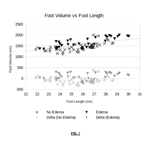
Figure 1 is a graph illustrating foot volume against foot length for a patient
with edema and a
patient without edema;
Figure 2 is a foot model created by 3D imaging of a foot without edema-
specific morphs (shown on
the left of the figure) and with edema specific morphs (shown on the right of
the figure) for a
patient.
Detailed Description.
In the course of evaluating the clinical effectiveness of our 3D limb volume
measurement system
described in the inventor's co-pending application (GB2542114B), the inventor
has surprisingly
found that it is possible to gain a remarkably quantitative single measurement
of fluid retention
indicative of edema in a limb by the difference in two volumes.
The first volume is that observed most directly from the patient's body part
of interest (e.g. by 3D
imaging, or water displacement, or estimation of cylindrical segment volumes
from multiple
circumference measurements, or any other method known in the art). The second
volume is that
calculated based on a mathematical relationship between certain measured
parameters on the patient
(e.g. patient height, weight, and bone lengths) and resulting in an estimate
of the volume of the
body part of interest, wherein the mathematical relationship provides an
estimate that is related to a
more general patient population, ideally without edema but with sufficient
variation in confounding
factors such as variation in body fat.
A simple example for the first volume measurement is to measure the volume, in
millilitres, of the
patient's foot up to a height of 20cm from the ground, by water displacement
when the patient
stands in a filled bucket of water. This provides an accurate (though not easy
to administer)
measurement of the total foot volume.
A simple example for the second volume measurement is to relate the volume of
the foot up to a
height of 20cm to the length of the foot, as measured from tip-of-big-toe to
back-of-heel, where the
volume estimate in millilitres is 60 times the foot-length in centimetres.
The difference of these two measurements, volume one minus volume two, gives a
number which is
surprisingly effective in classifying patients in a heart failure population
into those who were
determined by a clinician at an outpatient appointment as having pitting edema
present, and those
CA 03110604 2021-02-24
WO 2020/044277 PCT/IB2019/057279
who did not. Over 80% of patients clinically assessed as having pitting edema
had a positive
volume difference by this measure, whilst over 80% of patients clinically
assessed as not having
pitting edema, or presenting as healthy volunteers, had a negative volume
differences by this
measure. Moreover, the magnitude of this difference correlated well with the
subjective clinical
grading of degree of edema in patients.
Figure 1 displays a chart of the foot lengths and foot volumes split by
clinician assessment of edema
status.
Naturally a relationship based only on foot-length will be subject to error
resulting from body fat
percentage. More complex models including e.g. patient height and weight or
bioimpedance will
naturally provide better correlation with edema in the presence of e.g. body
fat as a confounding
factor.
An alternative, preferred approach is to use a single mathematical modelling
system to extract both
sets of volumes at once. For example, when using a 3D camera system it is
generally possible to fit
a deformable model to the observed 3D points collected from the camera. The
objective is generally
to allow certain model parameters, such as bone joint angles and lengths, to
adjust in order to
minimize the difference between points on the virtual model surface and the
observed 3D points.
Such a model also typically includes parameters that adjust the shape of the
model, often called
"morphs" in the 3D rendering community, which might for example alter the
volume of flesh
around the virtual bones and allow the volume of flesh to be scaled over a
range of numerical
values.
In building and testing such a model on the same heart failure patient
population, it was found that
certain of these "morphs" were very much more strongly correlated to
subjective clinical grading of
edema than others. For example, volume changes around the ankle that
essentially flatten out the
malleolus of the foot were found to correlate more strongly with clinical
grading of edema than with
patient BMI.
Figure 2 displays such three dimensional foot model, on the left without the
application of edema-
specific "morphs", and on the right with the application of these morphs, for
a particular patient.
The additional mass around the malleolus bone is particularly obvious in the
right model.
CA 03110604 2021-02-24
WO 2020/044277 PCT/IB2019/057279
6
Furthermore, a number of patients in the heart failure trial were monitored
whilst undergoing a
course of diuretic therapy, with the aim of reducing fluid retention and thus
edema. It was therefore
possible to observe which "morphs" correlated with change in foot volume over
time for a
particular patient, which implies a direct correlation to fluid retention
indicative of edema.
An approach to edema estimation was therefore enabled whereby the observed 3D
foot data was
modelled using the deformable model, with all morph parameters being allowed
to vary freely, in
order to provide the first volume. The second volume was then calculated by
resetting the values of
those morphs that were found to correlate with edema to population average
values. This enables
the general structure of the foot for this particular patient to be well
modelled, whilst estimating the
edema-free shape of the foot. The difference between the two volumes
calculated by this technique
was found to be particularly sensitive to changes in edema over time in the
patients underdoing
diuretic treatment, and also correlated well with the subjective clinical
evaluation of edema.
The difference between the two volumes can obviously be processed further to
provide various
useful indicators. For example, by expressing the percentage of patients with
and without clinician-
determined edema who exhibit more or less than various threshold volume
differences, it is possible
to estimate the probability that a patient exhibiting a particular volume
difference would be assessed
as having edema by a clinician. This is useful for risk-scoring and risk-
stratification of patients,
especially when combined with other clinical indicators that are also
probabilistic in nature.
This invention is not only useful in the clinic, in providing a numerical
measure of fluid retention
indicative of edema, but if implemented using suitable methods (such as 3D
cameras) could provide
a measure of a change in fluid retention from the patient's home. This could
not only save on
transport to hospital appointments for the patient or to the patient by the
clinical team, but if
implemented as part of a telemedicine system could provide alerts of worsening
patient health. Such
a system might plausibly reduce the presently very high repeat hospitalisation
rates for chronic
conditions such as heart failure, not only saving substantial healthcare
resources but providing
better quality of life for patients.
It will be obvious that a "difference" between volumes need not be a simple
subtractive difference,
but could also be ratiometric, or the result of any number of statistical
tests or other numerical
comparison.
CA 03110604 2021-02-24
WO 2020/044277 PCT/IB2019/057279
7
It will also be apparent that a multitude of volumetrically related model
outputs could be used in
embodiments of the invention as opposed to strictly using volume. For example,
the aforementioned
virtual 3D model could have the thickness or circumference virtually measured
at various locations,
and these virtual circumference measurements compared, or the surface-area of
3D models could be
compared. For the purposes of this invention, all of these types of comparison
are to be considered
as a non-limiting list of possible size comparisons within the meaning of the
claims.
