Note: Descriptions are shown in the official language in which they were submitted.
CA 03110749 2021-02-25
ANTI-PD-1/VEGFA BIFUNCTIONAL ANTIBODY,
PHARMACEUTICAL COMPOSITION THEREOF AND USE THEREOF
TECHNICAL FIELD
The present invention relates to the fields of tumor treatment and
immunobiology, particularly to an anti-PD-1/VEGFA bifunctional antibody, a
phaimaceutical composition thereof and use thereof. Specifically, the present
invention relates to an anti-human PD-1/human VEGFA bifunctional
antibody, a phaimaceutical composition thereof and use thereof.
BACKGROUND
Tumor, especially a malignant tumor, is a serious health-threatening disease
in the world today, and it is the second leading cause of death among various
diseases. In recent years, the incidence of the disease has been increasing
remarkably. Malignant tumor is characterized by poor treatment response,
high late metastasis rate and poor prognosis. Although conventional treatment
methods (such as radiotherapy, chemotherapy and surgical treatment) adopted
clinically at present alleviate the pain to a great extent and prolong the
survival time, the methods have great limitations, and it is difficult to
further
improve their efficacy.
There are two distinct stages of tumor growth, namely, from a slow growth
stage without blood vessels to a rapid proliferation stage with blood vessels.
The angiogenesis enables the tumor to acquire enough nutrition to complete
the blood vessel switching stage, and if there is no angiogenesis, the primary
tumor will be no more than 1-2 mm, and thus the metastasis cannot be
realized.
Vascular Endothelial Growth Factor (VEGF) is a growth factor which can
1
Date Recue/Date Received 2021-02-25
CA 03110749 2021-02-25
promote division and proliferation of endothelial cells, promote formation of
new blood vessels and improve blood vessel permeability, and it binds to
vascular endothelial growth factor receptors on the cell surface and plays a
role by activating tyrosine kinase signal transduction pathways. In tumor
tissues, tumor cells, and macrophages and mast cells invading into tumors can
secrete high-level VEGF, stimulate tumor vascular endothelial cells in a
paracrine form, promote proliferation and migration of endothelial cells,
induce angiogenesis, promote continuous growth of tumor, improve vascular
permeability, cause fibrin deposition in surrounding tissues, and promote
infiltration of mononuclear cells, fibroblast and endothelial cells, which
facilitates formation of tumor stroma and entry of tumor cells into new blood
vessels, and promote tumor metastasis. Therefore, inhibiting tumor
angiogenesis is considered to be one of the most promising tumor treatment
methods at present. The VEGF family includes: VEGFA, VEGFB, VEGFC,
VEGFD and PIGF. Vascular Endothelial Growth Factor Receptors (VEGFRs)
include VEGFR1 (also known as Fla), VEGFR2 (also known as KDR or
Flkl), VEGFR3 (also known as Flt4), and Neuropilin-1 (NRP-1). The first
three receptors are similar in structure, belong to a tyrosine kinase
superfamily, and are composed of an extramembrane region, a
transmembrane segment and an intramembrane region, where the
extramembrane region is composed of an immunoglobulin-like domain, and
the intramembrane region is a tyrosine kinase region. VEGFR1 and VEGFR2
are located primarily on the surface of vascular endothelial cells, and
VEGFR3 is located primarily on the surface of lymphatic endothelial cells.
Molecules of the VEGF family have different affinities for these receptors.
VEGFA mainly acts in combination with VEGFR1, VEGFR2 and NRP-1.
VEGFR1 is the earliest found receptor and has a higher affinity for VEGFA
than VEGFR2 under normal physiological conditions, but it has a lower
2
Date Recue/Date Received 2021-02-25
CA 03110749 2021-02-25
tyrosinase activity in intracellular segment than VEGFR2 (Ma Li, J. Chinese
Journal of Birth Health and Heredity, 24 (5): 146-148 (2016)).
VEGFR2 is the primary regulator of angiogenesis and vascular engineering,
and has a much higher tyrosine kinase activity than VEGFR1. VEGFR2, after
binding to ligand VEGFA, mediates the proliferation, differentiation and the
like of vascular endothelial cells, as well as the foimation process of blood
vessels and the peimeability of blood vessels (Roskoski R Jr. et al., Crit Rev
Oncol Hematol, 62(3): 179-213 (2007)). VEGFA, after binding to VEGFR2,
mediates the transcriptional expression of intracellular related protein genes
through the downstream PLC-y-PKC-Raf-MEK-MAPK signaling pathway,
and thus promotes the proliferation of vascular endothelial cells (Takahashi T
et al., Oncogene, 18(13): 2221-2230 (1999)).
VEGFR3 is one of the tyrosine kinase family members, and mainly expresses
embryonic vascular endothelial cell and adult lymphatic endothelial cells, and
VEGFC and VEGFD bind to VEGFR3 to stimulate proliferation and
migration of lymphatic endothelial cells and promote neogenesis of lymphatic
vessels; NRP-1 is a non-tyrosine kinase transmembrane protein and is
incapable of independently transducing biological signals, and it is able to
mediate signaling only after foiming a complex with a VEGF tyrosine kinase
receptor. (Ma Li, Chinese Journal of Birth Health and Heredity, 24(5):
146-148 (2016)).
VEGFA and VEGFR2 are mainly involved in regulation of angiogenesis,
where before and after the binding of VEGFA to VEGFR2, a cascade reaction
of numerous inteimediate signals in upstream and downstream pathways is
foimed, and finally the physiological functions are changed by proliferation,
survival, migration, peimeability increase and infiltration to peripheral
tissues, etc. of endothelial cells (Dong Hongchao et al., Sep. 2014, Journal
of
3
Date Recue/Date Received 2021-02-25
CA 03110749 2021-02-25
Modern Oncology, 22(9): 2231-3).
Currently, there are several humanized monoclonal antibodies targeting
human VEGF, particularly VEGFA, such as bevacizumab, which has been
approved by the U.S. Food and Drug Administration for the treatment of
various tumors such as non-small cell lung cancer, renal cell carcinoma,
cervical cancer, and metastatic colorectal cancer in succession during 2004.
The programmed cell death receptor-1 (PD-1), also known as CD279, is a
type I transmembrane glycoprotein membrane surface receptor, belongs to the
CD28 immunoglobulin superfamily, and is commonly expressed in T cells, B
cells, and myeloid cells. PD-1 has two natural ligands, PD-Li and PD-L2.
Both PD-Li and PD-L2 belong to the B7 superfamily and are expressed
constitutively or inducibly on the membrane surface a variety of cells,
including nonhematopoietic cells and a variety of tumor cells. PD-Li is
mainly expressed on T cells, B cells, DC and microvascular endothelial cells
and a variety of tumor cells, while PD-L2 is expressed only on antigen
presenting cells such as dendritic cells and macrophages. The interaction
between PD-1 and its ligands can inhibit the activation of lymph, the
proliferation of T cells, and the secretion of cytokines such as IL-2 and IFN-
y.
A large number of researches show that a tumor microenvironment can
protect tumor cells from being damaged by immune cells, expression of PD-1
in lymphocytes infiltrated in the tumor microenvironment is up-regulated, and
various primary tumor tissues are PD-Li positive in immunohistochemical
analysis, such as lung cancer, liver cancer, ovarian cancer, skin cancer,
colon
cancer and glioma. Meanwhile, the expression of PD-L1 in the tumor is
significantly correlated with poor prognosis of cancer patients. Blocking the
interaction between PD-1 and its ligands can promote the tumor-specific T
cell immunity and enhance the immune elimination efficiency of tumor cells.
A large number of clinical trials show that antibodies targeting PD-1 or
4
Date Recue/Date Received 2021-02-25
CA 03110749 2021-02-25
PD-Li can promote infiltration of CD8+ T cells into tumor tissues and
up-regulate anti-tumor immune effector factors such as IL-2, IFN-y,
granzyme B and perforin, thereby effectively inhibiting the growth of tumors.
In addition, anti-PD-1 antibodies may also be used in the treatment of viral
chronic infections. Viral chronic infections are often accompanied by a loss
of
function of virus-specific effector T cells and a reduction in its number. The
interaction between PD-1 and PD-Li can be blocked by injecting a PD-1
antibody, thereby effectively inhibiting the exhaustion of effector T cells in
viral chronic infection.
Due to the broad anti-tumor prospect and surprising efficacy of PD-1
antibodies, it is widely accepted in the industry that antibodies targeting
the
PD-1 pathway will bring about breakthroughs in the treatment of a variety of
tumors: for the treatment of non-small cell lung cancer, renal cell carcinoma,
ovarian cancer and melanoma (Hornet M. B., Parisi G., et al., Anti-PD-1
therapy in melanoma. Semin Oncol. 2015 Jun; 42(3): 466-473), and
lymphoma and anemia (Held SA, Heine A, et al., Advances in
immunotherapy of chronic myeloid leukemia CML. Curr Cancer Drug
Targets 2013 Sep; 13(7): 768-74).
The bifunctional antibody, also known as bispecific antibody, is a specific
medicament that targets two different antigens simultaneously, and can be
produced by immunoselection purification. In addition, the bispecific
antibody can also be produced by genetic engineering, which has certain
advantages due to corresponding flexibility in aspects such as the
optimization of binding sites, consideration of synthetic form, and yield.
Currently, the bispecific antibody has been demonstrated to exist in over 45
forms (Muller D, Kontermann RE. Bispecific antibodies for cancer
immunotherapy: current perspectives. BioDrugs 2010; 24: 89-98). A number
Date Recue/Date Received 2021-02-25
CA 03110749 2021-02-25
of bispecific antibodies have been developed in the fonn of IgG-ScFv,
namely the Morrison fonn (Coloma M. J., Morrison S. L. Design and
production of novel tetravalent bispecific antibodies. Nat Biotechnol., 1997;
15: 159-163), which has been demonstrated to be one of the ideal fonns of the
bispecific antibodies because of its similarity to the naturally existing IgG
fonn and advantages in antibody engineering, expression and purification
(Miller B. R., Demarest S. J., et al., Stability engineering of scFvs for the
development of bispecific and multivalent antibodies. Protein Eng Des Sel
2010; 23: 549-57; Fitzgerald J, Lugovskoy A. Rational engineering of
antibody therapeutics targeting multiple oncogene pathways. MA& 2011; 3:
299-309).
Currently, there is a need to develop a bifunctional antibody medicament
targeting both PD-1 and VEGF (e.g., VEGFA).
SUMMARY
Through in-depth research and creative efforts, and based on commercially
available VEGFA monoclonal antibody Avastin (bevacizumab) and
14C12H1L1 acquired before (see Chinese patent publication No.
CN106977602A), the inventors has acquired a humanized bifunctional
antibody named VP101, which is capable of simultaneously binding to
VEGFA and PD-1, and blocking the binding of VEGFA to VEGFR2 and that
of PD-1 to PD-Li.
The inventors have surprisingly found that VP101 is capable of:
effectively binding to PD-1 on the surface of human immune cells, relieving
immunosuppression mediated by PD-Li and PD-1, and promoting secretion
of IFN-y and IL-2 by human immune cells;
effectively inhibiting VEGFA-induced proliferation of vascular endothelial
6
Date Recue/Date Received 2021-02-25
CA 03110749 2021-02-25
cells, and thereby inhibiting tumor-induced angiogenesis, and/or
having the potential of being used for preparing medicaments for preventing
and treating malignant tumors such as liver cancer, lung cancer, melanoma,
renal tumor, ovarian cancer and lymphoma.
The present invention is detailed below.
One aspect of the present invention relates to a bispecific antibody, which
comprises:
a first protein functional region targeting VEGFA, and
a second protein functional region targeting PD-1,
preferably,
the first protein functional region is an anti-VEGFA antibody or an
antigen-binding fragment thereof, a heavy chain variable region of the
anti-VEGFA antibody comprising HCDR1-HCDR3 with amino acid
sequences set forth in SEQ ID NOs: 15-17 respectively, and a light chain
variable region of the anti-VEGFA antibody comprising LCDR1-LCDR3
with amino acid sequences set forth in SEQ ID NOs: 18-20 respectively; and
the second protein functional region is an anti-PD-1 antibody or an
antigen-binding fragment thereof, a heavy chain variable region of the
anti-PD-1 antibody comprising HCDR1-HCDR3 with amino acid sequences
set forth in SEQ ID NOs: 21-23 respectively, and a light chain variable region
of the anti-PD-1 antibody comprising LCDR1-LCDR3 with amino acid
sequences set forth in SEQ ID NOs: 24-26 respectively.
In some embodiments of the present invention, the bispecific antibody is
provided, wherein,
the anti-VEGFA antibody or the antigen-binding fragment thereof is selected
7
Date Recue/Date Received 2021-02-25
CA 03110749 2021-02-25
from Fab, Fab', F(ab')2, Fd, Fv, dAb, a complementarity determining region
fragment, a single chain antibody, a humanized antibody, a chimeric
antibody, and a diabody,
and/or,
the anti-PD-1 antibody or the antigen-binding fragment thereof is selected
from Fab, Fab', F(ab')2, Fd, Fv, dAb, a complementarity determining region
fragment, a single chain antibody, a humanized antibody, a chimeric
antibody, and a diabody.
In some embodiments of the present invention, the bispecific antibody is in
IgG-scFv foiiii.
In some embodiments of the present invention, the first protein functional
region is an immunoglobulin, a heavy chain variable region of the
immunoglobulin comprising HCDR1-HCDR3 with amino acid sequences set
forth in SEQ ID NOs: 15-17 respectively, and a light chain variable region of
the immunoglobulin comprising LCDR1-LCDR3 with amino acid sequences
set forth in SEQ ID NOs: 18-20 respectively; and the second protein
functional region is a single chain antibody, a heavy chain variable region of
the single chain antibody comprising HCDR1-HCDR3 with amino acid
sequences set forth in SEQ ID NOs: 21-23 respectively, and a light chain
variable region of the single chain antibody comprising LCDR1-LCDR3 with
amino acid sequences set forth in SEQ ID NOs: 24-26 respectively;
or,
the first protein functional region is a single chain antibody, a heavy chain
variable region of the single chain antibody comprising HCDR1-HCDR3 with
amino acid sequences set forth in SEQ ID NOs: 21-23 respectively, and a
light chain variable region of the single chain antibody comprising
8
Date Recue/Date Received 2021-02-25
CA 03110749 2021-02-25
LCDR1-LCDR3 with amino acid sequences set forth in SEQ ID NOs: 24-26
respectively; and the second protein functional region is an immunoglobulin,
a heavy chain variable region of the immunoglobulin comprising
HCDR1-HCDR3 with amino acid sequences set forth in SEQ ID NOs: 15-17
respectively, and a light chain variable region of the immunoglobulin
comprising LCDR1-LCDR3 with amino acid sequences set forth in SEQ ID
NOs: 18-20 respectively.
In a specific embodiment of the present invention, a bispecific antibody is
provided, which comprises:
a first protein functional region targeting VEGFA, and
a second protein functional region targeting PD-1,
wherein,
the first protein functional region is an immunoglobulin, a heavy chain
variable region of the immunoglobulin comprising HCDR1-HCDR3 with
amino acid sequences set forth in SEQ ID NOs: 15-17 respectively, and a
light chain variable region of the immunoglobulin comprising
LCDR1-LCDR3 with amino acid sequences set forth in SEQ ID NOs: 18-20
respectively; and the second protein functional region is a single chain
antibody, a heavy chain variable region of the single chain antibody
comprising HCDR1-HCDR3 with amino acid sequences set forth in SEQ ID
NOs: 21-23 respectively, and a light chain variable region of the single chain
antibody comprising LCDR1-LCDR3 with amino acid sequences set forth in
SEQ ID NOs: 24-26 respectively;
or,
the first protein functional region is a single chain antibody, a heavy chain
variable region of the single chain antibody comprising HCDR1-HCDR3 with
9
Date Recue/Date Received 2021-02-25
CA 03110749 2021-02-25
amino acid sequences set forth in SEQ ID NOs: 21-23 respectively, and a
light chain variable region of the single chain antibody comprising
LCDR1-LCDR3 with amino acid sequences set forth in SEQ ID NOs: 24-26
respectively; and the second protein functional region is an immunoglobulin,
a heavy chain variable region of the immunoglobulin comprising
HCDR1-HCDR3 with amino acid sequences set forth in SEQ ID NOs: 15-17
respectively, and a light chain variable region of the immunoglobulin
comprising LCDR1-LCDR3 with amino acid sequences set forth in SEQ ID
NOs: 18-20 respectively.
In some embodiments of the present invention, the bispecific antibody is
provided, wherein,
the amino acid sequence of the heavy chain variable region of the
immunoglobulin is set forth in SEQ ID NO: 5, and the amino acid sequence
of the light chain variable region of the immunoglobulin is set forth in SEQ
ID NO: 7; and the amino acid sequence of the heavy chain variable region of
the single chain antibody is set forth in SEQ ID NO: 9, and the amino acid
sequence of the light chain variable region of the single chain antibody is
set
forth in SEQ ID NO: 11;
or,
the amino acid sequence of the heavy chain variable region of the single chain
antibody is set forth in SEQ ID NO: 9, and the amino acid sequence of the
light chain variable region of the single chain antibody is set forth in SEQ
ID
NO: 11; and the amino acid sequence of the heavy chain variable region of
the immunoglobulin is set forth in SEQ ID NO: 5, and the amino acid
sequence of the light chain variable region of the immunoglobulin is set forth
in SEQ ID NO: 7.
Date Recue/Date Received 2021-02-25
CA 03110749 2021-02-25
In some embodiments of the present invention, the bispecific antibody is
provided, wherein,
the immunoglobulin comprises a non-CDR region derived from a species
other than murine, such as from a human antibody.
In some embodiments of the present invention, the bispecific antibody is
provided, wherein,
the immunoglobulin comprises constant regions derived from a human
antibody;
preferably, the constant regions of the immunoglobulin are selected from
constant regions of human IgGl, IgG2, IgG3, and IgG4.
In some embodiments of the present invention, the bispecific antibody is
provided, wherein,
the heavy chain constant region of the immunoglobulin is human Ig gamma-1
chain C region or human Ig gamma-4 chain C region, and its light chain
constant region is human Ig kappa chain C region.
In some embodiments of the present invention, the constant regions of the
immunoglobulin are humanized. For example, each heavy chain constant
region is Ig gamma-1 chain C region, ACCESSION: P01857, and each light
chain constant region is Ig kappa chain C region, ACCESSION: P01834.
In some embodiments of the present invention, the bispecific antibody is
provided, wherein the first protein functional region and the second protein
functional region are linked directly or via a linker fragment;
preferably, the linker fragment is (GGGGS)m, wherein m is a positive integer
such as 1, 2, 3, 4, 5, or 6, and GGGGS (SEQ ID NO: 14) is a constituent unit
of the linker.
11
Date Recue/Date Received 2021-02-25
CA 03110749 2021-02-25
In some embodiments of the present invention, the bispecific antibody is
provided, wherein the numbers of the first protein functional region and the
second protein functional region are each independently 1, 2 or more.
In some embodiments of the present invention, the bispecific antibody is
provided, wherein 1 immunoglobulin and 2 single chain antibodies,
preferably two identical single chain antibodies, are present.
In some embodiments of the present invention, the bispecific antibody is
provided, wherein the immunoglobulin is an IgG, IgA, IgD, IgE, or IgM,
preferably an IgG, such as an IgGl, IgG2, IgG3 or IgG4.
In some embodiments of the present invention, the bispecific antibody is
provided, wherein the single chain antibody is linked to the C-tenninus of the
heavy chain of the immunoglobulin. Since an immunoglobulin has two heavy
chains, two single chain antibody molecules are linked to one
immunoglobulin molecule. Preferably, the two single chain antibody
molecules are identical.
In some embodiments of the present invention, the bispecific antibody is
provided, wherein two single chain antibodies are present, and one tenninus
of each single chain antibody is linked to the C-tenninus or the N-tenninus of
one of the two heavy chains of the immunoglobulin.
In some embodiments of the present invention, a disulfide bond is present
between the VH and the VL of the single chain antibody. Methods for
introducing a disulfide bond between the VH and VL of an antibody are well
known in the art, see, for example, US 5,747,654; Rajagopal et al., Prot.
Engin. 10(1997)1453-1459; Reiter et al., Nat. Biotechnol.
14(1996)1239-1245; Reiter et al., Protein Engineering 8(1995)1323-1331;
Webber et al., Molecular Immunology 32(1995)249-258; Reiter et al.,
12
Date Recue/Date Received 2021-02-25
CA 03110749 2021-02-25
Immunity 2(1995)281-287; Reiter et al., JBC 269(1994)18327-18331; Reiter
et al., Inter. J. of Cancer 58(1994)142-149; or Reiter et al., Cancer Res.
54(1994)2714-2718, which are incorporated herein by reference.
In some embodiments of the present invention, the bispecific antibody is
provided, wherein the bispecific antibody binds to a VEGFA protein and/or a
PD-1 protein with a KD of less than 10-5 M, such as less than 10-6 M, 10-7 M,
10-8M, i0-9 M or 10-10 M or less; preferably, the KD is measured by a Fortebio
molecular interaction instrument.
In some embodiments of the present invention, the bispecific antibody is
provided, wherein,
the bispecific antibody binds to the VEGFA protein with an EC50 of less than
1 nM, less than 0.5 nM, less than 0.2 nM, less than 0.15 nM, or less than 0.14
nM, preferably, the EC50 is detected by indirect ELISA,
and/or,
the bispecific antibody binds to the PD-1 protein with an EC50 of less than 1
nM, less than 0.5 nM, less than 0.2 nM, less than 0.17 nM, less than 0.16 nM,
or less than 0.15 nM, preferably, the EC50 is detected by indirect ELISA.
Another aspect of the present invention relates to an isolated nucleic acid
molecule encoding the bispecific antibody according to any embodiment of
the present invention.
The present invention also relates to a vector comprising the isolated nucleic
acid molecule of the present invention.
The present invention also relates to a host cell comprising the isolated
nucleic acid molecule of the present invention or comprising the vector of the
present invention.
13
Date Recue/Date Received 2021-02-25
CA 03110749 2021-02-25
Another aspect of the present invention relates to a method for preparing the
bispecific antibody according to any embodiment of the present invention,
which comprises culturing the host cell of the present invention in a suitable
condition and isolating the bispecific antibody from the cell cultures.
Another aspect of the present invention relates to a conjugate, comprising a
bispecific antibody and a conjugated moiety, wherein the bispecific antibody
is the bispecific antibody according to any embodiment of the present
invention, and the conjugated moiety is a detectable label; preferably, the
conjugated moiety is a radioisotope, a fluorescent substance, a luminescent
substance, a colored substance, or an enzyme.
Another aspect of the present invention relates to a kit comprising the
bispecific antibody according to any embodiment of the present invention or
comprising the conjugate of the present invention;
preferably, the kit further comprises a second antibody capable of
specifically
binding to the bispecific antibody; optionally, the second antibody further
comprises a detectable label, such as a radioisotope, a fluorescent substance,
a
luminescent substance, a colored substance, or an enzyme.
Another aspect of the present invention relates to use of the bispecific
antibody according to any embodiment of the present invention in preparing a
kit for detecting the presence or level of VEGFA and/or PD-1 in a sample.
Another aspect of the present invention relates to a phamiaceutical
composition comprising the bispecific antibody according to any embodiment
of the present invention or comprising the conjugate of the present invention;
optionally, it further comprises a phaimaceutically acceptable excipient.
The bispecific antibody of the present invention or the phannaceutical
composition of the present invention may be fonnulated into any dosage fonn
14
Date Recue/Date Received 2021-02-25
CA 03110749 2021-02-25
known in the phaimaceutical field, such as tablet, pill, suspension, emulsion,
solution, gel, capsule, powder, granule, elixir, troche, suppository,
injection
(including injection solution, sterile powder for injection and concentrated
solution for injection), inhalant, and spray. The preferred dosage foim
depends on the intended mode of administration and therapeutic use. The
phaimaceutical composition of the present invention should be sterile and
stable under the conditions of manufacture and storage. One preferred dosage
foim is an injection. Such injections may be sterile injection solutions. For
example, sterile injection solutions can be prepared by the following method:
a necessary amount of the bispecific antibody of the present invention is
added in an appropriate solvent, and optionally, other desired ingredients
(including, but not limited to, pH regulators, surfactants, adjuvants, ionic
strength enhancers, isotonic agents, preservatives, diluents, or any
combination thereof) are added at the same time, followed by filtration and
sterilization. In addition, sterile injection solutions can be prepared as
sterile
lyophilized powders (e.g., by vacuum drying or lyophilizing) for convenient
storage and use. Such sterile lyophilized powders may be dispersed in a
suitable carrier (e.g., sterile pyrogen-free water) prior to use.
In addition, the bispecific antibody of the present invention may be present
in
a phaimaceutical composition in unit dose foim for ease of administration. In
some embodiments, the unit dose is at least 1 mg, at least 5 mg, at least 10
mg, at least 15 mg, at least 20 mg, at least 25 mg, at least 30 mg, at least
45
mg, at least 50 mg, at least 75 mg, or at least 100 mg. Where the
phaimaceutical composition is in a liquid (e.g., injection) dosage fonn, it
may
comprise the bispecific antibody of the present invention at a concentration
of
at least 0.1 mg/mL, such as at least 0.25 mg/mL, at least 0.5 mg/mL, at least
1
mg/mL, at least 2.5 mg/mL, at least 5 mg/mL, at least 8 mg/mL, at least 10
mg/mL, at least 15 mg/mL, at least 25 mg/mL, at least 50 mg/mL, at least 75
Date Recue/Date Received 2021-02-25
CA 03110749 2021-02-25
mg/mL, or at least 100 mg/mL.
The bispecific antibody or the pharmaceutical composition of the present
invention may be administered by any suitable method known in the art,
including, but not limited to, oral, buccal, sublingual, ocular, topical,
parenteral, rectal, intrathecal, intracisternal, inguinal, intravesical,
topical
(e.g., powder, ointment, or drop), or nasal route. However, for many
therapeutic uses, the preferred route/mode of administration is parenteral
(such as intravenous injection, subcutaneous injection, intraperitoneal
injection, and intramuscular injection). Those skilled in the art will
appreciate
that the route and/or mode of administration will vary depending on the
intended purpose. In a preferred embodiment, the bispecific antibody or the
pharmaceutical composition of the present invention is administered by
intravenous infusion or injection.
The bispecific antibody or the pharmaceutical composition provided herein
can be used alone or in combination, or used in combination with additional
pharmaceutically active agents (e.g., a tumor chemotherapeutic drug). Such
an additional pharmaceutically active agent may be administered prior to,
concurrently with, or subsequent to the administration of the bispecific
antibody of the present invention or the pharmaceutical composition of the
present invention.
In the present invention, the administration regimen may be adjusted to
achieve the optimal desired response (e.g., a therapeutic or prophylactic
response). For example, it may be a single administration, may be multiple
administrations over a period of time, or may be characterized by reducing or
increasing the dose proportionally with the emergency degree of the
treatment.
Another aspect of the present invention relates to use of the bispecific
16
Date Recue/Date Received 2021-02-25
CA 03110749 2021-02-25
antibody according to any embodiment of the present invention or the
conjugate of the present invention in preparing a medicament for preventing
and/or treating a malignant tumor, wherein preferably, the malignant tumor is
selected from colon cancer, rectal cancer, lung cancer such as non-small cell
lung cancer, liver cancer, ovarian cancer, skin cancer, glioma, melanoma,
renal tumor, prostate cancer, bladder cancer, gastrointestinal cancer, breast
cancer, brain cancer and leukemia.
Another aspect of the present invention relates to use of the bispecific
antibody according to any embodiment of the present invention or the
conjugate of the present invention in preparing:
(1)
a medicament or an agent for detecting the level of VEGFA in a sample,
a medicament or an agent for blocking binding of VEGFA to VEGFR2,
a medicament or an agent for down-regulating the activity or level of
VEGFA,
a medicament or an agent for relieving the stimulation of VEGFA on vascular
endothelial cell proliferation,
a medicament or an agent for inhibiting vascular endothelial cell
proliferation,
or
a medicament or an agent for blocking tumor angiogenesis,
and/or
(2)
a medicament or an agent for blocking the binding of PD-1 to PD-L1,
a medicament or an agent for down-regulating the activity or level of PD-1,
17
Date Recue/Date Received 2021-02-25
CA 03110749 2021-02-25
a medicament or an agent for relieving the immunosuppression of PD-1 in an
organism,
a medicament or an agent for promoting IFN-ysecretion in T lymphocytes, or
a medicament or an agent for promoting IL-2 secretion in T lymphocytes.
In one embodiment of the present invention, the use is non-therapeutic and/or
non-diagnostic.
Another aspect of the present invention relates to an in vivo or in vitro
method
comprising administering to a cell an effective amount of the bispecific
antibody according to any embodiment of the present invention or the
conjugate of the present invention, and the method is selected from:
(1)
a method for detecting the level of VEGFA in a sample,
a method for blocking the binding of VEGFA to VEGFR2,
a method for down-regulating the activity or level of VEGFA,
a method for relieving the stimulation of VEGFA on vascular
endothelial cell proliferation,
a method for inhibiting vascular endothelial cell proliferation, or
a method for blocking tumor angiogenesis,
and/or
(2)
a method for blocking the binding of PD-1 to PD-L1,
a method for down-regulating the activity or level of PD-1,
a method for relieving the immunosuppression of PD-1 in an
18
Date Recue/Date Received 2021-02-25
CA 03110749 2021-02-25
organism,
a method for promoting IFN-y secretion in T lymphocytes, or
a method for promoting IL-2 secretion in T lymphocytes.
In one embodiment of the present invention, the in vitro method is
non-therapeutic and/or non-diagnostic.
In the in vitro experiment of the present invention, the anti-VEGFA antibody
and the anti-VEGFA/PD-1 bifunctional antibody both can inhibit HUVEC
cell proliferation, and the anti-PD-1 antibody and the anti-VEGFA/PD-1
bifunctional antibody both can promote the secretion of IFN-y and/or IL-2
and activate immune reaction.
Another aspect of the present invention relates to a method for preventing
and/or treating a malignant tumor, comprising administering to a subject in
need an effective amount of the bispecific antibody according to any
embodiment of the present invention or the conjugate of the present
invention, wherein preferably, the malignant tumor is selected from colon
cancer, rectal cancer, lung cancer such as non-small cell lung cancer, liver
cancer, ovarian cancer, skin cancer, glioma, melanoma, renal tumor, prostate
cancer, bladder cancer, gastrointestinal cancer, breast cancer, brain cancer
and
leukemia.
A typical non-limiting range of a therapeutically or prophylactically
effective
amount of the bispecific antibody of the present invention is 0.02-50 mg/kg,
such as 0.1-50 mg/kg, 0.1-25 mg/kg, or 1-10 mg/kg. It should be noted that
the dose may vary with the type and severity of the symptom to be treated.
Furthennore, those skilled in the art will appreciate that for any particular
patient, the particular administration regimen will be adjusted over time
according to the needs of the patient and the professional judgment of the
19
Date Recue/Date Received 2021-02-25
CA 03110749 2021-02-25
physician; the dose ranges given herein are for illustrative purpose only and
do not limit the use or scope of the pharmaceutical composition of the present
invention.
In the present invention, the subject may be a mammal, such as a human.
Provided is the bispecific antibody or the conjugate according to any
embodiment of the present invention for use in preventing and/or treating a
malignant tumor, wherein preferably, the malignant tumor is selected from
colon cancer, rectal cancer, lung cancer such as non-small cell lung cancer,
liver cancer, ovarian cancer, skin cancer, glioma, melanoma, renal tumor,
prostate cancer, bladder cancer, gastrointestinal cancer, breast cancer, brain
cancer and leukemia.
Provided is the bispecific antibody or conjugate according to any embodiment
of the present invention for use in:
(1)
detecting the level of VEGFA in a sample,
blocking the binding of VEGFA to VEGFR2,
down-regulating the activity or level of VEGFA,
relieving the stimulation of VEGFA on vascular endothelial cell proliferation,
inhibiting vascular endothelial cell proliferation, or
blocking tumor angiogenesis,
and/or
(2)
blocking the binding of PD-1 to PD-Li,
Date Recue/Date Received 2021-02-25
CA 03110749 2021-02-25
down-regulating the activity or level of PD-1,
relieving the immuno suppression of PD-1 in an organism,
promoting IFN-y secretion in T lymphocytes, or
promoting IL-2 secretion in T lymphocytes.
Antibody drugs, especially monoclonal antibodies, have achieved good
efficacy in the treatment of various diseases. Traditional experimental
methods for acquiring these therapeutic antibodies are to immunize animals
with the antigen and acquire antibodies targeting the antigen in the
immunized animals, or to improve those antibodies with lower affinity for the
antigen by affinity maturation.
The variable regions of the light chain and the heavy chain determine the
binding of the antigen; the variable region of each chain contains three
hypervariable regions called Complementarily Determining Regions (CDRs)
(CDRs of the heavy chain (H Chain) comprise HCDR1, HCDR2, and
HCDR3, and CDRs of the light chain (L Chain) comprise LCDR1, LCDR2,
and LCDR3, which are named by Kabat et al., see Bethesda M.d., Sequences
of Proteins of Immunological Interest, Fifth Edition, NTH Publication (1-3)
1991: 91-3242).
Preferably, CDRs may also be defined by the IMGT numbering system, see
Ehrenmann, Francois, Quentin Kaas, and Marie-Paule Lefranc.
"IMGT/3Dstructure-DB and IMGT/DomainGapAlign: a database and a tool
for immunoglobulins or antibodies, T cell receptors, MHC, IgSF and
MhcSF." Nucleic acids research 38.suppl 1 (2009): D301-D307.
The amino acid sequences of the CDR regions of the monoclonal antibody
21
Date Recue/Date Received 2021-02-25
CA 03110749 2021-02-25
sequences in (1) to (13) below were analyzed by technical means well known
to those skilled in the art, for example by VBASE2 database and according to
the IMGT definition, and the results are as follows:
(1) Bevacizumab
The amino acid sequence of the heavy chain variable region is set forth in
SEQ ID NO: 5, and the amino acid sequence of the light chain variable region
is set forth in SEQ ID NO: 7.
The amino acid sequences of the 3 CDR regions of its heavy chain variable
region are as follows:
HCDR1: GYTFTNYG (SEQ ID NO: 15)
HCDR2: INTYTGEP (SEQ ID NO: 16)
HCDR3: AKYPHYYGSSHWYFDV (SEQ ID NO: 17)
The amino acid sequences of the 3 CDR regions of its light chain variable
region are as follows:
LCDR1: QDISNY (SEQ ID NO: 18)
LCDR2: FTS (SEQ ID NO: 19)
LCDR3: QQYSTVPWT (SEQ ID NO: 20)
(2) 14C12H1L1
The amino acid sequence of the heavy chain variable region is set forth in
SEQ ID NO: 9, and the amino acid sequence of the light chain variable region
is set forth in SEQ ID NO: 11.
The amino acid sequences of the 3 CDR regions of its heavy chain variable
region are as follows:
22
Date Recue/Date Received 2021-02-25
CA 03110749 2021-02-25
HCDR1: GFAFSSYD (SEQ ID NO: 21)
HCDR2: ISGGGRYT (SEQ ID NO: 22)
HCDR3: ANRYGEAWFAY (SEQ ID NO: 23)
The amino acid sequences of the 3 CDR regions of its light chain variable
region are as follows:
LCDR1: QDINTY (SEQ ID NO: 24)
LCDR2: RAN (SEQ ID NO: 25)
LCDR3: LQYDEFPLT (SEQ ID NO: 26)
(3) VP101
The amino acid sequences of the 9 CDR regions of its heavy chains are as
follows:
HCDR1: GYTFTNYG (SEQ ID NO: 15)
HCDR2: INTYTGEP (SEQ ID NO: 16)
HCDR3: AKYPHYYGSSHWYFDV (SEQ ID NO: 17)
HCDR4: GFAFSSYD (SEQ ID NO: 21)
HCDR5: ISGGGRYT (SEQ ID NO: 22)
HCDR6: ANRYGEAWFAY (SEQ ID NO: 23)
HCDR7: QDINTY (SEQ ID NO: 24)
HCDR8: RAN (SEQ ID NO: 25)
HCDR9: LQYDEFPLT (SEQ ID NO: 26)
The amino acid sequences of the 3 CDR regions of its light chain variable
region are as follows:
23
Date Recue/Date Received 2021-02-25
CA 03110749 2021-02-25
LCDR1: QDISNY (SEQ ID NO: 18)
LCDR2: FTS (SEQ ID NO: 19)
LCDR3: QQYSTVPWT (SEQ ID NO: 20)
In the present invention, unless otherwise defined, the scientific and
technical
telins used herein have the meanings generally understood by those skilled in
the art. In addition, the laboratory operations of cell culture, molecular
genetics, nucleic acid chemistry and immunology used herein are the routine
procedures widely used in the corresponding fields. Meanwhile, in order to
better understand the present invention, the definitions and explanations of
the
relevant telins are provided below.
As used herein, when referring to the amino acid sequence of VEGFA protein
(GenBank ID: NP 001165097.1), it includes the full length of the VEGFA
protein, as well as a fusion protein of VEGFA, such as a fragment fused to an
Fc protein fragment of mouse or human IgG (mFc or hFc). However, those
skilled in the art will appreciate that in the amino acid sequence of the
VEGFA protein, mutations or variations (including but not limited to,
substitutions, deletions and/or additions) can be naturally generated or
artificially introduced without affecting biological functions thereof.
Therefore, in the present invention, the telin "VEGFA protein" should include
all such sequences, including their natural or artificial variants. In
addition,
when describing the sequence fragment of the VEGFA protein, it also
includes the corresponding sequence fragments in its natural or artificial
variants. In one embodiment of the present invention, the amino acid
sequence of the VEGFA protein is shown as the underlined part of SEQ ID
NO: 1 (without the last 6 His, a total of 302 amino acids).
As used herein, when referring to the amino acid sequence of VEGFR2
24
Date Recue/Date Received 2021-02-25
CA 03110749 2021-02-25
protein (also known as KDR, GenBank ID: NP 002244), it includes the full
length of the VEGFR2 protein, or the extracellular fragment VEGFR2-ECD
of VEGFR2, or a fragment comprising VEGFR2-ECD, and it also includes a
fusion protein of VEGFR2-ECD, such as a fragment fused to an Fc protein
fragment of mouse or human IgG (mFc or hFc). However, those skilled in the
art will appreciate that in the amino acid sequence of the VEGFR2 protein,
mutations or variations (including but not limited to, substitutions,
deletions
and/or additions) can be naturally generated or artificially introduced
without
affecting biological functions thereof. Therefore, in the present invention,
the
teitn "VEGFR2 protein" should include all such sequences, including their
natural or artificial variants. In addition, when describing the sequence
fragment of the VEGFR2 protein, it also includes the corresponding sequence
fragments in its natural or artificial variants. In one embodiment of the
present
invention, the amino acid sequence of the extracellular fragment
VEGFR2-ECD of VEGFR2 is shown as the wavy-underlined part of SEQ ID
NO: 4 (766 amino acids).
As used herein, unless otherwise specified, the VEGFR is VEGFR1 and/or
VEGFR2, specific protein sequence thereof is a sequence known in the prior
art, and reference may be made to the sequence disclosed in the existing
literature or GenBank. For example, VEGFR1 (VEGFR1, NCBI Gene ID:
2321); VEGFR2 (VEGFR2, NCBI Gene ID: 3791).
As used herein, when referring to the amino acid sequence of PD-1 protein
(Programmed cell death protein 1, NCBI GenBank: NM 005018), it includes
the full length of the PD-1 protein, or the extracellular fragment PD-1ECD of
PD-1 or a fragment comprising PD-1ECD, and it also includes a fusion
protein of PD-1ECD, such as a fragment fused to an Fc protein fragment of a
mouse or human IgG (mFc or hFc). However, it will be appreciated by those
skilled in the art that in the amino acid sequence of PD-1 protein, mutations
or
variations (including but not limited to substitutions, deletions and/or
Date Recue/Date Received 2021-02-25
CA 03110749 2021-02-25
additions) can be naturally generated or artificially introduced without
affecting biological functions thereof. Therefore, in the present invention,
the
tenn "PD-1 protein" should include all such sequences, including their natural
or artificial variants. In addition, when describing the sequence fragment of
the PD-1 protein, it also includes the corresponding sequence fragments in its
natural or artificial variants.
As used herein, the tenn ECso refers to the concentration for 50% of maximal
effect, i.e. the concentration that can cause 50% of the maximal effect.
As used herein, the telin "antibody" refers to an immunoglobulin molecule
that generally consists of two pairs of polypeptide chains (each pair with one
"light" (L) chain and one "heavy" (H) chain). In a general sense, the heavy
chain can be interpreted as a polypeptide chain with a larger molecular weight
in an antibody, and the light chain refers to a polypeptide chain with a
smaller
molecular weight in an antibody. Light chains are classified as K and A light
chains. Heavy chains are generally classified as la, 6, y, a, or E, and
isotypes of
antibodies are defined as IgM, IgD, IgG, IgA, and IgE, respectively. In light
chains and heavy chains, the variable region and constant region are linked by
a "J" region of about 12 or more amino acids, and the heavy chain also
comprises a "D" region of about 3 or more amino acids. Each heavy chain
consists of a heavy chain variable region (VH) and a heavy chain constant
region (CH). The heavy chain constant region consists of 3 domains (CHi, CH2,
and CH3). Each light chain consists of a light chain variable region (VL) and
a
light chain constant region (CL). The light chain constant region consists of
one domain CL. The constant region of the antibody can mediate the binding
of immunoglobulins to host tissues or factors, including the binding of
various cells of the immune system (e.g., effector cells) to the first
component
(Cl q) of classical complement system. The VH and VL regions can be further
subdivided into highly variable regions (called Complementarity Determining
26
Date Recue/Date Received 2021-02-25
CA 03110749 2021-02-25
Regions (CDRs)), between which conservative regions called framework
regions (FRs) are distributed. Each VH and VI, consists of 3 CDRs and 4 FRs
arranged from amino teiminus to carboxyl terminus in the following order:
FR1, CDR1, FR2, CDR2, FR3, CDR3, FR4. The variable regions (VH and
VI) of each heavy chain/light chain pair foim antibody binding sites,
respectively. The assignment of amino acids to the regions or domains may be
based on Kabat Sequences of Proteins of Immunological Interest (National
Institutes of Health, Bethesda, Md. (1987 and 1991)), or Chothia & Lesk J.
Mol. Biol. 196(1987): 901-917; Chothia et al. Nature 342(1989): 878-883 or
the definition of IMGT numbering system, see Ehrenmann, Francois, Quentin
Kaas, and Marie-Paule Lefranc. "IMGT/3Dstructure-DB and
IMGT/DomainGapAlign: a database and a tool for immunoglobulins or
antibodies, T cell receptors, MHC, IgSF and MhcSF." Nucleic acids research
38.suppl 1 (2009): D301-D307. In particular, the heavy chain may also
comprise more than 3 CDRs, such as 6, 9, or 12. For example, in the
bispecific antibody of the present invention, the heavy chain may be a ScFv
with the C-teiminus of the heavy chain of IgG antibody linked to another
antibody, and in this case, the heavy chain comprises 9 CDRs. The teim
"antibody" is not limited by any specific method for producing antibody. For
example, the antibody includes, in particular, a recombinant antibody, a
monoclonal antibody, and a polyclonal antibody. Antibodies can be different
isotypes, such as antibody IgG (e.g., subtype IgGl, IgG2, IgG3 or IgG4),
IgAl, IgA2, IgD, IgE or IgM.
As used herein, the temi "antigen binding fragment", also known as the
"antigen binding portion", refers to a polypeptide comprising the fragment of
a full-length antibody, which maintains the ability to specifically bind to
the
same antigen to which the full-length antibody binds, and/or competes with
the full-length antibody for the specific binding to an antigen. See
generally,
27
Date Recue/Date Received 2021-02-25
CA 03110749 2021-02-25
Fundamental Immunology, Ch. 7 (Paul, W., ed., 2nd edition, Raven Press,
N.Y. (1989), which is incorporated by reference herein in its entirety for all
purposes. An antigen-binding fragment of an antibody can be produced by
recombinant DNA technique or by enzymatic or chemical cleavage of an
intact antibody. In some cases, the antigen binding fragment includes Fab,
Fab', F (ab')2, Fd, Fv, dAb, and complementarity detennining region (CDR)
fragment, single chain antibody fragment (e.g., scFv), chimeric antibody,
diabody and polypeptide that comprises at least a portion of an antibody
sufficient to impart specific antigen binding ability to a polypeptide.
As used herein, the tenn "Fd fragment" refers to an antibody fragment
consisting of Vil and Cm domains; the tenn "Fv fragment" refers to an
antibody fragment consisting of the VL and Vil domains of a single aim of an
antibody; the tenn "dAb fragment" refers to an antibody fragment consisting
of a Vil domain (Ward et al., Nature 341 (1989):544-546), the tenn "Fab
fragment" refers to an antibody fragment consisting of VL, VH, CL and Cm
domains; and the tenn "F(ab')2 fragment" refers to an antibody fragment
comprising two Fab fragments linked by the disulfide bridge on a hinge
region.
In some cases, the antigen binding fragment of the antibody is a single chain
antibody (e.g., scFv) in which the VL and Vil domains are paired to fonn a
monovalent molecule via a linker that enables them to produce a single
polypeptide chain (see, e.g., Bird et al., Science 242 (1988):423-426 and
Huston et al., Proc. Natl. Acad. Sci. USA 85 (1988):5879-5883). Such scFv
molecules may have a general structure: NH2-VL-linker-VH-COOH or
NH2-VH-linker-VL-COOH. An appropriate linker in prior art consists of a
repeating GGGGS amino acid sequence or a variant thereof For example, a
linker having the amino acid sequence (GGGGS)4 can be used, and variants
28
Date Recue/Date Received 2021-02-25
CA 03110749 2021-02-25
thereof can also be used (Holliger et al., Proc. Natl. Acad. Sci. USA 90
(1993): 6444-6448). Other linkers useful in the present invention are
described by Alfthan et al., Protein Eng. 8 (1995): 725-731, Choi et aL, Eur.
J. Immunol. 31(2001): 94-106, Hu et al., Cancer Res. 56(1996): 3055-3061,
Kipriyanov et al., J. Mol. Biol. 293 (1999): 41-56, and Roovers et al., Cancer
Immunol. (2001).
In some cases, the antigen binding fragment of the antibody is a diabody, that
is, a bivalent antibody, in which the \TH and VI, domains are expressed on a
single polypeptide chain. However, the linker used is too short to allow the
pairing of the two domains on the same chain, thereby the domains are forced
to pair with the complementary domains on the other chain and two antigen
binding sites are generated (see, e.g., Holliger P. et al., Proc. Natl. Acad.
Sci.
USA 90 (1993):6444-6448, and Poljak RJ et aL, Structure 2
(1994):1121-1123).
Antigen binding fragments (e.g., the above mentioned antibody fragments) of
antibodies can be obtained from given antibodies by using conventional
techniques known to those skilled in the art (e.g., recombinant DNA
technique or enzymatic or chemical cleavage), and the antigen binding
fragments of the antibodies are screened for specificity in the same way as
for
intact antibodies.
As used herein, unless otherwise clearly defined in the context, when
referring to the tenn "antibody", it includes not only intact antibodies but
also
antigen binding fragments of antibodies.
As used herein, the tenns "mAb" and "monoclonal antibody" refer to an
antibody or a fragment thereof that is derived from a group of highly
homologous antibodies, i.e. from a group of identical antibody molecules,
except for natural mutations that may occur spontaneously. The monoclonal
29
Date Recue/Date Received 2021-02-25
CA 03110749 2021-02-25
antibody has a high specificity for a single epitope on an antigen. The
polyclonal antibody, relative to the monoclonal antibody, generally comprises
at least two or more different antibodies which generally recognize different
epitopes on an antigen. Monoclonal antibodies can generally be obtained by
hybridoma technique first reported by Kohler et al. (Nature, 256:495, 1975),
and can also be obtained by recombinant DNA technique (for example, see
U.S. Patent 4,816,567).
As used herein, the term "chimeric antibody" refers to an antibody of which a
part of the light or/and heavy chains is derived from an antibody (which may
be derived from a specific species or belong to a specific antibody class or
subclass), and the other part of the light or/and heavy chains are derived
from
another antibody (which may be derived from the same or different species or
belong to the same or different antibody class or subclass). But in any case,
it
retains the binding activity for the target antigen (U.S. Patent 4,816,567 to
Cabilly et al.; Morrison et al., Proc. Natl. Acad. Sci. USA, 81
(1984): 6851-6855).
As used herein, the term "humanized antibody" refers to an antibody or
antibody fragment obtained when all or a part of CDR regions of a human
immunoglobulin (receptor antibody) are replaced by the CDR regions of a
non-human antibody (donor antibody), wherein the donor antibody may be a
non-human (e.g., mouse, rat or rabbit) antibody having expected specificity,
affinity or reactivity. In addition, some amino acid residues in the framework
regions (FRs) of the receptor antibody can also be replaced by the amino acid
residues of corresponding non-human antibodies or by the amino acid
residues of other antibodies to further improve or optimize the performance of
the antibody. For more details on humanized antibodies, see, e.g., Jones et
al.,
Nature, 321 (1986): 522-525; Reichmann et al., Nature, 332:323 329 (1988);
Presta, Curr. Op. Struct. Biol., 2 (1992): 593-596, and Clark, Immunol. Today
Date Recue/Date Received 2021-02-25
CA 03110749 2021-02-25
21(2000): 397-402.
As used herein, the teiiii "epitope" refers to a site on the antigen that an
immunoglobulin or antibody specifically binds to. "Epitope" is also called in
the art as an "antigenic determinant". The epitope or antigenic deteiminant
generally consists of chemically active surface groups of a molecule such as
amino acids or carbohydrates or sugar side chains, and usually has specific
three-dimensional structural characteristics and specific charge
characteristics. For example, the epitope generally includes at least 3, 4, 5,
6,
7, 8, 9, 10, 11, 12, 13, 14, or 15 consecutive or non-consecutive amino acids
in a unique spatial confoimation, which can be "linear" or "confoimational".
See, for example, Epitope Mapping Protocols in Methods in Molecular
Biology, Vol. 66, G. E. Morris, Ed. (1996). In a linear epitope, all
interacting
sites between a protein and an interacting molecule (e.g., an antibody) exist
linearly along the primary amino acid sequence of the protein. In a
confoimational epitope, the interacting sites exist across the protein amino
acid residues that are separated from each other.
As used herein, the teiiii "isolated" refers to obtained by artificial means
from
natural state. If a certain "isolated" substance or component appears in
nature,
it may be that change occurs in its natural environment, or that it is
isolated
from the natural environment, or both. For example, a certain non-isolated
polynucleotide or polypeptide naturally exists in a certain living animal, and
the same polynucleotide or polypeptide with a high purity isolated from such
a natural state is called isolated polynucleotide or polypeptide. The Willi
"isolated" does not exclude the existence of artificial or synthetic
substances
or other impurities that do not affect the activity of the substance.
As used herein, the teitii "vector" refers to a nucleic acid vehicle into
which a
polynucleotide can be inserted. When a vector allows for the expression of the
31
Date Recue/Date Received 2021-02-25
CA 03110749 2021-02-25
protein encoded by the inserted polynucleotide, the vector is called an
expression vector. A vector can be introduced into a host cell by
transformation, transduction, or transfection so that the genetic substance
elements carried by the vector can be expressed in the host cell. Vectors are
well known to those skilled in the art, including but not limited to:
plasmids,
phagemids, cosmids, artificial chromosomes, such as yeast artificial
chromosome (YAC), bacterial artificial chromosome (BAC), or P1-derived
artificial chromosome (PAC); phages such as lambda phages or M13 phages,
and animal viruses. Animal viruses that can be used as vectors include, but
are not limited to, retroviruses (including lentiviruses), adenoviruses,
adeno-associated viruses, herpes viruses (such as herpes simplex virus),
poxviruses, baculoviruses, papillomaviruses, and papovaviruses (such as
SV40). A vector can contain a variety of elements that control expression,
including, but not limited to, promoter sequences, transcription initiation
sequences, enhancer sequences, selection elements, and reporter genes. In
addition, the vector may further contain a replication initiation site.
As used herein, the term "host cell" refers to cells to which the vector can
be
introduced, including but not limited to prokaryotic cells such as E. coli or
bacillus subtilis, fungal cells such as yeast cells or a.spergillus, insect
cells
such as S2 drosophila cells or Sf9, or animal cells such as fibroblast, CHO
cells, COS cells, NSO cells, HeLa cells, BHK cells, HEK 293 cells, or human
cells.
As used herein, the term "specifically bind" refers to a non-random binding
reaction between two molecules, such as a reaction between an antibody and
an antigen it targets. In some embodiments, an antibody that specifically
binds to an antigen (or an antibody that is specific for an antigen) means
that
the antibody binds to the antigen with an affinity (KD) of less than about 10-
5
32
Date Recue/Date Received 2021-02-25
CA 03110749 2021-02-25
M, such as less than about 10-6 M, 10-7 M, 10-8 M, 10-9 M or 10-10 M or less.
In some embodiments of the present invention, the tenn "target" refers to
specific binding.
As used herein, the telin "KD" refers to a dissociation equilibrium constant
for
a specific antibody-antigen interaction, which is used to describe the binding
affinity between the antibody and the antigen. The smaller the equilibrium
dissociation constant, the tighter the antibody-antigen binding, and the
higher
the affinity between the antibody and the antigen. Generally, antibodies bind
to antigens with a dissociation equilibrium constant (KD) of less than about
10-5 M, such as less than about 10-6 M, 10 M, 10-8 M, 10-9 M or 10-10 M or
less, for example, as deteimined in a BIACORE instrument using Surface
Plasmon Resonance (SPR).
As used herein, the teims "monoclonal antibody" and "mAb" have the same
meaning and can be used interchangeably; the terms "polyclonal antibody"
and "PcAb" have the same meaning and can be used interchangeably; the
tenns "polypeptide" and "protein" have the same meaning and can be used
interchangeably. Besides, in the present invention, amino acids are generally
represented by single-letter and three-letter abbreviations known in the art.
For example, alanine can be represented by A or Ala.
As used herein, the temi "phaimaceutically acceptable excipient" refers to a
carrier and/or vehicle that is phannacologically and/or physiologically
compatible with the subject and the active ingredient, which is well known in
the art (see, e.g., Remington's Pharmaceutical Sciences. Edited by Gennaro
AR, 19th ed. Pennsylvania: Mack Publishing Company, 1995) and includes,
but is not limited to, pH regulators, surfactants, adjuvants, and ionic
strength
enhancers. For example, the pH regulators include, but are not limited to,
phosphate buffer; the surfactants include, but are not limited to, cationic,
33
Date Recue/Date Received 2021-02-25
CA 03110749 2021-02-25
anionic, or non-ionic surfactants, such as Tween-80, the ionic strength
enhancers include, but are not limited to, sodium chloride.
As used herein, the tenn "adjuvant" refers to a non-specific immune enhancer,
which can enhance the immune response of an organism to antigens or
change the type of immune response when delivered into the organism
together with the antigens or delivered into the organism in advance. There
are various adjuvants, including but not limited to aluminum adjuvant (such
as aluminum hydroxide), Freund's adjuvant (such as complete Freund's
adjuvant and incomplete Freund's adjuvant), corynebacterium parvum,
lipopolysaccharide, cytokine, etc. The Freund's adjuvant is the most
commonly used adjuvant in animal experiments. The aluminum hydroxide
adjuvant is used more in clinical trials.
As used herein, the tenn "effective amount" refers to an amount sufficient to
obtain or at least partially obtain desired effect. For example, a
prophylactically effective amount (e.g., for a disease associated with PD-1
binding to PD-Li or overexpression of VEGF, such as a tumor) is an amount
sufficient to prevent, stop, or delay the onset of the disease (e.g., a
disease
associated with PD-Li binding to PD-Li or overexpression of VEGF, such as
a tumor); a therapeutically effective amount is an amount sufficient to cure
or
at least partially stop the disease and its complications in a patient
suffering
from the disease. It is undoubtedly within the ability of those skilled in the
art
to detennine such an effective amount. For example, the amount effective for
therapeutic use will depend on the severity of the disease to be treated, the
overall state of the immune system of the patient, the general condition of
the
patient such as age, weight and sex, the mode of drug administration, and
other treatments administered concurrently, etc.
Advantages of the present invention:
34
Date Recue/Date Received 2021-02-25
CA 03110749 2021-02-25
The bispecific antibody VP101 can specifically bind to VEGFA well,
effectively block the binding of VEGFA to VEGFR2, and specifically relieve
the immunosuppression of VEGFA in an organism and the promoting effect
of VEGFA on angiogenesis; VP101 can specifically bind to PD-1 well,
effectively block the binding of PD-1 to PD-L1, and specifically relieve the
immunosuppression of PD-1 in an organism, and activate the immune
response.
BRIEF DESCRIPTION OF THE DRAWINGS
FIG. 1 shows the SDS-PAGE detection results of bifunctional antibody
VP101. The samples of the four lanes from left to right and their respective
loading amounts are: antibody in non-reduced protein electrophoresis loading
buffer, 1 pg, antibody in reduced protein electrophoresis loading buffer, 1
pg,
Marker, 5 L; BSA, 1 ug.
FIG. 2 shows the detection results of kinetic characteristic parameters of the
binding of antibody VP101 to PD-1.
FIG. 3 shows the detection results of kinetic characteristic parameters of the
binding of antibody BsAbB7 to PD-1.
FIG. 4 shows the detection results of kinetic characteristic parameters of the
binding of antibody BsAbB8 to PD-1.
FIG. 5 shows the detection results of kinetic characteristic parameters of the
binding of antibody 1 4C 12H 1 L 1 to PD- 1 .
FIG. 6 shows the detection results of kinetic characteristic parameters of the
binding of antibody nivolumab to PD-1.
FIG. 7 shows the detection results of kinetic characteristic parameters of the
binding of antibody VP101 to VEGF.
Date Recue/Date Received 2021-02-25
CA 03110749 2021-02-25
FIG. 8 shows the detection results of kinetic characteristic parameters of the
binding of antibody BsAbB7 to VEGF.
FIG. 9 shows the detection results of kinetic characteristic parameters of the
binding of antibody BsAbB8 to VEGF.
FIG. 10 shows the detection results of kinetic characteristic parameters of
the
binding of antibody bevacizumab to VEGF.
FIG. 11 shows the binding activity of antibody VP101 to VEGFA detected by
indirect ELISA.
FIG. 12 shows the respective binding activities of antibodies VP101,
BsAbB7, BsAbB8 and bevacizumab to VEGFA-his detected by indirect
ELISA.
FIG. 13 shows the binding activity of antibody VP101 to PD-1 detected by
indirect ELISA.
FIG. 14 shows the respective binding activities of antibodies VP101,
BsAbB7, BsAbB8, 14C12H IL I and nivolumab to PD-1 detected by indirect
ELISA.
FIG. 15 shows the activity of antibody VP101 in competing with VEGFR2
for binding to VEGFA detected by competitive ELISA.
FIG. 16 shows the activity of antibody VP101 in competing with PD-Li for
binding to PD-1 detected by competitive ELISA.
FIG. 17 shows the binding ECso of antibodies 14C12H1L1 and VP101 to
293T-PD-1 cell surface protein PD-1 detected by FACS.
FIG. 18 shows the binding ECso of antibodies VP101, BsAbB7 and BsAbB8
to 293T-PD-1 cell surface protein PD-1 detected by FACS.
36
Date Recue/Date Received 2021-02-25
CA 03110749 2021-02-25
FIG. 19 shows the activity of antibodies VP101 and 14C12H1L1 in
competing with PD-L1 for binding to 293T-PD-1 cell surface protein PD-1
detected by FACS.
FIG. 20 shows the activity of antibodies 14C12H1L1, VP101, BsAbB7,
BsAbB8, 14C12H1L and nivolumab in competing with PD-L1 for binding to
293T-PD-1 cell surface protein PD-1 detected by FACS.
FIG. 21 shows the neutralization bioactivity of antibodies VP101, BsAbB7
and BsAbB8 in blocking VEGF to activate NFAT signaling pathway.
FIG. 22 shows the effect of bevacizumab and VP101 on HUVEC cell
proliferation.
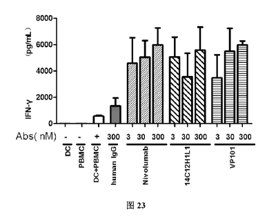
FIG. 23 shows the effect of VP101 on secretion of IFN-y in mixed culture
system of DC and PBMC cells.
FIG. 24 shows the effect of VP101, BsAbB7 and BsAbB8 on secretion of
IFN-y in mixed culture system of DC and PBMC cells.
FIG. 25 shows the effect of VP101, BsAbB7 and BsAbB8 on secretion of
IL-2 in mixed culture system of DC and PBMC cells.
FIG. 26 shows the effect of antibodies 14C12H1L1 and VP101 on secretion
of the cytokine IL-2 induced by mixed culture of PBMC and Raji-PD-Li cells
detected by ELISA.
FIG. 27 shows effect of antibodies 14C12H1L1 and VP101 on secretion of
the cytokine IFN-y induced by mixed culture of PBMC and Raji-PD-L1 cells
detected by ELISA.
FIG. 28 shows effect of antibodies 14C12H1L1, VP101, BsAbB7 and
BsAbB8 on secretion of the cytokine IFN-y induced by mixed culture of
PBMC and Raji-PD-Ll cells detected by ELISA.
37
Date Recue/Date Received 2021-02-25
CA 03110749 2021-02-25
FIG. 29 shows effect of antibodies 14C12H1L1, VP101, BsAbB7 and
BsAbB8 on secretion of the cytokine IL-2 induced by mixed culture of
PBMC and Raji-PD-Ll cells detected by ELISA.
FIG. 30 shows the inhibition of tumor growth by VP101 at different
concentrations.
FIG. 31 shows effect of VP101 at different concentrations on body weight of
mouse.
DETAILED DESCRIPTION
The embodiments of the present invention will be described in detail below
with reference to the examples. Those skilled in the art will understand that
the following examples are only used to illustrate the present invention, and
should not be regarded as limiting the scope of the present invention. The
cases without the specific descriptions of techniques or conditions were
carried out according to the technologies or conditions described in the
literature in the art (e.g., see, Guide to Molecular Cloning Experiments,
authored by J. Sambrook et al., and translated by Huang Peitang et al., third
edition, Science Press) or according to the product manual. Reagents or
instruments used are all commercially available conventional products if the
manufacturers thereof are not specified.
In the following examples of the present invention, the marketed antibody
bevacizumab (trade name Avasting) for the same target was purchased from
Roche as a control antibody, or was prepared according to Preparation
Example 4.
In the following examples of the present invention, the marketed antibody
nivolumab for the same target (trade name OpdivoS) was purchased from
BMS as a control antibody.
38
Date Recue/Date Received 2021-02-25
CA 03110749 2021-02-25
In the following examples of the present invention, the amino acid sequences
of the control antibodies BsAbB7 and BsAbB8 were identical to the amino
acid sequences of BsAbB7 and BsAbB8 respectively in Chinese Patent
Publication CN105175545A.
Preparation Example 1: Preparation of Fusion Proteins PD-1-mFc, PD-1-hFc
and PD-Li-hFc
The preparation of fusion proteins PD-1-mFc, PD-1-hFc and PD-Li-hFc and
the SDS-PAGE electrophoresis detection are carried out by fully referring to
Preparation Example 1 of Chinese Patent Publication CN106632674A.
The amino acid sequences and the encoding nucleotide sequences of the
fusion proteins PD-1-mFc, PD-1-hFc and PD-Ll-hFc in this preparation
example are the same as those of PD-1-mFc, PD-1-hFc and PDL-1-hFc
respectively in the Preparation Example 1 of Chinese Patent Publication
CN106632674A.
Fusion proteins PD-1-mFc, PD-1-hFc and PD-Li-hFc were thus obtained.
Preparation Example 2: Expression and Purification of Fusion Protein
VEGFA-His
1. Construction of plasmid VEGFA-His
PCR amplification was performed using VEGFA human cDNA (purchased
from Origene) as a template and the hVEGFA-His fragment was purified and
isolated using an ordinary DNA product purification kit. The isolated
hVEGFA-His fragment and an expression vector pcDNA3.1 were
enzyme-digested with XbaI&HindIII-HF, and a target gene fragment was
isolated by gel extraction and ligated with a linear expression vector by T4
ligase. Then all the ligation products were transformed into DH5a chemically
competent cells and coated on an Agar plate with Amp. Well separated single
39
Date Recue/Date Received 2021-02-25
CA 03110749 2021-02-25
colonies were selected for colony PCR identification, PCR positive clones
were inoculated to an LB culture medium for culture, and a bacteria solution
was taken and sent to Guangzhou Invitrogen Biotechnology for sequencing
verification. The alignment of the sequencing results showed that the
insertion
sequence of the positive recon was completely correct.
2. Expression and purification of fusion protein VEGFA-His
After the recombinant plasmid VEGFA-his was transfected into 293F cells
(purchased from Invitrogen) for 7 days according to the manual in
lipofectamin transfection kit (purchased from Invitrogen), the culture medium
was subjected to high-speed centrifugation, supernatant concentration and
buffer exchange into Binding Buffer A, and then loaded onto a HisTrap
column, and proteins were linearly eluted with Elution Buffer A. The primary
pure sample was subjected to buffer exchange into Binding Buffer B with a
HiTrap Desalting column and loaded onto a HiTrap Q column, proteins were
linearly eluted with Elution Buffer B, and the target sample was isolated and
buffer exchanged into PBS. The purified sample was added to a reduced
protein electrophoresis loading buffer for SDS -PAGE electrophoresis
detection.
The fusion protein VEGFA-His was thus obtained.
The amino acid sequence of VEGFA-His is as follows (171 aa):
APMAEGGGQNHHEVVKFMDVYQRSYCHPIETLVDIF QEYPDEIEYIF KP S C
VPLMRCGGCCNDEGLECVPTEESNITMQIMRIKPHQGQHIGEMSFLQHNKC
ECRPKKDRARQENPCGPC SERRKHLFVQDPQTCKC SCKNTDSRCKARQLE
LNERTCRCDKPRRHHHHHH ( SEQ ID NO: 1)
wherein, the underlined part is the amino acid sequence of VEGFA.
Date Recue/Date Received 2021-02-25
CA 03110749 2021-02-25
Nucleotide sequence encoding VEGFA-His (513 bp)
GCACCCATGGCCGAGGGCGGCGGCCAGAACCACCACGAGGTGGTGAAG
TTCATGGACGTGTACCAGAGAAGCTACTGCCACCCCATCGAGACCCTGG
TGGACATCTTCCAGGAGTACCCCGACGAGATCGAGTACATCTTCAAGCC
CAGCTGCGTGCCCCTGATGAGATGCGGCGGCTGCTGCAACGACGAGGG
CCTGGAGTGCGTGCCCACCGAGGAGAGCAACATCACCATGCAGATCAT
GAGAATCAAGCCCCACCAGGGCCAGCACATCGGCGAGATGAGCTTCCT
GCAGCACAACAAGTGCGAGTGCAGACCCAAGAAGGACAGAGCCAGAC
AGGAGAACCCCTGCGGCCCCTGCAGCGAGAGAAGAAAGCACCTGTTCG
TGCAGGACCCCCAGACCTGCAAGTGCAGCTGCAAGAACACCGACAGCA
GATGCAAGGCCAGACAGCTGGAGCTGAACGAGAGAACCTGCAGATGCG
ACAAGCCCAGAAGACATCATCACCATCACCAC ( SEQ ID NO: 2)
Preparation Example 3: Expression and Purification of Fusion Protein
VEGFR2-hFc
1. Synthesis of gene VEGFR2-hFc:
The amino acids corresponding to the extracellular fragment VEGFR2 ECD
of gene VEGFR2 (Vascular Endothelial Growth Factor Receptor 2, NCBI
GenBank: NP 002244) were fused with TEV and the Fc protein fragment of
human IgG (hFc) respectively (SEQ ID NO: 3). Genscript was entrusted to
synthesize corresponding encoding nucleotide sequence (SEQ ID NO: 4).
VEGFR2, Vascular Endothelial Growth Factor Receptor 2, NCBI GenBank
NP 002244;
hFc: Ig gamma-1 chain C region, ACCESSION: P01857, 106-330;
Amino acid sequence of fusion protein VEGFR2-hFc: (998 aa)
MQSKVLLAVALWLCVETRAASVGLPSVSLDLPRL SIQKDILTIKANTTLQIT
CRG RDLDWLWPNN SGSEQRVEVTECSDGLFCKTLTIPKVIGNDTGAYK
41
Date Recue/Date Received 2021-02-25
CA 03110749 2021-02-25
CFYRETDLASVIYVYV DYRSPFIASVSD HGVVYITENKNKTVVIPCLGSI
SNLNVSLCARYPEKRFVPDGNRISWDSKKGFTIPSYMISYAGMVFCEAKIN
DES YQ S IMYIVVVVGYRIYDVVL S P S HGI EL SVGEKLVLNCTARTELNVGID
FNWEYPS S KHQHKKLVNRDLKTQ S GS EMKKFL STLTIDGVTIC
AAS SGLMTKKNSTFVRVHEKPFVAFGSGMESLVEATVGERVRIPAKYLGY
PPPEIKWYKNGIPLESNHTIKAGHVLTIMEVSERDTGNYTVILTNPISKEK S
HVVSLVVYVPPQIGEKSLISPVDSYQYGTTQTLTCTVYAIPPPHHIHWYWQ
LEEECANEPS AVSVTNPYPCEEWRSVEDF GGNKIEVNKNQFALIEGKNK
TV_STLYJQAA_NY_S_AliCE_AQ_Q_YK VNKVGRGERVISFHVTRGPEITL PDM PTE
QESVSLWCTADRSTFENLTWYKLGPQPLPIHVGELPTPVCKNLDTLWKLN
ATMFSNS'I_:NDIJJM_E_LJ_Q_KNASL DQ_GYVCLA DRKTKKRHCVVR LTVLE
RVAPTITGNLEN TT S I GES IEV S CTAS GNPPP IMWFKDNETLVEDS GIVLK
DGNRNLTIRRVRKEDEGLYTC ACSVLGCAKVEAFFIIEGA EKTNLESRE
NLYFQGTHTCPPCPAPELLGGPSVFLFPPKPKDTLMISRTPEVTCVVVDVSH
EDPEVKFNWYVDGVEVHNAKTKPREEQYNSTYRVVSVLTVLHQDWLNG
KEYKCKVSNKALPAPIEKTISKAKGQPREPQVYTLPPSRDELTKNQVSLTCL
VKGFYPSDIAVEWESNGQPENNYKTTPPVLDSDGSFFLYSKLTVDKSRWQ
QGNVFSCSVMHEALHNHYTQKSLSLSPGK ( SEQ ID NO: 3)
wherein, the wavy-underlined part is the ECD part of VEGFR2, the framed
part is TEV enzyme digestion site, and the solid-underlined part is hFc part.
Nucleotide sequence encoding fusion protein VEGFR2-hFc: (2997 bp)
A TGCA GA GCAAGGTGCT GCTGGCC GTC GC C CT GTGGCT CT GC GTGGAGA
CCCGGGCCGCCTCTGTGGGTTTGCCTAGTGTTTCTCTTGATCTGCCCAGG
CTCAGCATACAAAAAGACATACTTACAATTAAGGCTAATACAACTCTTC
AAATTACTTGCAGGGGACAGAGGGA CTTGGA CTGGCTTTGGCCCAATA
ATCAGAGTGGCAGTGAGCAAAGGGTGGAGGTGACTGAGTGCAGCGATG
GCCTCTTCTGTAAGACACTCACAATTCCAAAAGTGATCGGAAATGACAC
TGGAGCCTACAAGTGCTTCTACCGGGAAACTGACTTGGCCTCGGTCATT
42
Date Recue/Date Received 2021-02-25
CA 03110749 2021-02-25
TATGTCTATGTTCAAGATTACAGATCTCCATTTATTGCTTCTGTTAGTGA
C CAACATGGAGTC GT GTACATTACTGAGAACAAAAACAAAACT GTGGT
GATT CCATGT CT CGGGT C CATTTCAAATCTCAACGT GT CACTTT GTGCAA
GATACCCAGAAAAGAGATTTGTTCCTGA TGGTA A CA GA ATTTCCTGGGA
CAGCAAGAAGGGCTTTACTATT CC CAGCTACAT GAT CAGCTAT GCT GGC
ATGGT CTT CT GT GAAGCAAAAATTAAT GAT GAAAGTTAC CAGTCTATTA
TGTACATAGTTGTCGTTGTAGGGTATAGGATTTATGATGTGGTTCTGAGT
CCGTCTCATGGAATTGAACTATCTGTTGGA GA A A A GCTTGTCTTA A A TT
GTACAGCAAGAACTGAACTAAATGT GGGGATT GACTTCAACTGGGAAT
AC CCTT CTTC GAAGCAT CAGCATAAGAAACTT GTAAAC C GAGACCTAAA
AAC C CAGT CT GGGAGTGAGATGAAGAAATTTTT GAGCAC CTTAACTATA
GAT GGT GTAAC CC GGAGTGAC CAAGGATTGTACAC CT GT GCAGCAT CC
AGT GGGCT GAT GAC CAAGAAGAACAGCACATTT GT CAGGGT CCAT GAA
AAAC CTTTTGTT GCTTTTGGAAGTGGCAT GGAAT CT CT GGTGGAAGC CA
C GGT GGGGGAGCGTGT CAGAAT C CCTGCGAAGTAC CTTGGTTAC CCAC C
C CCAGAAATAAAAT GGTATAAAAAT GGAATACC CCTT GAGT CCAAT CA
CACAATTAAAGCGGGGCATGTACT GAC GATTATGGAAGT GAGTGAAAG
AGACACAGGAAATTACACTGTCATCCTTACCAATCCCATTTCAAAGGAG
AAGCAGAGC CATGT GGTCTCTCT GGTT GT GTATGTC C CACC C CAGATT G
GTGA GA A A TCTCTA A TCTCTCCTGTGGATTCCTACCAGTACGGCACCAC
T CAAAC GCTGACATGTACGGT CTATGC CATT CCTC C CC CGCAT CACAT C
CACTGGTATT GGCAGTT GGAGGAAGAGTGCGC CAACGAGCC CAGCCAA
GCT GT CT CAGTGACAAAC CCATACC CTTGTGAAGAAT GGAGAAGT GTGG
AGGACTT C CAGGGAGGAAATAAAATTGAAGTTAATAAAAAT CAATTTG
CTCTAATTGAAGGAAAAAACAAA A CTGTA A GTA C CCTTGTTA TCC A A GC
GGCAAATGTGTCAGCTTTGTACAAATGTGAAGCGGTCAACAAAGTCGG
GAGAGGAGAGAGGGTGATCTC CTT CCAC GT GAC CAGGGGT CCTGAAAT
TACTTTGCAACCT GACAT GCAGC C CACT GAGCAGGAGAGC GT GT CTTT G
43
Date Recue/Date Received 2021-02-25
CA 03110749 2021-02-25
TGGTGCACTGCAGACAGATCTACGTTTGAGAACCTCACATGGTACAAGC
TTGGCCCACAGCCTCTGCCAATCCATGTGGGAGAGTTGCCCACACCTGT
TTGCAAGAACTTGGATACTCTTTGGAAATTGAATGCCACCATGTTCTCT
AATAGCACAAATGACATTTTGATCATGGAGCTTAAGAATGCATCCTTGC
AGGACCAAGGAGACTATGTCTGCCTTGCTCAAGACAGGAAGACCAAGA
AAAGACATTGCGTGGTCAGGCAGCTCACAGTCCTAGAGCGTGTGGCAC
CCACGATCACAGGAAACCTGGAGAATCAGACGACAAGTATTGGGGA A A
GCATCGAAGTCTCATGCACGGCATCTGGGAATCCCCCTCCACAGATCAT
GTGGTTTAAAGATAATGAGACCCTTGTAGAAGACTCAGGCATTGTATTG
AAGGATGGGAACCGGAACCTCACTATCCGCAGAGTGAGGAAGGAGGAC
GAAGGCCTCTACACCTGCCAGGCATGCAGTGTTCTTGGCTGTGCAAAAG
TGGAGGCATTTTTCATAATAGAAGGTGCCCAGGAAAAGACGAACTTGG
A ATCTAGAGAAAACCTGTATTTTCAGGGCACTCACACATGCCCACCGTG
CCCAGCACCTGAACTCCTGGGGGGACCGTCAGTCTTCCTCTTCCCCCCA
AAACCCAAGGACACCCTCATGATCTCCCGGACCCCTGAGGTCACATGCG
TGGTGGTGGACGTGAGCCACGAAGACCCTGAGGTCAAGTTCAACTGGT
ACGTGGACGGCGTGGAGGTGCATAATGCCAAGACAAAGCCGCGGGAGG
AGCAGTACAACAGCACGTACCGTGTGGTCAGCGTCCTCACCGTCCTGCA
CCAGGACTGGCTGAATGGCAAGGAGTACAAGTGCAAGGTCTCCAACAA
AGCCCTCCCAGCCCCCATCGAGAAAACCATCTCCAAAGCCAAAGGGCA
GCCCCGAGAACCACAGGTGTACACCCTGCCCCCATCCCGGGATGAGCT
GACCAAGAACCAGGTCAGCCTGACCTGCCTGGTCAAAGGCTTCTATCCC
AGCGACATCGCCGTGGAGTGGGAGAGCAATGGGCAGCCGGAGAACAA
CTACAAGACCACGCCTCCCGTGTTGGACTCCGACGGCTCCTTCTTCCTCT
ACAGCAAGCTCACCGTGGACAAGAGCAGGTGGCAGCAGGGGAACGTCT
TCTCATGCTCCGTGATGCATGAGGCTCTGCACAACCACTACACGCAGAA
GAGCCTCTCCCTGTCTCCCGGGAAATGA ( SEQ ID NO: 4)
wherein, the wavy-underlined part is the ECD part of VEGFR2, the framed
44
Date Recue/Date Received 2021-02-25
CA 03110749 2021-02-25
part is TEV enzyme digestion site, and the solid-underlined part is hFc part.
2. Construction of plasmid pUC57simp1e-VEGFR2-hFc:
The VEGFR2-hFc encoding gene synthesized by Genscript was cloned into
an expression vector pUC57simple (provided by Genscript), and a
pUC57simple-VEGFR2-hFc plasmid was obtained.
3. Construction of recombinant plasmid pcDNA3.1-VEGFR2-hFc:
The plasmid pUC57simple-VEGFR2-hFc was enzyme-digested (Xba I and
BamH I), and the fusion gene fragment VEGFR2-hFc isolated by
electrophoresis was ligated with expression vector pcDNA3.1 (purchased
from Invitrogen) to give pcDNA3.1-VEGFR2-hFc, which was transfected
into competent E. coli cell DH5a (purchased from TIANGEN), the
transfection and culture were performed according to the manual. The
positive pcDNA3.1-VEGFR2-hFc colonies were screened, E. coli was
amplified according to a conventional method, and a kit (purchased from
Tiangen Biotech (Beijing) Co., Ltd., DP103-03) was then used and a
recombinant plasmid pcDNA3.1-VEGFR2-hFc was extracted according to
the manual of the kit.
4. Transfection of recombinant plasmid pcDNA3.1-VEGFR2-hFc into 293F
cells
The recombinant plasmid pcDNA3.1-VEGFR2-hFc was transfected into 293F
cells (purchased from Invitrogen) according to the lipofectamin transfection
kit (purchased from Invitrogen).
5. SDS-PAGE electrophoresis detection of VEGFR2-hFc protein
After transfecting the recombinant plasmid pcDNA3.1-VEGFR2-hFc into 293F
cells for 7 days, the culture medium was subjected to high-speed
Date Recue/Date Received 2021-02-25
CA 03110749 2021-02-25
centrifugation, microporous membrane vacuum filtration and purification in a
Mabselect SuRe column to obtain a VEGFR2-hFc fusion protein sample, and
a part of the sample was added into a reduced protein electrophoresis loading
buffer for SDS-PAGE electrophoresis detection.
The fusion protein VEGFR2-hFc was thus obtained.
Preparation Example 4: Preparation of Anti-VEGFA Antibody Bevacizumab
Chinese Patent Publication CN1259962A is referred to for the amino acid
sequences of the heavy chain variable region and the light chain variable
region of the marketed VEGFA monoclonal antibody Avastin (bevacizumab).
Genscript was entrusted to synthesize nucleotide sequences encoding the
heavy chain variable region and the light chain variable region.
Amino acid sequence of the heavy chain variable region of bevacizumab:
(123 aa)
EVQLVE S GGGLVQPGGS LRL S CAA S GYTFTNYGMNWVRQAPGKGLEWVG
WINTYTGEPTYAADFKRRFTFSLDTSKSTAYLQMNSLRAEDTAVYYCAKY
PHYYGSSHWYFDVWGQGTLVTVSS ( SEQ ID NO: 5)
Nucleotide sequence encoding the heavy chain variable region of
bevacizumab: (369 bp)
GAGGTGCAGCTGGTCGAGTCCGGGGGGGGGCTGGTGCAGCCAGGCGGG
TCTCTGAGGCTGAGTTGCGCCGCTTCAGGGTACACCTTCACAAACTATG
GAATGAATTGGGTGCGCCAGGCACCAGGAAAGGGACTGGAGTGGGTCG
GCTGGATCAACACTTACACCGGGGAACCTACCTATGCAGCCGACTTTAA
GCGGCGGTTCACCTTCAGCCTGGATACAAGCAAATCCACTGCCTACCTG
CAGATGAACAGCCTGCGAGCTGAGGACACCGCAGTCTACTATTGTGCTA
AATATCCCCACTACTATGGGAGCAGCCATTGGTATTTTGACGTGTGGGG
GCAGGGGACTCTGGTGACAGTGAGCAGC ( SEQ ID NO: 6)
46
Date Recue/Date Received 2021-02-25
CA 03110749 2021-02-25
Amino acid sequence of the light chain variable region of bevacizumab: (107
aa)
DIQMTQSPS SL SASVGDRVTITC SAS QDI SNYLNWYQQKPGKAPKVLIYFT S
SLHSGVPSRF S GS GS GTDFTLTIS SLQPEDFATYYCQQYSTVPWTFGQGTKV
EIK ( SEQ ID NO: 7)
Nucleotide sequence encoding the light chain variable region of bevacizumab:
(321 bp)
GATATTCAGATGACTCAGAGCCCCTCCTCCCTGTCCGCCTCTGTGGGCG
ACAGGGTCACCATCACATGCAGTGCTTCACAGGATATTTCCAACTACCT
GAATTGGTATCAGCAGAAGCCAGGAAAAGCACCCAAGGTGCTGATCTA
CTTCACTAGCTCCCTGCACTCAGGAGTGCCAAGCCGGTTCAGCGGATCC
GGATCTGGAACCGACTTTACTCTGACCATTTCTAGTCTGCAGCCTGAGG
ATTTCGCTACATACTATTGCCAGCAGTATTCTACCGTGCCATGGACATTT
GGCCAGGGGACTAAAGTCGAGATCAAG ( SEQ ID NO: 8)
The heavy chain constant regions were all Ig gamma-1 chain C region,
ACCESSION: P01857; the light chain constant regions were all Ig kappa
chain C region, ACCESSION: P01834.
The heavy chain cDNA and the light chain cDNA of bevacizumab were
cloned into vector pcDNA3.1, and the recombinant expression plasmid of the
antibody bevacizumab was obtained. The recombinant plasmid was
transfected into 293F cells. The 293F cell culture medium was purified and
then detected.
The anti-VEGFA monoclonal antibody Avastin (bevacizumab) was thus
obtained.
Preparation Example 5: Preparation and Detection of Anti-PD-1 Humanized
Antibody 14C12H1L1
47
Date Recue/Date Received 2021-02-25
CA 03110749 2021-02-25
The preparation was carried out according to the Examples 3-4 described in
Chinese Patent Publication CN106977602A.
The amino acid sequences of the heavy chain variable region and the light
chain variable region of humanized antibody 14C12H1L1, and the nucleotide
sequence encoding the same are also the same as those described in Examples
3-4 of Chinese Patent Publication CN106977602A, and are also provided
herein as follows:
Amino acid sequence of the heavy chain variable region of humanized
antibody 14C12H1L1: (118 aa)
EVQLVES GGGLVQPGGSLRL S CAA S GFAF S SYDMSWVRQAPGKGLDWVA
TISGGGRYTYYPDSVKGRFTISRDNSKNNLYLQMNSLRAEDTALYYCANR
YGEAWFAYWGQGTLVTVSS ( SEQ ID NO: 9)
Nucleotide sequence encoding the heavy chain variable region of humanized
antibody 14C12H1L1: (354 bp)
GAAGTGCAGCTGGTCGAGTCTGGGGGAGGGCTGGTGCAGCCCGGCGGG
TCACTGCGACTGAGCTGCGCAGCTTCCGGATTCGCCTTTAGCTCCTACG
ACATGTCCTGGGTGCGACAGGCACCAGGAAAGGGACTGGATTGGGTCG
CTACTATCTCAGGAGGCGGGAGATACACCTACTATCCTGACAGCGTCAA
GGGCCGGTTCACAATCTCTAGAGATAACAGTAAGAACAATCTGTATCTG
CAGATGAACAGCCTGAGGGCTGAGGACACCGCACTGTACTATTGTGCCA
ACCGCTACGGGGAAGCATGGTTTGCCTATTGGGGGCAGGGAACCCTGGTG
ACAGTCTCTAGT (SEQ ID NO: 10)
Amino acid sequence of the light chain variable region of humanized
antibody 14C12H1L1: (107 aa)
DIQMTQSPS SMSASVGDRVTFTCRASQDINTYLSWFQQKPGKSPKTLIYRA
NRLV S GVP S RF S GS GS GQDYTLTI S SL QPEDMATYYCLQYDEF PLTF GAGT
48
Date Recue/Date Received 2021-02-25
CA 03110749 2021-02-25
KLELK ( SEQ ID NO: 11)
Nucleotide sequence encoding the light chain variable region of humanized
antibody 14C12H1L1: (321 bp)
GACATTCAGATGACTCAGAGCCCCTCCTCCATGTCCGCCTCTGTGGGCG
ACAGGGTCACCTTCACATGCCGCGCTAGTCAGGATATCAACACCTACCT
GAGCTGGTTTCAGCAGAAGCCAGGGAAAAGCCCCAAGACACTGATCTA
CCGGGCTAATAGACTGGTGTCTGGAGTCCCAAGTCGGTTCAGTGGCTCA
GGGAGCGGACAGGACTACACTCTGACCATCAGCTCCCTGCAGCCTGAG
GACATGGCAACCTACTATTGCCTGCAGTATGATGAGTTCCCACTGACCT
TTGGCGCCGGGACAAAACTGGAGCTGAAG ( SEQ ID NO: 12)
The anti-PD-1 humanized antibody 14C12H1L1 was thus obtained.
Preparation Example 6: Preparation and Identification of hIgG
The sequence of Human Anti-Hen Egg Lysozyme IgG (anti-HEL, i.e., human
IgG, abbreviated as hIgG) is derived from a variable region sequence of the
Fab F10.6.6 sequence in the research published by Acierno et al., which is
entitled "Affinity maturation increases the stability and plasticity of the Fv
domain of anti-protein antibodies" (Acierno et al., J Mal Biol. 2007; 374(1):
130-46). The preparation method is as follows:
Nanjing Genscript Biology was entrusted to carry out codon optimization of
amino acids and gene synthesis on heavy and light chain (complete sequence
or variable region) genes of human IgG antibody, and by referring to the
standard technologies introduced in the "Guide to Molecular Cloning
Experiments (Third Edition)" and using standard molecular cloning
technologies such as PCR, enzyme digestion, DNA gel extraction, ligation
transformation, colony PCR or enzyme digestion identification, the heavy and
light chain genes were respectively subcloned into the antibody heavy chain
49
Date Recue/Date Received 2021-02-25
CA 03110749 2021-02-25
expression vector and antibody light chain expression vector of the
mammalian expression system, and the heavy and light chain genes of the
recombinant expression vector were further sequenced and analyzed. After
the sequence was verified to be correct, endotoxin-free expression plasmids
were prepared in a large scale, and the heavy and light chain plasmids were
transiently co-transfected into HEK293 cells for expression of recombinant
antibody. After 7 days of culture, the cell culture medium was collected and
affinity purified using an rProtein A column (GE), and the quality of the
resulting antibody sample was determined using SDS-PAGE and SEC-HPLC
standard analysis techniques.
The hIgG was thus obtained, and used in Examples 8-9 below.
Example 1: Sequence Design, Preparation and Detection of Heavy and Light
Chains of Bifunctional Antibody VP101
1. Sequence design
The structure of the bifunctional antibody VP101 of the present invention is
in the Morrison form (IgG-scFv), i.e. C-termini of two heavy chains of an IgG
antibody are each linked to a scFv fragment of another antibody, and the main
composition design of the heavy and light chains is as shown in Table 1
below.
Table 1: Composition design of the heavy and light chains of VP101
Heavy chain
Bifunctional
Linker Light chain
antibody No. IgG part scFv part
fragment
Bevacizu 14C12H1v- Bevacizumab-
VP101 Linkerl
mab-H Linker1-14C12L1v L
In the Table 1 above:
(1) Those with "V" labeled at lower right corner refer to the variable region
of
Date Recue/Date Received 2021-02-25
CA 03110749 2021-02-25
corresponding heavy chain or the variable region of corresponding light chain.
For those without "V" label, the corresponding heavy or light chain is the
full
length comprising the constant region. The corresponding sequences
described in the above preparation examples are referred to for the amino acid
sequences of these variable regions or the full length and the nucleotide
sequences encoding them.
(2) The amino acid sequence of linker 1 is GGGGSGGGGSGG GGSGGGGS
(SEQ ID NO: 13)
2. Expression and purification of antibody VP101
The heavy chain cDNA sequence and the light chain cDNA sequence of
VP101 were each cloned into vector pUC57simple (provided by Genscript) to
obtain plasmids pUC57simp1e-VP101H and pUC57simp1e- VP101L,
respectively.
Plasmids pUC57simp1e-VP101H and pUC57simple-VP101L were
enzyme-digested (HindIII&EcoRI), and heavy and light chains isolated by
electrophoresis were subcloned into vector pcDNA3.1, and recombinant
plasmids were extracted to co-transfect 293F cells. After 7 days of cell
culture, the culture medium was centrifuged at high speed, and the
supernatant was concentrated and loaded onto a HiTrap MabSelect SuRe
column. The protein was further eluted in one step with Elution Buffer, and
the target sample antibody VP101 was isolated and buffer exchanged into
PBS.
2. Detection of antibody VP101
The purified sample was added to both a reduced protein electrophoresis
loading buffer and a non-reduced protein electrophoresis loading buffer, and
then boiled for SDS-PAGE electrophoresis detection. The electropherogram
51
Date Recue/Date Received 2021-02-25
CA 03110749 2021-02-25
of VP101 is shown in FIG. 1. The target protein of the reduced protein sample
is at 75 kD and 25 kD, and the target protein of the non-reduced protein
sample (single antibody) is at 200 kD.
Unless otherwise specified, the humanized antibody VP101 used in the
following experiments was prepared by the method of this example.
Example 2: Detection of Kinetic Parameters of Humanized Antibody VP101
1. Detection of kinetic parameters of the binding of humanized antibody
VP101 to PD-1-mFc
The sample dilution buffer was PBS (0.02% Tween-20, 0.1% BSA, pH7.4). 5
iag/mL antibody was immobilized to an AHC sensor with the immobilization
height being about 0.4 nM. The sensor was equilibrated in a buffer for 60 s,
and the antibody immobilized to the sensor bound to PD-1-mFc at a
concentration of 0.62-50 nM (three-fold gradient dilution) for 120 s, and then
the antigen and antibody dissociated in the buffer for 300 s. The data were
analyzed by 1:1 model fitting to obtain affinity constants. The data
acquisition
software was Fortebio Data Acquisition 7.0, and the data analysis software
was Fortebio Data Analysis 7Ø Kinetic parameters of the binding of
antibodies VP101, BsAbB7, BsAbB8, 14C12H1L1 and the control antibody
nivolumab to PD-1-mFc are shown in Table 2, and the detection results of the
kinetic characteristic parameters are shown in FIG. 2, FIG. 3, FIG. 4, FIG. 5
and FIG. 6, respectively.
Table 2: Kinetic parameters of the binding of humanized antibody VP101,
BsAbB7, BsAbB8, 14C12H1L1 and the control antibody nivolumab to
PD-1 -mF c
Sample ID KD (M) Kon (1/Ms) S E (kon) Kdis (1/s) S E (kdis) Rmax (nm)
VP101 1.68E-10 3.22E+05 1.44E+04 5.40E-05 3.16E-05 0.14-0.28
52
Date Recue/Date Received 2021-02-25
CA 03110749 2021-02-25
BsAbB7 1.62E-10
3.27E+05 2.60E+04 5.30E-05 6.24E-05 0.01-0.11
BsAbB8 4.06E-10
3.39E+05 2.04E+04 1.37E-04 4.61E-05 0.01-0.13
14C 12H1L1 1.64E-10 4.55E+05 1.61E+04 7.47E-05 2.98E-05 0.24-0.28
Nivolumab 2.32E-10 5.85E+05 2.03E+04 1.36E-04 3.47E-05 0.02-0.14
KD is affinity constant; kon is binding rate of antigen and antibody; kdis is
dissociation rate of antigen and antibody; KD = kdis/kon.
The results show that the antibodies VP101 and BsAbB7 are equivalent in
temis of affinity for PD-1-mFc, the affinity constant of VP101 for PD-1-mFc
is significantly smaller than that of BsAbB8, suggesting that VP101 has better
binding activity; the dissociation rate constant for VP101 and PD-1-mFc was
significantly smaller than BsAbB8 and 14C12H1L1, suggesting that VP101
binds to antigen more stably with a dissociation rate slower than that of
14C12H1L1 and BsAbB8.
2. Detection of kinetic parameters of the binding of humanized antibody
VP101 to VEGF-His
The sample dilution buffer was PBS (0.02% Tween-20, 0.1% BSA, pH7.4). 1
lig/mL VEGF-His was immobilized to the HIS1K sensor for 20 s, then the
sensor was equilibrated in a buffer for 60 s, and the VEGF immobilized on
the sensor bound to the antibody at a concentration of 12.34-1000 nM
(three-fold gradient dilution) for 120 s, and then the antigen and antibody
dissociated in the buffer for 300 s. The data were analyzed by 1:1 model
fitting to obtain affinity constants. The data acquisition software was
Fortebio
Data Acquisition 7.0, and the data analysis software was Fortebio Data
Analysis 7Ø
Kinetic parameters of the binding of antibodies VP101, BsAbB7, BsAbB8
and the control antibody bevacizumab to VEGF-His are shown in Table 3,
53
Date Recue/Date Received 2021-02-25
CA 03110749 2021-02-25
and the detection results of kinetic characteristic parameters are shown in
FIG. 7, FIG. 8, FIG. 9 and FIG. 10 respectively.
Table 3: Kinetic parameters of the binding of antibodies VP101, BsAbB7,
BsAbB8 and the control antibody bevacizumab to VEGF-His
Sample ID KD (M) Kon (1/Ms) S E (kon) Kdis (1/s) S E (kdis) Rmax (nm)
VP101 5.21E-10 1.55E+05 9.67E+03 8.05E-05 4.66E-05 0.39-0.60
BsAbB7 5.14E-10 1.57E+05 9.67E+03 8.05E-05 4.83E-05 0.36-0.53
BsAbB8 6.33E-10 1.71E+05 1.07E+04 1.08E-04 4.64E-05 0.39-0.56
Bevacizumab 7.24E-10 1.23E+05 7.09E+03 8.90E-05 4.53E-05 0.29-0.41
The results show that the antibodies VP101 and BsAbB7 are equivalent in
temis of affinity for the antigen, and the affinity constant of VP101 is
significantly smaller than that of BsAbB8 and the control antibody
bevacizumab, suggesting that VP101 has better binding activity; the
dissociation rate constant of VP101 for VEGF-His is significantly smaller
than that of BsAbB8, suggesting that VP101 binds to antigen more stably
with a slower dissociation rate than that of BsAbB8.
Example 3: Detection of Binding Activity of Antibody VP101 to Antigen by
ELISA
1. Detection of binding activity of antibody VP101 to antigen VEGFA-his by
indirect ELISA
The method is specified as follows:
The microplate was coated with VEGFA-His and incubated at 37 C for 2
hours. After being washed, the microplate was blocked with 1% BSA for 2
hours. After being washed, the microplate was added with the gradiently
diluted antibody and incubated at 37 C for 30 minutes. After being washed,
the microplate was added with the enzyme-labeled goat anti-human IgG
54
Date Recue/Date Received 2021-02-25
CA 03110749 2021-02-25
secondary antibody working solution and incubated for 30 minutes at 37 C.
After being washed, the microplate was added with TMB chromogenic
solution for color developing for 5 minutes in the absence of light, and then
stop solution was added to tenninate the chromogenic reaction. Then the
microplate was put into a microplate reader immediately, and the OD value of
each well in the microplate was read at 450 nm. SoftMax Pro 6.2.1 was used
to analyze and process the data.
The detection result of the binding of antibody VP101 to antigen VEGFA-His
is shown in FIG. 11. The absorbance intensities at each dose are shown in
Table 4. The binding ECso of antibody was calculated by curve fitting using
antibody concentration as the abscissa and absorbance value as the ordinate,
and the results are shown in Table 4 below.
Table 4: Binding of bifunctional antibody to VEGFA-his (Indirect ELISA)
Antibody concentration Coating: VEGFA-His (1 lig/mL)
(ig/mL)
VP101 Bevacizumab
1.0000 3.045 2.943 2.798 2.974
0.3333 3.037 2.861 2.816 2.993
0.1111 2.901 2.689 2.653 2.700
0.0370 2.597 2.460 2.445 2.555
0.0123 2.013 1.914 1.998 2.074
0.0041 1.115 1.086 1.446 1.363
0.0014 0.524 0.496 0.640 0.729
0.0000 0.099 0.091 0.094 0.083
Secondary antibody Goat anti-human IgG (H+L), HRP (1:5000)
EC50(nM) 0.036 0.035
The results show that antibody VP101 is able to bind to VEGFA protein
efficiently and its binding efficiency is dose-dependent, and the two
antibodies are equivalent in tenns of binding activity to human VEGFA.
2. Detection of respective binding activities of antibodies VP101, BsAbB7
Date Recue/Date Received 2021-02-25
CA 03110749 2021-02-25
and BsAbB8 to antigen VEGFA-His by indirect ELISA
The method is specified as follows:
The microplate was coated with VEGFA-His and incubated overnight at 4 C.
After being washed, the microplate was blocked with 1% BSA (dissolved in
PBS) for 2 hours. After being washed, the microplate was added with the
gradiently diluted antibody and incubated at 37 C for 30 minutes. After being
washed, the microplate was added with the horseradish peroxidase-labeled
goat anti-human IgG Fc (Jackson, 109-035-098) working solution and
incubated for 30 minutes at 37 C. After being washed, the microplate was
added with TMB (Neogen, 308177) for color developing for 5 minutes in the
absence of light, and then stop solution was added to terminate the
chromogenic reaction. Then the microplate was put into a microplate reader
immediately, and the OD value of each well in the microplate was read at 450
nm. SoftMax Pro 6.2.1 was used to analyze and process the data.
The result of the binding of antibody VP101 to antigen VEGFA-His is shown
in FIG. 12. The absorbance intensities at each dose are shown in Table 5. The
binding ECso of antibody was calculated by curve fitting using antibody
concentration as the abscissa and absorbance value as the ordinate, and the
results are shown in Table 5 below.
Table 5: Respective binding activities of VP101, BsAbB7, BsAbB8 and
bevacizumab to VEGFA-His (Indirect ELISA)
Antibody Antibody coating: VEGFA-His 1 pg/mL
concentration
VP101 BsAbB7 B sAbB 8 Bevacizumab
(pg/mL)
1.000 3.112 3.090 3.074 3.081 3.070 3.093 3.137 3.138
0.333 3.026 2.961 2.954 2.941 2.946 2.968 3.075 3.086
0.111 2.802 2.684 2.575 2.621 2.631 2.618 2.965 2.999
0.037 1.972 1.876 1.656 1.668 1.756 1.709 2.504 2.503
0.012 0.994 0.915 0.754 0.764 0.809 0.814 1.476 1.454
0.004 0.436 0.391 0.317 0.332 0.347 0.339 0.711 0.700
56
Date Recue/Date Received 2021-02-25
CA 03110749 2021-02-25
0.001 0.197 0.177 0.151 0.155 0.159 0.155 0.318
0.311
0 0.083
0.063 0.086 0.076 0.095 0.072 0.066 0.064
Secondary
Horseradish peroxidase-labeled goat anti-human IgG Fc, HRP (1:5000)
antibody
EC50(nM) 0.130 0.171 0.159 0.092
The results show that the antibodies VP101, BsAbB7, BsAbB8 and
bevacizumab all can bind to the VEGF protein efficiently and their binding
efficiency is dose-dependent, and antibody VP101 has a higher binding
activity to human VEGF than BsAbB7 and BsAbB8.
3. Detection of binding activity of antibody VP101 to antigen PD-1 by
indirect ELISA
The method is specified as follows:
The microplate was coated with human PD-1-mFc and incubated overnight at
4 C. After being blocked with 1% BSA at 37 C for 2 hours, the microplate
was added with antibody, and then incubated at 37 C for 30 minutes. After
the microplate was washed and patted dry, the HRP-labeled goat anti-human
IgG (H+L) secondary antibody (Jackson, 109-035-088) was added, and the
microplate was incubated at 37 C for 30 minutes. After the microplate was
washed and patted dry, TMB (Neogen, 308177) was added for color
developing for 5 minutes, and then stop solution was added to terminate the
color development. Then the microplate was put into a microplate reader
immediately, and the OD value of each well in the microplate was read at 450
nm. SoftMax Pro 6.2.1 was used to analyze and process the data.
The detection result of the binding of antibody VP101 to antigen PD-1 is
shown in FIG. 13. The absorbance intensities at each dose are shown in Table
6. By quantitative analysis of the bound antibody VP101, the curve simulation
was performed to obtain the binding efficiency EC50 of the antibody, which is
shown in Table 6 below.
57
Date Recue/Date Received 2021-02-25
CA 03110749 2021-02-25
Table 6: Binding of bifunctional antibody to PD-1 (Indirect ELISA)
Antibody Antibody coating: PD-1-mFc 0.5 iug/mL
dilution
VP101 Nivolumab 14C12H1L 1
gradient
0.333iug/m1 3.109
3.063 3.137 3.130 3.298 3.278
1:3 3.016
2.926 3.139 3.140 3.245 3.352
1:9 2.461
2.513 2.802 2.758 3.104 3.155
1:27 1.638 1.675
1.949 1.810 2.352 2.549
1:81 0.787
0.791 0.933 0.990 1.382 1.421
1:243 0.301
0.656 0.348 0.375 0.612 0.596
1:729 0.136
0.145 0.159 0.162 0.253 0.247
0 0.068
0.056 0.053 0.053 0.053 0.053
EC50(nM) 0.06 0.061 0.037
The results show that antibody VP101 is able to bind to PD-1 protein
efficiently and its binding efficiency is dose-dependent.
4. Detection of respective binding activities of antibodies VP101, BsAbB7
and BsAbB8 to antigen PD-1 by indirect ELISA
The method is specified as follows:
The microplate was coated with human PD-1-mFc and incubated overnight at
4 C. After being blocked with 1% BSA at 37 C for 2 hours, the microplate
was added with antibody, and then incubated at 37 C for 30 minutes. After
the microplate was washed and patted dry, the horseradish peroxidase-labeled
goat anti-human IgG Fc (Jackson, 109-035-098) was added, and the
microplate was incubated at 37 C for 30 minutes. After the microplate was
washed and patted dry, TMB (Neogen, 308177) was added for color
developing for 5 minutes, and then stop solution was added to tenninate the
color development. Then the microplate was put into a microplate reader
immediately, and the OD value of each well in the microplate was read at 450
nm. SoftMax Pro 6.2.1 was used to analyze and process the data.
The detection result of the binding of antibody VP101 to antigen PD-1 is
58
Date Recue/Date Received 2021-02-25
CA 03110749 2021-02-25
shown in FIG. 14. The absorbance intensities at each dose are shown in Table
7. By quantitative analysis of the bound antibody VP101, the curve simulation
was performed to give the binding efficiency ECso of the antibody, which is
shown in Table 7 below.
Table 7: Respective binding activities of antibodies VP101, BsAbB7,
BsAbB8, 14C12H1L1 and nivolumab to PD-1 (Indirect ELISA)
Antibody Antigen coating: PD-1-mFc 0.5 pg/mL
concentration
VP101 BsAbB7 BsAbB8 14C12 H1L1
Nivolumab
(pg/mL)
0.333 2.717 2.709 2.732 2.755 2.716 2.715 2.947 2.966 2.823 2.824
0.111 2.507
2.381 2.318 2.321 2.377 2.409 2.923 2.967 2.747 2.758
0.037 1.709
1.616 1.491 1.457 1.522 1.549 2.656 2.694 2.208 2.293
0.012 0.916 0.822 0.732 0.711 0.797 0.775 2.049 2.060 1.348 1.389
0.004 0.413 0.394 0.333 0.321 0.368 0.351 1.139 1.132 0.629 0.638
0.001 0.195
0.191 0.167 0.174 0.181 0.174 0.552 0.541 0.295 0.295
0.000 0.140 0.123 0.110 0.103 0.117 0.118 0.254 0.248 0.152 0.157
0.000 0.099 0.095 0.089 0.074 0.100 0.081 0.083 0.075 0.078 0.084
Secondary
Horseradish peroxidase-labeled goat anti-human IgG Fc, HRP (1:5000)
antibody
EC50(nM) 0.146 0.199 0.173 0.045 0.095
The results show that the antibody VP101 can bind to the PD-1 protein
efficiently and its binding efficiency is dose-dependent, and antibody VP101
has a higher binding activity to human PD-1 than BsAbB7 and BsAbB8.
5. Detection of activity of antibody VP101 in competing with VEGFR2 for
binding to antigen VEGFA by competitive ELISA
The method is specifically as follows:
The microplate was coated with VEGF-His and incubated at 37 C for 2
hours. After being washed, the microplate was blocked with 1% BSA for 1
hour at 37 C. After being washed, the microplate was added with the
gradiently diluted antibodies and human VEGFR2 ECD-mFc-bio (final
concentration: 0.02 iag/mL) and incubated at room temperature for 2 hours.
59
Date Recue/Date Received 2021-02-25
CA 03110749 2021-02-25
After being washed, the microplate was added with HRP-labeled streptavidin
SA-HRP (1:4000) working solution and incubated at 37 C for 30 minutes.
After being washed, the microplate was added with TMB chromogenic
solution for color developing for 5 minutes in the absence of light, and then
stop solution was added to terminate the chromogenic reaction. Then the
microplate was put into a microplate reader immediately, and the OD value of
each well in the microplate was read at 450 rim. SoftMax Pro 6.2.1 was used
to analyze and process the data.
The detection results are shown in FIG 15. The absorbance intensities at each
dose are shown in Table 8. By quantitative analysis of the absorbance
intensities of the bound antibodies, the curve simulation was performed to
give the binding efficiency EC50 of the antibodies (Table 8).
Table 8: Detection of antibody in competing with VEGFR2-mFc for binding
to the antigen VEGFA-His by competitive ELISA
Coating: VEGF-His (2 lig/mL)
Antibody concentration
VP101 Bevacizumab
(iag/mL)
10.000 0.133 0.133 0.103 0.104
3.333 0.161 0.149 0.114 0.109
1.111 0.624 0.563 0.374 0.351
0.370 1.055 1.051 0.905 0.982
0.123 1.059 1.075 0.964 1.049
0.041 1.137 1.068 1.062 1.141
0.014 1.106 1.138 1.010 1.169
0.000 1.155 1.131 1.173 1.153
Receptor VEGFR2
ECD-mFc-bio, 0.0214m1
Secondary antibody SA-HRP ( 1:4000)
ECso (nM) 5.324 5.086
The results show that the antibody VP101 can effectively bind to the antigen
VEGFA and inhibit the binding of VEGFR2 to VEGFA, and its efficiency in
Date Recue/Date Received 2021-02-25
CA 03110749 2021-02-25
inhibiting the binding of VEGFR2 to VEGFA is dose-dependent.
6. Detection of antibody VP101 in competing with PD-Li for binding to
antigen PD-1 by competitive ELISA
The method is specifically as follows:
The microplate was coated with PD-1-hFc and incubated overnight at 4 C.
After the microplate was blocked with 1% BSA for 2 hours, antibodies at
different concentrations were each mixed with PD-Li -hFc for 10 minutes (see
Table 10 for the dilution concentrations). After incubation at 37 C for 30
minutes, the microplate was washed and patted dry. Then enzyme-labeled
secondary antibody was added, and the microplate was incubated at 37 C for
30 minutes. After the microplate was washed and patted dry, TMB was added
for color developing for 5 minutes, and then stop solution was added to
teiminate the color development. Then the microplate was put into a
microplate reader immediately, and the OD value of each well in the
microplate was read at 450 nm (see Table 10). SoftMax Pro 6.2.1 was used to
analyze and process the data.
The detection results are shown in FIG 16. The absorbance intensities at each
dose are shown in Table 9. By quantitative analysis of the bound antibody
VP101, the curve simulation was perfoimed to give the binding efficiency
EC50 of the antibody (Table 9).
Table 9: Detection of bifunctional antibody competing with PD-Li for
binding to PD-1 by competitive ELISA
Antibody Antigen coating: PD-
1-hFc 0.5 jug/mL
dilution
gradient VP101 Nivolumab 14C12H1L1
g/rn1 0.096 0.063 0.058 0.058 0.062 0.063
1:3 0.064 0.077 0.059 0.059 0.061 0.064
1:9 0.166 0.160 0.061 0.062 0.066 0.071
1:27 0.867 0.848 0.284 0.335 0.262 0.193
61
Date Recue/Date Received 2021-02-25
CA 03110749 2021-02-25
Antibody Antigen coating: PD-
1-hFc 0.5 g/mL
dilution
gradient VP101 Nivolumab 14C12H1L1
1:81 1.217 1.149 0.973 1.007 0.968 0.882
1:243 1.196 1.949 1.139 1.144 1.122 1.051
1:729 1.183 1.250 1.127 1.185 1.052 1.059
0 1.153 1.276 0.960 1.071 1.027 1.024
Receptor PD-L1-mFc 0.3 jug/m1
Secondary
Goat anti-mouse IgG (H+L), HRP conjugated (1:5000)
antibody
EC50(nM) 1.216 0.842 0.745
The results show that antibody VP101 can effectively bind to antigen PD-1
and inhibit the binding of ligand PD-Li to PD-1, and its efficiency in
inhibiting the binding of PD-L1 to PD-1 is dose-dependent.
Example 4: Binding of antibody VP101 to cell membrane surface antigen
Firstly, 293T cells expressing PD-1 antigen was constructed, and then the
specific binding capacity of the antibody to the cell membrane surface antigen
was analyzed and verified by flow cytometry.
1. Construction of 293T cells expressing PD-1 antigen
The vector pLenti6.3-PD-1 of PD-1 (the vector pLenti6.3 was purchased from
Invitrogen) was transfected into 293T cells, and clone group 293T-PD-1 cells
which stably express PD-1 were obtained by screening.
2. Detection of binding of antibody to cell surface antigen
The 293T-PD-1 expressing antigen obtained in the previous step was digested
with pancreatin by a conventional pancreatin digestion method, and the
number of cells in each collection tube was made to be 2x10⁵. Antibody
diluting solutions with concentration gradiently diluted with PBSA (1% BSA)
were each incubated with 293T-PD-1 cells on ice for 2 hours, and then each
tube was added with 100 !IL of FITC goat anti-human IgG (1:500) and
62
Date Recue/Date Received 2021-02-25
CA 03110749 2021-02-25
incubated on ice for 1 hour. Then PBS was used for washing, and 300 idL, of
PBSA was used to resuspend the cells, and fluorescence signals (MFI) were
detected with FITC channel on a flow cytometer.
The results are shown in FIG. 17, and the MFI values at each concentration
are shown in Table 10. By fluorescence quantification analysis and curve
fitting of the bound 14C12H1L1 antibody, the binding EC50 of the VP101
antibody was calculated to be 3.5 nM.
Table 10: Analysis of fluorescence intensity of the binding of VP101 to
293T-PD-1 surface antigen detected by FACS
Antibody (nM) 0.14 0.41 1.23 3.70 11.11 33.33
100 ECso
Bevacizumab 3.2 2.2 2.0 2.3 2.7 3.8 5.7 -
Nivolumab 33.3 74.9 171.9 357.9 481.9 498.3 478.4 2.1
14C12H1L1 48.1 99.7 201.5 409.0 600.2 655.4 670.8 2.9
VP101 30.8 61.8 135.7 286.9 487.7 534.0 528.6 3.5
I
The results show that the VP101 antibody can effectively bind to the PD-1
antigen on the 293T-PD-1 host cell surface, and its binding efficiency is
dose-dependent, and bevacizumab has no binding activity to 293T-PD-1,
which indicates that the binding of VP101 to 293T-PD-1 is specific.
3. The binding of antibodies VP101, BsAbB7 and BsAbB8 to the cell surface
antigen was detected by referring to the experimental procedure described in
step 2 of this example.
The results are shown in FIG. 18, and the MFI values at each concentration
are shown in Table 11. By fluorescence quantification analysis and curve
fitting of the bound antibody, the binding ECso values of nivolumab,
14C12H1L1, VP101, BsAbB7 and BsAbB8 were calculated to be 7.853 nM,
3.607 nM, 7.896 nM, 9.943 nM and 10.610 nM, respectively.
63
Date Recue/Date Received 2021-02-25
CA 03110749 2021-02-25
Table 11: Analysis of fluorescence intensities of the binding of VP101,
BsAbB7 and BsAbB8 to 293T-PD-1 surface antigen detected by FACS
Antibody
0.014 0.14 0.41 1.23 3.7 11 30
100 EC50(nM)
(nM)
Bevacizumab 1.89 1.90 2.20 1.92 2.04 2.48 2.80 2.43 -
Nivoiumab 3.91 15.30 34.69 94.04 234.34 533.63 640.15 804.69 7.853
14C12H1L1 7.40 29.55 69.16 175.54 422.53 868.45 831.27 813.58 3.607
VP101 3.47 16.16 38.75 93.08 216.76 509.23 810.37 783.58 7.896
BsAbB7 3.85 14.86 37.45 83.78 202.40 465.10 837.61 846.80 9.943
BsAbB8 4.41 16.77 36.86 89.89 210.40 457.91 804.43 863.35 10.610
The results show that the VP101 antibody can bind to the membrane surface
PD-1 of 293T-PD1 in a dose-dependent manner. Bevacizumab has no binding
activity to 293T-PD-1, which indicates that the binding of VP101 to
293T-PD-1 is specific.
Example 5: Competitive binding of antibody VP101 to cell membrane surface
antigen
1. A competitive flow cytometry method was adopted to detect the EC50 of
the VP101 in competing with PD-Li for binding to the cell membrane surface
antigen PD-1, and the method is specified as follows:
The 293T-PD-1 cells was digested in a conventional way, and divided into
several samples with 300,000 cells for each, which were then subjected to
centrifugation and washing. Then each tube was added with 100 L of
corresponding gradiently diluted antibody and incubated on ice for 30
minutes; 100 L of PD-Ll-mFc was then added to each tube, and the mixture
was mixed well to reach a final concentration of 20 nM, and then incubated
on ice for 1 hour. Then 500 L of 1% PBSA was added, and the mixture was
centrifuged at 5600 rpm for 5 minutes to remove the supernatant. 100 L of
FITC coat anti mouse antibody diluted at a ratio of 1:500 was then added into
each tube, and the mixture was incubated on ice for 40 minutes in the absence
64
Date Recue/Date Received 2021-02-25
CA 03110749 2021-02-25
of light after being mixed well. Then the mixture was centrifuged, washed
and resuspended, and then transferred to a loading tube for testing.
The results are shown in FIG. 19, and the MFI values at each concentration
are shown in Table 12. By fluorescence quantification analysis and curve
fitting, the binding EC50 values of the antibodies VP101 and 14C12H1L1
were calculated to be 8.33 nM and 4.37 nM, respectively.
Table 12: Analysis of fluorescence intensities of 14C12H1L1 and VP101 in
competing for binding to 293T-PD-1 surface antigen detected by FACS
Antibody (nM) 0.05 0.14 0.41 1.23 3.70
11.11 33.33 100.00 EC50 R2
14C12H1L1 288.17 287.29 277.09 237.22 177.80 12.04 10.32 9.87 4.37
0.988
VP101 272.66
264.39 279.11 272.26 239.18 99.29 17.05 14.91 8.33 0.999
The results show that the VP101 antibody can effectively block the binding of
PDL-1 to PD-1 on the surface of 293T-PD-1 host cells in a dose-dependent
manner.
2. EC50 values of VP101, BsAbB7, BsAbB8, 14C12H1L1 and nivolumab in
competing with PD-L1 for binding to the cell membrane surface antigen PD-1
were detected by using competitive flow cytometry and referring to the
experimental procedure described in step 1 of this example.
The results are shown in FIG. 20, and the MFI values at each concentration
are shown in Table 13. By fluorescence quantification analysis and curve
fitting, the competitive binding EC50 values of antibodies VP101, BsAbB7,
BsAbB8, 14C12H1L1 and nivolumab were calculated to be 15.04 nM, 22.25
nM, 19.25 nM, 9.21 nM and 9.72 nM, respectively.
Table 13: Analysis of fluorescence intensities of VP101, BsAbB7, BsAbB8,
14C12H1L1 and nivolumab in competing for binding to 293T-PD-1 surface
antigen detected by FACS
Antibody
(nM) 0.14 0.41 1.23 3.7 11.11 33.33 100 300 EC50 R2
Date Recue/Date Received 2021-02-25
CA 03110749 2021-02-25
VP101 1441.94 1380.62 1368.15 1288.34
982.69 112.90 8.49 8.21 15.04 0.9971
BsAbB7 1412.62 1377.27 1339.60 1341.27 1094.35 417.70 9.23 9.18 22.25
0.9985
BsAbB8 1578.36 1521.50 1427.20 1429.85 1137.74 359.69 9.73 9.68 19.25
0.9962
14C12H1L1 1384.08 1551.05 1462.85 1296.64 580.45 12.93 13.37 14.99
9.21 0.9950
Nivoiumab 1539.58 1552.37 1483.84 1300.81 713.56 70.92 60.77 56.92 9.72
0.9969
The results show that the activity of the antibody 14C12H1L1 is equivalent to
that of the marketed antibody nivolumab targeting PD-1, and is superior to
that of the bifunctional antibody VP101. The activity of the antibody VP101
is superior that of to BsAbB7 and BsAbB8.
Example 6: Detection of Neutralization Bioactivity of Antibodies VP101,
BsAbB7 and BsAbB8 in Blocking VEGF to Activate NFAT Signaling
Pathway
1. Construction of 293T-NFAT-(opv)KDR(C7) cells
KDR (VEGFR2) vector pCDH-KDRFL(OPV)-GFP-Puro (Vector
pCDH-GFP-Puro is purchased from Youbio) and NFAT vector
pNFAT-luc-hygro (vector pGL4-luc2P-hygro is purchased from Promega)
were transfected into 293T cells, and a clone group 293T-NFAT-(opv)
KDR(C7) cells stably expressing KDR and NFAT luciferase reporter genes
were obtained by screening.
2. 293T-NFAT-(opv)KDR(C7) cells were collected and centrifuged for 5
minutes to remove the supernatant; DMEM+10%FBS medium was used to
resuspend the cells, and the cell number was counted and the cell viability
was detected; then the cell concentration was adjusted to be in a proper
range,
and 50000 cells/50 iaL cell suspension was added into each well of a black
96-well plate;
Corresponding antibodies (final concentrations being 300, 100, 10, 2, 0.2,
66
Date Recue/Date Received 2021-02-25
CA 03110749 2021-02-25
0.02, 0.002 nM) and VEGF (final concentration being 30 ng/mL) were diluted
according to the experimental design, and the antibodies targeting VEGF
were preincubated with VEGF for 1 hour at room temperature before being
added into the cells. Blank and isotype controls (final volume of each well
being 100 L) were designed and incubated in a carbon dioxide incubator at
37 C, 5% CO2 for 4 hours; 50 iaL of Luciferase Assay System was added to
each well, and Relative Fluorescence Units (RLUs) were detected by a
multi-label microplate tester within 5 minutes.
The experimental results are shown in FIG. 21, and the EC50 values for each
antibody are shown in Table 14.
Table 14: Detection of neutralization bioactivity of antibodies VP101,
BsAbB7 and BsAbB8 in blocking VEGF to activate NFAT signaling pathway
by reporter assay
Sample VP101 BsAbB7 BsAbB8 Bevacizumab
EC50(nM) 1.2400 1.2170 1.7280 0.7730
The results show that the EC50 of VP101 is 1.240 nM, the EC50 of BsAbB7 is
1.217 nM, the EC50 of BsAbB8 is 1.728 nM, and the EC50 of bevacizumab is
0.773 nM, and the experimental results show that the activity of VP101 and
BsAbB7 in blocking VEGF to activate NFAT signaling pathway is better than
that of BsAbB8.
Example 7: Experiment of VP101 Antibody Inhibiting VEGFA-Induced
HUVEC Cell Proliferation
HUVEC cells (purchased from Allcell) in a good growth state, after the cell
concentration was adjusted to be 1.5x10⁴/mL, were inoculated into a 96-well
plate at 200 L/well, and then incubated in an incubator at 37 C, 5% CO2 for
24 hours. Then it was observed that the cells adhered well, and then culture
67
Date Recue/Date Received 2021-02-25
CA 03110749 2021-02-25
medium was discarded. 20 nM VEGFA prepared by using 1640 containing
2% FBS was then added into the 96-well plate at 200 pt/well, and antibodies
at different concentrations were added, followed by incubation for 72 hours.
72 hours later, the culture medium was discarded and MTT was added. 4
hours later, the MTT was discarded and DMSO was added, and then a
microplate reader was used to measure the OD value at 490 rim.
The results are shown in FIG. 22. The results show that the humanized
antibodies VP101 and bevacizumab both can effectively inhibit
VEGFA-induced HUVEC cell proliferation in a dose-dependent manner, and
the phaimacological activity of VP101 in inhibiting VEGFA-induced
HUVEC cell proliferation is higher than that of bevacizumab at the same
dose.
Example 8: Promotion of Secretion of Cytokines IFN-y and IL-2 in Mixed
Lymphocyte Reaction
1. Promotion of secretion of IFN-y by VP101, 14C12H1L1 and nivolumab in
mixed culture system of DC and PBMC cells
PBMCs were isolated by Ficoll-Paque Plus (GE Healthcare) and added to
IL-4 (Peprotech 200-04, 1000 U/mL) and GM-CSF (Peprotech 300-03, 1000
U/mL) for 6 days of induction, and then TNF -a (Peprotech 300-01A, 200
U/mL) was additionally added for 3 days of induction to obtain mature DC
cells.
On the day of co-culture, fresh PBMCs were isolated from peripheral blood of
another donor, and the obtained mature DC cells were mixed with the freshly
isolated PBMCs of another donor at a ratio of 1:10, and meanwhile antibodies
at different concentrations (higG as a control) were added. After co-culture
for 5-6 days, cell supernatant was collected and assayed for IFN-y content
68
Date Recue/Date Received 2021-02-25
CA 03110749 2021-02-25
using an ELISA kit (purchased from Dakewe).
The effect of VP101 on secretion of IFN-y in mixed culture system of DC and
PBMC cells is shown in FIG. 23. As can be seen in FIG. 23, VP101 can
effectively promote secretion of IFN-y in a dose-dependent manner. In
addition, at doses of 30 nM and 300 nM, VP101 has greater activity in
promoting secretion of IFN-y than equivalent 14C12H1L1, and at dose level
of 30 nM, it has greater activity in promoting secretion of IFN-y than
equivalent nivolumab.
2. Promotion of secretion of IL-2 and IFN-y by VP101, BsAbB7 and
BsAbB8b in mixed culture system of DC and PBMC cells
Step 1 in this example was referred to for the experimental method, namely
PBMCs were isolated by Ficoll-Paque Plus (GE Healthcare) and added to
IL-4 (Peprotech 200-04, 1000 U/mL) and GM-CSF (Peprotech 300-03, 1000
U/mL) for 6 days of induction, and then TNF -a (Peprotech 300-01A, 200
U/mL) was additionally added for 3 days of induction to obtain mature DC
cell.
On the day of co-culture, fresh PBMCs were isolated from peripheral blood of
another donor, and the obtained mature DC cells were mixed with the freshly
isolated PBMCs of another donor at a ratio of 1:10, and meanwhile antibodies
at different concentrations (hIgG as a control) were added; after co-culture
for
5-6 days, cell supernatant was collected and assayed for IL-2 and IFN-y
content using an ELISA kit (purchased from Dakewe).
The effect of VP101 on secretion of IFN-y in mixed culture system of DC and
PBMC cells is shown in FIG. 24. As can be seen from FIG. 24, VP101 can
effectively promote secretion of IFN-y in a dose-dependent manner. The
pharmacological activity of VP101 in promoting secretion of IFN-y is
69
Date Recue/Date Received 2021-02-25
CA 03110749 2021-02-25
significantly better than that of BsAbB7 and BsAbB8.
The effect of VP101 on secretion of IL-2 in mixed culture system of DC and
PBMC cells is shown in FIG. 25. As can be seen from FIG. 25, VP101 can
effectively promote secretion of IL-2 in a dose-dependent manner, and the
pharmacological activity of VP101 in promoting secretion of IL-2 is better
than that of BsAbB7 and BsAbB8.
3. Promotion of secretion of IL-2 and IFN-y by VP101, 14C12H1L1 and
nivolumab in mixed culture system of PBMC and Raji-PD-Ll cells
PD-L1 was stably transfected into Raji cells through lentivirus infection, and
Raji-PD-L1 cells stably expressing PD-Li were obtained after dosing and
screening; PBMCs, after two days of stimulation by SEB, were cultured in
together with mitomycin C-treated Raji-PD-Ll.
The results are shown in FIGs. 26 and 27. The results show that VP101 can
effectively promote secretion of IL-2 and IFN-y, and at dose level of 300 nM,
the activity of VP101 in promoting secretion of IL-2 is significantly better
than that of equivalent 14C12H1L1 and nivolumab.
The isotype control antibody to be studied was Human Anti-Hen Lysozyme
(anti-HEL, i.e., human IgG, abbreviated as hIgG), and it was prepared as
described in Preparation Example 6 above.
4. The promotion of secretion of IL-2 and IFN-y by VP101, BsAbB7 and
BsAbB8 in mixed culture system of PBMC and Raji-PD-L1 cells was studied
by referring to the experimental method described in step 3 of this example.
The results of secretion of IFN-y are shown in FIG. 28. The results show that
VP101 can effectively promote secretion of IFN-y in a dose-dependent
manner. At the same time, VP101 is significantly better than BsAbB7 at the
same dose, while the phai _______________________________________________
macological activity of VP101 is significantly better
Date Recue/Date Received 2021-02-25
CA 03110749 2021-02-25
than that of BsAbB8 at dose levels of 3 nM and 30 nM.
The results of secretion of IL-2 are shown in FIG. 29. The results show that
VP101 can effectively promote secretion of IL-2 in a dose-dependent manner.
At the same time, the phaimacological activity of VP101 is significantly
better than that of BsAbB7 at doses of 3 nM and 300 nM, while VP101 is
equivalent to BsAbB8 at the same dose.
Example 9: Experiment of Inhibition of Tumor Growth In Vivo by VP101
To detect the in vivo tumor-inhibiting activity of VP101, U87MG cells
(human glioma cells, purchased from ATCC) were first inoculated
subcutaneously into 5-7 week old female Scid Beige mice (purchased from
Vital River), and the modeling and specific mode of administration were
shown in Table 15. After the administration, the length and width of each
group of tumors were measured, and the tumor volume was calculated.
Table 15: Dosing regimen of treating U87MG tumor xenograft Scid Beige
mouse model with VP101
Grouping n Tumor xenograft Condition of administration
Isotype control antibody, hIgG,
Isotype control
7 40 40
mg/kg, injected intravenously on days 0, 7
mg/kg
and 13
Bevacizumab Bevacizumab 30 mg/kg,
8 U-87MG, 5 million
3 Omg/kg injected intravenously on days 0, 7 and 13
cells/mouse
VP101 7 subcutaneously VP101 40 mg/kg,
40mg/kg injected intravenously on days 0, 7
and 13
VP101 7 VP101 4 mg/kg,
4mg/kg injected intravenously on days 0, 7
and 13
The results are shown in FIG. 30. The results show that compared with an
isotype control antibody hIgG (the preparation method is the same as that of
the Preparation Example 6), bevacizumab and VP101 at different doses can
71
Date Recue/Date Received 2021-02-25
CA 03110749 2021-02-25
effectively inhibit the growth of mouse tumors, and the high-dose VP101 is
better than that of low-dose VP 101 in inhibiting tumors.
Furtheimore, as shown in FIG. 31, VP101 does not affect the body weight of
tumor-bearing mouse.
While the content of the present invention has provided complete and clear
description of its disclosed embodiments, it is not limited thereto. For those
skilled in the art, modifications and replacements to the present invention
are
possible with the guidance of these descriptions, and such modifications and
replacements are included within the scope of the present invention. The full
scope of the present invention is given by the appended claims and any
equivalent thereof.
72
Date Recue/Date Received 2021-02-25