Note: Descriptions are shown in the official language in which they were submitted.
CA 03111066 2021-03-01
COMBINATION THERAPY OF A PD-1 ANTAGONIST AND LAG3 ANTAGONIST FOR
TREATING PATIENTS WITH NON-MICROSATELLITE INSTABILITY-HIGH OR
PROFICIENT MISMATCH REPAIR COLORECTAL CANCER
FIELD OF THE INVENTION
The present invention relates to combination therapies useful for the
treatment of cancer.
In particular, the invention relates to a combination therapy which comprises
an antagonist of a
Programmed Death 1 protein (PD-1) and an antagonist of Lymphocyte-Activation
Gene 3
(LAG3).
BACKGROUND OF THE INVENTION
PD-1 is recognized as an important molecule in immune regulation and the
maintenance
of peripheral tolerance. PD-1 is moderately expressed on naive T, B and NKT
cells and up-
regulated by T/B cell receptor signaling on lymphocytes, monocytes and myeloid
cells (1).
Two known ligands for PD-1, PD-Li (B7-H1) and PD-L2 (B7-DC), are expressed in
human cancers arising in various tissues. In large sample sets of e.g.
ovarian, renal, colorectal,
pancreatic, liver cancers and melanoma, it was shown that PD-Li expression
correlated with poor
prognosis and reduced overall survival irrespective of subsequent treatment (2-
13). Similarly,
PD-1 expression on tumor infiltrating lymphocytes was found to mark
dysfunctional T cells in
breast cancer and melanoma (14-15) and to correlate with poor prognosis in
renal cancer (16).
Thus, it has been proposed that PD-Li expressing tumor cells interact with PD-
1 expressing T
cells to attenuate T cell activation and evasion of immune surveillance,
thereby contributing to an
impaired immune response against the tumor.
Several monoclonal antibodies that inhibit the interaction between PD-1 and
one or both
of its ligands PD-Li and PD-L2 have been approved for treating cancer.
Pembrolizumab is a
potent humanized immunoglobulin G4 (IgG4) mAb with high specificity of binding
to the
programmed cell death 1 (PD 1) receptor, thus inhibiting its interaction with
programmed cell
death ligand 1 (PD-L1) and programmed cell death ligand 2 (PD-L2). Based on
preclinical in
vitro data, pembrolizumab has high affinity and potent receptor blocking
activity for PD-1.
Keytruda0 (pembrolizumab) is indicated for the treatment of patients across a
number of
indications.
Lymphocyte-Activation Gene 3 (LAG3) is an inhibitory immune modulatory
receptor that
regulates effector T cell homeostasis, proliferation, and activation, and has
a role in the
suppressor activity of regulatory T cells (Tregs). LAG3 is expressed on
activated CD8+ and
- 1 -
Date Recue/Date Received 2021-03-01
CA 03111066 2021-03-01
WO 2020/055702 PCT/US2019/050122
CD4+ T cells, Tregs and the Trl regulatory T-cell population, as well as on
natural killer cells
and a subset of tolerogenic plasmacytoid dendritic cells. Because of its
proposed role on both
effector T cells and Tregs, LAG3 is one of several immune checkpoint molecules
where
simultaneous blockade of both cell populations has the potential to enhance
antitumor immunity.
LAG3 is structurally related to cluster of differentiation (CD) 4 and a member
of the
immunoglobulin (Ig) superfamily. Like CD4, its ligand is major
histocompatibility complex
(MHC) Class II molecules. Interaction with its ligand leads to dimerization
and signal
transduction resulting in altered T-cell activation. Following T-cell
activation, LAG3 is
transiently expressed on the cell surface. A large proportion of LAG3
molecules are found in
intracellular stores and can be rapidly translocated to the cell membrane upon
T-cell activation.
LAG3 expression is regulated at the cell surface by extracellular cleavage to
yield a soluble form
of LAG3 (sLAG 3), which can be detected in serum. Expression of LAG3 is
tightly regulated
and represents a self-limiting mechanism to counter uncontrolled T-cell
activity.
In the United States (US), CRC is the third most common diagnosed cancer and
the third
leading cause of cancer death in both men and women. The American Cancer
Society estimated
that 132,640 people will be diagnosed with CRC and 49,700 people will die from
the disease in
2015. Despite recent advances, the intent of treatment for most of mCRC
participants is palliative
with few patients achieving long-term survival (5-year survival rate of
13.5%). Current standard
of care (SOC) treatments for mCRC in the early-line setting include
chemotherapy based on
fluoropyrimidine, oxaliplatin, and irinotecan used in combination or
sequentially, with option for
monoclonal antibodies targeting vascular endothelial growth factor (VEGF)
(e.g., bevacizumab,
Liv-aflibercept) or its receptors (eg, ramucirumab), and in patients with Ras
wild type tumors,
monoclonal antibodies targeting the epidermal growth factor (EGF) receptor
(e.g., cetuximab,
panitumumab). However, treatment options for heavily pre-treated patients
beyond the second-
line setting are especially limited and associated toxicities can be severe.
Lynch syndrome is a genetic disorder defined by defective mismatch repair that
increases
susceptibility to various cancer types, including CRC. Diagnosis can be
confirmed with one of
two biologically distinct but diagnostically equivalent tests, a) IHC
characterization of Mismatch
Repair (MMR) protein expression and b) PCR of genetic microsatellite markers
in tumor tissue.
The results of MMR IHC and PCR-based MSI testing have been shown to be largely
concordant
(97.80% concordance, exact 95% CI: 96.27-98.82). Bartley et. al. Cancer Prey
Res (Phila)
2012;5:320-327. Anti-cancer activity in the colorectal cancer (CRC) population
with anti-PD-1
therapies including pembrolizumab has been limited to cancers with the
deficient Mismatch
- 2 -
CA 03111066 2021-03-01
WO 2020/055702 PCT/US2019/050122
Repair (dMMR); Microsatellite Instability High (MST-H) phenotype, which
represents a minority
(-5%) of the Stage IV metastatic colorectal cancer (mCRC) population. By
contrast, anti-PD-1
therapy has demonstrated little to no benefit in mCRC tumors that are non-MSI-
H or have
proficient Mismatch Repair (pMMR). MSI-H colorectal tumors are found
predominantly in the
proximal colon, and are associated with a less aggressive clinical course than
are stage-matched
Microsatellite Instability Low (MSI-L) or Microsatellite Stable (MSS) tumors.
Since
approximately 95% of mCRC patients have tumors that are non-MST-H or pMMR,
there is a
need to develop combination regimens that would provide durable clinical
benefit. While high
response rates are reported in previously untreated mCRC population with
current standard
chemotherapeutic therapies, durability of clinical benefit is limited.
Furthermore, treatment
options for heavily pre-treated patients beyond the second-line setting are
limited, and associated
toxicities can be severe. Regorafenib and TAS-102 are accepted third line
standard of care (SOC)
therapies for patients with mCRC that is non MST-H/pMMR. These therapies are
approved for
mCRC patients who have been treated with fluoropyrimidine-, irinotecan-,
oxaliplatin-containing
chemotherapies, anti-VEGF or an anti-EGFR agent (if KRAS wild-type). Despite
regulatory
approval, regorafenib and TAS-102 offer minimal benefits as ORR is <2% for
both agents.
Minimal durability of clinical benefit is evidenced by a 6-month PFS rate of
¨15%. Clearly, there
is a high unmet medical need in developing novel combination regimens to
improve the clinical
outcome for patients with non-MST-HipMMR CRC.
SUMMARY OF THE INVENTION
In one embodiment, the invention provides a method for treating non-
microsatellite
instablility-high (non-MSI-H) or proficient mismatch repair (pMMR) colorectal
cancer (CRC) in
an individual comprising administering to the individual a combination therapy
which comprises
a PD-1 antagonist and a LAG3 antagonist. In one embodiment, the PD-1
antagonist and LAG3
antagonist are co-formulated. In another embodiment, the PD-1 antagonist and
LAG3
antagonist are co-administered. In a further embodiment, the tumor cells of
the individual is PD-
L1 expression positive. In one embodiment, the PD-1 antagonist is an anti-PD-1
antibody that
blocks the binding of PD-1 to PD-Li and PD-L2. In another embodiment, the LAG3
antagonist
is an anti-LAG3 antibody that blocks the binding of LAG3 to MHC Class II
molecules.
- 3 -
CA 03111066 2021-03-01
WO 2020/055702 PCT/US2019/050122
BRIEF DESCRIPTION OF THE DRAWINGS
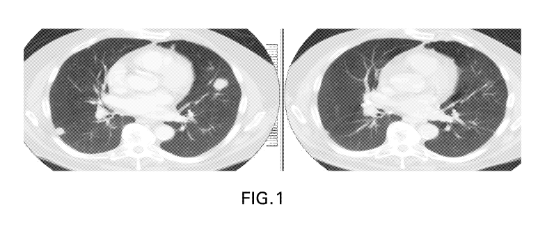
FIG. 1 CT scan of patient with non-MSI-H colorectal cancer before (left) and
after (right)
treatment with 21 mg anti-LAG3 antibody Ab6 and pembrolizumab. The patient
received 5 prior
lines of chemotherapy, no prior anti¨PD-1 or anti¨PD-Li therapy. The patient
had a partial
response with 45% reduction in tumor volume. There was also tumor volume
reduction in lung
lesions and lymph nodes, and stable presacral mass. The response is ongoing at
13.5 months.
FIG. 2 Waterfall plot of subjects with best target lesion change from baseline
based on
investigator assessment per RECIST 1.1 FAS population in the colorectal cancer
cohort (Part B)
using the PD-Li IHC Combined Positive (MIDS+TPS) (Positive: TPS>=1 OR MIDS
>=2) score.
Each bar represents an individual subject. Greater than a 30% decrease in
tumor size from
baseline (Y-axis) is considered a response; changes between a 30% decrease and
a 20% increase
is considered stable disease; changes greater than a 20% increase is
considered progressive
disease. PD-Li positive or negative tumors are indicated. Tumor samples with
less than 100
tumor cells cannot be interpreted. "Empty" indicate missing data.
FIG. 3 Waterfall plot of subjects with best target lesion change from baseline
based on
investigator assessment per RECIST 1.1 FAS population in the colorectal cancer
cohort (Part B)
using the PD-L1 IHC MIDS score. Each bar represents an individual subject.
Greater than a
30% decrease in tumor size from baseline (Y-axis) is considered a response;
changes between a
30% decrease and a 20% increase is considered stable disease, changes greater
than a 20%
increase is considered progressive disease. PD-L1 positive or negative tumors
are indicated.
Tumor samples with less than 100 tumor cells cannot be interpreted. "Empty"
indicate missing
data.
FIG. 4 Waterfall plot of subjects with best target lesion change from baseline
based on
investigator assessment per RECIST 1.1 FAS population in the colorectal cancer
expansion
cohort (Part B) using the PD-Li IHC Combined Positive (MIDS+TPS) score (CPS).
Each bar represents an individual subject. Greater than a 30% decrease in
tumor size from
baseline (Y-axis) is considered a response; changes between a 30% decrease and
a 20% increase
is considered stable disease; changes greater than a 20% increase is
considered progressive
disease. Tumor samples with CPS >=1 or <1 are indicated. Tumor samples with
less than 100
tumor cells cannot be interpreted.
DETAILED DESCRIPTION
Abbreviations. Throughout the detailed description and examples of the
invention the following
abbreviations will be used:
- 4 -
CA 03111066 2021-03-01
WO 2020/055702
PCT/US2019/050122
BOR Best overall response
BID One dose twice daily
CBR Clinical Benefit Rate
CDR Complementarity determining region
CHO Chinese hamster ovary
CR Complete Response
DCR Disease Control Rate
DFS Disease free survival
DLT Dose limiting toxicity
DOR Duration of Response
DSDR Durable Stable Disease Rate
FFPE Formalin-fixed, paraffin-embedded
FR Framework region
IgG Immunoglobulin G
IHC Immunohistochemistry or immunohistochemical
irRC Immune related response criteria
IV Intravenous
MTD Maximum tolerated dose
NCBI National Center for Biotechnology Information
NCI National Cancer Institute
ORR Objective response rate
OS Overall survival
PD Progressive disease
PD-1 Programmed Death 1
PD-Li Programmed Cell Death 1 Ligand 1
PD-L2 Programmed Cell Death 1 Ligand 2
PFS Progression free survival
PR Partial response
Q2W One dose every two weeks
Q3W One dose every three weeks
QD One dose per day
RECIST Response Evaluation Criteria in Solid Tumors
SD Stable disease
VH Immunoglobulin heavy chain variable region
VK Immunoglobulin kappa light chain variable region
- 5 -
CA 03111066 2021-03-01
WO 2020/055702 PCT/US2019/050122
I. DEFINITIONS
So that the invention may be more readily understood, certain technical and
scientific
terms are specifically defined below. Unless specifically defined elsewhere in
this document, all
other technical and scientific terms used herein have the meaning commonly
understood by one
of ordinary skill in the art to which this invention belongs.
As used herein, including the appended claims, the singular forms of words
such as "a,"
"an," and "the," include their corresponding plural references unless the
context clearly dictates
otherwise.
As used herein, an -Ab6 variant" means a monoclonal antibody which comprises
heavy
chain and light chain sequences that are substantially identical to those in
Ab6 (as described
below and in W02016028672, incorporated by reference in its entirety), except
for having three,
two or one conservative amino acid substitutions at positions that are located
outside of the light
chain CDRs and six, five, four, three, two or one conservative amino acid
substitutions that are
.. located outside of the heavy chain CDRs, e.g., the variant positions are
located in the FR regions
or the constant region, and optionally has a deletion of the C-terminal lysine
residue of the heavy
chain. In other words, Ab6 and a Ab6 variant comprise identical CDR sequences,
but differ from
each other due to having a conservative amino acid substitution at no more
than three or six other
positions in their full length light and heavy chain sequences, respectively.
An Ab6 variant is
substantially the same as Ab6 with respect to the following properties:
binding affinity to human
LAG3 and ability to block the binding of human LAG3 to human MHC Class II.
"Administration" as it applies to an animal, human, experimental subject,
cell, tissue,
organ, or biological fluid, refers to contact of an exogenous pharmaceutical,
therapeutic,
diagnostic agent, or composition to the animal, human, subject, cell, tissue,
organ, or biological
fluid. Treatment of a cell encompasses contact of a reagent to the cell, as
well as contact of a
reagent to a fluid, where the fluid is in contact with the cell. The term -
subject" includes any
organism, preferably an animal, more preferably a mammal (e.g., rat, mouse,
dog, cat, rabbit) and
most preferably a human.
As used herein, the term "antibody" refers to any form of antibody that
exhibits the
desired biological or binding activity. Thus, it is used in the broadest sense
and specifically
covers, but is not limited to, monoclonal antibodies (including full length
monoclonal
antibodies), polyclonal antibodies, multispecific antibodies (e.g., bispecific
antibodies),
humanized, fully human antibodies, chimeric antibodies and camelized single
domain antibodies.
- 6 -
CA 03111066 2021-03-01
WO 2020/055702 PCT/US2019/050122
"Parental antibodies" are antibodies obtained by exposure of an immune system
to an antigen
prior to modification of the antibodies for an intended use, such as
humanization of an antibody
for use as a human therapeutic.
In general, the basic antibody structural unit comprises a tetramer. Each
tetramer
includes two identical pairs of polypeptide chains, each pair having one
"light" (about 25 kDa)
and one "heavy" chain (about 50-70 kDa). The amino-terminal portion of each
chain includes a
variable region of about 100 to 110 or more amino acids primarily responsible
for antigen
recognition. The carboxy-terminal portion of the heavy chain may define a
constant region
primarily responsible for effector function. Typically, human light chains are
classified as kappa
and lambda light chains. Furthermore, human heavy chains are typically
classified as mu, delta,
gamma, alpha, or epsilon, and define the antibody's isotype as IgM, IgD, IgG,
IgA, and IgE,
respectively. Within light and heavy chains, the variable and constant regions
are joined by a "J"
region of about 12 or more amino acids, with the heavy chain also including a
"D" region of
about 10 more amino acids. See generally, Fundamental Immunology Ch. 7 (Paul,
W., ed., 2nd
.. ed. Raven Press, N.Y. (1989).
The variable regions of each light/heavy chain pair form the antibody binding
site. Thus,
in general, an intact antibody has two binding sites. Except in bifunctional
or bispecific
antibodies, the two binding sites are, in general, the same.
Typically, the variable domains of both the heavy and light chains comprise
three
hypervariable regions, also called complementarity determining regions (CDRs),
which are
located within relatively conserved framework regions (FR). The CDRs are
usually aligned by
the framework regions, enabling binding to a specific epitope. In general,
from N-terminal to C-
terminal, both light and heavy chains variable domains comprise FRI. CDR1,
FR2, CDR2, FR3,
CDR3 and FR4. The assignment of amino acids to each domain is, generally, in
accordance with
the definitions of Sequences of Proteins of Immunological Interest, Kabat, et
al.; National
=
Institutes of Health, Bethesda, Md. 5th , ed.; NIH Publ. No. 91-3242
(1991); Kabat (1978) Adv.
Prot. Chem. 32:1-75; Kabat, etal., (1977) J. Biol. Chem. 252:6609-6616;
Chothia, etal., (1987)
J Mol. Biol. 196:901-917 or Chothia, etal., (1989) Nature 342:878-883.
As used herein, unless otherwise indicated, "antibody fragment" or "antigen
binding
fragment" refers to antigen binding fragments of antibodies, i.e. antibody
fragments that retain
the ability to bind specifically to the antigen bound by the full-length
antibody, e.g. fragments
that retain one or more CDR regions. Examples of antibody binding fragments
include, but are
not limited to, Fab, Fab', F(ab)2, and FA/ fragments; diabodies; linear
antibodies; single-chain
- 7 -
CA 03111066 2021-03-01
WO 2020/055702 PCT/US2019/050122
antibody molecules, e.g., sc-Fv; nanobodies and multispecific antibodies
formed from antibody
fragments.
An antibody that -specifically binds to" a specified target protein is an
antibody that
exhibits preferential binding to that target as compared to other proteins,
but this specificity does
not require absolute binding specificity. An antibody is considered "specific"
for its intended
target if its binding is determinative of the presence of the target protein
in a sample, e.g. without
producing undesired results such as false positives. Antibodies, or binding
fragments thereof,
useful in the present invention will bind to the target protein with an
affinity that is at least two
fold greater, preferably at least ten times greater, more preferably at least
20-times greater, and
most preferably at least 100-times greater than the affinity with non-target
proteins. As used
herein, an antibody is said to bind specifically to a poly-peptide comprising
a given amino acid
sequence, e.g. the amino acid sequence of a mature human PD-1 or human PD-Li
molecule, if it
binds to polypeptides comprising that sequence but does not bind to proteins
lacking that
sequence.
"Chimeric antibody" refers to an antibody in which a portion of the heavy
and/or light
chain is identical with or homologous to corresponding sequences in an
antibody derived from a
particular species (e.g., human) or belonging to a particular antibody class
or subclass, while the
remainder of the chain(s) is identical with or homologous to corresponding
sequences in an
antibody derived from another species (e.g., mouse) or belonging to another
antibody class or
subclass, as well as fragments of such antibodies, so long as they exhibit the
desired biological
activity.
"Co-administration" as used herein for agents such as the PD-1 antagonist or
LAG3
antagonist means that the agents are administered so as to have overlapping
therapeutic activities,
and not necessarily that the agents are administered simultaneously to the
subject. The agents
may or may not be in physical combination prior to administration. In an
embodiment, the
agents are administered to a subject simultaneously or at about the same time.
For example, the
anti-PD-1 antibody and anti-LAG3 drug products contained in separate vials,
when in liquid
solution, may be mixed into the same intravenous infusion bag or injection
device, and
administered simultaneously to the patient.
-Co-formulated" or -co-formulation" or -coformulation" or "coformulated" as
used
herein refers to at least two different antibodies or antigen binding
fragments thereof which are
formulated together and stored as a combined product in a single vial or
vessel (for example an
injection device) rather than being formulated and stored individually and
then mixed before
- 8 -
CA 03111066 2021-03-01
WO 2020/055702 PCT/US2019/050122
administration or separately administered. In one embodiment, the co-
formulation contains two
different antibodies or antigen binding fragments thereof
"Human antibody" refers to an antibody that comprises human immunoglobulin
protein
sequences only. A human antibody may contain murine carbohydrate chains if
produced in a
mouse, in a mouse cell, or in a hybridoma derived from a mouse cell.
Similarly, "mouse
antibody" or -rat antibody" refer to an antibody that comprises only mouse or
rat
immunoglobulin sequences, respectively.
"Humanized antibody" refers to forms of antibodies that contain sequences from
non-
human (e.g., murine) antibodies as well as human antibodies. Such antibodies
contain minimal
sequence derived from non-human immunoglobulin. In general, the humanized
antibody will
comprise substantially all of at least one, and typically two, variable
domains, in which all or
substantially all of the hypervariable loops correspond to those of a non-
human immunoglobulin
and all or substantially all of the FR regions are those of a human
immunoglobulin sequence.
The humanized antibody optionally also will comprise at least a portion of an
immunoglobulin
constant region (Fc), typically that of a human immunoglobulin. The prefix
"hum", "hu" or "h"
is added to antibody clone designations when necessary to distinguish
humanized antibodies
from parental rodent antibodies. The humanized forms of rodent antibodies will
generally
comprise the same CDR sequences of the parental rodent antibodies, although
certain amino acid
substitutions may be included to increase affinity, increase stability of the
humanized antibody,
or for other reasons.
"Anti-tumor response" when referring to a cancer patient treated with a
therapeutic
regimen, such as a combination therapy described herein, means at least one
positive therapeutic
effect, such as for example, reduced number of cancer cells, reduced tumor
size, reduced rate of
cancer cell infiltration into peripheral organs, reduced rate of tumor
metastasis or tumor growth,
or progression free survival. Positive therapeutic effects in cancer can be
measured in a number
of ways (See, W. A. Weber, J. Null. Med. 50:1S-10S (2009); Eisenhauer et al.,
supra). In some
embodiments, an anti-tumor response to a combination therapy described herein
is assessed
using RECIST 1.1 criteria, bidimentional irRC or unidimensional irRC. In some
embodiments,
an anti-tumor response is any of SD, PR, CR, PFS, or DFS.
"Bidimensional irRC- refers to the set of criteria described in Wolchok JD, et
al. Guidelines
for the evaluation of immune therapy activity in solid tumors: immune-related
response criteria.
Clin Cancer Res. 2009;15(23):7412-7420. These criteria utilize bidimensional
tumor
- 9 -
CA 03111066 2021-03-01
WO 2020/055702 PCT/US2019/050122
measurements of target lesions, which are obtained by multiplying the longest
diameter and the
longest perpendicular diameter (cm2) of each lesion.
"Biotherapeutic agent" means a biological molecule, such as an antibody or
fusion
protein, that blocks ligand / receptor signaling in any biological pathway
that supports tumor
maintenance and/or growth or suppresses the anti-tumor immune response.
Classes of
biotherapeutic agents include, but are not limited to, antibodies to VEGF,
EGFR, Her2/neu, other
growth factor receptors, CD20, CD40, CD-40L, CTLA-4, OX-40, 4-1BB, and ICOS.
"CBR" or "Clinical Benefit Rate- means CR + PR + durable SD
"CDR" or "CDRs" as used herein means complementarily determining region(s) in
a
immunoglobulin variable region, defined using the Kabat numbering system,
unless otherwise
indicated.
"Chemotherapeutic agent" is a chemical compound useful in the treatment of
cancer.
Classes of chemotherapeutic agents include, but are not limited to:
alkylating agents,
antimetabolites, kinase inhibitors, spindle poison plant alkaloids,
cytoxic/antitumor antibiotics,
topisomerase inhibitors, photosensitizers, anti-estrogens and selective
estrogen receptor
modulators (SERMs), anti-progesterones, estrogen receptor down-regulators
(ERDs), estrogen
receptor antagonists, leutinizing hormone-releasing hormone agonists, anti-
androgens, aromatase
inhibitors, EGFR inhibitors, VEGF inhibitors, and anti-sense oligonucleotides
that inhibit
expression of genes implicated in abnormal cell proliferation or tumor growth.
Chemotherapeutic agents useful in the treatment methods of the present
invention include
cytostatic and/or cytotoxic agents.
"Chothia" as used herein means an antibody numbering system described in Al-
Lazikani
et al., JMB 273:927-948 (1997).
"Comprising" or variations such as "comprise", "comprises" or -comprised of'
are used
throughout the specification and claims in an inclusive sense, i.e., to
specify the presence of the
stated features but not to preclude the presence or addition of further
features that may materially
enhance the operation or utility of any of the embodiments of the invention,
unless the context
requires otherwise due to express language or necessary implication.
"Conservatively modified variants" or "conservative substitution" refers to
substitutions
of amino acids in a protein with other amino acids having similar
characteristics (e.g. charge,
side-chain size, hydrophobicity/hydrophilicity, backbone conformation and
rigidity, etc.), such
that the changes can frequently be made without altering the biological
activity or other desired
- 10-
CA 03111066 2021-03-01
WO 2020/055702 PCT/US2019/050122
property of the protein, such as antigen affinity and/or specificirty. Those
of skill in this art
recognize that, in general, single amino acid substitutions in non-essential
regions of a
polypeptide do not substantially alter biological activity (see, e.g., Watson
et al. (1987)
Molecular Biology of the Gene, The Benjamin/Cummings Pub. Co., p. 224 (4th
Ed.)). In
addition, substitutions of structurally or functionally similar amino acids
are less likely to disrupt
biological activity. Exemplary conservative substitutions are set forth in
Table 1 below.
TABLE 1. Exemplary Conservative Amino Acid Substitutions
Original residue Conservative substitution
Ala (A) Gly; S er
Arg (R) Lys; His
Asn (N) Gln; His
Asp (D) _Glu; Asn
Cys (C) Ser: Ala
Gln (Q) Asn
Glu (E) Asp; Gln
Gly (G) Ala
His (H) Asn; Gln
Ile (I) Leu; Val
Leu (L) Ile; Val
Lys (K) Arg; His
Met (M) Leu; Ile; Tyr
Phe (F) Tyr; Met; Leu
Pro (P) Ala
Ser (S) Thr
Thr (T) S er
Trp (W) \Tyr; Phe
Tyr (Y) Trp; Phe
Val (V) Ile; Leu
"Consists essentially of" and variations such as "consist essentially of" or
"consisting essentially
of," as used throughout the specification and claims, indicate the inclusion
of any recited
elements or group of elements, and the optional inclusion of other elements,
of similar or
different nature than the recited elements, that do not materially change the
basic or novel
properties of the specified dosage regimen, method, or composition. As a non-
limiting example,
a PD-1 antagonist that consists essentially of a recited amino acid sequence
may also include one
or more amino acids, including substitutions of one or more amino acid
residues, which do not
materially affect the properties of the binding compound.
-DCR" or -Disease Control Rate" means CR + PR + SD.
"Diagnostic anti-PD-L monoclonal antibody" means a mAb which specifically
binds to
the mature form of the designated PD-L (PD-Li or PDL2) that is expressed on
the surface of
- ii-
CA 03111066 2021-03-01
WO 2020/055702 PCT/US2019/050122
certain mammalian cells. A mature PD-L lacks the presecretory leader sequence,
also referred to
as leader peptide The terms "PD-L" and "mature PD-L" are used interchangeably
herein, and
shall be understood to mean the same molecule unless otherwise indicated or
readily apparent
from the context.
As used herein, a diagnostic anti-human PD-Li mAb or an anti-hPD-L1 mAb refers
to a
monoclonal antibody that specifically binds to mature human PD-L1. A mature
human PD-L1
molecule consists of amino acids 19-290 of the following sequence:
MRI FAVFI FMTYWHLLNAFTVTVP KDLYVVEYG S NMT I ECKFPVEKQL DLAAL IVYWEME DKN I I
QFVHGEEDLKVQHS SYRQRARLLKDQLSLGNAALQITDVKLQDAGVYRCMIS YGGADYKRITVKV
NAPYNKINQRILVVDPVT S EHELT CQAEGY PKAEVIWT SS DHQVLSGKTTTTNSKREEKLFNVTS
TLRINTTTNE I FYCT FRRLDPEENHTAELVIPELPLAH PPNERTHLVILGAILLCLGVALT Fl FR
LRKGRMMDVKKCGIQDTNSKKQS DTHLEET (SEQ ID NO:32).
Specific examples of diagnostic anti-human PD-Li mAbs useful as diagnostic
mAbs for
immunohistochemistry (IHC) detection of PD-Li expression in formalin-fixed,
paraffin-
embedded (FFPE) tumor tissue sections are antibody 20C3 and antibody 22C3,
which are
described in W02014/100079. Another anti-human PD-Li mAb that has been
reported to be
useful for IHC detection of PD-Li expression in FFPE tissue sections (Chen,
B.J. et al., Clin
Cancer Res 19: 3462-3473 (2013)) is a rabbit anti-human PD-L1 mAb publicly
available from
Sino Biological, Inc. (Beijing, P.R. China; Catalog number 10084-R015).
Table 2. Characteristics of Monoclonal Antibody MEB037.22C3 (22C3)
SEQ 1D
Antibody Feature Amino Acid Sequence
NO
Light Chain
CDRL1 KS SQ SLLHT STRKNYL A 13
CDRL2 WASTRES 14
CDRL3 KQSYDVVT 15
D IVM SQ SP S SLAVSAGEKVTMTCKS SQSLLHTSTRKNYLAWYQ
Mature Variable Region QKPGQSPKLLIYWASTRESGVPDRFTGSGSGTDFTLTISSVQAE 16
DLAVYYCKQSYDVVTFGAGTKLELK
Heavy Chain
CDRH1 Kabat Dern SY WIH 17
CDRH1 Chothia Dern GYTFTSYWIH 18
CDRH2 YINPS SGYHEYNQKFID 19
CDRH3 SGWL1HGDYYFDF 20
XVHLQQSGAELAKPGASVKMSCKASGYTFTSYWIHWIKQRPG
QGLEWIGYINPSSGYHEYNQKFIDKATLTADRSSSTAYMHLTSL
Mature Variable Region 21
TSED SAVYYCARS GWL IHGDYYFDFWGQ GTTL TVS S ,
wherein X = Q or pE (pyro-glutamate)
- 12 -
CA 03111066 2021-03-01
WO 2020/055702 PCT/US2019/050122
"PD-Li" or "PD-L2" expression as used herein means any detectable level of
expression
of the designated PD-L protein on the cell surface or of the designated PD-L
mRNA within a cell
or tissue. PD-L protein expression may be detected with a diagnostic PD-L
antibody in an IHC
assay of a tumor tissue section or by flow cytometry. Alternatively, PD-L
protein expression by
tumor cells may be detected by PET imaging, using a binding agent (e.g.,
antibody fragment,
affibody and the like) that specifically binds to the desired PD-L target,
e.g., PD-Li or PD-L2.
Techniques for detecting and measuring PD-L mRNA expression include RT-PCR,
realtime
quantitative RT-PCR, RNAseq, and the Nanostring platform (J. Clin. Invest.
2017.127(8):2930-
2940).
Several approaches have been described for quantifying PD-Li protein
expression in IHC
assays of tumor tissue sections. See, e.g., Thompson, R. H., et al., PNAS 101
(49); 17174-17179
(2004); Thompson, R. H. et al., Cancer Res. 66:3381-3385 (2006); Gadiot, J.,
et al., Cancer
117:2192-2201 (2011); Taube, J. M. et al., Sci Transl 1Vkd 4, 127ra37 (2012);
and Toplian. S. L.
et al., New Eng. J Med. 366 (26): 2443-2454 (2012). See US 20170285037 which
describes
Hematoxylin and Eosin staining used by the pathologist.
One approach employs a simple binary end-point of positive or negative for PD-
L1
expression, with a positive result defined in terms of the percentage of tumor
cells that exhibit
histologic evidence of cell-surface membrane staining. A tumor tissue section
is counted as
positive for PD-Ll expression if it is at least 1% of total tumor cells.
In another approach, PD-Li expression in the tumor tissue section is
quantified in the
tumor cells as well as in infiltrating immune cells, which predominantly
comprise lymphocytes.
The percentage of tumor cells and infiltrating immune cells that exhibit
membrane staining are
separately quantified as < 5%, 5 to 9%, and then in 10% increments up to 100%.
PD-Li
expression in the immune infiltrate is reported as a semi-quantitative
measurement called the
adjusted inflammation score (AIS), which is determined by multiplying the
percent of membrane
staining cells by the intensity of the infiltrate, which is graded as none
(0), mild (score of 1, rare
lymphocytes), moderate (score of 2, focal infiltration of tumor by
lymphohistiocytic aggregates),
or severe (score of 3, diffuse infiltration). A tumor tissue section is
counted as positive for PD-
L1 expression by immune infiltrates if the AIS is > 5.
The level of PD-L mRNA expression may be compared to the mRNA expression
levels
of one or more reference genes that are frequently used in quantitative RT-
PCR.
In some embodiments, a level of PD-Li expression (protein and/or mRNA) by
malignant
cells and/or by infiltrating immune cells within a tumor is determined to be
"overexpressed" or
- 13 -
CA 03111066 2021-03-01
WO 2020/055702 PCT/US2019/050122
"elevated" based on comparison with the level of PD-L1 expression (protein
and/ or mRNA) by
an appropriate control. For example, a control PD-L1 protein or mRNA
expression level may be
the level quantified in nonmalignant cells of the same type or in a section
from a matched normal
tissue. In some preferred embodiments, PD-Li expression in a tumor sample is
determined to be
elevated if PD-LI protein (and/or PD-L1 mRNA) in the sample is at least 10%,
20%, or 30%
greater than in the control.
"Tumor proportion score (TPS)" refers to the percentage of tumor cells
expressing PD-
L1 on the cell membrane at any intensity (weak, moderate or strong). Linear
partial or complete
cell membrane staining is interpreted as positive for PD-Ll.
"Mononuclear inflammatory density score (MIDS)" refers to the ratio of the
number of
PD-Li expressing mononuclear inflammatory cells (MIC) infiltrating or adjacent
to the tumor
(small and large lymphocytes, monocytes, and macrophages within the tumor
nests and the
adjacent supporting stroma) compared to the total number of tumor cells. The
MIDS is recorded
at a scale from 0 to 4 with 0=none; 1=present, but less than one MIC for every
100 tumor cells
(<1%); 2=at least one MIC for every 100 tumor cells, but less than one MIC per
10 tumor cells
(1-9%); 3=at least one MIC for every 10 tumor cells, but fewer MIC's than
tumor cells (10-99%);
4=at least as many MIC's as tumor cells (>100%).
"Combined positive score (CPS)" refers to the ratio of the number of PD-Li
positive
tumor cells and PD-L1 positive mononuclear inflammatory cells (MIC) within the
tumor nests
and the adjacent supporting stroma (numerator) compared to the total number of
tumor cells
(denominator; i.e., the number of PD-Li positive and PD-Li negative tumor
cells). PD-Li
expression at any intensity is considered positive, i.e., weak (1+), moderate
(2+), or strong (3+).
"PD-Li expression positive- refers to a Tumor Proportion Score, Mononuclear
Inflammatory Density Score or Combined Positive Score of at least 1%; MS is?
5; or elevated
level of PD-L1 expression (protein and/or mRNA) by malignant cells and/or by
infiltrating
immune cells within a tumor compared to an appropriate control.
"DSDR" or "Durable Stable Disease Rate" means SD for? 23 weeks.
"Framework region" or "FR" as used herein means the immunoglobulin variable
regions
excluding the CDR regions.
"Kabat" as used herein means an immunoglobulin alignment and numbering system
pioneered by Elvin A. Kabat ((1991) Sequences of Proteins of Immunological
Interest, 5th Ed.
Public Health Service, National Institutes of Health, Bethesda, Md.).
- 14 -
CA 03111066 2021-03-01
WO 2020/055702 PCT/US2019/050122
"LAG3 antagonist" means any chemical compound or biological molecule that
blocks
binding of LAG3 expressed on an immune cell (T cell, Tregs, or NK cell etc.)
to MI-IC Class II
molecules. Human LAG3 comprises the amino acid sequence:
MWEAQFLGLL FLQPLWVAPV KPLQPGAEVP VVWAQEGAPA QLPCSPTIPL QDLSLLRRAG
VTWQHQPDSG PPAAAPGHPL APGPHPAAPS SWGPRPRRYT VLSVGPGGLR SGRIPLQPRV
QLDERGRQRG DFSLWLRPAR RADAGEYRAA VHLRDRALSC RLRLRLGQAS MTASPPGSLR
ASDWVILNCS FSRPDRPASV HWFRNRGQGR VPVRESPHHH LAESFLFLPQ VSPMDSGPWG
CILTYRDGFN VSIMYNLTVL GLEPPTPLTV YAGAGSRVGL PCRLPAGVGT RSFLTAKWTP
PGGGPDLLVT GDNGDFTIRL EDVSQAQAGT YTCHIHLQEQ QLNATVTLAI ITVTPKSFGS
PGSLGKLLCE VT2VSGQERF VWSSLDTPSQ RSFSGPWLEA QEAQLLSQPW QCQLYQGERL
LGAAVYFTEL SSPGAQRSGR APGALPAGHL LLFLILGVLS LLLLVTGAFG FHLWRRQWRP
RRFSALEQGI HPPQAQSKIE ELEQEPEPEP EPEPEPEPEP EPEQL
(SEQ ID NO: 33); see also Uniprot accession no. P18627.
"Microsatellite instability (MSI)" refers to the form of genomic instability
associated with
defective DNA mismatch repair in tumors. See Boland et al., Cancer Research
58, 5258-5257,
1998. In one embodiment, MSI analysis can be carried out using the five
National Cancer
Institute (NCI) recommended microsatellite markers BAT25 (GenBank accession
no. 9834508),
BAT26 (GenBank accession no. 9834505), D55346 (GenBank accession no. 181171),
D25123
(GenBank accession no. 187953), D17S250 (GenBank accession no. 177030).
Additional
markers for example, BAT40, BAT34C4, TGF-13-RII and ACTC can be used.
Commercially
available kits for MSI analysis include, for example, the Promega MSI
multiplex PCR assay.
"High frequency microsatellite instability" or "microsatellite instability-
high (MSI-H)"
refers to if two or more of the five NCI markers show instability or >30-40%
of the total markers
demonstrate instability (i.e. have insertion/deletion mutations).
"Low frequency microsatellite instability" or "microsatellite instability-low
(MSI-L)"
refers to if one of the five NCI markers show instability or <30-40% of the
total markers exhibit
instability (i.e. have insertion/deletion mutations).
"Non-MSI-H colorectal cancer" as used herein refers to microsatellite stable
(MSS) and
low frequency MSI (MSI-L) colorectal cancer.
"Microsatellite Stable (MSS)" refers to if none of the five NCI markers show
instability
(i.e. have insertion/deletion mutations)
"Proficient mismatch repair (pMMR) colorectal cancer" refers to normal
expression of
MMR proteins (MLI-11, PMS2, MSH2, and MSH6) in a CRC tumor specimen by IHC.
Commercially available kits for MMR analysis include theVentana MMR IHC assay.
- 15 -
CA 03111066 2021-03-01
WO 2020/055702 PCT/US2019/050122
"Mismatch repair deficient (dMMR) colorectal cancer" refers to low expression
of one or
more MMR protein(s) (MLHI, PMS2, MSH2, and MSH6) in a CRC tumor specimen by
IHC.
"Monoclonal antibody" or "mAb" or -Mab", as used herein, refers to a
population of
substantially homogeneous antibodies, i.e., the antibody molecules comprising
the population are
identical in amino acid sequence except for possible naturally occurring
mutations that may be
present in minor amounts. In contrast, conventional (polyclonal) antibody
preparations typically
include a multitude of different antibodies having different amino acid
sequences in their
variable domains, particularly their CDRs, which are often specific for
different epitopes. The
modifier "monoclonal" indicates the character of the antibody as being
obtained from a
substantially homogeneous population of antibodies, and is not to be construed
as requiring
production of the antibody by any particular method. For example, the
monoclonal antibodies to
be used in accordance with the present invention may be made by the hybridoma
method first
described by Kohler et al. (1975) Nature 256: 495, or may be made by
recombinant DNA
methods (see, e.g., U.S. Pat. No. 4,816,567). The "monoclonal antibodies" may
also be isolated
from phage antibody libraries using the techniques described in Clackson et
al. (1991) Nature
352: 624-628 and Marks etal. (1991) Mol. Biol. 222: 581-597, for example. See
also Presta
(2005) J Allergy Clin. Immunol. 116:731.
"Non-responder patient", when referring to a specific anti-tumor response to
treatment
with a combination therapy described herein, means the patient did not exhibit
the anti-tumor
response.
"ORR" or "objective response rate" refers in some embodiments to CR + PR, and
ORR(week 24) refers to CR and PR measured using irRECIST in each patient in a
cohort after 24
weeks of anti-cancer treatment.
"Patient" or "subject" refers to any single subject for which therapy is
desired or that is
participating in a clinical trial, epidemiological study or used as a control,
including humans and
mammalian veterinary patients such as cattle, horses, dogs, and cats.
"PD-1 antagonist" means any chemical compound or biological molecule that
blocks
binding of PD-Li expressed on a cancer cell to PD-1 expressed on an immune
cell (T cell, B cell
or NKT cell) and preferably also blocks binding of PD-L2 expressed on a cancer
cell to the
immune-cell expressed PD-1. Alternative names or synonyms for PD-1 and its
ligands include:
PDCD1, PD1, CD279 and SLEB2 for PD-1; PDCD1L1, PDL1, B7H1, B7-4, CD274 and B7-
H
for PD-Li; and PDCDIL2, PDL2, B7-DC, Btdc and CD273 for PD-L2. In any of the
treatment
method, medicaments and uses of the present invention in which a human
individual is being
- 16 -
CA 03111066 2021-03-01
WO 2020/055702 PCT/US2019/050122
treated, the PD-1 antagonist blocks binding of human PD-Ll to human PD-1, and
preferably
blocks binding of both human PD-L1 and PD-L2 to human PD-1. Human PD-1 amino
acid
sequences can be found in NCBI Locus No.: NP 005009. Human PD-Li and PD-L2
amino acid
sequences can be found in NCBI Locus No.: NP_054862 and NP_079515,
respectively.
As used herein, a "pembrolizumab variant" means a monoclonal antibody which
comprises heavy chain and light chain sequences that are substantially
identical to those in
pembrolizumab, except for having three, two or one conservative amino acid
substitutions at
positions that are located outside of the light chain CDRs and six, five,
four, three, two or one
conservative amino acid substitutions that are located outside of the heavy
chain CDRs, e.g, the
variant positions are located in the FR regions or the constant region, and
optionally has a
deletion of the C-terminal lysine residue of the heavy chain. In other words,
pembrolizumab and
a pembrolizumab variant comprise identical CDR sequences, but differ from each
other due to
having a conservative amino acid substitution at no more than three or six
other positions in their
full length light and heavy chain sequences, respectively. A pembrolizumab
variant is
substantially the same as pembrolizumab with respect to the following
properties: binding
affinity to PD-1 and ability to block the binding of each of PD-Li and PD-L2
to PD-1.
"RECIST 1.1 Response Criteria" as used herein means the definitions set forth
in
Eisenhauer et al., E.A. et al., Eur. J Cancer 45:228-247 (2009) for target
lesions or nontarget
lesions, as appropriate based on the context in which response is being
measured.
"Responder patient" when referring to a specific anti-tumor response to
treatment with a
combination therapy described herein, means the patient exhibited the anti-
tumor response.
-Sustained response" means a sustained therapeutic effect after cessation of
treatment
with a therapeutic agent, or a combination therapy described herein. In some
embodiments, the
sustained response has a duration that is at least the same as the treatment
duration, or at least
1.5, 2.0, 2.5 or 3 times longer than the treatment duration.
"Tissue Section" refers to a single part or piece of a tissue sample, e.g., a
thin slice of
tissue cut from a sample of a normal tissue or of a tumor.
"Treat" or "treating" cancer as used herein means to administer a combination
therapy of
a PD-1 antagonist and LAG3 antagonist to a subject having cancer, or diagnosed
with cancer, to
achieve at least one positive therapeutic effect, such as for example, reduced
number of cancer
cells, reduced tumor size, reduced rate of cancer cell infiltration into
peripheral organs, or
reduced rate of tumor metastasis or tumor growth. Positive therapeutic effects
in cancer can be
measured in a number of ways (See, W. A. Weber, J. Nucl. Med. 50:1S-10S
(2009)). For
- 17 -
CA 03111066 2021-03-01
WO 2020/055702 PCT/US2019/050122
example, with respect to tumor growth inhibition, according to NCI standards,
a T/C 42% is the
minimum level of anti-tumor activity. A T/C < 10% is considered a high anti-
tumor activity level,
with T/C (%) = Median tumor volume of the treated/Median tumor volume of the
control x 100.
In some embodiments, response to a combination therapy described herein is
assessed using
RECIST 1.1 criteria or irRC (bidimensional or unidimensional) and the
treatment achieved by a
combination of the invention is any of PR, CR, OR, PFS, DFS and OS. PFS, also
referred to as
"Time to Tumor Progression" indicates the length of time during and after
treatment that the
cancer does not grow, and includes the amount of time patients have
experienced a CR or PR, as
well as the amount of time patients have experienced SD. DFS refers to the
length of time during
and after treatment that the patient remains free of disease. OS refers to a
prolongation in life
expectancy as compared to naive or untreated individuals or patients. In some
embodiments,
response to a combination of the invention is any of PR, CR, PFS, DFS, OR and
OS that is
assessed using RECIST 1.1 response criteria. The treatment regimen for a
combination of the
invention that is effective to treat a cancer patient may vary according to
factors such as the
disease state, age, and weight of the patient, and the ability- of the therapy
to elicit an anti-cancer
response in the subject. While an embodiment of any of the aspects of the
invention may not be
effective in achieving a positive therapeutic effect in every subject, it
should do so in a
statistically significant number of subjects as determined by any statistical
test known in the art
such as the Student's t-test, the chi2-test, the U-test according to Mann and
Whitney, the Kruskal-
Wallis test (H-test), Jonckheere-Terpstra-test and the Wilcoxon-test.
The terms "treatment regimen", "dosing protocol" and "dosing regimen" are used
interchangeably to refer to the dose and timing of administration of each
therapeutic agent in a
combination of the invention.
"Tumor" as it applies to a subject diagnosed with, or suspected of having,
cancer refers to
a malignant or potentially malignant neoplasm or tissue mass of any size, and
includes primary
tumors and secondary neoplasms. A solid tumor is an abnormal growth or mass of
tissue that
usually does not contain cysts or liquid areas. Different types of solid
tumors are named for the
type of cells that form them. Examples of solid tumors are sarcomas,
carcinomas, and
lymphomas. Leukemias (cancers of the blood) generally do not form solid tumors
(National
Cancer Institute, Dictionary of Cancer Terms).
"Tumor burden" also referred to as "tumor load", refers to the total amount of
tumor
material distributed throughout the body. Tumor burden refers to the total
number of cancer cells
or the total size of tumor(s), throughout the body, including lymph nodes and
bone marrow.
Tumor burden can be determined by a variety of methods known in the art, such
as, e.g. by
- 18-
CA 03111066 2021-03-01
WO 2020/055702 PCT/US2019/050122
measuring the dimensions of tumor(s) upon removal from the subject, e.g.,
using calipers, or
while in the body using imaging techniques, e.g., ultrasound, bone scan,
computed tomography
(CT) or magnetic resonance imaging (MRI) scans.
The term "tumor size" refers to the total size of the tumor which can be
measured as the
.. length and width of a tumor. Tumor size may be determined by a variety of
methods known in
the art, such as, e.g. by measuring the dimensions of tumor(s) upon removal
from the subject,
e.g., using calipers, or while in the body using imaging techniques, e.g.,
bone scan, ultrasound,
CT or MRI scans.
"Unidimensional irRC refers to the set of criteria described in Nishino M,
Giobbie-
Hurder A, Gargano M, Suda M, Ramaiya NH, Hodi FS. Developing a Common Language
for
Tumor Response to Immunotherapy: Immune-related Response Criteria using
Unidimensional
measurements. Clin Cancer Res. 2013 ;19(14) : 3936-3943). These criteria
utilize the longest
diameter (cm) of each lesion.
"Variable regions" or "V region" as used herein means the segment of IgG
chains which
is variable in sequence between different antibodies. Typically, it extends to
Kabat residue 109
in the light chain and 113 in the heavy chain.
PD-1 ANTAGONISTS AND LAG3 ANTAGONISTS
PD-1 antagonists useful in the treatment method, medicaments and uses of the
present
invention include a monoclonal antibody (mAb), or antigen binding fragment
thereof, which
specifically binds to PD-1 or PD-L1, and preferably specifically binds to
human PD-1 or human
PD-Li. The mAb may be a human antibody, a humanized antibody or a chimeric
antibody, and
may include a human constant region. In some embodiments the human constant
region is
selected from the group consisting of IgGl, IgG2, IgG3 and IgG4 constant
regions, and in
preferred embodiments, the human constant region is an IgG1 or IgG4 constant
region. In some
embodiments, the antigen binding fragment is selected from the group
consisting of Fab, Fab'-
SH, F(ab)2, scFy and FIT fragments.
Examples of mAbs that bind to human PD-1, and useful in the treatment method,
medicaments and uses of the present invention, are described in US7488802,
US7521051,
US 8008449, US 8354509, US8168757, W02004/004771, W02004/072286,
W02004/056875,
and US2011/0271358. Specific anti-human PD-1 mAbs useful as the PD-1
antagonist in the
treatment method, medicaments and uses of the present invention include:
pembrolizumab (also known as MK-3475), a humanized IgG4 mAb with the structure
described
- 19-
CA 03111066 2021-03-01
WO 2020/055702 PCT/US2019/050122
in WHO Drug Information, Vol. 27, No. 2, pages 161-162 (2013) and which
comprises the heavy
and light chain amino acid sequences shown in Table 3; nivolumab (BMS-936558),
a human
IgG4 mAb with the structure described in WHO Drug Information, Vol. 27, No. 1,
pages 68-69
(2013) and which comprises the heavy and light chain amino acid sequences
shown in Table 3;
the humanized antibodies h409A11, h409A16 and h409A17, which are described in
W02008/156712, and AMP-514, which is being developed by MedImmune.
Examples of mAbs that bind to human PD-L1, and useful in the treatment method,
medicaments and uses of the present invention, are described in W02013/019906,
W02010/077634 Al and US8383796. Specific anti-human PD-L1 mAbs useful as the
PD-1
antagonist in the treatment method, medicaments and uses of the present
invention include
MPDL3280A, BMS-936559, MEDI4736, MSB0010718C and an antibody which comprises
the
heavy chain and light chain variable regions of SEQ ID NO:24 and SEQ ID NO:21,
respectively,
of W02013/019906.
Other PD-1 antagonists useful in the treatment method, medicaments and uses of
the
present invention include an immunoadhesin that specifically binds to PD-1 or
PD-L1, and
preferably specifically binds to human PD-1 or human PD-L1, e.g., a fusion
protein containing
the extracellular or PD-1 binding portion of PD-Li or PD-L2 fused to a
constant region such as
an Fc region of an immunoglobulin molecule. Examples of immunoadhesion
molecules that
specifically bind to PD-1 are described in W02010/027827 and W02011/066342.
Specific
fusion proteins useful as the PD-1 antagonist in the treatment method,
medicaments and uses of
the present invention include AMP-224 (also known as B7-DCIg), which is a PD-
L2-FC fusion
protein and binds to human PD-1.
In some preferred embodiments of the treatment method, medicaments and uses of
the
present invention, the PD-1 antagonist is a monoclonal antibody, or antigen
binding fragment
thereof, which comprises: (a) light chain CDRs SEQ ID NOs: 1, 2 and 3 and (b)
heavy chain
CDRs SEQ ID NOs: 6, 7 and 8.
In other preferred embodiments of the treatment method, medicaments and uses
of the
present invention, the PD-1 antagonist is a monoclonal antibody, or antigen
binding fragment
thereof, which specifically binds to human PD-1 and comprises (a) a heavy
chain variable region
comprising SEQ ID NO:9 or a variant thereof, and (b) a light chain variable
region comprising
SEQ ID NO:4 or a variant thereof A variant of a heavy chain variable region
sequence is
identical to the reference sequence except having up to 17 conservative amino
acid substitutions
in the framework region (i.e., outside of the CDRs), and preferably has less
than ten, nine, eight,
- 20 -
CA 03111066 2021-03-01
WO 2020/055702 PCT/US2019/050122
seven, six or five conservative amino acid substitutions in the framework
region. A variant of a
light chain variable region sequence is identical to the reference sequence
except having up to
five conservative amino acid substitutions in the framework region (i.e.,
outside of the CDRs),
and preferably has less than four, three or two conservative amino acid
substitution in the
framework region.
In another preferred embodiment of the treatment method, medicaments and uses
of the
present invention, the PD-1 antagonist is a monoclonal antibody which
specifically binds to
human PD-1 and comprises (a) a heavy chain comprising SEQ ID NO: 10 and (b) a
light chain
comprising SEQ ID NO:5.
In yet another preferred embodiment of the treatment method, medicaments and
uses of
the present invention, the PD-1 antagonist is a monoclonal antibody which
specifically binds to
human PD-1 and comprises (a) a heavy chain comprising SEQ ID NO: 12 and (b) a
light chain
comprising SEQ ID NO:11.
In all of the above treatment method, medicaments and uses, the PD-1
antagonist inhibits
the binding of PD-Li to PD-1, and preferably also inhibits the binding of PD-
L2 to PD-1. In
some embodiments of the above treatment method, medicaments and uses, the PD-1
antagonist is
a monoclonal antibody, or an antigen binding fragment thereof, which
specifically binds to PD-1
or to PD-L1 and blocks the binding of PD-Li to PD-1. In one embodiment, the PD-
1 antagonist
is an anti-PD-1 antibody which comprises a heavy chain and a light chain, and
wherein the heavy
and light chains comprise the amino acid sequences in SEQ ID NO:10 and SEQ ID
NO:5,
respectively.
Table 3 below provides a list of the amino acid sequences of exemplary anti-PD-
1 mAbs
for use in the treatment method, medicaments and uses of the present
invention.
Table 3. Exemplary PD-1 Antibody Sequences
Antibody Amino Acid Sequence SEQ ID
Feature NO.
Pembrolizumab Light Chain
CDR1 RASKGVSTSGY SYLH 1
CDR2 LASYLES 2
CDR3 QHSRDLPLT 3
Variable EIVLTQSPATLSLSPGERATLSCRASKGVSTSGYSYLHWY 4
Region QQKP GQAPRLLIYLASYLE SGVPARFS GS GS GTDFTLTIS S
LEPEDFAVYYCQHSRDLPLTFGGGTKVEIK
Light Chain EIVLTQSPATLSLSPGERATLSCRASKGVSTSGYSYLHWY 5
QQKP GQAPRLLIYLASYLESGVPARFS G S GS GTDFTLTIS S
LEPEDF AVYYC QH S RDLPLTF GGGTKVEIKRTV AAP SVFI
- 21 -
CA 03111066 2021-03-01
WO 2020/055702 PCT/US2019/050122
Antibody Amino Acid Sequence SEQ ID
Feature NO.
FPPSDEQLKSGTASVVCLLNNFYPREAKVQWKVDNALQS
GNSQESVTEQDSKDSTYSLSSTLTLSKADYEKHKVYACE
VTHQGLSSPVTKSFNRGEC
Pembrolizumab Heavy Chain
CDR1 NYYMY 6
CDR2 GINPSNGGTNFNEKFKN 7
CDR3 RDYRFDMGFDY 8
Variable QVQLVQSGVEVKKPGASVKVSCKASGYTFTNYYMYWV 9
Region RQAPGQGLEWMGGINPSNGGTNFNEKFKNRVTLTTDSST
TTAYMELKSLQFDDTAVYYCARRDYRFDMGFDYWGQG
TTVTVSS
Heavy QVQLVQSGVEVKKPGASVKVSCKASGYTFTNYYMYWV 10
Chain RQAPGQGLEWMGGINPSNGGTNFNEKFKNRVTLTTDSST
TTAYMELKSLQFDDTAVYYCARRDYRFDMGFDYWGQG
TTVTVSSASTKGPSVFPLAPCSRSTSESTAALGCLVKDYFP
EPVTVSWNSGALTSGVHTFPAVLQSSGLYSLSSVVTVPSS
SLGTKTYTCNVDHKPSNTKVDKRVESKYGPPCPPCPAPE
FLGGPSVFLFPPKPKDTLMISRTPEVTCVVVDVSQEDPEV
QFNWYVDGVEVHNAKTKPREEQFNSTYRVVSVLTVLHQ
DWLNGKEYKCKVSNKGLPSSIEKTISKAKGQPREPQVYT
LPPSQEEMTKNQVSLTCLVKGFYPSDIAVEWESNGQPEN
NYKTTPPVLDSDGSFFLYSRLTVDKSRWQEGNVFSCSVM
HEALHNHYTQKSLSLSLGK
Nivolumab Light Chain
Light Chain EIVLTQSPATLSLSPGERATLSCRASQSVSSYLAWYQQKP 11
GQAPRLLIYDASNRATGIPARFSGSGSGTDFTLTISSLEPE
DFAVYYCQQSSNWPRTFGQGTKVEIKRTVAAPSVFIFPPS
DEQLKSGTASVVCLLNNFYPREAKVQWKVDNALQSGNS
QESVTEQDSKDSTYSLSSTLTLSKADYEKHKVYACEVTH
QGLSSPVTKSFNRGEC
Nivolumab Heavy Chain
Heavy QVQLVESGGGVVQPGRSLRLDCKASGITFSNSGMHWVR 12
Chain QAPGKGLEWVAVIWYDGSKRYYADSVKGRFTISRDNSK
NTLFLQMNSLRAEDTAVYYCATNDDYWGQGTLVTVSSA
STKGPSVFPLAPCSRSTSESTAALGCLVKDYFPEPVTVSW
NSGALTSGVHTFPAVLQSSGLYSLSSVVTVPSSSLGTKTY
TCNVDHKPSNTKVDKRVESKYGPPCPPCPAPEFLGGPSVF
LFPPKPKDTLMISRTPEVTCVVVDVSQEDPEVQFNWYVD
GVEVHNAKTKPREEQFNSTYRVVSVLTVLHQDWLNGKE
YKCKVSNKGLPSSIEKTISKAKGQPREPQVYTLPPSQEEM
TKNQVSLTCLVKGFYPSDIAVEWESNGQPENNYKTTPPV
LDSDGSFFLYSRLTVDKSRWQEGNVFSCSVMHEALHNH
YTQKSLSLSLGK
LAG3 antagonists useful in the treatment method, medicaments and uses of the
present
invention include a monoclonal antibody (mAb), or antigen binding fragment
thereof, which
specifically binds to LAG3. The mAb may be a human antibody, a humanized
antibody or a
chimeric antibody, and may include a human constant region. In some
embodiments the human
- 22 -
CA 03111066 2021-03-01
WO 2020/055702 PCT/US2019/050122
constant region is selected from the group consisting of IgG1 IgG2, IgG3 and
IgG4 constant
regions, and in preferred embodiments, the human constant region is an IgG1 or
IgG4 constant
region. In some embodiments; the antigen binding fragment is selected from the
group consisting
of Fab, Fab'-SH, F(a1:)2, scFv and Fv fragments.
In one embodiment, the anti-LAG3 antibody is Ab6.
Ab6: a light chain immunoglobulin comprising the amino acid sequence:
DIVMTQTPLSL SVTPGQPASISCKASQ SLD YEGD SDMNWYLQKP GQPPQLL IYGA SNLESGVPDRFSGSG
SG
TDFTLKISRVEAEDVGVYYCQQSTEDPRTFGGGTKVEIKRTVAAPSVFIFPPSDEQLKSGTASVVCLLNNFY
PREAKVQWKVDNALQSGN SQES VTEQD SKD STY SL SSTLTL SKADYEKHKVYACEVTHQGLSSPVTKSFN
RGEC
(SEQ ID NO: 22); and
a heavy chain immunoglobulin comprising the amino acid sequence:
QMQLVQ SGPEVKKPGTSVKVSCKAS GYTFTDYNVDWVRQARGQRLEWIGD INPNDGGTIYAQKFQERVTI
TVDK S T STA YMEL SSLR SED TA V Y YCARN Y RWF GA1VIDH W GQ GTTV TV SS ASTKGP S
VFPLAP C SR S TSE S
TAALGCLVKDYFPEPVTVSWNSGALTSGVHTFPAVLQSSGLYSLSSVVTVPSSSLGTKTYTCNVDHKPSNT
KVDKRVESKYGPPCPPCPAPEFLGGPSVFLFPPKPKDTLMISRTPEVTCVVVDVSQEDPEVQFNWYVDGVE
VHNAKTKPREEQFNSTYRVVSVLTVLHQDWLNGKEYKCKVSNKGLPSSIEKTISKAKGQPREPQVYTLPPS
QEEMTKNQVSLTCLVK GFYPSD IAVEWESNGQPENNYKTTPPVLD SD G SFFLYSRLTVDKSRWQEGNVFS
CSVMHEALHNHYTQKSL SLSLGK
(SEQ ID NO: 23); or
a light chain immunoglobulin variable domain comprising the amino acid
sequence:
DIVMTQTPLSL SVTPGQPASISCKASQ SLDYEGDSDMNWYLQKPGQPPQLLIYGASNLESGVPDRFSGSGSG
TDFTLKISRVEAEDVGVYYCQQS __ l'EDPRTEGGGTKVEIK
(SEQ ID NO: 24 (CDRs underscored)); and
a heavy chain immunoglobulin variable domain comprising the amino acid
sequence:
QMQLVQS GPEVKK? GT SVKVS CKASGYT FTDYNVDWVRQARGQRLEWIGDINPNDGGT I YAQKFQERVT I
TVDKST ST
AYMELS S IRS EDTAVYYCARNYRWFGAMDHWGQGTTVTVSS
(SEQ ID NO: 25 (CDRs underscored)); or comprising the CDRs:
CDR-L1: KASQSLDYEGDS DMN (SEQ ID NO: 26);
CDR-L2: GASNLES (SEQ ID NO: 27);
CDR-L3: QQ ST EDPRT (SEQ ID NO: 28);
CDR-H1: DYNVD (SEQ ID NO: 29);
CDR-H2: D IN PNDGGT I YAQKFQE (SEQ ID NO: 30); and
CDR-H3: NYRWFGAIvIDH (SEQ ID NO: 31)
In some preferred embodiments of the treatment method, medicaments and uses of
the
present invention, the LAG3 antagonist is a monoclonal antibody, or antigen
binding fragment
- 23 -
CA 03111066 2021-03-01
WO 2020/055702 PCT/US2019/050122
thereof, which comprises: (a) light chain CDRs SEQ ID NOs: 26, 27 and 28 and
(b) heavy chain
CDRs SEQ ID NOs: 29, 30 and 31.
In other preferred embodiments of the treatment method, medicaments and uses
of the
present invention, the LAG3 antagonist is a monoclonal antibody, or antigen
binding fragment
thereof, which specifically binds to human LAG3 and comprises (a) a heavy
chain variable
region comprising SEQ ID NO:25 or a variant thereof, and (b) a light chain
variable region
comprising SEQ ID NO:24 or a variant thereof A variant of a heavy chain
variable region
sequence is identical to the reference sequence except having up to 17
conservative amino acid
substitutions in the framework region (i.e., outside of the CDRs), and
preferably has less than
ten, nine, eight, seven, six or five conservative amino acid substitutions in
the framework region.
A variant of a light chain variable region sequence is identical to the
reference sequence except
having up to five conservative amino acid substitutions in the framework
region (i.e., outside of
the CDRs), and preferably has less than four, three or two conservative amino
acid substitution in
the framework region.
In another preferred embodiment of the treatment method, medicaments and uses
of the
present invention, the LAG3 antagonist is a monoclonal antibody which
specifically binds to
human LAG3 and comprises (a) a heavy chain comprising SEQ ID NO: 23 and (b) a
light chain
comprising SEQ ID NO:22. In another preferred embodiment of the treatment
method,
medicaments and uses of the present invention, the LAG3 antagonist is a
monoclonal antibody
which specifically binds to human LAG3 and comprises (a) a heavy chain
variable region
comprising SEQ ID NO: 25 and (b) a light chain variable region comprising SEQ
ID NO:24.
Other Examples of mAbs that bind to human LAG3, and useful in the treatment
method,
medicaments and uses of the present invention, are relatlimab, IMP731, IMP701,
anti-LAG3
antibodies disclosed in U52017101472. Other LAG3 antagonists useful in the
treatment method,
medicaments and uses of the present invention include an immunoadhesin that
specifically binds
to human LAG3, e.g., a fusion protein containing the extracellular LAG3 fused
to a constant
region such as an Fe region of an immunoglobulin molecule.
In one embodiment, the anti-PD-1 or anti-LAG3 antibody or antigen-binding
fragment
comprises a heavy chain constant region, e.g a human constant region, such as
71, 72, y3, or 74
human heavy chain constant region or a variant thereof In another embodiment,
the anti-LAG3
antibody or antigen-binding fragment comprises a light chain constant region,
e.g. a human light
chain constant region, such as lambda or kappa human light chain region or
variant thereof By
way of example, and not limitation, the human heavy chain constant region can
be y4 and the
- 24 -
CA 03111066 2021-03-01
WO 2020/055702 PCT/US2019/050122
human light chain constant region can be kappa. In an alternative embodiment,
the Fc region of
the antibody is y4 with a Ser228Pro mutation (Schuurman, J et. al., Alol.
Immunol, 38: 1-8,
2001).
In some embodiments, different constant domains may be appended to humanized
Vi. and
Vo regions derived from the CDRs provided herein. For example, if a particular
intended use of
an antibody (or fragment) of the present invention were to call for altered
effector functions, a
heavy chain constant domain other than human IgG1 may be used, or hybrid
IgG1/IgG4 may be
utilized.
Although human IgG1 antibodies provide for long half-life and for effector
functions,
such as complement activation and antibody-dependent cellular cytotoxicity,
such activities may
not be desirable for all uses of the antibody. In such instances a human IgG4
constant domain,
for example, may be used. The present invention includes the use of anti-PD-1
antibodies or
anti-LAG3 antibodies and antigen-binding fragments thereof which comprise an
IgG4 constant
domain. In one embodiment, the IgG4 constant domain can differ from the native
human IgG4
constant domain (Swiss-Prot Accession No. P01861.1) at a position
corresponding to position
228 in the EU system and position 241 in the KABAT system, where the native
Ser108 is
replaced with Pro, in order to prevent a potential inter-chain disulfide bond
between Cys106 and
Cys109 (corresponding to positions Cys 226 and Cys 229 in the EU system and
positions Cys
239 and Cys 242 in the KABAT system) that could interfere with proper intra-
chain disulfide
bond formation. See Angal et al. (1993)Mol. Imunol 30:105. In other instances,
a modified
IgG1 constant domain which has been modified to increase half-life or reduce
effector function
can be used.
METHODS, USES AND MEDICAMENTS
In one aspect of the invention, the invention provides a method for treating
non-MST-H or
pMMR colorectal cancer in an individual comprising co-administering to the
individual a PD-I
antagonist and LAG3 antagonist. In another aspect of the invention, the
invention provides a
method for treating non-MST-H or pMMR colorectal cancer in an individual
comprising
administering to the individual a composition comprising a PD-1 antagonist and
a LAG3
antagonist.
In another embodiment, the invention provides a medicament comprising a PD-1
antagonist for use in combination with a LAG3 antagonist for treating non-MSI-
H or pMMR
colorectal cancer. In yet another embodiment, the invention provides a
medicament comprising a
- 25 -
CA 03111066 2021-03-01
WO 2020/055702 PCT/US2019/050122
LAG3 antagonist for use in combination with a PD-1 antagonist for treating non-
MST-H or
pMMR colorectal cancer.
Other embodiments provide use of a PD-1 antagonist in the manufacture of a
medicament
for treating non-MSI-H or pMMR colorectal cancer in an individual when
administered in
combination with a LAG3 antagonist and use of a LAG3 antagonist in the
manufacture of a
medicament for treating non-MSI-H or pMMR colorectal cancer in an individual
when
administered in combination with a PD-1 antagonist.
In another embodiment, the invention provides a LAG3 antagonist for use in the
treatment of non-MSI-H or pMMR colorectal cancer in an individual, wherein
said use is in
combination with a PD-1 antagonist. In a further embodiment, the invention
provides a
combination of a PD-1 antagonist and a LAG3 antagonist for use in treatment of
a subject with
non-MSI-H or pMMR colorectal cancer.
In a still further embodiment, the invention provides use of a PD-1 antagonist
and a
LAG3 antagonist in the manufacture of a medicament for treating non-MSI-H or
pMMR
colorectal cancer in an individual. In some embodiments, the medicaments
comprise a kit, and
the kit also comprises a package insert comprising instructions for using the
PD-1 antagonist in
combination with the LAG3 antagonist to treat non-MSI-H or pMMR colorectal
cancer in an
individual.
In the foregoing methods, medicaments and uses, in one embodiment, the PD-1
antagonist and LAG3 antagonist are co-formulated. In another embodiment, the
PD-1 antagonist
and LAG3 antagonist are co-administered. The treatment may further comprise
administration of
mFOLFOX7 (Leucovorin (Calcium Folinate), Fluorouracil (5-FU). Oxaliplatin) or
FOLFIRI
(Leucovorin (Calcium Folinate), Fluorouracil (5-FU), Irinotecan
Hydrochloride). In one
embodiment, the PD-1 antagonist is an anti-PD-1 antibody that blocks the
binding of PD-1 to
PD-Li and PD-L2. In one embodiment, the LAG3 antagonist is an anti-LAG3
antibody that
blocks the binding of LAG3 to MHC Class II.
The combination therapy may also comprise one or more additional therapeutic
agents.
The additional therapeutic agent may be, e.g., a chemotherapeutic, a
biotherapeutic agent, an
immunogenic agent (for example, attenuated cancerous cells, tumor antigens,
antigen presenting
.. cells such as dendritic cells pulsed with tumor derived antigen or nucleic
acids, immune
stimulating cytokines (for example, 1L-2, 1FNIct2, GM-CSF), and cells
transfected with genes
encoding immune stimulating cytokines such as but not limited to GM-CSF). The
specific
dosage and dosage schedule of the additional therapeutic agent can further
vary, and the optimal
- 26 -
CA 03111066 2021-03-01
WO 2020/055702 PCT/US2019/050122
dose, dosing schedule and route of administration will be determined based
upon the specific
therapeutic agent that is being used.
Examples of chemotherapeutic agents include alkylating agents such as thiotepa
and
cyclosphosphamide; alkyl sulfonates such as busulfan, improsulfan and
piposulfan; aziridines
such as benzodopa, carboquone, meturedopa, and uredopa; ethylenimines and
methylamelamines
including altretamine, triethylenemelamine, trietylenephosphoramide,
triethylenethiophosphoramide and trimethylolomelamine; acetogenins (especially
bullatacin and
bullatacinone); a camptothecin (including the synthetic analogue topotecan);
bryostatin;
callystatin; CC-1065 (including its adozelesin, carzelesin and bizelesin
synthetic analogues);
.. cryptophycins (particularly cryptophycin 1 and cryptophycin 8); dolastatin;
duocarmycin
(including the synthetic analogues, KW-2189 and CBI-TM1); eleutherobin;
pancratistatin; a
sarcodictyin; spongistatin; nitrogen mustards such as chlorambucil,
chlomaphazine,
cholophosphamide, estramustine, ifosfamide, mechlorethamine, mechlorethamine
oxide
hydrochloride, melphalan, novembichin, phenesterine, prednimustine,
trofosfamide, uracil
mustard; nitrosureas such as carmustine, chlorozotocin, fotemustine,
lomustine, nimustine,
ranimustine; antibiotics such as the enediyne antibiotics (e.g. calicheamicin,
especially
calicheamicin gammalI and calicheamicin phiII, see, e.g., Agnew, Chem. Intl.
Ed. Engl.,
33:183-186 (1994); dynemicin, including dynemicin A; bisphosphonates, such as
clodronate; an
esperamicin; as well as neocarzinostatin chromophore and related chromoprotein
enediyne
antibiotic chromomophores), aclacinomysins, actinomycin, authramycin,
azaserine, bleomycins,
cactinomycin, carabicin, caminomycM, carzinophilin, chromomycins, dactinomycM,
daunorubicin, detorubicin, 6-diazo-5-oxo-L-norleucine, doxorubicin (including
morpholino-
doxorubicin, cyanomorpholino-doxorubicin, 2-pyrrolino-doxorubicin and
deoxydoxorubicin),
epirubicin, esorubicin, idarubicin, marcellomycin, mitomycins such as
mitomycin C,
mycophenolic acid, nogalamycin, olivomycins, peplomycin, potfiromycin,
puromycin,
quelamycin, rodorubicin, streptonigrin, streptozocin, tubercidin, ubenimex,
zinostatin, zorubicin;
anti-metabolites such as methotrexate and 5-fluorouracil (5-FU); folic acid
analogues such as
denopterin, methotrexate, pteropterin, trimetrexate; purine analogs such as
fludarabine, 6-
mercaptopurine, thiamiprine, thioguanine; pyrimidine analogs such as
ancitabine, azacifidine, 6-
azauridine, carmofur, cytarabine, dideoxyuridine, doxifluridine, enocitabine,
floxuridine;
androgens such as calusterone, dromostanolone propionate, epitiostanol,
mepitiostane,
testolactone; anti-adrenals such as aminoglutethimide, mitotane, trilostane;
folic acid replenisher
such as frolinic acid; aceglatone; aldophosphamide glycoside; aminolevulinic
acid; eniluracil;
amsacrine; bestrabucil; bisantrene; edatraxate; defofamine; demecolcine;
diaziquone;
- 27 -
CA 03111066 2021-03-01
WO 2020/055702 PCT/US2019/050122
elformithine; elliptinium acetate; an epothilone; etoglucid; gallium nitrate;
hydroxyurea;
lentinan; lonidamine; maytansinoids such as maytansine and ansamitocins;
mitoguazone;
mitoxantrone; mopidamol; nitracrine; pentostatin; phenamet; pirarubicin;
losoxantrone;
podophyllinic acid; 2-ethylhydrazide; procarbazine; razoxane; rhizoxin;
sizofuran:
spirogermanium; tenuazonic acid; triaziquone; 2, 2',2"-trichlorotriethylamine;
trichothecenes
(especially T-2 toxin, verracurin A, roridin A and anguidine); urethan;
vindesine; dacarbazine;
mannomustine; mitobronitol; mitolactol; pipobroman; gacytosine; arabinoside
("Ara-C");
cyclophosphamide; thiotepa; taxoids, e.g. paclitaxel and doxetaxel;
chlorambucil; gemcitabine;
6-thioguanine; mercaptopurine; methotrexate; platinum analogs such as
cisplatin and carboplatin;
vinblastine; platinum; etoposide (VP-16); ifosfamide; mitoxantrone;
vincristine; vinorelbine;
novantrone; teniposide; edatrexate; daunomycin; aminopterin; xeloda;
ibandronate; CPT-11;
topoisomerase inhibitor RFS 2000; difluoromethylomithine (DMF0); retinoids
such as retinoic
acid; capecitabine: and pharmaceutically acceptable salts, acids or
derivatives of any of the
above. Also included are anti-hormonal agents that act to regulate or inhibit
hoimone action on
tumors such as anti-estrogens and selective estrogen receptor modulators
(SERMs), including,
for example, tamoxifen, raloxifene, droloxifene, 4-hydroxytamoxifen,
trioxifene, keoxifene,
LY117018, onapristone, and toremifene (Fareston); aromatase inhibitors that
inhibit the enzyme
aromatase, which regulates estrogen production in the adrenal glands, such as,
for example, 4(5)-
imidazoles, aminoglutethimide, megestrol acetate, exemestane, formestane,
fadrozole, vorozole,
letrozole, and anastrozole; and anti-androgens such as flutamide, nilutamide,
bicalutamide,
leuprolide, and goserelin; and pharmaceutically acceptable salts, acids or
derivatives of any of
the above.
Each therapeutic agent in a combination therapy of the invention may be
administered
either alone or in a medicament (also referred to herein as a pharmaceutical
composition) which
comprises the therapeutic agent and one or more pharmaceutically acceptable
carriers, excipients
and diluents, according to standard pharmaceutical practice.
Each therapeutic agent in a combination therapy of the invention may be
administered
simultaneously (i.e., in the same medicament), concurrently (i.e., in separate
medicaments
administered one right after the other in any order) or sequentially in any
order. Sequential
administration is particularly useful when the therapeutic agents in the
combination therapy are
in different dosage forms (one agent is a tablet or capsule and another agent
is a sterile liquid)
and/or are administered on different dosing schedules, e.g., a
chemotherapeutic that is
administered at least daily and a biotherapeutic that is administered less
frequently, such as once
weekly, once every two weeks, or once every three weeks.
- 28 -
CA 03111066 2021-03-01
WO 2020/055702 PCT/US2019/050122
In some embodiments, the LAG3 antagonist is administered before administration
of the
PD-1 antagonist, while in other embodiments, the LAG3 antagonist is
administered after
administration of the PD-1 antagonist. In another embodiment, the LAG3
antagonist is
administered concurrently with the PD-1 antagonist.
In some embodiments, at least one of the therapeutic agents in the combination
therapy is
administered using the same dosage regimen (dose, frequency and duration of
treatment) that is
typically employed when the agent is used as monotherapy for treating the same
cancer. In other
embodiments, the patient receives a lower total amount of at least one of the
therapeutic agents in
the combination therapy than when the agent is used as monotherapy, e.g..
smaller doses, less
frequent doses, and/or shorter treatment duration.
Each small molecule therapeutic agent in a combination therapy of the
invention can be
administered orally or parenterally, including the intravenous, intramuscular,
intraperitoneal,
subcutaneous, rectal, topical, and transdermal routes of administration.
A combination therapy of the invention may be used prior to or following
surgery to
remove a tumor and may be used prior to, during or after radiation therapy.
In some embodiments, a combination therapy of the invention is administered to
a patient
who has not been previously treated with a biotherapeutic or chemotherapeutic
agent, i.e., is
treatment-naïve. In other embodiments, the combination therapy is administered
to a patient who
failed to achieve a sustained response after prior therapy with a
biotherapeutic or
chemotherapeutic agent, i.e., is treatment-experienced.
A combination therapy of the invention is typically used to treat a tumor that
is large
enough to be found by palpation or by imaging techniques well known in the
art, such as MRI,
ultrasound, or CAT scan.
A combination therapy of the invention is preferably administered to a human
patient
who has a non-MSI-H or pMMR colorectal cancer that tests positive for one or
both of PD-Li
and PD-L2, and preferably tests positive for PD-L1 expression. In some
preferred embodiments,
PD-L1 expression is detected using a diagnostic anti-human PD-L1 antibody, or
antigen binding
fragment thereof, in an IHC assay on an FFPE or frozen tissue section of a
tumor sample
removed from the patient. Typically, the patient's physician would order a
diagnostic test to
determine PD-Li expression in a tumor tissue sample removed from the patient
prior to initiation
of treatment with the PD-1 antagonist and the LAG3 antagonist , but it is
envisioned that the
physician could order the first or subsequent diagnostic tests at any time
after initiation of
treatment, such as for example after completion of a treatment cycle. In one
embodiment, the
- 29 -
CA 03111066 2021-03-01
WO 2020/055702 PCT/US2019/050122
PD-Ll expression is measured by the PD-Ll IHC 22C3 pharmDx assay. In another
embodiment, the patient has a Mononuclear Inflammatory Density Score for PD-L1
expression
>2. In another embodiment, the patient has a Mononuclear Inflammatory Density
Score for PD-
L1 expression >3. In another embodiment, the patient has a Mononuclear
Inflammatory Density
Score for PD-Li expression >4. In another embodiment, the patient has a Tumor
Proportion
Score for PD-Li expression >1%. In another embodiment, the patient has a Tumor
Proportion
Score for PD-Li expression >10%. In another embodiment, the patient has a
Tumor Proportion
Score for PD-Li expression >20%. In another embodiment, the patient has a
Tumor Proportion
Score for PD-L1 expression >30%. In a further embodiment, the patient has a
Combined
Positive Score for PD-Li expression >1%. In a further embodiment, the patient
has a Combined
Positive Score for PD-Li expression between 1 and 20 %. In a further
embodiment, the patient
has a Combined Positive Score for PD-L1 expression > 2%. In a further
embodiment, the patient
has a Combined Positive Score for PD-L1 expression > 5%. In yet a further
embodiment, the
patient has a Combined Positive Score for PD-Li expression > 10%. In a further
embodiment,
the patient has a Combined Positive Score for PD-Li expression > 15%. In yet a
further
embodiment, the patient has a Combined Positive Score for PD-Li expression >
20%.
Selecting a dosage regimen (also referred to herein as an administration
regimen) for a
combination therapy of the invention depends on several factors, including the
serum or tissue
turnover rate of the entity, the level of symptoms, the immunogenicity of the
entity, and the
accessibility of the target cells, tissue or organ in the individual being
treated. Preferably, a
dosage regimen maximizes the amount of each therapeutic agent delivered to the
patient
consistent with an acceptable level of side effects. Accordingly, the dose
amount and dosing
frequency of each biotherapeutic and chemotherapeutic agent in the combination
depends in part
on the particular therapeutic agent, the severity of the cancer being treated,
and patient
characteristics. Guidance in selecting appropriate doses of antibodies,
cytokines, and small
molecules are available. See, e.g., Wawrzynczak (1996) Antibody Therapy, Bios
Scientific Pub.
Ltd, Oxfordshire, UK; Kresina (ed.) (1991) Monoclonal Antibodies, Cytokines
and Arthritis,
Marcel Dekker, New York, NY; Bach (ed.) (1993) Monoclonal Antibodies and
Peptide Therapy
in Autoimmune Diseases, Marcel Dekker, New York, NY; Baert et al. (2003) New
Engl. J Med.
348:601-608; Milgrom et al. (1999) New Engl. J Med. 341:1966-1973; Slamon et
al. (2001)
New Engl. Med. 344:783-792; Beniaminovitz et al. (2000) New Engl. J. Med.
342:613-619;
Ghosh et al. (2003) New Engl. I Med. 348:24-32; Lipsky et al. (2000) New Engl.
I Med.
343:1594-1602; Physicians Desk Reference 2003 (Physicians' Desk Reference,
57th Ed);
Medical Economics Company; ISBN: 1563634457; 57th edition (November 2002).
- 30 -
CA 03111066 2021-03-01
WO 2020/055702 PCT/US2019/050122
Determination of the appropriate dosage regimen may be made by the clinician,
e.g., using
parameters or factors known or suspected in the art to affect treatment or
predicted to affect
treatment, and will depend, for example, the patient's clinical history (e.g.,
previous therapy), the
type and stage of the cancer to be treated and biomarkers of response to one
or more of the
therapeutic agents in the combination therapy.
Biotherapeutic agents in a combination therapy of the invention may be
administered by
continuous infusion, or by doses at intervals of, e.g., daily, every other
day, three times per week,
or one time each week, two weeks, three weeks, monthly, bimonthly, etc. A
total weekly dose is
generally at least 0.05 ug/kg, 0.2 ug/kg, 0.5 ug/kg, 1 ig/kg, 10 pg/kg, 100
pg/kg, 0.2 mg/kg, 1.0
mg/kg, 2.0 mg/kg, 10 mg/kg, 25 mg/kg, 50 mg/kg body weight or more. See, e.g.,
Yang et al.
(2003) New Engl. I Med. 349:427-434; Herold etal. (2002) New Engl. J Med.
346:1692-1698;
Liu et al. (1999) 1 Neurol. Neurosurg. Psych. 67:451-456; Portielji et al.
(20003) Cancer
Immunol. Immunother. 52:133-144.
In some embodiments that employ an anti-human PD-1 mAb as the PD-1 antagonist
in
the combination therapy, the dosing regimen will comprise administering the
anti-human PD-1
mAb at a dose of 1, 2, 3, 5 or 10mg/kg at intervals of about 14 days ( 2
days) or about 21 days
( 2 days) or about 30 days ( 2 days) throughout the course of treatment.
In other embodiments that employ an anti-human PD-1 mAb as the PD-1 antagonist
in
the combination therapy, the dosing regimen will comprise administering the
anti-human PD-1
mAb at a dose of from about 0.005 mg/kg to about 10 mg/kg, with intra-patient
dose escalation.
In other escalating dose embodiments, the interval between doses will be
progressively
shortened, e.g., about 30 days ( 2 days) between the first and second dose,
about 14 days ( 2
days) between the second and third doses. In certain embodiments, the dosing
interval will be
about 14 days ( 2 days), for doses subsequent to the second dose.
In certain embodiments, a subject will be administered an intravenous (IV)
infusion of a
medicament comprising any of the PD-1 antagonists described herein.
In one preferred embodiment of the invention, the PD-1 antagonist in the
combination
therapy is nivolumab, which is administered intravenously at a dose selected
from the group
consisting of: 1 mg/kg Q2W, 2 mg/kg Q2W, 3 mg/kg Q2W, 5 mg/kg Q2W, 10 mg Q2W,
1
mg/kg Q3W, 2 mg/kg Q3W, 3 mg/kg Q3W, 5 mg/kg Q3W, and 10 mg/kg Q3W.
In another preferred embodiment of the invention, the PD-1 antagonist in the
combination
therapy is pembrolizumab, or a pembrolizumab variant, which is administered in
a liquid
medicament at a dose selected from the group consisting of 1 mg/kg Q2W, 2
mg/kg Q2W, 3
-31 -
CA 03111066 2021-03-01
WO 2020/055702 PCT/US2019/050122
mg/kg Q2W, 5 mg/kg Q2W, 10 mg/kg Q2W, 1 mg/kg Q3W, 2 mg/kg Q3W, 3 mg/kg Q3W, 5
mg/kg Q3W, 10 mg/kg Q3W and flat-dose equivalents of any of these doses, i.e.,
such as 200 mg
Q3W. In some embodiments, pembrolizumab is provided as a liquid medicament
which
comprises 25 mg/ml pembrolizumab, 7% (w/v) sucrose, 0.02% (w/v) polysorbate 80
in 10 mM
histidine buffer pH 5.5. In other embodiments, pembrolizumab is provided as a
liquid
medicament which comprises about 125 to about 200 mg/mL of pembrolizumab, or
antigen
binding fragment thereof; about 10 mM histidine buffer; about 10 mM L-
methionine, or a
pharmaceutically acceptable salt thereof; about 7% (w/v) sucrose; and about
0.02 % (w/v)
polysorbate 80.
In some embodiments, the selected dose of pembrolizumab is administered by IV
infusion. In one embodiment, the selected dose of pembrolizumab is
administered by IV infusion
over a time period of between 25 and 40 minutes, or about 30 minutes.
In some embodiments, the patient is treated with the combination therapy for
at least 24
weeks, e.g., eight 3-week cycles. In some embodiments, treatment with the
combination therapy
continues until the patient exhibits evidence of PD or a CR.
The present invention also provides a medicament which comprises a PD-1 or
LAG3
antagonist as described above and a pharmaceutically acceptable excipient.
When the PD-1
antagonist or LAG3 antagonist is a biotherapeutic agent, e.g., a mAb, the
antagonist may be
produced in CHO cells using conventional cell culture and
recovery/purification technologies.
Pharmaceutically acceptable excipients of the present disclosure include for
instance,
solvents, bulking agents, buffering agents, tonicity adjusting agents, and
preservatives (see, e.g.,.
Pramanick et al., Pharma Times, 45:65-77, 2013). In some embodiments the
pharmaceutical
compositions may comprise an excipient that functions as one or more of a
solvent, a bulking
agent, a buffering agent, and a tonicity adjusting agent (e.g., sodium
chloride in saline may serve
as both an aqueous vehicle and a tonicity adjusting agent). The pharmaceutical
compositions of
the present disclosure are suitable for parenteral administration.
In some embodiments, the pharmaceutical compositions comprise an aqueous
vehicle as a
solvent. Suitable vehicles include for instance sterile water, saline
solution, phosphate buffered
saline, and Ringer's solution. In some embodiments, the composition is
isotonic.
The pharmaceutical compositions may comprise a bulking agent. Bulking agents
are
particularly useful when the pharmaceutical composition is to be lyophilized
before
administration. In some embodiments, the bulking agent is a protectant that
aids in the
stabilization and prevention of degradation of the active agents during freeze
or spray drying
- 32 -
CA 03111066 2021-03-01
WO 2020/055702
PCT/US2019/050122
and/or during storage. Suitable bulking agents are sugars (mono-, di- and
polysaccharides) such
as sucrose, lactose, trehalose, mannitol, sorbital, glucose and raffinose.
The pharmaceutical compositions may comprise a buffering agent. Buffering
agents
control pH to inhibit degradation of the active agent during processing,
storage and optionally
reconstitution. Suitable buffers include for instance salts comprising
acetate, citrate, phosphate
or sulfate. Other suitable buffers include for instance amino acids such as
arginine, glycine,
histidine, and lysine. The buffering agent may further comprise hydrochloric
acid or sodium
hydroxide. In some embodiments, the buffering agent maintains the pH of the
composition
within a range of 4 to 9. In some embodiments, the pH is greater than (lower
limit) 4, 5, 6, 7 or 8.
In some embodiments, the pH is less than (upper limit) 9, 8, 7, 6 or 5. That
is, the pH is in the
range of from about 4 to 9 in which the lower limit is less than the upper
limit.
The pharmaceutical compositions may comprise a tonicity adjusting agent.
Suitable
tonicity adjusting agents include for instance dextrose, glycerol, sodium
chloride, glycerin and
mannitol.
The pharmaceutical compositions may comprise a preservative. Suitable
preservatives
include for instance antioxidants and antimicrobial agents. However, in
preferred embodiments,
the pharmaceutical composition is prepared under sterile conditions and is in
a single use
container, and thus does not necessitate inclusion of a preservative.
In some embodiments, a medicament comprising an anti-PD-1 antibody as the PD-1
antagonist may be provided as a liquid formulation or prepared by
reconstituting a lyophilized
powder with sterile water for injection prior to use. WO 2012/135408 describes
the preparation
of liquid and lyophilized medicaments comprising pembrolizumab that are
suitable for use in the
present invention. In some embodiments, a medicament comprising pembrolizumab
is provided
in a glass vial which contains about 100 mg of pembrolizumab in 4 ml of
solution. Each 1 mL of
solution contains 25 mg of pembrolizumab and is formulated in: L-histidine
(1.55 mg),
polysorbate 80 (0.2 mg), sucrose (70 mg), and Water for Injection, USP. The
solution requires
dilution for IV infusion.
In some embodiments, a medicament comprising an anti-LAG3 antibody as the LAG3
antagonist may be provided as a liquid formulation or prepared by
reconstituting a lyophilized
powder with sterile water for injection prior to use. In one embodiment, the
liquid formulation
comprises about 25 mg/mL anti-LAG3 antibody; about 50 mg,'mL sucrose; about
0.2 mg/mL
polysorbate 80; about 10 mM L-histidine buffer at about pH 5.8-6.0; about 70
mM L-Arginine-
HC1 thereof; and optionally about 10 mM L-methionine.
- 33 -
CA 03111066 2021-03-01
WO 2020/055702 PCT/US2019/050122
The medicaments described herein may be provided as a kit which comprises a
first
container and a second container and a package insert. The first container
contains at least one
dose of a medicament comprising a PD-1 antagonist, the second container
contains at least one
dose of a medicament comprising a LAG3 antagonist, and the package insert, or
label, which
comprises instructions for treating a patient for non-MSI-H or pMMR colorectal
cancer using the
medicaments. The first and second containers may be comprised of the same or
different shape
(e.g., vials, syringes and bottles) and/or material (e.g., plastic or glass).
The kit may further
comprise other materials that may be useful in administering the medicaments,
such as diluents,
filters, IV bags and lines, needles and syringes. In some preferred
embodiments of the kit, the
.. PD-1 antagonist is an anti-PD-1 antibody and the instructions state that
the medicaments are
intended for use in treating a patient having a non-MSI-H or pMMR colorectal
cancer that tests
positive for PD-L1 expression by an IHC assay.
In other aspects, the medicament is a co-formulation of anti-LAG3 antibodies
or antigen
binding fragments and anti-PD-1 antibodies or antigen binding fragments with
arginine or a
pharmaceutically acceptable salt thereof at a total concentration of 10-1000
mM, and a buffer at
pH about 5-8, and optionally 3-100 mM of methionine. In one embodiment, the co-
formulation
comprises about 10 to 120 mg/mL of an anti-LAG3 antibody; about 10 to 120
mg/mL of an anti-
PD-1 antibody; about 30 to 120 mg/mL sucrose or trehalose; about 0.05 to 2
mg/mL polysorbate
80; about 3 to 30 mM L-histidine buffer at pH about 5.0-6.5; about 40 to 150
rriM L-arginine or a
pharmaceutically acceptable salt thereof; and optionally, about 5 to 70 mM L-
methionine.
These and other aspects of the invention, including the exemplary specific
embodiments
listed below, will be apparent from the teachings contained herein.
Exemplary Specific Embodiments of the Invention
1. A LAG3 antagonist for use in the treatment of non-microsatellite
instability-high (non-
MSI-H) or proficient mismatch repair (pMMR) colorectal cancer, wherein the use
is in
combination with a PD-1 antagonist.
2. The LAG3 antagonist for use of embodiment 1, wherein the PD-1 antagonist
is a
monoclonal antibody, or an antigen binding fragment thereof
3. The LAG3 antagonist for use of embodiment 1, wherein the individual is a
human and the
PD-1 antagonist is a monoclonal antibody, or an antigen binding fragment
thereof, which
specifically binds to human PD-1 and blocks the binding of human PD-Li to
human PD-
1.
- 34 -
CA 03111066 2021-03-01
WO 2020/055702 PCT/US2019/050122
4. The LAG3 antagonist for use of embodiment 3, wherein the PD-1 antagonist
also blocks
binding of human PD-L2 to human PD-1.
5. The LAG3 antagonist for use of embodiment 4, wherein the PD-1 antagonist
is a
monoclonal antibody, or antigen binding fragment thereof, which comprises: (a)
light
chain CDRs of SEQ ID NOs: 1, 2 and 3 and (b) heavy chain CDRs of SEQ ID NOs:
6, 7
and 8.
6. The LAG3 antagonist for use of embodiment 4, wherein the PD-1 antagonist
is an anti-
PD-1 monoclonal antibody which comprises a heavy chain and a light chain, and
wherein
the heavy chain comprises a heavy chain variable region comprising SEQ ID NO:9
and
the light chain comprises a light chain variable region comprising SEQ ID NO:
4.
7. The LAG3 antagonist for use of embodiment 4, wherein the PD-1 antagonist
is an anti-
PD-1 monoclonal antibody which comprises a heavy chain and a light chain, and
wherein
the heavy chain comprises SEQ ID NO:10 and the light chain comprises SEQ ID
NO:5.
8. The LAG3 antagonist for use of embodiment 4, wherein the PD-1 antagonist
is
pembrolizumab.
9. The LAG3 antagonist for use of embodiment 4, wherein the PD-1 antagonist
is a
pembrolizumab variant.
10. The LAG3 antagonist for use of embodiment 4, wherein the PD-1
antagonist is
nivolumab.
11. The LAG3 antagonist for use of any one of embodiments 1 to 10, wherein
the LAG3
antagonist is a monoclonal antibody, or an antigen binding fragment thereof
that blocks
the binding of LAG3 to MHC Class II molecules.
12. The LAG3 antagonist for use of any one of embodiments 1 to 10, wherein
the LAG3
antagonist is an antibody, or antigen binding fragment thereof, which
comprises: (a) light
chain CDRs of SEQ ID NOs: 26, 27 and 28 and (b) heavy chain CDRs of SEQ ID
NOs:
29, 30 and 31.
13. The LAG3 antagonist for use of any one of embodiments 1 to 10, wherein
the LAG3
antagonist is an anti-LAG3 antibody which comprises a heavy chain and a light
chain,
and wherein the heavy chain comprises a heavy chain variable region comprising
SEQ ID
NO:25 and the light chain comprises a light chain variable region comprising
SEQ ID
NO: 24.
- 35 -
CA 03111066 2021-03-01
WO 2020/055702 PCT/US2019/050122
14. The LAG3 antagonist for use of any one of embodiments 1 to 10,
wherein the LAG3
antagonist is an anti-LAG3 antibody which comprises a heavy chain and a light
chain,
and wherein the heavy chain comprises SEQ ID NO:23 and the light chain
comprises
SEQ ID NO:22.
15. The LAG3 antagonist for use of any one of embodiments 1 to 10, wherein
the LAG3
antagonist is an Ab6 variant.
16. The LAG3 antagonist for use of any one of embodiments 1 to 10, wherein
the LAG3
antagonist is relatlimab.
17. The LAG3 antagonist for use of embodiment 1, wherein the PD-1
antagonist is a
humanized anti-PD-1 antibody that comprises a heavy chain and a light chain,
and
wherein the heavy chain comprises a heavy chain variable region comprising
heavy chain
CDRs of SEQ ID NOs: 6, 7 and 8 and the light chain comprises a light chain
variable
region comprising light chain CDRs of SEQ ID NOs: 1, 2 and 3; and the LAG3
antagonist is a humanized anti-LAG3 antibody which comprises a heavy chain and
a light
chain, and wherein the heavy chain comprises a heavy chain variable region
comprising
heavy chain CDRs of SEQ ID NOs: 29, 30 and 31 and the light chain comprises a
light
chain variable region comprising light chain CDRs of SEQ ID NOs: 26, 27 and
28.
18. The LAG3 antagonist for use of embodiment 1, wherein the PD-1
antagonist is an anti-
PD-1 antibody that comprises a heavy chain and a light chain, and wherein the
heavy
chain comprises a heavy chain variable region comprising SEQ ID NO:9 and the
light
chain comprises a light chain variable region comprising SEQ ID NO: 4, and the
LAG3
antagonist is an anti-LAG3 antibody which comprises a heavy chain and a light
chain,
and wherein the heavy chain comprises a heavy chain variable region comprising
SEQ ID
NO:25 and the light chain comprises a light chain variable region comprising
SEQ ID
NO: 24.
19. The LAG3 antagonist for use of embodiment 1, wherein the PD-1
antagonist is an anti-
PD-1 antibody that comprises a heavy chain and a light chain, and wherein the
heavy
chain comprises SEQ ID NO:10 and the light chain comprises SEQ ID NO: 5; and
the
LAG3 antagonist is an anti-LAG3 antibody which comprises a heavy chain and a
light
chain, and wherein the heavy chain comprises SEQ ID NO:23 and the light chain
comprises SEQ ID NO: 22.
20. The LAG3 antagonist for use of any one of embodiments 1 to 19, wherein
the PD-1
antagonist and LAG3 antagonist are co-formulated.
- 36 -
CA 03111066 2021-03-01
WO 2020/055702 PCT/US2019/050122
21. The LAG3 antagonist for use of any one of embodiments 1 to 19, wherein
the PD-1
antagonist and LAG3 antagonist are co-administered.
22. The LAG3 antagonist for use of any one of embodiments 1 to 21, wherein
the individual
has not been previously treated with anti-PD-1 or anti-PD-Li therapy or is
confirmed
progressive while receiving prior anti-PD-1 therapy.
23. The LAG3 antagonist for use of any one of embodiments 1 to 22, wherein
the tumor cells
of the individual is PD-Li expression positive.
24. The LAG3 antagonist for use of any one of embodiments 1 to 22, wherein
the individual
has a Mononuclear Inflammatory Density Score for PD-Li expression > 2.
25. The LAG3 antagonist for use of any one of embodiments 1 to 22, wherein
the individual
has a Combined Positive Score for PD-Li expression >1%.
26. The LAG3 antagonist for use of embodiment 24 or 25, wherein the PD-Li
expression is
measured by the PD-Ll INC 22C3 pharmDx assay.
27. The LAG3 antagonist for use of any one of embodiments 1-26, further
comprising
administering mFOLFOX7 (Oxaliplatin, Leucovorin and 5-FU) or FOLFIRI
(irinotecan,
Leucovorin and 5-FU).
GENERAL METHODS
Standard methods in molecular biology are described Sambrook, Fritsch and
Maniatis
(1982 & 1989 2"d Edition, 2001 3rd Edition) Molecular Cloning, A Laboratory
Manual, Cold
.. Spring Harbor Laboratory Press, Cold Spring Harbor, NY; Sambrook and
Russell (2001)
Molecular Cloning. 3rd ed., Cold Spring Harbor Laboratory Press, Cold Spring
Harbor, NY; Wu
(1993) Recombinant DNA, Vol. 217, Academic Press, San Diego, CA). Standard
methods also
appear in Ausbel, et al. (2001) Current Protocols in Molecular Biology, Vols.1-
4, John Wiley
and Sons, Inc. New York, NY, which describes cloning in bacterial cells and
DNA mutagenesis
(Vol. 1), cloning in mammalian cells and yeast (Vol. 2), glycoconjugates and
protein expression
(Vol. 3), and bioinformatics (Vol. 4).
Methods for protein purification including immunoprecipitation,
chromatography,
electrophoresis, centrifugation, and crystallization are described (Coligan,
et al. (2000) Current
Protocols in Protein Science, Vol. 1. John Wiley and Sons, Inc., New York).
Chemical analysis,
chemical modification, post-translational modification, production of fusion
proteins,
glycosylation of proteins are described (see, e.g., Coligan, et al. (2000)
Current Protocols in
Protein Science, Vol. 2, John Wiley and Sons, Inc., New York; Ausubel, et al.
(2001) Current
- 37 -
CA 03111066 2021-03-01
WO 2020/055702 PCT/1JS2019/050122
Protocols in Molecular Biology, Vol. 3, John Wiley and Sons, Inc., NY, NY, pp.
16Ø5-16.22.17;
Sigma-Aldrich, Co. (2001) Products for Life Science Research, St. Louis, MO;
pp. 45-89;
Amersham Pharmacia Biotech (2001) BioDirectory, Piscataway, N.J., pp. 384-
391). Production,
purification, and fragmentation of polyclonal and monoclonal antibodies are
described (Coligan,
et al. (2001) Current Protcols in Immunology, Vol. /, John Wiley and Sons,
Inc., New York;
Harlow and Lane (1999) Using Antibodies, Cold Spring Harbor Laboratory Press,
Cold Spring
Harbor, NY; Harlow and Lane, supra). Standard techniques for characterizing
ligand/receptor
interactions are available (see, e.g., Coligan, et al. (2001) Current
Protocols in Immunology, Vol.
4, John Wiley, Inc., New York).
Monoclonal, polyclonal, and humanized antibodies can be prepared (see, e.g,
Sheperd
and Dean (eds.) (2000) Monoclonal Antibodies, Oxford Univ. Press, New York,
NY;
Kontermann and Dubel (eds.) (2001)Antibody Engineering, Springer-Verlag, New
York; Harlow
and Lane (1988) Antibodies A Laboratory Manual, Cold Spring Harbor Laboratory
Press, Cold
Spring Harbor, NY, pp. 139-243; Carpenter, etal. (2000)1 Immunol. 165:6205;
He, et al. (1998)
1 Immunol. 160:1029; Tang etal. (1999) J. Biol. Chem. 274:27371-27378; Baca
etal. (1997)
Biol. Chem. 272:10678-10684; Chothia et al. (1989) Nature 342:877-883; Foote
and Winter
(1992)1 Mol. Biol. 224:487-499; U.S. Pat. No. 6,329,511).
An altemative to humanization is to use human antibody libraries displayed on
phage or
human antibody libraries in transgenic mice (Vaughan et al. (1996) Nature
Biotechnol. 14:309-
314; Barbas (1995) Nature Medicine 1:837-839; Mendez etal. (1997) Nature
Genetics 15:146-
156; Hoogenboom and Chames (2000) Immunol. Today 21:371-377; Barbas et al.
(2001) Phage
Display: A Laboratory Manual, Cold Spring Harbor Laboratory Press, Cold Spring
Harbor, New
York; Kay et al. (1996) Phage Display of Peptides and Proteins: A Laboratory
Manual,
Academic Press, San Diego, CA; de Bruin etal. (1999)Nature Biotechnol. 17:397-
399).
Purification of antigen is not necessary for the generation of antibodies.
Animals can be
immunized with cells bearing the antigen of interest. Splenocytes can then be
isolated from the
immunized animals, and the splenocytes can fuse with a myeloma cell line to
produce a
hybridoma (see, e.g., Meyaard et al. (1997) Immunity 7:283-290; Wright et al.
(2000) Immunity
13:233-242; Preston et al. õsupra; Kaithamana et cd. (1999) 1. Immunol.
163:5157-5164).
Antibodies can be conjugated, e.g., to small drug molecules, enzymes,
liposomes,
polyethylene glycol (PEG). Antibodies are useful for therapeutic, diagnostic,
kit or other
purposes, and include antibodies coupled, e.g., to dyes, radioisotopes,
enzymes, or metals, e.g.,
colloidal gold (see, e.g., Le Doussal et al. (1991) 1. Immunol. 146:169-175;
Gibellini et al.
- 38 -
CA 03111066 2021-03-01
WO 2020/055702 PCT/1JS2019/050122
(1998)1 Immunol. 1 60 : 8 9 1 -3 8 98 ; Hsing and Bishop (1999)1 Immunol.
162:2804-2811; Everts
et al. (2002) J. Immunol. 168:883-889).
Methods for flow cytometry, including fluorescence activated cell sorting
(FACS), are
available (see, e.g., Owens, et al. (1994) Flow Cytometry Principles for
Clinical Laboratory
Practice, John Wiley and Sons, Hoboken, NJ; Givan (2001) Flow Cytometry, 2nd
ed.; Wiley-Liss,
Hoboken, NJ; Shapiro (2003) Practical Flow Cytometry, John Wiley and Sons,
Hoboken, NJ).
Fluorescent reagents suitable for modifying nucleic acids, including nucleic
acid primers and
probes, polypeptides, and antibodies, for use, e.g., as diagnostic reagents,
are available
(Molecular Probesy (2003) Catalogue, Molecular Probes, Inc., Eugene, OR; Sigma-
Aldrich
(2003) ('atalogue, St. Louis, MO).
Standard methods of histology of the immune system are described (see, e.g,
Muller-
Harmelink (ed.) (1986) Human Thymus: Histopathology and Pathology, Springer
Verlag, New
York, NY; Hiatt, et al. (2000) Color Atlas of Histology, Lippincott, Williams,
and Wilkins, Phila,
PA; Louis, et al. (2002) Basic Histology: Text and Atlas, McGraw-Hill, New
York, NY).
Software packages and databases for determining, e.g, antigenic fragments,
leader
sequences, protein folding, functional domains, glycosylation sites, and
sequence alignments, are
available (see, e.g., GenBank, Vector NTI Suite (Informax, Inc, Bethesda,
MD); GCG
Wisconsin Package (Accelrys, Inc., San Diego, CA); DeCypherk (TimeLogic Corp.,
Crystal
Bay, Nevada); Menne, et al. (2000) Bioinformatics 16: 741-742; Menne, et al.
(2000)
Bioinformatics Applications Note 16:741-742; Wren, et al. (2002) Comput.
Methods Programs
Biomed. 68:177-181; von Heijne (1983) Eur. I Biochem. 133:17-21; von Heijne
(1986) Nucleic
Acids Res. 14:4683-4690).
EXAMPLES
Example 1: Clinical Studies of anti-LAG3 antibody in advanced solid tumors
This is a multisite, open-label, dose-escalation study of anti-LAG3 antibody
Ab6
monotherapy (Part A, Arm 1) and Ab6 in combination with pembrolizumab (Part A,
Arm 2)
followed by both nonrandomized and randomized dose confirmation of Ab6 in
combination with
pembrolizumab along with efficacy evaluations of Ab6 as monotherapy and in
combination with
pembrolizumab (Part B) in subjects with a histologically or cytologically
confirmed diagnosis of
advanced solid tumors.
During Part A of the study, subjects were allocated by nonrandom assignment to
1 of 2
treatment arms:
- 39 -
CA 03111066 2021-03-01
WO 2020/055702
PCT/US2019/050122
Arm 1: Ab6 as monotherapy escalating doses 7, 21, 70, 210 or 700 mg every 3
weeks
(Q3W) via intravenous infusion (IV).
Arm 2: Ab6 escalating doses 7,21, 70, 210 or 700 mg every 3 weeks (Q3W) IV
in combination with pembrolizumab (200 mg Q3W) IV
Part B was a dose confirmation of Ab6 in combination with pembrolizumab.
Additionally,
expansion cohorts assesses the antitumor efficacy of Ab6 as monotherapy and in
combination
with pembrolizumab. Part B consists of 4 treatment arms:
Arm 1: Ab6 as monotherapy dose 200 mg every 3 weeks (Q3W) via intravenous
infusion
(IV).
Arm 2: Ab6 dose 200 or 700 mg every 3 weeks (Q3W) IV in combination with
pembrolizumab (200 mg Q3W) IV
Arm 3: Ab6 200 mg Q3W IV + pembrolizumab (200 mg Q3W IV) + mFOLFOX7
(oxaliplatin [85 mg/m21, leucovorin [calcium folinate, 400 mg/m21,
fluorouracil [5-FU, 2400
mg/m2 over 46 to 48 hours] every 2 weeks [Q2W])
Arm 4: Ab6 + pembrolizumab (200 mg Q3W) + FOLFIR1 (irinotecan [180 mg/m21,
leucovorin [calcium folinate, 400 mg/m21, 5-FU [2400 mg/m2 over 46 to 48
hours] Q2W)
Arm 5: Ab6A (400 mg [co-formulated fixed dose combination 200 mg Ab6 + 200 mg
pembrolizumab] Q3W)
Table 4
Dose/ Dose Route of Regimen/Treatment
Drug Use
Potency Frequency Administration Period
Part A, Arm 1
7 mg
21 mg
Intravenous Day 1 of each 21-day
Ab6 70 mg Q3W Experimental
(IV) Infusion cycle
210 mg
700 mg
Part A, Arm 2
7 mg
21 mg
Intravenous Day 1 of each 21-day
Ab6 70 mg Q3W Experimental
(IV) Infusion cycle
210 mg
700 mg
- 40 -
CA 03111066 2021-03-01
WO 2020/055702
PCT/US2019/050122
Dose/ Dose Route of Regimen/Treatment
Drug Use
Potency Frequency Administration Period
Day 1 of each 21-day
Pembrolizumab 200 mg Q3W IV Infusion Experimental
cycle
Part B, Ann 1
Day 1 of each 21-day
Ab6 200 mg Q3W IV infusion Experimental
cycle
Part B, Arm 2
200 mg Day 1 of each 21-day
Ab6 Q3W IV infusion Experimental
700 mg cycle
Day 1 of each 21-day
Pembrolizumab 200 mg Q3W IV Infusion Experimental
cycle
Part B, Ann 3
Day 1 of each 21-day
Ab6 200 mg Q3W IV infusion Experimental
cycle
Day 1 of each 21-day
Pembrolizumab 200 mg Q3W IV Infusion Experimental
cycle
85 mg/m2
m Oxaliplatin IV infusion
65 mg/m2 Odd Number Cycles:
F
Lencovorind Day 1, Day 15
0 Background
(Calcium 400 mg/m2 Q2W IV infusion
LF Therapy
Folinate) Even Number Cycles:
0
2400 mg/m2 Day 8
X7 5-FU IV infusion
2000 mg/m2
Part B, Arm 4
Day 1 of each 21-day
Ab6 200 mg Q3W IV infusion Experimental
cycle
Day 1 of each 21-day
Pembrolizumab 200 mg Q3W IV Infusion Experimental
cycle
180 mug/m2
Irinotecan IV infusion
F 150 mg/m2 Odd Number Cycles:
0 Leucovorind Day 1, Day 15
Background
LF (Calcium 400 mg/m2 Q2W IV infusion
Therapy
IR Folinate) Even Number Cycles:
I 2400 mg/m2 Day 8
5-FU IV infusion
2000 mg/m2
Arm 5
- 41 -
CA 03111066 2021-03-01
WO 2020/055702 PCT/US2019/050122
Dose/ Dose Route of Regimen/Treatment
Drug Use
Potency Frequency Administration Period
Day 1 of each 21-day
Ab6A 400 mg Q3W IV infusion Experimental
cycle
d. Depending on local practice guidelines, levofolinate calcium (200 mg/m2
Q2W) may be substituted for
leucovorin.
This trial used an adaptive design based on the pre-specified criteria of dose
limiting
toxicity (DLT). For dose escalation (Part A, Arm 1 and Arm 2), a 3+3 dose
escalation design was
utilized. For dose confirmation (Part B), the toxicity probability interval
(TPI) design is utilized
to refine the estimate of a preliminary recommended Phase 2 dose (RPTD) from
Part A, Arm 2.
Additionally, Part B compares the safety and antitumor efficacy of 2 doses of
Ab6 in
combination with pembrolizumab.
In Part A, Arm 1 (Ab6 monotherapy), the study began with a 3+3 design to
identify a
preliminary maximum tolerated dose (MTD) or maximum administered dose (MAD).
During
3+3 dose escalation in both arms of Part A, an initial cohort of 3 subjects
were enrolled to a dose
level. If none of the 3 subjects experienced a DLT during the first 21 day
cycle, escalation to the
next dose occurred. If 1 of the 3 subjects experienced a DLT, another 3
subjects enrolled at this
dose level. If 1 DLT was observed among the 6 subjects, the dose escalation
continued. If more
than 1 of 3 or more than 1 of 6 subjects at a dose level developed DLTs, dose
escalation was
terminated, and the study proceeded at the previous dose level.
Treatment in Part A, Arm 2 (Ab6 in combination with pembrolizumab) began with
a 3+3
design to identify a preliminary RPTD for Part B. The starting dose of Ab6 was
at least 1 dose
level below that being tested in Part A, Arm 1. A fixed dose of 200 mg
pembrolizumab was used
in Part A, Arm 2.
Doses of Ab6 in combination with pembrolizumab was at least 1 dose level
behind the
monotherapy dose, and would not exceed the MTD or MAD of Part A, Arm 1.
However, once
the MTD or MAD for Part A. Arm 1 was established, the dose of Ab6 in Part A,
Arm 2
continued escalation up to that dose. For enrollment to the last 2 dose levels
of Arm 2, all 3 (or 6)
subjects in the second highest dose level completed 1 cycle of treatment and
DLT evaluation
.. before the highest dose level began enrollment.
In Part B, dose confirmation and preliminary antitumor efficacy is assessed in
PD-1-treatment-naive head and neck squamous cell cancer (HNSCC), colorectal
cancer (CRC),
PD-1-treatment-failure HNSCC, and gastric cancer. A TPI design is used to
refine the estimate of
tolerability of the preliminary RPTD of Ab6 administered in combination with
pembrolizumab
- 42 -
CA 03111066 2021-03-01
WO 2020/055702 PCT/US2019/050122
identified in Part A, Arm 2 using the first 14 subjects enrolled in either of
the HNSCC cohorts or
the CRC cohort of Arm 2. For the PD-1-treatment-naive HNSCC and CRC cohorts of
Arm 2, the
antitumor efficacy of Ab6 in combination with pembrolizumab is examined using
an adaptive
expansion design. If an individual cohort experiences an objective response
rate (ORR) of at
least 20% in the first 10 subjects enrolled (ie, at least 2 of 10 subjects) as
assessed by response
evaluation criteria in solid tumors (RECIST) 1.1 criteria, this cohort is
expanded to enroll
additional subjects. The HNSCC cohort may enroll a maximum of 30 additional
subjects, for a
maximum total of 40 subjects. The CRC cohort may enroll a maximum of 90
additional subjects,
for a maximum total of 100 subjects. An additional cohort in Arm 2 enrolls 20
subjects with
HNSCC that have progressed following prior anti-PD-1/PD-L1 therapy. A final
cohort for Arm 2
employs a randomized comparison of 2 doses of Ab6 in combination with a fixed
dose of
pembrolizumab in 80 subjects with PD-1-treatment-naive gastric cancer. This
cohort initiates
enrollment only after the TPI dose confirmation of the first 14 subjects in
the CRC and HNSCC
cohorts has completed.
Additionally, Part B assessed the antitumor efficacy of Ab6 monotherapy (Arm
1) in 2
cohorts. 20 subjects are enrolled with PD-1-treatment-naive CRC and receives
Ab6 monotherapy
at the RPTD. Additionally, if antitumor activity is observed in the Arm 2
gastric cancer cohort
(>8 of 40 subjects with an objective response, irrespective of dose) an
additional 20 subjects with
gastric cancer are enrolled to receive Ab6 monotherapy at the RPTD. Enrollment
to the Arm 1
cohorts of Part B will not begin until their corresponding Arm 2 cohorts have
been fully enrolled.
Subjects with confirmed disease progression per irRECIST 1.1 in Arm 1 of both
Part A and Part
B will be allowed to crossover to Arm 2 and receive Ab6 at the RPTD in
combination with
pembrolizumab.
Part B also assesses the safety and antitumor efficacy of Ab6 (at the
preliminary RP2D)
administered in combination with pembrolizumab and mFOLFOX7 (up to 20
subjects) or
FOLF1RI (up to 20 subjects) in subjects with microsatellite stable (MSS) PD-1-
treatment-naive
CRC that have received < 1 prior line of therapy. Part B also assesses the
efficacy of Ab6A in
subjects with advanced PD-1-treatment-failure solid tumors including renal
cell carcinoma,
melanoma, gastric, NSCLC, and bladder cancer.
Subject Inclusion Criteria
1. Part A - Have a histologically or cytologically confirmed metastatic solid
tumor for which
there is no available therapy that may convey clinical benefit.
Part B ¨ Have I of the following histologically or cytologically confirmed
tumor types:
- 43 -
CA 03111066 2021-03-01
WO 2020/055702 PCT/US2019/050122
a. HNSCC that is considered incurable by local therapies. Subjects should have
progressed after receiving platinum-containing systemic therapy. Systemic
therapy given as part of multimodal treatment for locally advanced disease is
allowed. The eligible primary tumor locations are oropharynx, oral cavity,
hypopharynx, and larynx. Subjects may not have a primary tumor site of
nasopharynx (any histology). Subjects enrolled in the PD-1-treatment-naive
HNSCC cohort may not have been treated with prior anti-PD-1/PD-Ll therapy.
Subjects enrolled in the PD-1-treatment-failure HNSCC cohort must be
refractory
to an FDA approved anti-PD-1/PD-L1 monoclonal antibody (mAb) as either
monotherapy or in combination with other approved checkpoint inhibitors or
other
therapies according to their label, defined as (subjects must meet all of the
following criteria):
i. Have received at least 2 doses of anti-PD-1/PD-L1 mAb.
ii. Have progressive disease after anti-PD-1/PD-L1 mAb defined according
to RECIST 1.1. The initial evidence of PD is to be confirmed by a second
assessment, no less than 4 weeks from the date of the first documented PD,
in the absence of rapid clinical progression. (Note, this determination is
made by investigator. If PD is confirmed, the initial date of PD
documentation will be considered the date of disease progression.)
iii. Have documented PD within 24 weeks of the last dose of
anti-PD-1/PD-L1 mAb. Patients who were re-treated with
anti-PD-1/PD-L1 mAb and patients who were on maintenance with
anti-PD-1/PD-L1 mAb will be allowed to enter the trial as long as there is
documented PD within 24 weeks of the last treatment date (with
anti-PD-1/PD-L1 mAb).
b. Adenocarcinoma of the stomach and/or gastric-esophageal junction (GEJ) that
is
considered inoperable and that has received, and progressed on, at least 1
prior
chemotherapy regimen or HER2/neu-targeted approved therapy (if
HER2/neu-positive). In both cases, subjects must not have been treated with
prior
anti-PD- D-Li therapy.
c. CRC for Arm 1 and Arm 2: CRC originating in either the colon or rectum that
is
locally advanced unresectable or metastatic (ie, Stage IV) and that has
received,
and progressed on, all available standard-of-care therapies including
- 44 -
CA 03111066 2021-03-01
WO 2020/055702 PCT/US2019/050122
fluoropyrimidine, oxaliplatin, and irinotecan but has not been treated with
prior
anti-PD- UP D-L1 therapy.
d. CRC for Arm 3 and Arm 4: CRC originating in either the colon or rectum that
is
locally advanced unresectable or metastatic (ie, Stage IV) and has been
treated
with <1 line of systemic therapy but has not been treated with prior
anti-PD-1/PD-L1 therapy. Subjects eligible to receive EGFR-targeted therapy
must have previously received this treatment in order to be eligible for the
study.
Subjects with known MSI high or MMR deficient gastric cancer or CRC (as
determined by either PCR or IHC) are excluded from participating in this
study.
MSI high is defined as at least 2 allelic shifts occurring among the 5
analyzed
microsatellite markers as detected by PCR. MMR deficient is defined as loss of
expression of at least 1 of 4 proteins (MLH1, MSH2, MSH6, and/or PMS2) by
IHC. If a subject's MSI status is unknown, testing is not required to
determine
eligibility.
Subjects with CRC enrolled into Arm 3 or Arm 4 are not eligible to be re-
enrolled
in the study on Arm 1 or Arm 2 following discontinuation.
For Arm 5 Only:
e. Locally advanced or metastatic urothelial carcinoma of the renal pelvis,
ureter,
bladder, or urethra that is transitional cell type (or mixed transitional/non-
transitional if predominantly transitional) that is not suitable for local
therapy with
curative intent.
f. Unresectable stage III or metastatic melanoma not amenable to local
therapy (may
not have a diagnosis of uveal or ocular melanoma).
g. Advanced/unresectable or metastatic renal cell carcinoma with predominantly
clear cell elements.
h. Locally advanced unresectable or metastatic gastric or GEJ adenocarcinoma.
i. Stage IV metastatic NSCLC (according to AJCC version 8).
All subjects in Arm 5 must have been treated with, and progressed on, an FDA-
approved anti-PD-1/PD-L1 antibody either as monotherapy or in combination
with other agents according to the following criteria:
i. Have received at least 2 doses of anti-PD-1/PD-L1 mAb.
ii. Have progressive disease after anti-PD-1/PD-L1 mAb defined according
to RECIST 1.1. The initial evidence of PD is to be confirmed by a second
assessment, no less than 4 weeks from the date of the first documented PD,
in the absence of rapid clinical progression.
- 45 -
CA 03111066 2021-03-01
WO 2020/055702 PCT/US2019/050122
iii. Have documented PD within 24 weeks of the last dose of
anti -PD- I /PD-L1 mAb. Patients who were re-treated with
anti-PD-1/PD-L1 mAb and patients who were on maintenance with
anti-PD-1/PD-L1 mAb will be allowed to enter the trial as long as there is
documented PD within 24 weeks of the last treatment date (with
anti-PD-1/PD-L1 mAb).
2. Have measureable disease by irRECIST 1.1 criteria.
In Part A of the study, Ab6 given as monotherapy and in combination with
pembrolizumab 200 mg was well tolerated and had a manageable safety profile
across all doses
tested. Dose escalation proceeded to the maximum dose of 700 mg without any
DLTs.
In Part A combination Arm 2, out of six patients with non-MSI-H colorectal
cancer
(CRC), two experienced a partial response (ORR 33%). In Part B, dose
confirmation and
preliminary anti-tumor efficacy was assessed in PD-1-treatment-naive non-MSI-H
colorectal
cancer patients for with 200 mg anti-LAG3 antibody Ab6 in combination with 200
mg
pembrolizumab administered every three weeks. Of the 38 patients treated with
combination
therapy in the Part B study, there were 4 partial responses. The overall
response rate of all non-
MSI-H CRC patients (Parts A and B) treated with anti-LAG3 antibody Ab6 and
pembrolizumab
combination therapy is 13.6% (6/44). MSI status was tested centrally using a
PCR based assay.
This is in contrast to pembrolizumab monotherapy, which has demonstrated
little to no
antitumor efficacy in non-MST-H or pMMR CRC. In clinical trial KEYNOTE-016
(KN016) and
KEYNOTE-028 (KN028), 18 and 22 non-MSI-H or pMMR CRC patients, respectively,
were
treated with pembrolizumab monotherapy. Zero of 40 patients (0/18 in KNO16 and
0/22 in
KN028) achieved objective response by RECIST1.1 criteria. Patients in KN-016
received
pembrolizumab 10 mg/kg every 2 weeks for up to 2 years or until progression.
Patients treated in
KN028 received pembrolizumab 10 mg/kg every 2 weeks for up to 2 years and were
required to
have PD-1 positivity for Combined Positive Score of >1% of scorable cells. See
O'Neil BH et.
al. PLoS One. 2017; 12(12).
Example 2: Measurement of PD-Ll expression levels
Specimens from non-MSI-H colorectal cancer patients of Part B were analyzed
prior to
treatment. Specimens for analysis are formalin-fixed and paraffin-embedded
(FFPE) tissue
sections. The IHC staining for PD-Li expression was performed using the Dako
Autostainer
Link 48 platform (Dako AS480) and an automated staining protocol validated for
the PD-Li IHC
- 46 -
CA 03111066 2021-03-01
WO 2020/055702 PCT/US2019/050122
22C3 pharmDx assay according to US 2017/0285037, incorporated by reference in
its entirety.
The Waterfall plot of subjects with best target lesion change from baseline is
shown in Figures 2
and 3.
Figure 2 shows that 53% of CRC tumors in this set using the CPS scoring system
are PD-
Li positive. Of the PD-Li + tumors, 4 out of 15 are responders (27%). Of the
PD-L1- tumors, 0
out of 13 are responders (0%). Figure 3 shows that using a MIDS scoring system
of at least 2, of
the PD-Li + tumors, 4 out of 14 are responders (28%). Of the PD-L1- tumors
with a MIDS
score of less than 2, 0 out of 11 are responders (0%).
Additional IHC data was collected for an expansion cohort of Part B. Figure 4
shows that
54% of CRC tumors in this set using the CPS scoring system are PD-Li positive.
Of the PD-Li +
tumors (CPS>=1%), 4 out of 42 are responders (9%). Three responders had CPS
>=1%, and 1
responder had CPS of 7%. Of the PD-L1- tumors (CPS <1%), 1 out of 35 was a
responder (3%).
REFERENCES
1. Sharpe, A.H, Wherry, E.J., Ahmed R., and Freeman G.J. The function of
programmed cell
death 1 and its ligands in regulating autoimmunitv and infection. Nature
Immunology (2007);
8:239-245.
2. Dong H et al. Tumor-associated B7-H1 promotes T-cell apoptosis: a potential
mechanism of
immune evasion. Nat Med. 2002 Aug;8(8):793-800.
3. Yang et al. PD-1 interaction contributes to the functional suppression of T-
cell responses to
human uveal melanoma cells in vitro. Invest Ophthalmol Vis Sci. 2008 Jun;49(6
(2008): 49:
2518-2525.
4. Ghebeh etal. The B7-H1 (PD-L1) T lymphocyte-inhibitory molecule is
expressed in breast
cancer patients with infiltrating ductal carcinoma: correlation with important
high-risk prognostic
factors. Neoplasia (2006) 8: 190-198.
5. Hamanishi J etal. Programmed cell death 1 ligand 1 and tumor-infiltrating
CD8+ T
lymphocytes are prognostic factors of human ovarian cancer. Proceeding of the
National
Academy of Sciences (2007): 104: 3360-3365.
6. Thompson RH et al. Significance of B7-H1 overexpression in kidney cancer.
Clinical
genitourin Cancer (2006): 5: 206-211.
7. Nomi, T. Sho, M., Akahori, T., etal. Clinical significance and therapeutic
potential of the
programmed death- 1 ligand/programmed death-1 pathway in human pancreatic
cancer. Clinical
Cancer Research (2007);13:2151-2157.
- 47 -
CA 03111066 2021-03-01
WO 2020/055702 PCMJS2019/050122
8. Ohigashi Y et al. Clinical significance of programmed death-I ligand-1 and
programmed
death-1 ligand 2 expression in human esophageal cancer. Clin. Cancer Research
(2005): 11:
2947-2953.
9. Inman etal. PD-Li (B7-H1) expression by urothelial carcinoma of the bladder
and BCG-
induced granulomata: associations with localized stage progression. Cancer
(2007): 109: 1499-
1505 .
10. Shimauchi T etal. Augmented expression of programmed death-1 in both
neoplasmatic and
nonneoplastic CD4+ T-cells in adult T-cell Leukemia/ Lymphoma. Int. 1 Cancer
(2007):
121:2585-2590.
11. Gao et al. Overexpression of PD-Li significantly associates with tumor
aggressiveness and
postoperative recurrence in human hepatocellular carcinoma. Clinical Cancer
Research (2009)
15: 971-979.
12. Nakanishi J. Overexpression of B7-H1 (PD-L1) significantly associates with
tumor grade and
postoperative prognosis in human urothelial cancers. Cancer Immunol
Immunother. (2007) 56:
1173-1182.
13. Hino etal. Tumor cell expression of programmed cell death-1 is a
prognostic factor for
malignant melanoma. Cancer (2010): 00: 1-9.
14. Ghebeh H. Foxp3+ tregs and B7-H1+/PD-1+ T lymphocytes co-infiltrate the
tumor tissues of
high-risk breast cancer patients: implication for immunotherapy. BMC Cancer.
2008 Feb
23;8:57.
15. Ahmadzadeh M et al. Tumor antigen-specific CDR T cells infiltrating the
tumor express high
levels of PD-1 and are functionally impaired. Blood (2009) 114: 1537-1544.
16. Thompson RH et al. PD-1 is expressed by tumor infiltrating cells and is
associated with poor
outcome for patients with renal carcinoma. Clinical Cancer Research (2007) 15:
1757-1761.
All references cited herein are incorporated by reference to the same extent
as if each
individual publication, database entry (e.g. Genbank sequences or GeneID
entries), patent
application, or patent, was specifically and individually indicated to be
incorporated by
reference. This statement of incorporation by reference is intended by
Applicants, pursuant to 37
C.F.R. 1.57(b)(1), to relate to each and every individual publication,
database entry (e.g.
Genbank sequences or GeneID entries), patent application, or patent, each of
which is clearly
identified in compliance with 37 C.F.R. 1.57(b)(2), even if such citation is
not immediately
adjacent to a dedicated statement of incorporation by reference. The inclusion
of dedicated
statements of incorporation by reference, if any, within the specification
does not in any way
weaken this general statement of incorporation by reference. Citation of the
references herein is
- 48 -
CA 03111066 2021-03-01
WO 2020/055702 PCT/US2019/050122
not intended as an admission that the reference is pertinent prior art, nor
does it constitute any
admission as to the contents or date of these publications or documents. To
the extent that the
references provide a definition for a claimed term that conflicts with the
definitions provided in
the instant specification, the definitions provided in the instant
specification shall be used to
interpret the claimed invention.
- 49 -