Note: Descriptions are shown in the official language in which they were submitted.
CA 03111210 2021-02-26
WO 2020/096682 PCT/US2019/049384
TREATMENT OF NSCLC PATIENTS REFRACTORY FOR ANTI-PD-1
ANTIBODY
CROSS-REFERENCE TO RELATED APPLICATIONS
[0001] This application claims priority to U.S. Provisional Patent Application
No.
62/725,976, filed on August 31, 2018, and U.S. Provisional Patent Application
No.
62/726,919, filed on September 4, 2018, which are hereby incorporated by
reference in their
entirety.
SEQUENCE LISTING
[0002] The instant application contains a Sequence Listing which has been
submitted
electronically in ASCII format and is hereby incorporated by reference in its
entirety. Said
ASCII copy, created on August 23, 2019, is named 116983-5043-WO 5T25.txt and
is 168
kilobytes in size.
BACKGROUND OF THE INVENTION
[0003] Treatment of bulky, refractory cancers using adoptive transfer of tumor
infiltrating
lymphocytes (TILs) represents a powerful approach to therapy for patients with
poor
prognoses. Gattinoni, et al., Nat. Rev. Immunol. 2006, 6, 383-393. A large
number of TILs
are required for successful immunotherapy, and a robust and reliable process
is needed for
commercialization. This has been a challenge to achieve because of technical,
logistical, and
regulatory issues with cell expansion. IL-2-based TIL expansion followed by a
"rapid
expansion process" (REP) has become a preferred method for TIL expansion
because of its
speed and efficiency. Dudley, et at., Science 2002, 298, 850-54; Dudley, et
at., I Cl/n.
Oncol. 2005, 23, 2346-57; Dudley, et al., I Cl/n. Oncol. 2008, 26, 5233-39;
Riddell, et al.,
Science 1992, 257, 238-41; Dudley, et al., I Immunother. 2003, 26, 332-42. REP
can result
in a 1,000-fold expansion of TILs over a 14-day period, although it requires a
large excess
(e.g., 200-fold) of irradiated allogeneic peripheral blood mononuclear cells
(PBMCs, also
known as mononuclear cells (MNCs)), often from multiple donors, as feeder
cells, as well as
anti-CD3 antibody (OKT3) and high doses of IL-2. Dudley, et al., I Immunother.
2003, 26,
332-42. TILs that have undergone an REP procedure have produced successful
adoptive cell
therapy following host immunosuppression in patients with melanoma. Current
infusion
1
CA 03111210 2021-02-26
WO 2020/096682 PCT/US2019/049384
acceptance parameters rely on readouts of the composition of TILs (e.g., CD28,
CD8, or CD4
positivity) and on fold expansion and viability of the REP product.
[0004] Current TIL manufacturing and treatment processes are limited by
length, cost,
sterility concerns, and other factors described herein such that the potential
to treat patients
which are refractory to anti-PD1 and as such have been severely limited. There
is an urgent
need to provide TIL manufacturing processes and therapies based on such
processes that are
appropriate for use in treating patients for whom very few or no viable
treatment options
remain. The present invention meets this need by providing a shortened
manufacturing
process for use in generating TILs which can then be employed in the treatment
of non-small
cell lung carcinoma (NSCLC) patients whom are refractory to anti-PD-1
treatment.
BRIEF SUMMARY OF THE INVENTION
[0005] The present invention provides improved and/or shortened methods for
expanding
TILs and producing therapeutic populations of TILs for use in treatment of non-
small cell
lung carcinoma (NSCLC) patients whom are refractory to anti-PD-1 treatment.
[0006] The present invention provides a method of treating non-small cell lung
carcinoma
(NSCLC) with a population of tumor infiltrating lymphocytes (TILs) comprising
the steps of:
(a) obtaining and/or receiving a first population of TILs from surgical
resection, needle
biopsy, core biopsy, small biopsy, or other means for obtaining a sample that
contains
a mixture of tumor and TIL cells from a NSCLC tumor in a patient, including
from
multiple tumor fragments or biopsies;
(c) contacting the tumor fragments with a first cell culture medium;
(d) performing an initial expansion of the first population of TILs in the
first cell culture
medium to obtain a second population of TILs, wherein the second population of
TILs
is at least 5-fold greater in number than the first population of TILs,
wherein the first
cell culture medium comprises IL-2;
(e) performing a rapid expansion of the second population of TILs in a second
cell
culture medium to obtain a third population of TILs, wherein the third
population of
TILs is at least 50-fold greater in number than the second population of TILs
after 7
days from the start of the rapid expansion; wherein the second cell culture
medium
comprises IL-2, OKT-3 (anti-CD3 antibody), and optionally irradiated
allogeneic
peripheral blood mononuclear cells (PBMCs); and wherein the rapid expansion is
2
CA 03111210 2021-02-26
WO 2020/096682 PCT/US2019/049384
performed over a period of 14 days or less;
(f) harvesting the third population of TILs; and
(g) administering a therapeutically effective portion of the third population
of TILs to a
patient with the NSCLC;
wherein the NSCLC is refractory to treatment with an anti-PD-1 antibody.
[0007] In some embodiments, "obtaining" indicates the TILs employed in the
method and/or
process can be derived directly from the sample (including from a surgical
resection, needle
biopsy, core biopsy, small biopsy, or other sample) as part of the method
and/or process
steps. In some embodiments, 'receiving" indicates the TILs employed in the
method and/or
process can be derived indirectly from the sample (including from a surgical
resection, needle
biopsy, core biopsy, small biopsy, or other sample) and then employed in the
method and/or
process, (for example, where step (a) begins will TILs that have already been
derived from
the sample by a separate process not included in part (a), such TILs could be
refered to as
"received").
[0008] In some embodiments, obtaining the first population of TILs comprises a
multilesional sampling method.
[0009] In some embodiments, the refractory NSCLC has been previously treated
with an
anti-PD-1 and/or anti-PD-Li and/or anti-PD-L2 antibody.
[0010] In some embodiments, the refractory NSCLC has not been previously
treated with an
anti-PD-1 and/or anti-PD-Li antibody.
[0011] In some embodiments, the refractory NSCLC has been treated with a
chemotherapeutic agent.
[0012] In some embodiments, the refractory NSCLC has been previously treated
with an
anti-PD-1 and/or anti-PD-Li antibody and has been previously treated a
chemotherapeutic
agent.
[0013] In some embodiments, the refractory NSCLC has not been previously
treated with an
anti-PD-1 and/or anti-PD-Li antibody and has been previously treated a
chemotherapeutic
agent.
[0014] In some embodiments, the refractory NSCLC has been treated with a
chemotherapeutic agent but is not being currently treated with a
chemotherapeutic agent.
3
CA 03111210 2021-02-26
WO 2020/096682 PCT/US2019/049384
[0015] In some embodiments, the refractory NSCLC has low expression of PD-Li.
[0016] In some embodiments, the refractory NSCLC has been previously treated
with an
anti-PD-1 and/or anti-PD-Li antibody and has low expression of PD-Li.
[0017] In some embodiments, the refractory NSCLC has not been previously
treated with an
anti-PD-1 and/or anti-PD-Li antibody and has low expression of PD-Li.
[0018] In some embodiments, the refractory NSCLC has been treated with a
chemotherapeutic agent and has low expression of PD-Li.
[0019] In some embodiments, the refractory NSCLC has been treated with a
chemotherapeutic agent but is not being currently treated with a
chemotherapeutic agent and
has low expression of PD-Li
[0020] In some embodiments, the refractory NSCLC has not been previously
treated with an
anti-PD-1 and/or anti-PD-Li antibody and has bulky disease at baseline.
[0021] In some embodiments, the refractory NSCLC has been previously treated
with an
anti-PD-1 and/or anti-PD-Li antibody and has bulky disease at baseline.
[0022] In some embodiments, the refractory NSCLC has been treated with a
chemotherapeutic agent and has bulky disease at baseline.
[0023] In some embodiments, the refractory NSCLC has been treated with a
chemotherapeutic agent but is not being currently treated with a
chemotherapeutic agent and
has bulky disease at baseline.
[0024] In some embodiments, bulky disease is indicated where the maximal tumor
diameter
is greater than 7 cm measured in either the transverse or coronal plane or
swollen lymph
nodes with a short-axis diameter of 20 mm or greater.
[0025] In some embodiments, the refractory NSCLC is refractory to at least two
prior
systemic treatment courses, not including neo-adjuvant or adjuvant therapies.
[0026] In some embodiments, the refractory NSCLC is refractory to an anti-PD-1
antibody
selected from the group consisting of nivolumab, pembrolizumab, ipilimumab,
JS001, TSR-
042, pidilizumab, (BGB-A317, SHR-1210, REGN2810, MDX-1106, PDR001, anti-PD-1
from clone: RMP1-14; and an anti-PD-1 antibodies disclosed in U.S. Patent No.
8,008,449,
durvalumab, atezolizumab, avelumab, and fragments, derivatives, variants, as
well as
biosimilars thereof
4
CA 03111210 2021-02-26
WO 2020/096682 PCT/US2019/049384
[0027] In some embodiments, the refractory NSCLC is refractory to
pembrolizumab or a
biosimilar thereof.
[0028] In some embodiments, the refractory NSCLC is refractory to nivolumab or
a
biosimilar thereof.
[0029] In some embodiments, the refractory NSCLC is refractory to ipilimumab
or a
biosimilar thereof.
[0030] In some embodiments, the refractory NSCLC is refractory to ipilimumab
or a
biosimilar thereof and pembrolizumab or a biosimilar thereof.
[0031] In some embodiments, the refractory NSCLC is refractory to ipilimumab
or a
biosimilar thereof and nivolumab or a biosimilar thereof
[0032] In some embodiments, the refractory NSCLC is refractory to durvalumab
or a
biosimilar thereof.
[0033] In some embodiments, the refractory NSCLC is refractory to atezolizumab
or a
biosimilar thereof.
[0034] In some embodiments, the refractory NSCLC is refractory to avelumab or
a biosimilar
thereof.
[0035] In some embodiments, the initial expansion is performed over a period
of 21 days or
less.
[0036] In some embodiments, the initial expansion is performed over a period
of 14 days or
less.
[0037] In some embodiments, the initial expansion is performed over a period
of about 11
days and the rapid expansion is performed over a period of about 11 days.
[0038] In some embodiments, the IL-2 is present at an initial concentration of
between 1000
IU/mL and 6000 IU/mL in the first cell culture medium.
[0039] In some embodiments, the IL-2 is present at an initial concentration of
between 1000
IU/mL and 6000 IU/mL and the OKT-3 antibody is present at an initial
concentration of
about 30 ng/mL in the second cell culture medium.
[0040] In some embodiments, the initial expansion is performed using a gas
permeable
container.
CA 03111210 2021-02-26
WO 2020/096682 PCT/US2019/049384
[0041] In some embodiments, the rapid expansion is performed using a gas
permeable
container.
[0042] In some embodiments, the first cell culture medium further comprises a
cytokine
selected from the group consisting of IL-4, IL-7, IL-15, IL-21, and
combinations thereof
[0043] In some embodiments, the second cell culture medium further comprises a
cytokine
selected from the group consisting of IL-4, IL-7, IL-15, IL-21, and
combinations thereof
[0044] In some embodiments, the method further comprises the step of treating
the patient
with a non-myeloablative lymphodepletion regimen prior to administering the
third
population of TILs to the patient.
[0045] In some embodiments, the non-myeloablative lymphodepletion regimen
comprises
the steps of administration of cyclophosphamide at a dose of 60 mg/m2/day for
two days
followed by administration of fludarabine at a dose of 25 mg/m2/day for five
days.
[0046] In some embodiments, the method further comprises the step of treating
the patient
with an IL-2 regimen starting on the day after administration of the third
population of TILs
to the patient.
[0047] In some embodiments, the IL-2 regimen is a high-dose IL-2 regimen
comprising
600,000 or 720,000 IU/kg of aldesleukin, or a biosimilar or variant thereof,
administered as a
15-minute bolus intravenous infusion every eight hours until tolerance.
[0048] In some embodiments, the invention provides a method of treating non-
small cell lung
carcinoma (NSCLC) with a population of tumor infiltrating lymphocytes (TILs)
comprising
the steps of:
(a) resecting one or more tumors from a patient, the one or more tumors
comprising a
first population of TILs;
(b) fragmenting the one or more tumors into tumor fragments;
(c) contacting the tumor fragments with a first cell culture medium;
(d) performing an initial expansion of the first population of TILs in the
first cell culture
medium to obtain a second population of TILs, wherein the second population of
TILs
is at least 5-fold greater in number than the first population of TILs,
wherein the first
cell culture medium comprises IL-2;
(e) performing a rapid expansion of the second population of TILs in a second
cell
6
CA 03111210 2021-02-26
WO 2020/096682
PCT/US2019/049384
culture medium to obtain a third population of TILs, wherein the third
population of
TILs is at least 50-fold greater in number than the second population of TILs
after 7
days from the start of the rapid expansion; wherein the second cell culture
medium
comprises IL-2, OKT-3 (anti-CD3 antibody), and optionally irradiated
allogeneic
peripheral blood mononuclear cells (PBMCs); and wherein the rapid expansion is
performed over a period of 14 days or less;
(f) harvesting the third population of TILs; and
(g) administering a therapeutically effective portion of the third population
of TILs to a
patient with the cancer;
wherein the cancer is refractory to treatment with an anti-PD-1 antibody.
[0049] In some embodiments, the refractory NSCLC has been previously treated
with an
anti-PD-1 and/or anti-PD-Li antibody.
[0050] In some embodiments, the refractory NSCLC has not been previously
treated with an
anti-PD-1 and/or anti-PD-Li antibody.
[0051] In some embodiments, the refractory NSCLC has been treated with a
chemotherapeutic agent.
[0052] In some embodiments, the refractory NSCLC has been previously treated
with an
anti-PD-1 and/or anti-PD-Li antibody and has been previously treated a
chemotherapeutic
agent.
[0053] In some embodiments, the refractory NSCLC has not been previously
treated with an
anti-PD-1 and/or anti-PD-Li antibody and has been previously treated a
chemotherapeutic
agent.
[0054] In some embodiments, the refractory NSCLC has been treated with a
chemotherapeutic agent but is not being currently treated with a
chemotherapeutic agent.
[0055] In some embodiments, the refractory NSCLC has low expression of PD-Li.
[0056] In some embodiments, the refractory NSCLC has been previously treated
with an
anti-PD-1 and/or anti-PD-Li antibody and has low expression of PD-Li.
[0057] In some embodiments, the refractory NSCLC has not been previously
treated with an
anti-PD-1 and/or anti-PD-Li antibody and has low expression of PD-Li.
7
CA 03111210 2021-02-26
WO 2020/096682 PCT/US2019/049384
[0058] In some embodiments, the refractory NSCLC has been treated with a
chemotherapeutic agent and has low expression of PD-Li.
[0059] In some embodiments, the refractory NSCLC has been treated with a
chemotherapeutic agent but is not being currently treated with a
chemotherapeutic agent and
has low expression of PD-Li
[0060] In some embodiments, the refractory NSCLC has not been previously
treated with an
anti-PD-1 and/or anti-PD-Li antibody and has bulky disease at baseline.
[0061] In some embodiments, the refractory NSCLC has been previously treated
with an
anti-PD-1 and/or anti-PD-Li antibody and has bulky disease at baseline.
[0062] In some embodiments, the refractory NSCLC has been treated with a
chemotherapeutic agent and has bulky disease at baseline.
[0063] In some embodiments, the refractory NSCLC has been treated with a
chemotherapeutic agent but is not being currently treated with a
chemotherapeutic agent and
has bulky disease at baseline.
[0064] In some embodiments, bulky disease is indicated where the maximal tumor
diameter
is greater than 7 cm measured in either the transverse or coronal plane or
swollen lymph
nodes with a short-axis diameter of 20 mm or greater.
[0065] In some embodiments, the refractory NSCLC is refractory to at least two
prior
systemic treatment courses, not including neo-adjuvant or adjuvant therapies.
[0066] In some embodiments, the refractory NSCLC is refractory to an anti-PD-1
antibody
selected from the group consisting of nivolumab, pembrolizumab, ipilimumab,
JS001, TSR-
042, pidilizumab, (BGB-A317, SHR-1210, REGN2810, MDX-1106, PDR001, anti-PD-1
from clone: RMP1-14; and an anti-PD-1 antibodies disclosed in U.S. Patent No.
8,008,449,
durvalumab, atezolizumab, avelumab, and fragments, derivatives, variants, as
well as
biosimilars thereof
[0067] In some embodiments, the refractory NSCLC is refractory to
pembrolizumab or a
biosimilar thereof.
[0068] In some embodiments, the refractory NSCLC is refractory to nivolumab or
a
biosimilar thereof.
8
CA 03111210 2021-02-26
WO 2020/096682 PCT/US2019/049384
[0069] In some embodiments, the refractory NSCLC is refractory to ipilimumab
or a
biosimilar thereof.
[0070] In some embodiments, the refractory NSCLC is refractory to ipilimumab
or a
biosimilar thereof and pembrolizumab or a biosimilar thereof.
[0071] In some embodiments, the refractory NSCLC is refractory to ipilimumab
or a
biosimilar thereof and nivolumab or a biosimilar thereof
[0072] In some embodiments, the refractory NSCLC is refractory to durvalumab
or a
biosimilar thereof.
[0073] In some embodiments, the refractory NSCLC is refractory to atezolizumab
or a
biosimilar thereof.
[0074] In some embodiments, the refractory NSCLC is refractory to avelumab or
a biosimilar
thereof.
[0075] In some embodiments, the initial expansion is performed over a period
of 21 days or
less.
[0076] In some embodiments, the initial expansion is performed over a period
of 14 days or
less.
[0077] In some embodiments, the initial expansion is performed over a period
of about 11
days and the rapid expansion is performed over a period of about 11 days.
[0078] In some embodiments, the IL-2 is present at an initial concentration of
between 1000
IU/mL and 6000 IU/mL in the first cell culture medium.
[0079] In some embodiments, the IL-2 is present at an initial concentration of
between 1000
IU/mL and 6000 IU/mL and the OKT-3 antibody is present at an initial
concentration of
about 30 ng/mL in the second cell culture medium.
[0080] In some embodiments, the initial expansion is performed using a gas
permeable
container.
[0081] In some embodiments, the rapid expansion is performed using a gas
permeable
container.
[0082] In some embodiments, the first cell culture medium further comprises a
cytokine
selected from the group consisting of IL-4, IL-7, IL-15, IL-21, and
combinations thereof
9
CA 03111210 2021-02-26
WO 2020/096682 PCT/US2019/049384
[0083] In some embodiments, the second cell culture medium further comprises a
cytokine
selected from the group consisting of IL-4, IL-7, IL-15, IL-21, and
combinations thereof
[0084] In some embodiments, the method further comprises the step of treating
the patient
with a non-myeloablative lymphodepletion regimen prior to administering the
third
population of TILs to the patient.
[0085] In some embodiments, the non-myeloablative lymphodepletion regimen
comprises
the steps of administration of cyclophosphamide at a dose of 60 mg/m2/day for
two days
followed by administration of fludarabine at a dose of 25 mg/m2/day for five
days.
[0086] In some embodiments, the method further comprises the step of treating
the patient
with an IL-2 regimen starting on the day after administration of the third
population of TILs
to the patient.
[0087] In some embodiments, the IL-2 regimen is a high-dose IL-2 regimen
comprising
600,000 or 720,000 IU/kg of aldesleukin, or a biosimilar or variant thereof,
administered as a
15-minute bolus intravenous infusion every eight hours until tolerance.
[0088] In some embodiments, the invention provides a method for treating a
subject with
non-small cell lung carcinoma (NSCLC), wherein the cancer is refractory to
treatment with
an anti-PD-1 antibody, the method comprising administering expanded tumor
infiltrating
lymphocytes (TILs) comprising:
(a) obtaining and/or receiving a first population of TILs from one or more
tumors
resected from a subject by processing the one or more tumors obtained from the
subject into multiple tumor fragments;
(b) adding the tumor fragments into a closed system;
(c) performing a first expansion by culturing the first population of TILs in
a cell
culture medium comprising IL-2 to produce a second population of TILs, wherein
the first expansion is performed in a closed container providing a first gas-
permeable surface area, wherein the first expansion is performed for about 3-
113-
-14 days to obtain the second population of TILs, wherein the second
population of
TILs is at least 50-fold greater in number than the first population of TILs,
and
wherein the transition from step (b) to step (c) occurs without opening the
system;
(d) performing a second expansion by supplementing the cell culture medium of
the
second population of TILs with additional IL-2, OKT-3, and antigen presenting
cells (APCs), to produce a third population of TILs, wherein the second
expansion
CA 03111210 2021-02-26
WO 2020/096682 PCT/US2019/049384
is performed for about 7-117 111 days to obtain the third population of TILs,
wherein the third population of TILs is a therapeutic population of TILs which
comprises an increased subpopulation of effector T cells and/or central memory
T
cells relative to the second population of TILs, wherein the second expansion
is
performed in a closed container providing a second gas-permeable surface area,
and wherein the transition from step (c) to step (d) occurs without opening
the
system;
(e) harvesting therapeutic population of TILs obtained from step (d), wherein
the
transition from step (d) to step (e) occurs without opening the system; and
(f) transferring the harvested TIL population from step (e) to an infusion
bag,
wherein the transfer from step (e) to (f) occurs without opening the system;
(g) cryopreserving the infusion bag comprising the harvested TIL population
from
step (f) using a cryopreservation process; and
(h) administering a therapeutically effective dosage of the third population
of TILs
from the infusion bag in step (g) to the subject.
[0089] In some embodiments, "obtaining" indicates the TILs employed in the
method and/or
process can be derived directly from the sample (including from a surgical
resection, needle
biopsy, core biopsy, small biopsy, or other sample) as part of the method
and/or process
steps. In some embodiments, 'receiving" indicates the TILs employed in the
method and/or
process can be derived indirectly from the sample (including from a surgical
resection, needle
biopsy, core biopsy, small biopsy, or other sample) and then employed in the
method and/or
process, (for example, where step (a) begins will TILs that have already been
derived from
the sample by a separate process not included in part (a), such TILs could be
refered to as
"received").
[0090] In some embodiments, the refractory NSCLC has been previously treated
with an
anti-PD-1 and/or anti-PD-Li antibody.
[0091] In some embodiments, the refractory NSCLC has not been previously
treated with an
anti-PD-1 and/or anti-PD-Li antibody.
[0092] In some embodiments, the refractory NSCLC has been treated with a
chemotherapeutic agent.
11
CA 03111210 2021-02-26
WO 2020/096682 PCT/US2019/049384
[0093] In some embodiments, the refractory NSCLC has been previously treated
with an
anti-PD-1 and/or anti-PD-Li antibody and has been previously treated a
chemotherapeutic
agent.
[0094] In some embodiments, the refractory NSCLC has not been previously
treated with an
anti-PD-1 and/or anti-PD-Li antibody and has been previously treated a
chemotherapeutic
agent.
[0095] In some embodiments, the refractory NSCLC has been treated with a
chemotherapeutic agent but is not being currently treated with a
chemotherapeutic agent.
[0096] In some embodiments, the refractory NSCLC has low expression of PD-Li.
[0097] In some embodiments, the refractory NSCLC has been previously treated
with an
anti-PD-1 and/or anti-PD-Li antibody and has low expression of PD-Li.
[0098] In some embodiments, the refractory NSCLC has not been previously
treated with an
anti-PD-1 and/or anti-PD-Li antibody and has low expression of PD-Li.
[0099] In some embodiments, the refractory NSCLC has been treated with a
chemotherapeutic agent and has low expression of PD-Li.
[00100] In some embodiments, the refractory NSCLC has been treated with a
chemotherapeutic agent but is not being currently treated with a
chemotherapeutic agent and
has low expression of PD-Li.
[00101] In some embodiments, the refractory NSCLC has not been previously
treated
with an anti-PD-1 and/or anti-PD-Li antibody and has bulky disease at
baseline.
[00102] In some embodiments, the refractory NSCLC has been previously
treated with
an anti-PD-1 and/or anti-PD-Li antibody and has bulky disease at baseline.
[00103] In some embodiments, the refractory NSCLC has been treated with a
chemotherapeutic agent and has bulky disease at baseline.
[00104] In some embodiments, the refractory NSCLC has been treated with a
chemotherapeutic agent but is not being currently treated with a
chemotherapeutic agent and
has bulky disease at baseline.
[00105] In some embodiments, bulky disease is indicated where the maximal
tumor
diameter is greater than 7 cm measured in either the transverse or coronal
plane or swollen
lymph nodes with a short-axis diameter of 20 mm or greater.
12
CA 03111210 2021-02-26
WO 2020/096682 PCT/US2019/049384
[00106] In some embodiments, the refractory NSCLC is refractory to at
least two prior
systemic treatment courses, not including neo-adjuvant or adjuvant therapies.
[00107] In some embodiments, the refractory NSCLC is refractory to an anti-
PD-1
antibody selected from the group consisting of nivolumab, pembrolizumab,
ipilimumab,
JS001, TSR-042, pidilizumab, (BGB-A317, SHR-1210, REGN2810, MDX-1106, PDR001,
anti-PD-1 from clone: RMP1-14; and an anti-PD-1 antibodies disclosed in U.S.
Patent No.
8,008,449, durvalumab, atezolizumab, avelumab, and fragments, derivatives,
variants, as well
as biosimilars thereof.
[00108] In some embodiments, the refractory NSCLC is refractory to
pembrolizumab
or a biosimilar thereof.
[00109] In some embodiments, the refractory NSCLC is refractory to
nivolumab or a
biosimilar thereof.
[00110] In some embodiments, the refractory NSCLC is refractory to
ipilimumab or a
biosimilar thereof.
[00111] In some embodiments, the refractory NSCLC is refractory to
ipilimumab or a
biosimilar thereof and pembrolizumab or a biosimilar thereof.
[00112] In some embodiments, the refractory NSCLC is refractory to
ipilimumab or a
biosimilar thereof and nivolumab or a biosimilar thereof
[00113] In some embodiments, the refractory NSCLC is refractory to
durvalumab or a
biosimilar thereof.
[00114] In some embodiments, the refractory NSCLC is refractory to
atezolizumab or
a biosimilar thereof.
[00115] In some embodiments, the refractory NSCLC is refractory to
avelumab or a
biosimilar thereof.
[00116] In some embodiments, the initial expansion is performed over a
period of 21
days or less.
[00117] In some embodiments, the initial expansion is performed over a
period of 14
days or less.
[00118] In some embodiments, the initial expansion is performed over a
period of
about 3-11 days and the second expansion is performed over a period of about 7-
11 days.
13
CA 03111210 2021-02-26
WO 2020/096682 PCT/US2019/049384
[00119] In some embodiments, the initial expansion is performed over a
period of
about 11 days and the rapid expansion is performed over a period of about 11
days.
[00120] In some embodiments, the IL-2 is present at an initial
concentration of
between 1000 IU/mL and 6000 IU/mL in the first cell culture medium.
[00121] In some embodiments, the IL-2 is present at an initial
concentration of
between 1000 IU/mL and 6000 IU/mL and the OKT-3 antibody is present at an
initial
concentration of about 30 ng/mL in the second cell culture medium.
[00122] In some embodiments, the initial expansion is performed using a
gas
permeable container.
[00123] In some embodiments, the rapid expansion is performed using a gas
permeable
container.
[00124] In some embodiments, the first cell culture medium further
comprises a
cytokine selected from the group consisting of IL-4, IL-7, IL-15, IL-21, and
combinations
thereof.
[00125] In some embodiments, the second cell culture medium further
comprises a
cytokine selected from the group consisting of IL-4, IL-7, IL-15, IL-21, and
combinations
thereof.
[00126] In some embodiments, the method further comprises the step of
treating the
patient with a non-myeloablative lymphodepletion regimen prior to
administering the third
population of TILs to the patient.
[00127] In some embodiments, the non-myeloablative lymphodepletion regimen
comprises the steps of administration of cyclophosphamide at a dose of 60
mg/m2/day for two
days followed by administration of fludarabine at a dose of 25 mg/m2/day for
five days.
[00128] In some embodiments, the method further comprises the step of
treating the
patient with an IL-2 regimen starting on the day after administration of the
third population of
TILs to the patient.
[00129] In some embodiments, the IL-2 regimen is a high-dose IL-2 regimen
comprising 600,000 or 720,000 IU/kg of aldesleukin, or a biosimilar or variant
thereof,
administered as a 15-minute bolus intravenous infusion every eight hours until
tolerance.
14
CA 03111210 2021-02-26
WO 2020/096682 PCT/US2019/049384
BRIEF DESCRIPTION OF THE DRAWINGS
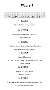
[00130] Figure 1: Exemplary Process 2A chart providing an overview of Steps A
through F.
[00131] Figure 2: Process Flow Chart of Process 2A.
[00132] Figure 3: Shows a diagram of an embodiment of a cryopreserved TIL
exemplary
manufacturing process (-22 days).
[00133] Figure 4: Shows a diagram of an embodiment of process 2A, a 22-day
process for
TIL manufacturing.
[00134] Figure 5: Comparison table of Steps A through F from exemplary
embodiments of
process 1C and process 2A.
[00135] Figure 6: Detailed comparison of an embodiment of process 1C and an
embodiment of process 2A.
[00136] Figure 7: Study Flowcharts for Combination Cohorts: Cohort 1A (MM),
Cohort 2A
(HNSCC), and Cohort 3A (NSCLC). Abbreviations: Cy=cyclophosphamide; E0A=end of
assessment; EOS=end of study; EOT=end of treatment; Flu=fludarabine; IL-
2=interleukin-2;
NMA-LD=nonmyeloablative lymphodepletion; Q3W=every 3 weeks; TIL=tumor
infiltrating
lymphocytes. Patients in Cohorts 1A, 2A, and 3A will receive a single infusion
of
pembrolizumab after the completion of their tumor resection for TIL production
and baseline
scans before the initiation of the NMA-LD regimen. For this particuarle study,
the next dose
of pembrolizumab was not administered earlier than following the completion of
IL-2 and
continue Q3W 3 days thereafter for < 2 years (24 months) or until disease
progression or
unacceptable toxicity, whichever occurred first.
[00137] Figure 8: Study Flowchart for Single-agent Cohort: Cohort 3B (NSCLC).
Abbreviations: Cy=cyclophosphamide; E0A=end of assessment; EOS=end of study;
EOT=end of treatment; Flu=fludarabine; IL-2=interleukin-2; NMA-
LD=nonmyeloablative
lymphodepletion; TIL=tumor infiltrating lymphocytes.
[00138] Figure 9: Shows a diagram of an embodiment of process 2A, a 22-day
process for
TIL manufacturing.
[00139] Figure 10: Provides the structures I-A and I-B, the cylinders refer to
individual
polypeptide binding domains. Structures I-A and I-B comprise three linearly-
linked TNFRSF
binding domains derived from e.g., 4-1BBL or an antibody that binds 4-1BB,
which fold to
form a trivalent protein, which is then linked to a second trivalent protein
through IgGl-Fc
CA 03111210 2021-02-26
WO 2020/096682 PCT/US2019/049384
(including CH3 and CH2 domains) is then used to link two of the trivalent
proteins together
through disulfide bonds (small elongated ovals), stabilizing the structure and
providing an
agonists capable of bringing together the intracellular signaling domains of
the six receptors
and signaling proteins to form a signaling complex. The TNFRSF binding domains
denoted
as cylinders may be scFv domains comprising, e.g., a VH and a VL chain
connected by a
linker that may comprise hydrophilic residues and Gly and Ser sequences for
flexibility, as
well as Glu and Lys for solubility.
BRIEF DESCRIPTION OF THE SEQUENCE LISTING
[00140] SEQ ID NO:1 is the amino acid sequence of the heavy chain of
muromonab.
[00141] SEQ ID NO:2 is the amino acid sequence of the light chain of
muromonab.
[00142] SEQ ID NO:3 is the amino acid sequence of a recombinant human IL-2
protein.
[00143] SEQ ID NO:4 is the amino acid sequence of aldesleukin.
[00144] SEQ ID NO:5 is the amino acid sequence of a recombinant human IL-4
protein.
[00145] SEQ ID NO:6 is the amino acid sequence of a recombinant human IL-7
protein.
[00146] SEQ ID NO:7 is the amino acid sequence of a recombinant human IL-15
protein.
[00147] SEQ ID NO:8 is the amino acid sequence of a recombinant human IL-21
protein.
[00148] SEQ ID NO:9 is the amino acid sequence of human 4-1BB.
[00149] SEQ ID NO:10 is the amino acid sequence of murine 4-1BB.
[00150] SEQ ID NO:11 is the heavy chain for the 4-1BB agonist monoclonal
antibody
utomilumab (PF-05082566).
[00151] SEQ ID NO:12 is the light chain for the 4-1BB agonist monoclonal
antibody
utomilumab (PF-05082566).
[00152] SEQ ID NO:13 is the heavy chain variable region (VH) for the 4-1BB
agonist
monoclonal antibody utomilumab (PF-05082566).
[00153] SEQ ID NO:14 is the light chain variable region (VL) for the 4-1BB
agonist
monoclonal antibody utomilumab (PF-05082566).
[00154] SEQ ID NO:15 is the heavy chain CDR1 for the 4-1BB agonist monoclonal
antibody
utomilumab (PF-05082566).
16
CA 03111210 2021-02-26
WO 2020/096682 PCT/US2019/049384
[00155] SEQ ID NO:16 is the heavy chain CDR2 for the 4-1BB agonist monoclonal
antibody utomilumab (PF-05082566).
[00156] SEQ ID NO:17 is the heavy chain CDR3 for the 4-1BB agonist monoclonal
antibody utomilumab (PF-05082566).
[00157] SEQ ID NO:18 is the light chain CDR1 for the 4-1BB agonist monoclonal
antibody
utomilumab (PF-05082566).
[00158] SEQ ID NO:19 is the light chain CDR2 for the 4-1BB agonist monoclonal
antibody
utomilumab (PF-05082566).
[00159] SEQ ID NO:20 is the light chain CDR3 for the 4-1BB agonist monoclonal
antibody
utomilumab (PF-05082566).
[00160] SEQ ID NO:21 is the heavy chain for the 4-1BB agonist monoclonal
antibody
urelumab (BMS-663513).
[00161] SEQ ID NO:22 is the light chain for the 4-1BB agonist monoclonal
antibody
urelumab (BMS-663513).
[00162] SEQ ID NO:23 is the heavy chain variable region (VH) for the 4-1BB
agonist
monoclonal antibody urelumab (BMS-663513).
[00163] SEQ ID NO:24 is the light chain variable region (VL) for the 4-1BB
agonist
monoclonal antibody urelumab (BMS-663513).
[00164] SEQ ID NO:25 is the heavy chain CDR1 for the 4-1BB agonist monoclonal
antibody urelumab (BMS-663513).
[00165] SEQ ID NO:26 is the heavy chain CDR2 for the 4-1BB agonist monoclonal
antibody urelumab (BMS-663513).
[00166] SEQ ID NO:27 is the heavy chain CDR3 for the 4-1BB agonist monoclonal
antibody urelumab (BMS-663513).
[00167] SEQ ID NO:28 is the light chain CDR1 for the 4-1BB agonist monoclonal
antibody
urelumab (BMS-663513).
[00168] SEQ ID NO:29 is the light chain CDR2 for the 4-1BB agonist monoclonal
antibody
urelumab (BMS-663513).
17
CA 03111210 2021-02-26
WO 2020/096682 PCT/US2019/049384
[00169] SEQ ID NO:30 is the light chain CDR3 for the 4-1BB agonist monoclonal
antibody
urelumab (BMS-663513).
[00170] SEQ ID NO:31 is an Fc domain for a TNFRSF agonist fusion protein.
[00171] SEQ ID NO:32 is a linker for a TNFRSF agonist fusion protein.
[00172] SEQ ID NO:33 is a linker for a TNFRSF agonist fusion protein.
[00173] SEQ ID NO:34 is a linker for a TNFRSF agonist fusion protein.
[00174] SEQ ID NO:35 is a linker for a TNFRSF agonist fusion protein.
[00175] SEQ ID NO:36 is a linker for a TNFRSF agonist fusion protein.
[00176] SEQ ID NO:37 is a linker for a TNFRSF agonist fusion protein.
[00177] SEQ ID NO:38 is a linker for a TNFRSF agonist fusion protein.
[00178] SEQ ID NO:39 is a linker for a TNFRSF agonist fusion protein.
[00179] SEQ ID NO:40 is a linker for a TNFRSF agonist fusion protein.
[00180] SEQ ID NO:41 is a linker for a TNFRSF agonist fusion protein.
[00181] SEQ ID NO:42 is an Fc domain for a TNFRSF agonist fusion protein.
[00182] SEQ ID NO:43 is a linker for a TNFRSF agonist fusion protein.
[00183] SEQ ID NO:44 is a linker for a TNFRSF agonist fusion protein.
[00184] SEQ ID NO:45 is a linker for a TNFRSF agonist fusion protein.
[00185] SEQ ID NO:46 is a 4-1BB ligand (4-1BBL) amino acid sequence.
[00186] SEQ ID NO:47 is a soluble portion of 4-1BBL polypeptide.
[00187] SEQ ID NO:48 is a heavy chain variable region (VH) for the 4-1BB
agonist
antibody 4B4-1-1 version 1.
[00188] SEQ ID NO:49 is a light chain variable region (VL) for the 4-1BB
agonist antibody
4B4-1-1 version 1.
[00189] SEQ ID NO:50 is a heavy chain variable region (VH) for the 4-1BB
agonist
antibody 4B4-1-1 version 2.
[00190] SEQ ID NO:51 is a light chain variable region (VL) for the 4-1BB
agonist antibody
4B4-1-1 version 2.
18
CA 03111210 2021-02-26
WO 2020/096682 PCT/US2019/049384
[00191] SEQ ID NO:52 is a heavy chain variable region (VH) for the 4-1BB
agonist
antibody H39E3-2.
[00192] SEQ ID NO:53 is a light chain variable region (VL) for the 4-1BB
agonist antibody
H39E3-2.
[00193] SEQ ID NO:54 is the amino acid sequence of human 0X40.
[00194] SEQ ID NO:55 is the amino acid sequence of murine 0X40.
[00195] SEQ ID NO:56 is the heavy chain for the 0X40 agonist monoclonal
antibody
tavolixizumab (MEDI-0562).
[00196] SEQ ID NO:57 is the light chain for the 0X40 agonist monoclonal
antibody
tavolixizumab (MEDI-0562).
[00197] SEQ ID NO:58 is the heavy chain variable region (VH) for the 0X40
agonist
monoclonal antibody tavolixizumab (MEDI-0562).
[00198] SEQ ID NO:59 is the light chain variable region (VL) for the 0X40
agonist
monoclonal antibody tavolixizumab (MEDI-0562).
[00199] SEQ ID NO:60 is the heavy chain CDR1 for the 0X40 agonist monoclonal
antibody
tavolixizumab (MEDI-0562).
[00200] SEQ ID NO:61 is the heavy chain CDR2 for the 0X40 agonist monoclonal
antibody
tavolixizumab (MEDI-0562).
[00201] SEQ ID NO:62 is the heavy chain CDR3 for the 0X40 agonist monoclonal
antibody
tavolixizumab (MEDI-0562).
[00202] SEQ ID NO:63 is the light chain CDR1 for the 0X40 agonist monoclonal
antibody
tavolixizumab (MEDI-0562).
[00203] SEQ ID NO:64 is the light chain CDR2 for the 0X40 agonist monoclonal
antibody
tavolixizumab (MEDI-0562).
[00204] SEQ ID NO:65 is the light chain CDR3 for the 0X40 agonist monoclonal
antibody
tavolixizumab (MEDI-0562).
[00205] SEQ ID NO:66 is the heavy chain for the 0X40 agonist monoclonal
antibody 11D4.
[00206] SEQ ID NO:67 is the light chain for the 0X40 agonist monoclonal
antibody 11D4.
19
CA 03111210 2021-02-26
WO 2020/096682 PCT/US2019/049384
[00207] SEQ ID NO:68 is the heavy chain variable region (VH) for the 0X40
agonist
monoclonal antibody 11D4.
[00208] SEQ ID NO:69 is the light chain variable region (VL) for the 0X40
agonist
monoclonal antibody 11D4.
[00209] SEQ ID NO:70 is the heavy chain CDR1 for the 0X40 agonist monoclonal
antibody
11D4.
[00210] SEQ ID NO:71 is the heavy chain CDR2 for the 0X40 agonist monoclonal
antibody
11D4.
[00211] SEQ ID NO:72 is the heavy chain CDR3 for the 0X40 agonist monoclonal
antibody
11D4.
[00212] SEQ ID NO:73 is the light chain CDR1 for the 0X40 agonist monoclonal
antibody
11D4.
[00213] SEQ ID NO:74 is the light chain CDR2 for the 0X40 agonist monoclonal
antibody
11D4.
[00214] SEQ ID NO:75 is the light chain CDR3 for the 0X40 agonist monoclonal
antibody
11D4.
[00215] SEQ ID NO:76 is the heavy chain for the 0X40 agonist monoclonal
antibody 18D8.
[00216] SEQ ID NO:77 is the light chain for the 0X40 agonist monoclonal
antibody 18D8.
[00217] SEQ ID NO:78 is the heavy chain variable region (VH) for the 0X40
agonist
monoclonal antibody 18D8.
[00218] SEQ ID NO:79 is the light chain variable region (VL) for the 0X40
agonist
monoclonal antibody 18D8.
[00219] SEQ ID NO:80 is the heavy chain CDR1 for the 0X40 agonist monoclonal
antibody
18D8.
[00220] SEQ ID NO:81 is the heavy chain CDR2 for the 0X40 agonist monoclonal
antibody
18D8.
[00221] SEQ ID NO:82 is the heavy chain CDR3 for the 0X40 agonist monoclonal
antibody
18D8.
CA 03111210 2021-02-26
WO 2020/096682 PCT/US2019/049384
[00222] SEQ ID NO:83 is the light chain CDR1 for the 0X40 agonist monoclonal
antibody
18D8.
[00223] SEQ ID NO:84 is the light chain CDR2 for the 0X40 agonist monoclonal
antibody
18D8.
[00224] SEQ ID NO:85 is the light chain CDR3 for the 0X40 agonist monoclonal
antibody
18D8.
[00225] SEQ ID NO:86 is the heavy chain variable region (VH) for the 0X40
agonist
monoclonal antibody Hu119-122.
[00226] SEQ ID NO:87 is the light chain variable region (VL) for the 0X40
agonist
monoclonal antibody Hu119-122.
[00227] SEQ ID NO:88 is the heavy chain CDR1 for the 0X40 agonist monoclonal
antibody
Hu119-122.
[00228] SEQ ID NO:89 is the heavy chain CDR2 for the 0X40 agonist monoclonal
antibody
Hu119-122.
[00229] SEQ ID NO:90 is the heavy chain CDR3 for the 0X40 agonist monoclonal
antibody
Hu119-122.
[00230] SEQ ID NO:91 is the light chain CDR1 for the 0X40 agonist monoclonal
antibody
Hu119-122.
[00231] SEQ ID NO:92 is the light chain CDR2 for the 0X40 agonist monoclonal
antibody
Hu119-122.
[00232] SEQ ID NO:93 is the light chain CDR3 for the 0X40 agonist monoclonal
antibody
Hu119-122.
[00233] SEQ ID NO:94 is the heavy chain variable region (VH) for the 0X40
agonist
monoclonal antibody Hu106-222.
[00234] SEQ ID NO:95 is the light chain variable region (VL) for the 0X40
agonist
monoclonal antibody Hu106-222.
[00235] SEQ ID NO:96 is the heavy chain CDR1 for the 0X40 agonist monoclonal
antibody
Hu106-222.
21
CA 03111210 2021-02-26
WO 2020/096682 PCT/US2019/049384
[00236] SEQ ID NO:97 is the heavy chain CDR2 for the 0X40 agonist monoclonal
antibody
Hu106-222.
[00237] SEQ ID NO:98 is the heavy chain CDR3 for the 0X40 agonist monoclonal
antibody
Hu106-222.
[00238] SEQ ID NO:99 is the light chain CDR1 for the 0X40 agonist monoclonal
antibody
Hu106-222.
[00239] SEQ ID NO:100 is the light chain CDR2 for the OX40 agonist monoclonal
antibody
Hu106-222.
[00240] SEQ ID NO:101 is the light chain CDR3 for the 0X40 agonist monoclonal
antibody
Hu106-222.
[00241] SEQ ID NO:102 is an 0X40 ligand (OX4OL) amino acid sequence.
[00242] SEQ ID NO:103 is a soluble portion of OX4OL polypeptide.
[00243] SEQ ID NO:104 is an alternative soluble portion of OX4OL polypeptide.
[00244] SEQ ID NO:105 is the heavy chain variable region (VH) for the 0X40
agonist
monoclonal antibody 008.
[00245] SEQ ID NO:106 is the light chain variable region (VL) for the 0X40
agonist
monoclonal antibody 008.
[00246] SEQ ID NO:107 is the heavy chain variable region (VH) for the 0X40
agonist
monoclonal antibody 011.
[00247] SEQ ID NO:108 is the light chain variable region (VL) for the 0X40
agonist
monoclonal antibody 011.
[00248] SEQ ID NO:109 is the heavy chain variable region (VH) for the 0X40
agonist
monoclonal antibody 021.
[00249] SEQ ID NO:110 is the light chain variable region (VL) for the 0X40
agonist
monoclonal antibody 021.
[00250] SEQ ID NO:111 is the heavy chain variable region (VH) for the 0X40
agonist
monoclonal antibody 023.
[00251] SEQ ID NO:112 is the light chain variable region (VL) for the 0X40
agonist
monoclonal antibody 023.
22
CA 03111210 2021-02-26
WO 2020/096682
PCT/US2019/049384
[00252] SEQ ID NO:113 is the heavy chain variable region (VH) for an 0X40
agonist
monoclonal antibody.
[00253] SEQ ID NO:114 is the light chain variable region (VL) for an 0X40
agonist
monoclonal antibody.
[00254] SEQ ID NO:115 is the heavy chain variable region (VH) for an 0X40
agonist
monoclonal antibody.
[00255] SEQ ID NO:116 is the light chain variable region (VL) for an 0X40
agonist
monoclonal antibody.
[00256] SEQ ID NO:117 is the heavy chain variable region (VH) for a humanized
0X40
agonist monoclonal antibody.
[00257] SEQ ID NO:118 is the heavy chain variable region (VH) for a humanized
0X40
agonist monoclonal antibody.
[00258] SEQ ID NO:119 is the light chain variable region (VL) for a humanized
0X40
agonist monoclonal antibody.
[00259] SEQ ID NO:120 is the light chain variable region (VL) for a humanized
0X40
agonist monoclonal antibody.
[00260] SEQ ID NO:121 is the heavy chain variable region (VH) for a humanized
0X40
agonist monoclonal antibody.
[00261] SEQ ID NO:122 is the heavy chain variable region (VH) for a humanized
0X40
agonist monoclonal antibody.
[00262] SEQ ID NO:123 is the light chain variable region (VL) for a humanized
0X40
agonist monoclonal antibody.
[00263] SEQ ID NO:124 is the light chain variable region (VL) for a humanized
0X40
agonist monoclonal antibody.
[00264] SEQ ID NO:125 is the heavy chain variable region (VH) for an 0X40
agonist
monoclonal antibody.
[00265] SEQ ID NO:126 is the light chain variable region (VL) for an 0X40
agonist
monoclonal antibody.
23
CA 03111210 2021-02-26
WO 2020/096682 PCT/US2019/049384
[00266] SEQ ID NO:127 is the heavy chain amino acid sequence of the PD-1
inhibitor
nivolumab.
[00267] SEQ ID NO: i28 is the light chain amino acid sequence of the PD-1
inhibitor
nivolumab.
[00268] SEQ ID NO: i29 is the heavy chain variable region (VH) amino acid
sequence
of the PD-1 inhibitor nivolumab.
[00269] SEQ ID NO:130 is the light chain variable region (VI) amino acid
sequence of
the PD-1 inhibitor nivolumab.
[00270] SEQ ID NO:131 is the heavy chain CDR1 amino acid sequence of the
PD-1
inhibitor nivolumab.
[00271] SEQ ID NO:132 is the heavy chain CDR2 amino acid sequence of the
PD-1
inhibitor nivolumab.
[00272] SEQ ID NO:133 is the heavy chain CDR3 amino acid sequence of the
PD-1
inhibitor nivolumab.
[00273] SEQ ID NO:134 is the light chain CDR1 amino acid sequence of the
PD-1
inhibitor nivolumab.
[00274] SEQ ID NO:135 is the light chain CDR2 amino acid sequence of the
PD-1
inhibitor nivolumab.
[00275] SEQ ID NO:136 is the light chain CDR3 amino acid sequence of the
PD-1
inhibitor nivolumab.
[00276] SEQ ID NO:137 is the heavy chain amino acid sequence of the PD-1
inhibitor
pembrolizumab.
[00277] SEQ ID NO:138 is the light chain amino acid sequence of the PD-1
inhibitor
pembrolizumab.
[00278] SEQ ID NO:139 is the heavy chain variable region (VH) amino acid
sequence
of the PD-1 inhibitor pembrolizumab.
[00279] SEQ ID NO:140 is the light chain variable region (VI) amino acid
sequence of
the PD-1 inhibitor pembrolizumab.
24
CA 03111210 2021-02-26
WO 2020/096682 PCT/US2019/049384
[00280] SEQ ID NO:141 is the heavy chain CDR1 amino acid sequence of the
PD-1
inhibitor pembrolizumab.
[00281] SEQ ID NO: i42 is the heavy chain CDR2 amino acid sequence of the
PD-1
inhibitor pembrolizumab.
[00282] SEQ ID NO:143 is the heavy chain CDR3 amino acid sequence of the
PD-1
inhibitor pembrolizumab.
[00283] SEQ ID NO:144 is the light chain CDR1 amino acid sequence of the
PD-1
inhibitor pembrolizumab.
[00284] SEQ ID NO:145 is the light chain CDR2 amino acid sequence of the
PD-1
inhibitor pembrolizumab.
[00285] SEQ ID NO:146 is the light chain CDR3 amino acid sequence of the
PD-1
inhibitor pembrolizumab.
[00286] SEQ ID NO:147 is the heavy chain amino acid sequence of the PD-Li
inhibitor durvalumab.
[00287] SEQ ID NO:148 is the light chain amino acid sequence of the PD-Li
inhibitor
durvalumab.
[00288] SEQ ID NO:149 is the heavy chain variable region (VH) amino acid
sequence
of the PD-Li inhibitor durvalumab.
[00289] SEQ ID NO:150 is the light chain variable region (VI) amino acid
sequence of
the PD-Li inhibitor durvalumab.
[00290] SEQ ID NO:151 is the heavy chain CDR1 amino acid sequence of the
PD-Li
inhibitor durvalumab.
[00291] SEQ ID NO:152 is the heavy chain CDR2 amino acid sequence of the
PD-Li
inhibitor durvalumab.
[00292] SEQ ID NO:153 is the heavy chain CDR3 amino acid sequence of the
PD-Li
inhibitor durvalumab.
[00293] SEQ ID NO:154 is the light chain CDR1 amino acid sequence of the
PD-Li
inhibitor durvalumab.
CA 03111210 2021-02-26
WO 2020/096682 PCT/US2019/049384
[00294] SEQ ID NO:155 is the light chain CDR2 amino acid sequence of the
PD-Li
inhibitor durvalumab.
[00295] SEQ ID NO:156 is the light chain CDR3 amino acid sequence of the
PD-Li
inhibitor durvalumab.
[00296] SEQ ID NO:157 is the heavy chain amino acid sequence of the PD-Li
inhibitor avelumab.
[00297] SEQ ID NO:158 is the light chain amino acid sequence of the PD-Li
inhibitor
avelumab.
[00298] SEQ ID NO:159 is the heavy chain variable region (VH) amino acid
sequence
of the PD-Li inhibitor avelumab.
[00299] SEQ ID NO: i60 is the light chain variable region (VI) amino acid
sequence of
the PD-Li inhibitor avelumab.
[00300] SEQ ID NO: 161 is the heavy chain CDR1 amino acid sequence of the
PD-Li
inhibitor avelumab.
[00301] SEQ ID NO: i62 is the heavy chain CDR2 amino acid sequence of the
PD-Li
inhibitor avelumab.
[00302] SEQ ID NO: i63 is the heavy chain CDR3 amino acid sequence of the
PD-Li
inhibitor avelumab.
[00303] SEQ ID NO: i64 is the light chain CDR1 amino acid sequence of the
PD-Li
inhibitor avelumab.
[00304] SEQ ID NO: i65 is the light chain CDR2 amino acid sequence of the
PD-Li
inhibitor avelumab.
[00305] SEQ ID NO: i66 is the light chain CDR3 amino acid sequence of the
PD-Li
inhibitor avelumab.
[00306] SEQ ID NO: i67 is the heavy chain amino acid sequence of the PD-Li
inhibitor atezolizumab.
[00307] SEQ ID NO: i68 is the light chain amino acid sequence of the PD-Li
inhibitor
atezolizumab.
26
CA 03111210 2021-02-26
WO 2020/096682 PCT/US2019/049384
[00308] SEQ ID NO:169 is the heavy chain variable region (VH) amino acid
sequence
of the PD-Li inhibitor atezolizumab.
[00309] SEQ ID NO:170 is the light chain variable region (VI) amino acid
sequence of
the PD-Li inhibitor atezolizumab.
[00310] SEQ ID NO:171 is the heavy chain CDR1 amino acid sequence of the
PD-Li
inhibitor atezolizumab.
[00311] SEQ ID NO:172 is the heavy chain CDR2 amino acid sequence of the
PD-Li
inhibitor atezolizumab.
[00312] SEQ ID NO:173 is the heavy chain CDR3 amino acid sequence of the
PD-Li
inhibitor atezolizumab.
[00313] SEQ ID NO:174 is the light chain CDR1 amino acid sequence of the
PD-Li
inhibitor atezolizumab.
[00314] SEQ ID NO:175 is the light chain CDR2 amino acid sequence of the
PD-Li
inhibitor atezolizumab.
[00315] SEQ ID NO:176 is the light chain CDR3 amino acid sequence of the
PD-Li
inhibitor atezolizumab.
DETAILED DESCRIPTION OF THE INVENTION
I. Introduction
[00316] Adoptive cell therapy utilizing TILs cultured ex vivo by the Rapid
Expansion
Protocol (REP) has produced successful adoptive cell therapy following host
immunosuppression in patients with cancer such as melanoma. Current infusion
acceptance
parameters rely on readouts of the composition of TILs (e.g., CD28, CD8, or
CD4 positivity)
and on the numerical folds of expansion and viability of the REP product.
[00317] Current REP protocols give little insight into the health of the TIL
that will be
infused into the patient. T cells undergo a profound metabolic shift during
the course of their
maturation from naive to effector T cells (see Chang, et at., Nat. Immunol.
2016, /7, 364,
hereby expressly incorporated in its entirety, and in particular for the
discussion and markers
of anaerobic and aerobic metabolism). For example, naïve T cells rely on
mitochondrial
respiration to produce ATP, while mature, healthy effector T cells such as TIL
are highly
27
CA 03111210 2021-02-26
WO 2020/096682 PCT/US2019/049384
glycolytic, relying on aerobic glycolysis to provide the bioenergetics
substrates they require
for proliferation, migration, activation, and anti-tumor efficacy.
[00318] Current TIL manufacturing and treatment processes are limited by
length, cost,
sterility concerns, and other factors described herein such that the potential
to treat patients
which are refractory to anti-PD1 and as such have been severly limited. There
is an urgent
need to provide TIL manufacturing processes and therapies based on such
processes that are
appropriate for use in treating patients for whom very few or no viable
treatment options
remain. The present invention meets this need by providing a shortened
manufacturing
process for use in generating TILs which can then be employed in the treatment
of non-small
cell lung carcinoma (NSCLC) patients whom are refractory to anti-PD-1
treatment.
Definitions
[00319] Unless defined otherwise, all technical and scientific terms used
herein have the
same meaning as is commonly understood by one of skill in the art to which
this invention
belongs. All patents and publications referred to herein are incorporated by
reference in their
entireties.
[00320] The terms "co-administration," "co-administering," "administered
in
combination with," "administering in combination with," "simultaneous," and
"concurrent,"
as used herein, encompass administration of two or more active pharmaceutical
ingredients
(in a preferred embodiment of the present invention, for example, a plurality
of TILs) to a
subject so that both active pharmaceutical ingredients and/or their
metabolites are present in
the subject at the same time. Co-administration includes simultaneous
administration in
separate compositions, administration at different times in separate
compositions, or
administration in a composition in which two or more active pharmaceutical
ingredients are
present. Simultaneous administration in separate compositions and
administration in a
composition in which both agents are present are preferred.
[00321] The term "in vivo" refers to an event that takes place in a subject's
body.
[00322] The term "in vitro" refers to an event that takes places outside of a
subject's body. In
vitro assays encompass cell-based assays in which cells alive or dead are
employed and may
also encompass a cell-free assay in which no intact cells are employed.
28
CA 03111210 2021-02-26
WO 2020/096682 PCT/US2019/049384
[00323] The term "ex vivo" refers to an event which involves treating or
performing a
procedure on a cell, tissue and/or organ which has been removed from a
subject's body.
Aptly, the cell, tissue and/or organ may be returned to the subject's body in
a method of
surgery or treatment.
[00324] The term "rapid expansion" means an increase in the number of
antigen-
specific TILs of at least about 3-fold (or 4-, 5-, 6-, 7-, 8-, or 9-fold) over
a period of a week,
more preferably at least about 10-fold (or 20-, 30-, 40-, 50-, 60-, 70-, 80-,
or 90-fold) over a
period of a week, or most preferably at least about 100-fold over a period of
a week. A
number of rapid expansion protocols are described herein.
[00325]
[00326] By "tumor infiltrating lymphocytes" or "TILs" herein is meant a
population of cells
originally obtained as white blood cells that have left the bloodstream of a
subject and
migrated into a tumor. TILs include, but are not limited to, CD8+ cytotoxic T
cells
(lymphocytes), Thl and Th17 CD4+ T cells, natural killer cells, dendritic
cells and M1
macrophages. TILs include both primary and secondary TILs. "Primary TILs" are
those that
are obtained from patient tissue samples as outlined herein (sometimes
referred to as "freshly
harvested"), and "secondary TILs" are any TIL cell populations that have been
expanded or
proliferated as discussed herein, including, but not limited to bulk TILs and
expanded TILs
("REP TILs" or "post-REP TILs"). TIL cell populations can include genetically
modified
TILs.
[00327] By "population of cells" (including TILs) herein is meant a number of
cells that
share common traits. In general, populations generally range from 1 X 10⁶ to 1
X 1010 in
number, with different TIL populations comprising different numbers. For
example, initial
growth of primary TILs in the presence of IL-2 results in a population of bulk
TILs of
roughly 1 x 10⁸ cells. REP expansion is generally done to provide populations
of 1.5 x 10⁹ to
1.5 x 10¹⁰ cells for infusion.
[00328] By "cryopreserved TILs" herein is meant that TILs, either primary,
bulk, or
expanded (REP TILs), are treated and stored in the range of about -150 C to -
60 C. General
methods for cryopreservation are also described elsewhere herein, including in
the Examples.
For clarity, "cryopreserved TILs" are distinguishable from frozen tissue
samples which may
be used as a source of primary TILs.
29
CA 03111210 2021-02-26
WO 2020/096682 PCT/US2019/049384
[00329] By "thawed cryopreserved TILs" herein is meant a population of TILs
that was
previously cryopreserved and then treated to return to room temperature or
higher, including
but not limited to cell culture temperatures or temperatures wherein TILs may
be
administered to a patient.
[00330] TILs can generally be defined either biochemically, using cell surface
markers, or
functionally, by their ability to infiltrate tumors and effect treatment. TILs
can be generally
categorized by expressing one or more of the following biomarkers: CD4, CD8,
TCR c43,
CD27, CD28, CD56, CCR7, CD45Ra, CD95, PD-1, and CD25. Additionally and
alternatively, TILs can be functionally defined by their ability to infiltrate
solid tumors upon
reintroduction into a patient.
[00331] The term "cryopreservation media" or "cryopreservation medium" refers
to any
medium that can be used for cryopreservation of cells. Such media can include
media
comprising 7% to 10% DMSO. Exemplary media include CryoStor CS10,
Hyperthermasol,
as well as combinations thereof The term "CS10" refers to a cryopreservation
medium which
is obtained from Stemcell Technologies or from Biolife Solutions. The CS10
medium may be
referred to by the trade name "CryoStorg CS10". The CS10 medium is a serum-
free, animal
component-free medium which comprises DMSO.
[00332] The term "central memory T cell" refers to a subset of T cells that in
the human are
CD45R0+ and constitutively express CCR7 (CCR7h1) and CD62L (CD62hi). The
surface
phenotype of central memory T cells also includes TCR, CD3, CD127 (IL-7R), and
IL-15R.
Transcription factors for central memory T cells include BCL-6, BCL-6B, MBD2,
and BMIl.
Central memory T cells primarily secret IL-2 and CD4OL as effector molecules
after TCR
triggering. Central memory T cells are predominant in the CD4 compartment in
blood, and in
the human are proportionally enriched in lymph nodes and tonsils.
[00333] The term "effector memory T cell" refers to a subset of human or
mammalian T
cells that, like central memory T cells, are CD45R0+, but have lost the
constitutive
expression of CCR7 (CCR710) and are heterogeneous or low for CD62L expression
(CD62L10). The surface phenotype of central memory T cells also includes TCR,
CD3,
CD127 (IL-7R), and IL-15R. Transcription factors for central memory T cells
include
BLIMP l. Effector memory T cells rapidly secret high levels of inflammatory
cytokines
following antigenic stimulation, including interferon-y, IL-4, and IL-5.
Effector memory T
cells are predominant in the CD8 compartment in blood, and in the human are
proportionally
CA 03111210 2021-02-26
WO 2020/096682 PCT/US2019/049384
enriched in the lung, liver, and gut. CD8+ effector memory T cells carry large
amounts of
perforin.
[00334] The term "closed system" refers to a system that is closed to the
outside
environment. Any closed system appropriate for cell culture methods can be
employed with
the methods of the present invention. Closed systems include, for example, but
are not
limited to closed G-containers. Once a tumor segment is added to the closed
system, the
system is no opened to the outside environment until the TILs are ready to be
administered to
the patient.
[00335] The terms "fragmenting," "fragment," and "fragmented," as used herein
to describe
processes for disrupting a tumor, includes mechanical fragmentation methods
such as
crushing, slicing, dividing, and morcellating tumor tissue as well as any
other method for
disrupting the physical structure of tumor tissue.
[00336] The terms "peripheral blood mononuclear cells" and "PBMCs" refers to a
peripheral
blood cell having a round nucleus, including lymphocytes (T cells, B cells, NK
cells) and
monocytes. Preferably, the peripheral blood mononuclear cells are irradiated
allogeneic
peripheral blood mononuclear cells. PBMCs are a type of antigen-presenting
cell.
[00337] The term "anti-CD3 antibody" refers to an antibody or variant thereof,
e.g., a
monoclonal antibody and including human, humanized, chimeric or murine
antibodies which
are directed against the CD3 receptor in the T cell antigen receptor of mature
T cells. Anti-
CD3 antibodies include OKT-3, also known as muromonab. Anti-CD3 antibodies
also
include the UHCT1 clone, also known as T3 and CD3E. Other anti-CD3 antibodies
include,
for example, otelixizumab, teplizumab, and visilizumab.
[00338] The term "OKT-3" (also referred to herein as "OKT3") refers to a
monoclonal
antibody or biosimilar or variant thereof, including human, humanized,
chimeric, or murine
antibodies, directed against the CD3 receptor in the T cell antigen receptor
of mature T cells,
and includes commercially-available forms such as OKT-3 (30 ng/mL, MACS GMP
CD3
pure, Miltenyi Biotech, Inc., San Diego, CA, USA) and muromonab or variants,
conservative
amino acid substitutions, glycoforms, or biosimilars thereof The amino acid
sequences of the
heavy and light chains of muromonab are given in Table 1 (SEQ ID NO:1 and SEQ
ID
NO:2). A hybridoma capable of producing OKT-3 is deposited with the American
Type
Culture Collection and assigned the ATCC accession number CRL 8001. A
hybridoma
31
CA 03111210 2021-02-26
WO 2020/096682 PCT/US2019/049384
capable of producing OKT-3 is also deposited with European Collection of
Authenticated Cell
Cultures (ECACC) and assigned Catalogue No. 86022706.
TABLE 1. Amino acid sequences of muromonab.
Identifier Sequence (One-Letter Amino Acid Symbols)
SEQ ID NO:1 QVQLQQSGAE LARPGASVKM SCKASGYTFT RYTMHWVYQR PGQGLEWIGY
INPSRGYTNY 60
Muromonab heavy NQKFKDKATL TTDKSSSTAY MQLSSLTSED SAVYYCARYY DDHYCLDYWG
QGTTLTVSSA 120
chain KTTAPSVYPL APVCGGTTGS SVTLGCLVKG YFPEPVTLTW NSGSLSSGVH
TFPAVLQSDL 180
YTLSSSVTVT SSTWPSQSIT CNVAHPASST KVDKKIEPRP KSCDKTHTCP PCPAPELLGG
240
PSVFLFPPKP KDTLMISRTP EVTCVVVDVS HEDPEVKFNW YVDGVEVHNA KTKPREEQYN
300
STYRVVSVLT VLHQDWLNGK EYKCKVSNKA LPAPIEKTIS KAKGQPREPQ VYTLPPSRDE
360
LTKNQVSLTC LVKGFYPSDI AVEWESNGQP ENNYKTTPPV LDSDGSFFLY SKLTVDKSRW
420
QQGNVFSCSV MHEALHNHYT QKSLSLSPGK
450
SEQ ID NO:2 QIVLTQSPAI MSASPGEKVT MTCSASSSVS YMNWYQQKSG TSPKRWIYDT
SKLASGVPAH 60
Muromonab light FRGSGSGTSY SLTISGMEAE DAATYYCQQW SSNPFTFGSG TKLEINRADT
APTVSIFPPS 120
chain SEQLTSGGAS VVCFLNNFYP KDINVYWKID GSERQNGVLN SWTDQDSKDS
TYSMSSTLTL 180
TKDEYERHNS YTCEATHKTS TSPIVKSFNR NEC
213
[00339] The term "IL-2" (also referred to herein as "IL2") refers to the T
cell growth factor
known as interleukin-2, and includes all forms of IL-2 including human and
mammalian
forms, conservative amino acid substitutions, glycoforms, biosimilars, and
variants thereof.
IL-2 is described, e.g., in Nelson, I Immunol. 2004, 172, 3983-88 and Malek,
Annu. Rev.
Immunol. 2008, 26, 453-79, the disclosures of which are incorporated by
reference herein.
The amino acid sequence of recombinant human IL-2 suitable for use in the
invention is
given in Table 2 (SEQ ID NO:3). For example, the term IL-2 encompasses human,
recombinant forms of IL-2 such as aldesleukin (PROLEUKIN, available
commercially from
multiple suppliers in 22 million IU per single use vials), as well as the form
of recombinant
IL-2 commercially supplied by CellGenix, Inc., Portsmouth, NH, USA (CELLGRO
GMP) or
ProSpec-Tany TechnoGene Ltd., East Brunswick, NJ, USA (Cat. No. CYT-209-b) and
other
commercial equivalents from other vendors. Aldesleukin (des-alany1-1, serine-
125 human IL-
2) is a nonglycosylated human recombinant form of IL-2 with a molecular weight
of
approximately 15 kDa. The amino acid sequence of aldesleukin suitable for use
in the
invention is given in Table 2 (SEQ ID NO:4). The term IL-2 also encompasses
pegylated
forms of IL-2, as described herein, including the pegylated IL2 prodrug NKTR-
214, available
from Nektar Therapeutics, South San Francisco, CA, USA. NKTR-214 and pegylated
IL-2
suitable for use in the invention is described in U.S. Patent Application
Publication No. US
2014/0328791 Al and International Patent Application Publication No. WO
2012/065086 Al,
the disclosures of which are incorporated by reference herein. Alternative
forms of
conjugated IL-2 suitable for use in the invention are described in U.S. Patent
Nos. 4,766,106,
5,206,344, 5,089,261 and 4902,502, the disclosures of which are incorporated
by reference
32
CA 03111210 2021-02-26
WO 2020/096682 PCT/US2019/049384
herein. Formulations of IL-2 suitable for use in the invention are described
in U.S. Patent No.
6,706,289, the disclosure of which is incorporated by reference herein.
TABLE 2. Amino acid sequences of interleukins.
Identifier Sequence (One-Letter Amino Acid Symbols)
SEQ ID NO:3 MAPTSSSTEK TQLQLEHLLL DLQMILNGIN NYENPELTRM LTFIKEYMPEK
ATELEHLQCL 60
recombinant EEELIKPLEEV LNLAQSENFH LRPRDLISNI NVIVLELEGS ETTFMCEYAD
ETATIVEFLN 120
human IL-2 RWITFCQSII STLT
134
(rhIL-2)
SEQ ID NO:4 PTSSSTEXTQ LQLEHLLLDL QMILNGINNY KNPELTRMLT FIKEYMPIKKAT
ELEHLQCLEE 60
Aldesleukin ELIKPLEEVLN LAQSENFHLR PRDLISNINV IVLELEGSET TFMCEYADET
ATIVEFLNRW 120
ITFSQSIIST LT
132
SEQ ID NO:5 MHECDITLQE IIKTLNSLTE QKTLCTELTV TDIFAASENT TEKETFCRAA
TVLRQFYSHH 60
recombinant EXDTRCLGAT AQQFHRHEQL IRFLERLDRN LWGLAGLNSC PVIKEANQSTL
ENFLERLIKTI 120
human IL-4 MREHYSECSS
130
(rhIL-4)
SEQ ID NO:6 MDCDIEGEDG EQYESVLMVS IDQLLDSMKE IGSNCLNNEF NFFERHICDA
NIKEGMFLFRA 60
recombinant ARKLRQFLEM NSTGDFDLHL LEVSEGTTIL LNCTGQVFGR KPAALGEAQP
THSLEENKSL 120
human IL-7 KEQXKLNDLC FLERLLQEIK TCWNKILMGT KEH
153
(rhIL-7)
SEQ ID NO:7 MNWVNVISDL KIKIEDLIQSM HIDATLYTES DVHPSCEVTA MECELLELQV
ISLESGDASI 60
recombinant HDTVENLIIL ANNSLSSNGN VTESGCXECE ELEEKNIKEF LQSFVHIVQM FINTS
115
human IL-15
(rhIL-15)
SEQ ID NO:8 MQDRHMIRMR QLIDIVDQLX NYVNDLVPEF LPAPEDVETN CEWSAFSCFQ
KAQLKSANTG 60
recombinant NNERIINVSI KELEREPPST NAGRRQKHRL TCPSCDSYEK EPPEEFLERF
ESLLQHMIHQ 120
human IL-21 HLSSRTHGSE DS
133
(rhIL-21)
[00340] The term "IL-4" (also referred to herein as "IL4") refers to the
cytokine known as
interleukin 4, which is produced by Th2 T cells and by eosinophils, basophils,
and mast cells.
IL-4 regulates the differentiation of naive helper T cells (Th0 cells) to Th2
T cells. Steinke
and Borish, Respir. Res. 2001, 2, 66-70. Upon activation by IL-4, Th2 T cells
subsequently
produce additional IL-4 in a positive feedback loop. IL-4 also stimulates B
cell proliferation
and class II MHC expression, and induces class switching to IgE and IgGi
expression from B
cells. Recombinant human IL-4 suitable for use in the invention is
commercially available
from multiple suppliers, including ProSpec-Tany TechnoGene Ltd., East
Brunswick, NJ,
USA (Cat. No. CYT-211) and TherrnoFisher Scientific, Inc., Waltham, MA, USA
(human
IL-15 recombinant protein, Cat. No. Gibco CTP0043). The amino acid sequence of
recombinant human IL-4 suitable for use in the invention is given in Table 2
(SEQ ID NO:5).
[00341] The term "IL-7" (also referred to herein as "IL7") refers to a
glycosylated tissue-
derived cytokine known as interleukin 7, which may be obtained from stromal
and epithelial
cells, as well as from dendritic cells. Fry and Mackall, Blood 2002, 99, 3892-
904. IL-7 can
stimulate the development of T cells. IL-7 binds to the IL-7 receptor, a
heterodimer
consisting of IL-7 receptor alpha and common gamma chain receptor, which in a
series of
signals important for T cell development within the thymus and survival within
the periphery.
Recombinant human IL-7 suitable for use in the invention is commercially
available from
33
CA 03111210 2021-02-26
WO 2020/096682 PCT/US2019/049384
multiple suppliers, including ProSpec-Tany TechnoGene Ltd., East Brunswick,
NJ, USA
(Cat. No. CYT-254) and ThermoFisher Scientific, Inc., Waltham, MA, USA (human
IL-15
recombinant protein, Cat. No. Gibco PHC0071). The amino acid sequence of
recombinant
human IL-7 suitable for use in the invention is given in Table 2 (SEQ ID
NO:6).
[00342] The term "IL-15" (also referred to herein as "IL15") refers to the T
cell growth
factor known as interleukin-15, and includes all forms of IL-2 including human
and
mammalian forms, conservative amino acid substitutions, glycoforms,
biosimilars, and
variants thereof IL-15 is described, e.g., in Fehniger and Caligiuri, Blood
2001, 97, 14-32,
the disclosure of which is incorporated by reference herein. IL-15 shares 0
and y signaling
receptor subunits with IL-2. Recombinant human IL-15 is a single, non-
glycosylated
polypeptide chain containing 114 amino acids (and an N-terminal methionine)
with a
molecular mass of 12.8 kDa. Recombinant human IL-15 is commercially available
from
multiple suppliers, including ProSpec-Tany TechnoGene Ltd., East Brunswick,
NJ, USA
(Cat. No. CYT-230-b) and ThermoFisher Scientific, Inc., Waltham, MA, USA
(human IL-15
recombinant protein, Cat. No. 34-8159-82). The amino acid sequence of
recombinant human
IL-15 suitable for use in the invention is given in Table 2 (SEQ ID NO:7).
[00343] The term "IL-21" (also referred to herein as "IL21") refers to the
pleiotropic
cytokine protein known as interleukin-21, and includes all forms of IL-21
including human
and mammalian forms, conservative amino acid substitutions, glycoforms,
biosimilars, and
variants thereof IL-21 is described, e.g., in Spolski and Leonard, Nat. Rev.
Drug. Disc. 2014,
/3, 379-95, the disclosure of which is incorporated by reference herein. IL-21
is primarily
produced by natural killer T cells and activated human CD4+ T cells.
Recombinant human IL-
21 is a single, non-glycosylated polypeptide chain containing 132 amino acids
with a
molecular mass of 15.4 kDa. Recombinant human IL-21 is commercially available
from
multiple suppliers, including ProSpec-Tany TechnoGene Ltd., East Brunswick,
NJ, USA
(Cat. No. CYT-408-b) and ThermoFisher Scientific, Inc., Waltham, MA, USA
(human IL-21
recombinant protein, Cat. No. 14-8219-80). The amino acid sequence of
recombinant human
IL-21 suitable for use in the invention is given in Table 2 (SEQ ID NO:8).
[00344] When "an anti-tumor effective amount", "an tumor-inhibiting effective
amount", or
"therapeutic amount" is indicated, the precise amount of the compositions of
the present
invention to be administered can be determined by a physician with
consideration of
individual differences in age, weight, tumor size, extent of infection or
metastasis, and
condition of the patient (subject). It can generally be stated that a
pharmaceutical composition
34
CA 03111210 2021-02-26
WO 2020/096682
PCT/US2019/049384
comprising the tumor infiltrating lymphocytes (e.g. secondary TILs or
genetically modified
cytotoxic lymphocytes) described herein may be administered at a dosage of 104
to 1011
cells/kg body weight (e.g., 105 to 106, i05 to 1-1o,
u 05 to 1011, 106 to 1-1 , u 106
to 1011,107 to
1011, o' to 1-1 , u 108 to 1011, 108 to 1-1 , u 109 to 1011,
or 109 to 1010 cells/kg body weight),
including all integer values within those ranges. Tumor infiltrating
lymphocytes (including in
some cases, genetically modified cytotoxic lymphocytes) compositions may also
be
administered multiple times at these dosages. The tumor infiltrating
lymphocytes (including
in some cases, genetically) can be administered by using infusion techniques
that are
commonly known in immunotherapy (see, e.g., Rosenberg et al., New Eng. I of
Med. 319:
1676, 1988). The optimal dosage and treatment regime for a particular patient
can readily be
determined by one skilled in the art of medicine by monitoring the patient for
signs of disease
and adjusting the treatment accordingly.
[00345] The term "microenvironment," as used herein, may refer to the solid or
hematological tumor microenvironment as a whole or to an individual subset of
cells within
the microenvironment. The tumor microenvironment, as used herein, refers to a
complex
mixture of "cells, soluble factors, signaling molecules, extracellular
matrices, and mechanical
cues that promote neoplastic transformation, support tumor growth and
invasion, protect the
tumor from host immunity, foster therapeutic resistance, and provide niches
for dominant
metastases to thrive," as described in Swartz, et at., Cancer Res., 2012, 72,
2473. Although
tumors express antigens that should be recognized by T cells, tumor clearance
by the immune
system is rare because of immune suppression by the microenvironment.
[00346] In an embodiment, the invention includes a method of treating a cancer
with a
population of TILs, wherein a patient is pre-treated with non-myeloablative
chemotherapy
prior to an infusion of TILs according to the invention. In some embodiments,
the population
of TILs may be provided wherein a patient is pre-treated with nonmyeloablative
chemotherapy prior to an infusion of TILs according to the present invention.
In an
embodiment, the non-myeloablative chemotherapy is cyclophosphamide 60 mg/kg/d
for 2
days (days 27 and 26 prior to TIL infusion) and fludarabine 25 mg/m2/d for 5
days (days 27
to 23 prior to TIL infusion). In an embodiment, after non-myeloablative
chemotherapy and
TIL infusion (at day 0) according to the invention, the patient receives an
intravenous
infusion of IL-2 intravenously at 720,000 IU/kg every 8 hours to physiologic
tolerance.
[00347] Experimental findings indicate that lymphodepletion prior to adoptive
transfer of
tumor-specific T lymphocytes plays a key role in enhancing treatment efficacy
by eliminating
CA 03111210 2021-02-26
WO 2020/096682 PCT/US2019/049384
regulatory T cells and competing elements of the immune system ("cytokine
sinks").
Accordingly, some embodiments of the invention utilize a lymphodepletion step
(sometimes
also referred to as "immunosuppressive conditioning") on the patient prior to
the introduction
of the rTILs of the invention.
[00348] The term "effective amount" or "therapeutically effective amount"
refers to that
amount of a compound or combination of compounds as described herein that is
sufficient to
effect the intended application including, but not limited to, disease
treatment. A
therapeutically effective amount may vary depending upon the intended
application (in vitro
or in vivo), or the subject and disease condition being treated (e.g., the
weight, age and
gender of the subject), the severity of the disease condition, or the manner
of administration.
The term also applies to a dose that will induce a particular response in
target cells (e.g., the
reduction of platelet adhesion and/or cell migration). The specific dose will
vary depending
on the particular compounds chosen, the dosing regimen to be followed, whether
the
compound is administered in combination with other compounds, timing of
administration,
the tissue to which it is administered, and the physical delivery system in
which the
compound is carried.
[00349] The terms "treatment", "treating", "treat", and the like, refer to
obtaining a desired
pharmacologic and/or physiologic effect. The effect may be prophylactic in
terms of
completely or partially preventing a disease or symptom thereof and/or may be
therapeutic in
terms of a partial or complete cure for a disease and/or adverse effect
attributable to the
disease. "Treatment", as used herein, covers any treatment of a disease in a
mammal,
particularly in a human, and includes: (a) preventing the disease from
occurring in a subject
which may be predisposed to the disease but has not yet been diagnosed as
having it;
(b) inhibiting the disease, i.e., arresting its development or progression;
and (c) relieving the
disease, i.e., causing regression of the disease and/or relieving one or more
disease
symptoms. "Treatment" is also meant to encompass delivery of an agent in order
to provide
for a pharmacologic effect, even in the absence of a disease or condition. For
example,
"treatment" encompasses delivery of a composition that can elicit an immune
response or
confer immunity in the absence of a disease condition, e.g., in the case of a
vaccine.
[00350] The term "heterologous" when used with reference to portions of a
nucleic acid or
protein indicates that the nucleic acid or protein comprises two or more
subsequences that are
not found in the same relationship to each other in nature. For instance, the
nucleic acid is
typically recombinantly produced, having two or more sequences from unrelated
genes
36
CA 03111210 2021-02-26
WO 2020/096682 PCT/US2019/049384
arranged to make a new functional nucleic acid, e.g., a promoter from one
source and a
coding region from another source, or coding regions from different sources.
Similarly, a
heterologous protein indicates that the protein comprises two or more
subsequences that are
not found in the same relationship to each other in nature (e.g., a fusion
protein).
[00351] The terms "sequence identity," "percent identity," and "sequence
percent identity"
(or synonyms thereof, e.g., "99% identical") in the context of two or more
nucleic acids or
polypeptides, refer to two or more sequences or subsequences that are the same
or have a
specified percentage of nucleotides or amino acid residues that are the same,
when compared
and aligned (introducing gaps, if necessary) for maximum correspondence, not
considering
any conservative amino acid substitutions as part of the sequence identity.
The percent
identity can be measured using sequence comparison software or algorithms or
by visual
inspection. Various algorithms and software are known in the art that can be
used to obtain
alignments of amino acid or nucleotide sequences. Suitable programs to
determine percent
sequence identity include for example the BLAST suite of programs available
from the U.S.
Government's National Center for Biotechnology Information BLAST web site.
Comparisons between two sequences can be carried using either the BLASTN or
BLASTP
algorithm. BLASTN is used to compare nucleic acid sequences, while BLASTP is
used to
compare amino acid sequences. ALIGN, ALIGN-2 (Genentech, South San Francisco,
California) or MegAlign, available from DNASTAR, are additional publicly
available
software programs that can be used to align sequences. One skilled in the art
can determine
appropriate parameters for maximal alignment by particular alignment software.
In certain
embodiments, the default parameters of the alignment software are used.
[00352] As used herein, the term "variant" encompasses but is not limited to
antibodies or
fusion proteins which comprise an amino acid sequence which differs from the
amino acid
sequence of a reference antibody by way of one or more substitutions,
deletions and/or
additions at certain positions within or adjacent to the amino acid sequence
of the reference
antibody. The variant may comprise one or more conservative substitutions in
its amino acid
sequence as compared to the amino acid sequence of a reference antibody.
Conservative
substitutions may involve, e.g., the substitution of similarly charged or
uncharged amino
acids. The variant retains the ability to specifically bind to the antigen of
the reference
antibody. The term variant also includes pegylated antibodies or proteins.
[00353] By "tumor infiltrating lymphocytes" or "TILs" herein is meant a
population of cells
originally obtained as white blood cells that have left the bloodstream of a
subject and
37
CA 03111210 2021-02-26
WO 2020/096682 PCT/US2019/049384
migrated into a tumor. TILs include, but are not limited to, CD8+ cytotoxic T
cells
(lymphocytes), Thl and Th17 CD4+ T cells, natural killer cells, dendritic
cells and M1
macrophages. TILs include both primary and secondary TILs. "Primary TILs" are
those that
are obtained from patient tissue samples as outlined herein (sometimes
referred to as "freshly
harvested"), and "secondary TILs" are any TIL cell populations that have been
expanded or
proliferated as discussed herein, including, but not limited to bulk TILs,
expanded TILs
("REP TILs") as well as "reREP TILs" as discussed herein. reREP TILs can
include for
example second expansion TILs or second additional expansion TILs (such as,
for example,
those described in Step D of Figure 8, including TILs referred to as reREP
TILs).
[00354] TILs can generally be defined either biochemically, using cell surface
markers, or
functionally, by their ability to infiltrate tumors and effect treatment. TILs
can be generally
categorized by expressing one or more of the following biomarkers: CD4, CD8,
TCR c43,
CD27, CD28, CD56, CCR7, CD45Ra, CD95, PD-1, and CD25. Additionally, and
alternatively, TILs can be functionally defined by their ability to infiltrate
solid tumors upon
reintroduction into a patient. TILS may further be characterized by potency ¨
for example,
TILS may be considered potent if, for example, interferon (IFNy) release is
greater than about
50 pg/mL, greater than about 100 pg/mL, greater than about 150 pg/mL, or
greater than about
200 pg/mL, greater than about 300 pg/mL, greater than about 400 pg/mL, greater
than about
500 pg/mL, greater than about 600 pg/mL, greater than about 700 pg/mL, greater
than about
800 pg/mL, greater than about 900 pg/mL, greater than about 1000 pg/mL.
[00355] The term "deoxyribonucleotide" encompasses natural and synthetic,
unmodified and
modified deoxyribonucleotides. Modifications include changes to the sugar
moiety, to the
base moiety and/or to the linkages between deoxyribonucleotide in the
oligonucleotide.
[00356] The term "RNA" defines a molecule comprising at least one
ribonucleotide residue.
The term "ribonucleotide" defines a nucleotide with a hydroxyl group at the 2'
position of a
b-D-ribofuranose moiety. The term RNA includes double-stranded RNA, single-
stranded
RNA, isolated RNA such as partially purified RNA, essentially pure RNA,
synthetic RNA,
recombinantly produced RNA, as well as altered RNA that differs from naturally
occurring
RNA by the addition, deletion, substitution and/or alteration of one or more
nucleotides.
Nucleotides of the RNA molecules described herein may also comprise non-
standard
nucleotides, such as non-naturally occurring nucleotides or chemically
synthesized
nucleotides or deoxynucleotides. These altered RNAs can be referred to as
analogs or analogs
of naturally-occurring RNA.
38
CA 03111210 2021-02-26
WO 2020/096682 PCT/US2019/049384
[00357] The terms "pharmaceutically acceptable carrier" or "pharmaceutically
acceptable
excipient" are intended to include any and all solvents, dispersion media,
coatings,
antibacterial and antifungal agents, isotonic and absorption delaying agents,
and inert
ingredients. The use of such pharmaceutically acceptable carriers or
pharmaceutically
acceptable excipients for active pharmaceutical ingredients is well known in
the art. Except
insofar as any conventional pharmaceutically acceptable carrier or
pharmaceutically
acceptable excipient is incompatible with the active pharmaceutical
ingredient, its use in
therapeutic compositions of the invention is contemplated. Additional active
pharmaceutical
ingredients, such as other drugs, can also be incorporated into the described
compositions and
methods.
[00358] The terms "about" and "approximately" mean within a statistically
meaningful
range of a value. Such a range can be within an order of magnitude, preferably
within 50%,
more preferably within 20%, more preferably still within 10%, and even more
preferably
within 5% of a given value or range. The allowable variation encompassed by
the terms
"about" or "approximately" depends on the particular system under study, and
can be readily
appreciated by one of ordinary skill in the art. Moreover, as used herein, the
terms "about"
and "approximately" mean that dimensions, sizes, formulations, parameters,
shapes and other
quantities and characteristics are not and need not be exact, but may be
approximate and/or
larger or smaller, as desired, reflecting tolerances, conversion factors,
rounding off,
measurement error and the like, and other factors known to those of skill in
the art. In
general, a dimension, size, formulation, parameter, shape or other quantity or
characteristic is
"about" or "approximate" whether or not expressly stated to be such. It is
noted that
embodiments of very different sizes, shapes and dimensions may employ the
described
arrangements.
[00359] The transitional terms "comprising," "consisting essentially of," and
"consisting
of," when used in the appended claims, in original and amended form, define
the claim scope
with respect to what unrecited additional claim elements or steps, if any, are
excluded from
the scope of the claim(s). The term "comprising" is intended to be inclusive
or open-ended
and does not exclude any additional, unrecited element, method, step or
material. The term
"consisting of' excludes any element, step or material other than those
specified in the claim
and, in the latter instance, impurities ordinary associated with the specified
material(s). The
term "consisting essentially of' limits the scope of a claim to the specified
elements, steps or
material(s) and those that do not materially affect the basic and novel
characteristic(s) of the
39
CA 03111210 2021-02-26
WO 2020/096682 PCT/US2019/049384
claimed invention. All compositions, methods, and kits described herein that
embody the
present invention can, in alternate embodiments, be more specifically defined
by any of the
transitional terms "comprising," "consisting essentially of," and "consisting
of."
[00360] The terms "antibody" and its plural form "antibodies" refer to
whole
immunoglobulins and any antigen-binding fragment ("antigen-binding portion")
or single
chains thereof. An "antibody" further refers to a glycoprotein comprising at
least two heavy
(H) chains and two light (L) chains inter-connected by disulfide bonds, or an
antigen-binding
portion thereof Each heavy chain is comprised of a heavy chain variable region
(abbreviated
herein as VH) and a heavy chain constant region. The heavy chain constant
region is
comprised of three domains, CHL CH2 and CH3. Each light chain is comprised of
a light
chain variable region (abbreviated herein as VL) and a light chain constant
region. The light
chain constant region is comprised of one domain, CL. The VH and VL regions of
an antibody
may be further subdivided into regions of hypervariability, which are referred
to as
complementarity determining regions (CDR) or hypervariable regions (HVR), and
which can
be interspersed with regions that are more conserved, termed framework regions
(FR). Each
VH and VL is composed of three CDRs and four FRs, arranged from amino-terminus
to
carboxy-terminus in the following order: FR1, CDR1, FR2, CDR2, FR3, CDR3, FR4.
The
variable regions of the heavy and light chains contain a binding domain that
interacts with an
antigen epitope or epitopes. The constant regions of the antibodies may
mediate the binding
of the immunoglobulin to host tissues or factors, including various cells of
the immune
system (e.g., effector cells) and the first component (Clq) of the classical
complement system.
[00361] The term "antigen" refers to a substance that induces an immune
response. In
some embodiments, an antigen is a molecule capable of being bound by an
antibody or a
TCR if presented by major histocompatibility complex (MHC) molecules. The term
"antigen", as used herein, also encompasses T cell epitopes. An antigen is
additionally
capable of being recognized by the immune system. In some embodiments, an
antigen is
capable of inducing a humoral immune response or a cellular immune response
leading to the
activation of B lymphocytes and/or T lymphocytes. In some cases, this may
require that the
antigen contains or is linked to a Th cell epitope. An antigen can also have
one or more
epitopes (e.g., B- and T-epitopes). In some embodiments, an antigen will
preferably react,
typically in a highly specific and selective manner, with its corresponding
antibody or TCR
and not with the multitude of other antibodies or TCRs which may be induced by
other
antigens.
CA 03111210 2021-02-26
WO 2020/096682
PCT/US2019/049384
[00362] The terms "monoclonal antibody," "mAb," "monoclonal antibody
composition," or their plural forms refer to a preparation of antibody
molecules of single
molecular composition. A monoclonal antibody composition displays a single
binding
specificity and affinity for a particular epitope. Monoclonal antibodies
specific to certain
receptors can be made using knowledge and skill in the art of injecting test
subjects with
suitable antigen and then isolating hybridomas expressing antibodies having
the desired
sequence or functional characteristics. DNA encoding the monoclonal antibodies
is readily
isolated and sequenced using conventional procedures (e.g., by using
oligonucleotide probes
that are capable of binding specifically to genes encoding the heavy and light
chains of the
monoclonal antibodies). The hybridoma cells serve as a preferred source of
such DNA.
Once isolated, the DNA may be placed into expression vectors, which are then
transfected
into host cells such as E. coil cells, simian COS cells, Chinese hamster ovary
(CHO) cells, or
myeloma cells that do not otherwise produce immunoglobulin protein, to obtain
the synthesis
of monoclonal antibodies in the recombinant host cells. Recombinant production
of
antibodies will be described in more detail below.
[00363] The terms "antigen-binding portion" or "antigen-binding fragment"
of an
antibody (or simply "antibody portion" or "fragment"), as used herein, refers
to one or more
fragments of an antibody that retain the ability to specifically bind to an
antigen. It has been
shown that the antigen-binding function of an antibody can be performed by
fragments of a
full-length antibody. Examples of binding fragments encompassed within the
term "antigen-
binding portion" of an antibody include (i) a Fab fragment, a monovalent
fragment consisting
of the VL, VH, CL and CH1 domains; (ii) a F(ab')2 fragment, a bivalent
fragment comprising
two Fab fragments linked by a disulfide bridge at the hinge region; (iii) a Fd
fragment
consisting of the VH and CH1 domains; (iv) a Fv fragment consisting of the VL
and VH
domains of a single arm of an antibody, (v) a domain antibody (dAb) fragment
(Ward, et al.,
Nature, 1989, 341, 544-546), which may consist of a VH or a VL domain; and
(vi) an isolated
complementarity determining region (CDR). Furthermore, although the two
domains of the
Fv fragment, VL and VH, are coded for by separate genes, they can be joined,
using
recombinant methods, by a synthetic linker that enables them to be made as a
single protein
chain in which the VL and VH regions pair to form monovalent molecules known
as single
chain Fv (scFv); see, e.g., Bird, et al., Science 1988, 242, 423-426; and
Huston, et al., Proc.
Natl. Acad. Sci. USA 1988, 85, 5879-5883). Such scFv antibodies are also
intended to be
encompassed within the terms "antigen-binding portion" or "antigen-binding
fragment" of an
41
CA 03111210 2021-02-26
WO 2020/096682 PCT/US2019/049384
antibody. These antibody fragments are obtained using conventional techniques
known to
those with skill in the art, and the fragments are screened for utility in the
same manner as are
intact antibodies.
[00364] The term "human antibody," as used herein, is intended to include
antibodies
having variable regions in which both the framework and CDR regions are
derived from
human germline immunoglobulin sequences. Furthermore, if the antibody contains
a
constant region, the constant region also is derived from human germline
immunoglobulin
sequences. The human antibodies of the invention may include amino acid
residues not
encoded by human germline immunoglobulin sequences (e.g., mutations introduced
by
random or site-specific mutagenesis in vitro or by somatic mutation in vivo).
The term
"human antibody", as used herein, is not intended to include antibodies in
which CDR
sequences derived from the germline of another mammalian species, such as a
mouse, have
been grafted onto human framework sequences.
[00365] The term "human monoclonal antibody" refers to antibodies
displaying a
single binding specificity which have variable regions in which both the
framework and CDR
regions are derived from human germline immunoglobulin sequences. In an
embodiment, the
human monoclonal antibodies are produced by a hybridoma which includes a B
cell obtained
from a transgenic nonhuman animal, e.g., a transgenic mouse, having a genome
comprising a
human heavy chain transgene and a light chain transgene fused to an
immortalized cell.
[00366] The term "recombinant human antibody", as used herein, includes
all human
antibodies that are prepared, expressed, created or isolated by recombinant
means, such as (a)
antibodies isolated from an animal (such as a mouse) that is transgenic or
transchromosomal
for human immunoglobulin genes or a hybridoma prepared therefrom (described
further
below), (b) antibodies isolated from a host cell transformed to express the
human antibody,
e.g., from a transfectoma, (c) antibodies isolated from a recombinant,
combinatorial human
antibody library, and (d) antibodies prepared, expressed, created or isolated
by any other
means that involve splicing of human immunoglobulin gene sequences to other
DNA
sequences. Such recombinant human antibodies have variable regions in which
the
framework and CDR regions are derived from human germline immunoglobulin
sequences.
In certain embodiments, however, such recombinant human antibodies can be
subjected to in
vitro mutagenesis (or, when an animal transgenic for human Ig sequences is
used, in vivo
somatic mutagenesis) and thus the amino acid sequences of the VH and VL
regions of the
recombinant antibodies are sequences that, while derived from and related to
human germline
42
CA 03111210 2021-02-26
WO 2020/096682 PCT/US2019/049384
VH and VL sequences, may not naturally exist within the human antibody
germline repertoire
in vivo.
[00367] As used herein, "isotype" refers to the antibody class (e.g., IgM
or IgG1) that
is encoded by the heavy chain constant region genes.
[00368] The phrases "an antibody recognizing an antigen" and "an antibody
specific
for an antigen" are used interchangeably herein with the term "an antibody
which binds
specifically to an antigen."
[00369] The term "human antibody derivatives" refers to any modified form
of the
human antibody, including a conjugate of the antibody and another active
pharmaceutical
ingredient or antibody. The terms "conjugate," "antibody-drug conjugate",
"ADC," or
"immunoconjugate" refers to an antibody, or a fragment thereof, conjugated to
another
therapeutic moiety, which can be conjugated to antibodies described herein
using methods
available in the art.
[00370] The terms "humanized antibody," "humanized antibodies," and
"humanized"
are intended to refer to antibodies in which CDR sequences derived from the
germline of
another mammalian species, such as a mouse, have been grafted onto human
framework
sequences. Additional framework region modifications may be made within the
human
framework sequences. Humanized forms of non-human (for example, murine)
antibodies are
chimeric antibodies that contain minimal sequence derived from non-human
immunoglobulin. For the most part, humanized antibodies are human
immunoglobulins
(recipient antibody) in which residues from a hypervariable region of the
recipient are
replaced by residues from a 15 hypervariable region of a non-human species
(donor antibody)
such as mouse, rat, rabbit or nonhuman primate having the desired specificity,
affinity, and
capacity. In some instances, Fv framework region (FR) residues of the human
immunoglobulin are replaced by corresponding non-human residues. Furthermore,
humanized antibodies may comprise residues that are not found in the recipient
antibody or in
the donor antibody. These modifications are made to further refine antibody
performance. In
general, the humanized antibody will comprise substantially all of at least
one, and typically
two, variable domains, in which all or substantially all of the hypervariable
loops correspond
to those of a non-human immunoglobulin and all or substantially all of the FR
regions are
those of a human immunoglobulin sequence. The humanized antibody optionally
also will
comprise at least a portion of an immunoglobulin constant region (Fc),
typically that of a
43
CA 03111210 2021-02-26
WO 2020/096682 PCT/US2019/049384
human immunoglobulin. For further details, see Jones, et al., Nature 1986,
321, 522-525;
Riechmann, et al., Nature 1988, 332, 323-329; and Presta, Curr. Op. Struct.
Biol. 1992,2,
593-596. The antibodies described herein may also be modified to employ any Fc
variant
which is known to impart an improvement (e.g., reduction) in effector function
and/or FcR
binding. The Fc variants may include, for example, any one of the amino acid
substitutions
disclosed in International Patent Application Publication Nos. WO 1988/07089
Al, WO
1996/14339 Al, WO 1998/05787 Al, WO 1998/23289 Al, WO 1999/51642 Al, WO
99/58572 Al, WO 2000/09560 A2, WO 2000/32767 Al, WO 2000/42072 A2, WO
2002/44215 A2, WO 2002/060919 A2, WO 2003/074569 A2, WO 2004/016750 A2, WO
2004/029207 A2, WO 2004/035752 A2, WO 2004/063351 A2, WO 2004/074455 A2, WO
2004/099249 A2, WO 2005/040217 A2, WO 2005/070963 Al, WO 2005/077981 A2, WO
2005/092925 A2, WO 2005/123780 A2, WO 2006/019447 Al, WO 2006/047350 A2, and
WO 2006/085967 A2; and U.S. Patent Nos. 5,648,260; 5,739,277; 5,834,250;
5,869,046;
6,096,871; 6,121,022; 6,194,551; 6,242,195; 6,277,375; 6,528,624; 6,538,124;
6,737,056;
6,821,505; 6,998,253; and 7,083,784; the disclosures of which are incorporated
by reference
herein.
[00371] The term "chimeric antibody" is intended to refer to antibodies in
which the
variable region sequences are derived from one species and the constant region
sequences are
derived from another species, such as an antibody in which the variable region
sequences are
derived from a mouse antibody and the constant region sequences are derived
from a human
antibody.
[00372] A "diabody" is a small antibody fragment with two antigen-binding
sites. The
fragments comprises a heavy chain variable domain (VH) connected to a light
chain variable
domain (VI) in the same polypeptide chain (VH-VL or VL-VH). By using a linker
that is too
short to allow pairing between the two domains on the same chain, the domains
are forced to
pair with the complementary domains of another chain and create two antigen-
binding sites.
Diabodies are described more fully in, e.g., European Patent No. EP 404,097,
International
Patent Publication No. WO 93/11161; and Bolliger, et at., Proc. Natl. Acad.
Sci. USA 1993,
90, 6444-6448.
[00373] The term "glycosylation" refers to a modified derivative of an
antibody. An
aglycoslated antibody lacks glycosylation. Glycosylation can be altered to,
for example,
increase the affinity of the antibody for antigen. Such carbohydrate
modifications can be
accomplished by, for example, altering one or more sites of glycosylation
within the antibody
44
CA 03111210 2021-02-26
WO 2020/096682 PCT/US2019/049384
sequence. For example, one or more amino acid substitutions can be made that
result in
elimination of one or more variable region framework glycosylation sites to
thereby eliminate
glycosylation at that site. Aglycosylation may increase the affinity of the
antibody for
antigen, as described in U.S. Patent Nos. 5,714,350 and 6,350,861.
Additionally or
alternatively, an antibody can be made that has an altered type of
glycosylation, such as a
hypofucosylated antibody having reduced amounts of fucosyl residues or an
antibody having
increased bisecting GlcNac structures. Such altered glycosylation patterns
have been
demonstrated to increase the ability of antibodies. Such carbohydrate
modifications can be
accomplished by, for example, expressing the antibody in a host cell with
altered
glycosylation machinery. Cells with altered glycosylation machinery have been
described in
the art and can be used as host cells in which to express recombinant
antibodies of the
invention to thereby produce an antibody with altered glycosylation. For
example, the cell
lines Ms704, Ms705, and Ms709 lack the fucosyltransferase gene, FUT8 (alpha
(1,6)
fucosyltransferase), such that antibodies expressed in the Ms704, Ms705, and
Ms709 cell
lines lack fucose on their carbohydrates. The Ms704, Ms705, and Ms709 FUT8¨/¨
cell lines
were created by the targeted disruption of the FUT8 gene in CHO/DG44 cells
using two
replacement vectors (see e.g. U.S. Patent Publication No. 2004/0110704 or
Yamane-Ohnuki,
et at., Biotechnol. Bioeng., 2004, 87, 614-622). As another example, European
Patent No. EP
1,176,195 describes a cell line with a functionally disrupted FUT8 gene, which
encodes a
fucosyl transferase, such that antibodies expressed in such a cell line
exhibit
hypofucosylation by reducing or eliminating the alpha 1,6 bond-related enzyme,
and also
describes cell lines which have a low enzyme activity for adding fucose to the
N-
acetylglucosamine that binds to the Fc region of the antibody or does not have
the enzyme
activity, for example the rat myeloma cell line YB2/0 (ATCC CRL 1662).
International
Patent Publication WO 03/035835 describes a variant CHO cell line, Lec 13
cells, with
reduced ability to attach fucose to Asn(297)-linked carbohydrates, also
resulting in
hypofucosylation of antibodies expressed in that host cell (see also Shields,
et at., I Biol.
Chem. 2002, 277, 26733-26740. International Patent Publication WO 99/54342
describes
cell lines engineered to express glycoprotein-modifying glycosyl transferases
(e.g., beta(1,4)-
N-acetylglucosaminyltransferase III (GnTIII)) such that antibodies expressed
in the
engineered cell lines exhibit increased bisecting GlcNac structures which
results in increased
ADCC activity of the antibodies (see also Umana, et at., Nat. Biotech. 1999,
17, 176-180).
Alternatively, the fucose residues of the antibody may be cleaved off using a
fucosidase
CA 03111210 2021-02-26
WO 2020/096682 PCT/US2019/049384
enzyme. For example, the fucosidase alpha-L-fucosidase removes fucosyl
residues from
antibodies as described in Tarentino, et al., Biochem. 1975, 14, 5516-5523.
[00374] "Pegylation" refers to a modified antibody, or a fragment thereof,
that
typically is reacted with polyethylene glycol (PEG), such as a reactive ester
or aldehyde
derivative of PEG, under conditions in which one or more PEG groups become
attached to
the antibody or antibody fragment. Pegylation may, for example, increase the
biological
(e.g., serum) half-life of the antibody. Preferably, the pegylation is carried
out via an
acylation reaction or an alkylation reaction with a reactive PEG molecule (or
an analogous
reactive water-soluble polymer). As used herein, the term "polyethylene
glycol" is intended
to encompass any of the forms of PEG that have been used to derivatize other
proteins, such
as mono (Ci-Cio)alkoxy- or aryloxy-polyethylene glycol or polyethylene glycol-
maleimide.
The antibody to be pegylated may be an aglycosylated antibody. Methods for
pegylation are
known in the art and can be applied to the antibodies of the invention, as
described for
example in European Patent Nos. EP 0154316 and EP 0401384 and U.S. Patent No.
5,824,778, the disclosures of each of which are incorporated by reference
herein.
[00375] The term "biosimilar" means a biological product, including a
monoclonal
antibody or protein, that is highly similar to a U.S. licensed reference
biological product
notwithstanding minor differences in clinically inactive components, and for
which there are
no clinically meaningful differences between the biological product and the
reference product
in terms of the safety, purity, and potency of the product. Furthermore, a
similar biological or
"biosimilar" medicine is a biological medicine that is similar to another
biological medicine
that has already been authorized for use by the European Medicines Agency. The
term
"biosimilar" is also used synonymously by other national and regional
regulatory agencies.
Biological products or biological medicines are medicines that are made by or
derived from a
biological source, such as a bacterium or yeast. They can consist of
relatively small
molecules such as human insulin or erythropoietin, or complex molecules such
as
monoclonal antibodies. For example, if the reference IL-2 protein is
aldesleukin
(PROLEUKIN), a protein approved by drug regulatory authorities with reference
to
aldesleukin is a "biosimilar to" aldesleukin or is a "biosimilar thereof' of
aldesleukin. In
Europe, a similar biological or "biosimilar" medicine is a biological medicine
that is similar
to another biological medicine that has already been authorized for use by the
European
Medicines Agency (EMA). The relevant legal basis for similar biological
applications in
Europe is Article 6 of Regulation (EC) No 726/2004 and Article 10(4) of
Directive
46
CA 03111210 2021-02-26
WO 2020/096682 PCT/US2019/049384
2001/83/EC, as amended and therefore in Europe, the biosimilar may be
authorized,
approved for authorization or subject of an application for authorization
under Article 6 of
Regulation (EC) No 726/2004 and Article 10(4) of Directive 2001/83/EC. The
already
authorized original biological medicinal product may be referred to as a
"reference medicinal
product" in Europe. Some of the requirements for a product to be considered a
biosimilar are
outlined in the CHMP Guideline on Similar Biological Medicinal Products. In
addition,
product specific guidelines, including guidelines relating to monoclonal
antibody biosimilars,
are provided on a product-by-product basis by the EMA and published on its
website. A
biosimilar as described herein may be similar to the reference medicinal
product by way of
quality characteristics, biological activity, mechanism of action, safety
profiles and/or
efficacy. In addition, the biosimilar may be used or be intended for use to
treat the same
conditions as the reference medicinal product. Thus, a biosimilar as described
herein may be
deemed to have similar or highly similar quality characteristics to a
reference medicinal
product. Alternatively, or in addition, a biosimilar as described herein may
be deemed to
have similar or highly similar biological activity to a reference medicinal
product.
Alternatively, or in addition, a biosimilar as described herein may be deemed
to have a
similar or highly similar safety profile to a reference medicinal product.
Alternatively, or in
addition, a biosimilar as described herein may be deemed to have similar or
highly similar
efficacy to a reference medicinal product. As described herein, a biosimilar
in Europe is
compared to a reference medicinal product which has been authorized by the
EMA.
However, in some instances, the biosimilar may be compared to a biological
medicinal
product which has been authorized outside the European Economic Area (a non-
EEA
authorized "comparator") in certain studies. Such studies include for example
certain clinical
and in vivo non-clinical studies. As used herein, the term "biosimilar" also
relates to a
biological medicinal product which has been or may be compared to a non-EEA
authorized
comparator. Certain biosimilars are proteins such as antibodies, antibody
fragments (for
example, antigen binding portions) and fusion proteins. A protein biosimilar
may have an
amino acid sequence that has minor modifications in the amino acid structure
(including for
example deletions, additions, and/or substitutions of amino acids) which do
not significantly
affect the function of the polypeptide. The biosimilar may comprise an amino
acid sequence
having a sequence identity of 97% or greater to the amino acid sequence of its
reference
medicinal product, e.g., 97%, 98%, 99% or 100%. The biosimilar may comprise
one or more
post-translational modifications, for example, although not limited to,
glycosylation,
oxidation, deamidation, and/or truncation which is/are different to the post-
translational
47
CA 03111210 2021-02-26
WO 2020/096682 PCT/US2019/049384
modifications of the reference medicinal product, provided that the
differences do not result
in a change in safety and/or efficacy of the medicinal product. The biosimilar
may have an
identical or different glycosylation pattern to the reference medicinal
product. Particularly,
although not exclusively, the biosimilar may have a different glycosylation
pattern if the
differences address or are intended to address safety concerns associated with
the reference
medicinal product. Additionally, the biosimilar may deviate from the reference
medicinal
product in for example its strength, pharmaceutical form, formulation,
excipients and/or
presentation, providing safety and efficacy of the medicinal product is not
compromised. The
biosimilar may comprise differences in for example pharmacokinetic (PK) and/or
pharmacodynamic (PD) profiles as compared to the reference medicinal product
but is still
deemed sufficiently similar to the reference medicinal product as to be
authorized or
considered suitable for authorization. In certain circumstances, the
biosimilar exhibits
different binding characteristics as compared to the reference medicinal
product, wherein the
different binding characteristics are considered by a Regulatory Authority
such as the EMA
not to be a barrier for authorization as a similar biological product. The
term "biosimilar" is
also used synonymously by other national and regional regulatory agencies.
III. TIL Manufacturing Processes
[00376] An exemplary TIL process known as process 2A containing some of these
features
is depicted in Figure 2, and some of the advantages of this embodiment of the
present
invention over process 1C are described in Figures F and G. An embodiment of
process 2A is
shown Figure 1.
[00377] As discussed herein, the present invention can include a step relating
to the
restimulation of cryopreserved TILs to increase their metabolic activity and
thus relative
health prior to transplant into a patient, and methods of testing said
metabolic health. As
generally outlined herein, TILs are generally taken from a patient sample and
manipulated to
expand their number prior to transplant into a patient. In some embodiments,
the TILs may be
optionally genetically manipulated as discussed below.
[00378] In some embodiments, the TILs may be cryopreserved. Once thawed, they
may also
be restimulated to increase their metabolism prior to infusion into a patient.
[00379] In some embodiments, the first expansion (including processes referred
to as the
preREP as well as processes shown in Figure 1 as Step A) is shortened to 3 to
14 days and the
48
CA 03111210 2021-02-26
WO 2020/096682 PCT/US2019/049384
second expansion (including processes referred to as the REP as well as
processes shown in
Figure 1 as Step B) is shorted to 7 to 14 days, as discussed in detail below
as well as in the
examples and figures. In some embodiments, the first expansion (for example,
an expansion
described as Step B in Figure 1) is shortened to 11 days and the second
expansion (for
example, an expansion as described in Step D in Figure 1) is shortened to 11
days. In some
embodiments, the combination of the first expansion and second expansion (for
example,
expansions described as Step B and Step D in Figure 1) is shortened to 22
days, as discussed
in detail below and in the examples and figures.
[00380] The "Step" Designations A, B, C, etc., below are in reference to
Figure 1 and in
reference to certain embodiments described herein. The ordering of the Steps
below and in
Figure 1 is exemplary and any combination or order of steps, as well as
additional steps,
repetition of steps, and/or omission of steps is contemplated by the present
application and
the methods disclosed herein.
A. STEP A: Obtain Patient tumor sample
[00381] In general, TILs are initially obtained from a patient tumor sample
("primary TILs")
and then expanded into a larger population for further manipulation as
described herein,
optionally cryopreserved, restimulated as outlined herein and optionally
evaluated for
phenotype and metabolic parameters as an indication of TIL health.
[00382] A patient tumor sample may be obtained using methods known in the art,
generally
via surgical resection, needle biopsy, core biopsy, small biopsy, or other
means for obtaining
a sample that contains a mixture of tumor and TIL cells. In some embodiments,
multilesional
sampling is used. In some embodiments, surgical resection, needle biopsy, core
biopsy, small
biopsy, or other means for obtaining a sample that contains a mixture of tumor
and TIL cells
includes multilesional sampling (i.e., obtaining samples from one or more
tumor cites and/or
locations in the patient, as well as one or more tumors in the same location
or in close
proximity). In general, the tumor sample may be from any solid tumor,
including primary
tumors, invasive tumors or metastatic tumors. The tumor sample may also be a
liquid tumor,
such as a tumor obtained from a hematological malignancy. The solid tumor may
be of lung
tissue. In some embodiments, useful TILs are obtained from non-small cell lung
carcinoma
(NSCLC).
[00383] Once obtained, the tumor sample is generally fragmented using sharp
dissection into
small pieces of between 1 to about 8 mm3, with from about 2-3 mm3 being
particularly
49
CA 03111210 2021-02-26
WO 2020/096682 PCT/US2019/049384
useful. The TILs are cultured from these fragments using enzymatic tumor
digests. Such
tumor digests may be produced by incubation in enzymatic media (e.g., Roswell
Park
Memorial Institute (RPMI) 1640 buffer, 2 mM glutamate, 10 mcg/mL gentamicine,
30
units/mL of DNase and 1.0 mg/mL of collagenase) followed by mechanical
dissociation (e.g.,
using a tissue dissociator). Tumor digests may be produced by placing the
tumor in
enzymatic media and mechanically dissociating the tumor for approximately 1
minute,
followed by incubation for 30 minutes at 37 C in 5% CO2, followed by repeated
cycles of
mechanical dissociation and incubation under the foregoing conditions until
only small tissue
pieces are present. At the end of this process, if the cell suspension
contains a large number
of red blood cells or dead cells, a density gradient separation using FICOLL
branched
hydrophilic polysaccharide may be performed to remove these cells. Alternative
methods
known in the art may be used, such as those described in U.S. Patent
Application Publication
No. 2012/0244133 Al, the disclosure of which is incorporated by reference
herein. Any of
the foregoing methods may be used in any of the embodiments described herein
for methods
of expanding TILs or methods treating a cancer.
[00384] In general, the harvested cell suspension is called a "primary cell
population" or a
"freshly harvested" cell population.
[00385] In some embodiments, fragmentation includes physical fragmentation,
including for
example, dissection as well as digestion. In some embodiments, the
fragmentation is physical
fragmentation. In some embodiments, the fragmentation is dissection. In some
embodiments,
the fragmentation is by digestion. In some embodiments, TILs can be initially
cultured from
enzymatic tumor digests and tumor fragments obtained from patients. In an
embodiment,
TILs can be initially cultured from enzymatic tumor digests and tumor
fragments obtained
from patients.
[00386] In some embodiments, where the tumor is a solid tumor, the tumor
undergoes
physical fragmentation after the tumor sample is obtained in, for example,
Step A (as
provided in Figure 1). In some embodiments, the fragmentation occurs before
cryopreservation. In some embodiments, the fragmentation occurs after
cryopreservation. In
some embodiments, the fragmentation occurs after obtaining the tumor and in
the absence of
any cryopreservation. In some embodiments, the tumor is fragmented and 10, 20,
30, 40 or
more fragments or pieces are placed in each container for the first expansion.
In some
embodiments, the tumor is fragmented and 30 or 40 fragments or pieces are
placed in each
container for the first expansion. In some embodiments, the tumor is
fragmented and 40
CA 03111210 2021-02-26
WO 2020/096682 PCT/US2019/049384
fragments or pieces are placed in each container for the first expansion. In
some
embodiments, the multiple fragments comprise about 4 to about 50 fragments,
wherein each
fragment has a volume of about 27 mm3. In some embodiments, the multiple
fragments
comprise about 30 to about 60 fragments with a total volume of about 1300 mm3
to about
1500 mm3. In some embodiments, the multiple fragments comprise about 50
fragments with
a total volume of about 1350 mm3. In some embodiments, the multiple fragments
comprise
about 50 fragments with a total mass of about 1 gram to about 1.5 grams. In
some
embodiments, the multiple fragments comprise about 4 fragments.
[00387] In some embodiments, the TILs are obtained from tumor fragments. In
some
embodiments, the tumor fragment is obtained by sharp dissection. In some
embodiments, the
tumor fragment is between about 1 mm3 and 10 mm3. In some embodiments, the
tumor
fragment is between about 1 mm3 and 8 mm3. In some embodiments, the tumor
fragment is
about 1 mm3. In some embodiments, the tumor fragment is about 2 mm3. In some
embodiments, the tumor fragment is about 3 mm3. In some embodiments, the tumor
fragment
is about 4 mm3. In some embodiments, the tumor fragment is about 5 mm3. In
some
embodiments, the tumor fragment is about 6 mm3. In some embodiments, the tumor
fragment
is about 7 mm3. In some embodiments, the tumor fragment is about 8 mm3. In
some
embodiments, the tumor fragment is about 9 mm3. In some embodiments, the tumor
fragment
is about 10 mm3. In some embodiments, the tumors are 1-4 mm x 1-4 mm x 1-4 mm.
In some
embodiments, the tumors are 1 mm x 1 mm x 1 mm. In some embodiments, the
tumors are 2
mm x 2 mm x 2 mm. In some embodiments, the tumors are 3 mm x 3 mm x 3 mm. In
some
embodiments, the tumors are 4 mm x 4 mm x 4 mm.
[00388] In some embodiments, the tumors are resected in order to minimize the
amount of
hemorrhagic, necrotic, and/or fatty tissues on each piece. In some
embodiments, the tumors
are resected in order to minimize the amount of hemorrhagic tissue on each
piece. In some
embodiments, the tumors are resected in order to minimize the amount of
necrotic tissue on
each piece. In some embodiments, the tumors are resected in order to minimize
the amount of
fatty tissue on each piece.
[00389] In some embodiments, the tumor fragmentation is performed in order to
maintain
the tumor internal structure. In some embodiments, the tumor fragmentation is
performed
without preforming a sawing motion with a scalpel. In some embodiments, the
TILs are
obtained from tumor digests. In some embodiments, tumor digests were generated
by
incubation in enzyme media, for example but not limited to RPMI 1640, 2 mM
GlutaMAX,
51
CA 03111210 2021-02-26
WO 2020/096682 PCT/US2019/049384
mg/mL gentamicin, 30 U/mL DNase, and 1.0 mg/mL collagenase, followed by
mechanical dissociation (GentleMACS, Miltenyi Biotec, Auburn, CA). After
placing the
tumor in enzyme media, the tumor can be mechanically dissociated for
approximately 1
minute. The solution can then be incubated for 30 minutes at 37 C in 5% CO2
and it then
mechanically disrupted again for approximately 1 minute. After being incubated
again for
30 minutes at 37 C in 5% CO2, the tumor can be mechanically disrupted a third
time for
approximately 1 minute. In some embodiments, after the third mechanical
disruption if
large pieces of tissue were present, 1 or 2 additional mechanical
dissociations were applied
to the sample, with or without 30 additional minutes of incubation at 37 C in
5% CO2. In
some embodiments, at the end of the final incubation if the cell suspension
contained a
large number of red blood cells or dead cells, a density gradient separation
using Ficoll can
be performed to remove these cells.
[00390] In some embodiments, the harvested cell suspension prior to the first
expansion step
is called a "primary cell population" or a "freshly harvested" cell
population.
[00391] In some embodiments, cells can be optionally frozen after sample
harvest and stored
frozen prior to entry into the expansion described in Step B, which is
described in further
detail below, as well as exemplified in Figure 1.
B. STEP B: First Expansion
[00392] In some embodiments, the present methods provide for obtaining young
TILs,
which are capable of increased replication cycles upon administration to a
subject/patient and
as such may provide additional therapeutic benefits over older TILs (i.e.,
TILs which have
further undergone more rounds of replication prior to administration to a
subject/patient).
Features of young TILs have been described in the literature, for example
Donia, at al.,
Scandinavian Journal of Immunology, 75:157-167 (2012); Dudley et al., Clin
Cancer Res,
16:6122-6131 (2010); Huang et al., J Immunother , 28(3):258-267 (2005); Besser
et al., Clin
Cancer Res, 19(17):0F1-0F9 (2013); Besser et al., J Immunother 32:415-423
(2009);
Robbins, et al., J Immunol 2004; 173:7125-7130; Shen et al., J Immunother,
30:123-129
(2007); Zhou, et al., J Immunother , 28:53-62 (2005); and Tran, et al., J
Immunother, 31:742-
751 (2008), all of which are incorporated herein by reference in their
entireties.
[00393] The diverse antigen receptors of T and B lymphocytes are produced
by
somatic recombination of a limited, but large number of gene segments. These
gene
segments: V (variable), D (diversity), J (joining), and C (constant),
determine the binding
52
CA 03111210 2021-02-26
WO 2020/096682 PCT/US2019/049384
specificity and downstream applications of immunoglobulins and T-cell
receptors (TCRs).
The present invention provides a method for generating TILs which exhibit and
increase the
T-cell repertoire diversity. In some embodiments, the TILs obtained by the
present method
exhibit an increase in the T-cell repertoire diversity. In some embodiments,
the TILs obtained
by the present method exhibit an increase in the T-cell repertoire diversity
as compared to
freshly harvested TILs and/or TILs prepared using other methods than those
provide herein
including for example, methods other than those embodied in Figure 1. In some
embodiments, the TILs obtained by the present method exhibit an increase in
the T-cell
repertoire diversity as compared to freshly harvested TILs and/or TILs
prepared using
methods referred to as process 1C, as exemplified in Figure 5 and/or Figure 6.
In some
embodiments, the TILs obtained in the first expansion exhibit an increase in
the T-cell
repertoire diversity. In some embodiments, the increase in diversity is an
increase in the
immunoglobulin diversity and/or the T-cell receptor diversity. In some
embodiments, the
diversity is in the immunoglobulin is in the immunoglobulin heavy chain. In
some
embodiments, the diversity is in the immunoglobulin is in the immunoglobulin
light chain. In
some embodiments, the diversity is in the T-cell receptor. In some
embodiments, the diversity
is in one of the T-cell receptors selected from the group consisting of alpha,
beta, gamma, and
delta receptors. In some embodiments, there is an increase in the expression
of T-cell receptor
(TCR) alpha and/or beta. In some embodiments, there is an increase in the
expression of T-
cell receptor (TCR) alpha. In some embodiments, there is an increase in the
expression of T-
cell receptor (TCR) beta. In some embodiments, there is an increase in the
expression of
TCRab (i.e., TCRa/f3).
[00394] After dissection or digestion of tumor fragments, for example such as
described in
Step A of Figure 1, the resulting cells are cultured in serum containing IL-2
under conditions
that favor the growth of TILs over tumor and other cells. In some embodiments,
the tumor
digests are incubated in 2 mL wells in media comprising inactivated human AB
serum with
6000 IU/mL of IL-2. This primary cell population is cultured for a period of
days, generally
from 3 to 14 days, resulting in a bulk TIL population, generally about 1 x 10⁸
bulk TIL cells.
In some embodiments, this primary cell population is cultured for a period of
7 to 14 days,
resulting in a bulk TIL population, generally about 1 x 10⁸ bulk TIL cells. In
some
embodiments, this primary cell population is cultured for a period of 10 to 14
days, resulting
in a bulk TIL population, generally about 1 x 10⁸ bulk TIL cells. In some
embodiments, this
53
CA 03111210 2021-02-26
WO 2020/096682 PCT/US2019/049384
primary cell population is cultured for a period of about 11 days, resulting
in a bulk TIL
population, generally about 1 x 10⁸ bulk TIL cells.
[00395] In a preferred embodiment, expansion of TILs may be performed using an
initial
bulk TIL expansion step (for example such as those described in Step B of
Figure 1, which
can include processes referred to as pre-REP) as described below and herein,
followed by a
second expansion (Step D, including processes referred to as rapid expansion
protocol (REP)
steps) as described below under Step D and herein, followed by optional
cryopreservation,
and followed by a second Step D (including processes referred to as
restimulation REP steps)
as described below and herein. The TILs obtained from this process may be
optionally
characterized for phenotypic characteristics and metabolic parameters as
described herein.
[00396] In embodiments where TIL cultures are initiated in 24-well plates, for
example,
using Costar 24-well cell culture cluster, flat bottom (Corning Incorporated,
Corning, NY,
each well can be seeded with 1 x 10⁶ tumor digest cells or one tumor fragment
in 2 mL of
complete medium (CM) with IL-2 (6000 IU/mL; Chiron Corp., Emeryville, CA). In
some
embodiments, the tumor fragment is between about 1 mm3 and 10 mm3.
[00397] In some embodiments, the first expansion culture medium is referred to
as "CM", an
abbreviation for culture media. In some embodiments, CM for Step B consists of
RPMI 1640
with GlutaMAX, supplemented with 10% human AB serum, 25 mM Hepes, and 10 mg/mL
gentamicin. In embodiments where cultures are initiated in gas-permeable
flasks with a 40
mL capacity and a 10 cm2 gas-permeable silicon bottom (for example, G-Rex10;
Wilson
Wolf Manufacturing, New Brighton, MN) (Fig. 1), each flask was loaded with 10-
40 x 106
viable tumor digest cells or 5-30 tumor fragments in 10-40 mL of CM with IL-2.
Both the G-
Rex10 and 24-well plates were incubated in a humidified incubator at 37 C in
5% CO2 and 5
days after culture initiation, half the media was removed and replaced with
fresh CM and IL-
2 and after day 5, half the media was changed every 2-3 days.
[00398] After preparation of the tumor fragments, the resulting cells (i.e.,
fragments) are
cultured in serum containing IL-2 under conditions that favor the growth of
TILs over tumor
and other cells. In some embodiments, the tumor digests are incubated in 2 mL
wells in
media comprising inactivated human AB serum (or, in some cases, as outlined
herein, in the
presence of aAPC cell population) with 6000 IU/mL of IL-2. This primary cell
population is
cultured for a period of days, generally from 10 to 14 days, resulting in a
bulk TIL
population, generally about lx108 bulk TIL cells. In some embodiments, the
growth media
54
CA 03111210 2021-02-26
WO 2020/096682 PCT/US2019/049384
during the first expansion comprises IL-2 or a variant thereof In some
embodiments, the IL
is recombinant human IL-2 (rhIL-2). In some embodiments the IL-2 stock
solution has a
specific activity of 20-30x106IU/mg for a 1 mg vial. In some embodiments the
IL-2 stock
solution has a specific activity of 20x106 IU/mg for a 1 mg vial. In some
embodiments the
IL-2 stock solution has a specific activity of 25x106IU/mg for a 1 mg vial. In
some
embodiments the IL-2 stock solution has a specific activity of 30x106 IU/mg
for a 1 mg vial.
In some embodiments, the IL- 2 stock solution has a final concentration of 4-
8x10⁶ IU/mg of
IL-2. In some embodiments, the IL- 2 stock solution has a final concentration
of 5-7x10⁶
IU/mg of IL-2. In some embodiments, the IL- 2 stock solution has a final
concentration of
6x10⁶ IU/mg of IL-2. In some embodiments, the IL-2 stock solution is prepare
as described
in Example 5. In some embodiments, the first expansion culture media comprises
about
10,000 IU/mL of IL-2, about 9,000 IU/mL of IL-2, about 8,000 IU/mL of IL-2,
about 7,000
IU/mL of IL-2, about 6000 IU/mL of IL-2 or about 5,000 IU/mL of IL-2. In some
embodiments, the first expansion culture media comprises about 9,000 IU/mL of
IL-2 to
about 5,000 IU/mL of IL-2. In some embodiments, the first expansion culture
media
comprises about 8,000 IU/mL of IL-2 to about 6,000 IU/mL of IL-2. In some
embodiments,
the first expansion culture media comprises about 7,000 IU/mL of IL-2 to about
6,000 IU/mL
of IL-2. In some embodiments, the first expansion culture media comprises
about 6,000
IU/mL of IL-2. In an embodiment, the cell culture medium further comprises IL-
2. In some
embodiments, the cell culture medium comprises about 3000 IU/mL of IL-2. In an
embodiment, the cell culture medium further comprises IL-2. In a preferred
embodiment, the
cell culture medium comprises about 3000 IU/mL of IL-2. In an embodiment, the
cell culture
medium comprises about 1000 IU/mL, about 1500 IU/mL, about 2000 IU/mL, about
2500
IU/mL, about 3000 IU/mL, about 3500 IU/mL, about 4000 IU/mL, about 4500 IU/mL,
about
5000 IU/mL, about 5500 IU/mL, about 6000 IU/mL, about 6500 IU/mL, about 7000
IU/mL,
about 7500 IU/mL, or about 8000 IU/mL of IL-2. In an embodiment, the cell
culture medium
comprises between 1000 and 2000 IU/mL, between 2000 and 3000 IU/mL, between
3000 and
4000 IU/mL, between 4000 and 5000 IU/mL, between 5000 and 6000 IU/mL, between
6000
and 7000 IU/mL, between 7000 and 8000 IU/mL, or about 8000 IU/mL of IL-2.
[00399] In some embodiments, first expansion culture media comprises about 500
IU/mL of
IL-15, about 400 IU/mL of IL-15, about 300 IU/mL of IL-15, about 200 IU/mL of
IL-15,
about 180 IU/mL of IL-15, about 160 IU/mL of IL-15, about 140 IU/mL of IL-15,
about 120
IU/mL of IL-15, or about 100 IU/mL of IL-15. In some embodiments, the first
expansion
CA 03111210 2021-02-26
WO 2020/096682 PCT/US2019/049384
culture media comprises about 500 IU/mL of IL-15 to about 100 IU/mL of IL-15.
In some
embodiments, the first expansion culture media comprises about 400 IU/mL of IL-
15 to about
100 IU/mL of IL-15. In some embodiments, the first expansion culture media
comprises
about 300 IU/mL of IL-15 to about 100 IU/mL of IL-15. In some embodiments, the
first
expansion culture media comprises about 200 IU/mL of IL-15. In some
embodiments, the
cell culture medium comprises about 180 IU/mL of IL-15. In an embodiment, the
cell culture
medium further comprises IL-15. In a preferred embodiment, the cell culture
medium
comprises about 180 IU/mL of IL-15.
[00400] In some embodiments, first expansion culture media comprises about 20
IU/mL of
IL-21, about 15 IU/mL of IL-21, about 12 IU/mL of IL-21, about 10 IU/mL of IL-
21, about 5
IU/mL of IL-21, about 4 IU/mL of IL-21, about 3 IU/mL of IL-21, about 2 IU/mL
of IL-21,
about 1 IU/mL of IL-21, or about 0.5 IU/mL of IL-21. In some embodiments, the
first
expansion culture media comprises about 20 IU/mL of IL-21 to about 0.5 IU/mL
of IL-21. In
some embodiments, the first expansion culture media comprises about 15 IU/mL
of IL-21 to
about 0.5 IU/mL of IL-21. In some embodiments, the first expansion culture
media comprises
about 12 IU/mL of IL-21 to about 0.5 IU/mL of IL-21. In some embodiments, the
first
expansion culture media comprises about 10 IU/mL of IL-21 to about 0.5 IU/mL
of IL-21. In
some embodiments, the first expansion culture media comprises about 5 IU/mL of
IL-21 to
about 1 IU/mL of IL-21. In some embodiments, the first expansion culture media
comprises
about 2 IU/mL of IL-21. In some embodiments, the cell culture medium comprises
about 1
IU/mL of IL-21. In some embodiments, the cell culture medium comprises about
0.5 IU/mL
of IL-21. In an embodiment, the cell culture medium further comprises IL-21.
In a preferred
embodiment, the cell culture medium comprises about 1 IU/mL of IL-21.
[00401] In an embodiment, the cell culture medium comprises OKT-3 antibody. In
some
embodiments, the cell culture medium comprises about 30 ng/mL of OKT-3
antibody. In an
embodiment, the cell culture medium comprises about 0.1 ng/mL, about 0.5
ng/mL, about 1
ng/mL, about 2.5 ng/mL, about 5 ng/mL, about 7.5 ng/mL, about 10 ng/mL, about
15 ng/mL,
about 20 ng/mL, about 25 ng/mL, about 30 ng/mL, about 35 ng/mL, about 40
ng/mL, about
50 ng/mL, about 60 ng/mL, about 70 ng/mL, about 80 ng/mL, about 90 ng/mL,
about 100
ng/mL, about 200 ng/mL, about 500 ng/mL, and about 1 pg/mL of OKT-3 antibody.
In an
embodiment, the cell culture medium comprises between 0.1 ng/mL and 1 ng/mL,
between 1
ng/mL and 5 ng/mL, between 5 ng/mL and 10 ng/mL, between 10 ng/mL and 20
ng/mL,
between 20 ng/mL and 30 ng/mL, between 30 ng/mL and 40 ng/mL, between 40 ng/mL
and
56
CA 03111210 2021-02-26
WO 2020/096682 PCT/US2019/049384
50 ng/mL, and between 50 ng/mL and 100 ng/mL of OKT-3 antibody. In some
embodiments,
the cell culture medium does not comprise OKT-3 antibody. In some embodiments,
the OKT-
3 antibody is muromonab.
TABLE 3: Amino acid sequences of muromonab (exemplary OKT-3 antibody)
Identifier Sequence (One-Letter Amino Acid Symbols)
SEQ ID NO:1 QVQLQQSGAE LARPGASVKM SCKASGYTFT RYTMHWVKQR PGQGLEWIGY
INPSRGYTNY 60
Muromonab heavy NQKFICDKATL TTDKSSSTAY MQLSSLTSED SAVYYCARYY DDHYCLDYWG
QGTTLTVSSA 120
chain KTTAPSVYPL APVCGGTTGS SVTLGCLVKG YFPEPVTLTW NSGSLSSGVH
TFPAVLQSDL 180
YTLSSSVTVT SSTWPSQSIT CNVAHPASST KVDKKIEPRP KSCDKTHTCP PCPAPELLGG
240
PSVFLFPPKP KDTLMISRTP EVTCVVVDVS HEDPEVKFNW YVDGVEVHNA KTKPREEQYN
300
STYRVVSVLT VLHQDWLNGK EYKCKVSNKA LPAPIEKTIS KAKGQPREPQ VYTLPPSRDE
360
LTKNQVSLTC LVKGFYPSDI AVEWESNGQP ENNYKTTPPV LDSDGSFFLY SKLTVDKSRW
420
QQGNVFSCSV MHEALHNHYT QKSLSLSPGK
450
SEQ ID NO:2 QIVLTQSPAI MSASPGEKVT MTCSASSSVS YMNWYQQKSG TSPKRWIYDT
SKLASGVPAH 60
Muromonab light FRGSGSGTSY SLTISGMEAE DAATYYCQQW SSNPFTFGSG TKLEINRADT
APTVSIFPPS 120
chain SEQLTSGGAS VVCFLNNFYP KDINVYWKID GSERQNGVLN SWTDQDSKDS
TYSMSSTLTL 180
TKDEYERHNS YTCEATHKTS TSPIVKSFNR NEC
213
[00402] In some embodiments, the cell culture medium comprises one or more
TNFRSF
agonists in a cell culture medium. In some embodiments, the TNFRSF agonist
comprises a 4-
1BB agonist. In some embodiments, the TNFRSF agonist is a 4-1BB agonist, and
the 4-1BB
agonist is selected from the group consisting of urelumab, utomilumab, EU-101,
a fusion
protein, and fragments, derivatives, variants, biosimilars, and combinations
thereof In some
embodiments, the TNFRSF agonist is added at a concentration sufficient to
achieve a
concentration in the cell culture medium of between 0.1 pg/mL and 100 pg/mL.
In some
embodiments, the TNFRSF agonist is added at a concentration sufficient to
achieve a
concentration in the cell culture medium of between 20 pg/mL and 40 pg/mL.
[00403] In some embodiments, in addition to one or more TNFRSF agonists, the
cell culture
medium further comprises IL-2 at an initial concentration of about 3000 IU/mL
and OKT-3
antibody at an initial concentration of about 30 ng/mL, and wherein the one or
more TNFRSF
agonists comprises a 4-1BB agonist.
[00404] In some embodiments, the first expansion culture medium is referred to
as "CM", an
abbreviation for culture media. In some embodiments, it is referred to as CM1
(culture
medium 1). In some embodiments, CM consists of RPMI 1640 with GlutaMAX,
supplemented with 10% human AB serum, 25 mM Hepes, and 10 mg/mL gentamicin. In
embodiments where cultures are initiated in gas-permeable flasks with a 40 mL
capacity and
a 10 cm2 gas-permeable silicon bottom (for example, G-Rex10; Wilson Wolf
Manufacturing,
New Brighton, MN) (Fig. 1), each flask was loaded with 10-40x106 viable tumor
digest cells
57
CA 03111210 2021-02-26
WO 2020/096682 PCT/US2019/049384
or 5-30 tumor fragments in 10-40mL of CM with IL-2. Both the G-Rex10 and 24-
well plates
were incubated in a humidified incubator at 37 C in 5% CO2 and 5 days after
culture
initiation, half the media was removed and replaced with fresh CM and IL-2 and
after day 5,
half the media was changed every 2-3 days. In some embodiments, the CM is the
CM1
described in the Examples, see, Example 1. In some embodiments, the first
expansion occurs
in an initial cell culture medium or a first cell culture medium. In some
embodiments, the
initial cell culture medium or the first cell culture medium comprises IL-2.
[00405] In some embodiments, the first expansion (including processes such as
for example
those described in Step B of Figure 1, which can include those sometimes
referred to as the
pre-REP) process is shortened to 3-14 days, as discussed in the examples and
figures. In
some embodiments, the first expansion (including processes such as for example
those
described in Step B of Figure 1, which can include those sometimes referred to
as the pre-
REP) is shortened to 7 to 14 days, as discussed in the Examples and shown in
Figures 4 and
5, as well as including for example, an expansion as described in Step B of
Figure 1. In some
embodiments, the first expansion of Step B is shortened to 10-14 days. In some
embodiments, the first expansion is shortened to 11 days, as discussed in, for
example, an
expansion as described in Step B of Figure 1.
[00406] In some embodiments, the first TIL expansion can proceed for 1 day, 2
days, 3 days,
4 days, 5 days, 6 days, 7 days, 8 days, 9 days, 10 days, 11 days, 12 days, 13
days, or 14 days.
In some embodiments, the first TIL expansion can proceed for 1 day to 14 days.
In some
embodiments, the first TIL expansion can proceed for 2 days to 14 days. In
some
embodiments, the first TIL expansion can proceed for 3 days to 14 days. In
some
embodiments, the first TIL expansion can proceed for 4 days to 14 days. In
some
embodiments, the first TIL expansion can proceed for 5 days to 14 days. In
some
embodiments, the first TIL expansion can proceed for 6 days to 14 days. In
some
embodiments, the first TIL expansion can proceed for 7 days to 14 days. In
some
embodiments, the first TIL expansion can proceed for 8 days to 14 days. In
some
embodiments, the first TIL expansion can proceed for 9 days to 14 days. In
some
embodiments, the first TIL expansion can proceed for 10 days to 14 days. In
some
embodiments, the first TIL expansion can proceed for 11 days to 14 days. In
some
embodiments, the first TIL expansion can proceed for 12 days to 14 days. In
some
embodiments, the first TIL expansion can proceed for 13 days to 14 days. In
some
embodiments, the first TIL expansion can proceed for 14 days. In some
embodiments, the
58
CA 03111210 2021-02-26
WO 2020/096682 PCT/US2019/049384
first TIL expansion can proceed for 1 day to 11 days. In some embodiments, the
first TIL
expansion can proceed for 2 days to 11 days. In some embodiments, the first
TIL expansion
can proceed for 3 days to 11 days. In some embodiments, the first TIL
expansion can proceed
for 4 days to 11 days. In some embodiments, the first TIL expansion can
proceed for 5 days
to 11 days. In some embodiments, the first TIL expansion can proceed for 6
days to 11 days.
In some embodiments, the first TIL expansion can proceed for 7 days to 11
days. In some
embodiments, the first TIL expansion can proceed for 8 days to 11 days. In
some
embodiments, the first TIL expansion can proceed for 9 days to 11 days. In
some
embodiments, the first TIL expansion can proceed for 10 days to 11 days. In
some
embodiments, the first TIL expansion can proceed for 11 days.
[00407] In some embodiments, a combination of IL-2, IL-7, IL-15, and/or IL-21
are
employed as a combination during the first expansion. In some embodiments, IL-
2, IL-7, IL-
15, and/or IL-21 as well as any combinations thereof can be included during
the first
expansion, including for example during a Step B processes according to Figure
1, as well as
described herein. In some embodiments, a combination of IL-2, IL-15, and IL-21
are
employed as a combination during the first expansion. In some embodiments, IL-
2, IL-15,
and IL-21 as well as any combinations thereof can be included during Step B
processes
according to Figure 1 and as described herein.
[00408] In some embodiments, the first expansion (including processes referred
to as the
pre-REP; for example, Step B according to Figure 1) process is shortened to 3
to 14 days, as
discussed in the examples and figures. In some embodiments, the first
expansion of Step B is
shortened to 7 to 14 days. In some embodiments, the first expansion of Step B
is shortened to
to 14 days. In some embodiments, the first expansion is shortened to 11 days.
[00409] In some embodiments, the first expansion, for example, Step B
according to Figure
1, is performed in a closed system bioreactor. In some embodiments, a closed
system is
employed for the TIL expansion, as described herein. In some embodiments, a
single
bioreactor is employed. In some embodiments, the single bioreactor employed is
for example
a G-REX -10 or a G-REX -100. In some embodiments, the closed system bioreactor
is a
single bioreactor.
C. STEP C: First Expansion to Second Expansion Transition
[00410] In some cases, the bulk TIL population obtained from the first
expansion, including
for example the TIL population obtained from for example, Step B as indicated
in Figure 1,
59
CA 03111210 2021-02-26
WO 2020/096682 PCT/US2019/049384
can be cryopreserved immediately, using the protocols discussed herein below.
Alternatively,
the TIL population obtained from the first expansion, referred to as the
second TIL
population, can be subjected to a second expansion (which can include
expansions sometimes
referred to as REP) and then cryopreserved as discussed below. Similarly, in
the case where
genetically modified TILs will be used in therapy, the first TIL population
(sometimes
referred to as the bulk TIL population) or the second TIL population (which
can in some
embodiments include populations referred to as the REP TIL populations) can be
subjected to
genetic modifications for suitable treatments prior to expansion or after the
first expansion
and prior to the second expansion.
[00411] In some embodiments, the TILs obtained from the first expansion (for
example,
from Step B as indicated in Figure 1) are stored until phenotyped for
selection. In some
embodiments, the TILs obtained from the first expansion (for example, from
Step B as
indicated in Figure 1) are not stored and proceed directly to the second
expansion. In some
embodiments, the TILs obtained from the first expansion are not cryopreserved
after the first
expansion and prior to the second expansion. In some embodiments, the
transition from the
first expansion to the second expansion occurs at about 3 days, 4, days, 5
days, 6 days, 7
days, 8 days, 9 days, 10 days, 11 days, 12 days, 13 days, or 14 days from when
fragmentation
occurs. In some embodiments, the transition from the first expansion to the
second expansion
occurs at about 3 days to 14 days from when fragmentation occurs. In some
embodiments,
the transition from the first expansion to the second expansion occurs at
about 4 days to 14
days from when fragmentation occurs. In some embodiments, the transition from
the first
expansion to the second expansion occurs at about 4 days to 10 days from when
fragmentation occurs. In some embodiments, the transition from the first
expansion to the
second expansion occurs at about 7 days to 14 days from when fragmentation
occurs. In some
embodiments, the transition from the first expansion to the second expansion
occurs at about
14 days from when fragmentation occurs.
[00412] In some embodiments, the transition from the first expansion to the
second
expansion occurs at 1 day, 2 days, 3 days, 4 days, 5 days, 6 days, 7 days, 8
days, 9 days, 10
days, 11 days, 12 days, 13 days, or 14 days from when fragmentation occurs. In
some
embodiments, the transition from the first expansion to the second expansion
occurs 1 day to
14 days from when fragmentation occurs. In some embodiments, the first TIL
expansion can
proceed for 2 days to 14 days. In some embodiments, the transition from the
first expansion
to the second expansion occurs 3 days to 14 days from when fragmentation
occurs. In some
CA 03111210 2021-02-26
WO 2020/096682 PCT/US2019/049384
embodiments, the transition from the first expansion to the second expansion
occurs 4 days to
14 days from when fragmentation occurs. In some embodiments, the transition
from the first
expansion to the second expansion occurs 5 days to 14 days from when
fragmentation occurs.
In some embodiments, the transition from the first expansion to the second
expansion occurs
6 days to 14 days from when fragmentation occurs. In some embodiments, the
transition from
the first expansion to the second expansion occurs 7 days to 14 days from when
fragmentation occurs. In some embodiments, the transition from the first
expansion to the
second expansion occurs 8 days to 14 days from when fragmentation occurs. In
some
embodiments, the transition from the first expansion to the second expansion
occurs 9 days to
14 days from when fragmentation occurs. In some embodiments, the transition
from the first
expansion to the second expansion occurs 10 days to 14 days from when
fragmentation
occurs. In some embodiments, the transition from the first expansion to the
second expansion
occurs 11 days to 14 days from when fragmentation occurs. In some embodiments,
the
transition from the first expansion to the second expansion occurs 12 days to
14 days from
when fragmentation occurs. In some embodiments, the transition from the first
expansion to
the second expansion occurs 13 days to 14 days from when fragmentation occurs.
In some
embodiments, the transition from the first expansion to the second expansion
occurs 14 days
from when fragmentation occurs. In some embodiments, the transition from the
first
expansion to the second expansion occurs 1 day to 11 days from when
fragmentation occurs.
In some embodiments, the transition from the first expansion to the second
expansion occurs
2 days to 11 days from when fragmentation occurs. In some embodiments, the
transition from
the first expansion to the second expansion occurs 3 days to 11 days from when
fragmentation occurs. In some embodiments, the transition from the first
expansion to the
second expansion occurs 4 days to 11 days from when fragmentation occurs. In
some
embodiments, the transition from the first expansion to the second expansion
occurs 5 days to
11 days from when fragmentation occurs. In some embodiments, the transition
from the first
expansion to the second expansion occurs 6 days to 11 days from when
fragmentation occurs.
In some embodiments, the transition from the first expansion to the second
expansion occurs
7 days to 11 days from when fragmentation occurs. In some embodiments, the
transition from
the first expansion to the second expansion occurs 8 days to 11 days from when
fragmentation occurs. In some embodiments, the transition from the first
expansion to the
second expansion occurs 9 days to 11 days from when fragmentation occurs. In
some
embodiments, the transition from the first expansion to the second expansion
occurs 10 days
61
CA 03111210 2021-02-26
WO 2020/096682 PCT/US2019/049384
to 11 days from when fragmentation occurs. In some embodiments, the transition
from the
first expansion to the second expansion occurs 11 days from when fragmentation
occurs.
[00413] In some embodiments, the TILs are not stored after the first expansion
and prior to
the second expansion, and the TILs proceed directly to the second expansion
(for example, in
some embodiments, there is no storage during the transition from Step B to
Step D as shown
in Figure 1). In some embodiments, the transition occurs in closed system, as
described
herein. In some embodiments, the TILs from the first expansion, the second
population of
TILs, proceeds directly into the second expansion with no transition period.
[00414] In some embodiments, the transition from the first expansion to the
second
expansion, for example, Step C according to Figure 1, is performed in a closed
system
bioreactor. In some embodiments, a closed system is employed for the TIL
expansion, as
described herein. In some embodiments, a single bioreactor is employed. In
some
embodiments, the single bioreactor employed is for example a G-REX -10 or a G-
REX -100.
In some embodiments, the closed system bioreactor is a single bioreactor.
1. Cytokines
[00415] The expansion methods described herein generally use culture media
with high
doses of a cytokine, in particular IL-2, as is known in the art.
[00416] Alternatively, using combinations of cytokines for the rapid expansion
and or
second expansion of TILS is additionally possible, with combinations of two or
more of IL-2,
IL-15 and IL-21 as is generally outlined in International Publication No. WO
2015/189356
and W International Publication No. WO 2015/189357, hereby expressly
incorporated by
reference in their entirety. Thus, possible combinations include IL-2 and IL-
15, IL-2 and IL-
21, IL-15 and IL-21 and IL-2, IL-15 and IL-21, with the latter finding
particular use in many
embodiments. The use of combinations of cytokines specifically favors the
generation of
lymphocytes, and in particular T-cells as described therein.
TABLE 4: Amino acid sequences of interleukins.
Identifier Sequence (One-Letter Amino Acid Symbols)
SEQ ID NO:3 MAPTSSSTEK TQLQLEHLLL DLQMILNGIN NYENPELTRM LTFIKEYMPEK
ATELEHLQCL 60
recombinant EEELIKPLEEV LNLAQSENFH LRPRDLISNI NVIVLELEGS ETTFMCEYAD
ETATIVEFLN 120
human IL-2 RWITFCQSII STLT
134
(rhIL-2)
SEQ ID NO:4 PTSSSTEXTQ LQLEHLLLDL QMILNGINNY KNPELTRMLT FIKEYMPIKKAT
ELEHLQCLEE 60
Aldesleukin ELIKPLEEVLN LAQSENFHLR PRDLISNINV IVLELEGSET TFMCEYADET
ATIVEFLNRW 120
ITFSQSIIST LT
132
SEQ ID NO:5 MHECDITLQE IIKTLNSLTE QKTLCTELTV TDIFAASENT TEKETFCRAA
TVLRQFYSHH 60
EXDTRCLGAT AQQFHRHEQL IRFLERLDRN LWGLAGLNSC PVIKEANQSTL ENFLERLIKTI
120
62
CA 03111210 2021-02-26
WO 2020/096682 PCT/US2019/049384
recombinant MREKYSKCSS
130
human IL-4
(rhIL-4)
SEQ ID NO:6 MDCDIEGKDG KQYESVLMVS IDQLLDSMKE IGSNCLNNEF NFFKRHICDA
NKEGMFLFRA 60
recombinant ARKLRQFLKM NSTGDFDLHL LKVSEGTTIL LNCTGQVKGR KPAALGEAQP
TKSLEENKSL 120
human IL-7 KEQKKLNDLC FLKRLLQEIK TCWNKILMGT KEH
153
(rhIL-7)
SEQ ID NO:7 MNWVNVISDL KKIEDLIQSM HIDATLYTES DVHPSCKVTA MKCFLLELQV
ISLESGDASI 60
recombinant HDTVENLIIL ANNSLSSNGN VTESGCKECE ELEEKNIKEF LQSFVHIVQM FINTS
115
human IL-15
(rhIL-15)
SEQ ID NO:8 MQDRHMIRMR QLIDIVDQLK NYVNDLVPEF LPAPEDVETN CEWSAFSCFQ
KAQLKSANTG 60
recombinant NNERIINVSI KKLKRKPPST NAGRRQKHRL TCPSCDSYEK KPPKEFLERF
KSLLQKMIHQ 120
human IL-21 HLSSRTHGSE DS
132
(rhIL-21)
D. STEP D: Second Expansion
[00417] In some embodiments, the TIL cell population is expanded in number
after harvest
and initial bulk processing for example, after Step A and Step B, and the
transition referred to
as Step C, as indicated in Figure 1). This further expansion is referred to
herein as the second
expansion, which can include expansion processes generally referred to in the
art as a rapid
expansion process (REP; as well as processes as indicated in Step D of Figure
1). The second
expansion is generally accomplished using a culture media comprising a number
of
components, including feeder cells, a cytokine source, and an anti-CD3
antibody, in a gas-
permeable container.
[00418] In some embodiments, the second expansion or second TIL expansion
(which can
include expansions sometimes referred to as REP; as well as processes as
indicated in Step D
of Figure 1) of TIL can be performed using any TIL flasks or containers known
by those of
skill in the art. In some embodiments, the second TIL expansion can proceed
for 7 days, 8
days, 9 days, 10 days, 11 days, 12 days, 13 days, or 14 days. In some
embodiments, the
second TIL expansion can proceed for about 7 days to about 14 days. In some
embodiments,
the second TIL expansion can proceed for about 8 days to about 14 days. In
some
embodiments, the second TIL expansion can proceed for about 9 days to about 14
days. In
some embodiments, the second TIL expansion can proceed for about 10 days to
about 14
days. In some embodiments, the second TIL expansion can proceed for about 11
days to
about 14 days. In some embodiments, the second TIL expansion can proceed for
about 12
days to about 14 days. In some embodiments, the second TIL expansion can
proceed for
about 13 days to about 14 days. In some embodiments, the second TIL expansion
can
proceed for about 14 days.
63
CA 03111210 2021-02-26
WO 2020/096682 PCT/US2019/049384
[00419] In an embodiment, the second expansion can be performed in a gas
permeable
container using the methods of the present disclosure (including for example,
expansions
referred to as REP; as well as processes as indicated in Step D of Figure 1).
For example,
TILs can be rapidly expanded using non-specific T-cell receptor stimulation in
the presence
of interleukin-2 (IL-2) or interleukin-15 (IL-15). The non-specific T-cell
receptor stimulus
can include, for example, an anti-CD3 antibody, such as about 30 ng/ml of
OKT3, a mouse
monoclonal anti-CD3 antibody (commercially available from Ortho-McNeil,
Raritan, NJ or
Miltenyi Biotech, Auburn, CA) or UHCT-1 (commercially available from
BioLegend, San
Diego, CA, USA). TILs can be expanded to induce further stimulation of the
TILs in vitro by
including one or more antigens during the second expansion, including
antigenic portions
thereof, such as epitope(s), of the cancer, which can be optionally expressed
from a vector,
such as a human leukocyte antigen A2 (HLA-A2) binding peptide, e.g., 0.3 [NI
MART-1 :26-
35 (27 L) or gpl 00:209-217 (210M), optionally in the presence of a T-cell
growth factor,
such as 300 IU/mL IL-2 or IL-15. Other suitable antigens may include, e.g., NY-
ESO-1,
TRP-1, TRP-2, tyrosinase cancer antigen, MAGE-A3, SSX-2, and VEGFR2, or
antigenic
portions thereof. TIL may also be rapidly expanded by re-stimulation with the
same
antigen(s) of the cancer pulsed onto HLA-A2-expressing antigen-presenting
cells.
Alternatively, the TILs can be further re-stimulated with, e.g., example,
irradiated, autologous
lymphocytes or with irradiated HLA-A2+ allogeneic lymphocytes and IL-2. In
some
embodiments, the re-stimulation occurs as part of the second expansion. In
some
embodiments, the second expansion occurs in the presence of irradiated,
autologous
lymphocytes or with irradiated HLA-A2+ allogeneic lymphocytes and IL-2.
[00420] In an embodiment, the cell culture medium further comprises IL-2. In
some
embodiments, the cell culture medium comprises about 3000 IU/mL of IL-2. In an
embodiment, the cell culture medium comprises about 1000 IU/mL, about 1500
IU/mL,
about 2000 IU/mL, about 2500 IU/mL, about 3000 IU/mL, about 3500 IU/mL, about
4000
IU/mL, about 4500 IU/mL, about 5000 IU/mL, about 5500 IU/mL, about 6000 IU/mL,
about
6500 IU/mL, about 7000 IU/mL, about 7500 IU/mL, or about 8000 IU/mL of IL-2.
In an
embodiment, the cell culture medium comprises between 1000 and 2000 IU/mL,
between
2000 and 3000 IU/mL, between 3000 and 4000 IU/mL, between 4000 and 5000 IU/mL,
between 5000 and 6000 IU/mL, between 6000 and 7000 IU/mL, between 7000 and
8000
IU/mL, or between 8000 IU/mL of IL-2.
64
CA 03111210 2021-02-26
WO 2020/096682 PCT/US2019/049384
[00421] In an embodiment, the cell culture medium comprises OKT-3 antibody. In
some
embodiments, the cell culture medium comprises about 30 ng/mL of OKT-3
antibody. In an
embodiment, the cell culture medium comprises about 0.1 ng/mL, about 0.5
ng/mL, about 1
ng/mL, about 2.5 ng/mL, about 5 ng/mL, about 7.5 ng/mL, about 10 ng/mL, about
15 ng/mL,
about 20 ng/mL, about 25 ng/mL, about 30 ng/mL, about 35 ng/mL, about 40
ng/mL, about
50 ng/mL, about 60 ng/mL, about 70 ng/mL, about 80 ng/mL, about 90 ng/mL,
about 100
ng/mL, about 200 ng/mL, about 500 ng/mL, and about 1 pg/mL of OKT-3 antibody.
In an
embodiment, the cell culture medium comprises between 0.1 ng/mL and 1 ng/mL,
between 1
ng/mL and 5 ng/mL, between 5 ng/mL and 10 ng/mL, between 10 ng/mL and 20
ng/mL,
between 20 ng/mL and 30 ng/mL, between 30 ng/mL and 40 ng/mL, between 40 ng/mL
and
50 ng/mL, and between 50 ng/mL and 100 ng/mL of OKT-3 antibody. In some
embodiments,
the cell culture medium does not comprise OKT-3 antibody. In some embodiments,
the OKT-
3 antibody is muromonab.
[00422] In some embodiments, the cell culture medium comprises one or more
TNFRSF
agonists in a cell culture medium. In some embodiments, the TNFRSF agonist
comprises a 4-
1BB agonist. In some embodiments, the TNFRSF agonist is a 4-1BB agonist, and
the 4-1BB
agonist is selected from the group consisting of urelumab, utomilumab, EU-101,
a fusion
protein, and fragments, derivatives, variants, biosimilars, and combinations
thereof In some
embodiments, the TNFRSF agonist is added at a concentration sufficient to
achieve a
concentration in the cell culture medium of between 0.1 pg/mL and 100 pg/mL.
In some
embodiments, the TNFRSF agonist is added at a concentration sufficient to
achieve a
concentration in the cell culture medium of between 20 pg/mL and 40 pg/mL.
[00423] In some embodiments, in addition to one or more TNFRSF agonists, the
cell culture
medium further comprises IL-2 at an initial concentration of about 3000 IU/mL
and OKT-3
antibody at an initial concentration of about 30 ng/mL, and wherein the one or
more TNFRSF
agonists comprises a 4-1BB agonist.
[00424] In some embodiments, a combination of IL-2, IL-7, IL-15, and/or IL-21
are
employed as a combination during the second expansion. In some embodiments, IL-
2, IL-7,
IL-15, and/or IL-21 as well as any combinations thereof can be included during
the second
expansion, including for example during a Step D processes according to Figure
1, as well as
described herein. In some embodiments, a combination of IL-2, IL-15, and IL-21
are
employed as a combination during the second expansion. In some embodiments, IL-
2, IL-15,
CA 03111210 2021-02-26
WO 2020/096682 PCT/US2019/049384
and IL-21 as well as any combinations thereof can be included during Step D
processes
according to Figure 1 and as described herein.
[00425] In some embodiments, the second expansion can be conducted in a
supplemented
cell culture medium comprising IL-2, OKT-3, antigen-presenting feeder cells,
and optionally
a TNFRSF agonist. In some embodiments, the second expansion occurs in a
supplemented
cell culture medium. In some embodiments, the supplemented cell culture medium
comprises
IL-2, OKT-3, and antigen-presenting feeder cells. In some embodiments, the
second cell
culture medium comprises IL-2, OKT-3, and antigen-presenting cells (APCs; also
referred to
as antigen-presenting feeder cells). In some embodiments, the second expansion
occurs in a
cell culture medium comprising IL-2, OKT-3, and antigen-presenting feeder
cells (i.e.,
antigen presenting cells).
[00426] In some embodiments, the second expansion culture media comprises
about 500
IU/mL of IL-15, about 400 IU/mL of IL-15, about 300 IU/mL of IL-15, about 200
IU/mL of
IL-15, about 180 IU/mL of IL-15, about 160 IU/mL of IL-15, about 140 IU/mL of
IL-15,
about 120 IU/mL of IL-15, or about 100 IU/mL of IL-15. In some embodiments,
the second
expansion culture media comprises about 500 IU/mL of IL-15 to about 100 IU/mL
of IL-15.
In some embodiments, the second expansion culture media comprises about 400
IU/mL of
IL-15 to about 100 IU/mL of IL-15. In some embodiments, the second expansion
culture
media comprises about 300 IU/mL of IL-15 to about 100 IU/mL of IL-15. In some
embodiments, the second expansion culture media comprises about 200 IU/mL of
IL-15. In
some embodiments, the cell culture medium comprises about 180 IU/mL of IL-15.
In an
embodiment, the cell culture medium further comprises IL-15. In a preferred
embodiment,
the cell culture medium comprises about 180 IU/mL of IL-15.
[00427] In some embodiments, the second expansion culture media comprises
about 20
IU/mL of IL-21, about 15 IU/mL of IL-21, about 12 IU/mL of IL-21, about 10
IU/mL of IL-
21, about 5 IU/mL of IL-21, about 4 IU/mL of IL-21, about 3 IU/mL of IL-21,
about 2 IU/mL
of IL-21, about 1 IU/mL of IL-21, or about 0.5 IU/mL of IL-21. In some
embodiments, the
second expansion culture media comprises about 20 IU/mL of IL-21 to about 0.5
IU/mL of
IL-21. In some embodiments, the second expansion culture media comprises about
15 IU/mL
of IL-21 to about 0.5 IU/mL of IL-21. In some embodiments, the second
expansion culture
media comprises about 12 IU/mL of IL-21 to about 0.5 IU/mL of IL-21. In some
embodiments, the second expansion culture media comprises about 10 IU/mL of IL-
21 to
about 0.5 IU/mL of IL-21. In some embodiments, the second expansion culture
media
66
CA 03111210 2021-02-26
WO 2020/096682 PCT/US2019/049384
comprises about 5 IU/mL of IL-21 to about 1 IU/mL of IL-21. In some
embodiments, the
second expansion culture media comprises about 2 IU/mL of IL-21. In some
embodiments,
the cell culture medium comprises about 1 IU/mL of IL-21. In some embodiments,
the cell
culture medium comprises about 0.5 IU/mL of IL-21. In an embodiment, the cell
culture
medium further comprises IL-21. In a preferred embodiment, the cell culture
medium
comprises about 1 IU/mL of IL-21.
[00428] In some embodiments the antigen-presenting feeder cells (APCs) are
PBMCs. In an
embodiment, the ratio of TILs to PBMCs and/or antigen-presenting cells in the
rapid
expansion and/or the second expansion is about 1 to 25, about 1 to 50, about 1
to 100, about 1
to 125, about 1 to 150, about 1 to 175, about 1 to 200, about 1 to 225, about
1 to 250, about 1
to 275, about 1 to 300, about 1 to 325, about 1 to 350, about 1 to 375, about
1 to 400, or about
1 to 500. In an embodiment, the ratio of TILs to PBMCs in the rapid expansion
and/or the
second expansion is between 1 to 50 and 1 to 300. In an embodiment, the ratio
of TILs to
PBMCs in the rapid expansion and/or the second expansion is between 1 to 100
and 1 to 200.
[00429] In an embodiment, REP and/or the second expansion is performed in
flasks with the
bulk TILs being mixed with a 100- or 200-fold excess of inactivated feeder
cells, 30 mg/mL
OKT3 anti-CD3 antibody and 3000 IU/mL IL-2 in 150 ml media. Media replacement
is done
(generally 2/3 media replacement via respiration with fresh media) until the
cells are
transferred to an alternative growth chamber. Alternative growth chambers
include G-REX
flasks and gas permeable containers as more fully discussed below.
[00430] In some embodiments, the second expansion (which can include processes
referred
to as the REP process) is shortened to 7-14 days, as discussed in the examples
and figures. In
some embodiments, the second expansion is shortened to 11 days.
[00431] In an embodiment, REP and/or the second expansion may be performed
using T-
175 flasks and gas permeable bags as previously described (Tran, et at., I
Immunother. 2008,
3/, 742-51; Dudley, et al., I Immunother. 2003, 26, 332-42) or gas permeable
cultureware
(G-Rex flasks). In some embodiments, the second expansion (including
expansions referred
to as rapid expansions) is performed in T-175 flasks, and about 1 x 10⁶ TILs
suspended in
150 mL of media may be added to each T-175 flask. The TILs may be cultured in
a 1 to 1
mixture of CM and AIM-V medium, supplemented with 3000 IU per mL of IL-2 and
30 ng
per ml of anti-CD3. The T-175 flasks may be incubated at 37 C in 5% CO2. Half
the media
may be exchanged on day 5 using 50/50 medium with 3000 IU per mL of IL-2. In
some
67
CA 03111210 2021-02-26
WO 2020/096682 PCT/US2019/049384
embodiments, on day 7 cells from two T-175 flasks may be combined in a 3 L bag
and 300
mL of AIM V with 5% human AB serum and 3000 IU per mL of IL-2 was added to the
300
ml of TIL suspension. The number of cells in each bag was counted every day or
two and
fresh media was added to keep the cell count between 0.5 and 2.0 x 10⁶
cells/mL.
[00432] In an embodiment, the second expansion (which can include expansions
referred to
as REP, as well as those referred to in Step D of Figure 1) may be performed
in 500 mL
capacity gas permeable flasks with 100 cm gas-permeable silicon bottoms (G-Rex
100,
commercially available from Wilson Wolf Manufacturing Corporation, New
Brighton, MN,
USA), 5 x 10⁶ or 10 x 106 TIL may be cultured with PBMCs in 400 mL of 50/50
medium,
supplemented with 5% human AB serum, 3000 IU per mL of IL-2 and 30 ng per ml
of anti-
CD3 (OKT3). The G-Rex 100 flasks may be incubated at 37 C in 5% CO2. On day 5,
250
mL of supernatant may be removed and placed into centrifuge bottles and
centrifuged at 1500
rpm (491 x g) for 10 minutes. The TIL pellets may be re-suspended with 150 mL
of fresh
medium with 5% human AB serum, 3000 IU per mL of IL-2, and added back to the
original
G-Rex 100 flasks. When TIL are expanded serially in G-Rex 100 flasks, on day 7
the TIL in
each G-Rex 100 may be suspended in the 300 mL of media present in each flask
and the cell
suspension may be divided into 3 100 mL aliquots that may be used to seed 3 G-
Rex 100
flasks. Then 150 mL of AIM-V with 5% human AB serum and 3000 IU per mL of IL-2
may
be added to each flask. The G-Rex 100 flasks may be incubated at 37 C in 5%
CO2 and after
4 days 150 mL of AIM-V with 3000 IU per mL of IL-2 may be added to each G-REX
100
flask. The cells may be harvested on day 14 of culture.
[00433] In an embodiment, the second expansion (including expansions referred
to as REP)
is performed in flasks with the bulk TILs being mixed with a 100- or 200-fold
excess of
inactivated feeder cells, 30 mg/mL OKT3 anti-CD3 antibody and 3000 IU/mL IL-2
in 150 ml
media. In some embodiments, media replacement is done until the cells are
transferred to an
alternative growth chamber. In some embodiments, 2/3 of the media is replaced
by
respiration with fresh media. In some embodiments, alternative growth chambers
include G-
REX flasks and gas permeable containers as more fully discussed below.
[00434] In an embodiment, the second expansion (including expansions referred
to as REP)
is performed and further comprises a step wherein TILs are selected for
superior tumor
reactivity. Any selection method known in the art may be used. For example,
the methods
described in U.S. Patent Application Publication No. 2016/0010058 Al, the
disclosures of
68
CA 03111210 2021-02-26
WO 2020/096682 PCT/US2019/049384
which are incorporated herein by reference, may be used for selection of TILs
for superior
tumor reactivity.
[00435] Optionally, a cell viability assay can be performed after the second
expansion
(including expansions referred to as the REP expansion), using standard assays
known in the
art. For example, a trypan blue exclusion assay can be done on a sample of the
bulk TILs,
which selectively labels dead cells and allows a viability assessment. In some
embodiments,
TIL samples can be counted and viability determined using a Cellometer K2
automated cell
counter (Nexcelom Bioscience, Lawrence, MA). In some embodiments, viability is
determined according to the standard Cellometer K2 Image Cytometer Automatic
Cell
Counter protocol.
[00436] In some embodiments, the second expansion (including expansions
referred to as
REP) of TIL can be performed using T-175 flasks and gas-permeable bags as
previously
described (Tran KQ, Zhou J, Durflinger KH, et al., 2008, J Immunother. ,
31:742-751, and
Dudley ME, Wunderlich JR, Shelton TE, et al. 2003, J Immunother. , 26:332-342)
or gas-per-
meable G-Rex flasks. In some embodiments, the second expansion is performed
using flasks.
In some embodiments, the second expansion is performed using gas-permeable G-
Rex flasks.
In some embodiments, the second expansion is performed in T-175 flasks, and
about 1 x 10⁶
TIL are suspended in about 150 mL of media and this is added to each T-175
flask. The TIL
are cultured with irradiated (50 Gy) allogeneic PBMC as "feeder" cells at a
ratio of 1 to 100
and the cells were cultured in a 1 to 1 mixture of CM and AIM-V medium (50/50
medium), supplemented with 3000 IU/mL of IL-2 and 30 ng/mL of anti-CD3. The T-
175
flasks are incubated at 37 C in 5% CO2. In some embodiments, half the media is
changed
on day 5 using 50/50 medium with 3000 IU/mL of IL-2. In some embodiments, on
day 7,
cells from 2 T-175 flasks are combined in a 3 L bag and 300 mL of AIM-V with
5%
human AB serum and 3000 IU/mL of IL-2 is added to the 300 mL of TIL
suspension. The
number of cells in each bag can be counted every day or two and fresh media
can be added
to keep the cell count between about 0.5 and about 2.0 x 10⁶ cells/mL.
[00437] In some embodiments, the second expansion (including expansions
referred to as
REP) are performed in 500 mL capacity flasks with 100 cm2 gas-permeable
silicon bottoms
(G-Rex 100, Wilson Wolf) (Fig. 1), about 5x10⁶ or 10x106 TIL are cultured with
irradiated
allogeneic PBMC at a ratio of 1 to 100 in 400 mL of 50/50 medium, supplemented
with 3000
IU/mL of IL-2 and 30 ng/ mL of anti-CD3. The G-Rex 100 flasks are incubated at
37 C in
5% CO2. In some embodiments, on day 5, 250mL of supernatant is removed and
placed into
69
CA 03111210 2021-02-26
WO 2020/096682 PCT/US2019/049384
centrifuge bottles and centrifuged at 1500 rpm (491g) for 10 minutes. The TIL
pellets can
then be resuspended with 150 mL of fresh 50/50 medium with 3000 IU/ mL of IL-2
and
added back to the original G-Rex 100 flasks. In embodiments where TILs are
expanded
serially in G-Rex 100 flasks, on day 7 the TIL in each G-Rex 100 are suspended
in the 300
mL of media present in each flask and the cell suspension was divided into
three 100 mL
aliquots that are used to seed 3 G-Rex 100 flasks. Then 150 mL of AIM-V with
5% human
AB serum and 3000 IU/mL of IL-2 is added to each flask. The G-Rex 100 flasks
are
incubated at 37 C in 5% CO2 and after 4 days 150 mL of AIM-V with 3000 IU/mL
of IL-2 is
added to each G-Rex 100 flask. The cells are harvested on day 14 of culture.
[00438] The diverse antigen receptors of T and B lymphocytes are produced by
somatic
recombination of a limited, but large number of gene segments. These gene
segments: V
(variable), D (diversity), J (joining), and C (constant), determine the
binding specificity and
downstream applications of immunoglobulins and T-cell receptors (TCRs). The
present
invention provides a method for generating TILs which exhibit and increase the
T-cell
repertoire diversity. In some embodiments, the TILs obtained by the present
method exhibit
an increase in the T-cell repertoire diversity. In some embodiments, the TILs
obtained in the
second expansion exhibit an increase in the T-cell repertoire diversity. In
some embodiments,
the increase in diversity is an increase in the immunoglobulin diversity
and/or the T-cell
receptor diversity. In some embodiments, the diversity is in the
immunoglobulin is in the
immunoglobulin heavy chain. In some embodiments, the diversity is in the
immunoglobulin
is in the immunoglobulin light chain. In some embodiments, the diversity is in
the T-cell
receptor. In some embodiments, the diversity is in one of the T-cell receptors
selected from
the group consisting of alpha, beta, gamma, and delta receptors. In some
embodiments, there
is an increase in the expression of T-cell receptor (TCR) alpha and/or beta.
In some
embodiments, there is an increase in the expression of T-cell receptor (TCR)
alpha. In some
embodiments, there is an increase in the expression of T-cell receptor (TCR)
beta. In some
embodiments, there is an increase in the expression of TCRab (i.e., TCRa/f3).
[00439] In some embodiments, the second expansion culture medium (e.g.,
sometimes
referred to as CM2 or the second cell culture medium), comprises IL-2, OKT-3,
as well as
the antigen-presenting feeder cells (APCs), as discussed in more detail below.
[00440] In some embodiments, the second expansion, for example, Step D
according to
Figure 1, is performed in a closed system bioreactor. In some embodiments, a
closed system
is employed for the TIL expansion, as described herein. In some embodiments, a
single
CA 03111210 2021-02-26
WO 2020/096682 PCT/US2019/049384
bioreactor is employed. In some embodiments, the single bioreactor employed is
for example
a G-REX -10 or a G-REX -100. In some embodiments, the closed system bioreactor
is a
single bioreactor.
1. Feeder Cells and Antigen Presenting Cells
[00441] In an embodiment, the second expansion procedures described herein
(for example
including expansion such as those described in Step D from Figure 1, as well
as those
referred to as REP) require an excess of feeder cells during REP TIL expansion
and/or during
the second expansion. In many embodiments, the feeder cells are peripheral
blood
mononuclear cells (PBMCs) obtained from standard whole blood units from
healthy blood
donors. The PBMCs are obtained using standard methods such as Ficoll-Paque
gradient
separation.
[00442] In general, the allogenic PBMCs are inactivated, either via
irradiation or heat
treatment, and used in the REP procedures, as described in the examples, which
provides an
exemplary protocol for evaluating the replication incompetence of irradiate
allogeneic
PBMCs.
[00443] In some embodiments, PBMCs are considered replication incompetent and
accepted
for use in the TIL expansion procedures described herein if the total number
of viable cells on
day 14 is less than the initial viable cell number put into culture on day 0
of the REP and/or
day 0 of the second expansion (i.e., the start day of the second expansion).
[00444] In some embodiments, PBMCs are considered replication incompetent and
accepted
for use in the TIL expansion procedures described herein if the total number
of viable cells,
cultured in the presence of OKT3 and IL-2, on day 7 and day 14 has not
increased from the
initial viable cell number put into culture on day 0 of the REP and/or day 0
of the second
expansion (i.e., the start day of the second expansion). In some embodiments,
the PBMCs are
cultured in the presence of 30 ng/ml OKT3 antibody and 3000 IU/ml IL-2.
[00445] In some embodiments, PBMCs are considered replication incompetent and
accepted
for use in the TIL expansion procedures described herein if the total number
of viable cells,
cultured in the presence of OKT3 and IL-2, on day 7 and day 14 has not
increased from the
initial viable cell number put into culture on day 0 of the REP and/or day 0
of the second
expansion (i.e., the start day of the second expansion). In some embodiments,
the PBMCs are
cultured in the presence of 5-60 ng/ml OKT3 antibody and 1000-6000 IU/ml IL-2.
In some
embodiments, the PBMCs are cultured in the presence of 10-50 ng/ml OKT3
antibody and
71
CA 03111210 2021-02-26
WO 2020/096682 PCT/US2019/049384
2000-5000 IU/ml IL-2. In some embodiments, the PBMCs are cultured in the
presence of 20-
40 ng/ml OKT3 antibody and 2000-4000 IU/ml IL-2. In some embodiments, the
PBMCs are
cultured in the presence of 25-35 ng/ml OKT3 antibody and 2500-3500 IU/ml IL-
2.
[00446] In some embodiments, the antigen-presenting feeder cells are PBMCs. In
some
embodiments, the antigen-presenting feeder cells are artificial antigen-
presenting feeder cells.
In an embodiment, the ratio of TILs to antigen-presenting feeder cells in the
second
expansion is about 1 to 25, about 1 to 50, about 1 to 100, about 1 to 125,
about 1 to 150,
about 1 to 175, about 1 to 200, about 1 to 225, about 1 to 250, about 1 to
275, about 1 to 300,
about 1 to 325, about 1 to 350, about 1 to 375, about 1 to 400, or about 1 to
500. In an
embodiment, the ratio of TILs to antigen-presenting feeder cells in the second
expansion is
between 1 to 50 and 1 to 300. In an embodiment, the ratio of TILs to antigen-
presenting
feeder cells in the second expansion is between 1 to 100 and 1 to 200.
[00447] In an embodiment, the second expansion procedures described herein
require a ratio
of about 2.5x10⁹ feeder cells to about 100x106 TILs. In another embodiment,
the second
expansion procedures described herein require a ratio of about 2.5x10⁹ feeder
cells to about
50x106 TILs. In yet another embodiment, the second expansion procedures
described herein
require about 2.5x10⁹ feeder cells to about 25x106 TILs.
[00448] In an embodiment, the second expansion procedures described herein
require an
excess of feeder cells during the second expansion. In many embodiments, the
feeder cells
are peripheral blood mononuclear cells (PBMCs) obtained from standard whole
blood units
from healthy blood donors. The PBMCs are obtained using standard methods such
as Ficoll-
Paque gradient separation. In an embodiment, artificial antigen-presenting
(aAPC) cells are
used in place of PBMCs.
[00449] In general, the allogenic PBMCs are inactivated, either via
irradiation or heat
treatment, and used in the TIL expansion procedures described herein,
including the
exemplary procedures described in the figures and examples.
[00450] In an embodiment, artificial antigen presenting cells are used in the
second
expansion as a replacement for, or in combination with, PBMCs.
2. Cytokines
[00451] The expansion methods described herein generally use culture media
with high
doses of a cytokine, in particular IL-2, as is known in the art.
72
CA 03111210 2021-02-26
WO 2020/096682 PCT/US2019/049384
[00452] Alternatively, using combinations of cytokines for the rapid expansion
and or
second expansion of TILS is additionally possible, with combinations of two or
more of IL-2,
IL-15 and IL-21 as is generally outlined in International Publication No. WO
2015/189356
and W International Publication No. WO 2015/189357, hereby expressly
incorporated by
reference in their entirety. Thus, possible combinations include IL-2 and IL-
15, IL-2 and IL-
21, IL-15 and IL-21 and IL-2, IL-15 and IL-21, with the latter finding
particular use in many
embodiments. The use of combinations of cytokines specifically favors the
generation of
lymphocytes, and in particular T-cells as described therein.
E. STEP E: Harvest TILS
[00453] After the second expansion step, cells can be harvested. In some
embodiments the
TILs are harvested after one, two, three, four or more expansion steps, for
example as
provided in Figure 1. In some embodiments the TILs are harvested after two
expansion steps,
for example as provided in Figure 1.
[00454] TILs can be harvested in any appropriate and sterile manner, including
for example
by centrifugation. Methods for TIL harvesting are well known in the art and
any such know
methods can be employed with the present process. In some embodiments, TILS
are harvest
using an automated system.
[00455] Cell harvesters and/or cell processing systems are commercially
available from a
variety of sources, including, for example, Fresenius Kabi, Tomtec Life
Science, Perkin
Elmer, and Inotech Biosystems International, Inc. Any cell based harvester can
be employed
with the present methods. In some embodiments, the cell harvester and/or cell
processing
systems is a membrane-based cell harvester. In some embodiments, cell
harvesting is via a
cell processing system, such as the LOVO system (manufactured by Fresenius
Kabi). The
term "LOVO cell processing system" also refers to any instrument or device
manufactured by
any vendor that can pump a solution comprising cells through a membrane or
filter such as a
spinning membrane or spinning filter in a sterile and/or closed system
environment, allowing
for continuous flow and cell processing to remove supernatant or cell culture
media without
pelletization. In some embodiments, the cell harvester and/or cell processing
system can
perform cell separation, washing, fluid-exchange, concentration, and/or other
cell processing
steps in a closed, sterile system.
[00456] In some embodiments, the harvest, for example, Step E according to
Figure 1, is
performed from a closed system bioreactor. In some embodiments, a closed
system is
73
CA 03111210 2021-02-26
WO 2020/096682 PCT/US2019/049384
employed for the TIL expansion, as described herein. In some embodiments, a
single
bioreactor is employed. In some embodiments, the single bioreactor employed is
for example
a G-REX -10 or a G-REX -100. In some embodiments, the closed system bioreactor
is a
single bioreactor.
[00457] In some embodiments, Step E according to Figure 1, is performed
according to the
processes described in Example G. In some embodiments, the closed system is
accessed via
syringes under sterile conditions in order to maintain the sterility and
closed nature of the
system. In some embodiments, a closed system as described in Example G is
employed.
[00458] In some embodiments, TILs are harvested according to the methods
described in
Example G. In some embodiments, TILs between days 1 and 11 are harvested using
the
methods as described in Section 8.5 (referred to as the Day 11 TIL harvest in
Example G). In
some embodiments, TILs between days 12 and 22 are harvested using the methods
as
described in Section 8.12 (referred to as the Day 22 TIL harvest in Example
G).
F. STEP F: Final Formulation/ Transfer to Infusion Bag
[00459] After Steps A through E as provided in an exemplary order in Figure 1
and as
outlined in detailed above and herein are complete, cells are transferred to a
container for use
in administration to a patient. In some embodiments, once a therapeutically
sufficient number
of TILs are obtained using the expansion methods described above, they are
transferred to a
container for use in administration to a patient.
[00460] In an embodiment, TILs expanded using APCs of the present disclosure
are
administered to a patient as a pharmaceutical composition. In an embodiment,
the
pharmaceutical composition is a suspension of TILs in a sterile buffer. TILs
expanded using
PBMCs of the present disclosure may be administered by any suitable route as
known in the
art. In some embodiments, the T-cells are administered as a single intra-
arterial or
intravenous infusion, which preferably lasts approximately 30 to 60 minutes.
Other suitable
routes of administration include intraperitoneal, intrathecal, and
intralymphatic.
G. Optional Cell Medium Components
1. Anti-CD3 Antibodies
[00461] In some embodiments, the culture media used in expansion methods
described
herein (including those referred to as REP, see for example, Figure 1) also
includes an anti-
CD3 antibody. An anti-CD3 antibody in combination with IL-2 induces T cell
activation and
74
CA 03111210 2021-02-26
WO 2020/096682 PCT/US2019/049384
cell division in the TIL population. This effect can be seen with full length
antibodies as well
as Fab and F(ab')2 fragments, with the former being generally preferred; see,
e.g., Tsoukas et
at., I Immunol. 1985, 135, 1719, hereby incorporated by reference in its
entirety.
[00462] As will be appreciated by those in the art, there are a number of
suitable anti-human
CD3 antibodies that find use in the invention, including anti-human CD3
polyclonal and
monoclonal antibodies from various mammals, including, but not limited to,
murine, human,
primate, rat, and canine antibodies. In particular embodiments, the OKT3 anti-
CD3 antibody
is used (commercially available from Ortho-McNeil, Raritan, NJ or Miltenyi
Biotech,
Auburn, CA).
TABLE 5: Amino acid sequences of muromonab (exemplary OKT-3 antibody)
Identifier Sequence (One-Letter Amino Acid Symbols)
SEQ ID NO:1 QVQLQQSGAE LARPGASVKM SCKASGYTFT RYTMHWVKQR PGQGLEWIGY
INPSRGYTNY 60
Muromonab heavy NQKFKDKATL TTDKSSSTAY MQLSSLTSED SAVYYCARYY DDHYCLDYWG
QGTTLTVSSA 120
chain KTTAPSVYPL APVCGGTTGS SVTLGCLVKG YFPEPVTLTW NSGSLSSGVH
TFPAVLQSDL 180
YTLSSSVTVT SSTWPSQSIT CNVAHPASST KVDKKIEPRP KSCDKTHTCP PCPAPELLGG
240
PSVFLFPPKP KDTLMISRTP EVTCVVVDVS HEDPEVKFNW YVDGVEVHNA KTKPREEQYN
300
STYRVVSVLT VLHQDWLNGK EYKCKVSNKA LPAPIEKTIS KAKGQPREPQ VYTLPPSRDE
360
LTKNQVSLTC LVKGFYPSDI AVEWESNGQP ENNYKTTPPV LDSDGSFFLY SKLTVDKSRW
420
QQGNVFSCSV MHEALHNHYT QKSLSLSPGK
450
SEQ ID NO:2 QIVLTQSPAI MSASPGEKVT MTCSASSSVS YMNWYQQKSG TSPKRWIYDT
SKLASGVPAH .. 60
Muromonab light FRGSGSGTSY SLTISGMEAE DAATYYCQQW SSNPFTFGSG TKLEINRADT
APTVSIFPPS 120
chain SEQLTSGGAS VVCFLNNFYP KDINVYWKID GSERQNGVLN SWTDQDSKDS
TYSMSSTLTL .. 180
TKDEYERHNS YTCEATHKTS TSPIVKSFNR NEC
213
2. 4-1BB (CD137) AGONISTS
[00463] In an embodiment, the TNFRSF agonist is a 4-1BB (CD137) agonist. The 4-
1BB
agonist may be any 4-1BB binding molecule known in the art. The 4-1BB binding
molecule
may be a monoclonal antibody or fusion protein capable of binding to human or
mammalian
4-1BB. The 4-1BB agonists or 4-1BB binding molecules may comprise an
immunoglobulin
heavy chain of any isotype (e.g., IgG, IgE, IgM, IgD, IgA, and IgY), class
(e.g., IgGl, IgG2,
IgG3, IgG4, IgAl and IgA2) or subclass of immunoglobulin molecule. The 4-1BB
agonist or
4-1BB binding molecule may have both a heavy and a light chain. As used
herein, the term
binding molecule also includes antibodies (including full length antibodies),
monoclonal
antibodies (including full length monoclonal antibodies), polyclonal
antibodies, multispecific
antibodies (e.g., bispecific antibodies), human, humanized or chimeric
antibodies, and
antibody fragments, e.g., Fab fragments, F(ab') fragments, fragments produced
by a Fab
expression library, epitope-binding fragments of any of the above, and
engineered forms of
antibodies, e.g., scFv molecules, that bind to 4-1BB. In an embodiment, the 4-
1BB agonist is
CA 03111210 2021-02-26
WO 2020/096682 PCT/US2019/049384
an antigen binding protein that is a fully human antibody. In an embodiment,
the 4-1BB
agonist is an antigen binding protein that is a humanized antibody. In some
embodiments, 4-
1BB agonists for use in the presently disclosed methods and compositions
include anti-4-1BB
antibodies, human anti-4-1BB antibodies, mouse anti-4-1BB antibodies,
mammalian anti-4-
1BB antibodies, monoclonal anti-4-1BB antibodies, polyclonal anti-4-1BB
antibodies,
chimeric anti-4-1BB antibodies, anti-4-1BB adnectins, anti-4-1BB domain
antibodies, single
chain anti-4-1BB fragments, heavy chain anti-4-1BB fragments, light chain anti-
4-1BB
fragments, anti-4-1BB fusion proteins, and fragments, derivatives, conjugates,
variants, or
biosimilars thereof Agonistic anti-4-1BB antibodies are known to induce strong
immune
responses. Lee, et at., PLOS One 2013, 8, e69677. In a preferred embodiment,
the 4-1BB
agonist is an agonistic, anti-4-1BB humanized or fully human monoclonal
antibody (i.e., an
antibody derived from a single cell line). In an embodiment, the 4-1BB agonist
is EU-101
(Eutilex Co. Ltd.), utomilumab, or urelumab, or a fragment, derivative,
conjugate, variant, or
biosimilar thereof. In a preferred embodiment, the 4-1BB agonist is utomilumab
or urelumab,
or a fragment, derivative, conjugate, variant, or biosimilar thereof
[00464] In a preferred embodiment, the 4-1BB agonist or 4-1BB binding molecule
may also
be a fusion protein. In a preferred embodiment, a multimeric 4-1BB agonist,
such as a
trimeric or hexameric 4-1BB agonist (with three or six ligand binding
domains), may induce
superior receptor (4-1BBL) clustering and internal cellular signaling complex
formation
compared to an agonistic monoclonal antibody, which typically possesses two
ligand binding
domains. Trimeric (trivalent) or hexameric (or hexavalent) or greater fusion
proteins
comprising three TNFRSF binding domains and IgGl-Fc and optionally further
linking two
or more of these fusion proteins are described, e.g., in Gieffers, et at.,
Mol. Cancer
Therapeutics 2013, 12, 2735-47.
[00465] Agonistic 4-1BB antibodies and fusion proteins are known to induce
strong immune
responses. In a preferred embodiment, the 4-1BB agonist is a monoclonal
antibody or fusion
protein that binds specifically to 4-1BB antigen in a manner sufficient to
reduce toxicity. In
some embodiments, the 4-1BB agonist is an agonistic 4-1BB monoclonal antibody
or fusion
protein that abrogates antibody-dependent cellular toxicity (ADCC), for
example NK cell
cytotoxicity. In some embodiments, the 4-1BB agonist is an agonistic 4-1BB
monoclonal
antibody or fusion protein that abrogates antibody-dependent cell phagocytosis
(ADCP). In
some embodiments, the 4-1BB agonist is an agonistic 4-1BB monoclonal antibody
or fusion
protein that abrogates complement-dependent cytotoxicity (CDC). In some
embodiments, the
76
CA 03111210 2021-02-26
WO 2020/096682
PCT/US2019/049384
4- 1BB agonist is an agonistic 4-1BB monoclonal antibody or fusion protein
which abrogates
Fc region functionality.
[00466] In some embodiments, the 4-1BB agonists are characterized by binding
to human 4-
1BB (SEQ ID NO:9) with high affinity and agonistic activity. In an embodiment,
the 4-1BB
agonist is a binding molecule that binds to human 4-1BB (SEQ ID NO:9). In an
embodiment,
the 4-1BB agonist is a binding molecule that binds to murine 4-1BB (SEQ ID
NO:10). The
amino acid sequences of 4-1BB antigen to which a 4-1BB agonist or binding
molecule binds
are summarized in Table 6.
TABLE 6. Amino acid sequences of 4-1BB antigens.
Identifier Sequence (One-Letter Amino Acid Symbols)
SEQ ID NO:9 MGNSCYNIVA TLLLVLNFER TRSLQDPCSN CPAGTFCDNN RNQICSPCPP
NSFSSAGGQR 60
human 4-1BB, TCDICRQCKG VFRTRKECSS TSNAECDCTP GFHCLGAGCS MCEQDCKQGQ
ELTKKGCKDC 120
Tumor necrosis CFGTFNDQKR GICRPWTNCS LDGKSVLVNG TKERDVVCGP SPADLSPGAS
SVTPPAPARE 180
factor receptor PGHSPQIISF FLALTSTALL FLLFFLTLRF SVVYRGRKKL LYIFKQPFMR
PVQTTQEEDG 240
superfamily, CSCRFPEEEE GGCEL 255
member 9 (Homo
sapiens)
SEQ ID NO:10 MGNNCYNVVV IVLLLVGCEK VGAVQNSCDN CQPGTFCRKY NPVCKSCPPS
TFSSIGGQPN 60
murine 4-1BB, CNICRVCAGY FRFKKFCSST HNAECECIEG FHCLGPQCTR CEKDORPGQE
LTKQGCKTCS 120
Tumor necrosis LGTFNDQNGT GVCRPWTNCS LDGRSVLKTG TTEKDVVCGP PVVSFSPSTT
ISVTPEGGPG 180
factor receptor GHSLQVLTLF LALTSALLLA LIFITLLFSV LKWIRKKFPH IFKQPFKKTT
GAAQEEDACS 240
superfamily, CRCPQEEEGG GGGYEL 256
member 9 (Mus
musculus)
[00467] In some embodiments, the compositions, processes and methods described
include a
4-1BB agonist that binds human or murine 4-1BB with a KD of about 100 pM or
lower, binds
human or murine 4-1BB with a KD of about 90 pM or lower, binds human or murine
4-1BB
with a KD of about 80 pM or lower, binds human or murine 4-1BB with a KD of
about 70 pM
or lower, binds human or murine 4-1BB with a KD of about 60 pM or lower, binds
human or
murine 4-1BB with a KD of about 50 pM or lower, binds human or murine 4-1BB
with a KD
of about 40 pM or lower, or binds human or murine 4-1BB with a KD of about 30
pM or
lower.
[00468] In some embodiments, the compositions, processes and methods described
include a
4-1BB agonist that binds to human or murine 4-1BB with a kassoc of about 7.5 x
105 1/Ms or
faster, binds to human or murine 4-1BB with a kassoc of about 7.5 x 10⁵ 1/Ms
or faster, binds
to human or murine 4-1BB with a kassoc of about 8 x 1051/M. s or faster, binds
to human or
murine 4-1BB with a kassoc of about 8.5 x 10⁵ 1/Ms or faster, binds to human
or murine 4-
1BB with a kassoc of about 9 x 10⁵ 1/M. s or faster, binds to human or murine
4-1BB with a
kassoc of about 9.5 x 10⁵ 1/Ms or faster, or binds to human or murine 4-1BB
with a kassoc of
about lx 106 1/M. s or faster.
77
CA 03111210 2021-02-26
WO 2020/096682 PCT/US2019/049384
[00469] In some embodiments, the compositions, processes and methods described
include a
4-1BB agonist that binds to human or murine 4-1BB with a kcossoc of about 2 x
10-5 1/s or
slower, binds to human or murine 4-1BB with a kchssoc of about 2.1 x 10⁻⁵ 1/s
or slower , binds
to human or murine 4-1BB with a kcossoc of about 2.2 x 10⁻⁵ 1/s or slower,
binds to human or
murine 4-1BB with a kcossoc of about 2.3 x 10⁻⁵ 1/s or slower, binds to human
or murine 4-
1BB with a kcossoc of about 2.4 x 10⁻⁵ 1/s or slower, binds to human or murine
4-1BB with a
kcossoc of about 2.5 x 10⁻⁵ 1/s or slower, binds to human or murine 4-1BB with
a kcossoc of
about 2.6 x 10⁻⁵ 1/s or slower or binds to human or murine 4-1BB with a
kchssoc of about 2.7 x
10-5 1/s or slower, binds to human or murine 4-1BB with a kcossoc of about 2.8
x 10-5 1/s or
slower, binds to human or murine 4-1BB with a kchssoc of about 2.9 x 10⁻⁵ 1/s
or slower, or
binds to human or murine 4-1BB with a kcossoc of about 3 x 10⁻⁵ 1/s or slower.
[00470] In some embodiments, the compositions, processes and methods described
include a
4-1BB agonist that binds to human or murine 4-1BB with an ICso of about 10 nM
or lower,
binds to human or murine 4-1BB with an ICso of about 9 nM or lower, binds to
human or
murine 4-1BB with an ICso of about 8 nM or lower, binds to human or murine 4-
1BB with an
ICso of about 7 nM or lower, binds to human or murine 4-1BB with an ICso of
about 6 nM or
lower, binds to human or murine 4-1BB with an ICso of about 5 nM or lower,
binds to human
or murine 4-1BB with an ICso of about 4 nM or lower, binds to human or murine
4-1BB with
an ICso of about 3 nM or lower, binds to human or murine 4-1BB with an ICso of
about 2 nM
or lower, or binds to human or murine 4-1BB with an ICso of about 1 nM or
lower.
[00471] In a preferred embodiment, the 4-1BB agonist is utomilumab, also known
as PF-
05082566 or MOR-7480, or a fragment, derivative, variant, or biosimilar
thereof.
Utomilumab is available from Pfizer, Inc. Utomilumab is an immunoglobulin G2-
lambda,
anti-[Homo sapiens TNFRSF9 (tumor necrosis factor receptor (TNFR) superfamily
member
9, 4-1BB, T cell antigen ILA, CD137)], Homo sapiens (fully human) monoclonal
antibody.
The amino acid sequences of utomilumab are set forth in Table EE. Utomilumab
comprises
glycosylation sites at Asn59 and Asn292; heavy chain intrachain disulfide
bridges at
positions 22-96 (VH-VL), 143-199 (CH1-CL), 256-316 (CH2) and 362-420 (CH3);
light chain
intrachain disulfide bridges at positions 22'-87' (VH-VL) and 136'-195' (CH1-
CL); interchain
heavy chain-heavy chain disulfide bridges at IgG2A isoform positions 218-218,
219-219,
222-222, and 225-225, at IgG2A/B isoform positions 218-130, 219-219, 222-222,
and 225-
225, and at IgG2B isoform positions 219-130 (2), 222-222, and 225-225; and
interchain
heavy chain-light chain disulfide bridges at IgG2A isoform positions 130-213'
(2), IgG2A/B
78
CA 03111210 2021-02-26
WO 2020/096682 PCT/US2019/049384
isoform positions 218-213' and 130-213', and at IgG2B isoform positions 218-
213' (2). The
preparation and properties of utomilumab and its variants and fragments are
described in U.S.
Patent Nos. 8,821,867; 8,337,850; and 9,468,678, and International Patent
Application
Publication No. WO 2012/032433 Al, the disclosures of each of which are
incorporated by
reference herein. Preclinical characteristics of utomilumab are described in
Fisher, et at.,
Cancer Immunolog. & Immunother. 2012, 61, 1721-33. Current clinical trials of
utomilumab
in a variety of hematological and solid tumor indications include U.S.
National Institutes of
Health clinicaltrials.gov identifiers NCT02444793, NCT01307267, NCT02315066,
and
NCT02554812.
[00472] In an embodiment, a 4-1BB agonist comprises a heavy chain given by SEQ
ID
NO:11 and a light chain given by SEQ ID NO:12. In an embodiment, a 4-1BB
agonist
comprises heavy and light chains having the sequences shown in SEQ ID NO:11
and SEQ ID
NO:12, respectively, or antigen binding fragments, Fab fragments, single-chain
variable
fragments (scFv), variants, or conjugates thereof. In an embodiment, a 4-1BB
agonist
comprises heavy and light chains that are each at least 99% identical to the
sequences shown
in SEQ ID NO:11 and SEQ ID NO:12, respectively. In an embodiment, a 4-1BB
agonist
comprises heavy and light chains that are each at least 98% identical to the
sequences shown
in SEQ ID NO:11 and SEQ ID NO:12, respectively. In an embodiment, a 4-1BB
agonist
comprises heavy and light chains that are each at least 97% identical to the
sequences shown
in SEQ ID NO:11 and SEQ ID NO:12, respectively. In an embodiment, a 4-1BB
agonist
comprises heavy and light chains that are each at least 96% identical to the
sequences shown
in SEQ ID NO:11 and SEQ ID NO:12, respectively. In an embodiment, a 4-1BB
agonist
comprises heavy and light chains that are each at least 95% identical to the
sequences shown
in SEQ ID NO:11 and SEQ ID NO:12, respectively.
[00473] In an embodiment, the 4-1BB agonist comprises the heavy and light
chain CDRs or
variable regions (VRs) of utomilumab. In an embodiment, the 4-1BB agonist
heavy chain
variable region (VH) comprises the sequence shown in SEQ ID NO:13, and the 4-
1BB agonist
light chain variable region (VI) comprises the sequence shown in SEQ ID NO:14,
and
conservative amino acid substitutions thereof In an embodiment, a 4-1BB
agonist comprises
VH and VL regions that are each at least 99% identical to the sequences shown
in SEQ ID
NO:13 and SEQ ID NO:14, respectively. In an embodiment, a 4-1BB agonist
comprises VH
and VL regions that are each at least 98% identical to the sequences shown in
SEQ ID NO:13
and SEQ ID NO:14, respectively. In an embodiment, a 4-1BB agonist comprises VH
and VL
79
CA 03111210 2021-02-26
WO 2020/096682 PCT/US2019/049384
regions that are each at least 97% identical to the sequences shown in SEQ ID
NO:13 and
SEQ ID NO:14, respectively. In an embodiment, a 4-1BB agonist comprises VH and
VL
regions that are each at least 96% identical to the sequences shown in SEQ ID
NO:13 and
SEQ ID NO:14, respectively. In an embodiment, a 4-1BB agonist comprises VH and
VL
regions that are each at least 95% identical to the sequences shown in SEQ ID
NO:13 and
SEQ ID NO:14, respectively. In an embodiment, a 4-1BB agonist comprises an
scFv
antibody comprising VH and VL regions that are each at least 99% identical to
the sequences
shown in SEQ ID NO:13 and SEQ ID NO:14.
[00474] In an embodiment, a 4-1BB agonist comprises heavy chain CDR1, CDR2 and
CDR3 domains having the sequences set forth in SEQ ID NO:15, SEQ ID NO:16, and
SEQ
ID NO:17, respectively, and conservative amino acid substitutions thereof, and
light chain
CDR1, CDR2 and CDR3 domains having the sequences set forth in SEQ ID NO:18,
SEQ ID
NO:19, and SEQ ID NO:20, respectively, and conservative amino acid
substitutions thereof.
[00475] In an embodiment, the 4-1BB agonist is a 4-1BB agonist biosimilar
monoclonal
antibody approved by drug regulatory authorities with reference to utomilumab.
In an
embodiment, the biosimilar monoclonal antibody comprises an 4-1BB antibody
comprising
an amino acid sequence which has at least 97% sequence identity, e.g., 97%,
98%, 99% or
100% sequence identity, to the amino acid sequence of a reference medicinal
product or
reference biological product and which comprises one or more post-
translational
modifications as compared to the reference medicinal product or reference
biological product,
wherein the reference medicinal product or reference biological product is
utomilumab. In
some embodiments, the one or more post-translational modifications are
selected from one or
more of: glycosylation, oxidation, deamidation, and truncation. In some
embodiments, the
biosimilar is a 4-1BB agonist antibody authorized or submitted for
authorization, wherein the
4-1BB agonist antibody is provided in a formulation which differs from the
formulations of a
reference medicinal product or reference biological product, wherein the
reference medicinal
product or reference biological product is utomilumab. The 4-1BB agonist
antibody may be
authorized by a drug regulatory authority such as the U.S. FDA and/or the
European Union's
EMA. In some embodiments, the biosimilar is provided as a composition which
further
comprises one or more excipients, wherein the one or more excipients are the
same or
different to the excipients comprised in a reference medicinal product or
reference biological
product, wherein the reference medicinal product or reference biological
product is
utomilumab. In some embodiments, the biosimilar is provided as a composition
which further
CA 03111210 2021-02-26
WO 2020/096682 PCT/US2019/049384
comprises one or more excipients, wherein the one or more excipients are the
same or
different to the excipients comprised in a reference medicinal product or
reference biological
product, wherein the reference medicinal product or reference biological
product is
utomilumab.
TABLE 7. Amino acid sequences for 4-1BB agonist antibodies related to
utomilumab.
Identifier Sequence (One-Letter Amino Acid Symbols)
SEQ ID NO:11 EVQLVQSGAE VKKPGESLRI SCKGSGYSFS TYWISWVRQM PGKGLEWMGK
IYPGDSYTNY 60
heavy chain for SPSFQGQVTI SADKSISTAY LQWSSLKASD TAMYYCARGY GIFDYWGQGT
LVTVSSASTK 120
utomilumab GPSVFPLAPC SRSTSESTAA LGCLVKDYFP EPVTVSWNSG ALTSGVHTFP
AVIQSSGLYS 180
LSSVVTVPSS NFGTQTYTCN VDIIKPSNTKV DKTVERKCCV ECPPCPAPPV AGPSVFLFPP
240
KPKDTLMISR TPEVTCVVVD VSHEDPEVQF NWYVDGVEVH NAKTKPREEQ FNSTFRVVSV
300
LTVVHQDWLN GKEYKCKVSN KGLPAPIEKT ISKTKGQPRE PQVYTLPPSR EEMTKNQVSL
360
TCLVKGFYPS DIAVEWESNG QPENNYKTTP PMLDSDGSFF LYSKLTVDKS RWQQGNVFSC
420
SVMHEALHNH YTQKSLSLSP G
441
SEQ ID NO:12 SYELTQPPSV SVSPGQTASI TCSGDNIGDQ YAHWYQQKPG QSPVLVIYQD
KNRPSGIPER 60
light chain for FSGSNSGNTA TLTISGTQAM DEADYYCATY TGFGSLAVFG GGTKLTVLGQ
PKAAPSVTLF 120
utomilumab PPSSEELQAN KATLVCLISD FYPGAVTVAW KADSSPVKAG VETTTPSKQS
NNKYAASSYL 180
SLTPEQWKSH RSYSCQVTHE GSTVEKTVAP TECS
214
SEQ ID NO:13 EVQLVQSGAE VIKKPGESLRI SCKGSGYSFS TYWISWVRQM PGKGLEWMG
KIYPGDSYTN 60
heavy chain YSPSFQGQVT ISADKSISTA YLQWSSLKAS DTAMYYCARG YGIFDYWGQ GTLVTVSS
118
variable region
for utomilumab
SEQ ID NO:14 SYELTQPPSV SVSPGQTASI TCSGDNIGDQ YAHWYQQKPG QSPVLVIYQD
KNRPSGIPER 60
light chain FSGSNSGNTA TLTISGTQAM DEADYYCATY TGFGSLAVFG GGTKLTVL
108
variable region
for utomilumab
SEQ ID NO:15 STYWIS 6
heavy chain CDR1
for utomilumab
SEQ ID NO:16 KIYPGDSYTN YSPSFQG 17
heavy chain CDR2
for utomilumab
SEQ ID NO:17 RGYGIFDY 8
heavy chain CDR3
for utomilumab
SEQ ID NO:18 SGDNIGDQYA H 11
light chain CDR1
for utomilumab
SEQ ID NO:19 QDKNRPS 7
light chain CDR2
for utomilumab
SEQ ID NO:20 ATYTGFGSLA V 11
light chain CDR3
for utomilumab
[00476] In a preferred embodiment, the 4-1BB agonist is the monoclonal
antibody urelumab,
also known as BMS-663513 and 20H4.9.h4a, or a fragment, derivative, variant,
or biosimilar
thereof. Urelumab is available from Bristol-Myers Squibb, Inc., and Creative
Biolabs, Inc.
Urelumab is an immunoglobulin G4-kappa, anti-[Homo sapiens TNFRSF9 (tumor
necrosis
factor receptor superfamily member 9, 4-1BB, T cell antigen ILA, CD137)], Homo
sapiens
(fully human) monoclonal antibody. The amino acid sequences of urelumab are
set forth in
Table EE. Urelumab comprises N-glycosylation sites at positions 298 (and
298"); heavy
chain intrachain disulfide bridges at positions 22-95 (VH-VL), 148-204 (CH1-
CL), 262-322
(CH2) and 368-426 (CH3) (and at positions 22"-95", 148"-204", 262"-322", and
368"-
426"); light chain intrachain disulfide bridges at positions 23'-88' (VH-VL)
and 136'-196'
81
CA 03111210 2021-02-26
WO 2020/096682 PCT/US2019/049384
(CH1-CL) (and at positions 23"-88" and 136"-196"); interchain heavy chain-
heavy chain
disulfide bridges at positions 227-227" and 230-230"; and interchain heavy
chain-light chain
disulfide bridges at 135-216' and 135"-216". The preparation and properties of
urelumab
and its variants and fragments are described in U.S. Patent Nos. 7,288,638 and
8,962,804, the
disclosures of which are incorporated by reference herein. The preclinical and
clinical
characteristics of urelumab are described in Segal, et al., Cl/n. Cancer Res.
2016, available at
http:/dx.doi.org/ 10.1158/1078-0432.CCR-16-1272. Current clinical trials of
urelumab in a
variety of hematological and solid tumor indications include U.S. National
Institutes of
Health clinicaltrials.gov identifiers NCT01775631, NCT02110082, NCT02253992,
and
NCT01471210.
[00477] In an embodiment, a 4-1BB agonist comprises a heavy chain given by SEQ
ID
NO:21 and a light chain given by SEQ ID NO:22. In an embodiment, a 4-1BB
agonist
comprises heavy and light chains having the sequences shown in SEQ ID NO:21
and SEQ ID
NO:22, respectively, or antigen binding fragments, Fab fragments, single-chain
variable
fragments (scFv), variants, or conjugates thereof. In an embodiment, a 4-1BB
agonist
comprises heavy and light chains that are each at least 99% identical to the
sequences shown
in SEQ ID NO:21 and SEQ ID NO:22, respectively. In an embodiment, a 4-1BB
agonist
comprises heavy and light chains that are each at least 98% identical to the
sequences shown
in SEQ ID NO:21 and SEQ ID NO:22, respectively. In an embodiment, a 4-1BB
agonist
comprises heavy and light chains that are each at least 97% identical to the
sequences shown
in SEQ ID NO:21 and SEQ ID NO:22, respectively. In an embodiment, a 4-1BB
agonist
comprises heavy and light chains that are each at least 96% identical to the
sequences shown
in SEQ ID NO:21 and SEQ ID NO:22, respectively. In an embodiment, a 4-1BB
agonist
comprises heavy and light chains that are each at least 95% identical to the
sequences shown
in SEQ ID NO:21 and SEQ ID NO:22, respectively.
[00478] In an embodiment, the 4-1BB agonist comprises the heavy and light
chain CDRs or
variable regions (VRs) of urelumab. In an embodiment, the 4-1BB agonist heavy
chain
variable region (VH) comprises the sequence shown in SEQ ID NO:23, and the 4-
1BB agonist
light chain variable region (VI) comprises the sequence shown in SEQ ID NO:24,
and
conservative amino acid substitutions thereof In an embodiment, a 4-1BB
agonist comprises
VH and VL regions that are each at least 99% identical to the sequences shown
in SEQ ID
NO:23 and SEQ ID NO:24, respectively. In an embodiment, a 4-1BB agonist
comprises VH
and VL regions that are each at least 98% identical to the sequences shown in
SEQ ID NO:23
82
CA 03111210 2021-02-26
WO 2020/096682 PCT/US2019/049384
and SEQ ID NO:24, respectively. In an embodiment, a 4-1BB agonist comprises VH
and VL
regions that are each at least 97% identical to the sequences shown in SEQ ID
NO:23 and
SEQ ID NO:24, respectively. In an embodiment, a 4-1BB agonist comprises VH and
VL
regions that are each at least 96% identical to the sequences shown in SEQ ID
NO:23 and
SEQ ID NO:24, respectively. In an embodiment, a 4-1BB agonist comprises VH and
VL
regions that are each at least 95% identical to the sequences shown in SEQ ID
NO:23 and
SEQ ID NO:24, respectively. In an embodiment, a 4-1BB agonist comprises an
scFv
antibody comprising VH and VL regions that are each at least 99% identical to
the sequences
shown in SEQ ID NO:23 and SEQ ID NO:24.
[00479] In an embodiment, a 4-1BB agonist comprises heavy chain CDR1, CDR2 and
CDR3 domains having the sequences set forth in SEQ ID NO:25, SEQ ID NO:26, and
SEQ
ID NO:27, respectively, and conservative amino acid substitutions thereof, and
light chain
CDR1, CDR2 and CDR3 domains having the sequences set forth in SEQ ID NO:28,
SEQ ID
NO:29, and SEQ ID NO:30, respectively, and conservative amino acid
substitutions thereof.
[00480] In an embodiment, the 4-1BB agonist is a 4-1BB agonist biosimilar
monoclonal
antibody approved by drug regulatory authorities with reference to urelumab.
In an
embodiment, the biosimilar monoclonal antibody comprises an 4-1BB antibody
comprising
an amino acid sequence which has at least 97% sequence identity, e.g., 97%,
98%, 99% or
100% sequence identity, to the amino acid sequence of a reference medicinal
product or
reference biological product and which comprises one or more post-
translational
modifications as compared to the reference medicinal product or reference
biological product,
wherein the reference medicinal product or reference biological product is
urelumab. In some
embodiments, the one or more post-translational modifications are selected
from one or more
of: glycosylation, oxidation, deamidation, and truncation. In some
embodiments, the
biosimilar is a 4-1BB agonist antibody authorized or submitted for
authorization, wherein the
4-1BB agonist antibody is provided in a formulation which differs from the
formulations of a
reference medicinal product or reference biological product, wherein the
reference medicinal
product or reference biological product is urelumab. The 4-1BB agonist
antibody may be
authorized by a drug regulatory authority such as the U.S. FDA and/or the
European Union's
EMA. In some embodiments, the biosimilar is provided as a composition which
further
comprises one or more excipients, wherein the one or more excipients are the
same or
different to the excipients comprised in a reference medicinal product or
reference biological
product, wherein the reference medicinal product or reference biological
product is urelumab.
83
CA 03111210 2021-02-26
WO 2020/096682 PCT/US2019/049384
In some embodiments, the biosimilar is provided as a composition which further
comprises
one or more excipients, wherein the one or more excipients are the same or
different to the
excipients comprised in a reference medicinal product or reference biological
product,
wherein the reference medicinal product or reference biological product is
urelumab.
TABLE 8: Amino acid sequences for 4-1BB agonist antibodies related to
urelumab.
Identifier Sequence (One-Letter Amino Acid Symbols)
SEQ ID NO:21 QVQLQQWGAG LLKPSETLSL TCAVYGGSFS GYYWSWIRQS PEKGLEWIGE
INHGGYVTYN 60
heavy chain for PSLESRVTIS VDTSKNQFSL KLSSVTAADT AVYYCARDYG PGNYDWYFDL
WGRGTLVTVS 120
urelumab SASTKGPSVF PLAPCSRSTS ESTAALGCLV HDYFPEPVTV SWNSGALTSG
VHTFPAVLQS 180
SGLYSLSSVV TVPSSSLGTK TYTCNVDIIKP SNTKVDKRVE SKYGPPCPPC PAPEFLGGPS
240
VFLFPPKPKD TLMISRTPEV TCVVVDVSQE DPEVQFNWYV DGVEVHNAKT KPREEQFNST
300
YRVVSVLTVL HQDWLNGKEY KCKVSNKGLP SSIEKTISKA KGQPREPQVY TLPPSQEEMT
360
KNQVSLTCLV KGFYPSDIAV EWESNGQPEN NYKTTPPVLD SDGSFFLYSR LTVDKSRWQE
420
GNVFSCSVMH EALHNHYTQK SLSLSLGK
448
SEQ ID NO:22 EIVLTQSPAT LSLSPGERAT LSCRASQSVS SYLAWYQQKP GQAPRLLIYD
ASNRATGIPA 60
light chain for RFSGSGSGTD FTLTISSLEP EDFAVYYCQQ RSNWPPALTF OGGTKVEIKR
TVAAPSVFIF 120
urelumab PPSDEQLKSG TASVVCLLNN FYPREAKVQW KVDNALQSGN SQESVTEQDS
KDSTYSLSST 180
LTLSKADYEK IIKVYACEVTH QGLSSPVTKS FNRGEC
216
SEQ ID NO:23 MKEILWFFLLL VAAPRWVLSQ VQLQQWGAGL LKPSETLSLT CAVYGGSFSG
YYWSWIRQSP 60
variable heavy EKGLEWIGEI NHGGYVTYNP SLESRVTISV DTSKNQFSLK LSSVTAADTA
VYYCARDYGP 120
chain for
urelumab
SEQ ID NO:24 MEAPAQLLFL LLLWLPDTTG EIVLTQSPAT LSLSPGERAT LSCRASQSVS
SYLAWYQQKP 60
variable light GQAPRLLIYD ASNRATGIPA RFSGSGSGTD FTLTISSLEP EDFAVYYCQQ
110
chain for
urelumab
SEQ ID NO:25 GYYWS 5
heavy chain CDR1
for urelumab
SEQ ID NO:26 EINHGGYVTY NPSLES 16
heavy chain CDR2
for urelumab
SEQ ID NO:27 DYGPGNYDWY FDL 13
heavy chain CDR3
for urelumab
SEQ ID NO:28 RASQSVSSYL A 11
light chain CDR1
for urelumab
SEQ ID NO:29 DASNRAT 7
light chain CDR2
for urelumab
SEQ ID NO:30 QQRSDWPPAL T 11
light chain CDR3
for urelumab
[00481] In an embodiment, the 4-1BB agonist is selected from the group
consisting of 1D8,
3Elor, 4B4 (BioLegend 309809), H4-1BB-M127 (BD Pharmingen 552532), BBK2
(Thermo
Fisher MS621PABX), 145501 (Leinco Technologies B591), the antibody produced by
cell
line deposited as ATCC No. HB-11248 and disclosed in U.S. Patent No.
6,974,863, 5F4
(BioLegend 31 1503), C65-485 (BD Pharmingen 559446), antibodies disclosed in
U.S.
Patent Application Publication No. US 2005/0095244, antibodies disclosed in
U.S. Patent
No. 7,288,638 (such as 20H4.9-IgG1 (BMS-663031)), antibodies disclosed in U.S.
Patent No.
6,887,673 (such as 4E9 or BMS-554271), antibodies disclosed in U.S. Patent No.
7,214,493,
antibodies disclosed in U.S. Patent No. 6,303,121, antibodies disclosed in
U.S. Patent No.
6,569,997, antibodies disclosed in U.S. Patent No. 6,905,685 (such as 4E9 or
BMS-554271),
84
CA 03111210 2021-02-26
WO 2020/096682 PCT/US2019/049384
antibodies disclosed in U.S. Patent No. 6,362,325 (such as 1D8 or BMS-469492;
3H3 or
BMS-469497; or 3E1), antibodies disclosed in U.S. Patent No. 6,974,863 (such
as 53A2);
antibodies disclosed in U.S. Patent No. 6,210,669 (such as 1D8, 3B8, or 3E1),
antibodies
described in U.S. Patent No. 5,928,893, antibodies disclosed in U.S. Patent
No. 6,303,121,
antibodies disclosed in U.S. Patent No. 6,569,997, antibodies disclosed in
International Patent
Application Publication Nos. WO 2012/177788, WO 2015/119923, and WO
2010/042433,
and fragments, derivatives, conjugates, variants, or biosimilars thereof,
wherein the
disclosure of each of the foregoing patents or patent application publications
is incorporated
by reference here.
[00482] In an embodiment, the 4-1BB agonist is a 4-1BB agonistic fusion
protein described
in International Patent Application Publication Nos. WO 2008/025516 Al, WO
2009/007120
Al, WO 2010/003766 Al, WO 2010/010051 Al, and WO 2010/078966 Al; U.S. Patent
Application Publication Nos. US 2011/0027218 Al, US 2015/0126709 Al, US
2011/0111494 Al, US 2015/0110734 Al, and US 2015/0126710 Al; and U.S. Patent
Nos.
9,359,420, 9,340,599, 8,921,519, and 8,450,460, the disclosures of which are
incorporated by
reference herein.
[00483] In an embodiment, the 4-1BB agonist is a 4-1BB agonistic fusion
protein as
depicted in Structure I-A (C-terminal Fc-antibody fragment fusion protein) or
Structure I-B
(N-terminal Fc-antibody fragment fusion protein), or a fragment, derivative,
conjugate,
variant, or biosimilar thereof, as provided in Figure 10.
[00484] In structures I-A and I-B, the cylinders refer to individual
polypeptide binding
domains. Structures I-A and I-B comprise three linearly-linked TNFRSF binding
domains
derived from e.g., 4-1BBL or an antibody that binds 4-1BB, which fold to form
a trivalent
protein, which is then linked to a second triavelent protein through IgGl-Fc
(including CH3
and CH2 domains) is then used to link two of the trivalent proteins together
through disulfide
bonds (small elongated ovals), stabilizing the structure and providing an
agonists capable of
bringing together the intracellular signaling domains of the six receptors and
signaling
proteins to form a signaling complex. The TNFRSF binding domains denoted as
cylinders
may be scFv domains comprising, e.g., a VH and a VL chain connected by a
linker that may
comprise hydrophilic residues and Gly and Ser sequences for flexibility, as
well as Glu and
Lys for solubility. Any scFv domain design may be used, such as those
described in de
Marco, Microbial Cell Factories, 2011, /0, 44; Ahmad, et at., Clin. & Dev.
Immunol. 2012,
980250; Monnier, et al., Antibodies, 2013,2, 193-208; or in references
incorporated
CA 03111210 2021-02-26
WO 2020/096682 PCT/US2019/049384
elsewhere herein. Fusion protein structures of this form are described in U.S.
Patent Nos.
9,359,420, 9,340,599, 8,921,519, and 8,450,460, the disclosures of which are
incorporated by
reference herein.
[00485] Amino acid sequences for the other polypeptide domains of structure I-
A are given
in Table GG. The Fc domain preferably comprises a complete constant domain
(amino acids
17-230 of SEQ ID NO:31) the complete hinge domain (amino acids 1-16 of SEQ ID
NO:31)
or a portion of the hinge domain (e.g., amino acids 4-16 of SEQ ID NO:31).
Preferred linkers
for connecting a C-terminal Fc-antibody may be selected from the embodiments
given in
SEQ ID NO:32 to SEQ ID NO:41, including linkers suitable for fusion of
additional
polypeptides.
TABLE 9: Amino acid sequences for TNFRSF fusion proteins, including 4-1BB
fusion
proteins, with C-terminal Fc-antibody fragment fusion protein design
(structure I-A).
Identifier Sequence (One-Letter Amino Acid Symbols)
SEQ ID NO:31 ESCDETHTCP PCPAPELLGG PSVFLEPPEP EDTLMISRTP EVTCVVVDVS
HEDPEVEFNW 60
Fc domain YVDGVEVHNA ETKPREEQYN STYRVVSVLT VLHQDWLNGE EYKORVSNIKA
LPAPIEKTIS 120
KAKGQPREPQ VYTLPPSREE MTKNOVSLTC LVEGFYPSDI AVEWESNGQP ENNYETTPPV
180
LDSDGSFFLY SELTVDESRW QQGNVFSCSV MHEALHNHYT QESLSLSPGIK
230
SEQ ID NO:32 GGPGSSESCD ETHTCPPCPA PE 22
linker
SEQ ID NO:33 GGSGSSESCD ETHTCPPCPA PE 22
linker
SEQ ID NO:34 GGPGSSSSSS SESCDETHTC PPCPAPE 27
linker
SEQ ID NO:35 GGSGSSSSSS SESCDETHTC PPCPAPE 27
linker
SEQ ID NO:36 GGPGSSSSSS SSSESCDETH TCPPCPAPE 29
linker
SEQ ID NO:37 GGSGSSSSSS SSSESCDETH TCPPCPAPE 29
linker
SEQ ID NO:38 GGPGSSGSGS SDETHTCPPC PAPE 24
linker
SEQ ID NO:39 GGPGSSGSGS DETHTCPPCP APE 23
linker
SEQ ID NO:40 GGPSSSGSDK THTCPPCPAP E 21
linker
SEQ ID NO:41 GGSSSSSSSS GSDETHTCPP CPAPE 25
linker
[00486] Amino acid sequences for the other polypeptide domains of structure I-
B are given
in Table HI-1. If an Fc antibody fragment is fused to the N-terminus of an
TNRFSF fusion
protein as in structure I-B, the sequence of the Fc module is preferably that
shown in SEQ ID
NO:42, and the linker sequences are preferably selected from those embodiments
set forth in
SED ID NO:43 to SEQ ID NO:45.
TABLE 10: Amino acid sequences for TNFRSF fusion proteins, including 4-1BB
fusion
proteins, with N-terminal Fc-antibody fragment fusion protein design
(structure I-B).
Identifier Sequence (One-Letter Amino Acid Symbols)
SEQ ID NO:42 METDTLLLWV LLLWVPAGNG DETHTCPPCP APELLGGPSV FLEPPEPEDT
LMISRTPEVT 60
86
CA 03111210 2021-02-26
WO 2020/096682 PCT/US2019/049384
Fc domain CVVVDVSHED PEVIKENWYVD GVEVHNAKTE PREEQYNSTY RVVSVLTVLH
QDWLNGKEYIK 120
CKVSNIKALPA PIEKTISKAR GQPREPQVYT LPPSREEMTE NQVSLTCLVE GFYPSDIAVE
180
WESNGQPENN YETTPPVLDS DGSFELYSEL TVIDESRWQQG NVFSCSVMHE ALHNHYTQES
240
LSLSPG
246
SEQ ID NO:43 SGSGSGSGSG S 11
linker
SEQ ID NO:44 SSSSSSGSGS GS 12
linker
SEQ ID NO:45 SSSSSSGSGS GSGSGS 16
linker
[00487] In an embodiment, a 4-1BB agonist fusion protein according to
structures I-A or I-B
comprises one or more 4-1BB binding domains selected from the group consisting
of a
variable heavy chain and variable light chain of utomilumab, a variable heavy
chain and
variable light chain of urelumab, a variable heavy chain and variable light
chain of
utomilumab, a variable heavy chain and variable light chain selected from the
variable heavy
chains and variable light chains described in Table GG, any combination of a
variable heavy
chain and variable light chain of the foregoing, and fragments, derivatives,
conjugates,
variants, and biosimilars thereof
[00488] In an embodiment, a 4-1BB agonist fusion protein according to
structures I-A or I-B
comprises one or more 4-1BB binding domains comprising a 4-1BBL sequence. In
an
embodiment, a 4-1BB agonist fusion protein according to structures I-A or I-B
comprises one
or more 4-1BB binding domains comprising a sequence according to SEQ ID NO:46.
In an
embodiment, a 4-1BB agonist fusion protein according to structures I-A or I-B
comprises one
or more 4-1BB binding domains comprising a soluble 4-1BBL sequence. In an
embodiment,
a 4-1BB agonist fusion protein according to structures I-A or I-B comprises
one or more 4-
1BB binding domains comprising a sequence according to SEQ ID NO:47.
[00489] In an embodiment, a 4-1BB agonist fusion protein according to
structures I-A or I-B
comprises one or more 4-1BB binding domains that is a scFv domain comprising
VH and VL
regions that are each at least 95% identical to the sequences shown in SEQ ID
NO:13 and
SEQ ID NO:14, respectively, wherein the VH and VL domains are connected by a
linker. In
an embodiment, a 4-1BB agonist fusion protein according to structures I-A or I-
B comprises
one or more 4-1BB binding domains that is a scFv domain comprising VH and VL
regions
that are each at least 95% identical to the sequences shown in SEQ ID NO:23
and SEQ ID
NO:24, respectively, wherein the VH and VL domains are connected by a linker.
In an
embodiment, a 4-1BB agonist fusion protein according to structures I-A or I-B
comprises one
or more 4-1BB binding domains that is a scFv domain comprising VH and VL
regions that are
each at least 95% identical to the VH and VL sequences given in Table 11,
wherein the VH and
VL domains are connected by a linker.
87
CA 03111210 2021-02-26
WO 2020/096682 PCT/US2019/049384
TABLE 11: Additional polypeptide domains useful as 4-1BB binding domains in
fusion
proteins or as scFv 4-1BB agonist antibodies.
Identifier Sequence (One-Letter Amino Acid Symbols)
SEQ ID NO:46 MEYASDASLD PEAPWPPAPR ARACRVLPWA LVAGLLLLLL LAAACAVFLA
CPWAVSGARA 60
4-1BBL SPGSAASPRL REGPELSPDD PAGLLDLRQG MFAQLVAQNV LLIDGPLSWY
SDPGLAGVSL 120
TGGLSYKEDT KELVVAKAGV YYVFFQLELR RVVAGEGSGS VSLALHLQPL RSAAGAAALA
180
LTVDLPPASS EARNSAFGFQ GRLLHLSAGQ RLGVHLHTEA RARHAWQLTQ GATVLGLFRV
240
TPEIPAGLPS PRSE
254
SEQ ID NO:47 LRQGMFAQLV AQNVLLIDGP LSWYSDPGLA GVSLTGGLSY KEDTKELVVA
KAGVYYVFFQ 60
4-1BBL soluble LELRRVVAGE GSGSVSLALH LQPLRSAAGA AALALTVDLP PASSEARNSA
FGFQGRLLHL 120
domain SAGQRLGVHL HTEARARHAW QLTQGATVLG LFRVTPEIPA GLPSPRSE
168
SEQ ID NO:48 QVQLQQPGAE LVEPGASVEL SCKASGYTFS SYWMHWVFQR PGQVLEWIGE
INPGNGHTNY 60
variable heavy NEFFESKATL TVIDESSSTAY MQLSSLTSED SAVYYaARSF TTARGFAYWG
QGTLVTVS 118
chain for 4B4-1-
1 version 1
SEQ ID NO:49 DIVMTQSPAT QSVTPGDRVS LSCRASQTIS DYLHWYQQES HESPRLLIKY
ASQSISGIPS 60
variable light RFSGSGSGSD FTLSINSVEP EDVGVYYCQD GHSFPPTFGG GTELEIK
107
chain for 4B4-1-
1 version 1
SEQ ID NO:50 QVQLQQPGAE LVEPGASVEL SCKASGYTFS SYWMHWVFQR PGQVLEWIGE
INPGNGHTNY 60
variable heavy NEKFXSIKATL TVDXSSSTAY MQLSSLTSED SAVYYCARSF TTARGFAYWG
QGTLVTVSA 119
chain for 4B4-1-
1 version 2
SEQ ID NO:51 DIVMTQSPAT QSVTPGDRVS LSCRASQTIS DYLHWYQQES HESPRLLIKY
ASQSISGIPS 60
variable light RFSGSGSGSD FTLSINSVEP EDVGVYYCQD GHSFPPTFGG GTELEIER
108
chain for 4B4-1-
1 version 2
SEQ ID NO:52 MDWTWRILFL VAAATGAHSE VQLVESGGGL VQPGGSLRLS CAASGFTFSD
YWMSWVRQAP 60
variable heavy GEGLEWVADI ENDGSYTNYA PSLTNRFTIS RDNAHNSLYL QMNSLRAEDT
AVYYCARELT 120
chain for H39E3-
2
SEQ ID NO:53 MEAPAQLLFL LLLWLPDTTG DIVMTQSPDS LAVSLGERAT INCESSQSLL
SSGNQKNYL 60
variable light WYQQFPGQPP ELLITYASTR QSGVPDRFSG SGSGTDFTLT ISSLQAEDVA
110
chain for H39E3-
2
[00490] In an embodiment, the 4-1BB agonist is a 4-1BB agonistic single-chain
fusion
polypeptide comprising (i) a first soluble 4-1BB binding domain, (ii) a first
peptide linker,
(iii) a second soluble 4-1BB binding domain, (iv) a second peptide linker, and
(v) a third
soluble 4-1BB binding domain, further comprising an additional domain at the N-
terminal
and/or C-terminal end, and wherein the additional domain is a Fab or Fc
fragment domain. In
an embodiment, the 4-1BB agonist is a 4-1BB agonistic single-chain fusion
polypeptide
comprising (i) a first soluble 4-1BB binding domain, (ii) a first peptide
linker, (iii) a second
soluble 4-1BB binding domain, (iv) a second peptide linker, and (v) a third
soluble 4-1BB
binding domain, further comprising an additional domain at the N-terminal
and/or C-terminal
end, wherein the additional domain is a Fab or Fc fragment domain, wherein
each of the
soluble 4-1BB domains lacks a stalk region (which contributes to trimerisation
and provides a
certain distance to the cell membrane, but is not part of the 4-1BB binding
domain) and the
first and the second peptide linkers independently have a length of 3-8 amino
acids.
[00491] In an embodiment, the 4-1BB agonist is a 4-1BB agonistic single-chain
fusion
polypeptide comprising (i) a first soluble tumor necrosis factor (TNF)
superfamily cytokine
domain, (ii) a first peptide linker, (iii) a second soluble TNF superfamily
cytokine domain,
88
CA 03111210 2021-02-26
WO 2020/096682 PCT/US2019/049384
(iv) a second peptide linker, and (v) a third soluble TNF superfamily cytokine
domain,
wherein each of the soluble TNF superfamily cytokine domains lacks a stalk
region and the
first and the second peptide linkers independently have a length of 3-8 amino
acids, and
wherein each TNF superfamily cytokine domain is a 4-1BB binding domain.
[00492] In an embodiment, the 4-1BB agonist is a 4-1BB agonistic scFv antibody
comprising any of the foregoing VH domains linked to any of the foregoing VL
domains.
[00493] In an embodiment, the 4-1BB agonist is BPS Bioscience 4-1BB agonist
antibody
catalog no. 79097-2, commercially available from BPS Bioscience, San Diego,
CA, USA. In
an embodiment, the 4-1BB agonist is Creative Biolabs 4-1BB agonist antibody
catalog no.
MOM-18179, commercially available from Creative Biolabs, Shirley, NY, USA.
3. 0X40 (CD134) AGONISTS
[00494] In an embodiment, the TNFRSF agonist is an 0X40 (CD134) agonist. The
0X40
agonist may be any 0X40 binding molecule known in the art. The 0X40 binding
molecule
may be a monoclonal antibody or fusion protein capable of binding to human or
mammalian
0X40. The 0X40 agonists or 0X40 binding molecules may comprise an
immunoglobulin
heavy chain of any isotype (e.g., IgG, IgE, IgM, IgD, IgA, and IgY), class
(e.g., IgGl, IgG2,
IgG3, IgG4, IgAl and IgA2) or subclass of immunoglobulin molecule. The 0X40
agonist or
0X40 binding molecule may have both a heavy and a light chain. As used herein,
the term
binding molecule also includes antibodies (including full length antibodies),
monoclonal
antibodies (including full length monoclonal antibodies), polyclonal
antibodies, multispecific
antibodies (e.g., bispecific antibodies), human, humanized or chimeric
antibodies, and
antibody fragments, e.g., Fab fragments, F(ab') fragments, fragments produced
by a Fab
expression library, epitope-binding fragments of any of the above, and
engineered forms of
antibodies, e.g., scFv molecules, that bind to 0X40. In an embodiment, the
0X40 agonist is
an antigen binding protein that is a fully human antibody. In an embodiment,
the 0X40
agonist is an antigen binding protein that is a humanized antibody. In some
embodiments,
0X40 agonists for use in the presently disclosed methods and compositions
include anti-
0X40 antibodies, human anti-0X40 antibodies, mouse anti-0X40 antibodies,
mammalian
anti-0X40 antibodies, monoclonal anti -0X40 antibodies, polyclonal anti-0X40
antibodies,
chimeric anti-0X40 antibodies, anti-0X40 adnectins, anti-0X40 domain
antibodies, single
chain anti-0X40 fragments, heavy chain anti-0X40 fragments, light chain anti-
0X40
fragments, anti-0X40 fusion proteins, and fragments, derivatives, conjugates,
variants, or
89
CA 03111210 2021-02-26
WO 2020/096682 PCT/US2019/049384
biosimilars thereof In a preferred embodiment, the 0X40 agonist is an
agonistic, anti-0X40
humanized or fully human monoclonal antibody (i.e., an antibody derived from a
single cell
line).
[00495] In a preferred embodiment, the 0X40 agonist or 0X40 binding molecule
may also
be a fusion protein. 0X40 fusion proteins comprising an Fc domain fused to
OX4OL are
described, for example, in Sadun, et at., I Immunother. 2009, 182, 1481-89. In
a preferred
embodiment, a multimeric 0X40 agonist, such as a trimeric or hexameric 0X40
agonist
(with three or six ligand binding domains), may induce superior receptor
(0X4OL) clustering
and internal cellular signaling complex formation compared to an agonistic
monoclonal
antibody, which typically possesses two ligand binding domains. Trimeric
(trivalent) or
hexameric (or hexavalent) or greater fusion proteins comprising three TNFRSF
binding
domains and IgGl-Fc and optionally further linking two or more of these fusion
proteins are
described, e.g., in Gieffers, et at., Mol. Cancer Therapeutics 2013, 12, 2735-
47.
[00496] Agonistic 0X40 antibodies and fusion proteins are known to induce
strong immune
responses. Curti, et al., Cancer Res. 2013, 73, 7189-98. In a preferred
embodiment, the 0X40
agonist is a monoclonal antibody or fusion protein that binds specifically to
0X40 antigen in
a manner sufficient to reduce toxicity. In some embodiments, the 0X40 agonist
is an
agonistic 0X40 monoclonal antibody or fusion protein that abrogates antibody-
dependent
cellular toxicity (ADCC), for example NK cell cytotoxicity. In some
embodiments, the 0X40
agonist is an agonistic 0X40 monoclonal antibody or fusion protein that
abrogates antibody-
dependent cell phagocytosis (ADCP). In some embodiments, the 0X40 agonist is
an
agonistic 0X40 monoclonal antibody or fusion protein that abrogates complement-
dependent
cytotoxicity (CDC). In some embodiments, the 0X40 agonist is an agonistic 0X40
monoclonal antibody or fusion protein which abrogates Fc region functionality.
[00497] In some embodiments, the 0X40 agonists are characterized by binding to
human
0X40 (SEQ ID NO:54) with high affinity and agonistic activity. In an
embodiment, the
0X40 agonist is a binding molecule that binds to human 0X40 (SEQ ID NO:54). In
an
embodiment, the 0X40 agonist is a binding molecule that binds to murine 0X40
(SEQ ID
NO:55). The amino acid sequences of 0X40 antigen to which an 0X40 agonist or
binding
molecule binds are summarized in Table 12.
CA 03111210 2021-02-26
WO 2020/096682 PCT/US2019/049384
TABLE 12: Amino acid sequences of 0X40 antigens.
Identifier Sequence (One-Letter Amino Acid Symbols)
SEQ ID NO:54 MCVGARRLGR GPCAALLLLG LGLSTVTGLH CVGDTYPSND RCCHECRPGN
GMVSRCSRSQ 60
human 0X40 NTVCRPCGPG FYNDVVSSRP CRPCTWCHLR SGSERKDLCT ATQDTVCRCR
AGTQPLDSYX 120
(Homo sapiens) PGVDCAPCPP GHFSPGDNQA CRPWTNCTLA GRHTLQPASN SSDAICEDRD
PPATQPQETQ 180
GPPARPITVQ PTEAWPRTSQ GPSTRPVEVP GGRAVAAILG LGLVLGLLGP LAILLALYLL
240
RRDQRLPPDA HRPPGGGSFR TPIQEEQADA HSTLARI
277
SEQ ID NO:55 MYVWVQQPTA LLLLGLTLGV TARRLNCVRH TYPSGIEKCCR ECQPGHGMVS
RCDHTRDTLC 60
murine OX40 HPCETGFYNE AVNYDTCRQC TQCNHRSGSE LKDNCTPTQD TVCRCRPGTQ
PRQDSGYRLG 120
(Mus musculus) VDCVPCPPGH FSPGNNQACR PWTNCTLSGR QTRHPASDSL DAVCEDRSLL
ATLLWETQRP 180
TFRPTTVQST TVWPRTSELP SPPTLVTPEG PAFAVLLGLG LGLLAPLTVL LALYLLRKAW
240
RLPNTPRPCW GNSFRTPIQE EHTDAHFTLA RI
272
[00498] In some embodiments, the compositions, processes and methods described
include a
0X40 agonist that binds human or murine 0X40 with a KD of about 100 pM or
lower, binds
human or murine 0X40 with a KD of about 90 pM or lower, binds human or murine
0X40
with a KD of about 80 pM or lower, binds human or murine 0X40 with a KD of
about 70 pM
or lower, binds human or murine 0X40 with a KD of about 60 pM or lower, binds
human or
murine 0X40 with a KD of about 50 pM or lower, binds human or murine 0X40 with
a KD of
about 40 pM or lower, or binds human or murine 0X40 with a KD of about 30 pM
or lower.
[00499] In some embodiments, the compositions, processes and methods described
include a
0X40 agonist that binds to human or murine 0X40 with a kassoc of about 7.5 x
105 1/M. s or
faster, binds to human or murine 0X40 with a kassoc of about 7.5 x 10⁵ 1/M. s
or faster, binds
to human or murine 0X40 with a kassoc of about 8 x 10⁵ 1/M. s or faster, binds
to human or
murine 0X40 with a kassoc of about 8.5 x 10⁵ 1/M. s or faster, binds to human
or murine 0X40
with a kassoc of about 9 x 10⁵ 1/M. s or faster, binds to human or murine 0X40
with a kassoc of
about 9.5 x 10⁵ 1/M. s or faster, or binds to human or murine 0X40 with a
kassoc of about 1 x
106 1/M. s or faster.
[00500] In some embodiments, the compositions, processes and methods described
include a
0X40 agonist that binds to human or murine 0X40 with a kaissoc of about 2 x 10-
5 1/s or
slower, binds to human or murine 0X40 with a kchssoc of about 2.1 x 10⁻⁵ 1/s
or slower , binds
to human or murine 0X40 with a kaissoc of about 2.2 x 10⁻⁵ 1/s or slower,
binds to human or
murine 0X40 with a kaissoc of about 2.3 x 10⁻⁵ 1/s or slower, binds to human
or murine 0X40
with a kaissoc of about 2.4 x 10⁻⁵ 1/s or slower, binds to human or murine
0X40 with a kaissoc
of about 2.5 x 10⁻⁵ 1/s or slower, binds to human or murine 0X40 with a
kaissoc of about 2.6 x
10-5 1/s or slower or binds to human or murine 0X40 with a kaissoc of about
2.7 x 10⁻⁵ 1/s or
slower, binds to human or murine 0X40 with a kchssoc of about 2.8 x 10⁻⁵ 1/s
or slower, binds
to human or murine 0X40 with a kaissoc of about 2.9 x 10⁻⁵ 1/s or slower, or
binds to human
or murine 0X40 with a kaissoc of about 3 x 10⁻⁵ 1/s or slower.
91
CA 03111210 2021-02-26
WO 2020/096682 PCT/US2019/049384
[00501] In some embodiments, the compositions, processes and methods described
include
0X40 agonist that binds to human or murine 0X40 with an ICso of about 10 nM or
lower,
binds to human or murine 0X40 with an ICso of about 9 nM or lower, binds to
human or
murine 0X40 with an ICso of about 8 nM or lower, binds to human or murine 0X40
with an
ICso of about 7 nM or lower, binds to human or murine 0X40 with an ICso of
about 6 nM or
lower, binds to human or murine 0X40 with an ICso of about 5 nM or lower,
binds to human
or murine 0X40 with an ICso of about 4 nM or lower, binds to human or murine
0X40 with
an ICso of about 3 nM or lower, binds to human or murine 0X40 with an ICso of
about 2 nM
or lower, or binds to human or murine 0X40 with an ICso of about 1 nM or
lower.
[00502] In some embodiments, the 0X40 agonist is tavolixizumab, also known as
MEDI0562 or MEDI-0562. Tavolixizumab is available from the MedImmune
subsidiary of
AstraZeneca, Inc. Tavolixizumab is immunoglobulin Gl-kappa, anti-[Homo sapiens
TNFRSF4 (tumor necrosis factor receptor (TNFR) superfamily member 4, 0X40,
CD134)],
humanized and chimeric monoclonal antibody. The amino acid sequences of
tavolixizumab
are set forth in Table KK. Tavolixizumab comprises N-glycosylation sites at
positions 301
and 301", with fucosylated complex bi-antennary CHO-type glycans; heavy chain
intrachain
disulfide bridges at positions 22-95 (VH-VL), 148-204 (CH1-CL), 265-325 (CH2)
and 371-429
(CH3) (and at positions 22"-95", 148"-204", 265"-325", and 371"-429"); light
chain
intrachain disulfide bridges at positions 23'-88' (VH-VL) and 134'-194' (CH1-
CL) (and at
positions 23'-88' and 134"-194"); interchain heavy chain-heavy chain
disulfide bridges
at positions 230-230" and 233-233"; and interchain heavy chain-light chain
disulfide bridges
at 224-214' and 224"-214". Current clinical trials of tavolixizumab in a
variety of solid
tumor indications include U.S. National Institutes of Health
clinicaltrials.gov identifiers
NCT02318394 and NCT02705482.
[00503] In an embodiment, a 0X40 agonist comprises a heavy chain given by SEQ
ID
NO:56 and a light chain given by SEQ ID NO:57. In an embodiment, a 0X40
agonist
comprises heavy and light chains having the sequences shown in SEQ ID NO:56
and SEQ ID
NO:57, respectively, or antigen binding fragments, Fab fragments, single-chain
variable
fragments (scFv), variants, or conjugates thereof. In an embodiment, a 0X40
agonist
comprises heavy and light chains that are each at least 99% identical to the
sequences shown
in SEQ ID NO:56 and SEQ ID NO:57, respectively. In an embodiment, a 0X40
agonist
comprises heavy and light chains that are each at least 98% identical to the
sequences shown
in SEQ ID NO:56 and SEQ ID NO:57, respectively. In an embodiment, a 0X40
agonist
92
CA 03111210 2021-02-26
WO 2020/096682 PCT/US2019/049384
comprises heavy and light chains that are each at least 97% identical to the
sequences shown
in SEQ ID NO:56 and SEQ ID NO:57, respectively. In an embodiment, a 0X40
agonist
comprises heavy and light chains that are each at least 96% identical to the
sequences shown
in SEQ ID NO:56 and SEQ ID NO:57, respectively. In an embodiment, a 0X40
agonist
comprises heavy and light chains that are each at least 95% identical to the
sequences shown
in SEQ ID NO:56 and SEQ ID NO:57, respectively.
[00504] In an embodiment, the 0X40 agonist comprises the heavy and light chain
CDRs or
variable regions (VRs) of tavolixizumab. In an embodiment, the 0X40 agonist
heavy chain
variable region (VH) comprises the sequence shown in SEQ ID NO:58, and the
0X40 agonist
light chain variable region (VI) comprises the sequence shown in SEQ ID NO:59,
and
conservative amino acid substitutions thereof In an embodiment, a 0X40 agonist
comprises
Vu and VL regions that are each at least 99% identical to the sequences shown
in SEQ ID
NO:58 and SEQ ID NO:59, respectively. In an embodiment, a 0X40 agonist
comprises Vu
and VL regions that are each at least 98% identical to the sequences shown in
SEQ ID NO:58
and SEQ ID NO:59, respectively. In an embodiment, a 0X40 agonist comprises Vu
and VL
regions that are each at least 97% identical to the sequences shown in SEQ ID
NO:58 and
SEQ ID NO:59, respectively. In an embodiment, a 0X40 agonist comprises VH and
VL
regions that are each at least 96% identical to the sequences shown in SEQ ID
NO:58 and
SEQ ID NO:59, respectively. In an embodiment, a 0X40 agonist comprises VH and
VL
regions that are each at least 95% identical to the sequences shown in SEQ ID
NO:58 and
SEQ ID NO:59, respectively. In an embodiment, an 0X40 agonist comprises an
scFv
antibody comprising Vu and VL regions that are each at least 99% identical to
the sequences
shown in SEQ ID NO:58 and SEQ ID NO:59.
[00505] In an embodiment, a 0X40 agonist comprises heavy chain CDR1, CDR2 and
CDR3
domains having the sequences set forth in SEQ ID NO:60, SEQ ID NO:61, and SEQ
ID
NO:62, respectively, and conservative amino acid substitutions thereof, and
light chain
CDR1, CDR2 and CDR3 domains having the sequences set forth in SEQ ID NO:63,
SEQ ID
NO:64, and SEQ ID NO:65, respectively, and conservative amino acid
substitutions thereof.
[00506] In an embodiment, the 0X40 agonist is a 0X40 agonist biosimilar
monoclonal
antibody approved by drug regulatory authorities with reference to
tavolixizumab. In an
embodiment, the biosimilar monoclonal antibody comprises an 0X40 antibody
comprising
an amino acid sequence which has at least 97% sequence identity, e.g., 97%,
98%, 99% or
100% sequence identity, to the amino acid sequence of a reference medicinal
product or
93
CA 03111210 2021-02-26
WO 2020/096682 PCT/US2019/049384
reference biological product and which comprises one or more post-
translational
modifications as compared to the reference medicinal product or reference
biological product,
wherein the reference medicinal product or reference biological product is
tavolixizumab. In
some embodiments, the one or more post-translational modifications are
selected from one or
more of: glycosylation, oxidation, deamidation, and truncation. In some
embodiments, the
biosimilar is a 0X40 agonist antibody authorized or submitted for
authorization, wherein the
0X40 agonist antibody is provided in a formulation which differs from the
formulations of a
reference medicinal product or reference biological product, wherein the
reference medicinal
product or reference biological product is tavolixizumab. The 0X40 agonist
antibody may be
authorized by a drug regulatory authority such as the U.S. FDA and/or the
European Union's
EMA. In some embodiments, the biosimilar is provided as a composition which
further
comprises one or more excipients, wherein the one or more excipients are the
same or
different to the excipients comprised in a reference medicinal product or
reference biological
product, wherein the reference medicinal product or reference biological
product is
tavolixizumab. In some embodiments, the biosimilar is provided as a
composition which
further comprises one or more excipients, wherein the one or more excipients
are the same or
different to the excipients comprised in a reference medicinal product or
reference biological
product, wherein the reference medicinal product or reference biological
product is
tavolixizumab.
TABLE 13: Amino acid sequences for 0X40 agonist antibodies related to
tavolixizumab.
Identifier Sequence (One-Letter Amino Acid Symbols)
SEQ ID NO:56 QVQLQESGPG LVKPSQTLSL TCAVYGGSFS SGYWNWIRKH PGKGLEYIGY
ISYNGITYHN 60
heavy chain for PSLKSRITIN RDTSKNQYSL QLNSVTPEDT AVYYCARYKY DYDGGHAMDY
WGQGTLVTVS 120
tavolixizumab SASTKGPSVF PLAPSSKSTS GGTAALGCLV KDYFPEPVTV SWNSGALTSG
VHTFPAVLQS 180
SGLYSLSSVV TVPSSSLGTQ TYICNVNHKP SNTKVDKRVE PKSCDKTHTC PPCPAPELLG
240
GPSVFLEPPK PKDTLMISRT PEVTCVVVDV SHEDPEVKFN WYVDGVEVHN AKTKPREEQY
300
NSTYRVVSVL TVLHQDWLNG KEYKCKVSNK ALPAPIEKTI SKAKGQPREP QVYTLPPSRE
360
EMTKNQVSLT CLVKGFYPSD IAVEWESNGQ PENNYKTTPP VLDSDGSFFL YSKLTVDKSR
420
WQQGNVFSCS VMHEALHNHY TQKSLSLSPG K
451
SEQ ID NO:57 DIQMTQSPSS LSASVGDRVT ITCRASQDIS NYLNWYQQKP GKAPKLLIYY
TSKLHSGVPS 60
light chain for RFSGSGSGTD YTLTISSLQP EDFATYYCQQ GSALPWTFGQ GTKVEIKRTV
AAPSVFIFPP 120
tavolixizumab SDEQLKSGTA SVVCLLNNFY PREAKVQWKV DNALQSGNSQ ESVTEQDSKD
STYSLSSTLT 180
LSKADYEKHK VYACEVTHQG LSSPVTKSFN RGEC
214
SEQ ID NO:58 QVQLQESGPG LVIKPSQTLSL TCAVYGGSFS SGYWNWIRKH PGKGLEYIGY
ISYNGITYHN 60
heavy chain PSLKSRITIN RDTSKNQYSL QLNSVTPEDT AVYYCARYKY DYDGGHAMDY WGQGTLVT
118
variable region
for
tavolixizumab
SEQ ID NO:59 DIQMTQSPSS LSASVGDRVT ITCRASQDIS NYLNWYQQKP GKAPKLLIYY
TSKLHSGVPS 60
light chain RFSGSGSGTD YTLTISSLQP EDFATYYCQQ GSALPWTFGQ GTKVEIKR
108
variable region
for
tavolixizumab
SEQ ID NO:60 GSFSSGYWN 9
heavy chain CDR1
for
tavolixizumab
94
CA 03111210 2021-02-26
WO 2020/096682 PCT/US2019/049384
SEQ ID NO:61 YIGYISYNGI TYH 13
heavy chain CDR2
for
tavolixizumab
SEQ ID NO:62 RYKYDYDGGH AMDY 14
heavy chain CDR3
for
tavolixizumab
SEQ ID NO:63 QDISNYLN 8
light chain CDR1
for
tavolixizumab
SEQ ID NO:64 LLIYYTSELH S 11
light chain COR2
for
tavolixizumab
SEQ ID NO:65 QQGSALPW 8
light chain CDR3
for
tavolixizumab
[00507] In some embodiments, the 0X40 agonist is 11D4, which is a fully human
antibody
available from Pfizer, Inc. The preparation and properties of 11D4 are
described in U.S.
Patent Nos. 7,960,515; 8,236,930; and 9,028,824, the disclosures of which are
incorporated
by reference herein. The amino acid sequences of 11D4 are set forth in Table
LL.
[00508] In an embodiment, a 0X40 agonist comprises a heavy chain given by SEQ
ID
NO:66 and a light chain given by SEQ ID NO:67. In an embodiment, a 0X40
agonist
comprises heavy and light chains having the sequences shown in SEQ ID NO:66
and SEQ ID
NO:67, respectively, or antigen binding fragments, Fab fragments, single-chain
variable
fragments (scFv), variants, or conjugates thereof. In an embodiment, a 0X40
agonist
comprises heavy and light chains that are each at least 99% identical to the
sequences shown
in SEQ ID NO:66 and SEQ ID NO:67, respectively. In an embodiment, a 0X40
agonist
comprises heavy and light chains that are each at least 98% identical to the
sequences shown
in SEQ ID NO:66 and SEQ ID NO:67, respectively. In an embodiment, a 0X40
agonist
comprises heavy and light chains that are each at least 97% identical to the
sequences shown
in SEQ ID NO:66 and SEQ ID NO:67, respectively. In an embodiment, a 0X40
agonist
comprises heavy and light chains that are each at least 96% identical to the
sequences shown
in SEQ ID NO:66 and SEQ ID NO:67, respectively. In an embodiment, a 0X40
agonist
comprises heavy and light chains that are each at least 95% identical to the
sequences shown
in SEQ ID NO:66 and SEQ ID NO:67, respectively.
[00509] In an embodiment, the 0X40 agonist comprises the heavy and light chain
CDRs or
variable regions (VRs) of 11D4. In an embodiment, the 0X40 agonist heavy chain
variable
region (VH) comprises the sequence shown in SEQ ID NO:68, and the 0X40 agonist
light
chain variable region (VI) comprises the sequence shown in SEQ ID NO:69, and
conservative amino acid substitutions thereof In an embodiment, a 0X40 agonist
comprises
CA 03111210 2021-02-26
WO 2020/096682 PCT/US2019/049384
\Tx and \/1_, regions that are each at least 99% identical to the sequences
shown in SEQ ID
NO:68 and SEQ ID NO:69, respectively. In an embodiment, a 0X40 agonist
comprises \Tx
and \/1_, regions that are each at least 98% identical to the sequences shown
in SEQ ID NO:68
and SEQ ID NO:69, respectively. In an embodiment, a 0X40 agonist comprises VH
and \/1_,
regions that are each at least 97% identical to the sequences shown in SEQ ID
NO:68 and
SEQ ID NO:69, respectively. In an embodiment, a 0X40 agonist comprises VH and
\/1_,
regions that are each at least 96% identical to the sequences shown in SEQ ID
NO:68 and
SEQ ID NO:69, respectively. In an embodiment, a 0X40 agonist comprises VH and
\/1_,
regions that are each at least 95% identical to the sequences shown in SEQ ID
NO:68 and
SEQ ID NO:69, respectively.
[00510] In an embodiment, a 0X40 agonist comprises heavy chain CDR1, CDR2 and
CDR3
domains having the sequences set forth in SEQ ID NO:70, SEQ ID NO:71, and SEQ
ID
NO:72, respectively, and conservative amino acid substitutions thereof, and
light chain
CDR1, CDR2 and CDR3 domains having the sequences set forth in SEQ ID NO:73,
SEQ ID
NO:74, and SEQ ID NO:75, respectively, and conservative amino acid
substitutions thereof.
[00511] In an embodiment, the 0X40 agonist is a 0X40 agonist biosimilar
monoclonal
antibody approved by drug regulatory authorities with reference to 11D4. In an
embodiment,
the biosimilar monoclonal antibody comprises an 0X40 antibody comprising an
amino acid
sequence which has at least 97% sequence identity, e.g., 97%, 98%, 99% or 100%
sequence
identity, to the amino acid sequence of a reference medicinal product or
reference biological
product and which comprises one or more post-translational modifications as
compared to the
reference medicinal product or reference biological product, wherein the
reference medicinal
product or reference biological product is 11D4. In some embodiments, the one
or more post-
translational modifications are selected from one or more of: glycosylation,
oxidation,
deamidation, and truncation. In some embodiments, the biosimilar is a 0X40
agonist
antibody authorized or submitted for authorization, wherein the 0X40 agonist
antibody is
provided in a formulation which differs from the formulations of a reference
medicinal
product or reference biological product, wherein the reference medicinal
product or reference
biological product is 11D4. The 0X40 agonist antibody may be authorized by a
drug
regulatory authority such as the U.S. FDA and/or the European Union's EMA. In
some
embodiments, the biosimilar is provided as a composition which further
comprises one or
more excipients, wherein the one or more excipients are the same or different
to the
excipients comprised in a reference medicinal product or reference biological
product,
96
CA 03111210 2021-02-26
WO 2020/096682 PCT/US2019/049384
wherein the reference medicinal product or reference biological product is
11D4. In some
embodiments, the biosimilar is provided as a composition which further
comprises one or
more excipients, wherein the one or more excipients are the same or different
to the
excipients comprised in a reference medicinal product or reference biological
product,
wherein the reference medicinal product or reference biological product is
11D4.
TABLE 14: Amino acid sequences for 0X40 agonist antibodies related to 11D4.
Identifier Sequence (One-Letter Amino Acid Symbols)
SEQ ID NO:66 EVQLVESGGG LVQPGGSLRL SCAASGFTFS SYSMNWVRQA PGKGLEWVSY
ISSSSSTIDY 60
heavy chain for ADSVKGRFTI SRDNAKNSLY LQMNSLRDED TAVYYCARES GWYLFDYWGQ
GTLVTVSSAS 120
11D4 TKGPSVFPLA PCSRSTSEST AALGCLVKDY FPEPVTVSWN SGALTSGVHT
FPAVIQSSGL 180
YSLSSVVTVP SSNFGTQTYT CNVDHKPSNT KVDKTVERKC CVECPPCPAP PVAGPSVFLF
240
PPKPKDTLMI SRTPEVTCVV VDVSHEDPEV QFNWYVDGVE VHNAKTKPRE EQFNSTFRVV
300
SVLTVVHQDW LNGKEYKCKV SNKGLPAPIE KTISKTKGQP REPQVYTLPP SREEMTKNQV
360
SLTCLVKGFY PSDIAVEWES NGQPENNYKT TPPMLDSDGS FFLYSKLTVD KSRWQQGNVF
420
SCSVMHEALH NHYTQKSLSL SPGK
444
SEQ ID NO:67 DIQMTQSPSS LSASVGDRVT ITCRASQGIS SWLAWYQQKP EKAPKSLIYA
ASSLQSGVPS 60
light chain for RFSGSGSGTD FTLTISSLQP EDFATYYCQQ YNSYPPTFGG GTKVEIKRTV
AAPSVFIFPP 120
11D4 SDEQLKSGTA SVVCLLNNFY PREAKVQWKV DNALQSGNSQ ESVTEQDSKD
STYSLSSTLT 180
LSKADYEKHK VYACEVTHQG LSSPVTKSFN RGEC
214
SEQ ID NO:68 EVQLVESGGG LVQPGGSLRL SCAASGFTFS SYSMNWVRQA PGKGLEWVSY
ISSSSSTIDY 60
heavy chain ADSVKGRFTI SRDNAKNSLY LQMNSLRDED TAVYYaARES GWYLFDYWGQ
GTLVTVSS 118
variable region
for 11D4
SEQ ID NO:69 DIQMTQSPSS LSASVGDRVT ITCRASQGIS SWLAWYQQKP EKAPKSLIYA
ASSLQSGVPS 60
light chain RFSGSGSGTD FTLTISSLQP EDFATYYCQQ YNSYPPTFGG GTKVEIK
107
variable region
for 11D4
SEQ ID NO:70 SYSMN
heavy chain CDR1
for 11D4
SEQ ID NO:71 YISSSSSTID YADSVKG 17
heavy chain CDR2
for 11D4
SEQ ID NO:72 ESGWYLFDY 9
heavy chain CDR3
for 11D4
SEQ ID NO:73 RASQGISSWL A 11
light chain CDR1
for 11D4
SEQ ID NO:74 AASSLQS 7
light chain CDR2
for 11D4
SEQ ID NO:75 QQYNSYPPT 9
light chain CDR3
for 11D4
[00512] In some embodiments, the 0X40 agonist is 18D8, which is a fully human
antibody
available from Pfizer, Inc. The preparation and properties of 18D8 are
described in U.S.
Patent Nos. 7,960,515; 8,236,930; and 9,028,824, the disclosures of which are
incorporated
by reference herein. The amino acid sequences of 18D8 are set forth in Table
MM.
[00513] In an embodiment, a 0X40 agonist comprises a heavy chain given by SEQ
ID
NO:76 and a light chain given by SEQ ID NO:77. In an embodiment, a 0X40
agonist
comprises heavy and light chains having the sequences shown in SEQ ID NO:76
and SEQ ID
NO:77, respectively, or antigen binding fragments, Fab fragments, single-chain
variable
97
CA 03111210 2021-02-26
WO 2020/096682 PCT/US2019/049384
fragments (scFv), variants, or conjugates thereof. In an embodiment, a 0X40
agonist
comprises heavy and light chains that are each at least 99% identical to the
sequences shown
in SEQ ID NO:76 and SEQ ID NO:77, respectively. In an embodiment, a 0X40
agonist
comprises heavy and light chains that are each at least 98% identical to the
sequences shown
in SEQ ID NO:76 and SEQ ID NO:77, respectively. In an embodiment, a 0X40
agonist
comprises heavy and light chains that are each at least 97% identical to the
sequences shown
in SEQ ID NO:76 and SEQ ID NO:77, respectively. In an embodiment, a 0X40
agonist
comprises heavy and light chains that are each at least 96% identical to the
sequences shown
in SEQ ID NO:76 and SEQ ID NO:77, respectively. In an embodiment, a 0X40
agonist
comprises heavy and light chains that are each at least 95% identical to the
sequences shown
in SEQ ID NO:76 and SEQ ID NO:77, respectively.
[00514] In an embodiment, the 0X40 agonist comprises the heavy and light chain
CDRs or
variable regions (VRs) of 18D8. In an embodiment, the 0X40 agonist heavy chain
variable
region (VH) comprises the sequence shown in SEQ ID NO:78, and the 0X40 agonist
light
chain variable region (VI) comprises the sequence shown in SEQ ID NO:79, and
conservative amino acid substitutions thereof In an embodiment, a 0X40 agonist
comprises
Vu and VL regions that are each at least 99% identical to the sequences shown
in SEQ ID
NO:78 and SEQ ID NO:79, respectively. In an embodiment, a 0X40 agonist
comprises Vu
and VL regions that are each at least 98% identical to the sequences shown in
SEQ ID NO:78
and SEQ ID NO:79, respectively. In an embodiment, a 0X40 agonist comprises VH
and VL
regions that are each at least 97% identical to the sequences shown in SEQ ID
NO:78 and
SEQ ID NO:79, respectively. In an embodiment, a 0X40 agonist comprises VH and
VL
regions that are each at least 96% identical to the sequences shown in SEQ ID
NO:78 and
SEQ ID NO:79, respectively. In an embodiment, a 0X40 agonist comprises VH and
VL
regions that are each at least 95% identical to the sequences shown in SEQ ID
NO:78 and
SEQ ID NO:79, respectively.
[00515] In an embodiment, a 0X40 agonist comprises heavy chain CDR1, CDR2 and
CDR3
domains having the sequences set forth in SEQ ID NO:80, SEQ ID NO:81, and SEQ
ID
NO:82, respectively, and conservative amino acid substitutions thereof, and
light chain
CDR1, CDR2 and CDR3 domains having the sequences set forth in SEQ ID NO:83,
SEQ ID
NO:84, and SEQ ID NO:85, respectively, and conservative amino acid
substitutions thereof.
[00516] In an embodiment, the 0X40 agonist is a 0X40 agonist biosimilar
monoclonal
antibody approved by drug regulatory authorities with reference to 18D8. In an
embodiment,
98
CA 03111210 2021-02-26
WO 2020/096682 PCT/US2019/049384
the biosimilar monoclonal antibody comprises an 0X40 antibody comprising an
amino acid
sequence which has at least 97% sequence identity, e.g., 97%, 98%, 99% or 100%
sequence
identity, to the amino acid sequence of a reference medicinal product or
reference biological
product and which comprises one or more post-translational modifications as
compared to the
reference medicinal product or reference biological product, wherein the
reference medicinal
product or reference biological product is 18D8. In some embodiments, the one
or more post-
translational modifications are selected from one or more of: glycosylation,
oxidation,
deamidation, and truncation. In some embodiments, the biosimilar is a 0X40
agonist
antibody authorized or submitted for authorization, wherein the 0X40 agonist
antibody is
provided in a formulation which differs from the formulations of a reference
medicinal
product or reference biological product, wherein the reference medicinal
product or reference
biological product is 18D8. The 0X40 agonist antibody may be authorized by a
drug
regulatory authority such as the U.S. FDA and/or the European Union's EMA. In
some
embodiments, the biosimilar is provided as a composition which further
comprises one or
more excipients, wherein the one or more excipients are the same or different
to the
excipients comprised in a reference medicinal product or reference biological
product,
wherein the reference medicinal product or reference biological product is
18D8. In some
embodiments, the biosimilar is provided as a composition which further
comprises one or
more excipients, wherein the one or more excipients are the same or different
to the
excipients comprised in a reference medicinal product or reference biological
product,
wherein the reference medicinal product or reference biological product is
18D8.
TABLE 15: Amino acid sequences for 0X40 agonist antibodies related to 18D8.
Identifier Sequence (One-Letter Amino Acid Symbols)
SEQ ID NO:76 EVQLVESGGG LVQPGRSLRL SCAASGFTFD DYAMHWVRQA PGKGLEWVSG
ISWNSGSIGY 60
heavy chain for ADSVEGRFTI SRDNAKNSLY LQMNSLRAED TALTYCAKDQ STADYYFYYG
MDVWGQGTTV 120
18D8 TVSSASTKGP SVFPLAPCSR STSESTAALG CLVRDYFPEP VTVSWNSGAL
TSGVHTFPAV 180
LQSSGLYSLS SVVTVPSSNF GTQTYTCNVD FIKPSNTKVDK TVERKCCVEC PPCPAPPVAG
240
PSVFLEPPKP KDTLMISRTP EVTCVVVDVS HEDPEVQFNW YVDGVEVHNA KTKPREEQFN
300
STFRVVSVLT VVHQDWLNGK EYKCKVSNKG LPAPIEKTIS KTKGQPREPQ VYTLPPSREE
360
MTKNQVSLTC LVRGFYPSDI AVEWESNGQP ENNYKTTPPM LDSDGSFFLY SKLTVDKSRW
420
QQGNVFSCSV MHEALHNHYT QKSLSLSPGK
450
SEQ ID NO:77 EIVVTQSPAT LSLSPGERAT LSCRASQSVS SYLAWYQQKP GQAPRLLIYD
ASNRATGIPA 60
light chain for RFSGSGSGTD FTLTISSLEP EDFAVYYCQQ RSNWPTFGQG TKVEIKRTVA
APSVFIFPPS 120
18D8 DEQLKSGTAS VVCLLNNFYP PEAKV.QWKVD NALQSGNSQE SVTEQDSKDS
TYSLSSTLTL 180
SKADYEKHKV YACEVTHQGL SSPVTKSENR GEC
213
SEQ ID NO:78 EVQLVESGGG LVQPGRSLRL SCAASGFTFD DYAMHWVRQA PGKGLEWVSG
ISWNSGSIGY 60
heavy chain ADSVKGRFTI SRDNAKNSLY LQMNSLRAED TALTYCAKDQ STADYYFYYG
MDVWGQGTTV 120
variable region TVSS
124
for 18D8
SEQ ID NO:79 EIVVTQSPAT LSLSPGERAT LSCRASQSVS SYLAWYQQKP GQAPRLLIYD
ASNRATGIPA 60
light chain RFSGSGSGTD FTLTISSLEP EDFAVYYCQQ RSNWPTFGQG =ELK
106
variable region
for 18D8
99
CA 03111210 2021-02-26
WO 2020/096682 PCT/US2019/049384
SEQ ID NO:80 DYAMH 5
heavy chain CDR1
for 18D8
SEQ ID NO:81 GISWNSGSIG YADSVEG 17
heavy chain CDR2
for 18D8
SEQ ID NO:82 DQSTADYYFY YGMDV 15
heavy chain CDR3
for 18D8
SEQ ID NO:83 RASQSVSSYL A 11
light chain CDR1
for 18D8
SEQ ID NO:84 DASNRAT 7
light chain CDR2
for 18D8
SEQ ID NO:85 QQRSNWPT 8
light chain CDR3
for 18D8
[00517] In some embodiments, the 0X40 agonist is Hu119-122, which is a
humanized
antibody available from GlaxoSmithKline plc. The preparation and properties of
Hu119-122
are described in U.S. Patent Nos. 9,006,399 and 9,163,085, and in
International Patent
Publication No. WO 2012/027328, the disclosures of which are incorporated by
reference
herein. The amino acid sequences of Hu119-122 are set forth in Table NN.
[00518] In an embodiment, the 0X40 agonist comprises the heavy and light chain
CDRs or
variable regions (VRs) of Hu119-122. In an embodiment, the 0X40 agonist heavy
chain
variable region (VH) comprises the sequence shown in SEQ ID NO:86, and the
0X40 agonist
light chain variable region (VI) comprises the sequence shown in SEQ ID NO:87,
and
conservative amino acid substitutions thereof In an embodiment, a 0X40 agonist
comprises
VH and VL regions that are each at least 99% identical to the sequences shown
in SEQ ID
NO:86 and SEQ ID NO:87, respectively. In an embodiment, a 0X40 agonist
comprises VH
and VL regions that are each at least 98% identical to the sequences shown in
SEQ ID NO:86
and SEQ ID NO:87, respectively. In an embodiment, a 0X40 agonist comprises VH
and VL
regions that are each at least 97% identical to the sequences shown in SEQ ID
NO:86 and
SEQ ID NO:87, respectively. In an embodiment, a 0X40 agonist comprises VH and
VL
regions that are each at least 96% identical to the sequences shown in SEQ ID
NO:86 and
SEQ ID NO:87, respectively. In an embodiment, a 0X40 agonist comprises VH and
VL
regions that are each at least 95% identical to the sequences shown in SEQ ID
NO:86 and
SEQ ID NO:87, respectively.
[00519] In an embodiment, a 0X40 agonist comprises heavy chain CDR1, CDR2 and
CDR3
domains having the sequences set forth in SEQ ID NO:88, SEQ ID NO:89, and SEQ
ID
NO:90, respectively, and conservative amino acid substitutions thereof, and
light chain
CDR1, CDR2 and CDR3 domains having the sequences set forth in SEQ ID NO:91,
SEQ ID
NO:92, and SEQ ID NO:93, respectively, and conservative amino acid
substitutions thereof.
100
CA 03111210 2021-02-26
WO 2020/096682 PCT/US2019/049384
[00520] In an embodiment, the 0X40 agonist is a 0X40 agonist biosimilar
monoclonal
antibody approved by drug regulatory authorities with reference to Hu119-122.
In an
embodiment, the biosimilar monoclonal antibody comprises an 0X40 antibody
comprising
an amino acid sequence which has at least 97% sequence identity, e.g., 97%,
98%, 99% or
100% sequence identity, to the amino acid sequence of a reference medicinal
product or
reference biological product and which comprises one or more post-
translational
modifications as compared to the reference medicinal product or reference
biological product,
wherein the reference medicinal product or reference biological product is
Hu119-122. In
some embodiments, the one or more post-translational modifications are
selected from one or
more of: glycosylation, oxidation, deamidation, and truncation. In some
embodiments, the
biosimilar is a 0X40 agonist antibody authorized or submitted for
authorization, wherein the
0X40 agonist antibody is provided in a formulation which differs from the
formulations of a
reference medicinal product or reference biological product, wherein the
reference medicinal
product or reference biological product is Hu119-122. The 0X40 agonist
antibody may be
authorized by a drug regulatory authority such as the U.S. FDA and/or the
European Union's
EMA. In some embodiments, the biosimilar is provided as a composition which
further
comprises one or more excipients, wherein the one or more excipients are the
same or
different to the excipients comprised in a reference medicinal product or
reference biological
product, wherein the reference medicinal product or reference biological
product is Hu119-
122. In some embodiments, the biosimilar is provided as a composition which
further
comprises one or more excipients, wherein the one or more excipients are the
same or
different to the excipients comprised in a reference medicinal product or
reference biological
product, wherein the reference medicinal product or reference biological
product is Hu119-
122.
TABLE 16: Amino acid sequences for 0X40 agonist antibodies related to Hu119-
122.
Identifier Sequence (One-Letter Amino Acid Symbols)
SEQ ID NO:86 EVQLVESGGG LVQPGGSLRL SCAASEYEFP SHDMSWVRQA PGEGLELVAA
INSDGGSTYY 60
heavy chain PDTMERRFTI SRDNAHNSLY LQMNSLRAED TAVYYCARHY DDYYAWFAYW
GQGTMVTVSS 120
variable region
for Hu119-122
SEQ ID NO:87 EIVLTQSPAT LSLSPGERAT LSCRASKSVS TSGYSYMHWY QQFPGQAPRL
LIYLASNLES 60
light chain GVPARFSGSG SGTDFTLTIS SLEPEDFAVY YCQHSRELPL TEGGGTEVEI K
111
variable region
for Hu119-122
SEQ ID NO:88 SHDMS 5
heavy chain CDR1
for Hu119-122
SEQ ID NO:89 AINSDGGSTY YPDTMER 17
heavy chain CDR2
for Hu119-122
101
CA 03111210 2021-02-26
WO 2020/096682 PCT/US2019/049384
SEQ ID NO:90 HYDDYYAWFA Y 11
heavy chain CDR3
for Hu119-122
SEQ ID NO:91 RASKSVSTSG YSYMH 15
light chain CDR1
for Hu119-122
SEQ ID NO:92 LASNLES 7
light chain CDR2
for Hu119-122
SEQ ID NO:93 QHSRELPLT 9
light chain CDR3
for Hu119-122
[00521] In some embodiments, the 0X40 agonist is Hu106-222, which is a
humanized
antibody available from GlaxoSmithKline plc. The preparation and properties of
Hu106-222
are described in U.S. Patent Nos. 9,006,399 and 9,163,085, and in
International Patent
Publication No. WO 2012/027328, the disclosures of which are incorporated by
reference
herein. The amino acid sequences of Hu106-222 are set forth in Table 00.
[00522] In an embodiment, the 0X40 agonist comprises the heavy and light chain
CDRs or
variable regions (VRs) of Hu106-222. In an embodiment, the 0X40 agonist heavy
chain
variable region (VH) comprises the sequence shown in SEQ ID NO:94, and the
0X40 agonist
light chain variable region (VI) comprises the sequence shown in SEQ ID NO:95,
and
conservative amino acid substitutions thereof In an embodiment, a 0X40 agonist
comprises
VH and VL regions that are each at least 99% identical to the sequences shown
in SEQ ID
NO:94 and SEQ ID NO:95, respectively. In an embodiment, a 0X40 agonist
comprises VH
and VL regions that are each at least 98% identical to the sequences shown in
SEQ ID NO:94
and SEQ ID NO:95, respectively. In an embodiment, a 0X40 agonist comprises VH
and VL
regions that are each at least 97% identical to the sequences shown in SEQ ID
NO:94 and
SEQ ID NO:95, respectively. In an embodiment, a 0X40 agonist comprises VH and
VL
regions that are each at least 96% identical to the sequences shown in SEQ ID
NO:94 and
SEQ ID NO:95, respectively. In an embodiment, a 0X40 agonist comprises VH and
VL
regions that are each at least 95% identical to the sequences shown in SEQ ID
NO:94 and
SEQ ID NO:95, respectively.
[00523] In an embodiment, a 0X40 agonist comprises heavy chain CDR1, CDR2 and
CDR3
domains having the sequences set forth in SEQ ID NO:96, SEQ ID NO:97, and SEQ
ID
NO:98, respectively, and conservative amino acid substitutions thereof, and
light chain
CDR1, CDR2 and CDR3 domains having the sequences set forth in SEQ ID NO:99,
SEQ ID
NO:100, and SEQ ID NO:101, respectively, and conservative amino acid
substitutions
thereof.
102
CA 03111210 2021-02-26
WO 2020/096682 PCT/US2019/049384
[00524] In an embodiment, the 0X40 agonist is a 0X40 agonist biosimilar
monoclonal
antibody approved by drug regulatory authorities with reference to Hu106-222.
In an
embodiment, the biosimilar monoclonal antibody comprises an 0X40 antibody
comprising
an amino acid sequence which has at least 97% sequence identity, e.g., 97%,
98%, 99% or
100% sequence identity, to the amino acid sequence of a reference medicinal
product or
reference biological product and which comprises one or more post-
translational
modifications as compared to the reference medicinal product or reference
biological product,
wherein the reference medicinal product or reference biological product is
Hu106-222. In
some embodiments, the one or more post-translational modifications are
selected from one or
more of: glycosylation, oxidation, deamidation, and truncation. In some
embodiments, the
biosimilar is a 0X40 agonist antibody authorized or submitted for
authorization, wherein the
0X40 agonist antibody is provided in a formulation which differs from the
formulations of a
reference medicinal product or reference biological product, wherein the
reference medicinal
product or reference biological product is Hu106-222. The 0X40 agonist
antibody may be
authorized by a drug regulatory authority such as the U.S. FDA and/or the
European Union's
EMA. In some embodiments, the biosimilar is provided as a composition which
further
comprises one or more excipients, wherein the one or more excipients are the
same or
different to the excipients comprised in a reference medicinal product or
reference biological
product, wherein the reference medicinal product or reference biological
product is Hu106-
222. In some embodiments, the biosimilar is provided as a composition which
further
comprises one or more excipients, wherein the one or more excipients are the
same or
different to the excipients comprised in a reference medicinal product or
reference biological
product, wherein the reference medicinal product or reference biological
product is Hu106-
222.
TABLE 17: Amino acid sequences for 0X40 agonist antibodies related to Hu106-
222.
Identifier Sequence (One-Letter Amino Acid Symbols)
SEQ ID NO:94 QVQLVQSGSE LEXPGASVIKV SCKASGYTFT DYSMHWVRQA PGQGLEWMGW
INTETGEPTY 60
heavy chain ADDFXGRFVF SLDTSVSTAY LQISSLIKAED TAVYYCANPY YDYVSYYAMD
YWGQGTTVTV 120
variable region SS
122
for Hu106-222
SEQ ID NO:95 DIQMTQSPSS LSASVGDRVT ITCKASQDVS TAVAWYQQFP GKAPELLIYS
ASYLYTGVPS 60
light chain RFSGSGSGTD FTFTISSLQP EDIATYYCQQ HYSTPRTFGQ GTELEIK
107
variable region
for Hu106-222
SEQ ID NO:96 DYSMH 5
heavy chain CDR1
for Hu106-222
SEQ ID NO:97 WINTETGEPT YADDFKG 17
heavy chain CDR2
for Hu106-222
103
CA 03111210 2021-02-26
WO 2020/096682 PCT/US2019/049384
SEQ ID NO:98 PYYDYVSYYA MDY 13
heavy chain CDR3
for Hu106-222
SEQ ID NO:99 KASQDVSTAV A 11
light chain CDR1
for Hu106-222
SEQ ID NO:100 SASYLYT 7
light chain CDR2
for Hu106-222
SEQ ID NO:101 QQHYSTPRT 9
light chain CDR3
for Hu106-222
[00525] In some embodiments, the 0X40 agonist antibody is MEDI6469 (also
referred to as
9B12). MEDI6469 is a murine monoclonal antibody. Weinberg, et al., I
Immunother. 2006,
29, 575-585. In some embodiments the 0X40 agonist is an antibody produced by
the 9B12
hybridoma, deposited with Biovest Inc. (Malvern, MA, USA), as described in
Weinberg, et
at., I Immunother. 2006, 29, 575-585, the disclosure of which is hereby
incorporated by
reference in its entirety. In some embodiments, the antibody comprises the CDR
sequences of
MEDI6469. In some embodiments, the antibody comprises a heavy chain variable
region
sequence and/or a light chain variable region sequence of MEDI6469.
[00526] In an embodiment, the 0X40 agonist is L106 BD (Pharmingen Product
#340420).
In some embodiments, the 0X40 agonist comprises the CDRs of antibody L106 (BD
Pharmingen Product #340420). In some embodiments, the 0X40 agonist comprises a
heavy
chain variable region sequence and/or a light chain variable region sequence
of antibody
L106 (BD Pharmingen Product #340420). In an embodiment, the 0X40 agonist is
ACT35
(Santa Cruz Biotechnology, Catalog #20073). In some embodiments, the 0X40
agonist
comprises the CDRs of antibody ACT35 (Santa Cruz Biotechnology, Catalog
#20073). In
some embodiments, the 0X40 agonist comprises a heavy chain variable region
sequence
and/or a light chain variable region sequence of antibody ACT35 (Santa Cruz
Biotechnology,
Catalog #20073). In an embodiment, the 0X40 agonist is the murine monoclonal
antibody
anti-mCD134/m0X40 (clone 0X86), commercially available from InVivoMAb,
BioXcell
Inc, West Lebanon, NH.
[00527] In an embodiment, the 0X40 agonist is selected from the 0X40 agonists
described
in International Patent Application Publication Nos. WO 95/12673, WO 95/21925,
WO
2006/121810, WO 2012/027328, WO 2013/028231, WO 2013/038191, and WO
2014/148895; European Patent Application EP 0672141; U.S. Patent Application
Publication
Nos. US 2010/136030, US 2014/377284, US 2015/190506, and US 2015/132288
(including
clones 20E5 and 12H3); and U.S. Patent Nos. 7,504,101, 7,550,140, 7,622,444,
7,696,175,
104
CA 03111210 2021-02-26
WO 2020/096682 PCT/US2019/049384
7,960,515, 7,961,515, 8,133,983, 9,006,399, and 9,163,085, the disclosure of
each of which is
incorporated herein by reference in its entirety.
[00528] In an embodiment, the 0X40 agonist is an 0X40 agonistic fusion protein
as
depicted in Structure I-A (C-terminal Fc-antibody fragment fusion protein) or
Structure I-B
(N-terminal Fc-antibody fragment fusion protein), or a fragment, derivative,
conjugate,
variant, or biosimilar thereof. The properties of structures I-A and I-B are
described above
and in U.S. Patent Nos. 9,359,420, 9,340,599, 8,921,519, and 8,450,460, the
disclosures of
which are incorporated by reference herein. Amino acid sequences for the
polypeptide
domains of structure I-A are given in Table GG. The Fc domain preferably
comprises a
complete constant domain (amino acids 17-230 of SEQ ID NO:31) the complete
hinge
domain (amino acids 1-16 of SEQ ID NO:31) or a portion of the hinge domain
(e.g., amino
acids 4-16 of SEQ ID NO:31). Preferred linkers for connecting a C-terminal Fc-
antibody may
be selected from the embodiments given in SEQ ID NO:32 to SEQ ID NO:41,
including
linkers suitable for fusion of additional polypeptides. Likewise, amino acid
sequences for the
polypeptide domains of structure I-B are given in Table HH. If an Fc antibody
fragment is
fused to the N-terminus of an TNRF SF fusion protein as in structure I-B, the
sequence of the
Fc module is preferably that shown in SEQ ID NO:42, and the linker sequences
are
preferably selected from those embodiments set forth in SED ID NO:43 to SEQ ID
NO:45.
[00529] In an embodiment, an 0X40 agonist fusion protein according to
structures I-A or I-
B comprises one or more 0X40 binding domains selected from the group
consisting of a
variable heavy chain and variable light chain of tavolixizumab, a variable
heavy chain and
variable light chain of 11D4, a variable heavy chain and variable light chain
of 18D8, a
variable heavy chain and variable light chain of Hu119-122, a variable heavy
chain and
variable light chain of Hu106-222, a variable heavy chain and variable light
chain selected
from the variable heavy chains and variable light chains described in Table
00, any
combination of a variable heavy chain and variable light chain of the
foregoing, and
fragments, derivatives, conjugates, variants, and biosimilars thereof
[00530] In an embodiment, an 0X40 agonist fusion protein according to
structures I-A or I-
B comprises one or more 0X40 binding domains comprising an OX4OL sequence. In
an
embodiment, an 0X40 agonist fusion protein according to structures I-A or I-B
comprises
one or more 0X40 binding domains comprising a sequence according to SEQ ID
NO:102. In
an embodiment, an 0X40 agonist fusion protein according to structures I-A or I-
B comprises
one or more 0X40 binding domains comprising a soluble OX4OL sequence. In an
105
CA 03111210 2021-02-26
WO 2020/096682 PCT/US2019/049384
embodiment, a 0X40 agonist fusion protein according to structures I-A or I-B
comprises one
or more 0X40 binding domains comprising a sequence according to SEQ ID NO:103.
In an
embodiment, a 0X40 agonist fusion protein according to structures I-A or I-B
comprises one
or more 0X40 binding domains comprising a sequence according to SEQ ID NO:104.
[00531] In an embodiment, an 0X40 agonist fusion protein according to
structures I-A or I-
B comprises one or more 0X40 binding domains that is a scFv domain comprising
VH and
VL regions that are each at least 95% identical to the sequences shown in SEQ
ID NO:58 and
SEQ ID NO:59, respectively, wherein the VH and VL domains are connected by a
linker. In
an embodiment, an 0X40 agonist fusion protein according to structures I-A or I-
B comprises
one or more 0X40 binding domains that is a scFv domain comprising VH and VL
regions that
are each at least 95% identical to the sequences shown in SEQ ID NO:68 and SEQ
ID NO:69,
respectively, wherein the VH and VL domains are connected by a linker. In an
embodiment,
an 0X40 agonist fusion protein according to structures I-A or I-B comprises
one or more
0X40 binding domains that is a scFv domain comprising VH and VL regions that
are each at
least 95% identical to the sequences shown in SEQ ID NO:78 and SEQ ID NO:79,
respectively, wherein the VH and VL domains are connected by a linker. In an
embodiment,
an 0X40 agonist fusion protein according to structures I-A or I-B comprises
one or more
0X40 binding domains that is a scFv domain comprising VH and VL regions that
are each at
least 95% identical to the sequences shown in SEQ ID NO:86 and SEQ ID NO:87,
respectively, wherein the VH and VL domains are connected by a linker. In an
embodiment,
an 0X40 agonist fusion protein according to structures I-A or I-B comprises
one or more
0X40 binding domains that is a scFv domain comprising VH and VL regions that
are each at
least 95% identical to the sequences shown in SEQ ID NO:94 and SEQ ID NO:95,
respectively, wherein the VH and VL domains are connected by a linker. In an
embodiment,
an 0X40 agonist fusion protein according to structures I-A or I-B comprises
one or more
0X40 binding domains that is a scFv domain comprising VH and VL regions that
are each at
least 95% identical to the VH and VL sequences given in Table 18, wherein the
VH and VL
domains are connected by a linker.
TABLE 18: Additional polypeptide domains useful as 0X40 binding domains in
fusion
proteins (e.g., structures I-A and I-B) or as scFv 0X40 agonist antibodies.
Identifier Sequence (One-Letter Amino Acid Symbols)
SEQ ID NO:102 MERVQPLEEN VGNAARPRFE RNKLLLVASV IQGLGLLLCF TYICLHFSAL
QVSHRYPRIQ .. 60
0X40L SIXVQFTEYX XEXGFILTSQ XEDEIMXVQN NSVIINCDGF YLISLXGYFS
QEVNISLHYQ 120
XIDEEPLFQLX KVRSVNSLMV ASLTYXDKVY LNVTTDNTSL DDFHVNGGEL ILIHQNPGEF
180
CVL
183
106
CA 03111210 2021-02-26
WO 2020/096682 PCT/US2019/049384
SEQ ID NO:103 SHRYPRIQSI XVQFTEYXXE XGFILTSQXE DEIMXVQNNS VIINCDGFYL
ISLXGYFSQE 60
0X40L soluble VNISLHYQXD EEPLFQLXXV RSVNSLMVAS LTYXDXVYLN VTTDNTSLDD
FHVNGGELIL 120
domain IHQNPGEFCV L 131
SEQ ID NO:104 YPRIQSIXVQ FTEYXXEXGF ILTSQXEDEI MXVQNNSVII NCDGFYLISL
XGYFSQEVNI 60
0X40L soluble SLHYQXDEEP LFQLXXVRSV NSLMVASLTY XDXVYLNVTT DNTSLDDFHV
NGGELILIHQ 120
domain NPGEFCVL 128
(alternative)
SEQ ID NO:105 EVQLVESGGG LVQPGGSLRL SCAASGFTFS NYTMNWVRQA PGXGLEWVSA
ISGSGGSTYY 60
variable heavy ADSVXGRFTI SRDNSXNTLY LQMNSLRAED TAVYYCAXDR YSQVHYALDY
WGQGTLVTVS 120
chain for 008
SEQ ID NO:106 DIVMTQSPDS LPVTPGEPAS ISCRSSQSLL HSNGYNYLDW YLQKAGQSPQ
LLIYLGSNRA 60
variable light SGVPDRFSGS GSGTDFTLXI SRVEAEDVGV YYCQQYYNHP TTFGQGTX 108
chain for 008
SEQ ID NO:107 EVQLVESGGG VVQPGRSLRL SCAASGFTFS DYTMNWVRQA PGXGLEWVSS
ISGGSTYYAD 60
variable heavy SRXGRFTISR DNSXNTLYLQ MNNLRAEDTA VYYCARDRYF RQQNAFDYWG
QGTLVTVSSA 120
chain for 011
SEQ ID NO:108 DIVMTQSPDS LPVTPGEPAS ISCRSSQSLL HSNGYNYLDW YLQKAGQSPQ
LLIYLGSNRA 60
variable light SGVPDRFSGS GSGTDFTLXI SRVEAEDVGV YYCQQYYNHP TTFGQGTX 108
chain for 011
SEQ ID NO:109 EVQLVESGGG LVQPRGSLRL SCAASGFTFS SYAMNWVRQA PGXGLEWVAV
ISYDGSNXYY 60
variable heavy ADSVXGRFTI SRDNSXNTLY LQMNSLRAED TAVYYCAXDR YITLPNALDY
WGQGTLVTVS 120
chain for 021
SEQ ID NO:110 DIQMTQSPVS LPVTPGEPAS ISCRSSQSLL HSNGYNYLDW YLQXPGQSPQ
LLIYLGSNRA 60
variable light SGVPDRFSGS GSGTDFTLXI SRVEAEDVGV YYCQQYXSNP PTEGQGTX 108
chain for 021
SEQ ID NO:111 EVQLVESGGG LVHPGGSLRL SCAGSGFTFS SYAMHWVRQA PGXGLEWVSA
IGTGGGTYYA 60
variable heavy DSVMGRFTIS RDNSXNTLYL QMNSLRAEDT AVYYCARYDN VMGLYWFDYW
GQGTLVTVSS 120
chain for 023
SEQ ID NO:112 EIVLTQSPAT LSLSPGERAT LSCRASQSVS SYLAWYQQXP GQAPRLLIYD
ASNRATGIPA 60
variable light RFSGSGSGTD FTLTISSLEP EDFAVYYCQQ RSNWPPAFGG GTXVEIXR 108
chain for 023
SEQ ID NO:113 EVQLQQSGPE LVXPGASVXM SCKASGYTFT SYVMHWVFQX PGQGLEWIGY
INPYNDGTXY 60
heavy chain NEXFXGKATL TSDXSSSTAY MELSSLTSED SAVYYCANYY GSSLSMDYWG
QGTSVTVSS 119
variable region
SEQ ID NO:114 DIQMTQTTSS LSASLGDRVT ISCRASQDIS NYLNWYQQXP DGTVXLLIYY
TSRLHSGVPS 60
light chain RFSGSGSGTD YSLTISNLEQ EDIATYFCQQ GNTLPWTFGG GTXLEIXR 108
variable region
SEQ ID NO:115 EVQLQQSGPE LVXPGASVXI SCHTSGYTFX DYTMHWVFQS HGXSLEWIGG
IYPNNGGSTY 60
heavy chain NQNFXDKATL TVDXSSSTAY MEFRSLTSED SAVYYCARMG YHGPHLDFDV
WGAGTTVTVS 120
variable region P 121
SEQ ID NO:116 DIVMTQSFIXF MSTSLGDRVS ITCKASQDVG AAVAWYQQXP GQSPXLLIYW
ASTRHTGVPD 60
light chain RFTGGGSGTD FTLTISNVQS EDLTDYFCQQ YINYPLTFGG GTXLEIXR 108
variable region
SEQ ID NO:117 QIQLVQSGPE LXXPGETVXI SCKASGYTFT DYSMHWVFQA PGXGLXWMGW
INTETGEPTY 60
heavy chain ADDFXGRFAF SLETSASTAY LQINNLXNED TATYFCANPY YLYVSYYAMD
YWGHGTSVTV 120
variable region SS 122
of humanized
antibody
SEQ ID NO:118 QVQLVQSGSE LXXPGASVXV SCKASGYTFT DYSMHWVRQA PGQGLXWMGW
INTETGEPTY 60
heavy chain ADDFXGRFVF SLDTSVSTAY LQISSLKAED TAVYYCANPY YDYVSYYAMD
YWGQGTTVTV 120
variable region SS 122
of humanized
antibody
SEQ ID NO:119 DIVMTQSFIXF MSTSVRDRVS ITCKASQDVS TAVAWYQQXP GQSPXLLIYS
ASYLYTGVPD 60
light chain RFTGSGSGTD FTFTISSVQA EDLAVYYCQQ HYSTPRTFGG GTXLEIX 107
variable region
of humanized
antibody
SEQ ID NO:120 DIVMTQSFIXF MSTSVRDRVS ITCKASQDVS TAVAWYQQXP GQSPXLLIYS
ASYLYTGVPD 60
light chain RFTGSGSGTD FTFTISSVQA EDLAVYYCQQ HYSTPRTFGG GTELEIK 107
variable region
of humanized
antibody
SEQ ID NO:121 EVQLVESGGG LVQPGESLIKL SCESNEYEFP SHDMSWVRET PEERLELVAA
INSDGGSTYY 60
heavy chain PDTMERRFII SRDNTEXTLY LQMSSLRSED TALYYCARHY DDYYAWFAYW
GQGTLVTVSA 120
variable region
of humanized
antibody
SEQ ID NO:122 EVQLVESGGG LVQPGGSLRL SCAASEYEFP SHDMSWVRQA PGEGLELVAA
INSDGGSTYY 60
heavy chain PDTMERRFTI SRDNAHNSLY LQMNSLRAED TAVYYCARHY DDYYAWFAYW
GQGTMVTVSS 120
variable region
of humanized
antibody
SEQ ID NO:123 DIVLTQSPAS LAVSLGQRAT ISCRASXSVS TSGYSYMHWY QQXPGQPPXL
LIYLASNLES 60
light chain GVPARFSGSG SGTDFTLNIH PVEEEDAATY YCQHSRELPL TFGAGTELEL K 111
107
CA 03111210 2021-02-26
WO 2020/096682 PCT/US2019/049384
variable region
of humanized
antibody
SEQ ID NO:124 EIVLTQSPAT LSLSPGERAT LSCRASKSVS TSGYSYMHWY QQFPGQAPRL
LIYLASNLES 60
light chain GVPARFSGSG SGTDFTLTIS SLEPEDFAVY YCQHSRELPL TFGGGTEVEI K
111
variable region
of humanized
antibody
SEQ ID NO:125 MYLGLNYVFI VFLLNGVQSE VELEESGGGL VQPGGSMELS CAASGFTFSD
AWMDWVRQSP 60
heavy chain EXGLEWVAEI RSKANNHATY YAESVNGRFT ISRDDSESSV YLQMNSLRAE
DTGIYYCTWG 120
variable region EVFYFDYWGQ GTTLTVSS
138
SEQ ID NO:126 MRPSIQFLGL LLFWLHGAQC DIQMTQSPSS LSASLGGEVT ITCESSQDIN
KYIAWYQHFP 60
light chain GEGPRLLIHY TSTLQPGIPS RFSGSGSGRD YSFSISNLEP EDIATYYCLQ
YDNLLTFGAG 120
variable region TELELK
126
[00532] In an embodiment, the 0X40 agonist is a 0X40 agonistic single-
chain fusion
polypeptide comprising (i) a first soluble 0X40 binding domain, (ii) a first
peptide linker,
(iii) a second soluble 0X40 binding domain, (iv) a second peptide linker, and
(v) a third
soluble 0X40 binding domain, further comprising an additional domain at the N-
terminal
and/or C-terminal end, and wherein the additional domain is a Fab or Fc
fragment domain. In
an embodiment, the 0X40 agonist is a 0X40 agonistic single-chain fusion
polypeptide
comprising (i) a first soluble 0X40 binding domain, (ii) a first peptide
linker, (iii) a second
soluble 0X40 binding domain, (iv) a second peptide linker, and (v) a third
soluble 0X40
binding domain, further comprising an additional domain at the N-terminal
and/or C-terminal
end, wherein the additional domain is a Fab or Fc fragment domain wherein each
of the
soluble 0X40 binding domains lacks a stalk region (which contributes to
trimerisation and
provides a certain distance to the cell membrane, but is not part of the 0X40
binding domain)
and the first and the second peptide linkers independently have a length of 3-
8 amino acids.
[00533] In an embodiment, the 0X40 agonist is an 0X40 agonistic single-chain
fusion
polypeptide comprising (i) a first soluble tumor necrosis factor (TNF)
superfamily cytokine
domain, (ii) a first peptide linker, (iii) a second soluble TNF superfamily
cytokine domain,
(iv) a second peptide linker, and (v) a third soluble TNF superfamily cytokine
domain,
wherein each of the soluble TNF superfamily cytokine domains lacks a stalk
region and the
first and the second peptide linkers independently have a length of 3-8 amino
acids, and
wherein the TNF superfamily cytokine domain is an 0X40 binding domain.
[00534] In some embodiments, the 0X40 agonist is MEDI6383. MEDI6383 is an 0X40
agonistic fusion protein and can be prepared as described in U.S. Patent No.
6,312,700, the
disclosure of which is incorporated by reference herein.
[00535] In an embodiment, the 0X40 agonist is an 0X40 agonistic scFy antibody
comprising any of the foregoing VH domains linked to any of the foregoing VL
domains.
108
CA 03111210 2021-02-26
WO 2020/096682 PCT/US2019/049384
[00536] In an embodiment, the 0X40 agonist is Creative Biolabs 0X40 agonist
monoclonal
antibody MOM-18455, commercially available from Creative Biolabs, Inc.,
Shirley, NY,
USA.
[00537] In an embodiment, the 0X40 agonist is 0X40 agonistic antibody clone
Ber-ACT35
commercially available from BioLegend, Inc., San Diego, CA, USA.
H. Optional Cell Viability Analyses
[00538] Optionally, a cell viability assay can be performed after the first
expansion
(sometimes referred to as the initial bulk expansion), using standard assays
known in the art.
For example, a trypan blue exclusion assay can be done on a sample of the bulk
TILs, which
selectively labels dead cells and allows a viability assessment. Other assays
for use in testing
viability can include but are not limited to the Alamar blue assay; and the
MTT assay.
1. Cell Counts, Viability, Flow Cytometry
[00539] In some embodiments, cell counts and/or viability are measured. The
expression of
markers such as but not limited CD3, CD4, CD8, and CD56, as well as any other
disclosed or
described herein, can be measured by flow cytometry with antibodies, for
example but not
limited to those commercially available from BD Bio-sciences (BD Biosciences,
San Jose,
CA) using a FACSCantoTm flow cytometer (BD Biosciences). The cells can be
counted
manually using a disposable c-chip hemocytometer (VWR, Batavia, IL) and
viability can be
assessed using any method known in the art, including but not limited to
trypan blue staining.
The cell viability can also be assayed based on USSN 15/863,634, incorporated
by reference
herein in its entirety.
[00540] In some cases, the bulk TIL population can be cryopreserved
immediately, using the
protocols discussed below. Alternatively, the bulk TIL population can be
subjected to REP
and then cryopreserved as discussed below. Similarly, in the case where
genetically modified
TILs will be used in therapy, the bulk or REP TIL populations can be subjected
to genetic
modifications for suitable treatments.
[00541] According to the present disclosure, a method for assaying TILs for
viability and/or
further use in administration to a subject. In some embodiments, the method
for assay tumor
infiltrating lymphocytes (TILs) comprises:
(i) obtaining a first population of TILs;
109
CA 03111210 2021-02-26
WO 2020/096682 PCT/US2019/049384
(ii) performing a first expansion by culturing the first population of TILs in
a cell
culture medium comprising IL-2, and optionally OKT-3, to produce a second
population of TILs; and
(iii) performing a second expansion by supplementing the cell culture medium
of the
second population of TILs with additional IL-2, OKT-3, and antigen presenting
cells
(APCs), to produce a third population of TILs, wherein the third population of
TILs is
at least 50-fold greater in number than the second population of TILs;
(iv) harvesting, washing, and cryopreserving the third population of TILs;
(v) storing the cryopreserved TILs at a cryogenic temperature;
(vi) thawing the third population of TILs to provide a thawed third population
of
TILs; and
(vii) performing an additional second expansion of a portion of the thawed
third
population of TILs by supplementing the cell culture medium of the third
population
with IL-2, OKT-3, and APCs for an additional expansion period (sometimes
referred
to as a reREP period) of at least 3 days, wherein the third expansion is
performed to
obtain a fourth population of TILs, wherein the number of TILs in the fourth
population of TILs is compared to the number of TILs in the third population
of TILs
to obtain a ratio;
(viii) determining based on the ratio in step (vii) whether the thawed
population of
TILs is suitable for administration to a patient;
(ix) administering a therapeutically effective dosage of the thawed third
population of
TILs to the patient when the ratio of the number of TILs in the fourth
population of
TILs to the number of TILs in the third population of TILs is determined to be
greater
than 5:1 in step (viii).
[00542] In some embodiments, the TILs are assayed for viability after step
(vii).
[00543] The present disclosure also provides further methods for assaying
TILs. In some
embodiments, the disclosure provides a method for assaying TILs comprising:
(i) obtaining a portion of a first population of cryopreserved TILs;
(ii) thawing the portion of the first population of cryopreserved TILs;
(iii) performing a first expansion by culturing the portion of the first
population of
110
CA 03111210 2021-02-26
WO 2020/096682 PCT/US2019/049384
TILs in a cell culture medium comprising IL-2, OKT-3, and antigen presenting
cells
(APCs) for an additional expansion period (sometimes referred to as a reREP
period)
of at least 3 days, to produce a second population of TILs, wherein the
portion from
the first population of TILs is compared to the second population of TILs to
obtain a
ratio of the number of TILs, wherein the ratio of the number of TILs in the
second
population of TILs to the number of TILs in the portion of the first
population of TILs
is greater than 5:1;
(iv) determining based on the ratio in step (iii) whether the first population
of TILs is
suitable for use in therapeutic administration to a patient;
(v) determining the first population of TILs is suitable for use in
therapeutic
administration when the ratio of the number of TILs in the second population
of TILs
to the number of TILs in the first population of TILs is determined to be
greater than
5:1 in step (iv).
[00544] In some embodiments, the ratio of the number of TILs in the second
population of
TILs to the number of TILs in the portion of the first population of TILs is
greater than 50:1.
[00545] In some embodiments, the method further comprises performing expansion
of the
entire first population of cryopreserved TILs from step (i) according to the
methods as
described in any of the embodiments provided herein.
[00546] In some embodiments, the method further comprises administering the
entire first
population of cryopreserved TILs from step (i) to the patient.
2. Cell Cultures
[00547] In an embodiment, a method for expanding TILs, including those
discussed above
as well as exemplified in Figure 1, may include using about 5,000 mL to about
25,000 mL of
cell medium, about 5,000 mL to about 10,000 mL of cell medium, or about 5,800
mL to
about 8,700 mL of cell medium. In some embodiments, the media is a serum free
medium. In
some embodiments, the media in the first expansion is serum free. In some
embodiments, the
media in the second expansion is serum free. . In some embodiments, the media
in the first
expansion and the second are both serum free. In an embodiment, expanding the
number of
TILs uses no more than one type of cell culture medium. Any suitable cell
culture medium
may be used, e.g., AIM-V cell medium (L-glutamine, 501.tM streptomycin
sulfate, and 10 [NI
gentamicin sulfate) cell culture medium (Invitrogen, Carlsbad CA). In this
regard, the
111
CA 03111210 2021-02-26
WO 2020/096682 PCT/US2019/049384
inventive methods advantageously reduce the amount of medium and the number of
types of
medium required to expand the number of TIL. In an embodiment, expanding the
number of
TIL may comprise feeding the cells no more frequently than every third or
fourth day.
Expanding the number of cells in a gas permeable container simplifies the
procedures
necessary to expand the number of cells by reducing the feeding frequency
necessary to
expand the cells.
[00548] In an embodiment, the cell medium in the first and/or second gas
permeable
container is unfiltered. The use of unfiltered cell medium may simplify the
procedures
necessary to expand the number of cells. In an embodiment, the cell medium in
the first
and/or second gas permeable container lacks beta-mercaptoethanol (BME).
[00549] In an embodiment, the duration of the method comprising obtaining a
tumor tissue
sample from the mammal; culturing the tumor tissue sample in a first gas
permeable
container containing cell medium therein; obtaining TILs from the tumor tissue
sample;
expanding the number of TILs in a second gas permeable container containing
cell medium
for a duration of about 7 to 14 days, e.g., about 11 days. In some embodiments
pre-REP is
about 7 to 14 days, e.g., about 11 days. In some embodiments, REP is about 7
to 14 days,
e.g., about 11 days.
[00550] In an embodiment, TILs are expanded in gas-permeable containers. Gas-
permeable
containers have been used to expand TILs using PBMCs using methods,
compositions, and
devices known in the art, including those described in U.S. Patent Application
Publication
No. 2005/0106717 Al, the disclosures of which are incorporated herein by
reference. In an
embodiment, TILs are expanded in gas-permeable bags. In an embodiment, TILs
are
expanded using a cell expansion system that expands TILs in gas permeable
bags, such as the
Xuri Cell Expansion System W25 (GE Healthcare). In an embodiment, TILs are
expanded
using a cell expansion system that expands TILs in gas permeable bags, such as
the WAVE
Bioreactor System, also known as the Xuri Cell Expansion System W5 (GE
Healthcare). In
an embodiment, the cell expansion system includes a gas permeable cell bag
with a volume
selected from the group consisting of about 100 mL, about 200 mL, about 300
mL, about 400
mL, about 500 mL, about 600 mL, about 700 mL, about 800 mL, about 900 mL,
about 1 L,
about 2 L, about 3 L, about 4 L, about 5 L, about 6 L, about 7 L, about 8 L,
about 9 L, and
about 10 L.
112
CA 03111210 2021-02-26
WO 2020/096682 PCT/US2019/049384
[00551] In an embodiment, TILs can be expanded in G-Rex flasks (commercially
available
from Wilson Wolf Manufacturing). Such embodiments allow for cell populations
to expand
from about 5x10⁵ cells/cm2 to between 10x106 and 30x106 cells/cm2. In an
embodiment this
is without feeding. In an embodiment, this is without feeding so long as
medium resides at a
height of about 10 cm in the G-Rex flask. In an embodiment this is without
feeding but with
the addition of one or more cytokines. In an embodiment, the cytokine can be
added as a
bolus without any need to mix the cytokine with the medium. Such containers,
devices, and
methods are known in the art and have been used to expand TILs, and include
those
described in U.S. Patent Application Publication No. US 2014/0377739A1,
International
Publication No. WO 2014/210036 Al, U.S. Patent Application Publication No. us
2013/0115617 Al, International Publication No. WO 2013/188427 Al, U.S. Patent
Application Publication No. US 2011/0136228 Al, U.S. Patent No. US 8,809,050
B2,
International publication No. WO 2011/072088 A2, U.S. Patent Application
Publication No.
US 2016/0208216 Al, U.S. Patent Application Publication No. US 2012/0244133
Al,
International Publication No. WO 2012/129201 Al, U.S. Patent Application
Publication No.
US 2013/0102075 Al, U.S. Patent No. US 8,956,860 B2, International Publication
No. WO
2013/173835 Al, U.S. Patent Application Publication No. US 2015/0175966 Al,
the
disclosures of which are incorporated herein by reference. Such processes are
also described
in Jin et at., I Immunotherapy, 2012, 35:283-292.
I. Optional Genetic Engineering of TILs
[00552] In some embodiments, the TILs are optionally genetically engineered to
include
additional functionalities, including, but not limited to, a high-affinity T
cell receptor (TCR),
e.g., a TCR targeted at a tumor-associated antigen such as MAGE-1, HER2, or NY-
ES0-1, or
a chimeric antigen receptor (CAR) which binds to a tumor-associated cell
surface molecule
(e.g., mesothelin) or lineage-restricted cell surface molecule (e.g., CD19).
J. Optional Cryopreservation of TILs
[00553] As discussed above, and exemplified in Steps A through E as provided
in Figure 1,
cryopreservation can occur at numerous points throughout the TIL expansion
process. In
some embodiments, the expanded population of TILs after the second expansion
(as provided
for example, according to Step D of Figure 1) can be cryopreserved.
Cryopreservation can be
generally accomplished by placing the TIL population into a freezing solution,
e.g., 85%
complement inactivated AB serum and 15% dimethyl sulfoxide (DMSO). The cells
in
113
CA 03111210 2021-02-26
WO 2020/096682 PCT/US2019/049384
solution are placed into cryogenic vials and stored for 24 hours at -80 C,
with optional
transfer to gaseous nitrogen freezers for cryopreservation. See, Sadeghi, et
at., Acta
Oncologica 2013, 52, 978-986. In some embodiments, the TILs are cryopreserved
in 5%
DMSO. In some embodiments, the TILs are cryopreserved in cell culture media
plus 5%
DMSO. In some embodiments, the TILs are cryopreserved according to the methods
provided in Examples F and G.
[00554] When appropriate, the cells are removed from the freezer and thawed in
a 37 C
water bath until approximately 4/5 of the solution is thawed. The cells are
generally
resuspended in complete media and optionally washed one or more times. In some
embodiments, the thawed TILs can be counted and assessed for viability as is
known in the
art.
K. Closed Systems for TIL Manufacturing
[00555] The present invention provides for the use of closed systems during
the TIL
culturing process. Such closed systems allow for preventing and/or reducing
microbial
contamination, allow for the use of fewer flasks, and allow for cost
reductions. In some
embodiments, the closed system uses two containers.
[00556] Such closed systems are well-known in the art and can be found, for
example, at
http://www.fda.gov/cber/guidelines.htm and
https://www.fda.gov/BiologicsBloodVaccines/GuidanceComplianceRegulatoryInformat
ion/G
uidances/Blood/ucm076779.htm.
[00557] Sterile connecting devices (STCDs) produce sterile welds between two
pieces of
compatible tubing. This procedure permits sterile connection of a variety of
containers and
tube diameters. In some embodiments, the closed systems include luer lock and
heat sealed
systems as described in for example, Example G. In some embodiments, the
closed system is
accessed via syringes under sterile conditions in order to maintain the
sterility and closed
nature of the system. In some embodiments, a closed system as described in
Example G is
employed. In some embodiments, the TILs are formulated into a final product
formulation
container according to the method described in Example G, section "Final
Formulation and
Fill".
[00558] In some embodiments, the closed system uses one container from the
time the tumor
fragments are obtained until the TILs are ready for administration to the
patient or
cryopreserving. In some embodiments when two containers are used, the first
container is a
114
CA 03111210 2021-02-26
WO 2020/096682 PCT/US2019/049384
closed G-container and the population of TILs is centrifuged and transferred
to an infusion
bag without opening the first closed G-container. In some embodiments, when
two containers
are used, the infusion bag is a HypoThermosol-containing infusion bag. A
closed system or
closed TIL cell culture system is characterized in that once the tumor sample
and/or tumor
fragments have been added, the system is tightly sealed from the outside to
form a closed
environment free from the invasion of bacteria, fungi, and/or any other
microbial
contamination.
[00559] In some embodiments, the reduction in microbial contamination is
between about
5% and about 100%. In some embodiments, the reduction in microbial
contamination is
between about 5% and about 95%. In some embodiments, the reduction in
microbial
contamination is between about 5% and about 90%. In some embodiments, the
reduction in
microbial contamination is between about 10% and about 90%. In some
embodiments, the
reduction in microbial contamination is between about 15% and about 85%. In
some
embodiments, the reduction in microbial contamination is about 5%, about 10%,
about 15%,
about 20%, about 25%, about 30%, about 35%, about 40%, about 45%, about 50%,
about
55%, about 60%, about 65%, about 70%, about 75%, about 80%, about 85%, about
90%,
about 95%, about 97%, about 98%, about 99%, or about 100%.
[00560] The closed system allows for TIL growth in the absence and/or with a
significant
reduction in microbial contamination.
[00561] Moreover, pH, carbon dioxide partial pressure and oxygen partial
pressure of the
TIL cell culture environment each vary as the cells are cultured.
Consequently, even though a
medium appropriate for cell culture is circulated, the closed environment
still needs to be
constantly maintained as an optimal environment for TIL proliferation. To this
end, it is
desirable that the physical factors of pH, carbon dioxide partial pressure and
oxygen partial
pressure within the culture liquid of the closed environment be monitored by
means of a
sensor, the signal whereof is used to control a gas exchanger installed at the
inlet of the
culture environment, and the that gas partial pressure of the closed
environment be adjusted
in real time according to changes in the culture liquid so as to optimize the
cell culture
environment. In some embodiments, the present invention provides a closed cell
culture
system which incorporates at the inlet to the closed environment a gas
exchanger equipped
with a monitoring device which measures the pH, carbon dioxide partial
pressure and oxygen
partial pressure of the closed environment, and optimizes the cell culture
environment by
automatically adjusting gas concentrations based on signals from the
monitoring device.
115
CA 03111210 2021-02-26
WO 2020/096682 PCT/US2019/049384
[00562] In some embodiments, the pressure within the closed environment is
continuously
or intermittently controlled. That is, the pressure in the closed environment
can be varied by
means of a pressure maintenance device for example, thus ensuring that the
space is suitable
for growth of TILs in a positive pressure state, or promoting exudation of
fluid in a negative
pressure state and thus promoting cell proliferation. By applying negative
pressure
intermittently, moreover, it is possible to uniformly and efficiently replace
the circulating
liquid in the closed environment by means of a temporary shrinkage in the
volume of the
closed environment.
[00563] In some embodiments, optimal culture components for proliferation of
the TILs can
be substituted or added, and including factors such as IL-2 and/or OKT3, as
well as
combination, can be added.
L. Optional Cryopreservation of TILs
[00564] Either the bulk TIL population or the expanded population of TILs can
be optionally
cryopreserved. In some embodiments, cryopreservation occurs on therapeutic TIL
population.
In some embodiments, cryopreservation occurs on the TILs harvested after the
second
expansion. In some embodiments, cryopreservation occurs on the TILs in
exemplary Step F
of Figure 1. In some embodiments, the TILs are cryopreserved in the infusion
bag. In some
embodiments, the TILs are cryopreserved prior to placement in an infusion bag.
In some
embodiments, the TILs are cryopreserved and not placed in an infusion bag. In
some
embodiments, cryopreservation is performed using a cryopreservation medium. In
some
embodiments, the cryopreservation media contains dimethylsulfoxide (DMSO).
This is
generally accomplished by putting the TIL population into a freezing solution,
e.g. 85%
complement inactivated AB serum and 15% dimethyl sulfoxide (DMSO). The cells
in
solution are placed into cryogenic vials and stored for 24 hours at -80 C,
with optional
transfer to gaseous nitrogen freezers for cryopreservation. See, Sadeghi, et
at., Acta
Oncologica 2013, 52, 978-986.
[00565] When appropriate, the cells are removed from the freezer and thawed in
a 37 C
water bath until approximately 4/5 of the solution is thawed. The cells are
generally
resuspended in complete media and optionally washed one or more times. In some
embodiments, the thawed TILs can be counted and assessed for viability as is
known in the
art.
116
CA 03111210 2021-02-26
WO 2020/096682 PCT/US2019/049384
[00566] In a preferred embodiment, a population of TILs is cryopreserved using
CS10
cryopreservation media (CryoStor 10, BioLife Solutions). In a preferred
embodiment, a
population of TILs is cryopreserved using a cryopreservation media containing
dimethylsulfoxide (DMSO). In a preferred embodiment, a population of TILs is
cryopreserved using a 1:1 (vol:vol) ratio of CS10 and cell culture media. In a
preferred
embodiment, a population of TILs is cryopreserved using about a 1:1 (vol:vol)
ratio of CS10
and cell culture media, further comprising additional IL-2.
[00567] As discussed above in Steps A through E, cryopreservation can occur at
numerous
points throughout the TIL expansion process. In some embodiments, the bulk TIL
population
after the first expansion according to Step B or the expanded population of
TILs after the one
or more second expansions according to Step D can be cryopreserved.
Cryopreservation can
be generally accomplished by placing the TIL population into a freezing
solution, e.g., 85%
complement inactivated AB serum and 15% dimethyl sulfoxide (DMSO). The cells
in
solution are placed into cryogenic vials and stored for 24 hours at -80 C,
with optional
transfer to gaseous nitrogen freezers for cryopreservation. See Sadeghi, et
at., Acta
Oncologica 2013, 52, 978-986.
[00568] When appropriate, the cells are removed from the freezer and thawed in
a 37 C
water bath until approximately 4/5 of the solution is thawed. The cells are
generally
resuspended in complete media and optionally washed one or more times. In some
embodiments, the thawed TILs can be counted and assessed for viability as is
known in the
art.
[00569] In some cases, the Step B TIL population can be cryopreserved
immediately, using
the protocols discussed below. Alternatively, the bulk TIL population can be
subjected to
Step C and Step D and then cryopreserved after Step D. Similarly, in the case
where
genetically modified TILs will be used in therapy, the Step B or Step D TIL
populations can
be subjected to genetic modifications for suitable treatments.
IV. Pharmaceutical Compositions, Dosages, and Dosing Regimens
[00570] In an embodiment, TILs expanded using the methods of the present
disclosure are
administered to a patient as a pharmaceutical composition. In an embodiment,
the
pharmaceutical composition is a suspension of TILs in a sterile buffer. TILs
expanded using
PBMCs of the present disclosure may be administered by any suitable route as
known in the
art. In some embodiments, the T-cells are administered as a single intra-
arterial or
117
CA 03111210 2021-02-26
WO 2020/096682 PCT/US2019/049384
intravenous infusion, which preferably lasts approximately 30 to 60 minutes.
Other suitable
routes of administration include intraperitoneal, intrathecal, and
intralymphatic
administration.
[00571] Any suitable dose of TILs can be administered. In some embodiments,
from about
2.3 x101 to about 13.7 x101 TILs are administered, with an average of around
7.8 x101 TILs,
particularly if the cancer is NSCLC. In an embodiment, about 1.2x 10¹⁰ to
about 4.3 x101 of
TILs are administered. In some embodiments, about 3 x101 to about 12x101
TILs are
administered. In some embodiments, about 4x101 to about 10x 101 TILs are
administered. In
some embodiments, about 5 x101 to about 8 x101 TILs are administered. In
some
embodiments, about 6x101 to about 8 x101 TILs are administered. In some
embodiments,
about 7 x101 to about 8 x101 TILs are administered. In some embodiments,
therapeutically
effective dosage is about 2.3 x101 to about 13.7 x101 . In some embodiments,
therapeutically
effective dosage is about 7.8 x101 TILs, particularly of the cancer is NSCLC.
In some
embodiments, therapeutically effective dosage is about 1.2x101 to about
4.3x10' of TILs. In
some embodiments, therapeutically effective dosage is about 3 x101 to about
12x101 TILs.
In some embodiments, therapeutically effective dosage is about 4x101 to about
10x101
TILs. In some embodiments, therapeutically effective dosage is about 5x 101
to about 8x10'
TILs. In some embodiments, therapeutically effective dosage is about 6x 10¹⁰
to about 8 x101
TILs. In some embodiments, therapeutically effective dosage is about 7x 10¹⁰
to about 8 x101
TILs.
[00572] In some embodiments, from about 1 x10⁹to about 150x 109 TILs are
administered,
particularly if the cancer is NSCLC. In some embodiments, therapeutically
effective dosage
is about 1 x10⁹to about 150x 109 TILs. In some embodiments, therapeutically
effective dosage
is about 1 x 10⁹ to about 140 x 109 TILs. In some embodiments, therapeutically
effective dosage
is about 1 x 10⁹ to about 130 x 109 TILs. In some embodiments, therapeutically
effective dosage
is about 1 x 10⁹ to about 120 x 109 TILs. In some embodiments, therapeutically
effective dosage
is about 1 x 10⁹ to about 110 x 109 TILs. In some embodiments, therapeutically
effective dosage
is about 1 x10⁹to about 100x 109 TILs. In some embodiments, therapeutically
effective dosage
is about 1 x10⁹to about 90x 109 TILs. In some embodiments, therapeutically
effective dosage
is about 1 x 10⁹ to about 80 x 109 TILs. In some embodiments, therapeutically
effective dosage
is about 1 x 10⁹ to about 70 x 109 TILs. In some embodiments, therapeutically
effective dosage
is about 1 x 10⁹ to about 60 x 109 TILs. In some embodiments, therapeutically
effective dosage
is about 1 x10⁹to about 50x 109 TILs. In some embodiments, therapeutically
effective dosage
118
CA 03111210 2021-02-26
WO 2020/096682 PCT/US2019/049384
is about lx109to about 40x109TILs. In some embodiments, therapeutically
effective dosage
is about 1 x 10⁹ to about 30x 109 TILs. In some embodiments, therapeutically
effective dosage
is about 1 x 10⁹ to about 20x 109 TILs. In some embodiments, therapeutically
effective dosage
is about lx109to about 10x109TILs. In some embodiments, therapeutically
effective dosage
is about 1 x 10⁹to about 5 x 10⁹ TILs.
[00573] In some embodiments, the number of the TILs provided in the
pharmaceutical
compositions of the invention is about lx 106, 2x10⁶, 3x10⁶, 4x10⁶, 5 x 10⁶,
6x10⁶, 7x10⁶,
8x10⁶, 9x10⁶, 1x10⁷, 2x10⁷, 3x10⁷, 4x10⁷, 5x10⁷, 6x10⁷, 7x10⁷, 8x10⁷, 9x10⁷,
1x10⁸, 2x10⁸,
3x10⁸, 4x10⁸, 5x10⁸, 6x10⁸, 7x10⁸, 8x10⁸, 9x10⁸, 1x10⁹, 2x10⁹, 3x10⁹, 4x10⁹,
5x10⁹, 6x10⁹,
7x10⁹, 8x10⁹, 9x10⁹, lx101 , 2x10¹⁰, 3x10¹⁰, 4x10¹⁰, 5x10¹⁰, 6x10¹⁰, 7x10¹⁰,
8x10¹⁰, 9x10¹⁰
,
lx1011, 2x10¹¹, 3x10¹¹, 4x10¹¹, 5x10¹¹, 6x10¹¹, 7x10¹¹, 8x10¹¹, 9x10¹¹,
lx1012, 2x10¹²,
3x10¹², 4x10¹², 5x10¹², 6x10¹², 7x10¹², 8x10¹², 9x10¹², lx1013, 2x10¹³,
3x10¹³, 4x10¹³,
5x10¹³, 6x10¹³, 7x101-3, 8x101-3, and 9x101-3. In an embodiment, the number of
the TILs
provided in the pharmaceutical compositions of the invention is in the range
of lx106 to
5x10⁶, 5x10⁶t0 1x10⁷, lx107to 5x10⁷, 5x10⁷t0 1x10⁸, lx108to 5x10⁸, 5x10⁸t0
1x10⁹,
lx109 to 5x10⁹, 5x10⁹ to lx101 , lx101 to 5x101 , 5x101 to lx1011, 5x10¹¹ to
lx1012,
lx1012 to 5x10¹², and 5x10¹² to lx1013.
[00574] In some embodiments, the concentration of the TILs provided in the
pharmaceutical
compositions of the invention is less than, for example, 100%, 90%, 80%, 70%,
60%, 50%,
40%, 30%, 20%, 19%, 18%, 17%, 16%, 15%, 14%, 13%, 12%, 11%, 10%, 9%, 8%, 7%,
6%,
5%, 4%, 3%, 2%, 1%, 0.5%, 0.4%, 0.3%, 0.2%, 0.1%, 0.09%, 0.08%, 0.07%, 0.06%,
0.05%,
0.04%, 0.03%, 0.02%, 0.01%, 0.009%, 0.008%, 0.007%, 0.006%, 0.005%, 0.004%,
0.003%,
0.002%, 0.001%, 0.0009%, 0.0008%, 0.0007%, 0.0006%, 0.0005%, 0.0004%, 0.0003%,
0.0002% or 0.0001% w/w, w/v or v/v of the pharmaceutical composition.
[00575] In some embodiments, the concentration of the TILs provided in the
pharmaceutical
compositions of the invention is greater than 90%, 80%, 70%, 60%, 50%, 40%,
30%, 20%,
19.75%, 19.50%, 19.25% 19%, 18.75%, 18.50%, 18.25% 18%, 17.75%, 17.50%, 17.25%
17%, 16.75%, 16.50%, 16.25% 16%, 15.75%, 15.50%, 15.25% 15%, 14.75%, 14.50%,
14.25% 14%, 13.75%, 13.50%, 13.25% 13%, 12.75%, 12.50%, 12.25% 12%, 11.75%,
11.50%, 11.25% 11%, 10.75%, 10.50%, 10.25% 10%, 9.75%, 9.50%, 9.25% 9%, 8.75%,
8.50%, 8.25% 8%, 7.75%, 7.50%, 7.25% 7%, 6.75%, 6.50%, 6.25% 6%, 5.75%, 5.50%,
5.25% 5%, 4.75%, 4.50%, 4.25%, 4%, 3.75%, 3.50%, 3.25%, 3%, 2.75%, 2.50%,
2.25%,
2%, 1.75%, 1.50%, 125%, 1%, 0.5%, 0.4%, 0.3%, 0.2%, 0.1%, 0.09%, 0.08%, 0.07%,
119
CA 03111210 2021-02-26
WO 2020/096682 PCT/US2019/049384
0.06%, 0.05%, 0.04%, 0.03%, 0.02%, 0.01%, 0.009%, 0.008%, 0.007%, 0.006%,
0.005%,
0.004%, 0.003%, 0.002%, 0.001%, 0.0009%, 0.0008%, 0.0007%, 0.0006%, 0.0005%,
0.00040 o, 0.00030 o, 0.0002% or 0.0001% w/w, w/v, or v/v of the
pharmaceutical
composition.
[00576] In some embodiments, the concentration of the TILs provided in the
pharmaceutical
compositions of the invention is in the range from about 0.0001% to about 50%,
about
0.001 A to about 40%, about 0.01 A to about 30%, about 0.02 A to about 29%,
about 0.03 A to
about 28%, about 0.04 A to about 27%, about 0.05 A to about 26%, about 0.06 A
to about
25%, about 0.07 A to about 24%, about 0.08 A to about 23%, about 0.09 A to
about 22%,
about 0.1% to about 21%, about 0.2% to about 20%, about 0.3% to about 19%,
about 0.4 A to
about 18%, about 0.500 to about 17%, about 0.6% to about 16%, about 0.7% to
about 1500,
about 0.8% to about 14%, about 0.9% to about 12% or about 1% to about 10% w/w,
w/v or
v/v of the pharmaceutical composition.
[00577] In some embodiments, the concentration of the TILs provided in the
pharmaceutical
compositions of the invention is in the range from about 0.001% to about 10%,
about 0.01%
to about 5%, about 0.02% to about 4.5%, about 0.03% to about 4%, about 0.04%
to about
3.5%, about 0.05% to about 3%, about 0.06% to about 2.5%, about 0.07% to about
2%, about
0.08 A to about 1.50o, about 0.09 A to about 1%, about 0.1% to about 0.9% w/w,
w/v or v/v of
the pharmaceutical composition.
[00578] In some embodiments, the amount of the TILs provided in the
pharmaceutical
compositions of the invention is equal to or less than 10 g, 9.5 g, 9.0 g, 8.5
g, 8.0 g, 7.5 g, 7.0
g, 6.5 g, 6.0 g, 5.5 g, 5.0 g, 4.5 g, 4.0 g, 3.5 g, 3.0 g, 2.5 g, 2.0 g, 1.5
g, 1.0 g, 0.95 g, 0.9 g,
0.85 g, 0.8 g, 0.75 g, 0.7 g, 0.65 g, 0.6 g, 0.55 g, 0.5 g, 0.45 g, 0.4 g,
0.35 g, 0.3 g, 0.25 g, 0.2
g, 0.15 g, 0.1 g, 0.09 g, 0.08 g, 0.07 g, 0.06 g, 0.05 g, 0.04 g, 0.03 g, 0.02
g, 0.01 g, 0.009 g,
0.008 g, 0.007 g, 0.006 g, 0.005 g, 0.004 g, 0.003 g, 0.002 g, 0.001 g, 0.0009
g, 0.0008 g,
0.0007 g, 0.0006 g, 0.0005 g, 0.0004 g, 0.0003 g, 0.0002 g, or 0.0001 g.
[00579] In some embodiments, the amount of the TILs provided in the
pharmaceutical
compositions of the invention is more than 0.0001 g, 0.0002 g, 0.0003 g,
0.0004 g, 0.0005 g,
0.0006 g, 0.0007 g, 0.0008 g, 0.0009 g, 0.001 g, 0.0015 g, 0.002 g, 0.0025 g,
0.003 g, 0.0035
g, 0.004 g, 0.0045 g, 0.005 g, 0.0055 g, 0.006 g, 0.0065 g, 0.007 g, 0.0075 g,
0.008 g, 0.0085
g, 0.009 g, 0.0095 g, 0.01 g, 0.015 g, 0.02 g, 0.025 g, 0.03 g, 0.035 g, 0.04
g, 0.045 g, 0.05 g,
0.055 g, 0.06 g, 0.065 g, 0.07 g, 0.075 g, 0.08 g, 0.085 g, 0.09 g, 0.095 g,
0.1 g, 0.15 g, 0.2 g,
120
CA 03111210 2021-02-26
WO 2020/096682 PCT/US2019/049384
0.25 g, 0.3 g, 0.35 g, 0.4 g, 0.45 g, 0.5 g, 0.55 g, 0.6 g, 0.65 g, 0.7 g,
0.75 g, 0.8 g, 0.85 g, 0.9
g, 0.95 g, 1 g, 1.5 g, 2 g, 2.5,3 g, 3.5,4 g, 4.5 g, 5 g, 5.5 g, 6 g, 6.5 g, 7
g, 7.5 g, 8 g, 8.5 g, 9
g, 9.5 g, or 10 g.
[00580] The TILs provided in the pharmaceutical compositions of the invention
are effective
over a wide dosage range. The exact dosage will depend upon the route of
administration, the
form in which the compound is administered, the gender and age of the subject
to be treated,
the body weight of the subject to be treated, and the preference and
experience of the
attending physician. The clinically-established dosages of the TILs may also
be used if
appropriate. The amounts of the pharmaceutical compositions administered using
the
methods herein, such as the dosages of TILs, will be dependent on the human or
mammal
being treated, the severity of the disorder or condition, the rate of
administration, the
disposition of the active pharmaceutical ingredients and the discretion of the
prescribing
physician.
[00581] In some embodiments, TILs may be administered in a single dose. Such
administration may be by injection, e.g., intravenous injection. In some
embodiments, TILs
may be administered in multiple doses. Dosing may be once, twice, three times,
four times,
five times, six times, or more than six times per year. Dosing may be once a
month, once
every two weeks, once a week, or once every other day. Administration of TILs
may continue
as long as necessary.
[00582] In some embodiments, an effective dosage of TILs is about lx106,
2x10⁶, 3x10⁶,
4x10⁶, 5x10⁶, 6x10⁶, 7x10⁶, 8x10⁶, 9x10⁶, 1x10⁷, 2x10⁷, 3x10⁷, 4x10⁷, 5x10⁷,
6x10⁷, 7x10⁷,
8x10⁷, 9x10⁷, 1x10⁸, 2x10⁸, 3x10⁸, 4x10⁸, 5x10⁸, 6x10⁸, 7x10⁸, 8x10⁸, 9x10⁸,
1x10⁹, 2x10⁹,
3x10⁹, 4x10⁹, 5x10⁹, 6x10⁹, 7x10⁹, 8x10⁹, 9x10⁹, xl 101 , 2x10¹⁰, 3 x 101 ,
4x10¹⁰ ,
5x101 ,
6x10m,
7x101 , 8x101 , 9x101 , lx 10", 2x10¹¹, 3x10", 4x10", 5x10¹¹, u 7x10",
8x10",
9x10", lx1012, 2x10¹², 3x10¹², 4x10¹², 5x10¹², 6x10¹², 7x10¹², 8 x 1-12,
u 9x10'2,
lx1013, 2x10", 3x10", 4x10", 5x10", 6x10", 7x10", 8x10", and 9x10". In some
embodiments, an effective dosage of TILs is in the range of lx106 to 5x10⁶,
5x10⁶ to lx i07,
lx107 to 5x10⁷, 5x10⁷ to lx108, lx108 to 5x10⁸, 5x10⁸ to lx109, 1x10⁹ to
5x10⁹, 5x10⁹ to
lx101 , lu to 5x10m, 5x101 to 1xpii,
u 5x10¹¹ to lxiou, ix, '1U12
to 5 x 10¹², and 5 x 10¹²
to lx1013.
[00583] In some embodiments, an effective dosage of TILs is in the range of
about 0.01
mg/kg to about 4.3 mg/kg, about 0.15 mg/kg to about 3.6 mg/kg, about 0.3 mg/kg
to about
121
CA 03111210 2021-02-26
WO 2020/096682 PCT/US2019/049384
3.2 mg/kg, about 0.35 mg/kg to about 2.85 mg/kg, about 0.15 mg/kg to about
2.85 mg/kg,
about 0.3 mg to about 2.15 mg/kg, about 0.45 mg/kg to about 1.7 mg/kg, about
0.15 mg/kg to
about 1.3 mg/kg, about 0.3 mg/kg to about 1.15 mg/kg, about 0.45 mg/kg to
about 1 mg/kg,
about 0.55 mg/kg to about 0.85 mg/kg, about 0.65 mg/kg to about 0.8 mg/kg,
about 0.7
mg/kg to about 0.75 mg/kg, about 0.7 mg/kg to about 2.15 mg/kg, about 0.85
mg/kg to about
2 mg/kg, about 1 mg/kg to about 1.85 mg/kg, about 1.15 mg/kg to about 1.7
mg/kg, about 1.3
mg/kg mg to about 1.6 mg/kg, about 1.35 mg/kg to about 1.5 mg/kg, about 2.15
mg/kg to
about 3.6 mg/kg, about 2.3 mg/kg to about 3.4 mg/kg, about 2.4 mg/kg to about
3.3 mg/kg,
about 2.6 mg/kg to about 3.15 mg/kg, about 2.7 mg/kg to about 3 mg/kg, about
2.8 mg/kg to
about 3 mg/kg, or about 2.85 mg/kg to about 2.95 mg/kg.
[00584] In some embodiments, an effective dosage of TILs is in the range of
about 1 mg to
about 500 mg, about 10 mg to about 300 mg, about 20 mg to about 250 mg, about
25 mg to
about 200 mg, about 1 mg to about 50 mg, about 5 mg to about 45 mg, about 10
mg to about
40 mg, about 15 mg to about 35 mg, about 20 mg to about 30 mg, about 23 mg to
about 28
mg, about 50 mg to about 150 mg, about 60 mg to about 140 mg, about 70 mg to
about 130
mg, about 80 mg to about 120 mg, about 90 mg to about 110 mg, or about 95 mg
to about
105 mg, about 98 mg to about 102 mg, about 150 mg to about 250 mg, about 160
mg to about
240 mg, about 170 mg to about 230 mg, about 180 mg to about 220 mg, about 190
mg to
about 210 mg, about 195 mg to about 205 mg, or about 198 to about 207 mg.
[00585] An effective amount of the TILs may be administered in either single
or multiple
doses by any of the accepted modes of administration of agents having similar
utilities,
including intranasal and transdermal routes, by intra-arterial injection,
intravenously,
intraperitoneally, parenterally, intramuscularly, subcutaneously, topically,
by transplantation,
or by inhalation.
V. Methods of Treating Patients
[00586] Methods of treatment begin with the initial TIL collection and culture
of TILs. Such
methods have been both described in the art by, for example, Jin et at., I
Immunotherapy,
2012, 35(3):283-292, incorporated by reference herein in its entirety.
Embodiments of
methods of treatment are described throughout the sections below, including
the Examples.
[00587] The expanded TILs produced according the methods described herein,
including for
example as described in Steps A through F above or according to Steps A
through F above
(also as shown, for example, in Figure 1) find particular use in the treatment
of patients with
122
CA 03111210 2021-02-26
WO 2020/096682 PCT/US2019/049384
cancer (for example, as described in Goff, et al., I Clinical Oncology, 2016,
34(20):2389-
239, as well as the supplemental content; incorporated by reference herein in
its entirety. In
some embodiments, TIL were grown from resected deposits of metastatic melanoma
as
previously described (see, Dudley, et al., J Immunother ., 2003, 26:332-342;
incorporated by
reference herein in its entirety). Fresh tumor can be dissected under sterile
conditions. A
representative sample can be collected for formal pathologic analysis. Single
fragments of 2
mm3 to 3 mm3 may be used. In some embodiments, 5, 10, 15, 20, 25 or 30 samples
per
patient are obtained. In some embodiments, 20, 25, or 30 samples per patient
are obtained. In
some embodiments, 20, 22, 24, 26, or 28 samples per patient are obtained. In
some
embodiments, 24 samples per patient are obtained. Samples can be placed in
individual wells
of a 24-well plate, maintained in growth media with high-dose IL-2 (6,000
IU/mL), and
monitored for destruction of tumor and/or proliferation of TIL. Any tumor with
viable cells
remaining after processing can be enzymatically digested into a single cell
suspension and
cryopreserved, as described herein.
[00588] In some embodiments, successfully grown TIL can be sampled for
phenotype
analysis (CD3, CD4, CD8, and CD56) and tested against autologous tumor when
available.
TIL can be considered reactive if overnight coculture yielded interferon-gamma
(IFN-y)
levels > 200 pg/mL and twice background. (Goff, et al., J Immunother., 2010,
33:840-847;
incorporated by reference herein in its entirety). In some embodiments,
cultures with
evidence of autologous reactivity or sufficient growth patterns can be
selected for a second
expansion (for example, a second expansion as provided in according to Step D
of Figure 1),
including second expansions that are sometimes referred to as rapid expansion
(REP). In
some embodiments, expanded TILs with high autologous reactivity (for example,
high
proliferation during a second expansion), are selected for an additional
second expansion. In
some embodiments, TILs with high autologous reactivity (for example, high
proliferation
during second expansion as provided in Step D of Figure 1), are selected for
an additional
second expansion according to Step D of Figure 1.
[00589] Cell phenotypes of cryopreserved samples of infusion bag TIL can be
analyzed by
flow cytometry (e.g., FlowJo) for surface markers CD3, CD4, CD8, CCR7, and
CD45RA
(BD BioSciences), as well as by any of the methods described herein. Serum
cytokines were
measured by using standard enzyme-linked immunosorbent assay techniques. A
rise in serum
IFN-g was defined as >100 pg/mL and greater than 4 3 baseline levels.
123
CA 03111210 2021-02-26
WO 2020/096682 PCT/US2019/049384
[00590] In some embodiments, the TILs produced by the methods provided herein,
for
example those exemplified in Figure 1, provide for a surprising improvement in
clinical
efficacy of the TILs. In some embodiments, the TILs produced by the methods
provided
herein, for example those exemplified in Figure 1, exhibit increased clinical
efficacy as
compared to TILs produced by methods other than those described herein,
including for
example, methods other than those exemplified in Figure 1. In some
embodiments, the
methods other than those described herein include methods referred to as
process 1C and/or
Generation 1 (Gen 1). In some embodiments, the increased efficacy is measured
by DCR,
ORR, and/or other clinical responses. In some embodiments, the TILS produced
by the
methods provided herein, for example those exemplified in Figure 1, exhibit a
similar time to
response and safety profile compared to TILs produced by methods other than
those
described herein, including for example, methods other than those exemplified
in Figure 1,
for example the Gen 1 process.
[00591] In some embodiments, IFN-gamma (IFN-y) is indicative of treatment
efficacy
and/or increased clinical efficacy. In some embodiments, IFN-y in the blood of
subjects
treated with TILs is indicative of active TILs. In some embodiments, a potency
assay for
IFN-y production is employed. IFN-y production is another measure of cytotoxic
potential.
IFN-y production can be measured by determining the levels of the cytokine IFN-
y in the
blood, serum, or TILs ex vivo of a subject treated with TILs prepared by the
methods of the
present invention, including those as described for example in Figure 1. In
some
embodiments, an increase in IFN-y is indicative of treatment efficacy in a
patient treated with
the TILs produced by the methods of the present invention. In some
embodiments, IFN-y is
increased one-fold, two-fold, three-fold, four-fold, or five-fold or more as
compared to an
untreated patient and/or as compared to a patient treated with TILs prepared
using other
methods than those provide herein including for example, methods other than
those embodied
in Figure 1. In some embodiments, IFN-y secretion is increased one-fold as
compared to an
untreated patient and/or as compared to a patient treated with TILs prepared
using other
methods than those provide herein including for example, methods other than
those embodied
in Figure 1. In some embodiments, IFN-y secretion is increased two-fold as
compared to an
untreated patient and/or as compared to a patient treated with TILs prepared
using other
methods than those provide herein including for example, methods other than
those embodied
in Figure 1. In some embodiments, IFN-y secretion is increased three-fold as
compared to an
untreated patient and/or as compared to a patient treated with TILs prepared
using other
124
CA 03111210 2021-02-26
WO 2020/096682 PCT/US2019/049384
methods than those provide herein including for example, methods other than
those embodied
in Figure 1. In some embodiments, IFN-y secretion is increased four-fold as
compared to an
untreated patient and/or as compared to a patient treated with TILs prepared
using other
methods than those provide herein including for example, methods other than
those embodied
in Figure 1. In some embodiments, IFN-y secretion is increased five-fold as
compared to an
untreated patient and/or as compared to a patient treated with TILs prepared
using other
methods than those provide herein including for example, methods other than
those embodied
in Figure 1. In some embodiments, IFN-y is measured using a Quantikine ELISA
kit. In some
embodiments, IFN-y is measured in TILs ex vivo of a subject treated with TILs
prepared by
the methods of the present invention, including those as described for example
in Figure 1. In
some embodiments, IFN-y is measured in blood of a subject treated with TILs
prepared by
the methods of the present invention, including those as described for example
in Figure 1. In
some embodiments, IFN-y is measured in TILs serum of a subject treated with
TILs prepared
by the methods of the present invention, including those as described for
example in Figure 1.
[00592] In some embodiments, the TILs prepared by the methods of the present
invention,
including those as described for example in Figure 1, exhibit increased
polyclonality as
compared to TILs produced by other methods, including those not exemplified in
Figure 1,
such as for example, methods referred to as process 1C methods. In some
embodiments,
significantly improved polyclonality and/or increased polyclonality is
indicative of treatment
efficacy and/or increased clinical efficacy. In some embodiments,
polyclonality refers to the
T-cell repertoire diversity. In some embodiments, an increase in polyclonality
can be
indicative of treatment efficacy with regard to administration of the TILs
produced by the
methods of the present invention. In some embodiments, polyclonality is
increased one-fold,
two-fold, ten-fold, 100-fold, 500-fold, or 1000-fold as compared to TILs
prepared using
methods than those provide herein including for example, methods other than
those embodied
in Figure 1. In some embodiments, polyclonality is increased one-fold as
compared to an
untreated patient and/or as compared to a patient treated with TILs prepared
using other
methods than those provide herein including for example, methods other than
those embodied
in Figure 1. In some embodiments, polyclonality is increased two-fold as
compared to an
untreated patient and/or as compared to a patient treated with TILs prepared
using other
methods than those provide herein including for example, methods other than
those embodied
in Figure 1. In some embodiments, polyclonality is increased ten-fold as
compared to an
untreated patient and/or as compared to a patient treated with TILs prepared
using other
125
CA 03111210 2021-02-26
WO 2020/096682 PCT/US2019/049384
methods than those provide herein including for example, methods other than
those embodied
in Figure 1. In some embodiments, polyclonality is increased 100-fold as
compared to an
untreated patient and/or as compared to a patient treated with TILs prepared
using other
methods than those provide herein including for example, methods other than
those embodied
in Figure 1. In some embodiments, polyclonality is increased 500-fold as
compared to an
untreated patient and/or as compared to a patient treated with TILs prepared
using other
methods than those provide herein including for example, methods other than
those embodied
in Figure 1. In some embodiments, polyclonality is increased 1000-fold as
compared to an
untreated patient and/or as compared to a patient treated with TILs prepared
using other
methods than those provide herein including for example, methods other than
those embodied
in Figure 1.
[00593] Measures of efficacy can include the disease control rate (DCR) as
well as overall
response rate (ORR), as known in the art as well as described herein.
1. Methods of Treating NSCLC
[00594] The compositions and methods described herein can be used in a method
for
treating non-small-cell lung cancer (NSCLC), wherein the NSCLC is refractory
to treatment
with an anti-PD-1 antibody. In some embodiments the anti-PD-1 antibody
includes, e.g., but
is not limited to nivolumab (BMS-936558, Bristol-Myers Squibb; Opdivog),
pembrolizumab
(lambrolizumab, MK03475 or MK-3475, Merck; Keytrudag), ipilimumab (Yervoyg),
humanized anti-PD-1 antibody JS001 (ShangHai JunShi), monoclonal anti-PD-1
antibody
TSR-042 (Tesaro, Inc.), Pidilizumab (anti-PD-1 mAb CT-011, Medivation), anti-
PD-1
monoclonal Antibody BGB-A317 (BeiGene), and/or anti-PD-1 antibody SHR-1210
(ShangHai HengRui), human monoclonal antibody REGN2810 (Regeneron), human
monoclonal antibody MDX-1106 (Bristol-Myers Squibb), and/or humanized anti-PD-
1 IgG4
antibody PDR001 (Novartis). In some embodiments, the PD-1 antibody is from
clone:
RMP1-14 (rat IgG) - BioXcell cat# BP0146. Other suitable antibodies suitable
for use in co-
administration methods with TILs produced according to Steps A through F as
described
herein are anti-PD-1 antibodies disclosed in U.S. Patent No. 8,008,449, herein
incorporated
by reference. In some embodiments, the antibody or antigen-binding portion
thereof binds
specifically to PD-Li and inhibits its interaction with PD-1, thereby
increasing immune
activity. Any antibodies known in the art which bind to PD-Li and disrupt the
interaction
between the PD-1 and PD-L1, and stimulates an anti-tumor immune response, are
include.
126
CA 03111210 2021-02-26
WO 2020/096682 PCT/US2019/049384
For example, antibodies that target PD-Li and are in clinical trials, include
BMS-936559
(Bristol-Myers Squibb) and MPDL3280A (Genentech). Other suitable antibodies
that target
PD-Li are disclosed in U.S. Patent No. 7,943,743, herein incorporated by
reference. It will be
understood by one of ordinary skill that any antibody which binds to PD-1 or
PD-L1, disrupts
the PD-1/PD-L1 interaction, and stimulates an anti-tumor immune response, are
included.
[00595] In some embodiments, the NSCLC has been treated with an anti-PD-1
antibody. In some embodiments, the NSCLC has been treated with an anti-PD-Li
antibody.
In some embodiments, the NSCLC subject is treatment naive. In some
embodiments, the
NSCLC has not been treated with an anti-PD-1 antibody. In some embodiments,
the NSCLC
has not been treated with an anti-PD-Li antibody. In some embodiments, the
NSCLC has
been previously treated with a chemotherapeutic agent. In some embodiments,
the NSCLC
has been previously treated with a chemotherapeutic agent but is no longer
being treated with
the chemotherapeutic agent. In some embodiments, the NSCLC patient is anti-PD-
1/PD-L1
naive. In some embodiments, the NSCLC subject has low expression of PD-Li. In
some
embodiments, the NSCLC subject has treatment naive NSCLC or is post-
chemotherapeutic
treatment but anti-PD-1/PD-L1 naive. In some embodiments, the NSCLC subject is
treatment
naive NSCLC or post-chemotherapuetic treatment but anti-PD-1/PD-L1 naive and
has low
expression of PD-Li. In some embodiments, the NSCLC subject has bulky disease
at
baseline. In some embodiments, the subject has bulky disease at baseline and
has low
expression of PD-Li. In some embodiments, the NSCLC subject has treatment
naive
NSCLC or post chemotherapy but anti-PD-1/PD-L1 naive who have low expression
of PD-
Li and/or have bulky disease at baseline. In some embodiments, bulky disease
is indicated
where the maximal tumor diameter is greater than 7 cm measured in either the
transverse or
coronal plane. In some embodiments, bulky disease is indicated when there are
swollen
lymph nodes with a short-axis diameter of 20 mm or greater. In some
embodiments, the
chemotherapeutic includes a standard of care therapeutic for NSCLC.
[00596] In some embodiments, the TILs prepared by the methods of the present
invention,
including those as described for example in Figure 1, exhibit increased
polyclonality as
compared to TILs produced by other methods, including those not exemplified in
Figure 1,
such as for example, methods referred to as process 1C methods. In some
embodiments,
significantly improved polyclonality and/or increased polyclonality is
indicative of treatment
efficacy and/or increased clinical efficacy for cancer treatment. In some
embodiments,
polyclonality refers to the T-cell repertoire diversity. In some embodiments,
an increase in
127
CA 03111210 2021-02-26
WO 2020/096682 PCT/US2019/049384
polyclonality can be indicative of treatment efficacy with regard to
administration of the TILs
produced by the methods of the present invention. In some embodiments,
polyclonality is
increased one-fold, two-fold, ten-fold, 100-fold, 500-fold, or 1000-fold as
compared to TILs
prepared using methods than those provide herein including for example,
methods other than
those embodied in Figure 1. In some embodiments, polyclonality is increased
one-fold as
compared to an untreated patient and/or as compared to a patient treated with
TILs prepared
using other methods than those provide herein including for example, methods
other than
those embodied in Figure 1. In some embodiments, polyclonality is increased
two-fold as
compared to an untreated patient and/or as compared to a patient treated with
TILs prepared
using other methods than those provide herein including for example, methods
other than
those embodied in Figure 1. In some embodiments, polyclonality is increased
ten-fold as
compared to an untreated patient and/or as compared to a patient treated with
TILs prepared
using other methods than those provide herein including for example, methods
other than
those embodied in Figure 1. In some embodiments, polyclonality is increased
100-fold as
compared to an untreated patient and/or as compared to a patient treated with
TILs prepared
using other methods than those provide herein including for example, methods
other than
those embodied in Figure 1. In some embodiments, polyclonality is increased
500-fold as
compared to an untreated patient and/or as compared to a patient treated with
TILs prepared
using other methods than those provide herein including for example, methods
other than
those embodied in Figure 1. In some embodiments, polyclonality is increased
1000-fold as
compared to an untreated patient and/or as compared to a patient treated with
TILs prepared
using other methods than those provide herein including for example, methods
other than
those embodied in Figure 1.
[00597] In some embodiments, the subject to be treated has Stage III or
Stage IV
NSCLC (squamous, adenocarcinoma, large cell carcinoma). In some embodiments,
the
subject to be treated has Stage III NSCLC (squamous, adenocarcinoma, large
cell
carcinoma). In some embodiments, the subject to be treated has Stage IV NSCLC
(squamous,
adenocarcinoma, large cell carcinoma). In some embodiments, the subject to be
treated has an
oncogene-driven tumor and had been treated with at least one effective
targeted therapy
directed toward the oncogene.
[00598] In some embodiments, the subject to be treated has Stage III or
Stage IV
NSCLC (squamous, adenocarcinoma, large cell carcinoma) and were previously
treated with
for example, a PD-1 inhibitor or PD-Li inhibitor, such as for example, for
example, anti-PD-
128
CA 03111210 2021-02-26
WO 2020/096682 PCT/US2019/049384
1 and/or anti-PD-Li. In some embodiments, the subject to be treated has Stage
III NSCLC
(squamous, adenocarcinoma, large cell carcinoma) and were previously treated
with systemic
therapy including for example, anti-PD-1 and/or anti-PD-Li. In some
embodiments, the
subject to be treated has Stage IV NSCLC (squamous, adenocarcinoma, large cell
carcinoma)
and were previously treated with systemic therapy including for example, anti-
PD-1 and/or
anti-PD-Li. In some embodiments, the subject to be treated has Stage III NSCLC
(squamous,
adenocarcinoma, large cell carcinoma) and were previously treated with
systemic therapy
including anti-PD-1. In some embodiments, the subject to be treated has Stage
IV NSCLC
(squamous, adenocarcinoma, large cell carcinoma) and were previously treated
with systemic
therapy including anti-PD-1. In some embodiments, the subject to be treated
has Stage III
NSCLC (squamous, adenocarcinoma, large cell carcinoma) and were previously
treated with
systemic therapy including anti-PD-Li. In some embodiments, the subject to be
treated has
Stage IV NSCLC (squamous, adenocarcinoma, large cell carcinoma) and were
previously
treated with systemic therapy including anti-PD-Li. In some embodiments, the
subject to be
treated has an oncogene-driven tumor and had been treated with at least one
effective targeted
therapy directed toward the oncogene.
[00599] In some embodiments, the subject to be treated has a histologically or
pathologically
confirmed diagnosis of Stage III or Stage IV NSCLC (squamous, nonsquamous,
adenocarcinoma, large cell carcinoma).
[00600] In some embodiments, the subject to be treated are immunotherapy
naïve. In some
embodiments, the subject to be treated has may have received up to 3 prior
systemic
anticancer therapies, including for example, systemic therapy in the adjuvant
or neoadjuvant
setting, or as part of definitive chemoradiotherapy. In some embodiments, the
subject to be
treated has oncogene mutations, including for example mutations in EGFR, ALK,
and/or
ROS.
[00601] In some embodiments, the subject to be treated had previously received
systemic
therapy with for example, a PD-1 inhibitor or PD-Li inhibitor, such as for
example, anti-PD-
1 and/or anti-PD-L1, as part of < 3 prior lines of systemic therapy. In some
embodiments, the
subject to be treated has oncogene mutations, including for example mutations
in EGFR,
ALK, and/or ROS.
129
CA 03111210 2021-02-26
WO 2020/096682 PCT/US2019/049384
[00602] In some embodiments, the subject to be treated has at least 1
resectable lesion (or
aggregate lesions) of at least about 1.5 cm in diameter post-resection for use
in TIL
preparation.
[00603] In some embodiments, the subject to be treated had a washout period
from one or
more prior anticancer therapies of a minimum duration, prior to the first
study treatment (i.e.,
start of nonmyeloablative lymphodepletion (NMA-LD) or pembrolizumab)
[00604] In some embodiments, the subject to be treated had prior targeted
therapy with an
epidermal growth factor receptor (EGFR), MEK, BRAF, ALK, ROS1 and/or other-
targeted
agents (including, for example, erlotinib, afatinib, dacomitinib, osimertinib,
crizotinib,
ceritinib, and/or lorlatinib) and a minimum washout of prior treatment of at
least 14 days
prior to the start of TIL treatment.
[00605] In some embodiments, the subject to be treated had adjuvant,
neoadjuvant or
definitive chemotherapy and/or chemoradiation and a minimum washout of prior
treatment of
at least 21 days prior to the start of treatment.
[00606] In some embodiments, the subject to be treated had prior checkpoint-
targeted
therapy with for example, a PD-1 inhibitor or PD-Li inhibitor, such as for
example, an anti-
PD-1, and anti-PD-L1, other mAbs, and/or vaccines and a minimum washout period
of
greater than or equal to 21 days before the start of nonmyeloablative
lymphodepletion
(NMA-LD).
a. PD-1 and PD-Li Inhibitors
[0001] Programmed death 1 (PD-1) is a 288-amino acid transmembrane
immunocheckpoint receptor protein expressed by T cells, B cells, natural
killer (NK) T cells,
activated monocytes, and dendritic cells. PD-1, which is also known as CD279,
belongs to
the CD28 family, and in humans is encoded by the Pdcdl gene on chromosome 2.
PD-1
consists of one immunoglobulin (Ig) superfamily domain, a transmembrane
region, and an
intracellular domain containing an immunoreceptor tyrosine-based inhibitory
motif (ITIM)
and an immunoreceptor tyrosine-based switch motif (ITSM). PD-1 and its ligands
(PD-Li
and PD-L2) are known to play a key role in immune tolerance, as described in
Keir, et at.,
Annu. Rev. Immunol. 2008, 26, 677-704. PD-1 provides inhibitory signals that
negatively
regulate T cell immune responses. PD-Li (also known as B7-H1 or CD274) and PD-
L2 (also
known as B7-DC or CD273) are expressed on tumor cells and stromal cells, which
may be
encountered by activated T cells expressing PD-1, leading to immunosuppression
of the T
130
CA 03111210 2021-02-26
WO 2020/096682 PCT/US2019/049384
cells. PD-Li is a 290 amino acid transmembrane protein encoded by the Cd274
gene on
human chromosome 9. Blocking the interaction between PD-1 and its ligands PD-
Li and
PD-L2 by use of a PD-1 inhibitor, a PD-Li inhibitor, and/or a PD-L2 inhibitor
can overcome
immune resistance, as demonstrated in recent clinical studies, such as that
described in
Topalian, et al., N. Eng. I Med. 2012, 366, 2443-54. PD-Li is expressed on
many tumor cell
lines, while PD-L2 is expressed is expressed mostly on dendritic cells and a
few tumor lines.
In addition to T cells (which inducibly express PD-1 after activation), PD-1
is also expressed
on B cells, natural killer cells, macrophages, activated monocytes, and
dendritic cells.
[0002] In an embodiment, the PD-1 inhibitor may be any PD-1 inhibitor or PD-
1 blocker
known in the art. In particular, it is one of the PD-1 inhibitors or blockers
described in more
detail in the following paragraphs. The terms "inhibitor," "antagonist," and
"blocker" are
used interchangeably herein in reference to PD-1 inhibitors. For avoidance of
doubt,
references herein to a PD-1 inhibitor that is an antibody may refer to a
compound or antigen-
binding fragments, variants, conjugates, or biosimilars thereof For avoidance
of doubt,
references herein to a PD-1 inhibitor may also refer to a small molecule
compound or a
pharmaceutically acceptable salt, ester, solvate, hydrate, cocrystal, or
prodrug thereof
[0003] In a preferred embodiment, the PD-1 inhibitor is an antibody (i.e.,
an anti-PD-1
antibody), a fragment thereof, including Fab fragments, or a single-chain
variable fragment
(scFv) thereof. In some embodiments the PD-1 inhibitor is a polyclonal
antibody. In a
preferred embodiment, the PD-1 inhibitor is a monoclonal antibody. In some
embodiments,
the PD-1 inhibitor competes for binding with PD-1, and/or binds to an epitope
on PD-1. In
an embodiment, the antibody competes for binding with PD-1, and/or binds to an
epitope on
PD-1.
[0004] In some embodiments, the PD-1 inhibitor is one that binds human PD-1
with a KD
of about 100 pM or lower, binds human PD-1 with a KD of about 90 pM or lower,
binds
human PD-1 with a KD of about 80 pM or lower, binds human PD-1 with a KD of
about 70
pM or lower, binds human PD-1 with a KD of about 60 pM or lower, binds human
PD-1 with
a KD of about 50 pM or lower, binds human PD-1 with a KD of about 40 pM or
lower, binds
human PD-1 with a KD of about 30 pM or lower, binds human PD-1 with a KD of
about 20
pM or lower, binds human PD-1 with a KD of about 10 pM or lower, or binds
human PD-1
with a KD of about 1 pM or lower.
[0005] In some embodiments, the PD-1 inhibitor is one that binds to human
PD-1 with a
131
CA 03111210 2021-02-26
WO 2020/096682 PCT/US2019/049384
kassoc of about 7.5 x 10⁵ 1/Ms or faster, binds to human PD-1 with a kassoc of
about 7.5 x 10⁵
1/Ms or faster, binds to human PD-1 with a kassoc of about 8 x 10⁵ 1/Ms or
faster, binds to
human PD-1 with a kassoc of about 8.5 x 10⁵ 1/Ms or faster, binds to human PD-
1 with a kassoc
of about 9 x 10⁵ 1/Ms or faster, binds to human PD-1 with a kassoc of about
9.5 x 10⁵ 1/M= s or
faster, or binds to human PD-1 with a kassoc of about 1 x 10⁶ 1/Ms or faster.
[0006] In some embodiments, the PD-1 inhibitor is one that binds to human
PD-1 with a
kaissoc of about 2 x 10⁻⁵ 1/s or slower, binds to human PD-1 with a kchssoc of
about 2.1 x 10⁻⁵
1/s or slower, binds to human PD-1 with a kaissoc of about 2.2 x 10⁻⁵ 1/s or
slower, binds to
human PD-1 with a kaissoc of about 2.3 x 10⁻⁵ 1/s or slower, binds to human PD-
1 with a kchssoc
of about 2.4 x 10⁻⁵ 1/s or slower, binds to human PD-1 with a kaissoc of about
2.5 x 10⁻⁵ 1/s or
slower, binds to human PD-1 with a kchssoc of about 2.6 x 10⁻⁵ 1/s or slower
or binds to human
PD-1 with a kaissoc of about 2.7 x 10⁻⁵ 1/s or slower, binds to human PD-1
with a kchssoc of
about 2.8 x 10⁻⁵ 1/s or slower, binds to human PD-1 with a kchssoc of about
2.9 x 10⁻⁵ 1/s or
slower, or binds to human PD-1 with a kchssoc of about 3 x 10⁻⁵ 1/s or slower.
[0007] In some embodiments, the PD-1 inhibitor is one that blocks or
inhibits binding of
human PD-Li or human PD-L2 to human PD-1 with an ICso of about 10 nM or lower,
blocks
or inhibits binding of human PD-Li or human PD-L2 to human PD-1 with an ICso
of about 9
nM or lower, blocks or inhibits binding of human PD-Li or human PD-L2 to human
PD-1
with an ICso of about 8 nM or lower, blocks or inhibits binding of human PD-Li
or human
PD-L2 to human PD-1 with an ICso of about 7 nM or lower, blocks or inhibits
binding of
human PD-Li or human PD-L2 to human PD-1 with an ICso of about 6 nM or lower,
blocks
or inhibits binding of human PD-Li or human PD-L2 to human PD-1 with an ICso
of about 5
nM or lower, blocks or inhibits binding of human PD-Li or human PD-L2 to human
PD-1
with an ICso of about 4 nM or lower, blocks or inhibits binding of human PD-Li
or human
PD-L2 to human PD-1 with an ICso of about 3 nM or lower, blocks or inhibits
binding of
human PD-Li or human PD-L2 to human PD-1 with an ICso of about 2 nM or lower,
or
blocks or inhibits binding of human PD-Li or human PD-L2 to human PD-1 with an
ICso of
about 1 nM or lower.
[0008] In an embodiment, the PD-1 inhibitor is nivolumab (commercially
available as
OPDIVO from Bristol-Myers Squibb Co.), or biosimilars, antigen-binding
fragments,
conjugates, or variants thereof Nivolumab is a fully human IgG4 antibody
blocking the PD-
1 receptor. In an embodiment, the anti-PD-1 antibody is an immunoglobulin G4
kappa, anti-
(human CD274) antibody. Nivolumab is assigned Chemical Abstracts Service (CAS)
132
CA 03111210 2021-02-26
WO 2020/096682
PCT/US2019/049384
registry number 946414-94-4 and is also known as 5C4, BMS-936558, MDX-1106,
and
ONO-4538. The preparation and properties of nivolumab are described in U.S.
Patent No.
8,008,449 and International Patent Publication No. WO 2006/121168, the
disclosures of
which are incorporated by reference herein. The clinical safety and efficacy
of nivolumab in
various forms of cancer has been described in Wang, et at., Cancer Immunol
Res. 2014, 2,
846-56; Page, et at., Ann. Rev. Med., 2014, 65, 185-202; and Weber, et at., I
Cl/n. Oncology,
2013, 3/, 4311-4318, the disclosures of which are incorporated by reference
herein. The
amino acid sequences of nivolumab are set forth in Table 48. Nivolumab has
intra-heavy
chain disulfide linkages at 22-96,140-196, 254-314, 360-418, 22-96", 140-196",
254-314",
and 360-418"; intra-light chain disulfide linkages at 23-88', 134-194', 23m-
88", and 134"-
194"; inter-heavy-light chain disulfide linkages at 127-214', 127-214", inter-
heavy-heavy
chain disulfide linkages at 219-219" and 222-222"; and N-glycosylation sites
(H CH2 84.4) at
290, 290".
[0009] In an embodiment, a PD-1 inhibitor comprises a heavy chain given by
SEQ ID
NO:463 and a light chain given by SEQ ID NO:128. In an embodiment, a PD-1
inhibitor
comprises heavy and light chains having the sequences shown in SEQ ID NO:463
and SEQ
ID NO:128, respectively, or antigen binding fragments, Fab fragments, single-
chain variable
fragments (scFv), variants, or conjugates thereof. In an embodiment, a PD-1
inhibitor
comprises heavy and light chains that are each at least 99% identical to the
sequences shown
in SEQ ID NO:463 and SEQ ID NO:128, respectively. In an embodiment, a PD-1
inhibitor
comprises heavy and light chains that are each at least 98% identical to the
sequences shown
in SEQ ID NO:463 and SEQ ID NO:128, respectively. In an embodiment, a PD-1
inhibitor
comprises heavy and light chains that are each at least 97% identical to the
sequences shown
in SEQ ID NO:463 and SEQ ID NO:128, respectively. In an embodiment, a PD-1
inhibitor
comprises heavy and light chains that are each at least 96% identical to the
sequences shown
in SEQ ID NO:463 and SEQ ID NO:128, respectively. In an embodiment, a PD-1
inhibitor
comprises heavy and light chains that are each at least 95% identical to the
sequences shown
in SEQ ID NO:463 and SEQ ID NO:128, respectively.
[0010] In an embodiment, the PD-1 inhibitor comprises the heavy and light
chain CDRs
or variable regions (VRs) of nivolumab. In an embodiment, the PD-1 inhibitor
heavy chain
variable region (VH) comprises the sequence shown in SEQ ID NO:129, and the PD-
1
inhibitor light chain variable region (VI) comprises the sequence shown in SEQ
ID NO:130,
and conservative amino acid substitutions thereof In an embodiment, a PD-1
inhibitor
133
CA 03111210 2021-02-26
WO 2020/096682 PCT/US2019/049384
comprises \Tx and \/1_, regions that are each at least 99% identical to the
sequences shown in
SEQ ID NO:129 and SEQ ID NO:130, respectively. In an embodiment, a PD-1
inhibitor
comprises \Tx and \/1_, regions that are each at least 98% identical to the
sequences shown in
SEQ ID NO: i29 and SEQ ID NO:130, respectively. In an embodiment, a PD-1
inhibitor
comprises \Tx and \/1_, regions that are each at least 97% identical to the
sequences shown in
SEQ ID NO: i29 and SEQ ID NO:130, respectively. In an embodiment, a PD-1
inhibitor
comprises \Tx and \/1_, regions that are each at least 96% identical to the
sequences shown in
SEQ ID NO:129 and SEQ ID NO:130, respectively. In an embodiment, a PD-1
inhibitor
comprises \Tx and \/1_, regions that are each at least 95% identical to the
sequences shown in
SEQ ID NO:129 and SEQ ID NO:130, respectively.
[0011] In an embodiment, a PD-1 inhibitor comprises heavy chain CDR1, CDR2
and
CDR3 domains having the sequences set forth in SEQ ID NO:131, SEQ ID NO:132,
and
SEQ ID NO:133, respectively, and conservative amino acid substitutions
thereof, and light
chain CDR1, CDR2 and CDR3 domains having the sequences set forth in SEQ ID
NO:134,
SEQ ID NO:135, and SEQ ID NO:136, respectively, and conservative amino acid
substitutions thereof. In an embodiment, the antibody competes for binding
with, and/or
binds to the same epitope on PD-1 as any of the aforementioned antibodies.
[0012] In an embodiment, the PD-1 inhibitor is an anti-PD-1 biosimilar
monoclonal
antibody approved by drug regulatory authorities with reference to nivolumab.
In an
embodiment, the biosimilar comprises an anti-PD-1 antibody comprising an amino
acid
sequence which has at least 97% sequence identity, e.g., 97%, 98%, 99% or 100%
sequence
identity, to the amino acid sequence of a reference medicinal product or
reference biological
product and which comprises one or more post-translational modifications as
compared to the
reference medicinal product or reference biological product, wherein the
reference medicinal
product or reference biological product is nivolumab. In some embodiments, the
one or more
post-translational modifications are selected from one or more of:
glycosylation, oxidation,
deamidation, and truncation. In some embodiments, the biosimilar is an anti-PD-
1 antibody
authorized or submitted for authorization, wherein the anti-PD-1 antibody is
provided in a
formulation which differs from the formulations of a reference medicinal
product or reference
biological product, wherein the reference medicinal product or reference
biological product is
nivolumab. The anti-PD-1 antibody may be authorized by a drug regulatory
authority such as
the U.S. FDA and/or the European Union's EMA. In some embodiments, the
biosimilar is
provided as a composition which further comprises one or more excipients,
wherein the one
134
CA 03111210 2021-02-26
WO 2020/096682 PCT/US2019/049384
or more excipients are the same or different to the excipients comprised in a
reference
medicinal product or reference biological product, wherein the reference
medicinal product or
reference biological product is nivolumab. In some embodiments, the biosimilar
is provided
as a composition which further comprises one or more excipients, wherein the
one or more
excipients are the same or different to the excipients comprised in a
reference medicinal
product or reference biological product, wherein the reference medicinal
product or reference
biological product is nivolumab.
TABLE 19. Amino acid sequences for PD-1 inhibitors related to nivolumab.
Identifier Sequence (One-Letter Amino Acid Symbols)
SEQ ID NO:463 QVQLVESGGG VVQPGRSLRL DCKASGITFS NSGMHWVRQA PGKGLEWVAV
IWYDGSKRYY 60
nivolumab ADSVKGRFTI SRDNSKNTLF LQMNSLRAED TAVYYCATND DYWGQGTLVT VSSASTKGPS
120
heavy chain VFPLAPCSRS TSESTAALGC LVKIDYFPEPV TVSWNSGALT SGVHTFPAVL
QSSGLYSLSS 180
VVTVPSSSLG TKTYTCNVDH KPSNTKVDKR VESKYGPPCP PCPAPEFLGG PSVFLEPPKP 240
KDTLMISRTP EVTCVVVDVS QEDPEVQFNW YVDGVEVHNA KTKPREEQFN STYRVVSVLT 300
VIHQDWLNGK EYKCKVSNKG LPSSIEKTIS KAKGQPREPQ VYTLPPSQEE MTKNQVSLTC 360
LVKGFYPSDI AVEWESNGQP ENNYKTTPPV LDSDGSFFLY SRLTVIHKSRW QEGNVFSCSV 420
MHEALHNHYT QKSLSLSLGK 440
SEQ ID NO:128 EIVLTQSPAT LSLSPGERAT LSCRASQSVS SYLAWYQQKP GQAPRLLIYD
ASNRATGIPA 60
nivolumab RFSGSGSGTD FTLTISSLEP EDFAVYYCQQ SSNWPRTFGQ GTKVEIKRTV AAPSVFIFPP
120
light chain SDEQLKSGTA SVVCLLNNFY PREAKVQ= DNALQSGNSQ ESVTEQDSKID
STYSLSSTLT 180
LSKADYEKHK VYACEVTHQG LSSPVTKSEN RGEC 214
SEQ ID NO:129 QVQLVESGGG VVQPGRSLRL DCKASGITFS NSGMHWVRQA PGKGLEWVAV
IWYDGSKRYY 60
nivolumab ADSVKGRFTI SRDNSKNTLF LQMNSLRAED TAVYYCATND DYWGQGTLVT VSS 113
variable heavy
chain
SEQ ID NO:130 EIVLTQSPAT LSLSPGERAT LSCRASQSVS SYLAWYQQKP GQAPRLLIYD
ASNRATGIPA 60
nivolumab RFSGSGSGTD FTLTISSLEP EDFAVYYCQQ SSNWPRTFGQ GTKVEIK 107
variable light
chain
SEQ ID NO:131 NSGMH
nivolumab
heavy chain
CDR1
SEQ ID NO:132 VIWYDGSKRY YADSVKG 17
nivolumab
heavy chain
CDR2
SEQ ID NO:133 NDDY 4
nivolumab
heavy chain
CDR3
SEQ ID NO:134 RASQSVSSYL A 11
nivolumab
light chain
CDR1
SEQ ID NO:135 DASNRAT 7
nivolumab
light chain
CDR2
SEQ ID NO:136 QQSSNWPRT 9
nivolumab
light chain
CDR3
[0013] In another embodiment, the PD-1 inhibitor comprises pembrolizumab
(commercially available as KEYTRUDA from Merck & Co., Inc., Kenilworth, NJ,
USA), or
antigen-binding fragments, conjugates, or variants thereof. Pembrolizumab is
assigned CAS
135
CA 03111210 2021-02-26
WO 2020/096682
PCT/US2019/049384
registry number 1374853-91-4 and is also known as lambrolizumab, MK-3475, and
SCH-
900475. Pembrolizumab has an immunoglobulin G4, anti-(human protein PDCD1
(programmed cell death 1)) (human-Mus musculus monoclonal heavy chain),
disulfide with
human-Mus musculus monoclonal light chain, dimer structure. The structure of
pembrolizumab may also be described as immunoglobulin G4, anti-(human
programmed cell
death 1); humanized mouse monoclonal [228-L-proline(H1O-S>P)]y4 heavy chain
(134-
218')-disulfide with humanized mouse monoclonal lc light chain dimer (226-
226":229-229")-
bisdisulfide. The properties, uses, and preparation of pembrolizumab are
described in
International Patent Publication No. WO 2008/156712 Al, U.S. Patent No.
8,354,509 and
U.S. Patent Application Publication Nos. US 2010/0266617 Al, US 2013/0108651
Al, and
US 2013/0109843 A2, the disclosures of which are incorporated herein by
reference. The
clinical safety and efficacy of pembrolizumab in various forms of cancer is
described in
Fuerst, Oncology Times, 2014, 36, 35-36; Robert, et at., Lancet, 2014, 384,
1109-17; and
Thomas, et at., Exp. Op/n. Biol. Ther., 2014, 14, 1061-1064. The amino acid
sequences of
pembrolizumab are set forth in Table 49. Pembrolizumab includes the following
disulfide
bridges: 22-96, 22-96", 23-92', 23m-92", 134-218', 134-218", 138-198', 138-
198", 147-
203, 147-203", 226-226", 229-229", 261-321, 261-321", 367-425, and 367"-425",
and the
following glycosylation sites (N): Asn-297 and Asn-297". Pembrolizumab is an
IgG4/kappa
isotype with a stabilizing 5228P mutation in the Fc region; insertion of this
mutation in the
IgG4 hinge region prevents the formation of half molecules typically observed
for IgG4
antibodies. Pembrolizumab is heterogeneously glycosylated at Asn297 within the
Fc domain
of each heavy chain, yielding a molecular weight of approximately 149 kDa for
the intact
antibody. The dominant glycoform of pembrolizumab is the fucosylated agalacto
diantennary glycan form (GOF).
[0014] In an embodiment, a PD-1 inhibitor comprises a heavy chain given by
SEQ ID
NO:137 and a light chain given by SEQ ID NO:138. In an embodiment, a PD-1
inhibitor
comprises heavy and light chains having the sequences shown in SEQ ID NO:137
and SEQ
ID NO:138, respectively, or antigen binding fragments, Fab fragments, single-
chain variable
fragments (scFv), variants, or conjugates thereof. In an embodiment, a PD-1
inhibitor
comprises heavy and light chains that are each at least 99% identical to the
sequences shown
in SEQ ID NO:137 and SEQ ID NO:138, respectively. In an embodiment, a PD-1
inhibitor
comprises heavy and light chains that are each at least 98% identical to the
sequences shown
in SEQ ID NO:137 and SEQ ID NO:138, respectively. In an embodiment, a PD-1
inhibitor
136
CA 03111210 2021-02-26
WO 2020/096682
PCT/US2019/049384
comprises heavy and light chains that are each at least 97% identical to the
sequences shown
in SEQ ID NO:137 and SEQ ID NO:138, respectively. In an embodiment, a PD-1
inhibitor
comprises heavy and light chains that are each at least 96% identical to the
sequences shown
in SEQ ID NO:137 and SEQ ID NO: i38, respectively. In an embodiment, a PD-1
inhibitor
comprises heavy and light chains that are each at least 95% identical to the
sequences shown
in SEQ ID NO:137 and SEQ ID NO:138, respectively.
[0015] In an embodiment, the PD-1 inhibitor comprises the heavy and light
chain CDRs
or variable regions (VRs) of pembrolizumab. In an embodiment, the PD-1
inhibitor heavy
chain variable region (VH) comprises the sequence shown in SEQ ID NO:139, and
the PD-1
inhibitor light chain variable region (VI) comprises the sequence shown in SEQ
ID NO:140,
and conservative amino acid substitutions thereof In an embodiment, a PD-1
inhibitor
comprises VH and VL regions that are each at least 99% identical to the
sequences shown in
SEQ ID NO:139 and SEQ ID NO:140, respectively. In an embodiment, a PD-1
inhibitor
comprises VH and VL regions that are each at least 98% identical to the
sequences shown in
SEQ ID NO:139 and SEQ ID NO:140, respectively. In an embodiment, a PD-1
inhibitor
comprises VH and VL regions that are each at least 97% identical to the
sequences shown in
SEQ ID NO:139 and SEQ ID NO:140, respectively. In an embodiment, a PD-1
inhibitor
comprises VH and VL regions that are each at least 96% identical to the
sequences shown in
SEQ ID NO:139 and SEQ ID NO:140, respectively. In an embodiment, a PD-1
inhibitor
comprises VH and VL regions that are each at least 95% identical to the
sequences shown in
SEQ ID NO:139 and SEQ ID NO:140, respectively.
[0016] In an embodiment, a PD-1 inhibitor comprises the heavy chain CDR1,
CDR2 and
CDR3 domains having the sequences set forth in SEQ ID NO:141, SEQ ID NO:142,
and
SEQ ID NO:143, respectively, and conservative amino acid substitutions
thereof, and light
chain CDR1, CDR2 and CDR3 domains having the sequences set forth in SEQ ID
NO:144,
SEQ ID NO:145, and SEQ ID NO:146, respectively, and conservative amino acid
substitutions thereof. In an embodiment, the antibody competes for binding
with, and/or
binds to the same epitope on PD-1 as any of the aforementioned antibodies.
[0017] In an embodiment, the PD-1 inhibitor is an anti-PD-1 biosimilar
monoclonal
antibody approved by drug regulatory authorities with reference to
pembrolizumab. In an
embodiment, the biosimilar comprises an anti-PD-1 antibody comprising an amino
acid
sequence which has at least 97% sequence identity, e.g., 97%, 98%, 99% or 100%
sequence
identity, to the amino acid sequence of a reference medicinal product or
reference biological
137
CA 03111210 2021-02-26
WO 2020/096682 PCT/US2019/049384
product and which comprises one or more post-translational modifications as
compared to the
reference medicinal product or reference biological product, wherein the
reference medicinal
product or reference biological product is pembrolizumab. In some embodiments,
the one or
more post-translational modifications are selected from one or more of:
glycosylation,
oxidation, deamidation, and truncation. In some embodiments, the biosimilar is
an anti-PD-1
antibody authorized or submitted for authorization, wherein the anti-PD-1
antibody is
provided in a formulation which differs from the formulations of a reference
medicinal
product or reference biological product, wherein the reference medicinal
product or reference
biological product is pembrolizumab. The anti-PD-1 antibody may be authorized
by a drug
regulatory authority such as the U.S. FDA and/or the European Union's EMA. In
some
embodiments, the biosimilar is provided as a composition which further
comprises one or
more excipients, wherein the one or more excipients are the same or different
to the
excipients comprised in a reference medicinal product or reference biological
product,
wherein the reference medicinal product or reference biological product is
pembrolizumab.
In some embodiments, the biosimilar is provided as a composition which further
comprises
one or more excipients, wherein the one or more excipients are the same or
different to the
excipients comprised in a reference medicinal product or reference biological
product,
wherein the reference medicinal product or reference biological product is
pembrolizumab.
TABLE 20. Amino acid sequences for PD-1 inhibitors related to pembrolizumab.
Identifier Sequence (One-Letter Amino Acid Symbols)
SEQ ID NO:137 QVQLVQSGVE VKKPGASVKV SCKASGYTFT NYYMYWVRQA PGQGLEWMGG
INPSNGGTNF 60
pembrolizumab NEKFKNRVTL TTDSSTTTAY MELKSLQFDD TAVYYCARRD YRFDMGFDYW
GQGTTVTVSS 120
heavy chain ASTKGPSVFP LAPCSRSTSE STAALGCLVK DYFPEPVTVS WNSGALTSGV
HTFPAVLQSS 180
GLYSLSSVVT VPSSSLGTKT YTCNVDHKPS NTKVDKRVES KYGPPCPPCP APEFLGGPSV 240
FLFPPKPKDT LMISRTPEVT CVVVDVSQED PEVQFNWYVD GVEVHNAKTK PREEQFNSTY 300
RVVSVLTVLH QDWLNGKEYK CKVSNKGLPS SIEKTISKAK GQPREPQVYT LPPSQEEMTK 360
NQVSLTCLVK GFYPSDIAVE WESNGQPENN YKTTPPVLDS DGSFFLYSRL TVDKSRWQEG 420
NVFSCSVMHE ALHNHYTQKS LSLSLGK 447
SEQ ID NO:138 EIVLTQSPAT LSLSPGERAT LSCRASKGVS TSGYSYLHWY QQKPGQAPRL
LIYLASYLES .. 60
pembrolizumab GVPARFSGSG SGTDFTLTIS SLEPEDFAVY YCQHSRDLPL TFGGGTKVEI
KRTVAAPSVF 120
light chain IFPPSDEQLK SGTASVVCLL NNFYPREAKV QWKVDNALQS GNSQESVTEQ
DSKDSTYSLS 180
STLTLSKADY EKHKVYACEV THQGLSSPVT KSFNRGEC 218
SEQ ID NO:139 QVQLVQSGVE VKKPGASVKV SCKASGYTFT NYYMYWVRQA PGQGLEWMGG
INPSNGGTNF 60
pembrolizumab NEKFKNRVTL TTDSSTTTAY MELKSLQFDD TAVYYCARRD YRFDMGFDYW
GQGTTVTVSS 120
variable heavy
chain
SEQ ID NO:140 EIVLTQSPAT LSLSPGERAT LSCRASKGVS TSGYSYLHWY QQKPGQAPRL
LIYLASYLES .. 60
pembrolizumab GVPARFSGSG SGTDFTLTIS SLEPEDFAVY YCQHSRDLPL TFGGGTKVEI K
111
variable light
chain
SEQ ID NO:141 NYYMY 5
pembrolizumab
heavy chain
CDR1
SEQ ID NO:142 GINPSNGGTN FNEKFK 16
pembrolizumab
heavy chain
CDR2
138
CA 03111210 2021-02-26
WO 2020/096682
PCT/US2019/049384
Identifier Sequence (One-Letter Amino Acid Symbols)
SEQ ID NO:143 RDYRFDMGFD Y 11
pembrolizumab
heavy chain
CDR3
SEQ ID NO:144 RASHGVSTSG YSYLH 15
pembrolizumab
light chain
CDR1
SEQ ID NO:145 LASYLES 7
pembrolizumab
light chain
CDR2
SEQ ID NO:146 QHSRDLPLT 9
pembrolizumab
light chain
CDR3
[0018] In an embodiment, the PD-1 inhibitor is a commercially-available
anti-PD-1
monoclonal antibody, such as anti-m-PD-1 clones J43 (Cat # BE0033-2) and RMP1-
14 (Cat
# BE0146) (Bio X Cell, Inc., West Lebanon, NH, USA). A number of commercially-
available anti-PD-1 antibodies are known to one of ordinary skill in the art.
[0019] In an embodiment, the PD-1 inhibitor is an antibody disclosed in
U.S. Patent No.
8,354,509 or U.S. Patent Application Publication Nos. 2010/0266617 Al,
2013/0108651 Al,
2013/0109843 A2, the disclosures of which are incorporated by reference
herein. In an
embodiment, the PD-1 inhibitor is an anti-PD-1 antibody described in U.S.
Patent Nos.
8,287,856, 8,580,247, and 8,168,757 and U.S. Patent Application Publication
Nos.
2009/0028857 Al, 2010/0285013 Al, 2013/0022600 Al, and 2011/0008369 Al, the
teachings of which are hereby incorporated by reference. In another
embodiment, the PD-1
inhibitor is an anti-PD-1 antibody disclosed in U.S. Patent No. 8,735,553 Bl,
the disclosure
of which is incorporated herein by reference. In an embodiment, the PD-1
inhibitor is
pidilizumab, also known as CT-011, which is described in U.S. Patent No.
8,686,119, the
disclosure of which is incorporated by reference herein.
[0020] In an embodiment, the PD-1 inhibitor may be a small molecule or a
peptide, or a
peptide derivative, such as those described in U.S. Patent Nos. 8,907,053;
9,096,642; and
9,044,442 and U.S. Patent Application Publication No. US 2015/0087581; 1,2,4-
oxadiazole
compounds and derivatives such as those described in U.S. Patent Application
Publication
No. 2015/0073024; cyclic peptidomimetic compounds and derivatives such as
those
described in U.S. Patent Application Publication No. US 2015/0073042; cyclic
compounds
and derivatives such as those described in U.S. Patent Application Publication
No. US
2015/0125491; 1,3,4-oxadiazole and 1,3,4-thiadiazole compounds and derivatives
such as
139
CA 03111210 2021-02-26
WO 2020/096682 PCT/US2019/049384
those described in International Patent Application Publication No. WO
2015/033301;
peptide-based compounds and derivatives such as those described in
International Patent
Application Publication Nos. WO 2015/036927 and WO 2015/04490, or a
macrocyclic
peptide-based compounds and derivatives such as those described in U.S. Patent
Application
Publication No. US 2014/0294898; the disclosures of each of which are hereby
incorporated
by reference in their entireties.
[0021] In an embodiment, the PD-Li or PD-L2 inhibitor may be any PD-Li or
PD-L2
inhibitor, antagonist, or blocker known in the art. In particular, it is one
of the PD-Li or PD-
L2 inhibitors, antagonist, or blockers described in more detail in the
following paragraphs.
The terms "inhibitor," "antagonist," and "blocker" are used interchangeably
herein in
reference to PD-Li and PD-L2 inhibitors. For avoidance of doubt, references
herein to a PD-
Li or PD-L2 inhibitor that is an antibody may refer to a compound or antigen-
binding
fragments, variants, conjugates, or biosimilars thereof For avoidance of
doubt, references
herein to a PD-Li or PD-L2 inhibitor may refer to a compound or a
pharmaceutically
acceptable salt, ester, solvate, hydrate, cocrystal, or prodrug thereof.
[0022] In some embodiments, the compositions, processes and methods
described herein
include a PD-Li or PD-L2 inhibitor. In some embodiments, the PD-Li or PD-L2
inhibitor is
a small molecule. In a preferred embodiment, the PD-Li or PD-L2 inhibitor is
an antibody
(i.e., an anti-PD-1 antibody), a fragment thereof, including Fab fragments, or
a single-chain
variable fragment (scFv) thereof In some embodiments the PD-Li or PD-L2
inhibitor is a
polyclonal antibody. In a preferred embodiment, the PD-Li or PD-L2 inhibitor
is a
monoclonal antibody. In some embodiments, the PD-Li or PD-L2 inhibitor
competes for
binding with PD-Li or PD-L2, and/or binds to an epitope on PD-Li or PD-L2. In
an
embodiment, the antibody competes for binding with PD-Li or PD-L2, and/or
binds to an
epitope on PD-Li or PD-L2.
[0023] In some embodiments, the PD-Li inhibitors provided herein are
selective for PD-
L1, in that the compounds bind or interact with PD-Li at substantially lower
concentrations
than they bind or interact with other receptors, including the PD-L2 receptor.
In certain
embodiments, the compounds bind to the PD-Li receptor at a binding constant
that is at least
about a 2-fold higher concentration, about a 3-fold higher concentration,
about a 5-fold
higher concentration, about a 10-fold higher concentration, about a 20-fold
higher
concentration, about a 30-fold higher concentration, about a 50-fold higher
concentration,
about a 100-fold higher concentration, about a 200-fold higher concentration,
about a 300-
140
CA 03111210 2021-02-26
WO 2020/096682 PCT/US2019/049384
fold higher concentration, or about a 500-fold higher concentration than to
the PD-L2
receptor.
[0024] In some embodiments, the PD-L2 inhibitors provided herein are
selective for PD-
L2, in that the compounds bind or interact with PD-L2 at substantially lower
concentrations
than they bind or interact with other receptors, including the PD-Li receptor.
In certain
embodiments, the compounds bind to the PD-L2 receptor at a binding constant
that is at least
about a 2-fold higher concentration, about a 3-fold higher concentration,
about a 5-fold
higher concentration, about a 10-fold higher concentration, about a 20-fold
higher
concentration, about a 30-fold higher concentration, about a 50-fold higher
concentration,
about a 100-fold higher concentration, about a 200-fold higher concentration,
about a 300-
fold higher concentration, or about a 500-fold higher concentration than to
the PD-Li
receptor.
[0025] Without being bound by any theory, it is believed that tumor cells
express PD-L1,
and that T cells express PD-1. However, PD-Li expression by tumor cells is not
required for
efficacy of PD-1 or PD-Li inhibitors or blockers. In an embodiment, the tumor
cells express
PD-Li. In another embodiment, the tumor cells do not express PD-Li. In some
embodiments, the methods can include a combination of a PD-1 and a PD-Li
antibody, such
as those described herein, in combination with a TIL. The administration of a
combination of
a PD-1 and a PD-Li antibody and a TIL may be simultaneous or sequential.
[0026] In some embodiments, the PD-Li and/or PD-L2 inhibitor is one that
binds human
PD-Li and/or PD-L2 with a Ku of about 100 pM or lower, binds human PD-Li
and/or PD-L2
with a KD of about 90 pM or lower, binds human PD-Li and/or PD-L2 with a KD of
about 80
pM or lower, binds human PD-Li and/or PD-L2 with a KD of about 70 pM or lower,
binds
human PD-Li and/or PD-L2 with a KD of about 60 pM or lower, a KD of about 50
pM or
lower, binds human PD-Li and/or PD-L2 with a KD of about 40 pM or lower, or
binds human
PD-Li and/or PD-L2 with a KD of about 30 pM or lower,
[0027] In some embodiments, the PD-Li and/or PD-L2 inhibitor is one that
binds to
human PD-Li and/or PD-L2 with a kassoc of about 7.5 x 10⁵ 1/M. s or faster,
binds to human
PD-Li and/or PD-L2 with a kassoc of about 8 x 10⁵ 1/M. s or faster, binds to
human PD-Li
and/ or PD-L2 with a kassoc of about 8.5 x 10⁵ 1/M. s or faster, binds to
human PD-Li and/or
PD-L2 with a kassoc of about 9 x 10⁵ 1/M. s or faster, binds to human PD-Li
and/or PD-L2
with a kassoc of about 9.5 x 10⁵ 1/M. s and/or faster, or binds to human PD-Li
and/or PD-L2
141
CA 03111210 2021-02-26
WO 2020/096682 PCT/US2019/049384
with a kassoc of about 1 x 10⁶ 1/M. s or faster.
[0028] In some embodiments, the PD-Li and/or PD-L2 inhibitor is one that
binds to
human PD-Li or PD-L2 with a kcossoc of about 2 x 10⁻⁵ 1/s or slower, binds to
human PD-1
with a kcossoc of about 2.1 x 10⁻⁵ 1/s or slower, binds to human PD-1 with a
kchssoc of about 2.2
x 10-5 1/s or slower, binds to human PD-1 with a kchssoc of about 2.3 x 10⁻⁵
1/s or slower,
binds to human PD-1 with a kcossoc of about 2.4 x 10⁻⁵ 1/s or slower, binds to
human PD-1
with a kcossoc of about 2.5 x 10⁻⁵ 1/s or slower, binds to human PD-1 with a
kcossoc of about 2.6
x 10-5 1/s or slower, binds to human PD-Li or PD-L2 with a kcossoc of about
2.7 x 10⁻⁵ 1/s or
slower, or binds to human PD-Li or PD-L2 with a kcossoc of about 3 x 10⁻⁵ 1/s
or slower.
[0029] In some embodiments, the PD-Li and/or PD-L2 inhibitor is one that
blocks or
inhibits binding of human PD-Li or human PD-L2 to human PD-1 with an ICso of
about 10
nM or lower; blocks or inhibits binding of human PD-Li or human PD-L2 to human
PD-1
with an ICso of about 9 nM or lower; blocks or inhibits binding of human PD-Li
or human
PD-L2 to human PD-1 with an ICso of about 8 nM or lower; blocks or inhibits
binding of
human PD-Li or human PD-L2 to human PD-1 with an ICso of about 7 nM or lower;
blocks
or inhibits binding of human PD-Li or human PD-L2 to human PD-1 with an ICso
of about 6
nM or lower; blocks or inhibits binding of human PD-Li or human PD-L2 to human
PD-1
with an ICso of about 5 nM or lower; blocks or inhibits binding of human PD-Li
or human
PD-L2 to human PD-1 with an ICso of about 4 nM or lower; blocks or inhibits
binding of
human PD-Li or human PD-L2 to human PD-1 with an ICso of about 3 nM or lower;
blocks
or inhibits binding of human PD-Li or human PD-L2 to human PD-1 with an ICso
of about 2
nM or lower; or blocks human PD-1, or blocks binding of human PD-Li or human
PD-L2 to
human PD-1 with an ICso of about 1 nM or lower.
[0030] In an embodiment, the PD-Li inhibitor is durvalumab, also known as
MEDI4736
(which is commercially available from Medimmune, LLC, Gaithersburg, Maryland,
a
subsidiary of AstraZeneca plc.), or antigen-binding fragments, conjugates, or
variants thereof
In an embodiment, the PD-Li inhibitor is an antibody disclosed in U.S. Patent
No. 8,779,108
or U.S. Patent Application Publication No. 2013/0034559, the disclosures of
which are
incorporated by reference herein. The clinical efficacy of durvalumab has been
described in
Page, et al., Ann. Rev. Med., 2014, 65, 185-202; Brahmer, et al., I Cl/n.
Oncol. 2014, 32, 5s
(supplement, abstract 8021); and McDermott, et at., Cancer Treatment Rev.,
2014, 40, 1056-
64. The preparation and properties of durvalumab are described in U.S. Patent
No.
8,779,108, the disclosure of which is incorporated by reference herein. The
amino acid
142
CA 03111210 2021-02-26
WO 2020/096682
PCT/US2019/049384
sequences of durvalumab are set forth in Table 50. The durvalumab monoclonal
antibody
includes disulfide linkages at 22-96, 22"-96", 23'-89', 23"-89", 135-195', 135-
195", 148-
204, 148-204", 215-224, 215-224", 230-230", 233-233", 265-325, 265"-325", 371-
429, and
371-429'; and N-glycosylation sites at Asn-301 and Asn-301".
[0031] In an embodiment, a PD-Li inhibitor comprises a heavy chain given by
SEQ ID
NO:147 and a light chain given by SEQ ID NO:148. In an embodiment, a PD-Li
inhibitor
comprises heavy and light chains having the sequences shown in SEQ ID NO:147
and SEQ
ID NO:148, respectively, or antigen binding fragments, Fab fragments, single-
chain variable
fragments (scFv), variants, or conjugates thereof. In an embodiment, a PD-Li
inhibitor
comprises heavy and light chains that are each at least 99% identical to the
sequences shown
in SEQ ID NO:147 and SEQ ID NO:148, respectively. In an embodiment, a PD-Li
inhibitor
comprises heavy and light chains that are each at least 98% identical to the
sequences shown
in SEQ ID NO:147 and SEQ ID NO:148, respectively. In an embodiment, a PD-Li
inhibitor
comprises heavy and light chains that are each at least 97% identical to the
sequences shown
in SEQ ID NO:147 and SEQ ID NO:148, respectively. In an embodiment, a PD-Li
inhibitor
comprises heavy and light chains that are each at least 96% identical to the
sequences shown
in SEQ ID NO:147 and SEQ ID NO:148, respectively. In an embodiment, a PD-Li
inhibitor
comprises heavy and light chains that are each at least 95% identical to the
sequences shown
in SEQ ID NO:147 and SEQ ID NO:148, respectively.
[0032] In an embodiment, the PD-Li inhibitor comprises the heavy and light
chain CDRs
or variable regions (VRs) of durvalumab. In an embodiment, the PD-Li inhibitor
heavy
chain variable region (VH) comprises the sequence shown in SEQ ID NO:149, and
the PD-Li
inhibitor light chain variable region (VI) comprises the sequence shown in SEQ
ID NO:150,
and conservative amino acid substitutions thereof In an embodiment, a PD-Li
inhibitor
comprises VH and VL regions that are each at least 99% identical to the
sequences shown in
SEQ ID NO:149 and SEQ ID NO:150, respectively. In an embodiment, a PD-Li
inhibitor
comprises VH and VL regions that are each at least 98% identical to the
sequences shown in
SEQ ID NO:149 and SEQ ID NO:150, respectively. In an embodiment, a PD-Li
inhibitor
comprises VH and VL regions that are each at least 97% identical to the
sequences shown in
SEQ ID NO:149 and SEQ ID NO:150, respectively. In an embodiment, a PD-Li
inhibitor
comprises VH and VL regions that are each at least 96% identical to the
sequences shown in
SEQ ID NO:149 and SEQ ID NO:150, respectively. In an embodiment, a PD-Li
inhibitor
comprises VH and VL regions that are each at least 95% identical to the
sequences shown in
143
CA 03111210 2021-02-26
WO 2020/096682
PCT/US2019/049384
SEQ ID NO:149 and SEQ ID NO:150, respectively.
[0033] In an
embodiment, a PD-Li inhibitor comprises heavy chain CDR1, CDR2 and
CDR3 domains having the sequences set forth in SEQ ID NO:151, SEQ ID NO:152,
and
SEQ ID NO:153, respectively, and conservative amino acid substitutions
thereof, and light
chain CDR1, CDR2 and CDR3 domains having the sequences set forth in SEQ ID
NO:154,
SEQ ID NO:155, and SEQ ID NO:156, respectively, and conservative amino acid
substitutions thereof. In an embodiment, the antibody competes for binding
with, and/or
binds to the same epitope on PD-Li as any of the aforementioned antibodies.
[0034] In an
embodiment, the PD-Li inhibitor is an anti-PD-Li biosimilar monoclonal
antibody approved by drug regulatory authorities with reference to durvalumab.
In an
embodiment, the biosimilar comprises an anti-PD-Li antibody comprising an
amino acid
sequence which has at least 97% sequence identity, e.g., 97%, 98%, 99% or 100%
sequence
identity, to the amino acid sequence of a reference medicinal product or
reference biological
product and which comprises one or more post-translational modifications as
compared to the
reference medicinal product or reference biological product, wherein the
reference medicinal
product or reference biological product is durvalumab. In some embodiments,
the one or
more post-translational modifications are selected from one or more of:
glycosylation,
oxidation, deamidation, and truncation. In some embodiments, the biosimilar is
an anti-PD-
Li antibody authorized or submitted for authorization, wherein the anti-PD-Li
antibody is
provided in a formulation which differs from the formulations of a reference
medicinal
product or reference biological product, wherein the reference medicinal
product or reference
biological product is durvalumab. The anti-PD-Li antibody may be authorized by
a drug
regulatory authority such as the U.S. FDA and/or the European Union's EMA. In
some
embodiments, the biosimilar is provided as a composition which further
comprises one or
more excipients, wherein the one or more excipients are the same or different
to the
excipients comprised in a reference medicinal product or reference biological
product,
wherein the reference medicinal product or reference biological product is
durvalumab. In
some embodiments, the biosimilar is provided as a composition which further
comprises one
or more excipients, wherein the one or more excipients are the same or
different to the
excipients comprised in a reference medicinal product or reference biological
product,
wherein the reference medicinal product or reference biological product is
durvalumab.
TABLE 21. Amino acid sequences for PD-Li inhibitors related to durvalumab.
144
CA 03111210 2021-02-26
WO 2020/096682
PCT/US2019/049384
Identifier Sequence (One-Letter Amino Acid Symbols)
SEQ ID NO:147 EVQLVESGGG LVQPGGSLRL SCAASGFTFS RYWMSWVRQA PGKGLEWVAN
IKQDGSEKYY 60
durvalumab VDSVKGRFTI SRDNAKNSLY LQMNSLRAED TAVYYCAREG GWFGELAFDY
WGQGTLVTVS 120
heavy chain SASTKGPSVF PLAPSSKSTS GGTAALGCLV KDYFPEPVTV SWNSGALTSG
VHTFPAVLQS 180
SGLYSLSSVV TVPSSSLGTQ TYICNVNHKP SNTKVDKRVE PKSCDKTHTC PPCPAPEFEG 240
GPSVFLEPPK PKIDTLMISRT PEVTCVVVDV SHEDPEVIKEN WYVDGVEVHN AKTKPREEQY 300
NSTYRVVSVL TVLHQDWLNG KEYKCKVSNK ALPASIEKTI SKAKGQPREP QVYTLPPSRE 360
EMTKNQVSLT CLVKGFYPSD IAVEWESNGQ PENNYKTTPP VLDSDGSFFL YSKLTVDKSR 420
WQQGNVFSCS VMHEALHNHY TQKSLSLSPG K 451
SEQ ID NO:148 EVQLVESGGG LVQPGGSLRL SCAASGFTFS RYWMSWVRQA PGKGLEWVAN
EIVLTQSPGT .. 60
durvalumab LSLSPGERAT LSCRASQRVS SSYLAWYQQK PGQAPRLLIY DASSRATGIP
DRFSGSGSGT 120
light chain DFTLTISRLE PEDFAVYYCQ QYGSLPWTFG QGTKVEIKRT VAAPSVFIFP
PSDEQLKSGT 180
ASVVCLLNNF YPREAKVQWK VDNALQSGNS QESVTEQDSK DSTYSLSSTL TLSKADYEKH 240
KVYACEVTHQ GLSSPVTKSF NRGEC 265
SEQ ID NO:149 EVQLVESGGG LVQPGGSLRL SCAASGFTFS RYWMSWVRQA PGKGLEWVAN
IKQDGSEKYY 60
durvalumab VDSVKGRFTI SRDNAKNSLY LQMNSLRAED TAVYYCAREG GWFGELAFDY
WGQGTLVTVS 120
variable 5 121
heavy chain
SEQ ID NO:150 EIVLTQSPGT LSLSPGERAT LSCRASQRVS SSYLAWYQQK PGQAPRLLIY
DASSRATGIP 60
durvalumab DRFSGSGSGT DFTLTISRLE PEDFAVYYCQ QYGSLPWTFG QGTKVEIK 108
variable
light chain
SEQ ID NO:151 RYWMS
durvalumab
heavy chain
CDR1
SEQ ID NO:152 NIKQDGSEKY YVDSVKG 17
durvalumab
heavy chain
CDR2
SEQ ID NO:153 EGGWFGELAF DY 12
durvalumab
heavy chain
CDR3
SEQ ID NO:154 RASQRVSSSY LA 12
durvalumab
light chain
CDR1
SEQ ID NO:155 DASSRAT 7
durvalumab
light chain
CDR2
SEQ ID NO:156 QQYGSLPWT 9
durvalumab
light chain
CDR3
[0035] In an embodiment, the PD-Li inhibitor is avelumab, also known as
MSB0010718C (commercially available from Merck KGaA/EMD Serono), or antigen-
binding fragments, conjugates, or variants thereof The preparation and
properties of
avelumab are described in U.S. Patent Application Publication No. US
2014/0341917 Al, the
disclosure of which is specifically incorporated by reference herein. The
amino acid
sequences of avelumab are set forth in Table 51. Avelumab has intra-heavy
chain disulfide
linkages (C23-C104) at 22-96, 147-203, 264-324, 370-428, 22-96", 147"-203",
264-324",
and 370-428"; intra-light chain disulfide linkages (C23-C104) at 22-90', 138'-
197', 22-90",
and 138"-197"; intra-heavy-light chain disulfide linkages (h 5-CL 126) at 223-
215' and 223"-
215"; intra-heavy-heavy chain disulfide linkages (h 11, h 14) at 229-229" and
232-232"; N-
145
CA 03111210 2021-02-26
WO 2020/096682
PCT/US2019/049384
glycosylation sites (H CH2 N84.4) at 300, 300"; fucosylated complex bi-
antennary CHO-type
glycans; and H CHS K2 C-terminal lysine clipping at 450 and 450'.
[0036] In an embodiment, a PD-Li inhibitor comprises a heavy chain given by
SEQ ID
NO:157 and a light chain given by SEQ ID NO:158. In an embodiment, a PD-Li
inhibitor
comprises heavy and light chains having the sequences shown in SEQ ID NO:157
and SEQ
ID NO:158, respectively, or antigen binding fragments, Fab fragments, single-
chain variable
fragments (scFv), variants, or conjugates thereof. In an embodiment, a PD-Li
inhibitor
comprises heavy and light chains that are each at least 99% identical to the
sequences shown
in SEQ ID NO: i57 and SEQ ID NO:158, respectively. In an embodiment, a PD-Li
inhibitor
comprises heavy and light chains that are each at least 98% identical to the
sequences shown
in SEQ ID NO: i57 and SEQ ID NO:158, respectively. In an embodiment, a PD-Li
inhibitor
comprises heavy and light chains that are each at least 97% identical to the
sequences shown
in SEQ ID NO: i57 and SEQ ID NO:158, respectively. In an embodiment, a PD-Li
inhibitor
comprises heavy and light chains that are each at least 96% identical to the
sequences shown
in SEQ ID NO: i57 and SEQ ID NO:158, respectively. In an embodiment, a PD-Li
inhibitor
comprises heavy and light chains that are each at least 95% identical to the
sequences shown
in SEQ ID NO: i57 and SEQ ID NO: i58, respectively.
[0037] In an embodiment, the PD-Li inhibitor comprises the heavy and light
chain CDRs
or variable regions (VRs) of avelumab. In an embodiment, the PD-Li inhibitor
heavy chain
variable region (VH) comprises the sequence shown in SEQ ID NO:159, and the PD-
Li
inhibitor light chain variable region (VI) comprises the sequence shown in SEQ
ID NO: i60,
and conservative amino acid substitutions thereof In an embodiment, a PD-Li
inhibitor
comprises Vu and VL regions that are each at least 99% identical to the
sequences shown in
SEQ ID NO:159 and SEQ ID NO:160, respectively. In an embodiment, a PD-Li
inhibitor
comprises Vu and VL regions that are each at least 98% identical to the
sequences shown in
SEQ ID NO:159 and SEQ ID NO:160, respectively. In an embodiment, a PD-Li
inhibitor
comprises Vu and VL regions that are each at least 97% identical to the
sequences shown in
SEQ ID NO:159 and SEQ ID NO:160, respectively. In an embodiment, a PD-Li
inhibitor
comprises Vu and VL regions that are each at least 96% identical to the
sequences shown in
SEQ ID NO:159 and SEQ ID NO:160, respectively. In an embodiment, a PD-Li
inhibitor
comprises Vu and VL regions that are each at least 95% identical to the
sequences shown in
SEQ ID NO:159 and SEQ ID NO:160, respectively.
[0038] In an embodiment, a PD-Li inhibitor comprises heavy chain CDR1, CDR2
and
146
CA 03111210 2021-02-26
WO 2020/096682
PCT/US2019/049384
CDR3 domains having the sequences set forth in SEQ ID NO:161, SEQ ID NO:162,
and
SEQ ID NO:163, respectively, and conservative amino acid substitutions
thereof, and light
chain CDR1, CDR2 and CDR3 domains having the sequences set forth in SEQ ID
NO:164,
SEQ ID NO:165, and SEQ ID NO:166, respectively, and conservative amino acid
substitutions thereof. In an embodiment, the antibody competes for binding
with, and/or
binds to the same epitope on PD-Li as any of the aforementioned antibodies.
[0039] In an
embodiment, the PD-Li inhibitor is an anti-PD-Li biosimilar monoclonal
antibody approved by drug regulatory authorities with reference to avelumab.
In an
embodiment, the biosimilar comprises an anti-PD-Li antibody comprising an
amino acid
sequence which has at least 97% sequence identity, e.g., 97%, 98%, 99% or 100%
sequence
identity, to the amino acid sequence of a reference medicinal product or
reference biological
product and which comprises one or more post-translational modifications as
compared to the
reference medicinal product or reference biological product, wherein the
reference medicinal
product or reference biological product is avelumab. In some embodiments, the
one or more
post-translational modifications are selected from one or more of:
glycosylation, oxidation,
deamidation, and truncation. In some embodiments, the biosimilar is an anti-PD-
Li antibody
authorized or submitted for authorization, wherein the anti-PD-Li antibody is
provided in a
formulation which differs from the formulations of a reference medicinal
product or reference
biological product, wherein the reference medicinal product or reference
biological product is
avelumab. The anti-PD-Li antibody may be authorized by a drug regulatory
authority such
as the U.S. FDA and/or the European Union's EMA. In some embodiments, the
biosimilar is
provided as a composition which further comprises one or more excipients,
wherein the one
or more excipients are the same or different to the excipients comprised in a
reference
medicinal product or reference biological product, wherein the reference
medicinal product or
reference biological product is avelumab. In some embodiments, the biosimilar
is provided
as a composition which further comprises one or more excipients, wherein the
one or more
excipients are the same or different to the excipients comprised in a
reference medicinal
product or reference biological product, wherein the reference medicinal
product or reference
biological product is avelumab.
TABLE 22. Amino acid sequences for PD-Li inhibitors related to avelumab.
Identifier Sequence (One-Letter Amino Acid Symbols)
SEQ ID NO:157 EVQLLESGGG LVQPGGSLRL SCAASGFTFS SYIMMWVRQA PGEGLEWVSS
IYPSGGITFY 60
avelumab ADTVEGRFTI SRDNSENTLY LQMNSLRAED TAVYYCARIK LGTVTTVDYW GQGTLVTVSS
120
147
CA 03111210 2021-02-26
WO 2020/096682
PCT/US2019/049384
Identifier Sequence (One-Letter Amino Acid Symbols)
heavy chain ASTKGPSVFP LAPSSKSTSG GTAALGCLVK DYFPEPVTVS WNSGALTSGV
HTFPAVLQSS 180
GLYSLSSVVT VPSSSLGTQT YICNVNHKPS NTKVDKKVEP KSCDKTHTCP PCPAPELLGG 240
PSVFLFPPKP KDTLMISRTP EVTCVVVDVS HEDPEVKFNW YVDGVEVHNA KTKPREEQYN 300
STYRVVSVLT VLHQDWLNGK EYKCKVSNKA LPAPIEKTIS KAKGQPREPQ VYTLPPSRDE 360
LTKNQVSLTC LVKGFYPSDI AVEWESNGQP ENNYKTTPPV LDSDGSFFLY SKLTVDKSRW 420
QQGNVFSCSV MHEALHNHYT QKSLSLSPGK 450
SEQ ID NO:158 QSALTQPASV SGSPGQSITI SCTGTSSDVG GYNYVSWYQQ HPGKAPKLMI
YDVSNRPSGV 60
avelumab SNRFSGSKSG NTASLTISGL QAEDEADYYC SSYTSSSTRV FGTGTKVTVL GQPKANPTVT
120
light chain LFPPSSEELQ ANKATLVCLI SDFYPGAVTV AWKADGSPVK AGVETTKPSK
QSNNKYAASS 180
YLSLTPEQWK SHRSYSCQVT HEGSTVEKTV APTECS 216
SEQ ID NO:159 EVQLLESGGG LVQPGGSLRL SCAASGFTFS SYIMMWVRQA PGKGLEWVSS
IYPSGGITFY 60
avelumab ADTVKGRFTI SRDNSKNTLY LQMNSLRAED TAVYYCARIK LGTVTTVDYW GQGTLVTVSS
120
variable
heavy chain
SEQ ID NO:160 QSALTQPASV SGSPGQSITI SCTGTSSDVG GYNYVSWYQQ HPGKAPKLMI
YDVSNRPSGV 60
avelumab SNRFSGSKSG NTASLTISGL QAEDEADYYC SSYTSSSTRV FGTGTKVTVL 110
variable
light chain
SEQ ID NO:161 SYIMM
avelumab
heavy chain
CDR1
SEQ ID NO:162 SIYPSGGITF YADTVKG 17
avelumab
heavy chain
CDR2
SEQ ID NO:163 IKLGTVTTVD Y 11
avelumab
heavy chain
CDR3
SEQ ID NO:164 TGTSSDVGGY NYVS 14
avelumab
light chain
CDR1
SEQ ID NO:165 DVSNRPS 7
avelumab
light chain
CDR2
SEQ ID NO:166 SSYTSSSTRV 10
avelumab
light chain
CDR3
[0040] In an embodiment, the PD-Li inhibitor is atezolizumab, also known as
MPDL3280A or RG7446 (commercially available as TECENTRIQ from Genentech, Inc.,
a
subsidiary of Roche Holding AG, Basel, Switzerland), or antigen-binding
fragments,
conjugates, or variants thereof In an embodiment, the PD-Li inhibitor is an
antibody
disclosed in U.S. Patent No. 8,217,149, the disclosure of which is
specifically incorporated
by reference herein. In an embodiment, the PD-Li inhibitor is an antibody
disclosed in U.S.
Patent Application Publication Nos. 2010/0203056 Al, 2013/0045200 Al,
2013/0045201
Al, 2013/0045202 Al, or 2014/0065135 Al, the disclosures of which are
specifically
incorporated by reference herein. The preparation and properties of
atezolizumab are
described in U.S. Patent No. 8,217,149, the disclosure of which is
incorporated by reference
herein. The amino acid sequences of atezolizumab are set forth in Table 52.
Atezolizumab
has intra-heavy chain disulfide linkages (C23-C104) at 22-96, 145-201, 262-
322, 368-426,
148
CA 03111210 2021-02-26
WO 2020/096682
PCT/US2019/049384
22"-96", 145-201", 262"-322", and 368"-426"; intra-light chain disulfide
linkages (C23-
C104) at 23'-88', 134-194', 23"-88", and 134-194"; intra-heavy-light chain
disulfide
linkages (h 5-CL 126) at 221-214' and 221-214"; intra-heavy-heavy chain
disulfide linkages
(h 11, h 14) at 227-227" and 230-230"; and N-glycosylation sites (H CH2
N84.4>A) at 298
and 298'.
[0041] In an embodiment, a PD-Li inhibitor comprises a heavy chain given by
SEQ ID
NO:167 and a light chain given by SEQ ID NO:168. In an embodiment, a PD-Li
inhibitor
comprises heavy and light chains having the sequences shown in SEQ ID NO:167
and SEQ
ID NO:168, respectively, or antigen binding fragments, Fab fragments, single-
chain variable
fragments (scFv), variants, or conjugates thereof. In an embodiment, a PD-Li
inhibitor
comprises heavy and light chains that are each at least 99% identical to the
sequences shown
in SEQ ID NO:167 and SEQ ID NO:168, respectively. In an embodiment, a PD-Li
inhibitor
comprises heavy and light chains that are each at least 98% identical to the
sequences shown
in SEQ ID NO:167 and SEQ ID NO:168, respectively. In an embodiment, a PD-Li
inhibitor
comprises heavy and light chains that are each at least 97% identical to the
sequences shown
in SEQ ID NO:167 and SEQ ID NO:168, respectively. In an embodiment, a PD-Li
inhibitor
comprises heavy and light chains that are each at least 96% identical to the
sequences shown
in SEQ ID NO:167 and SEQ ID NO:168, respectively. In an embodiment, a PD-Li
inhibitor
comprises heavy and light chains that are each at least 95% identical to the
sequences shown
in SEQ ID NO:167 and SEQ ID NO:168, respectively.
[0042] In an embodiment, the PD-Li inhibitor comprises the heavy and light
chain CDRs
or variable regions (VRs) of atezolizumab. In an embodiment, the PD-Li
inhibitor heavy
chain variable region (VH) comprises the sequence shown in SEQ ID NO:169, and
the PD-Li
inhibitor light chain variable region (VI) comprises the sequence shown in SEQ
ID NO:170,
and conservative amino acid substitutions thereof In an embodiment, a PD-Li
inhibitor
comprises VH and VL regions that are each at least 99% identical to the
sequences shown in
SEQ ID NO:169 and SEQ ID NO:170, respectively. In an embodiment, a PD-Li
inhibitor
comprises VH and VL regions that are each at least 98% identical to the
sequences shown in
SEQ ID NO:169 and SEQ ID NO:170, respectively. In an embodiment, a PD-Li
inhibitor
comprises VH and VL regions that are each at least 97% identical to the
sequences shown in
SEQ ID NO:169 and SEQ ID NO:170, respectively. In an embodiment, a PD-Li
inhibitor
comprises VH and VL regions that are each at least 96% identical to the
sequences shown in
SEQ ID NO:169 and SEQ ID NO:170, respectively. In an embodiment, a PD-Li
inhibitor
149
CA 03111210 2021-02-26
WO 2020/096682 PCT/US2019/049384
comprises \Tx and \/1_, regions that are each at least 95% identical to the
sequences shown in
SEQ ID NO:169 and SEQ ID NO:170, respectively.
[0043] In an embodiment, a PD-Li inhibitor comprises heavy chain CDR1, CDR2
and
CDR3 domains having the sequences set forth in SEQ ID NO:171, SEQ ID NO: i72,
and
SEQ ID NO:173, respectively, and conservative amino acid substitutions
thereof, and light
chain CDR1, CDR2 and CDR3 domains having the sequences set forth in SEQ ID
NO:174,
SEQ ID NO:175, and SEQ ID NO:176, respectively, and conservative amino acid
substitutions thereof. In an embodiment, the antibody competes for binding
with, and/or
binds to the same epitope on PD-Li as any of the aforementioned antibodies.
[0044] In an embodiment, the anti-PD-Li antibody is an anti-PD-Li
biosimilar
monoclonal antibody approved by drug regulatory authorities with reference to
atezolizumab.
In an embodiment, the biosimilar comprises an anti-PD-Li antibody comprising
an amino
acid sequence which has at least 97% sequence identity, e.g., 97%, 98%, 99% or
100%
sequence identity, to the amino acid sequence of a reference medicinal product
or reference
biological product and which comprises one or more post-translational
modifications as
compared to the reference medicinal product or reference biological product,
wherein the
reference medicinal product or reference biological product is atezolizumab.
In some
embodiments, the one or more post-translational modifications are selected
from one or more
of: glycosylation, oxidation, deamidation, and truncation. In some
embodiments, the
biosimilar is an anti-PD-Li antibody authorized or submitted for
authorization, wherein the
anti-PD-Li antibody is provided in a formulation which differs from the
formulations of a
reference medicinal product or reference biological product, wherein the
reference medicinal
product or reference biological product is atezolizumab. The anti-PD-Li
antibody may be
authorized by a drug regulatory authority such as the U.S. FDA and/or the
European Union's
EMA. In some embodiments, the biosimilar is provided as a composition which
further
comprises one or more excipients, wherein the one or more excipients are the
same or
different to the excipients comprised in a reference medicinal product or
reference biological
product, wherein the reference medicinal product or reference biological
product is
atezolizumab. In some embodiments, the biosimilar is provided as a composition
which
further comprises one or more excipients, wherein the one or more excipients
are the same or
different to the excipients comprised in a reference medicinal product or
reference biological
product, wherein the reference medicinal product or reference biological
product is
atezolizumab.
150
CA 03111210 2021-02-26
WO 2020/096682
PCT/US2019/049384
TABLE 23. Amino acid sequences for PD-Li inhibitors related to atezolizumab.
Identifier Sequence (One-Letter Amino Acid Symbols)
SEQ ID NO:167 EVQLVESGGG LVQPGGSLRL SCAASGFTFS DSWIHWVRQA PGKGLEWVAW
ISPYGGSTYY 60
atezolizumab ADSVKGRFTI SADTSKNTAY LQMNSLRAED TAVYYCARRH WPGGFDYWGQ
GTLVTVSSAS 120
heavy chain TKGPSVFPLA PSSKSTSGGT AALGCLVKDY FPEPVTVSWN SGALTSGVHT
FPAVLQSSGL 180
YSLSSVVTVP SSSLGTQTYI CNVNHKPSNT KVOKKVEPKS COKTHTCPPC PAPELLGGPS 240
VFLEPPKPKD TLMISRTPEV TCVVVDVSHE DPEVIKENWYV DGVEVHNAKT KPREEQYAST 300
YRVVSVLTVL HQDWLNGKEY KCKVSNKALP APIEKTISKA KGQPREPQVY TLPPSREEMT 360
KNQVSLTCLV KGFYPSDIAV EWESNGQPEN NYKTTPPVLD SOGSFFLYSK LTVOKSRWQQ 420
GNVFSCSVMH EALHNHYTQK SLSLSPGK 448
SEQ ID NO:168 DIQMTQSPSS LSASVGDRVT ITCRASQDVS TAVAWYQQKP GKAPKLLITS
ASFLYSGVPS 60
atezolizumab RFSGSGSGTD FTLTISSLQP EDFATYYCQQ YLYHPATFGQ GTKVEIKRTV
AAPSVFIFPP 120
light chain SDEQLKSGTA SVVCLLNNFY PREAKV.QWKV DNALQSGNSQ ESVTEQDSKD
STYSLSSTLT 180
LSKADYEKHK VYACEVTHQG LSSPVEKSEN RGEC 214
SEQ ID NO:169 EVQLVESGGG LVQPGGSLRL SCAASGFTFS DSWIHWVRQA PGKGLEWVAW
ISPYGGSTYY 60
atezolizumab ADSVKGRFTI SADTSKNTAY LQMNSLRAED TAVYYCARRH WPGGFDYWGQ
GTLVTVSA 118
variable
heavy chain
SEQ ID NO:170 DIQMTQSPSS LSASVGDRVT ITCRASQDVS TAVAWYQQKP GKAPKLLITS
ASFLYSGVPS 60
atezolizumab RFSGSGSGTD FTLTISSLQP EDFATYYCQQ YLYHPATFGQ GTKVEIKR 108
variable
light chain
SEQ ID NO:171 GFTFSDSWIH 10
atezolizumab
heavy chain
CORI
SEQ ID NO:172 AWISPYGGST YYADSVKG 18
atezolizumab
heavy chain
CDR2
SEQ ID NO:173 RHWPGGFDY 9
atezolizumab
heavy chain
CDR3
SEQ ID NO:174 RASQDVSTAV A 11
atezolizumab
light chain
CORI
SEQ ID NO:175 SASFLYS 7
atezolizumab
light chain
CDR2
SEQ ID NO:176 QQYLYHPAT 9
atezolizumab
light chain
CDR3
[0045] In an embodiment, PD-Li inhibitors include those antibodies
described in U.S.
Patent Application Publication No. US 2014/0341917 Al, the disclosure of which
is
incorporated by reference herein. In another embodiment, antibodies that
compete with any
of these antibodies for binding to PD-Li are also included. In an embodiment,
the anti-PD-
Li antibody is MDX-1105, also known as BMS-935559, which is disclosed in U.S.
Patent
No. US 7,943,743, the disclosures of which are incorporated by reference
herein. In an
embodiment, the anti-PD-Li antibody is selected from the anti-PD-Li antibodies
disclosed in
U.S. Patent No. US 7,943,743, which are incorporated by reference herein.
[0046] In an
embodiment, the PD-Li inhibitor is a commercially-available monoclonal
antibody, such as INVIVOMAl3 anti-m-PD-Li clone 10F.9G2 (Catalog # BE0101, Bio
X
151
CA 03111210 2021-02-26
WO 2020/096682
PCT/US2019/049384
Cell, Inc., West Lebanon, NH, USA). In an embodiment, the anti-PD-Li antibody
is a
commercially-available monoclonal antibody, such as AFFYMETRIX EBIO SCIENCE
(MIH1). A number of commercially-available anti-PD-Li antibodies are known to
one of
ordinary skill in the art.
[0047] In an
embodiment, the PD-L2 inhibitor is a commercially-available monoclonal
antibody, such as BIOLEGEND 24F.10C12 Mouse IgG2a, K isotype (catalog # 329602
Biolegend, Inc., San Diego, CA), SIGMA anti-PD-L2 antibody (catalog #
SAB3500395,
Sigma-Aldrich Co., St. Louis, MO), or other commercially-available anti-PD-L2
antibodies
known to one of ordinary skill in the art.
2. Optional Lymphodepletion Preconditioning of Patients
[00607] In an embodiment, the invention includes a method of treating a cancer
with a
population of TILs, wherein a patient is pre-treated with non-myeloablative
chemotherapy
prior to an infusion of TILs according to the present disclosure. In an
embodiment, the
invention includes a population of TILs for use in the treatment of cancer in
a patient which
has been pre-treated with non-myeloablative chemotherapy. In an embodiment,
the
population of TILs is for administration by infusion. In an embodiment, the
non-
myeloablative chemotherapy is cyclophosphamide 60 mg/kg/d for 2 days (days 27
and 26
prior to TIL infusion) and fludarabine 25 mg/m2/d for 5 days (days 27 to 23
prior to TIL
infusion). In an embodiment, after non-myeloablative chemotherapy and TIL
infusion (at day
0) according to the present disclosure, the patient receives an intravenous
infusion of IL-2
(aldesleukin, commercially available as PROLEUKIN) intravenously at 720,000
IU/kg every
8 hours to physiologic tolerance. In certain embodiments, the population of
TILs is for use in
treating cancer in combination with IL-2, wherein the IL-2 is administered
after the
population of TILs.
[00608] Experimental findings indicate that lymphodepletion prior to adoptive
transfer of
tumor-specific T lymphocytes plays a key role in enhancing treatment efficacy
by eliminating
regulatory T cells and competing elements of the immune system (cytokine
sinks').
Accordingly, some embodiments of the invention utilize a lymphodepletion step
(sometimes
also referred to as "immunosuppressive conditioning") on the patient prior to
the introduction
of the TILs of the invention.
152
CA 03111210 2021-02-26
WO 2020/096682 PCT/US2019/049384
[00609] In general, lymphodepletion is achieved using administration of
fludarabine or
cyclophosphamide (the active form being referred to as mafosfamide) and
combinations
thereof. Such methods are described in Gassner, et at., Cancer Immunol.
Immunother. . 2011,
60, 75-85, Muranski, et al., Nat. Cl/n. Pract. Oncol., 2006,3, 668-681,
Dudley, et al.,
Cl/n. Oncol. 2008, 26, 5233-5239, and Dudley, et at., I Cl/n. Oncol. 2005, 23,
2346-2357,
all of which are incorporated by reference herein in their entireties.
[00610] In some embodiments, the fludarabine is administered at a
concentration of 0.5
[tg/mL -10 [tg/mL fludarabine. In some embodiments, the fludarabine is
administered at a
concentration of 1 [tg/mL fludarabine. In some embodiments, the fludarabine
treatment is
administered for 1 day, 2 days, 3 days, 4 days, 5 days, 6 days, or 7 days or
more. In some
embodiments, the fludarabine is administered at a dosage of 10 mg/kg/day, 15
mg/kg/day,
20 mg/kg/day 25 mg/kg/day, 30 mg/kg/day, 35 mg/kg/day, 40 mg/kg/day, or 45
mg/kg/day.
In some embodiments, the fludarabine treatment is administered for 2-7 days at
35 mg/kg/day. In some embodiments, the fludarabine treatment is administered
for 4-5 days
at 35 mg/kg/day. In some embodiments, the fludarabine treatment is
administered for 4-
days at 25 mg/kg/day.
[00611] In some embodiments, the mafosfamide, the active form of
cyclophosphamide, is
obtained at a concentration of 0.5 [tg/mL -10 [tg/mL by administration of
cyclophosphamide.
In some embodiments, mafosfamide, the active form of cyclophosphamide, is
obtained at a
concentration of 1 [tg/mL by administration of cyclophosphamide. In some
embodiments, the
cyclophosphamide treatment is administered for 1 day, 2 days, 3 days, 4 days,
5 days, 6 days,
or 7 days or more. In some embodiments, the cyclophosphamide is administered
at a dosage
of 100 mg/m2/day, 150 mg/m2/day, 175 mg/m2/day 200 mg/m2/day, 225 mg/m2/day,
250
mg/m2/day, 275 mg/m2/day, or 300 mg/m2/day. In some embodiments, the
cyclophosphamide
is administered intravenously (i.e., i.v.) In some embodiments, the
cyclophosphamide
treatment is administered for 2-7 days at 35 mg/kg/day. In some embodiments,
the
cyclophosphamide treatment is administered for 4-5 days at 250 mg/m2/day i.v.
In some
embodiments, the cyclophosphamide treatment is administered for 4 days at 250
mg/m2/day
i.v.
[00612] In some embodiments, lymphodepletion is performed by administering the
fludarabine and the cyclophosphamide together to a patient. In some
embodiments,
fludarabine is administered at 25 mg/m2/day i.v. and cyclophosphamide is
administered at
250 mg/m2/day i.v. over 4 days.
153
CA 03111210 2021-02-26
WO 2020/096682 PCT/US2019/049384
[00613] In an embodiment, the lymphodepletion is performed by administration
of
cyclophosphamide at a dose of 60 mg/m2/day for two days followed by
administration of
fludarabine at a dose of 25 mg/m2/day for five days.
3. IL-2 Regimens
[00614] In an embodiment, the IL-2 regimen comprises a high-dose IL-2 regimen,
wherein
the high-dose IL-2 regimen comprises aldesleukin, or a biosimilar or variant
thereof,
administered intravenously starting on the day after administering a
therapeutically effective
portion of therapeutic population of TILs, wherein the aldesleukin or a
biosimilar or variant
thereof is administered at a dose of 0.037 mg/kg or 0.044 mg/kg IU/kg (patient
body mass)
using 15-minute bolus intravenous infusions every eight hours until tolerance,
for a
maximum of 14 doses. Following 9 days of rest, this schedule may be repeated
for another 14
doses, for a maximum of 28 doses in total. In some embodiments, IL-2 is
administered in 1,
2, 3, 4, 5, or 6 doses. In some embodiments, IL-2 is administered at a maximum
dosage of up
to 6 doses.
[00615] In an embodiment, the IL-2 regimen comprises a decrescendo IL-2
regimen.
Decrescendo IL-2 regimens have been described in O'Day, et at., I Cl/n. Oncol.
1999, /7,
2752-61 and Eton, et at., Cancer 2000, 88, 1703-9, the disclosures of which
are incorporated
herein by reference. In an embodiment, a decrescendo IL-2 regimen comprises 18
x 106
IU/m2 administered intravenously over 6 hours, followed by 18 x 106 IU/m2
administered
intravenously over 12 hours, followed by 18 x 106 IU/m2 administered
intravenously over 24
hours, followed by 4.5 x 10⁶ IU/m2 administered intravenously over 72 hours.
This treatment
cycle may be repeated every 28 days for a maximum of four cycles. In an
embodiment, a
decrescendo IL-2 regimen comprises 18,000,000 IU/m2 on day 1, 9,000,000 IU/m2
on day 2,
and 4,500,000 IU/m2 on days 3 and 4.
[00616] In an embodiment, the IL-2 regimen comprises administration of
pegylated IL-2
every 1, 2, 4, 6, 7, 14 or 21 days at a dose of 0.10 mg/day to 50 mg/day.
EXAMPLES
[00617] The embodiments encompassed herein are now described with reference to
the
following examples. These examples are provided for the purpose of
illustration only and the
disclosure encompassed herein should in no way be construed as being limited
to these
154
CA 03111210 2021-02-26
WO 2020/096682 PCT/US2019/049384
examples, but rather should be construed to encompass any and all variations
which become
evident as a result of the teachings provided herein.
EXAMPLE 1: PREPARATION OF MEDIA FOR PRE-REP AND REP PROCESSES
[00618] This Example describes the procedure for the preparation of tissue
culture media for
use in protocols involving the culture of tumor infiltrating lymphocytes (TIL)
derived from
various tumor types including non-small cell lung carcinoma (NSCLC). This
media can be
used for preparation of any of the TILs described in the present application
and Examples.
Preparation of CM1
[00619] Removed the following reagents from cold storage and warmed them in a
37 C
water bath: (RPMI1640, Human AB serum, 200mM L-glutamine). Prepared CM1 medium
according to Table 18 below by adding each of the ingredients into the top
section of a 0.2
p.m filter unit appropriate to the volume to be filtered. Store at 4 C.
TABLE 24: Preparation of CM1
Ingredient Final concentration Final Volume 500 Final Volume IL
ml
RPMI1640 NA 450 ml 900 ml
Human AB serum, 50 ml 100 ml
heat-inactivated 10%
200mM L-glutamine 2 mM 5 ml 10 ml
55mM BME 55 i.tM 0.5 ml 1 ml
50mg/m1 gentamicin 50 pg/m1 0.5 ml 1 ml
sulfate
[00620] On the day of use, prewarmed required amount of CM1 in 37 C water bath
and add
6000 IU/ml IL-2.
[00621] Additional supplementation - as needed according to Table 25.
TABLE 25: Additional supplementation of CM1, as needed.
Supplement Stock concentration Dilution Final
concentration
GlutaMAXTm 200mM 1:100 2mM
155
CA 03111210 2021-02-26
WO 2020/096682 PCT/US2019/049384
Penicillin/streptomycin 10,000 U/ml 1:100 100 U/ml penicillin
penicillin 10011g/m1
10,000 [tg/m1 streptomycin
streptomycin
Amphotericin B 250 g/m1 1:100 2.511g/m1
Preparation of CM2
[00622] Removed prepared CM1 from refrigerator or prepare fresh CM1 as per
Section 7.3
above. Removed AIM-Vg from refrigerator and prepared the amount of CM2 needed
by
mixing prepared CM1 with an equal volume of AIM-Vg in a sterile media bottle.
Added
3000 IU/ml IL-2 to CM2 medium on the day of usage. Made sufficient amount of
CM2 with
3000 IU/ml IL-2 on the day of usage. Labeled the CM2 media bottle with its
name, the
initials of the preparer, the date it was filtered/prepared, the two-week
expiration date and
store at 4 C until needed for tissue culture.
Preparation of CM3
[00623] Prepared CM3 on the day it was required for use. CM3 was the same as
AIM-Vg
medium, supplemented with 3000 IU/ml IL-2 on the day of use. Prepared an
amount of CM3
sufficient to experimental needs by adding IL-2 stock solution directly to the
bottle or bag of
AIM-V. Mixed well by gentle shaking. Label bottle with "3000 IU/ml IL-2"
immediately
after adding to the AIM-V. If there was excess CM3, stored it in bottles at 4
C labeled with
the media name, the initials of the preparer, the date the media was prepared,
and its
expiration date (7 days after preparation). Discarded media supplemented with
IL-2 after 7
days storage at 4 C.
Preparation of CM4
[00624] CM4 was the same as CM3, with the additional supplement of 2mM
GlutaMAX
(final concentration). For every 1L of CM3, added 10m1 of 200mM GlutaMAX.
Prepared
an amount of CM4 sufficient to experimental needs by adding IL-2 stock
solution and
GlutaMAXTm stock solution directly to the bottle or bag of AIM-V. Mixed well
by gentle
shaking. Labeled bottle with "3000 IL/nil IL-2 and GlutaMAX" immediately after
adding to
the AIM-V. If there was excess CM4, stored it in bottles at 4 C labeled with
the media name,
"GlutaMAX", and its expiration date (7 days after preparation). Discarded
media
supplemented with IL-2 after 7-days storage at 4 C.
156
CA 03111210 2021-02-26
WO 2020/096682 PCT/US2019/049384
EXAMPLE 2: USE OF IL-2, IL-15, AND IL-21 CYTOKINE COCKTAIL
[00625] This example describes the use of IL-2, IL-15, and IL-21 cytokines,
which serve as
additional T cell growth factors, in combination with the TIL process of
Examples A to G.
[00626] Using the processes described herein, TILs can be grown from non-small
cell lung
carcinoma (NSCLC) tumors in presence of IL-2 in one arm of the experiment and,
in place of
IL-2, a combination of IL-2, IL-15, and IL-21 in another arm at the initiation
of culture. At
the completion of the pre-REP, cultures were assessed for expansion,
phenotype, function
(CD107a+ and IFN-y) and TCR VP repertoire. IL-15 and IL-21 are described
elsewhere
herein and in Gruijl, et at., IL-21 promotes the expansion of CD27+CD28+ tumor
infiltrating
lymphocytes with high cytotoxic potential and low collateral expansion of
regulatory T cells,
Santegoets, S. I, J Transl Med., 2013, //:37 (located on the World Wide Web at
ncbi.nlm.nih.gov/pmc/articles/PMC3626797/).
[00627] The results can show that enhanced TIL expansion (>20%), in both CD4+
and CD8+
cells in the IL-2, IL-15, and IL-21 treated conditions can observed relative
to the IL-2 only
conditions. There was a skewing towards a predominantly CD8+ population with a
skewed
TCR VP repertoire in the TILs obtained from the IL-2, IL-15, and IL-21 treated
cultures
relative to the IL-2 only cultures. IFN-y and CD107a were elevated in the IL-
2, IL-15, and
IL-21 treated TILs, in comparison to TILs treated only IL-2.
EXAMPLE 3: QUALIFYING INDIVIDUAL LOTS OF GAMMA-IRRADIATED
PERIPHERAL MONONUCLEAR CELLS
[00628] This Example describes a novel abbreviated procedure for qualifying
individual lots
of gamma-irradiated peripheral mononuclear cells (PBMCs, also known as MNC)
for use as
allogeneic feeder cells in the exemplary methods described herein.
[00629] Each irradiated MNC feeder lot was prepared from an individual donor.
Each lot or
donor was screened individually for its ability to expand TIL in the REP in
the presence of
purified anti-CD3 (clone OKT3) antibody and interleukin-2 (IL-2). In addition,
each lot of
feeder cells was tested without the addition of TIL.to verify that the
received dose of gamma
radiation was sufficient to render them replication incompetent.
157
CA 03111210 2021-02-26
WO 2020/096682 PCT/US2019/049384
Background
[00630] Gamma-irradiated, growth-arrested MNC feeder cells were required for
REP of
TIL. Membrane receptors on the feeder MNCs bind to anti-CD3 (clone OKT3)
antibody and
crosslink to TIL in the REP flask, stimulating the TIL to expand. Feeder lots
were prepared
from the leukapheresis of whole blood taken from individual donors. The
leukapheresis
product was subjected to centrifugation over Ficoll-Hypaque, washed,
irradiated, and
cryopreserved under GMP conditions.
[00631] It is important that patients who received TIL therapy not be infused
with viable
feeder cells as this can result in Graft-Versus-Host Disease (GVHD). Feeder
cells are
therefore growth-arrested by dosing the cells with gamma-irradiation,
resulting in double
strand DNA breaks and the loss of cell viability of the MNC cells upon
reculture.
Evaluation Criteria and Experimental Set-Up
[00632] Feeder lots were evaluated on two criteria: 1) their ability to
expand TIL in co-
culture >100-fold and 2) their replication incompetency.
[00633] Feeder lots were tested in mini-REP format utilizing two primary pre-
REP TIL lines
grown in upright T25 tissue culture flasks. Feeder lots were tested against
two distinct TIL
lines, as each TIL line is unique in its ability to proliferate in response to
activation in a REP.
As a control, a lot of irradiated MNC feeder cells which has historically been
shown to meet
the criteria above was run alongside the test lots.
[00634] To ensure that all lots tested in a single experiment receive
equivalent testing,
sufficient stocks of the same pre-REP TIL lines were available to test all
conditions and all
feeder lots.
[00635] For each lot of feeder cells tested, there was a total of six T25
flasks: Pre-REP TIL
line #1 (2 flasks); Pre-REP TIL line #2 (2 flasks); and Feeder control (2
flasks). Flasks
containing TIL lines #1 and #2 evaluated the ability of the feeder lot to
expand TIL. The
feeder control flasks evaluated the replication incompetence of the feeder
lot.
EXPERIMENTAL PROTOCOL
Day -2/3, Thaw of TIL lines
Prepared CM2 medium. Warmed CM2 in 37 C water bath. Prepared 40 ml of CM2
supplemented with 30001U/ml IL-2. Keep warm until use. Placed 20 ml of pre-
warmed CM2
without IL-2 into each of two 50m1 conical tubes labeled with names of the TIL
lines used.
158
CA 03111210 2021-02-26
WO 2020/096682 PCT/US2019/049384
Removed the two designated pre-REP TIL lines from LN2 storage and transferred
the vials to
the tissue culture room. Thawed vials by placing them inside a sealed zipper
storage bag in a
37 C water bath until a small amount of ice remains.
[00636] Using a sterile transfer pipet, immediately transferred the contents
of vial into the
20m1 of CM2 in the prepared, labeled 50m1 conical tube. QS to 40m1 using CM2
without IL-
2 to wash cells. Centrifuged at 400 x CF for 5 minutes. Aspirated the
supernatant and
resuspend in 5m1 warm CM2 supplemented with 3000 IU/ml IL-2.
[00637] Removed small aliquot (20 1) in duplicate for cell counting using an
automated cell
counter. Record the counts. While counting, placed the 50m1 conical tube with
TIL cells into
a humidified 37 C, 5% CO2 incubator, with the cap loosened to allow for gas
exchange.
Determined cell concentration and diluted TIL to 1 x 10⁶ cells/ml in CM2
supplemented with
IL-2 at 3000 IU/ml.
[00638] Cultured in 2m1/well of a 24-well tissue culture plate in as many
wells as needed in
a humidified 37 C incubator until Day 0 of the mini-REP. Cultured the
different TIL lines in
separate 24-well tissue culture plates to avoid confusion and potential cross-
contamination.
Day 0, initiate Mini-REP
[00639] Prepared enough CM2 medium for the number of feeder lots to be tested.
(e.g., for
testing 4 feeder lots at one time, prepared 800m1 of CM2 medium). Aliquoted a
portion of the
CM2 prepared above and supplemented it with 3000 IU/ml IL-2 for the culturing
of the cells.
(e.g., for testing 4 feeder lots at one time, prepare 500m1 of CM2 medium with
3000 IU/ml
IL-2).
[00640] Working with each TIL line separately to prevent cross-contamination,
removed the
24-well plate with TIL culture from the incubator and transferred to the BSC.
[00641] Using a sterile transfer pipet or 100-1000 1Pipettor and tip, removed
about lml of
medium from each well of TIL to be used and place in an unused well of the 24-
well tissue
culture plate.
[00642] Using a fresh sterile transfer pipet or 100-1000 1Pipettor and tip,
mixed remaining
medium with TIL in wells to resuspend the cells and then transferred the cell
suspension to a
50m1 conical tube labeled with the TIL name and recorded the volume.
[00643] Washed the wells with the reserved media and transferred that volume
to the same
50m1 conical tube. Spun the cells at 400 x CF to collect the cell pellet.
Aspirated off the
159
CA 03111210 2021-02-26
WO 2020/096682 PCT/US2019/049384
media supernatant and resuspend the cell pellet in 2-5m1 of CM2 medium
containing 3000
IU/ml IL-2, volume to be used based on the number of wells harvested and the
size of the
pellet ¨ volume should be sufficient to ensure a concentration of >1.3 x 10⁶
cells/ml.
[00644] Using a serological pipet, mixed the cell suspension thoroughly and
recorded the
volume. Removed 200 1 for a cell count using an automated cell counter. While
counting,
placed the 50m1 conical tube with TIL cells into a humidified, 5% CO2, 37 C
incubator, with
the cap loosened to allow gas exchange. Recorded the counts.
[00645] Removed the 50m1 conical tube containing the TIL cells from the
incubator and
resuspend them cells at a concentration of 1.3 x10⁶ cells/ml in warm CM2
supplemented with
30001U/ml IL-2. Returned the 50m1 conical tube to the incubator with a
loosened cap.
[00646] Repeated steps above for the second TIL line.
[00647] Just prior to plating the TIL into the T25 flasks for the experiment,
TIL were diluted
1:10 for a final concentration of 1.3 x 10⁵ cells/ml as per below.
Prepare MACS GMP CD3 pure (OKT3) working solution
[00648] Took out stock solution of OKT3 (1mg/m1) from 4 C refrigerator and
placed in
BSC. A final concentration of 30ng/m1 OKT3 was used in the media of the mini-
REP.
[00649] 600ng of OKT3 were needed for 20m1 in each T25 flask of the
experiment; this was
the equivalent of 60[1.1 of a 10m/m1 solution for each 20m1, or 364.1 for all
6 flasks tested for
each feeder lot.
[00650] For each feeder lot tested, made 400 1 of a 1:100 dilution of lmg/m1
OKT3 for a
working concentration of 10 g/m1 (e.g., for testing 4 feeder lots at one time,
make 1600 1 of
a 1:100 dilution of lmg/m1 OKT3: 16 1 of lmg/m1 OKT3 + 1.584m1 of CM2 medium
with
30001U/ml IL-2.)
Prepare T25 flasks
[00651] Labeled each flask and filled flask with the CM2 medium prior to
preparing the
feeder cells. Placed flasks into 37 C humidified 5% CO2 incubator to keep
media warm while
waiting to add the remaining components. Once feeder cells were prepared, the
components
will be added to the CM2 in each flask.
160
CA 03111210 2021-02-26
WO 2020/096682 PCT/US2019/049384
TABLE 26: Solutions
Cf02: fu.413f: 1m larfq
li.3 MNC:2+3Ou1L-20M:
=1;m mi
(thai cr.micentration x 107.glasiO:
)KT3:. i CM2: + meau
000E: 60Vi:
Tit: 1:3 x1:Viqrr } :CM2 h iOCUL of
t3
0:
(f:imA cmc.eafratib:rk 105:M:aW:
Prepare Feeder Cells
[00652] A minimum of 78 x 106 feeder cells were needed per lot tested for this
protocol.
Each lml vial frozen by SDBB had 100 x 106 viable cells upon freezing.
Assuming a 50%
recovery upon thaw from LN2 storage, it was recommended to thaw at least two
lml vials of
feeder cells per lot giving an estimated 100 x 106 viable cells for each REP.
Alternately, if
supplied in 1.8m1 vials, only one vial provided enough feeder cells.
[00653] Before thawing feeder cells, pre-warmed approximately 50m1 of CM2
without IL-2
for each feeder lot to be tested. Removed the designated feeder lot vials from
LN2 storage,
placed in zipper storage bag, and place on ice. Thawed vials inside closed
zipper storage bag
by immersing in a 37 C water bath. Removed vials from zipper bag, spray or
wipe with 70%
Et0H and transferred vials to BSC.
[00654] Using a transfer pipet immediately transferred the contents of feeder
vials into 30m1
of warm CM2 in a 50m1 conical tube. Washed vial with a small volume of CM2 to
remove
any residual cells in the vial. Centrifuged at 400 x CF for 5 minutes.
Aspirated the
supernatant and resuspended in 4m1 warm CM2 plus 3000 IU/ml IL-2. Removed 200
IA for
cell counting using the Automated Cell Counter. Recorded the counts.
[00655] Resuspended cells at 1.3 x 10 cells/ml in warm CM2 plus 3000 IU/ml IL-
2. Diluted
TIL cells from 1.3 x 10⁶ cells/ml to 1.3 x 10⁵ cells/ml.
Setup Co-Culture
[00656] Diluted TIL cells from 1.3 x 10⁶ cells/ml to 1.3 x 10⁵ cells/ml. Added
4.5m1 of CM2
medium to a 15m1 conical tube. Removed TIL cells from incubator and
resuspended well
using a 10m1 serological pipet. Removed 0.5m1 of cells from the 1.3 x 10⁶
cells/ml TIL
suspension and added to the 4.5m1 of medium in the 15m1 conical tube. Returned
TIL stock
vial to incubator. Mixed well. Repeated for the second TIL line.
161
CA 03111210 2021-02-26
WO 2020/096682
PCT/US2019/049384
Transferred flasks with pre-warmed media for a single feeder lot from the
incubator to the
BSC. Mixed feeder cells by pipetting up and down several times with a lml
pipet tip and
transferred 1 ml (1.3 x 10' cells) to each flask for that feeder lot. Added 60
1 of OKT3
working stock (1011g/1111) to each flask. Returned the two control flasks to
the incubator.
[00657] Transferred 1 ml (1.3 x 10⁵) of each TIL lot to the correspondingly
labeled T25
flask. Returned flasks to the incubator and incubate upright. Did not disturb
until Day 5.
[00658] Repeated for all feeder lots tested.
Day 5, Media change
Prepared CM2 with 3000 IU/ml IL-2. 10m1 is needed for each flask. With a 10m1
pipette,
transferred 10m1 warm CM2 with 3000 IU/ml IL-2 to each flask. Returned flasks
to the
incubator and incubated upright until Day 7. Repeated for all feeder lots
tested.
Day 7, Harvest
[00659] Removed flasks from the incubator and transfer to the BSC, care as
taken not to
disturb the cell layer on the bottom of the flask. Without disturbing the
cells growing on the
bottom of the flasks, removed 10m1 of medium from each test flask and 15m1 of
medium
from each of the control flasks.
[00660] Using a 10m1 serological pipet, resuspended the cells in the remaining
medium and
mix well to break up any clumps of cells. After thoroughly mixing cell
suspension by
pipetting, removed 200 1 for cell counting. Counted the TIL using the
appropriate standard
operating procedure in conjunction with the automatic cell counter equipment.
Recorded
counts in Day 7.
[00661] Repeated for all feeder lots tested.
[00662] Feeder control flasks were evaluated for replication incompetence and
flasks
containing TIL were evaluated for fold expansion from Day 0 according to Table
TT below.
Day 7, Continuation of Feeder Control Flasks to Day 14
[00663] After completing the Day 7 counts of the feeder control flasks, added
15m1 of fresh
CM2 medium containing 3000 IU/ml IL-2 to each of the control flasks. Returned
the control
flasks to the incubator and incubated in an upright position until Day 14.
Day 14, Extended Non-proliferation of Feeder Control Flasks
162
CA 03111210 2021-02-26
WO 2020/096682 PCT/US2019/049384
[00664] Removed flasks from the incubator and transfer to the BSC, care was
taken not to
disturb the cell layer on the bottom of the flask. Without disturbing the
cells growing on the
bottom of the flasks, removed approximately 17m1 of medium from each control
flasks.
Using a 5m1 serological pipet, resuspended the cells in the remaining medium
and mixed well
to break up any clumps of cells. Recorded the volumes for each flask.
[00665] After thoroughly mixing cell suspension by pipetting, removed 200 1
for cell
counting. Counted the TIL using the appropriate standard operating procedure
in conjunction
with the automatic cell counter equipment. Recorded counts.
[00666] Repeated for all feeder lots tested.
RESULTS AND ACCEPTANCE CRITERIA
Results
[00667] The dose of gamma irradiation was sufficient to render the feeder
cells replication
incompetent. All lots were expected to meet the evaluation criteria and also
demonstrated a
reduction in the total viable number of feeder cells remaining on Day 7 of the
REP culture
compared to Day 0.
[00668] All feeder lots were expected to meet the evaluation criteria of 100-
fold expansion
of TIL growth by Day 7 of the REP culture.
[00669] Day 14 counts of Feeder Control flasks were expected to continue the
non-
proliferative trend seen on Day 7.
Acceptance Criteria
[00670] The following acceptance criteria were met for each replicate TIL line
tested for
each lot of feeder cells
[00671] Acceptance was two-fold, as follows (outlined in Table 27 below).
TABLE 27: Acceptance Criteria
-.......
...............................................................................
...............................................................................
..............................
}:rnEdiatiw tot iMNC Re..1:4icatolo
wav,411e..-wers=ekl a 7 ar"R1 14 daya
tnagapt7',*ace,
At '...east c eath fl1L
expaloskm:
011nknum :et 1.3, x 1D7
[00672] Evaluated whether the dose of radiation was sufficient to render the
MNC feeder
cells replication incompetent when cultured in the presence of 30ng/m1 OKT3
antibody and
163
CA 03111210 2021-02-26
WO 2020/096682 PCT/US2019/049384
3000 IU/ml IL-2. Replication incompetence was evaluated by total viable cell
count (TVC) as
determined by automated cell counting on Day 7 and Day 14 of the REP.
[00673] Acceptance criteria was "No Growth," meaning the total viable cell
number has not
increased on Day 7 and Day 14 from the initial viable cell number put into
culture on Day 0
of the REP.
[00674] Evaluated the ability of the feeder cells to support TIL expansion.
TIL growth was
measured in terms of fold expansion of viable cells from the onset of culture
on Day 0 of the
REP to Day 7 of the REP. On Day 7, TIL cultures achieved a minimum of 100-fold
expansion, (i.e., greater than 100 times the number of total viable TIL cells
put into culture on
REP Day 0), as evaluated by automated cell counting.
Contingency Testing of MNC Feeder Lots that do not meet acceptance criteria
[00675] In the event that an MNC feeder lot did not meet the either of the
acceptance criteria
outlined above, the following steps will be taken to retest the lot to rule
out simple
experimenter error as its cause.
[00676] If there are two or more remaining satellite testing vials of the lot,
then the lot was
retested. If there were one or no remaining satellite testing vials of the
lot, then the lot was
failed according to the acceptance criteria listed above.
[00677] In order to be qualified, the lot in question and the control lot had
to achieve the
acceptance criteria above. Upon meeting these criteria, the lot was then
released for use.
EXAMPLE 4: QUALIFYING INDIVIDUAL LOTS OF GAMMA-IRRADIATED
PERIPHERAL BLOOD MONONUCLEAR CELLS
[00678] This Example describes a novel abbreviated procedure for qualifying
individual lots
of gamma-irradiated peripheral blood mononuclear cells (PBMC) for use as
allogeneic feeder
cells in the exemplary methods described herein. This example provides a
protocol for the
evaluation of irradiated PBMC cell lots for use in the production of clinical
lots of TIL. Each
irradiated PBMC lot was prepared from an individual donor. Over the course of
more than
100 qualification protocols, it was been shown that, in all cases, irradiated
PBMC lots from
SDBB (San Diego Blood Bank) expand TIL >100-fold on Day 7 of a REP. This
modified
qualification protocol was intended to apply to irradiated donor PBMC lots
from SDBB
which were then further tested to verify that the received dose of gamma
radiation was
164
CA 03111210 2021-02-26
WO 2020/096682 PCT/US2019/049384
sufficient to render them replication incompetent. Once demonstrated that they
maintained
replication incompetence over the course of 14 days, donor PBMC lots were
considered
"qualified" for usage to produce clinical lots of TIL.
Background
[00679] Gamma-irradiated, growth-arrested PBMC were required for current
standard REP
of TIL. Membrane receptors on the PBMCs bind to anti-CD3 (clone OKT3) antibody
and
crosslink to TIL in culture, stimulating the TIL to expand. PBMC lots were
prepared from the
leukapheresis of whole blood taken from individual donors. The leukapheresis
product was
subjected to centrifugation over Ficoll-Hypaque, washed, irradiated, and
cryopreserved under
GMP conditions.
[00680] It is important that patients who received TIL therapy not be infused
with viable
PBMCs as this could result in Graft-Versus-Host Disease (GVHD). Donor PBMCs
are
therefore growth-arrested by dosing the cells with gamma-irradiation,
resulting in double
strand DNA breaks and the loss of cell viability of the PBMCs upon reculture.
Evaluation Criteria
[00681] 7.2.1 Evaluation criterion for irradiated PBMC lots was their
replication
incompetency.
Experimental Set-up
[00682] Feeder lots were tested in mini-REP format as if they were to be co-
cultured with
TIL, using upright T25 tissue culture flasks. Control lot: One lot of
irradiated PBMCs, which
had historically been shown to meet the criterion of 7.2.1, was run alongside
the experimental
lots as a control. For each lot of irradiated donor PBMC tested, duplicate
flasks were run.
Experimental Protocol
Day 0
[00683] Prepared ¨90m1 of CM2 medium for each lot of donor PBMC to be tested.
Kept
CM2 warm in 37 C water bath. Thawed an aliquot of 6 x 10⁶ IU/ml IL-2. Returned
the CM2
medium to the BSC, wiping with 70% Et0H prior to placing in hood. For each lot
of PBMC
tested, removed about 60m1 of CM2 to a separate sterile bottle. Added IL-2
from the thawed
6 x 10⁶ IU/ml stock solution to this medium for a final concentration of 3000
IU/ml. Labeled
this bottle as "CM2/IL2" (or similar) to distinguish it from the
unsupplemented CM2.
165
CA 03111210 2021-02-26
WO 2020/096682 PCT/US2019/049384
Prepare OKT3
[00684] Took out the stock solution of anti-CD3 (OKT3) from the 4 C
refrigerator and
placed in the BSC. A final concentration of 30ng/m1 OKT3 was used in the media
of the
mini-REP. Prepared a 10 g/m1 working solution of anti-CD3 (OKT3) from the
lmg/m1 stock
solution. Placed in refrigerator until needed.
[00685] For each PBMC lot tested, prepare 150 1 of a 1:100 dilution of the
anti-CD3
(OKT3) stock. For example, for testing 4 PBMC lots at one time, prepare 600 1
of 10 g/m1
anti-CD3 (OKT3) by adding 6W of the lmg/m1 stock solution to 594 1 of CM2
supplemented
with 3000 IU/ml IL-2.
Prepare Flasks
[00686] Added 19m1 per flask of CM2/IL-2 to the labeled T25 flasks and placed
flasks into
37 C, humidified, 5% CO2 incubator while preparing cells.
Prepare Irradiate PBMC
[00687] Retrieved vials of PBMC lots to be tested from LN2 storage. These were
placed at -
80 C or kept on dry ice prior to thawing. Placed 30m1 of CM2 (without IL-2
supplement) into
50m1 conical tubes for each lot to be thawed. Labeled each tube with the
different lot
numbers of the PBMC to be thawed. Capped tubes tightly and place in 37 C water
bath prior
to use. As needed, returned 50m1 conical tubes to the BSC, wiping with 70%
Et0H prior to
placing in the hood.
[00688] Removed a vial PBMC from cold storage and place in a floating tube
rack in a 37 C
water bath to thaw. Allowed thaw to proceed until a small amount of ice
remains in the vial.
Using a sterile transfer pipet, immediately transferred the contents of the
vial into the 30m1 of
CM2 in the 50m1 conical tube. Removed about lml of medium from the tube to
rinse the vial;
returned rinse to the 50m1 conical tube. Capped tightly and swirl gently to
wash cells.
[00689] Centrifuged at 400 x g for 5 minutes at room temperature. Aspirated
the supernatant
and resuspend the cell pellet in lml of warm CM2/IL-2 using a 1000 1 pipet
tip. Alternately,
prior to adding medium, resuspended cell pellet by dragging capped tube along
an empty tube
rack. After resuspending the cell pellet, brought volume to 4m1 using CM2/IL-2
medium.
Recorded volume.
[00690] Removed a small aliquot (e.g., 100 1) for cell counting using an
automated cell
counter. Performed counts in duplicate according to the particular automated
cell counter
166
CA 03111210 2021-02-26
WO 2020/096682 PCT/US2019/049384
SOP. It most likely was necessary to perform a dilution of the PBMC prior to
performing the
cell counts. A recommended starting dilution was 1:10, but this varied
depending on the type
of cell counter used. Recorded the counts.
[00691] Adjusted concentration of PBMC to 1.3 x 10⁷ cells/ml using CM2/IL-2
medium.
Mixed well by gentle swirling or by gently aspirating up-and-down using a
serological pipet.
Set Up Culture Flasks
[00692] Returned two labeled T25 flasks to the BSC from the tissue culture
incubator.
Returned the 10ps/m1 vial of anti-CD3/OKT3 to the BSC. Added lml of the 1.3 x
107 PBMC
cell suspension to each flask. Added 60 1 of the 10 g/m1 anti-CD3/OKT3 to each
flask.
Returned capped flasks to the tissue culture incubators for 14 days of growth
without
disturbance. Placed anti-CD3/OKT3 vial back into the refrigerator until needed
for the next
lot. Repeated for each lot of PBMC to be evaluated.
Day 14, Measurement of Non-proliferation of PBMC
[00693] Returned the duplicate T25 flasks to the BSC. For each flask, using a
fresh 10m1
serological pipet, removed ¨17ml from each of the flasks, then carefully
pulled up the
remaining media to measure the volume remaining in the flasks. Recorded
volume.
[00694] Mixed sample well by pipetting up and down using the same serological
pipet.
[00695] Removed a 200 1 sample from each flask for counting. Counted cells
using an
automated cell counter. Repeated steps 7.4.26 ¨ 7.4.31 for each lot of PBMC
being evaluated.
RESULTS AND ACCEPTANCE CRITERION
Results
[00696] The dose of gamma irradiation was expected to be sufficient to render
the feeder
cells replication incompetent. All lots were expected to meet the evaluation
criterion,
demonstrating a reduction in the total viable number of feeder cells remaining
on Day 14 of
the REP culture compared to Day 0.
Acceptance Criterion
[00697] The following acceptance criterion were met for each irradiated donor
PBMC lot
tested: "No growth" ¨ meant that the total number of viable cells on Day 14
was less than the
initial viable cell number put into culture on Day 0 of the REP.
Contingency Testing of PBMC lots which do not meet acceptance criterion.
167
CA 03111210 2021-02-26
WO 2020/096682
PCT/US2019/049384
[00698] In the event than an irradiated donor PBMC lot did not meet the
acceptance
criterion above, the following steps were taken to retest the lot to rule out
simple
experimenter error as the cause of its failure. If there were two or more
remaining satellite
vials of the lot, then the lot was retested. If there are one or no remaining
satellite vials of the
lot, then the lot was failed according to the acceptance criterion above.
[00699] To be qualified, a PBMC lot going through contingency testing had both
the control
lot and both replicates of the lot in question achieve the acceptance
criterion. Upon meeting
this criterion, the lot was then released for use.
EXAMPLE 5: PREPARATION OF IL-2 STOCK SOLUTION (CELLGENIX)
[00700] This Example describes the process of dissolving purified, lyophilized
recombinant
human interleukin-2 into stock samples suitable for use in further tissue
culture protocols,
including all of those described in the present application and Examples,
including those that
involve using rhIL-2.
Procedure
[00701] Prepared 0.2% Acetic Acid solution (HAc). Transferred 29mL sterile
water to a
50mL conical tube. Added lmL 1N acetic acid to the 50mL conical tube. Mixed
well by
inverting tube 2-3 times. Sterilized the HAc solution by filtration using a
Steriflip filter
[00702] Prepare 1% HSA in PBS. Added 4mL of 25% HSA stock solution to 96mL PBS
in
a 150mL sterile filter unit. Filtered solution. Stored at 4 C. For each vial
of rhIL-2 prepared,
fill out forms.
[00703] Prepared rhIL-2 stock solution (6 x 10⁶ IU/mL final concentration).
Each lot of
rhIL-2 was different and required information found in the manufacturer's
Certificate of
Analysis (COA), such as: 1) Mass of rhIL-2 per vial (mg), 2) Specific activity
of rhIL-2
(IU/mg) and 3) Recommended 0.2% HAc reconstitution volume (mL).
[00704] Calculated the volume of 1% HSA required for rhIL-2 lot by using the
equation
below:
(
.1U
Vtal Mass (mg) x B loiogical Activity ( .µ;\
.. ni,ty IIAc mit (mt) ¨1% HSA vol (ntl)
IU
6x1.06 ---r )
Ink,
168
CA 03111210 2021-02-26
WO 2020/096682 PCT/US2019/049384
[00705] For example, according to CellGenix's rhIL-2 lot 10200121 COA, the
specific
activity for the lmg vial is 25x106 IU/mg. It recommends reconstituting the
rhIL-2 in 2mL
0.2% HAc.
(img A.! 25x10b ____________________ µ
..
............................... mg 1¨ 7-zni = 7 167m1 PISA - lir - ,, a4.
2t,2.1 1 , 4 , t. .
6x10⁶ m'1,
1
[00706] Wiped rubber stopper of IL-2 vial with alcohol wipe. Using a 16G
needle attached
to a 3mL syringe, injected recommended volume of 0.2% HAc into vial. Took care
to not
dislodge the stopper as the needle is withdrawn. Inverted vial 3 times and
swirled until all
powder is dissolved. Carefully removed the stopper and set aside on an alcohol
wipe. Added
the calculated volume of 1% HSA to the vial.
[00707] Storage of rhIL-2 solution. For short-term storage (<72hrs), stored
vial at 4 C. For
long-term storage (>72hrs), aliquoted vial into smaller volumes and stored in
cryovials at -
20 C until ready to use. Avoided freeze/thaw cycles. Expired 6 months after
date of
preparation. Rh-IL-2 labels included vendor and catalog number, lot number,
expiration date,
operator initials, concentration and volume of aliquot.
EXAMPLE 6: CRYOPRESERVATION PROCESS
[00708] This example describes the cryopreservation process method for
TILs
prepared with the abbreviated, closed procedure described in Example G using
the CryoMed
Controlled Rate Freezer, Model 7454 (Thermo Scientific).
[00709] The equipment used was as follows: aluminum cassette holder rack
(compatible with C5750 freezer bags), cryostorage cassettes for 750 mL bags,
low pressure
(22 psi) liquid nitrogen tank, refrigerator, thermocouple sensor (ribbon type
for bags), and
CryoStore C5750 Freezing bags (OriGen Scientific).
[00710] The freezing process provides for a 0.5 C rate from nucleation to
-20 C and
1 C per minute cooling rate to -80 C end temperature. The program parameters
are as
169
CA 03111210 2021-02-26
WO 2020/096682 PCT/US2019/049384
follows: Step 1 - wait at 4 C; Step 2: 1.0 C/min (sample temperature) to -4
C; Step 3: 20.0
C/min (chamber temperature) to -45 C; Step 4: 10.0 C/min (chamber
temperature) to -10.0
C; Step 5: 0.5 C/min (chamber temperature) to -20 C; and Step 6: 1.0 C/min
(sample
temperature) to -80 C.
EXAMPLE 7: NSCLC TREATMENT WITH ANTI-PD-1 ANTIBODIES
Patient population:
[00711] Treatment naïve NSCLC or post chemotherapy but anti-PD-1/PD-L1
naïve
Treatment schedules:
[00712] Tumor fragment, treat with up to 4 doses of anti-PD-1/PD-L1, treat
the
primary refractory patients with TIL product which is cryo-preserved and ready
for use upon
immediate progression. Primary refractory patients may have progressed after 2
doses.
[00713] Relapse patients can also be treated upon progression (the timing
may vary
from months to years later).
[00714] Full strength IL-2 up to 6 doses.
[00715] Patient populations to further consider with the same
manufacturing
permutations noted earlier:
= Treatment naïve NSCLC or post chemotherapy but anti-PD-1/PD-L1 naïve.
= Treatment naïve NSCLC or post chemotherapy but anti-PD-1/PD-L1 naïve who
have low expression of PD-Li.
= Treatment naïve NSCLC or post chemotherapy but anti-PD-1/PD-L1 naïve who
have low expression of PD-Li and/or have bulky disease at baseline- (for
example,
bulky disease is indicated where the maximal tumor diameter is greater than 7
cm
measured in either the transverse or coronal plane or swollen lymph nodes with
a
short-axis diameter of 20 mm or greater on CT were defined as bulky; see for
example, Samejima, J., Japanese Journal of Clinical Oncology, 45(11): 1050-
10541
2015, incorporated herein by reference).
170
CA 03111210 2021-02-26
WO 2020/096682 PCT/US2019/049384
EXAMPLE 8: A PHASE 2, MULTICENTER STUDY OF AUTOLOGOUS TUMOR
INFILTRATING LYMPHOCYTES IN PATIENTS WITH SOLID TUMORS
STUDY DESIGN
Overview
[00716] This example describes a prospective, open-label, multi-cohort,
non-
randomized, multicenter Phase 2 study evaluating ACT using TIL in combination
with
pembrolizumab or TIL as a single therapy, using TILs prepared as described in
the present
application as well as in this example.
Objectives:
Primary:
[00717] To evaluate the efficacy of autologous TIL in combination with
pembrolizumab in MM, HNSCC, or NSCLC patients or TIL as a single therapy in
relapsed or
refractory (r/r) NSCLC patients, who had previously progressed on or after
treatment with
CPIs, as determined by objective response rate (ORR), using the Response
Evaluation
Criteria in Solid Tumors (RECIST 1.1), as assessed by Investigator.
[00718] To characterize the safety profile of TIL in combination with
pembrolizumab
in MM, HNSCC, and NSCLC patients or TIL as a single therapy in r/r NSCLC
patients as
measured by the incidence of Grade > 3 treatment-emergent adverse events
(TEAEs).
Secondary:
[00719] To further evaluate the efficacy of autologous TIL in combination
with
pembrolizumab in MM, HNSCC, and NSCLC patients or TIL as a single therapy in
r/r
NSCLC patients using complete response (CR) rate, duration of response (DOR),
disease
control rate (DCR), progression-free survival (PFS) using RECIST 1.1, as
assessed by
Investigator, and overall survival (OS).
Cohorts:
[00720] Cohort 1A: TIL therapy in combination with pembrolizumab in
patients with
Stage IIIC or Stage IV unresectable or MM with < 3 prior lines of systemic
therapy excluding
immunotherapy. If previously treated, patients must have had radiographically
documented
progression on or after most recent therapy.
171
CA 03111210 2021-02-26
WO 2020/096682 PCT/US2019/049384
[00721] Cohort 2A: TIL therapy in combination with pembrolizumab in
patients with
advanced, recurrent or metastatic HNSCC (e.g., Stages T1N1-N2B, T2-4N0-N2b)
with < 3
prior lines of systemic therapy, excluding immunotherapy. If previously
treated, patients must
have had radiographically documented progression on or after most recent
therapy.
[00722] Cohort 3A: TIL therapy in combination with pembrolizumab in
patients with
locally advanced or metastatic (Stage III¨ IV) NSCLC with <3 prior lines of
systemic
therapy, excluding immunotherapy. If previously treated, patients must have
had
radiographically documented progression on or after most recent therapy.
[00723] Cohort 3B: TIL therapy as a single agent in patients Stage III or
Stage IV
NSCLC who have previously received systemic therapy with CPIs (e.g., anti-PD-
1/anti-PD-
L1) as part of < 3 prior lines of systemic therapy. If previously treated,
patients must have
had radiographically documented progression on or after most recent therapy.
[00724] Patients in Cohorts 3A and 3B (NSCLC) with oncogene-driven tumors
with
available effective targeted therapy must have received at least one line of
targeted therapy.
[00725] All patients received autologous cryopreserved TIL therapy (with
or without
pembrolizumab, depending on cohort assignment), preceded by a nonmyeloablative
lymphodepletion (NMA-LD) preconditioning regimen consisting of
cyclophosphamide and
fludarabine. Following TIL infusion, up to 6 IV interleukin-2 (IL-2) doses
maximum were
administered.
[00726] The following general study periods took place in all 4 cohorts,
unless
specified otherwise.
[00727] Screening and Tumor Resection: Up to 4 weeks (28 days) from study
entry;
manufacturing of the TIL Product: approximately <22 days from tumor resection;
and
treatment period, as discussed below.
[00728] Treatment Period (Cohorts 1A, 2A, and 3A): up to 2 years,
including NMA-
LD (7 days), TIL infusion (1 day) followed by IL-2 administrations (1 to 4
days).Patients
receive a single infusion of pembrolizumab after the completion of their tumor
resection for
TIL production and baseline scans but before the initiation of the NMA-LD
regimen. The
next dose of pembrolizumab will be no earlier than following the completion of
IL-2 and
continue Q3W 3 days thereafter for < 2 years (24 months) or until disease
progression or
unacceptable toxicity, whichever occurs first. The end-of-treatment (EOT)
visit occurred
within 30 days after the last dose of pembrolizumab. The visit could be
combined with end-
172
CA 03111210 2021-02-26
WO 2020/096682 PCT/US2019/049384
of-assessment (EOA) visit if applicable (e.g., pembrolizumab discontinuation
occurred at
disease progression or at the start of new anticancer therapy).
[00729] Treatment Period (Cohort 3B): up to 12 days, including NMA-LD (7
days),
TIL, infusion (1 day) followed by IL-2 administrations (1 to 4 days). The EOT
visit occurred
once a patient received the last dose of IL-2. The EOT visit was performed
within 30 days
after treatment discontinuation and it may be combined with any scheduled
visit occurring
within this interval during the assessment period.
[00730] Assessment Period: began after TIL infusion on Day 0 and ends upon
disease
progression, with the start of a new anticancer therapy, partial withdrawal of
consent to study
assessments, or 5 years (Month 60), whichever occurred first. An end-of
assessment (EOA)
visit occurred once a patient reached disease progression or started a new
anticancer therapy.
[00731] The TIL autologous therapy with the TILs prepared as described
herein was
comprised of the following steps:
1. Tumor resection to provide the autologous tissue that serves as the source
of the
TIL cellular product;
2. TIL product produced at a central Good Manufacturing Practice (GMP)
facility;
3. A 7-day NMA-LD preconditioning regimen;
4. Cohorts 1A, 2A, and 3A: Patients receive a single infusion of pembrolizumab
after
the completion of their tumor resection for TIL production and baseline scans
but
before the initiation of NMA-LD regimen. The next dose of pembrolizumab will
be
no earlier than following the completion of IL-2 and continue Q3W 3 days
thereafter.
5. Infusion of the autologous TIL product (Day 0); and
6. IV IL-2 administrations for up to 6 doses maximum.
[00732] In Cohorts 1A, 2A, and 3A, the next dose of pembrolizumab was no
earlier
than following the completion of IL-2 and continue Q3W 3 days thereafter for
< 2 years (24
months), or until disease progression or unacceptable toxicity, whichever
occurred first.
[00733] Flowcharts for Cohorts 1A, 2A, and 3A can be found in Figure 7.
The
Flowchart for Cohort 3B can be found in Figure 8. Patients were assigned to
the appropriate
cohort by tumor indication.
TIL Therapy + Pembrolizumab (Cohorts 1A, 2A, and 3A)
173
CA 03111210 2021-02-26
WO 2020/096682 PCT/US2019/049384
[00734] Patients were screened and scheduled for surgery for tumor
resection. Patients
then had one or more tumor lesions resected, which were sent to a central
manufacturing
facility for TIL production.
[00735] Next, the NMA-LD regimen was imitated and consisted of 2 days of
IV
cyclophosphamide (60 mg/kg) with mesna (per site standard of care or
USPI/SmPC) on Days
-7 and Day -6 followed by 5 days of IV fludarabine (25 mg/m2: Day -5 through
Day -1).
[00736] Patients in Cohorts 1A, 2A, and 3A received a single infusion of
pembrolizumab after the completion of their tumor resection for TIL production
and baseline
scans and before the initiation of NMA-LD regimen. IL-2 administrations at a
dose of
600,000 IU/kg IV begun as soon as 3 hours after, but no later than 24 hours
after, completion
of the TIL infusion on Day 0. Additional IL-2 administrations will be given
approximately
every 8 to 12 hours for up to 6 doses maximum. The second dose of
pembrolizumab was no
earlier than following the completion of IL-2. Patients should have recovered
from all IL-2-
related toxicities (Grade <2), prior to the second pembrolizumab
administration.
Pembrolizumab will continue Q3W 3 days thereafter for <2 years (24 months)
or until
disease progression or unacceptable toxicity, whichever occurred first.
TIL Therapy as a Single Agent (Cohort 3B)
[00737] Patients were screened and scheduled for surgery for tumor
resection. Patients
then had one or more tumor lesions resected, which were sent to a central
manufacturing
facility for TIL production.
[00738] Next, the NMA-LD regimen consisted of 2 days of IV
cyclophosphamide (60
mg/kg) with mesna (per site standard of care or USPI/SmPC) on Day -7 and Day -
6 followed
by 5 days of IV fludarabine (25 mg/m2: Day -5 through Day -1).
Infusion of the tumor-derived autologous TIL product occurred no sooner than
24 hours after
last dose of fludarabine. IL-2 administrations at a dose of 600,000 IU/kg IV
may have begun
as soon as 3 hours after, but no later than 24 hours after, completion of the
TIL infusion.
[00739] Additional IL-2 administrations were given approximately every 8
to 12 hours
for up to 6 doses maximum.
Production and Expansion of Tumor Infiltrating Lymphocytes
[00740] The TIL autologous cellular product was composed of viable
cytotoxic T
lymphocytes derived from a patient's tumor/lesion, which are manufactured ex
vivo at a
174
CA 03111210 2021-02-26
WO 2020/096682 PCT/US2019/049384
central GMP facility. An exemplary flow diagram depicting the TIL production
process is
provided in Figure 9, for example.
[00741] The TIL manufacturing process begun at the clinical site after
surgical
excision of a primary or secondary metastatic tumor lesion(s) of >1.5 cm in
diameter in each
individual patient. Multiple tumor lesions from various anatomical locations
can be excised
to compile a total aggregate of tumor tissue; however, the aggregate should
not exceed 4.0
cm in diameter, or 10 g in weight, due to the limited quantity of the
biopreservation media
present in the transport bottle.
[00742] Once the tumor lesion(s) was placed in the biopreservation
transport bottle, it
is shipped at 2 C to 8 C using an express courier to a central GMP
manufacturing facility.
Upon arrival, the tumor specimen(s) were dissected into fragments, which were
then cultured
in a pre-rapid expansion protocol (Pre-REP) with human recombinant IL-2 for
¨11 days.
[00743] These pre-REP cells were then further expanded using a rapid
expansion
protocol (REP) for 11 days in the presence of IL-2, OKT3 (a murine monoclonal
antibody to
human CD3, also known as [muromonab-CD3]) and irradiated allogenic peripheral
blood
mononuclear cells (PBMC) as feeder cells.
[00744] The expanded cells were then harvested, washed, formulated,
cryopreserved,
and shipped to the clinical site via an express courier. The dosage form of
the TIL cellular
product was a cryopreserved autologous "live-cell suspension" that was ready
for infusion
into the patient from whom the TIL were derived. Patients were to receive the
full dose of
product that was manufactured and released, which contained between 1 x 10⁹
and 150 x 109
viable cells per the product specification. Clinical experience indicated that
objective tumor
responses were achieved across this dose range, which has also been shown to
be safe
(Radvanyi L.G., et al., Clin Cancer Res. 2012;18(24):6758-70). The full dose
of product was
provided in up to four infusion bags.
Preparation of Patients to Receive the TIL Cellular Product
[00745] The NMA-LD preconditioning regimen used in this study (i.e., 2
days of
cyclophosphamide plus mesna, followed by 5 days of fludarabine) was based on
the method
developed and tested by the National Cancer Institute ( Rosenberg S.A., et
al., Clin Cancer
Res. 2011;17(13):4550-7; Radvanyi L.G., et al., Clin Cancer Res.
2012;18(24):6758-70;
Dudley ME., et al., J Clin Oncol. 2008;26(32):5233-9; Pilon-Thomas S, et al.,
J Immunother.
2012;35(8):615-20; Dudley ME., et al., J Clin Oncol. 2005;23(10):2346-57; and
Dudley
175
CA 03111210 2021-02-26
WO 2020/096682 PCT/US2019/049384
M.E., et al., Science. 2002;298(5594):850-4). Following the 7-day
preconditioning regimen,
the patient was infused with the TIL cellular product.
[00746] The TIL infusion was followed by the administration of IV IL-2
(600,000
IU/kg) every 8 to 12 hours, with the first dose administered between 3 and 24
hours after the
completion of the TIL infusion and continuing for up to 6 doses maximum. Per
institutional
standards, the doses of IL-2 can be calculated on the basis of actual weight.
SELECTION OF PATIENT POPULATIONS
Cohort 1A:
[00747] Patients had a confirmed diagnosis of unresectable MM (Stage IIIC
or Stage
IV, histologically confirmed as per American Joint Committee on Cancer [AJCC]
staging
system). Ocular melanoma patients were excluded. Patients must not have
received prior
immuno-oncology targeted agents. If BRAF-mutation positive, patient could have
received
prior BRAF/MEKtargeted therapy.
Cohort 2A:
[00748] Patients had advanced, recurrent and/or metastatic HNSCC and can
be
treatment naive; histologic diagnosis of the primary tumor is required via the
pathology
report. Patients must not have received prior immunotherapy regimens.
Cohort 3A:
[00749] Patients had a confirmed diagnosis of Stage III or Stage IV NSCLC
(squamous, adenocarcinoma, large cell carcinoma). Patients with oncogene-
driven tumors
with available effective targeted therapy had received at least one line of
targeted therapy.
Cohort 3B:
[00750] Patients had a confirmed diagnosis of Stage III or Stage IV NSCLC
(squamous, adenocarcinoma, large cell carcinoma) and had previously received
systemic
therapy with CPIs (e.g., anti-PD-1/anti-PD-L1). Patients with oncogene-driven
tumors with
available effective targeted therapy had received at least one line of
targeted therapy.
[00751] All patients had received up to 3 prior systemic anticancer
therapies (see,
inclusion criteria below), excluding immunotherapy for Cohorts 1A, 2A, and 3A.
If
previously treated, patients had radiographically confirmed progression on or
after most
recent therapy.
176
CA 03111210 2021-02-26
WO 2020/096682 PCT/US2019/049384
Inclusion Criteria
[00752] Patients must have met ALL of the following inclusion criteria for
participation in the study:
1. All patients had a histologically or pathologically confirmed diagnosis of
malignancy of their respective histologies:
o Unresectable or metastatic melanoma (Cohort 1A)
o Advanced, recurrent or metastatic squamous cell carcinoma of the head and
neck (Cohort 2A)
o Stage III or Stage IV NSCLC (squamous, nonsquamous, adenocarcinoma,
large cell carcinoma) (Cohorts 3A and 3B).
2. Cohorts 1A, 2A, and 3A only: Patients were immunotherapy naive. If
previously
treated, patients had progressed on or after most recent therapy. Cohorts 1A,
2A, and
3A may have received up to 3 prior systemic anticancer therapies,
specifically:
o In Cohort 1A: Patients with unresectable or metastatic melanoma (Stage
IIIC
or Stage IV); if BRAF mutation-positive, patients could have received a
BRAF inhibitor.
o In Cohort 2A: Patients with unresectable or metastatic HNSCC. Those who
had received initial chemo-radiotherapy were allowed.
o In Cohort 3A: Patients with Stage III or Stage IV NSCLC (squamous,
nonsquamous, adenocarcinoma, or large cell carcinoma) and who were
immunotherapy naive and progressed after <3 lines of prior systemic therapy
in the locally advanced or metastatic setting. Patients who received systemic
therapy in the adjuvant or neoadjuvant setting, or as part of definitive
chemoradiotherapy, were eligible and were considered to have had one line of
therapy if the disease has progressed within 12 months of completion of prior
systemic therapy. Patients with known oncogene drivers (e.g., EGFR, ALK,
ROS) who had mutations that were sensitive to targeted therapies must had
progressed after at least 1 line of targeted therapy.
3. Cohort 3B only: Patients with Stage III or Stage IV NSCLC (squamous,
nonsquamous, adenocarcinoma, or large cell carcinoma) who had previously
received
systemic therapy with CPIs (e.g., anti-PD-1/anti-PD-L1) as part of < 3 prior
lines of
systemic therapy.
o Patients had radiographically confirmed progression on or after most
recent
therapy.
o Patients who received systemic therapy in the adjuvant or neoadjuvant
setting,
or as part of definitive chemoradiotherapy, were eligible and were considered
to have had 1 line of therapy if the disease had progressed within 12 months
of
completion of prior systemic therapy.
177
CA 03111210 2021-02-26
WO 2020/096682 PCT/US2019/049384
o Patients with known oncogene drivers (e.g., EGFR, ALK, ROS) who had
mutations that are sensitive to targeted therapies must have progressed after
at
least 1 line of targeted therapy.
4. Patients had at least 1 resectable lesion (or aggregate lesions) of a
minimum 1.5 cm
in diameter post-resection for TIL investigational product production. It was
encouraged that tumor tissue be obtained from multiple and diverse metastatic
lesions,
as long as the surgical resection did not pose additional risks to the
patient.
o If the lesion considered for resection for TIL generation is within a
previously
irradiated field, the lesion must have demonstrated radiographic progression
prior to resection.
o Patients must have an adequate histopathology specimen for protocol-
required
testing.
5. Patients had remaining measurable disease as defined by the standard and
well
known RECIST 1.1 guidelines (see, for example, Eisenhauer, European Journal of
Cancer 45:228-247 (2009), also available on the World Wide Web at
project.eortc.org/recist/wp-
content/uploads/sites/4/2015/03/RECISTGuidelines.pdf )
following tumor resection for TIL manufacturing:
o Lesions in previously irradiated areas were not be selected as target
lesions
unless there had been demonstrated progression of disease in those lesions;
o Lesions that were partially resected for TIL generation that were still
measurable per RECIST may be selected as nontarget lesions but could not
serve as a target lesion for response assessment.
6. Patients were > 18 years at the time of consent.
7. Patients had an Eastern Cooperative Oncology Group (ECOG) performance
status
of 0 or 1, and an estimated life expectancy of >3 months.
8. Patients of childbearing potential or those with partners of childbearing
potential
had to be willing to practice an approved method of highly effective birth
control
during treatment and continue for 12 months after receiving all protocol-
related
therapy (Note: Females of reproductive potential were to use effective
contraception
during treatment and for 12 months after their last dose of IL-2, or 4 months
after
their last dose of pembrolizumab whichever occurred later). Males could not
donate
sperm during the study or for 12 months after treatment discontinuation,
whichever
occurred later.
9. Patients had the following hematologic parameters:
o Absolute neutrophil count (ANC) >1000/mm3;
178
CA 03111210 2021-02-26
WO 2020/096682 PCT/US2019/049384
o Hemoglobin >9.0 g/dL;
o Platelet count >100,000/mm3.
10. Patients had adequate organ function:
o Serum alanine aminotransferase (ALT)/serum glutamic-pyruvic transaminase
(SGPT) and aspartate aminotransferase (AST)/SGOT <3 times the upper limit
of normal (ULN), patients with liver metastasis <5 times ULN.
o An estimated creatinine clearance >40 mL/min using the Cockcroft Gault
formula at Screening.
o Total bilirubin <2 mg/dL.
o Patients with Gilbert's Syndrome must have a total bilirubin <3 mg/dL.
11. Patients were seronegative for the human immunodeficiency virus (HIV1 and
HIV2). Patients with positive serology for hepatitis B virus surface antigen
(HBsAg),
hepatitis B core antibody (anti HBc), or hepatitis C virus (anti-HCV)
indicating acute
or chronic infection were enrolled depending on the viral load based on
polymerase
chain reaction (PCR) and the local prevalence of certain viral exposures.
12. Patients had a washout period from prior anticancer therapy(ies) of a
minimum
duration, as detailed below prior to the first study treatment (i.e., start of
NMA-LD or
pembrolizumab):
o Targeted therapy: prior targeted therapy with an epidermal growth factor
receptor (EGFR), MEK, BRAF, ALK, ROS1 or other-targeted agents (e.g.,
erlotinib, afatinib, dacomitinib, osimertinib, crizotinib, ceritinib,
lorlatinib)
was allowed provided the washout is a minimum of 14 days prior to the start
of treatment.
o Chemotherapy: adjuvant, neoadjuvant or definitive chemotherapy/
chemoradiation was allowed provided the washout is a minimum of 21 days
prior to the start of treatment.
o Immunotherapy for Cohort 3B only, prior checkpoint-targeted therapy with
an
anti-PD-1, other mAbs, or vaccines were allowed with a washout period of >
21 days before the start of NMA-LD.
o Palliative radiation therapy: prior external beam radiation was allowed
provided all radiation-related toxicities were resolved to Grade 1 or
baseline,
excluding alopecia, skin pigmentation change, or other clinically
insignificant
events, e.g., small area radiation dermatitis or rectal or urinary urgency
o The tumor lesion(s) being assessed as target for response via RECIST 1.1
were outside of the radiation portal; however, if within the portal, they must
have demonstrated progression (see Inclusion Criterion above).
o Surgery/pre-planned procedure: previous surgical procedure(s) was
permitted
provided that wound healing had occurred, all complications had resolved, and
179
CA 03111210 2021-02-26
WO 2020/096682
PCT/US2019/049384
at least 14 days have elapsed (for major operative procedures) prior to the
tumor resection.
13. Patients had recovered from all prior anticancer treatment-related adverse
events
(TRAEs) to Grade <1 (per Common Terminology Criteria for Adverse Events
[CTCAE]), except for alopecia or vitiligo, prior to cohort assignment.
14. Patients with stable Grade >2 toxicity from prior anticancer therapy were
considered on a case by case basis after consultation with the Medical
Monitor.
15. Cohorts 1A, 2A, and 3A patients with irreversible toxicity not reasonably
expected to be exacerbated by treatment with pembrolizumab were included only
after consultation with the Medical Monitor. For patients in Cohort 3B only,
patients
with documented Grade >2 or higher diarrhea or colitis as a result of a
previous
treatment with immune checkpoint inhibitor CPI(s) must have been asymptomatic
for
at least 6 months or had a normal by visual assessment colonoscopy post-
treatment
prior to tumor resection.
16. Patients must have provided written authorization for use and disclosure
of
protected health information.
Exclusion Criteria
[00753] Patients who meet ANY of the following criteria were excluded from
the
study:
1. Patients with melanoma of uveal/ocular origin
2. Patients who had received an organ allograft or prior cell transfer therapy
that
included a nonmyeloablative or myeloablative chemotherapy regimen within the
past
20 years. (Note: This criterion was applicable for patients undergoing
retreatment
with TIL, with the exception that they had a prior NMA-LD regimen with their
prior TIL treatment.)
3. Patients with symptomatic and/or untreated brain metastases.
o = Patients with definitively-treated brain metastases will be considered for
enrollment
after discussion with Medical Monitor; if, prior to the start of treatment the
patient is
clinically stable for >2 weeks, there are no new brain lesions via magnetic
resonance
imaging (MRI) post-treatment, and the patient does not require ongoing
180
CA 03111210 2021-02-26
WO 2020/096682 PCT/US2019/049384
corticosteroid treatment.
4. Patients who are on a systemic steroid therapy within 21 days of
enrollment.
5. Patients who are pregnant or breastfeeding.
6. Patients who had an active medical illness(es), which in the opinion of the
Investigator, posed increased risks for study participation; such as systemic
infections
(e.g., syphilis or any other infection requiring antibiotics), coagulation
disorders, or
other active major medical illnesses of the cardiovascular, respiratory, or
immune
systems.
7. Patients may not have active or prior documented autoimmune or inflammatory
disorders (including pneumonitis, inflammatory bowel disease [e.g., colitis or
Crohn's
disease], diverticulitis [with the exception of diverticulosis], systemic
lupus
erythematosus, sarcoidosis syndrome, or Wegener syndrome [granulomatosis with
polyangiitis, Graves' disease, rheumatoid arthritis, hypophysitis, uveitis,
etc.]). The
following were exceptions to this criterion:
o Patients with vitiligo or alopecia.
o Patients with hypothyroidism (e.g., following Hashimoto syndrome) stable
on
o hormone replacement.
o Any chronic skin condition that did not require systemic therapy.
o Patients with celiac disease controlled by diet alone.
8. Patients who had received a live or attenuated vaccination within 28 days
prior to
the start of treatment.
9. Patients who had any form of primary immunodeficiency (such as severe
combined
immunodeficiency disease [SCID] and acquired immune deficiency syndrome
[AIDS]).
10. Patients with a history of hypersensitivity to any component of the study
drugs.
TILs were not administered to patients with a known hypersensitivity to any
component of TIL product formulation including, but not limited to any of the
following:
o = NMA-LD (cyclophosphamide, mesna, and fludarabine)
o = Proleuking, aldesleukin, IL-2
o = Antibiotics of the aminoglycoside group (i.e., streptomycin, gentamicin
[excluding those who are skin-test negative for gentamicin hypersensitivity])
181
CA 03111210 2021-02-26
WO 2020/096682 PCT/US2019/049384
o = Any component of the TIL product formulation including dimethyl
sulfoxide
o [DMS0], HSA, IL-2, and dextran-40
o = Pembrolizumab
11. Patients who had a left ventricular ejection fraction (LVEF) <45% or who
are
New York Heart Association Class II or higher. A cardiac stress test
demonstrating
any irreversible wall movement abnormality in any patients >60 years of age or
in
patients who have a history of ischemic heart disease, chest pain, or
clinically
significant atrial and/or ventricular arrhythmias.
o Patients with an abnormal cardiac stress test could be enrolled if they
had
adequate ejection fraction and cardiology clearance with approval of the
Sponsor's Medical Monitor.
12. Patients who had obstructive or restrictive pulmonary disease and have a
documented FEV1 (forced expiratory volume in 1 second) of <60% of predicted
normal.
o = If a patient was not able to perform reliable spirometry due to
abnormal
upper airway anatomy (i.e., tracheostomy), a 6-minute walk test was used to
assess pulmonary function. Patients who were unable to walk a distance of at
least 80% predicted for age and sex or demonstrates evidence of hypoxia at
any point during the test (Sp02<90%) are excluded.
13. Patients who had another primary malignancy within the previous 3 years
(except
for those which did not require treatment or had been curatively treated
greater than 1
year ago, and in the judgment of the Investigator, did not pose a significant
risk of
recurrence including, but not limited to, non-melanoma skin cancer, DCIS,
LCIS,
prostate cancer Gleason score <6 or bladder cancer).
14. Participation in another clinical study with an investigational product
within 21
days of the initiation of treatment.
Study Endpoints and Planned Analyses
[00754] The primary and secondary endpoints were analyzed separately by
cohort.
Primary Endpoints:
[00755] The ORR was defined as the proportion of patients who achieved
either a
confirmed PR or CR as best response as assessed by Investigators per RECIST
1.1 among the
efficacy analysis set.
182
CA 03111210 2021-02-26
WO 2020/096682 PCT/US2019/049384
[00756] Objective response was evaluated per each disease assessment and
the ORR
was expressed as a binomial proportion with the corresponding 2-sided 90% CI.
The primary
analysis for each cohort occurred when all treated patients per cohort have an
opportunity to
be followed for 12 months, unless progressed/expired or discontinued early
from the
assessment period.
[00757] The safety primary endpoint was measured by any Grade 3 or higher
TEAE
incidence rate within each cohort expressed as binomial proportions with the
corresponding
2-sided 90% CI.
Secondary Endpoints:
Efficacy:
[00758] The secondary efficacy endpoints were defined as follows:
[00759] CR rate as based on responders who achieved confirmed CR as
assessed by
Investigators. DCR was derived as the sum of the number of patients who
achieved
confirmed PR/CR or sustained SD (at least 6 weeks) divided by the number of
patients in the
efficacy analysis set x 100%. The CR rate and DCR was summarized using a point
estimate
and its 2-sided 90% CI.
[00760] DOR was defined among patients who achieved objective response. It
was
measured from the first-time response (PR/CR) criteria are met until the first
date that
recurrent or progressive disease was objectively documented, or receipt of
subsequent
anticancer therapy or the patient dies (whichever is first recorded). Patients
not experiencing
PD or have not died prior to the time of data cut or the final database lock
will have their
event times censored on the last date that an adequate assessment of tumor
status is made.
[00761] PFS was defined as the time (in months) from the time of
lymphodepletion to
PD, or death due to any cause, whichever event is earlier. Patients not
experiencing PD or not
having expired at the time of the data cut or the final database lock had
their event times
censored on the last date that an adequate assessment of tumor status is made.
[00762] OS was defined as the time (in months) from the time of
lymphodepletion to
death due to any cause. Patients not having expired by the time of data cut or
the final
database lock had their event times censored on the last date of their known
survival status.
183
CA 03111210 2021-02-26
WO 2020/096682 PCT/US2019/049384
[00763] DOR, PFS, and OS was subjected to right censoring. The Kaplan-
Meier
method will be used to summarize the time-to-event efficacy endpoints. The
baseline data for
the tumor assessment was the last scan before the lymphodepletion for all
cohorts.
[00764] The above efficacy parameters will be estimated for applicable
cohort for
subsets defined by baseline disease characteristics; BRAF status (Cohort 1A
only), HPV
status (Cohort 2A only), squamous or non-squamous lung disease (Cohorts 3A and
3B only),
and anti-PD-Li status.
184