Note: Descriptions are shown in the official language in which they were submitted.
TITLE
_
PROCESS FOR MULTI-ANALYSES OF RARE CELLS EXTRACTED OR
ISOLATED FROM BIOLOGICAL SAMPLES THROUGH FILTRATION.
CROSS-REFERENCE TO RELATED APPLICATIONS
This application is claims priority under 35 U.S.C. 119(e) to U.S.
Provisional
Application No. 61/651,437, filed May 24, 2012 which is incorporated by
reference in its
entirety.
BACKGROUND OF THE INVENTION
Field of the Invention
The invention involves the isolation of rare cells from biological samples by
filtration and the subsequent analysis of these rare cells and their
components. Rare cells
have features or appear in biological samples at frequencies that distinguish
them from
other kinds of cells. Types of rare cells include rare tumor or rare cancer
cells, rare kinds
of endothelial cells, rare fetal cells and rare infected white blood cells
(leukocytes).
Description of the Related Art
Rare cells. Rare cells are present in absolute or relative low numbers in
biological
samples obtained from humans or animals. The presence of rare cells frequently
correlates
with a particular disease, disorder or condition. For example, rare tumor
cells can be found
in the blood of subjects having tumors or cancers.
Kinds of Rare Cells. There are many different kinds of rare cells and rare
cells
non-exclusively may be:
- epithelial cells and their progenitors, mesenchymal cells and their
progenitors,
mature and immature endothelial cells and their progenitors, fibroblasts and
their
progenitors, and melanocytes and their progenitors;
- monocytes and macrophages and their progenitors, activated lymphocytes
and
their progenitors, plasma cells and their progenitors, eosinophils and their
progenitors,
basophils and their progenitors and megakaryocytes and their progenitors;
- stem cells of any subtype;
- fetal cells of any origin and type including lymphoid, erythroid, myeloid,
stem
fetal cells, trophoblastic cells such as cytotrophoblasts and
syncytiotrophoblasts, and
embryonic cells; and
1
Date Recue/Date Received 2021-03-18
- tumor cells of any origin and type and of any degree of differentiation
including
stem tumor cells, tumor microemboli, aggregated tumor cells, collective tumor
cells of any
type, and atypical cells of any origin and type.
Some kinds of rare cells are pathological cells. Examples of such pathological
cells
include tumor or cancer cells such as cells derived or originating from lung
cancer, prostate
cancer, colon cancer, breast cancer, pancreas cancer, kidney cancer, liver
cancer, gastric
cancer, esophagus cancer, and any type of carcinoma, sarcoma, myelomas,
melanomas,
osteosarcomas, neuroblastomas, leukemias and lymphomas.
Rare cells are also associated with conditions where the number of rare cells
in a
biological sample is increased or decreased by the pathology. These include:
- endothelial cells present in pathologically higher numbers in the blood
of patients
with cancer or patients with cardiovascular disorders like heart stroke;
- cells carrying intracellular viruses, bacteria or other pathogenic
agents, like HIV,
HBV, HPV, shigella, leishmania, bacillum of tuberculosis, infected monocytes,
infected
macrophages, infected lymphocytes, activated lymphocytes; and
- cells carrying mutations which are associated with genetic diseases, like
fetal cells
from a fetus affected by a genetic disorder, like aneuploidy 21, 13, 18, XXY,
X0,
thalassemia, cystic fibrosis, spinal muscular atrophy, Duchenne's disease,
Huntington's
disease, etc., and cells carrying genetic mutations or molecular
characteristics associated
with susceptibility to defined pathologies like viral infections,
inflammations, chronic
degenerative diseases, Alzheimer, diabetes, metabolic disorders.
Rare cells may also be associated with non-pathological conditions, such as
pregnancy.
Rare cells can typically represent one cell in from about 103 to about 1010
cells,
from about 104 to about 1010 cells, from about 105 to about 1010 cells, from
about 106
to 101 cells, from about 107 to about 1010 cells, or even from about 108 to
about 1010 cells
of a cell population in a biological fluid. Rare cells can typically represent
less than 500
cells in 1 mL of biological fluid, less than 200 cells in 1 mL of biological
fluid, less than
100 cells in 1 mL of biological fluid, less than 50 cells in 1 mL of
biological fluid or even
less than 10 cells in 1 mL of biological fluid. For instance, circulating
tumor cells (CTC)
are known to be present typically 1-10 or 1 to 500 CTC among about 6x10⁶
leukocytes,
about 2x10⁸ platelets and about 4x10⁹ erythrocytes per mL of blood [751.
Prior Methods for Isolating Rare Cells.
2
Date Recue/Date Received 2021-03-18
Rare cells can be extracted or isolated from biological samples. Extracted
cells are
cells extracted from a liquid sample without isolation from other cells.
Isolated cells rare
cells are rare cells isolated from other kinds of cells present in a liquid
sample. The
proportion of rare versus non-rare cells extracted or isolated from biological
samples
varies, thus the degree of purity of extracted or isolated rare cells can be
variable.
Several methods have been proposed to extract or isolate rare cells from
biological
samples; in particular, several methods have been reported to isolate tumor or
fetal cells
from blood. However, these methods do not address the triple challenge of
extracting or
isolating of rare cells with (i) minimal or no loss, (ii) extraction or
isolation of rare cells
with minimal or no selection bias, and (iii) extraction or isolation of rare
cells in a way that
permits their facile or simultaneous use in multiple analytic procedures.
Methods that only recover some of the rare cells in a sample quantitatively
impair
the use of the isolated or extracted rare cells in subsequent analytic
procedures. These
methods can also introduce selection bias.
Selection bias occurs when an extraction or isolation method leads to loss of
one or
several types of selected rare cells in a sample. For example, a method that
isolates tumor
cells from a blood sample by binding the rare tumor cells to anti-epithelial
cell antibodies
results in the loss of rare tumor cells that do not express epithelial cell
antigens that bind to
the antibody.
Harsh extraction or isolation procedures or procedures that otherwise change
the
detectable features of the isolated or extracted rare cells compromise their
use in
subsequent analytic procedures.
Diagnostic Importance of Rare Cells. The detection and characterization of
rare
cells and their use in diagnosis and therapy is expected to be increasingly
important in
human and veterinary clinical practice and for research. Rare cells are
particular valuable
for use in personalized medicine or theranostics, a process of individualized
diagnostic
therapy for a patient based on his or her particular genetic characteristics
and on the
characteristics of his or her rare cells. In this setting, rare cells need to
be analyzed by
multiple approaches providing their diagnostic identification and extensive
characterization. As an example, rare cells isolated from blood of patients
affected by
cancer can be characterized by molecular analyses aimed to detect gene
mutations with
prognostic and/or theranostic value. However, if only molecular analyses
targeting gene
3
Date Recue/Date Received 2021-03-18
mutations are performed without analyses aiming to diagnose the presence or
absence of
tumor cells in blood, the test's result can be affected by bias. If fact, if
rare cells isolated
from blood of a given patient do not comprise tumor cells, the absence of gene
mutation in
the isolated rare cells will not indicate absence of gene mutation in
circulating tumor cells.
Hence, multiple analyses performed on rare cells extracted or isolated from
biological
samples are needed in order to obtain reliable information to be used to
select targeted
treatments, to follow their efficacy and to detect possible drug resistance.
Furthermore, rare cells extracted or isolated from blood or other biological
samples
may be used as an alternative to samples obtained through invasive surgical or
semi-
surgical methods, comprising non-exclusively surgical and semi-surgical
interventions,
biopsy, laparocentesis, thoracentesis, paracentesis, spinal puncture,
amniocentesis,
chorionic villus sampling and cordocentesis. In this setting, rare cells
represent precious
material that needs to be interrogated by multiple analyses for diagnostic
and/or theranostic
use and for extensive molecular and/or genetic characterization.
Lung Cancer Derived Rare Cells. Lung cancer is the most prevalent neoplasm and
the major cause of tumor-related mortality worldwide [1-5]. Despite recent
advances in the
management of resected lung cancers and more effective treatment of metastatic
tumors,
the cure rate of patients with lung cancer remains low. However, the recent
discovery of
driver oncogenic mutations in lung carcinomas and the increasing development
of targeted
therapies show new encouraging results in advanced stage patients [6-8]. Among
these
therapies, gefitinib and erlotinib, tyrosine-kinase inhibitors raised against
the epidermal
growth factor receptor (EGFR), which exhibit an activating tyrosine mutation
in 10 to 20%
of adenocarcinomas are used [7, 91. More recently, genomic alteration
involving the
anaplastic lymphoma kinase (ALK) (2p23) and the Echinoderm Microtubule
associated
protein Like-4 (EML4) (2p21) genes was identified in a subset of lung cancer
patients
having an outstanding favorable response to an ALK small molecule inhibitor
(crizotinib)
[7, 10-131. The ALK-gene rearrangement was found in 1 to 7 % of non-small cell
lung
cancers (NSCLCs) according to most of the series without KRAS and EGFR
associated
mutations in most of the tumors [10, 12-141. Specific histological features
characterize this
subset of ALK-positive lung adenocarcinomas, presenting with a solid or acinar
growth
pattern, a cribriform structure, the presence of mucous cells (signet-ring
cells or goblet
cells), abundant extracellular mucus, a lack of lepidic growth, and a lack of
significant
4
Date Recue/Date Received 2021-03-18
nuclear pleomorphism [14]. Moreover, patients with tumors with ALK
rearrangement were
younger, were more frequently males, in most of series, and were never
smokers/former
light smokers [12, 141.
Circulating tumor cells (CTCs) can be isolated in more than 40% of lung cancer
patients according to the series and methods [15-17]. Moreover, the prognosis
of lung
cancer patients, both in late and early-stages of the disease correlate to the
presence and
number of CTCs [15, 161. CTCs can be isolated by different direct and indirect
methods
[18, 191. Genomic alterations, particularly mutations occurring in the EGFR
gene, have
been demonstrated in CTCs isolated in NSCLC patients [20].
The inventors previously demonstrated that CTCs can be isolated by different
methods even in early-stage disease in patients undergoing surgery for lung
carcinomas
[15, 211. Moreover, the presence and number of CTCs were associated with worse
prognosis [15]. Interestingly, by using a direct method that isolated the CTCs
according to
their size (ISET, Isolation by Size of Epithelial Tumor cells) the inventors
defined
malignant cytopathological criteria, which allowed good characterization of
CTCs with
malignant features [22, 231. In addition, by applying an immunocytochemistry
(ICC)
approach to CTCs isolated by ISET from NSCLC patients our group and another
group
showed that a variable number of CTCs display an epithelio-mesenchymal
transition
(EMT) phenotype [17, 21, 24, 251.
The assessment of ALK-gene rearrangement in CTCs isolated from lung cancer
patients has not been reported. This is a relevant clinical goal for non-
invasive pre-
screening of lung cancer patients in avoiding potential morbidity related to
lung biopsy and
tumor tissue removal.
Trophoblastic Rare Cells. Non-invasive methods to isolate trophoblastic cells
from
maternal blood have been reported, for example, as described in the U.S.
patent 7,651,838
issued on January 26, 2010. However, there is a need for methods of obtaining
trophoblastic cells from cervical samples through a completely non-invasive
and safe (e.g.,
without risk of inducing miscarriage) approach. Such methods should
consistently recover
trophoblastic cells from pregnant women in order for this approach to be
useful for non
invasive prenatal diagnosis of genetic defects, diseases or disorders (Imudia
AN, Kumar S,
Diamond MP, DeCherney AH, Armant DR.Transcervical retrieval of fetal cells in
the
practice of modern medicine: a review of the current literature and future
direction. Fertil
5
Date Recue/Date Received 2021-03-18
Steril. 2010: 93:1725-30). For instance, the diagnosis of fetal trisomy 21 in
pregnant
women can be achieved by extracting free DNA and analyzing free fetal DNA by
next
Generation Sequencing. If the amount of free fetal DNA is too low reliable
results about
the presence or absence of fetal aneuploidy cannot be obtained, thus,
circulating fetal cells
can be analyzed to perform the non-invasive prenatal diagnosis. U.S. Patent
7,651,838
describes isolation of trophoblastic cells from blood through a noninvasive
method.
Trophoblastic cells could be isolated or extracted from cervical samples but
it was not
known how to consistently and non-invasively (without the risk of inducing
miscarriage)
obtain trophoblastic cells from cervical samples, from cervical mucous, or
from samples
obtained from mucous membrane (Imudia AN, et al Fertil Steril. 2010: 93:1725-
30).
The inventors sought to solve the problems described above by extracting rare
cells
from biological samples, such as blood and mucosal secretions using filtration
and the
other isolation and analytic procedures disclosed herein.
BRIEF SUMMARY OF THE INVENTION
The methods disclosed herein solve these problems and challenges by using
filtration as the most suitable way to extract or isolate rare cells from
biological samples.
After their extraction or isolation by filtration, the rare cells are present
in a condition
suitable for multiple or even simultaneous analytic procedures. This method
effectively
isolates or extracts the rare cells from a biological sample, identifies the
rare cells, and then
molecularly characterizes the rare cells for diagnostic purposes and to
select, guide,
monitor treatments and in particular to select targeted treatments and to
monitor the
response and/or resistance to them.
The invention comprises various modes of analyzing or characterizing rare
cells.
These include (i) the use of quantitative and qualitative analysis of rare
cells isolated by
filtration for diagnostic or theranostic purposes and to subsequently select a
therapy; (ii)
"qualitative analysis" includes multiple analyses performed on the same rare
cells isolated
by filtration. Multiple analyses on the same sample avoids problems associated
with
conditions in which rare cells are non-abundant or with biological samples
that contain low
numbers of rare cells; (iii) "qualitative analysis" including isolation of non
fixed (fresh)
rare cells by filtration allowing their culture and RNA analysis; (iv) use of
circulating
tumor cells isolated by filtration for early diagnosis of invasive cancers;
and (v) use of
6
Date Recue/Date Received 2021-03-18
trophoblastic cells isolated from cervical mucosal samples for non invasive
prenatal
diagnosis of genetic disorders
In one of its aspects, the invention is a process for identifying, diagnosing,
or
providing a prognosis for, a condition, disorder or disease associated with
rare cells
comprising (a) isolating or extracting rare cells by passing a biological
sample through a
filter and recovering the isolated rare cells on the filter; wherein the
filter has a pore size,
pore density or other physical characteristics that retain rare cells but
which permit passage
of other kinds of cells; (b) determining the cytomorphology of the isolated or
extracted
rare cells, and/or immunolabeling the isolated rare cells, and/or performing
molecular
analysis on the isolated rare cells; (c) identifying a condition, disorder or
disease and/or a
stage of a condition, disorder or disease associated with the rare cells
presence and/or
number and/or characteristics based on the cytomorphology, and/or
immunolabeling,
and/or molecular analysis of the isolated or extracted rare cells. This
process may be used
to isolate, extract, concentrate or otherwise purify rare cells in a
biological sample of
interest. The biological sample may be any that contains or that is suspected
of containing
rare cells. These include blood or other extracellular fluids, biological
fluids other than
blood, such as amniotic fluid, aqueous humour and vitreous humour, bile, blood
serum,
blood plasma, breast milk, cerebrospinal fluid, cerumen (earwax), endolymph,
perilymph,
female ejaculate, gastric juice, mucous including nasal drainage, phlegm and
other material
collected from a mucous membrane, peritoneal fluid, pleural fluid, saliva,
sebum (skin oil),
semen, sweat, tears, vaginal secretion, vomit and urine. Such biological
samples are
preferably obtained noninvasively, however samples may also be obtained from
biopsied
tissues or from cellular suspensions made from solid or semisolid tissue
samples.
A biological sample may be obtained from a subject of interest, such as one
known
to have cancer or a tumor, suspected of having cancer or a tumor, or at risk
of developing a
cancer or tumor. Samples may also be obtained from subjects known to have,
suspected of
having or at risk of developing any other condition, disorder or disease
associated with or
caused by rare cells, such as non-cancerous proliferative conditions,
disorders or diseases.
For example, a biological sample may be obtained from a subject who has an
inflammatory
and/or degenerative condition, disorder or disease, or who is suspected of
having or at risk
of having an inflammatory and/or degenerative condition, disorder or disease;
from a
subject who has a cardiovascular condition, disorder or disease, or who is
suspected of
having or at risk of having a cardiovascular condition, disorder or disease;
or from a
7
Date Recue/Date Received 2021-03-18
subject who has an infectious condition, disorder or disease, or who is
suspected of having
or at risk of having an infectious condition, disorder or disease.
In the process disclosed above in step (a) the rare cells may be isolated,
extracted,
concentrated or otherwise purified by passing the biological sample through a
polycarbonate filter, a PET (polyethylene terephthalate, or other suitable
porous filter or
material and recovering the rare cells on the polycarbonate filter.
A biological sample may be fresh, such as one recently taken from a subject, a
stored sample, such as preserved, refrigerated or frozen sample, or a sample
subjected to
another treatment such as fixation. Depending on the type of biological
sample, it may be
.. treated with a mucolytic agent, anticoaggulant, protease, or by treatment
with a lytic agent
that selectively removes particular types of cells in the biological sample
under conditions
that preserve rare cells in the sample.
Prior to passage through the filter, the biological sample may be diluted or
otherwise processed to facilitate the isolation, extraction, concentration or
purification of
the rare cells.
Rare cells that are isolated, extracted, concentrated or otherwise purified by
the
filtration process described herein may be transferred to a support before
further analyses
as in (b) or for culture.
Rare cells may be collected individually for molecular analysis after their
isolation
.. or extraction by filtration or multiple or all rare cells isolated or
extracted from the
biological sample by filtration may be collected for analysis in (b).
Moreover, the isolated
or extracted rare cells may be cultured or expanded prior to analysis in (b).
For example,
the rare cells may be cultured in the presence and absence of a specific drug
or agent, such
as a biological, chemical or radiological agent, in order to determine their
response to the
drug or agent compared to rare cells that were not so treated. This process
may be used to
select treatments targeted to rare cells isolated from a specific patient and
to monitor the
patient's response to a treatment or monitor development of resistance to
treatment with a
particular drug or agent.
Prior to analysis in (b) the isolated or extracted rare cells may be fixed or
stained
.. either in situ on the filters used to isolate them or after removal from
the filters. For
example, the isolated or extract rare cells may be analyzed in (b) by in situ
molecular
analysis after or before staining or immunostaining either on the filter or on
another
substrate; or (b) may comprise cytomorphological analysis of the isolated or
extracted rare
8
Date Recue/Date Received 2021-03-18
cells in situ on the filter or on another support to which the isolated rare
cells (or
subsequently cultured or multiplied rare cells) are transferred. The isolated
or extracted
rare cells may also be analyzed or evaluated by other methods that do not
require them to
be anchored to a support.
In the methods disclosed herein, (b) may comprise molecular analysis of the
proteins, nucleic acids, or other components of the isolated or extracted rare
cells in situ on
the filter or on another substrate to which the rare cells, or cultured rare
cells are applied.
For example, the molecular analysis in (b) can comprise molecular analysis of
the proteins,
peptides or polypeptides of the isolated or extracted rare cells; the DNA,
RNA, or
microRNA of the isolated or extracted rare cells; or other components of the
rare cells
besides polypeptides or nucleic acids
The processes disclosed herein may also further comprise (b1) visualizing the
images of the isolated or extracted rare cells after cytomorphological
analysis,
immunolabeling, or in situ molecular analysis and/or (b2) recording the images
of the
isolated or extracted rare cells after cytomorphological analysis,
immunolabeling, or in situ
molecular analysis.
In another embodiment, the invention is directed to a process of detection of
the
presence or absence of rare cells, comprising (a) isolating, extracting,
concentrating or
otherwise purifying rare cells by passing a biological sample through a filter
and
recovering the isolated rare cells on the filter; wherein the filter has a
pore size, pore
density or other physical characteristics that retain rare cells but which
permit passage of
other kinds of cells; (b) optionally, culturing the isolated or extracted rare
cells; (c)
optionally, fixing or staining of the isolated or extracted rare cells or
optionally cultured
rare cells; (d) analyzing the isolated or extracted rare cells from (a), (b)
or (c) by
immunolabeling, and/or in situ molecular analysis, and/or molecular analysis
of rare cells
DNA, RNA, and/or microRNA, and/or molecular analysis of rare cells protein
molecules.
This process may use the same kinds of biological samples described above and
may
isolated or extract the rare cells after dilution of the biological sample or
pretreatment of
the biological sample as described above. The rare cells after filtration may
also be fixed or
used fresh or subjected to the other treatments or steps described above. In
step (d), the
isolated, concentrated, extracted or otherwise purified rare cells may be
lysed or used
intact.
9
Date Recue/Date Received 2021-03-18
When the isolated or extracted rare cells are lysed (d) can comprise detecting
mutated protein(s) and/or mutated RNA and/or DNA mutation(s) associated with a
condition, disorder or disease in the lysed rare cells. For example, the rare
cells may be
lysed to isolate polypeptides or other immunological components contained
inside the rare
cells, lysed in order to isolate, concentrate or otherwise purify the
components to be
detected, or lysed in order to isolate nucleic acids for molecular analysis.
This process may further involve selecting a targeted treatment for
personalized
medicine, to evaluate treatment efficacy or to detect possible resistance to
treatment based
on the detection of mutated DNA, and/or mutated RNA and/or mutated protein(s)
in the
lysed rare cells. For example, after lysis of the rare cells (d) may involve
detecting the
presence or absence of ALK mutations in the lysed rare cells; detecting the
presence or
absence of ALK mutations in the lysed rare cells, wherein said process further
comprises
selecting a treatment for a subject, following the efficacy of a treatment, or
detecting
resistance to treatment based on the presence or absence of the ALK mutation;
detecting
the presence or absence of a K-RAS and/or EGFR mutation in the lysed rare
cells, wherein
said process further comprises selecting a treatment for a subject, following
the efficacy of
a treatment, or detecting resistance to treatment based on the presence or
absence of the K-
RAS and/or EGFR mutation; or detecting the presence or absence of a B-RAF
and/or
HER2 mutation in the lysed rare cells, wherein said process further comprises
selecting a
treatment for a subject, following the efficacy of a treatment, or detecting
resistance to
treatment based on the presence or absence of the B-RAF and/or HER2 mutations.
The invention also contemplates a personalized medicine treatment comprising
repeating the processes disclosed above using biological samples obtained from
the same
subject at different times. For example, rare cells samples may be isolated
from a subject
prior to treatment with a drug or other agent, again or several times during
the course of the
treatment, and again after treatment has terminated. This permits a
longitudinal evaluation
of the efficacy of the treatment.
Thus the biological samples are obtained from the same patient before and
after
treatment, at different points during treatment for a condition, or during
different treatment
regimens for a condition, disorder or disease associated with the rare cells.
This
personalized medicine treatment can also involve (e) and/or (f) that comprise
further
comparing the number of rare cells between samples obtained a different times
to
determine efficacy of a treatment regimen or to detect resistance to a
treatment regimen,
Date Recue/Date Received 2021-03-18
wherein a decrease in the relative number of rare cells detected indicates
relative efficacy
of a treatment regimen, and wherein an increase in the relative number of rare
cells
detected indicates resistance to or inefficacy of the treatment regimen; and
optionally, (f)
selecting an effective personalized targeted treatment for the subject based
on (e).
The kind or identity or origin of the rare cells may be determined, for
example,
immunologically, by staining, by physical appearance, or by molecular analysis
of their
proteins, nucleic acids, or other components. For example, (d) may comprise
analyzing the
isolated or extracted rare cells comprises determining the status of
epithelial to
mesenchymal transition of the rare cells; may comprise analyzing the isolated
or extracted
rare cells comprises determining the status of stem rare cells; or may
comprise analyzing
the isolated or extracted rare cells by determining whether the rare cells
have a gene-
expression signature associated with metastatic or invasive cells or whether
the rare cells
express determinants associated with metastasis or invasion.
The process described herein may also further comprise making an early
diagnosis
of a condition, disorder or disease associated with the rare cells based on
(d) or prognosing
the condition. For example, the processes described above may involve making
an early
diagnosis of cancer and/or invasive cancer associated with the rare cells
based on (d); may
involve making an early diagnosis of the organ where the cancer and/or the
invasive cancer
originated; or may involve making an early diagnosis of an infectious
condition, disorder
.. or disease associated with the rare cells based on (d).
The processes disclosed herein may further comprise evaluating an effect of a
candidate drug or candidate treatment on molecular characteristics of rare
cells, and
selecting a drug or treatment that reduces the number of rare cells in a
subject compared to
a control not given the drug or treatment, and selecting a drug or treatment
that reduces the
relative number of rare cells or modifies the molecular or immunological
characteristics of
the rare cells compared to the control.
The processes disclosed herein may also further comprise evaluating the
predisposition and/or risk of a subject developing a condition, disorder or
disease
associated with rare cells, wherein an increase in the relative number of rare
cells
.. compared to a baseline or control value indicates a predisposition or
increased risk of
developing said condition, disorder or disease or wherein a molecular or
immunological
change in the rare cells compared to a baseline or control value indicates a
predisposition
or increased risk of developing said condition, disorder or disease. For
example, they may
11
Date Recue/Date Received 2021-03-18
comprise evaluating the predisposition and/or risk of a subject developing a
genetic
condition, disorder or disease; cancer, tumor or a neoplastic condition,
disorder or disease;
or an infectious condition, disorder or disease.
In addition to the processes and methods disclosed herein, the invention is
also
directed to a kit comprising at least one of one or more filters for
extracting or isolating
rare cells from a biological fluid, one or more buffers, diluents, or other
agents for treating
the biological fluid before filtration, one or more buffers for suspending,
washing or
otherwise treating rare cells after they are extracted or isolated from a
biological fluid, one
or more transfer buffers for transferring the isolated or extracted rare cells
from a filter to a
different support, one or more cytomorphological and/or cytochemical staining
reagents or
other cellular stains, or buffers therefore, one or more antibodies or other
reagents for
immunolabeling rare cells or buffers therefore, one or more reagents for in
situ analysis of
rare cells on a filter or other support, one or more lytic agents or lysis
buffers for lysing
rare cells, one or more antibodies or other reagents for molecular analysis of
rare cell
proteins, or buffers therefore, one or more probes, primers, nucleotides,
enzymes or other
reagents for molecular analysis of rare cell nucleic acids including PCR.
The invention also is directed to composition comprising one or more rare
cells
isolated, concentrated, extracted or otherwise purified by passing a
biological sample
through a filter and recovering the isolated rare cells on the filter; wherein
the filter has a
pore size, pore density or other physical characteristics that retain rare
cells but which
permit passage of other kinds of cells, as well as a filter or other support
comprising the
rare cells.
A kit comprising tools, equipment and/or reagents to accomplish both the
filtration
step and various kinds of multiple analyses to be performed after filtration
may be
assembled to facilitate the methods described above.
BRIEF DESCRIPTION OF THE DRAWINGS
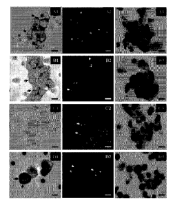
Figure 1A. A (Case 1) and B (Case 2). Al and B 1. Circulating tumor cells
showing an intense and cytoplasmic staining (score 3+) with some membrane
reinforcements (arrows) (ALK immunostaining using 5A4mAb, immunoperoxidase,
original magnification x 1000; bar: 16 vim). A2 and B2. Circulating cell
nuclei hybridized
with a dual-color 2p23 LSI ALK locus-specific split probe. The two probes (3',
red; 5',
green) show distinct separation of the red and green signals (arrowheads)
indicating a
12
Date Recue/Date Received 2021-03-18
rearrangement in the 2p23 ALK gene locus. The probes give overlapping signals
in nuclei
without the rearrangement (arrows). Isolated 3' signals (red) are also
observed (asterisks)
(original magnification x 1000; bar: 16 p.m). A3 and B3. Circulating cells
showing
malignant cytomorphological criteria isolated by the ISET method (original
magnification
x 1000; MGG staining; bar: 16 p.m). C. One patient with negative FISH ALK and
negative
IHC ALK in tissue tumor. Cl. Circulating tumor cells showing no staining
(score 0) (ALK
immunostaining using 5A4 mAb, immunoperoxidase, original magnification x 1000;
bar:
16 p.m). C2. Circulating cells nuclei hybridized with a dual-color 2p23 LSI
ALK locus-
specific split probe. The two probes (3', red; 5', green) gave overlapping
signals in nuclei
without the rearrangement (arrows). No split signal was detected in these
tumor cells
(original magnification x 1000; bar: 16 p.m). C3. Circulating cells showing
malignant
cytomorphological criteria isolated by the ISET method (original magnification
x 1000;
MGG staining; bar: 16 p.m). D. H22213 cells isolated by the ISET method. D1
ALK
immunostaining using 5A4 mAb (immunoperoxidase, original magnification x 1000)
showing an intense and cytoplasmic staining (score 3+) with some membrane
reinforcements (arrows). D2. FISH using the dual-color 2p23 LSI ALK locus-
specific split
probe on the H2228 tumor cell line spiked into peripheral blood and further
isolated by the
ISET method. The two probes (3', red; 5', green) show distinct separation of
the red and
green signals (arrowheads) indicating a rearrangement in the 2p23 ALK gene
locus. The
probes gave overlapping signals in nuclei without the rearrangement (arrows).
Isolated 3'
signals (red) are also observed (asterisks)H H original magnification x 1000;
bar: H 16 p.m).
D3. H2228 cells stained with MGG after blood filtration (original
magnification x 1000;
MGG staining; bar: 16 p.m).
Figure 1B. A (Case 3), B (Case 4) and C (Case 5). Al-Cl. Circulating tumor
cells
showing an intense and cytoplasmic staining (score 3+) with some membrane
reinforcements (arrows) (ALK immunostaining using 5A4mAb, immunoperoxidase,
original magnification x 1000; bar: 16 p.m). A2-C2. Circulating cell nuclei
hybridized with
a dual-color 2p23 LSI ALK locus-specific split probe. The two probes (3', red;
5', green)
show distinct separation of the red and green signals (arrowheads) indicating
a
rearrangement in the 2p23 ALK gene locus. The probes give overlapping signals
in nuclei
without the rearrangement (arrows). Isolated 3' signals (red) are also
observed (asterisks)
(original magnification x 1000; bar: 16 p.m). A3-C3. Circulating cells showing
malignant
13
Date Recue/Date Received 2021-03-18
cytomorphological criteria isolated by the ISET method (original magnification
x 1000;
MGG staining; bar: 16 vim).
Figure 2. Cytomorphological analysis of circulating non haematological cells
with
malignant features ¨ Circulating Tumor Cells (CTCs) detected using the ISET
method in
patients with COPD.
(A) and (B) CTCs isolated by the ISET method and identified by MGG staining
protocol in patients with COPD having developed lung cancer. (A) Isolated CTC
with
malignant cytomorphological features (double arrows: pores of the filter). (B)
Cluster
(CTM) composed of 9 CTCs with malignant cytomorphological features (Original
magnification x 1000; bars: 8 vim; double arrows: pores of the filters).
(C) and (D) Immunostained CTCs observed on filtered blood using the ISET
method for patients with COPD. (C) CTCs strongly expressing the pan-
cytokeratin antigen
only in patient with COPD. (D) CTCs co-expressing pan-cytokeratin and vimentin
antigens
in patient with COPD (Original magnification x 400; bars: 16 vim; immuno-
phosphatase
staining with a pan-cytokeratin antibody (ICL1) and immuno-peroxidase staining
with an
anti-vimentin antibody; double arrows: pores of the filters).
Figure 3. Detection of cytotrophoblast cells (A) and syncytiotrophoblasts (B)
in
cervical samples using ISET. The fetal nature of isolated cells was confirmed
by STR-
genotyping with informative markers D5S615 (A) and D21S11 (B).
DETAILED DESCRIPTION OF THE INVENTION
Samples. Biological samples comprise non-exclusively biological
fluids
comprising non-exclusively venous and arterial blood, lymph, urine, sperm,
ascites,
cerebrospinal fluid, pleural liquid, sputum, expectoration, nasal liquid,
articular fluid,
lacrymal liquid, liquid from urethra and ureter, biliary fluid, pancreatic
fluid, gastric fluid,
intestinal fluids, rectal fluid, vaginal fluid, samples collected non-
exclusively from mucosa
and organs like mouth, larynx, pharynx, uterus, cervix, vagina, esophagus,
stomach, small
and large intestine mucosa, samples collected non-exclusively by biopsy or
other surgical
intervention comprising non-exclusively samples from breast, prostate, liver,
lung, bone
marrow and any other organ.
Filters and Filtration. Filters that may be used to isolate or extract rare
cells
comprises nonexclusively a membrane of polycarbonate, PET (polyethylene
terephthalate)
or other material, having the thickness, and the pores size and density
adapted to the
14
Date Recue/Date Received 2021-03-18
extraction or isolation of defined rare cells. The filters, filtration
apparatus, filtration
methods, buffers and other equipment and supplies disclosed by Paterlini-
Brechot in
Published U.S. Patent Application US 2009/0226957 are hereby incorporated by
reference.
These include (i) a method involving the use of a filter comprising at least
one
basic filtration zone, whereby each basic filtration zone has a limited
surface area; and (ii)
the surface area of each basic filtration zone and the number of basic
filtration zones are
selected as a function of the type of liquid to be filtered, the type of
biological particles to
be separated and the volume of liquid to be filtered.
Accordingly, the invention relates to a process for separating biological
particles
and the fluid that contains them for the purposes of purification or analysis
and possibly for
diagnosis, comprising at least one vertical filtration stage through a filter
the porosity of
which is suited to the nature of the biological particles to be separated so
that said
biological particles are retained by the filter, characterised in that a
filter is used
comprising at least one elementary filtration area, each elementary filtration
area having a
limited surface, and in that the surface of each elementary filtration area
and the number of
elementary filtration areas is chosen according to the nature of the fluid to
be filtered, the
nature of the biological particles to be separated and the volume of fluid to
be filtered.
Each elementary filtration area of said process has a surface equal to that of
a disk
with a diameter of between 0.6 cm and 3 cm, and the number of elementary
filtration area
is chosen so that the ratio of the volume of fluid filtered to the filtration
surface is less than
40 ml/cm2, and preferably greater than 0.14 ml/cm2.
Preferably, each elementary filtration area has a surface equal to that of a
disk with
a diameter greater than or equal to 0.8 cm.
Preferably, the filter has pores calibrated to a size of between 3 vim and 100
vim and
a pore density of between 3 x 10³ and 5 x 10⁶ pores/cm2
Preferably, filtration is carried out by a reduction in pressure of between
0.05 bar
and 1 bar with, possibly, an increase in pressure of less than 1 bar.
To carry out filtration, it is preferable to use a filter forming a badge
suitable to be
associated with a means of analysing filtration residues by locating the
elementary
filtration areas.
Preferably, the badge forming the filter is incorporated in a single-use
filtration
module comprising at least one chamber for containing the fluid to be
filtered, and that can
be treated before use to sterilise it or to free it from enzymes that digest
DNA, RNA or
Date Recue/Date Received 2021-03-18
proteins.
The biological particles to be separated are, for example, cells. In this
case, prior to
filtering the fluid containing the cells, a sample of fluid for filtering may
be prepared from
a sample of fluid containing cells such as a biological fluid or cell culture
by pre-enriching
it with the cells to be separated and/or by diluting it.
The fluid containing the cells may be blood and, preferably, the filter in
this case
has calibrated pores of between 5 vim and 25 vim.
The fluid containing the biological particles is urine and the calibrated
pores of the
filter are between 8 vim and 100 vim.
The process can be used for the detection of cells for diagnostic purposes
such as
tumour, foetal, endothelial, fibroblastic, muscle, nerve or monocytal cells,
cell strains,
organ cells, precursors or haematopoietic cells, in a biological fluid such as
blood, urine,
ascites, cephalorachidian fluid, milk, pleural extravasation, fluid for
washing the neck of
the uterus, cell suspension fluid obtained by biopsy, by a surgical method or
by mouth
washing, or for the detection of animal or vegetable cells.
The invention also relates to a filtration module for implementing the
process, said
module comprising: a chamber block comprising at least one compartment closed
at its
lower portion by a base comprising at least one opening; a filter support
drawer comprising
at least one hole, each hole being arranged facing an opening in the chamber
block; a filter
gripped between the lower face of the chamber block and the support drawer.
In this module, the dimensions of each opening in the base of the chamber
block
and the dimensions of each hole in the filter support drawer are such that
each pair made
up of an opening in the base of the chamber block and the associated hole in
the filter
support draw, define an elementary filtration area of limited surface and in
that the useful
volume of each compartment is proportional to the number of elementary
filtration areas
situated in the base of the compartment.
Preferably, the surface of an elementary filtration area is equal to that of a
disk with
an equivalent diameter of between 0.6 cm and 3 cm, and the ratio of the useful
volume of
each compartment to the sum of the surfaces of the openings comprised in the
base of the
compartment is less than 40 ml/cm2, and preferably greater than 0.14 ml/cm2 as
well as all
intermediate values and subrange of the above mentioned ranges.
Preferably, the dimensions of at least one opening in the base of the chamber
block
and of a corresponding hole in the filter support drawer are such that the
surface of the
16
Date Recue/Date Received 2021-03-18
corresponding elementary filtration area is greater than or equal to that of a
disk 0.8 cm in
diameter.
Preferably, at least one compartment may be divided into part compartments by
at
least one removable separation wall, such that at least one part compartment
comprises in
its base at least one opening and that the ratio of the volume of said part
compartment to
the sum of the surfaces of the openings in the base of the part compartment is
less than 40
ml/cm2, and preferably greater than 0.14 ml/cm2 as well as all intermediate
values and
subrange of the above mentioned ranges.
Preferably, the filtration module comprises a grooved sealing joint arranged
.. between the base of the chamber block and the filter, comprising at least
one hole
corresponding to a hole in the base of the chamber block, the hole being
surrounded by at
least one projecting lip.
In addition, the filtration module preferably also comprises a plate joint
between
the filter and the filter support, comprising at least one opening opposite a
hole in the filter
support.
The filter may form a badge the central portion of which comprises at least
one
porous area and the periphery of which forms a frame comprising means for
indexing its
position on the filter support.
The indexation means are, for example, at least two holes of different
diameter
designed to cooperate with studs of corresponding diameter provided on the
filter support.
Preferably, at least a central porous portion of the filter comprises between
3 x 10³
and 5 x 10⁶ pores per cm2 of between 3 vim and 100 vim. All intermediate pore
size values
and subranges are contemplated within this range including 3, 4, 5, 6, 7, 8,
9, 10, 11, 12,
13, 14, 15, 16, 17, 18, 19, 20, 25, 27.5, 30, 35, 40, 45, 50, 55, 60, 70, 75,
80, 85, 87.5, 90,
92.5, 95, 97.5 and 100 m. All intermediate pore density ranges and subranges
are also
contemplated 1, 2, 3, 4, 5, 6, 7, 8, or 9 x 10³, 104, 1, 2, 3, 4, 5, 6, 7, 8 ,
or 9 x 10⁴, 105, 1, 2,
3, 4, 5, 6, 7, 8 , or 9 x 10⁵, 106 and 1, 2, 3, 4, 5, 6, 7, 8 , or 9 x 10⁶
pores/cm2.
Preferably, the filtration module also comprises at least one stopper for
closing the
upper opening of a compartment.
Preferably, the chamber block comprises, at its lower portion, a rim extending
outwards and cooperating with at least one assembly pin allowing the filter to
be gripped
between the filter support and the chamber block, the assembly pin comprising
a breakable
end extending above the rim of the chamber block.
17
Date Recue/Date Received 2021-03-18
Preferably, all its parts are made of materials suited to a sterilisation
operation or
designed to render them free from RNases, DNases or proteinases.
Finally, the invention relates to a filtration module support for retaining a
filtration
module on a filtration machine, comprising at least one cam that can move
between an
open position and a gripping position, designed to put pressure on the filter
between the
filter support and the chamber block.
Preferably, at least one cam is designed so that, if the filtration module
comprises at
least one fixing pin one end of which is breakable, the end of at least one
fixing pin is cut
when pressure is applied to the filter by at least one cam.
The support block forms part of a filtration machine.
Preferably, the filtration module also comprises a means designed to cooperate
with
a complementary means on a support block, so as to impose the orientation of
the filtration
module in relation to the support block, and the support block comprises a
means designed
to cooperate with a means on a filtration module, so as to index the
orientation of the
filtration module in relation to the support block.
The method for isolating biological particles contained in a fluid, according
to the
invention, consists of filtering the fluid on a filter with characteristics
suited to the nature
of the particles to be isolated. The biological particles may be cells, red
blood cells, platelet
aggregates, fibrins or tissue waste. The filtered fluid is in particular a
fluid obtained from a
sample of biological fluid that may have undergone prior treatment to
facilitate the
isolation by filtering operation. This prior operation, which will be
described in more detail
later, comprises in general, particularly when the particles to be isolated
are cells, one or a
plurality of the following operations: chemical treatment designed to pre-
enrich the cell to
be isolated, dilution, chemical treatment designed to facilitate separation by
filtration of the
cells to be isolated.
As well as these conditions for preparing samples of fluid for filtering, the
inventors noted that to achieve good reliability in the process of isolating
cells to be
detected, it was necessary to adapt certain characteristics of the filter to
the volume of fluid
filtered. In particular, the filter must be divided into elementary filtration
area each having
a surface equal to that of a disk of diameter of between 0.6 cm and 3 cm, and
preferably
greater than 0.8 cm and even better between 0.8 cm and 1.5 cm as well as all
intermediate
values and subrange of the above mentioned ranges. The elementary filtration
areas may
be in the shape of a disk, for example.
18
Date Recue/Date Received 2021-03-18
In addition, the quantity of fluid to be filtered, which must pass through
each of the
elementary filtration areas, must be between 1 ml and 100 ml, and preferably
this volume
should be between 8 ml and 15 ml. These ranges include all intermediate values
and
subranges of the above mentioned ranges.
Thus, to filter a particular sample a device must be used to define a number
of
elementary filtration areas on the filter in proportion to the volume of the
sample to be
filtered.
In general, the volume of the sample to be filtered depends on the one hand on
the
volume of biological fluid that could be taken initially, and on the other
hand on a possible
dilution which depends in particular on the nature of the biological particles
to be
separated. The volume taken depends in particular on the nature of the fluid
taken and the
age of the patient from whom the fluid is taken. A person skilled in the art
knows how to
determine the volumes to be taken depending on the nature of the fluid taken
and on the
patient from whom it is taken.
Dilution depends in particular on the number of particles per unit of volume
that
can be found in the fluid taken. Indeed, if filtration is to be carried out
under satisfactory
conditions, the number of particles to be isolated per unit of volume of fluid
to be filtered
should not be too great to avoid clogging the filter. Moreover, if the process
is intended to
detect particular rare cells mixed with a far greater number of cells, the
number of cells per
unit of volume should not be too small, so as to achieve a reasonable
probability of finding
the cells sought on the filter. A person skilled in the art also knows how to
determine these
dilution rates depending on the nature of the fluid in question and the type
of cell sought.
The biological sample taken from a patient may, for example, be blood, urine,
ascites, cephalorachidian fluid, milk or pleural extravasation; it may also be
fluid from
washing the neck of the uterus or any other fluid that may result from taking
a biological
sample from a patient.
The analysis method may also be used to search for cells in samples that have
not
been taken directly from patients, and for example, in samples taken in cell
culture
mediums made from smears or biopsies or from human or animal tissue samples
or,
further, in human or animal cell line culture mediums.
If the biological fluid taken is blood, the amount taken is generally between
1 ml
and 20 ml, and the blood is diluted by a ratio that varies from 1 in 5 to 1 in
20 to obtain a
sample of fluid for filtering which, in these conditions, is filtered over one
to 20
19
Date Recue/Date Received 2021-03-18
elementary filtration areas. These values include all intermediate values and
subrange of
the above mentioned ranges.
For all other fluids, the samples are approximately 5 ml to 10 ml and are
diluted in
a ratio of between 1 in 2 and 1 in 10, or they may not be diluted. These
samples are filtered
over a number of elementary filtration areas which may be as many as 5 or even
more,
particularly if it is a 10 ml sample that has been diluted in a ratio of 1 in
10. These values
include all intermediate values and subrange of the above mentioned ranges.
The cells that may be sought are in particular rare cells such as tumour
cells, foetal
cells, endothelial cells, fibroblastic cells, muscle cells, nerve cells,
monocytal cells, cell
strains, organ cells (hepatic, renal, etc. . . . ), precursors and
haematopoietic cells. This list,
which is given as an example, is not limitative.
Before filtration, the cells may be pre-enriched by treatment of the density
gradient
type or by lysis of cells that are of no interest, or by immunomediated
methods, by positive
or negative screening, by stimulating the cells sought to proliferate, etc.
This list is not limitative, and a person skilled in the art knows how to
choose a pre-
enrichment process suited to the nature of the cells that he or she seeks to
isolate.
As well as the pre-enrichment treatment, the fluid sample containing cells may
be
treated by a reagent according to the nature of the cells sought, to
facilitate the separation
by filtration operation.
The aim of the treatment may be to lyse red blood cells and anticoagulate the
blood
if the biological sample contains blood, and consists, for example, of adding
saponin and
EDTA.
The aim of the treatment may also be to fix nucleated cells, for example by
the
addition of formaldehyde, if the filtration is intended to isolate fixed
cells. In this case, the
object of the treatment is to make enrichment possible.
If the filtration is intended to isolate non-fixed cells, the biological
sample may be
treated with a reagent and under conditions suitable for temporarily rendering
biological
membranes rigid (for example, by the addition of polysaccharide, DMSO, by
cold, etc.).
A person skilled in the art knows how to choose the most suitable method,
according to the nature of the cells sought.
The biological sample which may have been diluted, pre-enriched or treated
with a
reagent to allow filtration suited to the end sought, is then filtered through
a filter made of
polycarbonate or an equivalent material that has calibrated pores of a size
between 1 vim
Date Recue/Date Received 2021-03-18
and 100 vim and suited to the nature of the particles to be separated. All
intermediate
values and subranges are contemplated within this range including 1, 2, 3, 4,
5, 6, 7, 8, 9,
10, 11, 12, 13, 14, 15, 16, 17, 18, 19, 20, 25, 27.5, 30, 35, 40, 45, 50, 55,
60, 70, 75, 80, 85,
87.5, 90, 92.5, 95, 97.5 and 100 m. This size is preferably between 3 vim and
25 vim, and
is about 8 vim, for example, particularly if tumour cells or epithelial cells
are to be isolated.
Pore density is suited to the nature of the particles to be separated.
Preferably, the
pore density of the filter is between 5 x 10³ and 5 x 10⁶ pores/cm2 and even
better between
5 x 10⁴ and 5 x 10⁵ pores/cm2 . All intermediate values and subranges within
these ranges
are contemplated as well as the following specific values: 1, 2, 3, 4, 5, 6,
7, 8, or 9 x 10³,
104, 1, 2, 3, 4, 5, 6, 7, 8 , or 9 x 10⁴, 105, 1, 2, 3, 4, 5, 6, 7, 8 , or 9 x
105, 106 and 1, 2, 3, 4,
5, 6, 7, 8 , or 9 x 10⁶ pores/cm2.
Filtration is performed preferably be a reduction in pressure of between 0.05
bar
and 1 bar, and preferably of approximately 0.1 bar. All intermediate values
and subranges
of this range are contemplated including 0.05, 0.06, 0.07, 0.08, 0.09, 0.10,
0.20, 0.30, 0.40,
0.50, 0.60, 0.70, 0.80, 0.90 and 1.0 bar. Filtration may be assisted by a
slight increase in
pressure on the fluid situated above the filter. This increase in pressure
must however be
less than 1 bar. These conditions are particularly suited to cell separation.
The process may be used for different objectives, for example to search for
rare
cells in suspension in a biological fluid, so as to allow diagnosis or to
purify a fluid to
allow analysis in good conditions of the elements in solution.
If the process is used to search for cells and analyse them, after filtration,
the filter
that has been used to filter the fluid is recovered ensuring that the
filtration areas are
clearly identified and that a link can be made between these filtration areas
and the sample
that was filtered. The filter is then used to analyse the cells that it may
have been possible
to recover in the filtration areas.
These analysis methods, which are known per se, are for example of the
following
types: cytological staining (haematoxylin, eosin, etc.), immunomarking
(immunohistochemistry, immunofluorescence) PNA, FISH, PRINS, PCR in situ or
other
molecular technique, spectrophotometry, laser microdissection followed by
targeted
.. molecular analyses on the DNA (DNA extraction, genotyping, quantitative
PCR, mutation
analysis, CGH (comparative genomic hybridisation)) RNA (extraction and
analysis by
PCR of transcripts, quantitative PCR) and proteins (protein extraction,
microsequencing,
etc.).
21
Date Recue/Date Received 2021-03-18
The molecular analyses may be performed on enriched cells held on the filter
and
transposed onto a slide by a technique similar to the Southern technique,
individually
micro-dissected from the filter or from the slide according to defined
criteria
(morphological characteristics of the cells with or without marking of
different natures)
and subjected to individual or pooled molecular analysis.
The cells may also be detached from the filter by washing with an appropriate
buffer to extract and analyse their DNA, RNA and proteins.
The elements isolated by filtration are then examined with a microscope and
analysis of the images obtained on the filter may be carried out manually or
by automated
means, in particular by using image analysis equipment.
The process may also be used to purify a biological fluid such as urine
containing
in solution the DNA, RNA or proteins that are to be analysed. The purpose of
purifying the
fluid is to eliminate all the biological particles present in the fluid, which
could interfere
with the analysis. In this case, the filters are not kept and it is the
filtered fluid that is
analysed.
This filtration method and the sample preparation and analysis methods may be
used as stated previously in particular for the purpose of diagnosis to detect
pathologies
associated with the presence of particular cells possibly in extremely small
quantities. In
particular, the process can be used to detect cancerous cells that may have
been released
into a patient's blood during a surgical operation. A person skilled in the
art knows what
cells can be searched for to detect a particular pathology.
Support. A support may represent a solid non porous support such as a slide or
a
petri dish or a culture well or any other support made of glass or plastic or
any solid
material which can be used as a support of cells for culture, treatment or
analyses of any
type: cytomorphological, immunolabelling, in situ molecular analyses,
comprising protein,
RNA or DNA analyses, and collection of cells for molecular analyses,
comprising protein,
RNA or DNA analyses.
Filtration. Filtration of a biological sample to extract, isolate, purify or
concentrate
rare cells, is carried out by using non-exclusively a membrane of
polycarbonate, PET
(polyethylene terephthalate) or other material, having the size and density of
pores adapted
to the extraction or isolation of defined rare cells and by using depression
applied under the
filter to isolate or extract rare cells.
22
Date Recue/Date Received 2021-03-18
Extraction of cells by vertical filtration of a biological sample allows one
to layer
them and make them available for further analyses, such as for detection and
characterization and diagnosis of rare cells. Isolation of cells by vertical
filtration of a
biological sample allows one to isolate rare cells away from smaller cells in
order to enrich
them and make them available for further analyses, such as for detection,
characterization,
and/or diagnosis of rare cells. Typically isolation of rare cells by
filtration of blood allows
to separate the majority of neutrophils and mature lymphocytes and
erythrocytes
(erythrocytes do not contain nucleus, thus they are not considered true cells
and are
generally lysed before filtration) which are the smallest cells in the body as
their size is 6
to 9 microns, and to retain on the filter cells larger than neutrophils and
mature
lymphocytes including activated lymphocytes, monocytes, macrophages, stem
cells, tumor
cells, cancer cells, tumor microemboli, mature and immature endothelial cells,
epithelial
cells, mesenchymal cells other than neutrophils and mature lymphocytes,
melanocytes
myeloblasts, promyelocytes, megakaryoblasts, megakaryocytes and in general all
the cells
of the body which are not neutrophils and mature lymphocytes. Furthermore,
isolation of
rare cells by filtration of blood also allows one to collect, on the other
side of the filter,
plasma and leukocytes or other components of a biological sample distinct from
the rare
cells isolated by filtration. Plasma contains free DNA and RNA, and proteins
including
free tumor DNA and tumor RNA, and tumor microRNA and proteins in patients with
cancer, and including free fetal DNA, fetal RNA and fetal micro RNA and
proteins in
pregnant women. The term -free" indicates -outside cells", thus free nucleic
acids in
plasma. Free tumor DNA is used for diagnosis of tumor mutations in patients
with cancer
and free fetal DNA is used for prenatal diagnosis of aneuploidies and other
genetic
disorders, fetal gender, RhD status, paternity tests. Collecting leukocytes at
the same time
as rare cells and plasma can be useful to analyze and obtain information about
the genetic
background of the individual. Free tumor DNA and/or RNA and/or protein is
expected to
derive from lysed, probably apoptotic, tumor cells from the tumor mass and/or
tumor
metastases and/or circulating tumor cells compartment. Analysis of free tumor
DNA and/or
RNA and/or proteins is performed by extracting DNA and/or RNA and/or proteins
from
plasma and looking for mutations by molecular analyses. For instance, a quick
analysis of
the presence of K-Ras mutations in patients with lung cancer can be performed
by
extracting free DNA from plasma and looking for mutated K-Ras molecules by
PCR,
CastPCR, Cold PCR, digital PCR and other targeted molecular tests. Thus,
analysis of K-
23
Date Recue/Date Received 2021-03-18
Ras mutations in circulating tumor cells can be associated to the analysis of
KRas mutation
in plasma DNA. In addition, the study may start with the search of KRas
mutation in
plasma, which is less expensive, and if the mutation is not found, it may go
on with the
search of KRas mutation in circulating tumor cells, which is more expensive.
The diagnosis of fetal gender is done easily and at low cost by analysis of
plasma
DNA. However, if the amount of free fetal DNA in plasma is low, a negative
signal with Y
specific molecular analyses does not allow obtaining reliable results. In this
case, the
possibility to add the analysis, more expensive, of circulating fetal cells,
will allow
obtaining a reliable diagnosis of fetal gender.
The detection of infectious diseases, like those caused by HBV, HCV, or HIV or
by
bacteria or other pathogens, can be performed by extracting molecules such as
DNA,
and/or RNA, and/or micro RNA and/or proteins from plasma and looking for the
presence
of viral or bacterial or other pathogens DNA, and/or RNA, and/or micro RNA
and/or
proteins by molecular analyses. As a complementary test, DNA, and/or RNA,
and/or micro
RNA and/or proteins specific to pathogens can be looked for in circulating
rare cells.
In specific cases, it can also be useful to obtain at the same time
information on
mutated or infectious molecules in rare cells, mutated or infectious molecules
in plasma,
and on the genomic characteristic of the individual. In this case, the
collection of plasma
and leukocytes after filtration to isolate circulating rare cells is extremely
useful. For
instance, in infected patients, it can be useful to look for infectious
molecules, such as
DNA from TBC bacillum, in circulating rare cells, in plasma and to look for
genetic
susceptibility traits in the leukocytes. For instance, in patients with cancer
of genetic
origin, it will be useful to look for a mutation, such as BRCA1 or BRCA2, in
the
circulating tumor cells, in plasma and in the leukocytes. For instance, in
pregnant women,
it can be useful to analyze rare circulating fetal cells for the presence of
Duchenne's
disease, a genetic disease that affects only male fetuses, to look for Y
sequences in the free
fetal DNA to know if the fetus is male and to look for the carrier status in
the maternal
leukocytes. For instance, in pregnant women, it can be useful to analyze rare
circulating
fetal cells for the presence of Huntington's disease, a dominant genetic
disease that may
have a late onset, to look for Huntington's mutated sequences in the free
fetal DNA, and to
look for the presence of Huntington's mutation in the maternal leukocytes. In
fact, since
the mutation is dominant, the presence of the mutation in one of the two
parents gives the
fetus 50% risk to be affected. If the genetic analysis discovers that the
fetus is affected, it
24
Date Recue/Date Received 2021-03-18
will be useful to check if the mutation was carried by the mother through
analysis of
maternal leukocytes.
These examples are not exclusive. In general, the possibility to isolate
circulating
rare cells and at the same time to collect plasma and leukocytes for immediate
analysis or
for storage and future analysis has a high potential and value in non-invasive
personalized
medicine.
Adaptation of the size of the pores of the filter to the biological samples to
be
filtered allows one to selectively isolate cells of discriminate size, for
instance, tumor
microemboli and syncytiotrophoblasts, groups of cells and multinucleated
cells, and
cellular material having a larger size than individual cells, thus efficiently
isolating such
material from blood or other fluids with high purity (low or absent
contamination by
leukocytes and other smaller cells) by filtration using pores larger than 20-
25 microns in
diameter, thus eliminating by filtration all leukocytes and erythrocytes.
By analogy, by studying the particular size of tumor cells of a given tumor
type
and/or in a given patient, and/or by studying the particular size of certain
rare cells to be
isolated, it is possible to adapt the pore size, pore density and other
chemical or physical
features of the filter to maximize recovery and purity of the isolated tumor
and/or rare
cells. For instance, fetal cells size may vary from 10 to 30 microns.
Syncytiotrophoblasts
size is generally larger than 100 microns. The size of mature endothelial
cells, which are
not round but elongated cells, is around 40 or 50 microns per 10 to 20
microns. Thus, the
pores size range of interest is between 5 microns and 30 microns and the
larger pores size
allows eliminating all the leukocytes. In fact, the larger leukocytes, which
are macrophages
and monocytes, have a size that generally is not larger than 20 microns. The
size of the
pores has to be adapted very closely to the pores density, and be in a range
from 0.5 to 2.0
E5 pores per cm2as enough filter material has to be between pores to allow
collection of
rare cells.
Tumor cell size. Tumor cells by definition are not -resting cells" as they
produce
proteins and may proliferate, thus their chromatin is open and never compacted
like the
chromatin of mature leukocytes as mature lymphocytes and mature neutrophils
which are
the majority leukocytes (as number) and are thus smaller than tumor cells.
Individual
tumor cells size may vary from 10 micron to 50 microns or more depending on
the type of
tumor cells. Ex size of Tumor cells from Small Cell Lung Carcinoma (SCLC): 1.5
to 3
times the size of lymphocytes (12 to 24 microns), size of Tumor cells from Non-
Small Cell
Date Recue/Date Received 2021-03-18
Lung Carcinoma (NSCLC): over 3 times the size of lymphocyte (24 microns).
Tumor
microemboli size is generally larger than 100 microns.
Advantages of filtration. Extraction or isolation of rare cells by filtration
has
several advantages, over other methods of extraction/isolation:
It permits isolation of rare cells with very high sensitivity, including
collection of
one individual rare cell that can be spiked before filtration in one ml of
blood, thus mixed
with several millions of leukocytes and several billions of erythrocytes.
It allows extraction or isolation of rare cells independently from the
antigens
expressed by rare cells, thus avoiding bias of isolation leading to the loss
of rare cells.
It facilitates the multi-analyses of rare cells, including morphological,
immunolabelling, in situ molecular analyses and molecular analyses without
interference
of other non rare cells.
It allows one to modulate the purity of the isolated rare cells, which make
easier
their multi-analyses, including morphological, immunolabelling, in situ
molecular analyses
and molecular analyses without interference of other non rare cells.
It allows one to collect rare cells individually, as single cells or groups of
single
cells, or as cells mixed with residual non rare cells for further molecular
analyses.
It speeds up the detection and counting the rare cells by getting rid of the
millions
of small leukocytes and billions of erythrocytes
It allows one to extract or isolate fixed or fresh cells for further analyses.
Other modes of filtration. Various modes filtration may be employed so long as
they permit rare cells to be isolated from other kinds of cells and/or layered
on the filter for
further analysis. For example, the force for separating rare cells from other
kinds of cells
that pass through a filter may be gravity, positive or negative pressure, or
centrifugal force.
Pretreatment of Samples. Biological samples can be diluted and/or treated
before
and/or after filtration by agents used to lyse erythrocytes like saponin,
ammonium chloride,
lytic antibodies, hypotonic solutions, anticoagulants like EDTA, heparin,
coumadin and
other Vitamin K antagonists, factor Xa antagonists, thrombin inhibitors;
aspirin (salicylic
acid) and other agents preventing platelet
aggregation
(http://en.wikipedia.org/wiki/Antip1ate1etdrug, accessed May 21, 2013),
mucolytic drugs,
and fixative agents (see below).
Rare cells extracted or isolated from biological samples by filtration can be
fixed
cells or fresh cells. Fixed or fresh rare cells extracted or isolated from
biological samples
26
Date Recue/Date Received 2021-03-18
by filtration can be transferred to a support comprising non-exclusively
slide, petri dish,
well or microwell or test tube or other support for analysis, molecular
analyses or culture.
Transfer of cells from the filter to a support can be obtained by using
collecting and/or
detaching means and/or buffers. For instance, in order to transfer all cells
from the filter to
a slide or to a solid support and avoid loosing rare cells, it is possible to
use commercially
available adhesive slides such as SuperFrost Ultra Plus slidesor ClearcellTM
and AdcellTM
BioAdhesion Slides and/or to treat the filter before filtration with agents
which prevent
sticking of cells to the filter with siliconizing agents like SigmacoteTM or
similar. In order
to transfer all the cells collected on the filter to a slide or to any other
support and to avoid
loosing rare cells it is also possible to help the transfer with solutions to
be applied to the
back of the filter, thus the side which does not stick to cells, and creating
a flux of buffer
through the pores toward the solid surface which will detach cells from the
filter so that
they can adhere to the slide or other solid support. This process of transfer
of cells and rare
cells collected on a filter to a slide or other solid support can also be
improved by using
positive air and/or liquid pressure applied to the back of the filter: air
and/or liquid will
pass through the pores and help cells to be transferred to the slide or other
solid support.
All these protocols to detach cells and rare cells from the filter and
transfer them to a slide
or solid support will work better if cells on the filter are not dried, thus
if the transfer is
performed soon after filtration.
Extraction or isolation of fixed cells by filtration is performed by fixing
them
before filtration using fixative agents comprising non-exclusively
formaldehyde,
paraformaldehyde, glutaraldehyde, RCL2, mercurials like B5 and Zenker's
fixative,
methyl alcohol and ethyl alcohol, picrates like Bouin's solution, Rigaud
fixative etc.
Fresh rare cells extracted or isolated from biological samples by filtration
can be
fixed after filtration using fixative agents comprising non-exclusively
formaldehyde,
paraformaldehyde, glutaraldehyde, RCL2, mercurials like B5 and Zenker's
fixative,
methyl alcohol and ethyl alcohol, picrates like Bouin's solution, Rigaud
fixative etc.
Culture of Rare Cells. Rare cells extracted or isolated from biological
samples can
be cultured in order to increase their number and facilitate their detection
and/or diagnosis
and/or characterization. The culture protocol may include means to stimulate
preferentially
the growth or rare cells versus the growth of non rare cells in order to
increase the purity of
rare cells. Means to stimulate preferentially the growth or rare cells versus
the growth of
non rare cells comprise non-exclusively specific growth factors and/or agents
stimulating
27
Date Recue/Date Received 2021-03-18
the growth of rare cells, co-culture of rare cells with other cell types
and/or use of feeder
layers helping the growth of rare cells, and means to block the growth and/or
survival of
non rare cells such as inhibiting antibodies, blockers of cell cycle,
proapoptotic factors,
siRNA, and drugs of any type specifically targeting non rare cells in order to
obtain the
block of their growth and/or their elimination.
Characterization of Rare Cells. Rare cells detection and/or diagnosis and/or
characterization can be obtained through cytomorphological and/or
immunolabelling
and/or molecular analyses. Cytomorphological and/or immunolabelling analyses
are
performed in situ on intact cells, i.e., on cells which plasma membrane and/or
cytoplasmic
limit and/or nuclear limit is recognizable.
Cytomorphological analyses non-exclusively comprise staining by Hematoxylin
and/or Eosin, May Grunwald and/or Giemsa staining, Papanicolau staining,
Feulgen
staining and all type of staining and stechiometric staining aiming to analyze
cellular
morphological details and analyze and/or quantify cellular components.
Cytomorphological analyses non-exclusively comprise cytochemical analyses
which non-
exclusively comprise PAS, Sudan, Alcian blue staining, enzymatic and non-
enzymatic
methods able to reveal cellular components which non-exclusively comprise
calcium,
lipids, polysaccharides, enzymes and others molecules. Immunolabeling non-
exclusively
comprises labeling cellular components with antibodies which non-exclusively
comprise
antibodies directed to epithelial antigens, to mesenchymal antigens, to organ-
specific
antigens, to tumor-specific antigens, to fetal-specific antigens, to stem
cells-specific
antigens, to transcription factors, to mutated proteins and to any protein
and/or peptide
and/or cellular component which detection may help cellular identification
and/or
diagnosis and/or characterization. Immunolabeling also non-exclusively
comprises
immunocytochemistry, immunofluorescence, immune-PCR, and all types of labeling
of
cellular structure through antibodies bound to a mean used to reveal the
immunological
link and the cellular target.
Molecular analyses comprising non-exclusively FISH (Fluorescence In Situ
Hybridization), PRINS (Primed In Situ labeling)), TUNEL (Terminal
deoxynucleotidyl
transferase dUTP nick end labeling), immunoPCR, PNA (peptide nucleic acid), in
situ
PCR and other methods using non-exclusively molecular probes can be performed
in situ
on intact cells.
28
Date Recue/Date Received 2021-03-18
Image analyses of rare cells after in situ staining and/or immunolabelling
and/or in
situ molecular analyses can be performed and images can be stored and/or
transferred
informatically before further rare cells molecular analyses implying cellular
lysis.
Molecular analyses comprising non-exclusively PCR, Reverse Transcriptase-PCR,
real time PCR, digital PCR, Whole Genomic Amplification, sequencing, High
Throughput
sequencing, Cast-PCR, Cold PCR, Comparative Genomic Hybridization (CGH), CGH
array, microarray analyses, methylation analyses, polymorphism analyses, etc.
can be
performed on cells which structure has been disrupted and/or lysed in order to
analyze
molecular components comprising non-exclusively DNA, RNA, microRNA and protein
molecules.
Molecular analyses comprising non-exclusively PCR, Reverse Transcriptase-PCR,
real time PCR, digital PCR, Whole Genomic Amplification, sequencing, High
Throughput
sequencing, Cast-PCR, Cold PCR, Comparative Genomic Hybridization (CGH), CGH
array, microarray analyses, methylation analyses, polymorphism analyses, etc.
can be
targeted to individual cells, or to groups of individual cells or to all cells
extracted or
isolated through filtration from a biological sample.
Targeted analyses to individual cells or groups of cells identified according
to
morphological criteria and/or immunolabelling can be performed by using laser
microdissection of the filter part containing the cells of interest, or of
cells after transfer to
a support, followed by lysis of cellular proteins and molecular analysis of
cellular DNA
(genomic and/or mitochondrial DNA), RNA, microRNA and protein molecules.
Alternatively, rare cells extracted or isolated by filtration can be detached
from the filter
and individual cells or groups of cells identified according to morphological
criteria and/or
immunolabelling can be isolated by magnetic field (Silicon biosystem or other
methods
based on magnetic field), or by manual or automated capillary micropipetting.
Alternatively, all cells extracted or isolated by filtration can be lysed on
the filter, or after
transfer to a support, by using an appropriated buffer in order to perform
their DNA
(genomic and/or mitochondrial DNA), RNA, microRNA or protein molecular
analysis by
using non-exclusively PCR, Reverse Transcriptase-PCR, real time PCR, digital
PCR,
Whole Genomic Amplification, sequencing, High Throughput sequencing, Cast-PCR,
Cold
PCR, Comparative Genomic Hybridization (CGH), CGH array, microarray,
methylation
analyses, polymorphism analyses, etc..
29
Date Recue/Date Received 2021-03-18
Non-invasive Theranostics. Among its other aspects, the invention makes it
possible to isolate circulating tumor cells from blood and use them for non-
invasive
theranostics. Theranostics is the use of a diagnostic result to guide
prescription of a
specific -targeted" treatment. These procedures take advantage of a particular
molecular
biomarker that may be present in tumor cells to predict their susceptibility
to respond or
not a given therapy. As cancer therapies are costly and often toxic,
theranostic is thus
important to minimize health care cost and burden due to adverse effects to
patients.
Theranostics biomarkers are now looked for in the primary tumor and metastases
tissues.
However, it has been demonstrated that tumor cells in the primary tumor are
genetically
heterogeneous and biopsies can miss tumor cell clusters carrying genetic
information
useful to set up optimal targeted treatments. Metastases are considered to be
a better
reference, but their biopsy is difficult to obtain. Indeed, the physical
conditions of cancer
patients often prevent to obtain samples from the primary tumor or metastases
through an
invasive approach (surgery or biopsy) due to the high rate of morbidity linked
to these
invasive procedures. Moreover, tumor cells genetic characteristics may change
under
pressure of targeted treatment making important to follow them during
treatment in order
to detect new -tumor cells mutants" escaping targeted treatments. However, an
invasive
follow up of cancer cells genetics characteristics is unfeasible in clinical
settings due to the
related morbidity. Finally, it would be useful to study non-invasively the
genetic
characteristics of tumor cells in certain patients in order to apply targeted
therapies
preoperatively on the purpose to decrease the tumor burden before the
intervention and
better remove the tumor.
Targeted therapies are now available for a number of common cancers (breast
cancer, lung cancers, colorectal cancer etc...) and have shown their efficacy
in a proportion
of cases. However, in the absence of demonstrated predictive factors of tumor
response,
these targeted expensive therapies will either be prescribed to the great
majority of
patients, with a huge increase of related costs without proportional benefit,
or will not be
prescribed, preventing patients from taking benefit from them.
The implementation of theranostic biomarkers is thus expected to lead to
impressive therapeutic improvements. However, theranostic analyses addressed
to tumor
tissues (primary tumor, metastases) imply invasive (surgical or semi-surgical)
procedures
that cannot be performed in debilitated patients, are very costly, and often
provide
Date Recue/Date Received 2021-03-18
incomplete data. Consequently, the analysis of tumor cells and tumor
microemboli
obtained non-invasively is of utmost importance in clinical oncology.
Genetic Characterization of Rare Cells. Information on the genetic
characteristics
of tumor cells can be obtained non-invasively from circulating tumor cells
(CTC),
circulating tumor microemboli (CTM), and tumor cells collected from biological
fluids
(urines, ascites, sperm, cerebrospinal fluid (CSF), sputum, expectoration
etc).
Methods to study genetic characteristics of tumor cells vary according to the
genetic abnormalities to be detected. They can rely on cytological,
cytochemical,
immunocytochemical analysis, FISH, PRINS, immunoPCRõ DNA and RNA extraction
and analyses targeted to genes or sequences of interest and/or to the whole
exome or the
whole genome/transcriptome sequence.
Several theranostic biomarkers have been validated in particular for Non-Small
Cell Lung Carcinoma (NSCLC) and colon cancer. Epidermal growth factor receptor
(EGFR) is a membrane receptor tyrosine kinase. Overexpression or overactivity
of EGFR
is found in many cancers. EGFR mutations are detected in 10% to 15% of all
patients with
NSCLC and in 80% of patients who clinically respond to EGFR tyrosine kinase
inhibitors
such as gefitinib (AstraZeneca) or erlotinib (Roche). Known EGFR mutations
include
mutations located in exons 18 to 21 of the EGFR tyrosine kinase domain. These
mutations
are well described in particular in lung cancer. Patients having a primary
tumor with the
L858R mutation or the deletion of exon 19 mutation have a better response to
EGFR
inhibitors than patients with wild-type KRAS tumors[13]. The L858R mutation
and the
deletion of exon 19 mutation are among the most frequent EGFR mutation in lung
tumors
with a frequency of approximately 43% and 48% of all EGFR mutations,
respectively
(http://www.mycancergenome.org, accessed May 21, 2013).
KRAS (also known as V-Ki-ras2 Kirsten rat sarcoma viral oncogene homolog)
encodes a 21 kDa GTPase localized to the inner membrane. This protein is a
core
component of the signal transduction pathway upstream of EGFR. KRAS mutations
occur
in 15-25% of lung adenocarcinoma (http://www.mycancergenome.org, accessed May
21,
2013). KRAS more frequent mutations results in a constitutive activation of
KRAS kinase
activity. Point mutations occur in codon 12 (82% of all KRAS mutations) and 13
(17% of
all KRAS mutations) in exon 2 of the KRAS gene. Standard sequencing detects in
one test
of all codon 12 and 13 mutations (G12C: c34G>T, G 12R : c34G>C, G 12A :
c35G>C,
G12D : c35G>A, G12V : c35G>A, G125 : c34G>A, G13C : c37G>T, G13D : c38G>A).
31
Date Recue/Date Received 2021-03-18
Wild-type KRAS is required for EGFR inhibitor efficacy in NSCLC patients [13].
Wild-type KRAS is also required for efficacy of anti-EGFR therapeutic
antibodies (Eli
Lilly, Bristol-Myers Squibb, Merck Serono) in patients with metastatic
colorectal cancer
[14,15]. Mutations in codons 61 and 146 have also been reported but they
represent a
minor proportion of KRAS mutations (1-4%) and their clinical relevance remains
obscure
[15].
Currently, NSCLC patients with KRAS mutations have no effective treatment
strategy. Ganetespib (Synta Pharmaceuticals) is a small molecule inhibitor of
the
chaperone Hsp90 that is currently ongoing a Phase III clinical trial. A phase
2 trial showed
tumor shrinkage in more than 60 percent of patients with KRAS-mutant NSCLC at
eight
weeks after treatment with ganetespib administered once weekly as a
monotherapy
(International Association for the Study of Lung Cancer, 14th World Conference
on Lung
Cancer).
More recently, a genomic alteration involving the anaplastic lymphoma kinase
(ALK) (2p23) and the Echinoderm Microtubule associated protein Like-4 (EMLA)
(2p21)
genes was identified in a subset of lung cancer patients. These patients have
an outstanding
favorable response to an ALK small molecule inhibitor, Crizotinib (Pfizer)
[16,17,18,19].
In fact, Crizotinib is currently tested in a phase III clinical trial and the
preliminary results
of the phase II trial evidenced an impressive 90% disease control rate in ALK
rearranged
lung tumors [20]. This rearrangement has been found in 1 to 7 % of NSCLC
according to
most of series, particularly in late stage adenocarcinomas, without EGFR and
KRAS
associated mutations [21].
Two other outstanding theranostic biomarkers have been validated in other
types of
cancer. Human Epidermal Growth Factor Receptor-2 (HER2) is a cell membrane
surface-
bound receptor tyrosine kinase and is normally involved in the signal
transduction
pathways leading to cell growth and differentiation. The HER2 locus is
amplified in 20-
30% of breast tumors and the presence of this amplification is an indication
of tumor
response to the targeted therapy trastuzumab (anti-HER2, Roche) [22].
Zelboraf (Roche) is a very effective drug that has been recently approved by
the
Food and Drug Administration (FDA) for the treatment of patients with melanoma
whose
tumors harbor the V-raf murine sarcoma viral oncogene homolog B1 (BRAF) gene
V600E
mutation. As this mutation is also present in 1% of lung cancer patients, lung
cancer
32
Date Recue/Date Received 2021-03-18
patients may become eligible for this drug in the future. Other theranostic
biomarkers
validated or ongoing clinical trials include:
- Breast and ovarian cancer: ER, PR, BRCA1, BRAC2
- Gastrointestinal cancer: c-Kit, CDC4, p53
- Non-small cell lung cancer: ROS1, HER2, FGFR, PDGFRA, VEGFR, PI3K
- MTOR, MEK, STAT3, AKT, RET fusions
- Prostate Cancer: recombination TMPRSS2 / ERG.
Concerning non-invasive theranostic analyses of tumor cells, current unsolved
issues addressed by the present invention are the following:
Tumor cells have to be extracted or isolated from biologic samples in a very
sensitive manner, to avoid their loss, and with maximum purity, i.e. with the
minimum of
contaminating non tumor cells (to avoid false negative results obtained by
molecular
analyses).
Furthermore, tumor cells obtained noninvasively are a rare, low-represented
material; still, they need to be the target of several analyses and molecular
analyses in
order to be used as a non-invasive theranostic test. These unsolved issues
prevent the
development and widespread application of non-invasive theranostic tests in
clinical
oncology. The present invention describes a solution to the problems mentioned
above
through the process involving the following steps:
(i) Filtration of the biologic sample by using a membrane which
characteristics of
material (polycarbonate, PET, etc), thickness, pores size and pores density
are specifically
tailored to the type and number of tumor cells to be isolated and to the
biological liquid to
be treated to isolate non-invasively tumor cells and tumor microemboli.
(ii) Staining/Labeling the isolated cells by cytological staining,
immunostaining,
FISH, Tunel, PRINS, immunoPCR, and any type of analyses performed without cell
lysis
as a theranostic test
(iii) Image analysis of the stained/labeled cells including scanning and High
Density record of cell images. Images can be analyzed for diagnostic purposes,
stored
and/or transferred informatically to specialists helping in the diagnostic
process.
(iv) Tumor cells-targeted lysis of fresh or fixed cells collected
noninvasively. The
targeted approach includes a Laser Capture Microdissection (LCM) step. LCM
allows
isolation of single tumor cells (or cluster of tumor cells) after isolation by
filtration. This
method allows addressing genetic analysis to pure tumor DNA. As tumor cells
are
33
Date Recue/Date Received 2021-03-18
heterogeneous and are mixed with normal cells (in tissues, as well as on
filters), LCM is
currently an available method to evaluate the percentage of mutant CTCs among
the
population of CTCs. Alternative to LCM are micromanipulation systems, DEPArray
(Silicon Biosystem) or CellCelector (ALS). After lysis, cells undergo DNA
and/or RNA
and/or protein analyses.
(v) Non-targeted lysis of all fixed or fresh cells extracted or isolated by
filtration
for their molecular (RNA, DNA and/or proteins) analyses comprising molecular
analyses
targeting mutated sequences mixed with non mutated sequences (cold PCR, Cast
PCR etc.)
(vi) Theranostic molecular analyses addressed to RNA, DNA and/or proteins
targeted to one or several biomarkers or applied to the whole exomic or
genomic sequence.
The advantages of this strategy are the following:
(a) Optimized approach to isolate rare cells from biological samples with
maximum sensitivity and purity and absence of bias of selection of rare cells
that could
derive from immunolabelling-based and/or marker-based capture systems.
(b) Possibility to perform several diagnostic and/or prognostic and/or
theranostic
analyses on the same rare cells collected noninvasively. For instance, in
patients with Lung
cancer, it will be possible to obtain from the same tumor cells collected
noninvasively the
results of ALK analysis (FISH) and of KRAS and EGFR molecular analysis (DNA
analyses after cell lysis). For instance, in patients with infectious
diseases, it will be
possible to obtain, from cells of the immune system such as activated
lymphocytes or
monocytes and macrophages, results of FISH analyses specific to intracellular
viruses
(HIV, etc) or microbiological agents (Shigella, TBC bacillum, Leishmania,
etc..) and
results of DNA and/or RNA and/or protein analysis specific to the infectious
agent.
For instance, in pregnant women, it will be possible to obtain non invasively,
from
the isolated rare fetal cells, results of cytopathological and/or
immunolabelling and/or in
situ molecular analyses (FISH or PRINS with Y specific probes) in order to
diagnose the
presence of male fetal cells and to obtain the DNA and/or RNA and/or protein
mutation
analysis performed on lysed cells which demonstrate the presence or absence of
mutated
molecules, thus the presence or absence of fetal genetic disorders.
(c) Possibility to
obtain diagnostic and/or prognostic and/or theranostic
information of the presence, or absence, and number of tumor cells or other
rare cells
extracted or isolated by filtration and to store images of cytopathological
and/or
immunolabelling and/or in situ molecular analyses before proceeding to the
cells lysis
34
Date Recue/Date Received 2021-03-18
which allows to obtain diagnostic and/or prognostic and/or theranostic
information about
the presence, or absence of DNA and/or RNA and/or protein mutations and about
the
presence, or absence of pathological DNA and/or RNA and/or protein molecules
in the rare
cells of the analyzed biological sample.
For instance, in patients with cancer, it will be possible to obtain from the
same
tumor cells collected non invasively the stored images of the results of
cytopathological
and/or immunolabelling and/or in situ molecular analyses in order to diagnose
the presence
of tumor cells and characterize them by in situ immunological and in situ
molecular
analyses and to obtain the DNA and/or RNA and/or protein mutation analysis
performed
on lysed cells.
For instance, in patients with infectious diseases, it will be possible to
obtain non
invasively, from the isolated rare cells, the stored images of the results of
cytopathological
and/or immunolabelling and/or in situ molecular analyses in order to diagnose
the presence
of infected cells and characterize them by in situ immunological and in situ
molecular
analyses, and to obtain the DNA and/or RNA and/or protein analysis from lysed
cells
which will demonstrate the presence or absence of molecules of the infectious
agents, thus
the presence or absence and number of infectious agents.
For instance, in pregnant women, it will be possible to obtain non invasively,
from
the isolated rare fetal cells, the stored images of the results of
cytopathological and/or
immunolabelling and/or in situ molecular analyses in order to diagnose the
presence of
fetal cells and characterize them by in situ immunological and in situ
molecular analyses,
and to obtain the DNA and/or RNA and/or protein mutation analysis performed on
lysed
cells which demonstrate the presence or absence of mutated molecules, thus the
presence
or absence of fetal genetic disorders.
These advantages are not provided at present by any process targeting rare
cells and
comprising processes for non-invasive theranostic analyses.
EXAMPLES
Example 1: Multianalysis of fresh, non-fixed, tumor cells enriched from blood
by filtration
Filtration. Fresh tumor cells were extracted and enriched from blood by
filtration
as described in published patent application US-2009-0226957.
Date Recue/Date Received 2021-03-18
The extracted, non-fixed cells were stained to determine their morphology and
their
nucleic acids are extracted and analyzed by RT-PCR or PCR after whole genomic
amplification.
To extract and enrich rare tumor cells from blood, a blood sample is diluted
20-fold
using a buffer for red blood cell lysis. Lysis is allowed to proceed for 5
minutes at room
temperature with a gentle agitation.
The treated blood sample is then immediately filtered at a depression of ¨ 6
mBar.
Filtration is stopped before it is totally complete in a way that about 200
vIL of solution
remains in the well.
The enriched fresh tumor cell material is collected by gently pipetting 3
times 1 mL
of cell culture media (DMEM HEPES 1% Fetal Calf Serum).
The collected material is then centrifuged at 1000 rpm for 5 minutes and the
supernatant carefully removed. The pellet is resuspended in cell culture
media.
The extracted cells can then be cultured using DMEM 10% Fetal Calf Serum. The
extracted or cultured cells can then be used to perform functional assays,
such as to test for
secreted proteins, in proliferation assays or in adhesion assays.
Individual tumor cells can be isolated (i) using a (micro)pipette under a
microscope
based on a simple cell size criteria or immunolabeling; or (ii) using the cell
sorter such as
the DEParray (Silicon Biosystems).
After isolating a single cell, the molecular analysis of the nucleic acids at
the DNA
and/or the RNA level can proceed.
DNA Analysis. For DNA analysis of fresh single cells, the cell is lysed for 15
minutes using 3-10 !IL of lysis buffer (100 mmol/L Tris-HC1, pH 8; 400 pg/mL
proteinase
K). Proteinase K is inactivated at 94 .0 for 15 min.
Alternatively, the cell can be lysed using a thermostable proteinase such as
prepGem (ZyGem) and its associated buffer (Gold buffer) for 5 min at 75 C
followed by
inactivation for 5 min at 95 C. Downstream processing of the DNA from a fresh
single
cell is identical the one of fixed microdissected cells described in the
example X.
RNA Analysis. For RNA analysis, fresh or fixed cells or a fresh or fixed
single cell
is lysed in a buffer containing 400 mM Tris-HC1 pH 8, 1000m/mL proteinase K
and 2.5 U
of RNAase Inhibitor. For reverse transcription, the RNA from the lysed cell is
denatured
for 10 min at 70 C. Reverse transcription is performed in a total volume of 40
vIL using 10
units of MMLV, 10 units of inhibitor, Random primers, dNTPs and 1X
concentration of
36
Date Recue/Date Received 2021-03-18
the reverse transcription buffer supplied with the enzyme. The reaction is
typically
incubated for 15 minutes at room temperature and 30 minutes at 42 C. Enzyme
are then
inactivated for 10 minutes at 70 C and chilled on ice before transcript-
specific
amplification by PCR or whole cDNA amplification using a commercial kit (Life
Technologies, Illumina).
In a specific example, ALK gene recombination can be detected using Taqman
assays (h503654556, Hs03654557, Hs03654558, Hs03654560, Hs03654559, Life
technologies) or by standard PCR. The detection of the EML4-ALK recombination
transcript variants 3a and b, the following primers can be used:
Forward primer:5'-GCATAAAGATGTCATCATCAACCAAG-3 (SEQ ID NO: 1)
Reverse primer: 5'-TCTTGCCAGCAAAGCAGTAGTTGG-3' (SEQ ID NO: 2)
Tumor cells are identified by multiplex immunolabeling using antibodies
against
epithelial marker and/or antibodies against proteins important for theranostic
(HER2, ALK
etc.). Molecular analysis targeted to circulating tumor cells are allowed
after single-cell
laser capture microdissection (LCM) using a Nikon TE 2000 U (Nikon Paris,
France)
equipped with a cell cut module(MMI, Zurich, Switzerland).
Each microdissected cell can be lysed in 2 to 15 pt of lysis buffer (100
mmol/L
Tris-HC1, pH 8; 400 pg/mL proteinase K). Proteinase K is inactivated at 94 C
for 15 min.
Alternatively, the cell can be lysed using a thermostable proteinase such as
prepGem
(ZyGem) and its associated buffer (Gold buffer) for 15 min at 75 C followed by
inactivation for 5 min at 95 C.
WGA can be performed using Primer Extension Preamplication (PEP) or
commercial kits (Rubicon Genomics, Sigma, Qiagen, Silicon Biosystems).
For primer extension preamplification (PEP) 5 pi, of a 400 p.M solution of
random
primers (genPEP), 10 pi, of 10X PCR buffer containing 15 mM MgC12 (Life
technologies), 0.6 pi, of a mixture of four dNTPs (each at 2 mM) and 1 pi, (5
U) of Taq
polymerase (Life technologies) in a final volume of 60 p.L , are added to the
lysed cell.
For WGA using PicoPlex from Rubicon Genomics, 5 p.L of preamplification
cocktail is first added to the lysed cell (total volume 15 p.L) and
preamplification is
performed according to the manufacturer's instructions.
Then, 60 pi, of amplification cocktail is added and amplification performed
according to the manufacturer instructions. WGA products may be purified using
the
Zymo Research D4014 kit according to the manufacturer instruction.
37
Date Recue/Date Received 2021-03-18
Gene-specific amplifications are performed in 60 ilL containing 6 410 mM Tris-
HC1, 50 mM KC1, 1.5 to 2.5 mM MgC12, 200 ii.1\4 of each deoxynucleotide, 0.5
pt of
primer and 2 U of Taq Gold (Life technologies). Two microliters of PCRouter
product
were re-amplified in 20 IA using 'inner' gene-specific primers and the same
PCR protocol.
Using WGA products from PicoPlex, nested PCR may not be necessary for all
genes. Furthermore, these WGA products are compatible with high-throughput
sequencing
and CGH microaffays analysis
Primer sequences and cycling conditions are indicated in the following Table
1:
Table 1: Primer sequences and cycling conditions
Primer Sequence Specific conditions
KRAS OUT AAAAGGTACTGGTGGAGTATTTG Annealing at 58 C for 30 seconds
Forward A (SEQ ID NO: 3) 2 mM MgCl2
KRAS OUT TCATGAAAATGGTCAGAGAAACC
Reverse (SEQ ID NO : 4)
KRAS IN GTATTAACCTTATGTGTGACA Annealing at 58 C for 30
seconds
Forward (SEQ ID NO: 5) 2 mM MgCl2
KRAS IN GTCCTGCACCAGTAATATGC (SEQ
Reverse ID NO : 6)
BRAF-OUT TTAGATCTCTTACCTAAACTCTTC Annealing at 55 C for 30 seconds
Forward A (SEQ ID NO: 7) 2.5 mM MgCl2
BRAF-OUT TCAGGGCCAAAAATTTAATCA
Reverse (SEQ ID NO: 8)
BRAF-IN TGCTTGCTCTGATAGGAAAATG Annealing at 60 C for 30
seconds
Forward (SEQ ID NO: 9) 2.2 mM MgC12
BRAF-IN CCACAAAATGGATCCAGACA (SEQ
Reverse ID NO : 10)
EGFR-exon TGC CAG TTA ACG TCT TCC TT Annealing at 61 C for 30 seconds
19- forward (SEQ ID NO: 11) 2.2 mM MgC12
EGFR-exon CAG GGT CTA GAG CAG AGC AG
19- reverse (SEQ ID NO: 12)
EGFR-exon CAT TCA TGC GTC TTC ACC TG Annealing at 55 C for 30 seconds
38
Date Recue/Date Received 2021-03-18
20- forward (SEQ ID NO: 13) 2.2 mM MgCl2
EGIR-exon TTA TCT CCC CTC CCC GTA TC
20- reverse (SEQ ID NO: 14)
EGI-R-exon CTT CCC ATG ATG ATC TGT CC Annealing at 60 C for 30 seconds
21- forward (SEQ ID NO: 15) 2.2 mM
MgC12
EGFR-exon GCTGCGAGCTGACCCAGAATGTC
21- reverse TGG (SEQ ID NO: 16)
VHL-exon 1- GCGCGTTCCATCCTCTAC (SEQ ID Annealing at 55 C for 30 seconds
part 1-forward NO: 17) 2.5 mM MgCl2
VHL-exon 1- GGCCTCCATCTCCTCCTC (SEQ ID
part 1-reverse NO: 18)
VHL-exon 1- GAGTACGGCCCTGAAGAAGA Annealing by touch down (65-
part 2-forward (SEQ ID NO: 19) 60 C) for 30 seconds
VHL-exon 1- CCGTCGAAGTTGAGCCATAC (SEQ 2.5 mM MgC12
part 2-reverse ID NO: 20)
VHL-exon 1- GCCGAGGAGGAGATGGAG (SEQ Annealing at 54 C for 30 seconds
part 3-forward ID NO: 21) 2.5 mM MgC12
VHL-exon 1- GCTTCAGACCGTGCTATCGT (SEQ
part 3-reverse ID NO: 22)
VHL-exon 2- ACCGGTGTGGCTCTTTAACA (SEQ Annealing at 56 C for 30 seconds
forward ID NO: 23) 2.5 mM MgCl2
VHL-exon 2- TCCTGTACTTACCACAACAACCTT
reverse (SEQ ID NO: 24)
VHL-exon 3- GCCACTGAGGATTTGGTTTT (SEQ Annealing at 58 C for 30 seconds
forward ID NO: 25) 2.5 mM MgCl2
VHL-exon 3- CAAAAGCTGAGATGAAACAGTG
reverse (SEQ ID NO: 26)
After gene-specific PCR, the mutational status of the gene is determined using
traditional sequencing, fragment analysis, SNP assays (Taqman assays), COLD-
PCR, cast-
PCR, or 11RM analysis.
Molecular analysis can also be performed without laser-capture microdissection
after lysis of all the cells present on a filtration spot. Rare cell
enrichment and purity can be
39
Date Recue/Date Received 2021-03-18
improved using filters with pores size and pore density adapted to increase
purity of
defined rare cells.
Tumor cell DNA mixed with wild-type DNA from leucocytes is collected after
protein lysis in a volume of at least 45 L. The extracted DNA can be split
into several
WGA reactions or purified and used for one WGA reaction.
As for single cells, WGA can be performed using Primer Extension
Preamplication
(PEP) or commercial kits (Rubicon Genomics, Sigma, Qiagen, Silicon
Biosystems).
After WGA, sensitive gene mutation-specific assays can be used to assess the
presence of mutated DNA among non-mutated DNA. This can be achieved using
different
.. methods such as digital PCR, COLD-PCR or cast-PCR.
In the case of Cast-PCR, 2 L of purified WGA DNA (about 20 ng of DNA) can be
used in a typical 20 L cast reaction.
Example 2: Genetic characterization of circulating tumor cells and detection
of a
theranostic mutation in the circulating tumor cells:
Lung Cancer. According to a preferred mode of implementation, rare cells can
be
isolated from blood of patients with lung cancer by filtration, tumor cells
can be identified
among the isolated rare cells by cytomorphological analyses and characterized
by ALK
specific antibodies and molecular probes.
ALK-gene rearrangement, a comparative analysis on circulating tumor cells and
tumor
tissue from lung adenocarcinoma patients
The aim of this work described below as /) to assess the ALK status in CTCs
having malignant cytopathological criteria isolated by ISET in 87 patients
with lung
adenocarcinomas and, 2) to compare the ALK status found in CTCs and in the
corresponding tumor tissue. For this purpose, an assay based on a dual
immunochemical
and FISH approach for ALK-gene rearrangement was used and applied to both CTCs
and
corresponding tumor tissue samples.
Patients and samples
In a previous study using the ISET method for patients undergoing surgery for
NSCLC, CTCs having cytomorphological malignant features were detected in
76/208
(37%) of cases [151
Date Recue/Date Received 2021-03-18
For the present work the inventors selected from this latter population 40
cases with
a primary adenocarcinoma. In addition, 47 lung adenocarcinoma patients
included in the
study between May and December 2011 had CTCs with malignant features. Among
these
87 patients, 34 had blood samples (10 ml) collected and treated by ISET by
using a
fixating buffer (containing a cell fixative agent) and as described in
published patent
application US 2009-0226957 at different times: before surgery, and at 7 and
15 days after
surgery. All patients gave their informed consent to participate in this
study. The main
clinicopathological features of the selected 87 patients are summarized in
Table 2.
Table 2: Main clinicopathological data of the 87 cases included in this study
Clinical and pathological parameters Nb of patients (%)
Overall 65 (100)
Age (years)
Mean 66
Range 37-85
Gender
Male 41(63)
Female 24 (37)
Tobacco exposure (PY)
Number 53 (81)
Average 38.2
Range 0 - 152
Tumor size (cm)
Mean 3.9
Range 0.4 - 18
Histology
Invasive adenocarcinoma (ADC) 65(100)
Acinar predominant ADC 33 (51)
Papillary predominant ADC 21(32)
Micropapillary predominant ADC .. 4 (6)
Lepidic predominant ADC 4 (6)
Solid predominant ADC with mucin production 3 (5)
Number of CTCs
> 50 CNHC-MF (range 51-500) 28 (43)
<50 CNHC-MF (range 14-49) 37 (57)
pTNM Staging
30(46)
IA 12
IB 18
II 16(25)
IIA 9
JIB 7
ifi 14(21)
IIIA 12
41
Date Recue/Date Received 2021-03-18
iliB 2
IV 5(8)
TTF1 antigen expression 44 (67)
Neoadjuvant therapy 14(21)
TNM : tumor node metastasis
PY : packs year
CNHC-MF : circulating non hematological cells with malignant features.
Tumors were classified according to the 7th pTNM classification and to the
last
histological classification of lung adenocarcinomas [26, 271. FISH analysis
was performed
on the tumor samples using a break-apart probe for the ALK gene (Vysis LSI ALK
Dual
Color, Abbott Molecular, Abbott Park, IL) (Supplementary Data). To be
adequately
interpreted, tumor cell nuclei should have at least one colocalisation signal.
To be
considered as ALK-rearranged, at least 15% of interpretable tumor cell nuclei
should
harbor an abnormal probe hybridization pattern [28]. Immunohistochemistry
(IHC) was
performed on deparaffinised sections using a primary antibody against the ALK
protein
(1:50, 5A4; Abcam, Cambridge, UK) incubated for 45 minutes at room temperature
(Supplementary Data). Targeted mutation analysis for, i) EGFR mutation hot
spots, ii)
KRAS mutation hot spots, and iii) BRAF mutations, was performed from DNA
extracted
from frozen tumor tissue sections by pyrosequencing, as previously described
[29, 301
(Supplementary Data).
Immunocytochemistry (ICC) and fluorescence in situ hybridization (FISH) on
ISET filters.
ICC and FISH were performed on CTCs isolated by the ISET method on unstained
spots of the corresponding filters containing CTCs with malignant features
detected by
MGG staining on 6 spots [15]. Two spots were used for ICC and two spots were
used for
FISH per filter. For ICC, the spots were incubated with a primary antibody
against the
ALK protein (1:50, 5A4; Abcam, Cambridge, UK) for 30 minutes at room
temperature.
The reactions were visualized with 3,3' ¨diaminobenzidine, followed by
counterstaining
with hematoxylin. Cytoplasmic staining was considered positive for ALK [30]
(Supplementary Data). FISH performed on two or more spots used a break-apart
probe for
the ALK gene (Vysis LSI ALK Dual Color, Abbott Molecular, Abbott Park, IL) in
accordance with the manufacturer's instructions. Cells showing split signals
or alone 3'
signals were considered positive for ALK rearrangement [31]. Filters were
examined
independently and blinded to clinical, IHC, ICC data and tissue genotype. We
tested the
42
Date Recue/Date Received 2021-03-18
reproducibility of the ICC and FISH results for ALK detection on CTCs of 102
filters of 34
patients who underwent blood sampling before surgery, and 7 and 15 days after
surgery.
14 to 500 interpretable tumor cell nuclei were analyzed for each patient. To
be
correctly interpreted, tumor cell nuclei should have at least one
colocalisation signal. To be
considered as ALK-rearranged, at least 15% of interpretable tumor cell nuclei
should
harbor an abnormal probes hybridization pattern.
Human NSCLC cell line H2228 obtained from ATCC (Manassas, VA) were used
as an ALK rearrangement positive control [32]. Cells were cultured and
maintained in
RPMI 1640 medium supplemented with 10% fetal bovine serum, as previously
described
[32]. Around 50 cells were mixed into 10 ml of a blood sample taken from
healthy
volunteers. Samples were then filtered using the ISET method, as described
previously
[23]. FISH using a break-apart probe and ICC with anti-ALK antibodies were
then
performed as described above.
Positive ALK immunostaining was found in five tumors corresponding to
adenocarcinomas with a solid predominant structure with mucin production.
These five
cases showed strong positive cytoplasmic staining (score 3+) for all tumor
cells as defined
previously, with membrane reinforcement in a couple of cells [31]. FISH
analysis
performed on the same paraffin block on serial sections, demonstrated ALK-
rearranged
adenocarcinomas. The other 82 tumors were negative for ALK immunostaining and
for
ALK-rearrangement using FISH analysis. Ten tumors (12%) were EGFR mutated (1
exon
18, 6 exon 19, and 3 exon 21 mutations) and 20 cases (24%) were KRAS mutated
(18
codon 12 of exon 2 and 2 codon 13 of exon 2). The BRAF mutation was not
detected. The
five ALK-rearranged tumors were EGFR, KRAS and BRAFwild-type.
Positive ALK immunostaining was found in CTCs isolated in five patients,
corresponding to the patients having ALK-rearrangement in tumors (Figure 1A,
Al and Bl,
and Figure 1B). The clinicopathological data of these five patients are
detailed in table 3.
43
Date Recue/Date Received 2021-03-18
Table 3. Clinicopathological data of the cases with FISH ALK-gene
rearrangement and positive immunocytochemistry using an anti-
ALK antibody in CTCs isolated by Isolation by Size of Epithelial Tumor cells
(ISET) method.
CASE NUMBER 1 2 3
4 5
Sex Male Male Female
Male Female
Age 45 years 48 years 47 years
52 years 43 years
Smoking status Never smoked Never smoked Never smoked
Never smoked Never smoked
Ethnicity Caucasian Caucasian Caucasian
Caucasian Caucasian
pTNM stage IIA IIA IIIB
IV IV
Histology Adenocarcinoma with Adenocarcinoma with
Adenocarcinoma with Adenocarcinoma with
Adenocarcinoma with
solid architecture solid architecture solid
architecture solid architecture solid architecture
Status for EGFR, Wild-type Wild-type Wild-type
Wild-type Wild-type
KRAS, BRAF
mutations
ALK FISH (tumor) Positive (40% of cells)
Positive (50% of cells) Positive (60% of cells) Positive (40% of
cells) Positive (50% of cells)
ALK IHC (tumor)
Positive (100% of cells) Positive (100% of
cells) Positive (100% of cells) Positive (100% of cells) Positive (100% of
cells)
Number of CNHC-MF 50 cells (70-90 cells) 50 cells (60-150
cells) 50 cells (70-100 cells) 50 cells (60-100
cells) 50 cells (80-120 cells)
ALK FISH (CTCs)
Positive (100% of cells) Positive (100% of
cells) Positive (100% of cells) Positive (100% of cells) Positive (100% of
cells)
ALK ICC (CTCs)
Positive (100% of cells) Positive (100% of
cells) Positive (100% of cells) Positive (100% of cells) Positive (100% of
cells)
Follow-up (5 years) Alive (no relapse) Alive (no relapse)
Deceased Deceased Deceased
Abbreviations: FNNI = tumour node metastasis; CNHC-MF: circulating non
hematological cells with malignant
44
Date Recue/Date Received 2021-03-18
The anti-ALK ICC using the 5A4 clone showed strong cytoplasmic staining (score
3+)
of 100% of the CTCs with membrane reinforcements in most of the cells (Figure
1, Al and
Bl, and Figure 1B). ALK FISH was informative in these five cases (Figure 1, A2
and B2, and
Figure 1B). All CTCs had abnormal signal patterns with at least 3 signals
observed per cell in
each case, consistent with either gene amplification or aneusomy (Figure 1, A2
and B2, and
Figure 1B). Moreover, FISH confirmed the presence of an ALK translocation, all
cases having
break apart of 5' and 3' probes and multiple signals per cells (Figure 1, A2
and B2, and Figure
1B). None of the five cases had loss of either part of the FISH probe.
Finally, for these five
patients, CTCs stained with MGG showed CTCs with malignant features, as
described
previously (Figure 1, A3 and B3, and Figure 1B) [22]. The positivity of ALK-
ICC and ALK-
FISH were controlled for each patient with CNHC-MF isolated using ISET at 7
and 15 days
after the first detection.
No positive immunostaining with the anti-ALK antibody and no ALK-rearrangement
using FISH analysis were demonstrated in the 82 others selected lung cancer
patients showing
CTCs with malignant features on MGG staining (Figure 1, Cl-C3). ALK-rearranged
H2228
cells diluted in blood samples, than filtered by the ISET method demonstrated
strong positive
ALK immunostaining and ALK translocation (Figure 1, Dl-D3).
The reproducibility of the results for detection of ALK by ICC and FISH was
tested on
CTCs of 102 filters of blood samples from 34 patients who underwent blood
sampling before
surgery, and 7 and 15 days after surgery. Of the 34 patients included, 5
patients had ALK
positive tumor tissue and 29 patients had ALK negative tumor tissue. Positive
results were
consistently obtained by ICC and FISH for CTC from the three different blood
samples
obtained from each of the 5 patients with ALK positive tumors. Negative
results for ALK
were consistently obtained by ICC and FISH on CTC from the three different
blood samples
obtained from each of the 29 patients with ALK negative tumors.
Supplementary Data: Patients and samples
FISH analysis was performed on the tumor samples using a break-apart probe for
the
ALK gene (Vysis LSI ALK Dual Color, Abbott Molecular, Abbott Park, IL)
(Supplementary
Data). Slides were read (MI, EL, CB) on an epifluorescence microscope (BX51,
Olympus,
Tokyo, Japan) using a 63 x objective and the images were analyzed using Soft
Imaging
system (Cell, Olympus) software. Results were independently assessed blinded
to clinical and
immunohistochemical data and genotype. When a discrepancy between the three
pathologists
Date Recue/Date Received 2021-03-18
was noted, the slides were reviewed in order to obtain a consensus. At least
50 interpretable
tumor cell nuclei were analyzed for each tumor. To be adequately interpreted,
tumor cell
nuclei should have at least one colocalisation signal.
Immunohistochemistry (IHC) was performed on deparaffinised sections using a
primary antibody against the ALK protein (1:50, 5A4; Abcam, Cambridge, UK)
incubated for
45 minutes at room temperature. The intensity of staining as well as
percentages of positive
cells were assessed semi-quantitatively as follows: 0 = no or faint staining
in <10% of tumor
cells; 1+ = faint staining in >10% of tumor cells; 2+ = moderate staining; 3+
= strong staining.
Positive ALK expression was considered as between 1+ and 3+.
Immunohistochemical
staining in specimens was independently assessed by three pathologists (MI, VH
and PH)
blinded to the clinical data and genotype. When a discrepancy between the
three pathologists
was noted, the slides were reviewed in order to obtain a consensus.
Targeted mutation analysis for, i) EGFR mutation hot spots in codons 719, 768,
790,
and 858-861, as well as deletions and complex mutations in exon 19, ii) KRAS
mutation hot
spots in codons 12, 13 and 61, and iii) BRAF mutations in codons 600 and 464-
469, was
performed from DNA extracted from frozen tumor tissue sections by
pyrosequencing, as
previously described [29, 301. PCR amplification was performed using the
corresponding
Therascreen Pyro kits (Therascreen EGFR Pyro kit, CE-IVD, Ref. 971480;
therascreen KRAS Pyro kit, CE-IVD, Ref. 971460; Therascreen BRAF Pyro kit,
CE-
IVD, Ref. 971470; Qiagen, Hilden, Germany) following the manufacturer's
protocols. PCR
products (10 p.1) were processed in a 24-well format for pyrosequencing
analysis using the
PyroMark Q24 MDx Vacuum Workstation (Qiagen), following the standard
manufacturer's
protocol. The plate was transferred directly to the PyroMark Q24 System
(Qiagen) for
sequence determination. Data were automatically analyzed with PyroMark Q24
Software
(Qiagen).
Supplementary Data: Immunocytochemistry (ICC) and fluorescence in situ
hybridization (FISH) on ISET filters
For ICC, heat-induced epitope retrieval was performed with a targeted
retrieval
solution (pH 9) (Dako, Carpinteria, CA) for ALK. The spots were treated with
3% hydrogen
peroxide for 20 minutes to block endogenous peroxidase activity, followed by
washing in
deionised water for 2-4 minutes. The spots were then incubated with a primary
antibody
against the ALK protein (1:50, 5A4; Abcam, Cambridge, UK) for 30 minutes at
room
temperature. The reactions were visualized with 3,3' ¨diaminobenzidine,
followed by
46
Date Recue/Date Received 2021-03-18
counterstaining with hematoxylin. Cytoplasmic staining was considered positive
for ALK
[30]. The intensity of staining as well as percentages of positive cells was
evaluated by three
pathologists (MI, VH and PH) semi-quantitatively as described above. Filters
were examined
independently and blinded to clinical, IHC data, and the tissue and cell
genotype. When a
discrepancy between the three pathologists was noted, the slides were reviewed
in order to
obtain a consensus.
The inventors have shown, using a dual ICC-FISH assay, that the ALK status can
be
detected non-invasively in CTCs characterized by a cytomorphological approach
in a subset
of lung cancer patients. Moreover, these results demonstrated a strict
correlation between the
.. ALK status determined in CTCs and in the corresponding tumor tissue samples
in a series of
87 lung adenocarcinoma patients. Five of these patients had
clinicopathological characteristics
previously reported to be associated with ALK-gene rearrangement in the
Western population
and showed ALK-gene rearrangement both in CTCs and in corresponding resected
tumor
samples [28]. Conversely, CTCs with an ALK-gene rearrangement were never found
in
patients with a tumor without ALK-gene rearrangement, as demonstrated by FISH
Recent
studies focused their interest on the relevance for prognosis of ALK-positive
lung cancer
patients, independently of ALK targeted therapy [33-35]. Some of these studies
demonstrated
that in patients not treated with pre-ALK inhibitors, ALK-positive patients
had a shortest
survival, and were associated with a higher risk of metastasis [33, 351.
Moreover, ALK-
positive patients were more resistant to EGFR tyrosine kinase inhibitor
treatment than ALK-
negative patients [33]. Conversely a recent study demonstrated that wild-type
EGFR ALK-
positive lung adenocarcinoma patients had a better outcome [34]. In the
present work, the 5-
years follow-up of the five EGFR wild-type ALK positive patients showed no
recurrence for
the two stage II patients who underwent surgery, whereas the three stage
IIlb/IV patients died
within 6 months after the diagnosis. No adjuvant therapy, in particular no
targeted therapy
against ALK rearrangement was administered in these patients.
A non-invasive assay to detect ALK-gene rearrangement through CTCs isolation
and
characterization is based on clinical considerations. Treatment with
crizotinib has to be
restricted to tumors with a proven ALK-gene rearrangement, which implies a
systematic pre-
.. screening of tumor samples with reliable technical approaches. However,
tumor tissue from
patients with lung cancer is not always available or in a sufficient amount to
perform both the
pathological examination and an increasing list of immuno/molecular analyses
aimed at
stratifying patients for the use of targeted therapies. At variance with free
tumor DNA/RNA in
plasma, which may be derived from apoptotic cells and lacks the tumor cell
invasive
47
Date Recue/Date Received 2021-03-18
properties, CTCs may represent a "liquid biopsy" and constitute the ideal
target for non-
invasive theranostic tests.
The inventors used the ISET approach to isolate CTCs, as they and others have
shown
that this method displays high sensitivity for CTC isolation in NSCLC patients
[15-17]. As
previously pointed out, CTC isolation by ISET is dependent on cellular size
and independent
of any cellular marker. Thus, tumor cells expressing epithelial markers as
well as those having
lost epithelial antigens, due to EMT, are efficiently isolated by ISET [17,
21, 24, 251.
Moreover, ICC and molecular analyses, including FISH, can be developed in CTCs
isolated
and characterized using ISET [17, 18, 21, 23-251. Interestingly, it was
demonstrated the
reproducibility of the results for detection of ALK by ICC and FISH on a
subgroup of 34
patients tested before surgery and, 7 days and 15 days after surgery, and
including 5 ALK-
positive patients for tumor tissue. Consistent results were obtained blindly
in the three
samples obtained from each patient. These results confirm the technical
reproducibility of ICC
and FISH analysis for ALK on filters and show the feasibility of a kinetic
real-time follow-up
of patients by CTC analysis.
Reliable assessment of ALK-gene rearrangement in lung tumor tissues is
recognized as
a diagnostic and technical challenge [31, 361. The ALK status on tumor samples
can be
evaluated using FISH, immunohistochemistry and/or the reverse transcriptase-
polymerase
chain reaction (RT-PCR) [31, 35-391. FISH is the diagnostic method used as an
eligibility
criterion in current clinical trials with crizotinib [38]. HC with antibodies
specific for the
human ALK protein (antibody clone ALK1) is diagnostic for an ALK rearrangement
in a
subset of anaplastic large cell lymphomas, having such sensitivity and
specificity that genetic
tests are considered unnecessary [38]. In NSCLCs, the expression of the ALK
protein from
the rearranged ALK gene is lower. However, the development and use of new ALK-
specific
antibodies has provided very interesting results.
The inventors have used the anti-ALK antibody, clone 5A4, which has been
recently
shown to accurately type 20/20 NSCLC tumor tissues [361.In the five ALK-
rearranged cases,
it was noted that no more than 50% of tumor cells were ALK-FISH positive in
the tumor,
whereas 100% of these cells were ALK positive by IHC. However, IHC for ALK was
heterogeneous in certain areas of the tumors, and some cells were only faintly
stained (1+)
whereas others were strongly stained (3+). Thus, as described in a recent
study, a correlation
between IHC and FISH for the ALK gene rearrangement can be observed in only
some areas
of tumors [40], raising the issue of a better and more appropriate comparison
between FISH
positive and "IHC 3+ only" positive cells. As preliminary data, the present
study shows that
48
Date Recue/Date Received 2021-03-18
ICC performed on CTCs may be a promising tool to detect ALK-rearrangement as
well as
other genomic alterations, such as EGFR mutations. In this regard, some EGFR
mutations can
be demonstrated by IHC in a subset of lung adenocarcinomas using specific
antibodies [31,
411. We can speculate that such EGFR mutations could also be demonstrated
using the ICC
approach on CTCs in this subset of lung cancers. It is noteworthy that all
CTCs detected in the
present study were ALK-FISH positive and strongly positive by ICC using a
specific antibody
against ALK. CTCs harboring this specific genomic alteration may have
facilitated migration
and represent an aggressive set of tumor cells. ALK-gene rearrangement can
also be detected
by RT-PCR [30, 361. RT-PCR is a challenging approach requiring high quality
RNA to afford
amplification of multiple transcripts with variable sizes [36]. Finally, a
quantitative real-time
PCR approach has been recently developed to quantify ALK transcripts and
obtained
encouraging results [36]. The inventors did not try to look for ALK
rearrangement using an
RT-PCR approach, since it was thought that the quantity and quality of the RNA
that could be
potentially extract from CTCs isolated by ISET would not be sufficient for the
test since the
commercial buffer used for blood dilution before filtration contains
formaldehyde. However,
this strategy can be tested using a new ISET buffer developed to isolate fresh
CTCs with
unchanged sensitivity as compared to fixed CTCs.
The use of non-invasive CTC-based tests may allow implementation of real-time
molecular theranostic follow-up of patients to identify potential new genomic
alterations
involved in resistance to targeted therapies [42]. In this regard, emergence
of acquired
resistance to crizotinib is a new challenge in the clinical care of ALK
positive lung cancer
patients [11, 43, 441. In fact, new genomic alteration (s) may occur during
crizotinib therapy
and can make the initial targeted treatment inefficient. Thus, real-time
monitoring could be
developed in aiming to detect potential additional genomic alterations through
molecular tests
for CTCs isolated by ISET and diagnostically characterized by a morphological
approach.
The inventors have shown the feasibility of detection of ALK-gene
rearrangement in
CTCs isolated by ISET and characterized as CTCs with malignant features.
Consistent results
using the ICC and FISH molecular approaches were found and, importantly, it
was also found
consistent results in CTCs as compared to tumor tissues in the 87 tested
patients. These
results provide a CTC-based theranostic approach for evaluation of non-
invasive ALK status
pre-screening of lung cancer patients.
49
Date Recue/Date Received 2021-03-18
Example 3: Application of the present invention to early diagnosis of invasive
cancers
"Sentinel" Circulating Tumor Cells allow early diagnosis of lung carcinoma in
patients
with chronic obstructive pulmonary disease.
Circulating tumor cells (CTC) are thought to circulate at a very early stage
in invasive
cancers; however, they have never been reported as invasive cancers first
hallmark. We report
about three out of 168 patients with Chronic Obstructive Pulmonary Disease
(COPD), a pre-
tumor condition of Non-Small Cell Lung Cancer (NSCLC), who were found to have
CTC
detected by ISET (Isolation by Size of Tumor Cells), a highly sensitive
"cytopathology-
based" CTC-platform. CT scan, performed yearly, detected a lung nodule only 1
to 4 years
after CTC appeared in blood leading to its surgical resection and pathological
diagnosis of
early stage NSCLC. ISET was then repeated 9 or 12 months after surgery and
showed
disappearance of CTC. These 3 cases show, as a proof of concept, the potential
of CTC to be
used as an early indicator of invasive lung cancer in "at risk" patients.
Circulating Tumor Cells (CTC) belong to the group of "circulating rare cells"
(CRC)
in blood, which detection may open new paths in non-invasive predictive
medicine. CRC are
not detectable by current blood analyses as their level may be as low as one
per ml of blood
(or lower), thus one cell mixed with an average 10 million leukocytes and 5
billion
erythrocytes. Furthermore CRC are of different types, including both
epithelial and
mesenchymal CTC, epithelial non-tumor cells, spread by inflammatory diseases
and
iatrogenic interventions, endothelial cells, stem cells and fetal cells (in
pregnant women).
Thus, specific detection of CTC implies their differential diagnosis from
other CRC and a
double technical challenge of sensitivity and diagnostic specificity [45].
Because of these technical difficulties, methods detecting circulating
epithelial cells,
but not diagnostic for CTC, have been used to develop prognostic/predictive
markers mostly
in patients with metastatic cancers [46]. However, a combination of data from
clinical,
molecular and animal studies have recently shown that invasive cancers spread
tumor cells at
a very early step of their development, suggesting that a sensitive and
diagnostic approach for
CTC detection could be of help for their early diagnosis [47, 481.
Lung cancer is an aggressive and highly invasive disease. Its early diagnosis
is a
critical issue since 94 million smokers are at elevated risk for the disease
that remains the
leading cause of death in US [49]. The National Lung Screening Trial, which
studied 53,454
persons at high risk for lung cancer, has recently shown that low dose CT
screening is
associated with a decrease of mortality for lung cancer of 20% [49]. However,
this result was
Date Recue/Date Received 2021-03-18
associated with an impressive 96.4% of false positive results, as out of
26,309 patients
screened, 7191 were found positive but only 649 were further revealed to have
lung cancer.
Furthermore, the total number of patients with lung cancer was 1060, including
411 false
negative which were missed by the CT screening
In this setting, the inventors reasoned that the highly invasive character of
lung cancer
could be used as its Achilles' heel and permit its early diagnosis through the
sensitive and
diagnostic detection of CTC. Thus, ISET (Isolation by Size of Tumor Cells) was
used which
is a straightforward approach for very sensitive isolation of intact CTCs
allowing their
cytopathological diagnosis [46, 50, 511
Three patients are described with Chronic obstructive pulmonary disease
(COPD), a
pathology which is considered a risk factor for lung cancer, who were found
having CTC in
blood years before lung tumor detection by imaging.
This report shows for the very first time in humans that the sensitive and
diagnostic
identification of CTC provides a promising test for early diagnosis of
invasive cancers
Case Reports
Case 1
In 1995, Patient XB, male, aged 49, was given a diagnosis of moderate (GOLD2)
Chronic obstructive pulmonary disease (COPD) in 1995 based on lung function
tests, FEV1
(forced expiratory volume in one second) between 50 and 79%, and Chest x-ray.
He had been
smoking 45 PY (pack-year: 1 daily pack of cigarettes per 45 years). Fourteen
years later
(October 2009), he was tested by ISET and found to have 67 CTC and 3 CTM
(Circulating
Tumor Microemboli) in 10 ml of blood identified by cytopathological analysis.
A low-dose
spiral CT-scan performed at the same date confirmed the diagnosis of COPD but
failed to
show lung nodules. CT-scan was then planned every year and, one year later
(October
2010), showed for the first time the presence of a lung nodule of 1.5 cm
diameter in the right
lower pulmonary lobe. Surgery was performed one month later. The pathological
analysis and
cancer staging revealed a tubulopapillary adenocarcinoma stage IA with no
spread to lymph-
nodes or distant metastasis (pT1aN0M0). Tumor genotyping showed a K-Ras
mutation in
codon 12. The patient did not receive any further treatment. He was tested by
ISET 9 months
after surgery and no CTC were found in his blood.
Case 2
Patient AC, male, was given a diagnosis of severe (GOLD 3) COPD in 1998, at
age of
54, based on lung function tests, FEV1 between 30 and 49%, and Chest x-ray. He
had been
smoking 60 PY. Eleven years later (May 2009), he was tested by ISET and found
to have 43
51
Date Recue/Date Received 2021-03-18
CTC and 1 CTM in 10 ml of blood, identified by cytopathological analysis. A
low-dose spiral
CT-scan performed at the same date showed the signs of COPD but was unable to
detect any
lung nodule. CT-scan was then repeated every year and 3 years later, in
September 2012,
showed for the first time the presence of a lung nodule of 2.4 cm diameter in
size in the left
superior pulmonary lobe. The patient underwent surgery one month later.
Pathological
analysis and cancer staging revealed a tubulopapillary adenocarcinoma stage IA
with no
spread to lymph-nodes or distant metastasis (pT1bN0M0). Tumor DNA analysis
showed a K-
Ras mutation in codon 12. The patient did not receive any further treatment.
He was tested by
ISET 12 months after surgery and no CTC were found in his blood.
Case 3
Patient BM, male, was given a diagnosis of moderate (GOLD2) COPD in 1999, at
age
47, based lung function tests, FEV1 between 50 and 79%, and chest x-ray. He
had been
smoking 35 PY. Nine year later (February 2008), he was tested by ISET and
found to have 32
CTC and 1 CTM in 10 ml of blood. A CT-scan performed at the same date
confirmed the
diagnosis of COPD but failed to show any lung nodule. A CT-scan was then
performed every
year and revealed 4 years later, in August 2012, a nodule of 1.4 cm diameter
in the right
superior pulmonary lobe. Surgery was performed one month later. The
pathological analysis
and cancer staging revealed an acinar adenocarcinoma stage IA with no spread
to lymph-
nodes or distant metastasis (pT 1 aNOM0). Tumor genotyping showed a K-Ras
mutation in
codon 12. The patient did not receive any further treatment. He was tested by
ISET 12 months
after surgery and no CTC were found in his blood.
Methods
The ISET method is an engine-powered blood filtration-based approach, which
enriches circulating CTC and CTM on a polycarbonate membrane with pores of 8
microns
[50, 511 Peripheral blood (10 mL) was collected in buffered EDTA, maintained
at 4 C and
processed within 1 hour of collection. Seven spots on the membrane were
processed for
immunocytochemistry and 3 spots for May Granwald Giemsa (MGG) staining for
cytological
analysis. Immunocytochemistry was performed as described previously, using
double
immunolabeling with a pan-cytokeratin antibody (mouse, clone KL-1, Immunotech,
Marseille), and an anti-vimentin (mouse, clone V9, Dako, Paris) antibody
applied to filters for
45 min at room temperature.
Using ISET, patients were considered positive for CTC based on
cytopathological
analysis of the isolated cells on MGG staining, and detection of cells with
characteristic
malignant features according to previously defined criteria [51] (Figure 2).
52
Date Recue/Date Received 2021-03-18
Results
Using ISET, 3 out of 168 (1.8%) of COPD patients were found positive for CTC
based
on isolation by ISET and cytopathological analysis of the isolated cells by
MGG staining,
allowing detection of cells with characteristic malignant features according
to previously
.. defined criteria [51].
The three COPD patients having CTCs at baseline detected by cytopathology
analysis
on ISET who developed a lung cancer in their follow-up were found to have
between 32 and
67 CTCs which were isolated or grouped into sheets (Figure 2). CTCs revealed
large nuclei,
with scattered nuclear grooves, heterochromatin clumps, and a moderate amount
of cytoplasm
with high Nuclear/Cytoplasmic ratio (Figure 2). Furthermore, these patients
demonstrated
occasional CTM as follows: patient 1 had 3 CTM composed of 3, 9 and 15 CTCs;
patient 2
had 1 CTM with 20 cells, and patient 3 had 1 CTM with 12 CTCs. Occasional
clusters
revealed tridimensional cohesive sheets of oval or polygonal CTCs showing
nuclear atypia,
moderate to prominent anisonucleosis, with frequent multiple nucleoli, and
nuclear
overlapping. The corresponding immunostained cells mainly expressed pan-
cytokeratin alone
(Figure 2). However, a small number of CTCs strongly expressed vimentin with a
weak
associated cytokeratin expression (Figure 2).
Isolated cells with more benign cytomorphological features were also detected
by
ISET in 3 out of 168 (1.8%) COPD patients. However, neither these 3 patients
nor the other
162 patients with COPD were shown to develop a lung nodule during the
subsequent follow
up (mean follow up time: 48 months). No CTC were detected in 42 control
smokers without
detectable pathology and in 35 non-smoking healthy individuals.
Discussion
Lung cancer is known to be a highly invasive cancer, with more than 75% of
patients
not eligible for surgery at diagnosis [521. Because of its high rate and
highly invasive
character, it is the leading cause of cancer-related death worldwide [531. In
this field, the
discovery of a diagnostic and non-invasive biomarker could be crucial to
unroll the following
steps of low-dose spiral CT-scan screening and early surgical intervention.
Since the highly
malignant behaviour of lung cancer is bound to its invasive potential, it was
thought that the
use of a highly sensitive and diagnostic detection of CTC could complement CT-
scan
investigations and help reducing the false positive and negative results
related to CT-scan
screening. The inventors thus targeted a population of 168 patients with COPD.
COPD is the
third leading cause of death in the U.S. and is projected to become the fourth
leading cause of
death worldwide by 2030, due to an increase in smoking rates. COPD is
considered as a
53
Date Recue/Date Received 2021-03-18
preneoplastic condition for lung cancer and it has been calculated that,
overall, 2.2% of COPD
patients develop lung cancer per year. However, the progression of COPD
increases the
susceptibility to lung carcinogenesis by up to 4-6 fold, an observation which
is thought to be
due to shared mechanisms in both COPD and lung cancer. Thus, early diagnosis
of COPD is
important because smoking cessation early in the COPD disease process slows
disease
progression and decreases morbidity and mortality [541.
Several methods have been applied to the isolation and detection of
Circulating Tumor
Cells, with variable sensitivity and specificity [45]. However, the inventors
thought that, in the
setting of early diagnosis of lung cancer, only a cytopathological diagnostic
approach could be
suitable to reveal "sentinel CTC/CTM" to be used in a combined approach for
early diagnosis
of lung cancer also including CT-scan screening.
ISET is a direct and rapid treatment of blood samples that isolates intact CTC
from
blood in a highly sensitive manner also allowing their immunocytopathological
and molecular
analysis.
The three cases reported here revealed a relevant number of CTC/CTM 1 to 4
years
before detection of a lung nodule by CT-scan. Unfortunately, ISET filters were
not stored at -
C making impossible to study DNA mutations in the CTC/CTM. However, for the
first
time, these data validate in humans the results obtained in animal models
showing that CTC
are spread by invasive cancers as early as at the stage of "in situ"
carcinoma. In this setting, it
20 is also important to notice that neither CTC nor CTM were found by ISET
in 42 smokers
without a detectable pathology and in 35 non-smoking healthy individuals.
Overall, 562
subjects without cancer have been studied by ISET by different groups and
shown to be
without CTC in their blood.
As shown herein for the first time about three patients at risk of developing
lung
cancer who were found to have CTC detected by ISET and cytopathology 1 to 4
years before
a lung nodule was identified by imaging. In these three patients the lung
cancer has been
diagnosed at an early stage (IA) allowing prompt surgical resection; they were
then shown to
be without CTC several months after surgery. Larger studies are now needed to
assess the
potential of diagnostic identification of CTC as reliable tool for early
diagnosis of lung cancer
in at risk patients.
Example 4: Application of the present invention to isolation and
characterization
of cervical trophoblasts from transcervical samples
54
Date Recue/Date Received 2021-03-18
Trophoblasts. According to a preferred mode of implementation, trophoblastic
cells
can be extracted by filtration from transcervical samples, identified by
cytomorphological
analyses and their genome can be characterized individually by PCR after laser
microdissection and whole genomic amplification
Transcervical samples are collected from pregnant woman between the 5th and
the
15th week of gestation using a cytobrush tool rotated in the central opening
of the cervix.
Transcervical samples are transferred into PBS solution supplemented by a
fixative reagent.
The sample can be stored at 4 C for months before filtration.
Transcervical samples are diluted with distilled sterile water according to
their
cellularity before filtration and analyzed by cytomorphological staining.
Single trophoblastic cells are then collected by laser capture microdissection
and
molecular analysis is performed for genotyping and genetic analyses as
reported by
publications (Saker et al, Prenatal Diagnosis 2006).
A new approach for non-invasive isolation of cervical trophoblasts in pregnant
women
at early term of pregnancy.
A major goal of modern prenatal care is to replace invasive prenatal
diagnosis, which
is bound to a 1 to 2% risk of fetal loss [55] with completely safe -non-
invasive" testings. Fetal
DNA can be retrieved non-invasively from three sources: circulating fetal
cells in maternal
blood, in particular circulating erythroid and trophoblast cells, which do not
persist in blood
after delivery or miscarriage; transcervical trophoblasts, in transit from the
uterine cavity to
the cervix, and free fetal DNA that is part of the total cell free DNA
circulating in maternal
blood. Non-invasive recovery of fetal cells is expected to provide pure (not
mixed with
maternal DNA) fetal DNA, allowing to develop a non-invasive and completely
reliable
alternative to amniocentesis and CVS. However, circulating fetal cells and
cervical
trophoblasts are very rare and their isolation is a technical challenge.
Highly powerful next
generation sequencing approaches targeting cell free fetal DNA are now
available and have
been proven to provide reliable and non invasive prenatal aneuploidy detection
[56, 57, 58,
59, 601. However, these methods cannot replace amniocentesis and CVS because
they do not
target pure fetal DNA. Furthermore, these approaches cannot be applied at
early terms of
pregnancy and require sophisticated and expensive technology.
Our team has demonstrated that circulating trophoblast cells can be easily
extracted
from maternal blood by ISET (Isolation by Size of Epithelial Tumor/Trophoblast
cells)
because trophoblasts are larger than peripheral blood leukocytes. Moreover,
isolated cells
Date Recue/Date Received 2021-03-18
were genetically analysed and their usefulness in non-invasive prenatal
diagnosis (NI-PND)
was demonstrated for two recessive disorders, spinal muscular atrophy and
cystic fibrosis [61,
62].
The presence of fetal cells in the endocervix was first demonstrated by
Shettles in
1971. Trophoblast cells are thought to be shed from regressing chorionic villi
in the uterine
cavity and from it toward the cervix [63, 641. The uterine cavity disappears
between 11 and 12
weeks gestation (WG), following the fusion of the decidua basalis and
parietalis, hence the
possibility of collecting those rare cells is expected to be transient and is
restricted to the early
terms of pregnancy. Collecting fetal cells from the cervix and the lower pole
of the uterine
cavity, called transcervical cell (TCC) sampling, is an alternative to the
isolation of fetal cells
from maternal blood and provides an additional source of pure fetal DNA for NI-
PND. Of
great advantage is the fact that TCCs are solely trophoblasts (cyto- and
syncytiotrophoblasts)
which are shed and do not persist beyond the current pregnancy... Different
TCC sampling
approaches were developed and include the following: intrauterine lavage,
endocervical
lavage, endocervical mucus aspiration as well as endocervical sampling using a
cytobrush.
Numerous studies aimed at ascertaining the best TCC sampling method have
established that
uterine and endocervical lavage are the most effective methods to consistently
yield fetal cells
as early as 5 weeks gestation [65, 66, 67, 68, 69, 701. However, although they
have been
described as minimally or semi invasive, the major concern regarding these
methods is the
risk of fetal loss [71, 721. In most studies, samples were collected
immediately prior to
termination of pregnancy, hence the effect on ongoing pregnancies has not been
sufficiently
examined.
An ideal sampling method should inflict no complications, should have no
negative
impact on the ongoing pregnancy, be easy to perform outside a hospital setting
and be cost
effective. A safe method was adopted as described in the Material and Methods
section
(collection of samples). Most importantly, the safety of this method is
demonstrated by the
fact that it is routinely performed during the first trimester of pregnancy.
In this study, a safe sampling approach to collect cervical samples was
combined with
ISET, a very practical method to study rare cells, not only because it
eliminates elements
according to size, such as sperms and leukocytes, but also because it forms an
optimal layer of
cells to aid laser microdissection and any other type of cell analysis. By
utilizing a unique
staining method, which facilitates the recognition of fetal cells, it was
possible to microdissect
single cytotrophoblasts and syncytiotrophoblasts and demonstrate their fetal
nature by
genotyping. It is also shown herein, as was the case for trophoblasts isolated
from blood, cells
56
Date Recue/Date Received 2021-03-18
are amenable to be used for NI-PND. A new approach is provided to consistently
and
noninvasively retrieve fetal cells usable as part of a true non-invasive
alternative to
amniocentesis and CVS for non-invasive prenatal diagnosis of genetic diseases.
MATERIALS AND METHODS
Collection of samples:
TCC samples were collected from pregnant women at risk of carrying a fetus
with a
monogenic disease (Hopital Necker-Enfants Malades) immediately before
chorionic villus
sampling, as well as from women undergoing elective termination of pregnancy
(TOP)
(Antoine Beclere). All women were between 7 and 12 weeks gestation. Cells were
obtained
with the use of a cytobrush, but unlike the conventional TCC sampling method,
in our study
the brush was not inserted into the endocervical canal but rather rotated at
the external os.
Cytobrushes were transferred to 10 ml of a methanol-containing preservative
solution.
Staining and fixation of samples with Alcian blue:
1 ml of each sample was mixed with 1 ml of a 1.1% Alcian blue 8GX/3 % glacial
acetic acid solution (Sigma-Aldrich, St. Louis, MO, USA) and incubated for 30
¨ 50 minutes.
Samples were then diluted 25 fold in water, leaving us with a total volume of
50 ml. Each
sample was thoroughly mixed and incubated for 5 minutes.
Filtration of samples with ISET:
ISET was carried out as previously described with only minor modifications
[61].
Briefly, diluted samples (50 ml) were filtered through polycarbonate filters
with calibrated 8-
pm-diameter, cylindrical pores. Cells from 1 ml of sample were concentrated on
ten 0.6-cm-
diameter spots on the filter.
Staining of cell nuclei with Red Nuclear Stain:
In order to stain the nuclei of the concentrated cells on the filter, each
spot was
covered with a 0.1 % nuclear fast red stain/5 % aluminum sulphate solution
(Sigma-Aldrich,
St. Louis, MO, USA), incubated for 2 minutes and then thoroughly rinsed with
water. Filters
were dried on air.
Analysis of filters and laser microdissection:
Typically, 2 filters per sample were made, and thus, processed on average 2 ml
of each
sample. Single cells not stained by Alcian blue and displaying a
cytotrophoblast-like or
syncytiotrophoblast-like morphology were retrieved from the filters by laser-
capture
microdissection using the Nikon TE 2000-U (Nikon Paris, France and MMI Zurich,
57
Date Recue/Date Received 2021-03-18
Switzerland) laser-equipped microscope. Each single cell was catapulted onto
the lid of a
microfuge tube suited for PCR.
Molecular analysis:
Each microdissected cell was lysed in 15 pt of lysis buffer (100 mmol/L Tris-
HC1, pH
8; 400 pg/mL proteinase K) for 2 h at 60 C, followed by proteinase K
inactivation at 94 C for
min. For primer extension preamplification (PEP) [73], to the lysed cell we
added 5 pt of
a 400 pM solution of random primers (Kit genPEP 75 OD, Genetix, Boston, USA),
6 pL of
PCR buffer (25 mM MgC12/gelatin (1 mg/mL), 100 mM Tris-HC1, pH 8.3, 500 mM
KC1), 3
pL of a mixture of four dNTPs (each at 2 mM) and 1 pL (5 U) of Taq polymerase
(Applied
10 Biosystem, Foster City, CA, USA) in a final volume of 60 pL. Single cell
genotyping was
performed to identify cells having a fetal genome by using STR primers found
to be
informative though the analysis of paternal and maternal genomic DNA.
Amplification was
performed in 60 pt containing 6 pt of the PEP product, 10 mM Tris-HC1, 50 mM
KC1, 2.5
mM MgCl2, 200pM of each deoxynucleotide, 0.5 pM of each STR 'outer' primer and
2 U of
15 Taq Gold (Applied Biosystems, Foster City, CA, USA). 2 pi of a 1:10
diluted PCR outer
product were re-amplified in 20 pt final volume using 'inner' fluoresceinated
STR primers
and the same PCR protocol. One pt of the 1:20 diluted inner PCR product was
then mixed
with 13.5 pL of deionised Hi-Di formamide and 0.5 pL of Genescan 400 HD (ROX)
marker
(Applied Biosystems) and loaded into an ABI Prism 3100 automated sequencer
(Applied
Biosystems). Profiles were analyzed using the Genescan and Genotyper software
programs
(Applied Biosystems).
The non-invasive prenatal diagnoses of SMA and cystic fibrosis were performed
as
previously described [62].
Invasive diagnoses were carried out at Hopital Necker-Enfants Malades,
Laboratoire
de Genetique Medicale, Paris.
RESULTS
A total of 21 cervical samples was screened, in which a cytobrush was used to
retrieve
cells solely at the level of the external os, from pregnant women between 7-12
weeks of
gestation. Among those were 14 scheduled for chorionic villus sampling (CVS)
because they
presented a risk of carrying a fetus with a monogenic disorder, and 7 women
were about to
undergo elective termination of pregnancy (TOP).
Cervical samples typically contain a variety of maternal cells. As shown in
Figure 3,
exocervical squamous epithelial cells are easily recognized in microscopic
images. However,
58
Date Recue/Date Received 2021-03-18
endocervial cells and fetal cytotrophoblasts can have a similar morphology and
are thus much
harder to discriminate. Alcian blue reacts with the mucus producing columnar
epithelial cells
of the endocervix, and was thus used to facilitate the recognition of fetal
cells that should
remain unstained. Cells displaying a cytotrophoblast-like morphology: round
cells with large,
irregular hyperchromatic nuclei (Fig. 3) were sought. In this manner, we were
able to isolate
single cytotrophoblasts, whose fetal genotypes were verified by fluorescent
PCR analysis of
informative STR markers (Fig. 3, Table 4).
Table 4. Isolation of fetal cells (cytotrophoblasts/syncytiotrophoblasts) from
21
cervical samples and exemplary non-invasive prenatal diagnoses for cystic
fibrosis.
Couple Term of Informative Cytotropho- Fetal Non- Invasive
pregnancy STR marker blasts/ cells/ml invasive
diagnosis
Syncytio- sample diagnosis
trophoblasts
1(CF) 12 weeks D5S816/D21S1437 3 3 No Confirmed
+ 1 day De1F508
2(CF) 12 weeks D7S486/D7S523 2 2 De1F508 Confirmed
+ 4 days carrier
3(CF) 12 weeks D21S1435 5 5 De1F508 Confirmed
+ 1 day carrier
4(CF) 12 weeks D7S523 3 3 De1F508 Confirmed
+ 6 days carrier
5(CF) 12 weeks D16S539/D7S523 5 5 De1F508 Confirmed
+ 2 days carrier
6(CF) 12 weeks D16S539/D7S523 3 3 No Confirmed
+ 4 days De1F508
7 D5S816/D21S1437 5 2.5
8 D16S539 1/2 1.5
9 D16S539/D5S816 4/2 2
10 D21S1435 5 5
11 D21S1435 3 3
12 D16S3018 10 3.3
13 D21S1435/D7S523 3 3
14 D16S539/D5S816 2 0.5
15(TOP) D21S11 7/2 4.5
16(TOP) D16S539/D21S1435 6 6
17(TOP) D16S3018/D5S615 6 3
18(TOP) D5S615/D16S539 4 2
19(TOP) D16S539/D5S816 4 2
20(TOP) D16S539/D21S11 3 1.5
21(TOP) D5S615/D5S816 2/1 1.5
TOP: Termination of pregnancy, CF: Cystic fibrosis.
59
Date Recue/Date Received 2021-03-18
Syncytiotrophoblasts have dense nuclei and are multinucleated and thus are
said to be
less amenable to molecular analysis. Furthermore, syncytiotrophoblasts can be
rather large
fragments, thus, the likelihood of mixed cell populations (fetal and maternal)
should be
increased. In order to augment the number of recovered fetal cells per sample,
however, we
started to also microdissect polynucleated fragments whose genotypes were
again determined
by STR analysis (Fig. 3, Table 4). While we were able to isolate
syncytiotrophoblasts whose
pure fetal nature was confirmed (Table 4), the majority of fragments contained
fetal as well as
maternal elements, as expected, and were thus excluded from this study.
We identified fetal cells (either cytotrophoblasts or cytotrophoblasts and
syncytiotrophoblasts) in all 21 samples, with a frequency of 0.5 to 6 fetal
cells per ml of
processed sample (Table 4).
In order to show that our previously published protocol for the non-invasive
diagnosis
of cystic fibrosis (CF) can be successfully applied to fetal cells isolated
from the cervix,
exemplary diagnoses were made for six CF families (Table 4). Non-invasive
diagnosis of CF
was based on the presence of the DelF508 mutation [62]. We determined that all
fetuses either
lacked the DelF508 mutation altogether or were only carriers (Table 4). Our
results were
confirmed by chorionic villus sampling.
DISCUSSION
The race to develop suitable techniques for the isolation of the rare
circulating fetal
cells has not been limited to the maternal bloodstream. While next generation
whole genome
sequencing approaches which target free fetal DNA in maternal serum seem
promising,
especially in aneuploidy detection, the range of inherited disorders that can
be detected are
limited simply because the fetal DNA is mixed with maternal DNA. Obtaining
fetal cells from
the cervix provides an alternative source and has advantages, such as the only
type of fetal cell
found in the cervix are trophoblasts which are known not to persist beyond the
current
pregnancy. The major challenge remains the retrieval and isolation of these
rare cells, be it in
the maternal bloodstream or cervix. Transcervical cell (TCC) sampling combines
different
methods developed to recover fetal cells from the endocervix and lower pole of
the uterine
cavity. A major drawback however is the fact that these techniques are
minimally or semi-
invasive and carry a risk of fetal loss. The least invasive TCC sampling
method uses a
cytobrush that is typically inserted about 2 cm into the endocervical canal to
retrieve cervical
mucus. However, larger studies in normal ongoing pregnancies, including long
term follow
ups need to be conducted before this method can be considered safe. Noteworthy
is also the
fact that the authors identified fetal cells by use of immunofluorescence
microscopy with
Date Recue/Date Received 2021-03-18
antigenic markers but did not verify their fetal nature by genotyping. A
completely non-
invasive and safe sampling method that is routinely administered during the
first trimester of
pregnancy, takes cells from the ectocervix (spatula) and a cytobrush is then
rotated in the
central opening of the cervix to retrieve cells from the transformation zone
where ecto- and
endocervix meet. We chose to adopt this approach to isolate trophoblastic
cells in our study
and combined it with ISET, a technique developed in our laboratory, which
simply layers the
cells onto a membrane and thus, poises them for microdissection. We were able
to find fetal
cells in all of our tested samples, and thus a source of pure fetal DNA for
genetic testing. We
show that as was the case for maternal blood, cells are amenable to genetic
analysis and
diagnosis and thus, demonstrate the potential for a variety of genetic tests.
Isolating fetal cells
from two sources completely non-invasively and by clinically approved sampling
methods
(blood draw and our safe cervical test) proven safe during pregnancy, could
pave the road for
the development of early, safe and reliable diagnostic tests for genetic
diseases.
The invention comprises the following specific embodiments:
1. A process for identifying, diagnosing, or providing a prognosis for a
condition,
disorder or disease associated with rare cells comprising:
(a) isolating or extracting rare cells by passing a biological sample through
a filter and
recovering the isolated rare cells on the filter; wherein the filter has a
pore size, pore density
or other physical characteristics that retain rare cells but which permit
passage of other kinds
of cells;
(b) determining the cytomorphology of the isolated or extracted rare cells,
and/or
immunolabeling the isolated rare cells, and/or performing molecular analysis
on the isolated
rare cells;
(c) identifying, diagnosing, or providing a prognosis for a condition,
disorder or
disease and/or a stage of a condition, disorder or disease associated with the
rare cells
presence and/or number and/or characteristics based on the cytomorphology,
and/or
immunolabeling, and/or molecular analysis of the isolated or extracted rare
cells.
2. The process of embodiment 1, wherein the biological sample is blood that
may
optionally be filtered in a manner that permits the separation and recovery of
the rare cells as
well as leukocytes and blood plasma separated from the rare cells for
molecular analysis.
3. The process of embodiment 1 or 2, wherein the biological sample is a
biological
fluid other than blood.
61
Date Recue/Date Received 2021-03-18
4. The process of any one of embodiments 1 to 3, wherein the biological sample
is
mucous or is obtained from a mucous membrane.
5. The process of any one of embodiments 1 to 4, wherein the biological sample
is
obtained from a subject who has cancer or a tumor, or who is suspected of
having or at risk of
having a tumor or cancer.
6. The process of any one of embodiments 1 to 5, wherein the biological sample
is
obtained from a subject who has cancer or a non-cancerous proliferative
condition, disorder,
or disease, or who is suspected of having or at risk of having a non-cancerous
proliferative
condition, disorder, or disease.
7. The process of any one of embodiments 1 to 6, wherein the biological sample
is
obtained from a subject who has an inflammatory and/or degenerative condition,
disorder or
disease, or who is suspected of having or at risk of having an inflammatory
and/or
degenerative condition, disorder or disease.
8. The process of any one of embodiments 1 to 7, wherein the biological sample
is
obtained from a subject who has a cardiovascular condition, disorder or
disease, or who is
suspected of having or at risk of having a cardiovascular condition, disorder
or disease.
9. The process of any one of embodiments 1 to 8, wherein the biological sample
is
obtained from a subject who has an infectious condition, disorder or disease,
or who is
suspected of having or at risk of having an infectious condition, disorder or
disease.
10. The process of any one of embodiments 1 to 9 comprising (a) isolating or
extracting rare cells by passing a biological sample through a polycarbonate
filter and
recovering the isolated or extracted rare cells on the polycarbonate filter.
11. The process of any one of embodiments 1 to 10 comprising (a) extracting or
isolating rare cells by passing a biological sample through a PET
(polyethylene terephthalate)
filter and recovering the extracted or isolated rare cells on the PET
(polyethylene
terephthalate) filter.
12. The process of any one of embodiments 1 to 11 comprising (a) extracting or
isolating rare cells by passing a biological sample through a filter made of
any porous material
and recovering the extracted or isolated rare cells on the said filter.
13. The process of any one of embodiments 1 to 12, wherein the biological
sample is
diluted prior to (a).
14. The process of any one of embodiments 1 to 13, wherein the biological
sample is
fresh prior to (a).
62
Date Recue/Date Received 2021-03-18
15. The process of any one of embodiments 1 to 14, wherein the biological
sample is
fixed prior to (a).
16. The process of embodiment 14 or 15, wherein the biological sample is
treated by a
cell lytic agent prior to (a).
17. The process of embodiment 14 or 15, wherein the biological sample is
treated by a
mucolytic agent prior to (a).
18. The process of embodiment 14 or 15, wherein the biological sample is
treated by a
proteolytic agent prior to (a).
19. The process of embodiment 14 or 15, wherein the biological sample is
treated by
an anticoagulant agent prior to (a).
20. A process of any one of embodiments lto 19 wherein the rare cells isolated
or
extracted by filtration are transferred to a support before further analyses
as in (b) or for
culture.
21. A process of any one of embodiments 1 to 20, wherein rare cells are
collected
individually for molecular analyses after their extraction or isolation by
filtration.
22. A process of any one of embodiments 1 to 21 wherein all cells extracted or
isolated by filtration are collected for use in (b).
23. The process of any one of embodiments 1 to 22, wherein the isolated or
extracted
rare cells are cultured prior to (b).
24. The process of any one of embodiments 1 to 23, wherein the isolated or
extracted
rare cells are cultured and used to test their sensitivity to specific drugs
and their different
doses
25. The process of any one of embodiments 1 to 24, wherein the isolated or
extracted
cultured rare cells are used to select treatments or targeted treatments to be
administered to the
patient and to monitor the response and/or resistance to them.
26. The process of any one of embodiments 1 to 25, wherein the isolated or
extracted
rare cells are fixed prior to (b).
27. The process of any one of embodiments 1 to 26, wherein isolated or
extracted
rare cells are stained or immunostained on the filter before (b).
28. The process of any one of embodiments 1 to 27, wherein isolated or
extracted rare
cells are analyzed in (b) by in situ molecular analyses after or before
staining or
immuno staining
63
Date Recue/Date Received 2021-03-18
29. The process of any one of embodiments 1 to 28, wherein (b) comprises
cytomorphological analysis of the isolated or extracted rare cells in situ on
the filter or other
support.
30. The process of any one of embodiments 1 to 29, wherein (b) comprises
immunolabeling the isolated or extracted rare cells in situ on the filter or
other support.
31. The process of any one of embodiments 1 to 30, wherein (b) comprises
molecular
analysis of the proteins, nucleic acids, or other components of the isolated
or extracted rare
cells in situ on the filter.
32. The process of any one of embodiments 1 to 32, wherein (b) comprises
molecular
analysis of the proteins, peptides or polypeptides of the isolated or
extracted rare cells.
33. The process of any one of embodiments 1 to 32, wherein (b) comprises
molecular
analysis of the DNA, RNA, or microRNA of the isolated or extracted rare cells.
34. The process of any one of embodiments 1 to 33, further comprising (b1)
visualizing the images of the isolated or extracted rare cells after
cytomorphological analysis,
immunolabeling, or in situ molecular analysis.
35. The process of any one of embodiments 1 to 34, further comprising (b2)
recording
the images of the isolated or extracted rare cells after cytomorphological
analysis,
immunolabeling, or in situ molecular analysis.
36. A process of detection of the presence or absence of rare cells,
comprising:
(a) isolating or extracting rare cells by passing a biological sample through
a filter
and recovering the isolated rare cells on the filter; wherein the filter has a
pore size, pore
density or other physical characteristics that retain rare cells but which
permit passage of other
kinds of cells;
(b) optionally, culturing the isolated or extracted rare cells;
(c) optionally, fixing or staining of the isolated or extracted rare cells or
optionally
cultured rare cells;
(d) analyzing the isolated or extracted rare cells from (a), (b) or (c) by
immunolabeling, and/or in situ molecular analysis, and/or molecular analysis
of rare cells
DNA, RNA, and/or microRNA, and/or molecular analysis of rare cells protein
molecules.
37. The process of embodiment 36, wherein the isolated or extracted rare cells
are
lysed for or during (d).
38. The process of embodiment 36 or 37, wherein the isolated or extracted rare
cells
are lysed and (d) comprises detecting mutated protein(s) and/or mutated RNA
and/or DNA
mutation(s) associated with a condition, disorder or disease in the lysed rare
cells.
64
Date Recue/Date Received 2021-03-18
39. The process of embodiment 38, further comprising selecting a targeted
treatment
for personalized medicine, to evaluate treatment efficacy or to detect
possible resistance to
treatment based on the detection of mutated DNA, and/or mutated RNA and/or
mutated
protein(s) in the lysed rare cells.
40. The process of any one of embodiments 36 to 39, wherein the isolated and
extracted rare cells are lysed and (d) comprises detecting the presence or
absence of ALK
mutations in the lysed rare cells.
41. The process of any one of embodiments 36 to 40, wherein the isolated or
extracted
rare cells are lysed and (d) comprises detecting the presence or absence of
ALK mutations in
the lysed rare cells, wherein said process further comprises selecting a
treatment for a subject,
following the efficacy of a treatment, or detecting resistance to treatment
based on the
presence or absence of the ALK mutation.
42. The process of any one of embodiments 36 to 41, wherein the isolated or
extracted
rare cells are lysed and (d) comprises detecting the presence or absence of a
K-RAS and/or
EGFR mutation in the lysed rare cells, wherein said process further comprises
selecting a
treatment for a subject, following the efficacy of a treatment, or detecting
resistance to
treatment based on the presence or absence of the K-RAS and/or EGFR mutation.
43. The process of any one of embodiments 36 to 42, wherein the isolated or
extracted
rare cells are lysed and (d) comprises detecting the presence or absence of a
B-RAF and/or
HER2 mutation in the lysed rare cells, wherein said process further comprises
selecting a
treatment for a subject, following the efficacy of a treatment, or detecting
resistance to
treatment based on the presence or absence of the B-RAF and/or HER2 mutations.
44. A personalized medicine treatment comprising repeating the process of any
one of
embodiments 36 to 43 using biological samples obtained from the same subject
at different
times.
45. The personalized medicine treatment of claim 44, wherein the biological
samples
are obtained from the same patient before and after treatment, at different
points during
treatment for a condition, or during different treatment regimens for a
condition, disorder or
disease associated with the rare cells.
46. The personalized medicine treatment of embodiment 45,
(e) further comprising comparing the number of rare cells between samples
obtained a
different times to determine efficacy of a treatment regimen or to detect
resistance to a
treatment regimen, wherein a decrease in the relative number of rare cells
detected indicates
relative efficacy of a treatment regimen, and wherein an increase in the
relative number of
Date Recue/Date Received 2021-03-18
rare cells detected indicates resistance to or inefficacy of the treatment
regimen; and
optionally,
(f) selecting an effective personalized targeted treatment for the subject
based on (e).
47. The process of any one of embodiments 36 to 43, wherein (d) analyzing the
isolated or extracted rare cells comprises determining the type and/or origin
of the rare cells.
48. The process of any one of embodiments 36 to 43 and 47, wherein (d)
analyzing
the isolated or extracted rare cells comprises determining the status of
epithelial to
mesenchymal transition of the rare cells.
49. The process of any one of embodiments 36 to 43 and 47-48, wherein (d)
analyzing
the isolated or extracted rare cells comprises determining the status of stem
rare cells.
50. The process of any one of embodiments 36 to 43 and 47 to 49, wherein (d)
analyzing the isolated or extracted rare cells comprises determining whether
the rare cells
have a gene-expression signature associated with metastatic or invasive cells
or whether the
rare cells express determinants associated with metastasis or invasion.
51. The process of any one of embodiments 36 to 43 and 47 to 50, further
comprising
making an early diagnosis of a condition, disorder or disease associated with
the rare cells
based on (d).
52. The process of any one of embodiments 36 to 43 and 47 to 51, further
comprising
making an early diagnosis of cancer and/or invasive cancer associated with the
rare cells
based on (d).
53. The process of any one of embodiments 36 to 43 and 47 to 52, further
comprising
making an early diagnosis of the organ where the cancer and/or the invasive
cancer originated.
54. The process of any one of embodiments 36 to 43 and 47 to 53, further
comprising
making an early diagnosis of an infectious condition, disorder or disease
associated with the
rare cells based on (d).
55. The process of any one of embodiments 36 to 43 and 47 to 54, further
comprising
evaluating an effect of a candidate drug or candidate treatment on molecular
characteristics of
rare cells, and selecting a drug or treatment that reduces the number of rare
cells in a subject
compared to a control not given the drug or treatment, and selecting a drug or
treatment that
reduces the relative number of rare cells or modifies the molecular or
immunological
characteristics of the rare cells compared to the control.
56. The process of any one of embodiments 36 to 43 and 47 to 55, further
comprising
evaluating the predisposition and/or risk of a subject developing a condition,
disorder or
disease associated with rare cells, wherein an increase in the relative number
of rare cells
66
Date Recue/Date Received 2021-03-18
compared to a baseline or control value indicates a predisposition or
increased risk of
developing said condition, disorder or disease or wherein a molecular or
immunological
change in the rare cells compared to a baseline or control value indicates a
predisposition or
increased risk of developing said condition, disorder or disease.
57. The process of any one of embodiments 36 to 43 and 47 to 56, wherein the
condition, disorder or disease is a genetic disorder.
58. The process of any one of embodiments 36 to 43 and 47 to 57, wherein the
condition, disorder or disease is a cancer or a neoplastic disease.
59. The process of any one of embodiment 36 to 43 and 47 to 56, wherein the
condition, disorder or disease is an infectious condition, disorder or
disease.
60. A kit comprising at least one of:
one or more filters for extracting or isolating rare cells from a biological
fluid,
one or more buffers, diluents, or other agents for treating the biological
fluid before
filtration,
one or more buffers for suspending, washing or otherwise treating rare cells
after they
are extracted or isolated from a biological fluid,
one or more transfer buffers for transferring the isolated or extracted rare
cells from a
filter to a different support,
one or more cytomorphological and/or cytochemical staining reagents or other
cellular
stains, or buffers therefore,
one or more antibodies or other reagents for immunolabeling rare cells or
buffers
therefore,
one or more reagents for in situ analysis of rare cells on a filter or other
support,
one or more lytic agents or lysis buffers for lysing rare cells,
one or more antibodies or other reagents for molecular analysis of rare cell
proteins, or
buffers therefore,
one or more probes, primers, nucleotides, enzymes or other reagents for
molecular
analysis of rare cell nucleic acids including PCR.
61. A composition comprising one or more rare cells isolated or extracted by
passing
a biological sample through a filter and recovering the isolated rare cells on
the filter; wherein
the filter has a pore size, pore density or other physical characteristics
that retain rare cells but
which permit passage of other kinds of cells.
62. A filter or other support comprising the composition of embodiment 61.
67
Date Recue/Date Received 2021-03-18
63. The process of any one of embodiments 36 to 43 and 47 to 59 further
comprising
making an early diagnosis of lung cancer associated with tumor cells based on
step d) of
embodiment 36 to 43 and 47 to 59.
64. The process of any one of embodiments 36 to 43 and 47 to 59 further
comprising
making an early diagnosis of presence and/or severity of cardiovascular
disease associated
with endothelial cells based on step d) of step to 43 and 47 to 59.
Incorporation by Reference
Each document, patent, patent application or patent publication cited by or
referred to
in this disclosure is incorporated by reference in its entirety, especially
with respect to the
specific subject matter surrounding the citation of the reference in the text.
However, no
admission is made that any such reference constitutes background art and the
right to
challenge the accuracy and pertinence of the cited documents is reserved.
References
1. Goya T, Asamura H, Yoshimura H et al., Prognosis of 6644 resected non-small
cell
lung cancers in Japan: a Japanese lung cancer registry study. Lung Cancer
2005;50: 227-34.
2. Jemal A, Siegel R, Ward E, et al., Cancer statistics, 2008. CA Cancer J
Clin
2008;58:71-96.
3. Naruke T, Tsuchiya R, Kondo H, Asamura, H. Prognosis and survival after
resection for bronchogenic carcinoma based on the 1997 TNM-staging
classification: the
Japanese experience. Ann Thorac Surg 2001;71: 1759-64.
4. Pfannschmidt J, Muley T, Bulzebruck H, et al., Prognostic assessment after
surgical
resection for non-small cell lung cancer: experiences in 2083 patients. Lung
Cancer
2007;55:371-7.
5. van Rens MT, de la Riviere AB, Elbers HR, van Den Bosch JM. Prognostic
assessment of 2,361 patients who underwent pulmonary resection for non-small
cell lung
cancer, stage I, II, and IIIA. Chest 2000;117:374-9.
6. Hirsch FR, Wynes MW, Gandara DR, Bunn PA Jr., The tissue is the issue:
personalized medicine for non-small cell lung cancer. Clin Cancer Res
2010;16:4909-11.
7. Mino-Kenudson M, Mark EJ., Reflex testing for epidermal growth factor
receptor
mutation and anaplastic lymphoma kinase fluorescence in situ hybridization in
non-small cell
lung cancer. Arch Pathol Lab Med 2011;135:655-64.
68
Date Recue/Date Received 2021-03-18
8. Pao W, Girard N., New driver mutations in non-small-cell lung cancer.Lancet
Oncol
2011;12:175-80.
9. Bria E, Milella M, Cuppone F, et al., Outcome of advanced NSCLC patients
harboring sensitizing EGFR mutations randomized to EGFR tyrosine kinase
inhibitors or
chemotherapy as first-line treatment: a meta-analysis. Ann Oncol. 2011;22:2277-
85.
10. Gerber DE, Minna JD., ALK inhibition for non-small cell lung cancer: from
discovery to therapy in record time. Cancer Cell 2010;18:548-51.
11. Sasaki T, Janne PA., New strategies for treatment of ALK-rearranged non-
small
cell lung cancers. Clin Cancer Res 2011;17:7213-8.
12. Shaw AT, Solomon B., Targeting anaplastic lymphoma kinase in lung cancer.
Clin
Cancer Res 2011;17:2081-6.
13. Shaw AT, Yeap BY, Solomon BJ, et al., Effect of crizotinib on overall
survival in
patients with advanced non-small-cell lung cancer harbouring ALK gene
rearrangement: a
retrospective analysis. Lancet Oncol 2011;12:1004-12.
14. Yoshida A, Tsuta K, Nakamura H, et al., Comprehensive histologic analysis
of
ALK-rearranged lung carcinomas. Am J Surg Pathol 2011;35:1226-34.
15. Hofman V, Bonnetaud C, lie MI, et al., Preoperative circulating tumor cell
detection using the isolation by size of epithelial tumor cell method for
patients with lung
cancer is a new prognostic biomarker. Clin Cancer Res 2011;17:827-35.
16. Krebs MG, Sloane R, Priest L, et al., Evaluation and prognostic
significance of
circulating tumor cells in patients with non-small-cell lung cancer. J Clin
Oncol.
2011a;29: 1556-63.
17. Krebs MG, Hou JM, Sloane R, et al., Analysis of circulating tumor cells in
patients
with non-small cell lung cancer using epithelial marker-dependent and -
independent
approaches. J Thorac Oncol 2011b Dec 14.
18. Paterlini-Brechot P, Benali NL., Circulating tumor cells (CTC) detection:
clinical
impact and future directions. Cancer Lett 2007;253:180-204.
19. Yu M, Stott S, Toner M, et al., Circulating tumor cells: approaches to
isolation and
characterization. J Cell Biol 2011;192:373-82.
20. Maheswaran S, Sequist LV, Nagrath S, et al,. Detection of mutations in
EGFR in
circulating lung-cancer cells. N Engl J Med 2008;359:366-77.
21. Hofman V, Ilie MI, Long E, et al., Detection of circulating tumor cells as
a
prognostic factor in patients undergoing radical surgery for non-small-cell
lung carcinoma:
69
Date Recue/Date Received 2021-03-18
comparison of the efficacy of the CellSearch Assay1m and the isolation by size
of epithelial
tumor cell method. Int J Cancer 2011;129:1651-60.
22. Hofman V, Long E, Ilie M, et al., Morphological analysis of circulating
tumor
cells in patients undergoing surgery for non-small cell lung carcinoma using
the isolation by
size of epithelial tumor cell (ISET) method. Cytopathology 2012;23:30-8
23. Vona G, Sabile A, Louha M, et al., Isolation by size of epithelial tumor
cells: a
new method for the immunomorphological and molecular characterization of
circulating
tumor cells. Am J Pathol 2000;156:57-63.
24. Hou JM, Krebs M, Ward T, et al., Circulating tumor cells as a window on
metastasis biology in lung cancer.Am J Pathol 2011;178:989-96.
25. Lecharpentier A, Vielh P, Perez-Moreno P, et al., Detection of circulating
tumor
cells with a hybrid (epithelial/mesenchymal) phenotype in patients with
metastatic non-small
cell lung cancer. Br J Cancer 2011;105:1338-41.
26. Lababede 0, Meziane M, Rice T., Seventh edition of the cancer staging
manual
.. and stage grouping of lung cancer: quick reference chart and diagrams.
Chest 2011;139:183-9.
27. Travis WD, Brambilla E, Noguchi M, et al., International association for
the study
of lung cancer/american thoracic society/european respiratory society
international
multidisciplinary classification of lung adenocarcinoma. J Thorac Oncol
2011;6:244-85.
28. Rodig SJ, Mino-Kenudson M, Dacic S, et al., Unique clinicopathologic
features
characterize ALK-rearranged lung carcinoma in the western population. Clin
Cancer Res
2009; 15:5216-23.
29. Ogino S, Kawasaki T, Brahmandam M, et al., Sensitive sequencing method for
KRAS mutation detection by pyrosequencing. J Mol Diagn 2005;7:413-21.
30. Richman SD, Seymour MT, Chambers P, et al., KRAS and BRAF mutations in
advanced colorectal cancer are associated with poor prognosis but do not
preclude benefit
from oxaliplatin or Irinotecan: Results From the MRC FOCUS Trial. J Clin Oncol
2009;
27:5931-7.
31. Hofman P, lie M, Hofman V, et al., Immunohistochemistry to identify EGFR
mutations or ALK rearrangements in patients with lung adenocarcinoma.Ann Oncol
2011
Nov 18.
32. Koivunen JP, Mermel C, Zejnullahu K, et al., EMLA-ALK fusion gene and
efficacy of an ALK kinase inhibitor in lung cancer.Clin Cancer Res2008;14:4275-
83.
Date Recue/Date Received 2021-03-18
33. Lee JK, Park HS, Kim DW, et al., Comparative analyses of overall survival
in
patients with anaplastic lymphoma kinase-positive and matched wild-type
advanced nonsmall
cell lung cancer. Cancer 2011 Nov 15.
34. Wu SG, Kuo YW, Chang YL, et al., EML4-ALK Translocation Predicts Better
Outcome in Lung Adenocarcinoma Patients with Wild-Type EGFR. J Thorac Oncol
2012;7:98-104.
35. Yang P, Kulig K, Boland JM, et al., Worse disease-free survival in never-
smokers
with ALK+ lung adenocarcinoma. J Thorac Oncol 2012;7:90-7
36. Just PA, Cazes A, Audebourg A, et al., Histologic subtypes,
immunohistochemistry, FISH or molecular screening for the accurate diagnosis
of ALK-
rearrangement in lung cancer: A comprehensive study of Caucasian non-smokers.
Lung
Cancer. 2011 Dec 6
37. Paik JH, Choi CM, Kim H, et al., Clinicopathologic implication of ALK
rearrangement in surgically resected lung cancer A proposal of diagnostic
algorithm for ALK-
rearranged adenocarcinoma. Lung Cancer 2011 Nov 28.
38. Sasaki T, Rodig SJ, Chirieac LR, et al., The biology and treatment of EML4-
ALK
non-small cell lung cancer. Eur J Cancer 2010;46:1773-80
39. Popat S, Gonzalez D, Min T, et al., ALK translocation is associated with
ALK
immunoreactivity and extensive signet-ring morphology in primary lung
adenocarcinoma.
Lung Cancer 2011 Aug 18. [Epub ahead of print]
40. Yi ES, Boland JM, Maleszewski JJ, et al., Correlation of ICH and FISH for
ALK
gene rearrangement in non-small cell lung carcinoma: ICH score algorithm for
FISH. J
Thorac Oncol 2011; 6:459-65.
41. Kitamura A, Hosoda W, Sasaki E, et al., Immunohistochemical detection of
EGFR
mutation using mutation-specific antibodies in lung cancer. Clin Cancer Res
2010;16:3349-
55.
42. Krebs MG, Hou JM, Ward TH, et al., Circulating tumor cells: their utility
in cancer
management and predicting outcomes.Ther Adv Med Oncol 2010;2:351-65.
43. Katayama R, Khan TM, Benes C, et al., Therapeutic strategies to overcome
crizotinib resistance in non-small cell lung cancers harboring the fusion
oncogene EML4-
ALK. Proc Natl Acad Sci US A 2011;108:7535-40.
44. Heuckmann JM, Holzel M, Sos ML, et al., ALK mutations conferring
differential
resistance to structurally diverse ALK inhibitors. Clin Cancer Res
2011;17:7394-401.
71
Date Recue/Date Received 2021-03-18
45. Paterlini-Brechot P, Benali NL., Circulating tumor cells (CTC) detection:
clinical
impact and future directions. Cancer Lett. 2007;253:180-204.
46. Krebs MG, Hou JM, Sloane R, Lancashire L, Priest L, Nonaka D, et al.,
Analysis
of circulating tumor cells in patients with non-small cell lung cancer using
epithelial marker-
dependent and -independent approaches. J Thorac Oncol. 2012;7:306-15.
47. Rhim AD, Mirek ET, Aiello NM, Maitra A, Bailey JM, McAllister F, et al.,
EMT
and dissemination precede pancreatic tumor formation. Cell. 2012;148:349-61.
48. Klein C., Parallel progression of primary tumours and metastases. Nature
Reviews
Cancer, 2009 (9): 302-312.
49. National Lung Screening Trial Research Team: Aberle DR, Adams AM, Berg CD,
Black WC, Clapp JD, Fagerstrom RM, Gareen IF, Gatsonis C, Marcus PM, Sicks JD.
Reduced lung-cancer mortality with low-dose computed tomographic screening. N
Engl J
Med. 2011 4;365:395-409.
50. Hofman V, Bonnetaud C, lie MI, Vielh P, Vignaud JM, Flejou JF, et
al.,Preoperative Circulating Tumor Cell Detection Using the Isolation by Size
of Epithelial
Tumor Cell Method for Patients with Lung Cancer Is a New Prognostic Biomarker.
Clin
Cancer Res. 2011;17:827-35.
51. Hofman V, Long E, lie M, Bonnetaud C, Vignaud JM, Flejou JF, et al.,
Morphological analysis of circulating tumour cells in patients undergoing
surgery for non-
small cell lung carcinoma using the isolation by size of epithelial tumour
cell (ISET) method.
Cytopathology. 2012;23:30-8.
52. Mazzone P, Mekhail T., Current and emerging medical treatments for non-
small
cell lung cancer: a primer for pulmonologists. Respir Med. 2012;106:473-92.
53. 2. Jemal A, Siegel R, Ward E, Hao Y, Xu J, Murray T, et al., Cancer
statistics,
2008. CA Cancer J Clin. 2008;58:71-96.
54. Mets OM, Buckens CF, Zanen P, Isgum I, van Ginneken B, Prokop M, Gietema
HA, Lammers TW, Vliegenthart R, Oudkerk M, van Klaveren RJ, de Koning HJ, Mali
WP, de
Jong PA., Identification of chronic obstructive pulmonary disease in lung
cancer screening
computed tomographic scans. JAMA. 2011; 306:1775-81.55. Mujezinovic F,
Alfirevic Z.
Procedure-related complications of amniocentesis and chorionic villous
sampling: a
systematic review. Obstet Gynecol 2007; 110:687-694.
56. Dan S, Wang W, Ren J, Li Y, Hu H, Xu Z, Lau TK, Xie J, Zhao W, Huang H et
al., Clinical application of massively parallel sequencing-based prenatal
noninvasive fetal
72
Date Recue/Date Received 2021-03-18
trisomy test for trisomies 21 and 18 in 11105 pregnancies with mixed risk
factors. Prenat
Diagn 2012; 32:1-8.
57. Zimmermann B, Hill M, Gemelos G, Demko Z, Banjevic M, Baner J, Ryan A,
Sigurjonsson S, Chopra N, Dodd M et al., Noninvasive prenatal aneuploidy
testing of
chromosomes 13, 18, 21, X, and Y, using targeted sequencing of polymorphic
loci. Prenat
Diagn 2012; 32:1233-1241.
58. Lo YM, Lun FM, Chan KC, Tsui NB, Chong KC, Lau TK, Leung TY, Zee BC,
Cantor CR, Chiu RW., Digital PCR for the molecular detection of fetal
chromosomal
aneuploidy. Proc Natl Acad Sci U S A 2007; 104:13116-13121.
59. Chiu RW, Chan KC, Gao Y, Lau VY, Zheng W, Leung TY, Foo CH, Xie B, Tsui
NB, Lun FM et al., Noninvasive prenatal diagnosis of fetal chromosomal
aneuploidy by
massively parallel genomic sequencing of DNA in maternal plasma. Proc Natl
Acad Sci U S
A 2008; 105:20458-20463.
60. Fan HC, Blumenfeld YJ, Chitkara U, Hudgins L, Quake SR., Noninvasive
diagnosis of fetal aneuploidy by shotgun sequencing DNA from maternal blood.
Proc Natl
Acad Sci U S A 2008; 105:16266-16271.
61. Vona G, Beroud C, Benachi A, Quenette A, Bonnefont JP, Romana S, Munnich
A,
Vekemans M, Dumez Y, Lacour B et al., Enrichment, immunomorphological, and
genetic
characterization of fetal cells circulating in maternal blood. Am J Path 2002;
160:51-58.
62. Mouawia H, Saker A, Jais JP, Benachi A, Bussieres L, Lacour B, Bonnefont
JP,
Frydman R, Simpson JL, Paterlini-Brechot P., Circulating trophoblastic cells
provide genetic
diagnosis in 63 fetuses at risk for cystic fibrosis or spinal muscular
atrophy. Reprod Biomed
Online 2012; 25:508-520.
63. Shettles LB. Use of the Y chromosome in prenatal sex determination. Nature
1971;
230:52-53.
64. Rhine SA, Cain JL, Cleary RE, Palmer CG, Thompson JF. Prenatal sex
detection
with endocervical smears: successful results utilizing Y-bodyfluorescence. Am
J Obstet
Gynecol 1975; 122:155-160.
65. Ergin T, Baltaci V, Zeyneloglu HB, Duran EH, ErgenelI MH, Batioglu S. Non-
invasive early prenatal diagnosis using fluorescent in situ hybridization on
transcervical cells:
comparison of two different methods for retrieval. Eur J Obstet Gynecol Reprod
Biol 2001;
95:37-41.
66. Cioni R, Bussani C, Bucciantini S, Scarselli G. Fetal cells in a
transcervical cell
sample collected at 5 weeks of gestation. J Mat-Fet Neonat Med 2005; 18:271-
273.
73
Date Recue/Date Received 2021-03-18
67. Cioni R, Bussani C, Scarselli B, Bucciantini S, Marchionni M, Scarselli G.
Comparison of two techniques for transcervical cell sampling performed in the
same study
population. Prenat Diagn 2005; 25:198-202.
68. Bussani C, Scarselli B, Cioni R, Bucciantini S, Scarselli G. Use of the
quantitative
fluorescent-PCR assay in the study of fetal DNA from micromanipulated
transcervical
samples. Mol Diagn 2004; 8:259-263.
69. Kingdom J, Sherlock J, Rodeck C, Adinolfi M. Detection of trophoblast
cells in
transcervical samples collected by lavage or cytobrush. Obstet Gynecol 1995;
86:283-288.
70. Massari A, Novelli G, Colosimo A, Sangiuolo F, Palka G, Calabrese G,
Camurri
L, Ghirardini G, Milani G, Giorlandino C et al. Non-invasive early prenatal
molecular
diagnosis using retrieved transcervical trophoblast cells. Hum Genet 1996;
97:150-155.
71. Chang SD, Lin SL, Chu KK, His BL. Minimally-invasive early prenatal
diagnosis
using fluorescence in situ hybridization on samples from uterine lavage.
Prenat Diagn 1997;
17:1019-1025.
72. Chou MM, Lin SK, Ho ES. Severe limb reduction defects after uterine lavage
at 7-
8 weeks' gestation. Prenat Diagn 1997; 17:77-80.
73. Zhang L, Cui X, Schmitt K, Hubert R, Navidi W, Arnheim N. Whole genome
amplification from a single cell: implications for genetic analysis. Proc Natl
Acad Sci U S A
1992; 89:5847-5851.
74. Imudia AN, Suzuki Y, Kilburn BA, Yelian FD, Diamond MP, Romero R, Armant
DR. Retrieval of trophoblast cells from the cervical canal for prediction of
abnormal
pregnancy: a pilot study. Hum Reprod 2009; 24:2086-2092.
75. Coumans, F. A. W., et al., Filter Characteristics Influencing Circulating
Tumor
Cell Enrichment from Whole Blood, PLOS ONE 8(4): e61770 (April 2013)
76. Coumans, F. A. W., et al., Filtration Parameters Influencing Circulating
Tumor
Cell Enrichment from Whole Blood, PLOS ONE 8(4): e61774 (April 2013)
74
Date Recue/Date Received 2021-03-18