Note: Descriptions are shown in the official language in which they were submitted.
WO 2014/080339
PCT/IB2013/060250
PROSTHETIC SYSTEM FORHEARTVALVE REPLACEMENT
TECHNICAL FIELD
This application relates to systems, devices and methods for
supporting transcatheter procedures for the therapeutic
treatment of dysfunctions associated with cardiac
pathologies.
BACKGROUND OF THE INVENTION
Historically, the corrective treatment of dysfunctions
related to the main cardiac pathologies has been associated
with surgical procedures which are highly invasive for the
patient and are frequently accompanied by high
intraoperative mortality. A typical example of these
procedures is that of the replacement or repair of
malfunctioning heart valves. In such a case, the surgical
procedure generally includes the surgical opening of the
chest, the emptying of the heart, requiring extracorporeal
circulation in what are known as heart-lung machines, and
the surgical opening of the heart itself to provide direct
access to the malfunctioning heart valve. The treatment of
the valve requires either its reconstruction by surgical
methods, often with the support of prosthetic devices such
as annuloplasty rings, or its complete removal and
replacement with an artificial prosthesis. Clearly, this
procedure, although necessary for survival, represents a
serious trauma for the patient. In some cases, the patient's
general condition, for example old age and the presence of
concomitant pathologies, mean that the risks of mortality
associated with these surgical procedures are so high as to
be considered unacceptable. Consequently the patient must be
denied to surgical treatment, and thus loses his access to a
therapy which is essential to the improvement of his quality
of life and any expectation of long-term survival.
Recently, methods of treatment and correction of cardiac
Date Recue/Date Received 2021-03-16
WO 2014/080339
PCT/I132013/060250
2
pathologies have been developed with the aim of providing
the same efficacy as surgical treatment, but with a drastic
reduction in the invasiveness of the procedure, thereby
greatly decreasing the incidence of intra- and post-
operative complications and almost completely eliminating
discomfort for the patient. These methods are essentially
based on the use of catheters, from which the general term
"transcatheter methods" is derived, as well as endoscopic
instruments and special prosthetic devices. These devices
may be reduced in their overall dimensions during their
introduction into the cardiac cavities via access ports with
low invasiveness (for example, transfemoral, transvenous,
transapical and other accesses), and then deployed in their
operating configuration when the implantation site has been
reached.
In this context, one of many possible examples is that of
the implantation of valve prostheses by transcatheter
methods in native aortic valves that have become stcnotic,
in other words malfunctioning, because of massive
calcification of the leaflets.
These methods usually require a set of devices, ancillary to
the procedure, which are intended to make the procedure
safer, faster and more effective. Staying with the example
of the transcatheter implantation of an aortic valve
prosLhesis, IL is normal prauLice for Lhe firsL sLep of Lhe
procedure to be that of crossing the malfunctioning valve
with a guide wire, usually metallic, this guide wire being
introduced through the access which is subsequently used for
the implantation system, after which the catheter which
carries the prosthesis itself to the implantation site is
made to slide along the guide wire. This preliminary
positioning of the guide wire makes the catheter navigation
more reliable and effective, while reducing the duration and
risk of the procedure.
Date Recue/Date Received 2021-03-16
3
In the same field of the treatment of malfunctioning heart
valves by transcatheter methods, treatments for restoring
valve function characterized by low invasiveness are under
development also for the mitral valve. For example, a recent
patent application, PCT W02012063228, describes a prosthetic
system capable of replacing the function of an
atrioventricular heart valve, in other words a mitral or a
tricuspid valve. In this system, a substantially annular
structure is deployed around the native valve, surrounding
the whole valvular and subvalvular apparatus. The correct
operation of the prosthetic body which is subsequently
released depends to a great extent on the correct
positioning of the annular structure around the native
valve. In fact, the annular structure must surround the
whole native valve, while also being positioned immediately
below the anatomical plane of the annulus, in contact with
its ventricular side. In this case also, the preliminary
positioning of guide wires make the
procedure
safer, more effective and more reliable. Furthermore, the
possibility of checking the correct positioning of the guide
wires before the start of the deployment of the prosthetic
component, and repositioning them if necessary, makes the
procedure fully reversible.
The use of a guide wire for guiding a catheter along a given
path into a cardiac cavity is also described, for example,
in the patent application US2009234318. This specific
invention relates to a method for repairing a mitral valve
damaged by dilative pathology. In this case, the catheter
surrounds only a portion of the mitral valve. By means of
the catheter, anchoring members interconnected by a wire are
implanted into the corresponding portion of the mitral
annulus. The tensioning of the wire has a
restraining action on the mitral valve, thereby remodelling
its shape and thus restoring its function, at least
Date Regue/Date Received 2022-10-06
Article 34 Amendments subMitieir:Wii;;;;;clin-iii;giZied¨'13.i.;;Ziiii
Printed: 24/10/2014 DESCPAMD
1E32013060250
PE1413
4
partially. In this case also, the first step in the
procedure is that of deploying a guide wire around the
posterior portion of the mitral valve. In this case also,
the positioning of the guide wire along a path dictated by
precise anatomical criteria ensures the correct outcome of
the reconstruction procedure. However, this application does
not describe any specific device, nor any particular
procedure, for correctly positioning the guide wire
according to the specific requirements of the therapeutic
system.
The two applications described above are mentioned solely by
way of example, and are not intended to limit the
multiplicity of therapeutic treatments that could make use
of a device capable of releasing a system of guide wires in
an accurate and controllable way in the cardiac cavities.
W02012063228 describes a known prosthetic system having an
annular support made of a flexible segment and a second
expansible component that embraces the ends of the segment.
US2008/004697 describes a known artificial mitral valve
comprising an open arched structure formed by several
segments interconnected.
W02012/087842 discloses a system for the substitution of
mitral valves that, in one embodiment, comprise two arched
unconnected structures.
DESCRIPTION
The present invention is intended to overcome the problems
of the prior art and in particular it is intended to provide
a prosthetic system for replacing a heart valve, the
components of which can be introduced into the ventricular
cavity in a simpler and safer way.
A further object is to provide a prosthetic system for heart
valve replacement comprising an annular support and a valved
prosthetic body, wherein the stability of positioning of the
annular support body is ensured throughout the procedure of
1
AMENDED SHEET
25/09/2014
Date Recue/Date Received 2021-03-16
4a
implanting the prosthetic system, and the precise spatial
reference between the various components of the prosthetic
system is ensured up to the moment of the final release of
the valved prosthetic body, thereby making the implantation
procedure much simpler and more reliable.
In order to achieve these objects, the present invention
proposes a prosthetic system for heart valve replacement,
together with amethod for implanting this prosthetic
system.
Date Recue/Date Received 2021-03-16
WO 2014/080339
PCT/IB2013/060250
The solution according to one or more embodiments of the
invention, together with further characteristics and the
advantages thereof, will be understood more fully by
reference to the following detailed description, given
purely for guidance and in a non-limiting way, to be read in
conjunction with the attached drawings (in which, for the
sake of simplicity, corresponding elements are indicated by
identical or similar references and their explanation is not
repeated). In this context, it is expressly intended that
the drawings are not necessarily to scale (some details may
be exaggerated and/or simplified) and that, unless specified
otherwise, they are simply used to provide a conceptual
illustration of the structures and procedures described. In
particular:
Fig. 1 shows an overall schematic representation of a device
for deploying guide structures for operational procedures
within the cardiac chambers (also referred to hereafter as a
"device") according to one embodiment of the invention,
Fig. 2 shows an example of an introducer catheter with a
double lumen, which is a component of the device of Fig. 1,
and an example of a pair of guide catheters, to be
positioned in the principal lumen of the introducer
catheter, to form the first stage of the device of Fig. 1,
Fig. 3 shows an example of a pair of catheters, provided
with controlled deflection mechanisms, forming the second
stage of the device, to be coupled to the guide catheters of
the first stage of Fig. 2,
Fig. 4 shows the guide wires in the device,
Fig. 5 shows an example of a guide catheter positioned in
the second lumen of the introducer catheter to form the
lateral stage of the device, and an example of the guide
wire capture system to be inserted into the guide catheter
forming the lateral stage of the device,
Date Recue/Date Received 2021-03-16
WO 2014/080339
PCT/I132013/060250
6
Fig. 6 shows an overall schematic representation of the
device of Fig. 1, in the configuration in which the system
of guide wires, positioned in the ventricular chamber
through the first lumen of the introducer catheter, is
captured by the capture system which is advanced through the
second lumen of the introducer catheter,
Figs. 7 and 8 show an overall schematic representation of
the device of Fig. 1, in the configuration in which the
distal ends of the guide wires are recovered to the outside
the cardiac chamber by the capture system,
Figs. 9a - 9c show different sectional views of a human
heart, with particular attention to the anatomy of the left
ventricular chamber,
Figs. 10a1 - 10g2 show details of an example of a procedure
for positioning a system of guide wires around the native
mitral valve, using the device of Fig. 1, Figs. 10a1 10a2
show the positioning of the introducer catheter in the left
ventricular chamber,
Figs. 10b1 -10b2 show the positioning of a pair of guide
catheters forming the first stage of the device,
Figs. 10c1 -10e2 show the positioning of a first and a
second catheter forming the second stage of the device,
Figs. 10d1 -10d2 show the positioning of a capture system,
with the capture device expanded immediately below the plane
of Lhe annulus of Lhe aorLic valve,
Figs. 10e1 -10e2 show the positioning around the mitral
valve of a pair of guide wires introduced into the left
ventricular chamber through the second stage and advanced
into the subaortic space until their distal ends pass
through the mesh of the capture device,
Figs. 10f1 -10f2 show the distal ends of the pair of guide
wires captured by the capture device, the sheath of which
has been advanced into the subaortic space,
Figs. 10g1 -10g2 show the system of guide catheters forming
Date Recue/Date Received 2021-03-16
WO 2014/080339
PCT/I132013/060250
7
the first and second stages of the deployment device removed
from the left ventricle, while the guide wires are kept in
position around the mitral valve,
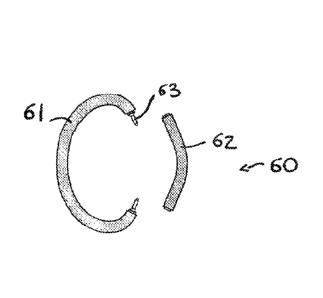
Figs. ha -11b show an example of an annular structure for
anchoring transcatheter valve prostheses for
atrioventricular valves, which can benefit significantly
from the use of a system of guide wires positioned with the
device of Fig. 1,
Fig. 12 shows the annular structure pre-mounted on an
example of a positioning and support device,
Figs. 13a -13b show an example of the use of a pair of guide
wires, previously positioned using a device according to
Fig. 1, to guide the introduction and positioning of the
annular structure of Fig. ha. The components of the annular
structure and the support structure are shown initially in
the collapsed configuration and then in the released
configuration.
Figs. 14a1 -14d2 show details of an example of a procedure
for transcatheter implantation of a prosthetic system for
mitral valve replacement, the system being formed by a
collapsible valve prosthesis and the annular structure of
Fig. 11a, using guide wires previously positioned by means
of the device of Fig. 1 as a guide for the implantation.
Figs. 14a1 -14a2 show the step of introducing the annular
sLrucLure inLo Lhe lea venLricle.
Figs. 14b1 -14b2 show the step of positioning a collapsible
valve prosthesis after the assembly of the annular
structure.
Fig. 14c1 - 14c2 show the step of releasing the collapsible
valve prosthesis,
Figs. 14d1 -14d2 show the result of the procedure of
implanting the prosthetic system on the mitral valve, after
the removal of the devices that are ancillary to the
implantation procedure.
Date Recue/Date Received 2021-03-16
WO 2014/080339
PCT/I132013/060250
8
With reference to Fig. 1, this shows an overall schematic
representation of a device 1 for deploying guide structures
for operational procedures within cardiac chambers according
to one embodiment of the invention. The device is composed
of various components having the principal purpose of being
introduced in a non-invasive manner into a cardiac chamber
and of navigating therein along desired paths controlled by
the operator. The device has been devised so as to be usable
with a beating heart, and therefore without any need for
extracorporeal blood circulation, without significantly
interfering with the operation of the native heart valves,
thus making the procedure entirely atraumatic and
reversible. The procedure in progress can be interrupted at
any time and the components of the device can be removed
from the cardiac chamber without any effects on the function
of the heart itself. Finally, the device is characterized by
a small radial overall dimension and a smooth profile, free
of discontinuities, making it particularly suitable for
introduction into the cardiac cavities by transcatheter
procedures.
The device 1 is essentially composed of a central body 10,
called the introducer, formed by a multi-lumen guide, in
other words one provided with various separate passages 12,
18 (also known as lumens) provided within it, and has the
primary purpose of oreaLing Lhe access channels Lc Lhe
cardiac chambers for the individual instruments that are
intended to operate within the heart. These instruments may
be of various types, since they are intended for specific
purposes. For example, they may be guide catheters with
their terminal parts pre-shaped in a permanent and non-
adjustable way. Guide catheters of this type may simply have
their terminal parts bent at a predetermined angle, so as to
deflect at this angle the devices that are advanced inside
them. Alternatively, they may have their distal parts pre-
Date Recue/Date Received 2021-03-16
WO 2014/080339
PCT/I132013/060250
9
formed in more complex curves or profiles which make them
particularly suitable for specific anatomical situations.
Other types of catheter that can be used in the device shown
schematically in Fig. I may include catheters or guide
catheters fitted with a deflection system which is
adjustable during the procedure according to the operator's
requirements. With this type of mechanism, known in the
prior art as a steering mechanism, the catheter can be
deflected and/or curved by an amount determined by the
operator according to the requirements of the procedure.
This degree of freedom makes the catheter better adapted and
more controllable in its navigation within the anatomical
structures whose configuration is difficult to predict.
Owing to the possibility of rotating the catheter in a
direct and effective way (without effects of hysteresis or
elastic effects), or the possibility of providing it with
multiple deflection systems on different planes, the
stccrability of this typc of catheter is almost total,
enabling it to be navigated in a controlled way in three-
dimensional spaces.
Since it generally has an inner lumen, any guide catheter
can obviously be used for positioning a guide wire, or for
positioning another catheter having an outside diameter
compatible with the diameter of the lumen of the preceding
sLage.
Other instruments that can be used in the application of the
device shown schematically in Fig. 1 include, without
limiting the general nature of the invention, endoluminal
capturing devices, known in the prior art as snaring
devices. These devices, usually composed of collapsible
looped structures made of metallic or polymeric materials,
are particularly suitable for capturing the tree ends of
guide wires or catheters of small gauge. This is because
they have structures that expand in space to generate a
Date Recue/Date Received 2021-03-16
WO 2014/080339
PCT/I132013/060250
capture volume. The tree ends of catheters or guide wires or
similar devices passing through the capture volume are
trapped by the structure when it is re-collapsed by a
procedure which is usually the reverse of the expansion
procedure. In this way, the distal end of a catheter or of a
guide wire can be secured in a given position, or can be
recovered to the outside of the cardiac chamber along the
same path as that used for inserting the capture system.
Endoluminal operating instruments of other types and with
other functions can conveniently be used in the spirit of
the invention described here, in order to deploy guide
structures for operational procedures in the cardiac
chambers.
Fig. 1 shows, in particular, a specific embodiment of the
invention, particularly suitable for use in a ventricular
chamber with access through the wall of the ventricle in the
proximity of the apical region of the heart. As is shown
morc fully and in greater detail in the subsequent figures
(Fig. 2 to Fig. 6), the whole device is composed of a
double-lumen introducer member 10, a pair of guide catheters
14 pre-formed at their distal ends with a fixed curvature,
having dimensions compatible with their advance within the
main lumen 12 of the introducer, a guide catheter 16 which
is substantially rectilinear but flexible, having dimensions
compaLible wiLh iLs advance wiLhin the laLeral lumen. 18 of
the introducer, a pair of catheters 20 which are
substantially rectilinear but are fitted in their distal
regions with an adjustable deflection mechanism and have
overall radial dimensions compatible with their advance
within the first set of guide catheters, and a capture
device 22, having radial dimensions compatible with its
advance within the lateral guide catheter.
The object of this device may be, for example, the
positioning of guide wires, introduced and advanced in the
Date Recue/Date Received 2021-03-16
WO 2014/080339
PCT/I132013/060250
11
ventricle through the second set of guide catheters,
following paths determined by the operator and formed by the
navigation of the second set of guide catheters in the
cardiac chamber. The distal ends of these guide wires can
then be captured by using the capture device, in order to
hold them in a fixed position in the ventricular chamber or
in order to draw them to the outside of the heart and make
them accessible to the operator.
It should be noted that the use of metallic materials and/or
radiopaque markers makes the components of the device
visible to X-rays and the intracardiac procedures can
therefore be guided by means of fluoroscopic visualization.
In some cases, echocardiographic support may also be useful.
With reference to the specific embodiment of the device
depicted in Fig. 1, Fig. 2 shows a possible solution for the
construction of the introducer catheter. The sectional view
shows the double-lumen nature of this component in this
specific embodiment of thc invention. Thc first lumen 12,
identified for the sake of simplicity as the main lumen,
runs parallel to the main axis of the introducer catheter,
the proximal orifice and the distal orifice 24 being
positioned, respectively, at the proximal end and the distal
end 26 of the catheter. The second lumen 18, identified for
simplicity as the secondary lumen, is characterized by a
recLilinear proximal porLiou 28, wiLh Lhe proximal orifice
positioned at the proximal end of the introducer catheter.
In the proximity of the intermediate region 30 of the
catheter, however, the secondary lumen 18 is deflected
towards the outside. The distal orifice 32 of the secondary
lumen is therefore positioned on the lateral surface 34 of
the introducer catheter. Thus the axis of advance of the
main lumen is offset from that of the secondary lumen, at an
angle determined by the curvature of the latter. An angle
compatible with the intended use of this type of device may
Date Recue/Date Received 2021-03-16
WO 2014/080339
PCT/I132013/060250
12
be within the range 15 -45 .
The constructional solution described above therefore
creates an access route to two different areas of the
cardiac chamber. The possible addition of further lumens,
also characterized by an outward curvature at an
intermediate level of the introducer catheter, would create
access paths to different areas of the cardiac chamber.
Still with reference to the specific embodiment depicted in
Fig. 1, Fig. 2 also shows a set of guide catheters 14 that
can be advanced in the main lumen 12 of the introducer
catheter. In this specific embodiment of the invention, this
first guide catheter stage is of the pre-formed type, with a
substantially rectilinear proximal portion 36 and a distal
end 38 pre-curved at about 90 with respect to the proximal
portion 36. More generally, the object of this catheter
stage is to deflect the axis of the devices that are
advanced within it from a direction parallel to the axis of
the introduccr to a direction at an angle to the preceding
one determined by the degree of curvature of the distal end
of the guide catheter. Depending on the application, this
angle may vary from 45 to 135 with respect to the axis of
the proximal portion of the catheter, which is substantially
parallel to the axis of the introducer. Thus the axis of
advance of a device such as a catheter or a guide wire
wiLhin Lhe cardiac chamber is made Lo be IndependenL of Lhe
axis required for its introduction into the heart.
The guide catheters are free to rotate axially, and the
distal curvature can therefore be orientated in different
directions. In the specific embodiment shown in Fig. 2, for
example, the distal ends 38 of the two guide catheters 14 of
the first stage can be orientated along opposite directions.
Consequently, the devices advanced in the lumen of the two
guide catheters are deflected in the same plane
perpendicular to the axis of the introducer catheter, but
Date Recue/Date Received 2021-03-16
W02014/080339
PCT/I132013/060250
13
along paths extending in opposite directions. Clearly, it
would also be possible to have a greater number of catheters
forming the first stage, provided that this is compatible
with the overall radial dimensions of the catheters.
The guide catheters 14 forming the first stage can be made
from a polymeric or metallic material or from a combination
of these. The material must be chosen so as to meet opposing
requirements. This is because the terminal part 38 must be
capable of being at least partially straightened when the
guide catheter is made to advance within the main lumen 12
of the introducer, recovering its pre-formed configuration
when it emerges into the cardiac chamber. On the other hand,
the pre-formed part must be sufficiently rigid to deflect
the device inserted into the lumen of the guide catheter. It
is also preferable for the guide catheter forming the first
stage to have characteristics of torsional rigidity, in
other words to be capable of transmitting a torque from the
proximal section to the distal section.
Still with reference to the specific embodiment depicted in
Fig. 1, Fig. 3 also shows a set of catheters 20, forming the
second guide catheter stage, characterized by overall radial
dimensions making them compatible with their advance within
the lumens of the guide catheters 14 forming the first
stage, as depicted in Fig. 2. In this specific embodiment of
Lhe invenLion, Lhe caLheLer belonging Lo Lhis second sLage
is substantially rectilinear and laterally flexible, so as
to pass through the distal curvature of the guide catheter
in which it is advanced, and is provided in its distal
portion 40 with one or more deflection mechanisms 42, known
as steering mechanisms, actuated by controls 43 positioned
on the handle 45 at the proximal end of the catheter. By
operating the control, a gradual and controlled deflection
of the distal portion of the guide catheter can be achieved,
so that the catheter becomes capable of navigation along
Date Recue/Date Received 2021-03-16
WO 2014/080339
PCT/I132013/060250
14
paths determined by the operator, even in the most complex
anatomical conditions. Preferably, the guide catheter is
substantially rectilinear, with a substantially rigid
proximal portion capable of transmitting a torque to the
distal portion. The distal portion, extending at least
halfway along the whole length of the catheter, is flexible
enough to be deflected, while also being rigid with respect
to torsion in a similar way to the proximal portion. Since
the whole guide catheter can be rotated as a single unit
simply by rotating the handle 45, without any significant
elastic delay or hysteresis, even in the presence of the
curvature created by the guide catheter 14 of the first
stage, the navigation capacity of the second stage is
considerably increased. In fact, each of the guide catheters
20 forming the second stage is free to slide and rotate
within the guide catheters 14 forming the first stage of the
system.
The optimal mechanical characteristics of the cathctcr,
namely the high lateral flexibility combined with torsional
rigidity, can be achieved by using correct constructional
solutions for the catheter. For example, the use of an
appropriate metallic reinforcement of wire mesh embedded in
a polymer matrix to form the catheter wall is a
constructional solution which provides high torsional
rigidAy, while preserving its bending deformabiliLy and
avoiding any risk of collapse in bending (known as kinking).
The distal end of the guide catheter forming the second
stage, and that of the guide catheter forming the first
stage, can be provided with an atraumatic tip 44 made in the
shape of an olive or made of soft, deformable material
adapted to prevent any possible damage to the walls of the
cardiac chamber or ot other anatomical structures present in
the chamber, even in the case of accidental impact or
friction of the catheter against them.
Date Recue/Date Received 2021-03-16
WO 2014/080339
PCT/I132013/060250
Fig. 4 shows how the catheters 20 forming the second stage
have inner lumens allowing the passage of devices for
interventional procedures, such as smaller gauge catheters
or guide wires 46 (as shown in the drawing) which can then
be inserted into the proximal opening of the catheter and
made to advance along its inner lumen until they reach the
cardiac chamber by emerging from the distal end of the
catheter 20. The guide wires 46 are inserted into the
proximal orifice of the guide catheter and are made to
advance therein until they emerge from the distal orifice,
within the cardiac chamber, at the point and along the path
made accessible by the system of guide catheters 14 and 20
of the first and second stages.
Still with reference to the specific embodiment of Fig. 1,
Fig. 5 shows the possibility of using the secondary lumen 18
of the introducer catheter 10 to advance devices for
interventional procedures, for example a further guide
cathctcr 16 (also called a lateral stage) in a direction
offset from the axis of the introducer, as shown in the
drawing. Guide catheters of the type depicted in Figs. 2 and
3 can also be used through the secondary lumen 18. This
lateral guide catheter creates an additional access way to
the cardiac chamber, in a different direction from that of
the main system of guide catheters. Fig. 5 shows how, in a
specific embodimen.L of Lhe invenLion, an endoluminal capLure
device 22 (snaring device) can be introduced into the
cardiac chamber through the guide catheter 16 inserted into
the secondary lumen of the introducer catheter. In the
specific embodiment of the invention shown in the drawing,
the capture device 22 is represented as a set of loops 48 of
metallic wire, with highly elastic properties, whose points
of origin are joined together at the distal end of a stem h0
which is also metallic. The stem 50 is thin and flexible,
and can adapt to the curvature of the path to be followed
Date Recue/Date Received 2021-03-16
WO 2014/080339
PCT/I132013/060250
16
for its access to the ventricle. The proximal end of the
stem is accessible to the operator, so that the positioning
of the capture device can be controlled. The loop structure
shown on the drawing is easily collapsible into a thin-
walled, small gauge sheath 52 (also shown in Fig. 5), on
removal of which the distal structure immediately returns to
its expanded configuration. In other words, the positioning
of the sheath relative to the capture device determines the
configuration of the latter, which is collapsed if the
sheath covers the device, or expanded if the sheath is
retracted at the position of the stem.
Because of the multiplicity of loops 48 and their flower-
like configuration, this device is capable of
multidirectional capture, so that its orientation relative
to the device to be captured becomes less critical. A wire
only needs to pass in any direction through one of the loops
of the expanded device in order to be captured when the
device is collapsed again. Clearly, there is a wide variety
of possible designs for the structure of the capture device,
and these designs may also vary according to the particular
function to be provided or any particular requirements to be
met. Most of these designs are known in the prior art.
By using materials with high mechanical performance, for
example superelastic metal alloys such as Nitinol, for the
capLure device, and by using Lechnopolymers such as
polyamide or polyamide reinforced with a metallic mesh for
the sheath, it is possible to limit the overall radial
dimensions of the capture system (including the sheath and
the capture device), making it compatible with endoluminal
use; in particular, in the illustrated example, the diameter
must be smaller than that of the lateral stage. More
generally, the overall radial dimensions of systems
currently in use for general endoluminal capture
applications are within the range from 1 to 3 millimetres,
Date Recue/Date Received 2021-03-16
W02014/080339
PCT/I132013/060250
17
although dimensions of less than one millimetre are also
possible.
In the light of the specific solutions depicted in Figs. 2
to 5, Fig. 6 shows an overall schematic representation of a
device for deploying guide structures for interventional
procedures within cardiac chambers according to one
embodiment of the invention in its operating configuration.
By using the main lumen 12 of the introducer catheter 10,
positioned through the outer wall of the heart to provide an
access port to the cardiac chamber, the operator can
position the guide catheters 14 and 20 of the first and
second stages according to his requirements, following the
paths required by the application. In the case of the
catheters 20 of the second stage, the various degrees of
freedom available in the movement of the distal end of the
catheter (axial advance, rotation about its own axis,
adjustable deflection mechanism) are such that the desired
paths can be followcd and thc final positions can be reached
even in the presence of particularly unfavourable anatomies.
The operator can introduce a capture system 22 into the
cardiac chamber through the guide catheter 16 positioned in
the secondary lumen 18 of the introducer catheter 10,
determining the end position of the system by means of the
control stem 50 and modifying its configuration (expanded or
collapsed) by auLiuy on Lhe corresponding conLaining 5.heaLh
52. Because of the specific geometry of the secondary lumen
18, the axis of the capture device 22 is offset with respect
to the catheters of the main lumen 12, making its action
simpler and more effective. This is because the operator can
advance guide wires 46 within the lumens of the catheters
forming the second stage until they emerge into the cardiac
chamber so that their distal ends 47 can be gripped by the
capture device 22. The capture device can be used, for
example, to stabilize the distal ends of the guide wires, in
Date Recue/Date Received 2021-03-16
WO 2014/080339
PCT/I132013/060250
18
support of subsequent intracardiac operations.
Figs. 7 and 8 show a use of the capture device which differs
from that described above. In this example, the capture
device 22 is used to recover the distal ends of the guide
wires to a proximal position. This makes it possible to
position one or more guide wires within a cardiac cavity
along a path determined by the operator, who has access, at
the end of the procedure, to both ends of the guide wire or
wires used for the purposes of this procedure.
In a first step, the operator advances and positions the
system of guide catheters 14, 20 by following the desired
path (over all or part of its length). The guide wire 46 (or
guide wires) is then introduced into the cardiac cavity
through the inner lumen of the second stage guide catheter
20, causing it to emerge from the distal orifice of this
catheter. The guide wire is advanced sufficiently within the
cardiac cavity to allow it to be captured by the capture
device 22. By removing thc capture system from the cardiac
cavity, the operator also recovers the distal end 47 of the
guide wire (or guide wires). Thus one or more guide wires 46
can be positioned within the cardiac cavity along paths
specified by the operator. At the end of the procedure, the
operator has simultaneous access to the proximal ends and
the distal ends 47 of the guide wires positioned in the
cardiac chamber. Fly. 7 depicLs the configuration of Lhe
device after the distal ends 47 of the guide wires 46 have
been captured and the capture system 22 has been drawn out:
the guide wires enter the cardiac cavity through the system
of catheters 14, 20 inserted into the main lumen 12 of the
introducer 10 and exit through the guide catheter 16
inserted into the secondary lumen 18. Finally, Fig. 8 shows
the removal of the whole system of guide catheters, leaving
in situ only the guide wires 46 which can thus be used as
guide structures for subsequent interventional procedures.
Date Recue/Date Received 2021-03-16
W02014/080339
PCT/I132013/060250
19
To provide a detailed illustration of an exemplary
application relating to the left ventricle of the device 1
for the deployment of guide structures for interventional
procedures within cardiac chambers as depicted in Fig. 1,
the diagrams of anatomical sections through a heart shown in
Figs. 9a to 9c will be used. In particular, Figs. 9a and 9b
show two views along the longitudinal axis of the left side
of the heart, in other words views of sections which
substantially cut the heart along the longitudinal axes of
the two chambers of the left side, from the apex (in other
words the lower point of the heart) to its top. These
sections therefore show both the left ventricle 100 (the
lower chamber, including the apex) and the left atrium 101
(the upper chamber). Fig. 9a shows the view obtained by
taking a section through the left side of the heart along a
plane identified by the nominal axis of the left ventricle
and the axis of the aortic valve 102. In this case, the
section plane cuts the mitral valve 103 along its
anteroposterior axis, following the mid-line of the
posterior leaflet and of the anterior leaflet, as well as
taking a section through the aortic valve. This section
therefore enables the aortic root 115 to be visualized with
the aortic valve apparatus 102 and the aortic subvalvular
chamber 117, usually referred to as the LVOT (left ventricle
uuLfluw LracL). Bulh leafleLs of Lhe miLral valve, namely
the anterior leaflet 135a and the posterior leaflet 135b,
are also visible in section. The mitral valve separates the
left atrium 101 from the left ventricle 100. The mitral
annulus 120, the bundles of the chordae tendineae 140 and
the papillary muscle 145 are other clearly identifiable
anatomical structures. A single group of papillary muscles
(and the corresponding chordae tendineae) is visible in this
view. In the case of Fig. 9b, the view of the left side of
the heart is shown as it appears if the section plane is
Date Recue/Date Received 2021-03-16
WO 2014/080339
PCT/I132013/060250
rotated about the axis of the ventricle until it is aligned
with the commissure-commissure axis of the mitral valve.
This view shows only the posterior leaflet 135b of the
mitral valve, with the corresponding portion of the annulus
120 and the corresponding subvalvular apparatus formed by
the chordae tendineae 140 and papillary muscles 145. This
section shows both papillary muscles (in section). Finally,
Fig. 9c shows a plan view of the mitral valve from a
supravalvular viewpoint, as it appears if the left atrium is
uncovered. The anterior mitral leaflet 135a and the
posterior leaflet 135b are visible. Both leaflets are
surrounded and connected to the muscular structure of the
left ventricle by the mitral annulus 120. The transition
regions between the two valve leaflets along the annulus are
the commissural regions 127. This view clearly shows the two
main orthogonal axes of orientation of the mitral valve,
namely an axis of symmetry in the anteroposterior direction,
passing through both leaflets along the mid-line, and an
axis orthogonal to the preceding one, aligned along the
commissure-commissure direction. Finally, the bundles of the
chordae tendineae 140 which secure the free margins of the
valve leaflets to the papillary muscles 145 are visible
through the orifice of the mitral valve.
Figs. 10a1 to 10g2 show details of a possible procedure
followed for Lhe deploymenL of a sysLem of guide wires Lo
surround the native mitral valve 103, by inserting the
system into the left ventricle through a transapical access,
using the device 1 for deploying guide structures for
interventional procedures within the cardiac chambers as
depicted in Fig. 1 as a specific embodiment of the
invention.
Figs. 10a] and 10a2 depict the initial step of the procedure
in the two different sections through the left side of the
heart. The same presentation mode is used in the subsequent
Date Recue/Date Received 2021-03-16
WO 2014/080339
PCT/I132013/060250
21
drawings depicting this procedure. The drawings show the
positioning of the distal end of the introducer catheter 10
adjacent to the ventricular wall, through a transapical
access, at the rear of the posterior leaflet 135b of the
mitral valve 103, on the mid-line of the latter. In this
position, the introducer catheter 10 creates a direct access
to the native annulus of the mitral valve, on its
ventricular side. The introducer 10 must be orientated
angularly on its axis in such a way that the distal orifice
of the secondary lumen 32 is directed towards the aortic
valve 102, in the direction along which the capture system
is to be advanced. Figs. 10b1 and 10b2 show, again in the
two different views of the left-hand side of the heart, the
positioning of the guide catheters 14 forming the first
stage. These are advanced along the main lumen 12 of the
introducer 10 until they are close to the plane of the
mitral annulus, on the ventricular side. They are then
orientated axially so that thcir curved distal ends 38 arc
both orientated tangentially to the mid-line of the mitral
annulus, but in opposite directions. This orientation
enables the catheters of the subsequent stage to be guided
in a direction parallel to the mitral annulus. Because of
the presence of radiopaque markers on the distal edge of
this catheter, and on other components of the system, the
orienLaLlon of Lhe sysLem can be visualized more immediately
by means of X-ray based imaging systems (such as
fluoroscopic systems).
Figs. 10c1 and 10c2 show the positioning of the catheters 20
forming the second stage of the device, each of which
surrounds one half of the mitral valve. The introducer 10
and the catheters 14 forming the first stage of the device
are positioned on the back of the posterior leaflet -11-ihb of
the mitral valve, while the distal ends of the catheters 20
forming the second stage of the device face the back of the
Date Recue/Date Received 2021-03-16
WO 2014/080339
PCT/I132013/060250
22
anterior leaflet 135a. The drawings show that the presence
of a controlled deflection mechanism at the distal end of
the catheters 20, as well as its capacity to be rotated
axially, improves the control of the navigation of the
distal end of the catheter. In specific regions of the
mitral valve anatomy, for example the commissural regions,
this is essential for the correct positioning of the
catheter.
Both of the distal ends of the catheters forming the second
stage of the device therefore face each other in the space
below the aortic valve (called the LVOT) 117, immediately
behind the anterior leaflet of the mitral valve.
Figs. 10d1 and 10d2 show the positioning of the guide
catheter 16 which forms the lateral stage of the device
within the secondary lumen 18 of the introducer catheter 10,
creating a further access route to the space below the
aortic valve (LVOT) 117, in a direction which is offset from
the nominal axis of the ventricle and from the plane of the
mitral annulus (that is to say, the plane on which the
catheters 20, forming the second stage of the device 1,
lie), but which substantially coincides with the axis of the
aortic valve. ne capture system 22 in its low-profile
configuration, with the capture device 48 completely
collapsed inside the sheath 52, is introduced into the LVOT
117 Lhrough Lhe laLeral guide caLheLer 16. As shown in Lhe
drawings, the capture device 48 is subsequently released
from the sheath 52 and expanded immediately below the aortic
valve 102. The shape and position of the capture device 22
are such that it creates a kind of net entirely covering the
portion of the left ventricle that opens into the aortic
valve, in other words the LVOT 117, while not interfering
with either the blood flow or the movement at the aortic
valve leaflets. The design and the elastic characteristics
of the capture device 22 are such that no interference is
Date Recue/Date Received 2021-03-16
WO 2014/080339
PCT/I132013/060250
23
permitted either with the aortic valve 102, which would
entail a risk of trauma to the native leaflets or to the
annulus, or with the electrical conduction system (the
atrioventricular node and the bundle of His) located on the
septal side of the LVOT, which would entail risks of
blockage of the left branch.
The distal ends of the catheters 20 forming the second stage
of the device substantially face the capture device 22, on
the ventricular side of the device.
Figs. 10e1 and 10e2 show a pair of guide wires 46 advanced
into the LVOT 117 from the two distal orifices of the
catheters 20 of the second stage. The position of the
catheters 20, together with the dragging action of the
systolic blood flow, which is ejected from the left
ventricle through the aortic valve, cause the guide wires to
be pushed through the loops of the capture device 22, so
that they are positioned across the aortic valve 102 up to
rise through the aortic root and the ascending aorta. The
use of the controlled deflection mechanism located at the
distal end of the catheters 20 can also contribute to the
guiding of the guide wires 46 through the capture device 22.
Tt should be borne in mind that all the components described
here (for example the guide wires and the capture device)
are intrinsically radiopaque, or are made radiopaque by
means of suitable markers (at the distal ends of the second
stage catheters, for example).
Figs. 10f1 and 10f2 show how the reclosing of the
collapsible device 48 inside its containing sheath 52 causes
the capture of the distal ends 47 of the pair of guide wires
46, which remain trapped in the loops of metallic wire of
the capture device.
Figs. 10g1 and 10g2 show the recovery to a proximal position
of the sheath 52 and of the collapsible device 48 through
the secondary (lateral) lumen 18 of the introducer catheter
Date Recue/Date Received 2021-03-16
WO 2014/080339
PCT/I132013/060250
24
10, and the recovery of the two pairs of guide catheters 14
and 20 forming the first and second stage, through the main
lumen 12 of the introducer catheter.
Thus the distal ends 47 of the pair of guide wires 46 are
also recovered to the outside of the left ventricle, leaving
the guide wires 46 deployed around the mitral valve 103 with
their proximal ends positioned inside the main lumen of the
introducer catheter 10 and their distal ends positioned
inside the secondary lumen of this introducer 10. The
operator is thus provided with a system of guide wires which
passes into and out of the left ventricle, after wrapping
around the mitral valve 103, through the same apical port,
but inside two different lumens 12, 18.
The introducer catheter 10 can then be removed, leaving in
situ only the pair of guide wires 46 wrapped around the
mitral valve. Both ends of each guide wire are recovered to
the outside of the heart through the apical port. A system
of guidc wircs has thus bccn fully dcploycd within a cardiac
chamber along paths determined by the operator.
The principle described with reference to Figs. 10a1 to 10g2
for a pair of guide wires can be extended to a greater
number of guide wires, by means of an obvious modification
of the deployment system depicted in Fig. 1, in which
multiple access ways for guide wires are created through the
main lumen of Lhe inLroducer caLheLer. IL will also be
evident to anyone skilled in the art that the configuration
shown in Figs. 10g1 and 10g2, where a pair of guide wires
surrounds the mitral valve 103, can easily be changed into a
configuration with a single guide wire wrapped around the
whole mitral valve. In fact, it is simply necessary to join
the two corresponding ends of the two guide wires 46 and to
recover one guide wire completely by recovering the other.
In the configuration shown in Figs. 10g1 and 10g2, the
joining of the distal ends produces a guide wire which is
Date Recue/Date Received 2021-03-16
WO 2014/080339
PCT/I132013/060250
entirely wrapped around the mitral valve, the loop being
completed on the reverse of the posterior leaflet 135b.
Conversely, if the proximal ends are joined, this produces
the symmetrical configuration, in which the loop around the
mitral valve 103 created by the guide wire which remains in
situ is completed on the reverse of the anterior leaflet
135a.
In an example of application, not in any way intended to
limit the general nature of the applications and operating
procedures which can benefit from this device or have an
extension of uses as a result of it, the use of the device
for the deployment of the guide wires within the cardiac
chamber in association with a transcatheter system for the
replacement of an atrioventricular valve is described below
with reference to Figs. ha to 14d2. The prosthetic system
is made up of two components, namely a prosthetic valved
body, to be expanded inside the native valve, and a
substantially annular support structure, positioncd so as to
surround the outside of the native valve, and serving to
create an anchorage and a sealing to backflow by entrapping
between the two components the native leaflets at the level
of the annulus.
The positioning of the annular structure is essential for
the correct operation of the whole prosthetic system. To
ensure Lhe reliable anchorage of Lhe pros Lhesis and reduce
the risk of paraprosthetic fluid leakage, the positioning
of the annular support structure must essentially meet two
requirements, namely that the annular structure must be
wrapped around the whole of the native valve, without
passing through its orifice or the subvalvular apparatus,
and that it must be positioned in contact with the annulus.
A system of guide wires deployed immediately below the
annulus of the native valve and capable of being wrapped
around the whole of the valve therefore provides an
Date Recue/Date Received 2021-03-16
WO 2014/080339
PCT/I132013/060250
26
effective guide for the positioning of the annular
component. Furthermore, advantageous versions of the design
of the annular support structure can be developed, because
of the possibility of having a separate pair of guide wires
accessible at both ends.
By way of example, without any intention to limit the
general nature of the application, Figs. ha -11b show an
annular support structure 60 made of two separate and
independent components 61, 62, with a connection system 63
which enables permanent and durable structural continuity to
be restored during the procedure of positioning and release
at the implant site (Fig. 11b).
Each component 61, 62 of the annular structure can be
anchored to the distal end of a separate support arm 64 and
65, forming part of the same positioning and support device
66 (Fig. 12). Alternatively, each component can be conveyed
to the inside of the ventricular chamber by means of its own
support and positioning dovico.
As shown in Figs. 13a and 13b, the components 61 and 62 of
the annular structure and the arms 64 and 65 of the
positioning and support device 66 can all be deformed to
provide a smaller overall radial dimension of the whole
system, compatible with its introduction into the
ventricular chamber through an apical access port. According
Lu Lhe presenL sLiaLe of knowledge in Lhe field of
transcatheter heart valve treatment technology, the maximum
diameter of the profile of the devices compatible with a
transapical procedure is about 10 mm.
The drawing shows that the pair of guide wires 46,
previously positioned around the mitral valve by the device
1 for deploying guide structures for interventional
procedures as proposed by the present invention, can be used
to guide the components 61 and 62 of the annular structure
60 inside the ventricular chamber. In fact, each component
Date Recue/Date Received 2021-03-16
W02014/080339
PCT/I132013/060250
27
of the annular structure is made with a hollow ("over the
wire÷) geometry, allowing the passage of a guide wire 46 and
providing an aperture 67 and 68, located about halfway along
the length of the component, for the exit of the wire. Each
end of each guide wire 46 is then made to advance within one
half of one of the components 61 and 62. The free end of the
guide wire is inserted into the orifice at the free end of
the component, and is made to emerge through the
intermediate aperture 67 and 68. The sequence followed for
the positioning of the guide wires 46 must be such that the
corresponding halves of the two components 61 and 62 slide
along the same guide wire, coming from the opposite ends
(Fig. 13a). The two components 61 and 62 of the annular
structure, when thus coupled to the guide wires 46, are then
introduced by the two opposite ends of the system of guide
wires and are advanced over the wire into the ventricular
chamber until they surround the native mitral valve 103 in
the corrcct manner, exactly at thc subannular level where
the guide wires 46 were positioned previously. The guide
wires are then also essential for the alignment of the free
ends of the components 61 and 62 of the annular structure in
order to promote their reconnection (Fig. 13h). Finally, by
suitably tensioning the guide wires 46, it is also achieved
the effect of applying a closing action to the locking
mechanism 63 beLween Lhe Lwo componehLs 61 and 62 of Lime
annular structure, tending to reduce the peripheral
extension of the structure.
The locking mechanism 63 comprises pins 55 adapted to engage
in suitable holes 56, and in particular it is composed of a
pair of pins 55 and corresponding holes 56. Each end of one
of the two components 61 is provided with a pin 55, while
each end of the other component 62 is provided with a hole
56. The two components can be connected by inserting each
pin into the corresponding hole.
Date Recue/Date Received 2021-03-16
WO 2014/080339
PCT/I132013/060250
28
Figures 14a1 to 14d2 provide a summary illustration of a
possible procedure for implanting a prosthetic system for
replacement of the mitral valve by a transcatheter technique
and transapical route. The following description omits the
preparatory procedure in which the two guide wires are
positioned so as to surround the mitral valve, since already
described above.
Figs. 14a1 and 14a2 show, from two different views, the
introduction and deployment (previously shown in Figs. 13a
and 13b) in the left ventricle of the two components 61 and
62 of the annular structure in their collapsed
configuration, mounted on the positioning and support device
66. The whole system can be initially collapsed into a
sheath 69 which can be used as an introducer. When its
distal edge has arrived in the proximity of the mitral valve
103, the introducer is fixed, and, by means of the
positioning and support device 66, the components 61 and 62
of thc annular structure arc deployed in the ventricle,
while still being guided by the guide wires 46.
When the components 61 and 62 of the annular structure have
been correctly positioned and interconnected with the aid of
the guide wires, the central valved body 72 of the
prosthetic system 70 is introduced, this body also being
collapsed and mounted on a positioning and release device 74
which is fully inLeyraLed wiLh Lhe similar device 66 used
for the annular structure (Figs. 14b1 and 14b2).
The drawings show, without any intention to limit the
general nature of the invention, a device 74 which slides
coaxially with the support device 66 of the annular
structure. The coaxial solution has the significant
advantage of providing a practically perfect alignment with
the orifice of the mitral valve. This significantly
simplifies the design of the positioning and release device
74 for the central valved body 72.
Date Recue/Date Received 2021-03-16
WO 2014/080339
PCT/I132013/060250
29
The central valved body 72 is positioned across the mitral
valve 103, in the final position before release. The main
advantage of the complete mutual integration of the two
devices 74 and 66 for positioning and releasing the
components 61, 62 and 72 of the prosthetic system 70 is that
the components can be positioned with respect to each other
with great accuracy, without any particular requirements for
skill on the part of the operator. Indeed, it is simply
enough to provide a reference mark, of a mechanical, optical
or other type, allowing to uniquely identify the
configuration in which the components of the prosthetic
system 60 and 72 are perfectly aligned for release and
mutually positioned for optimal coupling with each other. In
the example shown in the drawings, the structure of the
device itself ensures the coaxial placing of the two
components of the prosthesis. A simple mechanical stop,
which arrests the axial sliding of the two parts of the
release device of thc prosthetic system at a precise
position, also ensures optimal positioning immediately
before the final release.
Figs. 14cl and 14c2 show the release of the central valved
body 72 within the mitral valve 103 by the positioning and
release device 74, which is integrated with the positioning
and support device 66 of the annular structure 60. The
cenLral body expands, and Lhus, since Lhe ceaLral valved
body 72 is released within the mitral valve 103 and the
annular structure 60 is positioned outside the mitral valve,
in an immediately subannular position, the leaflets 104 of
the native mitral valve 103 are entrapped between the two
components. The leaflets, creating a continuity with the
annulus 120 of the valve along the whole periphery of the
prosthesis 70, provide an anchorage for the prosthesis 10
and a sealing to the backflow.
Finally, Figs. 14d1 and 14d2 show the valve prosthesis 70
Date Recue/Date Received 2021-03-16
WO 2014/080339
PCT/I132013/060250
implanted after the removal of the release and support
device through the apical port of the left ventricle.
The advantages of the embodiment described above include not
only the provision of a system of guide wires which ensures
the correct positioning of the components 61 and 62 so that
they wrap around the whole of the native valve at a
subannular level, but also those deriving from the
possibility of inserting the two components 61 and 62 of the
annular structure 60 separately and on opposite sides; thus
the introduction of the components 61 and 62 into the
ventricular cavity is made simpler and safer, being the
components shorter than those of an annular structure made
in one piece. However, the primary advantage is that the
annular structure can be held in position during the
implantation procedure by means of the supports 64 and 65
distributed along the whole periphery of the component.
These supports can be the same as those used for the
introduction of the components into the ventricular chamber,
and can be physically integrated with the positioning and
release system 74 of the central valved body 72. In other
words, when the system 66 for conveying, positioning and
releasing the annular structure 60 is integrated with the
corresponding system 74 for conveying, positioning and
releasing the central valved body 72, this ensures both the
sLabiliLy of Lhe posiLioning or Lhe annular supporL
structure 60 throughout the entire implantation procedure of
the prosthetic system and the precise spatial referencing
between the various components 60 and 72 of the prosthetic
system 70 at the time of the final release of the central
valved body.
The example described above demonstrates how a device
for deploying guide structures for interventional procedures
within cardiac chambers, according to the embodiments of the
invention, permits the fast, safe and effective execution of
Date Recue/Date Received 2021-03-16
WO 2014/080339
PCT/IB2013/060250
31
transcatheter or low-invasiveness procedures applied to
anatomical structures of the heart.
Date Recue/Date Received 2021-03-16