Note: Descriptions are shown in the official language in which they were submitted.
CA 03112589 2021-03-11
WO 2020/061148 PCT/US2019/051657
CATHETER HAVING A CLOSED TIP AND SLIT FOR A PERIPHERAL
INTRAVENOUS CATHETER ASSEMBLY
BACKGROUND
[0001] Infusion therapy, a common healthcare procedure, may be facilitated
by a vascular
access device. Hospitalized, home care, and other patients receive fluids,
pharmaceuticals, and
blood products via a vascular access device inserted into the vascular system.
Blood withdrawal is
another common healthcare procedure that may be facilitated by a vascular
access device.
[0002] A vascular access device may access a peripheral or central
vasculature of a patient. A
vascular access device may be indwelling for short term (days), moderate term
(weeks), or long
term (months to years). A vascular access device may be used for continuous
infusion therapy or
for intermittent therapy.
[0003] A common type vascular access device is an over-the-needle
peripheral intravenous
catheter (PIVC). As its name implies, the "over-the-needle" PIVC may be
mounted over an
introducer needle having a sharp distal tip. The sharp distal tip may be used
to pierce skin and the
vasculature of the patient. Insertion of the PIVC into the vasculature may
follow the piercing of
the vasculature by the introducer needle. The introducer needle and the PIVC
are generally inserted
at a shallow angle through the skin into the vasculature of the patient with a
bevel of the introducer
needle facing away from the skin of the patient. Once placement of the
introducer needle within
the vasculature has been confirmed, the clinician may temporarily occlude flow
in the vasculature
and withdraw the introducer needle, leaving the PIVC in place for future fluid
infusion and/or
blood withdrawal.
[0004] Currently, there may be several limitations to the use of a PIVC for
fluid infusion or
blood draw. The PIVC or vein may narrow, collapse, or clog with time, leading
to failure of the
-1-
CA 03112589 2021-03-11
WO 2020/061148 PCT/US2019/051657
PIVC. In some instances, risk of occlusion of the PIVC may lead to increased
flushing and risk of
infection. PIVCs may also be prone to removal from the vein, which may lead to
infiltration or
extravasation.
[0005] The subject matter claimed herein is not limited to embodiments that
solve any
disadvantages or that operate only in environments such as those described
above. Rather, this
background is only provided to illustrate one example technology area where
some
implementations described herein may be practiced.
SUMMARY
[0006] The present disclosure relates generally to devices, systems, and
methods for fluid
transfer through a placed or indwelling peripheral intravenous catheter
("PIVC") assembly. In
some instances, the PIVC may be fairly easily placed within the vein of the
patient by means of an
introducer needle, which may pierce skin and vasculature of a patient to
facilitate placement of the
PIVC within the vein. In some embodiments, the devices, systems, and methods
of the present
disclosure may take advantage of the introducer needle for placement of the
PIVC, while providing
benefits of a second catheter with a closed distal end and a valve to reduce
occlusion, flushing, and
risk of infection. In some embodiments, the second catheter with the closed
distal end and valve
may be threaded through the PIVC into the vein of the patient.
[0007] In the prior art, PIVCs may be prone to clotting because blood is
allowed to diffuse into
the PIVC. In some embodiments described in the present disclosure, the valve
of the second
catheter may include a slit. In some embodiments, the slit may open under
positive or negative
pressure to allow fluid infusion or blood withdrawal. In some embodiments, the
second catheter
may be resistant to occlusion and thrombosis because the slit may be closed
and blood may not be
-2-
CA 03112589 2021-03-11
WO 2020/061148 PCT/US2019/051657
allowed to diffuse into the second catheter under normal physiological
pressures. Thus, in some
embodiments, the PIVC assembly that includes the second catheter may be
flushed less frequently,
such as, for example, once per week, instead of, for example, once per shift
of a clinician.
[0008] In some embodiments, the second catheter and the valve may reduce
flushing of the
PIVC assembly from a high number of flushes, such as, for example, 3 times per
day and 7 days
per week (for a total of 21 flushes per week) to a low number of flushes, such
as, for example, one
flush per week. Reducing flushing may not only decrease a risk of infection,
but may also free the
clinician to focus on other matters in a healthcare setting.
[0009] One drawback of the PIVC of the prior art is that it may be easy to
pull out of the vein
due to several factors, including its length or extension into the vein. When
the PIVC pulls out of
the vein, this may lead to infiltration and/or extravasation. In some
embodiments, the devices,
systems, and methods of the present disclosure may reduce or eliminate
infiltration and
extravasations. In further detail, in some embodiments, even if a distal tip
of the PIVC pulls out of
the vein, the second catheter may be longer than the PIVC and positioned
further down the vein,
preventing removal of the second catheter from the vein and subsequent
infiltration and
extravasation.
[0010] In some embodiments, the second catheter may not be long enough to
reach the heart,
which may allow the second catheter to be placed within the vein without
ultrasound and/or
fluoroscopy in a simple placement procedure involving only a few steps.
[0011] It is to be understood that both the foregoing general description
and the following
detailed description are exemplary and explanatory and are not restrictive of
the invention, as
claimed. It should be understood that the various embodiments are not limited
to the arrangements
and instrumentality shown in the drawings. It should also be understood that
the embodiments may
-3-
CA 03112589 2021-03-11
WO 2020/061148 PCT/US2019/051657
be combined, or that other embodiments may be utilized and that structural
changes, unless so
claimed, may be made without departing from the scope of the various
embodiments of the present
invention. The following detailed description is, therefore, not to be taken
in a limiting sense.
BRIEF DESCRIPTION OF THE SEVERAL VIEWS OF THE DRAWINGS
[0012] Example embodiments will be described and explained with additional
specificity and
detail through the use of the accompanying drawings in which:
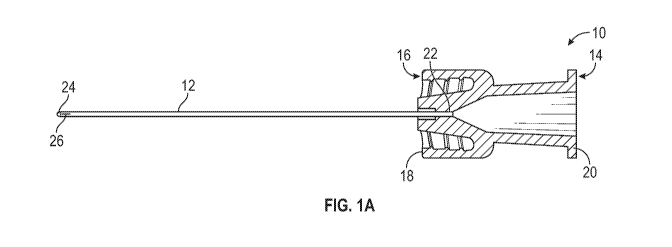
[0013] Figure lA is a cross-sectional view of an example connector and
example catheter
having a closed distal end and slit, according to some embodiments;
[0014] Figure 1B is an upper perspective view of the slit of Figure lA is a
closed position,
according to some embodiments;
[0015] Figure 1C is an upper perspective view of the slit of Figure lA in
an open position
during fluid infusion, according to some embodiments;
[0016] Figure 1D is an upper perspective view of the slit of Figure lA in
an open position
during blood withdrawal, according to some embodiments;
[0017] Figure 2 is a cross-sectional view of an example guidewire hub and
example guidewire,
according to some embodiments;
[0018] Figure 3A is a cross-sectional view of the guidewire hub of Figure 2
coupled with the
connector of Figure 1A;
[0019] Figure 3B is a cross-sectional view of the connector of Figure lA
and the guidewire hub
of Figure 2 coupled with an example catheter assembly, according to some
embodiments;
-4-
CA 03112589 2021-03-11
WO 2020/061148 PCT/US2019/051657
[0020] Figure 3C is a cross-sectional view of the connector of Figure lA
coupled with the
catheter assembly of Figure 3B and an example needleless connector, according
to some
embodiments;
[0021] Figure 3D is a cross-sectional view of an example ball feature,
according to some
embodiments;
[0022] Figure 4A is an upper perspective view of another example connector
that includes a
first piece and a second piece in a step of an example assembly process,
according to some
embodiments;
[0023] Figure 4B is an upper perspective view of the connector of Figure 4A
during another
step of the assembly process, according to some embodiments;
[0024] Figure 4C is an upper perspective view of the connector of Figure 4A
fully assembled,
according to some embodiments;
[0025] Figure 4D is an upper perspective view of the connector of Figure 4A
coupled to an
example catheter assembly, according to some embodiments;
[0026] Figure 4E is a cross-sectional view of the connector of Figure 4A
along the line 4E-4E
of Figure 4D, according to some embodiments;
[0027] Figure 5A is an upper perspective view of another example connector,
according to
some embodiments;
[0028] Figure 5B is an upper perspective view of the connector of Figure 5A
having an example
push tab, according to some embodiments;
[0029] Figure 5C is a cross-sectional view of the connector of Figure 5A,
according to some
embodiments; and
-5-
CA 03112589 2021-03-11
WO 2020/061148 PCT/US2019/051657
[0030] Figure 6 is a flow chart illustrating an example method, according
to some
embodiments.
DESCRIPTION OF EMBODIMENTS
[0031] The present disclosure relates generally to devices, systems, and
methods for fluid
transfer through a placed or indwelling peripheral intravenous catheter
("PIVC") assembly. In
some instances, the PIVC may be fairly easily placed within the vein of the
patient by means of an
introducer needle, which may pierce skin and vasculature of a patient to
facilitate placement of the
PIVC within the vein. In some embodiments, the devices, systems, and methods
of the present
disclosure may take advantage of the introducer needle for placement of the
PIVC, while providing
benefits of a second catheter with a closed distal end and a valve to reduce
occlusion, flushing, and
risk of infection. In some embodiments, the second catheter with the closed
distal end and valve
may be placed within the PIVC and extend through the PIVC into a vein of a
patient.
[0032] Referring now to Figure 1A, an example connector 10 and example
second catheter 12
are illustrated, according to some embodiments. In some embodiments, the
connector 10 may
include a proximal end 14 and a distal end 16. In some embodiments, the distal
end 16 of the
connector 10 may include a luer adapter, such as a slip or thread male or
female luer adapter, which
may be configured to couple with a proximal end of a catheter adapter of an
indwelling PIVC
assembly to provide a fluid tight seal. In some embodiments, the distal end 16
of the connector 10
may include a male luer adapter having a freely rotating collar, which may
allow coupling of the
connector 10 to a catheter adapter without having to rotate the catheter 12.
Figure 1A illustrates
the distal end 16 of the connector 10 having an example male luer adapter 18,
according to some
embodiments.
-6-
CA 03112589 2021-03-11
WO 2020/061148 PCT/US2019/051657
[0033] In some embodiments, the proximal end 14 of the connector 10 may
include another
luer adapter, such as a slip or thread male or female luer adapter, which may
be coupled to a blood
collection device, a fluid infusion device, or a needleless connector, for
example. Figure 1A
illustrates the proximal end 14 of the connector 10 having an example female
luer adapter 20,
according to some embodiments.
[0034] In some embodiments, a proximal end 22 of the second catheter 12 may
be secured
within the connector 10. In some embodiments, the second catheter 12 may
extend from the distal
end 16 of the connector 10. In some embodiments, the second catheter 12 may
include a closed
distal end 24 and a valve adjacent the closed distal end 24. In some
embodiments, the valve may
include a slit 26. In some embodiments, under normal physiological pressures,
the slit 26 may be
closed, as illustrated in Figure 1A. In some embodiments, the closed distal
end 24 may be rounded
or bullet-shaped.
[0035] In some embodiments, all or a portion of the second catheter 12 may
be constructed of
silicon. In some embodiments, a portion of the second catheter 12 that
includes the slit 26 may be
constructed of silicon. In some embodiments, all or a portion of the second
catheter 12 may be
constructed of polyurethane or another suitable plastic. In some embodiments,
the portion of the
second catheter 12 that includes the slit 26 may be constructed of
polyurethane or another suitable
plastic.
[0036] The second catheter 12 may have an outer diameter less than an inner
diameter of a 16g
or 18g PIVC. In some embodiments, the outer diameter of the second catheter 12
may be less than
about 0.052-0.054 inches. In some embodiments, the outer diameter of the
second catheter 12 may
be less than about 0.036-0.039 inches. In some embodiments, the outer diameter
of the second
catheter 12 may be between about 0.034-0.036 inches. In some embodiments, the
second catheter
-7-
CA 03112589 2021-03-11
WO 2020/061148 PCT/US2019/051657
12 may include lubrication in order to thread the second catheter 12 through
the indwelling PIVC
assembly. In some embodiments, the second catheter 12 may snugly fit within
the PIVC.
[0037] In some embodiments, dimensions of the second catheter 12 may vary.
In some
embodiments, the second catheter 12 may be about 3 inches in length. In some
embodiments, the
second catheter 12 may be less than about 5 inches in length. In some
embodiments, the second
catheter 12 may have a length between about 2 and 5 inches.
[0038] Referring now to Figure 1B, the slit 26 is illustrated in a closed
position, according to
some embodiments. In some embodiments, the slit 26 may include a longitudinal
slit oriented
along a longitudinal axis of the catheter 12. In some embodiments, when the
slit 26 is in the closed
position, opposing faces of the slit 26 may contact each other. In some
embodiments, the slit 26
may be in the closed position and sealed under normal physiological pressures,
preventing fluid
from flowing through the slit 26. In some embodiments, the second catheter 12
may be resistant
to occlusion and thrombosis because the slit 26 may be closed under normal
physiological
pressures, preventing blood from diffusing into the second catheter 12. Thus,
in some
embodiments, the PIVC assembly (illustrated, for example, in Figures 3B-3C)
that includes the
second catheter 12 may be flushed less frequently, such as, for example, once
per week, instead
of, for example, once per shift of a clinician.
[0039] In some embodiments, the second catheter 12 and the slit 26 may
reduce flushing of the
PIVC assembly from a high number of flushes, such as, for example, 3 times per
day and 7 days
per week (for a total of 21 flushes per week) to a low number of flushes, such
as, for example, one
flush per week. Reducing flushing may not only decrease a risk of infection,
but may also free the
clinician to focus on other matters in a healthcare setting.
-8-
CA 03112589 2021-03-11
WO 2020/061148 PCT/US2019/051657
[0040] Referring now to Figure 1C-1D, in some embodiments, in response to a
predetermined
pressure differential, the slit 26 may open. In some embodiments, the slit 26
may open during
infusion of fluid into the patient, as illustrated, for example, in Figure 1C.
In some embodiments,
the slit 26 may open during withdrawal of blood from the patient, as
illustrated, for example, in
Figure 1D.
[0041] Referring now to Figure 2, in some embodiments, a proximal end 28 of
a guidewire 30
may be secured within a guidewire hub 32 having a proximal end 34 and a distal
end 36. In some
embodiments, the guidewire 30 may extend from the distal end 36 of the
guidewire hub 32. In
some embodiments, the guidewire 30 may be constructed of metal or another
suitable material to
provide stiffening when disposed within the catheter 12.
[0042] In some embodiments, the proximal end 34 of the guidewire hub 32 may
include a luer
adapter, such as a slip or thread male or female luer adapter. In some
embodiments, the proximal
end 34 may include a female luer adapter, which may be used to pre-prime a
system (illustrated,
for example, in Figure 3B) with saline. In some embodiments, the distal end 36
of the guidewire
hub 32 may include a luer adapter, such as a slip or thread male or female
luer adapter. Figure 2
illustrates the distal end 36 of the guidewire hub 32 having a male luer
adapter 38, which may be
configured to couple with the proximal end 14 of the connector 10.
[0043] Referring now to Figure 3A, in some embodiments, the guidewire hub 32
may be
removably coupled to the proximal end 14 of the connector 10. In some
embodiments, the
guidewire 30 may be disposed within the second catheter 12 in response to the
guidewire hub 32
being coupled to the proximal end 14 of the connector 10. In some embodiments,
the guidewire
30 may be stiff enough to push through any bends in the second catheter 12
and/or through vessel
geometry, until the second catheter 12 is disposed in a position within the
vein for fluid delivery
-9-
CA 03112589 2021-03-11
WO 2020/061148 PCT/US2019/051657
and/or blood withdrawal. In some embodiments, the guidewire 30 may extend to
the closed distal
end 24 of the second catheter 12. In some embodiments, the distal end 40 of
the guidewire 30 may
extend near or adjacent the closed distal end 24 of the second catheter 12.
[0044] Referring now to Figure 3B, an example indwelling PIVC assembly 42
is illustrated,
according to some embodiments. In some embodiments, the PIVC assembly 42 may
include a
catheter adapter 44 and a PIVC 46 extending distally from the catheter adapter
44. In some
embodiments, a proximal end 51 of the PIVC 46 may be secured within the
catheter adapter 44.
In some embodiments, the catheter adapter 44 may include a blood control
feature such as a septum
(not illustrated) or any number of other features known in the art.
[0045] In some embodiments, the PIVC 42 may be "over-the-needle." As its
name implies, the
"over-the-needle" PIVC 42 may be mounted over an introducer needle (not
illustrated) having a
sharp distal tip. The sharp distal tip may be used to pierce skin and the vein
48 of the patient.
Insertion of the PIVC 42 into the vasculature may follow the piercing of the
vein 48 by the
introducer needle. The introducer needle and the PIVC 42 may be inserted at a
shallow angle
through the skin into the vein 48 of the patient. Once placement of the PIVC
42 within the vein 38
has been confirmed, the clinician may temporarily occlude flow in the vein 48
and withdraw the
introducer needle, leaving the PIVC 42 in place for future fluid infusion
and/or blood withdrawal.
Non-limiting examples of a needle assembly are illustrated in U.S. Patent
Application No.
15/481,166, filed April 6, 2017, entitled "INTRAVENOUS CATHETER ASSEMBLY WITH
SAFETY CLIP," which is hereby incorporated by reference in its entirety.
[0046] In some embodiments, the distal end 16 of the connector 10 may be
removably coupled
with a luer connector of the PIVC assembly 42, providing a seal. In some
embodiments, the
guidewire hub 32 and the connector 10 may be coupled together prior to
coupling of the connector
-10-
CA 03112589 2021-03-11
WO 2020/061148 PCT/US2019/051657
to the proximal end 14 of the catheter adapter 44 and extension of the
catheter 12 through the
PIVC 46. In some embodiments, the guidewire hub 32 and the connector 10 may be
coupled
together during manufacturing and/or prior to packaging, such that the
guidewire hub 32 and the
connector 10 are already connected when a user removes them from packaging. In
some
embodiments, after the PIVC assembly 42 is inserted into the vein 48 and the
introducer needle is
removed, the connector 10, which may have the guidewire hub 32 previously
coupled, may be
coupled to the proximal end 14 of the catheter adapter 44, and the catheter 12
may extend through
the PIVC 46. In some embodiments, the guidewire 30 disposed within the
catheter 12 may aid
movement of the catheter 12 through the PIVC assembly 42 and into the vein 48.
[0047] In some embodiments, when the connector 10 is coupled to the
catheter adapter 44, the
second catheter 12 may extend beyond a distal end 50 of the PIVC 46. In some
embodiments, the
second catheter 12 may extend aboutl inch beyond the distal end 50 of the PIVC
46. In some
embodiments, the second catheter 12 may extend about 2 inches beyond the
distal end 50 of the
PIVC 46. In some embodiments, the second catheter 12 may extend between about
1 and 2 inches
beyond the distal end 50 of the PIVC 46. In some embodiments, the second
catheter 12 may extend
between about 2 and 3 inches beyond the distal end 50 of the PIVC 46. In some
embodiments, the
second catheter 12 may extend less than about 1 inch beyond the distal end 50
of the PIVC 46. In
some embodiments, the second catheter 12 may extend more than about 2 inches
beyond the distal
end 50 of the PIVC 46. In some embodiments, the second catheter 12 may not be
long enough to
reach the heart, which may allow the second catheter 12 to be placed within
the vein 48 without
ultrasound and/or fluoroscopy in a simple placement procedure involving only a
few steps as
described in the present disclosure. In some embodiments, the second catheter
12 may extend
between about 3 and 5 inches from the connector 10. In some embodiments, the
second catheter
-11-
CA 03112589 2021-03-11
WO 2020/061148 PCT/US2019/051657
12 may extend less than 5 inches from the connector 10. In some embodiments,
the second catheter
12 may extend about 5 inches from the connector 10.
[0048] Referring now to Figure 3C, in some embodiments, after the guidewire
30 is used to
place the catheter 12 within the PIVC assembly 42, the guidewire 30 may be
removed by
uncoupling the guidewire hub 32 from the connector 10. In some embodiments,
after the guidewire
hub 32 is removed from the proximal end 14 of the connector 10, a fluid
infusion device and/or a
blood withdrawal device may be coupled to the proximal end 14 of the connector
10 and fluid
infusion and/or blood withdrawal may occur via the PIVC assembly 42 and the
catheter 12. In
some embodiments, the connector 10 may be uncoupled from the PIVC assembly 42
and the
second catheter 12 removed from the PIVC 46 for high-pressure fluid infusion
through the PIVC
46.
[0049] In some embodiments, a needleless connector 52 or another type of
connector may be
coupled to the proximal end 14 of the connector 10. In some embodiments, the
needleless
connector 52 may be coupled to the proximal end 14 of the connector 10 after
the guidewire hub
32 is removed from the proximal end 14 of the connector 10. In some
embodiments, after the
guidewire hub 32 is removed from the proximal end 14 of the connector 10, a
fluid infusion device
and/or a blood withdrawal device may be coupled to the needless connector 52
and fluid infusion
and/or blood withdrawal may occur via the PIVC assembly 42 and the catheter
12.
In some embodiments, various types of needleless connectors 52 may be used.
Some non-limiting
examples of needleless connectors are described in U.S. Patent No. 8,066,670,
filed November 5,
2007, entitled "VASCULAR ACCESS DEVICE SEPTUM VENTING," which is hereby
incorporated by reference.
-12-
CA 03112589 2021-03-11
WO 2020/061148 PCT/US2019/051657
[0050] Referring now to Figure 3D, in some embodiments, the distal end 40
of the guidewire
30 may include a rounded or ball feature 54. In some embodiments, the ball
feature 54 may be
smooth. In some embodiments, the closed distal end 24 of the second catheter
12 may allow the
guidewire 30 to include the ball feature 54. In some embodiments, the ball
feature 54 may contact
the closed distal end 24 and facilitate pushing of the second catheter 12 into
the vein 48. In some
embodiments, the ball feature 54 may be disposed proximate the closed distal
end 24 of the second
catheter 12.
[0051] In some embodiments, the connector 10 may be monolithically formed
as a single
unit. In some embodiments, the connector 10 may be constructed of multiple
pieces. Referring
now to Figures 4A-4D, in some embodiments, another example connector 55 is
illustrated,
according to some embodiments. In some embodiments, the connector 55 may
include or
correspond to the connector 10 of Figures 1-3. In some embodiments, the
connector 55 may
include one or more features of the connector 10. In some embodiments, the
connector 10 may
include one or more features of the connector 55.
[0052] In some embodiments, the connector 55 may include a distal piece 56
and a proximal
piece 58, which may be coupled together. In some embodiments, the distal piece
56 and the
proximal piece 58 may be coupled together during manufacture and/or prior to
packaging, such
that the distal piece 56 and the proximal piece 58 are pre-assembled or
already connected when
the user removes them from packaging. In some embodiments, the distal piece 56
and the proximal
piece 58 may be coupled via a snap fit, an interference fit, adhesive,
welding, or another suitable
method. In some embodiments, the distal piece 56 and the proximal piece 58 may
be
monolithically formed as a single unit.
-13-
CA 03112589 2021-03-11
WO 2020/061148 PCT/US2019/051657
[0053] In some embodiments, the proximal piece 58 may include a tubular
element 60, which
may be positioned within the proximal end 22 of the second catheter 12. In
some embodiments,
the tubular element 60 may include an outer diameter slightly less than an
inner diameter of the
second catheter 12 such that the tubular element 60 snugly fits within the
proximal end 22 of the
second catheter 12. In some embodiments, the tubular element 60 may be hollow,
and the
guidewire 30 may extend through the tubular element 60. In some embodiments,
the tubular
element 60 may be constructed of metal or another suitable material.
[0054] Figure 4A illustrates the tubular element 60 being inserted into the
second catheter 12,
according to some embodiments. In some embodiments, a length of the second
catheter 12 may be
fixed or the length of the second catheter 12 may be manually modified by the
user. In some
embodiments, the connector 55 may facilitate manual modification of the length
of the second
catheter 12 at or just prior to a time of use. In further detail, in some
embodiments, the user may
trim the proximal end 22 of the catheter 12 such that the length of the second
catheter 12 is a
desired clinically relevant length. In some embodiments, the trimmed second
catheter 12 can then
be threaded onto the tubular element 60. In some embodiments, the distal piece
56 and the proximal
piece 58 can then be coupled together to form a fluid tight seal.
[0055] Figure 4B illustrates the tubular element 60 fully inserted into the
second catheter 12,
according to some embodiments. In Figures 4A-4B, the second catheter 12
extends through the
distal piece 56, according to some embodiments. In some embodiments, the steps
illustrated in
Figures 4A-4B may be performed during manufacture and/or prior to packaging.
In some
embodiments, the distal end 16 of the connector 10 may include the male luer
adapter 18. In some
embodiments, the proximal end 14 of the connector 10 may include the female
luer adapter 20,
-14-
CA 03112589 2021-03-11
WO 2020/061148 PCT/US2019/051657
which may be coupled to a fluid infusion device, a blood withdrawal device,
the needleless
connector 52, or another device.
[0056] Referring now to Figure 4C, the distal piece 56 is illustrated
coupled to the proximal
piece 58, according to some embodiments. In some embodiments, the distal piece
56 and the
proximal piece 58 may be coupled via an interference fit, as illustrated in
Figure 4C. Referring
now to Figure 4D, in some embodiments, the fully assembled connector 10 and
the second catheter
12 may be coupled to a proximal end of the catheter adapter 44, similar to as
illustrated in Figure
3A, for example. In some embodiments, the guidewire hub 32 may be coupled to
the proximal end
14 of the connector 10, and the guidewire 30 may extend into the second
catheter 12.
[0057] In some embodiments, the distal piece 56 may include a compression
element 63, which
may include, for example, an annular sleeve. In some embodiments, the
compression element 63
may be disposed in a lumen of the distal piece 56. In some embodiments, a
length of the
compression element 63 may be greater than, equal to, or less than a length of
the tubular element
60. In some embodiments, the compression element 63 may keep the second
catheter 12 on the
tubular element 60 during infusion, under high pressure, and/or when the
second catheter 12
swells. In some embodiments, the compression element 63 may put radial
pressure on the proximal
end 22 of the second catheter 12 at a location of the tubular element 60,
which may facilitate
securement of the tubular element 60 within the proximal end 22 and decrease a
likelihood of the
proximal end 22 coming off of the tubular element 60. In some embodiments, the
compression
element 63 may provide a backup fluid tight seal against high pressures.
[0058] Referring now to Figure 5A, another connector 64 is illustrated,
according to some
embodiments. In some embodiments, the connector 64 may include or correspond
to the connector
of Figures 1-3 and/or the connector 55 of Figure 4. In some embodiments, the
connector 64
-15-
CA 03112589 2021-03-11
WO 2020/061148 PCT/US2019/051657
may include one or more features of the connector 10 and/or the connector 55.
In some
embodiments, the connector 10 and/or the connector 55 may include one or more
features of the
connector 64.
[0059] In some embodiments, the distal piece 56 of the connector 64 may be
coupled to the
catheter adapter 44. In some embodiments, the proximal piece 14 of the
connector 64 may include
a luer adapter, such as a slip or thread male or female luer adapter, which
may be coupled to a
blood collection device, a fluid infusion device, or a needleless connector,
for example. Figure 5C
illustrates the proximal end 14 of the connector 64 having an example female
luer adapter 20,
according to some embodiments. In some embodiments, the needleless connector
52 may be
coupled to the female luer adapter 20, as illustrated, for example, in Figures
5A-5B.
[0060] Referring now to Figure 5B, in some embodiments, the connector 64
may include a grip
62, which may extend from a winged portion 61 of the proximal piece 58.
Referring now to Figure
5C, an example snap fit between the distal piece 56 and the proximal piece 58
is illustrated.
[0061] Referring now to Figure 6, in some embodiments, a method 400 may begin
at block
402. In block 402, a first catheter may be inserted or threaded through a
second catheter of an
indwelling PIVC assembly and into vasculature of a patient. In some
embodiments, the first
catheter may correspond to the second catheter 12 described with respect to
one or more of Figures
1-5. In some embodiments, the second catheter and the PIVC assembly may
correspond to the
PIVC 46 and the PIVC assembly 42, respectively, described with respect to one
or more of Figures
1-5. In some embodiments, a proximal end of the first catheter may be secured
within a connector,
such as the connector 10 of Figures 1-3, the connector 55 of Figure 4, or the
connector 64 of Figure
S.
-16-
CA 03112589 2021-03-11
WO 2020/061148 PCT/US2019/051657
[0062] In some embodiments, the first catheter may include a closed distal
end and slit adjacent
the closed distal end. In some embodiments, the slit may be closed under
normal physiological
pressures. In some embodiments, block 402 may be followed by block 404.
[0063] At block 404, the connector may be coupled to the indwelling PIVC.
In some
embodiments, the first catheter may extend beyond a distal end of the second
catheter when the
connector is coupled to the indwelling PIVC assembly.
[0064] Although illustrated as discrete blocks, various blocks of method
400 may be divided
into additional blocks, combined into fewer blocks, or eliminated, depending
on the desired
implementation. In some embodiments, the method 400 may include, when the
connector is
coupled to the indwelling PIVC assembly, uncoupling a guidewire hub from a
proximal end of the
connector and removing a guidewire from within the first catheter. In some
embodiments, the
guidewire hub and the guidewire may correspond to the guide wire hub 32 and
the guidewire 30
described with respect to one or more Figures 1-5.
[0065] In some embodiments, the method 400 may include using the indwelling
PIVC
assembly coupled with the connector to infuse fluid or withdraw blood from a
patient at a
prolonged time after placement of the indwelling peripheral intravenous
catheter assembly within
vasculature of the patient. In some embodiments, the prolonged time may
include more than about
4, 6, 8, 10, 12, 24, or 72 hours. In some embodiments, the prolonged time may
include more than
1 week. In some embodiments, the prolonged time may include more than 1 month.
In some
embodiments, the first catheter may be inserted into the vasculature of the
patient without use of
fluoroscopy and ultrasound.
[0066] It is to be understood that both the foregoing general description
and the following
detailed description are exemplary and explanatory and are not restrictive of
the invention, as
-17-
CA 03112589 2021-03-11
WO 2020/061148
PCT/US2019/051657
claimed. It should be understood that the various embodiments are not limited
to the arrangements
and instrumentality shown in the drawings. It should also be understood that
the embodiments may
be combined, or that other embodiments may be utilized and that structural
changes, unless so
claimed, may be made without departing from the scope of the various
embodiments of the present
invention. The following detailed description is, therefore, not to be taken
in a limiting sense.
[0067] All
examples and conditional language recited herein are intended for pedagogical
objects to aid the reader in understanding the invention and the concepts
contributed by the
inventor to furthering the art, and are to be construed as being without
limitation to such
specifically recited examples and conditions. Although embodiments of the
present inventions
have been described in detail, it should be understood that the various
changes, substitutions, and
alterations could be made hereto without departing from the spirit and scope
of the invention.
-18-