Note: Descriptions are shown in the official language in which they were submitted.
CA 03112695 2021-03-12
[DESCRIPTION]
[Invention Title]
COMPOSITION FOR TREATING FIBROTIC DISEASES, COMPRISING
BENZHYDRYL THIOACETAMIDE COMPOUND AS ACTIVE INGREDIENT
[Technical Field]
The present invention relates to a composition for treating a fibrotic
disease,
which includes a benzhydryl thioacetamide compound as an active ingredient,
and
more particularly, to a composition for treating a fibrotic disease, which
suppresses the
expression of K.2.3 channel proteins in a cell membrane, and has an excellent
effect
of treating, particularly, liver fibrosis and pulmonary fibrosis.
[Background Art]
Fibrosis is a phenomenon of excessively accumulating an extracellular matrix
such as collagen in tissue, and occurs during the process of tissue damage and
recovery.
The fibrosis may occur in all organs in the body, and it easily occurs,
particularly,
.. when an injury is severe and extensive and when the process of tissue
injury and
recovery is repeated as in chronic diseases. When fibrosis occurs, damaged
tissue is
replaced with fibrous tissue, reducing the functions of an organ. Therefore,
when
fibrosis occurs extensively, the organ function is greatly reduced, thereby
causing
various types of diseases. Particularly, when fibrosis occurs in the internal
organs
that directly affect life, such as the liver, lung, kidney and heart, it may
have a fatal
effect on health.
Generally, a process of fibrosis may include 1) the exposure to a fibrosis-
inducing diseases(normally, chronic diseases) or materials, and 2) the
resulting fibrotic
1
Date Recue/Date Received 2021-03-12
CA 03112695 2021-03-12
process (inflammation, fibrosis, and angiogenesis). When inflammation and
injury
occur due to a fibrosis-inducing disease or material, fibrosis and
angiogenesis are
accelerated by growth factors and cytokines, which are secreted in cells
participating
in this process. Therefore, fibrotic diseases may be treated by removing
fibrosis
.. causes (diseases or materials) or suppressing the fibrotic process.
However, it is virtually impossible to completely remove the causes of
fibrosis.
The causes are unknown in many fibrotic diseases such as idiopathic pulmonary
fibrosis. Even if the causes of fibrotic diseases, such as chronic viral
hepatitis,
steatohepatitis, diabetes causing heart or kidney fibrosis, and aging
frequently causing
various types of fibrotic diseases, are known, it is often impossible to cure
the cause
diseases completely. Therefore, treatment of fibrotic diseases requires
concurrent
treatment for inhibiting the fibrotic process (inflammation, fibrosis,
angiogenesis) as
well as treatment of a causative disease. However, no therapeutic agent for
inhibiting
the fibrotic process has been developed.
In the fibrotic process, the formation of myofibroblasts and the activation of
hepatic stellate cells (in the liver, the activated hepatic stellate cells
serve as
myofibroblasts) are very important. The formation of myofibroblasts including
the
activation of hepatic stellate cells is induced by activation of fibroblasts
or smooth
muscle cells or endothelial-mesenchymal transition of endothelial cells. In
addition,
when myofibroblasts are formed, the number of the myofibroblasts greatly
increases
due to active cell proliferation, the production of an extracellular matrix
such as
collagen increases, and angiogenesis is stimulated due to active vascular
endothelial
cell proliferation. Such a fibrotic process, that is, myofibroblast formation
(including
2
Date Recue/Date Received 2021-03-12
CA 03112695 2021-03-12
the activation of hepatic stellate cells), myofibroblast proliferation,
extracellular
matrix production, the activation of vascular endothelial cells and
angiogenesis occur
via intracellular Ca2 -dependent signaling pathways. Therefore, Ca2+ plays a
very
important role in the fibrotic process.
For the increase in Ca2+ in fibroblasts, hepatic stellate cells and vascular
endothelial cells, Ca2 -activated K channels, that is, "Ka channels" are
significantly
important. The K+ channel activation-induced hyperpolarization promote Ca"
influx
through Ca' entry channels in these cells. The Ka channels playing such a role
in
these cells are the K.2.3 channel and the Kca3.1 channel. These two K
channels
are similar in structure and function, but there is a difference in cells in
which these
channels are distributed.
Since mRNA is found in most tissue cells, the K.2.3 channel is possibly
distributed in most tissues in the body (Naunyn Schmiedebergs Arch
Pharmaco1.2004;
369(6):602-15), and widely distributed in the liver, nerves and vascular
endothelial
cells. On the other hand, the Kca3.1 channel is generally distributed in
vascular
endothelial cells, fibroblasts, immune cells and red blood cells (Curr Med
Chem.
2007;14(13):1437-57; Expert Opin Ther Targets. 2013;17(10):1203-1220).
As described above, Kca2.3 or Kca3.1 channels, which are considered to
significantly contribute to the progression of fibrosis via promoting Ca2+
influx
through Ca2+ entry channels, are being studied as the main targets of
therapeutic agents
for fibrotic diseases. Particularly, it has been reported that a selective
inhibitor of the
Kca2.3 channel, apamin, has an inhibitory effect on endothelial-mesenchymal
transition that is critical for the fibrotic process, and has a therapeutic
effect on liver
3
Date Recue/Date Received 2021-03-12
CA 03112695 2021-03-12
fibrosis and biliary fibrosis (Biochem Biophys Res Commun. 2014; 450(1): 195-
201;
Int J. Mol Med. 2017;39(5):1188-1194).
The ion channel inhibitors, that have been developed so far, inhibits cell
functions via inhibiting the activity of an ion channel (inhibiting the flow
of ions
through a channel protein). Since the number of channel proteins expressed in
a cell
membrane affect cell function, cell functions can also be regulated by
reducing the
number of channel proteins expressed in a cell membrane (inhibition of the
expression
of a channel protein in a cell membrane). No drug for regulating an expression
level
of a channel protein in a cell membrane has been developed so far, and
molecules to
regulate the expression level can be a new therapeutic for various diseases
(Chem Med
Chem. 2012;7(10):1741-1755). Particularly, since the expression of the K.2.3
channel is increased by growth factors in fibrotic diseases, drugs for
inhibiting the
expression of K.2.3 channel proteins may be developed as therapeutic agents
for
fibrotic diseases.
Meanwhile, in U.S. Patent Nos. 4,066,686 and US 4,177,290, a benzhydryl
sulfinyl acetamide derivative included in the present invention is suggested
as drugs
for treating central nervous system disorders, and this compound was developed
as a
medication to treat narcolepsy by Lafon, France, and is sold under the generic
name
-modafinil."
Adrafinil, which is known as the modafinil precursor, that is,
diphenylmethyl-thioacetohydroxamic acid, was also developed as a medication
having
the same efficacy as modafinil (CNS Drug Reviews Vo15, No.3 193-212, 1999).
In addition, according to U.S. Patent No. 4,927,855, it has been suggested
that
the R-isomer of modafinil (Lafon), that is, (-)-benzhydryl sulfinyl acetamide,
has
4
Date Recue/Date Received 2021-03-12
CA 03112695 2021-03-12
therapeutic effects on anti-depressant, hypersomnia and Alzheimer's disease,
according to US Patent No. 6,180,678, it has been suggested that R-modafinil
(Vetoquinol, France) is effective in treatment of behavioral problems of an
older dog,
improvement in learning effect, bladder control, and memory improvement, and
according to US Patent No. 9,637,447, it has been suggested that 2-[bis(4-
fluorophenyl)methanesulfinyllacetamide, known under the generic name -
lauflumide,"
is effective against attention-deficit hyperactivity disorder (ADHD),
narcolepsy,
epilepsy, and lethargy.
In addition, the inventors have reported in Korean Patent Nos. 10-1345860 and
10-1414831 and the corresponding U.S. Patent No. 9,259,412 that modafinil and
their
derivatives can be used as drugs to treat vascular diseases and K.3.1 channel-
mediated diseases, that is, cancer and autoimmune diseases by increasing cAMP
to
relax blood vessels, and inhibit Kc.3.1 current.
[Disclosure]
[Technical Problem]
In the process of studying the pharmaceutical activity of benzhydryl
thioacetamide compounds including benzhydryl sulfinyl acetamide derivatives,
the
inventors found that such compounds surprisingly suppress the expression of
the
K.2.3 channel in a cell membrane, and further have a therapeutic effect on
fibrotic
diseases in mouse models.
The present invention is directed to providing a novel composition for
treating
fibrotic diseases, which includes a benzhydryl thioacetamide compound or a
pharmaceutically acceptable salt thereof as an active ingredient. For
reference, the
5
Date Recue/Date Received 2021-03-12
CA 03112695 2021-03-12
-benzhydryl thioacetamide compound" used herein is used as a concept including
-benzhydryl sulfinyl acetamide compound."
[Technical Solution]
A composition for treating a fibrotic disease according to the present
invention
includes a benzhydryl thioacetamide compound represented by Formula A below or
a
pharmaceutically acceptable salt thereof as an active ingredient.
[Formula Al
x,
Xt X5
tt,
0
Xa" xitt
xe
[In Formula A, Xi¨Xio may each be independently hydrogen (H) or fluorine
10 (F), all of which may be the same as or different from each other; Y is
sulfur (S) or
sulfoxide (S=0), * indicates a chiral position; RI is any one of hydrogen, a
methyl
group, an ethyl group, a methoxy group, an ethoxy group, a hydroxyl group, and
a
carbon compound having 3 to 6 carbon atoms.]
In the compound of Formula A, Xi¨Xio are each independently hydrogen (H)
15 or fluorine (F), Y is sulfur (S), and RI is hydrogen (H).
In the compound of Formula A, Xi¨Xio are each independently hydrogen (H)
or fluorine (F), Y is sulfoxide (S=0), and RI is hydrogen (H).
The compound of Formula A has an effect of suppressing the expression of the
KCa2.3 channel protein in a cell membrane.
6
Date Recue/Date Received 2021-03-12
CA 03112695 2021-03-12
The compound of Formula A has efficacy in treating, particularly, liver
fibrosis
and pulmonary fibrosis.
[Advantageous Effects]
It was confirmed that the benzhydryl thioacetamide compound according to the
present invention has an effect of suppressing the expression of a K.2.3
channel
protein in an in vitro experiment for culture cells, and further has an effect
of inhibiting
inflammation and fibrosis and improving liver functions in an in vivo
experiment for
mouse models in which liver and lung diseases are induced.
Accordingly, the benzhydryl thioacetamide compound according to the present
invention can be effectively used as a pharmaceutical composition for treating
various
types of inflammatory and fibrotic diseases that occur in the human body, and
particularly, inflammatory and fibrotic diseases in the liver and lungs, and
is expected
to be developed as a medication for animals, if needed.
[Description of Drawings]
FIGS. IA to IC show effects of PDGF, TGFp, and a compound of Formula Al
according to the present invention on the expression of Kca2.3 and Kca3. 1
channels in
vascular endothelial cells, fibroblasts, and hepatic stellate cells.
FIGS. 2A and 2B show the effects of a compound of Formula Al on the
expression of a fibrosis marker (FIG. 2A) and cell proliferation (FIG. 2B) in
fibroblasts
exposed to TGFp inducing an increase in expression of a Kca2.3 channel and
fibrosis.
FIG. 3 shows the effects of compounds of Formulas Al to A9 according to the
present invention on the expression of a Kca2.3 channel in hepatic stellate
cells.
7
Date Recue/Date Received 2021-03-12
CA 03112695 2021-03-12
FIG. 4 shows the Kca2.3 current in hepatic stellate cells reduced in
expression
of a Kca2.3 channel due to exposure to compounds of Formulas A2 to A4 and A9
according to the present invention for 24 hours.
FIG. 5 shows the effects of compounds of Formulas A2 to A5, A8 and A9
according to the present invention on cell proliferation in fibroblasts
exposed to TGFp
or PDGF inducing fibrosis for 24 hours.
FIGS. 6A to 6D show the inflammation inhibitory and fibrosis inhibitory
effects of the compound of Formula Al according to the present invention and
isomers
thereof in TAA-induced liver disease mouse models by a histological or
.. immunohistochemical method.
FIGS. 7A and FIG. 7B show results of testing liver functions according to the
presence or absence of the administration of the compounds of Formulas Al to
AS
according to the present invention in TAA or western diet-induced liver
disease mouse
models (FIG. 7A or 7B).
FIGS. 8A and 8B show the change in mRNA expression of inflammatory
cytokines according to the administration of the compounds of Formulas Al, and
A2
to AS according to the present invention in TAA-induced liver disease mouse
models,
and FIG. 8C shows the change in mRNA expression of inflammatory cytokines
according to the administration of the compound of Formula Al in western diet
(WD)-
induced liver disease mouse models.
FIG. 9 shows the change in mRNA expression of fibrosis markers according
to the presence or absence of the administration of the compound of Formula Al
in
TAA-induced liver disease mouse models.
8
Date Recue/Date Received 2021-03-12
CA 03112695 2021-03-12
FIGS. 10A and 10B show the effects of the R-isomer and S-isomer of the
compound of Formula Al on the expression of inflammation marker (FIG. 10A) and
fibrosis marker (FIG. 10B) proteins in TAA-induced liver disease mouse models.
FIG. 11 shows the effects of the R-isomer and S-isomer of the compound of
Formula Al on the expression of a Kca2.3 channel protein in TAA-induced liver
disease mouse models.
FIG. 12 shows the effects of the compound of Formula A9 on pulmonary
inflammation and fibrosis in bleomycin-induced pulmonary fibrosis mouse
models.
FIGS. 13A and 13B show the effect of the compound of Formula A9 on the
expression of inflammation marker (FIG. 13A) and fibrosis marker (FIG. 13B)
proteins in bleomycin-induced pulmonary fibrosis mouse models.
[Modes of the Invention]
A benzhydryl thioacetamide compound according to the present invention,
represented by Formula A, includes, specifically, compounds of Formulas Al to
A9
below.
[Formula All
o 0
The compound of Formula Al is known under the generic name -modafinil,"
and currently used as a medication to treat hypnolepsy, and clinical trials
for use in
treatment of other psychiatric diseases are ongoing. The chemical name of
modafinil
9
Date Recue/Date Received 2021-03-12
CA 03112695 2021-03-12
is 2-(benzhydrylsulfinyl)acetamide, and may be synthesized by a known method
or
commercially available.
[Formula A21
[Formula A31
PH
0
[Formula A41
=110
rr
0
[Formula A51
0= 0
1 0
[Formula A61
110 son
Date Recue/Date Received 2021-03-12
CA 03112695 2021-03-12
[Formula A71
11
0 0
[Formula A81
0 0
[Formula A91
t4112
0 0
All of the compounds of Formulas A2 to A9 have the effect of suppressing the
expression of a K.2.3 channel protein in a cell membrane according to the same
mechanism as the modafinil, and further have a therapeutic effect on fibrotic
diseases
in the human body. Among these, the compound of Formula A9 is known under the
generic name -lauflumide."
The chemical names of the compounds of Formulas Al to A9 are as follows.
The code names listed in parentheses at the end of each chemical name are code
names
used in the following examples by the inventors.
11
Date Recue/Date Received 2021-03-12
CA 03112695 2021-03-12
1) Formula Al; 2-(benzhydrylsulfinyl)acetamide (CBM-N1)
2) Formula
A2; 2-(benzhydrylthio)-N-Rtetrahydrofuran-2-
yl)methyllacetamide (CBM-N2)
3) Formula A3; 2-(benzhydrylthio)-N-phenylacetamide (CBM-N3)
4) Formula A4; 2-(benzhydrylsulfiny1)-N-methylacetamide (CBM-N4)
5) Formula
A5; 2-(benzhydrylsulfiny1)-N-Rtetrahydrofuran-2-
y1)methyllacetamide (CBM-N5)
6) Formula A6; 2-(benzhydrylthio)-ene-methylacetamide (CBM-N6)
7) Formula A7; 2-[bis(2-fluorophenyl)methanesulfinyllacetamide (CBM-N7)
8) Formula A8; 2-[bis(3-fluorophenyl)methanesulfinyllacetamide (CBM-N8)
9) Formula A9; 2-[bis(4-fluorophenyl)methanesulfinyllacetamide (CBM-N9)
The compounds of Formulas A2 to A6 may be synthesized by the methods
disclosed in Korean Patent No. 10-1345860, or commercially available, but no
effective methods of preparing the compounds of Formulas A7 to A9 are known.
Thus, in the present invention, methods of preparing the compounds of Formulas
A7
to A9 were described as examples.
The pharmaceutical composition according to the present invention includes a
pharmaceutically acceptable salt of the compound of Formula A. Here, the
-pharmaceutically acceptable salt" may commonly include a metal salt, a salt
with an
organic base, a salt with an inorganic acid, a salt with an organic acid, or a
salt with a
basic or acidic amino acid. In addition, the pharmaceutical composition
according to
the present invention may include both of a solvate and a hydrate of the
compound of
12
Date Recue/Date Received 2021-03-12
CA 03112695 2021-03-12
Formula A, also include all of available stereoisomers, and further include a
crystalline
or amorphous form of each compound.
The pharmaceutical composition according to the present invention may be
formulated in the form of a tablet, a pill, a powder, a granule, a capsule, a
suspension,
a liquid for internal use, an emulsion, a syrup, an aerosol, or a sterile
injection solution
according to a conventional method. In addition, the pharmaceutical
composition of
the present invention may be administered either orally or parenterally
according to
the purpose of use, and parenteral administration may be performed by dermal
injection for external use, intraperitoneal injection, intrarectal injection,
subcutaneous
injection, intravenous injection, intramuscular injection or intracardiac
injection.
A dose of the pharmaceutical composition according to the present invention
may vary according to a patient's body weight, age, sex, health condition,
diet, an
administration duration, an administration method, an excretion rate, and the
severity
of a disease. A daily dose is preferably 0.2 to 20 mg/kg, and more preferably
0.5 to
10 mg/kg based on an active ingredient, and may be administered once or twice
daily,
but the present invention is not limited thereto.
[Examples]
1) Synthesis of compounds
1-1) Synthesis of compound of Formula A9
A method of synthesizing a compound (lauflumide) of Formula A9 will be
described with reference to the following reaction scheme. 24 g of 4,4'-
bisdfluoro
benzhydrol (I) was put into a 500 mL round-bottom flask, dissolved in 150 mL
of
13
Date Recue/Date Received 2021-03-12
CA 03112695 2021-03-12
added trifluoroacetic acid, and stirred with 12.05 g of added thigolic acid
for
approximately 2 hours, followed by confirmation of the termination of the
reaction by
thin-layer chromatography. The
reaction product was subjected to vacuum
distillation to remove the trifluoroacetic acid, neutralized and extracted
with an ethyl
acetate organic solvent. The resulting extract was dried with magnesium
sulfate,
thereby obtaining 34.8 g of compound (II), which is a sticky yellow oil, with
a
quantitative yield.
34.8 g of the compound (II) was dissolved in 250 mL of anhydrous ethanol,
and 4.2 g of concentrated sulfuric acid was added, followed by reflux for 8
hours.
Subsequently, the resulting product was cooled to room temperature,
concentrated to
remove ethanol, dissolved in a methylene chloride solvent, and washed with
water
twice. The resulting product was washed again with a 5% NaHCO3 solution, and
dried with anhydrous magnesium sulfate, thereby obtaining 39.1 g of compound
(III),
which is a yellow oil, with a quantitative yield.
34.3 g of the compound (III) was put into a round-bottom flask (500 mL), 210
mL of methanol was added, 21.4mL of an acid catalyst (the acid catalyst was
prepared
by dissolving 4g of sulfuric acid in 90 mL of isopropyl alcohol), and a 35%
H202
solution was slowly added, followed by stirring overnight at room temperature.
Subsequently, 70 g of sodium chloride (NaCl) was added, extracted with a
methylene
chloride solution three times, dried with anhydrous magnesium sulfate and
concentrated, thereby obtaining compound (IV) with a quantitative yield.
5.1 g of the compound (IV) was added to a round-bottom flask (100 mL) with
13 mL of methanol, 1.3 g of ammonium chloride (NH4C1) was added, and 98 mL of
a
14
Date Recue/Date Received 2021-03-12
CA 03112695 2021-03-12
concentrated ammonium hydroxide solution (NH4OH) was then added. After
stirring
overnight, a white emulsion-type solution was filtered, thereby obtaining 4 g
of a solid
powder. The 4 g of the solid powder was dissolved in 28 g of isopropyl
alcohol,
refluxed and cooled to a room temperature, thereby obtaining 2.1 g of 2-[bis(4-
fluorophenyl)methanesulfinyllacetamide, which is a white crystal compound,
represented by Formula A9.
11-1 NMR(DMSO-d6): 6 7.68 (bs, 1H); 7.56-7.51 (m, 4H), 7.33 (bs, 1H), 7.29-
7.24 (m, 4H), 5.4 (s, 1H); 3.4 (d, J = 13.6 Hz, 1H); 3.16 (d, J =13.6 Hz, 1H)
F
Taitrztr,olic acid u s
OH ________________________________________ EKE
TFA
F t11) F F
0 C 0
1
1.1-14C1
1-0O2
N1-1-101-1/240t
Fir
(iv) F
1-2) Synthesis of compound of Formula A8
3,3'-bisfluoro benzhydrol was synthesized by a conventional method
(Tetrahedron Lett, vol 58, 442,2017, EP 1,433,744, J. Med. Chem. vol 40, 851,
1997).
This compound was used as a starting material, and a compound of Formula A8,
that
is, 2-[bis(3-fluorophenyl)methanesulfinyllacetamide, was synthesized by the
method
of synthesizing the compound of Formula A9.
Date Recue/Date Received 2021-03-12
CA 03112695 2021-03-12
IHNMR(DMSO-d6): 6 7.68 (bs, 1H); 7.5-7.2 (m, 9H), 5.4 (s, 1H); 3.4 (d, 1H);
3.16 (d, Hz, 1H)
1-3) Synthesis of compound of Formula A7
2,2'-bisfluoro benzhydrol was synthesized by a conventional method (EP
1,661,930, J. Med. Chem. vol 51, MI, 976, 2008). This compound was used as a
starting material, and the compound of Formula A7, that is, 2-[bis(2-
fluorophenyl)methanesulfinyl]acetamide, was synthesized using the method of
synthesizing the compound of Formula A9.
11-1 NMR(DMSO-d6): 6 7.68 (bs, 1H); 7.5-7.2 (m, 9H), 7.33 (bs, 1H), 5.4 (s,
1H); 3.4 (d, 1H); 3.16 (d, 1H)
2) Experimental method
2-1) Cell culture
Fibroblasts (CRL-2795; American Type Culture Collection, VA) were cultured
in a Dulbecco's Modified Eagle Medium (Hyclone, Logan, UT), human uterine
microvascular endothelial cells (PromoCell GmbH, Heidelberg, Germany) were
cultured in an MV2 medium (PromoCell GmbH), and human hepatic stellate cells
(Innoprot, Bizkia, Spain) were cultured in a P60126 medium (Innoprot).
All cells were maintained under a 5% humidified carbon dioxide condition at
37 C. The cultured cells were exposed to each of PDGF. TGFp, and the
compounds
of Formulas Al to A9 (CBM-N1 ¨ N9) for 24 hours, followed by performing
experiments.
16
Date Recue/Date Received 2021-03-12
CA 03112695 2021-03-12
2-2) Construction of liver disease mouse models
To confirm the effects of the compounds of Formula Al to A5 (CBM-Nl ¨ N5)
according to the present invention on liver inflammation and fibrosis,
subsequent
experiments were performed on C57BL/6 wild-type mouse (purchased from Orient
Bio). First, to induce liver disease in mice, thioacetamide (TAA) were
administered
to the mice (Experiment A), or the mice were raised on a western diet inducing
fatty
liver disease (Experiment B). The mice were divided into a normal control, a
disease-
induced group, and a drug-administered group, and among these three groups, 15
to
100 mice were used in each group for Experiment A, and 10 mice were used in
each
group for Experiment B. A drug treatment method for the mice in each group is
as
follows:
(1) Normal control: The normal control in Experiment A was intraperitoneally
injected three times a week with the same amount of TAA solvent used when TAA
was injected into a disease-induced group, and the normal control in
Experiment B
was raised on a normal diet. In both of the Experiments A and B, a CBM-Nl
solvent
was injected using an oral tube at the same amount as that of CBM-Nl injection
five
times a week. The TAA solvent was distilled water, the CBM-Nl solvent and a
derivative thereof were a 1:1 mixture of DMSO and distilled water. In the
accompanying drawings, C or Control refers to a normal control.
(2) Disease-induced group: The disease-induced group in the Experiment A
was intraperitoneally injected with TAA at 100 mg/kg three times a week, the
disease-
induced group in the Experiment B was raised on a western diet (WD, 45%
saturated
17
Date Recue/Date Received 2021-03-12
CA 03112695 2021-03-12
fat, 0.2% cholesterol, and water containing fructose and glucose). All CBM-N 1
solvents in the Experiments A and B were administered using an oral tube at
the same
amount as that of CBM-Nl administration five times a week. In the accompanying
drawings, TAA refers to a group in which a disease is induced by TAA, and WD
refers
to a group in which a disease is induced by a western diet.
(3) Drug-administered group: In the Experiment A, the compounds of
Formulas 1 to 5 (50 mg/kg/day, 5 times/week) were administered with TAA (100
mg/kg, 3 times/week), and in the Experiment B, CBM-Nl to N5 (50 mg/kg/day, 5
times/week) were administered with a western diet. The mice treated by the
above-
described method for 16 weeks were instantly killed by excessively
administering an
anesthetic, and then the livers and blood were extracted. In the accompanying
drawings, TAA+ CBM-N1, TAA+CBM-N2, TAA+CBM-N3, TAA+CBM-N4 and
TAA+CBM-N5 refer to disease-induced groups to which the compounds of Formulas
Al to AS were administered, respectively.
2-3) Construction of pulmonary inflammation and fibrosis mouse models
To confirm the effect of the compound of Formula A9 (CBM-N9) on
pulmonary inflammation and fibrosis caused by bleomycin, the following
experiments
were performed on C57BL/6 wild-type mice. First, the mice were divided into a
normal control, a disease-induced group, and a drug-administered group, and
ten mice
were included in each of the three groups. A drug treatment method for the
mice in
each group is as follows:
18
Date Recue/Date Received 2021-03-12
CA 03112695 2021-03-12
(1) Normal control: The same amount of distilled water as used when the
bleomycin was applied in the disease-induced group was instilled
intratracheally. In
addition, a CBM-N9 solvent was intraperitoneally injected at the same amount
used
when CBM-N9 was administered to the disease-induced group to be described
below
five times a week.
(2) Disease-induced group: 1.5 units of bleomycin was instilled
intratracheally.
In addition, a CBM-N9 solvent was intraperitoneally injected at the same
amount used
in CBM-N9 administration five times a week.
(3) Drug-administered group: 1.5 units of bleomycin was intratracheally
instilled. In addition, CBM-N9 (50 mg/kg) was injected intraperitoneally five
times
a week.
The mice treated with the drug for 4 weeks in the same manner as described
above were instantly killed by excessively administering an anesthetic, and
then the
lungs were extracted.
2-4) Preparation of paraffin tissue samples of liver and lung tissues and
observation of morphological changes thereof
To histologically confirm the therapeutic effect of the compound of Formula
Al (CBM-N1) or the compound of Formula A9 (CBM-N9) in each mouse model, a
paraffin tissue sample was prepared. Liver and lung tissues were fixed with a
paraformaldehyde solution, and sliced to a thickness of 1 to 2 mm. The
sectioned
tissues were embedded in paraffin, sliced to a thickness of 4 p.m to remove
paraffin
with xylene, and the xylene was removed with ethanol, followed by washing with
tap
19
Date Recue/Date Received 2021-03-12
CA 03112695 2021-03-12
water. The resulting tissues were subjected to hematoxylin and eosin staining
(H&E
staining) or immunohistochemistry.
(1) H&E staining: Nuclei were first stained (blue) with a Harris hematoxylin
staining solution for 5 minutes, and counter-stained (pink) with an eosin
solution.
(2) Immunohistochemistry for inflammation markers: Inflammation markers
(CD82 and CD45) were stained with specific antibodies, and lymphoid cells were
stained brown.
(3) Immunohistochemistry for fibrosis markers: Collagen was stained by
Masson's trichrome staining, or reticulin fibers were stained by reticulin
staining.
2-5) Liver function test
Aspartic acid aminotransferase (AST, GOT) and alanine aminotransferase
(ALT, GPT), total bilirubin and albumin concentrations were measured using
blood
collected from liver disease mouse models. A method for liver function testing
is
shown in Table 1 below.
[Table 1]
Test item Albumin AST(SGOT) ALT(SGPT) Total bilirubin
Modified IFCC UV Modified IFCC UV
Colorimetry (No pyridoxal (No pyridoxal
Test method Colorimetry
(BCG method) phosphate and phosphate and
sample blank) sample blank)
ALB2/ Aspartate Alanine Bilirub in total
Kit name/
Roche/ aminotransferase for aminotransferase for Gen.3/
manufacturer/
Germany IFCC/Roche! IFCC/Roche/ Roche/
Date Recue/Date Received 2021-03-12
CA 03112695 2021-03-12
manufacturing Germany Germany Germany
country
Analyzer name/ Cobas 8000c702/ Cobas 8000c702/ Cobas 8000c702/ Cobas
8000c702/
manufacturer/ Roche/ Roche/ Roche/ Roche/
country Germany Germany Germany Germany
2-6) Real-time polymerase chain reaction (Real-time PCR) analysis
The mRNA expression levels of inflammation or fibrosis factors in extracted
liver tissue were measured by real-time PCR. RNA of the liver tissue was
isolated
with a TRIzol reagent (Molecular Research Center, Cincinnati, OH), and single-
stranded cDNA was synthesized using BcaBEST polymerase (TakaraShuzo), followed
by a polymerase chain reaction.
Primer sequences (SEQ ID NOs: I to 30) of inflammatory cytokines and
fibrosis markers used herein are shown in Tables 2 to 4 below.
[Table 21 Primer sequences for Kea2.3 channel
SEQ. SEQ.
ClassificationSense Anti-sense
ID. NO: ID. NO:
F- R-
Kca2.3 1 2
CCCGGCTCCTCTCCTGGCTT GAAGTGGGGGCCCTGAACGC
F-CCGTATTGGGCGCCTGG R-
mGAPDH 3 4
TCA CCGGCCTTCTCCATGGTGGT
[Table 31 Primer sequences for inflammatory cytokines
21
Date Recue/Date Received 2021-03-12
CA 03112695 2021-03-12
SEQ. SEQ.
Classification Sense ID. Anti-sense ID.
NO: NO:
INF a F-CCCCAAAGGGATGAGAAGTT 5 R-CACTTGGTGGTTTGCTACGA 6
CCL2 F-CCCCAAGAAGGAATGGGTCC 7 R-TGCTTGAGGTGTTTGTGGAA 8
TGF F-TGGAGCAACATGTGGAACTC 9 R-TGCCGTACAACTCCAGTGAC 10
IL 1 a F-GAGCCGGGTGACAGTATCAG 11 R-ACTTCTGCCTGACGAGCTTC 12
F- R-
IL6 13 14
ACCAGAGGAAATTTTCAATAGGC TGATGCACTTGCAGAAAACA
F-AGACAGAAGTCATAGCCA R-
MIP-2 15 16
CTCTCAAG CCTCCTTTCCAGGTCAGTTAGC
IL-12 F-CAGTTGGCCAGGGTCATTC 17 R-GATGTCTTCAGCAGTGCAGG 18
mGAPDH F-CCGTATTGGGCGCCTGGTCA 19 R-CCGGCCTTCTCCATGGTGGT 20
[Table 4] Primer sequences for fibrosis markers
SEQ. SEQ.
Classification Sense Anti-sense
ID. NO: ID. NO:
F- R-
Coll a 21 22
ACAGTCCAGTTCTTCATTGC GCACTCTTCTCCTGGTCCTG
F-GCACAGCAGTCCAACGT R-
Co13a 23 24
AGA TCTCCAAATGGGATCTCTGG
F-AACAACGTCTGCAACTT R-
Co14a 25 26
CGC ACCGCACACCTGCTAATGAA
F-AGCTCCTCATCGTGTTGG R-
TGFR1 27 28
TG TGCAGTGGTCCTGATTGCAG
F-ACGTTCCCAAGTCGGATG R-
TGFR2 29 30
TG TGCAGTGGTCCTGATTGCAG
22
Date Recue/Date Received 2021-03-12
CA 03112695 2021-03-12
F-CTGACAGAGGCACCACTG R-
a- SMA 31 32
AA CATCTCCAGAGTCCAGCACA
F-CCGTATTGGGCGCCTGG R-
mGAPDH 33 34
TCA CCGGCCTT CT CCATGGTGGT
2-7) Western blotting analysis
Cells were lysed with a protein extraction buffer solution, a protein
concentration in a supernatant was determined by Bradford analysis, and 30 pg
of the
.. protein was loaded on an SDS-PAGE gel and then transferred to a
nitrocellulose
membrane. The nitrocellulose membrane was blocked with 5% BSA-containing
TBST (10 mM Tris-HC1, 150 mM NaCl, and 0.1%(v/v) Tween-20, pH 7.6) at room
temperature for 1 hour. Blots were incubated overnight with primary
antibodies, and
then incubated with horseradish peroxidase-conjugated secondary antibodies for
1
hour. Bands were
visualized by chemiluminescence. Data collection and
processing were performed using an image analyzer (LAS-3000) and IMAGE GAUSE
software (Fuji film, Japan).
2-8) Test method for MTT cell proliferation
1 5 Cells were
seeded in 96-well plates at 2x 10⁴ cells/well, and then exposed to
TGFp, which promotes cell proliferation, for 24 hours. In addition, 0.1 mg of
MTT
was added to each well, followed by exposure for 4 hours at 37 C. Afterward,
the
culture medium was removed, the cells were lysed with dimethyl sulfoxide, and
then
absorbance was measured at 590 nm using the following devices.
23
Date Recue/Date Received 2021-03-12
CA 03112695 2021-03-12
2-9) Electrophysiological analysis
A whole cell current through a cell membrane in the isolated and cultured
single cells was measured using a patch-clamp technique. A voltage ramp was
applied from -100 mV to +100 mV using a micro glass electrode in whole-cell
voltage
clamped cells, and the resulting current was amplified using an amplifier (EPC-
10,
HEKA, Lambrecht, Germany), followed by recording at a sampling rate of 1 to 4
kHz.
A standard external solution contained 150 mM NaCl, 6 mM KC1, 1.5 mM
CaCl2, 1 mM MgCl2, 10 mM HEPES and 10 mM glucose at pH 7.4 (titrated with
NaOH), and a micro glass electrode (pipette) solution contained 40 mM KC1, 100
mM
K-aspartate, 2 mM MgC12, 0.1 mM EGTA, 4 mM Na2ATP and 10 mM HEPES at pH
7.2 (titrated with KOH). A free Ca2+ concentration in the pipette solution was
adjusted to 1 p,M by adding an appropriate amount of Ca2+ in the presence of 5
mM
EGTA (calculated with CaBuf; G. Droogmans, Leuven, Belgium).
The Kca2.3 cm-rent was separated by the following method. Among the
currents recorded by injecting 1 p,M Ca2+ into whole-cell voltage clamped
cells using
a glass electrode and applying 1-ethyl-2-benzimidazolinone (1-EBIO, 100 p,M)
activating the Kca2.3 current, a current inhibited by apamin (200 nM), which
is a
Kca2.3 channel inhibitor, was determined as the Kca2.3 current, and the
recorded
current was divided by cell capacitance and normalized.
2-10) Statistical analysis
24
Date Recue/Date Received 2021-03-12
CA 03112695 2021-03-12
Experimental results were expressed as mean standard deviation (S.E.M).
Statistical analysis was performed using a Student's t-test, and 13 0.05 was
determined
as significant difference.
3) Results of experiments using cultured cells
The experiments were performed to identify effects of the compounds of
Formulas Al to A9 (CBM-N1¨N9) according to the present invention on in vitro
K.2.3 channel expression, and whether the compounds of Formulas Al to A9 (CBM-
N1¨N9) inhibit fibrosis.
3-1) Effects of growth factors and CBM-Nl on Kca2.1 and Kca3.1 channels
FIG. IA shows the effect of each of PDGF, TGFp, and CBM-N1 on the
expression of a Kca2.3 channel or Kca3.1 channel in vascular endothelial
cells. When
the vascular endothelial cells were exposed to PDGF (20 ng/ml) or TGFp (5
ng/ml) for
24 hours, the expression of the mRNA (left panel) and protein (middle panel)
of the
Kca2.3 channel increased. On the other hand, the expression of the Kca3.1
channel
did not increase due to PDGF treatment (right panel). In addition, when cells
in
which the expression of the Kca2.3 channel protein was increased with PDGF
were
treated with CBM-N1 for 24 hours, the expression of the Kca2.3 channel protein
significantly decreased (middle panel).
FIG. 1B shows the effect of CBM-N1 on the expression of a Kca2.3 channel
protein in fibroblasts (left panel) or hepatic stellate cells (right panel).
As a result,
the expression level of the stable Kca2.3 channel was reduced in fibroblasts
by CBM-
Date Recue/Date Received 2021-03-12
CA 03112695 2021-03-12
Ni treatment, and reduced in hepatic stellate cells in a concentration-
dependent
manner.
FIG. IC shows the effect of the inhibition of Kca2.3 channel expression in
hepatic stellate cells by CBM-N 1 on K.2.3 current. Kca2.3 current density at
a
membrane potential of +50 mV were compared between cells exposed to CBM-Ni for
24 hours and cells not exposed to CBM-N 1. The Kca2.3 current densities were
18.98 4.17 pA/pF in the cells not exposed to CBM-Ni and 7.77 2.65 mV/pF in the
cells exposed to CBM-NI. That is, the Kca2.3 current was significantly reduced
by
the inhibition of Kca2.3 channel expression due to CBM-N 1. As described
above,
the decreased Kca2.3 current in the cells in which the expression of the
Kca2.3 channel
protein was reduced by CBM-N1 means a decrease in cell membrane expression of
the channel protein by CBM-Ni.
3-2) Inhibitory effect of CBM-Nl on fibrosis
FIG. 2 shows the inhibitory effect of CBM-N 1 on TGFrinduced fibrosis in
fibroblasts. The fibrosis-inducing effect by TGFp was determined by expression
levels of fibrosis markers (FIG. 2A) and a cell proliferation-inducing effect
(FIG. 2B).
When the fibroblasts were exposed to TGFp (5 ng/ml) for 24 hours, the amounts
of the
fibrosis markers such as cc-smooth muscle actin (a-SMA), collagen la (Col la)
and
collagen 3a (Col3a) proteins increased, and cell proliferation was promoted.
When
the fibroblasts were exposed to TGFp +CBM-Ni for 24 hours, the expression
levels of
the fibrosis markers were reduced, and cell proliferation was inhibited. These
results
show that CBM-N1 inhibits fibrosis induced by a fibrosis-inducing factor.
26
Date Recue/Date Received 2021-03-12
CA 03112695 2021-03-12
3-3) Effect of CBM-N1 and its derivatives (CBM-N2 to CBM-N9) on Kca2.3
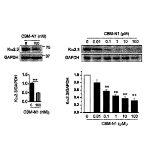
channel expression and cell proliferation
FIG. 3 shows the effect of CBM-N1 derivatives on the expression of a Kca2.3
channel protein in hepatic stellate cells. Specifically, by the exposure of
the cells to
CBM-N2, CBM-N5, CBM-N8 and CBM-N9 for 24 hours, the expression of the Kca2.3
channel was significantly reduced.
FIG. 4 shows the effect of the inhibition of Kca2.3 channel expression by CBM-
N2 to CBM-N4, and CBM-N9 in hepatic stellate cells on Kca2.3 current. As a
result
of the comparison of Kca2.3 current densities at a membrane potential of +50
mV
between the cells in which the K.2.3 channel expression is inhibited by
exposure to
the compound for 24 hours and the cells not exposed to the compound, due to
the
exposure to CBM-N2 to CBM-N4 and CBM-N9, the Kca2.3 current densities were
significantly decreased in cells in which the Kca2.3 channel expression was
reduced.
FIG. 5 shows the effect of CBM-N2 to CBM-N5, CBM-N8 and CBM-N9 on
TGFp or PDGF-induced cell proliferation in fibroblasts. When the cells were
exposed to a fibrosis-inducing factor such as TGFp (5 ng/ml) or PDGF (20
ng/ml) for
24 hours, the proliferation of fibroblasts significantly increased, and the
cell
proliferation was significantly reduced by the CBM-N2 to CBM-N5, CBM-N8 and
CBM-N9.
4) Results from liver disease mouse models
27
Date Recue/Date Received 2021-03-12
CA 03112695 2021-03-12
To identify the therapeutic effects of the compounds of Formulas Al to A5
(CBM-N 1 to CBM-N5) in liver disease mouse models, these experiments were
performed.
4-1) Histological or immunohistological analysis
FIG. 6A shows H&E staining results for the liver tissues, and the part
represented by a dotted line in the upper panel was enlarged and shown in the
lower
panel. In a disease-induced group (TAA), TAA-induced inflammation occurs in a
region near the central vein (CV), which can be confirmed by inflammation
cells
having large nuclei concentrated near the CV (arrows in FIG. 6A).
Particularly,
compared with the nottnal control, in the disease-induced group, inflammation
cells
significantly increased, and in the drug-administered group (TAA+CBM-N1),
compared with the disease-induced group, the number of inflammation cells
significantly decreased.
FIG. 6B shows staining results of an inflammation marker, CD82, in the liver
tissue, in which no cells stained brown were observed in liver tissue of the
normal
control (Control), indicating that there were no lymphoid cells. On the other
hand,
in the liver tissue of the group in which a disease is induced by TAA (TAA),
many
cells stained brown were found between CVs. However, a very small number of
cells
stained brown were found in the liver tissue (TAA+CBM-N1(R) or TAA+CBM-N1(S))
in a drug-administered group treated with TAA and a CBM-Nl R-isomer or S-
isomer.
FIG. 6C shows results of Masson's trichrome staining of collagen fibers in the
liver tissues, and the collagen was stained blue. The liver tissue in the
normal control
(Control) is a healthy state in which fibrosis has not progressed yet, whereas
the liver
28
Date Recue/Date Received 2021-03-12
CA 03112695 2021-03-12
tissue in the disease-induced group (TAA) is stained blue (indicated with
arrows) near
CVs or between CVs, demonstrating the progression of fibrosis. However, in the
liver tissue in the drug-administered group (TAA+CBM-N1(R), TAA+CBM-N1(S))
to which TAA and a CBM-N1 R-isomer or S-isomer were administered, fibrosis was
very slightly observed around CVs.
FIG. 6D shows staining results for reticulin fibers in the liver tissues, and
the
reticulin fibers were stained black. No reticulin fiber was observed in the
liver tissue
from the normal control (Control), whereas in the liver tissue in the disease-
induced
group (TAA), reticulin fibers (indicated with arrows) were observed around CVs
and
between CVs. In addition, in the liver tissue (TAA+CBM-N1(R) or (TAA+CBM-
N1(5)) in the drug-administered group administered with TAA and a CBM-N1 R-
isomer or S-isomer, the reticulin fibers were very slightly observed only
aroundr CVs.
4-2) Liver function test through blood ALT and AST analyses
FIG. 7A shows results of liver function testing for a normal control, a TAA-
mediated disease-induced group, and drug-administered groups treated with CBM-
N1
to CBM-N5. In a normal control (Control), a disease-induced group (TAA), and
drug-administered groups (TAA+CBM-N1, TAA+CBM-N2, TAA+CBM-N3,
TAA+CBM-N4, and TAA+CBM-N5), ALT levels were 41.6 7.9 units/L, 209.0 42.4
units/L, 70.3 14.7 units/L, 113.4 7.9 units/L, 103.0 6.9 units/L, 114.1 8.8
units/L
and 106.4 12.8 units/L, respectively, and AST blood levels were 60.4 7.5
units/L,
211.1 22.4 units/L, 62.7 11.6 units/L, 83.6 14.8 units/L, 73.4 7.4 units/L,
66.6 9.4
units/L, and 67.7 10.2 units/L, respectively.
29
Date Recue/Date Received 2021-03-12
CA 03112695 2021-03-12
In addition, FIG. 7B shows a test result of a disease-induced group by western
diet, and in a normal control (Control), a group in which a disease was
induced by a
western diet (WD), and a drug-administered group (WD+CBM-N1), ALT levels were
38.7 9.7 units/L, 189.8 37.6 units/L and 87.2 24.7 units/L, and AST blood
levels
were 47.4 18.2 units/L, 173.5 31.5 units/L and 71.4 19.8 units/L,
respectively.
From the test results, it can be seen that liver dysfunction induced by TAA or
a western diet is significantly recovered by compounds of Formulas Al to A5
(50
mg/kg/day).
4-3) Real time PCR for inflammation markers
FIG. 8 shows results of comparing the mRNA expression of inflammatory
cytokines in a normal control, a disease-induced group and a drug-administered
group.
As the inflammation markers, tumor necrosis factor alpha (TNFa),
chemoattractant
protein-1 (CCL2), interleukin-12 (IL12), transforming growth factor (TGF),
ILla, IL6,
and macrophage inflammatory protein-2 (MIP-2), which increase when
inflammation
occurs, were used. The mRNA level of an inflammation factor increased in the
disease-induced group (TAA) as compared with the normal control (Control), and
decreased in the CBM-Nl-administered group (TAA+CBM-N1) as compared with the
disease-induced group (TAA) (FIG. 8A).
The mRNA level of an inflammation factor also decreased in the CBM-N2 to
CBM-N5-administered groups (TAA+CBM-N2, TAA+CBM-N3, TAA+CBM- N4,
and TAA+CBM-N5) compared with the disease-induced group (FIG. 8B). Therefore,
it can be seen that the compounds of Formulas Al to AS decreased the
expression of
Date Recue/Date Received 2021-03-12
CA 03112695 2021-03-12
an inflammatory cytokine, thereby having a therapeutic effect on an
inflammatory liver
disease caused by TAA.
In addition, FIG. 8C shows results of comparing the mRNA expressions of
inflammatory cytokines in a normal control (Control), a group in which a
disease was
induced by a western diet (WD), and a drug-administered group (WD+CBM-N1).
Here, as inflammatory cytokines, CCL2. IL6 and IL la were measured. The mRNA
expression levels of these inflammation factors increased in the disease-
induced group
as compared with the normal control, and decreased in the drug-administered
group as
compared with the disease-induced group.
4-4) Real time PCR for fibrosis markers
FIG. 9 shows results of comparing mRNA expression levels of fibrosis markers
in a normal control, a disease-induced group and a drug-administered group. As
the
fibrosis markers. Col la, Col3a. Col4a, a-SMA and transforming growth factor
receptor 2 (TGFR2) were used. The fibrosis markers increased in the disease-
induced group (TAA) compared with the normal control (Control), indicating
that
fibrosis progresses due to inflammation. However, it was confirmed that, in
the
CBM-N 1-administered group (TAA+CBM-N1), the levels of these fibrosis factors
decreased.
4-5) Effect on protein expression of inflammation marker or fibrosis marker
proteins
31
Date Recue/Date Received 2021-03-12
CA 03112695 2021-03-12
FIG. 10A shows the effects of the R-isomer and S-isomer of CBM-N1 on the
protein expression of inflammation markers in TAA-mediated liver disease mouse
models. Compared with the control (Control), the protein expression levels of
TIMP-
2 and CCR2 greatly increased in liver tissue of the disease-induced group
(TAA),
indicating the progression of inflammation. However, in the CBM-N1 R-isomer or
S-isomer-administered group [TAA+CBM-N1(R), TAA+CBM-N1(S)], protein
expression levels of TIMP-2 and CCR2 decreased, confirming that inflammation
was
inhibited.
FIG. 10B shows the effect of the R-isomer or S-isomer of CBM-N1 on the
expression of fibrosis marker proteins in TAA-mediated liver disease mouse
models.
Compared with the normal control (Control), protein expression levels of a-SMA
and
Col la greatly increased in liver tissue of the disease-induced group (TAA),
indicating
the progression of fibrosis. However, the protein expression levels of a-SMA
and
Col la greatly decreased in the CBM-N1 R-isomer or S-isomer-administered
group,
confirming that fibrosis was inhibited.
4-6) Effect on expression of Kca2.3 channel protein
FIG. 11 shows the effect of the R-isomer or S-isomer of CBM-N1 on the
expression of a Kca2.3 channel protein in TAA-mediated liver disease mouse
models.
Compared with the normal control (Control), the expression level of the Kca2.3
channel protein greatly increased in liver tissue of the disease-induced group
(TAA).
However, the protein expression level of the Kca2.3 channel greatly decreased
in the
CBM-N1 R-isomer or S-isomer-administered group. These results show that a TAA-
32
Date Recue/Date Received 2021-03-12
CA 03112695 2021-03-12
induced liver disease is associated with the increase in Kca2.3 expression,
and the
therapeutic effect of CBM-N1 is associated with the decrease in Kca2.3
expression.
5) Experimental result for CBM-N9 in lung disease mouse models
To identify the therapeutic effect of the compound of Formula A9 (CBM-N9)
of the present invention in bleomycin-induced lung disease mouse models, this
experiment was performed.
5-1) Histological or immunohistological analysis
FIG. 12 shows results of H&E staining in lung tissue, immunohistochemistry
for CD45 (leukocyte common antigen, LCA staining), and Masson's trichrome
staining for collagen. It was confirmed that degrees of inflammation and
fibrosis
increased in the disease-induced group (Bleomycin) as compared to the normal
control,
and decreased in the CBM-N9-administered group (Bleomycin+CBM-N9) as
compared with the disease-induced group.
5-2) Analysis of protein expression of inflammation or fibrosis markers
FIG. 13A shows the effect of CBM-N9 on the expression of inflammation
markers in lung disease mouse models. Protein expression levels of
inflammation
markers such as TIMP-2 and CCR2 in lung tissue greatly increased in a disease-
induced group (bleomycin) as compared with a normal control (Control),
resulting in
the progression of inflammation. The protein expression levels of TIMP-2 and
CCR2
greatly decreased in a CBM-N9 drug-administered group (bleomycin+CBM-N9),
confirming the inhibition of inflammation.
33
Date Recue/Date Received 2021-03-12
CA 03112695 2021-03-12
FIG. 13B shows the effect of CBM-N9 on the expression of fibrosis marker
proteins in lung disease mouse models. The protein expression levels of
fibrosis
markers such as a-SMA and Col la greatly increased in lung tissue of a disease-
induced group (bleomycin) compared with a normal control (Control), indicating
the
progression of pulmonary fibrosis. On the other hand, the protein expression
levels
of TIMP-2 and CCR2 greatly decreased in a drug-administered group
(bleomycin+CBM-N9), confirming the inhibition of fibrosis.
6) Evaluation and conclusion
As seen above, when culture cells were administered with the compounds of
Formulas Al to A9 according to the present invention in vitro for a long time
(24 hours
or 16 weeks), the effect of inhibiting fibrosis was exhibited by the decrease
in
expression of a Kca2.3 channel protein. Specifically, it was confirmed that,
when
cultured hepatic stellate cells, fibroblasts, and vascular endothelial cells
were exposed
to the compounds of Formulas Al to A9 for 24 hours, the cell membrane
expression
of a Kca2.3 channel was inhibited, and the expression of fibrosis-related
factors (a-
SMA. Col la, etc.) and cell proliferation by growth factors inducing fibrosis
were
inhibited.
In addition, it was confirmed that the compounds of Formulas Al to A9
according to the present invention have inhibitory effects on inflammation and
fibrosis
in a liver disease-induced group even in an in vivo experiment for mouse
models.
Specifically, as a result of administering the compounds of Formulas Al to A9
to liver
34
Date Recue/Date Received 2021-03-12
CA 03112695 2021-03-12
disease or lung disease mouse models for 16 weeks, inflammation and fibrosis
were
significantly inhibited.
Meanwhile, as disclosed in Korean Patent Nos. 10-1345860 and 10-1414831
and U.S. Patent No. 9,259,412 corresponding thereto, the compounds of Formulas
Al
to A5 of the present invention have effects of inhibiting the activity of a
K.3.1 channel
due to K.3.1 channel phosphorylation induced by cAMP. However, it is
considered
that the suppression of the activity of the K.3.1 channel by increased cAMP
will have
little effect on fibrosis treatment.
The present invention relates to an effect exhibited when being exposed to the
compounds of Formulas Al to AS for a short time (within several minutes), and
the
increased cAMP due to these compounds reached the highest level in
approximately
minutes and then dramatically decreased such that the cAMP level became
similar
to that before drug administration within three hours (Endocrinology
144(4):1292-
1300). Therefore, this is because the effect caused by cAMP is exhibited only
for a
15 short time, for example, at most, approximately one hour, and as in the
case of the
present invention, is unlikely to last for 24 hours or 16 weeks.
In addition, as confirmed in FIG. IA of the present invention, when exposed to
a fibrosis-inducing factor, PDGF, for 24 hours, Kca2.3 channel expression
greatly
increased, whereas Kca3.1 channel expression did not increase. According to
this
20 result, it may be concluded that, in a fibrotic process, an increase in
Kca2.3 channel
expression is a very important requisite, and the fibrosis suppressing effect
of the
compounds of Formulas Al to A9 according to the present invention results from
a
decrease in Kca2.3 channel expression.
Date Recue/Date Received 2021-03-12
CA 03112695 2021-03-12
Meanwhile, in the present invention, due to realistic limitations, the above-
described experiments for all of compounds belonging to the compounds of
Formula
A were not performed. However, in consideration of chemical activities of the
compounds of Formula A and metabolic mechanisms in vivo, it is inferred that
all of
the compounds of Formula A have pharmacological effects the same as or similar
to
the compounds of Formulas Al to A9.
36
Date Recue/Date Received 2021-03-12