Note: Descriptions are shown in the official language in which they were submitted.
WO 2020/055952 PCT/US2019/050530
COMPOSITIONS AND METHODS FOR IMMUNOTHERAPY PROFILING
TECHNICAL FIELD OF THE INVENTION
This invention is generally related to immunotherapy and pharmacodynamic
monitoring
of immunotherapy.
BACKGROUND OF THE INVENTION
Immunotherapies harness the immune system to treat myriad diseases such as
cancer,
organ transplant rejection, infectious disease, allergic disease, autoimmunity
and chronic
inflammation. Immunotherapies employ both the humoral and cellular arms of the
immune
response using therapeutic antibodies (e.g. pembrolizumab/aPD-1), cytokines
(e.g prol eukin/IL-
2), and cell-based therapies (e.g Kymriah/CAR T cells). For example, emerging
techniques that
harness T cell immunity through adoptive transfer of engineered cells or
reinvigorating
endogenous anti-tumor CD8+ T cells through immune checkpoint blockade
antibodies have
placed immunotherapy at the forefront of cancer treatment research.
Immunotherapies that
dampen the T cell response through co-stimulation blockade (e.g.
abatacept/CTLA-4 Ig) have
also become a primary avenue of treatment research for preventing transplant
rejection or
treating autoimmune and chronic inflammatory disorders.
Despite the broad potential of immunotherapies, a majority of patients do not
achieve
clinical benefit, while others can develop immunotherapy resistance during or
between treatment
through poorly-understood mechanisms. Patients responding to immunotherapy
often exhibit
unconventional response patterns that can be misinterpreted as disease
progression. The full
potential benefit of immunotherapy is thus lacking, and techniques to identify
biomarkers of
immune responses are inadequate. Due to inadequacies in technologies for
response monitoring
and for identifying underlying resistance mechanisms, not only do diseases
persist in the
population, but drug development and clinical trials face significant
obstacles.
Tissue biopsy remains the gold standard diagnostic but is invasive and samples
less the
0.1% of the total disease site (Cyll, et al., Br J Cancer, 117(3):367-375
(2017)). Liquid biopsies
offer a noninvasive approach, but biomarker dilution in blood significantly
limits sensitivity
(Nagrath, S., et al., Nature, 450(7173):1235-1239 (2007); Hori, et al., Set
Transl Med,
3(109):109ra16 (2011)). Imaging techniques can also be limited by low
sensitivity and
specificity, as well as the unconventional response patterns commonly
associated with
1
Date Recue/Date Received 2021-03-11
WO 2020/055952 PCT/US2019/050530
immunotherapy that can result in misidentification of responding patients as
cases of treatment
failure. The development of better, non-invasive biomarkers will identify
responsive patients
sooner and illuminate mechanisms of new immunotherapies.
Therefore, it is an object of the invention to provide immune checkpoint
compositions
and methods for monitoring their efficacy.
SUMMARY OF THE INVENTION
Compositions and methods for pharmacodynamics monitoring of responses during
immunotherapy are provided herein. Exemplary compositions include an
immunotherapeutic
agent linked to a protease substrate that senses immune cell and disease site
protease activity and
produces a detectable signal in the presence of protease activity. Upon
administration, the
compositions target to sites of disease where proteases are upregulated during
responsive
immunotherapy and subsequently cleave the attached substrates. Cleavage
fragments are
detected in a sample from the body and detection of the fragments is
indicative of an effect of the
immunotherapeutic agent.
In one embodiment, the therapeutic agent is an immune checkpoint inhibitor
such as an
anti-PD1 or anti-CTLA4 antibody. The protease substrate can also include a
quencher molecule
and a fluorescent molecule flanking the substrate. In one embodiment, the
detectable signal is a
peptide fragment of the protease.
Another embodiment provides a method of treating or preventing disease in a
subject in
need thereof by administering to the subject an effective amount of a
therapeutic agent linked to
protease substrate that provides a detectable signal in response to protease
activity promoted by
the therapeutic agent, detecting and measuring the signal in a sample from the
subject,
determining an effect of the therapeutic agent on the subject, wherein the
subject is determined to
be responsive to the therapeutic agent if the detectable signal is detected,
and the subject is
determined to be non-responsive to the therapeutic agent if the detectable
signal is not detected,
and administering the same effective amount of the therapeutic agent to
responsive subjects, or
adjusting the effective amount of therapeutic agent administered to non-
responsive subjects. In
one embodiment, the therapeutic agent is an immune checkpoint inhibitor such
as an anti-PD1 or
anti-CTLA4 antibody.
In one embodiment, a subject determined to be non-responsive to the
immunotherapeutic
agent is given a different immunotherapeutic agent.
2
Date Recue/Date Received 2021-03-11
WO 2020/055952 PCT/US2019/050530
In another embodiment, detecting and measuring the signal includes collecting
a sample
from the subject, such as a urine sample or a blood sample, and measuring the
detectable signal
in the sample.
BRIEF DESCRIPTION OF THE DRAWINGS
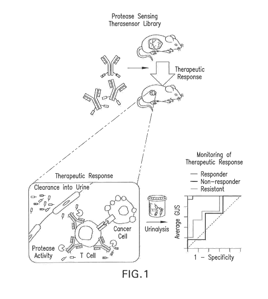
Figure 1 is a schematic illustration of an exemplary experimental use of the
disclosed
compositions and methods. Protease substrate funcitionalize therapeutic agents
target sites of
therapeutic activity, where the attached substrates are cleaved by proteases
upregulated during
responsive therapy, amplifying detection signals into urine. The urine sample
is analyzed by
mass spectrometry.
Figure 2A is a schematic illustration of amine coupling of GranzymeB (GzmB)
substrate
to aPD-1 to generate "aPD-1 therasensors". Figure 2B is a graph showing PD-1
binding by
aPD-1 modified with GzmB substrate (Therasensor) and unmodified PD-1 (aPD-1).
The X-axis
represents aPD-1 concentration (Ltg/mL; Log10) and the Y-axis represents PD-1
binding. Figure
2C is a flow plot of CD8 tumor infiltrating T cells showing equivalent
staining with unmodified
aPD-1 or aPD-1 modified with GzmB substrate (Therasensor). Figure 2D is a
graphical
summary of Figure 2C. Figure 2E is a graph showing the protease cleavage
kinetics of aPD-1
modified with GzmB substrate (Therasensor) incubated with or without GzmB or
control
protease thrombin.
Figure 3A is a schematic illustration of amine coupling of GzmB substrate to
CTLA-4 Ig
to generate "CLTA-4 Ig therasensors". Figures 3B-3C are graphs showing target
binding by
CTLA-4 IG modified with GzmB substrate (CTLA4-Ig Therasensor) or unmodified
CTLA-4 Ig
(CTLA4-Ig) in a CD80/CD86 antibody competition assay. Figure 3D is a bar graph
showing
proliferation of Cell Trace Violet (CTV) labeled BL/6 CD8+ cells co-incubated
with BALB/c
CDII c+ dendritic cells in the presence of aCD40L only, aCD40L + unmodified
CTLA4-Ig
(aCD40L + CTLA4-Ig), aCD40L + modified CTLA4-Ig (aCD40L + Therasensor). Figure
3E is
a line graph showing protease cleavage kinetics of CTLA-4 IG modified with
GzmB substrate
incubated with or without GzmB or the indicated protease (Abbreviations. CTSB,
Cathepsin B;
MMP2, matrix metalloproteinase 2, MMP9, matrix metalloproteinase 9; MMP15,
matrix
metalloproteinase 15; CIS, complement component Si; MASP1, mannose-associated
serine
protease 1).
3
Date Recue/Date Received 2021-03-11
WO 2020/055952 PCT/US2019/050530
Figure 4A is a schematic illustration of the cleavage of aPD-1 modified with
GzmB
substrate by GzmB in activated T cells, but not in tumor cell supernatant.
Figure 4B is a line
graph showing protease cleavage kinetics of aPD-1 modified with GzmB substrate
(GzmB
therasensor), control therasensor, or aPD-1 incubated with supernatant from
activated T cells,
CT26 cells, MC38 cells, B16 cells, or media alone. Figure 4C is a schematic
illustration of aPD-
1 therasensor cleavage during T cell killing of tumor cells. Figure 4D is a
bar graph showing
percent cytotoxicity, as measured by an LDH assay. Figure 4E is a bar graph
showing GzmB
protein secretion as determined by ELISA. Increased cell killing and GzmB
secretion was
observed as the effector to target ratio was increased (1:1, 5:1, 10:1).
Figure 4F is a bar graph
showing protease activity for control and aPD-1 therasensor across multiple
ratios of effector to
target cells. Figure 4G is a bar graph showing protease activity of the aPD-1
therasensor in cells
incubated with P-Mel or OT-1. Figure 4H is a bar graph showing protease
activity for CTLA-4
Ig therasensors added to supernatants from co-cultures of OT-I cells and
either OVA expressing
EG7 cells or the parental, non-OVA expressing EL4 cell line (E:T ratios of
1:1, 5:1, and 10:1).
Figure 5A is a line graph showing MC38 syngeneic tumor volume over time in
mice
treated with c1PD-1 modified with GzmB substrate (aPD-1 therasensor) or
isotype control
therasensor. Figure 5B is a panel of flow cytometry plots showing
intracellular GzmB staining
within CD8+ TILs isolated from MC38 tumors after two treatment doses. Figures
SC and 5D are
graphs showing the percentage (Fig. SC) and number (Fig. 5D) of GzmB positive
CD8 TILs per
tumor. Figure 5E is a schematic illustration of the experimental method for
urinalysis of
therasensors in MC38 tumor bearing mice. Figure 5F is a graph showing renal
clearance of
peptide fragments in tumor bearing mice treated with control therasensor or a-
PD1 therasensor.
Figures 5G-5H are graphs showing tumor volume over time in CT26 tumor bearing
mice treated
with a-CTLA4 monotherapy (Fig. 5G), a-PD1/CTLA-4 combination therapy (Fig. 5H)
or
untreated. The X-axis represents time (days) and the Y-axis represents tumor
volume (mm2).
The gray area represents the treatment window. Figure 51 is a panel of flow
cytometry plots
showing intracellular GzmB staining within CD8+ TILs isolated from CT26 tumors
on day 18.
Figure 5J-5K are graphs showing the percentage (Fig. 5J) and number (Fig. 5K)
of GzmB
positive CD8 TILs per tumor. Figure 5L is a schematic illustration of the
experimental method
for urinalysis of therasensors in CT26 tumor bearing mice. Figures 5M-5N are
graphs showing
4
Date Recue/Date Received 2021-03-11
WO 2020/055952 PCT/US2019/050530
renal clearance of cleaved fluorescent reporters in urine of tumor bearing
mice treated with
aCTLA-4, aPD-1/CTLA-4, or untreated.
Figure 6A is a timeline showing the experimental procedures. Figure 6B-6I are
photos
showing allograft rejection in skin over time. Figure 6J is a plot of
immunohistochemistry data
showing percent of CD8 staining in graft and healthy skin tissues from mice
bearing allo- and
iso-grafts. Figure 6K is a plot of immunohistochemistry data showing percent
of GzmB staining
in graft and healthy skin tissues from mice bearing alio- and iso-grafts.
Figure 6L is a plot of
skin graft scores showing graft quality of skin allograft in untreated mice,
treated mice
responding weakly ("non-responding") or strongly ("responding") to co-
stimulation blockade
therapy with CTLA4-Ig and aCD154. Figure 61 is a graft survival curve showing
percent
survival of grafts in untreated, non-responding, and responding grafts. Figure
6J is a graph
showing percent renal clearance of cleaved fluorescent reporters in urine at
POD -4, 7, and 15.
Figure 7A is a schematic of the patient cohort from Riaz, el al., 2017. Figure
7B is a
graph classifying responders from non-responders using 250 extracellular
proteases. Figure 7C
is a graph classifying responders from non-responders using 14 extracellular
proteases identified
as important by lasso algorithm. Figure 7D is a graph showing the relative
weights of
importance of the 14 extracellular proteases from Figure 7C. Figure 7E-7F are
graphs
identifying mechanisms of resistance via pathway analysis. Figure 7E shows non-
responding
patients with IFNy pathway expression loss were predicted with a panel of 12
proteases. Figure
7F shows the same panel of 12 proteases was used to classify non-responding
patients with MHC
I antigen presentation loss. Figure 7G is a graph showing the fraction of
pathways from each
molecular process (IFNy and MHC I antigen presentation) lost when comparing
gene expression
of responders and non-responders. Figure 7H is a graph showing the relative
weight of lasso
coefficients in classifying non-responders with or without MHC I presentation
loss.
DETAILED DESCRIPTION OF THE INVENTION
I. Definitions
It should be appreciated that this disclosure is not limited to the
compositions and
methods described herein as well as the experimental conditions described, as
such may vary. It
is also to be understood that the terminology used herein is for the purpose
of describing certain
5
Date Recue/Date Received 2021-03-11
WO 2020/055952 PCT/US2019/050530
embodiments only, and is not intended to be limiting, since the scope of the
present disclosure
will be limited only by the appended claims.
Unless defined otherwise, all technical and scientific terms used herein have
the same
meaning as commonly understood by one of ordinary skill in the art to which
this disclosure
belongs. Although any compositions, methods and materials similar or
equivalent to those
described herein can be used in the practice or testing of the present
invention. All publications
mentioned are incorporated herein by reference in their entirety.
The use of the terms "a," "an," "the," and similar referents in the context of
describing the
presently claimed invention (especially in the context of the claims) are to
be construed to cover
both the singular and the plural, unless otherwise indicated herein or clearly
contradicted by
context.
Recitation of ranges of values herein are merely intended to serve as a
shorthand method
of referring individually to each separate value falling within the range,
unless otherwise
indicated herein, and each separate value is incorporated into the
specification as if it were
individually recited herein.
Use of the term "about" is intended to describe values either above or below
the stated
value in a range of approx. +/- 10%; in other embodiments the values may range
in value either
above or below the stated value in a range of approx. +/- 5%; in other
embodiments the values
may range in value either above or below the stated value in a range of
approx. +/- 2%; in other
embodiments the values may range in value either above or below the stated
value in a range of
approx. +/- 1%. The preceding ranges are intended to be made clear by context,
and no further
limitation is implied. All methods described herein can be performed in any
suitable order unless
otherwise indicated herein or otherwise clearly contradicted by context. The
use of any and all
examples, or exemplary language (e.g., "such as") provided herein, is intended
merely to better
illuminate the invention and does not pose a limitation on the scope of the
invention unless
otherwise claimed. No language in the specification should be construed as
indicating any non-
claimed element as essential to the practice of the invention.
As used herein, a molecule is said to be able to "immunospecifically bind" a
second
molecule if such binding exhibits the specificity and affinity of an antibody
to its cognate
antigen. Antibodies are said to be capable of immunospecifically binding to a
target region or
conformation ("epitope") of an antigen if such binding involves the antigen
recognition site of
6
Date Recue/Date Received 2021-03-11
WO 2020/055952 PCT/US2019/050530
the immunoglobulin molecule. An antibody that immunospecifically binds to a
particular
antigen may bind to other antigens with lower affinity if the other antigen
has some sequence or
conformational similarity that is recognized by the antigen recognition site
as determined by,
e.g., immunoassays, BIACORE assays, or other assays known in the art, but
would not bind to
a totally unrelated antigen. In some embodiments, however, antibodies (and
their antigen
binding fragments) will not cross-react with other antigens. Antibodies may
also bind to other
molecules in a way that is not immunospecific, such as to FcR receptors, by
virtue of binding
domains in other regions/domains of the molecule that do not involve the
antigen recognition
site, such as the Fc region.
As used herein, the term "antibody" is intended to denote an immunoglobulin
molecule
that possesses a "variable region" antigen recognition site and include
antigen-binding fragments
of antibodies. The term "variable region" is intended to distinguish such
domain of the
immunoglobulin from domains that are broadly shared by antibodies (such as an
antibody Fe
domain). The variable region includes a "hypervariable region" whose residues
are responsible
for antigen binding. The hypervariable region includes amino acid residues
from a
"Complementarity Determining Region" or "CDR" (i.e., typically at
approximately residues 24-
34 (L1), 50-56 (L2) and 89-97 (L3) in the light chain variable domain and at
approximately
residues 27-35 (H1), 50-65 (H2) and 95-102 (H3) in the heavy chain variable
domain; Kabat et
at., Sequences of Proteins of Immunological Interest, 5th Ed. Public Health
Service, National
Institutes of Health, Bethesda, MD. (1991)) and/or those residues from a
"hypervariable loop"
(i.e., residues 26-32 (L1), 50-52 (L2) and 91-96 (L3) in the light chain
variable domain and 26-
32 (H1), 53-55 (H2) and 96-101 (H3) in the heavy chain variable domain;
Chothia and Lesk,
1987õ/ Mol. Biol. 196:901-917). "Framework Region" or "FR" residues are those
variable
domain residues other than the hypervariable region residues as herein
defined. The tei in
antibody includes monoclonal antibodies, multi-specific antibodies, human
antibodies,
humanized antibodies, synthetic antibodies, chimeric antibodies, camelized
antibodies (See e.g.,
Muyldeimans et al., 2001, Trends Biochem. Sci. 26.230; Nuttall c/at., 2000,
Cur. Pharm.
Biotech. 1:253; Reichmann and Muyldermans, 1999,1 Immunol Meth. 231:25;
International
Publication Nos. WO 94/04678 and WO 94/25591; U.S. Patent No. 6,005,079),
single-chain Fvs
(scFv) (see, e.g., see Pluckthun in The Pharmacology o/ Monoclonal Antibodies,
vol. 113,
Rosenburg and Moore eds. Springer-Verlag, New York, pp. 269-315 (1994)),
single chain
7
Date Recue/Date Received 2021-03-11
WO 2020/055952 PCT/US2019/050530
antibodies, disulfide-linked Fvs (sdFv), intrabodies, diabodies, triabodies,
tetrabodies, Bis-scFv,
minibodies, Fab2, Fab3and anti-idiotypic (anti-Id) antibodies (including,
e.g., anti-Id and anti-
anti-Id antibodies to antibodies). In particular, such antibodies include
immunoglobulin
molecules of any type (e.g., IgG, IgE, IgM, IgD, IgA and IgY), class (e.g.,
IgGI, IgG2, IgG3,
IgG4, IgAi and IgA2) or subclass.
As used herein, the term "antigen binding fragment" of an antibody refers to
one or more
portions of an antibody that contain the antibody's Complementarity
Determining Regions
("CDRs") and optionally the framework residues that include the antibody's
"variable region"
antigen recognition site, and exhibit an ability to immunospecifically bind
antigen. Such
fragments include Fab', F(ab')2, Fv, single chain (ScFv), and mutants thereof,
naturally occurring
variants, and fusion proteins including the antibody's "variable region"
antigen recognition site
and a heterologous protein (e.g., a toxin, an antigen recognition site for a
different antigen, an
enzyme, a receptor or receptor ligand, etc.).
As used herein, the term "fragment" refers to a peptide or polypeptide
including an amino
.. acid sequence of at least 5 contiguous amino acid residues, at least 10
contiguous amino acid
residues, at least 15 contiguous amino acid residues, at least 20 contiguous
amino acid residues,
at least 25 contiguous amino acid residues, at least 40 contiguous amino acid
residues, at least 50
contiguous amino acid residues, at least 60 contiguous amino residues, at
least 70 contiguous
amino acid residues, at least 80 contiguous amino acid residues, at least 90
contiguous amino
acid residues, at least 100 contiguous amino acid residues, at least 125
contiguous amino acid
residues, at least 150 contiguous amino acid residues, at least 175 contiguous
amino acid
residues, at least 200 contiguous amino acid residues, or at least 250
contiguous amino acid
residues.
As used herein the term "modulate" relates to a capacity to alter an effect,
result, or
activity (e.g., signal transduction) Such modulation can be agonistic or
antagonistic.
Antagonistic modulation can be partial (i.e., attenuating, but not abolishing)
or it can completely
abolish such activity (e.g., neutralizing). Modulation can include
internalization of a receptor
following binding of an antibody or a reduction in expression of a receptor on
the target cell.
Agonistic modulation can enhance or otherwise increase or enhance an activity
(e.g., signal
transduction). In a still further embodiment, such modulation can alter the
nature of the
interaction between a ligand and its cognate receptor so as to alter the
nature of the elicited signal
8
Date Recue/Date Received 2021-03-11
WO 2020/055952 PCT/US2019/050530
transduction. For example, the molecules can, by binding to the ligand or
receptor, alter the
ability of such molecules to bind to other ligands or receptors and thereby
alter their overall
activity. In some embodiments, such modulation will provide at least a 10%
change in a
measurable immune system activity, at least a 50% change in such activity, or
at least a 2-fold, 5-
fold, 10-fold, or at least a 100-fold change in such activity.
As used herein, the term "polypeptide" refers to a chain of amino acids of any
length,
regardless of modification (e.g., phosphorylation or glycosylation). The term
polypeptide
includes proteins and fragments thereof The polypeptides can be "exogenous,"
meaning that
they are "heterologous," i.e., foreign to the host cell being utilized, such
as human polypeptide
produced by a bacterial cell. Polypeptides are disclosed herein as amino acid
residue sequences
Those sequences are written left to right in the direction from the amino to
the carboxy terminus.
In accordance with standard nomenclature, amino acid residue sequences are
denominated by
either a three letter or a single letter code as indicated as follows. Alanine
(Ala, A), Arginine
(Arg, R), Asparagine (Asn, N), Aspartic Acid (Asp, D), Cysteine (Cys, C),
Glutamine (Gln, Q),
Glutamic Acid (Glu, E), Glycine (Gly, G), Histidine (His, H), Isoleucine (Ile,
I), Leucine (Leu,
L), Lysine (Lys, K), Methionine (Met, M), Phenylalanine (Phe, F), Proline
(Pro, P), Serine (Ser,
S), Threonine (Thr, T), Tryptophan (Trp, W), Tyrosine (Tyr, Y), and Valine
(Val, V).
As used herein, the terms "treat," "treating," "treatment" and "therapeutic
use" refer to
the elimination, reduction or amelioration of one or more symptoms of a
disease or disorder. As
used herein, a "therapeutically effective amount" refers to that amount of a
therapeutic agent
sufficient to mediate a clinically relevant elimination, reduction or
amelioration of such
symptoms. An effect is clinically relevant if its magnitude is sufficient to
impact the health or
prognosis of a recipient subject. A therapeutically effective amount may refer
to the amount of
therapeutic agent sufficient to delay or minimize the onset of disease, e.g.,
delay or minimize the
spread of cancer. A therapeutically effective amount may also refer to the
amount of the
therapeutic agent that provides a therapeutic benefit in the treatment or
management of a disease.
As used herein, the term "prophylactic agent" refers to an agent that can be
used in the
prevention of a disorder or disease prior to the detection of any symptoms of
such disorder or
disease. A "prophylactically effective" amount is the amount of prophylactic
agent sufficient to
mediate such protection. A prophylactically effective amount may also refer to
the amount of
the prophylactic agent that provides a prophylactic benefit in the prevention
of disease.
9
Date Recue/Date Received 2021-03-11
WO 2020/055952 PCT/US2019/050530
As used herein, the terms "immunologic," "immunological" or "immune" response
is the
development of a beneficial humoral (antibody mediated) and/or a cellular
(mediated by antigen-
specific T cells or their secretion products) response directed against a
peptide in a recipient
patient. Such a response can be an active response induced by administration
of immunogen or a
passive response induced by administration of antibody or primed T-cells. A
cellular immune
response is elicited by the presentation of polypeptide epitopes in
association with Class I or
Class II Mt-IC molecules to activate antigen-specific CD4+ T helper cells
and/or CD8+ cytotoxic
T cells. The response may also involve activation of monocytes, macrophages,
NK cells,
basophils, dendritic cells, astrocytes, microglia cells, eosinophils,
activation or recruitment of
.. neutrophils or other components of innate immunity. The presence of a cell-
mediated
immunological response can be determined by proliferation assays (CD4+ T
cells) or CTL
(cytotoxic T lymphocyte) assays. The relative contributions of humoral and
cellular responses to
the protective or therapeutic effect of an immunogen can be distinguished by
separately isolating
antibodies and T-cells from an immunized syngeneic animal and measuring
protective or
therapeutic effect in a second subject.
Activated T cells that are specific to molecular structures on an invading
pathogen
proliferate and attack the invading pathogen. Their attack can kill pathogens
directly or secrete
antibodies that enhance the phagocytosis of pathogens and disrupt the
infection. Some T cells
respond to APCs of the innate immune system, and indirectly induce immune
responses by
releasing or cytokines.
As used herein, an "immune cell" refers to any cell from the hemopoietic
origin
including, but not limited to, T cells, B cells, monocytes, dendritic cells,
and macrophages.
As used herein, "inflammatory molecules" refer to molecules that result in
inflammatory
responses including, but not limited to, cytokines and metalloproteases such
as including, but not
limited to, IL-113, 'TNF-a, TGF-beta, IFN-y, IL-18, IL-17, IL-6, IL-23, IL-22,
IL-21, and MMPs.
As used herein, the terms "individual," "host," "subject," and "patient" are
used
interchangeably herein, and refer to a mammal, including, but not limited to,
humans, rodents,
such as mice and rats, and other laboratory animals.
As used herein, the term "pharmaceutically acceptable carrier" encompasses any
of the
standard pharmaceutical carriers, such as a phosphate buffered saline
solution, water and
emulsions such as an oil/water or water/oil emulsion, and various types of
wetting agents.
Date Recue/Date Received 2021-03-11
WO 2020/055952 PCT/US2019/050530
As used herein, the term "immunosuppression" refers to the suppression of the
immune
system and its ability to fight infections and other diseases.
Immunosuppression may be
deliberately induced with drugs, or it can result from certain diseases,
environmental factors, or
as a side effect to other drugs such as anticancer drugs and steroids.
As used herein, the term "immunosuppressive disease" and "immunodeficiency
disease"
refer to diseases characterized by the partial or complete suppression or
dysfunction of the
immune response of a subject.
As used herein, the term "cancer" refers to a neoplasm or tumor resulting from
abnormal
uncontrolled growth of cells. As used herein, cancer explicitly includes
leukemias and
lymphomas. The term "cancer" refers to a disease involving cells that have the
potential to
metastasize to distal sites and exhibit phenotypic traits that differ from
those of non-cancer cells,
for example, formation of colonies in a three-dimensional substrate such as
soft agar or the
formation of tubular networks or web-like matrices in a three-dimensional
basement membrane
or extracellular matrix preparation. Non-cancer cells do not form colonies in
soft agar and form
distinct sphere-like structures in three-dimensional basement membrane or
extracellular matrix
preparations.
Compositions and Methods for Immunotherapy Profiling
Immunotherapeutic compositions and methods of their use for both treating
disease in a
subject in need thereof and profiling the subject's immune response to the
immunotherapy are
provided herein. An exemplary composition includes an immunotherapeutic agent
conjugated
with a protease substrate that is capable of being cleaved from the
immunotherapeutic agent by
disease- or tissue-specific proteases. In one embodiment, if the
immunotherapeutic agent
reaches the disease site and imparts a therapeutic effect, increased immune
protease activity will
cleave the attached protease substrate from the immunotherapeutic agent
releasing a peptide
fragment or detectable signal into circulation upon which it will be
selectively filtered into the
urine. The circulating cleavage fragment or detectable signal can be detected
in a sample from
the subject such as a blood sample or a urine sample.
A. Immunotherapeutic Agent
In one embodiment, the immunotherapeutic agent that is conjugated with a
protease
substrate is a checkpoint inhibitor. Immune checkpoint inhibitors typically
employ therapeutic
antibodies, such as Pembrolizumab (aPD1) or Ipilimumab (aCTLA-4), to reverse
immune
11
Date Recue/Date Received 2021-03-11
WO 2020/055952 PCT/US2019/050530
suppression within the tumor microenvironment by blocking inhibitory immune
checkpoint
molecules, such as PD-1 (Tumeh PC, et al., Nature, 515(7528):568-71 (2014)).
In one embodiment, the immunotherapeutic agent is an antibody, antigen-binding
fragment, fusion protein, or small molecule. In another embodiment, the
immunotherapeutic
agent is a T cell therapy, such as CAR-T cell therapy. In yet another
embodiment, the
immunotherapeutic agent is an immunosuppressive agent. Immunotherapeutic agent
targets are
described in detail below.
1. PD-1
Programmed Death-1 (PD-1) is a member of the CD28 family of receptors that
delivers a
negative immune response when induced on T cells. Contact between PD-1 and one
of its
ligands (B7-H1 or B7-DC) induces an inhibitory response that decreases T cell
multiplication
and/or the strength and/or duration of a T cell response. Suitable PD-1
antagonists are described
in U.S. Patent Nos. 8,114,845, 8,609,089, and 8,709,416, which are
specifically incorporated by
reference herein in their entities, and include compounds or agents that
either bind to and block a
ligand of PD-1 to interfere with or inhibit the binding of the ligand to the
PD-1 receptor, or bind
directly to and block the PD-1 receptor without inducing inhibitory signal
transduction through
the PD-1 receptor.
In some embodiments, the PD-1 receptor antagonist binds directly to the PD-1
receptor
without triggering inhibitory signal transduction and also binds to a ligand
of the PD-1 receptor
to reduce or inhibit the ligand from triggering signal transduction through
the PD-1 receptor. By
reducing the number and/or amount of ligands that bind to PD-1 receptor and
trigger the
transduction of an inhibitory signal, fewer cells are attenuated by the
negative signal delivered by
PD-1 signal transduction and a more robust immune response can be achieved.
It is believed that PD-1 signaling is driven by binding to a PD-1 ligand (such
as B7-H1 or
B7-DC) in close proximity to a peptide antigen presented by major hi
stocompatibility complex
(MEC) (see, for example, Freeman, Proc. Natl. Acad. Sci. U. S. A, 105:10275-
10276 (2008)).
Therefore, proteins, antibodies or small molecules that prevent co-ligation of
PD-1 and TCR on
the T cell membrane are also useful PD-1 antagonists.
In some embodiments, the PD-1 receptor antagonists are small molecule
antagonists or
antibodies that reduce or interfere with PD-1 receptor signal transduction by
binding to ligands
12
Date Recue/Date Received 2021-03-11
WO 2020/055952 PCT/US2019/050530
of PD-1 or to PD-1 itself, especially where co-ligation of PD-1 with TCR does
not follow such
binding, thereby not triggering inhibitory signal transduction through the PD-
1 receptor.
Other PD-1 antagonists contemplated by the methods of this invention include
antibodies
that bind to PD-1 or ligands of PD-1, and other antibodies.
Suitable anti-PD-1 antibodies include, but are not limited to, those described
in the
following US Patent Nos: 7332582, 7488802, 7521051, 7524498, 7563869, 7981416,
8088905,
8287856, 8580247, 8728474, 8779105, 9067999, 9073994, 9084776, 9205148,
9358289,
9387247, 9492539, 9492540, all of which are incorporated by reference in their
entireties.
Exemplary anti-B7-H1 (also referred to as anti-PD-L1) antibodies include, but
are not
limited to, those described in the following US Pat Nos: 8383796, 9102725,
9273135, 9393301,
and 9580507 all of which are specifically incorporated by reference herein in
their entirety.
For anti-B7-DC (also referred to as anti-PD-L2) antibodies see US Pat. Nos.:
7,411,051,
7,052,694, 7,390,888, 8188238, and 9255147 all of which are specifically
incorporated by
reference herein in their entirety.
Other exemplary PD-1 receptor antagonists include, but are not limited to B7-
DC
polypeptides, including homologs and variants of these, as well as active
fragments of any of the
foregoing, and fusion proteins that incorporate any of these. In some
embodiments, the fusion
protein includes the soluble portion of B7-DC coupled to the Fe portion of an
antibody, such as
human IgG, and does not incorporate all or part of the transmembrane portion
of human B7-DC.
The PD-1 antagonist can also be a fragment of a mammalian B7-H1, for example
from
mouse or primate, such as a human, wherein the fragment binds to and blocks PD-
1 but does not
result in inhibitory signal transduction through PD-1. The fragments can also
be part of a fusion
protein, for example an Ig fusion protein.
Other useful polypeptides PD-1 antagonists include those that bind to the
ligands of the
PD-1 receptor. These include the PD-1 receptor protein, or soluble fragments
thereof, which can
bind to the PD-1 ligands, such as B7-H1 or B7-DC, and prevent binding to the
endogenous PD-1
receptor, thereby preventing inhibitory signal transduction. B7-H1 has also
been shown to bind
the protein B7.1 (Butte et al., Immunity, Vol. 27, pp. 111-122, (2007)). Such
fragments also
include the soluble ECD portion of the PD-1 protein that includes mutations,
such as the A99L
mutation, that increases binding to the natural ligands (Molnar et al., PNAS,
105:10483-10488
(2008)). B7-1 or soluble fragments thereof, which can bind to the B7-H1 ligand
and prevent
13
Date Recue/Date Received 2021-03-11
WO 2020/055952 PCT/US2019/050530
binding to the endogenous PD-1 receptor, thereby preventing inhibitory signal
transduction, are
also useful.
PD-1 and B7-H1 anti-sense nucleic acids, both DNA and RNA, as well as siRNA
molecules can also be PD-1 antagonists. Such anti-sense molecules prevent
expression of PD-1
on T cells as well as production of T cell ligands, such as B7-H1, PD-Li
and/or PD-L2. For
example, siRNA (for example, of about 21 nucleotides in length, which is
specific for the gene
encoding PD-1, or encoding a PD-1 ligand, and which oligonucleotides can be
readily purchased
commercially) complexed with carriers, such as polyethyleneimine (see Cubillos-
Ruiz et al., J.
Clin. Invest. 119(8): 2231-2244 (2009), are readily taken up by cells that
express PD-1 as well as
ligands of PD-1 and reduce expression of these receptors and ligands to
achieve a decrease in
inhibitory signal transduction in T cells, thereby activating T cells.
2. CTLA4
Cytotoxic T-lymphocyte-associated protein 4 (CTLA4) is a is a protein receptor
that
functions as an immune checkpoint and downregulates immune responses. CTLA4 is
constitutively expressed in regulatory T cells but only upregulated in
conventional T cells after
activation. CTLA4 transmits an inhibitory signal to T cells. In some
embodiments, the
immunotherapeutic agent is an antagonist of CTLA4, for example an antagonistic
anti-CTLA4
antibody. An example of an anti-CTLA4 antibody contemplated for use in the
methods of the
invention includes an antibody as described in PCT/US2006/043690 (Fischkoff et
al.,
WO/2007/056539).
Specific examples of an anti-CTLA4 antibody useful in the methods of the
invention are
Ipilimumab, a human anti-CTLA4 antibody, administered at a dose of, for
example, about 10
mg/kg, and Tremelimumab a human anti-CTLA4 antibody, administered at a dose
of, for
example, about 15 mg/kg. See also Sammartino, et al., Clinical Kidney Journal,
3(2):135-137
(2010), published online December 2009.
In other embodiments, the antagonist is a small molecule. A series of small
organic
compounds have been shown to bind to the B7-1 ligand to prevent binding to
CTLA4 (see Erbe
et al., I Biol. Chem., 277:7363-7368 (2002). Such small organics could be
administered alone or
together with an anti-CTLA4 antibody to reduce inhibitory signal transduction
of T cells.
14
Date Recue/Date Received 2021-03-11
WO 2020/055952
PCT/US2019/050530
3. Other Immune Checkpoint Inhibitors
In another embodiment, the immunotherapeutic agent is an immune checkpoint
inhibitor
that inhibits the activity of other immune checkpoint molecules such as but
not limited to B7-H3,
B7-H4, BTLA, IDO, KIR, LAG3, NOX2, TIM3, VISTA, SIGLEC7, and SIGLEC9.
B7-H3, also known as CD276, is an immune checkpoint molecule from the B7
family.
B7-H3 participates in the regulation of T-cell-mediated immune response. It
also plays a
protective role in tumor cells by inhibiting natural-killer mediated cell
lysis as well as a role of
marker for detection of neuroblastoma cells. It is also involved in the
development of acute and
chronic transplant rejection and in the regulation of lymphocytic activity at
mucosal surfaces.
B7-H3 immunotherapeutic agents are known in the art. Exemplary anti-B7-H4
agents include,
but are not limited to, those described in the following US Pat Nos: 7847081,
8802091, and
9371395, all of which are specifically incorporated by reference herein in
their entirety.
Indoleamine 2,3-dioxygenase(IDO), is a tryptophan catabolic enzyme with immune-
inhibitory properties. IDO is known to suppress T and NK cells, generate and
activate Tregs and
myeloid-derived suppressor cells, and promote tumor angiogenesis. IDO
immunotherapeutic
agents are known in the art. Exemplary anti-IDO agents include, but are not
limited to, those
described in the following US Pat Nos: 7598287, 9598422, and 10323004, all of
which are
specifically incorporated by reference herein in their entirety.
Lymphocyte Activation Gene-3 (LAG3) is an inhibitory receptor on antigen
activated T-
cells. LAG3 delivers inhibitory signals upon binding to ligands, such as FGL1.
Following TCR
engagement, LAG3 associates with CD3-TCR in the immunological synapse and
directly
inhibits T-cell activation. LAG3 suppresses immune responses by action on
Tregs as well as
direct effects on CD8+ T cells. LAG3 immunotherapeutic agents are known in the
art.
Exemplary anti-LAG3 agents include, but are not limited to, those described in
the following US
Pat Nos. 10188730 and 10358495, both of which are specifically incorporated by
reference
herein in their entirety.
V-type immunoglobulin domain-containing suppressor of T-cell activation
(VISTA) is an
immunoregulatory receptor which inhibits the T-cell response. VISTA is
expressed on
hematopoietic cells. VISTA immunotherapeutic agents are known in the art.
Exemplary anti-
VISTA agents include, but are not limited to, those described in the following
US Pat Nos:
Date Recue/Date Received 2021-03-11
WO 2020/055952 PCT/US2019/050530
9381244 and 10273301, both of which are specifically incorporated by reference
herein in their
entirety.
4. CAR-T cells
Another form of immunotherapy that is contemplated for use in the disclosed
compositions and methods are CAR-T cells. Chimeric antigen receptor T cells
(CAR-T cells)
are T cells that have been genetically engineered to produce an artificial T
cell receptor. This
gives the engineered T cells the ability to target a specific protein. The
basis of CAR-T
immunotherapy is to modify T cells to recognize cancer cells in order to more
effectively target
and destroy them. T cells are harvested from a subject, genetically altered to
express specific T
cell receptors, then the resulting CAR-T cells are infused into subjects to
attack their tumors.
CAR-T cells can be either derived from T cells in a subject's own blood
(autologous) or derived
from the T cells of another healthy donor (allogeneic). Once isolated from a
subject, these T
cells are genetically engineered to express a specific CAR, which programs
them to target an
antigen that is present on the surface of tumors. For safety, CAR-T cells are
engineered to be
specific to an antigen expressed on a tumor that is not expressed on healthy
cells.
In one embodiment, CAR-T cells are conjugated with a protease substrate that
is cleaved
from the CAR-T cell by proteases that are produced when the CAR-T cell affects
a diseased cell.
In such an embodiment, the detection of the detached detectable signal in the
urine of a subject
indicates that the CAR-T cells are having an effect on the subject.
5. Immunosuppressive Agents
In another embodiment, the immunotherapeutic agent is an immunosuppressive
agent.
Immunosuppressive agents include, but are not limited to antibodies against
other lymphocyte
surface markers (e.g., CD40, alpha-4 integrin) or against cytokines), fusion
proteins (e.g., CTLA-
4-Ig (Orencia0), TNFR-Ig (Enbre10)), TNF-a blockers such as Enbrel, Remicade,
Cimzia and
Humira, cyclophosphamide (CTX) (i.e., Endoxan , Cytoxan , Neosar , Procytox ,
RevimmuneTm), methotrexate (MTX) (i.e., Rheumatrex , Trexall ,), belimumab
(i.e.,
Benlysta ,), or other immunosuppressive drugs (e.g., cyclosporin A, FK506-like
compounds,
rapamycin compounds, or steroids), anti-proliferatives, cytotoxic agents, or
other compounds
that may assist in immunosuppression.
The immunosuppressive agent can be a CTLA-4 fusion protein, such as CTLA-4-Ig
(abatacept). CTLA-4-Ig fusion proteins compete with the co-stimulatory
receptor, CD28, on T
16
Date Recue/Date Received 2021-03-11
WO 2020/055952 PCT/US2019/050530
cells for binding to CD80/CD86 (B7-11B7-2) on antigen presenting cells, and
thus function to
inhibit T cell activation. In another embodiment, the immunosuppressive agent
is a CTLA-4-Ig
fusion protein known as belatacept. Belatacept contains two amino acid
substitutions (L104E
and A29Y) that markedly increase its avidity to CD86 in vivo. In another
embodiment, the
immunosuppressive agent is Maxy-4.
In another embodiment, the immunosuppressive agent is cyclophosphamide (CTX).
Cyclophosphamide (the generic name for Endoxan , Cytoxan , Neosar , Procytox ,
RevimmuneTm), also known as cytophosphane, is a nitrogen mustard alkylating
agent from the
oxazophorines group. It is used to treat various types of cancer and some
autoimmune disorders.
Cyclophosphamide (CTX) is the primary drug used for diffuse proliferative
glomerulonephritis
in patients with renal lupus.
As used herein the term "rapamycin compound" includes the neutral tricyclic
compound
rapamycin, rapamycin derivatives, rapamycin analogs, and other macrolide
compounds which
are thought to have the same mechanism of action as rapamycin (e.g.,
inhibition of cytokine
function). The language "rapamycin compounds" includes compounds with
structural similarity
to rapamycin, e.g., compounds with a similar macrocyclic structure, which have
been modified
to enhance their therapeutic effectiveness. Exemplary Rapamycin compounds are
known in the
art (See, e.g. W095122972, WO 95116691, WO 95104738, U.S. Patent No.
6,015,809;
5,989,591; U.S. Patent No. 5,567,709; 5,559,112; 5,530,006; 5,484,790;
5,385,908; 5,202,332;
5,162,333; 5,780,462; 5,120,727).
The language "FK506-like compounds" includes FK506, and FK506 derivatives and
analogs, e.g., compounds with structural similarity to FK506, e.g., compounds
with a similar
macrocyclic structure which have been modified to enhance their therapeutic
effectiveness.
Examples of FK506-like compounds include, for example, those described in WO
00101385. In
some embodiments, the language "rapamycin compound" as used herein does not
include
FK506-like compounds.
B. Detectable Signal Molecule
The disclosed immunotherapeutic agents are conjugated with a protease
substrate that is
cleaved by proteases, releasing a peptide fragment or a detectable signal from
the therapeutic
agent. In some embodiments, the detection signal is the cleavage product or
peptide fragment of
17
Date Recue/Date Received 2021-03-11
WO 2020/055952 PCT/US2019/050530
the protease substrate itself. Upon cleavage a fragment of the protease is
released into
circulation and detected in urine by mass spectrometry.
In other embodiments, the detection signal is a protease substrate engineered
with a
quencher molecule before the cleavage site and a fluorescent reporter after
the cleavage site.
Upon cleavage of the protease substrate, the quencher and fluorescent reporter
are separated,
with the reporter being released into circulation. The fluorescent signal is
detected in the urine
by standard methods such as flow cytometry.
The protease substrate can be conjugated to the immunotherapeutic agent using
methods
known in the art. In one embodiment, the protease substrate is conjugated to
the
immunotherapeutic agent through the introduction of a linker that forms a
covalent conjugate
between the protease substrate and the immunotherapeutic agent. Exemplary
reactions that can
be used to link the protease substrate include but are not limited to amine-to-
amine crosslinkers
using NHS esters, thiol-to-thiol crosslinkers using maleimides, amine-to-thiol
crosslinkers using
NHS esters and maleimides, and biotin/streptavidin interactions. In one
embodiment, the
protease substrate is conjugated to the immunotherapeutic agent through an
amine coupling
reaction.
1. Protease substrate
The disclosed compositions and methods of their use to determine the efficacy
of a
therapeutic response rely on protease activity to cleave the protease
substrate and release a
peptide fragment or detectable signal from the therapeutic agent. Proteases
are a class of
enzymes that includes over 550 members encoded within the human genome, many
of which
have disease specific roles, including critical roles in immunity. For
example, cytotoxic T cell-
mediated target cell killing is a protease-driven process involving: 1) death
receptor signaling
and caspase activation, proteases whose activity mediates cell death, and 2)
secretion of
granzymes, proteases that enter target cells through a perforin dependent
mechanism to activate
caspase-mediated cell death. Moreover, proteases are central to other aspects
of immune activity
including cell migration, matrix degradation and repair, and complement
activation, while tumor
proteases such as intlarmnatoly and matrix degrading proteases are established
hallmarks of
cancer (Arias, et al., Trends Cancer, 3(6):407-422 (2017), Egeblad, et al.,
Nat Rev Cancer,
2(3):161-174 (2002)).
18
Date Recue/Date Received 2021-03-11
WO 2020/055952 PCT/US2019/050530
Proteases provide an innovative approach for immunotherapy response monitoring
given
that proteases play a central role in the underlying biology of immunity,
oncology, and the
pathophysiology of multiple diseases (Dudani, et al., Ann Rev of Cancer
Biology, (2018)). For
example, the mark of a "hot" tumor is signified by an effective immune
infiltrate of cytotoxic T
cells that kill cancer cells primarily through a perforin-dependent, granzyme-
mediated pathway,
the latter of which comprise a family of potent serine proteases (Larimer, et
al., Cancer Res,
77(9):2318-2327 (2017); Voskoboinik, et al., Nat Rev Immitnol, 15(6):388-400
(2015)). Tumor
expression of proteases, including inflammatory and matrix degrading
proteases, is well
established as a hallmark of fundamental tumor biology including angiogenesis,
growth, and
metastasis (Dudani, et al., Ann Rev of Cancer Biology, (2018)) These protease
signatures can be
used to stage cancer, monitor progression and regression, and provide early
indication of drug
response. In one embodiment, the disclosed immunotherapeutic agents have the
ability to
quantify the activity of immune and disease site specific proteases early in
treatment to allow
identification of activity biomarkers that predict treatment efficacy and
indicate resistance to
immunotherapy.
In one embodiment, catalytic proteases amplify detection signals at the
disease or
therapeutic site (x1000 fold). Following protease cleavage, the
immunotherapeutic agents
disclosed herein are concentrated into urine, instead of being diluted in
blood, further enriching
the signal up to 100-fold. This enables ultrasensitive and early detection of
T cell activity that
precedes radiographic detectable changes at the disease site.
Protease substrates contain a recognition sequence for the protease to cleave.
Cleavage
of the protease substrate conjugated to the immunotherapeutic agent releases a
peptide fragment
of the substrate of a detectable signal molecule linked to the substrate from
the
immunotherapeutic agent. In some embodiment, the protease substrates that are
conjugated to
the immunotherapeutic agent are tumor specific protease substrates. Exemplary
tumor
associated proteases include but are not limited to cathepsin B, cathepsin D,
cathepsin E,
cathepsin K, cathepsin L, kallikrein 1, kallikrein 3 (PSA), kallikrein 10,
kallikrein15, uPA,
uPAR, caspases, matrix metalloproteinases such as MMP1, MMP2, MMP8, MMP9,
MMP13,
MMP14, and ADAM. In another embodiment, the protease substrates are cell
specific protease
substrates, such as T cell specific protease substrates. Exemplary cell
specific proteases include
but are not limited to neutrophil serine proteases such as cathepsin G,
neutrophil elastase, and
19
Date Recue/Date Received 2021-03-11
WO 2020/055952 PCT/US2019/050530
proteinase 3, mucosa-associated lymphoid tissue 1 (MALT1), granzymes, and
cysteine
proteinases of the caspase family, such as caspase-3, -6, -7, -8.
2. Other Detection Molecules
In some embodiments, the detection signal is a protease substrate engineered
with a
quencher molecule before the cleavage site and a fluorophore or fluorescent
reporter after the
cleavage site. Quencher molecules are known in the art. Exemplary quencher
molecules include
but are not limited to Deep Dark Quenchers (Eurogentec), DABCYL, TAMRA, BHQ-
10, BHQ-
2 , BHQ-3 , BBQ -650, ECLIPSE, Iowa Black quenchers, and QSY. Exemplary
fluorophores or fluorescent reporters include but are not limited to 6-FAMTm,
TETTm, JOETM,
HEXTM, VIC , cyanine 3, ROXTM, LC Red 640, cyanine 5, fluorescein
isothiocyanate (FITC),
rhodamine (tetramethyl rhodamine isothiocyanate, TRITC, Oregon Green, Pacific
Blue, Pacific
Green, Pacific Orange, Texas Red, Alexa Fluor 350, Alexa Fluor 405, Alexa
Fluor 488, Alexa
Fluor 532, Alexa Fluor 546, Alexa Fluor 555, Alexa Fluor 568, Alexa Fluor 594,
Alexa Fluor
647, Alexa Fluor 680, and Alexa Fluor 750.
In some embodiment, the protease substrate is engineered with other detectable
molecules such as avidin, biotin, beta-galactosidase, luciferase, alkaline
phosphatase (AP), and
horseradish peroxidase (HRP). In such embodiment, the detectable molecule is
cleaved from the
protease substrate, which stays attached to the immunotherapeutic agent, and
released into
circulation. The detectable molecules are then detected in urine samples using
appropriate
detection method such as but not limited to ELISA, Western blotting,
immunoassays, and
bioluminescent assays.
C. Pharmaceutical Compositions
Pharmaceutical compositions including the disclosed activity sensing
immunotherapeutic
agents are provided. Pharmaceutical compositions containing the
immunotherapeutic agents can
be for administration by parenteral (intramuscular, intraperitoneal,
intravenous (IV) or
subcutaneous injection), transdermal (either passively or using iontophoresis
or electroporation),
or transmucosal (nasal, vaginal, rectal, or sublingual) routes of
administration or using
bioerodible inserts and can be formulated in dosage forms appropriate for each
route of
administration.
In some in vivo approaches, the compositions disclosed herein are administered
to a
subject in a therapeutically effective amount. As used herein the term
"effective amount" or
Date Recue/Date Received 2021-03-11
WO 2020/055952 PCT/US2019/050530
"therapeutically effective amount" means a dosage sufficient to treat,
inhibit, or alleviate one or
more symptoms of the disorder being treated or to otherwise provide a desired
pharmacologic
and/or physiologic effect. The precise dosage will vary according to a variety
of factors such as
subject-dependent variables (e.g., age, immune system health, etc.), the
disease, and the
.. treatment being effected.
For the disclosed immunomodulatory agents, as further studies are conducted,
information will emerge regarding appropriate dosage levels for treatment of
various conditions
in various patients, and the ordinary skilled worker, considering the
therapeutic context, age, and
general health of the recipient, will be able to ascertain proper dosing. The
selected dosage
.. depends upon the desired therapeutic effect, on the route of
administration, and on the duration
of the treatment desired. For the disclosed immunomodulatory agents, generally
dosage levels of
0.001 to 20 mg/kg of body weight daily are administered to mammals. Dosages
for anti-PD-1,
anti-B7-H1, and anti-CTLA4 antibody, are known in the art and can be in the
range of, for
example, 0.1 to 100 mg/kg, or with shorter ranges of 1 to 50 mg/kg, or 10 to
20 mg/kg. An
appropriate dose for a human subject can be between 5 and 15 mg/kg, with 10
mg/kg of antibody
(for example, human anti-PD-1 antibody) being a specific embodiment.
Generally, for
intravenous injection or infusion, dosage may be lower.
In certain embodiments, the immunomodulatory agent is administered locally,
for
example by injection directly into a site to be treated. Typically, the
injection causes an
.. increased localized concentration of the immunomodulatory agent composition
which is greater
than that which can be achieved by systemic administration. The
immunomodulatory agent
compositions can be combined with a matrix as described above to assist in
creating an increased
localized concentration of the polypeptide compositions by reducing the
passive diffusion of the
polypeptides out of the site to be treated.
1. Formulations for Parenteral Administration
In some embodiments, compositions disclosed herein, including those containing
peptides and polypeptides, are administered in an aqueous solution, by
parenteral injection. The
formulation may also be in the form of a suspension or emulsion. In general,
pharmaceutical
compositions are provided including effective amounts of a peptide or
polypeptide, and
optionally include pharmaceutically acceptable diluents, preservatives,
solubilizers, emulsifiers,
adjuvants and/or carriers. Such compositions optionally include one or more of
the following:
21
Date Recue/Date Received 2021-03-11
WO 2020/055952 PCT/US2019/050530
diluents, sterile water, buffered saline of various buffer content (e.g., Tris-
HC1, acetate,
phosphate), pH and ionic strength; and additives such as detergents and
solubilizing agents (e.g.,
TWEEN 20 (polysorbate-20), TWEEN 80 (polysorbate-80)), anti-oxidants (e.g.,
ascorbic acid,
sodium metabisulfite), and preservatives (e.g., Thimersol, benzyl alcohol) and
bulking
substances (e.g., lactose, mannitol). Examples of non-aqueous solvents or
vehicles are propylene
glycol, polyethylene glycol, vegetable oils, such as olive oil and corn oil,
gelatin, and injectable
organic esters such as ethyl oleate. The formulations may be lyophilized and
redissolved/resuspended immediately before use. The formulation may be
sterilized by, for
example, filtration through a bacteria retaining filter, by incorporating
sterilizing agents into the
compositions, by irradiating the compositions, or by heating the compositions.
2. Formulations for Oral Administration
In embodiments the compositions are formulated for oral delivery. Oral solid
dosage
forms are described generally in Remington's Pharmaceutical Sciences, 18th Ed.
1990 (Mack
Publishing Co. Easton Pa. 18042) at Chapter 89. Solid dosage forms include
tablets, capsules,
pills, troches or lozenges, cachets, pellets, powders, or granules or
incorporation of the material
into particulate preparations of polymeric compounds such as polylactic acid,
polyglycolic acid,
etc. or into liposomes. Such compositions may influence the physical state,
stability, rate of in
vivo release, and rate of in vivo clearance of the disclosed. See, e.g.,
Remington's Pharmaceutical
Sciences, 18th Ed. (1990, Mack Publishing Co., Easton, Pa. 18042) pages 1435-
1712 which are
herein incorporated by reference. The compositions may be prepared in liquid
form, or may be
in dried powder (e.g., lyophilized) form. Liposomal or proteinoid
encapsulation may be used to
formulate the compositions. Liposomal encapsulation may be used and the
liposomes may be
derivatized with various polymers (e.g., U.S. Patent No. 5,013,556). See also
Marshall, K. In:
Modern Pharmaceutics Edited by G. S. Banker and C. T. Rhodes Chapter 10, 1979.
In general,
the formulation will include the peptide (or chemically modified forms
thereof) and inert
ingredients which protect peptide in the stomach environment, and release of
the biologically
active material in the intestine.
The agents can be chemically modified so that oral delivery of the derivative
is
efficacious. Generally, the chemical modification contemplated is the
attachment of at least one
moiety to the component molecule itself, where the moiety permits uptake into
the blood stream
from the stomach or intestine, or uptake directly into the intestinal mucosa.
Also desired is the
22
Date Recue/Date Received 2021-03-11
WO 2020/055952 PCT/US2019/050530
increase in overall stability of the component or components and increase in
circulation time in
the body. PEGylation is an exemplary chemical modification for pharmaceutical
usage. Other
moieties that may be used include: propylene glycol, copolymers of ethylene
glycol and
propylene glycol, carboxymethyl cellulose, dextran, polyvinyl alcohol,
polyvinyl pyrrolidone,
polyproline, poly-1,3-dioxolane and poly-1,3,6-tioxocane [see, e.g.,
Abuchowski and Davis
(1981) "Soluble Polymer-Enzyme Adducts," in Enzymes as Drugs. Hocenberg and
Roberts, eds.
(Wiley-Interscience: New York, N.Y.) pp. 367-383; and Newmark, et al. (1982)J.
AppL
Biochein. 4:185-189].
Another embodiment provides liquid dosage forms for oral administration,
including
pharmaceutically acceptable emulsions, solutions, suspensions, and syrups,
which may contain
other components including inert diluents; adjuvants such as wetting agents,
emulsifying and
suspending agents; and sweetening, flavoring, and perfuming agents.
Controlled release oral foimulations may be desirable. The agent can be
incorporated
into an inert matrix which permits release by either diffusion or leaching
mechanisms, e.g.,
gums. Slowly degenerating matrices may also be incorporated into the
formulation. Another
form of a controlled release is based on the Oros therapeutic system (Alza
Corp.), i.e., the drug is
enclosed in a semipermeable membrane which allows water to enter and push drug
out through a
single small opening due to osmotic effects.
For oral formulations, the location of release may be the stomach, the small
intestine (the
duodenum, the jejunum, or the ileum), or the large intestine. In some
embodiments, the release
will avoid the deleterious effects of the stomach environment, either by
protection of the agent
(or derivative) or by release of the agent (or derivative) beyond the stomach
environment, such
as in the intestine. To ensure full gastric resistance a coating impermeable
to at least pH 5.0 is
essential. Examples of the more common inert ingredients that are used as
enteric coatings are
cellulose acetate trim ellitate (CAT), hydroxypropylmethylcellulose phthalate
(HPMCP),
HPMCP 50, HPMCP 55, polyvinyl acetate phthalate (PVAP), Eudragit L3ODTM,
AquatericTM,
cellulose acetate phthalate (CAP), Eudragit LTM, Eudragit STM, and ShellacTM.
These coatings
may be used as mixed films.
23
Date Recue/Date Received 2021-03-11
WO 2020/055952 PCT/US2019/050530
3. Formulations for Topical Administration
The disclosed immunotherapeutic agents can be applied topically. Topical
administration
does not work well for most peptide formulations, although it can be effective
especially if
applied to the lungs, nasal, oral (sublingual, buccal), vaginal, or rectal
mucosa.
Compositions can be delivered to the lungs while inhaling and traverse across
the lung
epithelial lining to the blood stream when delivered either as an aerosol or
spray dried particles
having an aerodynamic diameter of less than about 5 microns.
A wide range of mechanical devices designed for pulmonary delivery of
therapeutic
products can be used, including but not limited to nebulizers, metered dose
inhalers, and powder
inhalers, all of which are familiar to those skilled in the art. Some specific
examples of
commercially available devices are the Ultravent nebulizer (Mallinckrodt Inc.,
St. Louis, Mo.);
the Acorn II nebulizer (Marquest Medical Products, Englewood, Colo.); the
Ventolin metered
dose inhaler (Glaxo Inc., Research Triangle Park, N.C.); and the Spinhaler
powder inhaler
(Fisons Corp., Bedford, Mass.). Nektar, Alkeimes and Mannkind all have
inhalable insulin
powder preparations approved or in clinical trials where the technology could
be applied to the
formulations described herein.
Formulations for administration to the mucosa will typically be spray dried
drug
particles, which may be incorporated into a tablet, gel, capsule, suspension
or emulsion.
Standard pharmaceutical excipients are available from any formulator.
Transdermal formulations may also be prepared. These will typically be
ointments,
lotions, sprays, or patches, all of which can be prepared using standard
technology. Transdermal
formulations may require the inclusion of penetration enhancers.
D. Methods of Use
The disclosed activity sensing immunotherapeutic agents are useful for the
prediction and
pharmacodynamic monitoring of immunotherapy responses in a subject being
administered the
immunotherapeutic agent for the treatment of a disease or disorder. In such an
application, the
subject is being treated for a disease or disorder and being non-invasively
monitored for a
response to the treatment using a singular composition. In one embodiment, the
subject is
administered the immunotherapeutic agent or a composition including the
immunotherapeutic
agent. After a period of time, a sample is obtained from the subject. The
sample can be blood or
urine. The sample is analyzed for the presence of the detectable signal
associated with the
24
Date Recue/Date Received 2021-03-11
WO 2020/055952 PCT/US2019/050530
immunotherapeutic agent. In one embodiment, the detectable signal is analyzed
by ELISA, mass
spectrometry, flow cytometry, colorimetric analysis, bioluminescence, or
immunoassay.
In one embodiment, if the detectable signal molecule is present in the sample
above a
detectable limit, the subject is deemed responsive to the treatment and is
administered the
remainder of their therapeutic regimen at the effective dose initially
administered. If the
detectable signal molecule is not present in the sample, the subject is deemed
non-responsive and
either taken off of the therapeutic regimen, or the dose of the therapeutic
regimen is increased for
the next dose and the detection process is repeated. If the subject
continually shows no signs of
detectable signal molecule in their urine sample, the subject is taken off of
the therapeutic
regimen. In some embodiments, the subject is switched to a different
therapeutic agent disclosed
herein, or the subject is switched to a different type of therapy such as
chemotherapy or CAR-T
cell therapy.
In another embodiment, a plurality of immunotherapeutic agents or a
composition
including a plurality of immunotherapeutic agents are administered to the
subject and each of the
detectable signals are analyzed in the subject's urine to create a signal
profile. In such an
embodiment, the panel of immunotherapeutic agents can be used to differentiate
mechanisms of
resistance in non-responsive subjects. The disclosed immunotherapeutic agents
can determine if
a subject has primary resistance, or acquired resistance to the immunotherapy.
In primary
resistance the subject is non-responsive to the immunotherapeutic upon the
initial administration
of the immunotherapeutic. In some embodiments, the subject has primary
resistance because of
the lack of recognition by T cells because of the lack of tumor antigens. In
other embodiments,
the cancer cells may have tumor antigens but develop mechanisms to avoid
presenting them on
the surface restricted by MHC.
Acquired resistance is resistance to an immunotherapeutic upon subsequent
administration of the immunotherapeutic. In some embodiments, acquired
resistance occurs
because of loss of T cell function, lack of T cell recognition by
downregulation of tumor antigen
presentation, and development of escape mutation variants in the cancer. In
one embodiment,
panels of immunotherapeutic agents are constructed in which the expression
patterns can classify
subjects into different classes of resistance to the immunotherapeutic agent.
Common
mechanisms of immunotherapy resistance include but are not limited to loss of
sensitivity to
Date Recue/Date Received 2021-03-11
WO 2020/055952 PCT/US2019/050530
IFN-y, loss of expression of receptors on MHC, co-expression of inhibitory
receptors,
upregulation of alternate inhibitory checkpoints, and high mutation overload
in tumors.
In one embodiment, cancer resistance proteases are known in the art and panels
of such
proteases can be used to classify resistance. In one embodiment, resistance
due to loss of
signaling through IFN-y can be detemiined using a panel of immunotherapeutics
having
conjugated protease substrates including but not limited to all or some of
GZMA, PRSS55,
F'RSS48, KLK15, MMP21, CPAL MMP23A, CTRB1, MMP24, PRSS3P2, TPSG1, OVCH2,
PHEX, and KLK14. In another embodiment, resistance due to loss of beta-2-
microglobulin
(B2M) expression on MI-IC I can be determined using a panel of
immunotherapeutics having
conjugated protease substrates including but not limited to all or some of
PLAU, ADAM8,
CELA2B, CASP4, CPD, MMP25, MME, NUP98, CYLD, ASTL, ECE1, and USP32.
1. Subjects to be treated
a. Cancer
The disclosed compositions and methods can be used to treat cancer. Generally,
the
agents are used to stimulate or enhance an immune response to cancer in the
subject by
administering to the subject an amount of the disclosed activity sensing
immunotherapeutic
agent. The immunotherapeutic agent can bind an inhibitory immune checkpoint
molecule or its
receptor and promote or enhance an immune response by inhibiting signal
transduction through
the immune checkpoint molecule. The method can reduce one or more symptoms of
the cancer.
In one embodiment, the disclosed immunotherapeutic agents reverse immune
suppression
within the tumor microenvironment by blocking inhibitory immune checkpoint
molecules.
Cancer cells acquire a characteristic set of functional capabilities during
their
development through various mechanisms. Such capabilities include evading
apoptosis, self-
sufficiency in growth signals, insensitivity to anti-growth signals, tissue
invasion/metastasis,
limitless replicative potential, and sustained angiogenesis. The term "cancer
cell" is meant to
encompass both pre-malignant and malignant cancer cells. In some embodiments,
cancer refers
to a benign tumor, which has remained localized. In other embodiments, cancer
refers to a
malignant tumor, which has invaded and destroyed neighboring body structures
and spread to
distant sites. In yet other embodiments, the cancer is associated with a
specific cancer antigen
(e.g., pan-carcinoma antigen (KS 1/4), ovarian carcinoma antigen (CA125),
prostate specific
antigen (PSA), carcinoembryonic antigen (CEA), CD19, CD20, HER2/neu, etc.).
26
Date Recue/Date Received 2021-03-11
WO 2020/055952 PCT/US2019/050530
The methods and compositions disclosed herein are useful in the treatment or
prevention
of a variety of cancers or other abnormal proliferative diseases, including
(but not limited to) the
following: carcinoma, including that of the bladder, breast, colon, kidney,
liver, lung, ovary,
pancreas, stomach, cervix, thyroid and skin; including squamous cell
carcinoma; hematopoietic
.. tumors of lymphoid lineage, including leukemia, acute lymphocytic leukemia,
acute
lymphoblastic leukemia, B-cell lymphoma, T-cell lymphoma, Berketts lymphoma;
hematopoietic tumors of myeloid lineage, including acute and chronic
myelogenous leukemias
and promyelocytic leukemia; tumors of mesenchymal origin, including
fibrosarcoma and
rhabdomyoscarcoma; other tumors, including melanoma, seminoma,
tetratocarcinoma,
neuroblastoma and glioma; tumors of the central and peripheral nervous system,
including
astrocytoma, neuroblastoma, glioma, and schwannomas; tumors of mesenchymal
origin,
including fibrosarcoma, rhabdomyoscarama, and osteosarcoma; and other tumors,
including
melanoma, xenoderma pegmentosum, keratoactanthoma, seminoma, thyroid
follicular cancer
and teratocarcinoma.
Cancers caused by aberrations in apoptosis can also be treated by the
disclosed methods
and compositions. Such cancers may include, but are not be limited to,
follicular lymphomas,
carcinomas with p53 mutations, hormone dependent tumors of the breast,
prostate and ovary, and
precancerous lesions such as familial adenomatous polyposis, and
myelodysplastic syndromes.
In specific embodiments, malignancy or dysproliferative changes (such as
metaplasias and
dysplasias), or hyperproliferative disorders, are treated or prevented by the
methods and
compositions in the ovary, bladder, breast, colon, lung, skin, pancreas, or
uterus. In other specific
embodiments, sarcoma, melanoma, or leukemia is treated or prevented by the
methods and
compositions.
Specific cancers and related disorders that can be treated or prevented by
methods and
.. compositions disclosed herein include, but are not limited to, leukemias
including, but not
limited to, acute leukemia, acute lymphocytic leukemia, acute myelocytic
leukemias such as
myeloblastic, promyelocytic, myelomonocytic, monocytic, erythroleukemia
leukemias and
myelodysplastic syndrome, chronic leukemias such as but not limited to,
chronic myelocytic
(granulocytic) leukemia, chronic lymphocytic leukemia, hairy cell leukemia;
polycythemia vera;
lymphomas such as, but not limited to, Hodgkin's disease or non-Hodgkin's
disease lymphomas
(e.g., diffuse anaplastic lymphoma kinase (ALK) negative, large B-cell
lymphoma (DLBCL);
27
Date Recue/Date Received 2021-03-11
WO 2020/055952 PCT/US2019/050530
diffuse anaplastic lymphoma kinase (ALK) positive, large B-cell lymphoma
(DLBCL);
anaplastic lymphoma kinase (ALK) positive, ALK+ anaplastic large-cell lymphoma
(ALCL),
acute myeloid lymphoma (AML)); multiple myelomas such as, but not limited to,
smoldering
multiple myeloma, nonsecretory myeloma, osteosclerotic myeloma, plasma cell
leukemia,
solitary plasmacytoma and extramedullary plasmacytoma; Waldenstrom's
macroglobulinemia;
monoclonal gammopathy of undetermined significance; benign monoclonal
gammopathy; heavy
chain disease; bone and connective tissue sarcomas such as, but not limited
to, bone sarcoma,
osteosarcoma, chondrosarcoma, Ewing's sarcoma, malignant giant cell tumor,
fibrosarcoma of
bone, chordoma, periosteal sarcoma, soft-tissue sarcomas, angiosarcoma
(hemangiosarcoma),
fibrosarcoma, Kaposi's sarcoma, leiomyosarcoma, liposarcoma,
lymphangiosarcoma,
neurilemmoma, rhabdomyosarcoma, synovial sarcoma; brain tumors including but
not limited to,
glioma, astrocytoma, brain stem glioma, ependymoma, oligodendroglioma,
nonglial tumor,
acoustic neurinoma, craniopharyngioma, medulloblastoma, meningioma,
pineocytoma,
pineoblastoma, primary brain lymphoma; breast cancer including, but not
limited to,
adenocarcinoma, lobular (small cell) carcinoma, intraductal carcinoma,
medullary breast cancer,
mucinous breast cancer, tubular breast cancer, papillary breast cancer,
Paget's disease, and
inflammatory breast cancer; adrenal cancer, including but not limited to,
pheochromocytom and
adrenocortical carcinoma; thyroid cancer such as but not limited to papillary
or follicular thyroid
cancer, medullary thyroid cancer and anaplastic thyroid cancer; pancreatic
cancer, including but
not limited to, insulinoma, gastrinoma, glucagonoma, vipoma, somatostatin-
secreting tumor, and
carcinoid or islet cell tumor; pituitary cancers including but not limited to,
Cushing's disease,
prolactin-secreting tumor, acromegaly, and diabetes insipius; eye cancers
including, but not
limited to, ocular melanoma such as iris melanoma, choroidal melanoma, and
cilliary body
melanoma, and retinoblastoma; vaginal cancers, including, but not limited to,
squamous cell
carcinoma, adenocarcinoma, and melanoma; vulvar cancer, including but not
limited to,
squamous cell carcinoma, melanoma, adenocarcinoma, basal cell carcinoma,
sarcoma, and
Paget's disease, cervical cancers including, but not limited to, squamous cell
carcinoma, and
adenocarcinoma, uterine cancers including, but not limited to, endometrial
carcinoma and uterine
sarcoma; ovarian cancers including, but not limited to, ovarian epithelial
carcinoma, borderline
tumor, germ cell tumor, and stromal tumor; esophageal cancers including, but
not limited to,
squamous cancer, adenocarcinoma, adenoid cyctic carcinoma, mucoepidermoid
carcinoma,
28
Date Recue/Date Received 2021-03-11
WO 2020/055952
PCT/US2019/050530
adenosquamous carcinoma, sarcoma, melanoma, plasmacytoma, verrucous carcinoma,
and oat
cell (small cell) carcinoma; stomach cancers including, but not limited to,
adenocarcinoma,
fungating (polypoid), ulcerating, superficial spreading, diffusely spreading,
malignant
lymphoma, liposarcoma, fibrosarcoma, and carcinosarcoma; colon cancers; rectal
cancers; liver
cancers including, but not limited to, hepatocellular carcinoma and
hepatoblastoma, gallbladder
cancers including, but not limited to, adenocarcinoma; cholangiocarcinomas
including, but not
limited to, papillary, nodular, and diffuse; lung cancers including but not
limited to, non-small
cell lung cancer, squamous cell carcinoma (epidermoid carcinoma),
adenocarcinoma, large-cell
carcinoma and small-cell lung cancer; testicular cancers including, but not
limited to, germinal
tumor, seminoma, anaplastic, classic (typical), spermatocytic, nonseminoma,
embryonal
carcinoma, teratoma carcinoma, choriocarcinoma (yolk-sac tumor), prostate
cancers including,
but not limited to, adenocarcinoma, leiomyosarcoma, and rhabdomyosarcoma;
penal cancers;
oral cancers including, but not limited to, squamous cell carcinoma; basal
cancers; salivary gland
cancers including, but not limited to, adenocarcinoma, mucoepidermoid
carcinoma, and
adenoidcystic carcinoma; pharynx cancers including, but not limited to,
squamous cell cancer,
and verrucous; skin cancers including, but not limited to, basal cell
carcinoma, squamous cell
carcinoma and melanoma, superficial spreading melanoma, nodular melanoma,
lentigo
malignant melanoma, acral lentiginous melanoma; kidney cancers including, but
not limited to,
renal cell cancer, adenocarcinoma, hypernephroma, fibrosarcoma, transitional
cell cancer (renal
pelvis and/or uterer); Wilms' tumor; bladder cancers including, but not
limited to, transitional
cell carcinoma, squamous cell cancer, adenocarcinoma, carcinosarcoma. In
addition, cancers
include myxosarcoma, osteogenic sarcoma, endotheliosarcoma,
lymphangioendotheliosarcoma,
mesothelioma, synovioma, hemangioblastoma, epithelial carcinoma,
cystadenocarcinoma,
bronchogenic carcinoma, sweat gland carcinoma, sebaceous gland carcinoma,
papillary
carcinoma and papillary adenocarcinomas (for a review of such disorders, see
Fishman et al.,
1985, Medicine, 2d Ed., J.B. Lippincott Co., Philadelphia and Murphy et al.,
1997, Informed
Decisions: The Complete Book of Cancer Diagnosis, Treatment, and Recovery,
Viking Penguin,
Penguin Books U.S.A., Inc., United States of America).
b. Infectious Disease
The disclosed compositions and methods can be used to treat infections and
infectious
diseases. Generally, the agents are used to stimulate or enhance an immune
response to infection
29
Date Recue/Date Received 2021-03-11
WO 2020/055952 PCT/US2019/050530
in the subject by administering to the subject an amount of an activity
sensing
immunotherapeutic agent that modulates immune checkpoint molecule expression,
ligand
binding, crosslinking, suppressive signaling, or a combination thereof. In one
embodiment, the
immunotherapeutic agent inhibits, reduces, or blocks a suppressive immune
signal transduction
through the immune checkpoint molecule. In another embodiment, the
immunotherapeutic agent
induces, promotes, or enhances an immune response by inducing, promoting, or
enhancing signal
transduction through an immune checkpoint molecule. The method can reduce one
or more
symptoms of the infection.
The infection or disease can be caused by a bacterium, virus, protozoan,
helminth, or
other microbial pathogen that enters intracellularly and is attacked, i.e., by
cytotoxic T
lymphocytes.
The infection or disease can be acute or chronic. An acute infection is
typically an
infection of short duration. During an acute microbial infection, immune cells
begin expressing
immunomodulatory receptors. Accordingly, in some embodiments, the method
includes
increasing an immune stimulatory response against an acute infection.
The infection can be caused by, for example, but not limited to Candida albi
cans,
Listeria monocytogenes, Streptococcus pyogenes, Streptococcus pneumoniae,
Neisseria
meningitidis, Staphylococcus aureus, Escherichia coli, Acinetobacter
baumannii, Pseudomonas
aeruginosa or Mycobacterium.
In some embodiments, the disclosed compositions are used to treat chronic
infections, for
example infections in which T cell exhaustion or T cell anergy has occurred
causing the infection
to remain with the host over a prolonged period of time.
Exemplary infections to be treated are chronic infections cause by a hepatitis
virus, a
human immunodeficiency virus (HIV), a human T-lymphotrophic virus (HTLV), a
herpes virus,
an Epstein-Barr virus, or a human papilloma virus.
Because viral infections are cleared primarily by T cells, an increase in T-
cell activity
would be therapeutically useful in situations where more rapid or thorough
clearance of an
infective viral agent would be beneficial to an animal or human subject. Thus,
the disclosed
compositions can be administered for the treatment of local or systemic viral
infections,
including, but not limited to, immunodeficiency (e.g., HIV), papilloma (e.g.,
HPV), herpes (e.g.,
HSV), encephalitis, influenza (e.g., human influenza virus A), and common cold
(e.g., human
Date Recue/Date Received 2021-03-11
WO 2020/055952 PCT/US2019/050530
rhinovirus) and other viral infections, caused by, for example, HTLV,
hepatitis virus, respiratory
syncytial virus, vaccinia virus, and rabies virus. The molecules can be
administered topically to
treat viral skin diseases such as herpes lesions or shingles, or genital
warts. The molecules can
also be administered systemically to treat systemic viral diseases, including,
but not limited to,
AIDS, influenza, the common cold, or encephalitis.
Representative infections that can be treated, include but are not limited to
infections
cause by microorganisms including, but not limited to, Actinomyces, Anabaena,
Bacteroides, Bdellovibrio, Bordetella, Borrelia, Campylobacter, Caulobacter,
Chlamydia,
Chlorobium, Chromatium, Clostridium, Corynebacterium, Cytophaga, Deinococcus,
Escherichia, Francisella, Halobacterium, Heliobacter, Haemophilus, Hemophilus
influenza type
B (HIB), Hyphomicrohium, Legionella, Leptspirosis, Listeria, Meningococcus A,
B and C,
Methanobacterium, Micrococcus, Myobacterium, Mycoplasma, Myxococcus,
Neisseria,
Nitrobacter, Oscillatoria, Prochloron, Proteus, Pseudomonas, Phodospirillum,
Rickettsia,
Salmonella, Shigella, Spirillum, Spirochaeta, Staphylococcus, Streptococcus,
Streptomyces,
Sulfolobus, Thermopkisma, Thiobacillus, and Treponema, Vibrio, Yersinia,
Cryptococcus
negformans, Histoplasma capsulatum, Candida albicans, Candida tropicalis,
Nocardia
asteroides, Rickettsia ricketsii, Rickettsia typhi, Mycoplasma pneumoniae,
Chlamydial psittaci,
Chlamydial trachomatis, Plasmodium falciparum, Trypanosoma brzicei, Entamoeba
histolytica,
Toxoplasma gondii, Trichomonas vaginalis and Schistosoma mansoni
Other microorganisms that can be treated using the disclosed compositions and
methods
include, bacteria, such as those of Klebsiella, Serratia, Pasteurella;
pathogens associated with
cholera, tetanus, botulism, anthrax, plague, and Lyme disease; or fungal or
parasitic pathogens,
such as Candida (albicans, krusei, glabrata, tropicalis, etc.), Cryptococcus,
Aspergillus
(fitmigatus, niger, etc.), Genus Mitcorales (mucor, absidia, rhizophus),
,S'porothrix (schenkii),
Blastomyces (dermatitidis), Paracoccidioides (brasiliensis), Coccidioides
(immitis) and
Histoplasma (capsulatuma), Entamoeba, histolytica, Balantidium coli, Naegleria
fowleri,
Acantharnoeba sp., Giardia Zambia, Crjptosporidium sp., Pneumocystis carinii,
Plasmodium
vivax, Babesia microti, Trypanosoma brucei, Trypanosoma cruzi, Toxoplasma
gondi, etc.),
Sporothrix, Blastomyces, Paracoccidioides, Coccidioides, Histoplasma,
Entamoeba, Histolytica,
Balantidium, Naegleria, Acanthamoeba, Giardia, Cryptosporidium, Pneumocystis,
Plasmodium,
Babes/a, or Trypanosoma, etc.
31
Date Recue/Date Received 2021-03-11
WO 2020/055952 PCT/US2019/050530
c. Transplant Rejection
In another embodiment, the disclosed compositions and methods can be used
prophylactically or therapeutically to reduce or inhibit graft rejection or
graft verse host disease.
Transplant rejection occurs when a transplanted organ or tissue is not
accepted by the body of the
transplant recipient. Typically rejection occurs because the immune system of
the recipient
attacks the transplanted organ or tissue. The disclosed methods can be used to
promote immune
tolerance of the transplant or graft by the recipient by administering to the
subject an effective
amount of one or more of the disclosed activity sensing immunotherapeutic
agents. In one
embodiment, the induction of immune tolerance can be measured by analyzing the
amount of
detectable molecule that is released in the urine of the subject receiving the
immunotherapeutic
agent for the reduction or inhibition of transplant rejection
The transplanted material can be cells, tissues, organs, limbs, digits or a
portion of the
body, for example the human body. The transplants are typically allogenic or
xenogenic. The
disclosed compositions are administered to a subject in an effective amount to
reduce or inhibit
transplant rejection. The compositions can be administered systemically or
locally by any
acceptable route of administration. In some embodiments, the compositions are
administered to
a site of transplantation prior to, at the time of, or following
transplantation. In one embodiment,
compositions are administered to a site of transplantation parenterally, such
as by subcutaneous
inj ecti on.
In other embodiments, the compositions are administered directly to cells,
tissue or organ
to be transplanted ex vivo. In one embodiment, the transplant material is
contacted with the
compositions prior to transplantation, after transplantation, or both.
In other embodiments, the compositions are administered to immune tissues or
organs,
such as lymph nodes or the spleen.
The transplant material can also be treated with enzymes or other materials
that remove
cell Surface proteins, carbohydrates, or lipids that are known or suspected of
being involved with
immune responses such as transplant rejection.
i. Cells
Populations of any types of cells can be transplanted into a subject. The
cells can be
homogenous or heterogeneous. Heterogeneous means the cell population contains
more than
one type of cell. Exemplary cells include progenitor cells such as stem cells
and pluripotent cells
32
Date Recue/Date Received 2021-03-11
WO 2020/055952
PCT/US2019/050530
which can be harvested from a donor and transplanted into a subject. The cells
are optionally
treated prior to transplantation as mentioned above.
Tissues
Any tissue can be used as a transplant. Exemplary tissues include skin,
adipose tissue,
.. cardiovascular tissue such as veins, arteries, capillaries, valves; neural
tissue, bone marrow,
pulmonary tissue, ocular tissue such as corneas and lens, cartilage, bone, and
mucosal tissue.
Organs
Exemplary organs that can be used for transplant include but are not limited
to kidney,
liver, heart, spleen, bladder, lung, stomach, eye, tongue, pancreas,
intestine, etc. The organ to be
transplanted can also be modified prior to transplantation as discussed above.
One embodiment provides a method of inhibiting or reducing chronic transplant
rejection
in a subject by administering an effective amount of the composition to
inhibit or reduce chronic
transplant rejection relative to a control.
iv. Graft versus host disease (GVHD)
The disclosed compositions and methods can be used to treat graft-versus-host
disease
(GVHD) by administering an effective amount of the composition to alleviate
one or more
symptoms associated with GVHD. GVHD is a major complication associated with
allogeneic
hematopoietic stem cell transplantation in which functional immune cells in
the transplanted
marrow recognize the recipient as "foreign' and mount an immunologic attack.
It can also take
place in a blood transfusion under certain circumstances. Symptoms of GVHD
include skin rash
or change in skin color or texture, diarrhea, nausea, abnormal liver function,
yellowing of the
skin, increased susceptibility to infection, dry, irritated eyes, and
sensitive or dry mouth.
d. Autoimmunity and Chronic Infection
The disclosed immunotherapeutic agents can also be used to treat inflammatory
or
autoimmune diseases and disorders. In such an embodiment, the
immunotherapeutic agent is
one that modulates stimulatory immune checkpoint molecule expression, ligand
binding,
crosslinking, suppressive signaling, or a combination thereof Representative
inflammatory or
autoimmune diseases/disorders include, but are not limited to, rheumatoid
arthritis, systemic
lupus erythematosus, alopecia areata, ankylosing spondylitis, antiphospholipid
syndrome,
autoimmune Addison's disease, autoimmune hemolytic anemia, autoimmune
hepatitis,
autoimmune inner ear disease, autoimmune lymphoproliferative syndrome (alps),
autoimmune
33
Date Recue/Date Received 2021-03-11
WO 2020/055952 PCT/US2019/050530
thrombocytopenic purpura (ATP), Behcet's disease, bullous pemphigoid,
cardiomyopathy, celiac
sprue-dermatitis, chronic fatigue syndrome immune deficiency, syndrome
(CFIDS), chronic
inflammatory demyelinating polyneuropathy, cicatricial pemphigoid, cold
agglutinin disease,
Crest syndrome, Crohn's disease, Dego' s disease, dermatomyositis,
dermatomyositis - juvenile,
discoid lupus, essential mixed cryoglobulinemia, fibromyalgia ¨ fibromyositis,
grave's disease,
guillain-barre, hashimoto's thyroiditis, idiopathic pulmonary fibrosis,
idiopathic
thrombocytopenia purpura (ITP), Iga nephropathy, insulin dependent diabetes
(Type I), juvenile
arthritis, Meniere's disease, mixed connective tissue disease, multiple
sclerosis, myasthenia
gravis, pemphigus vulgaris, pernicious anemia, polyarteritis nodosa,
polychondritis,
polyglancular syndromes, polymyalgia rheumatica, polymyositis and
dermatomyositis, primary
agammaglobulinemia, primary biliary cirrhosis, psoriasis, Raynaud's
phenomenon, Reiter's
syndrome, rheumatic fever, sarcoidosis, scleroderma, Sjogren's syndrome, stiff-
man syndrome,
Takayasu arteritis, temporal arteritis/giant cell arteritis, ulcerative
colitis, uveitis, vasculitis,
vitiligo, and Wegener's granulomatosis.
In some embodiments the inflammation or autoimmune disease is caused by a
pathogen,
or is the result of an infection.
EXAMPLES
Example 1. Checkpoint blockade immunotherapy agents modified with protease
substrates
retain target binding and sense granzyme B activity.
Materials and Methods:
To combine therapeutic activity and response monitoring capability, aPD-1
cancer
immunotherapy antibodies were functionalized with granzyme B (GzmB) protease
sensing
biomarkers using amine reactive chemistry (Figure 2A).
Results:
aPD-1 maintained targeting ability when functionalized with a GzmB protease
substrate
as determined by a similar EC50 benchmarked against unmodified aPD-1 (Figure
2B).
Functionalized aPD-1 also retained target binding to tumor infiltrating CD8+ T
cells (Figure
2C).
To determine if GzmB could access and cleave antibody-conjugated substrates,
aPD-1
was functionalized with GzmB substrate engineered with a quencher molecule
before the
cleavage site and a fluorescent reporter (FAM) after (Figure 1). Following
cleavage, the reporter
34
Date Recue/Date Received 2021-03-11
WO 2020/055952 PCT/US2019/050530
is separated from the quencher, producing a fluorescent signal for
quantitation. Using an in vitro
cleavage assay, functionalized aPD-1 demonstrated specific cleavage by GzmB,
with no cross-
cleavage by thrombin, a common serum protease (Figure 2E).
Example 2. Functionalized co-stimulation blockade therapeutics are functional
and sense
granzyme B activity.
Materials and Methods:
To determine the applicability of this approach to other protein biologics,
abatacept, a
CTLA-4 Ig fusion protein that binds to CD80 and CD86 to block T cell co-
stimulation, was
functionalized with GzmB substrate as described above and in Figure 3A.
Results:
Functionalized CTLA-4 Ig targeted to CD80/CD86 with similar efficacy to
unmodified
CTLA-4 Ig, as determined by competitive binding with anti-CD80 and CD86
antibodies (Figures
3B-3C). Functionalization with GzmB protease substrates did not compromise the
ability of
.. CTLA-4 to dampen T cell activation and proliferation when benchmarked
against unmodified
protein (Figure 3D). Using an in vitro cleavage assay, modified CTLA-4 Ig
demonstrated
specific cleavage by GzmB, with no cross-cleavage by matrix, complement, or
immune
proteases (Figure 3E). Combined, this demonstrates that orthogonal
immunotherapeutic agents
(aPD-1 and CTLA-4 Ig) can be functionalized with protease sensing substrates
without loss of
function.
Example 3. Immunotherapeutic agents functionalized with GzmB sense CD8+ T cell
mediated cytotoxicity.
Materials and Methods:
To determine the ability of functionalized immunotherapeutic agents to detect
T cell
activity, aPD-1 functionalized with GzmB substrate was incubated with
supernatant isolated
from activated CD8+ T cells or various cancer cell lines (CT26, MC38, or B16
cell lines) (Figure
4A).
Results:
The functionalized aPD-1 was not cleaved when incubated with supernatants from
any of
the cancer cell lines but displayed increased fluorescent signal over time
when incubated with
Date Recue/Date Received 2021-03-11
WO 2020/055952 PCT/US2019/050530
activated T cell supernatants (Figure 4B). Control aPD-1 conjugated to a
control substrate
(LQRIYK, (SEQ ID NO:3)) for complement protease Cis was also not cleaved by
activated T
cell supernatants. GzmB activity sensing was tested during co-incubation of
CD8+ T cells
isolated from the Pmel-1 TCR transgenic mouse (gp100 specific) and B16
melanoma cells
.. (expresses gp100 and are recognized by Pmel T cells) (Figure 4C)
(Klebanoff, et al., Clin
Cancer Res, 17(16):5343-5352 (2011); Abad, et al., J Innintnother, 31(1):1-6
(2008); Overwijk,
et al., J Exp Med, 198(4):569-580 (2003)). Addition of functionalized aPD-1,
but not control
aPD-1, resulted in significantly increased fluorescence signals across
multiple T cell to target
cell ratios, corresponding with increased cell killing and GzmB protein
secretion (Figures 4D-
4F) Increased signal was not observed when co-culturing with OT-I T cells,
which do not
recognize B16 cells, verifying the protease activity measured corresponded
with antigen-specific
T cell mediated cellular cytotoxicity (Figure 4G). Cleavage of GzmB substrate
functionalized
CTLA-4 Ig was tested using a transgenic OT-I T cell system, which recognize
the peptide
epitope SIINFEKL (SEQ ID NO:4) from chicken ovalbumin (OVA) and target OVA
expressing
EG7 cells, but not the parental EL4 cell line that lacks OVA expression.
Incubation of OT-I T
cells with EG7-OVA cells, but not EL4 control cells, resulted in increased
fluorescent signaling
(Figure 4H). Combined, this demonstrates that immunotherapeutic agents (aPD-1
and CTLA-4
Ig) functionalized with protease sensing substrates can sense T cell activity
and specifically
sense cytotoxicity.
Example 4. Granzyme B protease activity corresponds with responsive
immunotherapy.
Results:
To determine the importance of protease activity as a biomarker of responsive
immunotherapy, GzmB protease expression kinetics were defined within tumor
infiltrating
CD8+ T cells during immunotherapy treatment in the PD-1 responsive MC38 tumor
model
(Figure SA). Responsive immunotherapy during PD-1 blockade corresponded with
increased
numbers of CD8+ TILs expressing the cytotoxic mediator GzmB (Figures 5B-5D).
MC38 mice
were next treated with aPD-1 or isotype control functionalized with GzmB
substrate, allowing
for quantification of protease activity before (day 11) and during early
treatment (day 14 and 17)
(Figures 5E-5F). Responsive therapy correlated with increased GzmB activity as
determined by
increased urine signal on Day 17 in the aPD-1, but not isotype control,
treated mice. Using a
36
Date Recue/Date Received 2021-03-11
WO 2020/055952 PCT/US2019/050530
CT26 tumor model, GzmB expression within CD8+ T cells and activity, as
detected by urine
secretion of cleaved biomarkers, was also increased early in treatment during
responsive aPD-
1/CTLA-4 combined therapy, but not during non-responsive aCTLA-4 monotherapy
(Figure 5G-
5N). Combined, these data demonstrate that GzmB protease activity can serve as
a biomarker
for early treatment response to immunotherapy. Future development of the
technology will
identify protease signatures that correspond to responsive immunotherapy to
inform building of a
multiplex biomarker library, including GzmB and other top enriched immune and
disease
specific proteases.
Example 5. C08 T cell accumulation and expression of granzyme B protease at
the graft
site corresponds with the onset of acute cellular rejection.
Results:
Histological criteria for staging severity of ACR include features, such as
tissue damage
and presence of apoptotic cells, which are downstream effects of anti-graft T
cell responses.
Activity measurements of proteases that drive disease pathology have the
potential to be early
biomarkers and anticipate disease trajectory, such as using MIVIP activity to
predict liver fibrosis
progression and regression. Therefore the potential of using GzmB activity
nanosensors, which
consists of an iron oxide nanoparticle core (IONP) conjugated with GzmB
protease substrates,
for early detection of ACR was investigated (Figure 6A). To quantify skin
graft health and
rejection kinetics, a score of 4 was assigned for healthy allografts, a score
of 0 was assigned for
full rejection, and intermediate scores were assigned based on features such
as the ratio of viable
to necrotic skin and the presence of ulcerations or scabs. According to these
metrics, graft scores
began to significantly decrease at day 9 after transplant and reached endpoint
when allografts
were completely rejected within two weeks post-transplant (Figures 6B-6H). To
identify the
earliest timepoint of GzmB upregulation, graft tissue was analyzed on day 7 by
immunohistochemistry and significant increases were found in both graft-
infiltrating CD8 T cells
and GzmB expression levels (Figures 6I-6J). Taken together, this data provides
evidence that
GzmB expression and activity are significantly upregulated in allograft tissue
at the onset of
acute cellular rejection.
37
Date Recue/Date Received 2021-03-11
WO 2020/055952 PCT/US2019/050530
Example 6. Responding and non-responding CTLA-4 Ig treatment groups can be
stratified
by granzyme B protease activity.
Results:
Abatacept, a CTLA-4 Ig fusion protein that binds to CD80 and CD86 to block T
cell co-
stimulation, is used in the clinic to prevent rejection of transplanted organs
and for treating
various chronic inflammatory and autoimmune diseases. A co-stimulation
blockade therapeutic
model was developed where skin graft recipient mice (BALB/c skin to BL/6
recipient mice)
were treated with CTLA-4 Ig and monitored for graft health and survival. CTLA-
4 Ig treatment
prolonged graft survival in a subset of animals ("responding"), while other
mice remained non-
responsive to treatment and ultimately rejected the graft ("non-responding")
at a rate similar to
untreated animals (Figures 6K-6L). Using GzmB functionalized CTLA-4 Ig,
significantly
increased GzmB activity was observed POD 15 in untreated and CTLa-4 Ig non-
responding
groups, but not in CTLA-4 responding groups, corresponding with prolonged
graft survival
(Figure 6M).
Example 7. Tumor protease signatures of ICB response and acquired resistance.
Results:
A significant fraction of patients who have objective responses will
eventually relapse
despite continued checkpoint inhibitor treatment (e.g., up to one third for
melanoma).
Mechanisms of resistance include impaired T cell recognition (loss of antigen-
presenting
machinery) or activity (insensitivity to IFN-y signaling). To identify changes
in protease
expression during checkpoint inhibitor response and resistance, an independent
study (Hugo, et
al., Cell, 165:35-44 (2016); Riaz, et al., Cell, 171:934-949 (2017)) of serial
biopsies of 68
melanoma patients before and early-on-treatment with ctPD-1 was studied (Fig.
7A). The
expression levels of 250 extracellular proteases were used as features to
classify responders from
non-responders with a binary classifier by Support Vector Machine (SVM) (Fig.
7B) In equally
split training and test validation cohorts, it was found that protease
expression could be used to
discriminate responders from non-responders with near-perfect AUROCs (>0.98).
It was then
asked of the 250 proteases which were the most important for classification,
and by applying the
Lasso algorithm, a shortened list of 14 key proteases was defined that could
be used to classify
the same patients with AUROC > 0.96 (Figs. 7C-7D). These results show that the
expression of
proteases can be used to classify patient responders from non-responders.
38
Date Recue/Date Received 2021-03-11
WO 2020/055952 PCT/US2019/050530
It was then determined whether proteases expression could be used to define
mechanisms
of resistance. Non-responding patient full gene transcripts were analyzed to
find genes that were
differentially expressed when compared to responding patients (genes with a
tscore > 100). With
these genes, pathway analysis was run on frequent mechanisms of resistance to
immune
.. checkpoint therapies focused on two pathways in particular: IFNy signaling
and MHC I antigen
presentation (Figs. 7E-7F). Using this approach, a panel of proteases that can
identify the
mechanism of resistance as loss of sensitivity to 'FN.)/ in non-responders was
found, and a panel
of proteases that can identify MHC I antigen presentation loss was also found
(Fig. 7H). The
fraction of pathways from each mechanism of resistance (IFNI/ and MHC I
presentation) showed
that loss was represented in separate individual patients (Fig. 7G).
While in the foregoing specification this invention has been described in
relation to
certain embodiments thereof, and many details have been put forth for the
purpose of illustration,
it will be apparent to those skilled in the art that the invention is
susceptible to additional
embodiments and that certain of the details described herein can be varied
considerably without
departing from the basic principles of the invention.
All references cited herein are incorporated by reference in their entirety.
The present
invention may be embodied in other specific forms without departing from the
spirit or essential
attributes thereof and, accordingly, reference should be made to the appended
claims, rather than
to the foregoing specification, as indicating the scope of the invention.
39
Date Recue/Date Received 2021-03-11