Note: Descriptions are shown in the official language in which they were submitted.
DEVICE IMPLANTATION USING A CARTRIDGE
CROSS-REFERENCES TO RELATED APPLICATIONS
[0001] Intentionally left blank.
TECHNICAL FIELD
[0002] The present disclosure is generally related to systems and methods for
implanting
probe devices.
BACKGROUND
[0003] Conventional methods to record from and/or stimulate neurological sites
are limited
by an inability to position and size electrodes such that they are able to
precisely record
and/or stimulate neurological sites of interest. Additionally, conventional
electrodes (and
electrode arrays) are unable to achieve reliable and conformal integration
with structures in
the nervous system. Moreover, conventional electrodes are prone to degradation
in the
quality of recorded and/or stimulated neurological signals over time.
[0004] Without electrodes that are able to record from and/or stimulate
neurological sites
with precision and over a reliable amount of time, implantable devices for
neurological sites
that use conventional electrodes have limited use for science and research
experiments,
neural prostheses (e.g., brain/nerve machine interfaces), and the treatment of
neuronal disease
(e.g., deep brain stimulation for the treatment of epilepsy).
[0005] Conventional approaches to implanting implantable devices having
electrodes into
neurological tissue suffer from limited depth, limited longevity, limited
targeting ability,
limitations due to their relatively large size, and limited bandwidth.
BRIEF SUMMARY
[0006] In some embodiments, a system for robotically implanting a probe device
into
biological tissue includes a biocompatible probe, an integrated circuit (IC)
chip tethered to
the probe, a cartridge comprising a temporary attachment surface by which the
probe is
removably coupled to the cartridge and a fastener for removably coupling the
IC chip to the
cartridge, a needle configured to reversibly engage with the probe, a robotic
arm configured
1
Date Recue/Date Received 2022-07-06
to hold the needle, a camera, and a microprocessor controller configured to
control the
robotic arm and the needle using the camera in order to: remove the probe from
the
temporary attachment surface using the needle, pierce the biological tissue
with the needle
and the probe, withdraw the needle while leaving the probe within the
biological tissue, and
detach the IC chip from the cartridge and leave the IC chip with the
biological tissue, the IC
chip still tethered to the probe.
[0006a] In some embodiments, a system for robotically implanting a probe
device into
biological tissue is provided. The system comprises a biocompatible probe; an
integrated
circuit (IC) chip tethered to the probe; a cartridge comprising a temporary
attachment surface
by which the probe is removably coupled to the cartridge, a fastener for
removably coupling
the IC chip to the cartridge, and a projection edge configured to mount the
probe in a position
to be engaged with by a needle for implantation; the needle configured to
reversibly engage
with the probe; a robotic arm configured to hold the needle; a camera; and a
microprocessor
controller configured to control the robotic arm and the needle using the
camera in order to
remove the probe from the temporary attachment surface using the needle;
pierce the
biological tissue with the needle and the probe; withdraw the needle while
leaving the probe
within the biological tissue; and detach the IC chip from the cartridge and
leave the IC chip
with the biological tissue, the IC chip still tethered to the probe.
100071 In some aspects, the probe device assembly may further include multiple
probes and
IC chips. In some aspects, the fastener comprises one or more of a magnetic
attachment or a
mechanical attachment.
[0008] In some aspects, the temporary attachment surface is formed of one or
more of
parylene or silicon and the cartridge further comprises an adhesive layer
beneath the
temporary attachment surface. In some aspects, the system further includes an
antenna
configured to relay data, electricity, or other signals.
[0009] In some aspects, the probe includes an electrode configured to be
inserted into
biological tissue and a receiving feature mounted on the cartridge for
engagement with the
needle. In some aspects, the robotic arm is a first robotic arm, and the
system further
includes a second robotic arm configured to couple with the cartridge.
[0010] In some embodiments, a method of implanting a probe device into
biological tissue
includes (i) providing a cartridge comprising a temporary attachment surface
by which the
cartridge is removably coupled a biocompatible probe, and a fastener by which
the cartridge
2
Date Recue/Date Received 2022-07-06
is removably coupled to an integrated circuit (IC) chip tethered to the probe,
(ii) reversibly
engaging a needle with the probe, (iii) removing the probe from the temporary
attachment
surface using the needle, (iv) piercing the biological tissue with the needle
and the probe, (v)
withdrawing the needle while leaving the probe within the biological tissue,
and (vi)
detaching the IC chip from the cartridge, leaving the IC chip with the
biological tissue, the IC
chip still tethered to the probe.
[0011] In some aspects, the cartridge has multiple probes and IC chips and the
method
further includes repeating steps (ii) ¨ (v) for each probe, of the multiple
probes. In some
aspects, reversibly engaging the needle with the selected receiving feature
includes rotating
the needle from about 5 degrees to about 180 degrees. In some aspects, the
probe is left
within the biological tissue at a depth of about one to about three
millimeters.
[0012] In some embodiments, a cartridge-and-probe-device assembly includes a
cartridge
comprising a first fastener, a second fastener configured to removably couple
the cartridge to
a robotic arm, and a temporary attachment surface; an integrated circuit (IC)
chip removably
coupled to the cartridge via the first fastener; and a biocompatible probe
tethered to the IC
chip and removably coupled to the temporary attachment surface of the
cartridge, wherein the
probe includes an electrode configured to be inserted into biological tissue.
[0012a] In some embodiments, a cartridge-and-probe-device assembly is
provided. The
cartridge-and-probe-device assembly comprises a cartridge comprising a first
fastener; a
second fastener configured to removably couple the cartridge to a robotic arm;
a temporary
attachment surface; a projection edge on the cartridge, the projection edge
configured to
mount the probe in a position to be engaged with by a needle for implantation;
an integrated
circuit (IC) chip removably coupled to the cartridge via the first fastener;
and the
biocompatible probe tethered to the IC chip and removably coupled to the
temporary
attachment surface of the cartridge, wherein the probe includes an electrode
configured to be
inserted into biological tissue.
[0013] In some aspects, the cartridge-and-probe-device assembly further
includes a
communications port arranged to be exposed outside of the biological tissue
and configured
to relay data, electricity, or other signals. Alternatively, or additionally,
in some aspects, the
cartridge-and-probe-device assembly further includes an antenna configured to
relay data,
electricity, or other signals.
3
Date Recue/Date Received 2022-07-06
[0014] In some aspects, the first fastener includes one or more of a magnetic
attachment or
a mechanical attachment. In some aspects, the second fastener includes one or
more of a
magnetic attachment or a mechanical attachment.
[0015] In some aspects, the cartridge-and-probe-device assembly further
includes multiple
probes and IC chips, and a projection edge on the cartridge, configured to
mount the multiple
probes in a position to be engaged with by a needle for implantation.
[0016] In some aspects, cartridge-and-probe-device assembly includes four
integrated
circuit chips and a storage package structure comprising four chip-
compaitinents, each chip-
compartment holding a respective IC chip. In some aspects, the temporary
attachment
surface is formed of one or more of parylene or silicon; and the cartridge
further includes an
adhesive layer between the temporary attachment surface and the cartridge. In
some aspects,
the probe has a thickness in a range of from about 2 micrometers (gm) to about
50 gm.
BRIEF DESCRIPTION OF THE DRAWINGS
[0017] Illustrative aspects of the present disclosure are described in detail
below with
reference to the following drawing figures. It is intended that that
embodiments and figures
disclosed herein are to be considered illustrative rather than restrictive.
3a
Date Recue/Date Received 2022-07-06
CA 03112875 2021-03-15
WO 2020/056169 PCT/US2019/050858
[0018] FIGS. lA and 1B illustrate a system for implanting a device, according
to an aspect
of the present disclosure.
[0019] FIG. 2 illustrates an inserter head including a needle for inserting a
probe, according
to an aspect of the present disclosure.
[0020] FIG. 3 illustrates a probe device stage including a cartridge-pillbox
assembly,
according to an aspect of the present disclosure.
[0021] FIG. 4 illustrates a probe device assembly, according to an aspect of
the present
disclosure.
[0022] FIG. 5 illustrates a probe device assembly, according to another aspect
of the
present disclosure.
[0023] FIGS. 6A and 6B illustrate a cartridge, according to an aspect of the
present
disclosure.
[0024] FIG. 7 illustrates a cartridge-pillbox assembly, according to an aspect
of the present
disclosure.
[0025] FIGS. 8A ¨ 8D illustrate selection and manipulation of a probe of a
probe device
assembly some aspects of the present disclosure.
[0026] FIGS. 9A ¨ 9K illustrate a neurosurgical process, according to an
aspect of the
present disclosure.
[0027] FIGS. 10A ¨ 10C illustrate implanted probe device assemblies, according
to another
aspect of the present disclosure.
[0028] FIG. 11 illustrates implanted probe device assemblies, according to
another aspect
of the present disclosure.
[0029] FIG. 12 is an example flowchart describing a method of implanting a
probe device,
according to aspects of the present disclosure.
[0030] FIG. 13 illustrates an example computer system that may be used to
implement
certain embodiments.
4
CA 03112875 2021-03-15
WO 2020/056169 PCT/US2019/050858
DETAILED DESCRIPTION
[0031] The present disclosure relates to systems and methods for implanting
implantable
devices (also referred to herein as "probe device assemblies" or "probe
devices") having
electrodes that are configured to record and/or stimulate biological tissue.
In some
embodiments, biological tissue can include neurological tissue (also referred
to as "brain
tissue"). "Implanting a probe device" may refer to implanting at least a
portion of a probe
device into tissue. Alternatively, or additionally, implanting a probe device
may include
disposing a portion of a probe device on, or in proximity to, tissue.
[0032] As noted above, conventional approaches to implanting probe devices
into
neurological tissue suffer from several limitations. Conventional brain
implants with
electrodes tend to have a limited depth of penetration. Such implants also
tend to have a
limited longevity or limited functional lifespan, in part because presently
existing neural
probes are engineered to be stiff enough to penetrate the brain, but evidence
suggests that this
stiffness along with subsequent mechanical impedance mismatch (i.e., the brain
is relatively
soft) leads to chronic micro-motion, which in turn leads to scarring and loss
of recording and
stimulating ability of the electrode. Further, such implants have limited
targeting ability. In
some prior systems, probe devices are fabricated in rigid two-dimensional (2D)
arrays, which
cannot be arranged with sufficient flexibility to, for example, be targeted to
avoid blood
vessels. The limited targeting ability also means that the electrodes as part
of conventional
structures or 2D arrays cannot can be targeted or placed at dynamically
selected or arbitrarily
selected positions throughout the brain. Moreover, such implants are limited
due to their
relatively large size, as compared to the tissue they are stimulating and/or
recording. Such
large implants can elicit immune and foreign-body responses. Further, such
implants have a
limited bandwidth in that prior technologies used in such applications can
record or modulate
only a small fraction of neurons. Further, in some prior systems, probes are
implanted using
stiffeners, which is a slow process.
[0033] Techniques described herein address these issues. Micron-scale probes
may be
implemented to address the problems associated with insertion of larger probe
devices.
Specialized systems can manipulate, aim, and implant these small probes, as it
would be
difficult to impossible to implant such micron-scale probes manually. Further,
the probes are
coupled to storage package structures which can be disposed on or near the
implantation area.
The storage package structures may serve to store and protect one or more
integrated circuit
5
(IC) chips. The integrated circuit chips may serve to collect and analyze data
gathered from
the probes. The probes and storage package structure collectively foim a probe
device
assembly. Such a probe device assembly may include thousands of channels,
which can be
implanted to targeted regions of tissue to obtain high-bandwidth streams of
information from
the tissue.
[0018] The disclosed probe device can include electrodes with improved depth
penetration
that are able to penetrate approximately below the surface of the cortex.
Example electrodes
can include those discussed in U.S. Provisional Application No. 62/731,496
entitled
"ELECTRODE FABRICATION AND DESIGN", filed September 14, 2018. Additionally,
the
disclosed probe devices can be implanted using an needle having sufficient
stiffness to
position and implant the electrodes of the probe device at a desired target.
In some
embodiments, the needle can disengage with the probe device once the probe
device is
implanted, leaving only a flexible electrode array in contact with the
biological tissue,
thereby reducing the chronic micro-motion, scarring, and loss of
recording/stimulating effects
common to conventional approaches. Additionally, in some embodiments, the
probe device
can be implanted using the needle and guidance from robotic surgery
techniques. The robotic
surgery techniques can include a touch-down sensor configured to determine a
tie of
implantation. Moreover, such robotic surgery techniques can include one or
more computer
vision techniques useful for targeting procedures that provide improved
targeting to avoid
blood vessels and the like. Example computer vision techniques are described
in U.S.
Provisional Application No. 62/731,520 entitled "COMPUTER VISION TECHNIQUES",
filed
September 14, 2018(hereinafter "the '520 application"). Additionally, the
disclosed probe
devices can include specially configured, application-specific integrated
circuits such as those
described in U.S. Provisional Application No. 62/644,217 entitled "NETWORK-ON-
CHIP FOR
NEUROLOGICAL DATA", filed on March 16, 2018, and related Non-Provisional
Application
No. 16/354,059, filed March 14, 2019.
[0019] Other approaches to implementing a needle for implanting a probe device
can be
found in PCT/US2015/066879 entitled "METHODS, COMPOSITIONS, AND SYSTEMS FOR
DEVICE IMPLANTATION" filed on December 18, 2015, and PCT/US2017/063492
entitled
"MICRONEEDLE FABRICATION AND DEVICE IMPLANTATION" filed on November 28, 2017.
6
Date Regue/Date Received 2022-07-06
CA 03112875 2021-03-15
WO 2020/056169 PCT/US2019/050858
[0036] As should be appreciated from the present disclosure and the
disclosures referenced
above, a probe device that overcomes the limitations of conventional
approaches to
implanting probe devices will have extremely small and fine electrodes. In
some
embodiments, an array of such electrodes will be connected as part of an
overall probe
device, where the electrodes are connected to each other and connected to a
central module of
the implant with similarly small and fine filament-like connections. In some
such aspects, the
probe device can be considered like a mesh, with electrodes extending out as
terminal points
of the implant, arranged and configured to send and receive signals. In some
aspects, the
probe device with such a plurality of terminal electrodes covering the brain
can be understood
as akin to a multiplexer, having multiple inputs and directing signal
individually or in
aggregate through an output signal. Of course, the probe device can also be
configured to
deliver signals through the plurality of electrodes, effectively operating in
the opposite
direction of current or data.
[0037] As should be further appreciated, the probe device assembly may include
a storage
.. package structure holding circuitry, which can have variable shapes and
sizes, and can be
configured to remain in vivo within a subject along with the probe device
following surgery.
The storage package structure can be, for example, disposed within a portion
of the skull of
an animal that has been shaved or carved out to accommodate the storage
package structure.
The probe device assembly can further include a communications relay to
transmit and
receive signals to and from the probe device. In some aspects, the probe
device assembly can
further include a port that extends through the skull and is exposed though
the skin of the
subject, providing an external access point outside of the biological tissue,
where the port can
relay data, electricity, or other signals. In other aspects, the probe device
assembly can
further include a wireless communications port, including an antenna
configured to transmit
on radio frequencies, Wi-Fi frequencies, or the like, in order to relay data,
electricity (e.g. for
charging the probe device), or other signals.
[0038] Many of the details, dimensions, angles and other features shown in the
Figures are
merely illustrative of particular embodiments. Accordingly, other embodiments
can include
other details, dimensions, angles and features without departing from the
spirit or scope of the
present invention. Various embodiments of the present technology can also
include
structures other than those shown in the Figures and are expressly not limited
to the structures
shown in the Figures. Moreover, the various elements and features shown in the
Figures may
7
CA 03112875 2021-03-15
WO 2020/056169 PCT/US2019/050858
not be drawn to scale. In the Figures, identical reference numbers identify
identical or at least
generally similar elements.
[0039] As used herein, the singular forms "a", "an", and "the" are intended to
include the
plural forms as well, unless the context clearly indicates otherwise. It will
be further
understood that the terms "includes" and/or "including", when used in this
specification,
specify the presence of stated features, integers, steps, operations,
elements, and/or
components, but do not preclude the presence or addition of one or more other
features,
integers, steps, operations, elements, components, and/or groups thereof.
[0040] Spatially relative terms, such as "beneath", "below", "lower", "above",
"upper", and
the like, may be used herein for ease of description to describe one element
or feature's
relationship to another element(s) or feature(s) as shown in the figures. It
will be understood
that the spatially relative terms are intended to encompass different
orientations of the device
in use or operation in addition to the orientation depicted in the figures.
For example, if the
device in the figures is turned over, elements described as "below" or
"beneath" other
.. elements or features would then be oriented "above" the other elements or
features. Thus,
term such as "below" can encompass both an orientation of above and below,
depending on
the context of its use. The device may be otherwise oriented (rotated 90
degrees or at other
orientations) and the spatially relative descriptors used herein are
interpreted accordingly.
[0041] Although the terms "first", "second", etc. may be used herein to
describe various
.. elements, components, regions, layers and/or sections, it should be
understood that they
should not be limited by these terms. These terms are used only to distinguish
one element,
component, region, layer, or section from another region, layer, or section.
Thus, a first
element, component, region, layer, or section discussed below could be termed
a second
element, component, region, layer, or section without departing from the
teachings of the
present invention.
[0042] As used herein, the terms "and/or" and "at least one of' include any
and all
combinations of one or more of the associated listed items.
[0043] As used herein, the terms "approximately" and "about" are used to
provide
flexibility to a numerical range endpoint by providing that a given value may
be within a
functional range greater than or less than the given value. As used herein,
unless otherwise
specified, the given value modified by approximately or about is modified by
10%.
8
CA 03112875 2021-03-15
WO 2020/056169 PCT/US2019/050858
Ss ________________________________ IBM OVERVIEW
[0044] FIGS. lA and 1B illustrate a system 100 for implanting a device
according to some
embodiments. FIG. lA is a front view of system 100. FIG. 1B is a side view of
the system
100. System 100 includes an inserter head 102, a probe device stage 104, and a
cleaner 106.
[0045] The inserter head 102 includes components for implanting a probe device
in
biological tissue, and is described in further detail below with respect to
FIG. 2. The probe
device stage 104 includes components for aligning a probe device for
implantation. Probe
device stage 104 is described in further detail below with respect to FIG, 3.
100461 The cleaner 106 may include functionality for cleaning components of
system 100.
Cleaner 106 may, for example, be an ultrasonic cleaner. Cleaner 106 may be
configured to
sterilize a needle after insertion and retraction of the needle. Cleaner 106
may facilitate
rapidly and automatically sterilizing the needle, which in turn facilitates
successful and rapid
insertion of probe devices.
[0047] FIG. 2 illustrates an inserter head 200 according to some embodiments.
Inserter
head 200 includes elements for implanting a portion of a probe device into
biological tissue.
Inserter head 200 may include targeting camera actuators 202, targeting
cameras 204, an
insertion camera 206, light pipe assemblies 208, a needle assembly 210
configured to hold a
needle 220, a pincher actuator 212, a needle actuator 214, and an insertion
arm 230.
[0048] Needle assembly 210 may include a needle 220 and pincher 222 movably
attached
to a substrate disposed on insertion arm 230. Needle assembly 210 may be small
(e.g. on the
millimeter scale). Needle assembly 210 may be configured to be quickly and
easily installed
or replaced on insertion arm 230.
[0049] Needle 220 may be configured for inserting portions of probe devices
into tissue.
Needle 220 may be composed of one or more materials such as tungsten, rhenium,
iridium, or
carbon. As a specific example, needle 220 may be composed of a tungsten-
rhenium wire.
Needle 220 may be milled from and/or electrochemically etched to achieve a
small diameter,
e.g., 24 pm, and/or 20 ¨ 50 pm. In some embodiments, needle 220 may be less
than 20 pm,
or more than 50 p,m in diameter. Needle 220 may include a cannula to
facilitate implantation
of probe devices.
9
CA 03112875 2021-03-15
WO 2020/056169 PCT/US2019/050858
[0050] Pincher 222 may be configured to guide probe devices for insertion.
Pincher 222
may be a wire composed of material such as tungsten. Pincher 222 may be bent
(e.g., at an
approximately 90 degree angle). Pincher 222 may be approximately 50 p.m in
diameter (e.g.,
of comparable scale to needle 220). Pincher 222 may serve to support probe
devices in
transport, as well as to ensure that probe devices, or portions thereof, are
inserted along a path
of needle 220. Pincher 220 may be configured to extend and retract in
operation. Pincher
220 may be configured to rotate to pinch a probe device, e.g., against needle
220. Needle 220
and pincher 222 are not shown to scale.
[0051] In some embodiments, needle assembly 210 may include a "touch-down"
sensor
(not pictured). The touch-down sensor may be configured to deploy from the
needle
assembly 210 and sense the presence of the biological tissue along a vertical
axis. In some
aspects, the touch-down sensor can include a force sensor. In other aspects,
the touch-down
sensor can include capacitor-based electrical sensor. The touch-down sensor
can be deployed
from needle assembly 210 and then retracted into needle assembly 210 when the
presence of
the biological tissue is detected. The touch-down sensor can be beneficial in
surgical
techniques that are performed on live biological tissue, such as the brain,
which can pulse
and/or move in the vertical direction during surgery.
[0052] Needle assembly 210 may be coupled to insertion arm 230 (also referred
to herein
as a robotic arm). Insertion arm 230 may be a structure for effecting motion
of the needle
assembly 210. Insertion arm 230 may include, or be coupled to, a motor for
driving motion.
The motor may, for example, be a linear motor configured for variable
insertion speeds and
rapid retraction acceleration. Insertion arm 230 may be coupled to a
microprocessor
controller and/or computing device for sending instructions to insertion arm
230 for
controlling motion of needle assembly 210.
[0053] The microprocessor controller may be configured to control motion of
one or more
of the insertion arm 230, the cartridge arm 320 of FIG. 3, and/or the
visualization components
(e.g., targeting camera(s) 204, insertion camera(s) 206, front camera stack
304, side camera
stack 302, and/or light pipe assemblies 208 and 306). The microprocessor
controller may
receive, as input, visualization information received from the visualization
components. The
visualization information may provide information for the microprocessor
controller to use to
determine direction and/or speed information for controlling the insertion arm
230 and/or
CA 03112875 2021-03-15
WO 2020/056169 PCT/US2019/050858
cartridge arm 320. The microprocessor controller may analyze the visualization
information
to determine motion instructions. The microprocessor controller may transmit
motion
instructions to the insertion arm 230 and/or cartridge arm 320 to control
motion thereof. The
microprocessor controller may further determine and transmit fine motion
instructions to the
pincher actuator 212 and needle actuator 214 in a similar fashion.
[0054] Insertion arm 230 may include pincher actuator 212 for causing motion
of the
pincher 222 and needle actuator 214 for causing motion of the needle 220. In
some
embodiments, motion of the insertion arm 230 as a whole corresponds to
relatively large-
scale motion, while motion of pincher actuator 212 and needle actuator 214
corresponds to
fine motions. Pincher actuator 212 and needle actuator 214 may be moved using
the same, or
different, microprocessor controllers, computing devices, and motors as
insertion arm 230.
[0055] Motion of inserter head 200 may be controlled using one or more
visualization
devices, which may include cameras (e.g., targeting cameras 204 and/or
insertion camera
206). Images obtained by the visualization devices may be used to determine
motion of
insertion arm 230.
[0056] Targeting cameras 204 and insertion camera 206 may be cameras for
capturing a
video feed and/or still images. Inserter head 200 may include one or more
targeting cameras
204 and/or insertion cameras 206. Targeting cameras 204 may be used to capture
images of
the area in which an implant is being inserted, which can be used to control
the insertion
process. Multiple targeting cameras 204 may be used to achieve a stereoscopic
effect. In the
example shown in FIG. 2, two targeting cameras 204 are provided.
Alternatively, one, three,
four, or more targeting cameras 204 may be used. Insertion camera 206 may be
configured to
focus on needle 220 during implantation. Similarly, one or more insertion
cameras 206 may
be implemented.
[0057] Targeting camera actuators 202 may control motion of targeting cameras
204.
Targeting camera actuators 202 may include mechanical components for moving
targeting
cameras 204, coupled to a microprocessor controller, which may be the same as,
or different
from, the microprocessor controller that controls motion of insertion arm 230
and/or
components thereof A targeting camera actuator 202 may be coupled to each
targeting
camera 204. In FIG. 2, two targeting camera actuators 202 are shown, one
coupled to each
targeting camera 204.
11
CA 03112875 2021-03-15
WO 2020/056169 PCT/US2019/050858
[0058] Light pipe assemblies 208 may include functionality for illuminating
the area in
which the implant is to be inserted. In some embodiments, the light pipe
assemblies 208 may
be configured to generate targeted wavelengths of light to facilitate computer
vision
techniques, as described in the '520 application.
.. [0059] FIG. 3 illustrates a probe device stage 300 according to some
embodiments. Probe
device stage 300 includes elements for guiding probe devices for implantation.
Probe device
stage 300 may include a side camera stack 302, a front camera stack 304, light
pipe
assemblies 306, a cartridge-pillbox assembly 308, a camera pan actuation
assembly 310, a
camera focus actuation assembly 312, and a cartridge arm 320.
[0060] Cartridge-pillbox assembly 308 (also be referred to herein as a
"cartridge-and-
probe-device assembly") includes a cartridge for guiding implantation of one
or more probe
device assemblies (also referred to as probe devices). The probe device
assemblies may
include one or more probes coupled to a storage package structure or
"pillbox." The storage
package structure may hold electronics such as one or more circuits which are
protected (e.g.,
hermetically sealed) by the storage package structure. The cartridge may be
removably
attached to the probes and/or storage package structure. "Removably attached"
or
"removably coupled" may refer to components that are attached and can be
detached
relatively easily. For example, magnetically attached components, and
components snapped
together via mechanical attachments that are loosened with a simple motion,
are removably
.. attached. Cartridge-pillbox assembly 308, cartridges, and probe device
assemblies are
described in detail below with respect to FIGS. 4 ¨ 7.
[0061] Cartridge arm 320 (also referred to as a robotic arm) may be removably
coupled to
cartridge-pillbox assembly 308. Cartridge arm 320 may be a structure for
effecting motion of
the cartridge-pillbox assembly 308. Cartridge arm 320 may include, or be
coupled to, a
motor for driving motion of cartridge arm 320. Cartridge arm 320 may be
coupled to a
microprocessor controller and/or computing device for sending instructions to
cartridge arm
320 for controlling motion of cartridge-pillbox assembly 308, as described
above with respect
to FIG. 2.
[0062] Alternatively, or additionally, in some embodiments, cartridge-pillbox
assembly 308
may be detached from the rest of system 100. For example, one or more
cartridge-pillbox
assemblies 308 may be placed in proximity to inserter head 200 of FIG. 2,
without attaching
12
CA 03112875 2021-03-15
WO 2020/056169 PCT/US2019/050858
cartridge-pillbox assembly 308 to a cartridge arm 320. Insertion arm 230 may
move to
engage with components of a fixed cartridge-pillbox assembly in this case.
[0063] If cartridge-pillbox assembly 308 is attached to cartridge arm 320,
motion of
cartridge arm 320 and attached cartridge-pillbox assembly 308 may be
controlled using one
or more visualization devices, which may include cameras (e.g., camera stacks
302 and 304).
Images obtained by the visualization devices may be used by the microprocessor
controller to
control motion of cartridge arm 320.
[0064] Camera stacks 302 and 304 may include cameras for capturing a video
feed and/or
still images. Front camera stack 304 and side camera stack 302 may be oriented
with respect
to one another to obtain front and side views of the target implantation
region. Camera stacks
302 and 304 may be used to capture images of the area in which an implant is
being inserted,
which can be used to control the insertion process. In some embodiments,
camera stacks 302
and 304 are fixedly coupled to one another (e.g., with a bracket or clamp). In
the example
shown in FIG. 3, two camera stacks 302 and 304 are provided. Alternatively,
one, three,
four, or more camera stacks may be used.
[0065] Light pipe assemblies 306 may include functionality for illuminating
the area in
which the implant is to be inserted. In some embodiments, the light pipe
assemblies 208 may
be configured to generate targeted wavelengths of light for use in computer
vision techniques.
[0066] Camera pan actuation assembly 310 may include mechanical elements for
causing
panning motion of camera stacks 302 and 304. Camera pan actuator assembly 310
may
swivel the cameras horizontally from a fixed position. In some embodiments,
camera pan
actuator assembly 310 may move the camera stacks 302 and 304 together so both
camera
stacks 302 and 304 pan in a same direction while maintaining a same
orientation with respect
to one another. Camera pan actuator assembly 310 may be communicatively
coupled to a
microprocessor controller and computing device for sending instructions to
camera pan
actuator assembly 310 to control motion thereof
[0067] Camera focus actuation assembly 312 may include mechanical elements for
causing
focusing motion of the camera stacks 302 and 304. Camera focus actuator
assembly 312 may
adjust the distance between the camera stacks 302 and 304 and a target area.
Camera focus
actuator assembly 312 may move the camera stacks 302 and 304 together so both
camera
stacks 302 and 304 focus in a same direction while maintaining a same
orientation with
13
CA 03112875 2021-03-15
WO 2020/056169 PCT/US2019/050858
respect to one another. Alternatively, or additionally, camera focus actuator
assembly 312
may include separate fine focus elements for controlling fine focus of side
camera stack 302
and front camera stack 304 separately. Camera focus actuator assembly 312 may
be
communicatively coupled to a microprocessor controller and computing device
for sending
instructions to camera focus actuator assembly 312 to control motion thereof.
Camera pan
actuator assembly 310 and camera focus actuator assembly 312 may be controlled
by the
same, or different, microprocessor controllers and computing devices.
PROBE DEVICE ASSEMBLY
[0068] The implantable devices to be implanted using the system 100 of FIG. 1
may
include implantable portions, or probes, which are biocompatible and micron-
scale, so as to
have a low profile in the insertion regions and interface with biological
tissue in a minimally
invasive manner. One or more such probes may be communicatively coupled to a
storage
package structure storing one or more integrated circuit chips to form a probe
device
assembly. FIGS. 4 and 5 illustrate two examples of such probe device
assemblies.
[0069] FIG. 4 illustrates a probe device assembly 400, depicted in an exploded
view,
according to some embodiments. Probe device assembly 400 may include one or
more
probes 421 communicatively coupled to a storage package structure 423.
[0070] Probe 421 is a device for implantation into biological tissue. Probe
421 may be a
biocompatible probe, e.g., composed of biocompatible material such as
polyimide or other
poly metric material. Probe 421 may include a wire 408 for insertion into the
biological
tissue, one or more electrodes 407 disposed on wire 408, and a receiving
feature 409.
[0071] Wire 408 may be a thin piece of polymer including one or more
biocompatible thin
film materials. Wire 408 may include conductive material to transmit
information. For
example, wire 408 may include a gold thin film trace. In some embodiments, the
gold thin
film trace is encased in polyimide substrate. For example, a thin film layer
of polyimide is
deposited, then a gold thin film layer is deposited, then another thin film
layer of polyimide is
deposited, such that the gold thin film layer is sandwiched between the
polyimide layers. In
some embodiments, wire 408 may include up to three layers of insulation and
two layers of
conductor. Although a single probe 421 is illustrated in FIG. 4, as one
skilled in the art
would recognize, multiple probes 421 may be included in the probe device
assembly 400.
14
CA 03112875 2021-03-15
WO 2020/056169 PCT/US2019/050858
[0072] Electrodes 407 are small pieces of electrically conductive materials.
Electrodes 407
may be configured for recording and/or stimulation of biological tissue (e.g.,
stimulating
neurons in the brain and recording neural spikes from the brain).
Alternatively, or
additionally, wire 408 may be dispersed with other conduits for conducting
information such
as a wave guide or microfluidic channel. In some embodiments, the electrodes
407 (or other
conduits) are spaced by approximately 50 pm, 75 p.m, and/or between 25 ¨ 100
p.m. Each
probe 421 may include approximately 32 electrodes 407 and/or between 1 ¨ 100
electrodes or
25 ¨ 75 electrodes 407. Electrodes 407 may be configured to be inserted into
biological
tissue (e.g., biocompatible and/or sized to be inserted into biological
tissue).
[0073] One end of wire 408 may terminate in a receiving feature 409. Receiving
feature
409 may be a feature for receiving a needle and/or engagement feature.
Examples of
receiving features 409 include a loop, hook, or clamp. For example, wire 408
may include a
(16 x 50) gm2 loop to receive an engagement feature of a micron-scale needle.
[0074] FIG. 4 is a schematic representation of probe device assembly 400 and
not to scale.
Probe 421 may be significantly smaller than storage package structure 423.
Probe 421 may
be dimensioned on the micron-scale (e.g., probe 421 has a size best measured
in micrometers
(gm)). For example, probe 421 may have a thickness in a range of from about 2
micrometers
(gm) to about 50 gm. As a specific example, the thickness of probe 421 may be
in the range
of about 4 gm to 6 gm, which is a suitable dimension for minimally invasive
implantation
into brain tissue. As another specific example, the thickness of probe 421 may
be in the
range of about 15 p.m to 30 p.m. In various other aspects, probe 421 can have
a width that is
less than 2 p.m or greater than 50 gm.
[0075] In some embodiments, probe 421 may have a length from the receiving
feature 409
to the chip-compai __ tment portion 413 that is in the range of from about 100
gm to 50 mm. As
a specific example, the length of probe 421 is approximately 20 millimeters
(mm). In various
other aspects, probe 421 can have a length that is less than 100 p.m or
greater than 50 mm.
While only four (4) electrodes 407 are represented in the partial illustration
of probe 421
shown in FIG. 4, it is understood that a plurality of electrodes can be
present along the length
of probe 421, ranging up to as many as 200 electrodes, or in other aspects,
greater than 200
electrodes. As a specific example, probe 421 may include 32 electrodes.
CA 03112875 2021-03-15
WO 2020/056169 PCT/US2019/050858
[0076] In some embodiments, probe device assembly 400 may include an antenna
403.
Antenna 403 may include functionality for wirelessly transmitting and
receiving data.
Alternatively, or additionally, a subset of probe device assemblies 400 may
include antenna
403. For example, a first probe device assembly may include an antenna, and
wired
connections to a set of additional probe device assemblies 400, such that the
first probe
device assembly 400 externally transmits data gathered from the additional
probe device
assemblies 400.
[0077] Probe device assembly 400 may further include one or more chips 405
(also referred
to herein as an integrated circuit chip). Chip 405 may be a specially
configured integrated
circuit (IC) or electrical chip 405. Chip 405 may be communicatively coupled
to the
electrodes 407 via wire 408. Chip 405 may be connected to at least one antenna
403
configured to wirelessly transmit and receive data and information to and from
the chip 405.
Accordingly, the chip 405 can be configured to be in electronic communication,
informational communication, and/or operational communication with both
antenna 403 and
electrodes 407.
[0078] Chips 405 can be disposed within a storage package structure 423.
Storage package
structure 423 may also be referred to herein to as an electronics storage
package structure or a
pillbox. Storage package structure 423 may protect chips 405 and other
electronics from
moisture and other impurities associated with proximity to biological tissue.
[0079] Storage package structure 423 may include one or more chip-compartments
411A,
411B, 411C, and 411D in which chips 405 can be disposed (collectively, chip-
compartment
portion 413). In some embodiments, a plurality of chips 405 can fit within the
chip-
compartment portion 413 of the pillbox. As illustrated in FIG. 4, chip-
compartment portion
413 includes four chip-compartments (411A ¨411D). Each chip-compartment (411A
¨
411D) may hold a corresponding chip 405. Accordingly, in some embodiments, a
storage
package structure 423 includes four chips 405. Alternatively, or additionally,
chip-
compartments may be provided for any suitable number of chips, such as one,
two, three,
five, or ten chips.
[0080] The chip-compartment portion 413 can include pillbox component
engagement
features 413A. A second portion, a cover 415 of storage package structure 423,
can be
configured to engage with the chip-compartment portion 413 via pillbox
component
16
CA 03112875 2021-03-15
WO 2020/056169 PCT/US2019/050858
engagement features 415B. Chip-compartment portion 413 of the pillbox may be
mechanically connected to cover 415 of storage package structure 423 by way of
the pillbox
component engagement features 413A and 415B. Alternatively, or additionally,
an adhesive
can be applied to affix cover 415 to chip-compartment portion 413. The
adhesive may, for
example, be epoxy or any other suitable materials. In some embodiments, the
adhesive is an
electrically isolated, thermally insulated material.
[0081] In some embodiments, at least one of the chip-compartment portion 413
or the
cover 415 of storage package structure 423 can include a pillbox-head plate
engagement
feature 419 that is configured to engage the pillbox with a head plate (e.g.,
head plate 901,
shown and described below with respect to FIG. 9B).
[0082] In some embodiments, probe device assembly 400 may include a pillbox-
cartridge
engagement feature 417 that is located on an outer surface of storage package
structure 423.
The pillbox-cartridge engagement feature 417 may include magnets and/or
mechanical
alignment elements configured to removably couple probe device assembly 400 to
a cartridge
(e.g., the cartridge depicted in FIGS. 6A ¨ 7), such that probe device
assembly 400 can
engage and disengage from the cartridge. In some embodiments, magnets can
engage the
pillbox and cartridge on a first axis (e.g., vertically), while alignment
elements can
mechanically secure the pillbox and cartridge such that they do not move along
a second axis
(e.g., horizontally).
[0083] In some embodiments, storage package structure 423 can differ in size,
orientation,
and shape based on the chip and/or antenna used. In some embodiments, storage
package
structure 423 can differ in size, orientation, and shape based on the
curvature of the brain. In
some embodiments, the location of pillbox-head plate engagement feature 419
can vary based
on whether the pillbox is configured for implantation on the right or left
hemisphere of the
brain. For example, pillbox-head plate engagement features 419 can be
positioned to
mechanically engage (e.g., clip-in) to the head plate along the outer
perimeter of the
biological tissue.
[0084] In some embodiments, storage package structure 423, including chip 405
and/or
antenna 403, can form a collection of components of the probe device assembly
that are
configured to be positioned at or above the surface of the biological tissue
when the probe is
implanted in biological tissue.
17
CA 03112875 2021-03-15
WO 2020/056169 PCT/US2019/050858
[0085] In some implementations of the systems and methods of the present
disclosure, the
shape and configuration of probe device assemblies 400 is the same for both
the left and right
sides of a brain. In other implementations, two separate types of probe device
assemblies 400
(often mirrored in shape and configuration) are used for the left and right
sides of a brain,
respectively.
[0086] In some embodiments, a portion of probe device assembly 400 may extend
beyond
the skull and skin of a subject. In some cases, a single probe device assembly
400, or a
condensed set of probe device assemblies 400, may be implanted so as to
protrude from the
skull and skin in a specific area (e.g., so as to appear similar a Mohawk
hairstyle). This may
be suitable for use in small animals such as a rat, where the space between
the brain and the
skull is relatively small. Alternatively, smaller probe devices may be placed
under the skin of
a subject, as described below with respect to FIG. 5. This configuration may
be preferred for
larger animals such as humans or non-human primates.
[0087] FIG. 5 illustrates another embodiment of a probe device assembly 500.
Probe device
assembly 500 includes a storage package structure 506 coupled to a plurality
of probes 504.
[0088] Probes 504 may be substantially similar to probes 421, described above
with respect
to FIG. 4. Probes 504 may include a wire (similar to wire 408 of Fig. 4), one
or more
electrodes (similar to electrodes 407 of FIG. 4), and receiving features
(similar to receiving
feature 409 of FIG. 4). The electrodes and receiving features, although not
visible at the
.. scale pictured in FIG. 5, are shown in FIG. 4.
[0089] Each probe device assembly 500 may include from about 1 to 200 probes
504 or
more than 200 probes 504. In particular, a probe device assembly may include
48 or 96
probes 504.
[0090] Proximate to the receiving feature, a portion of probe 504 may be
deposited on a
temporary attachment surface 502 that holds probe 504 in place until probe 504
is peeled off
of temporary attachment surface 502 (e.g., using the needle 220 and needle
actuator 214
shown above in FIG. 2). Suitable materials for the temporary attachment
surface 502 include
parylene (e.g., parylene C) and silicon. Temporary attachment surface 502 may
be fused to a
cartridge as described below with respect to FIGS. 6A ¨ 7.
18
CA 03112875 2021-03-15
WO 2020/056169 PCT/US2019/050858
[0091] A second end of each probe 504 may terminate at a chip in storage
package
structure 506. Storage package structure 506 may enclose and hermetically seal
one or more
chips therein. The probes 504 may be bonded or tethered to contacts on the
chip. Probes
504, of a given probe device assembly 500, may be connected to a single
circuit board.
Alternatively, probes 504 may be connected to multiple circuit boards (e.g.,
one probe 504
per circuit board). In various embodiments, the storage package structure 506
can have a size
that is about from 50 gm to 100 mm wide, 50 gm to 100 mm wide in length, and
50 gm to
100 mm wide deep. Probe device assembly 500 may be, relative to the embodiment
as
shown in FIG. 4, about three to four times more compact in volume. The density
of the
electronics within the electronics storage package structure of this
implementation may be
about ten times as dense as the embodiment as shown in FIG. 4.
[0092] Storage package structure 506 can have a rectangular shape (e.g., as
illustrated in
FIG. 7), but can also have an oblong, spherical, circular, triangular,
hexagonal, generally
convex, generally concave, or other such shape. As shown in FIG. 11, storage
package
structure may have a generally elliptical shape. Probes 504 may extend out of
the storage
package structure 506 for implantation.
[0093] In some embodiments, each probe device assembly 500 can be connected
via a
cable (e.g., flexible cable or wire) to a connector port. The connector port
may extend
outside of the body and skin of a subject. In some cases, the connector port
is the only
portion that is exposed outside of the body or skin. Alternatively, or
additionally, the total
number of probe device assemblies 500 implanted in a subject, and the related
electrodes
inserted into the neurological tissue, will be connected to a single
communications module
having a direct (wired) port or wireless transmission structure. In other
words, the chip,
container, and array are all completely under the skin, and moreover largely
on bone (e.g.
placed into a formed recess in the skull) when implanted, with only (in some
embodiments) a
port breaking the skin to provide for external access to the probe device
assembly once
implanted. In alternative embodiments, two communications modules could be
used (e.g.,
one for each hemisphere of the brain). In other alternative embodiments, two
separate
modules implanted in the subject could be used, one for communications and one
for power
(e.g., for charging the implanted device). Alternatively, or additionally, one
or more probe
device assemblies may include an antenna for wireless communication. One
configuration of
19
CA 03112875 2021-03-15
WO 2020/056169 PCT/US2019/050858
multiple probe device assemblies and communication elements is illustrated and
described in
detail below with respect to FIG. 11.
CARTRIDGE
[0094] FIGS. 6A ¨ 6B illustrate a cartridge 600 according to some embodiments.
FIG. 6A
illustrates a side perspective view of cartridge 600 and FIG. 6B illustrates a
top perspective
view of cartridge 600.
[0095] Cartridge 600 may have a generally trapezoidal shape. Alternatively,
cartridge 600
may have other shapes such as pyramidal or rectangular, as suitable to
accommodate the
probe device and/or subject animal of interest. Cartridge 600 may include a
projected edge
607. Projected edge 607 may extend from an upper surface 602 of cartridge 600.
Projected
edge 607 can be configured to align the receiving features (e.g., 409 of FIG.
4) of the probes
for engagement. Projected edge 607 may be similar to projected edge 705,
described below
with respect to FIG. 7.
[0096] As illustrated in FIG. 6A, a lower surface 603 of cartridge 600 may
include one or
more pillbox-cartridge engagement features 601. Pillbox-cartridge engagement
features 601
may be configured to removably couple the cartridge 600 with a storage package
structure of
a probe device assembly, such as storage package structure 423 of FIG. 4 or
storage package
structure 506 of FIG. 5. Pillbox-cartridge engagement features 601 may engage
with a
reciprocal pillbox-cartridge engagement feature located on the storage package
structure of
the probe device assembly (e.g., 417 of FIG. 4). The pillbox-cartridge
engagement feature
601 can include magnets, mechanical alignment elements, or a combination
thereof. The
lower surface 603 can have a shape configured to attach to a storage package
structure of the
probe device assembly, and can have projections, a latching mechanism, or a
bracketing
mechanism to mechanically couple with a storage package structure.
[0097] Cartridge 600 is shown attached to robotic arm 620. One or more side
surfaces 605
of the cartridge 600 can include robotic arm engagement features that are
configured to attach
cartridge 600 to a robotic arm 620 of a surgical robot or the like. In some
embodiments,
cartridge 600 may include robotic arm engagement features on both side
surfaces 605, shown
as exposed on one side (unattached to a robotic arm) in FIG. 6A, and coupled
to a robotic arm
(and thus blocked from view) in FIG. 6B. Cartridge 600 may be configured to
removably
attach to robotic arm 620 via magnetic and/or mechanical attachment means.
CA 03112875 2021-03-15
WO 2020/056169 PCT/US2019/050858
[0098] Cartridge 600 may further include a temporary attachment surface for
removable
attachment of one or more probes. Such a temporary attachment surface is
described in
further detail below with respect to FIG. 7.
[0099] In some implementations of the systems and methods of the present
disclosure, the
shape and configuration of the cartridge used for inserting a probe device
into neurological
tissue is the same for both the left and right sides of a brain. In other
implementations, two
separate types of cartridges (often mirrored in shape and configuration) are
used for inserting
a probe device into neurological tissue is the same for the left and right
sides of a brain,
respectively.
CARTRIDGE-PILLBOX ASSEMBLY
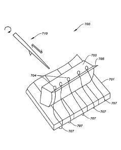
[0100] FIG. 7 illustrates a cartridge-pillbox assembly 700 according to some
embodiments.
Cartridge-pillbox assembly 700 includes a cartridge 703, a storage package
structure 701, and
a plurality of probes 707.
[0101] Cartridge 703 may be substantially similar to cartridge 600, described
above with
respect to FIGS. 6A and 6B. Cartridge 703 may include engagement features for
removable
attachment to a robotic arm (as shown in FIG. 6; not pictured in FIG. 7).
[0102] Storage package structure 701 may be a structure for containing
circuitry and/or
organizing connections for a set of probes 707. Storage package structure 701
may be similar
to storage package structure 423 of FIG. 4 and/or storage package structure
506 of FIG. 5.
As described above with respect to FIG. 6, storage package structure 701 and
cartridge 703
may be removably attached to one another, e.g., using magnetic and/or
mechanical
attachment means. In particular, Storage package structure 701 is shown with
cartridge 703
mounted on a top surface of storage package structure 701.
[0103] Probes 707 may be wires dispersed with electrodes for implantation into
biological
tissue, as described above with respect to probes 421 FIG. 4 and 504 of FIG.
5. Probes 707
may be removably coupled to cartridge 703. Probes 707 may be mounted on
cartridge 703 in
a position ready to be engaged with by an needle and implanted into biological
tissue.
[0104] Cartridge 703 may include a projected edge 705 that extends a distance
away from
the main body of the cartridge. Probes 707 may extend out of storage package
structure 701
and be arranged such that a receiving feature of each probe 707 is mounted on
projected edge
21
CA 03112875 2021-03-15
WO 2020/056169
PCT/US2019/050858
705 so as to present a receiving feature 704 (e.g., a loop) of each probe 707
in an engageable
position. In other words, an electrode-loaded filament-like wire is positioned
on projected
edge 705 of cartridge 703 such that a receiving feature 704 on an end of the
wire can be
engaged with by another structure, such as an needle.
____________________________________________________________________ [0105]
Projected edge 705 may extend outward from an upper surface of cai liidge
703 at
an angle that is arranged to best present the receiving portions of each probe
707. In some
implementations, projected edge 705 extends out at a 370 angle (relative to
the upper surface
of cartridge 303). In some aspects, projected edge 705 may extend out at an
angle that is
from about 27 to about 470 (relative to the upper surface of the cartridge).
[0106] FIG. 7 further illustrates a needle 710 that can be manipulated by a
robotic arm
system to reversibly engage with probe 707. In particular, a point of needle
710 can pass
through receiving feature 704 of probe 707, where the angle and force of
needle 710 as
needle 710 passes through receiving feature 704 can pull probe 707 away from
cartridge 703.
In further aspects, needle 710 may include an engagement feature, such as a
step, shelf, ledge,
prong, or other such structure, that is configured to physically couple with
(e.g. catch, hold,
or secure) the receiving feature of probe 707. In implementation, needle 710
may serially
engage with each probe 707, drive probe 707 in a direction (e.g., downward
into tissue), and
then disengage from probe 707 and retract back to a pre-engagement position.
Through this
process, a robotically controlled needle 710 can repeatedly, and in any
sequence, engage with
one or more probes 707 of a probe device assembly as mounted on projected edge
705 of
cartridge 703.
[0107] In some embodiments, probes 707 are removably adhered or coupled to a
temporary
attachment surface. In this context, the term adhere can be used to indicate
that the probes are
loosely associated with the flexible backing sheet such that they can be
removed from the
flexible backing sheet by an engaged needle. The probes 707 can be adhered to
the flexible
backing sheet in such a way that they remain associated with the sheet in an
organized
manner (e.g., with regular spacing forming an array of probes 707) until a
probe 707 is
engaged with needle 710 and peeled (delaminated) from the flexible backing
sheet. In other
words, needle 710 may engage with and pull probe 707 with sufficient
mechanical force to
overcome the strength of the adhesion between probe 707 and the flexible
backing sheet
without disturbing other probes 707 which are still affixed to the backing
sheet or already
22
CA 03112875 2021-03-15
WO 2020/056169 PCT/US2019/050858
implanted in tissue. In such implementations, needle 710 can be moved to catch
the
receiving element of probe 707 and to move with a peeling motion to pull probe
707 off of
the flexible backing sheet without damaging probe 707.
[0108] In some embodiments, probes 707 can adhere to the flexible backing
sheet by way
of being deposited on a thin film. The flexible backing sheet may be a
flexible material that
forms a thin film, such as parylene, a parylene-based polymer, and/or the
like. Such a thin
film may be attached to the cartridge via an adhesive layer. In some
embodiments, the
flexible backing sheet can include one or more dielectric layers to facilitate
release of probes
707 from the flexible backing sheet (e.g., in proximity to the electrodes). In
some
embodiments, the flexible backing sheet can be bonded or adhered to a solid
support (e.g.,
stainless steel such as magnetic stainless steel) that permits handling by a
machine and/or
human. As another example, probes 707 may be deposited on a silicon backing
such that the
probes can be peeled off of the silicon backing. The silicon backing may be
bonded to a
substrate such as a black glass slide. The substrate may be bonded (e.g., with
an adhesive
layer) to the cartridge. Alternatively, or additionally, an adhesive substance
can be used to
adhere the probes to the flexible backing sheet.
101091 The organized adherence of probes 707 to the flexible backing sheet can
enable the
use of robotic surgery techniques. As discussed above, computer vision
techniques can be
used to guide needle 710 to engage with probe 707 and remove probe 707 from
cartridge 703
in an organized fashion.
[0110] In some embodiments, a cartridge-pillbox assembly, includes (a) a
plurality of
probes that each have: (i) a biocompatible substrate (e.g., the wire 408 of
FIG. 4); (ii) at least
one electrode disposed on the biocompatible substrate; and (iii) a receiving
feature configured
for reversible engagement with a corresponding engagement feature of an
needle, and (b) a
flexible backing sheet to which the plurality of probes is adhered. The
cartridge-pillbox
assembly may further include a pillbox or storage package structure housing
electronics and
removably attached to the cartridge.
[0111] In some embodiments, the cartridge-pillbox assembly may be manufactured
as a
single unit. Upon implantation of the a probe device, the cartridge may be
removed from
cartridge arm 320 and replaced with a new cartridge-pillbox assembly.
23
CA 03112875 2021-03-15
WO 2020/056169 PCT/US2019/050858
PROBE SELECTION AND MANIPULATION
[0112] FIGS. 8A ¨ 8D illustrate engaging a needle with a probe and removing
the probe
from a cartridge. FIGS. 8A and 8B show two views of a needle assembly 801
preparing to
engage with a probe 802. Probe 802 includes a receiving feature 803. Needle
assembly 801
includes a needle 804 and a pincher 807. FIGS. 8C and 8D show two views of
needle
assembly 801 upon engagement with probe 802.
[0113] In FIGS. 8A and 8B, probes 802 are disposed on temporary attachment
surface 805
(e.g., a thin film of parylene or silicon, as described above with respect to
FIG. 7). Needle
assembly 801 may be guided towards receiving feature 803. As described above
with respect
to FIG. 2, the needle assembly may be attached to an insertion arm (e.g.,
insertion arm 230)
which may be coupled to a microprocessor controller that controls motion of
the insertion
arm and attached needle assembly. Given that the needle 804, receiving feature
803, and
probe 802 may be on the micron scale, specialized computer vision techniques
may be used
to guide the needle 804 to engage with the probe 802 (e.g., using light pipe
assemblies 306
and visualization devices 204, 206, 302, 304, as described above with respect
to FIGS. 2 and
3). Such computer vision techniques are described in detail in the '520
application.
[0114] In FIGS. 8C and 8D, needle 804 on needle assembly 801 engages with
receiving
feature 803 of a selected probe 802. Needle 804 may hook onto the receiving
feature 803 to
reversibly engage needle 804 to the probe 802. Pincher 807 may pinch receiving
feature 803
against needle 804 to secure probe 802 to needle 804. In order to pinch
receiving feature 803
against needle 804, pincher 807 may rotate. In some embodiments, pincher 807
extends from
needle assembly 801 as needle 801 is inserted into receiving feature 803.
[0115] When needle assembly 801 engages with probe 802 and moves upward,
exerting an
upward force on probe 802, probe 802 disengages from temporary attachment
surface 805
and peels off of temporary attachment surface 805. At this point, a selected
probe 802 is
attached to needle 804, forming a loaded needle (i.e., a needle that is
reversibly engaged with
a receiving feature of a probe). At this point, the needle is loaded with the
probe 802 and
ready to implant probe 802 in a target such as biological tissue.
24
CA 03112875 2021-03-15
WO 2020/056169 PCT/US2019/050858
IMPLANTATION METHODS
101161 FIGS. 9A ¨ 9K illustrate example steps involved in a surgical process
that implants
probe devices such as those described herein. While the illustrated example is
related to a
neurosurgical process that implants probes of a probe device assembly in the
brain, the
systems and processes described herein can be used with any suitable
biological tissue.
[0117] FIG. 9A shows a skull 900. Skull 900 may initially be prepared for
implantation.
In some embodiments, a craniotomy may be performed prior to implantation, as
shown in
FIG. 9B. A craniotomy may be suitable for implantation of relatively large
probe device
assembly 400 shown in FIG. 4. Alternatively, to implant a smaller probe device
assembly
such as probe device assembly 500 shown in FIG. 5, one or more holes may be
drilled into
skull 900.
[0118] FIG. 9B shows a skull with a head plate 901 attached thereupon. As
illustrated in
FIG. 9B, a head plate 901 can be positioned on top of skull 900. In some
embodiments, skull
900 can be mounted to a surgical stage (not shown). Head plate 901 can provide
an
organizational structure onto which probe device assemblies such as the one
depicted in FIG.
4 can be attached. In some embodiments, the head plate 901 can be made of
titanium or
similar materials. Head plate 901 can be specially sized to the skull 900
using information
obtained from computed tomography (CT) scans, magnetic resonance imaging (MRI)
scans,
and the like. Alternatively, to implant a smaller probe device as shown in
FIG. 5, no head
plate, or a much smaller head plate, may be implemented.
101191 FIG. 9C illustrates an early stage of a neurosurgical process. A needle
assembly
911 is guided to an initial position in proximity to a target implantation
region. Needle
assembly 911 may be mounted to and moved by a first robotic arm 906 (e.g.
insertion arm
230 of FIG. 2). A cartridge 903 releasably coupled to probe device assembly
905 may also
be guided to an initial position in proximity to the target implantation
region. Cartridge 903
may be mounted to and moved by a second robotic arm 907 (e.g., cartridge arm
320 of FIG.
3). As described above with respect to FIGS. 2 and 3, motion of first robotic
arm 906 and
second robotic arm 907 may be controlled by separate respective microprocessor
controllers
and/or a shared microprocessor controller.
CA 03112875 2021-03-15
WO 2020/056169 PCT/US2019/050858
[0120] FIG. 9D illustrates the neurosurgical process at a second time when the
first robotic
arm and second robotic arm have moved to begin the implantation process. Probe
device
assembly 905 has been secured to head plate 901. This may be accomplished by
mechanically latching or bracketing probe device assembly 905 on head plate
901. Robotic
arms 906 and 907 can be guided at least in part by computer vision techniques
involving
visualization devices 909 (e.g., cameras, photodetectors, photomultiplier
tubes, etc.).
[0121] As illustrated in FIG. 9D, the needle from the needle assembly 911 can
engage with
the receiving features of a probe located along the cartridge 903, implant the
probe into the
biological tissue (e.g., brain), and then disengage with the implanted probe.
The process can
be repeated, serially across cartridge 903 for all of the probes of a given
probe device
assembly 905, and sequentially for subsequent cartridges 903 coupled with
subsequent probe
device assemblies 905. In some embodiments, the probe can be driven one to two
millimeters (1 ¨ 3 mm) into the biological tissue. In this manner, the needle
assembly 911
can be used to implant a set of probes having receiving features assembled on
the cartridge
903. It should be understood that the receiving features of a probe can be
considered as
reciprocal engagement features to an engagement feature that is part of the
structure of an
needle.
[0122] In some embodiments, the implantation of the probe into the biological
tissue can
include the use of a "touch-down" sensor (described above with respect to FIG.
2). The
touch-down sensor can be deployed from the needle assembly 911 and then
retracted into the
needle-pincher assembly when the presence of the biological tissue is
detected. The needle
can then be configured to implant the probe upon retraction of the touch-down
sensor. The
touch-down sensor can be retracted and the needle can implant the probe in
less than
approximately one tenth of a second (<0.1 sec). The touch-down sensor can be
used for
.. improved targeting along the Z-axis. Computer vision techniques (e.g., as
described in the
520 application) can provide targeting along the X-axis and Y-axis of the
target biological
tissue.
[0123] Once the probes from a pillbox-cartridge assembly are implanted in the
biological
tissue, needle assembly 911 can travel out of the area of the head plate 901
as is illustrated in
FIG. 9E.
26
CA 03112875 2021-03-15
WO 2020/056169 PCT/US2019/050858
[0124] As illustrated in FIG. 9F, cartridge 903 can separate from probe device
assembly
905. In some embodiments, the separation can involve releasing the magnetic
hold between
probe device assembly 905 and cartridge 903. In some such aspects, the
magnetic hold can
be released by lowering a surgical stage on which the biological tissue is
placed. The
.. physical distance between probe device assembly 905 and cartridge 903 may
be increased
until the magnetic attraction between probe device assembly 905 and cartridge
903 is not
strong enough to retain their physical coupling. In other aspects, the
magnetic hold can be
released by lifting cartridge 903 vertically. In further embodiments, a
combination of the two
techniques can be used.
[0125] As illustrated in FIG. 9G, the process described above can be repeated
with a
subsequent cartridge-pillbox assembly, where a new cartridge-pillbox assembly
is loaded
onto the second robotic arm 907. After securing a first probe device assembly
905 to the
head plate 901 and implanting the probe(s), the remaining cartridge 903 on the
second robotic
arm 907 can be removed, and then replaced with a new paired cartridge 903 and
probe device
assembly 905 set for the next implantation cycle. In some embodiments, one or
more needles
can be used to implant a plurality of probes coupled to one or more
cartridges.
[0126] FIG. 9H illustrates a second probe device assembly 905 being positioned
for
implantation. FIG. 91 illustrates how a plurality of probe device assemblies
905 can appear
once their respective probes are implanted.
[0127] As illustrated in FIG. 9J, the process illustrated for the left
hemisphere of the brain
in FIGS. 9C ¨ 91 can be repeated on the right hemisphere of the brain. FIG. 9K
illustrates
how a plurality of probe device assemblies 905 can extend from the head plate
once the
probes are implanted in and on both sides of the brain.
IMPLANTED DEVICES
[0128] FIGS. 10A ¨ 10C illustrate implanted probe device assemblies 1003
according to
some embodiments. FIG. 10A is a first view of a cartridge 1001 and probe
device assemblies
1003 post-implantation. FIG. 10B is a second view of cartridge 1001 and probe
device
assemblies 1003 post-implantation, and FIG. 10C is a third view of cartridge
1001 and probe
device assemblies 1003 post-implantation.
[0129] As shown in FIGS. 10A ¨ 10C, cartridge 1001 has disengaged from probe
device
assemblies 1003. The probes have been implanted in the biological tissue. This
can be
27
CA 03112875 2021-03-15
WO 2020/056169 PCT/US2019/050858
understood in accordance with the techniques discussed in connection with FIG.
9A ¨ 9K and
FIG. 11. In particular, FIG. 10C represents the cartridge 1001 in a wireframe
rendering to
show the relative arrangement of the installed probe device assemblies 1003
beneath the
cartridge 1001.
[0130] As pictured, each probe device assembly 1003, or a collection of probe
device
assemblies 1003, may include an antenna configured to relay data, electricity,
or other
signals. Alternatively, or additionally, probe device assemblies 1003 may be
coupled to one
or more cables leading out of the skull and skin of a subject (not pictured).
A probe device
assembly 1003, or a collection of probe device assemblies 1003, may include or
be coupled
to a communications port arranged to be exposed outside of the biological
tissue and
configured to relay data, electricity, or other signals. As an example, each
probe device
assembly 1003 may be coupled to a wire, and the wires may connect to lead out
to a
communications port (e.g., USB C cable) disposed outside of the skin and skull
of the
subject. In either case, the data may be relayed to a computing device for
analyzing data
gathered by the probe device assemblies 1003.
[0131] FIG. 11 illustrates implanted probe device assemblies according to
other
embodiments. The probe device assemblies of FIG. 11 may correspond to the
probe device
assembly 500 shown in and described above with respect to FIG. 5.
[0132] A plurality of storage package structures 1103 may be positioned on or
above tissue
proximate to the implantation area. For example, storage package structure
1103 may rest on
the tissue or be affixed to the skull above the biological tissue implantation
area.
Accordingly, in some embodiments, the entire probe device assembly (e.g.,
storage package
structure 1103 and one or more probes, which are not visible in FIG. 11) may
be implanted
below the skin of a subject. Storage package structures 1103 may hold one or
more chips,
and the chips may be coupled to probes implanted in the tissue (not pictured
in FIG. 11).
[0133] The storage package structures 1103 may be coupled to respective
connecting leads
1107. Connecting leads 1107 may, for example, be wires or flexible cable.
Connecting leads
1107 may communicatively couple the storage package structures 1103 (e.g., the
circuitry
therein) to an internal hub 1105. Internal hub 1105 may gather data from each
of the storage
package structures 1103, as obtained via the respective probes thereof.
28
CA 03112875 2021-03-15
WO 2020/056169 PCT/US2019/050858
[0134] Internal hub 1105 may wirelessly communicate with an external hub 1109.
In
another embodiment, each probe device assembly may be in wireless
communication with
external hub 1109 (e.g., without internal hub 1105). External hub 1109 may
process and/or
store the received data. External hub 1109 may further transmit all or some of
the received
data to an external computing device with which external hub 1109 is in wired
or wireless
communication. For example, external hub 1109 may be coupled to an external
computing
device via a USB cable. Alternatively, or additionally, internal hub 1105 may
be
communicatively coupled to an external device via a wired connection.
[0135] In an example embodiment, which may correspond to the probe devices
shown in
any of FIGS. 10A ¨ 11, each probe may include approximately 1 ¨ 100 electrodes
configured
to record and/or stimulate tissue. As many as 200 probes may be coupled to a
storage
package structure to form a probe device assembly. Approximately five probe
device
assemblies may be arranged on a hemisphere of a brain for a total of ten probe
device
assemblies. Accordingly, the arrangement can include tens of thousands of
electrodes.
IMPLANTATION FLOW
[0136] FIG. 12 is a flowchart illustrating a method 1200 of implanting a probe
device into
biological tissue, with various steps, or portions thereof, represented in the
disclosed
flowchart blocks.
[0137] At block 1202, the method can begin with providing a cartridge that is
removably
coupled to probes and an IC chip (e.g., the cartridge-pillbox assembly of FIG.
7). The IC
chip may be may tethered to one or more probes. The cartridge-pillbox assembly
may be
removably coupled to a robot arm (e.g., cartridge arm 320 of FIG. 3). In some
embodiments,
the storage package structure may be attached to a head plate and/or directly
to a portion of a
subject's body such as a skull.
[0138] At block 1204, the method can include selecting and targeting a probe.
For
example, as shown in FIG. 7, the cartridge-pillbox assembly 700 includes five
probes 707,
each having a respective receiving feature 704. The system may select a
particular probe, of
the plurality of probes. The system may target the selected probe for
engagement. The
system may align the needle with the receiving feature of the selected probe.
29
CA 03112875 2021-03-15
WO 2020/056169 PCT/US2019/050858
[0139] At block 1206, the method can include reversibly engaging a needle with
the
selected probe. The needle may be guided to the receiving feature of the
selected probe. In
some embodiments, computer vision techniques may be used to guide the needle
to
engagement, as described in the '520 patent. Visualization components such as
cameras and
light pipes can be used to identify position information and transmit such
information to one
or more microprocessor controllers. The microprocessor controller(s) may
control motion of
one or more robot arms (e.g., insertion arm 230 of FIG. 2 and/or cartridge arm
320 of FIG. 3)
to move an engagement component of the needle towards the selected receiving
feature. The
system may move the one or more robot arms, in some embodiments using
visualization
components (e.g., a camera, lighting devices, and/or the like), to engage the
needle with the
selected receiving feature. As an example, an insertion arm and a cartridge
arm may move in
concert to engage the probe with the needle. As another example, an insertion
arm may move
to engage with a probe of a static cartridge-pillbox assembly.
[0140] An engagement feature (e.g., a hook or the like) of the needle may
catch the
receiving feature of the probe. In some aspects, the motion of the needle
engaging with the
receiving feature of the probe device can include a partial rotation of the
needle that aids with
both the insertion of the probe device into biological tissue and the
withdrawal of the needle
once the probe device potion has been secured within the biological tissue.
More
specifically, once the needle has engaged with the probe receiving feature,
the robotic arm
controlling the motion of the needle can rotate a predetermined amount,
further securing the
probe device on the engagement feature of the needle. In various aspects, this
rotation of the
needle can be from about five to about one hundred eighty degrees (5 -180 ),
or increments
or gradients of rotation within that range. In some aspects, where the probe
device is in part
made from a wire or filament, this rotation can encourage, nudge, or guide a
portion of the
probe device to align that wire or filament alongside the needle.
101411 The engagement of the needle and receiving feature may be aided using a
pincher to
guide the probe (e.g., pincher 22 of FIG. 2). A robot arm, such as insertion
arm 230 of FIG.
2, may pull upwards to peel the probe off of the cartridge, as illustrated in
FIGS. 8A ¨ 8D.
needle
[0142] At block 1208, the method can include piercing biological tissue with
the needle
and the probe. In some implementations, the target depth of probe insertion
can be from
CA 03112875 2021-03-15
WO 2020/056169 PCT/US2019/050858
about one to three millimeters (1 ¨ 3 mm), or increments or gradients of depth
within that
range. The loaded needle may be inserted into the biological tissue by
application of a
downward force on the needle via the insertion arm.
[0143] At block 1210, the method can include withdrawing the needle while
leaving the
probe within the biological tissue. The needle may be retracted by exerting a
substantially
upward force on the needle via the insertion arm. In some embodiments, the
needle may be
retracted by a linear motor at a rapid acceleration (e.g., up to 30,000 mm/s)
to encourage
separation of the needle from the probe. Alternatively, or additionally,
torque may be applied
to encourage disengagement of the probe and needle (e.g., by twisting the
needle).
[0144] At block 1212, there may be a determination whether additional probes
are on the
cartridge. For example, the cartridge may initially be coupled to five probes
as shown in
FIG. 7, or as many as 200 probes in other embodiments. The system may, for
example, use
computer vision techniques to identify the presence or absence of probes on
the cartridge. If
probes remain on the cartridge, then the method may return to block 1204 to
implant the next
probe. If probes no longer remain on the cartridge (e.g., all probes on the
cartridge have been
implanted in the biological tissue), then the process may proceed to block
1214.
[0145] At block 1214, the IC chip is detached from the cartridge, leaving the
IC chip with
the biological tissue. "With the biological tissue" may refer to being in
contact with, or
within about 7 mm of, the biological tissue. For example, the storage package
structure may
be coupled to a head plate such that the storage package structure is
touching, or hovering
slightly over, the brain tissue. Such an implantation scheme is illustrated,
for example, in
FIG. 10A. As another example, the storage package structure may be affixed to
a skull in
proximity to brain tissue, as illustrated in FIG. 11. The probes may extend
from the storage
package structure and into the brain tissue, with the storage package
structure disposed
thereupon to hold circuitry for gathering and analyzing information retrieved
via the probes.
[0146] The cartridge may be disengaged from the storage package structure by
applying an
upward force on the cartridge and/or a downward force on the target tissue to
overcome the
magnetic attraction between the storage package structure and the cartridge.
Disengagement
of the storage package structure and cartridge is further described above with
respect to FIG.
9F.
31
CA 03112875 2021-03-15
WO 2020/056169 PCT/US2019/050858
101471 In some embodiments, additional probe device assemblies may be
implanted by
repeating steps 1202 ¨ 1214. This may result in the implantation of multiple
probe devices.
For example, in FIG. 10A, ten probe device assemblies have been implanted, and
in FIG. 11,
four probe device assemblies have been implanted. In some aspects, when
implanting a
device into a brain, the next cartridge can be on the same brain hemisphere
side as the
previous installed cartridge, or on the opposite brain hemisphere side.
101351 The systems and methods described herein may be capable of inserting
about six
probes per minute. For example, with 32 electrodes per probe, the system can
insert up to
192 electrodes per minute. Further, the needle assembly can be replaced mid-
surgery in
under a minute. Accordingly, the techniques described herein enable rapid
implantation of
hundreds or up to tens of thousands of electrodes in biological tissue.
101481 The probe devices described herein can be used for science and research
experiments, neural prostheses (e.g., brain/nerve machine interfaces) and the
treatment of
neuronal disease (e.g., deep brain stimulation for the treatment of epilepsy,
sensory recording
and/or electrical stimulation for the treatment of Alzheimer's disease,
sensory recording
and/or electrical stimulation for the treatment of Parkinson's disease, or the
like).
101491 In some embodiments, the probe device can be configured for
implantation in
biological tissue. Biological tissue may include, but is not limited to, the
brain, muscle, liver,
pancreas, spleen, kidney, bladder, intestine, heart, stomach, skin, colon and
the like.
.. Additionally, the electrode array designs may be used in connection with
any suitable
multicellular organism including, but not limited to, invertebrates,
vertebrates, fish, bird,
mammals, rodents (e.g., mice, rats), ungulates, cows, sheep, pigs, horses, non-
human
primates, and humans. Moreover, biological tissue may be ex vivo (e.g., tissue
explant), or in
vivo (e.g., the method is a surgical procedure performed on a patient).
EXAMPLE COMPUTER SYSTEM
101501 FIG. 13 illustrates an example computer system 1300 that may be used to
implement certain embodiments. For example, in some embodiments, computer
system 1300
may be used to implement any of the systems for robotically implanting a probe
device into
biological tissue described above. As shown in FIG. 13, computer system 1300
includes
various subsystems including a processing subsystem 1304 that communicates
with a number
of other subsystems via a bus subsystem 1302. These other subsystems may
include a
32
CA 03112875 2021-03-15
WO 2020/056169 PCT/US2019/050858
processing acceleration unit 1306, an I/O subsystem 1308, a storage subsystem
1318, and a
communications subsystem 1324. Storage subsystem 1318 may include non-
transitory
computer-readable storage media including storage media 1322 and a system
memory 1310.
[0151] Bus subsystem 1302 provides a mechanism for letting the various
components and
subsystems of computer system 1300 communicate with each other as intended.
Although
bus subsystem 1302 is shown schematically as a single bus, alternative
embodiments of the
bus subsystem may utilize multiple buses. Bus subsystem 1302 may be any of
several types
of bus structures including a memory bus or memory controller, a peripheral
bus, a local bus
using any of a variety of bus architectures, and the like. For example, such
architectures may
.. include an Industry Standard Architecture (ISA) bus, Micro Channel
Architecture (MCA)
bus, Enhanced ISA (EISA) bus, Video Electronics Standards Association (VESA)
local bus,
and Peripheral Component Interconnect (PCI) bus, which can be implemented as a
Mezzanine bus manufactured to the IEEE P1386.1 standard, and the like.
[0152] Processing subsystem 1304 controls the operation of computer system
1300 and
may comprise one or more processors, application specific integrated circuits
(ASICs), or
field programmable gate arrays (FPGAs). The processors may include be single
core or
multicore processors. The processing resources of computer system 1300 can be
organized
into one or more processing units 1332, 1334, etc. A processing unit may
include one or
more processors, one or more cores from the same or different processors, a
combination of
cores and processors, or other combinations of cores and processors. In some
embodiments,
processing subsystem 1304 can include one or more special purpose co-
processors such as
graphics processors, digital signal processors (DSPs), or the like. In some
embodiments,
some or all of the processing units of processing subsystem 1304 can be
implemented using
customized circuits, such as application specific integrated circuits (ASICs),
or field
.. programmable gate arrays (FPGAs),
[0153] In some embodiments, the processing units in processing subsystem 1304
can
execute instructions stored in system memory 1310 or on computer readable
storage media
1322. In various embodiments, the processing units can execute a variety of
programs or
code instructions and can maintain multiple concurrently executing programs or
processes.
.. At any given time, some or all of the program code to be executed can be
resident in system
memory 1310 and/or on computer-readable storage media 1322 including
potentially on one
33
CA 03112875 2021-03-15
WO 2020/056169 PCT/US2019/050858
or more storage devices. Through suitable programming, processing subsystem
1304 can
provide various functionalities described above. In instances where computer
system 1300 is
executing one or more virtual machines, one or more processing units may be
allocated to
each virtual machine.
[0154] In certain embodiments, a processing acceleration unit 1306 may
optionally be
provided for performing customized processing or for off-loading some of the
processing
performed by processing subsystem 1304 so as to accelerate the overall
processing performed
by computer system 1300.
[0155] I/O subsystem 1308 may include devices and mechanisms for inputting
information
to computer system 1300 and/or for outputting information from or via computer
system
1300. In general, use of the tei in input device is intended to include all
possible types of
devices and mechanisms for inputting information to computer system 1300. User
interface
input devices may include, for example, a keyboard, pointing devices such as a
mouse or
trackball, a touchpad or touch screen incorporated into a display, a scroll
wheel, a click
wheel, a dial, a button, a switch, a keypad, audio input devices with voice
command
recognition systems, microphones, and other types of input devices. User
interface input
devices may also include motion sensing and/or gesture recognition devices
such as the
Microsoft Kinect motion sensor that enables users to control and interact
with an input
device, the Microsoft Xbox 360 game controller, devices that provide an
interface for
receiving input using gestures and spoken commands. User interface input
devices may also
include eye gesture recognition devices such as the Google Glass blink
detector that detects
eye activity (e.g., "blinking" while taking pictures and/or making a menu
selection) from
users and transforms the eye gestures as inputs to an input device (e.g.,
Google Glass ).
Additionally, user interface input devices may include voice recognition
sensing devices that
enable users to interact with voice recognition systems (e.g., Sin navigator)
through voice
commands.
[0156] Other examples of user interface input devices include, without
limitation, three
dimensional (3D) mice, joysticks or pointing sticks, gamepads and graphic
tablets, and
audio/visual devices such as speakers, digital cameras, digital camcorders,
portable media
players, webcams, image scanners, fingerprint scanners, barcode reader 3D
scanners, 3D
printers, laser rangefinders, and eye gaze tracking devices. Additionally,
user interface input
34
CA 03112875 2021-03-15
WO 2020/056169 PCT/US2019/050858
devices may include, for example, medical imaging input devices such as
computed
tomography, magnetic resonance imaging, position emission tomography, and
medical
ultrasonography devices. User interface input devices may also include, for
example, audio
input devices such as MIDI keyboards, digital musical instruments and the
like.
[0157] In general, use of the term output device is intended to include all
possible types of
devices and mechanisms for outputting information from computer system 1300 to
a user or
other computer. User interface output devices may include a display subsystem,
indicator
lights, or non-visual displays such as audio output devices, etc. The display
subsystem may
be a cathode ray tube (CRT), a flat-panel device, such as that using a liquid
crystal display
(LCD) or plasma display, a projection device, a touch screen, and the like.
For example, user
interface output devices may include, without limitation, a variety of display
devices that
visually convey text, graphics and audio/video information such as monitors,
printers,
speakers, headphones, automotive navigation systems, plotters, voice output
devices, and
modems.
[0158] Storage subsystem 1318 provides a repository or data store for storing
information
and data that is used by computer system 1300. Storage subsystem 1318 provides
a tangible
non-transitory computer-readable storage medium for storing the basic
programming and data
constructs that provide the functionality of some embodiments. Storage
subsystem 1318 may
store software (e.g., programs, code modules, instructions) that when executed
by processing
subsystem 1304 provides the functionality described above. The software may be
executed
by one or more processing units of processing subsystem 1304. Storage
subsystem 1318 may
also provide a repository for storing data used in accordance with the
teachings of this
disclosure.
[0159] Storage subsystem 1318 may include one or more non-transitory memory
devices,
including volatile and non-volatile memory devices. As shown in FIG. 13,
storage subsystem
1318 includes a system memory 1310 and a computer-readable storage media 1322.
System
memory 1310 may include a number of memories including a volatile main random
access
memory (RAM) for storage of instructions and data during program execution and
a non-
volatile read only memory (ROM) or flash memory in which fixed instructions
are stored. In
some implementations, a basic input/output system (BIOS), containing the basic
routines that
help to transfer information between elements within computer system 1300,
such as during
CA 03112875 2021-03-15
WO 2020/056169 PCT/US2019/050858
start-up, may typically be stored in the ROM. The RAM typically contains data
and/or
program modules that are presently being operated and executed by processing
subsystem
1304. In some implementations, system memory 1310 may include multiple
different types
of memory, such as static random access memory (SRAM), dynamic random access
memory
(DRAM), and the like.
[0160] By way of example, and not limitation, as depicted in FIG. 13, system
memory
1310 may load application programs 1312 that are being executed, which may
include
various applications such as Web browsers, mid-tier applications, relational
database
management systems (RDBMS), etc., program data 1314, and an operating system
1316. By
way of example, operating system 1316 may include various versions of
Microsoft
Windows , Apple Macintosh , and/or Linux operating systems, a variety of
commercially-
available I.JNIX or UNIX-like operating systems (including without limitation
the variety of
GNU/Linux operating systems, the Google Chrome OS, and the like) and/or
mobile
operating systems such as i0S, Windows Phone, Android OS, BlackBerry OS,
Palm OS
operating systems, and others.
[0161] Computer-readable storage media 1322 may store programming and data
constructs
that provide the functionality of some embodiments. Computer-readable media
1322 may
provide storage of computer-readable instructions, data structures, program
modules, and
other data for computer system 1300. Software (programs, code modules,
instructions) that,
when executed by processing subsystem 1304 provides the functionality
described above,
may be stored in storage subsystem 1318. By way of example, computer-readable
storage
media 1322 may include non-volatile memory such as a hard disk drive, a
magnetic disk
drive, an optical disk drive such as a CD ROM, DVD, a Blu-Ray disk, or other
optical
media. Computer-readable storage media 1322 may include, but is not limited
to, Zip
drives, flash memory cards, universal serial bus (USB) flash drives, secure
digital (SD) cards,
DVD disks, digital video tape, and the like. Computer-readable storage media
1322 may also
include, solid-state drives (SSD) based on non-volatile memory such as flash-
memory based
SSDs, enterprise flash drives, solid state ROM, and the like, SSDs based on
volatile memory
such as solid state RAM, dynamic RAM, static RAM, DRAM-based SSDs,
magnetoresistive
RAM (MRAM) SSDs, and hybrid SSDs that use a combination of DRAM and flash
memory
based SSDs.
36
CA 03112875 2021-03-15
WO 2020/056169 PCT/US2019/050858
[0162] In certain embodiments, storage subsystem 1318 may also include a
computer-
readable storage media reader 1320 that can further be connected to computer-
readable
storage media 1322. Reader 1320 may receive and be configured to read data
from a
memory device such as a disk, a flash drive, etc.
[0163] In certain embodiments, computer system 1300 may support virtualization
technologies, including but not limited to virtualization of processing and
memory resources.
For example, computer system 1300 may provide support for executing one or
more virtual
machines. In certain embodiments, computer system 1300 may execute a program
such as a
hypervisor that facilitated the configuring and managing of the virtual
machines. Each virtual
machine may be allocated memory, compute (e.g., processors, cores), I/O, and
networking
resources. Each virtual machine generally runs independently of the other
virtual machines.
A virtual machine typically runs its own operating system, which may be the
same as or
different from the operating systems executed by other virtual machines
executed by
computer system 1300. Accordingly, multiple operating systems may potentially
be run
concurrently by computer system 1300.
[0164] Communications subsystem 1324 provides an interface to other computer
systems
and networks. Communications subsystem 1324 serves as an interface for
receiving data
from and transmitting data to other systems from computer system 1300. For
example,
communications subsystem 1324 may enable computer system 1300 to establish a
communication channel to one or more client devices via the Internet for
receiving and
sending information from and to the client devices. For example, the
communication
subsystem may be used to receive speech input from a client device and send a
value to the
client device in response.
[0165] Communication subsystem 1324 may support both wired and/or wireless
communication protocols. For example, in certain embodiments, communications
subsystem
1324 may include radio frequency (RF) transceiver components for accessing
wireless voice
and/or data networks (e.g., using cellular telephone technology, advanced data
network
technology, such as 3G, 4G or EDGE (enhanced data rates for global evolution),
Wi-Fi
(IEEE 802.XX family standards, or other mobile communication technologies, or
any
combination thereof), global positioning system (GPS) receiver components,
and/or other
37
CA 03112875 2021-03-15
WO 2020/056169 PCT/US2019/050858
components. In some embodiments communications subsystem 1324 can provide
wired
network connectivity (e.g., Ethernet) in addition to or instead of a wireless
interface.
[0166] Communication subsystem 1324 can receive and transmit data in various
forms.
For example, in some embodiments, in addition to other forms, communications
subsystem
1324 may receive input communications in the form of structured and/or
unstructured data
feeds 1326, event streams 1328, event updates 1330, and the like. For example,
communications subsystem 1324 may be configured to receive (or send) data
feeds 1326 in
real-time from users of social media networks and/or other communication
services such as
Twitter feeds, Facebook updates, web feeds such as Rich Site Summary (RSS)
feeds,
and/or real-time updates from one or more third party information sources.
[0167] In certain embodiments, communications subsystem 1324 may be configured
to
receive data in the form of continuous data streams, which may include event
streams 1328 of
real-time events and/or event updates 1330, that may be continuous or
unbounded in nature
with no explicit end. Examples of applications that generate continuous data
may include, for
example, sensor data applications, financial tickers, network performance
measuring tools
(e.g. network monitoring and traffic management applications), clickstream
analysis tools,
automobile traffic monitoring, and the like.
[0168] Communications subsystem 1324 may also be configured to communicate
data
from computer system 1300 to other computer systems or networks. The data may
be
communicated in various different forms such as structured and/or unstructured
data feeds
1326, event streams 1328, event updates 1330, and the like to one or more
databases that may
be in communication with one or more streaming data source computers coupled
to computer
system 1300.
[0169] Computer system 1300 can be one of various types, including a handheld
portable
device (e.g., an iPhone cellular phone, an iPaci computing tablet, a PDA), a
wearable
device (e.g., a Google Glass head mounted display), a personal computer, a
workstation, a
mainframe, a kiosk, a server rack, or any other data processing system. Due to
the ever-
changing nature of computers and networks, the description of computer system
1300
depicted in FIG. 13 is intended only as a specific example. Many other
configurations having
.. more or fewer components than the system depicted in FIG. 13 are possible.
Based on the
38
CA 03112875 2021-03-15
WO 2020/056169 PCT/US2019/050858
disclosure and teachings provided herein, a person of ordinary skill in the
art will appreciate
other ways and/or methods to implement the various embodiments.
101701 It should be appreciated that the robotic system handling, coupling
with, and
engaging with one or more portions of a probe device can include a control
system (or
microprocessor controller) having one or more microprocessors/processing
devices that can
further be a component of the overall system. The control system can be local
or remote to
the robotic system, and can also include a display interface and/or
operational controls
configured to be handled by a user to alter the program of the robotic arm, to
visualize the
probe device, to visualize biological tissue into which the probe device is
being inserted, and
change configurations of the robotic device, and sub-portions thereof. Such
processing
devices can be communicatively coupled to a non-volatile memory device via a
bus. The
non-volatile memory device may include any type of memory device that retains
stored
information when powered off. Non-limiting examples of the memory device
include
electrically erasable programmable read-only memory ("ROM"), flash memory, or
any other
type of non-volatile memory. In some aspects, at least some of the memory
device can
include a non-transitory medium or memory device from which the processing
device can
read instructions. A non-transitory computer-readable medium can include
electronic,
optical, magnetic, or other storage devices capable of providing the
processing device with
computer-readable instructions or other program code. Non-limiting examples of
a non-
transitory computer-readable medium include (but are not limited to) magnetic
disk(s),
memory chip(s), ROM, random-access memory ("RAM"), an ASIC, a configured
processor,
optical storage, and/or any other medium from which a computer processor can
read
instructions. The instructions may include processor-specific instructions
generated by a
compiler and/or an interpreter from code written in any suitable computer-
programming
language, including, for example, C, C++, C#, Java, Python, Perl, JavaScript,
etc.
101711 While the above description describes various embodiments of the
invention and the
best mode contemplated, regardless how detailed the above text, the invention
can be
practiced in many ways. Details of the system may vary considerably in its
specific
implementation, while still being encompassed by the present disclosure. As
noted above,
particular terminology used when describing certain features or aspects of the
invention
should not be taken to imply that the terminology is being redefined herein to
be restricted to
any specific characteristics, features, or aspects of the invention with which
that terminology
39
CA 03112875 2021-03-15
WO 2020/056169 PCT/US2019/050858
is associated. In general, the terms used in the following claims should not
be construed to
limit the invention to the specific examples disclosed in the specification,
unless the above
Detailed Description section explicitly defines such terms. Accordingly, the
actual scope of
the invention encompasses not only the disclosed examples, but also all
equivalent ways of
practicing or implementing the invention under the claims.
[0172] The teachings of the invention provided herein can be applied to other
systems, not
necessarily the system described above. The elements and acts of the various
examples
described above can be combined to provide further implementations of the
invention. Some
alternative implementations of the invention may include not only additional
elements to
those implementations noted above, but also may include fewer elements.
Further any
specific numbers noted herein are only examples; alternative implementations
may employ
differing values or ranges, and can accommodate various increments and
gradients of values
within and at the boundaries of such ranges.
[0173] References throughout the foregoing description to features,
advantages, or similar
language do not imply that all of the features and advantages that may be
realized with the
present technology should be or are in any single embodiment of the invention.
Rather,
language referring to the features and advantages is understood to mean that a
specific
feature, advantage, or characteristic described in connection with an
embodiment is included
in at least one embodiment of the present technology. Thus, discussion of the
features and
advantages, and similar language, throughout this specification may, but do
not necessarily,
refer to the same embodiment. Furthermore, the described features, advantages,
and
characteristics of the present technology may be combined in any suitable
manner in one or
more embodiments. One skilled in the relevant art will recognize that the
present technology
can be practiced without one or more of the specific features or advantages of
a particular
embodiment. In other instances, additional features and advantages may be
recognized in
certain embodiments that may not be present in all embodiments of the present
technology.