Note: Descriptions are shown in the official language in which they were submitted.
CA 03113125 2021-03-17
WO 2020/061696
PCT/CA2019/051371
Magnetic Separation
Field
This invention relates to devices, methods and systems for automated magnetic
separation of a target from a biological sample. Such devices, methods and
systems find use in
a variety of clinical and laboratory settings.
Background
Magnetic separation has been utilised as a method to separate magnetic
impurities from
fluids through the application of a variety of different processes (U.S.
3,985,646; U.S.
4,054,513; and U.S. 5,137,629). Magnetic separation techniques have also been
applied to the
separation of populations of biological materials using magnetic beads that
have been coated
with antibodies or polymers to bind to various biological targets, including
viruses, bacteria,
and cells (U.S. 3,970,518; U.S. 4,219,411; U.S. 4,795,698; and U.S.
5,385,707). The biological
target can then be extracted from the fluid suspension using one of the
previously developed
magnetic separation devices described for example in U.S. 4,710,472; U.S.
5,691,208; U.S.
6,193,892; and Zborowski et al., (Journal of Magnetism and Magnetic Materials,
vol. 194, pp.
224-230, 1999). The magnetic field generated in the separation device applies
a force on the
magnetic beads suspended within, which can draw the bead out of fluid
suspension, as
described in Shevkoplyas et al., (Lab on a Chip, vol. 7, pp. 1294-1302, 2007)
and Warnke
(IEEE Transactions on Magnetics, vol. 39, issue 3, pp. 1771-1777, 2003) as
well as any
biological material bound to the magnetic bead. This allows for the desired
population to be
isolated, by either removing it from the fluid suspension (known as positive
selection), or by
removing all other populations from the fluid suspension to leave only the non-
magnetically
bound population of interest (known as negative selection). Isolation of
cells, such as T-cells
and stem cells, from heterogeneous cell populations is necessary for the
development of cell
therapies used to treat a variety of diseases.
One system utilizes static suspension within a surrounding magnet (EasySepTM
by
STEMCELL Technologies ). Other systems that are automated and use magnetic
beads to
isolate target populations are also known (AutoMACSO from Milytenyi Biotec,
and the
RoboSepTM from STEMCELLTm Technologies).
1
CA 03113125 2021-03-17
WO 2020/061696
PCT/CA2019/051371
There remains an unmet need for rapid and reliable magnetic separation of a
selected
target within a biological sample where the application of a magnetic field
may be automated,
customized and controlled for separation of the target with a desired high
yield and high purity.
Summary
The present invention provides a method for collecting a target biological
population
from a biological sample in an automated cell culture system, the method
comprising: a.
binding the target biological population to magnetic particles; b.
circulating the biological
sample through one or more fluidics pathways of the automated cell culture
system; c.
exposing the target biological population bound to the magnetic particles to a
magnetic
field gradient; d. repeating steps b-c one or more times; and e. collecting
the target biological
population bound to the magnetic particles.
Also provided herein is a method for collecting a target biological population
from a
biological sample in an automated cell culture system, the method comprising:
a. binding the
target biological population to magnetic particles; b. circulating the
biological sample through
one or more fluidics pathways of the automated cell culture system; c.
exposing the target
biological population bound to the magnetic particles to a magnetic field
gradient to capture
the target biological population bound to the magnetic particles; d.
circulating un-bound
components of the biological sample through one or more fluidics pathways of
the automated
cell culture system; e. inserting a magnetic field shield/barrier between the
target biological
population bound to the magnetic particles and the magnetic field to release
the target
biological population bound to the magnetic particles; f.
circulating the target biological
population bound to the magnetic particles through one or more fluidics
pathways of the
automated cell culture system; g. repeating steps b-f one or more times; and
h. collecting the
target biological population bound to the magnetic particles
In additional embodiments, provided herein is a method for collecting a target
biological population from a biological sample in an automated cell culture
system, the method
comprising: a. binding a non-target biological population to magnetic
particles; b. circulating
the biological sample through one or more fluidics pathways of the automated
cell culture
system; c. exposing the non-target biological population bound to the magnetic
particles to a
magnetic field gradient; d. repeating steps b-c one or more times; and e.
collecting the target
biological population;
2
CA 03113125 2021-03-17
WO 2020/061696
PCT/CA2019/051371
In still further embodiments, provided herein is a method for collecting a
target
biological population from a biological sample in an automated cell culture
system, the method
comprising: a. binding a non-target biological population to magnetic
particles; b. circulating
the biological sample through one or more fluidics pathways of the automated
cell culture
system; c. exposing the non-target biological population bound to the magnetic
particles to a
magnetic field gradient to capture the non-target biological population bound
to the magnetic
particles; d. circulating the target of the biological sample through one or
more fluidics
pathways of the automated cell culture system; e. inserting a magnetic field
shield/barrier
between the non-target biological population bound to the magnetic particles
and the magnetic
field to release the non-target biological population bound to the magnetic
particles; f.
circulating the non-target biological population bound to the magnetic
particles through one or
more fluidics pathways of the automated cell culture system; g. repeating
steps b-f one or more
times; and h. collecting the target biological population.
Brief Description of the Drawings
The following description of typical aspects described herein will be better
understood
when read in conjunction with the appended drawings. For the purpose of
illustrating the
invention, there are shown in the drawings aspects which are presently
typical. It should be
understood, however, that the invention is not limited to the precise
arrangements and
instrumentalities of the aspects shown in the drawings. It is noted that like
reference numerals
refer to like elements across different embodiments as shown in the drawings
and referred to
in the description.
The description herein will be more fully understood in view of the following
drawings:
Figure 1 shows magnetic field gradients used to move magnetic particles;
Figure 2 shows different sized magnetic beads used in cell separation
applications;
Figure 3 shows effect of bead size on net magnetic force acting on the bead in
response
to a magnetic field gradient;
Figure 4 shows different conceptual methods for producing an automatically
controlled
magnetic field;
Figure 5 shows a computational model of the ability of a high magnetic
permeability
and saturation material to block the magnetic flux density;
Figure 6 shows the incorporation of a magnetic separation tube on a cassette;
3
CA 03113125 2021-03-17
WO 2020/061696
PCT/CA2019/051371
Figure 7 shows Computer Aided Design (CAD) models of the separation tube 701
aligned with a magnet array 704 consisting of rare earth magnets with
alternating poles
702 and 703;
Figure 8 shows a CAD model assembly of the separation tube 701 aligned with
magnet
array 704, in the "on" position, placed within a magnetic field shield 801
that can be
rotated using a motor 802 and gear train 803;
Figure 9 shows a CAD assembly of the separation tube 701 aligned with the
magnet
array in the "off' position where the magnetic field shield 801 801 is between
the tube
701 and magnet array 704;
Figure 10 shows a cross section view of the assembly in the "on" or "off'
position;
Figure 11 shows a full assembly of a cassette 1101 consisting of a separation
tube 701
aligned with a cell culture instrument 1102 containing a magnetic separation
assembly
(704 and 801);
Figure 12 presents different, non-restrictive methods (parts 1201-1208) to
generate
localised high magnetic field gradients;
Figure 13 shows results obtained from working example 1 demonstrating
successful
separation of cells from magnetic beads at different flow rates;
Figure 14 shows results obtained from working example 2 demonstrating the
effect of
different flow rates on the capture and release of positively selected cells;
Figure 15 shows results from working example 3 of adding multiple passes to
the
magnetic separation process through the separation tube 701 in terms of the
capture and
release of positively selected cells;
Figure 16 shows results from working example 4 of increasing the magnetic bead
exposure to the magnetic field through the addition of a wait time in terms of
capture
and release of positively selected cells;
Figure 17 shows how various process modifications can modify the percentage of
false
positives obtained from working example 4;
Figure 18 shows results obtained from working example 5 of the purification of
mixed
cell populations flown through the separation tube 701 past a magnet array
704;
Figure 19 shows results obtained from working example 6 demonstrating how
modifying magnet 702 and 703 sizes can affect magnetic field properties
generated by
the magnet array 704 and cell capture; and
4
CA 03113125 2021-03-17
WO 2020/061696
PCT/CA2019/051371
Figure 20 shows results obtained from working example 7 on the effect of
adding a
bar/spacer 1201 to the cassette.
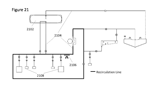
Figure 21 shows a schematic of a recirculating magnetic separation methods in
accordance with embodiments hereof
Figure 22A-22D show the recovery of purified and waste cells in accordance
with
embodiments hereof
Figure 23A-23B show the efficiency of bead release from cells in accordance
with
embodiments hereof
Figure 24A-24C show the impact of strength of contact with a magnetic field
and
multiple cycles.
Figure 24D-24E show capture and removal of cells in a separation tube, as
described
herein.
Figure 25A-25L show the results of positive and negative selection of target
cells as
described herein.
Figure 26A-26D show the recovery of cells in an automated cell culture system
as
described herein.
Figure 27A-27B show the recovery of magnetic particles after washing in an
automated
cell culture system as described herein.
Detailed Description
All publications, patent applications, patents, and other references mentioned
herein are incorporated by reference in their entirety. The publications and
applications
discussed herein are provided solely for their disclosure prior to the filing
date of the present
application. Nothing herein is to be construed as an admission that the
present invention is
not entitled to antedate such publication by virtue of prior invention. In
addition, the
materials, methods, and examples are illustrative only and are not intended to
be limiting.
In the case of conflict, the present specification, including definitions,
will control.
Unless defined otherwise, all technical and scientific terms used herein have
the same
meaning as is commonly understood by one of skill in the art to which the
subject matter
herein belongs. As used herein, the following definitions are supplied in
order to facilitate the
understanding of the present invention.
As used herein, the articles "a" and "an" preceding an element or component
are
intended to be non-restrictive regarding the number of instances (i.e.
occurrences) of the
CA 03113125 2021-03-17
WO 2020/061696
PCT/CA2019/051371
element or component. Therefore, "a" or "an" should be read to include one or
at least one,
and the singular word form of the element or component also includes the
plural unless the
number is obviously meant to be singular.
As used herein, the terms "invention" or "present invention" are non-limiting
terms and
not intended to refer to any single aspect of the particular invention but
encompass all possible
aspects as described in the specification and the claims.
As used herein the terms 'comprises', 'comprising', 'includes', 'including',
'having'
and their inflections and conjugates denote 'including but not limited to'.
As used herein, the term "about" modifying the quantity of an ingredient,
component,
or reactant employed refers to variation in the numerical quantity that can
occur, for example,
through typical measuring and liquid handling procedures used for making
concentrates or
solutions. Furthermore, variation can occur from inadvertent error in
measuring procedures,
differences in the manufacture, source, or purity of the ingredients employed
to make the
compositions or carry out the methods, and the like. In one aspect, the term
"about" means
within 10% of the reported numerical value. In another aspect, the term
"about" means within
5% of the reported numerical value. Yet, in another aspect, the term "about"
means within 10,
9, 8, 7, 6, 5, 4, 3, 2, or 1% of the reported numerical value.
Should a range of values be recited, it is merely for convenience or brevity
and includes
all the possible sub-ranges as well as individual numerical values within and
about the
boundary of that range. Any numeric value, unless otherwise specified,
includes also practical
close values and integral values do not exclude fractional values. Sub-range
values and
practically close values should be considered as specifically disclosed
values.
It will be understood that any component defined herein as being included may
be
explicitly excluded from the claimed invention by way of proviso or negative
limitation.
As may be used herein the terms 'close', 'approximate' and 'practically'
denote a
respective relation or measure or amount or quantity or degree that has no
adverse consequence
or effect relative to the referenced term or embodiment or operation or the
scope of the
invention.
As may be used herein any terms referring to geometrical relationships such as
'vertical', 'horizontal', 'parallel', 'opposite', 'straight', "lateral",
"parallel", "perpendicular"
and other angular relationships denote also approximate yet functional and/or
practical,
respective relationships.
6
CA 03113125 2021-03-17
WO 2020/061696
PCT/CA2019/051371
As may be used herein, the terms 'preferred', 'preferably', 'typical',
'typically' or
'optionally' do not limit the scope of the invention or embodiments thereof
As may be used herein the terms 'substantial', 'appreciable' (or synonyms
thereof)
denote with respect to the context a measure or extent or amount or degree
that encompass a
large part or most of a referenced entity, or an extent at least moderately or
much greater or
larger or more effective or more important relative to a referenced entity or
with respect to the
referenced subject matter.
As may be used herein the terms 'negligible', and 'slight' (or synonyms
thereof) denote,
a sufficiently small respective relation or measure or amount or quantity or
degree to not have
practical consequences relative to the referenced term and on the scope of the
invention.
As used herein the term 'may' denotes an option or an effect which is either
or not
included and/or used and/or implemented and/or occurs, yet the option
constitutes at least a
part of some embodiments of the invention or consequence thereof, without
limiting the scope
of the invention.
As used herein a "sample" can be any sample and can be a "biological sample"
that
may be derived from plant, human, animal, or microorganism sources. The sample
is typically
a heterogeneous sample from which a target is selected for separation and
collection. Targets
may be cells, DNA, RNA, proteins, peptides, microorganisms, viruses and so
forth. A
biological sample contains a target population.
The biological sample may comprise a body fluid sample, a body cell sample or
a
biological tissue sample. Examples of biological or body fluid samples include
urine, lymph,
blood, plasma, serum, saliva, cervical fluid, cervical-vaginal fluid, vaginal
fluid, breast fluid,
breast milk, synovial fluid, semen, seminal fluid, stool, sputum, cerebral
spinal fluid, tears,
mucus, interstitial fluid, follicular fluid, amniotic fluid, aqueous humor,
vitreous humor,
peritoneal fluid, ascites, sweat, lymphatic fluid, lung sputum and lavage or
samples derived
therefrom. Biological tissue samples are samples containing an aggregate of
cells, usually of a
particular kind, together with intercellular substances that form one of the
structural materials
of a human, animal, plant, bacterial, fungal or viral structure, including
connective, epithelium,
muscle and nerve tissues. Examples of biological tissue samples also include
organs, tumors,
lymph nodes, arteries and individual cell(s). For example, the sample can be a
tissue sample
suspected of being cancerous. Biological tissue samples may be first treated
to separate
aggregates of cells.
7
CA 03113125 2021-03-17
WO 2020/061696
PCT/CA2019/051371
In embodiments, the biological sample is a blood cell, white blood cell or
platelet.
White blood cells (leukocytes) include neutrophils, lymphocytes (T cells
inclusive of T helper
cells, cytotoxic T cells, T-killer cells, Natural Killer, and B lymphocytes),
monocytes,
eosinophils, basophils, macrophages, and dendritic cells.
As used herein, "target cells" are cells typically intended for separation or
concentration
from other cells (such as for examination or diagnosis), of particular type or
having distinct
characteristics relative to other cells, such as selective mutual affinity to
couple with certain
antibodies or other compounds or other particles. In particular embodiments, a
distinct
characteristic is selective affinity to couple or bind with magnetic beads to
form magnetic target
cells.
As used herein, the term "patient sample" is defined as a biological sample
taken from
any animal for whom diagnosis, screening, monitoring or treatment is
contemplated. Animals
include mammals. A patient refers to a subject such as a mammal, primate,
human or livestock
subject afflicted with a disease condition or for which a disease condition is
to be determined
or treated. A patient sample may be the source of a source biological
population.
As used herein the term "antibody" is intended to include polyclonal and
monoclonal
antibodies of any isotype (IgA, IgG, IgE, IgD, IgM), or an antigen-binding
portion thereof,
including, but not limited to, F(ab) and Fv fragments such as sc Fv, single
chain antibodies,
chimeric antibodies, humanized antibodies, recombinant engineered antibody and
a Fab
expression library. Bispecific antibodies can also be immobilized on a
magnetic particle.
As used herein, a "label moiety" is detectable, either directly or indirectly.
The label
moiety can be a detectable label and can be used in conjunction with magnetic
particles.
Direct label moieties include radioisotopes; enzymes whose products are
detectable (e.g.,
luciferase, B-galactosidase, and the like); fluorescent labels (e.g.,
fluorescein isothiocyanate
(FITC), rhodamine, phycoerythrin, a cyanine dye, Cascade Blue, PerCP, Cy5,
Cy7,
allophycocyanin (APC), PECy5 or other tandem conjugates of different
fluorochromes,
Texas Red, and the like); fluorescence emitting metals, e.g., 1521, or others
of the lanthanide
series, attached to the protein through metal chelating groups such as EDTA;
chemiluminescent compounds, e.g., luminol, isoluminol, acridinium salts, and
the like;
bioluminescent compounds, e.g., luciferin, aequorin (green fluorescent
protein), and the like;
and metallic compounds. Indirect label moieties include labeled molecules that
bind to the
polypeptide, e.g., antibodies specific for the polypeptide, wherein the
labeled binding
molecule is labeled as described above; and members of specific binding pairs,
e.g., biotin, (a
8
CA 03113125 2021-03-17
WO 2020/061696
PCT/CA2019/051371
member of the specific binding pair biotin-avidin), digoxigenin (a member of
the specific
binding pair digoxigenin-antibody to digoxigenin) and the like. Alternatively,
the label
moiety can be any suitable label including but not limited to those described
herein.
Magnetic particles labeled with a binding partner such as an antibody, a
protein, or a
nucleic acid molecule are commercially available from Miltenyi Biotec GmbH
(Friedrich
Ebert Str. 68, D-51429 Bergisch Gladbach, Germany). Methods for magnetically
labeling a
biomolecule are known in the art; any known method can be used. For example,
U.S. Pat. No.
6,020,210 describes methods for preparation of magnetic particles, and
attachment of
biomolecules thereto. A first member of a specific binding pair can be
associated with a
magnetic particle, wherein the biomolecule to be modified comprises a moiety
that binds to
the member of the specific binding pair. Alternatively, the magnetic particle
is coupled, e.g.
to the antibody or the immunologically reactive fragment thereof, through a
linker or a spacer
(such as, e.g., a nucleic acid linker). Addition of spacers or linkers will
allow biomolecules to
be presented in a more flexible fashion, and careful chemistry can attach
ligands in a specific
orientation. There are numerous chemistries used for these couplings as many
companies
have published protocols and will help the artisan skilled in the art with the
chemistry.
Examples of members of specific binding pairs that can be attached to a
magnetic
particle include, but are not limited to, oligo dT (for binding to nucleic
acid molecules
comprising, e.g., a poly-A tract at the 3' end); oligonucleotides having a
specific nucleotide
sequence (for binding to nucleic acid molecules comprising a complementary
nucleotide
sequence); avidin (e.g., streptavidin) (for binding to a biotinylated
biomolecule); an antigen-
binding polypeptide, e.g., an immunoglobulin (Ig) or epitope-binding fragment
thereof (for
binding to a biomolecule comprising an epitope recognized by the Ig);
polynucleotide
binding proteins (for binding to a polynucleotide), e.g., a transcription
factor, a translation
factor, and the like; Ni or Co chelate (to immobilize poly-histidine-tagged
proteins); receptor-
ligand systems, or other specific protein-protein interacting pairs; aptamers
(e.g., nucleic acid
ligands for three-dimensional molecular targets); lectins (for binding
glycoproteins); lipids
and phospholipids (binding to lipid-binding proteins), e.g., phosphatidyl
serine and annexin
V. Those skilled in the art will recognize other members of specific binding
pairs that may be
attached to a magnetic particle.
A biomolecule can also be coupled (covalently or non-covalently) to a magnetic
particle by direct chemical conjugation or by physical association. Such
methods are well
known in the art. Biochemical conjugations are described in, e.g.,
"Bioconjugate Techniques"
9
CA 03113125 2021-03-17
WO 2020/061696
PCT/CA2019/051371
Greg T. Hermanson, Academic Press. Non-covalent interactions, such as ionic
bonds,
hydrophobic interactions, hydrogen bonds, and/or van der Waals attractions can
also be used
to couple a biomolecule with a magnetic particle. For example, standard non-
covalent
interactions used to bind biomolecules to chromatographic matrices can be
used. One non-
limiting example of such a non-covalent interaction that can be used to bind a
biomolecule to
a magnetic particle are DNA binding to silica in the presence of chaotropic
salts. Those
skilled in the art are aware of other such non-covalent binding and conditions
for achieving
same. See, e.g., Molecular Cloning, Sambrook and Russell, Cold Spring Harbor
Laboratory
Press.
As used herein "magnetic particles" are used as labels for biomolecule targets
in a
biological sample such as, but not limited to, antibodies, DNA, polypeptides
and cells to aid in
their separation from complex mixtures of a sample. Magnetic particles may be
classified
according to size: microbeads that are about <50 nm; nanobeads that are about
100 to about
200 nm; and dynabeads that are about 1-5 p.m. Furthermore, magnetic particles
can be adapted
for selective affinity (functionalized) for coupling or binding with a desired
biomolecule target
such as with a fluorescent label, antibody, nucleic acid and so forth.
Different magnetic particles are available from a number of sources, including
for
example, Dynal (Norway), Advanced Magnetics (Cambridge, Mass., U.S.A.),
Immuncon
(Philadelphia, U.S.A.), Immunotec (Marseilles, France), and Miltenyi Biotec
GmbH
(Germany). Preferred magnetic labeling methods include colloidal
superparamagnetic particles
in a size range of 5 to 200 nm, preferably in a size of 10 to 100 nm. These
magnetic particles
allow a quantitative magnetic labeling of cells, thus the amount of coupled
magnetic label is
proportional to the amount of bound product. Colloidal particles with various
specificities are
available, for example, through Miltenyi Biotec GmbH.
As used herein "separation" includes isolation or collection accumulation of
target cells
from a surrounding fluid bulk, where the bulk is, for example, a fluidic
mixture or suspension
of emulsion of cells or a combination thereof, implying also concentration or
enrichment of
target cells relative to the surrounding bulk or a provided sample of cells
(obtaining a
precipitate in analogy to precipitation or centrifugation).
As used herein "depletion" with respect to separation, is the removal of
target cells from
the bulk (obtaining a supernatant in analogy to precipitation or
centrifugation).
As used herein "high qualitative" (separation, depletion) is meaning high
purity,
separation of target cells substantially exclusive of other cells, or
comprising negligible
CA 03113125 2021-03-17
WO 2020/061696
PCT/CA2019/051371
amounts of other cells such as between about 10% and about 1% or less of the
separated cells,
and conversely a depletion.
As used herein "high quantitative" (separation, depletion) is meaning high
recovery,
separation of substantially all the target cells, or very high amount of the
target cells from the
sample, such as between about 80% to about 99% or more or the separated cells,
and conversely
a depletion.
It is noted that whenever a reference is made herein to cells attaching or
sticking or
adhering to a wall of a tube, or similar terms to that effect, it does not
necessarily mean that the
cells attach directly to the wall, but rather, that they also connect or link
or are attracted
indirectly to the wall such as by chains of cells or groups of cells.
As used herein "magnetic shielding" reduces and/or blocks the magnetic field
in a space
by blocking the field with a "magnetic field shield" (also referred to herein
as a magnetic field
shield/barrier, with both terms being interchangeable).
As used herein "HMPSM" denotes a high magnetic permeability and saturation
material that results in a highly concentrated magnetic field within itself
that effectively reduces
and/or eliminates the influence of the magnetic field.
As used herein "magnetic field shield/barrier" is a structure that can be
controlled with
respect to use with a magnetic field.
As used herein an "electromagnet" is a type of magnet in which the magnetic
field is
produced by an electric current. The magnetic field disappears when the
current is turned off
Electromagnets usually consist of wire wound into a coil. A current through
the wire creates a
magnetic field which is concentrated in the hole in the center of the coil.
The wire turns are
often wound around a magnetic core made from a ferromagnetic or ferrimagnetic
material such
as iron; the magnetic core concentrates the magnetic flux and makes a more
powerful magnet.
As used herein a "permanent magnet is a magnet that is permanent, in contrast
to an
electromagnet, which only behaves like a magnet when an electric current is
flowing through
it. Permanent magnets are made out of substances like magnetite (Fe304), the
most magnetic
naturally occurring mineral, or neodymium, a powerfully magnetic synthetic
substance.
As used herein "magnet array" is one or more magnets. The one or more magnets
can
be permanent magnets or electromagnets. One or more permanent magnets may be
in a linear
array, in different sizes, different strengths, configured in opposite pole
directions
perpendicular to the axis of the linear array or configured with 90 rotations
to one another in
a plane perpendicular to the axis of the linear array. Any number of magnets
in the array may
11
CA 03113125 2021-03-17
WO 2020/061696
PCT/CA2019/051371
be physically held together or adhesively held together. Permanent magnets may
be of a
material selected from iron, neodymium, samarium-cobalt or alnico.
A general non-limiting overview of the invention and practising the invention
is
presented below. The overview outlines exemplary practice of
embodiments/aspects of the
invention, providing a constructive basis for variant and/or alternative
and/or divergent
aspects/embodiments, some of which are subsequently described.
The present disclosure relates to devices, methods and systems for
magnetically
separating and collecting a desired biomolecule target in a biological sample
through positive
or negative selection. As presented herein, a magnetic field is produced that
is substantially
adjacent to a biological sample containing a desired magnetized biomolecule
target. The
magnetic field can be switched "ON" and "OFF" in an automatic manner such to
provide a
magnetic field of a desired strength, continuous time duration, intermittent
duration, pulsatile
duration and combinations thereof This is achieved by the introduction of a
magnetic field
shield (also referred to herein as a magnetic field shield/barrier) to
functionally control the
application of the magnetic field encountered/applied to the biological
sample. The magnetic
field shield/barrier is positioned between the source of the magnetic field
and the biological
sample and as a function of its high magnetic permeability and saturation
materials (HMPSM),
results in a highly concentrated magnetic field within itself that effectively
reduces and/or
eliminates the influence of the magnetic field on the biological sample
containing the
magnetized biomolecule target.
In an aspect of the invention, cellular biologic material is cultured in a
bioreactor vessel,
and a desired cell is the biomolecule target for magnetic separation and
collection.
The devices, methods and systems herein described generally employ an approach
whereby a biological sample (a heterogeneous biological population), which is
typically, but
not limited to, cells, has magnetic beads bound to a specific biomolecule
target (a specific cell
type) in the sample creating a "magnetized cell". Typical binding methods may
include: i)
direct binding of a magnetic bead that is conjugated to an antibody of the
biological target; and
ii) using a multi-step process where the biological target is bound to an
antibody that is
conjugated with another antigen or binding pair. This antigen/binding pair is
then bound to the
magnetic bead which is conjugated to the respective antibody/binding pair.
During magnetic
separation, the magnetized cells which are the target cells attached to the
magnetic beads
(expressing the antigen; positively selected) are attracted to a location near
the magnet, while
cell populations not attached to beads (negatively selected) remain in the
media of the
12
CA 03113125 2021-03-17
WO 2020/061696
PCT/CA2019/051371
biological sample and are easily removed from the bound population. An
alternative process
to magnetic cell selection is the use of antigen-presenting magnetic
microbeads to stimulate
some type of biological process on the target (such as T cell activation with
anti-CD3 and anti-
CD28 coupled to magnetic activation beads). After stimulation, the magnetic
beads must be
removed prior to downstream processing, which requires treatment of the cell
suspension with
an effective magnet.
During separation, a magnetically inclined particle experiences a force
vector, F, from
an applied magnetic field, B, acting on a paramagnetic particle as defined by
equation 1 below
(Pamme, 2006).
F (B = V)B (1)
tto
Where V is the volume of the particle, Ax is the difference in magnetic
susceptibility
(capacity to become magnetized) of the particle and the surrounding media, yo
is the
magnetic permeability of a vacuum, and B = V is the dot product between the
magnetic field
and the gradient operator. From this equation, clearly the success of a
magnetic separation
system is dependent on a number of parameters. First, particle size, where
larger particles
experience a stronger magnetic force. There are 3 typical size classifications
for magnetic
particles, i) <50 nm (e.g. MACS MicroBeads by Miltenyi Biotec), ii) 100-200
nm (e.g.
Nanobeads by BioLegend0), or iii) 1-5 p.m (e.g. Dynabeads0 by Invitrogen),
which are more
easily separated with increasing size. Next, increasing the magnetic
susceptibility of the bead
relative to the surrounding media. Since most beads typically consist of an
iron core, and the
surrounding media is practically not magnetisable, this value is typically
relatively large
already. Finally, increasing the magnetic field gradient can drastically
increase the force
applied to a magnetic bead. This is because a magnetic field gradient
generates uneven forces
on the North and South poles of a magnetic particle, due to the uneven spatial
quality of the
high-gradient field (Figure 1A). This uneven force on a particle leads to
particle movement.
In a perfectly homogeneous magnetic field, equal and opposite forces are
generated on the
two poles of the magnetic particle, leading to zero net force on the particle
and no net
movement (Figure 1B). Furthermore, with larger sized magnetic particles, the
difference of
magnetic force applied from the gradient is larger between the two poles
compared to a
smaller bead (Figure 2 and 3).
There are means to induce a "switchable" (can be turned on and off) magnetic
field
that can generate a gradient to attract magnetic beads for automated
separation, isolation and
collection. For instance, electromagnets are formed by winding a current
carrying wire
13
CA 03113125 2021-03-17
WO 2020/061696
PCT/CA2019/051371
around a rod of magnetically susceptible material (e.g. iron) (Figure 4A).
These can be
switched on or off by applying or removing the current through the wire
respectively.
Another method is an electro-permanent magnet, which consists of a hard magnet
with a high
magnetic coercivity (high magnetic field to switch the magnet poles), and a
soft magnet with
a low coercivity (Figure 4B). Both magnets are connected to each other with a
paramagnetic
material (such as iron) to complete the magnetic circuit. The soft magnet is
wrapped in a
current carrying wire and, by pulsing a strong current in the wire, the
magnetic poles of the
soft magnet can be switched. When the poles are mis-aligned, the magnetic
"current" flows
through the paramagnetic material, and no external magnetic field is observed.
However,
when the poles are aligned, the magnetic current travels through the air, and
an external
magnetic field is generated. A final method is the use of permanent magnets to
generate the
magnetic field. By using a material with a high magnetic saturation, it is
possible to block the
magnetic field on one side of the magnet (Figure 4C).
In an aspect of the invention, an array of strong permanent magnets with
alternating
orientations are used (Figure 4C). This creates strong magnetic gradients
radially from the
array, as well as gradients linearly along the axis of the array. A
modification of this design is
a Halbach array, where magnets in an array are rotated 90 with respect to
each other (Figure
4D). This induces a significant increase in the magnetic field on one side of
the magnet, while
diminishing it on the other side (Kang et al.).
The controllable magnetic field can be designed to perform the sequential
activities of
controlling the isolation of targeted biological fractions containing the
biomolecule target
(either through positive or negative selection) while simultaneously enabling
non-targeted
biological fractions to be removed and discarded.
During the processing of biological samples using magnetic separation, the
ability to
switch the magnetic field on and off, thereby enabling automation of both
positive and negative
cell selection, is a major operational requirement. The devices and methods
described herein
enable the automated collection of target biological populations thereby
reducing overall
process complexity and reducing operational costs.
The "on" and "off' switchable magnetic field described herein is by the
introduction of
a magnetic field shield/barrier to control the magnetic field encountered by a
biological sample
labelled with magnetic particles such as magnetic beads. Through this
controllable magnetic
field, the sequential steps of target biological retention followed by
secondary release and
14
CA 03113125 2021-03-17
WO 2020/061696
PCT/CA2019/051371
capture enables the production of magnetic separation systems that are very
compact and
energy efficient.
The magnetic field shield/barrier is functional by the inherent properties of
HMPSM.
This high magnetic permeability and saturation results in a highly
concentrated magnetic field
within this material. The deployment of a HMPSM as a magnetic field
shield/barrier between
the source of the magnetic field and the biological sample enables the
practical elimination of
the influence of the magnetic field on the biological sample. Through this
controllable
activation of the magnetic barrier, the magnetic separation of target
populations from biological
samples can be achieved with high reproducibility and relatively low cost.
Figure 1 presents the theory as to how magnetic particles/beads (such as those
used for
cell separation and activation) respond to a magnetic field gradient. This is
mathematically
exemplified in equation 1. Each magnetic bead has a North and South pole. When
exposed to
a magnetic field gradient, the magnetic forces (one attractive, one repulsive)
acting on each
pole of the particle will be different, resulting in a net force that can move
the particle (Figure
1A). In comparison, when there is no magnetic field gradient, the forces
acting on each pole
will be equal and opposite (Figure 1B). Therefore, no net force will be
applied to the magnetic
particle, and no motion will be induced. Arrays of magnets arranged such that
the poles are
alternated between North (N) and South (S) can be used to generate these
necessary magnetic
field gradients. Furthermore, as shown in Figure 1C, high magnetic field
gradients can be
generated by using arrays of multiple strong, small magnets arranged with
poles with
alternative North to South direction in the array.
Figure 2 presents the sizes of magnetic beads typically used for the magnetic
separation
of cells. There is a range of bead sizes that currently exist, ranging from
about 50 nm MACS
MicroBeads by Miltenyi up to about 5 p.m Dynabeads0 by Invitrogen. Increasing
bead size
typically increases the susceptibility of a bead to respond to a magnetic
field due to the
increased difference of forces acting on each pole of the bead.
Figure 3 outlines a method whereby larger-sized magnetic beads are more
efficiently
forced out of a fluid in response to an induced magnetic field. In some
embodiments, beads are
exposed simultaneously to an attractive force and a repulsive force from the
magnetic field on
each of the beads respective poles. Moving away from the magnetic field
source, the magnitude
and the gradient of the magnetic field decreases. The net force that the bead
experiences is,
therefore, dependent on the distance from the source of the magnetic field,
and the diameter of
the bead. Larger beads have a larger distance between both poles, resulting in
a larger
CA 03113125 2021-03-17
WO 2020/061696
PCT/CA2019/051371
difference in magnetic force acting on the poles. Similarly, a stronger
gradient (i.e. closer to
the magnet) scales the difference of the forces acting on the respective poles
of the bead. This
increase in net force results in more rapid attraction of the bead towards the
magnet and out of
the fluid.
Figure 4 presents methods to develop a magnetic field that can be turned
on/off with
respect to application to a biological sample using an automatic control
system. In Figure 4A,
an electromagnet is presented, where wire is wrapped around a ferromagnetic
material, such as
iron. By applying a current to the wire, the magnetic field can quickly and
easily be turned on
and, conversely, off However, the magnetic field generated from this would be
low.
Furthermore, electromagnets produce significant levels of heat, which is
significantly
problematic for the local cell culture environment. Figure 4B presents an
electro-permanent
magnet consisting of both a non-switchable rare earth magnet, and a pole
switchable Alnico
permanent magnet, both set in a ferromagnetic material forming a magnetic
circuit. When the
poles of the permanent magnets are aligned, the carbon steel adopts the same
orientation,
producing a net magnetic flux in the air surrounding the magnet (which can be
used to pull a
magnetic bead out of fluid suspension). When the poles of the two permanent
magnets are
opposite, however, the magnetic flux is confined to the ferromagnetic
material, preventing the
extraction of magnetic beads. The orientation of the Alnico magnet is switched
by applying a
very high magnitude and short duration pulse of current through the coil of
wire surrounding
it, thereby producing a high-magnitude, transient magnetic field. The magnetic
gradient
generated from such a setup would, however, also be quite low compared to a
rare earth
magnet, and this method would also produce electromagnetic interference that
could have
unknown adverse impacts on any surrounding electronics. Figure 4C presents an
array of
permanent magnets with reversing pole orientations, with a HMPSM, such as
iron, cobalt iron,
and Hiperco 50, on one side. By utilising this HMPSM, the magnetic flux of the
magnetic field
will be amplified on the opposite side, while being diminished to negligible
levels on the side
with the HMPSM. By actuating the HMPSM such that it lies between the permanent
magnets
and magnetic beads, the force acting on the beads can be reduced to
effectively zero.
Conversely, moving the HMPSM to the opposite side of the permanent magnet, a
very strong
magnet force can be induced on the bead. Finally, in Figure 4D a Halbach
linear array is
presented where 90 rotations of the magnets allows for the generation of an
amplified
magnetic field on one side of the array, while a significantly diminished or
abolished field on
the opposite side of the array.
16
CA 03113125 2021-03-17
WO 2020/061696
PCT/CA2019/051371
Figure 5 presents computational modeling demonstrating the function of the
HMPSM
(labeled x) on a permanent magnet. The magnetic flux primarily transmits
through the air
surrounding the magnetic. However, the magnetic flux is not strong enough to
transmit through
the HMPSM. This results in no significant magnetic flux density on the
opposite side of the
HMPSM to the magnet, while the flux density is primarily amplified at the two
poles of the
permanent magnet.
Figure 6 demonstrates the layout of an example cassette typically used within
an
automated cell culture system with a separation tube for magnetic separation
using magnetic
beads running along the length of the cassette face. This tube aligns with a
permanent magnet
array located within the automated cell culture system. The separation tubing
is connected to
various tubing and bags within the cassette that are used for either positive
or negative binding
of the cells of interest. The use of as long a tube as possible increases the
volume that can be
loaded into the separation tubing, thereby reducing the processing time for
magnetic separation
to occur.
Figure 7 provides an embodiment of the separation process presented in Figure
6.
Magnetically bound beads either attached or unattached to cells can be loaded
into the
separation tube 701 (Figure 7A). In some aspects the separation tube 701 is
aligned with the
permanent magnet array 704 as presented in Figure 7B. In other aspects the
separation tube
701 is aligned with an electromagnet that replaces the permanent magnet array
704. The
permanent magnet array 704 consists of permanent magnets with alternating
North (702) and
South (703) poles to generate as high a magnetic flux gradient as possible.
Magnetic beads in
the separation tube 701 are attracted by the magnet array 704, thus
successfully separating out
the magnetic beads from the fluid suspension.
Figure 8 presents an embodiment of the magnetic field shield/barrier with the
magnetic
field turned "on". The separation tube 701 runs along the length of the
permanent magnet array
704. Surrounding a portion of the permanent magnet array 704 is a magnetic
field shield 801
(shown as a paramagnetic sheath 801) produced from an HMSPM. In some aspects
the
magnetic field shield 801 is produced from pure iron. In some aspects the
magnetic field shield
801 is produced from a soft magnetic iron alloy, such as ferritic steel,
silicon iron, nickel iron,
or cobalt iron. In some aspects the magnetic field shield 801 is produced from
a soft-magnet
alloy of cobalt, vanadium, and iron such as an alloy of about 49% cobalt,
about 2% vanadium,
and the balance iron. In some aspects the sheath 801 can be produced from
Hiperco50 or
Hiperco 50A. In some aspects the paramagnetic properties of the magnetic field
shield 801
17
CA 03113125 2021-03-17
WO 2020/061696
PCT/CA2019/051371
amplifies the magnetic field gradient applied to the separation tube 701 by
the magnet array
704. This, in effect, increases the magnetic force acting on the magnetic
beads allowing them
to be more effectively removed from fluid suspension, while non-magnetic
matter will not be
affected and will be able to pass through the separation tubing unimpeded. The
permanent
magnet array 704 and magnetic field shield 801 are also connected to a servo
802 and a gear
train 803, allowing the entire assembly to be completely rotated, and turned
off and on as
needed.
Figure 9 presents the same assembly found in Figure 8, but instead with the
magnet in
the "off' position. The separation tube 701 still runs along the permanent
magnet array 704.
However, in the off position, the magnet array 704 and magnetic field shield
801 is rotated
using the rotation servo 802 and gear train 803 such that the magnetic field
shield 801 is
between the separation tube 701 and the permanent magnet 704. As described
with Figures 4
and 5, the HMPSM material used for the magnetic field shield 801 prevents the
magnetic flux
from passing through it. Because of this, the magnetic flux gradient that the
separation tube
701 is exposed to is significantly reduced, thereby losing the hold of the
magnetic field on the
magnetic beads. The application of a high flow rate of liquid or gas to the
separation tube 701
can be used to flush out the magnetic beads and magnetically bound cells.
Figure 10 is a cross-section of the interface between the separation tube 701,
the
magnetic field shield 801, and the permanent magnet array 704. When in the
"on" position, as
presented in Figure 10A, there exists a significant magnetic field acting on
separation tube 701
that can attract magnetic particles. When in the "off' position, as presented
in Figure 10B, the
magnetic field acting on the separation tube 701 is negligible, and previously
bound cells and
beads can be effectively removed.
Figure 11 is a side view of the interfacing of the cassette 1101 and the
automated cell
culture instrument 1102. The separation tube 701 is attached to the cassette
1101, allowing for
either the magnetic or non-magnetic fraction to be moved from and towards
different regions
within the cassette 1101. Conversely, the magnetic separation assembly
(consisting of 704 and
801 together) is contained within the automated cell culture instrument 1102.
Control systems
associated with the instrument 1102 can control the servo 802 and gear train
803 to rotate the
magnet array 704 and magnetic field shield 801, thereby turning the magnetic
field "on" or
"off' as it pertains to the separation tube 701. When the cassette 1101 and
instrument 1102 are
interfaced together, the separation tube 701 and separation assembly (704 and
801 together)
are aligned, allowing for effective magnetic separation to be performed.
Furthermore, using the
18
CA 03113125 2021-03-17
WO 2020/061696
PCT/CA2019/051371
peristaltic pump associated with the instrument 1102, fluids (either with or
without magnetic
beads and cells) can be transported or removed from the separation tubing 701.
Figure 12 consists of side views of the separation tube 701 situated between
the outside
wall of the cassette 1101 and the outside wall of the cell culture instrument
1102 with various
means of increasing the number of and magnitude of magnetic field gradients
within the
separation tube 701. Figure 12A presents a paramagnetic/non-magnetic spacer
1201 that runs
the length of the separation tube 701 on the outside face of the cassette
1101. This spacer 1201
acts to compress the tube 701 to minimize the distance between magnetised
elements flowing
through the tube 701 and the magnet array 704. As well, magnetisation of the
spacer 1201 (if
paramagnetic) from the magnet array 704 will result in the formation of
another magnetic field
gradient in the separation tube 701 on the side closest to the cassette 1101.
Furthermore, cutting
the spacer 1201 into smaller subsections along the length of the spacer 1201
(not shown) can
allow for the generation of high gradient magnetic fields at points at the end
of each spacer
subsection 1201. Figure 12B presents a paramagnetic wire mesh 1202 within the
separation
tube 701 that, when exposed to the magnetic field generated by the permanent
magnet array
704, generates high magnetic field gradients around the strands of the mesh
1202. Figure 12C
presents paramagnetic particles 1203 situated within the separation tube 701
that generate
localised magnetic field gradients when exposed to the magnetic field from the
permanent
magnet array 704. Figure 12D presents a paramagnetic rod 1204 that runs along
the length of
the separation tube 701 and is broken into smaller subsections lengthwise (not
shown) and,
when exposed to the magnet array 704 are able to generate high magnetic field
gradients at the
ends of each rod 1204. Figure 12E shows a series of paramagnetic rods 1205
running the length
of the separation tube 701 where high magnetic field gradients are formed
between the rods
1205 when they are exposed to a magnetic field from the array 704. Figure 12F
presents a
paramagnetic scaffold 1206 running the length of the separation tube 701 where
high magnetic
field gradients are formed within the pores when exposed to a magnetic field
from the array
704. Figure 12G shows a paramagnetic coating 1207 of the separation tube 701
that consists
of small aberrations that generate high magnetic field gradients between them
when exposed
to a magnetic field from the array 704. Figure 12H shows a paramagnetic filter
1208 placed
within the separation tube 701 that generates high magnetic field gradients
within the filter
pores when exposed to a magnetic field from the array 704.
Optionally or additionally, in some aspects of the invention various
parameters are
adjustable such as magnetic field intensity, spatial distribution
(concentration) of biomolecule
19
CA 03113125 2021-03-17
WO 2020/061696
PCT/CA2019/051371
targets and/or other parameters such as temperature. For example, the flow
rate and/or viscosity
and/or elasticity of the biological sample fluid may be adjusted such as to
allow separation of
target cells yet preventing, at least substantially, coagulation of non-target
cells. In some
embodiments, a fluid may be used to wash out the separated target cells. The
flow regime and
rate of the washing fluid is optionally adjusted to promote dislodging (i.e.
promote the removal
or release) the target cells from the tube wall, such abruptly altering the
flow thereby inducing
turbulences or shocks that help eroding or destabilize the target cells on the
tube wall.
In certain embodiments, release of separated cells off of the separation tube
by a
variety of methods and combinations of methods (e.g. degaussing, bubbling,
vibrations,
enzymes, sonic and combinations thereof) may be carried out prior to and/or
concurrent with
washing the cells out of the tube. This peripheral processing may be done to
improve
separation and the characteristics of the desired target population with
respect to quality
and/or quality. For example, using a Red Blood Cell (RBC) lysis may help to
remove sticky
RBCs, improve purity and therefore make it easier to separate the T cells from
the general
PBMC population.
Enzymes as noted such as DNase may be used to help with cell release.
Furthermore, when high quality or purity depletion is intended (rather than
collection
of the target cells), sufficiently strong magnetic fields may be applied that
is stronger than
used for collection, at the expense of non-target cells adhering to the wall
and/or coagulating.
The devices, systems and methods may be embodied in a kit, as well as its use,
for
practicing one or more methods of the invention comprising one or more
reagents, one or
more magnetic particle, one or more binding partner, magnet array, magnetic
field shield,
and/or instructions for use.
Without further description, it is believed that one of ordinary skill in the
art can, using
the preceding description and the following illustrative examples, make and
utilize the present
invention and practice the claimed methods. The following working examples
therefore,
specifically point out typical aspects of the present invention, and are not
to be construed as
limiting in any way the remainder of the disclosure. Thus, the examples are
for illustrative
purposes only and should not be used to limit the scope of the present
invention in any manner.
Examples:
Example 1 ¨ Separation of Beads from Fluid
CA 03113125 2021-03-17
WO 2020/061696
PCT/CA2019/051371
T cells (derived from PBMCs from Lonza) that were activated and expanded using
CD3/CD28 Activation Dynabeads0 (by Invitrogen) were processed using the
magnetic
separation system (704 and 801) with the goal of separating the T cells from
the beads. 31 ml
of the bead-cell (1.6x10⁶ cells/m1) fluid suspension (T cell medium, 94% X-
Vivo 15, 5% HS,
1% P/S, 10 ng/ml IL-2) was loaded through the separation tube 701 at different
flow rates (5,
10, and 20 ml/min) with the magnetic separation assembly (704 and 801) set to
the "on"
position. The fluid collected from the separation tube was termed the "cell
fraction", as cells
(not magnetically bound) were unlikely to be removed from the fluid
suspension. The magnet
assembly (704 and 801) was manually rotated (without the use of 802 and 803)
to the "off'
position, such that the paramagnetic material 801 was between the separation
tube 701 and the
magnet array 704. Three flushing cycles (consisting of alternating air and
fluid rinses at 40
ml/min for 4 ml each) in the separation tube 701 were performed to rinse off
and collect the
Dynabeads0 and any cells attached to the tubing 701 wall. This was termed the
"magnetic
fraction" as it consisted of all magnetically attracted cells and beads.
Using cell counts from both fractions, the percentage of cells successfully
separated
from the Dynabeads0 (i.e. percentage of the total cells obtained in the "cell
fraction") was
calculated. At all applied flow rates, the percentage of cells that were
successfully separated
from the Dynabeads0 were approximately 95% (Figure 13A). Both fluid fractions
were also
counted using a hemocytometer to measure the number of Dynabeads0 in each
respective
fraction. At all flow rates, the percentage of Dynabeads0 removed from the
cells was at least
95% (Figure 13B).
Example 2 ¨ Continuous Flow of Bead Bound Cells through Separation Tube
Streptavidin nanobeads (from BioLegend0) were bound to passaged Jurkats
outside of
the cassette 1101 for positive selection using the magnetic separation
assembly (704 and 801).
The cells (107 cells/m1) were first blocked for non-specific binding adding a
blocking agent (5
p1/107 cells, Human TruStain FcXTM, Biolegend0) that binds to Fc receptors on
cells by
incubating the cells and agent together for 10 minutes at room temperature. A
biotin conjugated
primary antibody cocktail (10 p1/107 cells, Human CD14+ Monocyte Isolation,
Biolegend0)
that binds to the cells of interest was added and the mixture was incubated at
2-8 C for 15
minutes. The streptavidin coated Nanobeads (10 p1/107 cells) were similarly
added to the cell
suspension at 2-8 C for an additional 15 minutes. The streptavidin on the
Nanobeads binds to
the biotin on the antibody of the cell of interest, thereby magnetically
binding the cell. To
21
CA 03113125 2021-03-17
WO 2020/061696
PCT/CA2019/051371
ensure a pure population of magnetically bound cells, the bound cells were pre-
sorted by
loading into an EasySepTM (by STEMCELL Technologies ) magnet for 5 minutes,
and then
the unbound cell filled supernatant was poured off
To separate the Jurkats using the magnet array 704, the Jurkats (1.5-2 ml/min,
3 ml)
were passed through the separation tube 701 at different flow rate (1-5
ml/min) while the
magnetic separation assembly (704 and 801). Magnetically attracted cells were
pulled out of
the flow suspension and adhered to the wall of tubing 701 nearest to the
magnet array 704. All
cells not removed from flow with the magnet turned on were captured as the
"negative fraction"
(collected at a volume of 9-12 m1). The magnet assembly (704 and 801) was
manually rotated
to the "off" position, and three flushing cycles (described in example 1) were
performed to
capture the "positive fraction". All above steps were performed using
isolation buffer (98%
DPBS, 2% FBS).
Using cell counts of both the negative and positive fractions, it was possible
to
determine the percentage of cells that were failed to be captured (all cells
in the "negative
fraction" compared to the number of cells loaded), as well as the release
efficiency of positively
captured cells (cells that were captured that were then successfully obtained
in the "positive
fraction"). Increasing the capture flow rate in the separation tube 701
resulted in increased
numbers of cells failed to be captured by the systems (Figure 14A and C),
which is attributed
to the reduced time the bound cells would be exposed to the magnetic field.
However,
increasing capture flow rates to 5 ml/min greatly improved the release rate of
the bound cells
up to almost 100% (Figure 14B and D). This is likely due to fewer cells
leaving suspension at
the higher flow rate and becoming trapped at various junctions in the tubing
circuit. Modifying
the separation tube inner diameter (thick tubing ¨ 1/8" ID, thin tubing ¨
3/32" ID) resulted in
slightly increased failure to capture (Figure 14A and C), but slightly
improved cell release
(Figure 14B and D). These results are likely due to the increased flow
velocity and wall shear
stress in the thin tube.
Example 3 ¨ Multiple Passes of Bead Bound Cells through the Separation Tube
Jurkats were magnetically bound and pre-selected for as described in example
2.
Similar to example 2, the Jurkats (2x10⁶ cells/ml, 3 ml) were passed through
the separation
tube 701 in isolation buffer at a flow rate of 5 ml/min while the magnetic
separation assembly
(704 and 801) was turned on. The cells that were not captured by the magnet
array 704 were
collected as the negative fraction of the first pass (collection volume of 12
ml) in isolation
22
CA 03113125 2021-03-17
WO 2020/061696
PCT/CA2019/051371
buffer. The cells were again loaded into the separation tube 701 at a capture
flow rate of 5
ml/min, and the cells that were still not captured were collected as the
negative fraction (again
collected as 12 ml) of the second pass. After the second pass, the separation
assembly (704 and
801) was turned to the "off' position, and the captured cells in the
separation tube 701 were
exposed to 3 flushing cycles (described in example 1) to acquire the positive
fraction. All above
steps were performed with isolation buffer.
As with example 2, the cells from each fraction were counted and quantified in
terms
of failure to capture (for both passes as a percentage of the total cells
loaded into tube 701),
and release efficiency. Adding an additional pass of the cells past the
separation tube 701 did
successfully reduce the percentage of cells that were failed to be captured
(Figure 15A) and
didn't result in any negative reduction in release (Figure 15B) compared to a
single pass.
Example 4 ¨ "Wait Time" in Separation Tube
Jurkats were magnetically bound and pre-selected as described in example 2. To
effectively increase the duration in which the bound cells were exposed to the
magnetic field,
after being loaded into the separation tube 701, the Jurkats (1.5x10⁶, 3 ml)
were kept static for
different durations of time (1-5 minutes) with the magnetic assembly (704 and
801) turned to
the "on" position. The tube 701 was gently rinsed for 12 ml at 5 ml/min, and
the outflow was
collected as the negative fraction. After the wait duration, the separation
assembly (704 and
801) was turned to the "off' position, and three flushing cycles (described in
example 1) were
applied to collect the positive fraction. The above steps were all performed
using isolation
buffer.
The collected negative and positive fraction were again counted and quantified
in terms
of failure to capture and release rate. A significant trend of increasing wait
time leading to
reduced failure to capture was observed (Figure 16A), which is likely due to
the additional time
that the magnetic particles can respond to the magnetic field. Additionally,
despite the
increased wait time, the release rate for each wait duration was close to 100%
(Figure 16B),
suggesting that the improved capture rates are not simply due to increased
cell loss in junctions
in the tubing circuit.
With the addition of a wait duration to the process, it is more likely for
negative cells
to be captured by the process (hereafter referred to as "false positives"). To
quantify the
percentage of false positives, Jurkats that did not undergo binding with
magnetic beads were
loaded (3x10⁶ cells/ml, 3 ml) into the separation tube 701 at different
capture flow rates (5
23
CA 03113125 2021-03-17
WO 2020/061696
PCT/CA2019/051371
ml/min or 10 ml/min). As well, the cells waited for different durations of
time (1 min or 3 min),
and exposed to different "negative fraction" flushes, consisting of high flow
rate air or fluid
slushes, with the magnetic assembly (704 and 801) still in the "on" position.
Since the cells
that were loaded were unbound, all cells were expected to come out in the
"negative fraction",
and any that didn't were deemed to be "false positives". Although using a more
conventional
capture sequence (5 ml/min capture flow rate, 3 min wait time) resulted in a
poor rate of false
positives (30%), both increasing the capture flow rate and reducing the wait
time reduced this
to about 20% (Figure 17). Furthermore, the incorporation of both a significant
reduction in wait
time and increase in capture flow rate (condition 5) and addition of a 40
ml/min air flush
(condition 7) could reduce the false positive rate to <3% (Figure 17).
Example 5 ¨ Separation and Purification of Mixed Population of Cells
Thawed human peripheral blood mononuclear cells (PBMC) were bound with
Dynabeads0 for selecting CD3+ cells from a heterogenous cell population by
positive
selection (from ThermoFisher Scientific). The PBMCs (107 cells/m1) were first
incubated with
CD3 antibody (5 p1/107 cells, FlowCompTM Human CD3 Antibody, Invitrogen) at 2-
8 C for
minutes. The PBMCs were then bound with FlowCompTM Dynabeads (15 0/107 cells,
Invitrogen) for 15 minutes under rocking and tilting at room temperature. The
bead bound cells
(5-10x106 cells/ml, 1.5 ml) were loaded through the separation tube 701 past a
magnet array
704 using different process parameters (flow rate, wait times, number of
passes, described in
Figure 18). All cells not captured by the magnet (CD3 negative cells) were
sent to waste and
not characterized. The magnet array 704 was turned to the "off' position and
the tube 701
underwent three flushing cycles to acquire the "positive fraction", which
would consist of bead
bound CD3+ cells. All steps above were performed in isolation buffer
supplemented with 2
mM EDTA.
The pre- and post-separation fractions (300k cells for each well) were
fluorescently
stained for viability (0.031 p1/100 1, Live/DeadTM Green, Invitrogen), CD3 (5
0/100 1, PE
mouse anti-human CD3, BD Biosciences) and, in some experiments, CD14 (0.625
0/100 1,
CD14 Monoclonal Antibody - Pacific Blue, Invitrogen), and were analysed using
flow assisted
cell sorting (FACS) to assess the phenotype of the obtained fractions. The
initial loaded
population was found to be heterogenous, but primarily CD3+ (Figure 18 ¨ Black
lines). For
comparison, various process parameters were tested using the magnet array 704.
It was
24
CA 03113125 2021-03-17
WO 2020/061696
PCT/CA2019/051371
observed that modifying the wait time from 0 to 2 min with Dynabead0 bound
cells had a
limited effect on the purity of the output cells (Figure 18A, B and Figure
18F, G). However,
increasing the wait time further to 5 min significantly reduced the purity of
the cells (Figure
18C, D). Flow rate also had a limited effect on cell purity between 3 ml/min
and 10 ml/min
(Figure 18G, F). The most substantial improvement on cell purity came by
adding a second
pass to the process, which improved purity to 97.5% CD3+ cells (Figure 18E).
Example 6 ¨ Different Sized Magnets for Capturing Magnetic Beads
Different sized (1/8" and 1" long) permanent magnets (702 and 703) were used
to
assemble magnet arrays 704. The magnitude of the magnetic field was measured
at different
specific distances from the magnet using a Gaussmeter (AlphaLab Inc.) (Figure
19A). From
the magnitude measurements an estimate of the magnetic field gradient was
calculated (Figure
19B). It was determined that to generate the strongest gradient, a smaller
length magnet was
more desirable. However, this stronger gradient degraded more rapidly than
with a longer
magnet, thereby reducing the effective range of the magnet. This demonstrated
the potential of
using different magnet sizes in the magnet array 704 to achieve different
separation goals, such
as short-range separation of weakly bound targets, and long-range separation
of strongly bound
targets.
To test the effect of magnet 702 and 703 size on cell separation, Jurkat cells
were
bound by BioLegend0 Nanobeads as described in example 2. The bead bound cells
(2.5x10⁶
cells/ml, 1.5 ml) were flown past the magnet array 704 within the separation
tubing 701 at a
flow rate of 5 ml/min and with a wait time of 5 min The unbound cells were
flushed out of
the tubing 701 with 12 ml of isolation buffer (98% PBS, 2% FBS, 2 mM EDTA) at
5 ml/min.
The magnetic field generated by the array 704 was removed from the tubing 701
and three
flushing cycles (described in example 1) were performed to remove the positive
fraction from
the tubing. The results were compared to those obtained from the typical
magnet assembly
(704 and 801). It was observed that the 1/8" long magnets 702 and 703 reduced
failure to
capture compared to the 1" long magnets 702 and 703 (Figure 19C). All
preceding steps were
performed using isolation buffer.
Example 7 ¨ Add-ons to promote the capture of weakly bound biological targets
One method to alter or improve the ease of capture for weakly bound
targets/small
beads is reducing the distance between the magnet array 704 and the separation
tube 701,
CA 03113125 2021-03-17
WO 2020/061696
PCT/CA2019/051371
thereby increasing the average magnetic field magnitude and gradient
experienced within the
tube 701 as demonstrated by Example 6. To do this, a 3/32" thick spacer 1201
was placed
between the separation tube 701 and cassette 1101. Jurkat cells (2.5-5x10⁶
cells/ml, 1.5 ml)
bound with BioLegend0 Nanobeads, as described in example 2, were flown into
the separation
tube 701 at a flow rate of 5 ml/min and allowed to wait for 3 or 5 minutes.
The non-captured
cells were then flown out at 5 ml/min (11-13 ml), and three flushing cycles
(described in
example 1) were performed to collect the captured cell fraction. Results
obtained with a spacer
1201 added to the setup demonstrated an improvement in terms of failure to
capture using the
spacer and a 3-minute wait time to levels obtained with a 5-minute wait time
(Figure 20A).
The preceding steps were performed using isolation buffer.
Another method to reduce the distance between the biological targets and the
magnetic
field is to include magnetisable objects (1202-1208) within the separation
tube 701. The
magnetic field produced by the magnet array 704 can be amplified by these
objects (1202-
1208), producing a magnetic field gradient inversely proportional to the size
of the object
(1202-1208), and with an effective range proportional to the size of the
object (1202-1208). To
demonstrate this, a paramagnetic mesh 1202 was added to the separation tube
701. Jurkat cells
(1.25x10⁷ cells/m1) were bound with MACS MicroBeads (20 p.1/107 cells, CD3
Microbeads
- Human, Miltenyi Biotec) by incubating the cells and pre-conjugated beads
together for 15
minutes at 4-8 C. After binding, the cells were flown into the separation tube
701 containing
the paramagnetic mesh 1202 (9x10⁶ cells/ml, 1.5 ml) at 5 ml/min and were
allowed to wait for
minutes. The uncaptured cells were flushed from the tube 701 at 5 ml/min with
12 ml of
isolation buffer. The magnet array 704 was turned to the off position, and
three flushing cycles
(described in example 1) were applied to remove the positively captured cells
from the tube
701. To account for non-specific capture by the mesh 1202, the result was
normalised to those
obtained from bead-free controls (Jurkats not bound by the MicroBeads), where
all capture
would be due to physical arrest from the mesh 1202. Using a paramagnetic mesh
resulted in a
relative increase in cells captured of 10% with the beads compared to without
the beads (Figure
20B). The preceding steps were performed using isolation buffer supplemented
with 2 mM
EDTA.
Methods Utilizing Recirculation and Magnetic Fields
As described herein, in exemplary embodiments, the methods for magnetic
separation
of targets within a biological sample suitably utilize recirculation of a
sample through multiple
26
CA 03113125 2021-03-17
WO 2020/061696
PCT/CA2019/051371
(i.e., 2, 3, 4, 5, 6, 7, 8, 9, 10, etc.) cycles of magnetic separation.
Through such recirculation,
the yield of a desired target is dramatically increased.
The methods described herein are suitably carried out in an automated cell
culture
system, and in embodiments can take place in a cassette within the automated
cell culture
systems. Figure 6 shows an exemplary cassette, and Figure 21 shows the
placement of this
cassette in the flow diagram of an automated cell culture system. As shown in
Figure 21, the
automated cell culture system suitably includes a cell proliferation chamber
2102 and several
fluidics pathways 2104 as well as a magnetic field source 2106, as well an
input locations for
reagents 2108.
Figure 21 shows an exemplary embodiment in which a recirculation path/line is
illustrated, where a biological sample containing a target biological
population is circulated
multiple times through the recirculation path/line (bold line). Suitably,
during each pass, the
sample is exposed to a magnetic field gradient by magnetic field source 2106
(e.g., a permanent
or electromagnet).
The methods described herein that utilize recirculation of the biological
sample to
remove a target biological population can rely on positive or negative
selections methods, or
combinations of both.
Positive Selection Methods
In embodiments that utilize positive selection methods for isolating and
capturing the
target biological population, provided herein are methods for collecting a
target biological
population from a biological sample in an automated cell culture system, the
method
comprising: binding the target biological population to magnetic particles;
circulating the
biological sample through one or more fluidics pathways of the automated cell
culture system;
exposing the target biological population bound to the magnetic particles to a
magnetic field
gradient; repeating the circulating and exposing steps one or more times; and
collecting the
target biological population bound to the magnetic particles. In additional
embodiments, the
methods can further include removing the target biological population from the
bound
magnetic particles.
Such positive selection methods rely on direct removal of the target
biological
population from a biological sample, utilizing a magnetic field to positively
select the desired
target population from the sample.
27
CA 03113125 2021-03-17
WO 2020/061696
PCT/CA2019/051371
As described herein, suitably the target biological population is bound to a
magnetic
particle. Methods for binding the magnetic particles to the target biological
population are
described herein and suitably use an antibody, a protein or a nucleic acid. As
described herein,
the target biological population suitably includes one or more cells, viruses,
bacteria, proteins,
DNA and/or RNA. In exemplary embodiments, the target biological population is
a population
of T cells, suitably T cells that have been produced to include a desired
receptor. The biological
sample from which the target population is removed can also include other
cells, viruses,
bacteria, proteins, DNA, RNA, etc., that are undesired (i.e., a non-target
population).
Additional steps that can be included in the positive selection methods
described herein
include washing of the biological sample (e.g., a cell population), washing of
the magnetic
particles, transferring of target biological population to a cell culture zone
(e.g., a proliferation
chamber), and transfer of a non-target biological population to a waste
chamber, and ultimate
removal from the automated cell culture system.
The biological sample is circulated through one or more fluidics pathways of
the
automated cell culture system, for example as illustrated in Figure 21. In
embodiments, the
biological sample can begin as a cell culture sample in an input location
point 2108 of the
system, or in embodiments, in the cell proliferation chamber 2102, after which
it is transferred
to an area where magnetic particles containing an antibody or other agent are
provided, such
that the magnetic particles bind to the desired, target population (e.g.,
cells). This binding to
the magnetic particles can also occur within the proliferation chamber 2102 or
any input
location 2108 or chamber within the system.
The biological sample is then passed through the section of the automated cell
culture
system that includes the source of the magnetic field 2106, such that the
target biological
population bound to the magnetic particles is exposed to a magnetic field
gradient. As a result
of this exposure, the target biological population (e.g., a population of
desired cells) becomes
bound to the source of the magnetic field (e.g., collects against the side of
a separation tube
701 or other similar device), that is adjacent the magnetic field source that
produces the
magnetic field. This separation pulls the target biological population (or at
least a portion of
the target biological population) out of the sample. The target biological
population that is
bound to the magnetic particles is then suitably collected. Exemplary methods
of collecting
the target biological population include removing and washing the target
biological population
after exposure to the magnetic field. In embodiments, the target biological
population is
collected by circulating a gas phase fluid followed by a liquid phase fluid
one or more times,
28
CA 03113125 2021-03-17
WO 2020/061696
PCT/CA2019/051371
through the system. Suitably, the gas phase fluid comprises one or more of
air, nitrogen,
oxygen and carbon dioxide. In further embodiments, the liquid phase comprises
one or more
of water, buffered saline solution, culture medium, animal serum, chelating
agents and
enzymes.
As described herein, it has been determined that recirculation of a sample
through
multiple (i.e., 2, 3, 4, 5, 6, 7, 8, 9, 10, etc.) cycles of magnetic
separation increases the amount
of target population that is removed from a sample. Thus, in suitable
embodiments, the steps
of the positive selection method in which the biological sample is circulated
through one or
more fluidics pathways of the automated cell culture system and the target
biological
population bound to the magnetic particles is exposed to a magnetic field
gradient, are suitably
repeated two or more (e.g., 2, 3, 4, 5, 6, 7, 8, 9, 10, etc., or more) times.
As shown in Figure
21, this recirculation suitably occurs through the recirculation cycle, where
the sample is passed
adjacent the source of the magnetic field 2106 to bind the target population,
and then the sample
is re-circulated and again passed adjacent the source of the magnetic field to
remove even more
of the target population that may not have been captured in prior passes,
prior to ultimately
collecting the final target sample. This cycle can be repeated as many times
as desired until
either a goal is reached of the target population, or it is determined either
statistically or via
other means that additional cycles will not dramatically increase the yield
and/or purity of the
target population. Following the collection of the target biological
population, the methods
suitably include removing the target biological population from the bound
magnetic particles,
so that the target population can be further processed or utilized in various
procedures etc., as
described herein.
In additional embodiments, the recirculation methods described herein can
include a
rinsing of a target population that is bound to the magnetic source (e.g., a
separation tube used
in the magnetic-based methods), followed by transferring the washed target
population to the
proliferation chamber for further processing and/or expansion. These elements
of capture,
rising and transferring can then be carried out with another biological sample
that includes a
magnetically-bound target population.
In embodiments, the magnetic field gradient to which the target population is
exposed
is provided by one or more permanent magnets. Exemplary materials that can be
utilized in
permanent magnets are described herein and suitably include magnetite,
neodymium,
samarium-cobalt and/or Alnico. As described herein, in embodiments, the
permanent magnet
is suitably configured in a linear array, such as the magnet array 704 in
Figure 7B.
29
CA 03113125 2021-03-17
WO 2020/061696
PCT/CA2019/051371
In additional embodiments, the magnetic field gradient is provided by one or
more
electromagnets, as described herein.
In further embodiments, a positive selection method is provided herein that
includes
collecting a target biological population from a biological sample in an
automated cell culture
system, the method comprising binding the target biological population to
magnetic particles,
circulating the biological sample through one or more fluidics pathways of the
automated cell
culture system, exposing the target biological population bound to the
magnetic particles to a
magnetic field gradient to capture the target biological population bound to
the magnetic
particles, circulating un-bound components of the biological sample through
one or more
fluidics pathways of the automated cell culture system, inserting a magnetic
field shield/barrier
between the target biological population bound to the magnetic particles and
the magnetic field
to release the target biological population bound to the magnetic particles,
circulating the target
biological population bound to the magnetic particles through one or more
fluidics pathways
of the automated cell culture system, repeating the circulating the biological
sample through
circulating the target biological population steps one or more times; and
collecting the target
biological population bound to the magnetic particles. In additional
embodiments, the positive
selection methods can further include removing the target biological
population from the bound
magnetic particles.
As described herein, such positive selection methods utilize a design in which
a
biological sample is passed, for example, through a separation tube 701 (such
as shown in
Figure 7A). Within the biological sample, a target biological population is
bound to magnetic
particles. The method suitably includes circulating the biological sample
through one or more
fluidics pathways prior to or including the separation tube 701 and the
magnetic source. The
target biological population bound to the magnetic particles is suitably
exposed to a magnetic
field gradient to capture the target biological population bound to the
magnetic particles (and
is suitably recirculated through this magnetic field one or more times). For
example, as
illustrated in Figure 8, the biological sample passes through the separation
tube 701, and the
target sample with the bound magnetic particles is captured against the side
of tube by the
magnetic field (see also Figure 24D-24E).
Un-bound components in the biological sample (i.e., undesired cells, proteins,
DNA, or
other structures) are then circulated through one or more fluidics pathways to
remove them
from the separation tube 701.
CA 03113125 2021-03-17
WO 2020/061696
PCT/CA2019/051371
A magnetic field shield/barrier is then suitably inserted between the target
biological
population bound to the magnetic particles and the magnetic field to release
the target
biological population bound to the magnetic particles from the magnet. The
target biological
population bound to the magnetic particles is then circulated through one or
more fluidics
pathways of the automated cell culture system and collected, for example, in a
separate area of
the automated cell engineering system. Various methods for collecting the
target biological
population are described herein.
Suitably, the steps of circulating the biological sample, exposing the sample
(and the
target biological population bound to the magnetic particles), inserting the
magnetic
shield/barrier between the target population and the magnetic field, and the
collection of the
target biological population, are repeated one or more times (suitably 2 or
more, 3 or more, 4
or more, 5 or more, etc., times). Each time through this cycling increases the
yield of the target
biological preparation. As described herein, the target biological population
is then suitably
removed from the bound magnetic particles.
As described herein, suitably the target biological population comprises one
or more
of cells, viruses, bacteria, proteins, DNA and/or RNA, and in embodiments
comprises T cells.
Methods and compounds for binding the magnetic particles to a target
biological population
are described herein and suitably include the use of an antibody, a protein or
a nucleic acid.
Exemplary magnetic fields are described herein, and suitably are generated by
permanent magnets or electromagnets. Materials for use in preparing permanent
magnets are
described herein and include, for example, magnetite, neodymium, samarium-
cobalt and/or
Alnico. In embodiments, the permanent magnet is configured in a linear array.
In
embodiments in which an electromagnet is utilized, the insertion of the
magnetic field
shield/barrier can be replaced by turning off the electromagnet, for example
by simply
removing an electric current from the electromagnet to stop the magnetic
field.
As described throughout, in embodiments, the magnetic field shield/barrier
suitably
comprises high magnetic permeability and saturation materials. As described
herein, in
embodiments, the magnetic field shield/barrier rotates to insert the magnetic
field shield/barrier
between the target biological population bound to the magnetic particles and
the magnetic field.
Such an embodiment is illustrated in Figure 8, 9 and 10A-10B, and described
herein in detail.
Negative Selection Methods
31
CA 03113125 2021-03-17
WO 2020/061696
PCT/CA2019/051371
In embodiments that utilize negative selection methods for the target
biological
population, provided herein are methods for collecting a target biological
population from a
biological sample in an automated cell culture system, the method comprising
binding a non-
target biological population to magnetic particles; circulating the biological
sample through
one or more fluidics pathways of the automated cell culture system; exposing
the non-target
biological population bound to the magnetic particles to a magnetic field
gradient; repeating
the circulating through exposing steps one or more times; and collecting the
target biological
population. The methods can also further include collecting the non-target
biological
population, suitably for elimination as waste.
As used herein, methods of negative selection utilize binding magnetic
particles to a
"non-target biological sample," which refers to one or more cells, proteins,
DNA, RNA, etc.,
that are not included in the "target biological population" and thus are
sought to be removed
from the biological sample, leaving behind the target biological population.
In such negative
selection methods, magnetic separation is used to separate out the non-target
biological
population, allowing for collection of the remaining target biological
population from the
sample.
As described herein, suitably the non-target biological population is bound to
a
magnetic particle. Methods for binding the magnetic particles to the non-
target biological
population are described herein and suitably use an antibody, a protein or a
nucleic acid. As
described herein, the non-target biological population suitably includes one
or more cells,
viruses, bacteria, proteins, DNA and/or RNA. In exemplary embodiments, the
target biological
population is a population of T cells, suitably T cells that have been
produced to include a
desired receptor, while the non-target biological population includes any
other cells, proteins,
etc., in the sample that are needed to be removed, leaving behind the target
population. The
biological sample from which the target population is removed can also include
other cells,
viruses, bacteria, proteins, DNA, RNA, etc., that are undesired.
The biological sample is circulated through one or more fluidics pathways of
the
automated cell culture system, for example as illustrated in Figure 21. In
embodiments, the
biological sample can begin as a cell culture sample in an input location
2108, or the cell
proliferation chamber 2102, or at other positions within the system, after
which it is transferred
to an area where magnetic particles containing an antibody or other agent are
provided, such
that the magnetic particles bind to the undesired, non-target population
(e.g., undesired cells,
32
CA 03113125 2021-03-17
WO 2020/061696
PCT/CA2019/051371
protein, DNA, etc.). This binding to the magnetic particles can also occur
within the
proliferation chamber, at an input location 2108, or other chamber within the
system.
The biological sample is then passed through the section of the automated cell
culture
system that includes the source of the magnetic field 2106, such that the non-
target biological
population bound to the magnetic particles is exposed to a magnetic field
gradient (the target
biological population is also exposed, but does not react to the magnetic
field). As a result of
this exposure, the non-target biological population (e.g., a population of
undesired cells,
proteins, etc.) becomes bound to the source of the magnetic field (e.g.,
collects against the side
of a separation tube 701 or other similar device), that is adjacent the
magnetic field. This
separation pulls the non-target biological population (or at least a portion
of the non-target
biological population) out of the sample. The target biological population
that is NOT bound
to the magnetic particles is then suitably collected. Exemplary methods of
collecting the target
biological population include filtering, removing and washing the target
biological population
from the sample after exposure to the magnetic field (and thus removal of the
non-target
biological population).
In embodiments, the target biological population is collected by circulating a
gas phase
fluid followed by a liquid phase fluid one or more times, through the system.
Suitably, the gas
phase fluid comprises one or more of air, nitrogen, oxygen and carbon dioxide.
In further
embodiments, the liquid phase comprises one or more of water, buffered saline
solution, culture
medium, animal serum, chelating agents and enzymes.
As described herein, it has been determined that recirculation of a sample
through
multiple (i.e., 2, 3, 4, 5, 6, 7, 8, 9, 10, etc.) cycles of magnetic
separation increases the amount
and/or purity of target population that is removed from a sample. Thus, in
suitable
embodiments, the steps of the negative selection method in which the
biological sample is
circulated through one or more fluidics pathways of the automated cell culture
system, the
non-target biological population bound to the magnetic particles is exposed to
a magnetic field
gradient; and the collection of the target biological population are suitably
repeated two or
more (e.g., 2, 3, 4, 5, 6, 7, 8, 9, 10, etc., or more) times. As shown in
Figure 21, this
recirculation suitably occurs through the recirculation cycle, where the
sample is passed
adjacent the source of the magnetic field 2106 to bind the non-target
population, the target
population is collected, and then the sample is re-circulated and again passed
adjacent the
source of the magnetic field to remove even more of the non-target population
that may not
have been captured in prior passes. This cycle can be repeated as many times
as desired until
33
CA 03113125 2021-03-17
WO 2020/061696
PCT/CA2019/051371
either a goal is reached of the target population, or it is determined either
statistically or via
other means that additional cycles will not dramatically increase the yield
and/or purity of the
target population. Following the collection of the target biological
population, the methods
suitably include further processing, filtering or utilization of the target
biological population in
various procedures etc., as described herein.
In embodiments, the magnetic field gradient to which the non-target population
is
exposed is provided by one or more permanent magnets. Exemplary materials that
can be
utilized in permanent magnets are described herein and suitably include
magnetite,
neodymium, samarium-cobalt and/or Alnico. As described herein, in embodiments,
the
permanent magnet is suitably configured in a linear array, such as the magnet
array 704 in
Figure 7B.
In additional embodiments, the magnetic field gradient is provided by one or
more
electromagnets, as described herein.
In further embodiments, a negative selection method is provided herein that
includes
collecting a target biological population from a biological sample in an
automated cell culture
system, the method comprising binding a non-target biological population to
magnetic
particles; circulating the biological sample through one or more fluidics
pathways of the
automated cell culture system; exposing the non-target biological population
bound to the
magnetic particles to a magnetic field gradient to capture the non-target
biological population
bound to the magnetic particles; circulating the target biological population
of the biological
sample through one or more fluidics pathways of the automated cell culture
system; inserting
a magnetic field shield/barrier between the non-target biological population
bound to the
magnetic particles and the magnetic field to release the non-target biological
population bound
to the magnetic particles; circulating the non-target biological population
bound to the
magnetic particles through one or more fluidics pathways of the automated cell
culture system;
repeating the circulating of the biological sample through collecting of the
target biological
population steps one or more times; and collecting the target biological
population
As described herein, such negative selection methods utilize a design in which
a
collecting the target biological population; biological sample is passed, for
example, through a
separation tube 701 (such as shown in Figure 7A). Within the biological
sample, a non-target
biological population is bound to magnetic particles. The method suitably
includes circulating
the biological sample through one or more fluidics pathways prior to or
including the separation
tube 701. The non-target biological population bound to the magnetic particles
is suitably
34
CA 03113125 2021-03-17
WO 2020/061696
PCT/CA2019/051371
exposed to a magnetic field gradient to capture the non-target biological
population bound to
the magnetic particles. For example, as illustrated in Figure 8, the
biological sample passes
through the separation tube 701, and the non-target sample with the bound
magnetic particles
is captured against the side of tube by the magnetic field (see also Figure
24D-24E).
Un-bound components in the biological sample (i.e., the target biological
population,
including cells, proteins, DNA, or other structures that are desired,
including T-Cells) are then
circulated through one or more fluidics pathways, and are suitably collected
by filtration or
other mechanism, for example, in a separate area of the automated cell
engineering system
A magnetic field shield/barrier is then suitably inserted between the non-
target
biological population bound to the magnetic particles and the magnetic field
to release the non-
target biological population bound to the magnetic particles from the magnet.
The non-target
biological population bound to the magnetic particles is then circulated
through one or more
fluidics pathways of the automated cell culture system.
Suitably, the steps of circulating the biological sample, exposing the sample
(and the
non-target biological population bound to the magnetic particles), inserting
the magnetic
shield/barrier between the non-target population and the magnetic field, and
the collection of
the target biological population is repeated one or more times (suitably 2 or
more, 3 or more, 4
or more, 5 or more, etc., times). Each time through this recycling increases
the yield and/or
purity of the target biological preparation, allowing for removal of more and
more of the non-
target biological population and separation and collection of more of the
desired, target
biological preparation.
Additional steps that can be included in the negative selection methods
described herein
include washing of the biological sample (e.g., a cell population), washing of
the magnetic
particles, transferring of target biological population to a cell culture zone
(e.g., a proliferation
chamber), and transfer of the non-target biological population to a waste
chamber, and ultimate
removal from the automated cell culture system.
As described herein, suitably the non-target biological population comprises
one or
more of cells, viruses, bacteria, proteins, DNA and/or RNA, and in embodiments
the target
biological population comprises one or more of cells, viruses, bacteria,
proteins, DNA and/or
RNA, and suitably includes T cells. Methods and compounds for binding the
magnetic
particles to a non-target biological population are described herein and
suitably include the use
of an antibody, a protein or a nucleic acid.
CA 03113125 2021-03-17
WO 2020/061696
PCT/CA2019/051371
Exemplary magnetic fields are described herein, and suitably are generated by
permanent magnets or electromagnets. Materials for use in preparing permanent
magnets are
described herein and include, for example, magnetite, neodymium, samarium-
cobalt and/or
Alnico. In embodiments, the permanent magnet is configured in a linear array.
In embodiments
in which an electromagnet is utilized, the insertion of the magnetic field
shield/barrier can be
replaced by turning off the electromagnet, for example by simply removing an
electric current
from the electromagnet to stop the magnetic field.
As described throughout, in embodiments, the magnetic field shield/barrier
suitably
comprises high magnetic permeability and saturation materials. As described
herein, in
embodiments, the magnetic field shield/barrier rotates to insert the magnetic
field shield/barrier
between the non-target biological population bound to the magnetic particles
and the magnetic
field. Such an embodiment is illustrated in Figure 8, 9 and 10A-10B, and
described herein in
detail.
In further embodiments, provided herein is a method for washing and recovering
magnetic particles in an automated cell culture system.
Suitably, the method includes a. circulating the magnetic particles through
one or more
fluidics pathways of the automated cell culture system; b. exposing the
magnetic particles to a
magnetic field gradient to capture the magnetic particles; c. collecting the
magnetic particles
by applying a gas fluid phase followed by a liquid fluid phase; d. circulating
the magnetic
particles through one or more fluidics pathways of the automated cell culture
system; and e.
repeating steps c-d one or more times (e.g., 2 or more, 3 or more, 4 or more,
5 or more, 6 or
more, 7 or more, 8 or more, 9 or more, 10 or more times, etc.).
As described herein, in embodiments, the magnetic particle is bound to a
target
biological population, which suitably occurs via an antibody, a protein or a
nucleic acid.
In additional embodiments, the magnetic particle is bound to a non-target
biological
population, including via an antibody, a protein or a nucleic acid.
Exemplary non-target and target biological populations are described herein,
and
suitably the target population is any one or more of cells, viruses, bacteria,
proteins, DNA and
RNA, including T cells.
In embodiments, the magnetic field gradient is provided by one or more
permanent
magnets, including permanent magnets comprising magnetite, neodymium, samarium-
cobalt
or Alnico. Permanent magnets can be configured in a linear array, as described
herein. In
additional embodiments, the magnetic field gradient is provided by an
electromagnet.
36
CA 03113125 2021-03-17
WO 2020/061696
PCT/CA2019/051371
Suitably, as described herein, a magnetic field shield/barrier is inserted
between the
magnetic particles and the magnetic field to allow collection of the magnetic
particles by
blocking the magnetic field, and allowing for the release of the magnetic
particles that have
bound to the magnetic source. Exemplary materials for use in the magnetic
field shield/barrier
include high magnetic permeability and saturation materials. In embodiments in
which an
electromagnet is utilized, an electric current can removed from the one or
more electromagnets
to allow collection of the magnetic particles, simply shutting off the source
of the
electromagnet.
Exemplary gas phase fluids that can be utilized in the methods include one or
more of
air, nitrogen, oxygen, and carbon dioxide. Exemplary liquid phase fluids that
can be used
include one or more of water, buffered saline solution, culture medium, animal
serum, chelating
agents, and enzymes.
Bead Recovery Methods
Data from Negative and Positive Selection Methods, and Magnetic Particle
Recovery,
Including Multiple Magnetic Separations
Figure 22A-22D show the binding of magnetic particles to both target cells
(purified
cells) and waste cells (non-target population). As illustrated, the "improved
process" which
utilizes optimization of reagent quantities, demonstrates a high binding
between magnetic
particles and the waste cells (non-target population for negative selection),
with lower "false
negatives."
Figure 23A-23B represent the release from cells of magnetic particles. As
illustrated,
in Figure 23A the attachment mechanisms utilized in the automated cell culture
system
(COCOON) shows similar bead-cell release as that of controls, illustrating the
ability to recover
cells (or other target biological populations) following a positive selection
method. Figure 23B
shows the percentage of beads remaining among the target cells after release
and bead removal
using the automated cell culture system.
To determine the benefits of multiple magnetic separations (i.e.,
recirculating the
sample through the automated cell culture system to be exposed to the magnetic
field 2, 3, 4,
5, etc., times), experiments were performed looking at increasing exposure
time to the magnet
and reducing the flow rate. A shown in Figure 24A, increasing the magnet
exposure time (from
left to right), increasing the percent of bound cells captured from about 65%
to at least 90%,
37
CA 03113125 2021-03-17
WO 2020/061696
PCT/CA2019/051371
equivalent to control. Similarly, reducing the flow rate (right to left) in
Figure 24B illustrates
an increase in bound cells capture from about 60% to about 85%, again, similar
to control.
Table 1 below shows the increase in both bound cells acquired, and total cell
yield,
using multiple passes using the magnetic separation methods described herein.
TABLE 1: Impact of Yield ¨ Multiple Passes through Magnetic Field
Process Parameter Result
% Bound Cells Acquired ¨ 1st Pass 84.7%
% Bound Cells Acquired ¨ 2nd and 3rd Pass 11.6% (96.3% Total)
% Total Cell Yield ¨ 1st Pass 74.6%
% Total Cell Yield ¨ 2nd and 3rd Pass 94.1%
Figure 24C shows the impact of number of cycles of fluid flushes to release
the captured
biological samples from the separation line, illustrating significant recovery
after the first two
flushes.
Figure 24D-24E show the capture of cells bound to magnetic particles (top) in
the
separation tube using the magnetic field, and release of cells (bottom)
following turning off the
magnetic field and applying cycles of fluid flushes (as per Figure 24C),
illustrating the
effectiveness of the methods described herein.
In Figure 25A-25F, the positive and negative selection methods illustrate a
high level
of purification of the target biological population (cells), with similar
recovery between control
and the automated cell culture system (COCOON) described herein for both
positive and
negative selection applications. Figure 25G-25I show purification of cells
using negative
selection showing improvement in purity by performing multiple passes using
the recirculation
line. CD3+CD14+ populations are undesired bead bound monocyte-T cell
aggregates. Figure
25J-25L show population purification using a negative selection process.
Multiple passes
successfully remove the unwanted bead bound cells (highlighted by white
circles).
Figure 26A-26C show the advantages of the magnetic separation in the automated
cell
culture systems described herein, with Figure 26A illustrating the significant
reduction in the
process duration, Figure 26B illustrating the low cell loss, and Figure 26C
showing the
reduction in volume loss, as compared to bag and culture-vessel-based
controls. Figure 26D
shows population purification using a positive selection process. Multiple
passes through the
recirculation line improves the cell yield compared to a single pass.
38
CA 03113125 2021-03-17
WO 2020/061696
PCT/CA2019/051371
Figure 27A-27B show the ability to recover magnetic particles after washing in
the
automated cell culture systems described herein, with Figure 27A illustrating
the impact of
subsequent rinses of the separation tube 701 on absolute recovery, and Figure
27B illustrating
the impact on cumulative particle recovery of subsequent rinses of the
separation tube 701
relative to a typical manual process.
The descriptions of the various embodiments and/or examples of the present
invention
have been presented for purposes of illustration but are not intended to be
exhaustive or limited
to the embodiments and/or examples disclosed. Many modifications and
variations will be
apparent to those of ordinary skill in the art without departing from the
scope and spirit of the
described embodiments. The terminology used herein was chosen to best explain
the principles
of the embodiments, the practical application or technical improvement over
technologies
found in the marketplace, or to enable others of ordinary skill in the art to
understand the
embodiments disclosed herein.
39