Note: Descriptions are shown in the official language in which they were submitted.
CA 03113663 2021-03-19
WO 2020/061507
PCT/US2019/052245
PCT PATENT APPL ICA TION
APPARATUS FOR ASSESSMENT OF MICROVASCULAR DYSFUNCTION
INVENTORS
Robert S. Schwartz
Jon H. Hoem
Martin T. Rothman
TECHNICAL FIELD
[000-1] Assessment of microvascular dysfunction (MVD) and other
diseases
of the microvasculature of many organs, including the heart.
CLAIM OF PRIORITY AND RELATED APPLICATIONS
[0002] This application claims the benefit under 35 U.S.C. 119(e) of
U.S.
Provisional Ser. No. 62/734,364, filed Sep. 21, 2018, which is hereby
incorporated
by reference in its entirety.
[00031 This application is related to:
U.S. Patent Application 15/398,470, filed January 4,20.17, published as US
2017/0189654 Al july 6, 2017, and which claims the benefit of: U.S.
Provisional
Ser. No. 62/274,744 filed January 4, 201.6; U.S. Provisional Ser. No.
62/320,230
filed April 8, 2016; U.S. Provisional Ser. No. 62/358,433 filed July 5, 2016;
and
U.S. Provisional Ser. No. 62/379,074 filed August 24, 2016; and
PCI Patent Application Ser. No. PCT/US17/1218 I published as
W02017120229A1 on july 13, 2017, which claims priority to all of the
aforementioned patent applications; and
U.S. Provisional Patent Application Ser. No. 62/560,545, filed September 19,
2017; and
U.S. Provisional Patent Application Ser. No. 62/640,932 filed March 9, 2018,
all of which are collectively referred to as the "Incorporated Applications."
All of
the Incorporated Applications are hereby incorporated by reference in their
entirety.
CA 03113663 2021-03-19
WO 2020/061507
PCT/US2019/052245
BACKGROUND
100041 Heart attack or STEMI ('STEMI' defined as acute ECG ST segment
myocardial infarction) is caused by sudden occlusion of an epicardial coronary
artery, typically by fibrin and platelet rich clot, with associated embolic
plaque and
debris. Electrocardiographic signs of acute transmural myocardial infarction
(heart
attack) are ST segment elevation (STEMI) in multiple anatomic leads. ST
segment
elevation is a hallmark of severe coronary artery occlusion or narrowing,
causing
ischemic myocardial injury and cell death. Large vessel occlusion is often
associated with small vessel stenosis occlusion (termed microvascular
occlusion or
MVO) by hemodynamic collapse, dot with embolic debris and other effects which
cause reduced blood supply. MVO is an independent predictor of late adverse
events including death and heart failure, without successful therapy to date.
[00051 Interventional cardiology is proficient at opening severely
narrowed
or occluded epicardial coronary arteries in the cardiac catheterization
laboratory
using catheters, guide wires, balloons, and stems. However, microvascular
obstruction cannot be diagnosed nor treated in the catheter laboratory.
Importantly.
MVO cannot be treated even ifi'when it could be accurately diagnosed.
100061 Heart muscle salvage (saving muscle from death due to
ischemiallack of blood and oxygen) is a critical concern to ensure good long-
term
outcomes in patients suffering STPMI A key
component of good long-temi
outcome involves minimizing the time between coronary artery occlusion (at
home
or outside the hospital) and opening the occluded artery in the catheter
laboratory.
Interventional cardiologists can reduce artery occlusion time by implementing
streamlined and efficient emergency medical systems whose goal is to bring
STEMI
patients to the catheterization laboratory as soon as possible, avoiding long
term
STEM' complications. Complications resulting from STEMI and MVO include
systolic and diastolic heart failure, arrhythmias, aneurysms, ventricular
rupture and
multiple other serious complications. 'These complications can markedly
shorten life
and impose severe limitations on quality of life.
2
CA 03113663 2021-03-19
WO 2020/061507
PCT/US2019/052245
100071 Modem interventional therapy for acute myocardial infarction
has
matured over time with impressive clinical results. Heart attack/STEMI death
rates
at 30 days have dropped from more than 30% to less than 5%, achieved by
reperfusing the heart with blood as soon as possible after coronary arterial
occlusion. This goal is accomplished by streamlining clinical care systems to
open
coronary arteries in the catheterization lab as rapidly as possible after
heart attack
onset. Emergency procedures including stenting and balloon angioplasty are
undisputed as necessary for improving early and late clinical results of acute
heart
attack therapy.
100081 However, substantial challenges remain for treating STEW
patients
and reducing on term complications. These problems include heart failure (poor
cardiac function and cardiac enlargement), cardiac/ventricular rupture,
persistent
ischemic chest pain/angina, left ventricular aneurysm and dot, and malignant
arrhythmias.
100091 Late heart failure complicates 25-50% of STEM!, and consists of
poor left ventricular function and damaged myocardium. Heart failure is
worsened
as the heart remodels in shape and size with associated firrictional loss.
Nearly half
of all new heart failure in patients under 75 years is linked to STEM1.
100.101 Many years investigating STEME therapy show that opening the
epicardial/large coronary artery is insufficient to salvage heart muscle and
optimize
long term patient outcomes. A very common reason for poor late results after
heart
attack is microvascular obstruction (MVO). MVO is occlusion or severe flow
limitation in the internal cardiac microvessels. These microvessels are
impervious to
stenting and conventional thrombolytic therapy due to their size and number.
Thus.:
despite widely patent epicardial coronary arteries, residual MVO obstructs
blood
flow into the heart causing cell ischemia and death and resulting in severe
long term
heart muscle damage.
100.111 MVO thus remains a critical frontier in cardiology. Cardiac
microvessels comprise small arteries, arterioles, capillaries and venules
which are
frequently collapsed and filled with cells, clot and debris (platelets,
fibrin, and
3
CA 03113663 2021-03-19
WO 2020/061507
PCT/US2019/052245
embolic plaque material) during STEM. Too often, obstructed microvesseis (MVO)
do not resolve even after stent placement and have serious long-term negative
prognostic implications.
[00 1 2] MVO is very common in STEM.I patients, even though stenting and
balloon angioplasty are successffil at opening epicardial coronary arteries.
MVO
occurs in more than half of all STEM patients, even with good blood flow
through
open the epicardial arteries and newly placed stents.
100131 MVO extent is key to the severity of myocardial damage and
patient
outcome. MVO is best imaged via cardiac MRI which measures MVO location,
extent and severity. kau, however, cannot be performed emergently or during a
cardiac catheterization procedure since it requires patients to be in a
separate
imaging area and within a large, separate MR1 scanner.
[00/41] Important features of MVO may be summarized by the following:
[00 L5] 1. MVO and microvascular dysfunction in STEMI is the principal
cause of major complications early and late after heart attack.
[00161 2. Angiographic "no-reflow" or "low-reflow" is caused by MVO
and is due to obstructed microvessels within the heart. MVO in severe cases is
fluoroscopically characterized by very slow radiographic contrast filling the
epicardial coronary arteries as visualized during coronaiy treatment in the
catheterization laboratory. Radiographic contrast filling, however, is only
able to
diagnose the severe no-refiow cases and thus is not able to detect the
majority of the
patients with MVO.
[00-17] 3. MVO causes myocardial cell injury and death from prolonged
ischemiallack of oxygen, blood, and key metabolic nutrients such as glucose.
MVO
microscopic analysis shows collapsed microvessels with red cells, platelet and
fibrin
clot, dead myocardial cells, inflammatoiy cells, myocyte cell death, and
endothelial
cell death along the obstructed intramyocardial capillaries.
100181 4. MVO studied acutely shows cardiac arterioles and capillaries
completely occluded by platelet and fibrin-rich thrombus, platelet-neutrophil
aggregates, dying blood cells and embolic debris.
4
CA 03113663 2021-03-19
WO 2020/061507
PCT/US2019/052245
100191 5. When MVO complicates acute STEMIlmyocardial infarction, far
greater heart/myocardial damage occurs, and poor ventricular function occurs
early.
[00201 6. MVO is veiy common. It occurs in:
100211 a. 53% of all STEMI and NSTEMI regardless of epicardial flow,
100221 b. 90% of Large Transmural STEMIõ
100231 c. 40% of MI with TINE III (normal) X-ray visualized flow, and
100241 d. MVO is the single most potent prognostic marker of events
after
controlling for infarct size
[0025] 7. Patients with microvascular obstruction have more late major
adverse cardiovascular events (MACE) than those without MVO (45% versus 9%)
100261 8. MVO is the best predictor of acute and chronic
cardiovascular
adverse outcomes.
100271 9. MVO acutely becomes late fibrous scar and causes poor
cardiac
function.
100281 MVO cannot be diagnosed in a conventional catheterization
laboratoiy. Moreover, no effective conventional therapies were available. Many
possible prior therapies all proved essentially ineffective, and in some
cases,
dangerous.
100291 A major complication from myocardial infarction is cell death
or
ischemia. Myocardial infarction may cause short, but profound ischemia, which
is
reversible ("stunning"); chronic ischemia that occurs when myocardial cells
are
alive but without sufficient oxygen or nutrients to contract normally
("hibernation");
or necrosis and infarction via prolonged ischemia. It typically spreads as a
wave,
beginning in endocardium and spreads across the myocardial wall. Each of these
events can be characterized by noninvasive imaging and testing such as
nuclear,
echo, and PET methods. However, an exceptionally good test is provided by
cardiac MIti. The use of gadolinium contrast can visualize microvascular
obstruction.
[00301 Myocardial infarction NI) resulting in microvascular
obstruction
(MVO) has profound clinical impact. While epicardial coronary arterial
occlusion
CA 03113663 2021-03-19
WO 2020/061507
PCT/US2019/052245
is well known, it has been hypothesized that microscopic/microvascular
plugging by
thrombus-platelets and fibrin of the microvasculature also occurs.
Histopathologic
studies do show limited fibrin and platelet aggregation in both human cases
and in
animal models. Microvascular plugging also occurs due to red blood cells,
white
cells and fibin-platelet aggregates which. are not visible to light microscopy
may
occur, but can only be seen via immunostains and EM/SEMITEM. To date,
heterotopic platelet aggregation is possible but unproven.
[00311 However, MVO is only one disorder of several disorders under a
larger classification of microvascular dysfunction. Microvascular dysfunction
also
occurs in patients without epicardial arteiy occlusion and as such. affects a
much
larger patient group than the acute coronaly occlusion (sTEmD patient group.
The
effects of occlusion of vessels less than 200 microns in diameter in patients
without
epicardial artery (vessels larger than 2mm) occlusion are poorly understood
despite
years of study and many failed therapeutic strategies.
100321 There is therefore a need in the art for apparatus and methods
that
can assess microvascular function and dysfunction in this larger patient
population.
Such apparatus and methods may benefit patients by providing an assessment in
real-time or near real-time. There is also a need in the art for apparatus and
methods
that can diagnose and treat microvascular dysfunction, including microvascular
obstruction (MVO) and tissue necrosis/infarction. 'There is further a need for
apparatus and methods that enhance assessment of problems in real time or near
real-time, permit treatment decisions, and/or allow real time estimation of
microvascular dysfunction and efficacy of treatment.
SUMMARY
[0033] Methods and apparatus for the real time or near real time
assessment
of microvascular dysfunction. In various embodiments, the microvascular
dysfunction includes clinical syndromes such as STEMI/NSTEMI , microvascular
obstruction (MVO), no-reflow, cardiogenic shock, and other dysfunctional
diseases
of the microvasculature. The present subject matter is applicable to many
organs
including the heart. More particularly, non-limiting embodiments include novel
6
CA 03113663 2021-03-19
WO 2020/061507
PCT/US2019/052245
devices and methods to successfully diagnose, restore patencyõ open and
preserve
flow, and limit reperfusion injury in organs and cases with microvascular
dysfunction. Applications include but are not limited to therapy for organ
systems
including the heart (acute myocardial infarction - primary percutaneous
coronary
intervention (PPCI)), brain (stroke (C,VA.)õ bowel ischemialinfarction,
pulmonary
emboli/infarction, critical limb ischemialinfarctionõ renal
ischemialinfarction, liver,
peripheral vascular, neurovascular and others.
100341 Using various embodiments of the present subject matter, a
system
comprising specialized infusion and sensing catheter, diagnostic agents,
therapeutic
agents, and control console with specialized algorithms can be used to both
diagnose and treat microvascular dysfunction in general, and the diseases
falling in
that classification, such as MVO, by eliminating the microvascular clot and
debris
causing the narrowing and/or obstruction. The techniques include various
embodiments whereby a combination of novel devices, methods, and software to
simultaneously diagnose and treat microvascular dysfunction, such as MVO. The
present subject matter permits operation in real-time with real-time operator
feedback for diagnostic and therapeutic decision making, and so create a
system
capable of performing interventional procedures.
100351 Systems and apparatus are included that are configured to
perform
microvascular function assessment. In various embodiments, such assessment is
done in real time. Systems and apparatus are also included in various
embodiments
to diagnose and treat microvascular dysfunction, such as microvascular
obstruction
MVO). In various embodiments, the system and apparatus allow for real time use
using invasive, catheterization methods. In various embodiments, the present
subject matter provides controlled coronary flow infusion (CoFI) as a catheter-
based
technique capable of accurate, continuous microvascular function assessment in
real
time. Studies were performed using CoFI to explore STEMI effects on
microvasculature function.
[00361 Methods for assessing microvascular obstruction in an organ
using a
defined flow infusion to a site, and pressure measurement of the resulting
7
CA 03113663 2021-03-19
WO 2020/061507
PCT/US2019/052245
superposition of infused and native fluids are provided. These methods include
applying a first fluid pulse at defined, elevated pressures and/or flows to
open
microvessels, and then applying a defined flow of inflisate at defined
pressures/flows, which typically (but not necessarily) are lower than the
elevated
pressure to treat the microvascular obstruction and to reduce, avoid or
eliminate
ischemia and necrosis of organ tissue. The present disclosure also provides
various
catheter designs for delivery of infusates, drugs, and/or other fluids and
medicines
while at the same time providing a controllable flow/pressure to the vessel or
organ.
Open and closed loop delivery apparatus and method are provided which can
provide customized assessment of tissues by adjusting variables such as the
injectate
pressure, flow, concentration, oxygenation, mixture of native blood flow to
infusate,
among other things. The system is also programmable to provide feedback to
control flow, pressure, intracoronary ECG and/or other variables. The system
is
also programmable to be timed to a patient's cardiac rhythm for a number of
different assessment options.
[00371 This Summary is an overview of some of the teachings of the
present
application and not intended to be an exclusive or exhaustive presentation of
the
present subject matter. Further details about the present subject matter are
found in
the detailed description and appended claims. The scope of the present
invention is
defined by the appended claims and their legal equivalents.
BRIEF DESCRIPTION OF THE DRAWINGS
The present disclosure is illustrated by way of example and not
limitation in the figures of the accompanying drawings, in which like
references
indicate similar elements and in which:
[00391 FIG. I illustrates an example of a modular computerized
diasmostic
and infusion system for coronary and other human/animal vasculature; in
accordance with some embodiments of the present subject matter;
8
CA 03113663 2021-03-19
WO 2020/061507
PCT/US2019/052245
[00401 FIGS. 2A-2B illustrate an example of an infusion catheter
having an
occlusion balloon, in accordance with some embodiments of the present subject
matter;
[00411 FIG. 3A illustrates an example of a central portion of an
infusion
catheter, in accordance with some embodiments of the present subject matter;
100421 FIG. 3B illustrates an example of a distal portion of an
infusion
catheter, in accordance with some embodiments of the present subject matter;
100431 FIG. 3C illustrates an example of a distal portion of an
infusion
catheter having a pressure chamber, in accordance with some embodiments of the
present subject matter;
[00441 FIG. 3D illustrates an example cross section of a distal
portion of an
infusion catheter having a pressure chamber, in accordance with some
embodiments
of the present subject matter;
[0045] FIGS. 4A-4B illustrate a graph of an infusion sequence, in
accordance with some embodiments of the present subject matter;
[00461 FIG. 5A illustrates a distal portion of an infusion catheter
including
hemodynamic vanes or tins to urge centering of the distal portion of the
catheter in
the vessel or organ in which flow is measured, in accordance with some
embodiments of the present subject matter;
[00471 FIG. 5B illustrates a distal portion of an infusion catheter
including
holes for infusate to be delivered in the vessel or organ in which flow is
measured,
in accordance with some embodiments of the present subject matter;
[0048] FIG. 5C illustrates a distal portion of an infusion catheter
including
jets for infusate to be delivered in the vessel or organ in which flow is
measured, in
accordance with some embodiments of the present subject matter;
[00491 FIGS. 6A-61) illustrate an infusion catheter with coaxial
infusion and
guidewire lumens, guidewires, infusion holes, and the ability to direct
antegrade and
retrograde infusate, in accordance with some embodiments of the present
subject
matter;
9
CA 03113663 2021-03-19
WO 2020/061507
PCT/US2019/052245
[0050] FIGS. 7A-7E illustrate an infusion catheter with coaxial
infitsion and
guidewire lumens, pressure sensor, integrated intra-coronary ECG electrode,
and
infusion holes, in accordance with some embodiments of the present subject
matter;
[0051] FIG. 8 shows an open loop block diagram of a system for
delivery of
the preparatory pulse and following pulses/infusions according to one
embodiment
of the present subject matter;
[00521 FIG. 9 shows a closed loop block diagram of a system for
delivery of
the preparatoiy pulse and following pulses/infusions according to one
embodiment
of the present subject matter;
[0053] FIG. 1.0 shows a plot of microvascular resistance, distal
pressure and
pump flow for a controlled flow infusion performed according to one embodiment
of the present subject matter;
[0054] FIG. 11 shows a plot of coronary pressure versus pump flow for
a
controlled flow infusion performed according to one embodiment of the present
subject matter;
[00551 FIG. 12 shows a chart of microvascular resistance pre- and post-
STEM from one study; and
[0056] FIG. 13 shows a plot of dynamic myocardial vascular resistance
(drvIVR) versus flow rate from one study which demonstrates that
microcirculation
reduces exponentially as flow approaches zero.
DETAILED DESCRIPTION
[00571 The following detailed description of the present subject
matter
refers to subject matter in the accompanying drawings which show, by way of
illustration, specific aspects and embodiments in which the present subject
matter
may be practiced. These embodiments are desciibed in sufficient detail to
enable
those skilled in the at to practice the present subject matter. References to
"an",
"one", or "various" embodiments in this disclosure are not necessarily to the
same
embodiment, and such references contemplate more than one embodiment. The
following detailed description is demonstrative and not to be taken in a
limiting
CA 03113663 2021-03-19
WO 2020/061507
PCT/US2019/052245
sense. The scope of the present subject matter is defined by the appended
claims,
along with the full scope of legal equivalents to which such claims are
entitled.
posi The present subject matter includes devices, systems and
methods for
unique techniques for measuring dynamic Microvascular Resistance (dMVR) to
assess, diagnose and treat microvascular dysfunction, such as STEMI/NSTEMI ,
microvascular obstruction (MVO), no-reflow, cardiogenic shock, and other
dysfunctional diseases of the microvasculature. The present subject matter is
applicable to assessment of many organs, including the heart More
particularly,
non-limiting embodiments include novel devices and methods to successfully
diagnose, restore potency, open and preserve flow, and limit reperfusion
injury in
organs and cases with microvascular dysfunction. Applications include but are
not
limited to procedures for organ systems including the heart (acute myocardial
infarction - primary percutaneous coronary intervention (PPC1)), brain (stroke
(C VA), bowel ischemialinfarction, pulmonary emboli/infarction, critical limb
ischemialinfarction, renal ischemialinfarctionõ liver, peripheral vascular,
neurovascular and others. obstruction (MVO) and tissue necrosis/infarction.
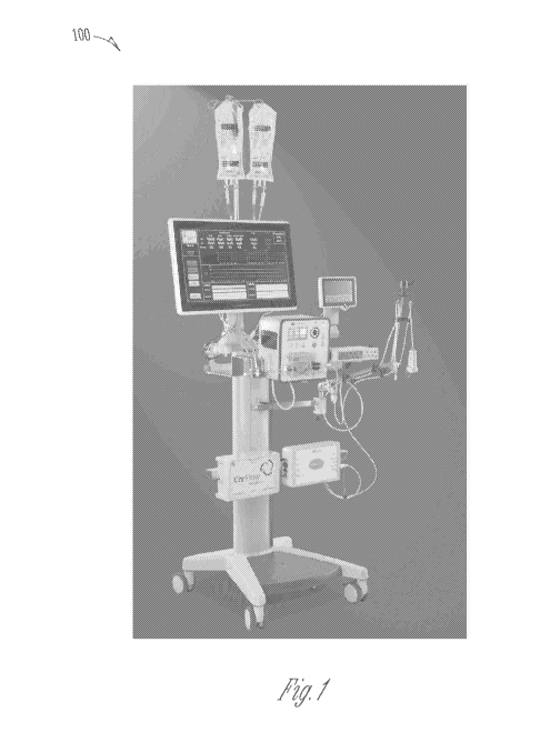
[00591 FIG. I illustrates an example of a modular computerized
diagnostic
and infusion system .100 (hereinafter "infusion system") for coronary and
other
human/animal vasculature and organs; in accordance with some embodiments of
the
present subject matter. The infusion system 100 can be a clinical ready
modular
system and can be configured in a mobile console form. The infusion system
1.00
can enable direct measurement and diagnosis of microvascular dysfunction,
including MVO and other microvascular abnormalities by:
100601 reakime coronary artery pressure and flow;
[00611 pressure/resistance time parameters;
[00621 Waterfall Pressure or Coronary Wedge or Coronaiy artety
Residual
Pressure;
100631 intracoronary electrocardiography (ECG); and/or
[00641 fractional flow reserve (HR.) measurements in the epicardial
arteries.
POW The infusion system 100 can enable therapy by:
11
CA 03113663 2021-03-19
WO 2020/061507
PCT/US2019/052245
100661 infusion of approved agent(s);
100671 targeted, controlled and low flow infusion; and/or
[00681 continuous monitoring of diagnostic parameters.
[00691 FIG. 2A illustrates an example 2.00 of an infusion catheter
having an
occlusion balloon 206, balloon markers 208 and 210, and infiision port 202 in
fluid
communication with an infusion lumen 212 in accordance with some embodiments
of the present subject matter. Guidewire lumen 204 is provided so that the
infusion
catheter can be slid along a guidewire to a desired position.
[00701 FIG. 2B illustrates an example 300 of an infusion catheter 250
having an occlusion balloon 206 placed over a 0.014" pressure measwing
guidewire
201 in a rapid-exchange (RX) fashion, in accordance with some embodiments of
the
present subject matter. In the example shown, the catheter 250 can slide over
a
guidewire 201 via guidewire lumen 204. Infusion port 202. can deliver fluids
via
infusion lumen 212 while guidewire 201 is disposed in lumen 204.
100711 FIG. 3A illustrates an example of a central portion of an
infusion
catheter 3.10, in accordance with some embodiments of the present subject
matter.
The central portion shows a cross section with an infusion lumen 312
encircling a
guidewire lumen 3.11. It is understood that in various embodiments, the
infusion
lumen may be side-by-side or may be in a nonlinear path about the guidewire
lumen. Other configurations are possible. One aspect is to provide a small
cross
sectional area to allow the catheter to be introduced into smaller vessels for
therapy.
[00721 FIG. 3B illustrates an example of a distal portion of an
infusion
catheter, in accordance with some embodiments of the present subject matter.
In
this embodiment, the guidewire 301 exits the distal portion of the catheter
and can
be used for placement of the catheter in the proper anatomical location. In
embodiments where the guide wire also provides pressure sensing, the guidewire
can be positioned outside or within the catheter lumen to provide various
pressure
sensing at the distal end of the catheter in situ.
[00731 FIG. 3C illustrates an example of a distal portion of an
infusion
catheter having a pressure chamber 306, in accordance with some embodiments of
12
CA 03113663 2021-03-19
WO 2020/061507
PCT/US2019/052245
the present subject matter. The pressure chamber is designed to provide a
region of
stable pressure measurement in the distal arterial segment. It is an integral
component of the device holding the guidewire 301 and permits pressure
measurement at locations different than near or distal to the catheter tip.
100741 FIG. 3D illustrates an example cross section of a distal
portion of an
infusion catheter having a pressure chamber, in accordance with some
embodiments
of the present subject matter. In various embodiments, the pores or slits or
slots 323
provided by the design provide both for better dispersion of infusate at the
distal end
of the catheter and also more precise pressure measurement. Such pores, slits,
or
slots 323 can also be patterned to provide an infusate flow pattern desired
for a
particular therapy. In various embodiments, different lumen configurations may
be
used, such as lumens 321 and 322, which can be used for guidewire lumens,
infusion lumens, or other lumen and port applications.
[0075] FIG. SA illustrates a distal portion of an infusion catheter
including
hemodynamic vanes or fins 509 to facilitate centering of the distal portion of
the
catheter in the vessel or organ in which flow is measured, in accordance with
some
embodiments of the present subject matter. Hydrodynamic forces are symmetric
and
facilitate centering of the catheter distal end within a flow field.
100761 FIG. 5B illustrates a distal portion of an infusion catheter
520
including holes for infusate 52.3 to be delivered in the vessel or organ in
which flow
is measured, in accordance with some embodiments of the present subject
matter.
In various embodiments the front end of the catheter has a taper 505 so that
the
transition from guidewire 501 to diameter of the catheter is more gradual.
100771 FIG. 5C, illustrates a distal portion of an infusion catheter
including
jets for infusate to be safely delivered in the vessel or organ in which flow
is
measured, in accordance with some embodiments of the present subject matter.
The
figure demonstrates that jets can be aimed to provide collision of infusate
flow 536
if desired for a particular therapeutic benefit, and their multiplicity will
create
slower flow and hence lower jet velocity to make vessel dissection of damage
lower
likelihood. The jets can be retrograde 533 or antegrade 534 jets, in various
13
CA 03113663 2021-03-19
WO 2020/061507
PCT/US2019/052245
combinations. In various embodiments the front end of the catheter has a taper
535
so that the transition from guidewire 501 to diameter of the catheter is more
gradual.
100781 FIGS. 6A-
61) illustrate an infusion catheter 610 with coaxial infusion
lumen 612 and guidewire lumen 611, guidewires, infusion holes 623, and a cap
613.
The design can direct antegrade 634 and retrograde 633 inflisate, in
accordance with
some embodiments of the present subject matter. The resulting flows can be
combined to provide a high flow zone 636.
[00791 FIGS. 7A-
7E illustrate an infusion catheter with coaxial infusion and
guidewire lumens, pressure sensor, integrated intra-coronary ECG electrode,
and
= =
infirsion holes, in accordance with some embodiments of the present subject
matter.
FIG. 7A shows a design 710 having a central lumen 711 surrounded by an
infusion
lumen 712 in a coaxial configuration. In various embodiments the central lumen
may be employed to receive a guidewire 701. In various embodiments, the
guidewire may be pressure sensing with a sensor 719. Although the example of
FIG. 7A is coaxial, it is understood that the lumens may be configured
differently,
such as side-by-side. Therefore, variations in cross section and dimensions
are
possible without departing from the scope of the present subject matter. FIG.
7C
shows a guidewire lumen portion of a catheter where a pressure sensing
guidewire
701 is able to be used to deploy the catheter. The guidewire may be retracted
to
perform pressure sensing. In various embodiments, the guidewire lumen may
include pressure ports to facilitate sensing of infusion pressure. Sensing of
infusion
pressure may be made with different sensing configurations, such as a pressure
transducer 719 at or near the distal end of the catheter 720, at or near the
proximal
end of the catheter, and/or at other locations along the catheter. In various
embodiments, the guidewire lumen or guidewire may be used for intra-coronary
ECG sensing or measurement FIG. 71) shows a portion of an infusion catheter
including an infusion lumen and a guidewire lumen with a guidewire extending
from the guidewire lumen. FIG. 7E shows a portion of an infusion catheter 740
with an ECG electrode 741 for sensing ECG signals. In various embodiments, the
ECG electrode is integrated into the catheter to obtain intra-coronary ECG
signals.
14
CA 03113663 2021-03-19
WO 2020/061507
PCT/US2019/052245
In various embodiments, the various sensing aspects of the infUsion catheter
can be
combined, so as to provide various sensing functions by the same infusion
catheter.
For example, the catheter may include both pressure sensing and ECG sensing,
among other things. Therefore, the present subject matter is demonstrated by
these
embodiments, but is not restricted to the particular combinations shown.
[00891 The infusion catheters as shown in FIGS. 2-3 and 5-7 can be
used in
systems/deviceslmethods described herein to controllably occlude a desired
vessel,
infuse desired fluids and measure pressure inside the vessel in real time and
distal to
the occlusion balloon. The infusion catheters as shown in FIGS. 2-3 and 5-7
can
include: a 6F guide sheath compatible catheter, a compliant 5x lOrnm occlusion
balloon, and can be received over 0Ø14" pressure guide wire. The infusion
catheters as shown in FIGS. 2-3 can include a wide flow infusion range, for
example, 5-50 mIlmin and can include axial flow infusion.
NOM In some embodiments, the catheter can be inserted into a
myocardial
vessel supplying blood to a patient's myocardium. In some embodiments, the
myocardial vessel or nearby vessels may or may not include microvascular
dysfunction, such as MVO and may or may not include myocardial infarction. The
catheter can controllably block antegrade blood flow within the myocardial
vessel
around the catheter by inflating a balloon. In some embodiments, the
myocardial
vessel can include a stent and the catheter can block antegrade blood flow
from
within the stent, by inflating a balloon.
[00821 FIGS. 4A-4B illustrate a graph 400 of an occlusion and infusion
algorithm, in accordance with some embodiments of the present subject matter.
In
various embodiments, the infusion algorithm is generated by modular
computerized
infusion system .100 such as is shown in FIG. 1. The infusion system 100 can
perform diagnosis of the vessel as set forth in the Incorporated Applications,
including, but not limited to, that set forth in U.S. Provisional Patent
Application
Ser. No. 62/560,545 filed September 19, 2017, which is incorporated by
reference in
its entirety.
CA 03113663 2021-03-19
WO 2020/061507
PCT/US2019/052245
[00831 The system provides an initial flow or pressure pulse, a
"preparatory
pulse" 402 which may include infusate at a higher flow or pressure and of
variable
temporal duration to inflate, open, or otherwise clear channels of the
microvasculature which has obstructive debris and has collapsed. The system
thereafter provides pulses of similar or possibly smaller pulse amplitudes
(404, 406,
410, etc.) to provide therapeutic infusion to the vessel or organ, as
described herein.
The pressures, numbers of steps, pulses and times of infusion can be
varied within the scope of the present disclosure. An example of a pressure
response is shown in FIG. 4B, where the line 420 is the waterfall pressure
(WP)
which is the baseline pressure of the tissue under analysis. Curves 422 and
424
show the variation in applied pressure and applied pressure with blood flow
due to
application of the pulses in FIG. 4A. Flow improves over the course of the
applied
therapeutic pulses.
[0085] FIG. 8 shows an open loop block diagram 800 for delivery of the
preparatory pulse and following pulses/infusions according to one embodiment
of
the present subject matter. In the open loop configuration, flow or pressure
pulses
are infused at fixed or predetermined parameters. In various embodiments, the
pump controller 8.10 receives inputs (e.g., 801, 803) to perform algorithmic
control
of the pump and the delivered infusate or infusates (e.g., 81.1,81.2, and/or
81.3 of
FIG. 8). The infusates are delivered to the infusion lumen of the infusion
catheter
830. In various embodiments, the system can control the infusate delivery,
including the type, pressure, flow, dose, temperature, and other parameters of
the
infusate. In various embodiments, the system can control pressure and
inflation of
one or more occlusion balloon(s). In various embodiments, the system can
control
multiple aspects of the system, such as infusate and balloon parameters, among
other things.
[0086] FIG. 9 shows a closed loop block diagram 900 for delivery of
the
preparatory pulse and following pulses/infusions according to one embodiment
of
the present subject matter. In this configuration infusion pressure, flows,
volumes or
rates may be governed in real time or according to measured/sensed vessel
16
CA 03113663 2021-03-19
WO 2020/061507
PCT/US2019/052245
parameters including flow, anatomy, pressure, resistance, intracomnaiy ECG, or
similar physiologic measurements. In various embodiments, the pump controller
91.0 receives inputs (e.g., 901, 903, etc.) from an operator and inputs from
one or
more feedback signals (950, 925, 915) sensed by one or more sensors (e.g.,
930,
941, etc.) to perform closed loop algorithmic control of the pump and the
delivered
infusate or infusates (e.g..: 909, 912, and/or 913 of FIG. 9). The inflisates
are
delivered to the infusion lumen of the infusion catheter 930. Such a design
allows
feedback from sensed signals to help the controller provide an algorithmically
controlled infusate. Such sensors can modify infusion based on physiologic
state
and/or measured parameters. Some of the parameters sensed include, but are not
limited to, pressure, flow, impedance, cardiac cycle, etc. In various
embodiments,
the system can use the measured parameters to control the infusate delivery,
including the type, pressure, flow, dose, temperature, and other parameters of
the
infusate. In various embodiments, the system can use the measured parameters
to
control pressure and inflation of one or more occlusion balloon(s). In various
embodiments, the system can use the measured parameters to control multiple
aspects of the system, such as infusate and balloon parameters, among other
things.
[0087] Therapy Based on Restoring Microvascular Flow
[00881 In the course of investigating micmvascular dysfunction, MI and
MVO, it has been observed that epicardial coronary artery obstruction causes
acute
and profound loss of distal pressure, including and especially the intra-
myocardial
capillaries. Intramural pressure in the contracting ventricle is cyclic with
systole-
diastole. Capillaries are thus likely dose either completely or partially, and
open for
more than what occurs in the case of normal blood flow and normal blood
pressure
in the epicardial coronary arteries which feed the microvasculature. This is
shown
by epicardial flow velocity measurements and in histologic evaluation of acute
myocardial infarction which shows capillaries too small to accommodate red
blood
cells or white blood cells (e.g. less than 10 pm microvasculature diameter),
and with
interspersed thrombotic elements such as platelets or fibrin. These
observations
strongly suggest epicardial coronary artery occlusion causes microvasculature
17
CA 03113663 2021-03-19
WO 2020/061507
PCT/US2019/052245
hypotension.: creating conditions for catastrophic dynamic collapse and
partial or
complete microvasculature obstruction.
100891 One method to model microvasculature collapse is to perform a
hydrodynamic analysis of the microvasculature in myocardial contracting
tissue.
The law of Laplace governs pressure required to sustain an open capillary:
100901 T P x
[009.11 Where I is the tension in the blood vessel wall (e.g., units of
ke./(s2),
P is the pressure across the vessel wall (e.g., kPa), and R is the radius of
the blood
vessel (e.g., mm). From Laplace's equation, it can be observed that as the
radius
becomes very small, the pressure required to open a close capillary is very
large.
Further, Poiseulle's Equation provides a way to model resistance to flow:
Vessel Resistance (VR.) is proportional to (blood viscosity x Length of
vessel)/R4.
[00921 Therefore, assuming blood viscosity is relatively constant,
vascular
resistance is inversely proportional to the fourth power of the radius of the
vessel.
As the vessel radius shrinks by half, the original vascular resistance VRO
increases
sixteen-fold: VR = VR.0/(0.54) = VR01(0.0625) = 16 VRO.
[00931 Thus, restoration of blood pressure and blood flow via
interventions
such as stenting of the coronary arteries do not supply enough pressure to
open a
closed capillary bed, resulting in the capillaries remaining partially or
completely
closed with continuing periodic compression/relaxation during the heart cycle.
These physiologic disturbances of normal capillary t'unction are key
components of
microvascular obstruction.: chronic capillary occlusion (with slow flow as
evidenced
by MR1 imaging showing very late gadolinium enhancement at infarcted sites).
100941 The present subject matter provides a mechanism to open not
only
epicardial coronary arteries, but also reverse capillary occlusion due to low
pressure
and also to mitigate thrombus, microvascular spasm, and other causes of low or
no-
flow in the capillaries resulting in myocardial cell death. Thus, the present
subject
matter addresses chronic complications of MI and resulting ischemia,
congestive
heart failure, arrhythmias, ventricular aneurysms, myocardial rupture, poor
prognosis, recurrent clinical events and a multiplicity of severe negative
cardiac
18
CA 03113663 2021-03-19
WO 2020/061507
PCT/US2019/052245
complications. It is further understood that the present subject matter can be
applied
to other diseases, such as peripheral vascular disease (limbs), stroke
(brain), renal
failure (kidney) and diseases affecting blood flow to other bodily parts.
THERAPEUTICS
100951 Several therapeutic components of this application include
physiologic-biophysical mitigation of microvascular compromise including
stenosis,
obstruction, inflammation, reperfusion injury, and chronic malfunction. In
various
embodiments, addition of chemotherapeutic agents infused locally through a
coronary artery catheter systemically may be followed for longer time periods
by
routes such as intravenously. In various embodiments, coronary direct drug
infitsion
becomes a systemic infusion. Several drug classes are described including, but
not
limited to, antiplatelet agents, acute and chronic Thrombin Inhibitors (both
direct
and indirect), and vasodilators including nitric oxide donors and stimulators
of nitric
oxide synthase.
100961 For example, in various embodiments, antiplateld agents in the
form
of anti-aggregatory agents such as direct thrombin inhibitors (hirudin and its
molecular analogues; platelet receptor inhibitors - GP inhibitors; factor X
inhibitors; low molecular weight heparin and fibrin inhibitors and fibrin
fibrinolytics) are available for use.
[00971 'Vasodilator drugs may be used for real-time vasodilating
microvasculature as lytic therapeutic is infused, which have therapeutic and
diagnostic properties. Some examples include nitroglycerin (TNG), low dose
dopamine, Adenosine, acetyl choline. Papaverine, hydralazine, calcium channel
blockers, and others.
DEVICES FOR THERAPEUTIC INFUSION
[00981 The present subject matter provides various infusion catheters
for
assessment of microvascular dysfunction. In various embodiments a catheter is
adapted to receive a guidewire which may have a pressure sensing capability,
for
delivery of the distal tip of the catheter to a site and to deliver infusate
from the
proximal end of the catheter to the distal end of the catheter via a lumen. In
various
19
CA 03113663 2021-03-19
WO 2020/061507
PCT/US2019/052245
embodiments, the infusate is delivered by an infusion lumen. In various
embodiments, the catheter includes a guidewire lumen to receive a pressure
sensing
or standard guidewire.
[00991 In certain embodiments, the catheter includes a multiplicity of
lumens. In embodiments including an infusion lumen and a guidewire lumen, the
intbsion lumen and guidewire lumen may be separate and oriented to be adjacent
to
each other or coaxial to each other. The infusion lumen may be used for drug
delivery or for delivery of infitsate for diagnostic or therapeutic infusion,
or
combinations thereof. In various embodiments, the catheter includes a lumen
for
pressure monitoring either directly or via pressure sensing wire. In various
embodiments the lumen for pressure monitoring may receive a pressure sensing
guidewire. In various embodiments, the catheter includes dedicated lumens for
delivery of infusate and pressure sensing. In various embodiments, the
catheter
includes dedicated lumens for delivery of infusate, pressure sensing, and for
accommodating the guidewire. In various embodiments, the catheter includes
dedicated lumens for delivery of infusate, drug delivery, and pressure
sensing. In
various embodiments, the catheter includes dedicated lumens for delivery of
infusate, drug delivery, pressure sensing, and for accommodating the
guidewire.
Infusate lumina may have holes, slots, or otherwise be able to diftlise flow
(diagnostic or therapeutic) for safer injection into blood vessels.
[00100] In various embodiments, the catheter includes vanes or fins
adapted
to urge the catheter away from the walls of the vessel it resides in to
provide safer
and more consistent pressure measurement. In various embodiments, the vanes or
tins are adapted to center the catheter within the vessel it resides in. In
various
embodiments, the vanes or fins include hydrodynamic qualities adapted to urge
the
catheter away from the walls of the vessel and/or to center the catheter in
vessel.
[001011 In various embodiments, shaft design, vanes or fins with
hydrodynamic impact may be placed on the surface of the catheter distally to
equalize hydrodynamic flow around the catheter and to force a catheter into
the
central steams of blood flow via the Bernoulli principle.
CA 03113663 2021-03-19
WO 2020/061507
PCT/US2019/052245
1001021 These vanes may also direct blood into an open chamber at the
distal
end of the catheter to facilitate accurate pressure measurement in the
surrounding
artery or vascular structure.
[001031 In various embodiments of the catheter, at least a portion of
the distal
end of the catheter is made more flexible. In various embodiments, flexibility
is
enhanced by a change in dummeter of catheter material.: or a pattern of cuts
or both.
In various embodiments, the cuts are performed so as to make spirals or other
patterns for flow diffusion (avoid jetting, for safer injection). In various
embodiments the patterns are circles, irregular patterns, all typically made
by laser
or other micro-machining methods. Differential stiffness can be created by
these
patterns, or by other methods such as holes a multiplicity of patterns,
changing size
and density to allow the tip segment to have differential flexibility in a
pattern
beneficial for tracking but also for admitting blood into the distal pressure
chamber.
[001041 In various embodiments a plurality of micro holes with changing
size, shape, and density allow for variations in catheter tip or proximal
component
compliance.
100105] In various embodiments, a distal hole or lumen for guidewire
insertion and exit through the distal tip of the catheter permits utilizing a
pressure
guidewire to place the catheter using standard interventional methods,
including a
"rapid exchange" configuration. When proper placement is achieved the pressure
guidewire may be pulled retrograde back to facilitate a pressure sensing mode.
The
wire is pulled back into a chamber within the catheter body that ensures fitll
exposure to blood pressure though cuts, holes, or slots, because blood or
other fluids
(such as diagnostic and/or infUsates) combined provide accurate pressure
measurement.
ROM] In various embodiments, the system allows to measure
intracoronary
ECG either over the guide wire, pressure guide wire or an electrode located on
the
distal end of the catheter.
1001071 In various embodiments, the differential hole pattern may vary
longitudinally to not only after compliance but also to alter resistance to
flow of
21
CA 03113663 2021-03-19
WO 2020/061507
PCT/US2019/052245
infusates. In this configuration differential exit of flow longitudinally down
the
catheter can be achieved. In various embodiments, equal exit of flow can be
achieved using holes, spirals, and their patterns, which are varied
systematically to
decrease or increased resistance as a function of longitudinal direction down
the axis
of the catheter. In various embodiments, the patterns of holes, cuts, and
spirals have
multi-function of the control relative fluid exit at various pressures and to
alter
compliance of the distal tip to facilitate catheter steering and tracking over
a
guidewire which may be a pressure wire to allow distal pressure sensing.
[001081 In wire-based embodiments allowing insertion of the guidewire,
a
distal tip of the catheter may also include a multiplicity of holes, cuts,
wedges,
spirals or other apertures. In various embodiments, the aperture patterns are
chamfered or tilted to force or urge blood into the resulting enclosed
chamber.
[00109] Additional catheter designs are provided, such as those
described in
the incorporated Applications:
U.S. Patent Application 15/398,470, filed January 4, 2017, published as US
201.7
0189654 Al July 6, 2017, and which claims the benefit of: U.S. Provisional
Ser. No.
62/274,744 filed January 4, 2016; U.S. Provisional Ser. No. 62/320,230 filed
April
8, 2016; U.S. Provisional Ser. No. 62/358,433 filed july 5, 2016; and U.S.
Provisional Ser. No. 62/379,074 filed August 24, 2016; and
PO' Patent Application Ser. No. PCTIUS17/12181 published as W02017120229A1
on July 13, 2017, which claims priority to all of the aforementioned patent
applications; and
U.S. Provisional Patent Application Ser. No. 62/560,545, filed September 19,
2017,
all of which are incotporated by reference in their entirety herein.
LOCAL DRUG AND INFUSATES INFUSION PROFILES
100110] Acute, semi-acute, and chronic myocardial infarctions result
from
micro vessel occlusion, microvascular obstruction, catastrophic microvascular
collapse all of which may cause both intraluminal plugging by thrombus, cells,
proteinaceous materials and relative local myocardial hypotension which in
turn
decrease capillary size and prevent normal blood flow creating severe ischemia
and
CA 03113663 2021-03-19
WO 2020/061507
PCT/US2019/052245
necrosis. In various embodiments of the present subject matter, the therapy
involves
infusion protocols and the local agent that is infused.
1001111 Various embodiments of the present subject matter provide
controlled infusate profiles to treat microvascular collapse resulting from
hypertension. It has been found that microvessels can be opened far better
utilizing
continuous flow as from an external pump than utilizing periodic blood
pressure
supplied to the arteries via the heart.
100112] For example, an external pump permits continuous application of
pressure rather than cyclically varying systolic-diastolic pressure as is
typically
supplied via the natural cardiac contraction. This can be demonstrated by
calculating pressure-time integrals (and using rms equivalent pressure) which
show
that continuous pressure on the microvasculature to both initially open and
maintain
opening is far better to maintain flow to the tissue of interest. In some
calculations,
the flow improvement is greater by a factor of .10 or more for comparable
pressures
by pump.
1001131 Another therapeutic benefit of an external pump is that
pressure by
the pump may be inserted at supra-physiologic values. For example, in some
embodiments of the present subject matter elevated pump pressures are created
by
continuous or cyclic flow infusion. Infusion into distal microvasculature
creates a
back pressure via Ohm's law applied to hydrodynamics, P = Q x VR, or pressure
equals flow times microvascular resistance, VR.
100114] In various applications of the present subject matter, flow
infusion
may be placed in a closed loop system to achieve regular and continuous
accurate
pressure control in "real-time". Markedly elevated intravascular pressures do
not
have the negative effects that cardiac pressures generated via the left or
right
ventricle supply. For example, high pressure values (such as 200 mmHg or
higher)
that are generated by the left ventricle in hypertension put excessive stress
and strain
on the myocardial wall and thus intense closing pressure on the
microvasculature
during systole. Moreover, these very high pressures will subject the entire
body to
hypertension, which even acutely may have profoundly negative clinical
23
CA 03113663 2021-03-19
WO 2020/061507
PCT/US2019/052245
consequence. It is thus very difficult to consider raising local myocardial
intravascular pressure to open hemodynamically closed capillaries by induction
of
hypertension.
[001151 Conversely, supplying substantially elevated local pressure by
catheter can be achieved. In various embodiments of the present subject
matter, the
proximal vessel is blocked by balloon, thus protecting the body from local
hypertension.
100116] In various additional embodiments, the balloon occlusion is not
essential. A controlled flow rate will vary the pressure microvascular
resistance,
and can be adjusted to establish flow to the microvasculature to a level
deemed
therapeutic, whether or not a drug is included with the infusate.
100117] Infusion pressure at variable infusion rates is a direct
measure of
microvascular resistance, and as discussed in the incorporated Applications,
is
diagnostic regarding the function or dysfunction of the local microvascular
structures.
1001181 Specifically, elevated pressure supplied by pump is to be used
for
initial opening of hydrostatically closed capillaries is a 'preparatory pulse'
that is
utilized to prepare the microvessels to better accept therapeutic solutions.
As these
capillaries open, a measurable drop in distal infusion pressure will be
visualized that
reflects decreased hydrodynamic resistance. This pressure change or drop can
be
measured in real time and be used as feedback to the operator for when
hydrostatic
opening has occurred. The pressure drop may also be measured and applied to a
closed loop control program which adjusts the infusion pressure for desired
outcomes. For example, the controller may be adjusted to maintain a constant
infusate flow. The controller may be adjusted to maximize the delivery rate of
a
drug to the microvasculature. The controller may also be used to generate a
pulsed
pressure waveform to obtain a dynamic measure of microvascular dysfunction,
such
as MVO.
1001191 Infusion of drug containing infusate may clear aggregated and
congested cells. Such procedure can affect the platelets, white blood cells,
red
24
CA 03113663 2021-03-19
WO 2020/061507
PCT/US2019/052245
blood cells, and proteinaceous matter found in obstructed microvasculature
during
an ischemic event.
1001.20] Infusion Pulse Sequences
[001211 In various embodiments of the present subject matter employing
infitsate pumps, the delivered controlled infusion has a multiplicity of
effects and
can be applied in distinct, coupled, and temporally related flow/pressure
and/or
pulse sequences. The pulse sequences may be controlled manually or by
automated
control systems which may include a feedback mechanism to stabilize and create
precise flow patterns by the pump. Safety of the infusion is enhanced via
feedback
and closed loop control. For example, if flow creates pressures that are too
high, the
pump flow can be shut down, decreased, or otherwise limited according to the
principles of systems and control theory. In various embodiments, in addition
to
visualization of real time pressure, resistance, and flow, a visual or verbal
alarm
may be triggered to alert the operator of over-pressure or under-pressure
conditions.
1001221 In various embodiments of the present subject matter, the
infusion
profile may be separated into components. For example, in various approaches,
the
pulse sequence may include a "preparatory pulse" and "follow-on pulses" or
flow
infusions.
1001231 The "Preparatory Pulse"
1001241 A "preparatory pulse" which prepares the microvessels to accept
flow, opens them or extends them, while simultaneously delivering drug. The
preparatory pulse is a preparation step to open stenosed or occluded
microvasculature and in some cases begin drug delivery. The preparatory pulse
can
be, for example, of high flow or pressure designed to open hydrostatically
occluded
microvessels. The infusate for this may be a simple liquid such as lactated
Ringer's
solution, other crystalloid solutions containing beneficial concentrations of
sodium,
chloride, potassium, glucose, lactate and the like, may in addition be a drug
containing solution.
1001251 In various embodiments, the duration of this preparatory pulse
can be
guided by feedback from local, distal pressure measurements and real time
CA 03113663 2021-03-19
WO 2020/061507
PCT/US2019/052245
observation of myocardium resistance, flow, or cardiac function (pressures and
ventricular function measures or intracoronary ECG). In various embodiments,
the
duration of the preparatory pulse can be limited or discontinued when pressure
or
resistance calculated drops to a certain predetermined value, or a relative
percentage
value of initial pressure or resistance.
1001261 The preparatory pulse may be high pressure, which is safer than
"hypertension" since it is not generated by the ventricle itself and does not
generate
an elevated intra-myocardial pressure that closes microvessels in the elevated
pressure clinical syndromes.
1001271 In various embodiments, the preparatory pulse may be timed for
diastole utilizing the QRS-electrocardiographic complex, or via distal
pressure
measurement which is cyclic, or any other means to determine lower pressure in
the
intramural component of the ventricular wall. Moreover, the cyclic natural
myocardial contractions and microvessel pulsations themselves provide a
potentially
useful agitation of diagnostic/therapeutic solutions.
1001281 In various embodiments, the preparatory pulse is guided through
feedback which tracks a rise in distal pressure that saturates at a given
value,
indicating that the capillaries in microvasculature have been completely
filled and
cannot accept more flow without increased pressure in a fixed pattern.
1001291 Follow-On Pulses or Flow Infusions
1001.30] After the preparatory pulse, a subsequent flow infusion is
performed
to maximize drug delivery to occluded vessels of the microvasculature in
comparison to patent or partially patent microvessels. In various embodiments,
the
follow-on pulses or flow infusion is a controlled-flow infusion with
monitoring of
distal pressure for purposes of safety and efficacy. If the measured distal
pressure
rises to unsafe levels flow can be automatically and controllably reduced or
discontinued using a computer adjusted algorithm. In various embodiments, the
flow can be controllably reduced or discontinued by a manually controlled
operator
based system which provides a signal to the operator.
26
CA 03113663 2021-03-19
WO 2020/061507
PCT/US2019/052245
100131I In various embodiments, during the infusion phase, low
pressures
may be employed for the purposes of generating advantageously the steep
increase
in microvascular resistance at low pressures. Studies show a natural
logarithmic
relationship between myocardial flow (Q) and resistance (It), with steep
resistance
increases as flow rate drops. In various embodiments, this low-flow-low-
pressure
inffision strategy can more equalize resistance of obstructed microvasculature
and
patent microvasculature (due to low pressure-low flow infusion). This in turn
equalizes these two parallel resistances (obstructedinonobstructed
microvaculature),
which thus delivers proportionally and absolutely more flow to occluded or
partially
occluded microvessels. Sequences of preparation and impulses may be chained
and
repeated through time.
100132] In various embodiments of the present subject matter, proximal
balloon occlusion is part of these preparatory and therapeutic infusion
sequences.
As the proximal vessel is occluded virtually all pump flow is directed into
the distal
vessels.
1001331 In various embodiments of the present subject matter, partial
balloon
occlusion is achieved by monitoring distal pressure. Pressure values lying
between
coronary residual pressure (CRP) and systolic pressure will indicate a partial
balloon occlusion, and may be used to keep, in a feedback loop, the vessel in
a
partially occluded state. Experimental and theoretical modeling studies
demonstrate
that the infusion-flow relation constitutes a linear system. Because the
system is
linear, superposition of flows (pump flow and native coronary artery flow) is
a
viable modality. One advantage of this approach is that the vessel is perfused
by a
mixture of inflisate plus antegrade native (oxygenated) blood, and thus such
pulse
sequences can be performed for very long times without risking distal
myocardial
ischemia. Linear superposition of flows in a linear system permit accurate
measurement of distal microvascular resistance via requisite infusion pressure
-via
pump, in parallel with native antegrade blood flow.
1001341 In various embodiments, dual or higher (e.g. 3X, 4X or more)
flow
superposition as described also permits measurement of native flow. In this
CA 03113663 2021-03-19
WO 2020/061507
PCT/US2019/052245
approach, incremental flow supplied by the pump is added to native flow to
provide
an incremental pressure rise. Linearity permits distal resistance measurement,
which
is equal to incremental pressure divided by incremental known controlled flow
(from the pump). This known resistance can then be used to calculate native
flow.
1001351 For example, while pump flow is still running, total flow is
the
summation of native plus pump flows. If resistance is known, flows can be
individually calculated. When the pump flow is discontinued and resistance and
residual pressure are known, the native flow can be similarly calculated.
[001361 Flow superposition provides additional measurement options. For
example coronary artery flow can be measured using methods, such as the
following. In various embodiments, a catheter is placed in position, a
pressure wire
is placed, and flow infusion is begun. The incremental pressure divided by
known
inserted flow equals resistance. Consequently, total flow is calculated, and
native
flow is subtraction of total flow minus flow.
VI4/1371 Fractional Flow Reserve
[001.381 Another measurement that can be accomplished using the flow
superposition described herein includes the measurement of fractional flow
reserve
(HR.), a parameter for determining stenosis severity in an epicardial coronary
artery. In one embodiment of the present subject matter, a method for
measuring
FFR is obtained by combinations of the following steps. A flow infusion
catheter is
delivered proximal to the stenosis. Aortic pressure is measured by guide
catheter or
by a separate pressure guidewire measures pressure proximal to stenosis. .A
guidewire with pressure sensing used to cross the stenosis. Incremental flow
by a
pump through infusion catheter is initiated. An incremental pressure
measurement
is obtained with a known flow from pump. A stenosis resistance is calculated
as
pressure drop across stenosis divided by known comnaiy flow, which may be
calculated as total flow minus injected flow.
1001391 Absolute Myocardial Resistance
1001401 in various embodiments, the methods described herein provide an
approach for measuring absolute dynamic myocardial vascular resistance (dMVR).
28
CA 03113663 2021-03-19
WO 2020/061507
PCT/US2019/052245
The MVR is called "dynamic" as the resistance varies at different flow
infusion
rates and is exponentially increasing at low flow values. FIG. .13 shows a
plot of
dynamic myocardial vascular resistance (dMVR) versus flow rate from one study
which demonstrates that microcirculation reduces exponentially as flow
approaches
zero. A. calculation utilizing known flow and distal pressure can provide the
absolute myocardial resistance (Resistance = Pressure/Flow) at each flow rate.
The
approach provided herein permits simultaneous distal microvascular resistance
and
stenosis FFR measurements.
[001411 In various embodiments, a constant pressure method will also
provide a determination of microvascular resistance in an organ. In one
embodiment of the method a pump is utilized either with or without input
arterial
occlusion (such as by a balloon or other obsttucting device). Pressure is
monitored
as infusion is begun. In various embodiments, an infusion sequence of
increasing or
decreasing pressure levels is instituted: adjusting the pump rate, monitor
back
pressure, and that each pressure level record pump flow. This results in and
composite set of pressure-flow or within any desired range. The flow is
measured at
each range, generating a pressure-flow relationship which may be analyzed as
per
description below.
1001421 Determining Microvascular Resistance: Importance of Inflisate.
1001431 One diagnostic method of controlled flow infusion as well as
controlled pressure for determining microvascular resistance of any organ can
be
modified depending on the infusion or infusate that is used as a diagnostic
fluid.
[001441 Specifically, the importance of whether a fluid is Newtonian or
non-
Newtonian has important effects on resistance results. Utilization of a
Newtonian
fluid such as any electrolyte-water-based medical fluid is far superior to
blood or
other non-newtonian fluids. Utilization of Newtonian fluids, as proven by
experiments permit far more accurate demonstration of the microvascular
resistance,
particularly in the heart, which is highly linear. Utilization of a non-
newtonian fluid
can make the microvascular resistance of such an organ appeared to be
nonlinear.
Newtonian fluids also serve as excellent diagnostic solutions and can be used
to
29
CA 03113663 2021-03-19
WO 2020/061507
PCT/US2019/052245
better control ischemia because they typically lack oxygen. Newtonian fluids
also
act as a powerful vasodialator, so the heart tissue will not clamp down,
further
linearizing flow which enhances diagnostic testing and therapeutic perfUsion.
The
use of Newtonian fluids can therefore provide improved pressure response at
relatively low flow rates. In various applications, a flow rate as low as 5
mUminute
can give an excellent pressure response which allows for diagnostic and
therapeutic
applications at pressure levels that are safe for the tissue.
100145] Saline, Lactated Ringer's Solution, or other water based
electrolyte
fluids are useful for multiple reasons. They provide diagnostic effects and
benefits,
such as:
(I) Linearity can be proven in the microvasculature.
(2) Low viscosity fluids more easily access distal micro-vessels, for example
in
endocardium which represent the terminal capillaries, quite small and subject
to
changing resistance by virtue of their microscopic size and distal location.
Distal
location of these vessels creates a secondary issue, being at the far end of
the blood
pressure cascade, markedly compounding the problem of unimpeded flow during a
diagnostic sequence.
(3) Induced controlled hypoxia:
(a.) Electrolyte solutions as noted are also highly advisable because they
induce
ischemia in the myocardium. Utilization of blood or other oxygen -containing
fluids
alter diagnostic effects since oxygen maximum vasodilation. A fluid such as
crystalloid above contains little to no oxygen and thus fulfills separate
roles, not
only as a hydrodynamic agent, but also as it is lacking oxygen is an optimal
fluid for
inducing hypoxic vasodilation.
(b.) Manufactured crystalloid solutions such as lactated Ringer solution
moreover provide a very consistent product comparison within a patient or
across a
population of patients utilization of drugs or blood as it infusate induce
substantial
errors in determining population values or even within diagnostic runs in the
same
patient because the diagnostic fluid adversely affects the quality of
diagnosis as
creates adverse changes in the diagnostic system by its effects on the
CA 03113663 2021-03-19
WO 2020/061507
PCT/US2019/052245
microvasculature. Consistent across runs and patients. While using blood,
blood
products or other biologic fluids may have benefits for other diagnoses, the
changing nature of blood such as hemoglobin, proteins, micro thrombi and other
biologic alterations will induce inaccuracies in the determination of
microvascular
resistance.
[00.1,461 Fluids with high protein content can similarly cause
nonlinearities.
[001471 in summary, the nonlinearity of the coronary microvasculature
are
commensurate with the non-Newtonian hydrodynamic characteristics of blood.
Experiments have demonstrated that the microvascular resistance is linear when
using a Newtonian fluid such as a crystalloid infusate.
1001481 Osmolarity
100149] Infusates can be chosen based on a number of parameters,
including
osmolarity. For example, a hyperosmolar infusate can be used to draw fluid
from
tissue. For example, an osmolar gradient can be used to reduce or prevent
edema in
treated tissue.
1001501 Accordingly, infusates can be selected based on a number of
characteristics, including, but not limited to, how Newtonian the solution is,
the
percentage of oxygenation of the solution, and the osmolarity of the solution.
1001511 Methods of Detemining Microvascular Resistance
1001521 Methods which determine microvascular resistance in an organ
are
fraught with inaccuracy by simply dividing infusion pressure by infusion flow.
This
is especially the case when there is an offset (either constant or variable)
pressure
which confounds the resistance calculation.
1001531 Utilizing a derivative approach across a series of increasing
or
decreasing flow or pressure stair steps eliminate this offset and generate a
highly
accurate microvascular resistance measurement. Experiments show that in the
case
of biologic organs the resistance is highly linear: resulting from plotting
the
pressure-flow derivative. Curve fitting this line generates accurate record of
vascular resistance as the slope, while the intercept is a "zero flow
pressure" which
is similarly useful for diagnosis as it reflects the DC offset.
31
CA 03113663 2021-03-19
WO 2020/061507
PCT/US2019/052245
1001541 In a typical heart, the pressure DC offset results from
collateral
capillary flow into the distal myocardial bed and is often referred to as the
"Coronary Wedge Pressure". This pressure is the most obvious cause of
substantial
errors in measuring microvascular resistance. It can be eliminated by
subtraction but
drawbacks to this method is that they require wedge pressure measurement, and
moreover me change throughout process.
1001551 This derivative- intercept diagnostic method may be used with
or
without an occlusion device such as a balloon. However, it is likely less
accurate
when utilizing flowing blood for reasons above, due to the non-Newtonian fluid
inducing nonlinearities. Traditional methods of measuring microvascular
resistance
suffer from this problem the 1MR, FM, CFR and similar indices.
100156] Therapeutic Effects of Inflisate Makeup
[001571 Therapeutic effects from infusion fluids also benefit from the
Newtonian fluid. Not only does this fluid convert a nonlinear system into a
linear
system, the lower viscosity of crystalloid permits access of therapeutic fluid
containing drugs or other therapeutic agents to reach the smallest
microvessels of a
biologic system. Other therapeutic agents may include those containing oxygen
(after for example a diagnostic run has been made without oxygen), all of the
therapeutic drugs previously described drug combination such as glucose-
insulin-
potassium ((AK) which provides a hyperosmolar solution to remove fluid from an
edematous biologic organ where the edema is causing interference with norinal
organ fUnction.
[001581 Catheters for Use Diagnostic and Therapeutic Fluid Infusion
Using
the Constant Flow or Defined Pressure Method
1001591 The infusion catheter is an important element of accurately
determining microvascular function including microvascular resistance.
Important
features include:
(i.) pressure measurement sensor proximal:
(ii.) capability of placing a pressure measuring guidewire or other sensor
distal to
the catheter tip, and
32
CA 03113663 2021-03-19
WO 2020/061507
PCT/US2019/052245
(iii.) when infiision is generated by appropriate parameters the 13.-()
derivative-
intercept method is applicable. The vessel obstruction by balloon or other
method is
not required for this method to be functional.
100160] The pressure measurement capability does not require vessel
occlusion since the method is impervious to ongoing constant blood flow.
Defined
fluid infusion generates a perturbation to this baseline flow in this
perturbation is
used in the derivative calculation.
100161] Novel diagnostic Considerations
100162] The vulnerability to ischemia and infarction of the cardiac
endocardium is well known. A model for assessing myocardial infarct size can
be
derived from myocardial microvascular resistance. Anatomic considerations of
the
distal myocardial microvasculature result in well-defined shapes for ischemic
and
infarct it myocardial tissue these typically appear as linear endocardial
zones which
may be in various states of health as defined by levels of ischemia and
duration of
ischemia. Myocardial infarct size is related to the thickness of the
myocardial
necrotic wave which progresses from endocardium to epicardial over time.
1001631 Diagnostic methods for measuring infarct size by catheter
methods
can eliminate the need for external imaging procedures such as magnetic
resonance
imaging.
100.1641 Microvascular resistance is measured thus down the capillary
network from epicardial (where the large epicardial coronary arteries are the
blood
source) to the Terry distal endocardium which represents the teminal zones of
capillary blood supply.
100165] A parallel system analytic method models microvascular
resistance
as parallel resistances from epicardial to endocardium. A typical analytic
approach
may be as follows:
(i) a three compartment model consisting of tissue which is I) healthy, 2.)
edematous but alive, 3) alive but not fiinctioning, visualized by late get
lending
enhancement using CMR Techniques, or 4) Dead As Visualized by Microvascular
Obstruction in CMR Imaging
33
CA 03113663 2021-03-19
WO 2020/061507
PCT/US2019/052245
(ii) this cascade of healthy, dying, or dead tissue is characterized by a
three-or
more compartment model which utilizes tissue conductivity due to capillary
patency
or progressive obstruction, the area of the muscle to be studied, and the
thickness of
the muscle as in the case of cardiac wall thickness, and
(iii) systems simultaneous equations can be written that relate microvascular
resistance is measured from the epicardial comnaiy artery to myocardial
infarct size
utilizing expression R=pLiA.
100.1.661 Absolute coronary artery collateral flow
1001671 In various embodiments, the methods described herein enable an
approach for measuring absolute coronary artery collateral flow. In various
embodiments of the present subject matter, coronary artery collateral flow is
measured by:
Placing a proximal balloon occlusion coronary artery with distal controlled
flow infusion via infusion catheter and pressure sensing, as described above;
2. Providing incremental flow infusion with incremental pressure change
yields
distal myocardial resistance;
3. The balloon remains inflated, pump flow stopped; and
4. Residual pressure with balloon occlusion is measured. The known Coronary
Residual Pressure divided by distal resistance equals collateral flow in
absolute
terms (mmHg/flow (ml)).
[001681 These techniques can be used for other organ flow such as, but
not
limited to, brain, lung, kidney, visceral organs, and distal extremities.
1001691 Distal Pressure-Controlled Feedback Loop To Pump Flow
100170] In various embodiments, the controlled flow infusion system may
be
operated either in open-loop or closed-loop function. In an open-loop
function,
infusion flow is set at a given value or values predetermined and the pressure
distally is measured in an open-loop configuration.
1001711 In a feedback configuration, an input signal is used to control
the
pump and govern flow. One output signal usefirl for feedback is the peripheral
resistance measured by the pressure distal to the balloon. There are multiple
34
CA 03113663 2021-03-19
WO 2020/061507
PCT/US2019/052245
applications for the feedback signal. For example, a servo loop can be created
so
that tight control of pressure, and hence resistance, is possible by
maintaining
constant distal pressure through flow changes.
[001721 In various embodiments, the feedback system provides an
important
safety mechanism. For example, prevention of over-pressurization due to
increased
resistance or elevated pump based flow can be prevented by capping the maximum
obtainable pressure. This pressure typically will be a physical physiologic
pressure
such as 90 mmHg or any value pushed by the user. The measured pressure cap can
be set beforehand and/or changed dynamically within a procedure.
1001731 In various embodiments, the feedback system is used to test the
integrity of the endothelium-smooth muscle-vascular tone mechanism.
Autoregulation is a natural physiologic mechanism that maintains cardiac flow
at a
desired value as obtained by multiple physiologic input signals. The integrity
of the
autoregulation system is testable with methods described herein and
utilization in
the clinical laboratory implemented by putting setting fixed high-level flow
values
and observing the vascular response to this high-level flow. Specifically, the
microvasculature contracts progressively as it attempts to limit flow. By
doing so,
the body increases resistance which manifests as an increasing pressure line
over
time. This experiment has been performed and has been verified and documented
in
animal models. Several methods for complex physiologic measurements (not
currently available) are made possible by this feedback-control loop,
including but
not limited to the following.
[001741 Autoreaulation
1001751 In embodiments where pressure output is controlled to be
relatively
constant the input signal to maintain a constant flow, that is the feedback in
the
control loop, is an accurate representation of resistance and can be used as a
measure of dynamic autoregulation. In various embodiments, such measurements
can be made in real time. Components of autoregulation include flow sensing by
endothelial shear, feedback to smooth muscle in the arterial wall, blood
supply to
the coronary artery included.
CA 03113663 2021-03-19
WO 2020/061507
PCT/US2019/052245
ROM] Viability
100177] In various embodiments, myocardial viability is measured as it
relates to the magnitude of the phasic pressure that results from myocardial
contraction of intra-myocardial coronary capillaries. In various embodiments
determination of phasic pressure may be used to determine myocardial
viability,
either de novo or by a drug infusion such. as dopamine, dobutamine,
epinephrine or
other inotropic pharmacologic agents which will stimulate increased myocardial
contractility. This is reflected by increasing phasic resistance signals and
increased
phasic resistance. Stress tests for viability are interpreted by failure to
respond, or a
graded level of response to myocardial-capillaiy constriction on a mechanical
basis.
Larger or increased pressure pulsations indicate more potent contraction in a
fixed
and measurable fashion.
[00178] Myocardial Stunning or Hibernation and Differentiation From
Permanent Cell Death
ROM] Transient ischemia that 'shocks' the heart muscle
physiologically so
that it does not contract and hence there is reduced or absent phasic
myocardial
microvascular resistance. Response to drug suggests viability as phasic
resistance
grows with drug infusion. Conversely drug infusion nonresponse suggests little
or
no viability. Similarly, hibernating myocardium may be detected as
contractility
increasing agents either augment or fail to augment microvascular resistance.
100180] Bubble Filter
100181] In various embodiments of the present controlled feedback
system a
bubble filter is incorporated into the proximal portion of the infusion
system. It
comprises a chamber including an inlet followed by passage through a screen of
a
very hydrophobic material.
100182] Servo loop control system
[00183] Various embodiments of the present controlled feedback system
include a closed loop mode whereby pressure in the distal muscle post balloon
occlusion is fed back to the pump-computer system for safety. For example, a
predetermined flow safety threshold may be set manually or it may be
automatically
36
CA 03113663 2021-03-19
WO 2020/061507
PCT/US2019/052245
set and determined by systemic blood pressure at the time of or before vessel
occlusion. As another example, the method is adapted to assure that distal
pressure
as generated by flow will never be excessive. Excessive pressures can be
clearly
harmful to the distal micro- and epicardial vasculature. As another example,
by
using a measured or set limit, the pump directing flow can never reach a value
which is potentially harmful or dangerous, since the value never exceeds that
of
physiologic magnitude. A person of skill in the art would understand that
other
safety advantages are provided by the dosed loop system, and the ones stated
herein
are not intended in an exclusive or exhaustive sense.
1001841 Balloon Inflation-Deflation Durino Infusion
1001851 In various embodiments, coronary occlusion balloon inflation
and
deflation is automated by algorithm and is computer controlled. This allows
the
system to control balloon inflation and deflation as part of therapeutic or
diagnostic
sequences and associated parameters, such as infusion pressures,
concentrations,
permits reoxygenation and fosters long tem perfusion. The resistance can be
adjusted from low-high by adjustment of balloon inflation. The system allows
for
intermittent calculation of pressure values and relaxation times following
balloon
occlusion. It also allows flow and oxygenation to be controlled. The protocol
of the
present system can be automated for relatively long periods of time. The
present
system can keep a drug flowing at lower concentrations and can set and adjust
mixture and ratios. It is envisioned that the system can adaptively change
these
settings as needed for any given therapy requirements.
DIAGNOSTICS
1001861 The present subject matter can be performed using an occlusion
balloon, an occlusion balloon with variable inflation levels to modify the
degree of
occlusion, or without an occlusion balloon (or a deflated occlusion balloon).
The
resultant distal pressure sensed as the superposition of injected flow and
ambient
pressure is recorded and used as part of a control algorithm adapted to adjust
variables such as one or more of:
Infusion rate and profile of crystalloid fluids (isotonic or otherwise) being
37
CA 03113663 2021-03-19
WO 2020/061507
PCT/US2019/052245
delivered;
Transfusion rate of blood or blood products being delivered;
Infusion rate and profile of drugs being delivered; and/or
Amount of occlusion provided by the occlusion device.
1001871 The amount of occlusion may vary from full occlusion to partial
occlusion to virtually no occlusion by the device inserted in the blood vessel
or
organ. The timing of the infusions may also be timed to occlusion levels and
to
heart activity. Other variables may be applied without departing from the
scope of
the present subject matter.
100188l Waveforms and Flow
100189] The present subject matter includes a programmable system that
can
provide a constant flow infirsion, combined with occlusion control between
firll,
partial and virtually no occlusion delivery states in order to controllably
adjust and
control one or more of: concentration of delivered fluids, local concentration
of
delivered and native fluids, flow rate of blood past the occlusive device
(e.g., a
balloon or other occlusion device), blood supply or resupply to the vessel or
organ
under therapy, reperfusion therapy, microvascular resistance measurements
(which
may be obtained simultaneously with other control aspects), delivery of bolus
or
infusion, ischemia therapy to provide long term local infusion to reduce or
avoid
ischemia. In various embodiments, these control aspects may be provided
simultaneously or serially in various combinations and on an as-needed basis.
Other
controls may be performed, including modulation of oxygenation levels of
infused
fluids or of the blood local to the distal end of the catheter device site,
cellular
therapies, and others, whether singly, serially, or parallel, and in various
combinations.
100190] In various embodiments, the system may utilize known waveform
insertion, including constant flow as a method to differentiate native
resistance
changes from inserted flow determination of resistance changes. For example,
flow
into the microvasculature that results from collateral vasculature is cyclic
in nature.
By inserting a constant flow waveform that change in pressure/voltage is known
due
38
CA 03113663 2021-03-19
WO 2020/061507
PCT/US2019/052245
to the inserted flow in comparison with the "native flow" originating from the
heart
itself 'Ibis method will utilize potentially other waveforms besides constant
flow
and allow interrogation of distal resistance features.
[001.911 The "coronary residual pressure waveform" originates from
internal
heart function, and relates to microvascular flow. In the complete absence of
antegrade direct coronary arterial flow this flow must be from collateral
vessels.
Thus, sensing pressure drive against collateral flow, manifest as "zero flow
pressure,
or "coronary residual pressure" in reality is collateral flow. This is thus a
method of
directly assessing the state of collateral flow. Importantly collateral flow
is believed
to be dynamic, changing with cardiovascular conditions, and not fixed in time.
Knowledge of this flow will be highly useful clinically and ischemia, and
understanding microvascular obstruction.
[001.921 Utilization of a parameter consisting of distal occlusive/wedge
pressure of the coronary artery in relationship to systemic pressure or other
pressures within the heart will be usefirl in assessing macrovascular
obstruction. The
ratio for example of systemic, not obstructed coronary pressure to
obstructed/wedge
pressure is a direct assessment of a combination of microvascular obstruction
and
collateral flow.
1001931 To address the microvascular obstruction, in various
embodiments,
determination of therapeutic efficacy is at least in part indicated by the
time course
of efficacy via distal flow through the "sponge and bulk mass" comprising the
microvasculature. In various embodiments, the system detemines the "wavefront
of microvascular obstruction."
1001941 In various embodiments, the system applies agitation of flow
and
pressure to enhance microvascular clot lysis via pump starting- stopping in
conjunction with balloon inflation-deflation. These physical phenomena will
assist
in making drug or accessible to lyse the microvascular thrombus.
1001951 In various embodiments dIENIR or the differential dP/d(). is
the
instantaneous slope or instantaneous flow through it the coronary artery or
microvasculature. This resistance is measured directly as the back pressure in
a zero
39
CA 03113663 2021-03-19
WO 2020/061507
PCT/US2019/052245
flow situation of the epicardial coronary artery. In the event that
microvascular
resistance is a linear function of pressure and flow, PIQ is effective and
measures
resistance directly. Conversely that the differential expression Old() is more
generalizable as it measures differential changes in resistance in real-time.
1001961 Utilization of the present subject matter is applicable to
procedures
involving the kidney, brain (e.g., treatment or avoidance of stroke) or other
neurologic tissues (peripheral nerves, spinal cord), peripheral vasculature,
other
abdominal viscera including intestine (large or small bowel), pancreas, liver,
spleen.
[001.971 The present subject matter is also used to determine the state
of
endothelial function of any artery or vein. In various applications, the
present
subject matter can be used to diagnose and/or treat macro or micro vessel size
changes and subsequent flow changes in relationship to stimulus such as
hypoxia,
electrolyte change, drug injection such as acetylcholine or other endothelial
dependent vasodilators.
1001981 The present subject matter is also useful for the detection and
quantification of the autoregulation of specific biologic tissue regulating
optimal
flow into and out of that organ as occurs in the heart, brain, kidneys,
muscles and
others. Infusing flow directly into these organs and quantitative fashions
permits
quantitation of the organ vascular response to that flow. This is thus a
direct
quantitation of the intact state of autoregulation, and its magnitude.
100199] The present subject matter is also available for procedures
using
intracardiac electrocardiography, placing a monopole or, bipolar, or
multipolar lead
within a guidewire to measure injury status of the myocardium as a supplement
to
determining efficacy of flow infusion to lyse microvascular clot.
1002001 The present subject matter is also available for mitigation of
reperfusion injury by chemical or physical properties (cold, heat etc.). The
present
subject matter is also available for injection of lytics normally given
intravenously
directly into a coronary artery to obtain exquisite control of concentration
and
markedly enhance concentration for better efficacy.
CA 03113663 2021-03-19
WO 2020/061507
PCT/US2019/052245
1002011 The present subject matter can provide "algorithmic" infiision,
which
includes, for example, interspersed with rest periods, changing of amplitudes
and
timing to enhance infiision and ly tic capability.
1002021 The present subject matter provides for, among other things,
"closing
the loop" according to systems analysis and theory, utilizing diagnostics in
real time
conjunction with therapeutics to understand progress efficacy and judging of
procedural completion.
100203] Distal Pressure-Controlled Feedback Loop To Pump Flow
[002041 In various embodiments, the controlled flow infusion system may
be
operated either in open-loop or closed-loop function. In an open-loop
function,
infusion flow is set at a given value or values predetermined and the pressure
distally is measured in an open-loop configuration.
[002051 In a feedback configuration, an input signal is used to control
the
pump and governance flow. One output signal is the peripheral resistance
measured
by the pressure distal to the balloon. There are multiple applications for the
feedback signal. For example, a servo loop can be created so that rigid
control of
pressure, and hence resistance, is possible by maintaining constant distal
pressure
through flow changes.
1002061 In various embodiments, the feedback system provides an
important
safety mechanism. For example, prevention of over pressurization due to
increased
resistance or elevated pump-based flow can be prevented by putting a system
cap on
the maximum obtainable pressure. This pressure typically will be a physical
physiologic pressure such as 90 mmHg or any value pushed by the user. The
measured pressure cap can be then used for further diagnosis and therapeutic
efficacy determination.
1002071 In various embodiments, the feedback system is used to test the
integrity of the endothelium-smooth muscle-vascular tone mechanism.
Autoregulation is a mechanism that maintains cardiac flow at a desired value
as
obtained by multiple physiologic input signals. The integrity of the
autoregulation
system is testable and utilization in the clinical physiology laboratory by
putting in a
41
CA 03113663 2021-03-19
WO 2020/061507
PCT/US2019/052245
fixed high-level flow and observing the vascular response to this high-level
flow.
Specifically, the microvasculature contracts progressively in an attempt to
limit flow
and by doing so increases resistance. This in turn manifests as an increasing
pressure over time. This experiment has been done and has been documented
several times in the animal model. Several methods for complex physiologic
measurements (not currently available) are made possible by this feedback-
control
loop, including but not limited to the following.
100208] Autoregulation
[002091 In embodiments where pressure output is controlled to be
relatively
constant, the input signal is used to maintain a constant flow as in an
inverting input
feedback operational amplifier, the feedback in the control loop is an
accurate
representation of resistance and can be used as a measure of dynamic
autoregulation. In various embodiments, such measurements can be made in real
time. Components of autoregulation include flow sensing by endothelial shear,
feedback to smooth muscle in the arterial wall, blood supply to the coronary
artery
included.
100210] Viability
[0021 II In various embodiments, myocardial viability is measured as it
relates to the magnitude of the phasic pressure resulting from myocardial
contraction of myocardial coronary artery capillaries. In various embodiments
to
test myocardial viability during the infusion a test drug may be added such as
dopamine, dobutamine, epinephrine or other inotropic pharmacologic agents
which
will stimulate increased myocardial contractility. This is reflected by
increasing
phasic resistance signals. Stress tests for viability are interpreted by
failure to
respond, or a graded level of response to myocardial-capillary constriction on
a
mechanical basis. Larger or increased pressure pulsations indicate more potent
contraction in a fixed and measurable fashion.
1002121 Myocardial Stunning or Hibernation and Differentiation From
Permanent Cell Death
42
CA 03113663 2021-03-19
WO 2020/061507
PCT/US2019/052245
1002131 Stunned or hibernating myocardium is alive, with viable
myocardial
cells which are NOT contracting at all or are or hypo-contractile due to
transient
myocardial ischemia or real-time ischemia and lack of myocardial energetics to
contract. As viability studies previously are carried out utilizing an
inotropic agent
stunned or hibernating myocardium is still alive and can recover given the
appropriate circumstances of normalization of electrolytes and glucose for
physiologic energy. A viable response to drug suggests living and potentially
functioning cells as phasic resistance grows with drug infirsion. Conversely,
drug
infusion nonresponse suggests little or no viability.
1002141 Novel Air and Gas Bubble Filter
1002151 In various embodiments of the present controlled feedback
system a
bubble filter is incorporated into the proximal portion of the infirsion
system. It
comprises a chamber including an inlet followed by passage through a screen of
a
very hydrophobic material. In various embodiments, specialized bubble filters
are
made for this device. These bubble filters have a tine screen of hydrophobic
polymer, and placed in a capsule which is in line with the pump flow.
Hydrophobicity will not allow bubbles to pass through the screen. High flows
can
be obtained with this method, and safety against bubbles is maintained.
1114)21.61 Valve and connectors
11)02171 Valves and connectors made in "block formation" such that a
single
plug may connect all flows to the proper antegrade source. The devices are
indexed
so they fit a certain direction the guarantee proper and solid connections.
[002181 Various embodiments of the present controlled feedback system
include a closed loop mode whereby pressure in the distal muscle post balloon
occlusion is fed back to the pump-computer system for safety. For example, a
predetermined flow safety threshold may be set manually or it may be
automatically
set and determined by systemic blood pressure at the time of or before vessel
occlusion. As another example, the method is adapted to assure that distal
pressure
as generated by flow will never be excessive. Excessive pressures can be
clearly
harmful to the distal micro and epicardial vasculature. As another example, by
43
CA 03113663 2021-03-19
WO 2020/061507
PCT/US2019/052245
using a measured or set limit the pump directing flow can never set a value
which is
potentially harmful or dangerous, since the value never exceeds that of
physiologic
magnitude. A person of skill in the at would understand that other safety
advantages are provided by the closed loop system, and the ones stated herein
are
not intended in an exclusive or exhaustive sense.
1002191 Characterization of Native Microvascular Resistance:
Mathematical
Representation of Microvascular Resistance
100220] Research has demonstrated the benefits of modelling the
microvascular resistance at the time of and during therapy as well as prior to
therapy
to establish a baseline microvascular status. Microvascular resistance is not
a single
number; it is variable and depends on the flow rate, MNIR M. Research shows
This feature is modelled very well in a dosed form approximation using an
inverse
natural logarithm function with 2 constants a. and 13:
1002211 MYR (Q) =-ax hi (Q)
1002221 The dosed form equation is useful to measure the function
quantitatively, is usable in real time, and the constants are a simple method
to
determine state of the myocardial resistance distal to a coronary balloon or
injection
catheter at any time, it thus is a method top determine the need for therapy
and 2)
observe therapeutic effects in real time, and 3) determine when therapy may be
discontinued.
1002231 Determination of the 2 constants is performed by a step-
Iiinction
infusion at rates varying from 0.1 ml/min up to 50 or more ml/min, and
performing
nonlinear curve fit methods to the resulting flow step function resistance
response.
[002241 Balloon Inflation-Deflation During Infusion
[002251 In various embodiments, balloon inflation and deflation is
automated
by algorithm and is computer controlled. That allows the system to control
balloon
inflation and deflation as it changes other parameters, such as infusion
pressures,
concentrations, permits reoxygenation and fosters long term perfusion. The
resistance can be adjusted from low-high by adjustment of balloon inflation.
The
system allows for intermittent calculation of Tau, or pressure decay following
44
CA 03113663 2021-03-19
WO 2020/061507
PCT/US2019/052245
coronary artery balloon occlusion. It also allows flow and oxygenation to be
controlled. The protocol of the present system can be automated for relatively
long
periods of time. The present system can keep a drug flowing at lower
concentrations and can set and adjust mixture and ratios. It is envisioned
that the
system can adaptively change these settings as needed for any given therapy
requirements.
[002261 In various embodiments, control of the occlusion balloon is
automated utilizing an algorithm to alternately inflate and deflate the
balloon at
strategic times. For example, during infusion of a drug an algorithm will keep
the
drug infusion going at a specified level; and the occlusion balloon will be
alternatively inflated and deflated rhythmically. Timing of this inflation-
deflation
will be such that during deflation enough proximal blood will flow into the
distal
vessel to keep the heart appropriately oxygenated and supplied with
appropriate
electrolytes.
1002271 Inflation-deflation will also agitate the drug solution and
permit
improved entry into slow or occluded micro channels. It is noted that slow
flow will
increase microvascular resistance and more perfectly match the flows between
open
and dosed channels. By running a series of progressively increasing and
decreasing
stepwise flows evidence of this match and improved drug delivery can be
obtained
the solution of this equilibrium point is a function of parallel resistances
by
algorithm. These infusion algorithms in a stepwise fashion can be performed in
real
time, ongoing, and adjusted to optimize flow into the closed channel as these
dosed
channels lower their resistance permit additional flow of drug.
1002281 Alternating balloon inflation and deflation causes Changes in
infusion pressures
100229] In various embodiments, alternating balloon inflation and
deflation
causes changes in infusion pressures and may change drug concentration. In
various embodiments, the system simultaneously permits reoxygenation between
occlusion cycles. In various embodiments, the system allows for very on term
CA 03113663 2021-03-19
WO 2020/061507
PCT/US2019/052245
perfusion. The vessel continues to be perfiused and at the same time receives
drug
for therapeutic alleviation of microvascular obstruction.
100230] In various embodiments, each balloon inflation pemits
repetitive and
nearly constant 'au pressure decay calculation. In various embodiments and
applications, this is a secondary and confirmatory measure of microvascular
resistance, useful for determining efficacy of drug in enhancing flow. In
various
embodiments and applications, the process also keeps drug flowing at lower
concentrations. It can also set a mixture and a ratio and can change this
instantaneously during the infusion.
1002311 Controlled Flow Infusion And Dynamic Coronary Microvascular
Function Study
100232] Dynamic Coronary microvascular function can be characterized by
controlled coronary flow infusion (Cal, FIG. I) which is catheter-based,
accurate,
and yields continuous results in real time. A controlled flow infusion study
was
performed to characterize microvascular function and dysfunction across a
variety
of flow rates, including those occurring in clinical syndromes such as
STEMINSTEMI, microvascular obstruction, no-reflow, and cardiogenic shock.
[002331 Dynamic microvasculature resistance and function was assessed
in
animal studies using controlled flow infusion (CoFI). An intracoronary
catheter
with proximal balloon inflation completely blocked antegrade coronary blood
flow,
and a distal infusion port was used for precise crystalloid delivery via an
external
pump to the distal coronary microvasculature. Distal intracoronary pressure
was
measured via pressure wire, yielding the back-pressure derived from the pump-
derived microvasculature flow. Pump flow infusion was a step function with
ranges
across a broad flow range. Time dependent pump flow, Q(t) and distal pressure
P(t)
were linearly related, according to the equation P(t)=R(t) x Q(t) + PO, where
R is
resistance, and PO is a linear constant. Dynamic microvascular resistance was
thus:
R.(0.= dP(Old()(t) Ro
where iko is the Zero Flow Resistance. dMVR. was evaluated at across a broad
flow
range, 0-40 mlimin in steps of 5, 10, 20, 30 and 40 ml/min for 15 sec each.
46
CA 03113663 2021-03-19
WO 2020/061507
PCT/US2019/052245
Coronary pressure waveforms at each flow step showed both tonic and phasic
microvascular resistance, derived from basal tone and cyclic intramyocardial
compression (FIG. 10). FIG. 10 shows a plot of microvascular resistance,
distal
pressure and pump flow for a controlled flow infusion performed according to
one
embodiment of the present subject matter.
1002341 FIG. 10 shows resulting real time dMVR. (vascular resistance)
and
coronary pressure (distal pressure) for a controlled flow infusion performed
according to one embodiment of the present subject matter. drvIVR varied
inversely
and linearly with infused flow ranges from 3.17(5 ml/mm) to 0.85 (40 mIlmin).
drvIVR was derived from the controlled flow step function (5, 1.0, 20, 30, and
40
mlimin).
[002351 FIG. 11 shows a plot of coronary pressure versus pump flow for
a
controlled flow infusion performed according to one embodiment of the present
subject matter. The pressure-flow relationship is highly linear across flow
rates 5-40
ml/min. drvIVR uniforinly varied inversely and linearly with infused flow
ranges
across all subjects. The linearity of the relationship is reflected by R2=
0.9925. The
mean dMVR was 0.53 4- 0.14 inWIJ for the mid-LAD location.
[002361 As CoFI flow was decreased below .10-15 ml/mm, mean
microvascular resistance increased to mean 1.67 -I-- .8 rnIVII: a 3-fold (3.06
.4- 0.89)
increase. Pressure corresponding to this flow threshold was mean ¨25-30 mmHg
and peak systolic ¨55 mmHg).
[002371 The results of this study contimi that controlled flow infusion
is a
novel catheter based method for determining dynamic microvascular resistance.
It
is rapid, simple, accurate and yields real time measurement if desired. In
this
application, microvascular resistance is dynamic and fundamentally linear
across
physiologic pressure and flow. This contrasts with prior studies showing a
nonlinear
P-Q microvascular relationship, which is likely due to the non-Newtonian
nature of
blood, and physiologic mechanisms such as autoregulation.
1002381 This study has important implications for clinical practice.
During
acute coronary syndromes (STEM1INSTEMUShock), coronary occlusion limits
47
CA 03113663 2021-03-19
WO 2020/061507
PCT/US2019/052245
blood flow to the distal microvasculature and thus induces ischemia based on
low
flow. Low flow arises from low intraluminal pressure, in turn causing
microvascular
instability and dysfunction, with rapid and marked resistance increase. This
data
suggest this phenomenon begins at 50-60 mmHg (systolic), correlating well with
clinical experience. Prevention and therapy of microvascular dysffinction may
be
alleviated by reinstating normal pressure and flow, assisted by both
hydrodynamic
and phamiacologic means.
1002391 Real Time Absolute Dynamic Microvascular Resistance Using
Controlled Flow Infusion Study
1002401 Microvascular dysfunction distal to coronary artery occlusion
in
STEM' is common. The effects are poorly understood despite years of study, and
many failed therapeutic strategies. Controlled coronary flow inffision (CoFI,
FIG. 1)
is a novel catheter-based technique capable of accurately and continuous
microvascular function assessment and in real time. This preclinical study
used
CoFI to explore STEMI effects on microvasculature function in a porcine model.
1002411 STEMI was induced in 12 subject pigs by LAD balloon occlusion
for
90 minutes. CoFI assessed the distal microvasculature using LAD intracoronary
balloon occlusion to block antegrade flow, with simultaneous crystalloid
infusion of
the distal coronary microvascular bed via step function controlled digital
pump.
Coronary Back-pressure from the controlled step infusion flow 0.(t) was
measured
by pressure wire. This study characterized the LAD microvasculature across a
large
dynamic flow range, 0-40 mlimin in steps of 5, 10, 20, 30 and 40 ml/min for 15
sec
each.
1002421 Absolute dynamic microvascular resistance (dMVR) was derived as
the time dependent slope of the function P(t)9(t):
R(t) = dP(t)IdQ(t) Ro
where R(t) is time dependent resistance, P(t) is coronary pressure, 0.(t) is
Ro is the
constant Zero Flow Resistance. Coronaiy pressure waveforms at each flow step
showed both tonic and phasic microvascular resistance, derived from basal tone
and
cyclic intramyocardial compression (FIG. 11). FIG. 11 shows a plot of coronary
48
CA 03113663 2021-03-19
WO 2020/061507
PCT/US2019/052245
pressure versus pump flow for a controlled flow infusion performed according
to
one embodiment of the present subject matter.
100243] FIG. 12 shows a chart of microvascular resistance pre- and post-
STEMI from one study. Microvascular Resistance dMVII derived from the
pressure-flow relationships pre- and post-STEMI showed a marked increase in
post-
STEMI microvascular resistance (mWLT)õ 0.49+ .07 vs 0.71 + .1 mean, a 44%
increase.
100244] Dynamic myocardial vascular resistance (dMVR) was also studied.
FIG. 13 shows a plot of dynamic myocardial vascular resistance (dMVIO versus
flow rate from one study which demonstrates that microcirculation reduces
exponentially as flow approaches zero.
100245] Microvascular resistance increased substantially in anterior
wall
STEMI was efficiently and safely measured by controlled flow infusion. Severe
microvascular dysfunction and collapse at low perfusion pressure may be
profound
in both noimal and infarct myocardial territories. This dynamic resistance may
explain serious clinical instability in STEM': patients, predisposing them to
cardiogenic shock and no-reflow syndromes. Therapeutic catheter-based
strategies
may be devised to limit microvascular dysfunction to prevent potentially
serious
early and late complications.
1002461 Dynamic microvascular resistance may explain serious clinical
instability in STEM1 patients, predisposing them to cardiogenic shock and no-
reflow syndromes. Therapeutic catheter-based strategies may be devised to
limit
microvascular dysfunction to prevent potentially serious early and late
cornplications.
EXAMPLES
100247] Some aspects of the present subject matter include one or more
of the
following:
1002481 Example 1 of the present subject matter includes a method for
asssessment of microvascular dysfunction in an organ or limb using apparatus
for
providing controlled flow infusion of at least a first solution to a vessel
for
49
CA 03113663 2021-03-19
WO 2020/061507
PCT/US2019/052245
assessment and diagnosis of microvascular iiinction and for providing a second
solution to the vessel for therapeutic benefit of the microvascular function.
100249] Example 2 includes the subject matter of Example 1, wherein the
first solution is a Newtonian fluid chosen to enhance linearity of the flow to
better
assess microvascular parameters.
100250] Example 3 includes the subject matter of Example I, wherein the
first solution lacks oxygenation to control hypoxia.
100251] Example 4 includes the subject matter of Example 1, wherein the
first solution lacks oxygenation to vasodialate the microvasculature.
[002521 Example 5 includes the subject matter of Example I, wherein the
first solution is a crystalloid.
100253] Example 6 includes the subject matter of any one or any
combination
of Examples 1-5 and further includes: infusing the first solution and the
second
solution to the vessel using a computerized diagnostic and infusion system;
and
electronically preforming the assessment of microvascular function
automatically in
real-time using the computerized diagnostic and infusion system.
100254] Example 7 includes the subject matter of any one or any
combination
of Examples 1-6 and further includes applying the method to treat acute
myocardial
CA 03113663 2021-03-19
WO 2020/061507
PCT/US2019/052245
infarction, wherein the controlled flow infusion comprises a controlled
coronary
flow infusion (Cap.
100255] Example 8 includes the subject matter of Example 7 and further
includes applying the method to treat microvascular obstruction MVO).
1002561 Example 9 includes the subject matter of Example 8, wherein the
therapeutic benefit comprises elimination of microvascular clot and debris.
100257] Example 10 includes the subject matter of any one or any
combination of Examples 1-9, wherein the assessment and diagnosis of
microvascular function comprises measuring a pressure in the vessel.
100258] Example 11 includes the subject matter of Example 10, wherein
measuring the pressure in the vessel comprises measuring the pressure
resulting
from superposition of infused and native fluids.
100259] Example 12 includes the subject matter of Example 10, wherein
the
assessment and diagnosis of microvascular function comprises determining
microvascular resistance.
100260j Example 13 includes the subject matter of any one or any
combination of Examples 1-1.2, wherein the assessment of microvascular
dysfunction comprises: applying a pulse of the first solution at defined,
elevated at
least one of pressures or flows to open microvessels; and applying a defined
flow of
the second solution at defined, elevated at least one of pressures or flows to
reduce,
avoid, or eliminate ischemia and necrosis of tissue of the organ.
100261] Example 14 of the present subject matter includes an apparatus
for
measuring microvascular dysfunction in an organ or limb having a vessel and
microvasculature connected to the vessel. The apparatus includes: an infusion
catheter comprising a plurality of expandable structures connected to one or
more
lumens of the catheter to remotely control expansion and contraction of the
expandable structures and at least one infusion lumen for delivery of infusate
to the
catheter proximal the expandable structures; an infirsion pump in
communication
with the infusion lumen of the infusion catheter; a plurality of separate
solutions in
separate reservoirs in communication with the infusion pump; and a
computerized
51
CA 03113663 2021-03-19
WO 2020/061507
PCT/US2019/052245
controller configured to communicate with the infitsion pump and to control
operation of the infusion pump to perform controlled flow infusion of at least
a first
solution of the plurality of solutions to the infusion lumen of the catheter
and a
second solution of the plurality of solutions to the infusion lumen of the
catheter,
wherein the first solution is associated with assessment of microvascular
function
and the second solution is associated with changes to microvascular function.
1002621 Example 15 includes the subject matter of Example 14, wherein
the
first solution is a solution that is associated with dialation of
microvasculature.
[002631 Example 16 includes the subject matter of Example 15, wherein
the
first solution is a Newtonian fluid chosen to enhance linearity of the flow to
better
assess microvascular parameters.
100264] Example 17 includes the subject matter of Example 1.5, wherein
the
first solution lacks oxygenation to control hypoxia.
[002651 Example 18 includes the subject matter of Example 15, wherein
the
first solution lacks oxygenation to vasodilate the microvasculature.
1002661 Example 19 includes the subject matter of Example 15, wherein
the
first solution is a crystalloid.
[002671 Example 20 includes the subject matter of Example 15, wherein
the
second solution is a solution for reducing, avoiding, or eliminating ischemia
and
necrosis of tissue of the organ or limb.
100268] Example 21 includes the subject matter of Example 20, wherein
the
second solution is a solution for dissolution of a microvascular clot or
debris in a
heart,
1002691 Example 22 includes the subject matter of any one or any
combination of Examples 15-21, wherein the controller is programmed to cause
the
pump to: apply a pulse of the first solution at defined, elevated at least one
of
52
CA 03113663 2021-03-19
WO 2020/061507
PCT/US2019/052245
pressures or flows; and apply a defined flow of the second solution at
defined,
elevated at least one of pressures or flows.
100270] Example 23 includes the subject matter of any one or any
combination of Examples .14-22, wherein the controller is configured to
perform
assessment of microvascular function automatically in reakime.
1002711 Example 24 includes the subject matter of Example 23 and
further
includes a pressure sensor configured to sense a pressure in the vessel,
wherein the
controller is configured to perforin the assessment of microvascular function
using
the sensed pressure.
1002721 Example 25 includes the subject matter of Example 24, wherein
the
pressure sensor is attached to the infusion catheter.
100273] Example 26 includes the subject matter of any one or any
combination of Examples 14 and 25, wherein the controller is configured to
perform
assessment of microvascular function using the sensed pressure resulting from
superposition of infused and native fluids.
1002741 Example 2.7 includes the subject matter of any one or any
combination of Examples 14-26, wherein the controller is configured to
determine a
microvascular resistance and to assess microvascular function using the
determined
microvascular resistance.
1002751 Example 2.8 includes the subject matter of any one or any
combination of Examples 14-27, wherein the controller is configured to control
the
pump to perform controlled coronary flow infusion (CoFI).
[002761 Example 29 includes a method for assessment of microvascular
obstruction in an organ or limb using a controlled flow infusion to a site and
pressure measurement response of the resulting superposition of infused and
native
[002771 Example 30 includes the subject matter of Example 29, including
applying a first fluid pulse at an elevated pressure to open microvessels, and
applying a constant flow of infusate at a second pressure, lower than the
elevated
53
CA 03113663 2021-03-19
WO 2020/061507
PCT/US2019/052245
pressure, to treat the microvascular obstruction and to reduce or avoid
ischemia and
avoid necrosis of organ tissue.
100278] Example 31 includes the subject matter of Example 29, including
calculating the microvascular resistance over a flow range which in
combination
constitutes a dynamic microvascular resistance at different flow rates.
1002791 Example 32 includes the subject matter of Example 31, including
applying a first fluid pulse at an elevated pressure to open microvessels;
applying a
constant flow of inflisate at a second pressure, lower than the elevated
pressure, to
reduce or avoid ischemia; and using the calculated microvascular resistance to
define the status of the microvasculature.
1002801 Example 33 of the present subject matter includes a method to
infuse
therapeutic agents to the distal microcirculation during native vessel
occlusion at
physiologic adapted infusion rates using the already measured values before
vessel
occlusion, the dynamic microvascular resistance or other physiologic values
such as
intracoronaiy ECG to guide the infusion rate and infiision slopes.
100281 Example 34 includes the subject matter of Example 33 in an
automated feedback loop to control the timing of the occlusion balloon to
optimize
the therapeutic effect.
1002821 Example 35 includes the subject matter of Example 33 in a non-
automated feedback loop to allow an operator to manually control the timing of
the
occlusion balloon to optimize the therapeutic effect.
100283] Example 36 of the present subject matter includes a method to
use
the slope of the dynamic microvascular resistance in an automated feedback
loop to
control the infusion rate, drug selection and/or the timing of the balloon
inflation/deflation.
100284] Example 37 of the present subject matter includes a method to
use
the dynamic microvascular resistance absolute value and relative change over
time
in an automated feedback loop to control the infusion rate, drug selection
and/or the
timing of the balloon inflation/deflation.
54
CA 03113663 2021-03-19
WO 2020/061507
PCT/US2019/052245
1002851 Example 38 of the present subject matter includes a method to
use
the intracoronary ECG ST-segment elevation absolute value and relative change
in
an automated feedback loop to control the infusion rate, drug selection and/or
the
timing of the balloon inflation/deflation.
1002861 Example 39 includes the subject matter of Examples 36, 37, and
38
including to allow the user to manually control the infusion rate, drug
selection
and/or timing of the balloon inflation/deflation.
100287] Example 40 includes apparatus to perform any of the foregoing
methods, including an infusion pump, a controller, and a plurality of separate
solutions, the system programmable to provide any of the separate solutions to
an
infusion catheter for delivery of infusate to the catheter.
100288] Example 41 includes apparatus for measuring micmvascular
dysfunction in an organ or limb having a vessel and microvasculature connected
to
the vessel, the apparatus comprising: an infusion catheter comprising a
plurality of
expandable structures connected to one or more lumens of the catheter to
remotely
control expansion and contraction of the expandable structures and at least
one
infusion lumen for delivery of infusate to the catheter proximal the
expandable
structures; an infusion pump in communication with the infusion lumen of the
infusion catheter; a plurality of separate solutions in separate reservoirs in
communication with the infusion pump; and a computerized controller configured
to
communicate with the infusion pump and to control operation of the infusion
pump
to perform controlled flow infusion of at least a first solution of the
plurality of
solutions to the infusion lumen of the catheter and a second solution of the
plurality
of solutions to the infusion lumen of the catheter, wherein the first solution
is
associated with assessment of microvascular function and the second solution
is
associated with changes to microvascular function.
[002891 Example 42 includes the subject matter of Example 41 wherein
the
first solution is a solution that is associated with dialation of
micmvasculature.
CA 03113663 2021-03-19
WO 2020/061507
PCT/US2019/052245
1002901 Example 43 includes the subject matter of any of Examples 41
and
42 wherein the first solution is a Newtonian fluid chosen to enhance linearity
of the
flow to better assess microvascular parameters.
[002911 Example 44 includes the subject matter of any of Examples 41
43,
wherein the first solution lacks oxygenation to control hypoxia.
1002921 Example 45 includes the subject matter of any of Examples 41 ¨
44,
wherein the first solution lacks oxygenation to vasodilate the
microvasculature.
100293] Example 46 includes the subject matter of any of Examples 41 ¨
45,
wherein the first solution is a crystalloid.
1002941 Example 47 includes the subject matter of any of Examples 41 ¨
46,
wherein the second solution is a solution for reducing, avoiding, or
eliminating
ischemia and necrosis of tissue of the organ or limb.
[002951 Example 48 includes the subject matter of any of Examples 41 -
47,
wherein the second solution is a solution for dissolution of a microvascular
clot or
debris in a heart.
1002961 Example 49 includes the subject matter of any of Examples 41-
48,
wherein the controller is programmed to cause the pump to: apply a pulse of
the first
solution at defined, elevated at least one of pressures or flows; and apply a
defined
flow of the second solution at defined, elevated at least one of pressures or
flows.
1002971 Example 50 includes the subject matter of any of Examples 41-
49,
wherein the controller is configured to perform assessment of microvascular
function automatically in real-time.
[002981 Example 51 includes the subject matter of Example 50, further
comprising a pressure sensor configured to sense a pressure in the vessel, and
wherein the controller is configured to perform the assessment of
microvascular
function using the sensed pressure.
[002991 Example 52 includes the subject matter of Example 51, wherein
the
pressure sensor is attached to the infusion catheter.
100300) Example 53 includes the subject matter of any of Examples 41-
52.,
wherein the controller is configured to perform assessment of microvascular
56
CA 03113663 2021-03-19
WO 2020/061507
PCT/US2019/052245
function using the sensed pressure resulting from superposition of infused and
native fluids.
1003011 Example 54 includes the subject matter of any of Examples 41-
53,
wherein the controller is configured to determine a microvascular resistance
and to
assess microvascular tlinction using the determined microvascular resistance.
1003021 Example 55 includes the subject matter of any of Examples 41-
54,
wherein the controller is configured to control the pump to perform controlled
coronary flow infusion (CoFI).
[003031 The foregoing aspects and examples are not limiting or
exclusive,
and the scope of the present subject matter is to be determined by the
specification
as a whole, including the claims and drawings.
100304] The above description includes references to the accompanying
drawings, which form a part of the detailed description. The drawings show, by
way of illustration, varying embodiments in which the invention can be
practiced.
The application also refers to "examples." Such examples can include elements
in
addition to those shown or described. The foregoing examples are not intended
to
be an exhaustive or exclusive list of examples and variations of the present
subject
matter.
1003051 Method aspects and examples described herein can be machine or
computer-implemented at least in part. Some examples can include a computer-
readable medium or machine-readable medium encoded with instructions operable
to configure an electronic device to perfom methods as described in the above
examples. An implementation of such methods can include code, such as
microcode, assembly language code, a higher-level language code, or the like.
Such
code can include computer readable instructions for performing various
methods.
The code may form portions of computer program products. Further, in an
example,
the code can be tangibly stored on one or more volatile, non-transitory, or
non-
volatile tangible computer-readable media, such as during execution or at
other
times. Examples of these tangible computer-readable media can include, but are
not
limited to, hard disks, removable magnetic disks, removable optical disks
(e.g.,
57
CA 03113663 2021-03-19
WO 2020/061507
PCT/US2019/052245
compact disks and digital video disks), magnetic cassettes, memory cards or
sticks,
random access memories (RAMs), read only memories (ROMs), and the like.
[00306] The above description is intended to be illustrative, and not
restrictive. For example, the above-described examples (or one or more aspects
thereof) may be used in combination with each other. Other embodiments can be
used. such as by one of ordinary skill in the art upon reviewing the above
description.
[00397/ The scope of the invention should be determined with reference
to
the appended claims, along with the full scope of equivalents to which such
claims
are entitled .
58