Note: Descriptions are shown in the official language in which they were submitted.
CA 03113818 2021-03-22
WO 2020/070303 PCT/EP2019/076947
1
COMBINATION THERAPY FOR TREATING CANCER WITH AN
INTRAVENOUS ADMINISTRATION OF A RECOMBINANT MVA AND AN
IMMUNE CHECKPOINT ANTAGONIST OR AGONIST
FIELD OF THE INVENTION
[001] The present invention relates to a combination therapy for the treatment
of
cancers, the treatment includes an intravenously administered recombinant
modified
vaccinia Ankara (MVA) virus comprising a nucleic acid encoding CD4OL in
combination
with an antagonist or agonist of an immune checkpoint molecule.
BACKGROUND OF THE INVENTION
[002] Recombinant poxviruses have been used as immunotherapy vaccines against
infectious organisms and, more recently, against tumors (Mastrangelo et al.
(2000) J Clin
Invest. 105(8):1031-1034).
[003] One poxviral strain that has proven useful as an immunotherapy vaccine
against infectious disease and cancer is the Modified Vaccinia Ankara (MVA)
virus. MVA
was generated by 516 serial passages on chicken embryo fibroblasts of the
Ankara strain of
vaccinia virus (CVA) (for review see Mayr et al. (1975) Infection 3: 6-14). As
a
consequence of these long-term passages, the genome of the resulting MVA virus
had
about 31 kilobases of its genomic sequence deleted and, therefore, was
described as highly
host cell restricted for replication to avian cells (Meyer et al. (1991) J.
Gen. Virol. 72:
1031-1038). It was shown in a variety of animal models that the resulting MVA
was
significantly avirulent (Mayr & Danner (1978) Dev. Biol. Stand. 41: 225-34).
Strains of
MVA having enhanced safety profiles for the development of safer products,
such as
vaccines or pharmaceuticals, have been described. (See International PCT
publication
W02002042480; see also, e.g., U.S. Pat. Nos. 6,761,893 and 6,913,752, all of
which are
incorporated by reference herein). Such variants are capable of reproductive
replication in
non-human cells and cell lines, especially in chicken embryo fibroblasts
(CEF), but are
replication incompetent in human cell lines, in particular including HeLa,
HaCat and 143B
cell lines. Such strains are also not capable of reproductive replication in
vivo, for
example, in certain mouse strains, such as the transgenic mouse model AGR 129,
which is
severely immune-compromised and highly susceptible to a replicating virus (see
U.S. Pat.
CA 03113818 2021-03-22
WO 2020/070303 PCT/EP2019/076947
2
No. 6,761,893). Such MVA variants and its derivatives, including recombinants,
referred
to as "MVA-BN," have been described (see International PCT publication
W02002/042480; see also, e.g., U.S. Pat. Nos. 6,761,893 and 6,913,752).
[004] The use of poxviral vectors that encode tumor-associated antigens (TAAs)
have been shown to successfully reduce tumor size as well as increase overall
survival rate
of cancer patients (see, e.g., WO 2014/062778). It has been demonstrated that
when a
cancer patient is administered a poxviral vector encoding a TAA, such as HER2,
CEA,
MUC1, and/or Brachyury, a robust and specific T-cell response is generated by
the patient
to fight the cancer (Id.; see also, Guardino et al. ((2009) Cancer Res. 69
(24), doi
10.1158/0008-5472.SABCS-09-5089), Heery et al. (2015) JAMA Oncol. 1: 1087-95).
[005] In addition to their effectiveness with TAAs, poxviruses, such as MVA
have
been shown to have enhanced efficacy when combined with a CD40 agonist such as
CD40
Ligand (CD4OL) (see WO 2014/037124). CD40/CD4OL is a member of the tumor
necrosis
factor receptor/tumor necrosis factor ("TNFR/TNF") superfamily. While CD40 is
constitutively expressed on many cell types, including B-cells, macrophages
and DCs, its
ligand CD4OL is predominantly expressed on activated CD4+ T-cells (see Lee et
al. (2002)
J. Immunol. 171(11): 5707-5717; Ma and Clark (2009) Semin. Immunol. 21(5): 265-
272).
The cognate interaction between DCs and CD4+ T-cells early after infection or
immunization 'licenses' DCs to prime CD8+ T-cell responses (Ridge et al.
(1998) Nature
393(6684): 474-478). DC licensing results in the upregulation of co-
stimulatory molecules,
increased survival and better cross-presenting capabilities of DCs. This
process is mainly
mediated via CD40/CD4OL interaction (Bennet et al. (1998) Nature 393(6684):
478-480;
Schoenberger et al. (1998) Nature 393(6684): 480-483), but CD40/CD4OL-
independent
mechanisms also exist (CD70, LT.I3.R). Interestingly, a direct interaction
between CD4OL
expressed on DCs and CD40 expressed on CD8+ T-cells has also been suggested,
providing a possible explanation for the generation of helper-independent CTL
responses
(Johnson et al. (2009) Immunity 30(2): 218-227).
[006] Several studies indicate that agonistic anti-CD40 antibodies may be
useful
as a vaccine adjuvant. In addition, recombinant adenovirus (Kato et al. (1998)
J. Clin.
Invest. 101(5):1133-1141) and vaccinia virus (Bereta et al. (2004) Cancer Gen.
Ther.
CA 03113818 2021-03-22
WO 2020/070303 PCT/EP2019/076947
3
11(12): 808-818) encoding CD4OL have been created that showed superior
immunogenicity in vitro and in vivo compared to non-adjuvanted viruses.
[007] CD4OL, when encoded as part of an MVA, was shown to be able to induce
and enhance the overall T-cell response for a disease associated antigen (WO
2014/037124). In WO 2014/037124 it was shown that a recombinant MVA encoding
CD4OL and a heterologous antigen was able to enhance DC activation in vivo,
increase T-
cell responses specific to the heterologous antigen and enhance the quality
and quantity of
CD8 T-cells (Id.).
[008] The use of checkpoint inhibitors, or antagonists or agonists of immune
checkpoints molecules, for cancer therapy has also seen considerable success
in the past
several years. Inhibitory receptors on immune cells are pivotal regulators of
immune
escape in cancer (Woo et al. (2011) Cancer Res. 72(4): 917-27). Among these
inhibitory
receptors, CTLA-4 (Cytotoxic T-Lymphocyte-Associated protein 4) serves as a
dominant
off-switch while other receptors such as PD-1 (programmed death 1, CD279) and
LAG-3
(lymphocyte activation gene, CD223) seem to serve more subtle rheostat
functions (Id).
[009] CTLA-4 is an immune checkpoint molecule, which is up-regulated on
activated T-cells (Mackiewicz (2012) Wspolczesna Onkol 16 (5):363-370). An
anti-
CTLA4 mAb can block the interaction of CTLA-4 with CD80/86 and switch off the
mechanism of immune suppression and enable continuous stimulation of T-cells
by DCs.
Two IgG monoclonal antibodies (mAb) directed against CTLA-4, ipilimumab and
tremelimumab, have been used in clinical trials in patients with melanoma.
However,
treatments with anti-CTLA-4 antibodies have shown high levels of immune-
related adverse
events (Id).
[010] Another human mAb modulating the immune system is BMS-936558
(MDX-1106) directed against the programmed cell death-1 receptor (PD-1), the
ligand of
which (PD-L1) can be directly expressed on melanoma cells (Id). PD-1 is a part
of the
B7:CD28 family of co-stimulatory molecules that regulate T-cell activation and
tolerance,
and thus PD-1 antagonists such as PD-1 antibodies can play a role in breaking
tolerance
(Id).
[011] Engagement of the PD-1/PD-L1 pathway results in inhibition of T-cell
effector function, cytokine secretion and proliferation (Turnis et al. (2012)
CA 03113818 2021-03-22
WO 2020/070303 PCT/EP2019/076947
4
OncoImmunology 1(7): 1172-1174). High levels of PD-1 are associated with
exhausted or
chronically stimulated T cells (Id). Moreover, increased PD-1 expression
correlates with
reduced survival in cancer patients (Id).
[012] There are currently several PD-1 and PD-Li antibodies approved for the
treatment of cancers. Some of these include Nivolumab, Pembrolizumab,
Atezolizumab,
Avelumab and Durvalumab, while more are currently under development
(Pidilizumab,
AMP-224, AMP-514, PDR001, Cemiplimab, BMS-936559, and CK-3012).
[013] Another immune checkpoint inhibitor, LAG-3, is a negative regulatory
molecule expressed upon activation of various lymphoid cell types (Id). LAG-3
is required
for the optimal function of both natural and induced immunosuppressive Treg
cells (Id).
[014] Combinatorial blockade of PD-1 and LAG-3 with monoclonal antibodies
synergistically limited the growth of established tumors (Woo et al. (2011)
Cancer Res.
72(4): 917-27). Although anti¨LAG-3/anti¨PD-1 combinatorial immunotherapy
effectively cleared established SalN and MC38 tumors, this therapy was not
effective
against established B16 tumors (Id). Turnis et al. reported that their study
"highlighted the
difficulty in predicting the outcome of combination treatments" (Turnis et al.
(2012)
OncoImmunology 1(7): 1172-1174).
[015] The inducible co-stimulatory molecule (ICOS) has been reported to be
highly expressed on Tregs infiltrating various tumors, including melanoma and
ovarian
cancers (Faget et al. (2013) OncoImmunology 2:3, e23185). It has also been
reported that
the ICOS/ICOSL interaction occurs during the interaction of tumor-associated
(TA)-Tregs
with TA-pDCs in breast carcinoma (Id). Antagonist antibodies against ICOS have
been
used to inhibit ICOS/ICOS-L interaction and abrogate proliferation of Treg
induced by
pDC (see WO 2012/131004). An antagonist antibody was used in a murine model of
mammary tumor to reduce tumor progression (Id).
[016] An agonist antibody directed against ICOS has been suggested as being
useful in combination with a blocking anti-CTLA-4 antibody and a blocking anti-
PD-1
antibody for the treatment of tumors (see WO 2011/041613).
[017] There is clearly a substantial unmet medical need for additional cancer
treatments, including active immunotherapies and cancer vaccines. In many
aspects, the
CA 03113818 2021-03-22
WO 2020/070303 PCT/EP2019/076947
embodiments of the present disclosure address these needs by providing
combination
therapies that increase and improve the cancer treatments currently available.
BRIEF SUMMARY OF THE INVENTION
[018] It was determined in the various embodiments of the present invention
that a
recombinant MVA encoding a CD4OL antigen, when administered intravenously to a
patient in combination with an administration of an immune checkpoint
antagonist or
agonist enhances treatment of a cancer patient, more particularly increases
reduction in
tumor volume and/or increases survival of the cancer patient.
[019] Accordingly, in one embodiment, the present invention includes a
combination for use in reducing tumor size and/or increasing survival in a
cancer patient,
the combination comprising: a) a recombinant modified vaccinia virus Ankara
(MVA)
comprising a first nucleic acid encoding a tumor-associated antigen (TAA) and
a second
nucleic acid encoding CD4OL that when administered intravenously induces both
an
enhanced Natural Killer (NK) cell response and an enhanced T cell response in
the cancer
patient as compared to a NK cell and T cell response induced by a non-
intravenous
administration of a recombinant MVA comprising a first nucleic acid encoding a
TAA and
a second nucleic acid encoding CD4OL; and b) at least one antagonist or
agonist of an
immune checkpoint molecule; wherein administration of a) and b) to the cancer
patient
reduces tumor size and/or increases the survival rate of the cancer patient as
compared to a
non-intravenous administration of either a) or b) alone.
[020] In an additional embodiment, there is a method for reducing tumor size
and/or increasing survival in a cancer patient, the method comprising: a)
administering to
the cancer patient a recombinant modified Vaccinia Ankara (MVA) virus
comprising a first
nucleic acid encoding a tumor-associated antigen (TAA) and a second nucleic
acid
encoding CD4OL, that when administered intravenously induces both an enhanced
Natural
Killer (NK) cell response and an enhanced T cell response as compared to an NK
cell
response and a T cell response induced by a non-intravenous administration of
a
recombinant MVA virus comprising a first nucleic acid encoding a TAA and a
second
nucleic acid encoding CD4OL; and b) administering to the cancer patient at
least one
CA 03113818 2021-03-22
WO 2020/070303 PCT/EP2019/076947
6
antagonist or agonist of an immune checkpoint molecule; wherein (a) and (b)
are to be
administered as a combination treatment; and wherein administration of a) and
b) to the
cancer patient reduces tumor size and/or increases the survival rate of the
cancer patient as
compared to a non-IV administration of a) or an administration of b) alone.
[021] In preferred embodiments, the at least one antagonist or agonist of an
immune checkpoint molecule comprises a CTLA-4 antagonist, a PD-1 antagonist, a
-PD-
Li antagonist, a LAG-3 antagonist, a TIM-3 antagonist, or an ICOS agonist. In
more
preferred embodiments, the at least one antagonist or agonist of an immune
checkpoint
molecule comprises a CTLA-4 antagonist, a PD-1 antagonist, or a PD-Li
antagonist.
[022] In still more embodiments, the at least one of antagonist or agonist of
an
immune checkpoint molecule comprises an antibody able to block the function of
the
immune checkpoint molecule. In preferred embodiments, the antibody is selected
from a
CTLA-4 antibody, a PD-1 antibody, a PD-Li antibody, a LAG-3 antibody, an ICOS
antibody, and a TIM-3 antibody, respectively. In more preferred embodiments,
the at least
one antagonist or agonist comprises a CTLA-4, a PD-1, or a PD-Li antibody.
[023] In still additional embodiments, the first nucleic acid encoding the TAA
is
selected from the group consisting of: carcinoembryonic antigen (CEA), Mucin
1, cell
surface associated (MUC-1), Prostatic Acid Phosphatase (PAP), Prostate
Specific Antigen
(PSA), human epidermal growth factor receptor 2 (HER2), survivin, tyrosine
related
protein 1 (TRP1), tyrosine related protein 2 (TRP2), Brachyury antigen, or
combinations
thereof.
[024] In one or more preferred embodiments, the recombinant MVA is MVA-BN
or a derivative thereof.
[025] Additional objects and advantages of the invention will be set forth in
part in
the description which follows, and in part will be obvious from the
description or may be
learned by practice of the invention. The objects and advantages of the
invention will be
realized and attained by means of the elements and combinations particularly
pointed out in
the appended claims.
[026] The accompanying drawings, which are incorporated in and constitute a
part
of this specification, illustrate one or more embodiments of the invention and
together with
the description, serve to explain the principles of the invention.
CA 03113818 2021-03-22
WO 2020/070303 PCT/EP2019/076947
7
BRIEF DESCRIPTION OF THE DRAWINGS
[027] Figures 1A-1G show that intravenous (IV) administration of MVA-OVA
(rMVA) leads to a stronger systemic activation of NK cells as compared to
subcutaneous
(SC) administration. NK cell activation is further enhanced when the MVA
encodes
CD4OL (rMVA-CD4OL). Shown are the results of Example 1, wherein staining to
assess
NK cell frequencies and expression (shown as Geometric Mean Fluorescence
Intensity
(GMFI)) of the named protein markers in NKp46 CD3- cells was assessed in the
spleen. A)
NKp46+ CD3- cells; B) CD69; C) NKG2D; D) FasL; E); Bc1-XL; F), CD70; and G)
IFN-y.
[028] Figures 2A-2G show that IV administration of MVA-OVA (rMVA) leads to
a stronger systemic activation of NK cells as compared to SC administration.
NK cell
activation is further enhanced when the MVA encodes CD4OL (rMVA-CD4OL). Shown
are the results of Example 1, wherein staining to assess NK cell frequencies
and expression
(shown as Geometric Mean Fluorescence Intensity (GMFI)) of the named protein
markers
in NKp46 CD3- cells was assessed in the liver. A) NKp46+ CD3- cells; B) CD69;
C)
NKG2D; D) FasL; E); Bc1-XL; F), CD70; and G) IFN-y.
[029] Figures 3A-3G show that IV administration of MVA-OVA (rMVA) leads to
a stronger systemic activation of NK cells as compared to SC administration.
NK cell
activation is further enhanced when the MVA encodes CD4OL (rMVA-CD4OL). Shown
are the results of Example 1, wherein staining to assess NK cell frequencies
and expression
(shown as Geometric Mean Fluorescence Intensity (GMFI)) of the named protein
markers
in NKp46 CD3- cells was assessed in the lung. A) NKp46+ CD3- cells; B) CD69;
C)
NKG2D; D) FasL; E); Bc1-XL; F), CD70; and G) IFN-y.
[030] Figures 4A-4F show that intravenous (IV) administration of MVA-HER2v1-
Twist-CD4OL leads to a stronger systemic activation of NK cells as compared to
subcutaneous (SC) administration. Shown are the results of Example 1, wherein
staining to
assess NK cell frequencies and expression (shown as Geometric Mean
Fluorescence
Intensity (GMFI)) of the named protein markers in NKp46 CD3- cells was
assessed in the
spleen. A) NKp46+ CD3- cells; B) CD69; C) FasL; D); Bc1-XL; E), CD70; and F)
IFN-y.
CA 03113818 2021-03-22
WO 2020/070303 PCT/EP2019/076947
8
[031] Figures 5A-5F show that IV administration of MVA-HER2v1-Twist-CD4OL
leads to a stronger systemic activation of NK cells as compared to SC
administration.
Shown are the results of Example 1, wherein staining to assess NK cell
frequencies and
expression (shown as Geometric Mean Fluorescence Intensity (GMFI)) of the
named
protein markers in NKp46 CD3- cells was assessed in the liver. A) NKp46+ CD3-
cells; B)
CD69; C) FasL; D); Bel-XL; E), CD70; and F) IFN-y.
[032] Figures 6A-6F show that IV administration of MVA-HER2v1-Twist-CD4OL
leads to a stronger systemic activation of NK cells as compared to SC
administration.
Shown are the results of Example 1, wherein staining to assess NK cell
frequencies and
expression (shown as Geometric Mean Fluorescence Intensity (GMFI)) of the
named
protein markers in NKp46 CD3- cells was assessed in the lung. A) NKp46+ CD3-
cells; B)
CD69; C) FasL; D); Bel-XL; E), CD70; and F) IFN-y.
[033] Figures 7A-7F show that IV administration of MVA-OVA-CD4OL (rMVA-
CD4OL) leads to enhanced levels of IL-12p70 and IFN-y in the serum. Shown are
the
results of Example 2. A) The concentration of IFN-y was higher after rMVA-
CD4OL as
compared to MVA-OVA (rMVA) immunization. B) The NK cell activating cytokine IL-
12p70 was only detectable after MVA-CD4OL immunization. High serum levels of
IFN-y
are in line with higher frequencies of IFN-y NK cells (see Fig. 1G) and CD69
+ granzyme
B NK cells in the spleen C) after rMVA-CD4OL immunization. Similar responses
were
seen in NHPs (Macaca fascicularis) after IV injection of MVA-MARV-GP-huCD40L
(rMVA-CD4OL), namely higher serum concentrations of IFN-y (D) and IL-12p40/70
(E) as
well as more proliferating (Ki67 ) NK cells (F) as compared to MVA-MARV-GP
(rMVA).
[034] Figure 8 shows that IV immunization induces stronger CD8 T cell
responses
than SC immunization. Described in Example 3, C57BL/6 mice were immunized
either SC
or IV with MVA-OVA on days 0 and 15. OVA-specific CD8 T cell responses in the
blood
were assessed after staining with H-2Kb/OVA257-264 dextramers.
[035] Figure 9 shows that CD8 T cell responses can be further enhanced by MVA-
CD4OL. Described in Example 4, C57BL/6 mice were immunized IV with MVA-OVA
(rMVA) or MVA-OVA-CD4OL (rMVA-CD4OL) on days 0 and 35. OVA-specific CD8 T
cell responses in the blood were assessed after staining with H-2Kb/OVA257_264
dextramers.
CA 03113818 2021-03-22
WO 2020/070303 PCT/EP2019/076947
9
[036] Figures 10A-10B shows repeated NK cell activation and proliferation
after
prime/boost immunization. Described in Example 5, C57BL/6 mice were immunized
IV
either with PBS, MVA-OVA (rMVA) or MVA-OVA-CD4OL (rMVA-CD4OL) as shown in
Table 1. NK cells (NKp46 CD3-) were analyzed in the blood by flow cytometry
one and
four days after second and third immunization. A) Shows GMFI CD69 and B) shows
frequency of Ki67+ NK cells.
[037] Figures 11A-11M show systemic cytokine responses after prime/boost
immunization. Described in Example 6, C57BL/6 mice were immunized IV either
with
PBS, MVA-OVA (rMVA) or MVA-OVA-CD4OL (rMVA-CD4OL) as shown in Table 1.
Serum cytokine levels were measured at 6 hours post immunization. Shown are
the results
A) IL-6; B) CXCL10; C) IFN-a; D) IL-22; E) IFN-y; F) CXCL1; G) CCL4; H) CCL7);
I)
CCL2; J) CCL5; K) TNF-a; L) IL-12p70; and M) IL-18.
[038] Figures 12A-12B show CD8 and CD4 effector T cell induction after MVA
and MVA-CD4OL prime/boost immunization. Described in Example 7, C57BL/6 mice
were immunized IV either with PBS, MVA-OVA (rMVA) or MVA-OVA-CD4OL (rMVA-
CD4OL). Phenotypically, effector T cells were identified by the expression of
CD44 and
the lack of surface CD62L. A) CD44+ CD62L- CD8 T cells and B) CD4 T cells in
the
blood were monitored.
[039] Figures 13A-13B show superior anti-tumor effect of IV rMVA-CD4OL
immunization in a heterologous prime boost scheme in a melanoma model. C57BL/6
mice
bearing palpable B16.0VA tumors were primed (dotted line) either with PBS, MVA-
OVA
(rMVA) or MVA-OVA-CD4OL (rMVA-CD4OL) SC or W as described in Example 8.
Mice received subsequent boosts with FPV-OVA 7 and 14 days after prime (dashed
lines).
Tumor growth was measured at regular intervals. Shown are A) tumor mean volume
and B)
survival of tumor-bearing mice by day 45 after tumor inoculation.
[040] Figure 14 shows efficient tumor control after a single IV immunization
with
MVA-OVA-CD4OL (rMVA-CD4OL). C57BL/6 mice bearing palpable B16.0VA tumors
were primed IV or received IV prime and boost as described in Example 9. Tumor
growth
was measured at regular intervals. Shown is the tumor mean volume.
[041] Figures 15A-15C show increased T cell infiltration in the tumor
microenvironment (TME) after rMVA-CD4OL immunization. C57BL/6 mice bearing
CA 03113818 2021-03-22
WO 2020/070303 PCT/EP2019/076947
palpable B16.0VA tumors were immunized IV either with PBS, MVA-OVA (rMVA) or
MVA-OVA-CD4OL (rMVA-CD4OL) as described in Example 10. Seven days later, mice
were sacrificed. A) Frequency of CD8 + T cells among CD45+ leukocytes in
spleen, tumor-
draining lymph nodes (TDLN) and tumor tissues; B) distribution of
0VA257_264¨specific
CD8 + T cells in different organs upon immunization; C) GMFI of PD-1 and Lag3
on
tumor-infiltrating 0VA257-264¨specific CD8 + T cells.
[042] Figure 16 show a long-term reduction of regulatory T cells (Treg) in the
TME after rMVA-CD4OL immunization. Purified OVA-specific TCR-transgenic CD8 T
cells (0T-I) were IV transferred into B16.0VA tumor bearers when tumors were
palpable
as described in Example 11. When tumors reached at least 60 mm3 in volume
animals were
immunized IV with MVA-BN , MVA-OVA (rMVA) or MVA-OVA-CD4OL (rMVA-
CD4OL). 17 days later, mice were sacrificed for further analysis. Frequency of
Foxp3+
CD4+ Treg among CD4+ T cells in tumor tissues.
[043] Figures 17A-17F show persistence of TAA-specific CD8 T cells with a less
exhausted phenotype in the TME after rMVA-CD4OL immunization. Purified OVA-
specific TCR-transgenic CD8 T cells (0T-I) were IV transferred into B16.0VA
tumor
bearers. When tumors reached at least 60 mm3 in volume animals were immunized
IV with
MVA-BN , MVA-OVA (rMVA) or MVA-OVA-CD4OL (rMVA-CD4OL). 17 days later,
mice were sacrificed and analyzed for: A) Frequency of CD8 + T cells among
leukocytes in
tumor tissues; B) Frequency of Lag3+ PD1+ within CD8 + T cells; C) Frequency
of Eomes+
PD1+ T cells within CD8 + T cells; D) Presence of OT-I-transgenic CD8 + T
cells within the
TME upon immunization; E) Frequency of Lag3+ PD1+ exhausted T cells within OT-
I
CD8 + T cells; and F) Frequency of Eomes+ PD1+ exhausted T cells within OT-I
CD8 + T
cells.
[044] Figures 18A-18D show transgene expression of MVA-HER2v1-Brachyury-
CD4OL. HeLa cells were left untreated (Mock; filled grey line) or infected
with MVA-BN
(filled black line) or MVA-HER2v1-Brachyury-CD4OL (open black line) as
described in
Example 12. Then, protein expression from A) MVA, B) HER2v1, C) Brachyury, and
D)
CD4OL was determined by flow cytometry (see histograms).
[045] Figures 19A-19D show dose dependent and enhanced activation of human
DCs by MVA-HER2v1-brachyury-CD4OL as compared to MVA-HER2v1-brachyury.
CA 03113818 2021-03-22
WO 2020/070303 PCT/EP2019/076947
11
Monocyte-derived DCs were generated after enrichment of CD1r monocytes from
human
PBMCs and cultured for 7 days in the presence of GM-CSF and IL-4 as described
in
Example 14. DCs were stimulated with MVA-HER2v1-brachyury or MVA-HER2v1-
brachyury-CD4OL. Expression of A) CD4OL; B) CD86; and C) MHC class II was
measured by flow cytometry. D) The concentration of IL-12p70 was quantified.
[046] Figure 20 shows increased infiltration of HER2-specific CD8+ T cells
producing IFN-y in the tumor microenvironment upon IV MVA-HER2v1-Twist-CD4OL
immunization. Balb/c mice bearing palpable CT26.HER2 tumors were immunized
either
with PBS or MVA-HER2v1-Twist-CD4OL IV as described in Example 16. Seven days
later, spleen and tumor-infiltrating CD8+ T cells isolated by magnetic cell
sorting and
cultured in the presence of HER2 peptide-loaded dendritic cells for 5 hours.
Graph shows
percentage of CD44+ IFN-y cells among CD8+ T cells.
[047] Figure 21 shows increased overall survival and tumor reduction in IV
administration of rMVA-CD4OL combined with anti-PD1 checkpoint blockade.
C57BL/6
mice bearing 85 mm3 MC38 colon cancer were immunized IV either with MVA-AH1A5-
pl5e-TRP2-CD4OL (shown as rMVA-p15eCD40L), or received PBS. Immunization was
subsequently followed by anti PD-1 antibody administration as described in
Example 17.
Tumor growth was measured at regular intervals. Shown are the tumor mean
volume (A)
and tumor-free survival (B).
[048] Figure 22 shows increased overall survival and tumor reduction in IV
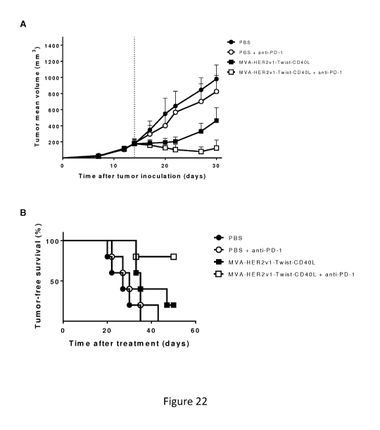
administration of MVA-Twist-Her2-CD4OL combined with anti-PD1 checkpoint
blockade.
C57BL/6 mice bearing 85 mm3 MC38.HER2 colon cancer were immunized IV either
with
MVA-Twist-Her2v1-CD4OL, MVA-Twist-Her2v1-CD4OL and PD-1, PD-1 alone, or
received PBS. Immunization was subsequently followed by anti PD-1 antibody
administration as described in Example 18. Tumor growth was measured at
regular
intervals. Shown are the tumor mean volume (A) and tumor-free survival (B).
[049] Figures 23A, 23B, and 23C show the antitumor effect of intravenous
injection of MVA virus encoding the endogenous retroviral antigen Gp70.
CT26.wt tumor-
bearing Balb/c mice (n=5/group) were grouped and received intravenous (i.v.)
PBS or
5x10⁷ TCID50 MVA, MVA-Gp70, or MVA-Gp70-CD4OL at day 12 after tumor
inoculation. Tumor growth was measured at regular intervals. Shown are tumor
mean
CA 03113818 2021-03-22
WO 2020/070303 PCT/EP2019/076947
12
diameter (Figure 23A) and tumor mean volume (Figure 23B). Seven days after
immunization, blood cells were restimulated and the percentage of CD44+ IFN-y+
cells
among CD8+ T cells in blood upon stimulation is shown (Figure 23C).
[050] Figures 24A and 24B show the antitumor effect of intravenous injection
of
MVA virus encoding the endogenous retroviral antigen Gp70. B16.F10 tumor-
bearing
C57BL/6 mice (n=5/group) were grouped and received intravenous (i.v.) PBS or
5x10⁷
TCID50 MVA, MVA-Gp70 or MVA-Gp70-CD4OL at day 7 after tumor inoculation.
Tumor growth was measured at regular intervals. Shown are tumor mean volume
(Figure
24A) and percentage of CD44+ IFN-y+ cells among CD8+ T cells in blood upon
stimulation with pl5e peptide 7 days after immunization (Figure 24B).
DETAILED DESCRIPTION OF THE INVENTION
[051] It is to be understood that both the foregoing Summary and the following
Detailed Description are exemplary and explanatory only and are not
restrictive of the
invention, as claimed.
[052] CD4OL, when encoded as part of an MVA, was shown to be able to induce
and enhance the overall T-cell response for a disease associated antigen. WO
2014/037124. In WO 2014/037124 it was shown that a recombinant MVA encoding
CD4OL and a heterologous antigen was able to enhance DC activation in vivo,
increase T-
cell responses specific to the heterologous antigen and enhance the quality
and quantity of
CD8 T-cells. Id. To induce synergistic anti-tumor responses, the various
pharmaceutical
combinations of the present invention were developed. In several aspects, the
various
combinations induce both highly effective tumor specific killer T cells and
natural killer
(NK) cells that are able to kill tumor cells when combined with a checkpoint
antagonist or
agonist. This enhanced NK cell and T cell activation when combined with the
enhanced
killer T cell response also induced by the MVA, is shown to synergistically
increase tumor
reduction and overall survival rate in cancer subjects when combined with a
checkpoint
antagonist or agonist.
[053] In one embodiment, the present invention is a combination, or
combination
therapy, comprising: a) an intravenous (IV) administration of a recombinant
MVA that
comprises a first nucleic acid encoding a tumor-associated antigen (TAA) and a
second
CA 03113818 2021-03-22
WO 2020/070303 PCT/EP2019/076947
13
nucleic acid encoding CD4OL, and b) at least one antagonist or agonist of an
immune
checkpoint molecule As noted herein, in various embodiments, the at least one
antagonist
or agonist of an immune checkpoint molecule is selected from a CTLA-4
antagonist, a PD-
1 antagonist, a PD-Li antagonist, a LAG-3 antagonist, a TIM-3 antagonist, and
a ICOS
agonist. Also noted herein, in more preferable embodiments, the at least one
antagonist or
agonist of an immune checkpoint molecule comprises an antibody. Still, in more
preferable embodiments, the CTLA-4 antagonist, PD-1 antagonist, PD-Li
antagonist,
LAG-3 antagonist, TIM-3 antagonist, and the ICOS agonist comprise a CTLA-4
antibody,
a PD-1 antibody, a PD-Li antibody, a LAG-3 antibody, a TIM-3 antibody, and an
ICOS
antibody, respectively.
[054] Described and illustrated in the present application, the combination
and/or
combination therapy of the present invention enhances multiple aspects of a
cancer
patient's immune response. In at least one aspect, the combination
synergistically
enhances both the innate and adaptive immune responses and, when combined with
an
antagonist or agonist of an immune checkpoint molecule, reduces tumor volume
and
increase survival of a cancer patient. One or more of the enhanced effects of
the
combination and/or therapy are summarized as follows.
[055] IV administration of recombinant MVA enhances NK cell response. In
one aspect, the present invention includes a recombinant MVA administered
intravenously
to a subject, wherein the IV administration induces an enhanced innate immune
response,
more particularly an enhanced NK cell response in the subject as compared to a
NK cell
response induced by a non-IV administration of a recombinant MVA to the
subject. Shown
in Figures 1-7 and 10-12, IV administration of recombinant MVA induced a
robust
systemic NK cell response in several compartments in both a single IV
administration and
when administered intravenously as a homologous prime-boost, as compared to a
non-IV
administration.
[056] Illustrated in Figures 1-6, the quality of the NK cell response was
enhanced
as compared to a non-IV administration. The activation marker CD69 is
increased in all
organs analyzed (spleen, liver and lung). The anti-apoptotic Bc1-family member
Bc1-XL,
that enhances NK cell survival, co-stimulatory CD70 and the effector cytokine
IFN-y were
increased both in spleen and lung. Expression of the activating Natural Killer
Group 2D
CA 03113818 2021-03-22
WO 2020/070303 PCT/EP2019/076947
14
(NKG2D) receptor was especially enhanced in liver and lung after IV compared
to SC
injection. NKG2D binds to ligands on tumor cells promoting their elimination
(Garcia-
Cuesta et al., 2015, Reviewed in Spear et al., 2013).
[057] IV administration of recombinant MVA encoding CD4OL further
enhances NK cell response. In another aspect of the present invention it was
determined
that an IV administration of the CD4OL antigen in addition to the recombinant
MVA
further enhanced the NK cell response as compared to an IV administration of
recombinant
MVA alone. As illustrated in Figures 1-7 and 10-12, a recombinant MVA encoding
a
CD4OL antigen induced a stronger NK cell response as compared to a recombinant
MVA
without CD4OL in both a single administration and when administered as a
homologous
prime boost. Further, the quality of the NK cell response was enhanced as
compared to the
IV administration of the recombinant MVA alone. Increased expression by NK
cells of the
effector cytokine IFN-y was observed in all organs analyzed (Figures 1-6,
spleen, liver,
lung), as well as expression of CD69 by NK cells in all organs analyzed
(Figure 5C).
Moreover, Figure 7 shows increased serum levels of IFN-y 6 hours after IV
immunization
with rMVA-CD4OL compared to recombinant MVA and, more importantly of the NK
cell
activating cytokine IL-12p70, both in mice and NHPs. In addition, enhanced
proliferation
of NK cells, demonstrated by the expression of Ki67, was observed not only
systemically
in mice (Figure 7B) but also in NHP peripheral blood (Figure 6F). These
results show that
IV immunization of rMVA-CD4OL compared to rMVA improves NK cell quality in
several animal research models.
[058] While recombinant MVA viruses have been previously administered
intravenously (see, e.g., W02002/42480, W02014/037124), it was previously
understood
that recombinant MVA administration and treatment was associated with
enhancement of
an adaptive immune response, such as CD8 T cell responses. For example, in
W02002/42480, CTL responses were measured after immunizations using non-
recombinant MVA were done either by IV administration of 107 pfu MVA-BN per
mouse,
or by subcutaneous administration of 107 pfu or 108 pfu MVA-BN per mouse. In
W02014/037124, mice were intravenously inoculated with recombinant MVA and
recombinant MVA encoding CD4OL (see, W02014/037124). CTL responses were
CA 03113818 2021-03-22
WO 2020/070303 PCT/EP2019/076947
enhanced and it was determined that an increased immunogenicity of the
recombinant
MVA-CD4OL was independent of CD4 + T cells but dependent upon CD40 in the
host.
[059] In at least one aspect, the enhanced NK cell response seen by the
present
invention is unexpected as it was understood in the art that MVA-induced NK
cell
activation was shown to be dependent on lymph node-resident CD169-positive
subcapsular
sinus (SCS) macrophages after subcutaneous immunization (Garcia et al. (2012)
Blood
120: 4744-50).
[060] In other aspects, the pharmaceutical combination of the present
invention is
administered as part of a homologous and/or heterologous prime-boost regimen.
Illustrated
in Figures 10-12, a recombinant MVA encoding CD4OL administered to a subject
as part of
a homologous and/or heterologous prime boost regimen prolongs and reactivates
enhanced
NK cell responses as well as increases a subject's CD8 and CD4 T cell
responses.
[061] In at least one aspect of the present invention, the enhanced NK cell
responses resulting from the repeated recombinant MVA IV administration and
the
recombinant MVA-CD4OL were surprising. In at least one aspect, it was
surprising to
observe increased NK cell activation and proliferation 24 hours after boost IV
immunizations in the absence of an IFN-a increase. Indeed, it was understood
that NK cell
activation and priming in secondary infections and cancer is largely driven by
IFN-a (see,
e.g., Stackaruk et al. (2013) Expert Rev. Vaccines. 12(8): 875-84; and Mueller
et al. (2017)
Front. Immunol. 8: 304). Surprisingly, no increase in IFN-a serum levels were
observed 6
hours after rMVA hom, rMVA-CD4OL hom or rMVA-CD4OL het IV boost immunizations
(Figure 11C). Altogether, repeated homologous or heterologous IV immunizations
with
rMVA comprising a nucleic acid encoding one or more heterologous antigens
resulted in
unexpected NK cell activation and proliferation independent of IFN-a.
[062] Prior to the present invention, it was understood that CD4OL encoded by
recombinant MVA can substitute for CD4 T cell help (Lauterbach et al. (2013)
Front.
Immunol. 4: 251). Further no effect of recombinant MVA-encoded CD4OL on CD4 T
cells
was known. Unexpectedly, we saw expansion of memory CD4 + T cells 25 days
after prime
immunization (Figure 12B), which corresponds with 4 days after boost IV
immunization
with rMVA-CD4OL (rMVA-CD4OL hom and rMVA-CD4OL het) (Day 21, see Table 1).
This fact is supported by the increased IL-22 production, an important
cytokine indicative
CA 03113818 2021-03-22
WO 2020/070303 PCT/EP2019/076947
16
of T helper cell responses, quantified 6 hours after boost IV immunization in
MVA-CD4OL
horn and MVA-CD4OL het groups (Figure 11D). This unexpected observation is
relevant
for the maintenance of memory responses by rMVA-CD4OL. Furthermore, CD4 T
cells
can support tumor-specific CD8 T cells at the tumor site, avoid activation-
induced cell
death and also become cytotoxic themselves (reviewed in Kennedy and Celis
(2008)
Immunol. Rev. 222: 129-44; Knutson and Disis (2005) Curr. Drug Targets Immune
Endocr. Metabol. Disord. 5: 365-71). These results are unexpected because
other viral
vectors, such as Adenovirus and Herpes Simplex Virus, induce vector-specific
immunity
that impede the induction of immune responses to the vaccine-encoded antigens
upon boost
immunization (Lauterbach et al. (2005) J. Gen. Virol. 86: 2401-10; Pine et al.
(2011) PLoS
One doi:10.1371/journal.pone.0018526).
[063] IV administration of MVA reduces a tumor's immunosuppressive
effects. Illustrated in Figures 13-15 and 19, intravenously administered
recombinant MVA
encoding a heterologous antigen and a CD4OL, induced infiltration of CD8+ T
cells in the
tumor and reduced multiple immunosuppressive effects typically employed by
tumors to
evade the immune system. In addition to increased endogenous CD8+ cells within
the
tumors upon recombinant MVA with or without CD4OL challenge, antigen (OVA)-
specific
T cells were increased in spleen and tumors upon IV administration of a
recombinant MVA
with CD4OL compared to MVA without CD4OL. In addition, HER2 antigen-specific T
cells producing the effector cytokine IFN-y were enhanced in the tumor
microenvironment
upon IV administration of a recombinant MVA with CD4OL (Figure 19).
Surprisingly,
immunosuppressive T regulatory cell (Treg) numbers in the tumor
microenvironment were
decreased when recombinant MVA encoding a heterologous antigen and a CD4OL was
administered (Figure 16).
[064] The recombinant MVA encoding CD4OL in combination with a
checkpoint antagonist or agonist reduces tumor burden and increases survival
rate in
cancer patients. In various embodiments, the combination includes a) an IV
administration of a recombinant MVA encoding a CD4OL and b) an administration
of an
antagonist or agonist of an immune checkpoint molecule. Shown in Figures 20
and 21, the
combinations of the present disclosure resulted in a reduction in tumor volume
and an
increase in overall survival rate. Type I and II interferons, which are
induced by both
CA 03113818 2021-03-22
WO 2020/070303 PCT/EP2019/076947
17
vectors (Figures 11C and 11E), are known inducers of PD-1 and PD-Li expression
(reviewed by Dong et al. (2017) Oncotarget 8: 2171-2186). In at least one
aspect, the
enhanced anti-tumor effects of the pharmaceutical combination (e.g., reduced
tumor
volume and/or increased survival rate) is achieved from the synergistic
combining of
tackling the tumor-induced immune suppressive microenvironment via checkpoint
blockade and the enhancements of the innate and adaptive T cell responses
described
herein. In one exemplary embodiment, these enhancements include one or more of
those
listed above, e.g., an enhanced innate (e.g., NK cell) response, and an
enhanced adaptive T
cell response.
Definitions
[065] As used herein, the singular forms "a," "an," and "the," include plural
references unless the context clearly indicates otherwise. Thus, for example,
reference to
"a nucleic acid" includes one or more of the nucleic acids and reference to
"the method"
includes reference to equivalent steps and methods known to those of ordinary
skill in the
art that could be modified or substituted for the methods described herein.
[066] Unless otherwise indicated, the term "at least" preceding a series of
elements
is to be understood to refer to every element in the series. Those skilled in
the art will
recognize, or be able to ascertain using no more than routine experimentation,
many
equivalents to the specific embodiments of the invention described herein.
Such
equivalents are intended to be encompassed by the present invention.
[067] Throughout this specification and the claims which follow, unless the
context requires otherwise, the word "comprise" and variations such as
"comprises" and
"comprising" will be understood to imply the inclusion of a stated integer or
step or group
of integers or steps but not the exclusion of any other integer or step or
group of integer or
step. When used herein the term "comprising" can be substituted with the term
"containing" or "including" or sometimes when used herein with the term
"having". Any of
the aforementioned terms (comprising, containing, including, having), though
less
preferred, whenever used herein in the context of an aspect or embodiment of
the present
invention can be substituted with the term "consisting of." When used herein,
"consisting
of' excludes any element, step, or ingredient not specified in the claim
element. When used
CA 03113818 2021-03-22
WO 2020/070303 PCT/EP2019/076947
18
herein, "consisting essentially of" does not exclude materials or steps that
do not materially
affect the basic and novel characteristics of the claim.
[068] As used herein, the conjunctive term "and/or" between multiple recited
elements is understood as encompassing both individual and combined options.
For
instance, where two elements are conjoined by "and/or", a first option refers
to the
applicability of the first element without the second. A second option refers
to the
applicability of the second element without the first. A third option refers
to the
applicability of the first and second elements together. Any one of these
options is
understood to fall within the meaning, and therefore satisfy the requirement
of the term
"and/or" as used herein. Concurrent applicability of more than one of the
options is also
understood to fall within the meaning, and therefore satisfy the requirement
of the term
"and/or."
[069] "Mutated" or "modified" protein or antigen as described herein is as
defined
herein any a modification to a nucleic acid or amino acid, such as deletions,
additions,
insertions, and/or substitutions.
[070] A "host cell" as used herein is a cell that has been introduced with a
foreign
molecule, virus, or microorganism. In one non-limiting example, as described
herein, a
cell of a suitable cell culture as, e.g., CEF cells, can be infected with a
poxvirus or, in other
alternative embodiments, with a plasmid vector comprising a foreign or
heterologous gene.
Thus, a suitable host cell and cell cultures serve as a host to poxvirus
and/or foreign or
heterologous gene.
[071] "Percent (%) sequence homology or identity" with respect to nucleic acid
sequences described herein is defined as the percentage of nucleotides in a
candidate
sequence that are identical with the nucleotides in the reference sequence
(i.e., the nucleic
acid sequence from which it is derived), after aligning the sequences and
introducing gaps,
if necessary, to achieve the maximum percent sequence identity, and not
considering any
conservative substitutions as part of the sequence identity. Alignment for
purposes of
determining percent nucleotide sequence identity or homology can be achieved
in various
ways that are within the skill in the art, for example, using publicly
available computer
software such as BLAST, ALIGN, or Megalign (DNASTAR) software. Those skilled
in the
art can determine appropriate parameters for measuring alignment, including
any
CA 03113818 2021-03-22
WO 2020/070303 PCT/EP2019/076947
19
algorithms needed to achieve maximum alignment over the full length of the
sequences
being compared.
[072] For example, an appropriate alignment for nucleic acid sequences is
provided by the local homology algorithm of Smith and Waterman, (1981),
Advances in
Applied Mathematics 2:482- 489. This algorithm can be applied to amino acid
sequences
by using the scoring matrix developed by Dayhoff, Atlas of Protein Sequences
and
Structure, Dayhoff (ed.), 5 suppl. 3: 353-358, National Biomedical Research
Foundation,
Washington, D.C., USA, and normalized by Gribskov (1986), Nucl. Acids Res.
14(6):
6745-6763. An exemplary implementation of this algorithm to determine percent
identity
of a sequence is provided by the Genetics Computer Group (Madison, Wis.) in
the
"BestFit" utility application. The default parameters for this method are
described in the
Wisconsin Sequence Analysis Package Program Manual, Version 8 (1995)
(available from
Genetics Computer Group, Madison, Wis.). A preferred method of establishing
percent
identity in the context of the present invention is to use the MPSRCH package
of programs
copyrighted by the University of Edinburgh, developed by John F. Collins and
Shane S.
Sturrok, and distributed by IntelliGenetics, Inc. (Mountain View, Calif). From
this suite of
packages the Smith-Waterman algorithm can be employed where default parameters
are
used for the scoring table (for example, gap open penalty of 12, gap extension
penalty of
one, and a gap of six). From the data generated the "Match" value reflects
"sequence
identity." Other suitable programs for calculating the percent identity or
similarity between
sequences are generally known in the art, for example, another alignment
program is
BLAST, used with default parameters. For example, BLASTN and BLASTP can be
used
using the following default parameters: genetic code=standard; filter=none;
strand=both;
cutoff=60; expect=10; Matrix=BLOSUM62; Descriptions=50 sequences; sort by=HIGH
SCORE; Databases=non- redundant, GenBank+EMBL+DDBJ+PDB+ GenBank CDS
translations+Swiss protein+Spupdate+PIR. Details of these programs can be
found at the
following intern& address: blast.ncbi.nlm.nih.gov/.
[073] The term "prime-boost vaccination" or "prime-boost regimen" refers to a
vaccination strategy or regimen using a first priming injection of a vaccine
targeting a
specific antigen followed at intervals by one or more boosting injections of
the same
vaccine. Prime-boost vaccination may be homologous or heterologous. A
homologous
CA 03113818 2021-03-22
WO 2020/070303 PCT/EP2019/076947
prime-boost vaccination (sometimes referred to herein as "hom") uses a vaccine
comprising the same antigen and vector for both the priming injection and the
one or more
boosting injections. A heterologous prime-boost vaccination (sometimes
referred to herein
as "het") uses a vaccine comprising the same antigen for both the priming
injection and the
one or more boosting injections but different vectors for the priming
injection and the one
or more boosting injections. For example, a homologous prime-boost vaccination
may use
a recombinant poxvirus comprising nucleic acids expressing one or more
antigens for the
priming injection and the same recombinant poxvirus expressing one or more
antigens for
the one or more boosting injections. In contrast, a heterologous prime-boost
vaccination
may use a recombinant poxvirus comprising nucleic acids expressing one or more
antigens
for the priming injection and a different recombinant poxvirus expressing one
or more
antigens for the one or more boosting injections.
[074] The term "recombinant" means a polynucleotide, virus or vector of
semisynthetic, or synthetic origin which either does not occur in nature or is
linked to
another polynucleotide in an arrangement not found in nature.
[075] As used herein, reducing or a reduction in tumor volume can be
characterized as a reduction in tumor volume and/or size but can also be
characterized in
terms of clinical trial endpoints understood in the art. Some exemplary
clinical trial
endpoints associated with a reduction in tumor volume and/or size can include,
but are not
limited to, Response Rate (RR), Objective response rate (ORR), and so forth.
[076] As used herein an increase in survival rate can be characterized as an
increase in survival of a cancer patient, but can also be characterized in
terms of clinical
trial endpoints understood in the art. Some exemplary clinical trial endpoints
associated
with an increase in survival rate include, but are not limited to, overall
survival rate (OS),
Progression free survival (PFS) and so forth.
[077] As used herein, a "transgene" or "heterologous" gene is understood to be
a
nucleic acid or amino acid sequence which is not present in the wild-type
poxviral genome
(e.g., Vaccinia, Fowlpox, or MVA). The skilled person understands that a
"transgene" or
"heterologous gene", when present in a poxvirus, such as Vaccinia Virus, is to
be
incorporated into the poxviral genome in such a way that, following
administration of the
recombinant poxvirus to a host cell, it is expressed as the corresponding
heterologous gene
CA 03113818 2021-03-22
WO 2020/070303 PCT/EP2019/076947
21
product, i.e., as the "heterologous antigen" and\or "heterologous protein."
Expression is
normally achieved by operatively linking the heterologous gene to regulatory
elements that
allow expression in the poxvirus-infected cell. Preferably, the regulatory
elements include a
natural or synthetic poxviral promoter.
[078] A "vector" refers to a recombinant DNA or RNA plasmid or virus that can
comprise a heterologous polynucleotide. The heterologous polynucleotide may
comprise a
sequence of interest for purposes of prevention or therapy, and may optionally
be in the
form of an expression cassette. As used herein, a vector needs not be capable
of replication
in the ultimate target cell or subject. The term includes cloning vectors and
viral vectors.
Combinations and Methods
[079] In various embodiments, the present invention includes a combination for
treating a cancer patient by reducing tumor volume and/or increasing survival
in the cancer
patient. The combination comprises a) a recombinant MVA comprising a first
nucleic acid
encoding a tumor-associated antigen (TAA) and a second nucleic acid encoding
CD4OL,
that when administered intravenously induces both an enhanced Natural Killer
(NK) cell
response and an enhanced T cell response as compared to a NK cell response and
a T cell
response induced by a non-intravenous administration of a recombinant MVA
virus
comprising a first nucleic acid encoding a TAA and a second nucleic acid
encoding CD4OL
antigen; and b) at least one antagonist or agonist of an immune checkpoint
molecule.
Enhanced NK Cell Response
[080] In one aspect, an "enhanced NK cell response" according to the present
disclosure is characterized by one or more of the following: 1) an increase in
NK cell
frequency, 2) an increase in NK cell activation, and/or 3) an increase in NK
cell
proliferation. Thus, whether an NK cell response is enhanced in accordance
with the
present disclosure can be determined by measuring the expression of one or
more
molecules which are indicative of an increased NK cell frequency, increased NK
cell
activation, and/or increased NK cell proliferation. Exemplary markers that are
useful in
measuring NK cell frequency and/or activity include one or more of: NKp46, IFN-
y, CD69,
CD70, NKG2D, FasL, granzyme B, CD56, and/or Bc1-XL. Exemplary markers that are
CA 03113818 2021-03-22
WO 2020/070303 PCT/EP2019/076947
22
useful in measuring NK cell activation include one or more of IFN-y, CD69,
CD70,
NKG2D, FasL, granzyme B and/or Bel-XL. Exemplary markers that are useful in
measuring NK cell proliferation include: Ki67. These molecules and the
measurement
thereof are validated assays that are understood in the art and can be carried
out according
to known techniques. See, e.g. Borrego et al. ((1999) Immunology 97: 159-165).
Additionally, assays for measuring the molecules can be found in Examples 1-3,
and 9 of
the present disclosure. At least in one aspect, 1) an increase in NK cell
frequency can be
defined as at least a 2-fold increase in CD3-NKp46+ cells compared to pre-
treatment/baseline; 2) an increase in NK cell activation can be defined as at
least a 2-fold
increase in IFN-y, CD69, CD70, NKG2D, FasL, granzyme B and/or Bel-XL
expression
compared to pre-treatment/baseline expression; and/or 3) an increase in NK
cell
proliferation is defined as at least a 1.5 fold increase in Ki67 expression
compared to pre-
treatment/baseline expression.
Enhanced T Cell response
[081] In accordance with the present application, an "enhanced T cell
response" is
characterized by one or more of the following: 1) an increase in frequency of
CD8 T cells;
2) an increase in CD8 T cell activation; and/or 3) an increase in CD8 T cell
proliferation.
Thus, whether a T cell response is enhanced in accordance with the present
application can
be determined by measuring the expression of one or more molecules which are
indicative
of 1) an increase in CD8 T cell frequency 2) an increase in CD8 T cell
activation; and/or 3)
an increase CD8 T cell proliferation. Exemplary markers that are useful in
measuring CD8
T cell frequency, activation, and proliferation include CD3, CD8, IFN-y, TNF-
a, IL-2,
CD69 and/or CD44, and Ki67, respectively. Measuring antigen specific T cell
frequency
can also be measured by ELIspot or MHC Multimers such as pentamers or
dextramers as
shown by the present application. Such measurements and assays are validated
and
understood in the art.
[082] In one aspect, an increase in CD8 T cell frequency is characterized by
an at
least a 2-fold increase in IFN-y and/or dextramer+ CD8 T cells compared to pre-
treatment/baseline. An increase in CD8 T cell activation is characterized as
at least a 2-fold
increase in CD69 and/or CD44 expression compared to pre-treatment/baseline
expression.
CA 03113818 2021-03-22
WO 2020/070303 PCT/EP2019/076947
23
An increase in CD8 T cell proliferation is characterized as at least a 2-fold
increase in Ki67
expression compared to pre-treatment/baseline expression.
[083] In an alternative aspect, an enhanced T cell response is characterized
by an
increase in CD8 T cell expression of effector cytokines and/or an increase of
cytotoxic
effector functions. An increase in expression of effector cytokines can be
measured by
expression of one or more of IFN-y, TNF-a, and/or IL-2 compared to pre-
treatment/baseline. An increase in cytotoxic effector functions can be
measured by
expression of one or more of CD107a, granzyme B, and/or perform and/or antigen-
specific
killing of target cells.
[084] The assays, cytokines, markers, and molecules described herein and the
measurement thereof are validated and understood in the art and can be carried
out
according to known techniques. Additionally, assays for measuring the T cells
responses
can be found in Examples 3,7,10 and 15, wherein T cell responses were
analyzed.
[085] The enhanced T cell response realized by the present invention is
particularly advantageous in combination with the enhanced NK cell response,
as the
enhanced T cells effectively target and kill those tumor cells that have
evaded and/or
survived past the initial innate immune responses in the cancer patient.
Furthermore,
antibody treatment can enhance MHC class I presentation of TAAs, resulting in
higher
susceptibility of TAA-expressing tumors to lysis by TAA- specific T cells
(Kono et al.
(2004) Clin. Cancer Res. 10: 2538-44).
[086] In additional embodiments, the combination further comprises at least
one
antagonist or agonist of an immune checkpoint molecule. In preferred
embodiments, the at
least one antagonist or agonist of an immune checkpoint molecule comprises a
CTLA-4
antagonist, a PD-1 antagonist, a -PD-Li antagonist, a LAG-3 antagonist, a TIM-
3
antagonist, or an ICOS agonist. In more preferred embodiments, the at least
one antagonist
or agonist of an immune checkpoint molecule comprises a CTLA-4 antagonist, a
PD-1
antagonist, or a -PD-Li antagonist.
[087] In still more embodiments, the at least one of antagonist or agonist of
an
immune checkpoint molecule comprises an antibody able to block the function of
the
immune checkpoint molecule. In preferred embodiments, the antibody is selected
from
CTLA-4 antibody, a PD-1 antibody, a PD-Li antibody, a LAG-3 antibody, an ICOS
CA 03113818 2021-03-22
WO 2020/070303 PCT/EP2019/076947
24
antibody, and a TIM-3 antibody, respectively. In more preferred embodiments
the at least
one antagonist or agonist comprises a CTLA-4, a PD-1, or a PD-Li antibody.
[088] In yet additional embodiments, the combinations and methods described
herein are for use in treating a human cancer patient. In preferred
embodiments, the cancer
patient is suffering from and/or is diagnosed with a cancer selected from the
group
consisting of: breast cancer, lung cancer, head and neck cancer, thyroid,
melanoma, gastric
cancer, bladder cancer, kidney cancer, liver cancer, melanoma, pancreatic
cancer, prostate
cancer, ovarian cancer, urothelial, cervical, or colorectal cancer. In yet
additional
embodiments, the combinations and methods described herein are for use in
treating a
human cancer patient suffering from and/or diagnosed with a breast cancer,
colorectal
cancer or melanoma, preferably a melanoma, more preferably a colorectal cancer
or most
preferably a colorectal cancer.
Certain Exemplary Tumor-Associated Antigens
[089] In certain embodiments, an immune response is produced in a subject
against a cell-associated polypeptide antigen. In certain such embodiments, a
cell-
associated polypeptide antigen is a tumor-associated antigen (TAA).
[090] The term "polypeptide" refers to a polymer of two or more amino acids
joined to each other by peptide bonds or modified peptide bonds. The amino
acids may be
naturally occurring as well as non-naturally occurring, or a chemical analogue
of a
naturally occurring amino acid. The term also refers to proteins, i.e.
functional
biomolecules comprising at least one polypeptide; when comprising at least two
polypeptides, these may form complexes, be covalently linked, or may be non-
covalently
linked. The polypeptide(s) in a protein can be glycosylated and/or lipidated
and/or
comprise prosthetic groups.
[091] Preferably, the TAA includes, but is not limited to, HER2, PSA, PAP,
CEA,
MUC-1, survivin, TRP1, TRP2, or Brachyury alone or in combinations. Such
exemplary
combination may include CEA and MUC-1, also known as CV301. Other exemplary
combinations may include PAP and PSA.
[092] Numerous TAAs are known in the art. Exemplary TAAs include, but are
not limited to, 5 alpha reductase, alpha-fetoprotein, AM-1, APC, April, BAGE,
beta-
CA 03113818 2021-03-22
WO 2020/070303 PCT/EP2019/076947
catenin, Bc112, bcr-abl, CA-125, CASP-8/FLICE, Cathepsins, CD19, CD20, CD21,
CD23,
CD22, CD33 CD35, CD44, CD45, CD46, CD5, CD52, CD55, CD59, CDC27, CDK4,
CEA, c-myc, Cox-2, DCC, DcR3, E6/E7, CGFR, EMBP, Dna78, farnesyl transferase,
FGF8b, FGF8a, FLK-1/KDR, folic acid receptor, G250, GAGE-family, gastrin 17,
gastrin-
releasing hormone, GD2/GD3/GM2, GnRH, GnTV, GP1, gp100/Pme117, gp-100-in4,
gp15, gp75/TRP1, hCG, heparanse, Her2/neu, HMTV, Hsp70, hTERT, IGFR1, IL-13R,
iNOS, Ki67, KIAA0205, K-ras, H-ras, N-ras, KSA, LKLR-FUT, MAGE-family,
mammaglobin, MAP17, melan-A/MART-1, mesothelin, MIC A/B, MT-MMPs, mucin,
NY-ESO-1, osteonectin, p15, P170/MDR1, p53, p97/melanotransferrin, PAT-1,
PDGF,
uPA, PRAME, probasin, progenipoientin, PSA, PSM, RAGE-1, Rb, RCAS1, SART-1,
SSX-family, STAT3, STn, TAG-72, TGF-alpha, TGF-beta, Thymosin-beta-15, TNF-
alpha,
TRP1, TRP2, tyrosinase, VEGF, ZAG, pl6INK4, and glutathione-S-transferase.
[093] A preferred PSA antigen comprises the amino acid change of isoleucine to
leucine at position 155. See U.S. Patent 7,247,615, which is incorporated
herein by
reference.
[094] In one or more preferred embodiments of present invention, the
heterologous TAA is selected from HER2 and/or Brachyury.
[095] In various additional embodiments, the TAA may include a mutated or
modified HER2 antigen selected from HER2v1 and HER2v2. HER2v1 and HER2v2
comprise SEQ ID NO: 1 and SEQ ID NO: 3, respectively. The HER2v1 and HER2v2
antigen may be encoded by nucleic acids comprising SEQ ID NOs: 2 and 4,
respectively.
[096] In preferred embodiments, the HER2 antigen comprises an amino acid
sequence having at least 90%, 95%, 97% 98%, or 99% identity to SEQ ID NOs:1 or
3. In a
most preferred embodiment, the HER2 antigen comprises SEQ ID NOs: 1 or 3.
[097] In additional embodiments, the TAA may include a Brachyury antigen. In
preferred embodiments, the Brachyury antigen comprises an amino acid sequence
having at
least 90%, 95%, 97% 98%, or 99% identity to SEQ ID NOs: 5,7, 9, or 11. In
still
additional embodiments, the Brachyury antigen is selected from SEQ ID NOs: 5,
7, 9, and
11, which may be encoded by nucleic acids comprising SEQ ID NOs: 6, 8, 10, and
12,
respectively.
Modified Tumor-Associated Antigens
CA 03113818 2021-03-22
WO 2020/070303 PCT/EP2019/076947
26
[098] In certain embodiments, a cell-associated polypeptide antigen is
modified
such that a CTL response is induced against a cell which presents epitopes
derived from a
polypeptide antigen on its surface, when presented in association with an MHC
Class I
molecule on the surface of an APC. In certain such embodiments, at least one
first foreign
TH epitope, when presented, is associated with an MHC Class II molecule on the
surface of
the APC. In certain such embodiments, a cell-associated antigen is a tumor-
associated
antigen.
[099] Exemplary APCs capable of presenting epitopes include dendritic cells
and
macrophages. Additional exemplary APCs include any pino- or phagocytizing APC,
which
is capable of simultaneously presenting 1) CTL epitopes bound to MHC class I
molecules
and 2) TH epitopes bound to MHC class II molecules.
[0100] In certain embodiments, modifications to one or more of the TAAs, such
as,
but not limited to, CEA, MUC-1, PAP, PSA, HER2, survivin, TRP1, TRP2, or
Brachyury,
are made such that, after administration to a subject, polyclonal antibodies
are elicited that
predominantly react with the one or more of the TAAs described herein. Such
antibodies
could attack and eliminate tumor cells as well as prevent metastatic cells
from developing
into metastases. The effector mechanism of this anti-tumor effect would be
mediated via
complement and antibody dependent cellular cytotoxicity. In addition, the
induced
antibodies could also inhibit cancer cell growth through inhibition of growth
factor
dependent oligo-dimerisation and internalization of the receptors. In certain
embodiments,
such modified TAAs could induce CTL responses directed against known and/or
predicted
TAA epitopes displayed by the tumor cells.
[0101] In certain embodiments, a modified TAA polypeptide antigen comprises a
CTL epitope of the cell-associated polypeptide antigen and a variation,
wherein the
variation comprises at least one CTL epitope or a foreign TH epitope. Certain
such
modified TAAs can include in one non-limiting example one or more HER2
polypeptide
antigens comprising at least one CTL epitope and a variation comprising at
least one CTL
epitope of a foreign TH epitope, and methods of producing the same, are
described in U.S.
Patent No. 7,005,498 and U.S. Patent Pub. Nos. 2004/0141958 and 2006/0008465.
[0102] Certain such modified TAAs can include in one non-limiting example one
or more MUC-1 polypeptide antigens comprising at least one CTL epitope and a
variation
CA 03113818 2021-03-22
WO 2020/070303 PCT/EP2019/076947
27
comprising at least one CTL epitope of a foreign epitope, and methods of
producing the
same, are described in U.S. Patent Pub. Nos. 2014/0363495.
[0103] Additional promiscuous T-cell epitopes include peptides capable of
binding
a large proportion of HLA-DR molecules encoded by the different HLA-DR. See,
e.g.,
WO 98/23635 (Frazer IH et al., assigned to The University of Queensland);
Southwood et
al. (1998) J. Immunol. 160: 3363 3373; Sinigaglia et al. (1988) Nature 336:
778 780;
Rammensee et al. (1995) Immunogenetics 41: 178-228; Chicz et al. (1993) J.
Exp. Med.
178: 27-47; Hammer et al. (1993) Cell 74: 197-203; and Falk et al. (1994)
Immunogenetics
39: 230-242. The latter reference also deals with HLA-DQ and -DP ligands. All
epitopes
listed in these references are relevant as candidate natural epitopes as
described herein, as
are epitopes which share common motifs with these.
[0104] In certain other embodiments, the promiscuous T-cell epitope is an
artificial
T-cell epitope which is capable of binding a large proportion of haplotypes.
In certain such
embodiments, the artificial T-cell epitope is a pan DR epitope peptide
("PADRE") as
described in WO 95/07707 and in the corresponding paper Alexander et al.
(1994)
Immunity 1: 751 761.
CD4OL
[0105] As illustrated by the present disclosure the inclusion of CD4OL as part
of the
combination and related method further enhances the decrease in tumor volume,
prolongs
progression-free survival and increase survival rate realized by the present
invention.
Thus, in various embodiments, the combination further comprises administering
CD4OL to
a cancer patient. In preferred embodiments, the CD4OL is encoded as part of a
recombinant MVA as described herein.
[0106] While CD40 is constitutively expressed on many cell types, including B
cells, macrophages, and dendritic cells, its ligand CD4OL is predominantly
expressed on
activated T helper cells. The cognate interaction between dendritic cells and
T helper cells
early after infection or immunization 'licenses' dendritic cells to prime CTL
responses.
Dendritic cell licensing results in the up-regulation of co-stimulatory
molecules, increased
survival and better cross-presenting capabilities. This process is mainly
mediated via
CD40/CD4OL interaction. However, various configurations of CD4OL are
described, from
CA 03113818 2021-03-22
WO 2020/070303 PCT/EP2019/076947
28
membrane bound to soluble (monomeric to trimeric) which induce diverse
stimuli, either
inducing or repressing activation, proliferation, and differentiation of APCs.
[0107] In one or more preferred embodiments, CD4OL is encoded by the MVA of
the present invention. In one or more other preferred embodiments, CD4OL is a
human
CD4OL. In still more preferred embodiments, the CD4OL comprises a nucleic acid
having
at least 90%, 95%, 97% 98%, or 99% identity to SEQ ID NO:13. In even more
preferred
embodiments, the CD4OL comprises a nucleic acid encoding SEQ ID NO: 13. In a
most
preferred embodiment, the CD4OL comprises SEQ ID NO:13. In additional
embodiments,
the CD4OL is encoded by a nucleic acid having at least 90%, 95%, 97% 98%, or
99%
identity to SEQ ID NO:14. In a most preferred embodiment, the nucleic acid
comprises
SEQ ID NO:14
Antagonists of Immune Checkpoint Molecules
[0108] As described herein, at least in one aspect, the invention encompasses
the
use of immune checkpoint antagonists. Such immune checkpoint antagonists
function to
interfere with and/or block the function of the immune checkpoint molecule.
Some
preferred immune checkpoint antagonists include, Cytotoxic T-Lymphocyte
Antigen 4
(CTLA-4), Programmed Cell Death Protein 1 (PD-1), Programmed Death-Ligand 1
(PD-
L1), Lymphocyte-activation gene 3 (LAG-3), and T-cell immunoglobulin and mucin
domain 3 (TIM-3).
[0109] Additionally, exemplary immune checkpoint antagonists can include, but
are not limited to CTLA-4, PD-1, PD-L1, PD-L2, LAG-3, TIM-3, T cell
Immunoreceptor
with Ig and ITIM domains (TIGIT) and V-domain Ig Suppressor of T cell
Activation
(VISTA).
[0110] Such antagonists of the immune checkpoint molecules can include
antibodies which specifically bind to immune checkpoint molecules and inhibit
and/or
block biological activity and function of the immune checkpoint molecule.
[0111] Other antagonists of the immune checkpoint molecules can include
antisense nucleic acids RNAs that interfere with the expression of the immune
checkpoint
molecules; and small interfering RNAs that interfere with the expression of
the immune
checkpoint molecules.
CA 03113818 2021-03-22
WO 2020/070303 PCT/EP2019/076947
29
[0112] Antagonists can additionally be in the form of small molecules that
inhibit
or block the function of the immune checkpoint. Some non-limiting examples of
these
include NP12 (Aurigene), (D) PPA-1 by Tsinghua Univ, high affinity PD-1
(Stanford);
BMS-202 and BMS-8 (Bristol Myers Squibb (BMS), and CA170/ CA327
(Curis/Aurigene); and small molecule inhibitors of CTLA-4, PD-1, PD-L1, LAG-3,
and
TIM-3.
[0113] Antagonists can additionally be in the form of Anticalins that inhibit
or
block the function of the immune checkpoint molecule. See, e.g., Rothe et al.
(2018)
BioD rugs 32: 233-243.
[0114] It is contemplated that antagonists can additionally be in the form of
Affimers . Affimers are Fc Fusion proteins that inhibit or block the function
of the
immune checkpoint molecule. Other Fusion proteins that can serve as
antagonists of
immune checkpoints are immune checkpoint fusion proteins (e.g., anti-PD-1
protein AMP-
224) and anti-PD-Li proteins such as those described in US2017/0189476.
[0115] Candidate antagonists of immune checkpoint molecules can be screened
for
function by a variety of techniques known in the art and/or disclosed within
the instant
application, such as for the ability to interfere with the immune checkpoint
molecules
function in an in vitro or mouse model.
Agonist of ICOS
[0116] The invention further encompasses agonists of ICOS. An agonist of ICOS
activates ICOS. ICOS is a positive co-stimulatory molecule expressed on
activated T cells
and binding to its ligand promotes their proliferation (Dong (2001) Nature
409: 97-101).
[0117] In one embodiment, the agonist is ICOS-L, an ICOS natural ligand. The
agonist can be a mutated form of ICOS-L that retains binding and activation
properties.
Mutated forms of ICOS-L can be screened for activity in stimulating ICOS in
vitro.
Antibodies
[0118] In one embodiment, the antagonist and/or agonist of an immune
checkpoint
molecules each comprises an antibody. Antibodies can be synthetic, monoclonal,
or
polyclonal and can be made by techniques well known in the art. Such
antibodies
specifically bind to the immune checkpoint molecule via the antigen-binding
sites of the
CA 03113818 2021-03-22
WO 2020/070303 PCT/EP2019/076947
antibody (as opposed to non-specific binding). Immune checkpoint peptides,
fragments,
variants, fusion proteins, etc., can be employed as immunogens in producing
antibodies
immunoreactive therewith. More specifically, the polypeptides, fragment,
variants, fusion
proteins, etc. contain antigenic determinants or epitopes that elicit the
formation of
antibodies.
[0119] In preferred embodiments, the antibodies of present invention are those
that
are approved, or in the process of approval by the government of a sovereign
nation, for the
treatment of a human cancer patient. Some non-limiting examples of these
antibodies
already approved, or in the approval process include the following: CTLA-
4(Ipilimumab
and Tremelimumab); PD-1 (Pembrolizumab, Lambrolizumab, Amplimmune-224 (AMP-
224), Amplimmune -514 (AMP-514), Nivolumab, MK-3475 (Merck), . BI 754091
(Boehringer Ingelheim)), and PD-Li (Atezolizumab, Avelulmab, Durvalumab,
MPDL3280A (Roche), MED14736 (AZN), MSB0010718C (Merck)); LAG-3 (IMP321,
BMS-986016, BI754111 (Boehringer Ingelheim), LAG525 (Novartis), MK-4289
(Merck),
TSR-033 (Tesaro).
[0120] These antigenic determinants or epitopes can be either linear or
conformational (discontinuous). Linear epitopes are composed of a single
section of amino
acids of the polypeptide, while conformational or discontinuous epitopes are
composed of
amino acids sections from different regions of the polypeptide chain that are
brought into
close proximity upon protein folding (Janeway, Jr. and Travers, ImmunoBiology
3: 9
(Garland Publishing Inc., 2nd ed. 1996)). Because folded proteins have complex
surfaces,
the number of epitopes available is quite numerous; however, due to the
conformation of
the protein and steric hindrances, the number of antibodies that actually bind
to the epitopes
is less than the number of available epitopes (Janeway, Jr. and Travers,
ImmunoBiology 2:
14 (Garland Publishing Inc., 2nd ed. 1996)). Epitopes can be identified by any
of the
methods known in the art.
[0121] Antibodies, including scFV fragments, which bind specifically to the
immune checkpoint molecules such as CTLA-4, PD-1, PD-L1, LAG-3, TIM-3, or ICOS
and either block its function ("antagonist antibodies") or enhance/ activate
its function
("agonist antibodies"), are encompassed by the invention. Such antibodies can
be
generated by conventional means.
CA 03113818 2021-03-22
WO 2020/070303 PCT/EP2019/076947
31
[0122] In one embodiment, the invention encompasses monoclonal antibodies
against immune checkpoint molecules that either block ("antagonist
antibodies") or
enhance/activate ("agonist antibodies") the function of the immune checkpoint
molecules.
Exemplary blocking monoclonal antibodies against PD-1 are described in WO
2011/041613, which is hereby incorporated by reference.
[0123] Antibodies are capable of binding to their targets with both high
avidity and
specificity. They are relatively large molecules (-150kDa), which can
sterically inhibit
interactions between two proteins (e.g., PD-1 and its target ligand) when the
antibody
binding site falls within proximity of the protein-protein interaction site.
The invention
further encompasses antibodies that bind to epitopes within close proximity to
an immune
checkpoint molecule ligand binding site.
[0124] In various embodiments, the invention encompasses antibodies that
interfere
with intermolecular interactions (e.g., protein-protein interactions), as well
as antibodies
that perturb intramolecular interactions (e.g., conformational changes within
a molecule).
Antibodies can be screened for the ability to block or enhance/activate the
biological
activity of an immune checkpoint molecule.
[0125] Both polyclonal and monoclonal antibodies can be prepared by
conventional
techniques.
[0126] In one exemplary aspect, the immune checkpoint molecules CTLA-4, PD-1,
PD-L1, LAG-3, TIM-3, and ICOS and peptides based on the amino acid sequence of
CTLA-4, PD-1, PD-L1, LAG-3, TIM-3, and ICOS can be utilized to prepare
antibodies
that specifically bind to CTLA-4, PD-1, PD-L1, LAG-3, TIM-3, or ICOS. The term
"antibodies" is meant to include polyclonal antibodies, monoclonal antibodies,
fragments
thereof, such as F(ab')2 and Fab fragments, single-chain variable fragments
(scFvs), single-
domain antibody fragments (VHHs or Nanobodies), bivalent antibody fragments
(diabodies), as well as any recombinantly and synthetically produced binding
partners.
[0127] In another exemplary aspect, antibodies are defined to be specifically
binding if they to an immune checkpoint molecule if they bind with a Ka of
greater than or
equal to about 107 M-1. Affinities of binding partners or antibodies can be
readily
determined using conventional techniques, for example those described by
Scatchard et al.
((1949) Ann. N.Y. Acad. Sci. 51: 660).
CA 03113818 2021-03-22
WO 2020/070303 PCT/EP2019/076947
32
[0128] Polyclonal antibodies can be readily generated from a variety of
sources,
for example, horses, cows, goats, sheep, dogs, chickens, rabbits, mice, or
rats, using
procedures that are well known in the art. In general, purified CTLA-4, PD-1,
PD-L1,
LAG-3, TIM-3, and ICOS or a peptide based on the amino acid sequence of CTLA-
4, PD-
1, PD-L1, LAG-3, TIM-3, and ICOS that is appropriately conjugated is
administered to the
host animal typically through parenteral injection. The immunogenicity of CTLA-
4, PD-1,
PD-Li LAG-3, TIM-3, and ICOS can be enhanced through the use of an adjuvant,
for
example, Freund's complete or incomplete adjuvant. Following booster
immunizations,
small samples of serum are collected and tested for reactivity to CTLA-4, PD-
1, PD-L1,
LAG-3, TIM-3, and ICOS polypeptide. Examples of various assays useful for such
determination include those described in Antibodies: A Laboratory Manual,
Harlow and
Lane (eds.), Cold Spring Harbor Laboratory Press, 1988; as well as procedures,
such as
countercurrent immuno-electrophoresis (CIEP), radioimmunoassay, radio-
immunoprecipitation, enzyme-linked immunosorbent assays (ELISA), dot blot
assays, and
sandwich assays. See U.S. Pat. Nos. 4,376,110 and 4,486,530.
[0129] Monoclonal antibodies can be readily prepared using well known
procedures. See, for example, the procedures described in U.S. Pat. Nos. RE
32,011,
4,902,614, 4,543,439, and 4,411,993; Monoclonal Antibodies, Hybridomas: A New
Dimension in Biological Analyses, Plenum Press, Kennett, McKeam, and Bechtol
(eds.),
1980.
[0130] For example, the host animals, such as mice, can be injected
intraperitoneally at least once and preferably at least twice at about 3 week
intervals with
isolated and purified immune checkpoint molecule optionally in the presence of
adjuvant.
Mouse sera are then assayed by conventional dot blot technique or antibody
capture (ABC)
to determine which animal is best to fuse. Approximately two to three weeks
later, the
mice are given an intravenous boost of the immune checkpoint molecule. Mice
are later
sacrificed, and spleen cells fused with commercially available myeloma cells,
such as
Ag8.653 (ATCC), following established protocols. Briefly, the myeloma cells
are washed
several times in media and fused to mouse spleen cells at a ratio of about
three spleen cells
to one myeloma cell. The fusing agent can be any suitable agent used in the
art, for
example, polyethylene glycol (PEG). Fusion is plated out into plates
containing media that
CA 03113818 2021-03-22
WO 2020/070303 PCT/EP2019/076947
33
allows for the selective growth of the fused cells. The fused cells can then
be allowed to
grow for approximately eight days. Supernatants from resultant hybridomas are
collected
and added to a plate that is first coated with goat anti-mouse Ig. Following
washes, a label,
such as a labeled immune checkpoint molecule polypeptide, is added to each
well followed
by incubation. Positive wells can be subsequently detected. Positive clones
can be grown
in bulk culture and supernatants are subsequently purified over a Protein A
column
(Pharmacia).
[0131] The monoclonal antibodies of the invention can be produced using
alternative techniques, such as those described by Alting-Mees et al. (1990)
Strategies in
Molecular Biology 3: 1-9, "Monoclonal Antibody Expression Libraries: A Rapid
Alternative to Hybridomas," which is incorporated herein by reference.
Similarly, binding
partners can be constructed using recombinant DNA techniques to incorporate
the variable
regions of a gene that encodes a specific binding antibody. Such a technique
is described
in Larrick et al. ((1989) Biotechnology 7: 394).
[0132] Antigen-binding fragments of such antibodies, which can be produced by
conventional techniques, are also encompassed by the present invention.
Examples of such
fragments include, but are not limited to, Fab and F(ab')2 fragments. Antibody
fragments
and derivatives produced by genetic engineering techniques are also provided.
[0133] The monoclonal antibodies of the present invention include chimeric
antibodies, e.g., humanized versions of murine monoclonal antibodies. Such
humanized
antibodies can be prepared by known techniques and offer the advantage of
reduced
immunogenicity when the antibodies are administered to humans. In one
embodiment, a
humanized monoclonal antibody comprises the variable region of a murine
antibody (or
just the antigen binding site thereof) and a constant region derived from a
human antibody.
Alternatively, a humanized antibody fragment can comprise the antigen binding
site of a
murine monoclonal antibody and a variable region fragment (lacking the antigen-
binding
site) derived from a human antibody. Procedures for the production of chimeric
and further
engineered monoclonal antibodies include those described in Riechmann et al.
(1988)
Nature 332: 323; Liu et al. (1987) Proc. Nat'l. Acad. Sci. 84: 3439; Larrick
et al. (1989)
Bio/Technology 7: 934; and Winter and Harris (1993) TIPS 14: 139. Procedures
to
CA 03113818 2021-03-22
WO 2020/070303 PCT/EP2019/076947
34
generate antibodies transgenically can be found in GB 2,272,440 and U.S. Pat.
Nos.
5,569,825 and 5,545,806, each of which is incorporated by reference herein.
[0134] Antibodies produced by genetic engineering methods, such as chimeric
and
humanized monoclonal antibodies, comprising both human and non-human portions,
which
can be made using standard recombinant DNA techniques, can be used. Such
chimeric and
humanized monoclonal antibodies can be produced by genetic engineering using
standard
DNA techniques known in the art, for example using methods described in
Robinson et al.,
International Publication No. WO 87/02671; Akira et al., European Patent
Application
0184187; Taniguchi, European Patent Application 0171496; Morrison et al.,
European
Patent Application 0173494; Neuberger et al., PCT International Publication
No. WO
86/01533; Cabilly et al., U.S. Pat. No. 4,816,567; Cabilly et al., European
Patent
Application 0125023; Better et al. (1988) Science 240: 1041-1043; Liu et al.
(1987) Proc.
Nat'l. Acad. Sci. 84: 3439-3443; Liu et al. (1987) J. Immunol. 139: 3521-3526;
Sun et al.
(1987) Proc. Nat'l. Acad. Sci. 84: 214-218; Nishimura et al. (1987) Cancer
Res. 47: 999-
1005; Wood et al. (1985) Nature 314: 446-449; and Shaw et al. (1988) J. Nat'l.
Cancer
Inst. 80: 1553-1559; Morrison (1985) Science 229: 1202-1207; Oi et al. (1986)
BioTechniques 4: 214; Winter, U.S. Pat. No. 5,225,539; Jones et al. (1986)
Nature 321:
552-525; Verhoeyan et al. (1988) Science 239: 1534; and Beidler et al. (1988)
J. Immunol.
141: 4053-4060.
[0135] In connection with synthetic and semi-synthetic antibodies, such terms
are
intended to cover but are not limited to antibody fragments, isotype switched
antibodies,
humanized antibodies (e.g., mouse-human, human-mouse), hybrids, antibodies
having
plural specificities, and fully synthetic antibody-like molecules.
[0136] For therapeutic applications, "human" monoclonal antibodies having
human
constant and variable regions are often preferred so as to minimize the immune
response of
a patient against the antibody. Such antibodies can be generated by immunizing
transgenic
animals which contain human immunoglobulin genes. See Jakobovits et al. (1995)
Ann.
NY Acad. Sci. 764: 525-535.
[0137] Human monoclonal antibodies against an immune checkpoint molecule can
also be prepared by constructing a combinatorial immunoglobulin library, such
as a Fab
phage display library or a scFv phage display library, using immunoglobulin
light chain
CA 03113818 2021-03-22
WO 2020/070303 PCT/EP2019/076947
and heavy chain cDNAs prepared from mRNA derived from lymphocytes of a
subject.
See, e.g., McCafferty et al., PCT publication WO 92/01047; Marks et al. (1991)
J. Mol.
Biol. 222:581-597; and Griffths et al. (1993) EMBO J. 12: 725-734. In
addition, a
combinatorial library of antibody variable regions can be generated by
mutating a known
human antibody. For example, a variable region of a human antibody known to
bind the
immune checkpoint molecule can be mutated, by for example using randomly
altered
mutagenized oligonucleotides, to generate a library of mutated variable
regions which can
then be screened to bind to the immune checkpoint molecule. Methods of
inducing random
mutagenesis within the CDR regions of immunoglobin heavy and/or light chains,
methods
of crossing randomized heavy and light chains to form pairings and screening
methods can
be found in, for example, Barbas et al., PCT publication WO 96/07754; Barbas
et al.
(1992) Proc. Nat'l Acad. Sci. USA 89: 4457-4461.
[0138] An immunoglobulin library can be expressed by a population of display
packages, preferably derived from filamentous phage, to form an antibody
display library.
Examples of methods and reagents particularly amenable for use in generating
antibody
display library can be found in, for example, Ladner et al., U.S. Pat. No.
5,223,409; Kang
et al., PCT publication WO 92/18619; Dower et al., PCT publication WO
91/17271;
Winter et al. PCT publication WO 92/20791; Markland et al. PCT publication WO
92/15679; Breitling et al. PCT publication WO 93/01288; McCafferty et al. PCT
publication WO 92/01047; Garrard et al. PCT publication WO 92/09690; Ladner et
al.
PCT publication WO 90/02809; Fuchs et al. (1991) Bio/Technology 9: 1370 1372;
Hay et
al. (1992) Hum. Antibod. Hybridomas 3: 81-85; Huse et al. (1989) Science 246:
1275-
1281; Griffiths et al. (1993) supra; Hawkins et al. (1992) J. Mol. Biol. 226:
889-896;
Clackson et al. (1991) Nature 352: 624-628; Gram et al. (1992) Proc. Nat'l.
Acad. Sci. 89:
3576-3580; Garrad et al. (1991) Bio/Technology 9: 1373-1377; Hoogenboom et al.
(1991)
Nucl. Acid Res. 19: 4133-4137; and Barbas et al. (1991) Proc. Nat'l. Acad.
Sci. 88: 7978-
7982. Once displayed on the surface of a display package (e.g., filamentous
phage), the
antibody library is screened to identify and isolate packages that express an
antibody that
binds an immune checkpoint molecule.
Recombinant MVA
CA 03113818 2021-03-22
WO 2020/070303 PCT/EP2019/076947
36
[0139] In more preferred embodiments of the present invention, the one or more
proteins and nucleotides disclosed herein are included in a recombinant MVA.
As
described and illustrated by the present disclosure, the intravenous
administration of the
recombinant MVAs of the present disclosure induces in various aspects an
enhanced
immune response in cancer patients. Thus, in one or more preferred
embodiments, the
invention includes a recombinant MVA comprising a first nucleic acid encoding
one or
more of the TAAs described herein and a second nucleic acid encoding CD4OL.
[0140] Example of MVA virus strains that are useful in the practice of the
present
invention and that have been deposited in compliance with the requirements of
the
Budapest Treaty are strains MVA 572, deposited at the European Collection of
Animal
Cell Cultures (ECACC), Vaccine Research and Production Laboratory, Public
Health
Laboratory Service, Centre for Applied Microbiology and Research, Porton Down,
Salisbury, Wiltshire 5P4 OJG, United Kingdom, with the deposition number ECACC
94012707 on January 27, 1994, and MVA 575, deposited under ECACC 00120707 on
December 7, 2000, MVA-BN, deposited on Aug. 30, 2000 at the European
Collection of
Cell Cultures (ECACC) under number V00083008, and its derivatives, are
additional
exemplary strains.
[0141] "Derivatives" of MVA-BN refer to viruses exhibiting essentially the
same
replication characteristics as MVA-BN, as described herein, but exhibiting
differences in
one or more parts of their genomes. MVA-BN, as well as derivatives thereof,
are
replication incompetent, meaning a failure to reproductively replicate in vivo
and in vitro.
More specifically in vitro, MVA-BN or derivatives thereof have been described
as being
capable of reproductive replication in chicken embryo fibroblasts (CEF), but
not capable of
reproductive replication in the human keratinocyte cell line HaCat (Boukamp et
al. (1988)
J. Cell Biol. 106: 761-771), the human bone osteosarcoma cell line 143B (ECACC
Deposit
No. 91112502), the human embryo kidney cell line 293 (ECACC Deposit No.
85120602),
and the human cervix adenocarcinoma cell line HeLa (ATCC Deposit No. CCL-2).
Additionally, MVA-BN or derivatives thereof have a virus amplification ratio
at least two-
fold less, more preferably three-fold less than MVA-575 in Hela cells and
HaCaT cell
lines. Tests and assay for these properties of MVA-BN and derivatives thereof
are
CA 03113818 2021-03-22
WO 2020/070303 PCT/EP2019/076947
37
described in WO 02/42480 (U.S. Patent Application No. 2003/0206926) and WO
03/048184 (U.S. Patent Application No. 2006/0159699).
[0142] The term "not capable of reproductive replication" or "no capability of
reproductive replication" in human cell lines in vitro as described in the
previous
paragraphs is, for example, described in WO 02/42480, which also teaches how
to obtain
MVA having the desired properties as mentioned above. The term applies to a
virus that
has a virus amplification ratio in vitro at 4 days after infection of less
than 1 using the
assays described in WO 02/42480 or in U.S. Patent No. 6,761,893.
[0143] The term "failure to reproductively replicate" refers to a virus that
has a
virus amplification ratio in human cell lines in vitro as described in the
previous paragraphs
at 4 days after infection of less than 1. Assays described in WO 02/42480 or
in U.S. Patent
No. 6,761,893 are applicable for the determination of the virus amplification
ratio.
[0144] The amplification or replication of a virus in human cell lines in
vitro as
described in the previous paragraphs is normally expressed as the ratio of
virus produced
from an infected cell (output) to the amount originally used to infect the
cell in the first
place (input) referred to as the "amplification ratio". An amplification ratio
of "1" defines
an amplification status where the amount of virus produced from the infected
cells is the
same as the amount initially used to infect the cells, meaning that the
infected cells are
permissive for virus infection and reproduction. In contrast, an amplification
ratio of less
than 1, i.e., a decrease in output compared to the input level, indicates a
lack of
reproductive replication and therefore attenuation of the virus.
Expression Cassettes/Control Sequences
[0145] In various aspects, the one or more nucleic acids described herein are
embodied in in one or more expression cassettes in which the one or more
nucleic acids are
operatively linked to expression control sequences. "Operably linked" means
that the
components described are in relationship permitting them to function in their
intended
manner, e.g., a promoter to transcribe the nucleic acid to be expressed. An
expression
control sequence operatively linked to a coding sequence is joined such that
expression of
the coding sequence is achieved under conditions compatible with the
expression control
sequences. The expression control sequences include, but are not limited to,
appropriate
CA 03113818 2021-03-22
WO 2020/070303 PCT/EP2019/076947
38
promoters, enhancers, transcription terminators, a start codon at the
beginning a protein-
encoding open reading frame, splicing signals for introns, and in-frame stop
codons.
Suitable promoters include, but are not limited to, the 5V40 early promoter,
an RSV
promoter, the retrovirus LTR, the adenovirus major late promoter, the human
CMV
immediate early I promoter, and various poxvirus promoters including, but not
limited to
the following vaccinia virus or MVA¨derived and FPV-derived promoters: the 30K
promoter, the 13 promoter, the PrS promoter, the PrS5E promoter, the Pr7.5K,
the PrHyb
promoter, the Pr13.5 long promoter, the 40K promoter, the MVA-40K promoter,
the FPV
40K promoter, 30k promoter, the PrSynIIm promoter, the PrLE1 promoter, and the
PR1238
promoter. Additional promoters are further described in WO 2010/060632, WO
2010/102822, WO 2013/189611, WO 2014/063832, and WO 2017/021776 which are
incorporated fully by reference herein.
[0146] Additional expression control sequences include, but are not limited
to,
leader sequences, termination codons, polyadenylation signals and any other
sequences
necessary for the appropriate transcription and subsequent translation of the
nucleic acid
sequence encoding the desired recombinant protein (e.g., HER2, Brachyury,
and/or
CD4OL) in the desired host system. The poxvirus vector may also contain
additional
elements necessary for the transfer and subsequent replication of the
expression vector
containing the nucleic acid sequence in the desired host system. It will
further be
understood by one skilled in the art that such vectors are easily constructed
using
conventional methods (Ausubel et al. (1987) in "Current Protocols in Molecular
Biology,"
John Wiley and Sons, New York, N.Y.) and are commercially available.
Methods and Dosing regimens for administering the Combination
[0147] In one or more aspects, the combinations of the present invention can
be
administered as part of a homologous and/or heterologous prime-boost regimen.
Illustrated
in Figures 10-12, a homologous and/or heterologous prime boost regimen
prolongs and
reactivates enhanced NK cell responses as well as increases a subject's
specific CD8 and
CD4 T cell responses. Thus, in one or more embodiments there is a combination
and/or
method for a reducing tumor size and/or increasing survival in a cancer
patient comprising
CA 03113818 2021-03-22
WO 2020/070303 PCT/EP2019/076947
39
administering to the cancer patient a combination of the present disclosure,
wherein the
combination is administered as part of a homologous or heterologous prime-
boost regimen.
Generation of recombinant MVA viruses comprising Transgenes
[0148] The recombinant MVA viruses provided herein can be generated by routine
methods known in the art. Methods to obtain recombinant poxviruses or to
insert
exogenous coding sequences into a poxviral genome are well known to the person
skilled
in the art. For example, methods for standard molecular biology techniques
such as
cloning of DNA, DNA and RNA isolation, Western blot analysis, RT-PCR and PCR
amplification techniques are described in "Molecular Cloning, A Laboratory
Manual" (2nd
Ed.) (J. Sambrook et al., Cold Spring Harbor Laboratory Press (1989), and
techniques for
the handling and manipulation of viruses are described in "Virology Methods
Manual"
(Mahy et al. (eds.), Academic Press (1996)). Similarly, techniques and know-
how for the
handling, manipulation and genetic engineering of MVA are described in
"Molecular
Virology: A Practical Approach" (Davison & Elliott (eds.), The Practical
Approach Series,
IRL Press at Oxford University Press, Oxford, UK (1993) (see, e.g., Chapter 9:
Expression of genes by Vaccinia virus vectors) and "Current Protocols in
Molecular
Biology" (John Wiley & Son, Inc. (1998), see, e.g., Chapter 16, Section IV:
"Expression of
proteins in mammalian cells using vaccinia viral vector").
[0149] For the generation of the various recombinant MVA viruses disclosed
herein, different methods may be applicable. The DNA sequence to be inserted
into the
virus can be placed into an E. coli plasmid construct into which DNA
homologous to a
section of DNA of the poxvirus has been inserted. Separately, the DNA sequence
to be
inserted can be ligated to a promoter. The promoter-gene linkage can be
positioned in the
plasmid construct so that the promoter-gene linkage is flanked on both ends by
DNA
homologous to a DNA sequence flanking a region of poxviral DNA containing a
non-
essential locus. The resulting plasmid construct can be amplified by
propagation within E.
coli bacteria and isolated. The isolated plasmid containing the DNA gene
sequence to be
inserted can be transfected into a cell culture, e.g., of chicken embryo
fibroblasts (CEFs), at
the same time the culture is infected with MVA virus. Recombination between
CA 03113818 2021-03-22
WO 2020/070303 PCT/EP2019/076947
homologous MVA viral DNA in the plasmid and the viral genome, respectively,
can
generate a poxvirus modified by the presence of foreign DNA sequences.
[0150] According to a preferred embodiment, a cell of a suitable cell culture
as,
e.g., CEF cells, can be infected with a MVA virus. The infected cell can be,
subsequently,
transfected with a first plasmid vector comprising a foreign or heterologous
gene or genes,
such as one or more of the nucleic acids provided in the present disclosure;
preferably
under the transcriptional control of a poxvirus expression control element. As
explained
above, the plasmid vector also comprises sequences capable of directing the
insertion of the
exogenous sequence into a selected part of the MVA viral genome. Optionally,
the
plasmid vector also contains a cassette comprising a marker and/or selection
gene operably
linked to a poxviral promoter. Suitable marker or selection genes are, e.g.,
the genes
encoding the green fluorescent protein, 13-galactosidase, neomycin-
phosphoribosyltransferase or other markers. The use of selection or marker
cassettes
simplifies the identification and isolation of the generated recombinant
poxvirus. However,
a recombinant poxvirus can also be identified by PCR technology. Subsequently,
a further
cell can be infected with the recombinant poxvirus obtained as described above
and
transfected with a second vector comprising a second foreign or heterologous
gene or
genes. In case, this gene shall be introduced into a different insertion site
of the poxviral
genome, the second vector also differs in the poxvirus-homologous sequences
directing the
integration of the second foreign gene or genes into the genome of the
poxvirus. After
homologous recombination has occurred, the recombinant virus comprising two or
more
foreign or heterologous genes can be isolated. For introducing additional
foreign genes
into the recombinant virus, the steps of infection and transfection can be
repeated by using
the recombinant virus isolated in previous steps for infection and by using a
further vector
comprising a further foreign gene or genes for transfection.
[0151] Alternatively, the steps of infection and transfection as described
above are
interchangeable, i.e., a suitable cell can at first be transfected by the
plasmid vector
comprising the foreign gene and, then, infected with the poxvirus. As a
further alternative,
it is also possible to introduce each foreign gene into different viruses, co-
infect a cell with
all the obtained recombinant viruses and screen for a recombinant including
all foreign
genes. A third alternative is ligation of DNA genome and foreign sequences in
vitro and
CA 03113818 2021-03-22
WO 2020/070303 PCT/EP2019/076947
41
reconstitution of the recombined vaccinia virus DNA genome using a helper
virus. A
fourth alternative is homologous recombination in E.coli or another bacterial
species
between a MVA virus genome cloned as a bacterial artificial chromosome (BAC)
and a
linear foreign sequence flanked with DNA sequences homologous to sequences
flanking
the desired site of integration in the MVA virus genome.
[0152] The one or more nucleic acids of the present disclosure may be inserted
into
any suitable part of the MVA virus or MVA viral vector. Suitable parts of the
MVA virus
are non-essential parts of the MVA genome. Non-essential parts of the MVA
genome may
be intergenic regions or the known deletion sites 1-6 of the MVA genome.
Alternatively,
or additionally, non-essential parts of the recombinant MVA can be a coding
region of the
MVA genome which is non-essential for viral growth. However, the insertion
sites are not
restricted to these preferred insertion sites in the MVA genome, since it is
within the scope
of the present invention that the nucleic acids of the present invention
(e.g., HER2,
Brachyury, and CD4OL) and any accompanying promoters as described herein may
be
inserted anywhere in the viral genome as long as it is possible to obtain
recombinants that
can be amplified and propagated in at least one cell culture system, such as
Chicken
Embryo Fibroblasts (CEF cells).
[0153] Preferably, the nucleic acids of the present invention may be inserted
into
one or more intergenic regions (IGR) of the MVA virus. The term "intergenic
region"
refers preferably to those parts of the viral genome located between two
adjacent open
reading frames (ORF) of the MVA virus genome, preferably between two essential
ORFs
of the MVA virus genome. For MVA, in certain embodiments, the IGR is selected
from
IGR 07/08, IGR 44/45, IGR 64/65, IGR 88/89, IGR 136/137, and IGR 148/149.
[0154] For MVA virus, the nucleotide sequences may, additionally or
alternatively,
be inserted into one or more of the known deletion sites, i.e., deletion sites
I, II, III, IV, V,
or VI of the MVA genome. The term "known deletion site" refers to those parts
of the
MVA genome that were deleted through continuous passaging on CEF cells
characterized
at passage 516 with respect to the genome of the parental virus from which the
MVA is
derived from, in particular the parental chorioallantois vaccinia virus Ankara
(CVA) e.g.,
as described in Meisinger-Henschel et al. (2007) J. Gen. Virol. 88: 3249-3259.
Vaccines
CA 03113818 2021-03-22
WO 2020/070303 PCT/EP2019/076947
42
[0155] In certain embodiments, the recombinant MVA of the present disclosure
can
be formulated as part of a vaccine. For the preparation of vaccines, the MVA
virus can be
converted into a physiologically acceptable form.
[0156] An exemplary preparation follows. Purified virus is stored at -80 C
with a
titer of 5 x 10⁸ TCID50/m1 formulated in 10 mM Tris, 140 mM NaCl, pH 7.4. For
the
preparation of vaccine shots, e.g., 1 x10⁸-1 x 10⁹ particles of the virus can
be lyophilized in
phosphate-buffered saline (PBS) in the presence of 2% peptone and 1% human
albumin in
an ampoule, preferably a glass ampoule. Alternatively, the vaccine shots can
be prepared
by stepwise, freeze-drying of the virus in a formulation. In certain
embodiments, the
formulation contains additional additives such as mannitol, dextran, sugar,
glycine, lactose,
polyvinylpyrrolidone, or other additives, such as, including, but not limited
to, antioxidants
or inert gas, stabilizers or recombinant proteins (e.g., human serum albumin)
suitable for in
vivo administration. The ampoule is then sealed and can be stored at a
suitable
temperature, for example, between 4 C and room temperature for several months.
However, as long as no need exists, the ampoule is stored preferably at
temperatures below
-20 C, most preferably at about -80 C.
[0157] In various embodiments involving vaccination or therapy, the
lyophilisate is
dissolved in 0.1 to 0.5 ml of an aqueous solution, preferably physiological
saline or Tris
buffer such as 10mM Tris, 140mM NaCl pH 7.7. It is contemplated that the
recombinant
MVA, vaccine or pharmaceutical composition of the present disclosure can be
formulated
in solution in a concentration range of 104 to 1010 TCID50/ml, 105 to 5x10⁹
TCID50/ml,
106 to 5x10⁹ TCID50/ml, or 107 to 5x10⁹ TCID50/ml. A preferred dose for humans
comprises between 106 to 1010 TCID50, including a dose of 106 TCID50, 107
TCID50,
108 TCID50, 5x10⁸TCID5o, 109 TCID50, 5x10⁹ TCID50, or 1010 TCID50.
Optimization of
dose and number of administrations is within the skill and knowledge of one
skilled in the
art.
[0158] In one or more preferred embodiments, as set forth herein, the
recombinant
MVA is administered to a cancer patient intravenously.
[0159] In additional embodiments, the immune checkpoint antagonist or agonist,
or
preferably antibody can be administered either systemically or locally, i.e.,
by
CA 03113818 2021-03-22
WO 2020/070303 PCT/EP2019/076947
43
intraperitoneal, parenteral, subcutaneous, intravenous, intramuscular,
intranasal,
intradermal, or any other path of administration known to a skilled
practitioner.
Kits, Compositions, and Methods of Use
[0160] In various embodiments, the invention encompasses kits, pharmaceutical
combinations, pharmaceutical compositions, and/or immunogenic combination,
comprising
the a) recombinant MVA that includes the nucleic acids described herein and b)
one or
more antibodies described herein.
[0161] It is contemplated that the kit and/or composition can comprise one or
multiple containers or vials of a recombinant poxvirus of the present
disclosure, one or
more containers or vials of an antibody of the present disclosure, together
with instructions
for the administration of the recombinant MVA and antibody. It is contemplated
that in a
more particular embodiment, the kit can include instructions for administering
the
recombinant MVA and antibody in a first priming administration and then
administering
one or more subsequent boosting administrations of the recombinant MVA and
antibody.
[0162] The kits and/or compositions provided herein may generally include one
or
more pharmaceutically acceptable and/or approved carriers, additives,
antibiotics,
preservatives, adjuvants, diluents and/or stabilizers. Such auxiliary
substances can be
water, saline, glycerol, ethanol, wetting or emulsifying agents, pH buffering
substances, or
the like. Suitable carriers are typically large, slowly metabolized molecules
such as
proteins, polysaccharides, polylactic acids, polyglycolic acids, polymeric
amino acids,
amino acid copolymers, lipid aggregates, or the like.
CERTAIN EXEMPLARY EMBODIMENTS
[0163] Embodiment 1 is a combination, or pharmaceutical combination, for use
in
reducing tumor size and/or increasing survival in a cancer patient, the
combination
comprising: a) a recombinant modified Vaccinia Ankara (MVA) virus comprising a
first
nucleic acid encoding a heterologous tumor-associated antigen (TAA) and a
second nucleic
acid encoding CD40 Ligand (CD4OL), that when administered intravenously
induces both
an enhanced Natural Killer (NK) cell response and an enhanced T cell response
as
compared to an NK cell response and a T cell response induced by a non-
intravenous
CA 03113818 2021-03-22
WO 2020/070303 PCT/EP2019/076947
44
administration of a recombinant MVA virus comprising a first nucleic acid
encoding a
TAA and a second nucleic acid encoding CD4OL; and b) at least one antagonist
or agonist
of an immune checkpoint molecule wherein (a) and (b) are to be administered as
a
combination treatment; and wherein administration of a) and b) to the cancer
patient
reduces tumor size and/or increases the survival rate of the cancer patient as
compared to a
non-IV administration of a) or an administration of b) alone.
[0164] Embodiment 2 is a method for reducing tumor size and/or increasing
survival in a cancer patient comprising: a) administering to the cancer
patient a)
administering a recombinant modified Vaccinia Ankara (MVA) virus comprising a
first
nucleic acid encoding a heterologous TAA and a second nucleic acid encoding
CD4OL,
that when administered intravenously induces both an enhanced Natural Killer
(NK) cell
response and an enhanced T cell response as compared to an NK cell response
and a T cell
response induced by a non-intravenous administration of a recombinant MVA
virus
comprising a nucleic acid encoding a CD4OL; and administering to the cancer
patient b) at
least one of an antagonist or agonist of an immune checkpoint molecule;
wherein (a) and
(b) are to be administered as a combination treatment; and wherein
administration of a) and
b) to the cancer patient reduces tumor size and/or increases the survival rate
of the cancer
patient as compared to a non-IV administration of a) or an administration of
b) alone.
[0165] Embodiment 3 is a combination therapy for reducing tumor size and/or
increasing survival in a cancer patient, the combination comprising: a) a
recombinant
modified Vaccinia Ankara (MVA) virus comprising a first nucleic acid encoding
a
heterologous TAA and a second nucleic acid encoding CD4OL, that when
administered
intravenously induces both an enhanced Natural Killer (NK) cell response and
an enhanced
T cell response as compared to an NK cell response and a T cell response
induced by a
non-intravenous administration of a recombinant MVA virus comprising a nucleic
acid
encoding a CD4OL; and b) at least one of an antagonist or agonist of an immune
checkpoint
molecule; wherein (a) and (b) are to be administered as a combination
treatment; and
wherein administration of a) and b) to the cancer patient reduces tumor size
and/or
increases the survival rate of the cancer patient as compared to a non-IV
administration of
a) or an administration of b) alone.
CA 03113818 2021-03-22
WO 2020/070303 PCT/EP2019/076947
[0166] Embodiment 4, is a combination for use, a method, and/or combination
therapy of any one of Embodiments 1-3, wherein the antagonist or agonist of an
immune
checkpoint molecule comprises an antibody to the immune checkpoint molecule.
[0167] Embodiment 5, is a combination for use, a method, and/or combination
therapy of any one of Embodiments 1-4, wherein the antagonist or agonist of an
immune
checkpoint molecule comprises an a CTLA-4 antagonist, a PD-1 antagonist, a PD-
Li
antagonist, a LAG-3 antagonist, a TIM-3 antagonist, or an ICOS agonist.
[0168] Embodiment 6, is a combination for use, a method, and/or combination
therapy of any one of Embodiments 1-5, wherein the antagonist or agonist of an
immune
checkpoint molecule comprises an a CTLA-4 antibody, a PD-1 antibody, a PD-Li
antibody, a LAG-3 antibody, a TIM-3 antibody, or an ICOS antibody.
[0169] Embodiment 7, is a combination for use, a method, and/or combination
therapy of any one of Embodiments 1-6, wherein the antagonist or agonist of an
immune
checkpoint molecule comprises an a CTLA-4 antibody, a PD-1 antibody, and/or a
PD-Li
antibody.
[0170] Embodiment 8, is a combination for use, a method, and/or combination
therapy of any one of Embodiments 1-7, wherein the antagonist or agonist of an
immune
checkpoint molecule comprises a PD-1 antibody and/or a PD-Li antibody.
[0171] Embodiment 9 is a combination for use, a method, and/or combination
therapy of Embodiments 1-8, wherein b) is a PD-1 antibody.
[0172] Embodiment 10 is a combination for use, a method, and/or combination
therapy of any one of Embodiments 1-9, wherein the recombinant MVA further
comprises
a second nucleic acid encoding a heterologous tumor-associated antigen (TAA).
[0173] Embodiment 11 is a combination for use, a method, and/or combination
therapy of Embodiment 1-10, wherein the heterologous tumor-associated antigen
(TAA) is
selected from the group consisting of: carcinoembryonic antigen (CEA), Mucin
1, cell
surface associated (MUC-1), Prostatic Acid Phosphatase (PAP), Prostate
Specific Antigen
(PSA), human epidermal growth factor receptor 2 (HER2), survivin, tyrosine
related
protein 1 (TRP1), tyrosine related protein 2 (TRP2), Brachyury antigen, or
combinations
thereof.
CA 03113818 2021-03-22
WO 2020/070303 PCT/EP2019/076947
46
[0174] Embodiment 12 is a combination for use, a method, and/or combination
therapy of Embodiment 1-11, wherein the heterologous tumor-associated antigen
(TAA) is
selected from the group consisting of: carcinoembryonic antigen (CEA), Mucin
1, cell
surface associated (MUC-1).
[0175] Embodiment 13 is a combination for use, a method, and/or combination
therapy of any one of Embodiments 1-12, wherein the heterologous tumor-
associated
antigen (TAA) is human epidermal growth factor receptor 2 (HER2).
[0176] Embodiment 14 is a combination for use, a method, and/or combination
therapy of Embodiment 1-13, wherein the TAA is selected from the group
consisting of: 5-
a-reductase, a-fetoprotein (AFP), AM-1, APC, April, B melanoma antigen gene
(BAGE),
I3-catenin, Bc112, bcr-abl, Brachyury, CA-125, caspase-8 (CASP-8), Cathepsins,
CD19,
CD20, CD21/complement receptor 2 (CR2), CD22/BL-CAM, CD23/FccRII, CD33,
CD35/complement receptor 1 (CR1), CD44/PGP-1, CD45/1eucocyte common antigen
(LCA), CD46/membrane cofactor protein (MCP), CD52/CAMPATH-1, CD55/decay
accelerating factor (DAF), CD59/protectin, CDC27, CDK4, carcinoembryonic
antigen
(CEA), c-myc, cyclooxygenase-2 (cox-2), deleted in colorectal cancer gene
(DCC), DcR3,
E6/E7, CGFR, EMBP, Dna78, farnesyl transferase, fibroblast growth factor-8a
(FGF8a),
fibroblast growth factor-8b (FGF8b), FLK-1/KDR, folic acid receptor, G250, G
melanoma
antigen gene family (GAGE-family), gastrin 17, gastrin-releasing hormone,
ganglioside 2
(GD2)/ganglioside 3 (GD3)/ganglioside-monosialic acid-2 (GM2), gonadotropin
releasing
hormone (GnRH), UDP-G1cNAc:R1Man(a1-6)R2 [GlcNAc to Man(a1-6)] f31,6-N-1-
acetylglucosaminyltransferase V (GnT V), GP1, gp100/Pme117, gp-100-in4, gp15,
gp75/tyrosine-related protein-1 (gp75/TRP1), human chorionic gonadotropin
(hCG),
heparanase, HER2, human mammary tumor virus (HMTV), 70 kiloDalton heat-shock
protein ("HSP70"), human telomerase reverse transcriptase (hTERT), insulin-
like growth
factor receptor-1 (IGFR-1), interleukin-13 receptor (IL-13R), inducible nitric
oxide
synthase ("iNOS"), Ki67, KIAA0205, K-ras, H-ras, N-ras, KSA, LKLR-FUT,
melanoma
antigen-encoding gene 1 (MAGE-1), melanoma antigen-encoding gene 2 (MAGE-2),
melanoma antigen-encoding gene 3 (MAGE-3), melanoma antigen-encoding gene 4
(MAGE-4), mammaglobin, MAP17, Melan-A/melanoma antigen recognized by T-cells-1
(MART-1), mesothelin, MIC A/B, MT-MMPs, mucin, testes-specific antigen NY-ESO-
1,
CA 03113818 2021-03-22
WO 2020/070303 PCT/EP2019/076947
47
osteonectin, p15, P170/MDR1, p53, p97/melanotransferrin, PAT-1, platelet-
derived growth
factor (PDGF), PA, PRAME, probasin, progenipoietin, prostate-specific antigen
(PSA),
prostate-specific membrane antigen (PSMA), RAGE-1, Rb, RCAS1, SART-1, SSX-
family,
STAT3, STn, TAG-72, transforming growth factor-alpha (TGF-a), transforming
growth
factor-beta (TGF-I3), Thymosin-beta-15, tumor necrosis factor-alpha ("TNF-a"),
TRP1,
TRP2, tyrosinase, vascular endothelial growth factor (VEGF), ZAG, pl6INK4, and
glutathione-S-transferase (GST)
[0177] Embodiment 15 is a combination for use, a method, and/or combination
therapy of any one of Embodiments 1-14, wherein the MVA is MVA-BN or a
derivative of
MVA-BN.
[0178] Embodiment 16 is a combination for use, a method, and/or combination
therapy of any one of Embodiments 1-15, wherein a) is administered at the same
time as or
prior to b).
[0179] Embodiment 17 is a combination for use, a method, and/or combination
therapy of any one of Embodiments 1-16, wherein a) and b) are administered to
the cancer
patient in a priming administration followed by one or more boosting
administrations of a)
and b) to the cancer patient.
[0180] Embodiment 18 is a combination for use, a method, and/or combination
therapy of any one of Embodiments 1-17, wherein the cancer patient is
suffering from
and/or is diagnosed with a cancer selected from the group consisting of:
breast cancer,
lung cancer, head and neck cancer, thyroid, melanoma, gastric cancer, bladder
cancer,
kidney cancer, liver cancer, melanoma, pancreatic cancer, prostate cancer,
ovarian cancer,
or colorectal cancer.
[0181] Embodiment 19 is a combination for use, a method, and/or combination
therapy of Embodiment 18, wherein the breast cancer is a HER2 overexpressing
breast
cancer.
[0182] Embodiment 20 is a combination for use, a method, and/or combination
therapy of Embodiment 19, wherein the HER2 antigen has at least 90%, 95%, 97%
98%, or
99% identity to SEQ ID NO:1 or SEQ ID NO:3.
CA 03113818 2021-03-22
WO 2020/070303 PCT/EP2019/076947
48
[0183] Embodiment 21 is a combination for use, a method, and/or combination
therapy of Embodiment 19, wherein the HER2 antigen has at least 90%, 95%, 97%
98%, or
99% identity to SEQ ID NO:1 or SEQ ID NO:3.
[0184] Embodiment 22 is a combination for use, a method, and/or combination
therapy of Embodiment 19, wherein the HER2 antigen comprises SEQ ID NO:1 or
SEQ ID
NO:3.
[0185] Embodiment 23 is a combination for use, a method, and/or combination
therapy of Embodiment 11-13, wherein the Brachyury antigen comprises an amino
acid
sequence having at least 90%, 95%, 97% 98%, or 99% identity to SEQ ID NO: 5,
SEQ ID
NO: 7, SEQ ID NO: 9, or SEQ ID NO: 11.
[0186] Embodiment 24 is use of the combination of any one of Embodiments 1-23
in the preparation of a pharmaceutical or medicament for reducing tumor volume
and/or
increasing survival of a cancer patient.
[0187] Embodiment 25 is a pharmaceutical combination comprising:
a) a recombinant modified Vaccinia Ankara (MVA) virus comprising a first
nucleic
acid encoding a heterologous tumor associated antigen (TAA) and a second
nucleic acid
encoding CD4OL; and b) at least one antagonist or agonist of an immune
checkpoint
molecule.
[0188] Embodiment 26 is a combination according to Embodiment 25, wherein
the
antagonist or agonist of an immune checkpoint molecule comprises a CTLA-4
antagonist, a
PD-1 antagonist, a PD-Li antagonist, a LAG-3 antagonist, a TIM-3 antagonist,
or an ICOS
agonist.
[0189] Embodiment 27 is a combination according to Embodiments 25-26,
wherein
the antagonist or agonist of an immune checkpoint molecule comprises a CTLA-4
antagonist, a PD-1 antagonist, or a PD-Li antagonist.
[0190] Embodiment 28 is a combination according to Embodiments 25-27,
wherein
the antagonist or agonist of an immune checkpoint molecule comprises a CTLA-4
antagonist, a PD-1 antagonist, or a PD-Li antagonist.
[0191] Embodiment 29 is a combination according to Embodiments 25-28,
wherein
the antagonist or agonist of an immune checkpoint molecule comprises a PD-1
antagonist,
or a PD-Li antagonist.
CA 03113818 2021-03-22
WO 2020/070303 PCT/EP2019/076947
49
[0192] Embodiment 30 is a combination according to Embodiments 25-29,
wherein
the antagonist or agonist of an immune checkpoint molecule comprises an
antibody.
[0193] Embodiment 31 is a combination according to Embodiments 25-30,
wherein
the CTLA-4 antagonist, the PD-1 antagonist, the PD-Li antagonist, the LAG-3
antagonist,
the TIM-3 antagonist, and the ICOS agonist comprisea CTLA-4 antibody, a PD-1
antibody,
a PD-Li antibody, a LAG-3 antibody, and an ICOS antibody, respectively.
[0194] Embodiment 32 is a combination according to Embodiments 25-31,
wherein
the antagonist or agonist of an immune checkpoint molecule comprisesa PD-1
antibody or
PD-Li antibody.
[0195] Embodiment 33 is a combination according to Embodiments 25-32,
wherein
the heterologous tumor-associated antigen (TAA) is selected from the group
consisting of:
carcinoembryonic antigen (CEA), Mucin 1, cell surface associated (MUC-1),
Prostatic
Acid Phosphatase (PAP), Prostate Specific Antigen (PSA), human epidermal
growth factor
receptor 2 (HER2), survivin, tyrosine related protein 1 (TRP1), tyrosine
related protein 2
(TRP2), Brachyury antigen, or combinations thereof.
[0196] Embodiment 34 is a combination according to Embodiments 25-33,
wherein
the heterologous tumor-associated antigen (TAA) is selected from the group
consisting of:
carcinoembryonic antigen (CEA), Mucin 1, cell surface associated (MUC-1).
[0197] Embodiment 35 is a combination according to Embodiments 25-34,
wherein
the heterologous tumor-associated antigen (TAA) is human epidermal growth
factor
receptor 2 (HER2).
[0198] Embodiment 36 is a combination according to Embodiments 25-34,
wherein
the TAA is selected from the group consisting of: 5-a-reductase, a-fetoprotein
(AFP), AM-
1, APC, April, B melanoma antigen gene (BAGE), I3-catenin, Bc112, bcr-abl,
Brachyury,
CA-125, caspase-8 (CASP-8), Cathepsins, CD19, CD20, CD21/complement receptor 2
(CR2), CD22/BL-CAM, CD23/FccRII, CD33, CD35/complement receptor 1 (CR1),
CD44/PGP-1, CD45/1eucocyte common antigen (LCA), CD46/membrane cofactor
protein
(MCP), CD52/CAMPATH-1, CD55/decay accelerating factor (DAF), CD59/protectin,
CDC27, CDK4, carcinoembryonic antigen (CEA), c-myc, cyclooxygenase-2 (cox-2),
deleted in colorectal cancer gene (DCC), DcR3, E6/E7, CGFR, EMBP, Dna78,
farnesyl
transferase, fibroblast growth factor-8a (FGF8a), fibroblast growth factor-8b
(FGF8b),
CA 03113818 2021-03-22
WO 2020/070303 PCT/EP2019/076947
FLK-1/KDR, folic acid receptor, G250, G melanoma antigen gene family (GAGE-
family),
gastrin 17, gastrin-releasing hormone, ganglioside 2 (GD2)/ganglioside 3
(GD3)/ganglioside-monosialic acid-2 (GM2), gonadotropin releasing hormone
(GnRH),
UDP-G1cNAc:RiMan(a1-6)R2 [GlcNAc to Man(a1-6)] 01,6-N-
acetylglucosaminyltransferase V (GnT V), GP1, gp100/Pme117, gp-100-in4, gp15,
gp75/tyrosine-related protein-1 (gp75/TRP1), human chorionic gonadotropin
(hCG),
heparanase, HER2, human mammary tumor virus (HMTV), 70 kiloDalton heat-shock
protein ("HSP70"), human telomerase reverse transcriptase (hTERT), insulin-
like growth
factor receptor-1 (IGFR-1), interleukin-13 receptor (IL-13R), inducible nitric
oxide
synthase ("iNOS"), Ki67, KIAA0205, K-ras, H-ras, N-ras, KSA, LKLR-FUT,
melanoma
antigen-encoding gene 1 (MAGE-1), melanoma antigen-encoding gene 2 (MAGE-2),
melanoma antigen-encoding gene 3 (MAGE-3), melanoma antigen-encoding gene 4
(MAGE-4), mammaglobin, MAP17, Melan-A/melanoma antigen recognized by T-cells-1
(MART-1), mesothelin, MIC A/B, MT-MMPs, mucin, testes-specific antigen NY-ESO-
1,
osteonectin, p15, P170/MDR1, p53, p97/melanotransferrin, PAT-1, platelet-
derived growth
factor (PDGF), PA, PRAME, probasin, progenipoietin, prostate-specific antigen
(PSA),
prostate-specific membrane antigen (PSMA), RAGE-1, Rb, RCAS1, SART-1, SSX-
family,
STAT3, STn, TAG-72, transforming growth factor-alpha (TGF-a), transforming
growth
factor-beta (TGF-I3), Thymosin-beta-15, tumor necrosis factor-alpha ("TNF-a"),
TRP1,
TRP2, tyrosinase, vascular endothelial growth factor (VEGF), ZAG, pl6INK4, and
glutathione-S-transferase (GST)
[0199] Embodiment 37 is a combination according to Embodiments 25-36,
wherein
the MVA is MVA-BN or a derivative of MVA-BN.
[0200] Embodiment 38 is a combination according to Embodiments 25-37,
wherein
a) is administered at the same time as or after b).
[0201] Embodiment 39 is a combination according to Embodiments 25-36,
wherein
a) and b) are administered to the cancer patient in a priming administration
followed by one
or more boosting administrations of a) and b) to the cancer patient.
[0202] Embodiment 40 is a combination according to Embodiments 25-39,
wherein
the cancer patient is suffering from and/or is diagnosed with a cancer
selected from the
group consisting of: breast cancer, lung cancer, head and neck cancer,
thyroid, melanoma,
CA 03113818 2021-03-22
WO 2020/070303 PCT/EP2019/076947
51
gastric cancer, bladder cancer, kidney cancer, liver cancer, melanoma,
pancreatic cancer,
prostate cancer, ovarian cancer, or colorectal cancer.
[0203] Embodiment 41 is a combination according to Embodiment 40, wherein
the
breast cancer is a HER2 overexpressing breast cancer.
[0204] Embodiment 42 is a combination, combination for use, a method,
and/or
combination therapy according to Embodiments 1-40, wherein the cancer is a MUC-
1
overexpressing cancer.
[0205] Embodiment 43 is a combination, combination for use, a method,
and/or
combination therapy according to Embodiments 1-40, wherein the cancer is a CEA
overexpressing cancer.
[0206] Embodiment 44 is a combination, combination for use, a method,
and/or
combination therapy according to Embodiments 1-40, wherein the cancer is a PSA
overexpressing cancer.
[0207] Embodiment 45 is a combination, combination for use, a method,
and/or
combination therapy according to Embodiments 1-40, wherein the cancer is a
Brachyury
overexpressing cancer.
[0208] Embodiment 46 is a combination for use, a method, and/or
combination
therapy of any one of Embodiments 1-17, wherein the cancer patient is
suffering from
and/or is diagnosed with a cancer selected from the group consisting of:
breast cancer,
lung cancer, melanoma, bladder cancer, prostate cancer, ovarian cancer, or
colorectal
cancer.
[0209] Embodiment 47 is a combination for use, a method, and/or
combination
therapy of any one of Embodiments 1-17, wherein the cancer patient is
suffering from
and/or is diagnosed with breast cancer.
[0210] Embodiment 48 is a combination for use, a method, and/or
combination
therapy of any one of Embodiments 1-17, wherein the cancer patient is
suffering from
and/or is diagnosed with colorectal cancer.
[0211] Embodiment 49 is a combination for use according to Embodiments 1-
24,
wherein the combination is a pharmaceutical combination.
CA 03113818 2021-03-22
WO 2020/070303 PCT/EP2019/076947
52
[0212] The references included as part of the present disclosure are
hereby
incorporated by reference in their entirety, including the following: World
Health Report
(2013), World Health Organization; Torre (2012) CA: A Cancer Journal for
Clinicians 65:
doi 10.3322/caac.21262, "Global Cancer Statistics"; Ross (2003) Oncologist 8:
307-325,
"The HER2/neu gene and protein in breast cancer 2003: biomarker and target of
therapy";
Palena (2007) Clin. Cancer Res. 13: 2471-8, "The human T-box mesodermal
transcription
factor Brachyury is a candidate target for T-cell-mediated cancer
immunotherapy"; Hynes
and Lane (2005) Nat. Rev. Cancer 5: 341-54, "ERBB receptors and cancer: the
complexity
of targeted inhibitors"; Cho (2003) Nature 421: 756-60, "Structure of the
extracellular
region of HER2 alone and in complex with the Herceptin Fab"; Satyanarayanajois
(2009)
Chem. Biol. Drug Des. 74: doi 10.1111/j.1747-0285.2009.00855.x, "Design,
Synthesis, and
Docking Studies of Peptidomimetics based on HER2-Herceptin Binding Site with
Potential
Antiproliferative Activity Against Breast Cancer Cell Lines"; Franklin (2004)
Cancer Cell
5: 317-28, "Insights into ErbB signaling from the structure of the ErbB2-
pertuzumab
complex"; Yang (2009), Targeting The Dimerization Of ERBB Receptor, All Theses
and
Dissertations (ETDs), Paper 391; Tan (2005) Cancer Res. 65: 1858-67, "ErbB2
promotes
Src synthesis and stability: novel mechanisms of Src activation that confer
breast cancer
metastasis"; Roskoski (2014) Pharmacol. Res. 87: 42-59, "ErbB/HER protein-
tyrosine
kinases: Structures and small molecule inhibitors"; Roselli (2012) Clin.
Cancer Res. 18:
3868-79, "Brachyury, a driver of the epithelial-mesenchymal transition, is
overexpressed in
human lung tumors: an opportunity for novel interventions against lung
cancer"; Stoller
and Epstein (2005) Hum. MoL Gen. 14: 885-92, "Identification of a novel
nuclear
localization signal in Tbx 1 that is deleted in DiGeorge syndrome patients
harboring the
1223delC mutation"; Lauterbach (2013) Front. Immunol. 4: 251, "Genetic
Adjuvantation
of Recombinant MVA with CD4OL Potentiates CD8 T Cell Mediated Immunity";
Guardino (2009) Cancer Res. 69 (24 Supp Abstract nr 5089), "Results of Two
Phase I
Clinical Trials of MVA-BN -HER2 in HER2 Overexpressing Metastatic Breast
Cancer
Patients," Heery et al. (2015) J. Immunother. Cancer 3 (Suppl. 2): P132,
"Phase I, dose-
escalation, clinical trial of MVA-Brachyury-TRICOM vaccine demonstrating
safety and
brachyury-specific T cell responses"; Brodowicz et al. (2001) Br. J. Cancer
85: 1764-70,
"Anti-HER2/neu antibody induces apoptosis in HER2/neu overexpressing breast
cancer
CA 03113818 2021-03-22
WO 2020/070303 PCT/EP2019/076947
53
cells independently from p53 status"; Stackaruk et al. (2013) Expert Rev.
Vaccines 12: 875-
84, "Type I interferon regulation of natural killer cell function in primary
and secondary
infections"; Muller et al. (2017) Front. Immunol., "Type I Interferons and
Natural Killer
Cell Regulation in Cancer"; Yamashita et al. (2016) Scientific Reports 6:
(article number
19772), "A novel method for evalulating antibody dependent cell-mediated
cytotoxicity by
flow cytometry using human peripheral blood mononuclear cells," Broussas et
al. (2013)
Methods Mol. Biol. 988: 305-317, "Evaluation of antibody-dependent cell
cytotoxicity
using lactate dehydrogenase (LDH) measurement"; Tay et al. (2016) Human
Vaccines and
Immunotherapeutics 12: 2790-96, "TriKEs and BiKEs join CARs on the cancer
immunotherapy highway"; Kono et al. (2004) Clin. Cancer Res. 10: 2538-44,
"Trastuzumab (Herceptin) Enhances Class I-Restricted Antigen Presentation
Recognized
by Her2/neu Specific T Cytotoxic Lymphocytes."
EXAMPLES
[0213] The following examples illustrate the invention but should not be
construed
as in any way limiting the scope of the claims.
Example 1: Intravenous administration of recombinant MVA results in stronger
activation of NK cells
[0214] C57BL/6 mice were immunized subcutaneously (SC) or intravenously (IV)
with 5 x 10⁷ TCID50 MVA-OVA (shown as rMVA) or MVA-OVA-CD4OL (shown as
rMVA-CD4OL). PBS was injected SC. One day later, NK cell frequencies and
protein
expression (shown as Geometric Mean Fluorescence Intensity (GMFI)) were
assessed
using flow cytometry in the spleen (shown in Figures 1A-1G), in the liver
(shown in
Figures 2A-2G), and in the lung (shown in Figures 3A-3G) by staining for A)
NKp46+
CD3- cells; B) CD69; C) NKG2D; D) FasL; E); Bc1-XL; F), CD70; and G) IFN-y.
[0215] Additionally, C57BL/6 mice were immunized subcutaneously (SC) or
intravenously (IV) with 5 x 10⁷ TOD50 of a recombinant MVA encoding HER2v1,
TWIST, and CD4OL antigens (shown as MVA-HER2v1-Twist-CD4OL). PBS was injected
subcutaneously (SC). One day later, NK cell frequencies and protein expression
(shown as
Geometric Mean Fluorescence Intensity (GMFI)) were assessed using flow
cytometry in
CA 03113818 2021-03-22
WO 2020/070303 PCT/EP2019/076947
54
the spleen (shown Figures 4A-4F), in the liver (shown in Figures 5A-5F), and
in the lung
(shown in Figures 6A-6F) by staining for A) NKp46+ CD3- cells; B) CD69; C)
FasL; D);
Bc1-XL; E), CD70; and F) IFN-y.
[0216] Shown in the Figures, splenic NK cell frequencies dropped or were
maintained after rMVA, rMVA-CD4OL, and MVA-HER2v1-Twist-CD4OL injection
regardless of the application route. In A) IV rMVA application increased NK
cell
frequencies in liver and lung as compared to SC application. In B) CD69 is a
stimulatory
receptor for NK cells (Borrego et al., Immunology 1999) and is strongly
upregulated after
IV but not SC injection of rMVA, rMVA-CD4OL, and MVA-HER2v1-Twist-CD4OL. The
highest CD69 expression was induced by rMVA-CD4OL IV application. In Figures 1-
3C)
the activating C-type lectin-like receptor NKG2D is upregulated on NK cells
after rMVA
and rMVA-CD4OL immunization as compared to PBS treatment. In Figures 1-3(D)
and
Figures 4-6(C) the apoptosis-inducing factor FasL (CD95L) is upregulated on NK
cells
after rMVA and rMVA-CD4OL immunization as compared to PBS treatment. In
Figures
1-3(D) and Figures 4-6(C) spleen and lung, FasL expression was highest after
IV rMVA-
CD4OL and MVA-HER2v1-Twist-CD40Linjection. In Figures 1-3(E) and 4-6(D) IV
rMVA-CD4OL and MVA-HER2v1-Twist-CD4OL immunization also lead to a higher
expression of the anti-apoptotic Bel family member Bc1-xL as compared to SC
immunization. In Figures 1-3(F) and 4-6 (E) upregulation of the co-stimulatory
molecule
CD70, a member of the tumor necrosis factor (TNF) superfamily, is induced by
IV
injection of rMVA, rMVA-CD4OL, and MVA-HER2v1-Twist-CD4OL, especially on
splenic NK cells. In Figures 1-3(G) and 4-6(F) importantly, the effector
cytokine IFN-y is
most strongly expressed after IV rMVA-CD4OL or IV MVA-HER2v1-Twist-CD4OL
immunization in spleen, lung and liver. These data show that IV immunization
with either
rMVA-CD4OL or MVA-HER2v1-Twist-CD4OL but not SC immunization leads to a
strong, systemic NK cell activation.
Example 2: Intravenous administration of recombinant MVA-CD4OL results in
stronger systemic activation of NK cells
[0217] C57BL/6 mice were immunized IV with 5 x 10⁷ TCID50 MVA-OVA
(rMVA), MVA-OVA-CD4OL (rMVA-CD4OL), or PBS. Six hours after injection, serum
CA 03113818 2021-03-22
WO 2020/070303 PCT/EP2019/076947
cytokine levels (A) IFN-y, (B) IL-12p70, and (C) CD69+ granzyme B were
quantified by a
bead assay (Luminex) (A and B) and flow cytometry (C), as shown in Figures 7A-
7F. The
NK cell activating cytokine IL-12p70 was only detectable after rMVA-CD4OL
immunization. The concentration of IFN-y was higher after rMVA-CD4OL as
compared to
rMVA immunization. The increased serum levels of IFN-y are in line with higher
GMFI
IFN-y of NK cells (compared to Fig. 1G) and higher frequencies of spleen CD69+
Granzyme B NK cells 48 hours after rMVA-CD4OL immunization.
[0218] Similar responses were seen in NHPs (Macaca fascicularis) after IV
injection of MVA-MARV-GP-huCD40L, namely higher serum concentrations of IFN-y
(D) and IL-12p40/70 (E) as well as more proliferating (Ki67 ) NK cells (F) as
compared to
MVA-MARV-GP. These data, shown in Figure 7D-E, demonstrate that CD4OL-encoding
MVA vaccines have comparable immunological properties in mice and NHPs.
Example 3: Intravenous administration of recombinant MVA induces strong CD8 T
cell responses
[0219] C57BL/6 mice were immunized intravenously (IV) or subcutaneously (SC)
with 5 x 10⁷ TOD50 MVA-OVA on days 0 and 16. On days 7 and 22, OVA-specific
CD8
T cell responses in the blood were assessed by flow cytometry after staining
with H-
2Kb/0VA257-264 dextramers. Shown in Figure 8, on day 7 the frequency of OVA-
specific
CD8 T-cells was 9-fold higher as compared to SC injections. On day 22, OVA-
specific T-
cells were 4-fold higher than after SC injection.
Example 4: Intravenous administration of recombinant MVA-CD4OL further
enhances CD8 T cell responses
[0220] Shown in Figure 9, C57BL/6 mice were immunized intravenously with 5 x
107 TCID50 MVA-OVA or MVA-OVA-CD4OL on days 0 and 35. OVA-specific CD8 T
cell responses in the blood were assessed by flow cytometry after staining
with H-
2Kb/0VA257-264 dextramers. At the peak of the primary (day 7) and secondary
(day 39)
response, the frequency of OVA-specific CD8 T cells was enhanced 4-fold and 2-
fold,
respectively after MVA-OVA-CD4OL compared to MVA-OVA immunization (Lauterbach
et al. (2013), op. cit.).
CA 03113818 2021-03-22
WO 2020/070303 PCT/EP2019/076947
56
Example 5: Prime-Boost Immunization shows repeated NK cell activation and
proliferation
Table 1. Examples 5-7 IV immunization scheme
groups prime day 0 boost day 21 boost day 42
PBS PBS PBS PBS
rMVA horn rMVA rMVA rMVA
rMVA-CD4OL horn rMVA-CD4OL rMVA-CD4OL rMVA-CD4OL
rMVA-CD4OL het rMVA-CD4OL rMVA rMVA
[0221] C57BL/6 mice were immunized IV as shown in Table 1 (recombinant MVA
dosages were at 5 x 10⁷ TCID50). Note "horn" refers to "homologous prime-
boost," and
"het" to "heterologous prime-boost." NK cells (CD3- NKp46+) were analyzed in
the blood
by flow cytometry one and four days after second and third immunization. Shown
in
Figures 10A and 10B are the GMFI CD69 (A) and the frequency of Ki67+ NK cells
(B).
Figures 10A and 10B illustrate that NK cells are activated by each
immunization despite
the presence of anti-vector immunity. This unexpected finding supports
combination of
antibody therapy with boost immunizations that would activate NK cells. Thus,
when
cancer patients are treated multiple times with recombinant MVA and mount anti-
vector
responses, NK cell activation is not impaired. In contrast, each treatment
leads to de novo
NK cell activation.
Example 6: Prime-Boost Immunization shows stronger induction of CD4 T helper
cells
[0222] C57BL/6 mice were immunized as shown in Table 1 (recombinant MVA
dosages were at 5 x 10⁷ TCID50). Serum cytokine levels were quantified at 6
hours post
immunization by a multiplex bead assay (Luminex). Shown are the results from
the
expression of the named cytokines. 11A) IL-6; 11B) CXCL10; 11C) IFN-a; 11D) IL-
22;
11E) IFN-y; 11F) CXCL1; 11G) CCL4; 11H) CCL7; 111) CCL2; 11J) CCL5; 11K) TNF-
a;
11L) IL-12p70; and 11M) IL-18.
[0223] Shown in Figures 11A-11M, rMVA-CD4OL hom-treated mice (i.e., mice
treated with a homologous prime-boost) had a similar cytokine profile as mice
primed with
CA 03113818 2021-03-22
WO 2020/070303 PCT/EP2019/076947
57
rMVA and boosted with rMVA-CD4OL (rMVA-CD4OL het). rMVA horn-treated mice
displayed lower levels of IL-6, IL12p70, IL-22, IFN-a, TNF-a, CCL2, CCL5 and
CXCL1
after the first and second immunization compared to mice primed with rMVA-
CD4OL. A
cytokine absent after the prime but highly produced after second and third
immunization
was IL-22. IL-22 is largely produced by effector T helper cells and
subpopulations of
innate lymphocyte cells. The higher expression of IL-22 in rMVA-CD4OL het or
rMVA-
CD4OL horn-treated mice thus indicates stronger induction CD4 T helper
responses by
rMVA-CD4OL immunization. Overall, IV rMVA and rMVA-CD4OL immunization
induced high systemic cytokine responses that are highest in mice primed with
rMVA-
CD4OL.
Example 7: Prime-Boost Immunization shows stronger effector memory CD8 and
CD4 T cell responses
[0224] C57BL/6 mice were immunized IV as shown in Table 1. The results are
shown in Figure 12. Phenotypically, effector and effector memory T cells can
be identified
by the expression of CD44 and the lack of surface CD62L. Monitoring CD44+
CD62L-
CD8 (A) and CD4 (B) T cells in the blood demonstrated that repeated IV
immunization
induces expansion of effector and effector memory T cells. Interestingly, mice
that
received either rMVA-CD4OL horn or rMVA-CD4OL het had about 2.5 fold more
circulating effector CD4 T cells than mice primed with rMVA (B, day 25). This
indicates
that systemic priming with rMVA-CD4OL induces stronger CD4 T cell responses
than
rMVA.
Example 8: Intravenous administration of recombinant MVA results in strong
anti-
tumor effects in treating Melanoma
[0225] B16.0VA tumors express the foreign model antigen ovalbumin ("OVA").
C57BL/6 mice bearing palpable B16.0VA tumors were primed (dotted line) either
IV or
SC with PBS, MVA-OVA (rMVA) or MVA-OVA-CD4OL (rMVA-CD4OL) (recombinant
MVA dosages were at 5 x 10⁷ TCID50). At 7 and 14 days after prime
immunization, the
mice received subsequent boosts with FPV-OVA at 5 x 10⁷ TCID50 (dashed lines).
Tumor
growth was measured at regular intervals. Shown in Figure 13 are tumor mean
volume (A)
CA 03113818 2021-03-22
WO 2020/070303 PCT/EP2019/076947
58
and survival of tumor-bearing mice by day 45 after tumor inoculation (B).
Thus, priming
of B16.0VA tumor bearing mice IV with rMVA-CD4OL provides a stronger anti-
tumor
effect as compared to both SC rMVA-CD4OL or SC or IV rMVA.
Example 9: A single intravenous administration of recombinant MVA results in
strong anti-tumor effects
Table 2. Vaccination scheme corresponding to Example 9.
Day 8 Day 15
G¶:),Lp Prime Boost
PBS PBS none
r, VA rMVA none
1, VA 2x rMVA rMVA
,
-1..."VA4rNA-CD4OL rMVA rMVA-CD4OL
rMVA-CD4OL none
rl,'VA-CDLIOL4 rMVA-CD4OL rMVA
A-CD4OL 2x rMVA-CD4OL rMVA-CD4OL
'IA-CD4OL4rFPV rMVA-CD4OL rFPV
[0226] C57BL/6 mice bearing palpable B16.0VA tumors were IV vaccinated as
shown in Table 2. Tumor growth was measured at regular intervals. Shown in
Figure 14 is
tumor mean volume. The results indicate that a single therapeutic immunization
with
rMVA-CD4OL is as strong as homologous or heterologous prime/boost
immunizations.
Importantly, these data highlight the potent anti-tumor activity of rMVA-
CD4OL.
Example 10: Intravenous administration of recombinant MVA-CD4OL increased T-
cell infiltration in the tumor microenvironment
[0227] C57BL/6 mice bearing palpable B16.0VA tumors were immunized
intravenously with PBS, rMVA (MVA-OVA) or rMVA-CD4OL (MVA-OVA-CD4OL)
(recombinant MVA dosages were at 5 x 10⁷ TCID50). After 7 days, mice were
sacrificed.
As shown in Figure 15A and 15B, the frequency and distribution of CD8+ T cells
and
0VA257-264¨specific CD8+ T cells was analyzed among leukocytes in spleen,
tumor-
draining lymph nodes (TDLN) and tumor tissues. Shown in Figure 15C, geometric
mean
CA 03113818 2021-03-22
WO 2020/070303 PCT/EP2019/076947
59
fluorescence intensity (GMFI) of PD-1 and Lag3 on tumor-infiltrating 0VA257-
264¨specific
CD8 + T cells was analyzed.
[0228] Taken together, these data show that rMVA-CD4OL immunization leads to a
more pronounced infiltration of TAA-specific CD8 T cells into the tumor
microenvironment (TME) compared to PBS and that rMVA-CD4OL immunization leads
a
reduction in PD-1 and Lag3 expression.
Example 11: Intravenous administration of recombinant MVA-CD4OL decreased
levels of Treg in tumor microenvironment
[0229] Purified OVA-specific TCR-transgenic CD8 T cells (0T-I) were
intravenously transferred into B16.0VA tumor bearers when tumors were
palpable. When
tumors reached at least 60 mm3 in volume animals were immunized with MVA-BN,
MVA-OVA (rMVA), or MVA-OVA-CD4OL (rMVA-CD4OL) (recombinant MVA dosages
were at 5x10⁷ TCID50). After 17 days mice were sacrificed and analyzed for
frequency of
Foxp3+ CD4+ Treg among CD4+ T cells in tumor tissues. The results are shown in
Figure
16. Taken together, these data show that even after prolonged exposure to the
TME, a
single immunization with rMVA-CD4OL leads to a reduction of Treg compared to
control
treatment (MVA-BN without encoded TAA) or rMVA immunization.
Example 12: Intravenous administration of recombinant MVA-CD4OL increased
longevity of T-cell infiltration of tumor microenvironment
[0230] TCR-transgenic OVA-specific CD8 T cells (0T-I) were intravenously
transferred into B16.0VA tumor bearers when tumors were palpable. When tumors
reached at least 60 mm3 in volume animals were immunized with MVA-BN, MVA-OVA
(rMVA), or MVA-OVA-CD4OL (rMVA-CD4OL) (recombinant MVA dosages were at 5 x
107 TCID50). After 17 days, mice were sacrificed and analyzed for A) Frequency
of CD8+
T cells among leukocytes in tumor tissues; B) Frequency of Lag3 + PD1+ within
CD8 + T
cells; C) Frequency of Eomes+ PD1+ T cells within CD8 + T cells; D) Presence
of OT-I-
transgenic CD8 + T cells within the TME upon immunization; and E) Frequency of
Lag3+
PD1+ exhausted T cells within OT-I CD8 + T cells; and F) Frequency of Eomes+
PD1+
exhausted T cells within OT-I CD8 + T cells. The results are shown in Figure
17. These
CA 03113818 2021-03-22
WO 2020/070303 PCT/EP2019/076947
data indicate that TAA-specific CD8 T cells that are recruited into the TME
upon rMVA-
CD4OL immunization show less signs of immune exhaustion than after control
treatment
(MVA-BN without encoded TAA) or rMVA immunization even after prolonged
exposure
to the TME.
[0231] Shown in Figures 15 and 17, the expression of PD1+ and Lag3+ decreased
upon intravenous administration with rMVA with the expression of PD1+ and
Lag3+ being
further decreased upon intravenous administration with rMVA-CD4OL.
Example 13: Construction of Recombinant MVA viruses MVA-mBN445, MVA-
mBN451, MVA-mBNbc197, MVA-mBNbc195, MVA-mBNbc388, MVA-mBN bc389,
and MVA-mBN484
[0232] Generation of recombinant MVA viruses that embody elements of the
combination therapy (e.g., MVA-mBN445, MVA-mBN451 and MVA-mBN484) was done
by insertion of the indicated transgenes with their promoters into the vector
MVA-BN.
Transgenes were inserted using recombination plasmids containing the
transgenes and a
selection cassette, as well as sequences homologous to the targeted loci
within MVA-BN.
Homologous recombination between the viral genome and the recombination
plasmid was
achieved by transfection of the recombination plasmid into MVA-BN infected CEF
cells.
The selection cassette was then deleted during a second step with help of a
plasmid
expressing CRE-recombinase, which specifically targets loxP sites flanking the
selection
cassette, therefore excising the intervening sequence.
[0233] For construction of mBNbc346 the recombination plasmid included the
transgenes AH1A5, pl5e, and TRP2 each preceded by a promoter sequence, as well
as
sequences which are identical to the targeted insertion site within MVA-BN to
allow for
homologous recombination into the viral genome.
[0234] For construction of mBNbc354 the recombination plasmid included the
transgenes AH1A5, pl5e, and TRP2, and CD4OL, each preceded by a promoter
sequence,
as well as sequences which are identical to the targeted insertion site within
MVA-BN to
allow for homologous recombination into the viral genome.
[0235] For the construction of mBN451, the recombination plasmid included two
transgenes HER2v1 and Brachyury (SEQ ID NO: 1 and SEQ ID NO: 5, respectively),
each
CA 03113818 2021-03-22
WO 2020/070303 PCT/EP2019/076947
61
preceded by a promoter sequence, as well as sequences which are identical to
the targeted
insertion site within MVA-BN to allow for homologous recombination into the
viral
genome. The HER2 and Brachyury coding sequences (or nucleotide sequences) are
SEQ
ID NO: 2 and SEQ ID NO: 6, respectively.
[0236] For the construction of mBN445 the recombination plasmid included the
three transgenes HER2v1, Brachyury, and CD4OL (SEQ ID NO: 1, SEQ ID NO: 5, and
SEQ ID NO: 13, respectively), each preceded by a promoter sequence, as well as
sequences
which are identical to the targeted insertion site within MVA-BN to allow for
homologous
recombination into the viral genome. The HER2, Brachyury, and CD4OL coding
sequences (or nucleotide sequences) are SEQ ID NO: 2, SEQ ID NO: 6, and SEQ ID
NO:
14, respectively.
[0237] For construction of mBNbc388 the recombination plasmid included the
three transgenes HER2v1, Twist, and CD4OL (amino acid sequences SEQ ID NO: 1,
SEQ
ID NO: 15, and SEQ ID NO: 17, respectively), each preceded by a promoter
sequence, as
well as sequences which are identical to the targeted insertion site within
MVA-BN to
allow for homologous recombination into the viral genome. The HER2v1, Twist,
and
CD4OL coding sequences (or nucleotide sequences) are SEQ ID NO: 2, SEQ ID NO:
16,
and SEQ ID NO: 18, respectively.
[0238] For construction of mBNbc389 the recombination plasmid included the two
transgenes HER2v1 and Twist (amino acid sequences SEQ ID NO: 1, SEQ ID NO: 15,
respectively), each preceded by a promoter sequence, as well as sequences
which are
identical to the targeted insertion site within MVA-BN to allow for homologous
recombination into the viral genome. The HER2v1, Twist, and CD4OL coding
sequences
(or nucleotide sequences) are SEQ ID NO: 2 and SEQ ID NO: 16 respectively.
[0239] For construction of mBN484 the recombination plasmid included the three
transgenes HER2v2, Brachyury, and CD4OL (amino acid sequences SEQ ID NO: 3,
SEQ
ID NO: 5, and SEQ ID NO: 13, respectively), each preceded by a promoter
sequence, as
well as sequences which are identical to the targeted insertion site within
MVA-BN to
allow for homologous recombination into the viral genome. The HER2v2, Twist,
and
CD4OL coding sequences (or nucleotide sequences) are SEQ ID NO: 4, SEQ ID NO:
6, and
SEQ ID NO: 14, respectively.
CA 03113818 2021-03-22
WO 2020/070303 PCT/EP2019/076947
62
[0240] For generation of the above described mBN MVAs, (e.g, .mBN445,
mBN451, and mBN484), CEF cell cultures were each inoculated with MVA-BN and
transfected each with the corresponding recombination plasmid. In turn,
samples from
these cell cultures were inoculated into CEF cultures in medium containing
drugs inducing
selective pressure, and fluorescence-expressing viral clones were isolated by
plaque
purification. Loss of the fluorescent protein-containing selection cassette
from these viral
clones was mediated in a second step by CRE-mediated recombination involving
two loxP
sites flanking the selection cassette in each construct. After the second
recombination step
only the transgene sequences (e.g., HER2, Brachyury, and/or CD4OL) with their
promoters
inserted in the targeted loci of MVA-BN were retained. Stocks of plaque-
purified virus
lacking the selection cassette were prepared.
[0241] Expression of the identified transgenes was subsequently demonstrated
in
cells inoculated with the described construct (See, e.g., Figure 17).
[0242] Generation of mBNbc388, mBNbc389, mBNbc346, and mBNbc354 was
carried out by using a cloned version of MVA-BN in a bacterial artificial
chromosome
(BAC). Recombination plasmids each containing the different transgenes for
mBNbc388
and mBNbc389, and mBNbc346 and mBNbc354 were used. The plasmids included
sequences that are also present in MVA and therefore allow for specific
targeting of the
integration site. Nucleotide sequences encoding the AH1A5, pl5e, OVA, Her2 vi,
Twist,
TRP2, and/or CD4OL antigens were present between the MVA sequences that allow
for
recombination into the MVA viral genome. Thus, a plasmid was constructed for
each
construct that contained the AH1A5, pl5e, OVA, HER2v1, Twist, TRP2 and/or
CD4OL
coding sequences, each downstream of a promoter. Briefly, infectious viruses
were
reconstituted from BACs by transfecting BAC DNA into BHK-21 cells and
superinfecting
them with Shope fibroma virus as a helper virus. After three additional
passages on CEF
cell cultures, helper- virus free MVA-mBNbc388 and MVA-mBNbc389 were obtained.
An exemplary MVA generation is also found in Baur et al. (2010) Virol. 84:
8743-52,
"Immediate-early expression of a recombinant antigen by modified vaccinia
virus Ankara
breaks the immunodominance of strong vector-specific B8R antigen in acute and
memory
CD8 T-cell responses."
CA 03113818 2021-03-22
WO 2020/070303 PCT/EP2019/076947
63
Example 14: Heterologous expression of MVA-HER2-Brachyury-CD4OL
[0243] HeLa cells were left untreated (mock) or infected with MVA-BN or MVA-
HER2v1-Brachyury-CD4OL (MVA-mBN445). After overnight culture, cells were
stained
with anti-HER2-APC (clone 24D2), anti-Brachyury (rabbit polyclonal) + anti-
rabbit IgG-
PE and anti-CD4OL-APC (clone TRAP1). Shown in Figure 18A-18D, flow cytometric
analysis revealed expression of all three transgenes.
Example 15: Enhanced activation of human DCs by MVA-HER2-Brachyury-CD4OL
[0244] Monocyte-derived dendritic cells (DCs) were generated after enrichment
of
CD14+ monocytes from human PBMCs and cultured for 7 days in the presence of GM-
CSF and IL-4 according to protocol (Miltenyi, MO-DC generation tool box). DCs
were
stimulated with MVA-HER2v1-Brachyury or MVA-HER2-Brachyury-CD4OL. Shown in
Figure 19 expression of A) CD4OL, B) CD86, and C) and MHC class II was
analyzed by
flow cytometry. Shown in D), the concentration of IL-12p70 in the supernatant
was
quantified by Luminex after over-night culture.
[0245] This experiment demonstrates that rMVA-HER2v1-Brachyury-CD4OL
stimulates human DCs, inducing their activation and thus enhancing their
capability to
present antigens. The production of the Thl polarizing and NK cell activating
cytokine IL-
12p70 by stimulated human DCs indicates that MVA-HER2v1-Brachyury-CD4OL
activates human DCs towards a pro-inflammatory phenotype.
Example 16: Intravenous Administration of MVA-HER2-Twist-CD4OL (mBNbc388)
enhances infiltration of HER2 specific CD8+ T cells into tumors.
[0246] Balb/c mice bearing CT26.HER2 tumors received intravenously either PBS
or 5x10⁷ TCID50 MVA-HER2v1-Twist-CD4OL. Seven days later, mice were
sacrificed,
spleen and tumor-infiltrating CD8+ T cells isolated by magnetic cell sorting
and cultured in
the presence of HER2 peptide-loaded dendritic cells for 5 hours. Graph shows
percentage
of CD44+ IFNy+ cells among CD8+ T cells. Results are shown as Mean SEM. The
results, illustrated in Figure 20, demonstrate that the various embodiments of
the present
invention are tumor specific.
CA 03113818 2021-03-22
WO 2020/070303 PCT/EP2019/076947
64
Example 17: Increased overall survival and tumor reduction in IV
administration of
rMVA-CD4OL combined with PD-1 checkpoint antagonist blockade
Table 3. Vaccination scheme.
i. v. immunization i.p. injection
groups day 0 day 0 day 3 day 7 day 10
PBS - PBS PBS PBS PBS
rMVA-C D4OL rMVA-C D4OL - - - -
anti- PD- 1 - anti-PD-1 anti- PD- 1 anti- PD- 1
anti- PD- 1
rMVA-C D4OL + anti- PD- 1 rMVA-C D4OL anti-PD-1 anti- PD- 1 anti-
PD- 1 anti- PD-1
[0247] C57BL/6 mice bearing 90 mm3 MC38 colon cancer were immunized IV
with 5x10⁷ TCID50 MVA-AH1A5-p15e-TRP2 -CD4OL (shown in Figure 20 as rMVA-
pl5e-CD4OL). Immunization was subsequently followed by administration of
10mg/kg
PD-1 antibody or PBS on the same day followed by three additional antibody
administrations within two weeks after immunization, as described in Table 3.
Tumor
growth was measured at regular intervals. Shown in Figure 21 are the tumor
mean volume
(A) and tumor-free survival (B). These data indicate that PD-1 checkpoint
blockade
enhances antitumor effects exerted by single therapeutic immunization with a
recombinant
MVA encoding a tumor-associated antigen and CD4OL, hence inducing tumor
rejection in
a colon cancer model.
Example 18: Increased overall survival and tumor reduction in IV
administration of
rMVA-HER2-Twist-CD4OL combined with anti-PD-1 checkpoint blockade in a
HER2 expressing colon carcinoma.
[0248] C57BL/6 mice bearing 85 mm3 MC38.HER2 colon cancers were
immunized W either with MVA- HER2v1-Twist-CD4OL, or received PBS. Immunization
was subsequently followed by a PD-1 antibody administration. Tumor growth was
measured at regular intervals. Shown in Figure 22 are the tumor mean volume
(A) and
tumor-free survival (B). These data indicate that PD-1 checkpoint blockade
enhances
antitumor effects exerted by single therapeutic immunization with rMVA-HER2v1-
Twist-
CD4OL, hence inducing tumor rejection in a HER2-expressing colon cancer model.
CA 03113818 2021-03-22
WO 2020/070303 PCT/EP2019/076947
[0249] Shown in both Figures 21 and 22, surprisingly, when adding the
checkpoint
antagonist PD-1 to the recombinant MVA encoding a TAA and CD4OL there was an
increased anti-tumor effect. Shown in Figures 15 and 17, the expression of PD1
and Lag3
decreased upon intravenous administration with rMVA and expression further
decreased
upon intravenous administration with rMVA-CD4OL.
[0250] Because the mouse homolog of Brachyury is neither highly expressed in
normal mouse tissues nor predominantly expressed in mouse tumor tissues, the
efficacy of
Brachyury as a target for an active immunotherapy cannot be studied
effectively in a mouse
model system (see WO 2014/043535, which is incorporated by reference herein).
Twist,
the mouse homolog of the Human Brachyury is used in mouse models is a
predictive model
for Brachyury function in humans. This was demonstrated in WO 2014/043535.
Like
Brachyury, the mouse homolog of the EMT regulator Twist both promotes the EMT
during
development by down-regulating E-cadherin-mediated cell-cell adhesion and up-
regulating
mesenchymal markers and is predominantly expressed in mouse tumor tissue (see,
e.g.,
Figure 5 and Example 8 of WO 2014/043535). Therefore, the study of a Twist-
specific
cancer vaccine in mice is very likely to have strong predictive value
regarding the efficacy
of a Brachyury-specific cancer vaccine in humans. Id.
Example 19: Increased overall survival and tumor reduction in IV
administration of
rMVA-CD4OL combined with -CTLA-4 checkpoint blockade
[0251] C57BL/6 mice bearing 85 mm3 MC38 colon cancer are immunized IV with
MVA-AH1A5-p15e-TRP2-CD4OL (rMVA-CD4OL), or receive PBS. Immunization is
subsequently followed by a CTLA-4 antibody administration. Tumor growth is
measured
at regular intervals.
Example 20: Increased overall survival and tumor reduction in IV
administration of
rMVA-CD4OL combined with Lag3 checkpoint blockade
[0252] C57BL/6 mice bearing 85 mm3 MC38 colon cancer are immunized IV with
MVA-AH1A5-p15e-TRP2-CD4OL (rMVA-CD4OL), or receive PBS. Immunization is
subsequently followed by a Lag3 antibody administration. Tumor growth is
measured at
regular intervals.
CA 03113818 2021-03-22
WO 2020/070303 PCT/EP2019/076947
66
Example 21: Increased overall survival and tumor reduction in IV
administration of
rMVA-CD4OL combined with TIM-3 checkpoint blockade
[0253] C57BL/6 mice bearing 85 mm3 MC38 colon cancer are immunized IV with
MVA-AH1A5-pl5e-TRP2 -CD4OL (rMVA-CD4OL), or receive PBS. Immunization is
subsequently followed by a Tim3 antibody administration. Tumor growth is
measured at
regular intervals.
Example 22: Intravenous administration of recombinant MVA-CD4OL increased
longevity of T-cell infiltration of tumor microenvironment
[0254] TCR-transgenic OVA-specific CD8 T cells (0T-I) are intravenously
transferred into B16.0VA tumor bearers when tumors were palpable. When tumors
reach
at least 60 mm3 in volume animals are immunized with MVA-BN, MVA-OVA (rMVA),
or
MVA-OVA-CD4OL (rMVA-CD4OL) (recombinant MVA dosages were at 5 x 10⁷
TCID50). After 17 days, mice are sacrificed and analyzed for A) Frequency of
Lag3+
within CD8+ T cells; and B) Frequency of TIM3+ CD8+ T cells.
Example 23: Antitumor effect of intravenous injection of MVA virus encoding
the
endogenous retroviral antigen Gp70 and CD4OL on CT26.wt tumors
[0255] Recombinant MVAs encoding the murine endogenous retroviral antigen
(ERV) protein Gp70 (envelope protein of the murine leukemia virus) with or
without the
costimulatory molecule CD4OL were generated. The anti-tumor potential of these
constructs was evaluated using the CT26.wt colon carcinoma model. CT26.wt
cells have
been shown to express high levels of Gp70 (see Scrimieri (2013) Oncoimmunol.
2:
e26889).
[0256] CT26.wt tumor-bearing mice were intravenously immunized when tumors
were at least 5x5mm. Treatment with MVA alone induced a mild growth delay in
tumors,
whereas treatment with MVA encoding GP70 resulted in complete rejection of 3/5
tumors
(Figure 23 A and B). Treatment with MVA-Gp70-CD4OL produced even more dramatic
results of rejection of 4/5 tumors (Figure 23 A and B).
[0257] To determine whether the anti-tumor response was correlated with the
induction of Gp70-specific T cells after immunization, a blood re-stimulation
was
CA 03113818 2021-03-22
WO 2020/070303 PCT/EP2019/076947
67
performed using the H-2Kd-restricted gp70 epitope AHl. The results show a
strong
induction of Gp70-specific CD8 T cell responses in MVA-Gp70 and MVA-Gp70-CD4OL
treated mice (Figure 23 C).
Example 24: Antitumor effect of intravenous injection of MVA virus encoding
the
endogenous retroviral antigen Gp70 and CD4OL on B16.F10 tumors
[0258] B16.F10 is a melanoma cell line derived from C57BL/6. Similar to
CT26.wt cells, B16.F10 cells express high levels of Gp70 (see Scrimieri (2013)
Oncoimmunol. 2: e26889). B16.F10 tumor-bearing mice were generated and used to
further evaluate the antitumor effect of MVA encoding gp70 with or without
CD4OL
(designated MVA-gp70-CD4OL and MVA-gp70, respectively).
[0259] Treatment with MVA ("MVA-BN") alone led to some tumor growth delay
of B16.F10 tumors which was comparable to the effect of MVA-Gp70 (Figure 24A),
whereas MVA-Gp70-CD4OL resulted in a stronger anti-tumor effect. Evaluation of
CD8 T
cell responses showed no significant increase of T cell responses was observed
when
CD4OL was encoded (Figure 24B).
[0260] It will be apparent that the precise details of the methods or
compositions
described herein may be varied or modified without departing from the spirit
of the
described invention. We claim all such modifications and variations that fall
within the
scope and spirit of the claims below.
CA 03113818 2021-03-22
WO 2020/070303 PCT/EP2019/076947
68
SEQUENCE LISTING
The nucleic and amino acid sequences listed in the accompanying sequence
listing are
shown using standard letter abbreviations for nucleotide bases, and either one
letter code or
three letter code for amino acids, as defined in 37 C.F.R. 1.822. Only one
strand of each
nucleic acid sequence is shown, but the complementary strand is understood as
included by
any reference to the displayed strand.
SEQ ID NO:1
Synthetic Her2 vi amino acid sequence (1,145 amino acids):
MELAALCRWGLLLALLPPGAASTQVCTGTDMKLRLPASPETHLDMLRHLYQGCQ
VVQGNLELTYLPTNASLSFLQDIQEVQGYVLIAHNQVRQVPLQRLRIVRGTQLFED
NYALAVLDNGDPLNNTTPVTGASPGGLRELQLRSLTEILKGGVLIQRNPQLCYQDT
ILWKD IFH KNNQLALTLIDTNRS RACHPCS PMC KGS RCWGES S EDC QS LTRTVCAG
GCARCKGPLPTDCCHEQCAAGCTGPKHSDCLACLHFNHSGICELHCPALVTYNTR
TFKSMPNPEGRYTFGASCVTACPYNYLSTDVGSCTLVCPAANQEVTAEDGTQRCE
KCSKPCARVCYGLGMEHLREVRAVTSANIQEFAGCKKIFGSLAFLPESFDGDPASN
TAPLQPEQLQVFETLEEITGYLYISAWPDSLPDLSVFQNLQVIRGRILHNGAYSLTL
QGLGISWLGLRS LRELGS GLALIHHNTHLCFVHTVPWD QLFRNPHQALLHTANRP
EDECVGEGLACHQLCARGHCWGPGPTQCVNCS QFLRGQECVEECRVLQGLPREY
VNARHCLPCHPECQPQNGSVTCFGPAADQCVACAHYKDPPACVARCPSGVKPDL
SYMPIVVAFPDEEGACQPCPINCTHSCVDLDDKGCPAEQRASPLTSIISAVVGILLVV
VLGVVFGILIKRRQQKIRKYTMRRLLQETELVEPLTPSGAMPNQAQMRILKETELR
KVKVLGSGAFGTVYKGIVVIPDGENVKIPVAIMVLRENTSPKANKEILDEAYVMAG
VGSPYVSRLLGICLTSTVQLVTQLMPYGCLLDHVRENRGRLGS QDLLNWCMQIAK
GMSYLEDVRLVHRDLAARNVLVKSPNHVKITDFGLARLLDIDETEYHADGGKVPI
KWMALESILRRRFTHQSDVWSYGVTVWELMTFGAKPYDGIPAREIPDLLEKGERL
PQPPICTIDVYMIMVKCWMIDSECRPRFRELVSEFSRMARDPQRFVVIQNEDLGPAS
PLD S TFYRS LLEDD DMGD LVDAEEALVPQ QGFFCPD PAPGAGGMVHHRHRS S S TR
SGGGDLTLGLEPSEEEAPRSPLAPSEGAGSDVFDGDLGMGAAKGLQSLPTHDPSPL
QRYSEDPTVPLPSETDGYVAPLTCSPQPELGLDVPV
SEQ ID NO:2
Synthetic Her2 vi nucleotide sequence (3441 nucleotides):
atggaactggctgctctgtgtagatggggactgctgcttgctctgttgcctcctggagctgcttctacccaagtgtgca
caggcaccg
acatgaagctgagactgcctgcttctcctgagacacacctggacatgctgagacacctgtaccagggatgtcaggtggt
gcaggg
aaatctggaactgacctacctgcctaccaacgccagcctgagctttctgcaggacatccaagaggtgcagggatacgtg
ctgatcg
ctcacaatcaagtgagacaggtgccactgcagaggctgagaatcgttagaggcacccagctgttcgaggacaactatgc
tctggct
gtgctggacaatggcgaccctctgaacaacaccacacctgtgacaggagcttctcctggtggactgagagaactgcagc
tgagaa
gcctgaccgagatcctgaaaggaggagtgctgatccageggaaccctcagctgtgctaccaggacaccatcctgtggaa
ggaca
NOZAS'IGd'ISGdMVS I X'IXSI I HETIEZA0'10EdO'IdVINSVdGSGZ SEd'IZV'ISSZ IMM3
SVZ HO I NVS IAYEAE2Y11-IENS'ISX3A2=1V3VMS DVE32:10ISG EVIAEONVVd DATIDVSVG I
S'IX.NIXd3VIADSVSZIX2ISEdNdNSMVEHINXIA'IVd3VUEDISSI-INZWIDV'IDGSI-DIdal,
3SVV30E1-133G Id 'IdSM32:1V3SSVDADIYISODG ES SESMMISSM3Nd S3d1-13YES2=INIG I
TYIV'IONNMHZ I GMITI I IGOXY10d.N12:10 LIASS?I'l I EI'IS2=f10'1E2YISSd
SVSIAdLINN'l
(IGS.NIG'IAV'IVXNG EZ'IOIS2=1ADY12:10'IdA02:1AONFIVLIAXSOAHO I GO'IZ S'ISVNId
'1=1
TINSOAAODSOX'11-12YING'11-IIEd SVd'DMINGISIDAOISVVSdd71V7I'ISNVEYIVVUEN
:(spIou ouTtuu gi7T`T) aouanbas mou ouTtuu zn zJaH opotpuAs
:ON (II OHS
aregmgpAgie
gpouMloye-cooacoloolologloacappououregicuo-a-aoac-cooapp000-cl000-a-co
acourauacoapuoolorcooacououo-c000glopi2-coolouacycooRpacMieugporeacoaomi2
oao-c-aaloacac-calonooloolooloreacloolo-a-c-caeglorcooacolo-cMlo-couglora-co
-aoac-eacoo-coacooloacuaco-co-a-co-coreorcuacouacooloalooregl000RlollolloM
-coacolooli2gployeacugloo-cloo-egi.Micouougic-c-eglogl000ra-coulonoo-co-couglo
l000acooRpouMpreacoyeacooreolon-c-eacoloorauacooRiaolopi.12-coolloye
acacona-c000-a-coaoacouoraragi.1212-e-colgienareoulouorcoo-cogpielooloo-colo
oap-au-a-cM-e-c-c-aalogloreac000reacac-coogpoorcoo-counoo-c-coo-aonoo-egicapac
M.1212-cou-aoullo.12.1-coolacoo-c000-con-eaco-cacaporcooreaRi.oloalaRi2E-coreo
oo-c-co-agiegloo-coo-clac-co-caco-coicreglogi.oacloalo-cono-aoo-coreac-co-coo
yeloololacuoloo-c-c-a-cooglooreac-co-coolouaci.gicaoloo-cloacgicoME-c000l
EacogicogloyeapapouacololoMlouacoacure-c-a-caco-corealoaloogloo-apoogie
gpacolouolgloac000-co-coo-egi.olgloreoMloalouaeloi2121-ap000acogplogicoulo
o-ao-calooreacac-c-co-c-cooac-c000l-c-couoyeacauglogicoreoolgloon-cacai2o-c-a-
cac
o-egi.000regi.oreo-c-c-couli2-co-cogmoo-ci.olooloac-c12-e-c-aoRi.o-coo-c-c-eac-
c-c
lonuogicacolocoo-c-cloogieloaoac000-cougloloo-c-coacalouacac-coglogi.ouaco
reoacoulyaool-a-c-eacoacoo-a-cae-corealooreooli212.pacMlonlogi.00reoMi2
oi2ooappreorappoalacoolopoaca-coacoo-coolaluMuurcougloo-cogloaco-c000-co
gloycoreloolgpooacooRnoaccaugicacoomuoMpre000gicoulougloacglooyei2-aoac
loolgicacoogi2ogloo-coopoo-aacurelacolAgloogi2Aacoreooglo-coo-cipaicouoac
ogica-colooacoAac000reolanooRplglacouaelooyeacluoolooRpEM-cogi.onacul
laa-eugi2ogieu-c-coo-a-egloo.112-cooaelgloyai21212-co-couloo-cgloo-
cMapglacouacaelo
lappacoacolgpogloo-c-aoRpeal-augloo-caco-c-cooaco-co-coglogi.oloacoreolooreu
omgpacoacMp0000-co-coonAgloo-c000ure-couoacooregploapoRpregpoacaauglo
ooreaeglouMpoacorcuMlooacuogl000-cgplon-upo-ao-c-couoglooreu-couacorei2E
uoRpou-a-coonoolgpougpoRpo-couacooRicooacoreoulapouloaco-coreacacaglououac
3.11212-cooloaco-c-egloo-cogi.oloologlourcuo-cooglooraoRicono-
augloogloon000looaco
i.11ora-c-eac-coapalogi.112-a-c-cooreo-c-coAacoo-cooacaci2E-eacaloo-co-
caRicoolou
oull-a-cloogpoo-c-coael-e-a-cogicacac000-coo-aacooaco-a12-e-eacuore-coogloaloo
lamolo-co-coglo-coMlo-coo-co-capoulo-c-counoolgloo-coal212.1.3-cooacgmoo-
coureaco
yegporculooRicoacacuonoo-ao-couoyeacloo-coologloolgp-coapaoglorcuolo-cooycono
-co100.1.00.1.00glieglolo-coac-c-c00cacoulgicapacA21-c-co-co-
cool0gl0c00cl00gi.o.pou
ye-cogia-coAgicacalo2-co-c-c-coo-egloo-a-coogii.-a-coaco-cacacMgpgicaelolo
acuAgic000lologpoorcoogpo-a-coaca-cou-coo-couoregpuougplooacoacuoyeacuo-coonol
69
Lt69L0/610Z411/13d
00LO/OZOZ OM
ZZ-0-TZOZ 8T8ETTE0 VD
CA 03113818 2021-03-22
WO 2020/070303 PCT/EP2019/076947
LQVIRGRILHNGAYSLTLQGLGI SWLGLRSLRELGSGLAL I HHNTHLCFVHTVPWDQLFRN
PHQALLHTANRPEDECVGEGLACHQLCARGHCWGPGPTQCVNCSQFLRGQECVEECRVLQG
LPREYVNARHCLPCHPECQPQNGSVTCFGPAADQCVACAHYKDPPACVARCPSGVKPDLSY
MP IWAFPDEEGACQPCP INCTHSCVDLDDKGCPAEQRAS PL TSI I SAVVGILLVVVLGVVF
GIL IKRRQQKIRKYTMRRLLQETELVEPLTPSGAMPNQAQMRILKETELRKVKVLGSGAFG
TVYKGIWI PDGENVKI PVAIMVLRENT S PKANKE I LDEAYVMAGVGS PYVSRLLGICLT S T
VQLVT QLMPYGCLL DHVRENRGRLGS QDLLNWCMQ I AKGMS YLE DVRLVHRDLAARNVLVK
S PNHVKI T DFGLARLLDI DE TEYHADGGKVP IKWMALE S I LRRRFTHQS DVWS YGVTVWEL
MT FGAKPYDGI PARE I PDLLEKGERL PQP P I CT I DVYMIMVKCWMI DSECRPRFRELVSEF
SRMARDPQRFVVIQNEDLGPAS PLDS TFYRSLLEDDDMGDLVDAEEALVPQQGFFCPDPAP
GAGGMVHHRHRS S S TRSGGGDLTLGLEPSEEEAPRS PLAPSEGAGSDVFDGDLGMGAAKGL
QSLPTHDPS PLQRY SEDPTVPL P SE T DGYVAPL TCS PQPELGLDVPV
SEQ ID NO:4
Synthetic Her2 v2 nucleotide sequence (3441 nucleotides):
atggaactggctgctctgtgtagatggggactgctgcttgctctgttgcctcctggagctg
cttctacccaagtgtgcacaggcaccgacatgaagctgagactgcctgcttctcctgagac
acacctggacatgctgagacacctgtaccagggatgtcaggtggtgcagggaaatctggaa
ctgacctacctgcctaccaacgccagcctgagctttctgcaggacatccaagaggtgcagg
gatacgtgctgatcgctcacaatcaagtgagacaggtgccactgcagaggctgagaatcgt
tagaggcacccagctgttcgaggacaactatgctctggctgtgctggacaatggcgaccct
ctgaacaacaccacacctgtgacaggagcttctcctggtggactgagagaactgcagctga
gaagcctgaccgagatcctgaaaggaggagtgctgatccagcggaaccctcagctgtgcta
ccaggacaccatcctgtggaaggacatcttccacaagaacaaccagctggctctgacactg
atcgacaccaacagaagcagagcctgccatccttgctctcccatgtgcaagggctctagat
gttggggagagagcagcgaggattgccagagcctgaccagaacagtgtgtgctggaggatg
tgccagatgcaaaggacctctgcctaccgactgctgccacgagcaatgtgcagctggatgt
acaggaccaaagcactctgattgcctggcctgcctgcacttcaaccactctggaatctgcg
agctcgcctgtcctgctctggtcacctacaacacacggaccgccaagagcatgcctaatcc
tgaaggcagatacacctttggagccagctgtgtgacagcctgtccttacaactacctgagc
accgacgctggagcctgcacactcgtttgtcctgctgccaatcaagaagtgacggccgagg
acggcacccagagatgcgaggcctgtagcaaggcttgcgctagagtgtgttacggactcgg
catggaacacctgagagaagtgagagccgtgaccagtgccaacatccaagagtttgctggc
tgcaagaagatctttggcagcctcgccttcctgcctgagagcttcgatggcgatcctgcca
gcaatactgctcctctgcagcctgaacagctccaggtgttcgagacactggaagagatcac
aggctacctgtacatcagcgcatggccagacagcctgcctgacctgtccgtgttccagaac
ctgcaagtgatcagaggcagaatcctgcacaacggagcctattctctgaccctgcaaggcc
tgggaatcagctggctgggactgagatccctgagagagcttggatctggcctggctctgat
ccaccacaatacccacctgtgcttcgtgcacaccgtgccttgggaccagctgtttcggaat
cctcatcaggctctgctgcacacagccaacagacctgaggatgagtgtgttggcgaaggcc
tggcttgtcaccagctctgtgctagaggacactgttggggacctggacctacacagtgtgt
gaactgtagccagttcctgagaggccaagaatgcgtggaagagtgtagagttctgcaggga
ctgcctcgcgagtacgtgaacgctagacactgtctgccttgtcatcccgagtgccagcctc
agaatggcagcgtgacatgttttggaccagctgccgatcagtgcgtggcctgtgctcacta
taaggaccctccagcctgcgtggccagatgtcctagcggagtgaagcctgacctgagctac
CA 03113818 2021-03-22
WO 2020/070303 PCT/EP2019/076947
71
atgcccatctgggcatttccagatgaggaaggagcttgccagccttgtcctatcaactgca
cccacagctgcgtggacctggacgataagggatgtccagccgagcagagagcctctccact
gacctctatcatctctgccgtcgtgggcatcctgctggtggtggttctgggagttgtgttc
ggcatcctgatcaagagacggcagcagaagatccggaagtacaccatgcggagactgctgc
aagagactgagctggtggaacctctgacacccagcggagctatgcctaaccaggctcagat
gcggattctgaaagaaaccgagctgcggaaagtgaaggtgctcggctctggagcctttggc
acagtgtacaaaggcatctggatccctgacggagagaacgtgaagattcctgtggccatca
tggtgctgagagagaacacaagtcccaaggccaacaaagagatcctggacgaggcctacgt
gatggctggtgttggcagcccttatgtgtctagactgctgggcatctgtctgaccagcacc
gtgcagctggtcactcagctgatgccttacggctgcctgctggatcacgtgagagagaata
gaggcagactgggctctcaggacctgctgaactggtgcatgcagatcgccaagggcatgag
ctacctcgaggatgtgagactggtccacagagatctggctgccagaaacgtgctcgtgaag
tctcctaaccacgtgaagatcaccgacttcggactggctaggctgctggatatcgacgaga
cagagtaccacgctgatggaggcaaggtgcccatcaagtggatggctctggaatccatcct
gagacggagattcacccaccagtccgatgtgtggtcttacggagtgacagtgtgggagctg
atgaccttcggagccaagccttacgacggcatccctgccagagagatcccagatctgctgg
aaaagggagagagactgcctcagcctcctatctgcaccatcgacgtgtacatgattatggt
caagtgttggatgatcgacagcgagtgcagacccagattcagagaactggtgtccgagttc
tctcggatggccagagatcctcagagattcgtggtcatccagaacgaggatctgggacctg
ccagccctctggacagcaccttctacagatccctgctggaagatgacgacatgggtgacct
ggtggacgctgaagaagctctggttcctcagcagggcttcttctgccctgatcctgctcca
ggagcaggtggaatggtgcatcacagacacagaagctccagcaccagaagcggaggcggag
atctgacactgggactcgagccatctgaggaagaggctcctagatctcctctggctccttc
tgaaggagctggaagcgacgttttcgacggagatcttggaatgggagctgccaaaggactc
cagtctctgcccacacacgacccatctccactgcagagatacagcgaggaccctaccgtgc
ctctgccaagcgagacagatggatatgtggcacctctgacctgctctcctcagccagaact
gggacttgatgtgcctgtttgatga
SEQ ID NO:5
Synthetic Brachyury amino acid sequence (427 amino acids nucleotides):
MSSPGTESAGKSLQYRVDHLLSAVENELQAGSEKGDPTERELRVGLEESELWLRF
KELTNEMIVTKNGRRMFPVLKVNVSGLDPNAMYSFLLDFVAADNHRWKYVNGE
WVPGGKPEPQAPS CVYIHPD S PNFGAHWMKAPVS FS KVKLTNKLNGGGQIMLNS L
HKYEPRIHIVRVGGPQRMITSHCFPETQFIAVTAYQNEEITALKIKYNPFAKAFLDA
KERS DHKEMMEEPGDS QQPGYS QWGWLLPGTSTLCPPANPHPQFGGALSLPSTHS
CDRYPTLRS HYAHRNNS PTYS DNS PACLS MLQS HDNWS S LGMPAHPS MLPVS HN
AS PPTS S S QYPS LWS VS NGAVTPGS QAAAVS NGLGAQFFRGS PAHYTPLTHPVS AP
SSSGSPLYEGAAAATDIVDS QYDAAAQGRLIASWTPVS PPS M
SEQ ID NO:6
Synthetic Brachyury nucleotide sequence (1,287 nucleotides):
33.1212.1.000-c000ugpl000acoulac000gpooacoaumoli2-c000acMpogic-coai2oogl000
acoloraapacou000gic-coolo-ai.gloo-cl000-clac000lo-coacoo-cl000000acoogic-co-
coo
oi2i2000glogicoupoo-c000RpogicoMpo-cooloyeaaacooacanapainaegpogp000000
loyeaco-couloo-c0000-cou-co-c-coo-c0000urc000ac0000-cl0000acoyeaco-cooacogpoo-
c000
oureacoaoRpacououanaelooRpoaegpoo-aooli2-col000-col000-c-cooRp0000A2placoolo
acoacooglogi.oloM-coo-co-cloglooacoacoo-co-c0000-c-eac-cgiegic-c-a-c-c-co-coo-
co
aoacac-c-c000-aaponooac-c000n0000-c-coulacuorayegp0000-core-c-a-aoyeacoo-cl000
o-c000rcipaco-co-ca0000noRp-coo-coo-corealyeacac0000-coMloA2n-co-coorc000
oacoulacuo-coglooacoyeglogicora-cooacoo-calo-c-co-c-coougloyei2E-c-coaconooloo
000acaraalo-c00000nre-c0000acou0000-cooreoulogloac00000-colooaegloo-c-c-coo
-coogi.M.12-coo-c-coure-c-cgi2aco-coo-c-co-c0000gi2onouglogi.o.mo-coulgic000-c-
c000
o-egl0000lo-c-egi.ac-c-cal00000ngico-a-coo-c-eac-coo-coregicaco-c-coo-egloye-c-
c-c
oilogi.oRi2i.o-coolacae-aapoM.12-a-calo-c-cM-eacoo-c0000-aoME-eacool000aco
loare-eac gi2000acaloalorcoo-c gi.Moo-clacoglooacac-c0000ac -a-co-
cogi.000acougie
T=8LISTO =oN uoIss000y TreguaD T uuojosi uplaid A.TruCtiou.Tg .10j gouanbas
1.1Tpop
(u 80P) 8:0N m Oas
TAISddSAdIMSVIIIIDOVVVCIAOSCIAIaLvvvvoax-LiSDSSSdVSAdl-II
IdIAHVdSDITAdOVDIDNSAVVVOSDdIAVONSASAKISdAOSSSIddSVNHSAdliAl
SdHVdTAIDISSAANCIHSOITAISIDVdSNCISAIdSNNITHVAdSdAdSSITHSITTIAAITCD
SHISdISIVDDlOdHdNVddalLSIDdlIAADMOSADdOOsupdaamAimmusliax
VallV)IVAdNANDFIVIMNOAVIAVIAO,LadADHSLITAIITOdDDAITAIHRIdaANH
ISNITATIODDDNINKLINANSASAdVNIAIMHVDANdSCHHIAADSdVOdad)IDDdAM
aDNAANAAITHNCWVAACITIASAIAIVNdCrIDSANAN'IAddIAIITITDN)LLAIIAlaNrla)I
AITIAKIasaalonlnallabicoxasovOlaNanvSTIHCIAITAOISNDVsaindssw
T=8LISTO =oN uoIss000v TreguaD -Luau T uuojosi uplaid A.TnAtioalg
(uu ga) L:ON m Oas
-egie
lgicoolloo-c000aci2100-cougpol000lugloa-comuo-c000glogiegiel-colon-coreo-a00-
coo
acoal000acacual-c10.101.000acuoaco-coaclooloapllacooreo-co-cappouo-coulo-
coloapoo-co
-eacononac000-empaal-c-c10.1121000gloacoloiegi.00-coui2.10-aaie-cooloacgmoo
acl000mac01010-cou-com.00-e00101000-e-co-coloplapoglogielon000-
cologpoaluumlooacoo
nu-couo-c000lacoglogicoaegplapogpooure-couo-couloom000-cou-couye-co-coloomo-
cooac
uaegp-coupooure-couogloaco-co-co-coulooglooaegpooaconacolooreoloore-coo-
c001.000
lgp-cou00.100-coacooaloglogicmgicuolon-cloaloo-coacooacouoglooyeacugiegic-c-c-
c-eu
0-coo-co-coac-c-c-c000-egi.001100-e-c000110001.-e-coulac-corayegi.000-co-core-
c-a-ao-c-cac
00-eno00-clacoorconaco-coual000ni2.10-cooacoo-coregicoacol000aegpacacoloreo-
coo
reacloo-egielacuo-coapoacoualoal-coreacooacaoyegloac-cou-coo-capacal2-e-c-
coacono
012121.0010-e-egicalo-c000-aono-c-c1000-cougi.000-con-
coulogloaclooloacolooyegi.00-e-c-eu
acacooal.m.1-a-co-c-co-clac-cala-co-coo-c-
cougloacogi20.preglogi.onloacoulai.u000-e
upoo-ci.0-e00.12120-e-c-c-c-egi.A21000.pgico-a-coo-c-a-c-coo-coical-a-co-c-coo-
egloye
ye-coli2oglogi2.1.0acoolacacaalouolacaegi.0-a-cac-c-a-coulooicacm-e-caoacaapac
oaloyegic-c-ai210-aloalorcoo-calac-coulacoolooacycoglogplacaco-
cogi.000aelolgie
zL
Lt69L0/610Z411/13d
00LO/OZOZ OM
ZZ-0-TZOZ 8T8ETTE0 VD
CA 03113818 2021-03-22
WO 2020/070303 PCT/EP2019/076947
73
cccctagctccagcggcagccctctgtatgaaggcgccgctgcagccaccgatatcgtggacagccagtacgatgccgc
cgctc
agggcagactgatcgccagctggacccccgtgtctccccccagcatgtga
SEQ ID NO:9 (435 aa)
Brachyury protein Isoform 1 (L254V).
MSSPGTESAGKSLQYRVDHLLSAVENELQAGSEKGDPTERELRVGLEESELWLRF
KELTNEMIVTKNGRRMFPVLKVNVSGLDPNAMYSFLLDFVAADNHRWKYVNGE
WVPGGKPEPQAPSCVYIHPDSPNFGAHWMKAPVSFSKVKLTNKLNGGGQIMLNSL
HKYEPRIHIVRVGGPQRMITSHCFPETQFIAVTAYQNEEITALKIKYNPFAKAFLDA
KERSDHKEMMEEPGDSQQPGYSQWGWLLPGTSTVCPPANPHPQFGGALSLPSTHS
CDRYPTLRSHRSSPYPSPYAHRNNSPTYSDNSPACLSMLQSHDNWSSLGMPAHPS
MLPVSHNASPPTSSSQYPSLWSVSNGAVTPGSQAAAVSNGLGAQFFRGSPAHYTPL
THPVSAPS S SGSPLYEGAAAATDIVDS QYDAAAQGRLIASWTPVSPPSM
SEQ ID NO: 10 (1308 nt)
Coding sequence encoding Brachyury protein Isoform 1 with L254V.
atgagctcccctggcaccgagagcgcgggaaagagcctgcagtaccgagtggaccacctgctgagcgccgtggagaatg
agct
gcaggcgggcagcgagaagggcgaccccacagagcgcgaactgcgcgtgggcctggaggagagcgagctgtggctgcgc
tt
caaggagctcaccaatgagatgatcgtgaccaagaacggcaggaggatgtttccggtgctgaaggtgaacgtgtctggc
ctggac
cccaacgccatgtactccttcctgctggacttcgtggcggcggacaaccaccgctggaagtacgtgaacggggaatggg
tgccg
gggggcaagccggagccgcaggcgcccagctgcgtctacatccaccccgactcgcccaacttcggggcccactggatga
agg
ctcccgtctccttcagcaaagtcaagctcaccaacaagctcaacggagggggccagatcatgctgaactccttgcataa
gtatgag
cctcgaatccacatagtgagagttgggggtccacagcgcatgatcaccagccactgcttccctgagacccagttcatag
cggtgac
tgettatcagaacgaggagatcacagctcttaaaattaagtacaatccatttgcaaaggctttccttgatgcaaaggaa
agaagtgatc
acaaagagatgatggaggaacccggagacagccagcaacctgggtactcccaatgggggtggcttcttcctggaaccag
cacc
gtttgtccacctgcaaatcctcatcctcagtttggaggtgccctctccctcccctccacgcacagctgtgacaggtacc
caaccctga
ggagccaccggtcctcaccctaccccagcccctatgctcatcggaacaattctccaacctattctgacaactcacctgc
atgtttatcc
atgctgcaatcccatgacaattggtccagccttggaatgcctgcccatcccagcatgctccccgtgagccacaatgcca
gcccacc
taccagctccagtcagtaccccagcctgtggtctgtgagcaacggcgccgtcaccccgggctcccaggcagcagccgtg
tccaa
cgggctgggggcccagttcttccggggctcccccgcgcactacacacccctcacccatccggtctcggcgccctcttcc
tcggga
tccccactgtacgaaggggcggccgcggccacagacatcgtggacagccagtacgacgccgcagcccaaggccgcctca
tag
cctcatgg acacctgtgtcgccaccttccatgtg a
SEQ ID NO: 11 (449 aa)
Brachyury-I3 fusion protein
MKNNLYEEKMNMSKKSSPGTESAGKSLQYRVDHLLSAVENELQAGSEKGDPTER
ELRVGLEESELWLRFKELTNEMIVTKNGRRMFPVLKVNVSGLDPNAMYSFLLDFV
AADNHRWKYVNGEWVPGGKPEPQAPSCVYIHPDSPNFGAHWMKAPVSFSKVKL
TNKLNGGGQIMLNSLHKYEPRIHIVRVGGPQRMITSHCFPETQFIAVTAYQNEEITA
LKIKYNPFAKAFLDA KERSDHKEMMEEPGDS QQPGYS QWGWLLPGTS TVCPPAN
PHPQFGGALSLPSTHSCDRYPTLRSHRSSPYPSPYAHRNNSPTYSDNSPACLSMLQS
HDNWSSLGMPAHPSMLPVSHNASPPTSSSQYPSLWSVSNGAVTPGSQAAAVSNGL
GAQFFRGSPAHYTPLTHPVS APS SS GSPLYEGAAAATDIVDS QYDAAAQGRLIASW
TPVSPPSM
CA 03113818 2021-03-22
WO 2020/070303 PCT/EP2019/076947
74
SEQ ID NO:12 (1350 nt)
Coding sequence encoding 13 Brachyury fusion protein of SEQ ID NO 9.
atgaaaaataacttgtatgaagaaaaaatgaacatgagtaagaaaagctcccctggcaccgagagcgcgggaaagagcc
tgcag
taccgagtggaccacctgctgagcgccgtggagaatgagctgcaggcgggcagcgagaagggcgaccccacagagcgcg
aa
ctgcgcgtgggcctggaggagagcgagctgtggctgcgcttcaaggagctcaccaatgagatgatcgtgaccaagaacg
gcag
gaggatgtttccggtgctgaaggtgaacgtgtctggcctggaccccaacgccatgtactccttcctgctggacttcgtg
gcggcgg
acaaccaccgctggaagtacgtg aacgggg aatgggtgccggggggc
aagccggagccgcaggcgcccagctgcgtctac at
ccaccccgactcgcccaacttcggggcccactggatgaaggctcccgtctccttcagcaaagtcaagctcaccaacaag
ctcaac
gg agggggccag atc atgctg aactccttgc ataagtatg agcctcg aatccac atagtg ag
agttgggggtccac agcgcatg at
caccagccactgcttccctgagacccagttcatagcggtgactgcttatcagaacgaggagatcacagctcttaaaatt
aagtacaat
ccatttgcaaaggctttccttgatgcaaaggaaagaagtgatcacaaagagatgatggaggaacccggagacagccagc
aacctg
ggtactcccaatgggggtggcttcttcctggaaccagcaccgtttgtccacctgcaaatcctcatcctcagtttggagg
tgccctctcc
ctccectccacgcacagctgtgacaggtacccaaccctgaggagccaccggtcctcaccctaccccagcccctatgctc
atcgga
acaattctccaacctattctgacaactcacctgcatgtttatccatgctgcaatcccatgacaattggtccagccttgg
aatgcctgccc
atcccagcatgctccccgtgagccacaatgccagcccacctaccagctccagtcagtaccccagcctgtggtctgtgag
caacgg
cgccgtcaccccgggctcccaggcagcagccgtgtccaacgggctgggggcccagttcttccggggctcccccgcgcac
taca
cacccctcacccatccggtctcggcgccctcttcctcgggatccccactgtacgaaggggcggccgcggccacagacat
cgtgg
acagccagtacg acgccgcagcccaaggccgcctc atagcctcatggacacctgtgtcgccaccttccatgtg a
SEQ ID NO:13
hCD4OL from NCBI RefSeq NP_000065.1. (261 amino acids)
MIETYNQTSPRSAATGLPISMKIFMYLLTVFLITQMIGSALFAVYLHRRLDKIEDER
NLHEDFVFMKTIQRCNTGERSLSLLNCEEIKS QFEGFVKDIMLNKEETKKENSFEM
QKGDQNPQIAAHVISEASSKTTSVLQWAEKGYYTMSNNLVTLENGKQLTVKRQG
LYYIYAQVTFCSNREASSQAPFIASLCLKSPGRFERILLRAANTHSSAKPCGQQSIHL
GGVFELQPGASVFVNVTDPSQVSHGTGFTSFGLLKL
SEQ ID NO:14
hCD4OL from NCBI RefSeq NP_000065.1. (789 nucleotides)
nt-Sequence:
atgatcgagacatacaaccagacaagccctagaagcgccgccacaggactgcctatcagcatgaagatcttcatgtacc
tgctgac
cgtgttcctg atcaccc ag atg atcggc agcgccctgtttgccgtgtacctgcacagacggctggac aag
atcg aggac gagag a
aacctgcacgaggacttcgtgttcatgaagaccatccagcggtgcaacaccggcgagagaagtctgagcctgctgaact
gcgag
gaaatcaagagccagttcgagggcttcgtgaaggacatcatgctgaacaaagaggaaacgaagaaagagaactccttcg
agatg
cagaagggcgaccagaatcctcagatcgccgctcacgtgatcagcgaggccagcagcaagacaacaagcgtgctgcagt
ggg
ccgagaagggctactacaccatgagcaacaacctggtcaccctggagaacggcaagcagctgacagtgaagcggcaggg
cct
gtactacatctacgcccaagtgaccttctgcagcaacagagaggccagctctcaggctcctttcatcgccagcctgtgc
ctgaagtc
tcctggcagattcgagcggattctgctgagagccgccaacacacacagcagcgccaaaccttgtggccagcagtctatt
cacctcg
gcggagtgtttgagctgcagcctggcgcaagcgtgttcgtgaatgtgacagaccctagccaggtgtcccacggcaccgg
ctttac
atctttcggactgctgaagctgtgatga
CA 03113818 2021-03-22
WO 2020/070303 PCT/EP2019/076947
SEQ ID NO:15
Synthetic Twist amino acid sequence (205 amino acids):
MQDVSSSPVSPADDSLSNSEEEPDRQQPASGKRGARKRRSSRRSAGGSAGPGGAT
GGGIGGGDEPGSPAQGKRGKKSAGGGGGGGAGGGGGGGGGSSSGGGSPQSYEEL
QTQRVMANVRERQRTQSLNEAFAALRKIIPTLPSDKLSKIQTLKLAARYIDFLYQV
LQSDELDSKMASCSYVAHERLSYAFSVWRMEGAWSMSASH
SEQ ID NO:16
Synthetic Twist nucleotide sequence (618 nucleotides):
Atgcaggacgtgtccagcagccctgtgtctcctgccgacgacagcctgagcaacagcgaggaagaacccgacagacagc
agc
ccgcctctggcaagagaggcgccagaaagagaagaagctccagaagaagcgctggcggctctgctggacctggcggagc
tac
aggeggaggaattggaggeggagatgagcctggctctccagcccagggcaagaggggcaagaaatctgctggcggaggc
gg
cggaggaggagctggaggcggaggaggaggcggcggaggatcaagttctggcggaggaagccctcagagctacgaggaa
c
tgcagacccagcgcgtgatggccaacgtgcgcgagagacagagaacccagagcctgaacgaggccttcgccgccctgag
aaa
gatcatccccaccctgcccagcgacaagctgagcaagatccagaccctgaagctggccgccagatatatcgacttcctg
tatcaag
tgctgcagagcgacgagctggacagcaagatggccagctgctcctacgtggcccacgagagactgagctacgccttcag
cgtgt
ggcgg atgg aaggcgcctggtctatg agcgccagccactg a
SEQ ID NO:17
Synthetic murine CD4OL amino acid sequence (260 amino acids):
MIETYSQPSPRSVATGLPASMKIFMYLLTVFLITQMIGSVLFAVYLHRRLDKVEEEVNLHE
DFVFIKKLKRCNKGEGSLSLLNCEEMRRQFEDLVKDITLNKEEKKENSFEMQRGDEDPQIA
AHVVSEANSNAASVLQWAKKGYYTMKSNLVMLENGKQLTVKREGLYYVYTQVTFCSNREPS
SQRPFIVGLWLKPSSGSERILLKAANTHSSSQLCEQQSVHLGGVFELQAGASVFVNVTEAS
QVIHRVGFSSFGLLKL
SEQ ID NO:18
Synthetic murine CD4OL nucleotide sequence (786 nucleotides):
atgatcgagacatacagccagcccagccccagaagcgtggccacaggactgcctgccagca
tgaagatctttatgtacctgctgaccgtgttcctgatcacccagatgatcggcagcgtgct
gttcgccgtgtacctgcacagacggctggacaaggtggaagaggaagtgaacctgcacgag
gacttcgtgttcatcaagaaactgaagcggtgcaacaagggcgagggcagcctgagcctgc
tgaactgcgaggaaatgagaaggcagttcgaggacctcgtgaaggacatcaccctgaacaa
agaggaaaagaaagaaaactccttcgagatgcagaggggcgacgaggaccctcagatcgct
gctcacgtggtgtccgaggccaacagcaacgccgcttctgtgctgcagtgggccaagaaag
gctactacaccatgaagtccaacctcgtgatgctggaaaacggcaagcagctgacagtgaa
gcgcgagggcctgtactatgtgtacacccaagtgacattctgcagcaacagagagcccagc
agccagaggcccttcatcgtgggactgtggctgaagcctagcagcggcagcgagagaatcc
tgctgaaggccgccaacacccacagcagctctcagctgtgcgagcagcagagcgtgcacct
gggcggagtgttcgagctgcaagctggcgcctccgtgttcgtgaacgtgacagaggccagc
caagtgatccacagagtgggcttcagcagctttggactgctgaaactgtaatga