Note: Descriptions are shown in the official language in which they were submitted.
METAPHYSEAL REFERENCING TECHNIQUE AND INSTRUMENT
BACKGROUND
Related Application
100011 This
application claims priority to U.S. Provisional Application No.
62/740257, filed October 2, 2018.
Field
[0002] This application is directed to guides for assisting in the preparation
of end
portions of long bones as part of a joint replacement or repair procedures,
particularly for
preparing a proximal (or superior) portion of a humerus for implanting a
humeral component
of an artificial joint.
Description of the Related Art
[0003]
Arthroplasty is the standard of care for the treatment of shoulder joint
arthritis. A typical humeral head replacement is implanted following exposure
of the humeral
head, resection of the head and various procedures to create space in the
humerus for sub-
surface stems or anchors to which an artificial head can be coupled. The
humeral head
replacement might articulate with the native bone or an opposing glenoid
resurfacing device,
which may be manufactured from UHMWPE or any other acceptable material.
[0004] For
more severe cases of shoulder arthritis, the standard treatment is a
reverse reconstruction, which includes reversing the kinematics of the
shoulder joint. This
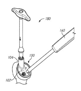
is performed by securing a semi-spherical device to the glenoid, referred to
as a glenoid
sphere, and implanting a humeral stem and an articular component coupled to
the stem that
is capable of receiving the glenoid sphere. In a reverse reconstruction, the
humeral stem can
attach to a modular tray or include an integrated tray. The tray is configured
to receive the
articular component.
[0005]
Preparing the humerus involves resecting the humeral head. Following
resection, an awl may be used to create a space distal the resection plane in
which the stem
or other anchor can be disposed, while a reamer is used to prepare the
metaphysis.
Historically, reaming is done independently from stem body preparation.
However,
-1-
Date Recue/Date Received 2022-11-15
CA 03113833 2021-03-22
WO 2020/072466
PCT/US2019/054024
because there is no link between stem and tray placement, there is the
potential for
implant misalignment.
SUMMARY
[0006] In a
diaphyseal referencing technique, a space is first formed in the
humerus in the shape of the stem and the metaphyseal or bowl cavity is reamed
second.
However, due to anatomical offset between the axis of the canal and the center
of the
humeral head, the bowl or tray may break through a proximal portion of the
cortical bone
P, which compromises proximal fixation (see Figures 1A-1B). In contrast, in a
metaphy seal reference technique, the bowl or tray is centered within a
resection surface at
the proximal end of the humerus and the stem canal is prepared second.
However,
misalignment between the tray and the stem could lead to distal cortical bone
impingement D or distal bone voids V between the canal and the implant, which
may
compromise stem fixation (see Figures 2A-2B).
[0007] To
solve these issues, the present disclosure is directed toward
instruments for evaluating the metaphyseal and diaphyseal axes and techniques
for
properly implanting a stem and/or tray within a long bone. The instruments
include
guides that link the position of an implant stem axis to the position of a
proximal bowl or
tray. For example, the guides can be shaped and/or sized to represent a
proximal face
and/or a stem inclination angle of different final implant stems. These guides
allow the
surgeon to evaluate the approximate stem axis position relative to the humeral
canal
before committing to the bowl placement in the metaphysis. Using these guides,
the
surgeon can select the appropriate implant and prepare the bone accordingly.
This
technique transfers the shape of the implant to the bone and ensures proper
alignment of
the prepared geometry in the bone and the implant geometry. These features
also allow
the surgeon to visualize the resection angle relative to the humeral canal to
help avoid
varus or valgus implant alignment.
[0008] The
instrumentation can include a system for sizing the resected
surface to provide metaphyseal referencing and to properly guide a tool into a
central
portion of the canal in the diaphysis. The system can include a sizing feature
to
approximate the size of the metaphysis. The system can also include a base
configured to
contact the metaphysis and a guide feature configured to guide a tool along a
central
portion of the canal in the diaphysis. The sizing feature can be a separate
disk component
or integral with the base.
-2-
CA 03113833 2021-03-22
WO 2020/072466
PCT/US2019/054024
[0009] The
instrumentation can include a guide having a base configured to
provide metaphyseal referencing. The base can include a first surface
configured to
contact the metaphysis (pre- or post-reaming) and a second surface opposite
the first
surface. The guide can include a guide feature having a central axis disposed
to guide a
tool, for example an awl or a sounder, into a diaphysis of the bone along a
central portion
of a canal in the diaphysis. The guide feature can extend into and/or outward
of the base.
[0010] The
guide can form part of a kit including multiple guides. Each guide
can be configured to position the tool at a different angle relative to the
face of the
resection surface and/or the metaphyseal axis. A sizing feature can be
included in the kit
as a separate disk or integrated into the base of one or more of the humeral
guides. The
sizing feature can help approximate a size of the metaphysis.
[0011]
Preparing the long bone can include sizing a proximal portion of the
bone to properly center and seat the tray within the metaphysis. A central
guide pin may
be positioned in the bone to center other instruments. Based on the
appropriate sizing, a
suitable guide may be selected. After sizing, the metaphysis is prepared using
a reamer,
and the selected guide can guide a tool down a central portion of the canal.
The reaming
step can take place prior to beginning canal preparation or after canal
preparation begins.
[0012] Any
feature, structure, or step disclosed herein can be replaced with or
combined with any other feature, structure, or step disclosed herein, or
omitted. Further,
for purposes of summarizing the disclosure, certain aspects, advantages, and
features of
the inventions have been described herein. It is to be understood that not
necessarily any
or all such advantages are achieved in accordance with any particular
embodiment of the
inventions disclosed herein. No individual aspects of this disclosure are
essential or
indispensable.
BRIEF DESCRIPTION OF THE DRAWINGS
[0013]
Various embodiments are depicted in the accompanying drawings for
illustrative purposes, and should in no way be interpreted as limiting the
scope of the
embodiments. Furthermore, various features of different disclosed embodiments
can be
combined to form additional embodiments, which are part of this disclosure.
[0014]
Figures 1A-1B illustrate complications from a diaphyseal referencing
technique.
[0015]
Figures 2A-2B illustrate complications from a metaphyseal referencing
technique.
-3-
CA 03113833 2021-03-22
WO 2020/072466
PCT/US2019/054024
[0016] Figures 3A-3C illustrate the use of an example of a humeral
guide
providing a link between the diaphysis axis and the metaphysis axis of the
humerus bone.
[0017] Figures 4A and 4B illustrate the humeral guide shown in
Figures 3A-
3C, separate from the humerus bone.
[0018] Figure 5 illustrates another humeral guide including a depth
stop
collar.
[0019] Figure 6 illustrates another humeral guide including a
retroversion
indicator.
[0020] Figures 7A-7D illustrate another humeral guide adapted to
receive a
sounder.
[0021] Figures 8A-8E illustrate another humeral guide and components
thereof.
[0022] Figures 9A-9C illustrate a kit including the humeral guide
shown in
Figures 8A-8E.
[0023] Figures 10A-10K illustrate a technique for positioning an
implant
using the humeral guides shown in at least Figures 4A-4B.
[0024] Figure 11 illustrates another humeral guide including an
integrated
sizing feature.
[0025] Figures 12A-12D illustrate a technique for positioning an
implant
using the humeral guide shown in Figure 11.
[0026] Figures 13A-13C illustrate another humeral guide.
[0027] Figure 14 illustrates the humeral guide shown in Figure 13A
with an
awl extending through the guide feature.
[0028] Figures 15A-15C illustrate another humeral guide.
[0029] Figures 16A-16B illustrate steps for bone preparation using
the
humeral guide shown in Figure 13A.
DETAILED DESCRIPTION
[0030] The instrumentation and techniques described herein provide a
link
between the position of the implant stem axis and the position of the proximal
bowl/tray
to avoid the above-described complications associated with independent
diaphysis and
metaphysis preparation. The instrumentation allows the surgeon to evaluate the
metaphysis and diaphysis and prepare the bone according to the shape of the
implant.
The instrumentation also provides opportunities to modify the version, if
necessary.
-4-
CA 03113833 2021-03-22
WO 2020/072466
PCT/US2019/054024
Although certain instruments and techniques have been described herein in
connection
with a humeral bone, the instrumentation and techniques described herein can
be used
with other long bones, including the femur.
[0031]
Figures 3A and 3B illustrate a humeral guide 100 positioned in a
reamed cavity of a metaphysis of a humerus. The base 102 of the humeral guide
100 is
centered in the reamed cavity and provides metaphy seal referencing. As shown
in Figure
3B, the humeral guide 100 includes a guide feature 104 to guide the starter
awl 180
toward or into a central portion of the canal. Figure 3C illustrates the
relative positions
between the starter awl 180 and the final implant 190. As shown, the stem axis
of the
final implant 190 is aligned with the axis of the starter awl 180, while the
implant tray is
centered in the reamed cavity. Although this and other humeral guides herein
are
described in connection with a starter awl, the humeral guides can guide any
tool,
including different sized awls, sounders, broaches, punches, or other tools.
[0032] Figure
4A illustrates the humeral guide 100 shown in Figure 3A. As
discussed above, the humeral guide 100 includes a base 102 and a guide feature
104. The
base 102 includes a first or lateral surface 106 configured to contact the
metaphysis and a
second or medial surface 108 opposite the first surface 106.
[0033] The
base 102 is configured to be centered within an outer periphery of
the metaphysis so that the final implant does not break through a proximal
portion of the
cortical bone. For example, as shown in Figure 3B, the curved profile of the
first
surface 106 enables the base 102 to be positioned within a reamed cavity in
the
metaphysis. Although, as shown in later examples, the first surface 106 can
take on any
profile, including planar, conical, cylindrical, or otherwise, depending on
how the
metaphysis is prepared.
[0034] The
humeral guide 100 can include an indicator, for example text,
color, surface modifications, etc., e.g., a size indicator 112 of a particular
size and/or
inclination angle indicator 114 of a particular angle of the humeral guide
100. As
detailed further below, the humeral guide 100 can form part of a kit including
a plurality
of humeral guides 100. The humeral guides 100 may vary in size, e.g.,
diameter, as
indicated by the size indicator 112. The size of the humeral guide 100 can
mimic the size
of the final implant, for example a proximal face of the final implant stem.
[0035] The
humeral guides 100 may provide different inclination angles
between the guide feature 104 and the base 102 as indicated by the inclination
angle
-5-
CA 03113833 2021-03-22
WO 2020/072466
PCT/US2019/054024
indicator 114. The inclination angle of the humeral guide 100 can represent a
stem
inclination angle of the final implant. The humeral stem is usually offered in
one fixed
inclination angle, e.g., between 125 degrees and 155 degrees. The humeral stem
can be
configured with a fixed 135 degree inclination angle. The humeral stem can be
configured with a fixed 145 degree inclination angle.
[0036] The
surgeon may prefer to use a handle 160 to position the humeral
guide 100 on the anatomy. Accordingly, the second surface 108 can optionally
include
one or more handle attachment features 116, 118 configured to interface with a
modular
handle 160 (see Figure 3A). As shown in Figure 4A, the humeral guide 100 can
include a
right handle attachment feature 116 and/or a left handle attachment feature
118,
depending on which arm is being prepared, preferences of the surgeon, and/or
attachment
interface on the handle 160. For example, the right handle attachment feature
116 is
accessible through an anterior incision accessing the right arm, while the
left handle
attachment feature 118 is accessible through an anterior incision accessing
the left arm.
The handle attachment features 116, 118 can be positioned at an inferior
region of the
humeral guide 100. For example, each handle attachment feature 116, 118 can be
an
angled opening on a second surface 108 of the humeral guide 100. Other
possible
configurations are shown in later examples.
[0037] The
humeral guide 100 can include a guide feature 104 on or
accessible from the second surface 108. As shown in Figure 4B, the guide
feature 104
defines a lumen 120 extending from a proximal opening 122 of the guide feature
104 to a
distal opening 124 of the base 102 such that a tool can be advanced through
the humeral
guide 100. The guide feature 104 is configured to guide a tool into a
diaphysis of the
humerus bone along a central portion of a canal in the diaphysis. The guide
feature 104
can be positioned at a superior side or region of the base 102 so the guide
feature 104 can
guide a tool into the diaphysis. In this context, superior includes a side of
the guide that
would be opposite to an inferior portion of the humerus regardless of the
orientation of
the patient in surgery.
[0038]
Although not required, the guide feature 104 can extend outwardly
from a surface of the base 102 to provide additional stabilization and support
for the tool.
For example, as shown in Figure 4A, the guide feature 104 extends away from
the second
surface 108. The guide feature 104 can have a cylindrical profile. In
other
-6-
CA 03113833 2021-03-22
WO 2020/072466
PCT/US2019/054024
configurations, the proximal opening 122 of the guide feature may be
positioned at or
flush with the second surface 108.
100391 The
guide feature 104 can include a relief 132 at a transition between
the guide feature 104 and the base 102 to facilitate manufacturing or surgical
use. For
example, in certain surgical techniques the relief 132 also allows the base
102 to fully sit
within the reamed cavity so the guide feature 104 does not obstruct proper
positioning of
the base 102 (see Figure 3B).
[0040] Figure
5 illustrates another humeral guide 200 that can include any of
the features described above with respect to Figures 4A and 4B. The humeral
guide 200
includes a depth stop 226 configured to control a depth of the humeral guide
200 relative
to the bone. In use, the depth stop 226 rests on the resection surface. For
example, in
certain techniques the humerus is resected, creating a generally planar
resection surface.
The bone at the resection surface can be altered with a reamer to create a
space for the
guide 200. The guide can be inserted into the reamed space until the depth
stop 226 rests
on the resection surface of the humerus around the reamed area. These and
related
methods are elaborated below. As shown, the depth stop 226 includes a collar
that
extends transversely, e.g., radially outwardly, from the first surface 206.
However, as
described in later examples, the depth stop 226 can be a modular component
separately
attached to a guide that may be otherwise similar to the humeral guide 200.
[0041] Figure
6 illustrates another embodiment of a humeral guide 300 that
can include any of the features described above with respect to Figures 4A,
4B, and 5.
The humeral guide 300 includes a retroversion indicator 328. The humeral guide
300
also includes a retroversion rod 330 to allow the surgeon to evaluate the
version. The
retroversion rod 330 can be moveable relative to the cylindrical body of the
guide feature
304. For example, the retroversion rod 330 can be configured to swivel with
respect to
the retroversion indicator 328. If the proximal humeral resection was not
accurate or for
other reasons dictated by surgeon judgement, the surgeon can modify the
version by
forcing the guide 300 to an appropriate version angle. This technique can also
be used to
fine tune stem access as controlled by the guide 300 as discussed in greater
detail below.
[0042]
Figures 7A-7D illustrate another embodiment of a humeral guide 400
that can include any of the features described above with respect to Figures
4A, 4B, 5,
and 6. As shown in Figures 7C and 7D, the humeral guide 400 can include a base
402
with a first surface 406 and a second surface 408. The second surface 408 can
include a
-7-
CA 03113833 2021-03-22
WO 2020/072466
PCT/US2019/054024
depth stop collar 426 extending radially outward beyond the first surface 406.
The depth
stop collar 426 is configured to rest on the resection surface and control a
depth of the
humeral guide 400.
[0043] The
humeral guide 400 can include a guide feature 404 adapted to
receive a sounder 484 (see Figure 7D) or other bone preparation instrument.
The guide
feature 404 can extend from a proximal opening 422 at the second surface 408
to a distal
opening 424 at the first surface 406. The sounder 484 can include a transverse
non-
circular profile to mimic the shape of the final implant stem. The sounder 484
may be a
starter sounder and progressively larger sounders may be used thereafter to
enlarge the
opening.
[0044] The
profile of the guide feature 404, for example at the proximal
opening 422 and/or at the distal opening 424, can be non-circular and/or
shaped to match
the sounder 484 or other tool to prevent rotation of the sounder 484. For
example, the
periphery of the proximal opening 422 can have at least one non-circular
portion, e.g., an
inflection point where a circular arc joins a linear segment or joins an
arcuate section with
a different radius of curvature. A portion of the periphery of the proximal
opening 422
can be open such that a portion of the sounder or other instrument can be
disposed inside
the opening 422 and a portion can extend through the side of the periphery out
of the
opening 422.
[0045] The
surgeon may prefer to use a handle to position the humeral guide
400 on the anatomy. Accordingly, the second surface 08 can optionally include
one or
more handle attachment features 416 configured to interface with a modular
handle.
[0046]
Figures 8A-8E illustrate another humeral guide 500 and various
components thereof The humeral guide 500 can include any of the features
discussed
above with respect to humeral guides 100, 200, 300, and 400. Figure 8A
illustrates the
humeral guide 500 configured as an assembly with a modular depth stop 526 and
a
modular handle 560 attached to a base 502. Figure 8B illustrates the humeral
guide 500
without the modular depth stop 526. Figure 8C illustrates a partial exploded
view of the
humeral guide 500 and the modular depth stop 526.
[0047] As
shown in Figures 8B and 8C, the humeral guide 500 includes the
base 502 and a guide feature 504. The base 502 includes a first or lateral
surface 506
configured to contact the metaphysis and a second or medial surface 508
opposite the first
surface 506.
-8-
CA 03113833 2021-03-22
WO 2020/072466
PCT/US2019/054024
[0048] The
base 502 is configured to be centered within an outer periphery of
the metaphysis so that the final implant which is later to be disposed in the
same position
as the base 502 does not break through a proximal portion of the cortical
bone. As
shown, the first surface 506 has a curved profile that enables the base 502 to
be
positioned within a reamed cavity in the metaphysis. However, the first
surface 506 can
take on any profile, including planar, conical, cylindrical, or otherwise,
depending on
how the metaphysis is prepared.
[0049] The
second surface 508 can include an indicator, for example text,
color, surface modifications, etc., e.g., a size indicator 512 of a particular
size and/or
inclination angle indicator 514 of a particular angle of the humeral guide
500. The size of
the humeral guide 500 can mimic the size of the final implant, for example a
proximal
face of the final implant stem. The inclination angle can represent a stem
inclination
angle of the final implant. The humeral stem is usually offered in one fixed
inclination
angle, e.g., between 125 degrees and 155 degrees. The humeral stem can be
configured
with a fixed 135 degree inclination angle. The humeral stem can be configured
with a
fixed 145 degree inclination angle.
[0050] The
surgeon may prefer to use a handle 560 to position the humeral
guide 500 on the anatomy. Accordingly, as shown in the Figure 8C, the humeral
guide
500 can optionally include one or more handle attachment features 516, 518
configured to
interface with a modular handle 560. The handle attachment features 516, 518
can be
positioned at an inferior region of the humeral guide 500. For example, each
handle
attachment feature 516, 518 can be an opening extending in a transverse
direction or
perpendicular to a longitudinal axis L of the humeral guide 500. The handle
attachment
features 516, 518 can be disposed on a portion of the base 502 opposite to the
guide
feature 504.
[0051] The
guide feature 504 of the guide 500 can be disposed on or
accessible from the second surface 508. The guide feature 504 defines a lumen
extending
from a proximal opening 522 of the guide feature 504 to a distal opening of
the base 502
such that a tool can be advanced through the humeral guide 500. The guide
feature 504 is
configured to guide a tool into a diaphysis of the humerus bone along a
central portion of
a canal in the diaphysis. The guide feature 504 can be positioned at a
superior side or
region of the base 502 so the guide feature 504 can guide a tool into the
diaphysis.
-9-
CA 03113833 2021-03-22
WO 2020/072466
PCT/US2019/054024
[0052] The
guide feature 504 can extend proximally from a surface of the
base 502 such that the proximal opening 522 is disposed away from (proximal
of) the
second surface 508 to provide additional stabilization and support for the
tool. The guide
feature 504 can have a cylindrical profile. The guide feature 504 does not
extend
proximally in some embodiments.
[0053] The
guide feature 504 can include a retroversion indicator 528. The
humeral guide 500 also can include or be coupled with a retroversion rod to
allow the
surgeon to evaluate the version. Although the retroversion rod is not shown,
the guide
feature 504 can include a connector 536 adapted to receive the retroversion
rod. The
retroversion rod and connector 536 can be moveable relative to the cylindrical
body of
the guide feature 504. For example, the retroversion rod and connector 536 can
be
configured to swivel with respect to the retroversion indicator 528. If the
proximal
humeral resection was not accurate or for other reasons dictated by surgeon
judgement,
the surgeon can modify the version by forcing the guide 500 to an appropriate
version
angle. This technique can also be used to fine tune stem access as controlled
by the guide
500.
[0054] As
shown in Figure 8C, the humeral guide 500 can include a modular
stop collar 526 configured to abut or be joined or coupled to the second
surface 508 of the
base 502. The stop collar 526 can be shaped according to the profile of the
second
surface 508. For example, the stop collar 526 may at least partially surround
a periphery
of the guide 500 at or proximal of the second surface 508 and can at least
partially
surround the guide feature 504 and/or handle attachment features 516, 518 in
some
embodiments. The stop collar 526 is configured to control a depth of the
humeral guide
500 relative to the bone. In use, the stop collar 526 rests on the resection
surface defining
the position and location of the guide 500 relative to the resection surface.
The stop
collar 526 has three discrete areas of contact in one embodiment. First and
second
arcuate segments 527a, 527b are provided on opposite sides of the collar 526.
A
projection 529 of the stop collar 526 disposed between the segments 527a, 527b
provides
contact at a third position. The modular stop collar 526 may be advantageous
if the
surgeon plans to change the version using the retroversion rod.
[0055] The
base 502 can include one or more interfacing features 538 adapted
to align with and/or join one or more corresponding interfacing features 540
on the stop
collar 526. For example, the base interfacing features 538 can include one or
more
-10-
CA 03113833 2021-03-22
WO 2020/072466
PCT/US2019/054024
openings on the second surface 508 of the base 502 and the stop collar
interfacing
features 540 can include one or more projections on an underside of the stop
collar 526,
or vice versa. Each interfacing feature 538, 540 can be integral with or
separate with the
base 502 or stop collar 526. Any of the interfacing features 538, 540 can be
threaded or
include other interlocking features to join the base 502. Any of the
interfacing features
538, 540 may not include an interlocking feature and simply provide alignment.
As
shown in Figure 8E, the stop collar 526 can include an integral projection
with a smooth
outer surface and a separate threaded connector for joining the base 502.
[0056] As
shown in Figures 9A-9C, the humeral guide 500 can form part of a
kit including a plurality of humeral guides 500A, 500B, 500C. The humeral
guides 500
may vary in size as indicated by the size indicator(s) 512 and/or provide
different
inclination angles between the guide feature 504 and the base 502 as indicated
by the
inclination angle indicator(s) 514.
[0057]
Figures 10A-10K illustrate methods of implanting a final implant using
the humeral guide 100. These methods can utilize the humeral guides 200, 300,
400, 500
or other humeral guides discussed or covered by the claims herein.
[0058] After
the surgeon gains access to the humeral head, the superior or
proximal end portion of the humerus is resected. The surgeon may be provided
with one
or more sizing disks 150 to determine a size of the metaphysis, for example,
two, three,
four, or more different sized disks. For example, each sizing disk 150 can
include an
arcuate body, e.g., a circular body 166, representative of the diameter of a
proximal face
of a stem of the final implant 190. The diameter of the circular body 166 may
vary
between the different sized disks 150. Each sizing disk 150 can include a
sizing indicator
112 representative of the size of the sizing disk 150. As described in more
detail below,
the selected sizing disk 150 can indicate the size of at least some of the
tools and/or
implants the surgeon should use to prepare the bone.
[0059] As
shown in Figure 10A, the sizing disks 150 may include one or more
tabs 164 or other sizing features extending transversely, e.g., radially
outward from the
circular body 166. The tabs 164 facilitate visualization of the space between
the implant
to be implanted (visualized with reference to the circular body 166) and the
cortical
boundary of the bone. If the outer periphery of the sizing disk 150 hangs over
the inner
cortical boundary, then the surgeon should select a different sized, e.g., a
smaller, sizing
-11-
CA 03113833 2021-03-22
WO 2020/072466
PCT/US2019/054024
disk 150. The thickness of the tabs 164 measured from the circular body 166 to
the outer
periphery of the tabs 164 can change between the different sizing disks 150.
[0060]
Optionally, each sizing disk 150 can be positioned using a modular
handle 162. The surgeon will select the appropriate sized disk 150 that
centers a
cannulation hole 152 at the center of the resection surface and fits within
the cortical
boundary of the resected surface, but does not hang over the periphery of the
resected
surface (see Figure 10A). The cannulation hole 152 is configured to receive a
central
guide pin 154 that subsequently is used to center other humeral preparation
instruments.
[0061] The
sizing disk 150 can also include a plurality of inclination holes
156, for example two, three, four, or more holes, providing a different
inclination angle
relative to the face of the resection surface. The angle of the inclination
holes 156 can be
representative of a resection angle or a stem inclination angle, e.g., between
an axis
extending through a distal end of the stem and an axis extending through a
proximal face
of the stem, of the final implant. The inclination angle can represent the
angle between
the metaphyseal bowl and the stem to allow the surgeon to evaluate approximate
stem
axis position relative to the humeral canal before committing to the bowl
placement in the
metaphysis.
[0062] Each
inclination hole 156 can be provided with an inclination
indicator 114 that indicates the inclination angle of each inclination hole
156. For
example, in Figure 10A, the inclination hole 156 identified as "145"
represents a 145
degree angle relative to the face of the resection surface. The inclination
hole 156
identified as "135" represents a 135 degree angle relative to the face of the
resection
surface. The surgeon can rotate the sizing disk 150 to select the appropriate
inclination
angle.
[0063] As
shown in Figure 10B, the surgeon can place a pin 158, for example
a drill pin, through one of the inclination holes 156 to visualize the
location of the pin
relative to the diaphysis axis Y. If the pin is displaced from the diaphysis
axis Y, for
example, superior or lateral to the diaphysis axis Y, the surgeon may select a
different
sized disk 150 to move the pin 158 closer to the diaphysis axis Y. If the pin
158 and the
diaphysis axis Y are misaligned, there is a risk of distal cortical bone
impingement (see
Figure 2E). The pin 158 can be placed prior to or after positioning the
central guide pin
154. Alternative to the pin, a structure, such as a retroversion indicator,
may be
integrated with and/or extend from the sizing disk 150 to provide a visual
marker.
-12-
CA 03113833 2021-03-22
WO 2020/072466
PCT/US2019/054024
[0064] The
sizing disk 150 can also help verify that the angle of the resected
surface is appropriate. If the pin 158 is not in line with or parallel the
diaphysis axis Y,
then the angle of the resected surface may be off and the surgeon can recut
the resected
surface or make another adjustment to improve the positioning in the humerus.
[0065] After
selecting the appropriate sizing disk 150, a corresponding sized
reamer 170 may be selected and delivered over the central guide pin 154 to
ream the
metaphysis (see Figure 10C). The reamer 170 produces a generally concave
surface in
the resected humerus. The surface can generally match the curvature of the
first or lateral
surface 106, though being an inverse thereof.
[0066] After
reaming, the first surface 106 of any of the above-described
humeral guides may be positioned in the reamed cavity. A humeral guide 100 is
selected
based on the selected sizing disk 150 and/or selected inclination angle. The
diameter of
the base 102 corresponds to the diameter of the circular body 166 of the
selected sizing
disk 150. The orientation of the guide feature 104 corresponds to the selected
inclination
angle.
[0067] The
humeral guide 100 may be positioned in the metaphysis using a
modular handle 160. As previously discussed with respect to Figure 3B, the
base 102
should be fully seated within the reamed cavity to provide metaphy seal
referencing. The
relief 132 allows the base 102 to fully sit within the reamed cavity. For
example, the
transition from the concave reamed surface to the generally planar resection
surface of
the humerus can be partly received in the relief 132. A starter awl 180 may be
selected
based on the selected sizing disk 150.
[0068] As
shown in Figure 10D, the starter awl 180 is delivered through the
guide feature 104 to create a pilot hole in line with the diaphysis axis. The
pilot hole may
extend toward or through the canal and in some techniques can extend at least
the length
of the final implant 190. Although not shown, after creating the pilot hole,
different sized
awls or sounders may be utilized to compact or otherwise prepare bone.
[0069] After
creating the pilot hole, a compactor 172 may be selected based
on the size and shape of the final implant 190. As shown in Figure 10E, the
compactor
172 may be delivered using an inserter handle 174. The tip of the compactor
172 is
placed into the pilot hole until the depth stop 176 rests on the resected
surface of the
humerus around the concave surface formed by reaming, as discussed above.
Multiple
-13-
sized compactors 172 may be utilized to get up to the size of the desired
final implant 190.
[0070] With
the compactor 172 in place, a surface planer 178 may be utilized to
ensure a flat resection true to the implant (see Figure 10F). An appropriately
sized surface
planer 178 may be selected based on the selected sizing disk 150. While
preparing the
glenoid or during other surgical steps not involving humeral preparation, an
appropriately
sized cut protector 182 may be provided on the resection surface to protect
the resection
from retractors. The protector 182 may be selected based on the selected
sizing disk 150.
[0071] After the humeral bone has been prepared, an anatomical trial implant
(Figure
10H) or a reverse trial implant (Figure 101) may be positioned in the humeral
bone.
Thereafter, the trial implant 192 may be removed, and the final anatomical
implant (Figure
10J) or final reverse implant (Figure 10K) may be implanted. The final
anatomical implant
can take any suitable configuration, such as any that are described in
Application US
62/740,642, titled "MODULAR HUMERAL HEAD," which was filed on October 2, 2018.
[0072] As mentioned above, the surgeon may be provided with an instrumentation
kit including a plurality of sizing disks 150 and a plurality of humeral
guides 100 (or humeral
guides 200, 300, 400, 500). The different components can be designed to
transfer the shape
of each available final implant to the bone. For example, the kit may include
at least three
different-sized disks 150. Each sizing disk 150 can include at least two
different inclination
angles. Thus, the kit can include at least three different sized humeral
guides 100. Each
humeral guide size can have at least two different guide feature 104
orientations for different
inclination angles. Further, each humeral guide size can include a
corresponding sized starter
awl or other starter tool.
[0073] Figure 11 illustrates a humeral guide 600 with integrated sizing
features and
functions. The humeral guide 600 allows the surgeon to create the pilot hole
-14-
Date Recue/Date Received 2022-11-15
CA 03113833 2021-03-22
WO 2020/072466
PCT/US2019/054024
in the humerus toward or into the medullary canal prior to reaming the
metaphysis.
Because the humeral guide 600 has integrated metaphyseal sizing, the guide 600
still
allows for metaphyseal referencing while forming the pilot hole as part of
preparing the
diaphysis. The humeral guide 600 can include any of the features described
above with
respect to humeral guides 100, 200, 300, 400, and 500.
[0074] As
discussed above, the humeral guide 600 includes a base 602 and a
guide feature 604. The base 602 includes a first or lateral surface 606
configured to
contact the metaphysis and a second or medial surface 608 opposite the first
surface 606.
[0075] As
shown in Figure 11, the first surface 606 has a planar profile or
configuration. As described in greater detail below, the humeral guide 600 is
positioned
on the bone after the superior or proximal end portion of the humerus is
resected, but
before the resected surface is reamed. Thus, the first surface 606 can be
formed on or can
be disposed in a single plane.
[0076] The
second surface 608 can include an indicator, for example text,
color, surface modifications, etc., e.g., a size indicator 612 of a particular
size and/or
inclination angle indicator 614 of a particular angle of the humeral guide
600.
[0077] As
detailed further below, the humeral guide 600 can form part of a kit
including a plurality of humeral guides 600. The humeral guides 600 may vary
in size as
indicated by the size indicator 612 and/or provide different inclination
angles between the
guide feature 604 and the base 602 as indicated by the inclination angle
indicator 614.
The inclination angle can be representative of a resection angle or a stem
inclination
angle, e.g., between an axis extending through a distal end of the stem and an
axis
extending through a proximal face of the stem, of the final implant. The
inclination angle
can provide the angle between the metaphyseal bowl and the stem. As shown, the
humeral guide 600 has an inclination angle of 145 degrees, but the humeral
guide 600
may have other inclination angles, for example between 125 degrees and 155
degrees,
e.g., 135 degrees.
[0078] The
base 602 is configured to be centered within an outer periphery of
the metaphysis so that the final implant centered on the same portion of the
resected
humerus upon which the base 602 is centered, in use, does not break through a
proximal
portion of the cortical bone. The humeral guide 600 can include an arcuate
body, e.g., a
circular body 666, representative of the diameter or major axis of a proximal
face of a
stem of the final implant 190 to be located at the resection plane of the
humerus (see FIG.
-15-
CA 03113833 2021-03-22
WO 2020/072466
PCT/US2019/054024
3C). The diameter of the circular body 666 may vary between the different
sized humeral
guides 600. Each humeral guide 600 can include a sizing indicator 612
representative of
the size of the humeral guide 600. As described in more detail below, the
selected
humeral guide 600 can indicate the size of at least some of the tools the
surgeon should
use to prepare the bone.
[0079] As
shown in Figure 11, the humeral guide 600 may include one or
more tabs 664 or other sizing features extending transversely, e.g., radially
outward from
the circular body 666. The tabs 664 can be radial projections formed on or
extending
from a circumferential surface of the guide 600. The projections can extend to
a free end.
The radial length of the tabs between the circumferential surface and the free
end can be
indicative of size, as discussed below. The tabs 664 facilitate visualization
of the space
between the implant to be implanted (visualized with reference to the circular
body 666)
and the cortical boundary of the bone. If the outer periphery of the humeral
guide 600
hangs over the inner cortical boundary, then the surgeon should select a
different sized,
e.g., a smaller, humeral guide 600. The thickness (also referred to herein as
radial length)
of the tabs 664 measured from the circular body 666 to the outer periphery of
the tabs 664
can change between the different humeral guides 600.
[0080] The
surgeon may prefer to use a handle to position the humeral guide
600 on the anatomy. Accordingly, the second surface 608 can optionally include
one or
more handle attachment features 616, 618 configured to interface with a
modular handle.
As shown in Figure 11, the humeral guide 100 can include a right handle
attachment
feature 616 and a left handle attachment feature 618, depending on the arm
being
prepared, preferences of the surgeon, and/or or attachment interface on the
handle. For
example, the right handle attachment feature 116 is accessible through an
anterior
incision accessing the right arm, while the left handle attachment feature 118
is accessible
through an anterior incision accessing the left arm. The handle attachment
features 616,
618 can be positioned at an inferior region of the humeral guide 600. For
example, each
handle attachment feature 616, 618 can be an angled opening on a second
surface 608 of
the humeral guide 600.
[0081] The
guide feature 604 of the humeral guide 600 can be disposed on or
accessible from the second surface 608. The guide feature 604 defines a lumen
extending
from a proximal opening 622 of the guide feature 604 to a distal opening of
the base 602
such that a tool can be advanced through the humeral guide 600. The guide
feature 604 is
-16-
CA 03113833 2021-03-22
WO 2020/072466
PCT/US2019/054024
configured to guide a tool into a diaphysis of the humerus bone along a
central portion of
a canal in the diaphysis. The guide feature 604 can be positioned at a
superior side or
region of the base 602 so the guide feature 604 can guide a tool into the
diaphysis.
[0082]
Although not required, the guide feature 604 can extend outwardly
from (proximally or medially of) a surface of the base 602 to provide
additional
stabilization and support for the tool. For example, as shown in Figure 11,
the guide
feature 604 extends away from the second surface 608. The guide feature 604
can have a
cylindrical profile. In other configurations, the proximal opening 622 of the
guide feature
may be positioned at or flush with the second surface 608.
[0083] Any of
the guides or sizing disks described herein can include one or
more stabilization features, such as stabilization holes 634 extending through
the base
602 or barbs or other anchors on the first surface 606. One or more
stabilization pins can
be driven through a respective stabilization hole 634 to hold the guide 600 in
place during
diaphyseal preparation. As shown in Figure 11, the one or more stabilization
holes 634
can be offset from the center of the guide 600 and/or positioned at an oblique
angle away
from the central axis of the base 602 or canal, so the stabilization pins do
not obstruct the
tool being delivered through the guide 604. After a pilot hole is created in
line with the
diaphysis axis, a central guide pin can be driven through a cannulation hole
652 to guide
other instruments.
[0084]
Figures 12A-12D illustrate a method of implanting a final implant
using the humeral guide 600.
[0085] After
the surgeon gains access to the humeral head, the superior or
proximal end portion of the humerus is resected. The surgeon may be provided
with one
or more humeral guides 600 to determine a size of the metaphysis and evaluate
the
diaphysis, for example, two, three, four, or more different sized humeral
guides. As
explained above, each humeral guide 600 can include an arcuate body, e.g., a
circular
body 666, representative of the diameter of a proximal face of a stem of the
final implant
190. Each humeral guide 600 can also include one or more sizing features
(e.g., tabs 664)
to facilitate visualization of the space between the implant to be implanted
(visualized
with reference to the circular body 666) and the cortical boundary of the
bone. The
selected humeral guide 600 can indicate the size of at least some of the tools
and/or
implants the surgeon should use to prepare the bone.
-17-
CA 03113833 2021-03-22
WO 2020/072466
PCT/US2019/054024
[0086] As
shown in Figure 12A, optionally, each humeral guide 600 can be
positioned using a modular handle 660. The surgeon will select the appropriate
humeral
guide 600 that centers a cannulation hole 652 at the center of the resection
surface and fits
within the cortical boundary of the resected surface, but does not hang over
the periphery
of the resected surface. The cannulation hole 652 is configured to receive a
central guide
pin 654 that subsequently is used to center other humeral preparation
instruments (see
Figure 12B).
[0087] Within
each size, the surgeon may be provided with or may select one
or more humeral guides 600 having different inclination angles, which can
represent an
angle relative to the face of the resection surface or the stem inclination
angle of the final
implant stem to be implanted. The inclination angle allows the surgeon to
evaluate
approximate stem axis position relative to the humeral canal before committing
to the
bowl placement in the metaphysis. The cylindrical extension of the guide
feature 604 can
provide a visual indicator of the inclination angle to help the surgeon select
the
appropriate inclination angle and/or verify that the angle of the resected
surface is
appropriate. If the cylindrical extension of the guide feature 604 is not in
line with or
parallel the diaphysis axis Y, then the angle of the resected surface may be
off and the
surgeon can recut the resected surface or make another adjustment to improve
the
positioning in the humerus.
[0088] After
selecting the appropriate humeral guide 600, optionally, one or
more stabilization pins can be driven through a respective stabilization hole
634 to hold
the guide 600 in place during diaphyseal preparation. The starter awl 680 or
other tool is
delivered through the guide feature 604 to create a pilot hole in line with
the diaphysis
axis. The pilot hole may extend toward or through the canal and in some
techniques can
extend at least the length of the final implant 190. Although not shown, after
creating the
pilot hole, different sized awls or sounders may be utilized to compact or
otherwise
prepare bone. At any time, for example after the pilot hole is created, the
central guide
pin 654 may be driven through the cannulation hole 652 to guide other
instruments.
[0089] After
creating the pilot hole, a corresponding sized reamer 670 may be
selected and delivered over the central guide pin 654 to ream the metaphysis
(see
Figure 12C). After reaming, a compactor 672 may be selected based on the size
and
shape of the final implant. As shown in Figure 12D, the compactor 672 may be
delivered
using an inserter handle 674. The tip of the compactor 672 is placed into the
pilot hole
-18-
CA 03113833 2021-03-22
WO 2020/072466
PCT/US2019/054024
until the depth stop 676 rests on the resected surface of the humerus around
the concave
surface formed by reaming, as discussed above. Multiple sized compactors 672
may be
utilized to get up to the size of the desired final implant.
[0090]
Following compacting, the same preparation steps described above
with respect to Figures 10F-10K may be utilized.
[0091] Any of
the guides described herein may have a patient specific design
that matches the metaphyseal and diaphyseal axes of the patient's bone. The
guides can
be generated based on pre-operative or intra-operative imaging, such as CT
scan, MM
scan, X-ray, or other imaging, and formed utilizing, for example, 3-D printing
technology
or the like.
[0092]
Figures 13A-13C illustrate another humeral guide 700. The humeral
guide 700 can include any of the features discussed above with respect to any
one or
more of humeral guides 100, 200, 300, 400, 500, and 600.
[0093] As
shown in Figures 13A-13C, the humeral guide 700 includes the
base 702 and a guide feature 704. The base 702 includes a first or lateral
surface 706
configured to contact the metaphysis and a second or medial surface 708
opposite the first
surface 706. The first surface 706 has a planar profile or configuration (see
Figure 13C).
However, the first surface 706 can take on any profile, including spherical,
tiered,
conical, cylindrical, or otherwise, for example depending on how the
metaphysis is
prepared.
[0094] The
second surface 708 can include one or more indicators, for
example text, color, surface modifications, etc., e.g., a size indicator of a
particular size
and/or inclination angle indicator of a particular angle of the humeral guide
700. As
shown in Figure 13A, the humeral guide 700 may include two different size
indicators
712, e.g., a text indicator and a color indicator.
[0095] The
base 702 is configured to be centered within an outer periphery of
the metaphysis so that the final implant at least partially centered on the
same portion of
the resected humerus upon which the base 702 is centered, in use, does not
break through
a proximal portion of the cortical bone. The humeral guide 700 can include an
arcuate
body, e.g., a circular or partial circular body 766, representative of the
diameter or major
axis of a proximal face of a stemmed or stemless anchor of the final implant
190 to be
located at or adjacent to resection plane of the humerus (see FIG. 3C). The
diameter of
the circular body 766 may vary between the different sized humeral guides 700.
-19-
CA 03113833 2021-03-22
WO 2020/072466
PCT/US2019/054024
[0096] The
humeral guide 700 may include a depth stop 726 configured to
control a depth of the humeral guide 700 relative to the bone. The shape
and/or size of
the depth stop 726 may correspond to the shape and/or size of a collar on the
final
implant. The second surface 708 may have a greater diameter and project
radially
outward of the first surface 706, thus forming the depth stop 726. However, as
described
in earlier examples, the depth stop 726 can be a modular component separately
attached
to a guide. As explained further below, the depth stop 726 rests on a recessed
surface in
the bone in some techniques.
[0097] The
guide feature 704 of the humeral guide 700 can be disposed on or
accessible from the second surface 708. The guide feature 704 defines a lumen
extending
from a second opening 722 of the guide feature 704 to a first opening 705 of
the base 702
such that a tool can be advanced through the humeral guide 700 (see Figure
14). A rear
side of the humeral guide may include an open channel 721 from the guide
feature lumen
to the first opening 705. The open channel 721 prevents the guide feature 704
from
impinging on the resection. Although not required, the guide feature 704 can
extend
outwardly from (laterally of) a surface of the base 702 to provide additional
stabilization
and support for the tool. The guide feature 704 can have a cylindrical
profile. In other
configurations, the second opening 722 of the guide feature 704 may be
positioned at or
flush with the second surface 708.
[0098] The
guide feature 704 is configured to guide a tool into a diaphysis of
the humerus bone along a central portion of a canal in the diaphysis. The
guide feature
704 can be positioned at a superior side or region of the base 702 so the
guide feature 704
can guide a tool into the diaphysis.
[0099] The
humeral guide 700 may also include a retroversion indicator 728,
for example on the guide feature 704. As the retroversion rod 730 on the awl
780 is
moved relative to the guide feature 704, the retroversion rod 730 allows the
surgeon to
evaluate the version (see Figure 14). For example, when the retroversion rod
730 is
pointed toward a patient's elbow and parallel with a long axis of the forearm,
the position
of the indicator 781 on the awl 780 relative to the retroversion indicator 728
provides
information on the version. If the proximal humeral resection was not accurate
(for
example, if the indicator 781 is entirely offset from the retroversion
indicator 728) or for
other reasons dictated by surgeon judgement, the surgeon can modify the
version by
-20-
CA 03113833 2021-03-22
WO 2020/072466
PCT/US2019/054024
adjusting the guide 700 to an appropriate version angle. This technique can
also be used
to fine tune stem access as controlled by the guide 700 as discussed above.
[0100] The
surgeon may prefer to use a handle 760 to position the humeral
guide 700 on the anatomy (see Figure 16B). The humeral guide 700 can
optionally
include one or more handle attachment features 716, 718 configured to
interface with a
modular handle 760. The handle attachment features 716, 718 can extend from
the
second surface 708 in a central region thereof. For example, each handle
attachment
feature 716, 718 can be an opening extending in a transverse direction or
perpendicular to
a longitudinal axis L of the humeral guide 700 on a projection 717 extending
from the
second surface 708. Providing the handle attachment features 716, 718 in the
central
region of the humeral guide 700 prevents the guide from tilting when
manipulating the
handle.
[0101] The
humeral guide 700 may form part of a kit including a plurality of
humeral guides 700. The humeral guides 700 may vary in size as indicated by
the size
indicator 712. As explained above, the size of the humeral guide 700 may be
selected
based on a selected sizing desk that indicates the size of at least some of
the tools and/or
implants the surgeon should use to prepare the bone.
[0102] The
kit may also include humeral guides 700 with different inclination
angles between the guide feature 704 and the base 702. The inclination angle
can be
representative of a resection angle or a stem inclination angle, e.g., between
an axis
extending through a distal end of the stem and an axis extending through a
proximal face
of the stem of the final implant. The inclination angle can be measured
between an axis
aligned with a central longitudinal axis of an elongate distal portion of a
stem and an axis
extending perpendicular to a proximal face of the stem of the final implant.
The
inclination angle can be measured between an axis aligned with a central
longitudinal
axis of the humerus and an axis extending perpendicular to a proximal face of
a humeral
anchor with or without a stem portion. The inclination angle can provide the
angle
between a metaphy seal bowl portion and a stem portion of an implant. As
shown, the
humeral guide 700 has an inclination angle of 145 degrees, but the humeral
guide 700
may have other inclination angles, for example angles of or between 125
degrees and 155
degrees, e.g., 135 degrees.
[0103] Figure
14 shows the humeral guide 700 with an awl 780 extending
through the guide feature 704. The awl 780 may correspond to the selected size
of the
-21-
CA 03113833 2021-03-22
WO 2020/072466
PCT/US2019/054024
humeral guide 700. As shown in Figure 14, the awl 780 may include a size
indicator 782,
for example a size specifying text, color, surface modifications, or
combination of two or
more such indicators. A single awl 780 may be suitable for more than one sized
humeral
guide 700.
[0104]
Figures 15A-15C show another humeral guide 800 that resembles the
humeral guide 700 except as described below. Accordingly, numerals used to
identify
features of the humeral guide 700 are incremented by a factor of one hundred
(100) to
identify like features of the humeral guide 800. The description of the guide
700 will be
considered to supplement the description of the guide 800 where consistent
rather than
repeating such descriptions. Similarly the descriptions of the guide 800 may
supplement
those of the guide 700.
[0105] As
described above, the surgeon may prefer to use a handle to position
the humeral guide on the anatomy. Accordingly, as shown in the Figures 15A-
15C, the
humeral guide 800 can optionally include one or more handle attachment
features 816,
818 configured to interface with a modular handle (e.g., the handle 760 in
FIG. 16B).
The handle attachment features 816, 818 can be positioned at an inferior
region of the
humeral guide 800. For example, each handle attachment feature 816, 818 can be
an
opening extending in a transverse direction or perpendicular to a longitudinal
axis L of
the humeral guide 800. The handle attachment features 816, 818 can be disposed
on a
portion of the base 802 opposite to the guide feature 804. The handle
attachment features
816, 818 can be located inferior of (or distal of) a second surface 808 of the
guide 800.
Providing the handle attachment features 816, 818 at the periphery of the base
802 makes
it easier to machine the handle attachment features 816, 818.
[0106] As
described above, a rear side of the humeral guide may include an
open channel from the guide feature lumen to the first opening on the first
surface of the
humeral guide. However, other configurations are possible. For example, as
shown in
Figure 15C, a rear side of the guide feature 804 may include a channel 821
providing
access to the lumen of the guide feature 804. This channel 821 may be distinct
from the
first opening 805 on the first surface 806 of the humeral guide 800.
[0107]
Figures 16A-16B illustrate a method of implanting a final implant
using the humeral guide 700.
[0108] After
the surgeon gains access to the humeral head, the superior or
proximal end portion of the humerus is resected. The surgeon may evaluate the
size of
-22-
CA 03113833 2021-03-22
WO 2020/072466
PCT/US2019/054024
the metaphysis using any of the techniques described herein. For example, the
surgeon
may be provided with one or more sizing disks to determine a size of the
metaphysis.
Using the selected sizing disk, the surgeon can place a pin.
[0109] After
selecting the appropriate sizing disk, a corresponding sized
reamer 770 may be selected and delivered over the guide pin 754 to ream the
metaphysis
(see Figure 16A). The guide pin 754 can be placed in the resected humerus
using the
selected sizing disk. The reamer 770 produces a cavity in the resected
humerus. The
cavity may be hemispherical, cylindrical, tiered, conical, or another shape
such as
including two or more cylindrical areas. For example, the reamer 770 may
include a
distal portion 773 configured to form a recess or surface in the metaphysis
that generally
matches the shape of the metaphyseal portion of a stemmed implant or all or a
portion of
an external surface of an anchor of a stemless implant, e.g., being an inverse
thereof. The
reamer 770 may also include a proximal portion 771 configured to form a
recessed
surface or counter sunk area below the resection plane. The recessed surface
may
surround at least a portion of the opening of the cavity. The recessed surface
can be
shaped to receive the depth stop 726 of the humeral guide 700 or the collar of
a final
implant. As shown in Figure 16B, the humeral guide 700 is positioned in the
bone such
that the second surface 708 is flush with resection plane.
[0110] As
shown in Figure 16B, optionally, each humeral guide 700 can be
positioned using a modular handle 760. The surgeon will select the appropriate
humeral
guide 700 based on the selected sizing disk. When positioned on the bone, the
humeral
guide 700 fits within the cortical boundary of the resected surface, but does
not hang over
the periphery of the resected surface.
[0111] Within
each size, the surgeon may be provided with or may select one
or more humeral guides 700 having different inclination angles, which can
represent an
angle relative to the face of the resection surface or the stem inclination
angle of the final
implant stem to be implanted. The inclination angle allows the surgeon to
evaluate
approximate stem axis position relative to the humeral canal before committing
to the
bowl placement in the metaphysis. The cylindrical extension of the guide
feature 704 can
provide a visual indicator of the inclination angle to help the surgeon select
the
appropriate inclination angle and/or verify that the angle of the resected
surface is
appropriate. If the cylindrical extension of the guide feature 704 is not in
line with or
parallel the diaphysis axis, then the angle of the resected surface may be off
and the
-23-
CA 03113833 2021-03-22
WO 2020/072466
PCT/US2019/054024
surgeon can recut the resected surface or make another adjustment to improve
the
positioning in the humerus.
[0112] After
placing the appropriate humeral guide 700, the starter awl 780 or
other tool is delivered through the guide feature 704 to create a pilot hole
in line with the
diaphysis axis. The pilot hole may extend toward or through the canal and in
some
techniques can extend at least the length of the final implant. Although not
shown, after
creating the pilot hole, different sized awls or sounders may be utilized to
compact or
otherwise prepare bone.
[0113] After
reaming, a compactor may be selected based on the size and
shape of the final implant. Compacting and following steps may include the
same steps
described above with respect to Figures 10E-10K.
[0114] Any of
the guides described herein may have a patient specific design
that matches the metaphyseal and diaphyseal axes of the patient's bone. The
guides can
be generated based on pre-operative or intra-operative imaging, such as CT
scan, MRI
scan, X-ray, or other imaging, and formed utilizing, for example, 3-D printing
technology
or the like.
Terminology
[0115] As
used herein, the relative terms "lateral" and "medial" shall be
defined relative to the anatomy. Thus, medial refers to the direction toward
the midline
and lateral refers to the direction away from the midline.
[0116]
Although certain embodiments and examples have been described
herein, it will be understood by those skilled in the art that many aspects of
the delivery
systems shown and described in the present disclosure may be differently
combined
and/or modified to form still further embodiments or acceptable examples. All
such
modifications and variations are intended to be included herein within the
scope of this
disclosure. A wide variety of designs and approaches are possible. No feature,
structure,
or step disclosed herein is essential or indispensable.
[0117] For
purposes of this disclosure, certain aspects, advantages, and novel
features are described herein. It is to be understood that not necessarily all
such
advantages may be achieved in accordance with any particular embodiment. Thus,
for
example, those skilled in the art will recognize that the disclosure may be
embodied or
carried out in a manner that achieves one advantage or a group of advantages
as taught
-24-
CA 03113833 2021-03-22
WO 2020/072466
PCT/US2019/054024
herein without necessarily achieving other advantages as may be taught or
suggested
herein.
[0118]
Moreover, while illustrative embodiments have been described herein,
the scope of any and all embodiments having equivalent elements,
modifications,
omissions, combinations (e.g., of aspects across various embodiments),
adaptations
and/or alterations as would be appreciated by those in the art based on the
present
disclosure. The limitations in the claims are to be interpreted broadly based
on the
language employed in the claims and not limited to the examples described in
the present
specification or during the prosecution of the application, which examples are
to be
construed as non-exclusive. Further, the actions of the disclosed processes
and methods
may be modified in any manner, including by reordering actions and/or
inserting
additional actions and/or deleting actions. It is intended, therefore, that
the specification
and examples be considered as illustrative only, with a true scope and spirit
being
indicated by the claims and their full scope of equivalents.
[0119]
Conditional language used herein, such as, among others, "can,"
"might," -may," "e.g.," and the like, unless specifically stated otherwise, or
otherwise
understood within the context as used, is generally intended to convey that
some
embodiments include, while other embodiments do not include, certain features,
elements, and/or states. Thus, such conditional language is not generally
intended to
imply that features, elements, blocks, and/or states are in any way required
for one or
more embodiments or that one or more embodiments necessarily include logic for
deciding, with or without author input or prompting, whether these features,
elements
and/or states are included or are to be performed in any particular
embodiment.
[0120] The
ranges disclosed herein also encompass any and all overlap, sub-
ranges, and combinations thereof Language such as "up to," "at least,"
"greater than,"
"less than," "between," and the like includes the number recited. Numbers
preceded by a
term such as "about" or "approximately" include the recited numbers and should
be
interpreted based on the circumstances (e.g., as accurate as reasonably
possible under the
circumstances, for example 1%, 5%, +10%, 15%, etc.). For example, "about
0.01
inches" includes "0.01 inches." Phrases preceded by a term such as
"substantially"
include the recited phrase and should be interpreted based on the
circumstances (e.g., as
much as reasonably possible under the circumstances). For example,
"substantially
linear" includes "linear."
-25-