Note: Descriptions are shown in the official language in which they were submitted.
CA 03114262 2021-03-25
WO 2020/094756
PCT/EP2019/080468
Device and method for editing a panoramic radiography image
The present invention relates to a device and a method for editing a panoramic
radiography image of an examination object generated by a panoramic X-ray
machine. Moreover, the invention relates to a diagnosis system for examining a
craniomaxillofacial area of a patient.
X-ray examinations have been a widely-used standard procedure for many
years particularly in medicine but also in industry as well as in other fields
of
application. X-rays are absorbed differently in structures with various thick-
nesses within an examination object so that an image of an inner structure can
be generated. In medical diagnostics, the inside of a human body can thus be
examined.
In this respect, single X-ray images can be generated, several images taken
from different angles can be composed to a panoramic image (panoramic X-ray
machine) or three-dimensional models can be reconstructed from spatially dif-
ferent projections (X-ray tomography or X-ray computed tomography). The var-
ious methods differ particularly in the level of radiation that the
examination ob-
ject is exposed to.
In odontology, images of single teeth, groups of teeth or of the whole jaw are
particularly common. As X-ray radiation penetrates the human head during the
1
CA 03114262 2021-03-25
WO 2020/094756
PCT/EP2019/080468
examination, the used level of radiation is of high relevance. Almost all
dental
surgeries have X-ray machines but different machines are used or different im-
ages are generated depending on the type of examination carried out.
Extraoral panoramic X-ray machines, i.e. machines without an X-ray source
and/or an X-ray receiver in the patient's mouth, travel around the head and
the
jaw of a patient. For this purpose, an X-ray source is usually attached to one
side of an arm and an X-ray detector to the other. To generate a sharp image
within the area of interest in the patient's jaw, specific machine
trajectories are
used to travel around the head. A machine trajectory thereby defines an image
surface of a panoramic radiography image as that surface, within the object
that
is penetrated by x-ray radiation from several angles, that is reproduced as a
sharp image (focused).
A challenge in the use of panoramic radiography images is the presence of im-
age artifacts. In particular when a panoramic radiography image of a human jaw
arch is generated, radiation passes, in some areas of the trajectory, through
the
jaw of interest as well as through the opposing jaw on the opposite side. This
may result in structures of the opposing jaw superimposing structures of the
jaw
of interest as artifacts in the image. This makes a diagnosis more difficult.
Even
if only a hemifacial image is taken, i.e. an image of half the jaw, such
opposing
jaw artifacts (for example bright shadows or stripes) are visible. Metallic
foreign
objects or other objects on the patient may result in further unexpected and
possibly disturbing opposing jaw artifacts.
To solve this problem, the machine trajectory (may also be called trajectory
curve) can be adjusted. So-called artifact-reduced trajectory curves have the
effect that unavoidable opposing jaw artifacts are moved to other image sec-
tions that are diagnostically irrelevant. However, this approach requires
deviat-
ing from an ideal radiography direction of the jaw arch, which may possibly in-
crease the level of radiation exposure and/or decrease image quality. A
further
approach is the use of three-dimensional methods. In this connection,
DE 10 2010 040 096 Al describes a method for generating an image from a 3D
volume. A virtual dental image is generated from a 3D volume with volumetric
2
CA 03114262 2021-03-25
WO 2020/094756
PCT/EP2019/080468
image data. At first, a part volume of the 3D volume is defined and then a
virtual
projection image from a certain radiography direction is generated for this
part
volume by computer-aided calculation of the volumetric image data in this radi-
ography direction. A calculation of a simulated panoramic radiography image
without involvement of the opposing jaw is thus made possible. However, the
level of radiation exposure of a 3D image is higher and the resolution is
worse.
On this basis, the present invention addresses the problem of improving diag-
noses based on panoramic radiography images. The level of radiation exposure
shall be kept at a minimum. In particular, a possibility for examining a human
jaw arch shall be achieved.
For solving this problem, an aspect of the present invention relates to a
device
for editing a panoramic radiography image of an examination object generated
by a panoramic X-ray machine, comprising:
an input interface for receiving the panoramic radiography image as well
as reconstruction data of the panoramic radiography image, the reconstruction
data including information on a course of projection lines of the panoramic
radi-
ography image between an X-ray source and an X-ray detector of the pano-
ramic X-ray machine as well as information on an image surface of the pano-
ramic radiography image;
an evaluation unit for evaluating the reconstruction data and for determin-
ing an image section of the panoramic radiography image that has been gener-
ated on the basis of one of the projection lines with two intersection points
with
the image surface;
an image editing unit for editing the panoramic radiography image based
on the determined image section; and
an output unit for outputting the edited panoramic radiography image.
A further aspect of the present invention relates to a diagnosis system for ex-
amining a craniomaxillofacial area of a patient, comprising:
3
CA 03114262 2021-03-25
WO 2020/094756
PCT/EP2019/080468
an extraoral panoramic dental X-ray machine with an X-ray source that is
movable around a patient's head and with an X-ray detector that is fixedly con-
nected with the X-ray source; and
a device as described above.
Further aspects of the invention relate to a method configured according to
the
device described above as well as to a computer program product with program
code for executing the steps of the method when the program code is executed
on a computer, as well as a storage medium on which a computer program is
stored, which, when executed on a computer, effects an execution of the
method described herein.
Preferred embodiments of the invention are described in the dependent claims.
It is understood that the features mentioned hereinbefore and those to be com-
mented on hereinafter may be used not only in the specified combination but
also in other combinations or in isolation without departing from the scope of
the
present invention. In particular, the method and the computer program product
may be configured in accordance with the embodiments of the device and the
diagnosis system described in the dependent claims.
According to the invention, it is provided that, on the one hand, a panoramic
radiography image and, on the other hand, the reconstruction data of the pano-
ramic radiography image are received. The panoramic radiography image in
particular corresponds to respective image data. The reconstruction data in
par-
ticular comprise geometric information on the machine trajectory of the pano-
ramic X-ray machine as well as on the geometric arrangement of the radiator,
screen, and detector and of the examination object (radiation geometry) as
well
as the resulting focus and/or image surface, in which a sharp image has been
generated. Additionally, the reconstruction data preferably comprise
information
on the geometry on the basis of which an overall image has been generated
from several recorded X-ray images.
On the basis of the reconstruction data, an image section within the panoramic
radiography image may be determined, which potentially includes artifacts, in
4
CA 03114262 2021-03-25
WO 2020/094756
PCT/EP2019/080468
particular opposing jaw artifacts. This image section corresponds to a section
that has been generated on the basis of at least one projection line that
crosses
the image surface twice. To determine the respective image section, the image
surface as well as the course of the projection lines relative to the image
surface
.. has to be known. According to the invention, the determined image section
is
edited. Thus, a section of the panoramic radiography image is edited in which
there may possibly be an artifact. The panoramic radiography image with the
edited image section is output via the output unit.
In comparison with known approaches of displaying panoramic radiography im-
ages, according to the invention, an image section is edited before the image
is
output. Editing may occur before, during or after an evaluation. In
particular,
editing may occur prior to a diagnosis based on the image. It is not necessary
to increase the level of radiation exposure. No additional image is generated
but
an already generated image is edited. The accuracy of the diagnosis is im-
.. proved. Impairment of the evaluation of a panoramic radiography image be-
cause of artifacts is avoided or reduced respectively.
In a preferred embodiment, the input interface is configured to receive the
pan-
oramic radiography image as well as the reconstruction data from an extraoral
panoramic dental X-ray machine. Preferably, the device according to the inven-
tion is used in the field of dental diagnostics. Opposing jaw artifacts and/or
re-
spective image sections can be recognized. The accuracy of the diagnosis is
increased. Qualified medical staff may evaluate panoramic radiography images
of a human jaw without unidentified opposing jaw artifacts causing
inaccuracies.
In a further advantageous embodiment, the reconstruction data comprise a rel-
ative position of the image surface in relation to the projection lines and/or
a
description of a movement of a movable X-ray source in relation to the exami-
nation object. It is particularly advantageous if the relative position of the
image
surface is described for instance by geometric information and/or a
description
by means of a coordinate system. Respective geometric information may also
be used for the description of the movement of the X-ray source. Such recon-
struction data allow for an efficient evaluation and a reliable determination
of the
5
CA 03114262 2021-03-25
WO 2020/094756
PCT/EP2019/080468
image section of the panoramic radiography image that potentially includes ar-
tifacts. Preferably, the X-ray source is fixedly connected with an X-ray
detector
that is also moveable.
In a further advantageous embodiment, the image editing unit is configured to
mark a section and/or a spot in the panoramic radiography image. The mark
preferably comprises a color tag and/or a frame. Advantageously, the section
within the panoramic radiography image that potentially includes artifacts is
marked. For this purpose, a respective frame (circle, rectangle, etc.) or a
color
tag (for example yellow shadow etc.) may be used. By marking the relevant
section of the image, it is indicated that any possible artifacts should be
paid
attention to when the image is evaluated within this section or at this spot
re-
spectively. In this respect, marking serves to simplify evaluation.
In an advantageous embodiment, the evaluation unit is configured to determine
a multi-part, in particular a two-part, image section that corresponds to all
pro-
jection lines with two intersection points with the image surface. In
particular, it
is possible to fully mark the section that potentially includes artifacts.
Advanta-
geously, the image section is correspondingly marked twice, i.e. on both sides
of a panoramic radiography image. When a panoramic radiography image thus
edited is evaluated, it is hereby defined in which sections artifacts may
possibly
exist.
In an advantageous embodiment, the image editing unit is configured to
subtract
an image value of a first intersection point, preferably close to the X-ray
source,
of a projection line with the image surface from an image value of a second
intersection point of the projection line with the image surface. Editing may
in
particular comprise a step of subtracting an image value. Subtracting an image
value may compensate for artifacts. For example, a weighting may be carried
out. This further increases the accuracy of the diagnosis.
Advantageously, the input interface is configured to receive a marking
position,
which corresponds to a first intersection point of a first projection line
with the
image surface. Further, the evaluation unit is configured to determine an
image
6
CA 03114262 2021-03-25
WO 2020/094756
PCT/EP2019/080468
section, which corresponds to a second intersection point of the first
projection
line with the image surface. A marking position may additionally be received
via
the input interface. On this basis, a corresponding image section on an
opposite
side is then determined. By simultaneously viewing the marking position as
well
as the determined opposite section an impression can be obtained as to the
possible structures in this opposite section that represent the source of a
possi-
ble artifact. On this basis, an improved diagnosis is made possible as it can
be
checked immediately if an object in the section of the marking position may
pos-
sibly be traced back to an artifact. The diagnosis is further simplified. The
relia-
bility is increased.
In an advantageous embodiment, the device comprises a user interface for en-
tering the marking position by a user of the device. Preferably, the marking
po-
sition may be entered by choosing a position on a representation of the pano-
ramic radiography image by means of a touch screen, touch pad, touch pen,
and/or a computer mouse. It is made possible that a user, for example a mem-
ber of the evaluating qualified medical staff, makes an entry and it is
directly
displayed for this entry if and where there is an opposite image section from
which possible artifacts may originate. It is thus achieved that in case of a
no-
ticeable object in the panoramic radiography image, it can be clarified
directly
and interactively if this noticeable object was caused by an artifact on the
oppo-
site side.
In an embodiment, the output unit is configured to output the edited panoramic
radiography image on a display. Preferably, a computer-based output is possi-
ble. This allows for interaction with the user. Moreover, functions of
computer-
aided display, such as magnifications etc., may be used.
In a further advantageous embodiment of the diagnosis system, the panoramic
dental X-ray machine is configured to generate the panoramic radiography im-
age from a composition of a plurality of single images. The single images are
generated at different positions of the X-ray source and the X-ray detector
rela-
tive to the examination object. It is particularly advantageous to use data of
a
7
CA 03114262 2021-03-25
WO 2020/094756
PCT/EP2019/080468
panoramic dental X-ray machine that has recorded and composed several sin-
gle images. The accuracy of the diagnosis may be increased.
Herein, an examination object may be any organic or inorganic object. In par-
ticular, a body part of a human being may be examined. Preferably, a jaw area
of a human being is examined as examination object. The panoramic radiog-
raphy image may in particular be available in the form of digital image data.
A
course of projection lines may in particular follow geometric specifications
within
a coordinate system in the panoramic X-ray machine. A projection line corre-
sponds to an imagined line between X-ray source and X-ray detector. In partic-
ular, projection lines are herein understood as rays that fan out across the
dis-
tance. A projection fan or projection cone advantageously comprises several
projection lines. In particular, a course of projection lines may comprise
and/or
represent a geometric description of a fan or a cone. Correspondingly, an
inter-
section point may herein also feature a spatial dilation and insofar
correspond
to an intersection area. The editing of the panoramic radiography image accord-
ing to the invention may comprise an editing of single pixels or image values.
A
craniomaxillofacial area of a patient in particular describes the area of the
jaw
and/or the jaw arch of a patient (dental area).
The invention is described and explained in more detail by means of a number
of selected embodiments in connection with the enclosed Figures hereinafter.
In the Figures:
Figure 1 shows a schematic illustration of a diagnosis system according to the
invention for examining a craniomaxillofacial area of a patient;
Figure 2 shows a schematic illustration of a panoramic X-ray image without a
complete representation of the temporomandibular joint;
Figure 3 shows a schematic illustration of a top view of an examination object
in a diagnosis system according to the invention;
Figure 4 shows a schematic illustration of an image surface;
8
CA 03114262 2021-03-25
WO 2020/094756
PCT/EP2019/080468
Figure 5 shows a schematic illustration of a device according to the
invention;
Figure 6 shows a schematic illustration of a panoramic radiography image ac-
cording to the invention;
Figure 7 shows a schematic illustration of a further panoramic radiography im-
age according to the invention; and
Figure 8 shows a schematic illustration of a method according to the
invention.
Figure 1 illustrates a diagnosis system 10 according to the invention for exam-
ining a craniomaxillofacial area of a patient. The diagnosis system 10
comprises
a panoramic X-ray machine 14 as well as a device 16 for editing a panoramic
radiography image generated by a panoramic X-ray machine 14. In particular,
a panoramic X-ray machine 14 for use in dental diagnostics is shown (pano-
ramic dental X-ray machine).
In the exemplary illustrated embodiment, the head of the patient is examined
as
examination object 12. An X-ray source 18 that is movable around the exami-
nation object 12 emits X-ray radiation that penetrates the examination object
12
and is received by an X-ray detector 20. X-ray source 18 and X-ray detector 20
are fixedly connected via a respective rotatable arc 22 (may also be referred
to
as arm). Usually, the arc 22 is revolvably or rotatably mounted at a position
above the examination object 12. X-ray source 18 and X-ray detector 20 travel
around the examination object 12 along a machine trajectory. This machine tra-
jectory does not have to correspond to a circular trajectory. In the exemplary
illustrated embodiment, the device 16 is integrated in the panoramic X-ray ma-
chine 14. It is also possible that the device 16 is arranged elsewhere. For in-
stance, the device 16 may be fully or partly integrated in a separate
evaluation
computer. The device 16 may be fully or partly implemented in software. It is
also possible that the device is implemented as a cloud service.
Figure 2 shows an exemplary panoramic radiography image of the jaw arch 26
of a patient that can be generated by means of a panoramic X-ray machine. In
9
CA 03114262 2021-03-25
WO 2020/094756
PCT/EP2019/080468
the exemplary illustrated embodiment, the panoramic radiography image 24
corresponds to a composition of a plurality of single images that have been
rec-
orded during a movement of the X-ray source and the connected X-ray detector
around the examination object.
In Figure 3, the movement of the X-ray detector 20 and the X-ray source 18
around the examination object 12 or the patient's head, respectively, is
shown.
The view corresponds to a top view from a bird's eye view. In particular, a
cross-
section in a plane perpendicular to the z-axis (cf. Fig. 1) is shown.
As illustrated, a projection line 28 runs from the X-ray source 18 to the X-
ray
detector 20. The projection line 28 corresponds to an imagined line, wherein
the
projection or ray geometry may also be based on a fanning out and a respective
projection fan or projection cone. A projection fan or a projection cone
respec-
tively are herein regarded as several projection lines 28.
The movement of the X-ray source 18 and the X-ray detector 20 during gener-
ation of the panoramic radiography image usually occurs along a predetermined
machine trajectory that, in most cases, does not correspond to a circular
trajec-
tory. To generate a sharp image of the jaw arch 26, it is necessary for an
image
surface 30 to be within the area of the jaw arch 26 as much as possible. The
image surface 30 corresponds to a surface that has been determined on the
basis of the ray geometry, the trajectory curve, and the reconstruction to
gener-
ate a sharp image of the jaw arch 26.
As illustrated schematically, there are sections in which the projection line
28
intersects the image surface 30 twice, thus comprising a first intersection
point 32 in the vicinity of the X-ray source as well as a second intersection
point 34 averted from the X-ray source. The image surface mostly lies on the
side of the second intersection point 34 that is averted from the X-ray source
(in
the vicinity of the sensor or detector). Due to the fact that the projection
line 28
intersects the image surface 30 once in the first intersection point 32 before
it
reaches the second intersection point 34 or the X-ray detector 20
respectively,
there are overlays in the image section of the panoramic radiography image,
CA 03114262 2021-03-25
WO 2020/094756
PCT/EP2019/080468
which corresponds to the second intersection point 34 of the projection line
28.
In the field of dental X-ray technology, the resulting artifacts are described
as
opposing jaw artifacts.
Figure 4 illustrates a schematic illustration of an image surface 30 in a
spatial
representation. The distance of the image surface 30 to the X-ray source 18
and/or the X-ray detector 20, from which distance a sharp panoramic image
results, is mostly not constant while the panoramic image is being generated.
In
other words, the image points or the image surface, respectively, are defined
by
the position of X-ray source 18 and X-ray detector 20. The initial
characteristics
of the image surface 30 with respect to position, form, and course in
particular
results from the movement of the X-ray source 18 and the X-ray detector 20
around the examination object along the machine trajectory. Thus, on the one
hand, the image surface 30 depends on the geometry of the panoramic dental
X-ray machine as well as, on the other hand, on the chosen ray geometry and
reconstruction specific to the trajectory (reconstruction data). To describe
the
reconstruction, a geometric description of the movement of the X-ray source in
relation to the examination object and/or a description of the image surface
by
means of geometric data may particularly be used. In most cases, it is
possible
to predetermine a machine trajectory of a panoramic dental X-ray machine
within certain parameters and to thus influence and/or define the
characteristics
of the image surface. For example, a different machine trajectory can be used
for a child than for an adult so as to reach, based on a fixed position of the
patient's head in relation to the X-ray machine, a circulation of the X-ray
source
and the X-ray detector that is as customized as possible.
Figure 5 schematically illustrates a device 16 according to the invention. The
device comprises an input interface 36, an evaluation unit 38, an image
editing
unit 40 as well as an output unit 42. Optionally, the device 16 according to
the
invention also comprises a user interface 44. The different units may
particularly
be configured as a processor, a processor module or as software for a proces-
sor. The device 16 according to the invention may partly or completely be im-
plemented in software and/or hardware. For instance, the device 16 may be
11
CA 03114262 2021-03-25
WO 2020/094756
PCT/EP2019/080468
integrated in an X-ray machine or may be implemented in an evaluation com-
puter in the form of software.
Via the input interface 36, a panoramic radiography image as well as recon-
struction data of the panoramic radiography image are received. The input in-
terface 36 may for instance be implemented in hardware as a plug connection.
But it is also possible that the input interface 36 is configured as a
respective
software interface for receiving data. The panoramic radiography image itself
preferably comprises the digital image data (pixel values and/or image values)
that have been generated by a respective panoramic X-ray machine. In partic-
ular, image data are received after the panoramic radiography image has been
reconstructed on the basis of several single images. Insofar, data processing
of
the device according to the invention and/or the diagnosis system according to
the invention is based on X-ray data that have been obtained using a (digital)
panoramic sensor.
In addition to the actual image data of the panoramic radiography image, the
input interface 36 is configured to receive reconstruction data. The
reconstruc-
tion data comprise information on a course of projection lines as well as
infor-
mation on an image surface. In particular, it is possible that reconstruction
data
with a geometric description of the course of the projection lines as well as
the
image surface are received. The geometric description may for example be re-
ceived in a Euclidean or other coordinate system or in other manners.
The received reconstruction data are evaluated in the evaluation unit 38 in
order
to identify an image section of the panoramic radiography image that has been
generated on the basis of a projection line with two intersection points with
the
image surface. In other words, based on the position of the image plane, it is
calculated which image sections of the panoramic radiography image may pos-
sibly be affected by artifacts due to a double intersection point of the
projection
lines with the image surface. Different mathematical-geometric methods may be
used for this purpose. In particular, it is possible that an approximate
consider-
ation is used for more complex geometries of an image surface.
12
CA 03114262 2021-03-25
WO 2020/094756
PCT/EP2019/080468
In the image editing unit 40, the determined section within the image is
edited.
In particular, editing is understood to mean an adjustment of the gray and/or
color values of the single pixels. For instance, the determined image section
can be marked by adding a color to each pixel value. It is also possible to
gen-
erate a frame around the determined section or to carry out editing in another
form.
It is particularly advantageous if a complete or partial compensation of
artifacts
is carried out. For this purpose, an image value of a second intersection
point
of a projection line with the image surface may be subtracted from an image
value of a first intersection point of the projection line with the image
surface. In
other words, a reprojection of the panoramic image onto itself may be carried
out. Thereby, artifacts are reduced as at least a partial compensation is
gener-
ated within the image.
The output unit 42 is configured to output the edited panoramic radiography
image. In particular, the output unit 42 may be directly connected to a
respective
display device, e.g. a (touch screen) display, on which the edited panoramic
radiography image is displayed. For example, a display may be connected in a
dental surgery.
Via the optional user interface 44, it is possible for a user, for instance a
dentist
or a member of the qualified medical staff, to define a position (marking posi-
tion); in the evaluation unit, an image section is then automatically
identified that
corresponds to a related opposite and/or second intersection point of a projec-
tion line that has a first intersection point with the image surface at the
marking
position. It is thus, for example, made possible to mark or click an image
section
by means of a computer mouse or a touch screen and to then directly visualize
from which section corresponding artifacts may originate. Insofar, it can be
clar-
ified interactively if a non-attributable object within the image might
possibly cor-
respond to an artifact.
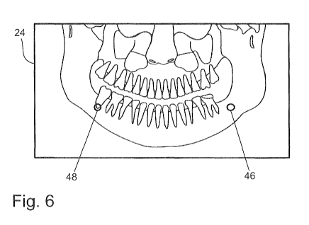
Figures 6 and 7 show two examples of a determined and edited image sec-
tion 46 or a panoramic radiography image 24 edited on the basis of the
13
CA 03114262 2021-03-25
WO 2020/094756
PCT/EP2019/080468
determined image section 46, respectively. The determined image section 46
may be a two-part (Figure 7) or one-part (Figure 6) image section. It is also
possible that a multi-part image section 46 is determined.
In Figure 6, it is shown that a comparatively small one-part image section 46,
for example a single image spot, is determined. For this purpose, an
interaction
with qualified medical staff may take place via the user interface. For
example,
a marking position 48 may be chosen by means of a touch screen or a computer
mouse, and a corresponding opposite spot on the opposite part of the jaw may
then automatically be defined as image section 46. Qualified dental staff may
thus directly see for a certain structure from which section an opposing jaw
ar-
tifact might possibly originate. On this basis, it can be recognized if the
currently
examined structure possibly does not originate from the currently examined
part
of the jaw at all. It is understood that it is also possible to choose a
section as
marking position 48.
In Figure 7, it is shown that a bigger, two-part image section 46 may also be
determined and edited. In particular, an image section 46 may be determined
that corresponds to all projection lines with two intersection points with the
im-
age surface. The two sections of the panoramic radiography image that might
possibly be affected by artifacts are marked. Marking may take place by
coloring
or also by framing. Based on marking, it can be considered directly during
eval-
uation that any objects that are disturbing or not easily recognizable may
result
from artifacts.
Figures 6 and 7 each show a two-dimensional view of a panoramic radiography
image. It is understood that the processing and editing of the determined
image
sections according to the invention may be carried out correspondingly with a
three-dimensional view of a panoramic radiography image.
Figure 8 schematically shows a method according to the invention. The method
comprises steps of receiving S10 the panoramic radiography image, of evaluat-
ing S12 the reconstruction data and determining an image section, of edit-
ing S14 the panoramic radiography image and of outputting S16 the edited
14
CA 03114262 2021-03-25
WO 2020/094756
PCT/EP2019/080468
panoramic radiography image. The method according to the invention may for
example be implemented as software for a diagnosis system or also as stand-
alone software.
In the Figures, a use of the invention in the field of odontology was
particularly
addressed. It is understood that a use in other fields, for example within
medi-
cine or industrial radiography, is also possible.
The invention was comprehensively described and explained by means of the
Figures and the description. The description and explanations are to be re-
garded as examples and not as limiting the scope. The invention is not limited
to the disclosed embodiments. For the person skilled in the art, other embodi-
ments or variants follow from the use of the present invention as well as from
a
thorough analysis of the Figures, the description and the following patent
claims.
In the patent claims, the words "comprise" and "include" do not exclude the
presence of further elements or steps. The indefinite article "a" or "an" does
not
exclude the presence of a plural. A single element or a single unit may
execute
the functions of several of the units named in the patent claims. An element,
a
unit, a device and a system may, partly or entirely, be implemented in
hardware
and/or software. The mere mention of some measures in several different de-
pendent patent claims is not to be understood to the effect that a combination
of these measures cannot also be used advantageously. A computer program
may be stored/distributed on a non-volatile data carrier, for example on an op-
tical memory device or on a solid state drive (SSD). A computer program may
be distributed together with hardware and/or as part of hardware, for example
on the Internet or via wire-bound or wireless communication systems. Refer-
ence signs in the patent claims are not to be understood as limiting the scope
of the invention.