Note: Descriptions are shown in the official language in which they were submitted.
CA 03114285 2021-02-09
WO 2020/036809
PCT/US2019/045794
SYSTEM FOR TREATING EMBOLISM AND
ASSOCIATED DEVICES AND METHODS
TECHNICAL FIELD
[0001] The present technology relates generally to systems, methods, and
devices for the
intravascular treatment of emboli and/or thrombi within a blood vessel of a
human patient. In
particular, some embodiments of the present technology relate to systems for
releasing stored
vacuum pressure to aspirate clot material from a blood vessel.
BACKGROUND
[0002] Thromboembolic events are characterized by an occlusion of a blood
vessel.
Thromboembolic disorders, such as stroke, pulmonary embolism, heart attack,
peripheral
thrombosis, atherosclerosis, and the like, affect many people. These disorders
are a major cause
of morbidity and mortality.
[0003] When an artery is occluded by a clot, tissue ischemia develops. The
ischemia will
progress to tissue infarction if the occlusion persists. However, infarction
does not develop or
is greatly limited if the flow of blood is reestablished rapidly. Failure to
reestablish blood flow
can accordingly lead to the loss of limb, angina pectoris, myocardial
infarction, stroke, or even
death.
[0004] In the venous circulation, occlusive material can also cause serious
harm. Blood
clots can develop in the large veins of the legs and pelvis, a common
condition known as deep
venous thrombosis (DVT). DVT commonly occurs where there is a propensity for
stagnated
blood (e.g., long distance air travel, immobility, etc.) and clotting (e.g.,
cancer, recent surgery,
such as orthopedic surgery, etc.). DVT can obstruct drainage of venous blood
from the legs
leading to swelling, ulcers, pain and infection. DVT can also create a
reservoir in which blood
clots can collect and then travel to other parts of the body including the
heart, lungs, brain
(stroke), abdominal organs, and/or extremities.
[0005] In the pulmonary circulation, the undesirable material can cause
harm by
obstructing pulmonary arteries¨a condition known as pulmonary embolism. If the
obstruction
is upstream, in the main or large branch pulmonary arteries, it can severely
compromise total
blood flow within the lungs, and therefore the entire body. This can result in
low blood pressure
-1-
CA 03114285 2021-02-09
WO 2020/036809
PCT/US2019/045794
and shock. If the obstruction is downstream, in large to medium pulmonary
artery branches, it
can prevent a significant portion of the lung from participating in the
exchange of gases to the
blood resulting in low blood oxygen and buildup of blood carbon dioxide.
[0006] There are many existing techniques to reestablish blood flow through
an occluded
vessel. Embolectomies, for example, are a surgical technique involving
incising a blood vessel
and placing a balloon-tipped device (such as the Fogarty catheter) at the
location of the
occlusion. The balloon is then inflated at a point beyond the clot and used to
withdraw the
obstructing material back to the point of incision. The obstructing material
is then removed by
the surgeon. Although such surgical techniques have been useful, exposing a
patient to surgery
may be traumatic and best avoided when possible. Additionally, the use of a
Fogarty catheter
may be problematic due to the possible risk of damaging the interior lining of
the vessel as the
catheter is being withdrawn.
[0007] Percutaneous methods are also utilized for reestablishing blood
flow. A common
percutaneous technique is referred to as balloon angioplasty where a balloon-
tipped catheter is
introduced to a blood vessel (e.g., typically through an introducing
catheter). The balloon-tipped
catheter is then advanced to the point of the occlusion and inflated to dilate
the stenosis. Balloon
angioplasty is appropriate for treating vessel stenosis, but it is generally
not effective for treating
acute thromboembolisms as none of the occlusive material is removed and
restenosis regularly
occurs after dilation. Another percutaneous technique involves placing a
catheter near the clot
and infusing streptokinase, urokinase, or other thrombolytic agents to
dissolve the clot.
Unfortunately, thrombolysis typically takes hours to days to be successful.
Additionally,
thrombolytic agents can cause hemorrhage, and in many patients the
thrombolytic agents cannot
be used at all.
[0008] Various devices exist for performing a thrombectomy or removing
other foreign
material. However, such devices have been found to have structures which are
either highly
complex, cause trauma to the treatment vessel, or lack the ability to be
appropriately fixed against
the vessel. Furthermore, many of the devices have highly complex structures
that lead to
manufacturing and quality control difficulties as well as delivery issues when
passing through
tortuous or small diameter catheters. Less complex devices may allow the user
to pull through
the clot, particularly with inexperienced users, and such devices may not
completely capture
and/or collect all of the clot material.
[0009] Thus, there exists a need for improved systems and methods for
embolic extraction.
-2-
CA 03114285 2021-02-09
WO 2020/036809
PCT/US2019/045794
BRIEF DESCRIPTION OF THE DRAWINGS
[0010] Many aspects of the present technology can be better understood with
reference to
the following drawings. The components in the drawings are not necessarily to
scale. Instead,
emphasis is placed on illustrating clearly the principles of the present
disclosure.
[0011] Figure 1 is a partially schematic side view of a clot removal system
configured in
accordance with the present technology.
[0012] Figure 2 is a side view of a locking syringe configured in
accordance with the
present technology.
[0013] Figure 3A is a side view of a locking syringe configured in
accordance with the
present technology.
[0014] Figure 3B is a side view of an adaptor for connecting the locking
syringe of Figure
3A to the clot removal system of Figure 1 configured in accordance with the
present technology.
[0015] Figure 3C is a side view of the adaptor of Figure 3B coupled to the
locking syringe
of Figure 3A.
[0016] Figure 3D is a side view of the locking syringe of Figure 3A coupled
to the clot
removal system of Figure 1 via the adaptor of Figure 3B.
[0017] Figure 4A is a perspective side view of another pressure source
configured in
accordance with the present technology, and Figures 4B and 4C are enlarged
schematic side
views of the pressure source of Figure 4A during operation.
[0018] Figure 5 is a cross-sectional side view of an automatic release
syringe configured
in accordance with the present technology.
[0019] Figure 6 is a perspective top view of a syringe configured in
accordance with the
present technology.
[0020] Figure 7 is a side view of an over-wire locking syringe configured
in accordance
with the present technology.
[0021] Figure 8 is a flow diagram of a process or method for operating a
clot removal
system in accordance with the present technology.
-3-
CA 03114285 2021-02-09
WO 2020/036809
PCT/US2019/045794
[0022] Figures 9A-9C are side views of a proximal portion of the clot
removal system of
Figure 1 during a clot removal procedure using the locking syringe of Figure 3
in accordance
with the present technology.
[0023] Figures 10A and 10B are schematic illustrations of a distal portion
of the clot
removal system of Figure 1 during a clot removal procedure in accordance with
the present
technology.
[0024] Figure 11 is a partially schematic side view of another clot removal
system
configured in accordance with the present technology.
[0025] Figure 12 is a flow diagram of another process or method for
operating a clot
removal system in accordance with the present technology.
[0026] Figures 13A-14C are schematic illustrations of a distal portion of
the clot removal
system of Figure 11 during a clot removal procedure in accordance with the
present technology.
[0027] Figure 15 is a flow diagram of another process or method for
operating a clot
removal system in accordance with the present technology.
[0028] Figures 16A-16E are schematic illustrations of a distal portion of
the clot removal
system of Figure 11 during a clot removal procedure in accordance with the
present technology.
[0029] Figure 17 is a partially schematic side view of another clot removal
system
configured in accordance with the present technology.
[0030] Figures 18A-18H are side views of a distal portion of the clot
removal system
shown of Figure 17 during a clot removal procedure in accordance with the
present technology.
[0031] Figure 19 is a perspective side view of a pressure source for
filtering blood from
aspirated clot material during a clot removal procedure configured in
accordance with the present
technology.
[0032] Figure 20A is a partially-exploded side view of a filter device and
pressure source
configured in accordance with the present technology.
[0033] Figure 20B is a perspective side view of the syringe of Figure 20A
coupled to the
filter device of the Figure 20A.
[0034] Figure 20C is a side view of the filter device and syringe of Figure
20B coupled to
the clot removal system of Figure 1.
-4-
CA 03114285 2021-02-09
WO 2020/036809
PCT/US2019/045794
[0035]
Figures 20D and 20E are side views of the syringe of Figure 20A coupled to the
clot removal system of Figure 1 for reintroducing blood to a patient.
[0036] Figure
21A is a partially-exploded side view of a filter device, a pressure source,
and a reinfusion syringe configured in accordance with the present technology.
[0037] Figure
21B is a perspective side view of the filter device of Figure 21A coupled to
the pressure source and the reinfusion syringe of Figure 21A.
[0038] Figure
22 is a partially-exploded side view of a filter device configured in
accordance with the present technology.
[0039] Figure
23 is a partially-exploded side view of a filter device configured in
accordance with the present technology.
[0040] Figure
24 is an enlarged isometric view of the clot removal system of Figure 1
configured in accordance with the present technology.
[0041] Figure
25 is an enlarged isometric view of the clot removal system of Figure 1
configured in accordance with the present technology.
DETAILED DESCRIPTION
[0042] The
present technology is generally directed to methods and systems for removing
clot material from a blood vessel of a human patient. In some embodiments, a
catheter can be
intravascularly positioned within a blood vessel such that a distal portion
(e.g., a distal opening)
of the catheter is positioned proximate to clot material within the blood
vessel. The catheter can
be fluidly coupled to a pressure source via a valve or other fluid control
device positioned outside
of the patient. With the valve closed, the pressure source can be activated to
charge a vacuum
chamber of the pressure source with a vacuum. The valve can then be opened to
apply the
vacuum to the catheter to thereby aspirate at least a portion of the clot
material from the blood
vessel into the catheter. In some embodiments, an interventional device can be
delivered through
the catheter and used to engage the clot material before and/or after the
vacuum is applied to the
catheter.
[0043] In one
aspect of the present technology, the pressure source is configured to
generate a vacuum and store the vacuum before the pressure source is fluidly
connected to the
catheter.
Therefore, opening the fluid control device can instantaneously or nearly
instantaneously apply the stored vacuum pressure to the catheter, thereby
generating suction
-5-
CA 03114285 2021-02-09
WO 2020/036809
PCT/US2019/045794
throughout the catheter. In particular, the suction is applied at the distal
portion of the catheter
proximate to the clot material. Pre-charging or storing the vacuum before
applying the vacuum
to the catheter can generate greater suction forces (and corresponding fluid
flow velocities) at
and/or near the distal portion of the catheter compared to, for example,
simply activating the
pressure source while it is fluidly connected to the catheter. The greater
suction forces generated
by application of the stored vacuum can be used to aspirate or otherwise
remove clot material
from within a blood vessel of a human patient.
[0044] Although many of the embodiments are described below with respect to
devices,
systems, and methods for treating a pulmonary embolism, other applications and
other
embodiments in addition to those described herein are within the scope of the
technology (e.g.,
intravascular procedures other than the treatment of emboli, intravascular
procedures for treating
cerebral embolism, intravascular procedures for treating deep vein thrombosis
(DVT), etc.).
Additionally, several other embodiments of the technology can have different
configurations,
states, components, or procedures than those described herein. Moreover, it
will be appreciated
that specific elements, substructures, advantages, uses, and/or other features
of the embodiments
described with reference to Figures 1-25 can be suitably interchanged,
substituted or otherwise
configured with one another in accordance with additional embodiments of the
present
technology. Furthermore, suitable elements of the embodiments described with
reference to
Figures 1-25 can be used as standalone and/or self-contained devices. A person
of ordinary skill
in the art, therefore, will accordingly understand that the technology can
have other embodiments
with additional elements, or the technology can have other embodiments without
several of the
features shown and described below with reference to Figures 1-25.
[0045] With regard to the terms "distal" and "proximal" within this
description, unless
otherwise specified, the terms can reference a relative position of the
portions of a catheter
subsystem with reference to an operator and/or a location in the vasculature.
Also, as used
herein, the designations "rearward," "forward," "upward," "downward," etc. are
not meant to
limit the referenced component to use in a specific orientation. It will be
appreciated that such
designations refer to the orientation of the referenced component as
illustrated in the Figures;
the systems of the present technology can be used in any orientation suitable
to the user.
[0046] The headings provided herein are for convenience only and should not
be construed
as limiting the subject matter disclosed.
-6-
CA 03114285 2021-02-09
WO 2020/036809
PCT/US2019/045794
I. Selected Embodiments of Clot Removal Systems
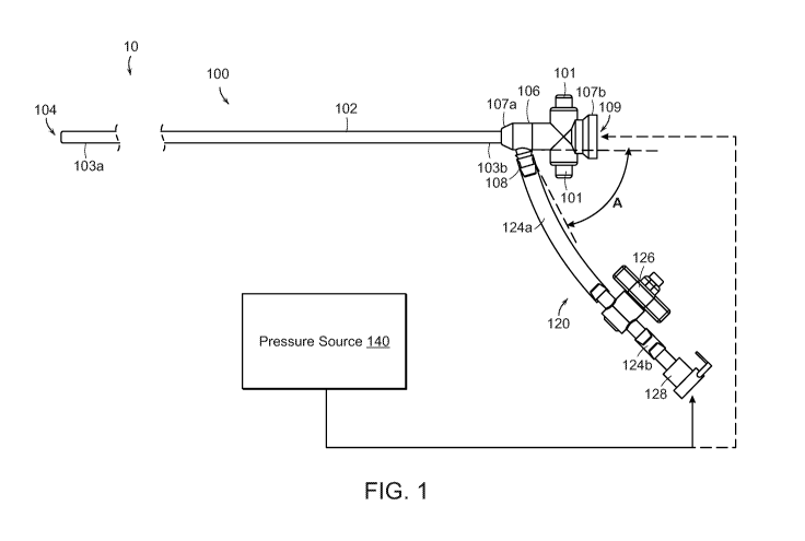
[0047] Figure 1 is a partially schematic side view of a clot treatment or
clot removal
system comprising an aspiration assembly 10 ("assembly 10") configured in
accordance with an
embodiment of the present technology. In the illustrated embodiment, the
assembly 10 includes
a catheter subsystem 100, a tubing subsystem 120, and a pressure source 140.
The catheter
subsystem 100 includes a catheter 102 (e.g., an aspiration catheter)
comprising an elongated
shaft defining a lumen 104 and having a distal portion 103a and a proximal
portion 103b. The
catheter subsystem 100 further includes a valve 106 that can be integral with
or coupled to the
proximal portion 103b of the catheter 102.
[0048] In the illustrated embodiment, the valve 106 includes a distal
portion 107a, a
proximal portion 107b, and a lumen 109 extending therethrough from the distal
portion 107a to
the proximal portion 107b. The valve 106 further includes a flow controller
(obscured in Figure
1) in the lumen 109. In some embodiments, the valve is a hemostasis valve that
is configured to
maintain hemostasis during a clot removal procedure by preventing fluid flow
in the proximal
direction through the valve 106 as various components such as delivery
sheaths, pull members,
guidewires, interventional devices, other aspiration catheters (e.g., as
described in detail with
reference to Figures 11-16E), etc., are inserted through the valve 106 to be
delivered through
the catheter 102 to a treatment site in a blood vessel. The valve 106 further
includes a branch or
side port 108 positioned distally of the flow controller in the lumen 109 and
configured to fluidly
couple the lumen 104 of the catheter 102 to the tubing subsystem 120. In the
illustrated
embodiment, the valve 106 includes buttons 101 that can be actuated (e.g.,
depressed) to open a
conduit within the lumen 109. In some embodiments, the valve 106 can be a
valve of the type
disclosed in U.S. Patent Application No. 16/117,519, filed August 30, 2018,
and titled
"HEMOSTASIS VALVES AND METHODS OF USE," which is incorporated herein by
reference in its entirety. In some embodiments, the proximal portion 107b of
the valve 106 is
further configured to be detachably coupled (e.g., via a snap-fit arrangement)
to a
retraction/aspiration device for aspirating the lumen 104 of the catheter 102
and/or for retracting
an interventional device, catheter, delivery sheath, catheter, etc.,
positioned within the lumen
104. Specific details of such retraction/aspiration devices and associated
methods are disclosed
in U.S. Patent Application No. 9,526,864, filed June 9, 2015, and titled
"RETRACTION AND
ASPIRATION DEVICE FOR TREATING EMBOLISM AND ASSOCIATED SYSTEMS
AND METHODS," which is incorporated herein by reference in its entirety.
-7-
CA 03114285 2021-02-09
WO 2020/036809
PCT/US2019/045794
[0049] The tubing subsystem 120 fluidly couples the catheter subsystem 100
to the
pressure source 140. More specifically, the tubing subsystem 120 can include
one or more tubing
sections 124 (individually labeled as a first tubing section 124a and a second
tubing section
124b), at least one fluid control device 126 (e.g., a valve), and at least one
connector 128 for
fluidly coupling the tubing subsystem 120 to the pressure source 140 and/or
other suitable
components. More specifically, in the illustrated embodiment, the fluid
control device 126 is a
stopcock that is fluidly coupled to (i) the side port 108 of the valve 106 via
the first tubing section
124a and (ii) the connector 128 via the second tubing section 124b. In some
embodiments, the
fluid control device 126 can define a lumen having a diameter (or other cross-
sectional
dimension) that is greater than or equal to a diameter of the lumen 104 of the
catheter 102, a
diameter of the first tubing section 124a, and/or a diameter of the second
tubing section 124b.
[0050] The fluid control device 126 is externally operable by a user to
regulate the flow
of fluid therethrough and, specifically, from the lumen 104 of the catheter
102 to the pressure
source 140. In other embodiments, the fluid control device 126 can be a clamp
that can be
actuated (e.g., compressed or squeezed by the hand of a user) to partially or
fully restrict fluid
flow through the tubing section 124a and/or the tubing section 124b. In yet
other embodiments,
the fluid control device 126 can be omitted and its functionality incorporated
into the pressure
source 140 (e.g., as described in detail below with reference to Figure 5). In
some embodiments,
the fluid control device 126 can include a quick-release mechanism (e.g., a
spring-loaded
apparatus) for rapidly opening, unclamping, etc., the fluid control device 126
to (e.g.,
instantaneously or nearly instantaneously) fluidly connect the pressure source
140 and the
catheter 102. In some embodiments, the fluid control device 126 can be
opened/closed
automatically (e.g., by a motor, switch, etc.). When the pressure source 140
is pre-charged with
a vacuum, as described in detail below, such a quick-release fluid control
device 126 can reduce
the time needed for pressure in the assembly 10 to equalize after opening of
the fluid control
device 126, and can thereby increase suction forces generated at the distal
portion 103a of the
catheter 102.
[0051] In some embodiments, the connector 128 is a quick-release connector
(e.g., a quick
disconnect fitting) that enables rapid coupling/decoupling of the catheter 102
and the fluid
control device 126 to/from the pressure source 140. In other embodiments, the
tubing subsystem
120 can have more or fewer tubing sections, connectors, and/or fluid control
devices, and can
-8-
CA 03114285 2021-02-09
WO 2020/036809
PCT/US2019/045794
have other suitable configurations. In some embodiments, one or more of the
components can
be permanently connected and/or integrally formed.
[0052] The pressure source 140 is configured to generate (e.g., form,
create, charge, build-
up, etc.) a vacuum (e.g., negative relative pressure) and store the vacuum for
subsequent
application to the catheter subsystem 100. Further details of suitable
pressure sources are
described in detail below with reference to Figures 2-7. During operation of
the assembly 10, a
user can first close the fluid control device 126 before activating the
pressure source 140 to build
up vacuum pressure within the pressure source 140 (e.g., a vacuum chamber of
the pressure
source 140). In some embodiments, the user can control or select the volume of
the generated
vacuum. In this manner, a vacuum is charged within the pressure source 140
before the pressure
source 140 is fluidly connected to the catheter subsystem 100. To aspirate the
lumen 104 of the
catheter 102, the user can open the fluid control device 126 to fluidly
connect the pressure source
140 to the catheter subsystem 100 and thereby apply or release the vacuum
stored in the pressure
source 140 to the lumen 104 of the catheter 102. Opening of the fluid control
device 126
instantaneously or nearly instantaneously applies the stored vacuum pressure
to the tubing
subsystem 120 and the catheter 102, thereby generating suction throughout the
catheter 102. In
particular, the suction is applied at the distal portion 103a of the catheter
102. In one aspect of
the present technology, pre-charging or storing the vacuum before applying the
vacuum to the
lumen 104 of the catheter 102 is expected to generate greater suction forces
(and corresponding
fluid flow velocities) at and/or near the distal portion 103a of the catheter
102 compared to
simply activating the pressure source 140 while it is fluidly connected to the
catheter 102. As
described in detail below, the suction forces generated by application of the
stored vacuum can
be used to aspirate or otherwise remove clot material from within a blood
vessel of a human
patient.
Selected Embodiments of Pressure Sources for Use with Clot Removal Systems
[0053] As described in detail above with reference to Figure 1, the
assembly 10 of the
present technology includes a pressure source (e.g., a vacuum source, negative
pressure source,
etc.) configured to charge a vacuum that can be applied to the catheter
subsystem 100 to generate
suction forces for aspirating clot material from within a blood vessel. In
general, the pressure
source can be any suitable source or combination of sources for generating
and/or storing
negative pressure. In some embodiments, the pressure source can be a pump
(e.g., an electric
pump coupled to a vacuum chamber) while, in other embodiments, the pressure
source can
-9-
CA 03114285 2021-02-09
WO 2020/036809
PCT/US2019/045794
include one or more syringes that can be actuated or otherwise activated by a
user of the assembly
to generate and store a vacuum therein.
[0054] Figure 2 is a side view of a pressure source 240 comprising a vacuum-
pressure
locking syringe ("syringe 240") configured in accordance with the present
technology. In some
embodiments, the syringe 240 can be of the kind sold under the trademark
"VacLok" by Merit
Medical System, Inc. In the illustrated embodiment, the syringe 240 includes a
plunger 242
slidably and rotatably positioned within a chamber or barrel 244. The barrel
244 is shown as
transparent in Figure 2 for the sake of clarity. The plunger 242 includes a
seal 243 and a plurality
of index members 246 defining slots 248 between adjacent pairs thereof A tab
member 245
projects inwardly from the interior surface of the barrel 244 and is
configured to be removably
positioned in the slots 248 for locking the plunger 242 in position relative
to the barrel 244. In
some embodiments, the barrel 244 can be made of a transparent material that
permits a user to
visualize material (e.g., clot material) within the barrel 244 and to
visualize the relative position
between the slots 248 and tab member 245 for locking the syringe 240.
[0055] Referring to both Figures 1 and 2 together, the syringe 240 further
includes a tip
247 for coupling the syringe 240 to the tubing subsystem 120. In the
illustrated embodiment,
the tip 247 is a standard luer connector that can be coupled to the connector
128 via one or more
suitable adaptors. The tip 247 further defines a lumen or bore 249 having an
inner diameter Di.
In some embodiments, the diameter Di is about 0.103", or about 0.080" to about
0.200", or about
0.100" to about 0.150", or about 0.100" to about 0.110". In some embodiments,
the inner
diameter Di is about 14 French.
[0056] During operation of the assembly 10, a user can first close the
fluid control device
126 and then grip the plunger 242 and/or the barrel 244 to withdraw (e.g.,
retract) the plunger
242 at least partially out of the barrel 244 to thereby generate a vacuum in
the barrel 244. Once
the user has withdrawn the plunger 242 to a sufficient or desired volume, the
user can lock the
plunger 242 by rotating the plunger 242 relative to the barrel 244 such that
the tab member 245
is positioned within a corresponding one of the slots 248. In other
embodiments, the syringe
240 may not be a locking syringe, and the user can instead hold the plunger
242 in position
relative to the barrel 244. Moreover, the user can control the volume of the
vacuum¨by
withdrawing the plunger 242 more or less¨to provide a desired amount or level
of
suction/aspiration upon opening of the fluid control device 126. In some
embodiments, the
syringe has a volume of about 60 cc or less than about 60 cc.
CA 03114285 2021-02-09
WO 2020/036809
PCT/US2019/045794
[0057] Figure 3A is a side view of a pressure source 340 comprising a
vacuum-pressure
locking syringe ("syringe 340") configured in accordance with the present
technology. The
syringe 340 can have some features generally similar to the features of the
syringe 240 described
above with reference to Figure 2. For example, the syringe 340 includes a
plunger 342 slidably
and rotatably positioned within a barrel 344, and the plunger 342 includes a
plurality of index
members 346 defining slots 348 between adjacent pairs thereof The barrel 344
is shown as
transparent in Figure 3A (and Figure 3C) for the sake of clarity. While
withdrawing the plunger
342, a user can lock the plunger 342 at a specified volume by rotating the
plunger 342 relative
to the barrel 344 such that a tab member 345 on the interior surface of the
barrel 344 is positioned
within a corresponding one of the slots 348. In some embodiments, the syringe
340 has a
maximum volume of about 60 cc or greater than 60 cc.
[0058] In the illustrated embodiment, the syringe 340 includes a large-bore
tip 347, such
as a Toomey tip, defining an inner lumen or bore 349. In some embodiments, the
bore 349 can
have an inner diameter D2 that is greater than or equal to the largest inner
diameter of the
assembly 10 (e.g., of the catheter 102 and tubing subsystem 120). In certain
embodiments, the
tip 347 can be about 26 French or greater. Accordingly, referring to Figures 2
and 3A together,
the diameter D2 can be greater than the dimension Di. For example, the
dimension D2 can be
about two, three, four, or more times greater than the diameter Di.
[0059] Figure 3B is a side view of an adaptor 350 for connecting the
syringe 340 to the
catheter subsystem 100 configured in accordance with the present technology.
Figure 3C is a
side view of the adaptor 350 coupled to the syringe 340, and Figure 3D is a
side view of the
syringe 340 coupled to the tubing subsystem 120 via the adaptor 350. The
adaptor 350 is shown
as partially transparent in Figure 3C for the sake of illustration. Referring
to Figure 3B, the, the
adaptor 350 includes (i) a first portion 351 defining a first lumen or bore
352 having an inner
diameter D3, (ii) a second portion 353 defining a second lumen or bore 354,
and (iii) a stepped
surface or interface 355 between the first and second portions 351, 353. The
first portion 351
can further include a seal 357 such as an 0-Ring around an exterior surface
thereof
[0060] Referring to Figures 3A-3D together, the second bore 354 of the
adaptor 350 is
configured to removably receive the tip 347 of the syringe 340 therein. In
some embodiments,
the tip 347 can be snuggly received in the second bore 354 via an interference
fit. In some
embodiments, a seal (e.g., an 0-ring) can be positioned between an exterior
surface of the tip
347 and an interior surface of the second bore 354. In other embodiments, the
syringe 340 can
-11-
CA 03114285 2021-02-09
WO 2020/036809
PCT/US2019/045794
be permanently coupled or integrally formed with the adaptor 350. The first
portion 351 of the
adaptor 350 is configured to be removably positioned within the connector 128
of the tubing
subsystem 120 to fluidly couple the syringe 340 to the tubing subsystem 120.
In some
embodiments, the first portion 351 of the adaptor 350 can be pushed into the
connector 128 until
the interface 355 abuts the connector 128. When the first portion 351 of the
adaptor 350 is
positioned within the connector 128, the seal 357 seals the interface between
the connector 128
and the adaptor 350.
[0061] The diameter D3 of the first bore 352 of the adaptor 350 can be
selected to be about
the same as or greater than the greatest inner diameter of the assembly 10
(e.g., of the catheter
102 and the tubing subsystem 120). For example, the catheter 102 can be about
9 French or
greater, and the diameter D3 can be selected to be larger than the size of the
catheter 102.
Accordingly, when the fluid control device 126 is open, the continuous lumen
between the
catheter 102 and the syringe 340 can have a generally constant diameter and/or
does not contain
any narrowing at the interface between the syringe 340 and the tubing
subsystem 120. That is,
the adaptor 350 can connect the syringe 340 and the tubing subsystem 120
without any restriction
or narrowing of the fluid path. In contrast, a standard luer connector (e.g.,
the syringe 240) can
only provide a continuous lumen for catheters of about 8 French or smaller.
Any narrowing of
the fluid pathway between the catheter 102 and the syringe 340 can reduce the
volumetric flow
rate (e.g., suction forces and fluid velocities) that can be generated when a
vacuum stored in the
syringe 340 is applied to the catheter 102.
[0062] In general, the syringe 340 and the adaptor 350 can reduce the fluid
resistance in
the assembly 10 and therefore facilitate a more rapid pressure equalization in
the assembly 10
when the fluid control device 126 is opened to apply the charged vacuum to the
catheter 102. In
some embodiments, for example, when the syringe 240 (Figure 2) is charged with
a 60 cc
vacuum and the fluid control device 126 is opened, the pressure in the
assembly 10 can take
about 1-2 seconds to equalize. In contrast, when the syringe 340 is charged
with a 60 cc vacuum
and the fluid control device 126 is opened, the pressure in the assembly 10
can take less than
about 1 second (e.g., about 0.5 seconds) to equalize. More specifically, Table
1 illustrates
representative pressure equalization times and associated flow rates when the
syringe 240 is
coupled to a 20 French catheter (i.e., the catheter 102). Table 2 illustrates
representative pressure
equalization times and associated flow rates when the syringe 340 and the
adaptor 350 are
coupled to a 20 French catheter (i.e., the catheter 102).
-12-
CA 03114285 2021-02-09
WO 2020/036809
PCT/US2019/045794
Table 1
Pressure Equalization Time (seconds) Flow Rate (cc/sec)
2.0 30.0
1.9 31.6
1.8 33.3
1.7 35.3
1.6 37.5
1.5 40.0
1.4 42.9
1.3 46.2
Table 2
Pressure Equalization Time (seconds) Flow Rate (cc/sec)
0.9 66.7
0.8 75.0
0.7 85.7
0.6 100.0
0.5 120.0
0.4 150.0
0.3 200.0
0.2 300.0
0.1 600.0
[0063] In each instance, the syringe 340 provides for relatively faster
equalization times
and correspondingly greater flow rates. It is expected that the more rapid
pressure equalization
and flow rates provided by the syringe 340 will provide correspondingly
greater suction forces
at the distal portion 103a of the catheter 102. That is, in general, it is
expected that increasing
-13-
CA 03114285 2021-02-09
WO 2020/036809
PCT/US2019/045794
the bore size of a syringe used to provide vacuum pressure will provide
greater suction forces
over a smaller period of time (e.g., will provide a larger vacuum impulse). In
some
embodiments, the greater suction forces can facilitate the removal of clot
material from a blood
vessel of a patient even where the clot material is strongly lodged or
attached within the blood
vessel (e.g., a chronic clot).
[0064] Moreover, as shown in Figure 3D, the adaptor 350 can couple the
syringe 340 to
the connector 128 without the need for any intervening tubing sections or
additional adaptors.
This arrangement can minimize the total length, volume, etc., of the
components fluidly coupling
the catheter 102 to the syringe 340. It is expected that the magnitude of
suction forces generated
at the distal portion 103a of the catheter 102¨e.g., when a vacuum charged in
the syringe 340
is applied to the catheter 102 by opening of the fluid control device 126¨is
proportional to the
length of the fluid path between the pressure source 340 and catheter 102.
Thus, operation of
the assembly 10 with the syringe 340 and adaptor 350 is expected to increase
the suction forces
generated at the distal portion 103a of the catheter 102. In some embodiments,
the greater
suction forces can facilitate the removal of clot material from a blood vessel
of a patient even
where the clot material is strongly lodged or attached within the blood vessel
(e.g., a chronic
clot).
[0065] Figure 4A is a side perspective view a pressure source 400 including
the syringe
340 ("primary syringe 340") shown in Figures 3A-3D and a secondary syringe 460
configured
in accordance with the present technology. The secondary syringe 460 can
include a plunger
462 slidably positioned within a chamber or barrel 464. The primary and
secondary syringes
340, 460 can have the same volume or different volumes. In the illustrated
embodiment, a tip
463 of the secondary syringe 460 is coupled to a first one-way valve (e.g., a
check valve) 470
via a coupling member 465, such as a tube. The first one-way valve 470 is
configured to fluidly
connect the secondary syringe 460 to the ambient environment or another device
coupled to the
first one-way valve 470. A second one-way valve (e.g., a check valve) 472
spans between and
is configured to fluidly connect the primary syringe 340 to the secondary
syringe 460. More
specially, in the illustrated embodiment the second one-way valve 472 is
connected between the
first portion 351 of the adaptor 350 and the coupling member 465. In other
embodiments, the
second one-way valve 472 can couple the primary and secondary syringes 340,
460 in different
manners. For example, the second one-way valve 472 can span between and
directly connect
the barrels 344,464. The primary and secondary syringes 340, 460 can be
coupled or fastened
-14-
CA 03114285 2021-02-09
WO 2020/036809
PCT/US2019/045794
together via one or more connectors 474 that fix the positions of the barrel
344, 464 relative to
one another.
[0066] In some embodiments, the second one-way valve 472 is a normally-open
check
valve configured to (i) permit fluid (e.g., air) flow from the primary syringe
340 and the adaptor
350 to the secondary syringe 460 and (ii) inhibit fluid flow in the opposite
direction from the
secondary syringe 460 into the primary syringe 340. In some embodiments, the
second one-way
valve 472 has a cracking (e.g., opening) pressure of about 0 psi. In one
aspect of the present
technology, this arrangement maximizes the magnitude of the vacuum that can be
charged within
the primary syringe 340. That is, the cracking pressure of the second one-way
valve 472 does
not reduce the effective vacuum within the primary syringe 340. In other
embodiments a
normally-closed or other type of valve could be used for the second one-way
valve 472.
However, in such embodiments the vacuum efficiency of the pressure source 400
would be
reduced by the cracking pressure of the second one-way valve 472. Similarly,
the first one-way
valve 470 can be a check valve configured to (i) permit fluid flow from the
secondary syringe
460 to the ambient environment (or other device) and (ii) inhibit fluid flow
in the opposite
direction from the ambient environment into the secondary syringe 460.
[0067] Figures 4B and 4C are enlarged schematic side views of the pressure
source 400
during operation. More specifically, Figures 4B and 4C illustrate fluid flow
paths through the
first and second one-way valves 470, 472 during retraction and advancement,
respectively, of
the plunger 462 through the barrel 464 of the secondary syringe 460. Referring
first to Figures
4A and 4B together, during retraction/withdrawal of the plunger 462, (i) the
first one-way valve
470 is closed to inhibit fluid from flowing into the secondary syringe 460
while (ii) the second
one-way valve is open 472 to permit fluid to flow from the primary syringe
340, the catheter
subsystem 100 (Figure 1), and/or the tubing subsystem 120 (Figure 1) into the
secondary syringe
460. This flow path is indicated by the arrows R in Figure 4B. Referring to
Figures 4A and 4C
together, during advancement of the plunger 462, (i) the first one-way valve
470 is open to
permit fluid flow (e.g., fluid expulsion) from the secondary syringe 460 to
the ambient
environment (or other device) while (ii) the second one-way valve 472 is
closed to inhibit fluid
flow from the secondary syringe 460 into (e.g., back into) the primary syringe
360, the catheter
subsystem 100, and/or the tubing subsystem 120. This flow path is indicated by
the arrows A in
Figure 4C.
-15-
CA 03114285 2021-02-09
WO 2020/036809
PCT/US2019/045794
[0068] Referring to Figures 1 and 3A-4C together, the pressure source 400
can be coupled
to the tubing subsystem 120 by coupling the primary syringe 340 to the
connector 128 (e.g., as
shown in Figure 3D). When the pressure source is coupled to the tubing
subsystem 120,
retraction of the plunger 462 of the secondary syringe 460 evacuates an
evacuatable volume of
the assembly 10. For example, when the fluid control device 126 is closed,
retraction of the
plunger 462 of the secondary syringe 460 evacuates fluid, through the second
one-way valve
472, from (i) the primary syringe 340 (e.g., from the barrel 344, the tip 347,
and/or the adaptor
350) and (ii) the portion of the tubing subsystem 120 between the fluid
control device 126 and
the primary syringe 340. This can enable a greater charged/stored vacuum to be
generated for
subsequent application to the catheter subsystem 100 for aspirating clot
material. In some
embodiments, the plunger 462 of the secondary syringe 460 can be
withdrawn/advanced (e.g.,
"cycled") one or more times before withdrawing the plunger 342 of the primary
syringe 340 to
evacuate air from (i) the tip 347 of the primary syringe 340 and/or (ii) the
portion of the tubing
subsystem 120 between the fluid control device 126 and the tip 347. In other
embodiments, the
plunger 462 of the secondary syringe 460 can alternatively or additionally be
withdrawn after
withdrawing the plunger 342 of the primary syringe 340 to further evacuate the
barrel 344 of the
primary syringe 340. In some embodiments, the plunger 462 can be cycled when
the fluid
control device 126 is open to, for example, facilitate the removal of clot
material stuck or clogged
within the catheter subsystem 100. That is, cycling the secondary syringe 460
when the fluid
control device 126 is open can generate vacuum pressure and suction in the
catheter 102 to aid
in the aspiration/removal of clot material.
[0069] In some embodiments, the volumes of the primary and secondary
syringes 340,
460 can be selected based on one or more desired characteristics of a clot
removal procedure
using the pressure source 400. For example, the secondary syringe 460 can have
a larger volume
than the primary syringe 340 to permit a high vacuum to be charged within the
primary syringe
340 while also limiting blood loss from the patient.
[0070] In one aspect of the present technology, the pressure source 340
permits a greater
vacuum to be generated without increasing the volume of the primary syringe
340. For example,
the vacuum generated by the primary syringe 340 alone is directly proportional
to the volume of
the primary syringe 340. Thus, to generate a greater vacuum using the primary
syringe 340
alone, the volume of the primary syringe 340 must be increased. In contrast,
inclusion of the
secondary syringe 460 in the pressure source 400 and the configuration of the
first and second
-16-
CA 03114285 2021-02-09
WO 2020/036809
PCT/US2019/045794
one-way valves 470,472 allows the (e.g., maximum) generated vacuum to be
independent of the
volume of the primary syringe 340. Therefore, for example, the generated
vacuum can be
increased without correspondingly increasing the volume of blood withdrawn
from the patient
when applying the vacuum to the catheter subsystem 100.
[0071] In some embodiments, (e.g., as described in greater detail below
with reference to
Figure 19), the primary syringe 340 of the pressure source 400 can be replaced
with a simple
pressure vessel or other volume, such as a canister, barrel, tube, etc. In
such embodiments, a
vacuum can be generated in the canister simply by cycling the secondary
syringe 460 one or
more times. In some embodiments, the secondary syringe 460 can comprise a pump
or vacuum
source other than a syringe. Likewise, the secondary syringe 460 or other
vacuum source can
be fluidly coupled to the primary syringe 340 in other manners (e.g., via a
different arrangement
of check valves) to produce the same or similar flow patterns as shown in
Figures 4B and 4C.
Moreover, in some embodiments the first and second one-way valves 470, 472 can
be other types
of flow control devices that are mechanically activated/deactivated (e.g.,
opened and closed)
rather than passively operated via pressure differentials within the pressure
source 400. For
example, the flow control devices 470, 472 can be mechanically coupled to the
plunger 462 of
the secondary syringe 460 such that cycling the plunger 462
activates/deactivates the flow
control devices 470, 472 to operate the pressure source 400 in the manner
illustrated in Figures
4B and 4C.
[0072] Figure 5 is a side cross-sectional view of a pressure source 540
comprising an
automatic release syringe ("syringe 540") configured in accordance with the
present technology.
In general, the syringe 540 is configured to automatically apply a charged
vacuum of a selected
volume to the catheter subsystem 100 without requiring the actuation of an
intervening fluid
control device, such as the fluid control device 126 shown in Figure 1. The
syringe 540 can have
some features generally similar to the features of the syringes 240, 340
described in detail above
with reference to Figures 2 and 3A-3D. For example, the syringe 540 includes a
first plunger
542 slidably positioned within a chamber or barrel 544. The first plunger 542
further includes a
first seal 543 that engages an interior surface of the barrel 544 such that a
vacuum is formed
within the barrel 544 as the first plunger 542 is withdrawn through the barrel
544. Likewise,
referring to both Figures 1 and 5 together, the syringe 540 includes a tip 547
(e.g., a Toomey tip)
for coupling the syringe 540 to the tubing subsystem 120 (e.g., via a Toomey
tip adaptor) and
defining a bore 549. In some embodiments, the bore 549 has a relatively large
diameter selected
-17-
CA 03114285 2021-02-09
WO 2020/036809
PCT/US2019/045794
to provide rapid pressure equalization in the assembly 10 after a vacuum
stored in the syringe
540 is released.
[0073] The first plunger 542 can further include (i) a grip portion 541
configured to be
engaged by a user for retracting the first plunger 542 and (ii) a lumen 581
extending lengthwise
therethrough. In the illustrated embodiment, a plunger assembly 582 is
slidably positioned
within and extends through the lumen 581 of the first plunger 542. The plunger
assembly 582
includes (i) a second plunger 583 and (ii) a release member 584 slidably
and/or rotatably
positioned within a lumen 585 of the second plunger 583. The release member
584 includes an
engagement member 586 configured to engage the grip portion 541 of the first
plunger 542 when
the first plunger 542 is withdrawn from the barrel 544. The second plunger 583
includes a
second seal 587 configured to engage and seal an interior surface of the bore
549 of the syringe
540 to enable a vacuum to be formed in the barrel 544 as the first plunger 542
is withdrawn
through the barrel 544. That is, the second seal 587 can seal (e.g., fluidly
disconnect) the barrel
544 of the syringe from the tubing subsystem 120 and the catheter subsystem
100. In some
embodiments, the syringe 540 can further include an 0-ring 579 or other
suitable component for
sealing an interface between the first and second plungers 542, 582 to
maintain the vacuum
formed within the barrel 544, while also permitting the first plunger 542 to
move (e.g., translate)
relative to the second plunger 583.
[0074] The plunger assembly 582 further includes a locking mechanism (not
shown)
configured to permit/inhibit the release member 584 from moving longitudinally
relative to the
second plunger 583. In some embodiments, for example, rotation of the release
member 584 in
a first direction relative to the second plunger 583 can lock the two
components in position,
while rotation of the release member 584 in a second direction relative to the
second plunger
583 can unlock the two components so that the release member 584 can be
withdrawn or pushed
into the lumen 585 of the second plunger 583. In other embodiments, the
release member 584
and the second plunger 583 can be integrally formed or permanently locked
together.
[0075] The plunger assembly 582 enables (i) a user of the syringe 540 to
select a desired
volume for a vacuum to be formed in the syringe 540 and (ii) the automatic
release or application
of a generated vacuum via opening (e.g., unplugging) of the bore 549.
Specifically, during
operation of the syringe 540, a user can first unlock the release member 584
and slide the release
member 584 to a position corresponding to a desired vacuum volume. For
example, the release
member 584 can have tick marks 588 or other indicia along its length that
correspond to a volume
-18-
CA 03114285 2021-02-09
WO 2020/036809
PCT/US2019/045794
of the syringe 540 (e.g., a vacuum chamber volume). After selecting a desired
volume, the user
can lock the release member 584 relative to the second plunger 583 (e.g., by
rotating the release
member 584) to inhibit relative movement of the two components. After locking
the release
member 584, the user can grasp the grip portion 541 to retract the first
plunger 542 relative to
the barrel 544 and the plunger assembly 582 to generate a vacuum within the
barrel 544 between
the first and second seals 543, 587. When the first plunger 542 has been
retracted to the desired
volume, the grip portion 541 engages the engagement member 586 of the release
member 584
such that further retraction of the first plunger 542 simultaneously retracts
the plunger assembly
582. As the plunger assembly 582 is retracted, the second seal 587 of the
second plunger 583 is
pulled out of the bore 549, thereby releasing the vacuum stored in the barrel
544. In this manner,
the syringe 540 provides for the automatic release of charged vacuum pressure
at a specified
volume and with a single retraction of the first plunger 542. Put differently,
the syringe 540 has
a built-in fluid control device and thus eliminates the need for a separate
fluid control device 126
and/or an additional step for opening the fluid control device 126.
[0076] Figure 6 is a top perspective view of a pressure source 640
comprising a syringe
("syringe 640") configured in accordance with the present technology. The
syringe 640 can
include some features generally similar to the features of the syringes 240,
340, and 540
described in detail above with reference to Figures 2-3D and 5. For example,
the syringe 640
includes a plunger 642 slidably positioned within a barrel 644, and a tip 647
(e.g., a large-bore
tip). In the illustrated embodiment, the syringe 640 further includes a lever
or handle 690
operably coupled to the plunger 642. The handle 690 provides mechanical
leverage for
withdrawing the plunger 642 to create a vacuum within the barrel 644. More
specifically, the
handle 690 can be coupled to a crossbar 691 that rotates relative to the
plunger 642 via actuation
(e.g., rotation) of the handle 690. The crossbar 691 can be coupled to a gear
(obscured in Figure
6) configured to engage a track 692 on the plunger 642. Accordingly, rotation
of the handle 690
in a first direction retracts the plunger 642 relative to the barrel 644 to
charge a vacuum in the
barrel 644. And, rotation of the handle 690 in a second (e.g., opposite)
direction advances the
plunger 642 into the barrel 644 to, for example, expel fluid, material, etc.,
from the barrel 644.
[0077] In one aspect of the present technology, the handle 690 provides
additional
mechanical leverage relative to a standard syringe, and can thus reduce the
force (e.g., strain,
energy, etc.) required by a user of the syringe 640 to form a vacuum in the
syringe 640.
Therefore, use of the syringe 640 can reduce the time needed to remove clot
material with the
-19-
CA 03114285 2021-02-09
WO 2020/036809
PCT/US2019/045794
assembly 10. In some embodiments, the syringe 640 can have a volume greater
than 60 cc (e.g.,
greater than 80 cc, greater than 100 cc, greater than 120 cc, greater than 140
cc, etc.). In a
particular embodiment, for example, the syringe 640 can have a volume of about
140 cc. With
such large volumes, it may be difficult for some users to manually retract the
plunger 642 without
the additional mechanical leverage provided by the handle 690. Thus, the
syringe 640 can enable
the use of larger volume syringes that can generate correspondingly greater
suction forces in the
catheter subsystem 100.
[0078] Referring again to Figure 1, it is expected that less tortuous
(e.g., more linear) fluid
paths between the pressure source 140 and the catheter subsystem 100 will
produce greater
suction forces and corresponding fluid velocities at the distal portion 103a
of the catheter 102
when stored vacuum pressure is applied to the catheter subsystem 100.
Accordingly, in some
embodiments the side port 108 of the valve 106 can be formed to have an angle
A that is less
than about 90 , less than about 75 , less than about 60 , less than about 45 ,
less than about 30 ,
less than about 15 etc. Reducing the relative angle between the side port 108
and the lumen
109 of the valve 106 (and thus the lumen 104 of the catheter 102) reduces the
tortuosity of the
fluid path between the pressure source 140 and the catheter 102. Moreover, in
some
embodiments, the pressure source 140 can be coupled to the proximal portion
107b of the valve
106 instead of or in addition to the side port 108 to provide a more linear
fluid path between the
pressure source 140 and the catheter 102. For example, Figure 24 is an
enlarged isometric view
of the assembly 10 showing the pressure source 340 coupled directly to the
proximal portion
107b of the valve rather than to the connector 128 of the tubing subsystem 120
and the side port
108 of the valve 106. Although the pressure source 340 is illustrated in
Figure 24, any of the
pressure sources described in detail above with reference to Figures 2-6 can
be configured to be
coupled to the proximal portion 107b of the valve 106 rather than the side
port 108. In other
embodiments, the side port 108 can be omitted and the valve 106 and the tubing
subsystem 120
can be coupled to the catheter 102 via a Y-connector. For example, Figure 25
is an enlarged
isometric view of the assembly 10 showing the valve 106 and the tubing
subsystem 120 coupled
to the catheter 102 via a Y-connector 2590. In yet other embodiments, the
tubing system 120 is
linearly coupled to the catheter 102, and the valve 106 protrudes at an angle
from the catheter
102.
[0079] In some embodiments, however, a guidewire or other component is
positioned
within the valve 106 during the duration of a clot removal procedure (e.g.,
for delivering
-20-
CA 03114285 2021-02-09
WO 2020/036809
PCT/US2019/045794
interventional devices to a treatment site within a patient). Accordingly, in
some embodiments,
to facilitate coupling of the pressure source 140 to the proximal portion 107b
of the valve 106¨
even when a guidewire is inserted therethrough¨the pressure source 140 can be
a syringe
configured for over-wire delivery. For example, Figure 7 is a side view of a
pressure source 740
comprising a vacuum-pressure locking syringe ("syringe 740") configured in
accordance with
the present technology for delivery and operation over a guidewire 794. The
syringe 740 can
have some features generally similar to the features of the syringe 340
described in detail above
with reference to Figure 3. For example, the syringe 740 includes a plunger
742 slidably and
rotatably positioned within a barrel 744. The barrel 744 is shown as
transparent in Figure 7 for
the sake of clarity. In the illustrated embodiment, the plunger 742 includes a
lumen 796 (shown
in broken lines) extending longitudinally therethrough. The guidewire 794 can
be inserted
through the lumen 796 of the plunger 742 such that the syringe 740 can be
advanced over the
guidewire 794 for attachment to the proximal portion 107b of the valve 106.
The syringe 740
can further include one or more sealing components (e.g., valves, 0-rings,
etc.; not shown) for
maintaining a seal between the guidewire 794 and the plunger 742 to permit
build-up and storage
of a vacuum in the barrel 744.
[0080] In general, one skilled in the art will understand that the various
embodiments of
pressure sources disclosed herein may be combined to, for example, include
multiple pressure
sources or pressure sources having different components or combinations of
components. For
example, in some embodiments the secondary syringe 460 (Figures 4A-4C) can be
coupled via
one or more one-way valves to the syringes 240, 540, 640 or 740 (Figures 2 and
5-7,
respectively) to generate additional vacuum. In some embodiments, multiple
pressure sources
can be coupled to the catheter 102 via the tubing subsystem 120 and/or via the
valve 106.
Moreover, the individual pressure sources can be the same or different, and
can be coupled to
the catheter subsystem 100 via a single fluid control device, such as the
fluid control device 126,
or can be coupled to the catheter subsystem 100 via separate fluid control
devices. Therefore,
the profile of the vacuum applied to the catheter 102 can be selected or
adjusted by using multiple
different pressure sources. For example, a specific vacuum profile can depend
at least on (i) the
individual characteristics of the multiple pressure sources (e.g., volume,
bore-size, etc.), (ii) the
manner in which the pressure sources are coupled to the catheter subsystem 100
(e.g., via
individual valves, via the same valve, etc.), and (iii) the timing of the
application or release of
the vacuum of each pressure source to the catheter subsystem 100 (e.g.,
staggered release,
simultaneous release, etc.). As one example, in some embodiments, the syringe
240 (Figure 2)
-21-
CA 03114285 2021-02-09
WO 2020/036809
PCT/US2019/045794
and the syringe 340 (Figure 3) can both be coupled to the tubing subsystem 120
via, for example,
a Y-connector. After charging both syringes 240, 340 with vacuum pressure,
opening the fluid
control device 126 can simultaneously apply the combined vacuum to the
catheter 102. The
larger-bored syringe 340 can provide a short but powerful impulse of vacuum
pressure, while
the smaller-bored syringe 240 can provide a longer and more sustained vacuum
pull. This
combination can apply a large, fast-acting suction force to dislodge and
capture clot material in
the catheter 102, and simultaneously apply a more sustained suction force to
capture more clot
material.
III. Selected Embodiments of Methods of Clot Removal
[0081] Figure 8 is a flow diagram of a process or method 800 for operating
a clot removal
system including the assembly 10 to remove clot material from within a blood
vessel (e.g., a
pulmonary blood vessel) of a human patient in accordance with the present
technology. Figures
9A-9C are side views of a proximal portion of the assembly 10, and Figures 10A
and 10B are
schematic illustrations of a distal portion of the assembly 10, during a clot
removal procedure in
accordance with embodiments of the present technology. In particular, Figures
9A-9C are side
views of the assembly 10 including the syringe 340 and adaptor 350 (Figures 3A-
3D), and
Figures 10A and 10B are side views of the catheter 102 with the distal portion
103a of the
catheter 102 positioned proximate to an embolism or clot material PE within a
blood vessel BV
(e.g., a pulmonary blood vessel). Although some features of the method 800 are
described in
the context of the embodiments shown in Figures 1, 3A-3D, and 9A-10B for the
sake of
illustration, one skilled in the art will readily understand that the method
800 can be carried out
using other suitable systems and/or devices described herein. In particular,
although described
in the context of the syringe 340, the method 800 can be carried out using any
one or combination
of the pressure sources described in detail above with reference to Figures 2-
7.
[0082] At block 802, the method 800 includes positioning the distal portion
103a of the
catheter 102 proximate to clot material within a blood vessel of a human
patient (e.g., at a
treatment site). For example, in the embodiment illustrated in Figure 10A, a
distal terminus of
the distal portion 103a of the catheter 102 is positioned proximate to a
proximal portion of the
clot material PE. It is expected that reducing the distance between the distal
terminus of the
catheter 102 and the proximal portion of the clot material PE¨without
contacting the clot
material PE with the catheter 102¨will maximize the suction forces on the clot
material PE
when the fluid control device 126 is opened. It is also expected that reducing
the distance (e.g.,
-22-
CA 03114285 2021-02-09
WO 2020/036809
PCT/US2019/045794
clearance) between the inner diameter of the blood vessel BV and the outer
diameter of the
catheter will maximize the suction forces on the clot material PE. However, in
other
embodiments, the distal terminus of the catheter 102 can be positioned at
least partially within
the clot material PE, or the distal terminus of the catheter 102 can be
positioned distal of the clot
material PE.
[0083] Access to the pulmonary vessels can be achieved through the
patient's vasculature,
for example, via the femoral vein. In some embodiments, the catheter subsystem
100 can include
an introducer (e.g., a Y-connector with a hemostasis valve; not shown) that
can be partially
inserted into the femoral vein. A guidewire (not shown) can be guided into the
femoral vein
through the introducer and navigated through the right atrium, the tricuspid
valve, the right
ventricle, the pulmonary valve, and into the main pulmonary artery. Depending
on the location
of the embolism, the guidewire can be guided to one or more of the branches of
the right
pulmonary artery and/or the left pulmonary artery. In some embodiments, the
guidewire can be
extended entirely or partially through the clot material PE. In other
embodiments, the guidewire
can be extended to a location just proximal of the clot material PE. After
positioning the
guidewire, the catheter 102 can be placed over the guidewire and advanced
(e.g., as indicated by
arrow Al) to a position proximate to the clot material PE as illustrated in
Figure 10A.
[0084] In some embodiments, to confirm the position of the distal portion
103a of the
catheter 102, a contrast agent can be injected through the catheter 102 and
viewed using
fluoroscopic imaging techniques, as is known in the art. In some embodiments,
the valve 106
can be opened to determine the position of the distal portion 103a of the
catheter 102 relative to
the clot material PE. For example, the activation buttons 101 can be depressed
to open the lumen
109 of the valve 106. If there is substantially no back-bleeding through the
valve 106, the
operator can determine that the distal portion 103a of the catheter 102 is
fully engaged with the
clot material PE. Conversely, if there is some back-bleeding through the valve
106, the operator
can determine that the distal portion 103a of the catheter is not fully
engaged with the clot
material PE. Accordingly, to locate the distal portion 103a of the catheter
102 just proximal of
the clot material PE, the operator can (i) first determine that distal portion
103a of the catheter
is fully engaged with the clot material PE by activating the valve 106 and
detecting no back-
bleeding and (ii) then reposition the catheter 102 (e.g., by withdrawing the
catheter 102
proximally) and activate the valve 106 until back-bleeding is detected¨thereby
confirming that
the distal portion 103a of the catheter 102 is positioned proximal of the clot
material PE. In
-23-
CA 03114285 2021-02-09
WO 2020/036809
PCT/US2019/045794
some embodiments, the valve 106 can be opened during retraction of the
catheter 102 until back-
bleeding is detected. In other embodiments, the valve 106 can be closed during
retraction of the
catheter 102, and the catheter 106 can be retracted a set (e.g.,
predetermined) distance before the
valve 106 is opened again. In one aspect of the present technology,
determining the position of
the distal portion 103a of the catheter 102 via activation of the valve 106
can be used when it is
difficult to determine the position of the catheter 102 via radiographic
techniques. In contrast,
many conventional hemostasis valves cannot be activated in this manner.
[0085] In some embodiments, the guidewire can then be withdrawn while, in
other
embodiments, the guidewire can remain and can be used to guide other catheters
(e.g., delivery
catheters, additional aspiration catheters, etc.), interventional devices,
etc., to the treatment site.
It will be understood, however, that other access locations into the venous
circulatory system of
a patient are possible and consistent with the present technology. For
example, the user can gain
access through the jugular vein, the subclavian vein, the brachial vein, or
any other vein that
connects or eventually leads to the superior vena cava. Use of other vessels
that are closer to the
right atrium of the patient's heart can also be advantageous as it reduces the
length of the
instruments needed to reach the pulmonary embolism.
[0086] At block 804, the method 800 includes coupling a pressure source
(e.g., the syringe
340) to the catheter 102 via the fluid control device 126. For example, in the
embodiment
illustrated in Figure 9A, the tip 347 (shown in Figures 3A and 3C but obscured
in Figure 9A) of
the syringe 340 can be coupled to the connector 128 via the adaptor 350. Once
the syringe 340
is coupled to the catheter 102, (i) opening the fluid control device 126
fluidly connects the
syringe 340 to the lumen 104 of the catheter 102, and (ii) closing the fluid
control device 126
fluidly disconnects the syringe 340 from the lumen 104 of the catheter 102.
The fluid control
device 126 is in an open position in Figure 9A.
[0087] At block 806, the method 800 includes activating the syringe 340 to
generate a
vacuum while the fluid control device 126 is closed. For example, as shown in
Figure 9B, the
user can first actuate the fluid control device 126 to close the fluid control
device 126, and then
retract the plunger 342 to generate a vacuum in the barrel 344 of the syringe
340. The user can
subsequently lock the plunger 342 relative to the barrel 344, as described in
detail above, to store
or maintain a vacuum of known volume in the syringe 340. In this manner, the
syringe 340 can
be pre-charged with a vacuum before the vacuum is applied to the catheter 102.
In contrast,
many conventional aspiration techniques include activating a negative pressure
source (e.g., a
-24-
CA 03114285 2021-02-09
WO 2020/036809
PCT/US2019/045794
pump, a syringe, etc.) while the pressure source is fluidly connected to a
lumen to be aspirated.
In some embodiments, when the pressure source 400 with the secondary syringe
460 (Figures
4A-4C) is used with the primary syringe 340; the secondary syringe 460 can be
cycled one or
more times before or after retracting the plunger 342 to increase the vacuum
pressure.
[0088] At block 808, the method 800 includes opening the fluid control
device 126 to
apply the vacuum to the lumen 104 of the catheter 102. For example, with
reference to Figure
9C, the user can actuate (e.g., twist a handle of) the fluid control device
126 to open the fluid
control device 126 and apply the vacuum stored in the syringe 340 to the
catheter subsystem
100. As shown in Figure 10B, application of the vacuum causes suction at the
distal tip 103a of
the catheter 102 (e.g., as indicated by arrow A2) that aspirates at least a
portion of the clot
material PE from the blood vessel BV and into the lumen 104 of the catheter
102. In some
embodiments, opening the fluid control device 126 instantaneously or nearly
instantaneously
generates suction at the distal portion 103a of the catheter 102. In certain
embodiments,
application of the vacuum can generate suction for less than about 1 second
(e.g., about 0.5
second), substantially less than about 1 second (e.g., about 0.3 second, about
0.1 second, etc.)
less than about 2 seconds, or greater than about 2 seconds¨until the pressure
in the assembly
equalizes. In some embodiments, depending on the volume of the vacuum chamber
formed
in the syringe 340 and the dimensions of the catheter subsystem 100 and the
tubing subsystem
120 (e.g., where the syringe 340 has a volume that is greater than or about
equal to a volume of
the catheter subsystem 100), at least some of the clot material PE can be
aspirated entirely
through the lumen 104 of the catheter 102 and into the barrel 344 of the
syringe 340. In some
such embodiments, the user can determine whether subsequent steps for treating
the clot material
PE are necessary or desirable by visualizing the amount of clot material
collected in the syringe
340. Figure 9C, for example, illustrates the syringe 340 and the tubing
subsystem 120 after the
fluid control device 126 has been opened to apply the vacuum stored in the
syringe 340 to the
catheter 102. In the illustrated embodiment, some of the clot material PE is
visible in the syringe
340.
[0089] In some embodiments, the fluid control device 126 or another fluid
control device
can be intermittently operated to provide discrete bursts of suction. For
example, the fluid
control device 126 can be quickly opened and closed to provide a first burst
of suction (e.g.,
vacuum release) without fully equalizing the pressure in the assembly 10. The
fluid control
device 126 can then be opened again to provide a second burst of suction, or
opened and closed
-25-
CA 03114285 2021-02-09
WO 2020/036809
PCT/US2019/045794
repeatedly to provide a desired suction pattern. In some embodiments, the
assembly 10 can be
specifically configured to facilitate the application of multiple bursts of
suction. For example,
(i) the fluid control device 126 can be spring-loaded, electronically
controlled, etc., to rapidly
open and close the valve, and/or (ii) the pressure source 140 can have a large
vacuum chamber
and/or small bore size to increase the time required for pressure in the
assembly 10 to equalize
(e.g., to increase a discharge time of the pressure source 140).
[0090] Sometimes, as shown in Figure 10B, discharging the vacuum stored in
the pressure
source to aspirate the lumen 104 of the catheter 102 may not remove all of the
clot material PE
(or a desired amount of the clot material PE) from the blood vessel By. That
is, a single
aspiration may not adequately remove the clot material PE from the blood
vessel By. In such
instances, the user of the assembly 10 may wish to again apply vacuum pressure
(conduct an
"aspiration pass") to remove all or a portion of the remaining clot material
PE in the blood vessel
By. In such instances, the pressure source can be disconnected from the tubing
subsystem 120
and drained (e.g., aspirated clot removal removed) before the method 800
returns to block 802.
For example, the adaptor 350 and the syringe 340 can be decoupled from the
connector 128, and
the plunger 342 can be pushed into the barrel 344 to expel the clot material
PE and associated
fluid from the barrel 344 via the tip 347. With the distal portion of the
catheter 102 positioned
proximate to the remaining clot material PE (e.g., unmoved relative the last
aspiration pass), the
pressure source can then be re-coupled to the connector 128 (block 804),
primed again (block
806), and the vacuum pressure discharged (block 808) to aspirate all or a
portion of the remaining
clot material PE.
[0091] Blocks 802-808 can be repeated until a desired amount of clot
material is removed
from the patient or until the catheter 102 becomes clogged. In some
embodiments, to check for
clogging of the catheter 102, the fluid control device 126 and/or the valve
106 can be opened to
check for back bleeding. A lack of back bleeding can indicate that the
catheter 102 is likely
clogged. Similarly, if the barrel 344 of the syringe 340 contains mostly air
and relatively little
blood and clot material (e.g., less than 5-10 cc) after aspiration of the
catheter 102 (block 808),
it can indicate that the catheter 102 is likely clogged. When the catheter 102
is clogged or a
sufficient amount of clot material PE has been removed from the patient, the
method 800 can
proceed to block 810 and the catheter 102 can be removed from the patient.
When the catheter
102 is clogged, the catheter 102 can be flushed and cleared prior to reentry
into the patient (block
-26-
CA 03114285 2021-02-09
WO 2020/036809
PCT/US2019/045794
802). In other embodiments, a different (e.g., new, unused, etc.) catheter can
be inserted into the
patient and positioned to remove the remaining clot material PE from the
patient.
[0092] In some embodiments, rather than removing the catheter 102 from the
patient if the
catheter 102 is clogged, the syringe 340 can be recharged and used to apply
one or more
subsequent vacuum pulses to the catheter 102. More specifically, the fluid
control device 126
can be closed and the syringe 340 can be removed from the connector 128 and
evacuated to
remove the clot material and blood therein. Then, blocks 804-808 can be
repeated to apply
another pulse of vacuum to the catheter 102. That is, rather than removing the
catheter 102 after
a clog is detected, the syringe 340 can be "cycled" until the vacuum force on
the clot material
PE overcomes the forces between the clot material PE and the catheter 102 and
sucks the clot
material PE into the syringe 340. In some embodiments, when the pressure
source 400 with the
secondary syringe 460 (Figures 4A-4C) is used with the primary syringe 340,
the secondary
syringe 460 can be cycled one or more times to increase the vacuum in the
assembly 10 (e.g., in
the catheter 102) and thus increase the suction force exerted against the clot
material PE. That
is, rather than removing the catheter 102 after a clog is detected, the
secondary syringe 460 can
be cycled until the vacuum force on the clot material PE overcomes the forces
between the clot
material PE and the catheter 102 and sucks the clot material PE into the
syringe 340. In some
embodiments, as described in detail below with reference to Figures 15-16E, a
second clot
removal assembly can be telescoped through the first assembly 10 to facilitate
removal of the
clogged clot material PE.
[0093] In some embodiments, an interventional device such as a clot removal
and/or clot
treatment device can be delivered to the treatment site through the catheter
102 for engaging and
facilitating clot removal before and/or after application of a stored vacuum
to the catheter 102.
Suitable interventional devices and associated methods are disclosed in U.S.
Patent Application
No. 9,526,864, filed June 9, 2015, and titled "RETRACTION AND ASPIRATION
DEVICE
FOR TREATING EMBOLISM AND ASSOCIATED SYSTEMS AND METHODS," and U.S.
Patent Application No. 8,784,434, filed March 15, 2013, and titled "METHODS
AND
APPARATUS FOR TREATING EMBOLISM," both of which are incorporated herein by
reference in their entireties. In some embodiments, for example, the user can
first advance an
interventional device to the treatment site and at least partially engage the
clot material PE with
the interventional device to loosen (e.g., scour) the clot material PE. Such
loosening of the clot
material PE can facilitate the removal of the clot material PE upon a
subsequent aspiration pass.
-27-
CA 03114285 2021-02-09
WO 2020/036809
PCT/US2019/045794
Likewise, in some embodiments, the user can use an interventional device to
engage residual
clot material PE (Figure 10B) after a first aspiration pass.
IV.
Selected Embodiments of Telescoping Clot Removal Systems and Associated
Methods
of Clot Removal
[0094] Figure
11 is a partially schematic side view of another clot treatment or clot
removal system configured in accordance with the present technology. In the
illustrated
embodiment, the clot removal system includes a first aspiration assembly 20
and a second
aspiration assembly 30. The first and second aspiration assemblies 20, 30
("assemblies 20, 30")
can include some features generally similar to the features of the aspiration
assembly 10
described in detail above with reference to Figures 1-10B. For example, the
first aspiration
assembly 20 includes (i) a first catheter subsystem 1000 having a first
catheter 1002 and a first
valve 1006, (ii) a first tubing subsystem 1020 having a first fluid control
device 1026 (e.g., a
stopcock), and (iii) a first pressure source 1040 that can be fluidly coupled
to the first catheter
subsystem 1000 via the first tubing subsystem 1020. Likewise, the second
aspiration assembly
30 includes (i) a second catheter subsystem 1100 having a second catheter 1102
and a second
valve 1106, (ii) a second tubing subsystem 1120 having a second fluid control
device 1126 (e.g.,
a stopcock), and (iii) a second pressure source 1140 that can be fluidly
coupled to the second
catheter subsystem 1100 via the second tubing subsystem 1120.
[0095] The
first and second catheters 1002, 1102 each comprise an elongated shaft
defining a lumen 1004, 1104 and having a distal portion 1003a, 1103a,
respectively. The first
and second valves 1006, 1106 each include (i) a distal portion 1007a, 1107a,
(ii) a proximal
portion 1007b, 1107b, (iii) a lumen 1009, 1109 extending therethrough, and
(iv) a flow controller
(obscured in Figure 10) in the lumen 1009, 1109, respectively. The first fluid
control device
1026 is operable to regulate or control fluid flow between (e.g., fluidly
connect or disconnect)
the first pressure source 1040 and the first catheter subsystem 1000. The
second fluid control
device 1126 is operable to regulate or control fluid flow between (e.g.,
fluidly connect or
disconnect) the second pressure source 1140 and the second catheter subsystem
1100.
[0096] In the
illustrated embodiment, the second catheter 1102 has a smaller cross-
sectional dimension (e.g., diameter) than the first catheter 1002 so that the
second catheter 1102
can be inserted through the first valve 1006 and into the lumen 1004 of the
first catheter 1002.
In some embodiments, the second catheter 1102 can be telescoped through the
lumen 1004 of
the first catheter 1002 until the distal portion 1103a of the second catheter
1102 extends beyond
-28-
CA 03114285 2021-02-09
WO 2020/036809
PCT/US2019/045794
a distal terminus of the first catheter 1002. Accordingly, the second catheter
1102 can be longer
than the first catheter 1002. In some embodiments, the second catheter 1102
can have a size of
16 French or smaller and the first catheter 1002 can have a size of 20 French
or greater. The
first valve 1006 can provide a hemostatic seal that inhibits fluid flow (e.g.,
blood flow) through
the first valve 1006 and from the first catheter subsystem 1000 when the
second catheter 1102
is positioned within the first catheter 1002. In some embodiments (e.g., as
described in detail
below with reference to Figures 14A-14C), a sealing member 1499 can be
positioned between
the first catheter 1002 and the second catheter 1102 for sealing the lumen
1004 of the first
catheter 1002 when the second catheter 1102 is advanced distally past the
sealing member.
[0097] In some embodiments, the first and second pressure sources 1040,
1140 ("pressure
sources 1040, 1140") are separate sources each configured to generate and
store a vacuum for
subsequent application to the first and second catheter subsystems 1000, 1100,
respectively, as
described in detail above with reference to Figures 1-10B. In other
embodiments, one or both
of the pressure sources 1040, 1140 can be configured to provide sustained
negative pressure
rather than a charge or burst of stored vacuum pressure. In yet other
embodiments, one of the
pressures sources 1040, 1140 can be omitted, or the pressure sources 1040,
1140 can be fluidly
coupled and/or integrally formed.
[0098] Figure 12 is a flow diagram of a process or method 1280 for
operating a clot
removal system including the assemblies 20 and 30 to remove clot material from
within a blood
vessel (e.g., a pulmonary blood vessel) of a human patient in accordance with
the present
technology. Figures 13A-13C are schematic illustrations of a distal portion of
the assemblies
20, 30 during a clot removal procedure in accordance with the present
technology. Figures 14A-
14C are schematic side views of a distal portion of the assemblies 20, 30
during a clot removal
procedure and including an optional sealing member in accordance with the
present technology.
Although some features of the method 1280 are described in the context of the
embodiments
shown in Figures 11 and 13A-14C for the sake of illustration, one skilled in
the art will readily
understand that the method 1280 can be carried out using other suitable
systems and/or devices.
[0099] At block 1282, the method 1280 includes intravascularly positioning
the first
catheter 1002 within a human patient. Figure 13A, for example, illustrates the
first catheter 1002
after it has been advanced (e.g., as indicated by arrow Al) to a position
within a blood vessel
BV (e.g., a pulmonary blood vessel). More specifically, the first catheter
1002 can be advanced
within the blood vessel BV until the distal portion 1003a of the first
catheter 1002 is positioned
-29-
CA 03114285 2021-02-09
WO 2020/036809
PCT/US2019/045794
proximal to clot material PE within the blood vessel By. In some embodiments,
the position of
the distal portion 1003a of the first catheter 1002 relative to the clot
material PE can be
determined by activating the first valve 1006 and determining whether there is
back-bleeding
through the first valve 1006, as described in detail above. In the illustrated
embodiment, the clot
material PE is located within a branch (e.g., a reduced diameter portion) of
the blood vessel By.
In some embodiments, access to the blood vessel BV can be achieved using an
introducer and
guidewire as described in detail above with reference to Figure 8.
[0100] At block 1284, the method 1280 includes advancing the second
catheter 1102
through the first catheter 1002 until the distal portion 1103a of the second
catheter 1102 is
positioned proximate to the clot material PE within the blood vessel BV (e.g.,
at a treatment
site). To advance the second catheter 1102 through the first catheter 1002,
the user can first
insert the distal portion 1103a of the second catheter 1102 through the first
valve 1006 before
advancing the second catheter 1102 (e.g., as indicated by the arrow Al)
through the lumen 1004
of the first catheter 1002. In some embodiments, the first valve 1006 can be
actuated (e.g., by
depressing one or more buttons) to open the lumen 1009 of the first valve 1006
so that the second
catheter 1102 can be inserted therethrough. In some embodiments, the position
of the distal
portion 1103a of the second catheter 1102 relative to the clot material PE can
be determined by
activating the second valve 1106 and determining whether there is back-
bleeding through the
second valve 1106, as described in detail above. In other embodiments, the
(smaller) second
catheter 1102 can be intravascularly positioned proximate to the clot material
PE before
intravascularly positioning the (larger) first catheter 1002. In such
embodiments, the second
catheter 1102 can act as a guide or rail for guiding the advancement of the
first catheter 1002 to
the treatment site.
[0101] Figure 13A illustrates the second catheter 1102 after it has been
advanced through
the first catheter 1002 and past a distal terminus of the first catheter 1002
to position a distal
terminus of the second catheter 1102 proximate to a proximal portion of the
clot material PE. In
other embodiments, the distal terminus of the second catheter 1102 can be
positioned at least
partially within the clot material PE, or the distal terminus of the second
catheter 1102 can be
positioned distal of the clot material PE. In one aspect of the present
technology, because the
second catheter 1102 has a smaller cross-sectional dimension than the first
catheter 1002, the
second catheter 1102 can be advanced to narrower (e.g., more distal) treatment
sites within the
blood vessel By. In the embodiment illustrated in Figure 13A, for example, the
first catheter
-30-
CA 03114285 2021-02-09
WO 2020/036809
PCT/US2019/045794
1002 may be too large to be positioned within the branch of the blood vessel
By, while the
second catheter 1102 can be positioned within the branch proximate to or
within the clot material
PE.
[0102] At block 1286, the method 1280 includes coupling the second pressure
source 1140
to the second catheter 1102 via the second fluid control device 1126. For
example, any one or
combination of the pressure sources described in detail above with reference
to Figures 2-7 can
be coupled to the second catheter 1102 via the second tubing subsystem 1120.
Once the second
pressure source 1140 is coupled to the second catheter 1102, (i) opening of
the second fluid
control device 1126 fluidly connects the second pressure source 1140 to the
lumen 1104 of the
second catheter 1102, and (ii) closing of the second fluid control device 1126
fluidly disconnects
the second pressure source 1140 from the lumen 1104 of the second catheter
1102. In some
embodiments, the method 1280 can further include coupling the first pressure
source 1040 to the
first catheter 1002 (e.g., via the first tubing subsystem 1020).
[0103] At block 1288, the method 1280 includes activating the second
pressure source
1140 to generate a vacuum while the second fluid control device 1126 is
closed. In particular,
the second pressure source 1140 can be activated to build-up or pre-charge a
vacuum for
subsequent application to the second catheter 1102. In some embodiments, the
first pressure
source 1040 can also be activated to generate and store a vacuum for
subsequent application to
the first catheter 1002.
[0104] At block 1290, the method 1280 includes opening the second fluid
control device
1126 to apply the vacuum stored in second pressure source 1140 to the lumen
1104 of the second
catheter 1102. As shown in Figure 13B, application of the vacuum causes
suction (e.g., as
indicated by arrow A2) that aspirates at least a portion of the clot material
PE from the blood
vessel BV and into the lumen 1104 of the second catheter 1102. In some
embodiments, opening
the second fluid control device 1126 instantaneously or nearly instantaneously
generates suction
at the distal portion 1103a of the second catheter 1102. In one aspect of the
present technology,
pre-charging or storing the vacuum before applying the vacuum to the lumen
1104 of the second
catheter 1102 is expected to generate greater suction forces (and
corresponding fluid flow
velocities) at and/or near the distal portion 1103a of the second catheter
1102 compared to simply
activating the second pressure source 1140 while it is fluidly connected to
the second catheter
1102.
-31-
CA 03114285 2021-02-09
WO 2020/036809
PCT/US2019/045794
[0105] In some embodiments, where the first pressure source 1040 is also
activated to
generate and store a vacuum (e.g., at block 1288), the method 1280 can further
comprise opening
the first fluid control device 1026 to generate suction at the distal portion
1003a of the first
catheter 1002. One skilled in the art will understand that the suction profile
in the blood vessel
BV can be selected or modified based on the characteristics of the pressure
sources 1040, 1140
(e.g., volume, bore size, etc.) and the timing of the opening of the first and
second fluid control
devices 1026, 1126. For example, the first fluid control device 1026 can be
opened at the same
time as the second fluid control device 1126 to generate a combined and
relatively large suction
force in the blood vessel By. In other embodiments, the first fluid control
device 1026 can be
opened after the second fluid control device 1126 to generate staggered or
stepped suction forces
in the blood vessel By. For example, the first fluid control device 1026 can
be opened after the
second fluid control device 1126 to aspirate any of the clot material PE (i)
remaining in the blood
vessel BV after aspiration of the second catheter 1102 and/or (ii) stuck to or
extending from the
second catheter 1102. In other embodiments, the first pressure source 1040 can
be a pump or
other source for providing sustained negative pressure¨rather than a built-up
charge of negative
pressure¨and thus can generate sustained (e.g., constant) suction at the
distal portion 1003a of
the first catheter 1002. In some such embodiments, the first fluid control
device 1026 can remain
open during the clot removal procedure to provide sustained suction throughout
the procedure.
[0106] In some embodiments, an interventional device can be delivered
through the
second catheter 1102 and used to engage the clot material PE before and/or
after the vacuum is
applied to the second catheter 1102. Specific details of suitable
interventional devices and
associated methods of use are disclosed in, for example, provisional U.S.
Patent Application No.
16/258,344, filed January 25, 2019, and titled "SINGLE INSERTION DELIVERY
SYSTEM
FOR TREATING EMBOLISM AND ASSOCIATED SYSTEMS AND METHODS," which is
incorporated herein by reference in its entirety.
[0107] At block 1292, the method 1280 includes retracting the second
catheter 1102
proximally through the first catheter 1002. In some embodiments, multiple
aspiration passes
can be performed with the second catheter 1102 before retracting the second
catheter 1102. In
some embodiments, as shown in Figure 13C, the first pressure source 1040 or
another pressure
source coupled to the first catheter 1002 can be activated to generate suction
(e.g., as indicated
by arrow A3) at the distal portion 1003a of the first catheter 1002 during
retraction of the second
catheter 1102. The suction can be constant or provided in one or more bursts,
as described in
-32-
CA 03114285 2021-02-09
WO 2020/036809
PCT/US2019/045794
detail above. In some embodiments, the second catheter 1102 can be fully
withdrawn from the
patient and disposed of or cleaned (e.g., flushed with a sterile liquid) for
reuse.
[0108] Sometimes, the clot material PE is not fully pulled into the second
catheter 1102
when the vacuum is applied to the second catheter 1102 (block 1290) and can
therefore stick to
or dangle from the distal portion 1103a of the second catheter 1102. Figure
14A, for example,
is an enlarged view of the distal portion of the assemblies 20, 30 shown in
Figure 13C and
illustrating a portion of the clot material PE stuck to or dangling from the
distal portion 1103a
of the second catheter 1102. In the illustrated embodiment, an optional seal
1499 is disposed
between the first and second catheters 1002, 1102 to facilitate the removal of
such dangling clot
material PE. More specifically, the seal 1499 (shown in cross-section) can be
disposed between
an outer surface of the second catheter 1102 and an inner surface of the first
catheter 1002. The
seal 1499 can be an 0-ring, grommet, or other suitable component that fluidly
disconnects the
lumen 1004 of the first catheter 1002 from the blood vessel BV when the second
catheter 1102
is positioned therethrough (e.g., when the distal terminus of the second
catheter 1102 is
positioned distally of the seal 1499).
[0109] Figures 14B and 14C are enlarged views of the distal portion of the
assemblies 20,
30 and illustrating further retraction of the second catheter 1102 (and the
dangling clot material
PE) into the lumen 1004 of the first catheter 1002. In some embodiments, the
first pressure
source 1040 can be activated to charge a vacuum in the lumen 1004 of the first
catheter 1002.
For example, after the second catheter 1102 is advanced through the first
catheter 1002 and past
the seal 1499 (e.g., block 1284)¨thereby sealing the lumen 1004 of the first
catheter 1002¨the
operator can open the first fluid control device 1026 and activate the first
pressure source 1040
to build up the vacuum in the lumen 1004 of the first catheter 1002. Referring
to Figure 14C,
when the distal terminus of the second catheter 1102 is retracted proximally
past the seal 1499,
the lumen 1004 of the first catheter 1002 becomes fluidly connected to the
blood vessel BV and
the vacuum is instantaneously or nearly instantaneously released to generate
suction (e.g., as
indicated by arrows A4). In the illustrated embodiment, the suction acts to
separate or otherwise
dislodge the clot material PE from the second catheter 1102 and pull the clot
material PE
proximally through the lumen 1004 of the first catheter 1002. In this manner,
a second burst of
suction is automatically applied via the first catheter 1002 during retraction
of the second
catheter 1102. In one aspect of the present technology, the user does not need
to take any
-33-
CA 03114285 2021-02-09
WO 2020/036809
PCT/US2019/045794
additional step to release the vacuum stored in the first catheter 1002¨as
release is automatically
triggered by retraction of the second catheter 1102.
[0110] At block 1294, the user can determine whether it is necessary or
desirable to
redeploy the second catheter 1102 or another catheter through the first
catheter 1002 in order to
remove any residual clot material PE that was not removed during the first
aspiration pass and/or
any clot material located elsewhere in the blood vessel BV (e.g., to initiate
a second aspiration
pass). In some embodiments, the operator can visualize the amount of clot
material PE collected
in the first pressure source 1040 and/or the second pressure source 1140 to at
least partially
determine whether another aspiration pass is needed. In other embodiments, the
operator can
rely on imaging (e.g., fluoroscopic imaging) of the blood vessel BV or other
techniques known
in the art to determine whether an additional aspiration pass is necessary or
desirable.
[0111] If another pass is not needed (e.g., the clot material PE was
adequately removed),
the user can elect to fully withdraw the assemblies 20, 30 from the patient at
block 1296. If clot
material PE remains in the vessel, the method can return to block 1284. In
particular, the same
second catheter 1102 can be cleaned (e.g., flushed with saline) and advanced
again through the
first catheter 1002 until the distal portion 1103a of the second catheter 1102
is positioned
proximate to the remaining clot material PE within the blood vessel By. In
some embodiments,
a new second catheter 1102 can be used for each pass to reduce the likelihood
of contamination
(e.g., reintroduction of clot material PE). In some embodiments, the first
catheter 1002 can be
aspirated (e.g., via the first pressure source 1040) prior to redeployment of
the second catheter
1102 to, for example, remove any clot material PE that may be in the first
catheter 1002 to inhibit
its reintroduction into the blood vessel BV as the second catheter 1102 is
advanced therethrough
during another pass. Once the desired amount of clot material PE has been
removed from the
patient, the assemblies 20, 30 may be fully withdrawn from the patient (block
1294).
[0112] In one aspect of the present technology, the method 1280 provides
for an aspiration
catheter to be deployed multiple times without requiring that the first
catheter 1002 be removed
after each deployment. Accordingly, the present technology allows for only a
single insertion
of a guide catheter during a procedure including multiple passes to remove
clot material¨
increasing the speed of the procedure and reducing trauma to the patient since
the guide catheter
does not need to be reintroduced (e.g., advanced through the vasculature and
past the heart)
before each pass. Moreover, in certain embodiments, the present technology can
enable the first
catheter 1002 to be relocated to an alternate treatment site within the
patient without removing
-34-
CA 03114285 2021-02-09
WO 2020/036809
PCT/US2019/045794
the first catheter 1002 from the patient and, therefore, without reintroducing
the first catheter
1002 through the heart. For example, the first catheter 1002 can be relocated
to another treatment
site within the lungs including a treatment site in the opposite lung. More
specifically, (i) a
dilator can be reintroduced into the first catheter 1002, (ii) the first
catheter 1002 can be
withdrawn into the main pulmonary artery, (iii) a guidewire can be redirected
to the new
treatment site, (iv) the first catheter 1002 can be advanced over the
guidewire to the new
treatment site, and (v) the dilator can be removed.
[0113] Figure 15 is a flow diagram of another process or method 1580 for
operating a clot
removal system including the assemblies 20, 30 (Figure 1) to remove clot
material from within
a blood vessel (e.g., a pulmonary blood vessel) of a human patient in
accordance with the present
technology. Figure 16A is an enlarged side view of a distal portion of the
first assembly 20, and
Figures 16B-16E are side views of a distal portion of the assemblies 20, 30
during a clot removal
procedure in which clot material clogs the first assembly 20 in accordance
with the present
technology. Although some features of the method 1580 are described in the
context of the
embodiments shown in Figures 11 and 16A-16E for the sake of illustration, one
skilled in the
art will readily understand that the method 1580 can be carried out using
other suitable systems
and/or devices.
[0114] Some features of the method 1580 are generally similar to those of
the methods
880 and/or 1280 described in detail above with reference to Figures 8 and 12,
respectively. For
example, at block 1582 the method includes intravascularly positioning the
first catheter 1002
of the first assembly 20 within a human patient. At block 1584, the method
1580 includes
coupling the first pressure source 1040 to the first catheter 1002 via the
first fluid control device
1026. For example, any one or combination of the pressure sources described in
detail above
with reference to Figures 2-7 can be coupled to the second catheter 1002 via
the first tubing
subsystem 1020. At block 1586, the method 1580 includes activating the first
pressure source
1040 to generate a vacuum while the first fluid control device 1026 is closed.
In particular, the
first pressure source 1040 can be activated to build-up or pre-charge a vacuum
for subsequent
application to the first catheter 1002. At block 1588, the method 1580
includes opening the first
fluid control device 1026 to apply the vacuum stored in the first pressure
source 1040 to the
lumen 1004 of the first catheter 1002. As described in detail above, opening
the first fluid control
device 1026 instantaneously or nearly instantaneously generates suction at the
distal portion
1003a of the first catheter 1002.
-35-
CA 03114285 2021-02-09
WO 2020/036809
PCT/US2019/045794
[0115] Sometimes, however, clot material is not fully pulled into the first
catheter 1002
and/or clogs the first catheter 1002 when the vacuum is applied to the first
catheter 1002 (block
1588). Figure 16A, for example, is an enlarged view of the distal portion of
the first assembly
20 illustrating a portion of clot material PE that extends beyond from the
distal portion 1003a of
the first catheter 1002 and blocks/clogs the lumen 1004 of the first catheter
1002. As such, a
portion of the clot material PE is not within the first catheter 1002.
Accordingly, at block 1590,
the method 1580 can include determining whether the first catheter 1002 is
clogged. In some
embodiments, the operator can determine that the first catheter 1002 is
clogged based on the
vacuum chamber of the first pressure source 1040 containing little to no clot
material PE and
blood. For example, since the clot material PE clogs the first catheter 1002,
the vacuum chamber
of the first pressure source 1040 cavitates when the first fluid control
device 1026 is opened. If
the first catheter 1002 is not clogged, the method 1580 can proceed to block
1598 and the first
catheter 1002 can be withdrawn from the patient or the operator can perform
another aspiration
pass (e.g., as described in detail above with reference to blocks 808 and 810
of the method 800
shown in Figure 8).
[0116] If the first catheter 1002 is clogged, the method 1580 can proceed
to block 1592
which includes advancing the second catheter 1102 through the first catheter
1002 until the distal
portion 1103a of the second catheter 1102 is positioned in or proximate to the
clogging clot
material PE. For example, Figure 16B illustrates the second catheter 1102
after it has been
advanced to a position within the first catheter 1002 in which the distal
terminus of the second
catheter 1102 is at or proximate to the clogging clot material PE. To advance
the second catheter
1102 through the first catheter 1002, the user can first insert the distal
portion 1103a of the
second catheter 1102 through the first valve 1006 (Figure 11) before advancing
the second
catheter 1102 through the lumen 1004 of the first catheter 1002.
[0117] At block 1594, the method 1580 includes activating the second
pressure source
1140 (Figure 11) coupled to the second catheter 1102. More specifically, the
second pressure
source 1140 (e.g., any one or combination of the pressure sources described in
detail above with
reference to Figures 2-7) can be coupled to the second catheter 1102 via the
second fluid control
device 1126 (Figure 11), and the second pressure source 1140 can be activated
to build-up or
pre-charge a vacuum while the second fluid control device 1126 is closed. The
second fluid
control device 1126 can then be actuated to apply the vacuum stored in the
second pressure
source 1140 to the lumen 1104 of the second catheter 1102. In other
embodiments, the second
-36-
CA 03114285 2021-02-09
WO 2020/036809
PCT/US2019/045794
pressure source 1140 can simply provide a sustained vacuum rather than an
instantaneous release
of vacuum. That is, in some embodiments the second pressure source 1140 is not
pre-charged
with a vacuum.
[0118] Applying the vacuum to second catheter 1102 can aspirate at least a
portion of the
clogging clot material PE into the second catheter 1102 and/or suck the clot
material PE against
the distal terminus of the second catheter 1102. Figure 16C, for example,
illustrates a portion of
the clot material PE stuck to or extending from the distal portion 1103a of
the second catheter
1102 after aspirating the second catheter 1102. In the embodiment illustrated
in Figure 16C, the
added vacuum pressure generated through the second catheter 1102 is still not
enough to break
apart the clot material PE such that it can be fully aspirated through the
first and/or second
catheters 1002, 1102. That is, the clot material PE clogs the lumen 1004 of
the first catheter
1002. In other embodiments, the added vacuum pressure from the second pressure
source 1140
is sufficient to break apart the clot material PE such that it is aspirated
into, for example, the
vacuum chambers of the first and/or second pressure sources 1040, 1140.
[0119] At block 1596, the method can include retracting the second catheter
1102 and the
clot material PE through the lumen 1004 of the first catheter 1002. For
example, Figure 16D
illustrates retracting the second catheter 1102, which in turn retracts the
attached clot material
PE, through the lumen 1004 of the first catheter 1002. In some embodiments,
the second catheter
1102 and clot material PE can be fully withdrawn through the first catheter
1002. In other
embodiments, retracting the clot material PE through the first catheter 1002
causes the clot
material PE to break apart and be aspirated into the vacuum chambers of the
first and/or second
pressure sources 1040, 1140. Figure 16E, for example, illustrates the clot
material PE breaking
apart as the vacuum of the first and/or second pressure sources 1040, 1140 is
instantaneously or
nearly instantaneously released to suck the clot material PE proximally (e.g.,
as indicated by
arrows A5).
[0120] At block 1598, the first and second catheters 1002, 1102 can be
withdrawn from
the patient or the operator can perform another aspiration pass using one or
both of the first and
second catheters 1002, 1102.
[0121] In one aspect of the present technology, the method 1580 removes
clot material
even when a first aspiration pass clogs the first catheter 1002. More
particularly, the second
catheter 1102 can be used to remove clogged clot material PE without requiring
the first catheter
1002 and the clogged clot material PE to be withdrawn through the blood vessel
By.
-37-
CA 03114285 2021-02-09
WO 2020/036809
PCT/US2019/045794
V.
Additional Selected Embodiments of Clot Removal Systems and Associated Methods
of
Clot Removal
[0122] From
the foregoing, it will be appreciated that specific embodiments of the present
technology have been described herein for purposes of illustration, but that
various modifications
may be made without deviating from the scope of the present technology. For
example, in many
of the embodiments described above, stored vacuum pressure can be used to
aspirate or suck
clot material from a blood vessel and into a catheter without the need to
engage an interventional
device with the clot material. However, one skilled in the art will understand
that the aspiration
devices and techniques disclosed herein can be used in conjunction with any
suitable
interventional device and/or during a clot removal procedure utilizing an
interventional device.
In some embodiments, for example, a clot removal system can be configured to
apply stored
vacuum pressure to a guide catheter to generate a burst of suction while an
interventional device
is retracted into and/or through the guide catheter.
[0123] Figure
17, for example, is a partially schematic view of a clot removal system 1700
("system 1700") configured in accordance with the present technology. The
system 1700
includes some features generally similar to the features of the clot removal
system described in
detail above with reference to Figure 1. For example, the system 1700 includes
a catheter or
sheath 1702 comprising an elongated shaft, and a valve 1706 coupled to a
proximal portion of
the sheath 1702. The valve 1706 has a side port 1708 that fluidly couples a
lumen of the sheath
1702 to a tubing subsystem 1720 and a pressure source 1740 (shown
schematically). A fluid
control device 1726 (e.g., a stopcock or clamp; shown schematically) is
operable to fluidly
disconnect or connect the pressure source 1740 from/to the lumen of the sheath
1702. The
pressure source 1740 can be any suitable pressure source for generating and
storing vacuum
pressure, as described in detail above.
[0124] In the
illustrated embodiment, the system 1700 further includes (i) a self-expanding
(e.g., mesh) funnel 1780 coupled to a proximal portion of the sheath 1702 and
(ii) an
interventional device (e.g., a thrombus extraction device) 1790. In the
illustrated embodiment,
the interventional device 1790 includes an expandable coring element (e.g., a
first portion) 1792
coupled to an expandable cylindrical element (e.g., a second portion) 1794. In
some
embodiments, the interventional device 1790 is configured to self-expand from
a compressed
delivery state to an expanded deployed state. The interventional device 1790
is shown in the
deployed state in Figure 17. An elongated shaft 1782 and/or one or more shafts
positioned within
the elongated shaft 1782 (e.g., an intermediate shaft 1884 and an inner shaft
1886 as shown in
-38-
CA 03114285 2021-02-09
WO 2020/036809
PCT/US2019/045794
Figures 18E and 18F, respectively) are coupled to the interventional device
1790 and configured
to retract, advance, and/or manipulate (e.g., move between the delivery and
deployed states) the
interventional device 1790. In some embodiments, the system 1700 can be
generally the same
as or similar to any of the clot removal systems disclosed in U.S. Patent
Application Publication
No. 2018/0193043, filed April 26, 2017, and titled "DEVICES AND METHODS FOR
TREATING VASCULAR OCCLUSION," which is incorporated herein by reference in its
entirety.
[0125] In the illustrated embodiment, the system 1700 is shown
intravascularly positioned
within a blood vessel BV of a human patient and proximate to clot material DV
(e.g., a deep
vein thrombus) within the blood vessel BV. Specifically, Figure 17 shows the
system 1700 after
(i) advancing the sheath 1702 to a position proximate to a proximal portion
1785b of the clot
material DV, (ii) deploying the funnel 1780, (iii) deploying the
interventional device 1790 from
the sheath 1702 (e.g., by advancing the interventional device 1790 through the
valve 1706 and
the sheath 1702 to a position distal of a distal portion 1785a of the clot
material DV), and (iv)
expanding the interventional device 1790 from the compressed delivery state to
the deployed
state.
[0126] Figures 18A-18H are enlarged views of a distal portion of the system
1700 during
a clot removal procedure in accordance with the present technology. In
general, Figures 18A-
18H illustrate the proximal retraction of the interventional device 1790
through the clot material
DV to capture at least a portion of the clot material DV, and the subsequent
joint retraction of
the interventional device 1790 and the captured clot material DV into the
funnel 1780 and the
sheath 1702. In one aspect of the present technology, charged vacuum pressure
generated in the
vacuum source 1740 can be applied to the sheath 1702 at one or more times
during the illustrated
process to generate suction for aspirating the captured clot material DV
through the sheath 1702
and/or to inhibit clogging of the sheath 1702.
[0127] Referring first to Figure 18A, proximal retraction of the
interventional device 1790
causes the coring element 1792 to separate and/or core the distal end portion
1785a of the clot
material DV from the walls W of the blood vessel BV. As shown in Figure 18B,
continued
proximal retraction of the interventional device 1790 through the clot
material DV causes the
cylindrical element 1794 to capture the distal end portion 1785a of the clot
material therein.
Figures 18C-18E illustrate further proximal retraction of the interventional
device 1790 which
causes further separation, coring, and/or capture of the clot material DV. As
seen in Figure 18E,
-39-
CA 03114285 2021-02-09
WO 2020/036809
PCT/US2019/045794
the proximal end portion 1785b of the clot material DV is cored and captured
as the
interventional device 1790 is proximally retracted toward the funnel 1780 and
the sheath 1702.
As further shown in Figure 18E, a first radiopaque marker 1887a can be
positioned on a distal
end portion of the inner shaft 1884 and a second radiopaque marker 1887b can
be positioned on
a distal end portion of the sheath 1702.
[0128] In some embodiments, as shown in Figure 18F, the interventional
device 1790 can
be proximally retracted until a portion of the coring element 1792 is
contained (e.g., positioned)
within the funnel 1780. More specifically, the interventional device 1790 can
be proximally
retracted until a mouth 1895 of the coring element 1792 is contained within
the funnel 1780. In
some embodiments, the containment of the mouth 1895 within the funnel 1780 can
be
fluoroscopically verified by visualization of the radiopaque markers 1887
(Figure 18E). In some
embodiments, for example, the mouth 1895 can be determined as wholly contained
within the
funnel 1780 via fluoroscopic monitoring based on the alignment of the distal
end portion of the
inner shaft 1884 (e.g., the first radiopaque marker 1885a) relative to the
distal end portion of the
sheath 1702 (e.g., the second radiopaque marker 1885b). In some embodiments,
when the mouth
1895 of the coring element 1792 is positioned within the funnel 1780, the
interventional device
1790 can be moved or transformed from the expanded deployed state to the
compressed delivery
state to compress and secure the clot material DV captured by the
interventional device 1790.
In some embodiments, for example, the intermediate shaft 1884 can be unlocked
and/or
decoupled from the inner shaft 1886 (e.g., via user actuation of a plunger or
other device) such
that the inner shaft 1886 can be advanced distally relative to the
intermediate shaft 1884 to
collapse or compress the interventional device 1790.
[0129] After the interventional device 1790 has been collapsed, the
interventional device
1790 can be proximally retracted through the funnel 1780 and into the sheath
1702 as depicted
in Figure 18G. As shown in Figure 18H, the interventional device 1790 can
continue to be
proximally retracted until the interventional device 1790 and the captured
clot material DV are
fully contained within the sheath 1702. In some embodiments, the
interventional device 1790
and the captured clot material DV can then be withdrawn through the sheath
1702 and the valve
1706 (Figure 17), and from the patient's body.
[0130] In some embodiments, the collapse of the interventional device 1790
and/or the
retraction of the interventional device 1790 into the funnel 1780 and/or the
sheath 1702 can
result in one or more portions of the clot material DV breaking away from the
clot material DV
-40-
CA 03114285 2021-02-09
WO 2020/036809
PCT/US2019/045794
contained in the interventional device 1790. For example, all or a portion of
the captured clot
material DV can be extruded through pores of the (e.g., mesh) cylindrical
element 1794 as the
interventional device 1790 collapses. In some embodiments, any such clot
material can be
captured by the funnel 1780. Referring to Figure 17, in some embodiments, the
pressure source
1740 can be activated to charge a vacuum, and the fluid control device 1726
can subsequently
be opened to apply the charged vacuum to the sheath 1702 (as described in
detail above). The
vacuum can be applied to the sheath 1702 at any point during retraction of the
interventional
device 1790. As shown in Figures 18G and 18H, application of the vacuum can
generate
instantaneous or nearly instantaneous suction (e.g., as indicated by arrows
A6) at the distal end
portion the sheath 1702 that can aspirate the extruded portions and/or other
portions of the clot
material DV into and/or through the sheath 1702. In particular, the generated
suction can aspirate
some or all of the clot material DV captured by the funnel 1780. Moreover, in
some
embodiments, application of a vacuum from the pressure source 1740 can
facilitate smooth
retraction of the captured clot material DV through the sheath 1702. For
example, a burst of
suction generated by application of the vacuum can help inhibit clogging of
the sheath 1702,
and/or help resolve (e.g., break apart) a clog formed in the sheath 1702
during retraction.
VI.
Selected Embodiments of Clot Removal Systems Having Filters and Associated
Methods
of Clot Removal
[0131] The
systems and methods for clot removal described herein can include applying
a pre-charged vacuum to generate suction for aspirating clot removal from the
blood vessel of a
patient. In one aspect of the present technology, aspiration of the clot
material also aspirates
blood from the patient. It can be advantageous to reintroduce the aspirated
blood to the patient
to lessen the trauma to the patient¨especially where the removal procedure may
comprise
multiple aspiration passes that can together withdraw a significant amount of
blood. However,
the aspirated blood is often mixed with clot material and is therefore not
suitable for
reintroduction into the patient. Figures 19-20E illustrate various devices for
filtering aspirated
blood from removed clot material to reintroduce the aspirated blood into the
patient without
reintroducing a significant amount of clot material.
[0132] For
example, Figure 19 is a perspective side view of a pressure source 1900 for
filtering blood from aspirated clot material during a clot removal procedure
configured in
accordance with the present technology. The pressure source 1900 is generally
similar to the
pressure source 400 described in detail above with reference to Figures 4A-4C.
For example,
-41-
CA 03114285 2021-02-09
WO 2020/036809
PCT/US2019/045794
the pressure source 1900 includes the secondary syringe 460 ("syringe 460")
and the first and
second one-way valves 470 and 472. However, the secondary syringe 460 is
coupled to a
canister 1940 rather than the primary syringe 340 (Figures 4A-4C). The
canister 1940 includes
a tip (obscured) coupled to the adaptor 350 and is configured to be removably
positioned within
the connector 128 of the tubing subsystem 120 (Figure 1) to fluidly couple the
canister 1940 to
the tubing subsystem 120. Because the canister 1940 does not include a plunger
or other
component for changing a volume thereof, the syringe 460 is the only vacuum
source for
evacuating the canister 1940 (e.g., via repeated cycling of the secondary
syringe 460).
[0133] In the illustrated embodiment, the canister 1940 further includes a
filter 1942. The
canister 1940 is shown as transparent in Figure 19 for the sake of clarity.
The filter 1942 is
coupled to and/or covers a removable end cap 1944 having a blood separation
port 1946. In
operation, when blood and clot material are aspirated into the canister 1940
(e.g., via any of the
methods described in detail above), the filter 1942 separates the blood from
the clot material
within the canister 1940. The filtered blood can be removed via the blood
separation port 1946.
For example, a syringe (not shown) or other device can be fluidly coupled to
the blood separation
port 1946 and used to draw the blood through the filter 1942 and out of the
canister 1940. The
filtered blood can then be reintroduced to the patient via, for example, the
fluid control device
126 and/or the connector 128 of the tubing subsystem 120. Once the blood is
removed from the
canister 1940, the end cap 1944 can be removed from the canister 1940 (e.g.,
by unscrewing the
end cap 1944 from the body of the canister 1940) for removing the captured
clot material. In
some embodiments, the filter 1942 is attached to the end cap 1944 such that
removing the end
cap 1944 removes the filter 1942 and permits clot material to be dumped,
scooped, or otherwise
removed from the canister 1940.
[0134] Figures 20A-20E illustrate a filter device 2050 for filtering blood
from aspirated
clot material during a clot removal procedure configured in accordance with
the present
technology. The filter device 2050 is configured as an in-line filter for use
with, for example,
one or more of the pressure sources described in detail above with reference
to Figures 2-7. For
example, Figure 20A is a partially-exploded side view of the filter device
2050 and the pressure
source 340 (Figures 3A-3D). In the illustrated embodiment, the filter device
2050 comprises a
filter portion 2060 that is removably positionable within a barrel portion
2070. In the illustrated
embodiment, the barrel portion 2070 includes a barrel 2072 that defines a
chamber 2074, and a
large bore tip 2076 configured to fluidly couple the chamber 2074 to external
components, such
-42-
CA 03114285 2021-02-09
WO 2020/036809
PCT/US2019/045794
as the tubing subsystem 120 (e.g., as shown in Figure 20C). The filter portion
2060 includes a
seal 2062 configured to engage (i) an interior surface of the barrel 2072 when
the filter portion
2060 is positioned within the chamber 2074 of the barrel portion 2070 and (ii)
an exterior surface
of the syringe 340 (e.g., an exterior surface of the barrel 344) when the
syringe 340 is inserted
into the filter device 2050. In other embodiments, the filter portion 2060 can
be permanently
attached to or integrally formed with the barrel portion 2070. The filter
portion 2060 further
includes a filter (e.g., a mesh) 2064 configured (e.g., sized and shaped) to
inhibit clot material
from passing therethrough. In some embodiments, the filter 2064 can be
configured to inhibit
clots larger than about 100 p.m (e.g., larger than about 110 p.m) from passing
therethrough.
[0135] Figure 20B is a perspective side view of the syringe 340 coupled to
the filter
device 2050. The barrel 2072 of the barrel portion 2070 is shown as
transparent in Figure 20B
(and Figures 20C-20E) for the sake of clarity. In the illustrated embodiment,
the seal 2062 is
positioned between the exterior surface of the barrel 344 of the syringe 340
and the interior
surface of the barrel 2072 of the barrel portion 2070. The filter 2064 is
positioned around (e.g.,
covers) the tip 347 of the syringe 340 to inhibit clot material from entering
the barrel 344 of the
syringe 340 during operation.
[0136] Figure 20C is a side view of the filter device 2050 and syringe 340
coupled to the
tubing subsystem 120 of the assembly 10. More specifically, the tip 2076 can
be inserted into
the connector 128 of the tubing subsystem 120 as described in detail above.
When the filter
device 2050 and the syringe 340 are coupled to the tubing subsystem 120, the
filter device 2050
is positioned in-line (e.g., in series) with the syringe 340. In the
embodiment illustrated in Figure
20C, the plunger 342 of the syringe 340 has been withdrawn to generate
negative pressure in the
combined volume of the barrels 2072 and 344. As described in detail above,
opening the fluid
control device 126 nearly instantaneously applies the negative pressure to the
catheter 102 to
generate suction therein. When clot material and blood are aspirated through
the catheter 102
and the tubing subsystem 120, the filter portion 2060 inhibits the clot
material from entering the
barrel 344 of the syringe 340. Thus, aspirated blood is collected in the
barrel 344 of the syringe
340 while the aspirated clot material is collected in the barrel 2072 of the
barrel portion 2070 of
the filter device 2050. In this manner, clot material and blood can be
separated during aspiration.
[0137] In one aspect of the present technology, separating the blood from
the clot material
such that the blood is within the syringe 340 permits the blood to be easily
reintroduced to the
patient. For example, Figures 20D and 20E are side views of the syringe 340
coupled to the
-43-
CA 03114285 2021-02-09
WO 2020/036809
PCT/US2019/045794
tubing subsystem 120 of the assembly 10 for reintroducing blood to a patient.
In some
embodiments, as shown in Figure 20D, the syringe 340 can be decoupled from the
filter device
2050 and directly coupled to the connector 128. With the fluid control device
126 in an open
position, the blood can then be reintroduced to the patient through the
assembly 10 by depressing
the plunger 342 of the syringe 340. In some embodiments, as shown in Figure
20E, the syringe
340 can be decoupled from the filter device 2050 and directly coupled to a
port on the fluid
control device 126. With the fluid control device 126 in a closed position,
the blood can then be
reintroduced to the patient through the assembly 10 by depressing the plunger
342 of the syringe
340. Referring to Figures 20A-20E together, after or before reintroducing
filtered blood to the
patient, the filter portion 2060 of the filter device 2050 can be removed from
the barrel portion
2070 so that the collected clot material can be removed and the filter device
2050 cleaned. In
some embodiments, the filter device 2050 and a coupled pressure source can be
used to filter
blood from clot material after¨as opposed to during¨an aspiration pass. For
example, the filter
device 2050 and coupled pressure source could be used to withdraw blood and
clot material
collected in the canister 1940 of the pressure source 1900 (e.g., where the
canister 1940 does not
include the filter 1942).
[0138] Figures 21A and 21B illustrate a filter device 2150 for filtering
blood from
aspirated clot material during a clot removal procedure configured in
accordance with the present
technology. The filter device 2150 is configured for use with, for example,
one or more of the
pressure sources described in detail above with reference to Figures 2-7. For
example, Figure
21A is a partially-exploded side view of the filter device 2150 and the
pressure source 340
(Figures 3A-3D). In the illustrated embodiment, the filter device 2150
includes a housing 2152
defining a chamber 2154, a filter 2156 configured to be positioned within the
housing 2152, and
a cap assembly 2160 configured to be releasably coupled to the housing 2152
(e.g., via a threaded
connection, snap-fit connection, etc.). In some embodiments, the filter 2156
can have a porosity
of between about 50-200 microns.
[0139] The housing 2152 can include a port 2153 configured to be removably,
fluidly
coupled to the pressure source 340 via a tubing subsystem 2120. In the
illustrated embodiment,
the tubing subsystem 2120 includes tubing sections 2124 (individually labeled
as a first tubing
section 2124a and a second tubing section 2124b), a fluid control device 2126
(e.g., a valve, stop
cock, clamp, etc.), and a connector 2128 (e.g., a large bore connector) for
fluidly coupling the
tubing subsystem 2120 to the pressure source 340. In the illustrated
embodiment, the cap
-44-
CA 03114285 2021-02-09
WO 2020/036809
PCT/US2019/045794
assembly 2160 includes a fluid connector 2162 (e.g., a standard Luer or large
bore connector)
configured to be connected to a receiving/reinfusion syringe 2170 via, for
example, a tubing
section 2164. In some embodiments, the cap assembly 2160 can include a valve
(e.g., a one-
way valve, a check valve, etc.) that provides for one-way fluid flow through
filter assembly 2150.
[0140] In operation, during a clot removal procedure, the pressure source
340 can be
decoupled from the connector 128 (Figure 1) after an aspiration pass and when
the pressure
source 340 is full of blood and clot material. After connecting the filter
device 2150 to the
receiving syringe 2170, the pressure source 340 can be coupled to the filter
device 2150. For
example, Figure 21B is a perspective side view of the filter device 2150
coupled to (i) the
pressure source 340 via the tubing subsystem 2120 and (ii) the reinfusion
syringe 2170 via the
tubing section 2164. More specifically, referring to Figures 21A and 21B
together, the tip 347
of the pressure source 340 can be coupled to the connector 2128 of the tubing
subsystem 2120,
and a tip 2172 of the reinfusion syringe 2170 can be coupled to the tubing
section 2164. In other
embodiments, the filter device 2150 can be coupled to the pressure source 340
and/or the
reinfusion syringe 2170 in other manners (e.g., directly such that the all or
part of the tubing
subsystem 120 is omitted). Alternatively, the filter device 2150 can be
directly attached to the
side port 108 (Figure 1), an IV line (not shown), or another suitable
connection point for
reintroducing blood to the patient,
[0141] After coupling the pressure source 340 to the filter device 2150,
the fluid control
device 2128 can be opened to fluidly connect the pressure source 340 to the
filter device 2150.
Then, the operator can depress the plunger 342 of the pressure source 340 to
drive the blood and
clot material from the pressure source 340 into and/or through the filter
device 2150. The filter
2156 of the filter device 2150 filters the blood from the clot material such
that the blood flows
into the reinfusion syringe 2170 and the clot material remains in the chamber
2154 of the filter
device 2150. For example, as shown in Figure 21B, blood B fills the reinfusion
syringe 2170
and clot material PE remains within the chamber 2154 of the filter device 2150
after depressing
the plunger 342 of the pressure source 340 in the direction indicated by the
arrow H.
[0142] Next, the reinfusion syringe 2170 can be decoupled from the filter
device 2150 so
that the blood B can be reintroduced to the patient. For example, the
reinfusion syringe 2170
could be directly coupled to a port on the fluid control device 126 (Figure
1). The cap assembly
2160 can be decoupled from the housing 2152 of the filter device 2150 to, for
example, permit
-45-
CA 03114285 2021-02-09
WO 2020/036809
PCT/US2019/045794
an operator to remove the clot material PE collected in the housing 2152 and
thereby clean and
prepare the filter device 2150 for another use.
[0143] Figure 22 is a partially-exploded side view of a filter device 2250
for filtering blood
from aspirated clot material during a clot removal procedure configured in
accordance with the
present technology. The filter device 2250 is configured for use with, for
example, one or more
of the pressure sources described in detail above with reference to Figures 2-
7. In general, the
filter device 2250 is generally similar to the filter device 2150 described in
detail with reference
to Figures 21A and 21B. For example, the filter device 2250 includes a housing
2252 defining
a chamber 2254, a filter 2256 configured to be positioned within the housing
2252, and a cap
assembly 2260 configured to be releasably coupled to the housing 2252.
However, in the
illustrated embodiment the filter device 2250 includes a port 2253 that is
directly connected to a
connector 2228 configured to be coupled to a pressure source (e.g., the
pressure source 340
shown in Figures 3A-3D). The cap assembly 2260 includes a fluid connector 2162
(e.g., a
standard Luer or large bore connector) configured to be connected to a
reinfusion syringe, a
sheath, an IV line, etc., (not shown). In some embodiments, the fluid
connector 2262 is angled
relative to the filter 2260 and/or the housing 2252. For example, the fluid
connector 2262 is
formed to have an approximately right angle in Figure 22. In one aspect of the
present
technology, this arrangement makes the filter device more ergonomic during
use.
[0144] Figure 23 is a partially-exploded side view of a filter device 2350
for filtering blood
from aspirated clot material during a clot removal procedure configured in
accordance with the
present technology. The filter device 2350 is configured for use with, for
example, one or more
of the pressure sources described in detail above with reference to Figures 2-
7. The filter device
2350 is generally identical to the filter device 2250 described in detail with
reference to Figure
22¨including, for example, the housing 2252 ("a first housing 2252"), the
filter 2256 ("a first
filter 2256"), and the cap assembly 2260 including the fluid connector 2262
("a first fluid
connector 2262"). However, in the illustrated embodiment a second housing 2382
and a second
filter 2386 are fluidly connected to the fluid connector 2262. The second
housing 2382 includes
a second fluid connector 2384 that can be fluidly connected to a reinfusion
syringe, a sheath, an
IV line, etc., (not shown). The second filter 2386 is configured to provide a
second stage of
filtration. For example, in some embodiments the first filter 2256 has a
larger porosity than the
second filter 2386. For example, the first filter 2256 can have a porosity of
between about 50-
200 microns and the second filter 2386 can have a porosity of between about 50-
170 microns.
-46-
CA 03114285 2021-02-09
WO 2020/036809
PCT/US2019/045794
[0145] In general, one skilled in the art will understand that the various
embodiments of
filter devices disclosed herein may have different components or combinations
of components.
For example, the filter devices 2050, 2150, 2250, and/or 2350 ("the filter
devices") could be
utilized with any of several different pressure sources other than the syringe
340 (e.g., those
shown in Figures 2 and 4-7). In some embodiments, the filter devices can be
formed as a
component of the tubing subsystem 120 (Figure 1). Moreover, the filter devices
can include any
number of filters and/or housings to provide any number of filtration stages.
VII. Examples
[0146] Several aspects of the present technology are set forth in the
following examples:
[[TO ADD ONCE CLAIMS ARE FINALIZED]]
Conclusion
[0147] The above detailed descriptions of embodiments of the technology are
not intended
to be exhaustive or to limit the technology to the precise form disclosed
above. Although specific
embodiments of, and examples for, the technology are described above for
illustrative purposes,
various equivalent modifications are possible within the scope of the
technology as those skilled
in the relevant art will recognize. For example, although steps are presented
in a given order,
alternative embodiments may perform steps in a different order. The various
embodiments
described herein may also be combined to provide further embodiments.
[0148] From the foregoing, it will be appreciated that specific embodiments
of the
technology have been described herein for purposes of illustration, but well-
known structures
and functions have not been shown or described in detail to avoid
unnecessarily obscuring the
description of the embodiments of the technology. Where the context permits,
singular or plural
terms may also include the plural or singular term, respectively.
[0149] Moreover, unless the word "or" is expressly limited to mean only a
single item
exclusive from the other items in reference to a list of two or more items,
then the use of "or" in
such a list is to be interpreted as including (a) any single item in the list,
(b) all of the items in
the list, or (c) any combination of the items in the list. Additionally, the
term "comprising" is
used throughout to mean including at least the recited feature(s) such that
any greater number of
the same feature and/or additional types of other features are not precluded.
It will also be
appreciated that specific embodiments have been described herein for purposes
of illustration,
but that various modifications may be made without deviating from the
technology. Further,
-47-
CA 03114285 2021-02-09
WO 2020/036809
PCT/US2019/045794
while advantages associated with some embodiments of the technology have been
described in
the context of those embodiments, other embodiments may also exhibit such
advantages, and
not all embodiments need necessarily exhibit such advantages to fall within
the scope of the
technology. Accordingly, the disclosure and associated technology can
encompass other
embodiments not expressly shown or described herein.
-48-