Note: Descriptions are shown in the official language in which they were submitted.
CA 03116735 2021-04-15
WO 2020/150491
PCT/US2020/013907
1
METHODS FOR CHARACTERIZING DISULFIDE BONDS
TECHNICAL FIELD OF THE INVENTION
The invention is generally related to systems and methods of characterizing
antibodies, in
particular disulfide bonds.
CROSS-REFERENCE TO RELATED APPLICATIONS
This application claims benefit of and priority to US Provisional Patent
Application No.
62/792,994 filed January 16, 2019, incorporated herein by reference in its
entirety.
BACKGROUND OF THE INVENTION
During the development of monoclonal antibodies (mAbs) from drug candidate to
marketed product, issues with stability, post-translational modifications, or
other changes to the
antibody can occur. Alterations in antibody structure and function can cause
problems such as
poor shelf-life or even immunogenicity in the patient. It is therefore
important to properly
characterize antibody structure and monitor it throughout production. Antibody
quality control
and quality assurance are critical to the purity and safety of mAb products.
Disulfide bonds are important for structural integrity, stability, and
biological functions
of mAbs. Non-native disulfide bonds can cause changes in the structure and
stability of mAbs.
Binding affinity of mAbs to antigens can be affected by up to 50% if disulfide
bonds are
incomplete (Xiang, T., et al., Anal Chem, 81:8101-8108 (2009)). The low
dissociation energy of
disulfide bonds and the high flexibility of the hinge region frequently lead
to modifications and
cleavages at the hinge region (Moritz, B., and Stracke, JO., Electrophoresis,
36:769-785
(2017)). In addition, administration of non-native disulfide bonded structures
to humans has the
potential to trigger unwanted immune responses. Analysis of disulfide bonds is
therefore
important for quality control assessment of mAbs. Current methods of analyzing
mAb disulfide
bonds are time-consuming and labor intensive.
Therefore, it is an object of the invention to provide systems and methods for
characterizing monoclonal antibodies, in particular disulfide bonds in
monoclonal antibodies. .
SUMMARY OF THE INVENTION
Compositions and methods for characterizing disulfide bonds are provided. One
embodiment provides a method for identifying scrambled disulfide bonds in a
protein drug
product and includes the steps of preparing peptide standards having regions
of the protein drug
CA 03116735 2021-04-15
WO 2020/150491
PCT/US2020/013907
2
product containing one or more disulfide bonds. The peptide standards can be
made to contain
each different kind of scrambled disulfide bond. For example one standard can
include a crossed
disulfide bond, and another standard can include an intra-chain disulfide
bond. Figure 1A shows
exemplary forms of disulfide bonds that can be present in the peptide
standard. In one
.. embodiment, the peptide standard contains a normal or parallel disulfide
bond. Each peptide
standard has a different, known liquid chromatography retention time compared
to the other
peptide standards. The method includes digesting a sample of protein drug
product into peptides,
and analyzing a sample containing protein drug product peptides and the
peptide standards using
a liquid chromatography tandem mass spectrometry system (LC-MS2 system).
Peptides detected
at the retention times of the different standards are indicative to the
presence in the protein drug
product of the type of disulfide bond in the specific peptide standard. In one
embodiment, the
protein drug product is a monoclonal antibody. In other embodiments, the
protein drug product
is a recombinant protein, a fusion protein, or a combination thereof
The peptide standards can be prepared using conventional techniques. For
example an
.. oxidation reaction can be used to generate disulfide bonds in the peptide
standards. In one
embodiment, the oxidation reaction is performed using Cu'.
Another embodiment provides a method of producing a protein drug product
including
the steps of producing the protein drug product in a cell culture and
identifying scrambled
disulfide bonds of the protein drug product using the method describe above.
The method
includes modifying one or more cell culture, purification or excipient
conditions to reduce the
amount of crossed hinge disulfide bonds of the protein drug product to less
than 1.0%. The one
or more conditions can include cell culture conditions such as temperature,
pH, oxygen levels,
reactive oxygen species, surfactants, or combinations thereof.
Another embodiment provides a pharmaceutical composition including monoclonal
antibodies having less than 30% scrambled disulfide bonds
BRIEF DESCRIPTION OF THE DRAWINGS
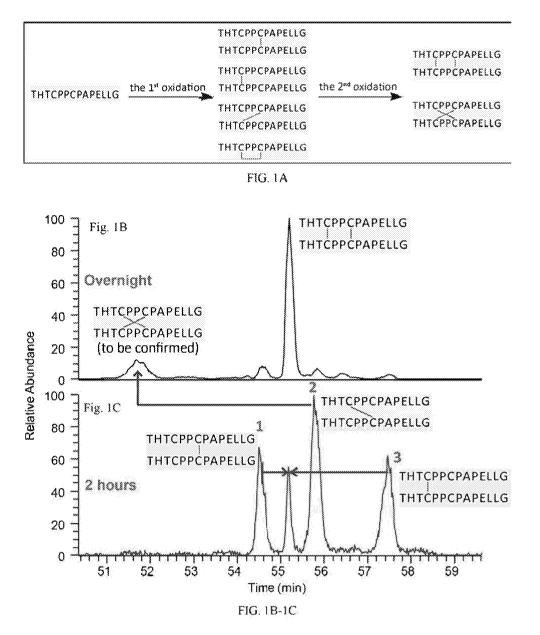
Figure lA is a schematic illustration showing the oxidation of a reduced hinge
peptide
having the sequence THTCPPCPAPELLG (SEQ ID NO:1). Figure 1B is a chromatogram
showing hinge peptides that were oxidized overnight, resulting in peptides
with two crossed
disulfide bonds and hinge peptides with two parallel disulfide bonds. The
peptides have the
sequence THTCPPCPAPELLG (SEQ ID NO:1). Figure 1C is a chromatogram showing
peptides
CA 03116735 2021-04-15
WO 2020/150491
PCT/US2020/013907
3
with one parallel (Peak 1 and 3) or crossed (Peak 2) disulfide bond. The X
axis represents time
(minutes) and the Y axis represents abundance.
Figures 2A-2B are chromatograms showing disulfide bond formation in peptides
that
were incubated with Cu2+ overnight. The peptides were either ¨0.1 [tg/m1
(Figure 2A) or ¨8
[tg/m1 (Figure 2B). The peptides have the sequence THTCPPCPAPELLG (SEQ ID
NO:1). The
X axis represents time (minutes) and the Y axis represents relative abundance.
Figures 3A-3H are chromatograms showing the results of N-terminal analysis of
the
parallel hinge peptide standard. The X axes represent time (minutes) and the Y
axes represent
mV. Figure 31 is a schematic illustration of the various peptides that can be
detected during the
cycles of N-terminal analysis. The peptide sequences are as follows:
THTCPPCPAPELLG (SEQ
ID NO:1), C-PTH (SEQ ID NO:2), and PPCPAPELLG (SEQ ID NO:3).
Figures 4A-4H are chromatograms showing the results of N-terminal analysis of
the
crossed hinge peptide standard. The X axes represent time (minutes) and the Y
axes represent
mV. Figure 41 is a schematic illustration of the various peptides that can be
detected during the
various cycles of N-terminal analysis. The peptide sequences are as follows:
THTCPPCPAPELLG (SEQ ID NO:1), C-PTH (SEQ ID NO:2), and PPCPAPELLG (SEQ ID
NO:3).
Figure 5A and 5B are chromatograms showing results from LC-MS analysis of the
remaining peptides after four cycles of Edman degradation. Figure 5A shows
peptides with
crossed disulfide bonds and Figure 5B shows peptides with parallel disulfide
bonds. Figure 5C-
5F are chromatograms of the individual peptides from Figure 5A. The peptide
sequences are as
follows: C-PTH (SEQ ID NO:2) and PPCPAPELLG (SEQ ID NO:3).
Figure 6A is a chromatogram of a parallel hinge peptide standard for IgG1
mAbl. Figure
6B is a chromatogram of a crossed hinge peptide standard for IgG1 mAb. Figure
6C is a
chromatogram of IgG1 mAbl peptides. The X axis represent time (minutes) and
the Y axis
represents relative abundance. The peptides have the sequence THTCPPCPAPELLG
(SEQ ID
NO:1).
Figure 7A is a chromatogram of a parallel hinge peptide standard for IgG4
mAbl. Figure
7B is a chromatogram of a crossed hinge peptide standard for IgG4 mAbl. Figure
7C is a
chromatogram of IgG4 mAbl peptides. The X axis represent time (minutes) and
the Y axis
CA 03116735 2021-04-15
WO 2020/150491
PCT/US2020/013907
4
represents relative abundance. The peptides have the sequence YGPPCPPCPAPEFLG
(SEQ ID
NO:4).
DETAILED DESCRIPTION OF THE INVENTION
I. Definitions
It should be appreciated that this disclosure is not limited to the
compositions and
methods described herein as well as the experimental conditions described, as
such may vary. It
is also to be understood that the terminology used herein is for the purpose
of describing certain
embodiments only, and is not intended to be limiting, since the scope of the
present disclosure
will be limited only by the appended claims.
Unless defined otherwise, all technical and scientific terms used herein have
the same
meaning as commonly understood by one of ordinary skill in the art to which
this disclosure
belongs. Although any compositions, methods and materials similar or
equivalent to those
described herein can be used in the practice or testing of the present
invention. All publications
mentioned are incorporated herein by reference in their entirety.
The use of the terms "a," "an," "the," and similar referents in the context of
describing the
presently claimed invention (especially in the context of the claims) are to
be construed to cover
both the singular and the plural, unless otherwise indicated herein or clearly
contradicted by
context.
Recitation of ranges of values herein are merely intended to serve as a
shorthand method
of referring individually to each separate value falling within the range,
unless otherwise
indicated herein, and each separate value is incorporated into the
specification as if it were
individually recited herein.
Use of the term "about" is intended to describe values either above or below
the stated
value in a range of approx. +/- 10%; in other embodiments the values may range
in value either
above or below the stated value in a range of approx. +/- 5%; in other
embodiments the values
may range in value either above or below the stated value in a range of
approx. +/- 2%; in other
embodiments the values may range in value either above or below the stated
value in a range of
approx. +/- 1%. The preceding ranges are intended to be made clear by context,
and no further
limitation is implied. All methods described herein can be performed in any
suitable order
unless otherwise indicated herein or otherwise clearly contradicted by
context. The use of any
CA 03116735 2021-04-15
WO 2020/150491
PCT/US2020/013907
and all examples, or exemplary language (e.g., "such as") provided herein, is
intended merely to
better illuminate the invention and does not pose a limitation on the scope of
the invention unless
otherwise claimed. No language in the specification should be construed as
indicating any non-
claimed element as essential to the practice of the invention.
5 "Protein" refers to a molecule comprising two or more amino acid
residues joined to each
other by a peptide bond. Protein includes polypeptides and peptides and may
also include
modifications such as glycosylation, lipid attachment, sulfation, gamma-
carboxylation of
glutamic acid residues, alkylation, hydroxylation and ADP-ribosylation.
Proteins can be of
scientific or commercial interest, including protein-based drugs, and proteins
include, among
other things, enzymes, ligands, receptors, antibodies and chimeric or fusion
proteins. Proteins
are produced by various types of recombinant cells using well-known cell
culture methods, and
are generally introduced into the cell by genetic engineering techniques
(e.g., such as a sequence
encoding a chimeric protein, or a codon-optimized sequence, an intronless
sequence, etc.) where
it may reside as an epi some or be integrated into the genome of the cell.
"Antibody" refers to an immunoglobulin molecule consisting of four polypeptide
chains,
two heavy (H) chains and two light (L) chains inter-connected by disulfide
bonds. Each heavy
chain has a heavy chain variable region (HCVR or VH) and a heavy chain
constant region. The
heavy chain constant region contains three domains, CH1, CH2 and CH3. Each
light chain has a
light chain variable region and a light chain constant region. The light chain
constant region
consists of one domain (CL). The VH and VL regions can be further subdivided
into regions of
hypervariability, termed complementarity determining regions (CDR),
interspersed with regions
that are more conserved, termed framework regions (FR). Each VH and VL is
composed of
three CDRs and four FRs, arranged from amino-terminus to carboxy-terminus in
the following
order: FR1, CDR1, FR2, CDR2, FR3, CDR3, FR4. The term "antibody" includes
reference to
both glycosylated and non-glycosylated immunoglobulins of any isotype or
subclass. The term
"antibody" includes antibody molecules prepared, expressed, created or
isolated by recombinant
means, such as antibodies isolated from a host cell transfected to express the
antibody. The term
antibody also includes bispecific antibody, which includes a heterotetrameric
immunoglobulin
that can bind to more than one different epitope. Bispecific antibodies are
generally described in
.. US Patent No. 8,586,713, which is incorporated by reference into this
application.
CA 03116735 2021-04-15
WO 2020/150491
PCT/US2020/013907
6
"Hinge region" refers to the flexible amino acid stretch in the central part
of the heavy
chains of the IgG and IgA immunoglobulin classes, which links these 2 chains
by disulfide
bonds. In IgG immunoglobulins the hinge region is located between the CH1 and
CH3 constant
domains. The hinge region affords flexibility to the antibody, and allows
easier binding to the
antigen.
"Fc fusion proteins" comprise part or all of two or more proteins, one of
which is an Fc
portion of an immunoglobulin molecule, which are not otherwise found together
in nature.
Preparation of fusion proteins comprising certain heterologous polypeptides
fused to various
portions of antibody-derived polypeptides (including the Fc domain) has been
described, e.g., by
Rath, T., et al., Crit Rev Biotech, 35(2): 235-254 (2015), Levin, D., et al.,
Trends Biotechnol,
33(1): 27-34 (2015)) "Receptor Fc fusion proteins" comprise one or more
extracellular
domain(s) of a receptor coupled to an Fc moiety, which in some embodiments
comprises a hinge
region followed by a CH2 and CH3 domain of an immunoglobulin. In some
embodiments, the
Fc-fusion protein comprises two or more distinct receptor chains that bind to
one or more
ligand(s). For example, an Fc-fusion protein is a trap, such as for example an
IL-1 trap or VEGF
trap.
The term "disulfide bond" refers to the linkage formed by the oxidation of two
SH
groups, each attached to a cysteine. Disulfide bonds play an important role in
the folding and
stability of many proteins. IgGs include two heavy chains (HC) and two light
chains (LC)
covalently linked by a total of 16 inter- or intra-molecular disulfide bonds.
IgG mAbs contain 32
cysteine residues, 5 cysteine residues on each LC and 11 cysteine residues on
each HC. Each LC
contains one variable domain and one constant domain with a disulfide bond
connection. The 5th
cysteine on the LC is linked to either the 3' or 5th cysteine of the HC to
form an interchain
disulfide bond. The heavy chains include an N-terminal variable domain (VH)
and three
constant domains (CH1, CH2, and CH3) with a hinge region between CH1 and CH2
(Vidarsson,
G., et al., Front Immunol, 5:520 (2014)). The 6111 and 7th cysteine on each HC
are bonded
forming the hinge region. The hinge region of an immunoglobulin helps form the
Y-shaped
structure of the immunoglobulin molecule. The Y shape makes possible the
flexibility of the
immunoglobulin molecules required in antigen binding.
"Intra-chain disulfide bond" refers to bonds that are formed between two
cysteines within
the same protein chain.
CA 03116735 2021-04-15
WO 2020/150491
PCT/US2020/013907
7
"Inter-chain disulfide bond" refers to bonds that are formed between two
cysteines of
individual chains of the same protein or between two cysteines of distinct
proteins.
"Scrambled disulfide bond" refers to a disulfide bond in which a cysteine
bonds to a
cysteine to which it does not normally bond. For example, cysteine X binds to
cysteine Z instead
of cysteine Y. Exemplary scrambled disulfide bonds include but not limited to
crossed and intra-
chain disulfide bonds.
As used herein, the term "crossed-hinge" refers to an antibody hinge region in
which the
disulfide bonds within the hinge region of the antibody are in a crossed
instead of parallel
formation as seen in the bottom right of Figure 1A.
The term "LC-MS" refers to liquid chromatography¨mass spectrometry which is an
analytical chemistry technique that combines the physical separation
capabilities of liquid
chromatography (or HPLC) with the mass analysis capabilities of mass
spectrometry (MS).
Methods of Characterizing Disulfide Bonds
Disulfide bonds are critical for IgG tertiary structure, stability, and
biological function.
Cysteine residues are involved in disulfide bonds. Each subclass of human IgG
molecules has a
well-defined homogenous disulfide structure; however, there are many reported
cases in which
disulfide bond heterogeneity exists. Any two cysteines in close proximity will
form a covalent
bond, even cysteines that do not naturally pair together. The formation of
disulfide bonds
between non-naturally paired cysteines is called scrambling or aggregation.
Disclosed herein are
different methods for identifying disulfide bonds. Also disclosed herein are
methods for
producing protein drug products with less than 30% scrambled disulfide bonds
A. Characterizing Disulfide Bonds
Analysis of disulfide bonds is important for quality control assessment of
mAbs. In one
embodiment, the disulfide bonds are in the hinge region. Traditional methods
for mAb hinge
region disulfide bond pattern analysis involves proteolysis, fractionation and
Edman degradation
analysis, which is time-consuming and labor-intensive. In addition,
traditional methods such as
MS2¨based techniques fail to distinguish between crossed and parallel hinge
peptides.
Identifying scrambled disulfide bonds is difficult because of the very low
number of scrambled
disulfide bonds that occur. Antibodies with scrambled disulfide bonds in the
hinge region can be
less stable and have a potential for inducing immunogenicity if administered
to a subject.
CA 03116735 2021-04-15
WO 2020/150491
PCT/US2020/013907
8
Disclosed herein are compositions and methods of use thereof for
characterizing disulfide bonds
in proteins, for example monoclonal antibodies. Peptide standards with native
and scrambled
disulfide bond patterns are provided herein. These peptide standards can be
used in mass
spectrometry analysis to focus the analysis on peptides that elute with the
peptide standards.
Methods of applying the disclosed peptide standards to hinge region disulfide
bond
characterization are also provided.
1. Peptide Standards for mAb Disulfide Bond Pattern Analysis
In one embodiment the peptide standard is formed by two peptides covalently
bound
together by one or more disulfide bonds. The scrambled disulfide bonds occur
when a disulfide
bond forms between two amino acids that are not directly opposite of each
other. Figure 7A
shows the natural parallel disulfide bond. Figure 7B shows an exemplary
crossed disulfide bond
also referred to as a scrambled disulfide bond. Figure lA shows parallel,
crossed and intra-chain
disulfide bonds. In one embodiment, the peptide standards can be used to
identify the presence
of parallel, crossed, or intra-chain disulfide bonds in a protein sample, for
example scrambled
disulfide bonds or native disulfide bonds in an antibody, for example a
monoclonal antibody. In
another embodiment, the peptide standards can detect intra-chain disulfide
bonds in a protein or
peptide. Further details for making and using the disclosed disulfide bond
peptides are provided
below.
i. Synthesis
One embodiment provides a method for synthesizing disulfide bond peptide
standards.
Peptide standards can be synthesized using techniques known in the art,
including but not limited
to liquid phase synthesis, solid phase peptide synthesis, and recombinant
technology
(Stawikowski, M., and Fields, G.B., Current Protoc Protein Sci, Chapter: Unit
18.1 (2002)).
The peptide standards can include fragments of the protein containing the
disulfide bonds
to be analyzed. The protein can be fragmented or sections of the protein
containing the disulfide
bonds to be analyzed can be synthesized and used to produce disulfide bond
peptide standards.
In some embodiments, the peptide standard sequence has 100% sequence identity
to the region
of the protein or protein drug product of interest that includes the disulfide
bond. In other
embodiments, the peptide standard sequence has at least 90% sequence identity
to the region of
the protein or protein drug product of interest that includes the disulfide
bond. The peptide
standard sequence can have 75%, 80%, 85%, 90%, 95%, 97%, 98%, or 99% sequence
identity to
CA 03116735 2021-04-15
WO 2020/150491
PCT/US2020/013907
9
the region of the protein or protein drug product of interest that includes
the disulfide bond. In
other embodiments, the sequence of the peptide standard only represents a
portion of the region
that includes the disulfide bond. The peptide standards are typically 5 to 20
amino acids in
length.
The formation of disulfide bonds in the peptide standards can be induced
through
oxidation of cysteine residues in the peptide standard. Methods of forming
disulfide bonds at
cysteine include but are not limited to air oxidation, chemical oxidation, and
exposing the
peptide to copper (Cu') or zinc. Air oxidation occurs by mixing thiol and
cysteine containing
peptides in buffer open to the air. In another embodiment, the formation of
disulfides on a
peptide can be accomplished by disulfide exchange, for example by using 5,5-
dithiobis(2-
nitrobenzoate) (DTNB or Ellman's Reagent). In one embodiment, other common
chemicals for
inducing oxidation of cysteine residues are activated reagents, including but
not limited to
iodine, sulfenyl halides, iodoacetamides, maleimides, benzylic halides and
bromomethylketones.
In another embodiment, disulfide bonds can be formed by exposing the peptide
to copper or zinc.
This can be achieved by using an inert platinum electrode or a sacrificial
electrode (copper or
zinc) or by generating metallic ions in electrospray ionization mass
spectrometry (ESI-MS). In
one embodiment, the molar ratio of peptide:Cu' needed to induce the formation
of disulfide
bonds is 5:1 Higher peptide concentration can preferentially induce the
formation of hinge
dimers over the formation of intra-chain disulfide bonds.
The peptide standards can be exposed to Cu' to induce single parallel or
crossed
disulfide bonds. In one embodiment, the peptide standards are oxidized for
about 1 hour to about
6 hours. In a preferred embodiment, the peptide standards are oxidized for 2
hours. In another
embodiment, the peptide standards can be oxidized for up to 24 hours in order
to induce two or
more parallel or crossed disulfide bonds.
ii. Authentication
In one embodiment, the characteristics of the synthesized peptide standards
are
determined. The characteristics include the retention time and m/z of each
peptide standard.
The synthesized peptide standards can be separated or fractionated using
various
chromatography methods. Peptide standards containing parallel disulfide bonds
are
distinguishable from peptide standards containing crossed or intra-chain
disulfide bonds.
CA 03116735 2021-04-15
WO 2020/150491
PCT/US2020/013907
In one embodiment, N-terminal sequence analysis can be used to confirm the
identity of
the peptide standards. N-terminal sequence analysis involves a series of
chemical reactions that
derivatize and remove one amino acid at a time from the N-terminus of purified
peptides or
intact proteins. N-terminal analysis can detect disulfide bonds because the
reaction to remove
5 one amino acid at a time from the N-terminus does not disrupt the bonds
between the cysteine
residues in the disulfide bond.
In another embodiment, Edman degradation can be utilized to sequence the
disulfide
bond peptide standards. Edman degradation is similar to N-terminal analysis in
that it detects the
sequence of a protein or peptide in order by removing one amino acid at a time
from the N-
10 terminus of the protein or peptide. However, the first round of Edman
degradation is often
contaminated by impurities and therefore does not give an accurate
determination of the N-
terminal amino acid. Edman degradation can detect disulfide bonds because the
reaction to
remove one amino acid at a time from the N-terminus does not disrupt the bonds
between the
cysteine residues in the disulfide bond. In one embodiment, N-terminal
analysis can be
combined with Edman degradation to give a complete, ordered sequence of the
synthesized
disulfide bond peptide standards.
Other methods of sequencing peptides are considered. These include but are not
limited
to C-terminal analysis and mass spectrometry.
2. Methods for Characterizing Disulfide Bonds in the Hinge
Region
One embodiment provides methods for identifying and characterizing disulfide
bonds in
a protein drug product. In another embodiment, the methods identify and
characterize disulfide
bonds specifically in the hinge region of an antibody. In one embodiment, the
antibody is an IgG
antibody. An exemplary method includes preparing scrambled disulfide bond
peptide standards
and native disulfide bond peptide standards according to the sequence of the
protein drug
product, cleaving a sample of protein drug product into peptides, analyzing
the peptide standards
and the protein drug product peptides, identifying scrambled and native
disulfide bonds in
peptides by comparing retention time, and quantifying the level of scrambled
disulfide bond
peptides. Detecting peptides having the same retention time or m/z as the
peptide standard
indicates that the type of disulfide bond in the peptide standard is present
in the protein drug
product.
1. Protein Sample Preparation
CA 03116735 2021-04-15
WO 2020/150491
PCT/US2020/013907
11
The protein or protein drug product of interest can be obtained for example
from a
bioreactor containing cells engineered to produce monoclonal antibodies.
In one embodiment, the protein or protein drug product of interest is digested
into
peptides. Methods of digesting proteins are known in the art. Proteins can be
digested by
enzymatic digestion with proteolytic enzymes or by non-enzymatic digestion
with chemicals.
Exemplary proteolytic enzymes for digesting proteins include but are not
limited to trypsin,
pepsin, chymotrypsin, thermolysin, papain, pronase, Arg-C, Asp-N, Glu-C, Lys-
C, and Lys-N.
Combinations of proteolytic enzymes can be used to ensure complete digestion.
Exemplary
chemicals for digesting proteins include but are not limited to formic acid,
hydrochloric acid,
acetic acid, cyanogen bromide, 2-nitro-5-thiocyanobenzoate, and
hydroxyalamine.
In one embodiment, the protein drug product can be subjected to double
digesting. In
this embodiment, the first digestion can be performed using a broad-
specificity protease, such as
but not limited to proteinase K, thermolysin, substilisin, papain,
chymotrypsin, or elastase. The
second digestion can be performed using trypsin. In one embodiment, FabRICATOR
enzyme is
used to digest the protein or protein drug product. FabRICATOR enzyme digests
antibodies at
a specific site below the hinge therefore generating F(ab')2 and Fc/2
fragments. FabRICATOR
digestion can be combined with tryptic digestion.
Hinge Region Disulfide Bond Pattern Analysis
The digested peptide mixture from the protein or protein drug product can be
analyzed by
.. liquid chromatography-mass spectrometry (LC-MS or LC-MS2) to determine the
mass of the
digested peptides. In one embodiment, the digested peptide mixture is
separated by liquid
chromatography, for example size-exclusion chromatography.
The peptide mixture can then be analyzed using mass spectrometry. Methods of
performing mass spectrometry are known in the art. See for example
(Aeberssold, M., and
.. Mann, M., Nature, 422:198-207 (2003)) Commonly utilized types of mass
spectrometry include
but are not limited to tandem mass spectrometry (MS/MS), electrospray
ionization mass
spectrometry, liquid chromatography-mass spectrometry (LC-MS), and Matrix-
assisted laser
desorption /ionization (MALDI). In another embodiment, selected reaction
monitoring (SRM) is
performed on the peptide mixture. In SRM, an ion of a particular mass is
selected in the first
stage of a tandem mass spectrometer and an ion product of fragmentation of the
precursor ion is
selected in the second mass spectrometer for detection.
CA 03116735 2021-04-15
WO 2020/150491
PCT/US2020/013907
12
In one embodiment, the hinge peptide standards are also analyzed. The
standards are
used to characterize the hinge region of the protein drug product of interest.
In one embodiment,
the retention time of the known hinge peptide standards are compared to the
retention time of the
peptide mixture from the protein drug product of interest. Detecting peptides
having the same
retention time or m/z as the peptide standard indicates that the type of
disulfide bond in the
peptide standard is present in the protein drug product.
B. Proteins of Interest
In one embodiment the protein of interest is a protein drug product or is a
protein of
interest suitable for expression in prokaryotic or eukaryotic cells. For
example, the protein can
be an antibody or antigen-binding fragment thereof, a chimeric antibody or
antigen-binding
fragment thereof, an ScFv or fragment thereof, an Fc-fusion protein or
fragment thereof, a
growth factor or a fragment thereof, a cytokine or a fragment thereof, or an
extracellular domain
of a cell surface receptor or a fragment thereof. Proteins in the complexes
may be simple
polypeptides consisting of a single subunit, or complex multisubunit proteins
comprising two or
more subunits. The protein of interest may be a biopharmaceutical product,
food additive or
preservative, or any protein product subject to purification and quality
standards
In some embodiments, the protein of interest is an antibody, a human antibody,
a
humanized antibody, a chimeric antibody, a monoclonal antibody, a
multispecific antibody, a
bispecific antibody, an antigen binding antibody fragment, a single chain
antibody, a diabody,
.. triabody or tetrabody, a dual-specific, tetravalent immunoglobulin G-like
molecule, termed dual
variable domain immunoglobulin (DVD-IG), an IgD antibody, an IgE antibody, an
IgM
antibody, an IgG antibody, an IgG1 antibody, an IgG2 antibody, an IgG3
antibody, or an IgG4
antibody. In one embodiment, the antibody is an IgG1 antibody. In one
embodiment, the
antibody is an IgG2 antibody. In one embodiment, the antibody is an IgG4
antibody. In another
embodiment, the antibody comprises a chimeric hinge. In still other
embodiments, the antibody
comprises a chimeric Fc. In one embodiment, the antibody is a chimeric
IgG2/IgG4 antibody. In
one embodiment, the antibody is a chimeric IgG2/IgG1 antibody. In one
embodiment, the
antibody is a chimeric IgG2/IgG1/IgG4 antibody.
In some embodiments, the antibody is selected from the group consisting of an
anti-
Programmed Cell Death 1 antibody (e.g., an anti-PD1 antibody as described in
U.S. Pat. Appin.
Pub. No. U52015/0203579A1), an anti-Programmed Cell Death Ligand-1 (e.g., an
anti-PD-Li
CA 03116735 2021-04-15
WO 2020/150491
PCT/US2020/013907
13
antibody as described in in U.S. Pat. Appin. Pub. No. U52015/0203580A1), an
anti-D114
antibody, an anti-Angiopoetin-2 antibody (e.g., an anti-ANG2 antibody as
described in U.S. Pat.
No. 9,402,898), an anti- Angiopoetin-Like 3 antibody (e.g., an anti-AngPt13
antibody as
described in U.S. Pat. No. 9,018,356), an anti-platelet derived growth factor
receptor antibody
(e.g., an anti-PDGFR antibody as described in U.S. Pat. No. 9,265,827), an
anti-Erb3 antibody,
an anti- Prolactin Receptor antibody (e.g., anti-PRLR antibody as described in
U.S. Pat. No.
9,302,015), an anti-Complement 5 antibody (e.g., an anti-05 antibody as
described in U.S. Pat.
Appin. Pub. No US2015/0313194A1), an anti-TNF antibody, an anti-epidermal
growth factor
receptor antibody (e.g., an anti-EGFR antibody as described in U.S. Pat. No.
9,132,192 or an
anti-EGFRvIII antibody as described in U.S. Pat. Appin. Pub. No.
US2015/0259423A1), an anti-
Proprotein Convertase Subtilisin Kexin-9 antibody (e.g., an anti-PCSK9
antibody as described in
U.S. Pat. No. 8,062,640 or U.S. Pat. No. 9,540,449), an Anti-Growth and
Differentiation Factor-
8 antibody (e.g. an anti-GDF8 antibody, also known as anti-myostatin antibody,
as described in
U.S. Pat Nos. 8,871,209 or 9,260,515), an anti-Glucagon Receptor (e.g. anti-
GCGR antibody as
described in U.S. Pat. Appin. Pub. Nos. U52015/0337045A1 or U52016/0075778A1),
an anti-
VEGF antibody, an anti-IL1R antibody, an interleukin 4 receptor antibody
(e.g., an anti-IL4R
antibody as described in U.S. Pat. Appin. Pub. No. US2014/0271681A1 or U.S.
Pat Nos.
8,735,095 or 8,945,559), an anti-interleukin 6 receptor antibody (e.g., an
anti-IL6R antibody as
described in U.S. Pat. Nos. 7,582,298, 8,043,617 or 9,173,880), an anti-IL1
antibody, an anti-IL2
antibody, an anti-IL3 antibody, an anti-IL4 antibody, an anti-IL5 antibody, an
anti-IL6 antibody,
an anti-IL7 antibody, an anti-interleukin 33 (e.g., anti- IL33 antibody as
described in U.S. Pat.
Nos. 9,453,072 or 9,637,535), an anti-Respiratory syncytial virus antibody
(e.g., anti-RSV
antibody as described in U.S. Pat. Appin. Pub. No. 9,447,173), an anti-Cluster
of differentiation
3 (e.g., an anti-CD3 antibody, as described in U.S. Pat. Nos. 9,447,173and
9,447,173, and in U.S.
Application No. 62/222,605), an anti- Cluster of differentiation 20 (e.g., an
anti-CD20 antibody
as described in U.S. Pat. Nos. 9,657,102 and U520150266966A1, and in U.S. Pat.
No.
7,879,984), an anti-CD19 antibody, an anti-CD28 antibody, an anti- Cluster of
Differentiation-48
(e.g. anti-CD48 antibody as described in U.S. Pat. No. 9,228,014), an anti-Fel
dl antibody (e.g.
as described in U.S. Pat. No. 9,079,948), an anti-Middle East Respiratory
Syndrome virus (e.g.
an anti-MERS antibody as described in U.S. Pat. Appin. Pub. No.
U52015/0337029A1), an anti-
Ebola virus antibody (e.g. as described in U.S. Pat. Appin. Pub. No.
U52016/0215040), an anti-
CA 03116735 2021-04-15
WO 2020/150491
PCT/US2020/013907
14
Zika virus antibody, an anti-Lymphocyte Activation Gene 3 antibody (e.g. an
anti-LAG3
antibody, or an anti-CD223 antibody), an anti-Nerve Growth Factor antibody
(e.g. an anti-NGF
antibody as described in U.S. Pat. Appin. Pub. No. US2016/0017029 and U.S.
Pat. Nos.
8,309,088 and 9,353,176) and an anti-Protein Y antibody. In some embodiments,
the bispecific
.. antibody is selected from the group consisting of an anti-CD3 x anti-CD20
bispecific antibody
(as described in U.S. Pat. Appin. Pub. Nos. US2014/0088295A1 and
US20150266966A1), an
anti-CD3 x anti-Mucin 16 bispecific antibody (e.g., an anti-CD3 x anti-Muc16
bispecific
antibody), and an anti-CD3 x anti- Prostate-specific membrane antigen
bispecific antibody (e.g.,
an anti-CD3 x anti-PSMA bispecific antibody). In some embodiments, the protein
of interest is
selected from the group consisting of abciximab, adalimumab, adalimumab-atto,
ado-
trastuzumab, alemtuzumab, alirocumab, atezolizumab, avelumab, basiliximab,
belimumab,
benralizumab, bevacizumab, bezlotoxumab, blinatumomab, brentuximab vedotin,
brodalumab,
canakinumab, capromab pendetide, certolizumab pegol, cemiplimab, cetuximab,
denosumab,
dinutuximab, dupilumab, durvalumab, eculizumab, elotuzumab, emicizumab-kxwh,
.. emtansinealirocumab, evinacumab, evolocumab, fasinumab, golimumab,
guselkumab,
ibritumomab tiuxetan, idarucizumab, infliximab, infliximab-abda, infliximab-
dyyb, ipilimumab,
ixekizumab, mepolizumab, necitumumab, nesvacumab, nivolumab, obiltoxaximab,
obinutuzumab, ocrelizumab, ofatumumab, olaratumab, omalizumab, panitumumab,
pembrolizumab, pertuzumab, ramucirumab, ranibizumab, raxibacumab, reslizumab,
rinucumab,
.. rituximab, sarilumab, secukinumab, siltuximab, tocilizumab, tocilizumab,
trastuzumab,
trevogrumab, ustekinumab, and vedolizumab.
In some embodiments, the protein of interest is a recombinant protein that
contains an Fc
moiety and another domain, (e.g., an Fc-fusion protein). In some embodiments,
an Fc-fusion
protein is a receptor Fc-fusion protein, which contains one or more
extracellular domain(s) of a
receptor coupled to an Fc moiety. In some embodiments, the Fc moiety comprises
a hinge
region followed by a CH2 and CH3 domain of an IgG. In some embodiments, the
receptor Fc-
fusion protein contains two or more distinct receptor chains that bind to
either a single ligand or
multiple ligands. For example, an Fc-fusion protein is a TRAP protein, such as
for example an
IL-1 trap (e.g., rilonacept, which contains the IL-1RAcP ligand binding region
fused to the Il-
1R1 extracellular region fused to Fc of hIgGl; see U.S. Pat. No. 6,927,004,
which is herein
incorporated by reference in its entirety), or a VEGF trap (e.g., aflibercept
or ziv-aflibercept,
CA 03116735 2021-04-15
WO 2020/150491
PCT/US2020/013907
which comprises the Ig domain 2 of the VEGF receptor FM fused to the Ig domain
3 of the
VEGF receptor Flkl fused to Fc of hIgGl; see U.S. Pat. Nos. 7,087,411 and
7,279,159). In other
embodiments, an Fc-fusion protein is a ScFv-Fc-fusion protein, which contains
one or more of
one or more antigen-binding domain(s), such as a variable heavy chain fragment
and a variable
5 light chain fragment, of an antibody coupled to an Fc moiety.
C. Producing mAb with Native Disulfide Bond Pattern
One embodiment provides methods of producing a protein drug product containing
less
than 30% scrambled disulfide bonds. An exemplary method includes culturing
cells producing
the antibody in a cell culture under suitable conditions to produce the
antibody, purifying the
10 antibody under suitable conditions to extract the antibody, admixing the
antibody with excipients
under suitable conditions to stabilize the antibody, obtaining a sample of the
antibody from the
cell culture, following purification of the antibody from the cell culture, or
following the addition
of excipients to the purified antibody, characterizing disulfide bonds of the
antibody according to
the disclosed methods, and modifying one or more cell culture, purification or
excipient
15 conditions to reduce the amount of crossed hinge disulfide bonds of the
antibody.
The one or more cell culture, purification, or excipient conditions that are
changed to
reduce the amount of scrambled disulfide bonds in the antibody include but are
not limited to
temperature, pH, oxygen levels, reactive oxygen species, surfactants, or
combinations thereof. In
one embodiment, an amino acid free strategy of cell culture could affect
disulfide bond
formation.
In one embodiment, the cells producing the antibody are Chinese hamster ovary
cells. In
another embodiment, the cells are hybridoma cells.
In one embodiment, the protein drug product can have less than 30% scrambled
disulfide
bonds in the hinge region. The protein drug product can have less than 30%,
25%, 20%, 18%,
16%, 14%, 12%, 10%, 9%, 8%, 7%, 6%, 5%, 4%, 3%, 1%, 0.5%, or 0.1% scrambled
disulfide
bonds in the hinge region.
In another embodiment, the protein drug product can have less than 10%
scrambled
disulfide bonds overall. The protein drug product can have less than 10%,
9.5%, 9%, 8.5%, 8%,
7.5%, 7%, 6.5%, 6%, 5.5%, 5%, 4.5%, 4%, 3.5%, 3%, 2.5%, 2%, 1.5%, 1%, 0.5%, or
0.1%
scrambled disulfide bonds.
EXAMPLES
CA 03116735 2021-04-15
WO 2020/150491
PCT/US2020/013907
16
Example 1: Synthesis of Parallel and Crossed Hinge Peptides
Methods:
Cross-linking
Cysteine containing peptides were purchased from a commercial vendor. The
peptides
were cross-linked by incubation with 1 mM Cu' as the oxidant in the presence
of air. The molar
ratio of peptide to Cu' was 5:1.
N-terminal analysis/Edman Degradation
The cross-linked peptides were suspended in water and were placed into a
protein
sequencer. The peptides were exposed to phenyl isothiocyanate (PITC). PITC
couples with the
N-terminal residue to form a PTC polypeptide. Trifluoroacetic acid was added
to the reaction
and the PTC N-terminal residue underwent acid cleavage, resulting in the
release of an unstable
ATZ-amino acid. The ATZ-amino acid was separated from the peptide solution
into a
conversion flask containing ethyl acetate. The ATZ-amino acid was converted
into a stable
PTH-amino acid with 25% TFA, v/v in water. The PTH-amino acid solution was
injected onto
an HPLC. Each amino acid of the peptide is identified by HPLC.
Results
Cu' has been reported to induce the formation of disulfide bonds by producing
radicals
(Prudent, M., and Girault, H.H., Metallomics, 1:157-165 (2009)). Peptides
exposed to Cu2+ at a
molar ratio of peptide/Cu' of 5/1 formed disulfide bonds as illustrated in
Figure 1A. The first
oxidation formed a single, non-selective bond that was either parallel or
crossed in nature
(Figures 1A and 1C). During the second disulfide bond formation, parallel
connectivity was
found to be the preferred connection (Figure 1B). The peptide concentration
was found to affect
the type of disulfide bond that was formed. A higher concentration of peptide,
8 jig/ml, induced
the formation of more parallel hinge dimers than a concentration of 0.1 [tg/m1
(Figures 2A-2B).
Higher peptide concentration favors inter-molecular bridges.
The identity of the peptides was confirmed using N-terminal analysis and Edman
degradation (Figures 3A-3I, Figures 4A-4I, and Figures 5A-5B).
Example 2: Analysis of the Hinge Region of two mAbs
Methods
Hinge DSB Characterization
CA 03116735 2021-04-15
WO 2020/150491
PCT/US2020/013907
17
Antibodies were first digested into peptides. IgG1 antibodies were subjected
to dual-
enzyme digestion using proteinase K followed by trypsin. IgG4 antibodies were
subjected to
digestion by FabRICATOR followed by trypsin. Hinge peptide standards were
prepared as
above, using the hinge region sequence of the IgG1 and IgG4 antibodies to
prepare the peptides.
.. The digested peptide mixtures were subjected to LC-MS analysis. Hinge
peptide standards were
also subjected to LC-MA analysis. Retention time analysis was performed to
compare the
retention time of the antibody peptides to the retention time of the hinge
peptide standards.
Results
IgG1 mAbl was subjected to digestion into peptides and the resulting peptides
were
.. subjected to LC/MS analysis. The hinge peptide standards described above
were also subjected
to LC/MS analysis. As shown in Figures 6A-6C, IgG1 mAbl has about 0.9% crossed
hinge
disulfide bonds.
A second antibody, IgG4 mAbl, was also analyzed using the disclosed methods
and
hinge disulfide bond peptide standards. As shown in Figures 7A-7C, IgG4 mAbl
had about
0.6% crossed hinge disulfide bonds.
While in the foregoing specification this invention has been described in
relation to
certain embodiments thereof, and many details have been put forth for the
purpose of illustration,
it will be apparent to those skilled in the art that the invention is
susceptible to additional
embodiments and that certain of the details described herein can be varied
considerably without
departing from the basic principles of the invention.
All references cited herein are incorporated by reference in their entirety.
The present
invention may be embodied in other specific forms without departing from the
spirit or essential
attributes thereof and, accordingly, reference should be made to the appended
claims, rather than
to the foregoing specification, as indicating the scope of the invention.