Note: Descriptions are shown in the official language in which they were submitted.
CA 03117051 2021-04-19
WO 2020/096959 PCT/US2019/059661
METHODS OF PRODUCING TWO CHAIN PROTEINS IN
PROKARYOTIC HOST CELLS
CROSS-REFERENCE TO RELATED APPLICATIONS
[0001] This application claims the priority benefit of U.S. Provisional
Application No.
62/755,915, filed November 5, 2018, which is hereby incorporated by reference
in its entirety.
SUBMISSION OF SEQUENCE LISTING ON ASCII TEXT FILE
[0002] The content of the following submission on ASCII text file is
incorporated herein by
reference in its entirety: a computer readable form (CRF) of the Sequence
Listing (file name:
1463920440405EQLI5T.TXT, date recorded: October 28, 2019, size: 6 KB).
FIELD
[0003] This disclosure relates to methods of producing recombinant
polypeptides, such as
antibodies (e.g., bispecific antibodies, half-antibodies, one-armed
antibodies, antibody
fragments, and the like), as well as prokaryotic host cells that may find use
in said methods.
BACKGROUND
[0004] Recombinant protein production in prokaryotic host cells has been a
source of many
important therapeutic agents since the production of human insulin in E. coil
in 1978. As
molecular biology tools and knowledge has advanced, the complexity of
recombinant
therapeutics has also increased. Production of these recombinant proteins
requires that the
products exhibit properties such as proper translation, folding, assembly,
disulfide bonding, and
transport to the periplasm. It is known that expression of many recombinant
proteins, particularly
those with disulfide bonds (e.g., two chain proteins, including without
limitation antibodies and
antibody fragments), leads to the formation of inclusion bodies in prokaryotic
host cells (Spadiut
et al., Trends in Biotechnology, 32:54, 2014). Accordingly, there is a demand
for expression
systems and processes for the recombinant production of properly folded and
assembled two
chain proteins in prokaryotic host cells on an industrial scale.
[0005] Monoclonal antibodies represent one of the fastest growing types of
recombinant
therapeutic agent, with numerous monoclonal antibodies already approved or
under review for
-1-
CA 03117051 2021-04-19
WO 2020/096959 PCT/US2019/059661
the treatment of various diseases (Nelson et at., Nature Review Drug
Discovery, 9:767, 2010).
Traditional monoclonal antibodies bind a single target antigen. For many
diseases, it may be
advantageous to employ antibodies that bind more than one target antigen,
i.e., multispecific
antibodies. Such antibodies can be employed in combinatorial approaches
directed against
multiple therapeutic targets (see, e.g., Bostrom et at., Science, 323:1610,
2009; and Wu et at.,
Nature Biotechnology, 25:1290, 2007). For instance, bispecific antibodies can
be produced that
simultaneously bind an epitope expressed on the surface of a cancer cell and
an epitope
expressed on a T cell to induce T cell-mediated killing of tumor cells
(Shalaby et at., Clinical
Immunology, 74:185, 1995). Other monoclonal antibody formats have also been
used, such as
antibody fragments and one-armed antibodies (see, e.g., Merchant et at., Proc.
Natl. Acad. Sci.
110:E2987-E2996, 2013).
[0006] The use of antibodies in the clinic requires the ability to produce two
chain proteins in
industrially relevant amounts. Vector components that improve recombinant
protein production
in prokaryotic host cells have been described (see, e.g., Schlapschy et at.,
Protein Engineering,
Design and Selection, 19:385, 2006; and Simmons et at., Journal of
Immunotogicat Methods,
263: 133, 2002), and in particular the expression of chaperone protein(s) has
been used to
increase antibody titer. However, these chaperone protein(s) are typically
expressed from a
plasmid in the host cell. This means that for every new recombinant protein to
be expressed,
considerable time and cost must be spent to construct unique expression
plasmids encoding both
the recombinant product and the chaperone(s) and tune their expression (e.g.,
by testing different
promoters and/or translation initiation regions). This also necessitates the
use of larger plasmid
sizes in order to accommodate the coding sequence(s) and associated regulatory
elements for the
chaperone protein(s). Plasmid expression also typically leads to higher
expression levels for the
chaperone protein(s) (as plasmids can be present in at least 10-15 copies per
cell), and in some
cases this necessitates additional purification step(s) in order to remove
chaperone protein from
the recombinant product titer.
[0007] All references cited herein, including patent applications, patent
publications, and
UniProtKB/Swiss-Prot Accession numbers are herein incorporated by reference in
their entirety,
as if each individual reference were specifically and individually indicated
to be incorporated by
reference.
-2-
CA 03117051 2021-04-19
WO 2020/096959 PCT/US2019/059661
SUMMARY
[0008] There remains a need for optimal methods for efficiently producing
recombinant two
chain proteins on a preparative scale. In particular, the integration into the
prokaryotic host cell
chromosome of translational unit(s) encoding chaperone proteins, and/or the
integration of non-
native promoters to drive expression of native chaperone proteins, would allow
for a single host
cell that could be used to express a variety of recombinant protein products
and simplify the
plasmid engineering and protein purification protocols required for
production.
[0009] To meet these and other demands, provided herein are prokaryotic host
cells and
methods of using the same in order to produce two-chain polypeptides.
Advantageously, these
host cells and methods allow for more efficient production of two-chain
polypeptides, e.g.,
without requiring up-front time and cost to optimize chaperone expression
plasmids or
downstream purification steps to remove chaperone proteins.
[0010] In one aspect, provided herein are methods of producing a polypeptide
comprising two
chains in a prokaryotic host cell comprising a host cell chromosome, the
methods comprising: (a)
culturing the host cell to express the two chains of the polypeptide in a
culture medium under
conditions suitable for expression of the two chains of the polypeptide,
whereby upon expression
the two chains fold and assemble to form a biologically active polypeptide in
the host cell;
wherein the host cell comprises: (1) a first polynucleotide comprising a first
translational unit
encoding a first chain of the polypeptide; (2) a second polynucleotide
comprising a second
translational unit encoding a second chain of the polypeptide, wherein the
first and second
polynucleotides are part of one or more extra-chromosomal polynucleotides; and
(3) a third
polynucleotide comprising a third translational unit encoding a chaperone
protein selected from
the group consisting of peptidyl-prolyl isomerases and protein disulfide
oxidoreductases,
wherein the third translational unit is part of the host cell chromosome,
wherein the third
translational unit is in operable combination with a promoter that is
integrated in the host cell
chromosome and drives transcription of the third translational unit, and
wherein the combination
of the third translational unit and the promoter is non-native to the host
cell chromosome; and (b)
recovering the biologically active polypeptide from the host cell.
[0011] In some embodiments, the promoter is an inducible promoter. In some
embodiments,
the inducible promoter is a Pho promoter that drives transcription of the
third translational unit
when phosphate in the culture medium has been depleted. In some embodiments,
the inducible
-3-
CA 03117051 2021-04-19
WO 2020/096959 PCT/US2019/059661
promoter is an isopropyl beta-D-thiogalactoside (IPTG)-inducible promoter that
drives
transcription of the third translational unit when IPTG is present in the
culture medium. In some
embodiments, the promoter is a constitutive promoter. In some embodiments, the
constitutive
promoter is a CP25 promoter. In some embodiments, the third translational unit
is native to the
host cell chromosome. In some embodiments, the third translational unit is non-
native to the
host cell chromosome. In some embodiments, the chaperone protein is a peptidyl-
prolyl
isomerase. In some embodiments, the peptidyl-prolyl isomerase is an FkpA
protein. In some
embodiments, the FkpA is E. coil FkpA. In some embodiments, the chaperone
protein is a
protein disulfide oxidoreductase. In some embodiments, the protein disulfide
oxidoreductase is a
DsbC protein. In some embodiments, the protein disulfide oxidoreductase is E.
coil DsbC. In
some embodiments, the protein disulfide oxidoreductase is a DsbA protein. In
some
embodiments, the protein disulfide oxidoreductase is E. coil DsbA.
[0012] In another aspect, provided herein are methods of producing a
polypeptide comprising
two chains in a prokaryotic host cell comprising a host cell chromosome, the
methods
comprising: (a) culturing the host cell to express the two chains of the
polypeptide in a culture
medium under conditions suitable for expression of the two chains of the
polypeptide, whereby
upon expression the two chains fold and assemble to form a biologically active
polypeptide in
the host cell; wherein the host cell comprises: (1) a first polynucleotide
comprising a first
translational unit encoding a first chain of the polypeptide; (2) a second
polynucleotide
comprising a second translational unit encoding a second chain of the
polypeptide, wherein the
first and second polynucleotides are part of one or more extra-chromosomal
polynucleotides; (3)
a third polynucleotide comprising a third translational unit encoding a
protein disulfide
oxidoreductase, wherein the third translational unit is part of the host cell
chromosome, wherein
the third translational unit is in operable combination with a first promoter
that is integrated in
the host cell chromosome and drives transcription of the third translational
unit, wherein the
combination of the third translational unit and the first promoter is non-
native to the host cell
chromosome; and (4) a fourth polynucleotide comprising a fourth translational
unit encoding a
peptidyl-prolyl isomerase, wherein the fourth translational unit is part of
the host cell
chromosome, wherein the fourth translational unit is in operable combination
with a second
promoter that is integrated in the host cell chromosome and drives
transcription of the fourth
translational unit, wherein the combination of the fourth translational unit
and the second
-4-
CA 03117051 2021-04-19
WO 2020/096959 PCT/US2019/059661
promoter is non-native to the host cell chromosome; and (b) recovering the
biologically active
polypeptide from the host cell.
[0013] In some embodiments, the first and second promoters are both inducible
promoters. In
some embodiments, the first and second promoters are both Pho promoters that
drive
transcription of the third and fourth translational units, respectively, when
phosphate in the
culture medium has been depleted. In some embodiments, one of the first and
second promoters
is an inducible promoter, and the other of the first and second promoters is a
constitutive
promoter. In some embodiments, the first promoter is a Pho promoter that
drives transcription of
the third translational unit when phosphate in the culture medium has been
depleted, and the
second promoter is a CP25 promoter. In some embodiments, the second promoter
is an
inducible promoter, and the first promoter is a constitutive promoter. In some
embodiments, one
or both of the third translational unit and fourth translational unit are
native to the host cell
chromosome. In some embodiments, the third translational unit and the fourth
translational unit
are both native to the host cell chromosome. In some embodiments, one or both
of the third
translational unit and fourth translational unit are non-native to the host
cell chromosome. In
some embodiments, the protein disulfide oxidoreductase is a DsbC protein. In
some
embodiments, the protein disulfide oxidoreductase is E. coil DsbC. In some
embodiments, the
peptidyl-prolyl isomerase is an FkpA protein. In some embodiments, the FkpA is
E. coil FkpA.
In some embodiments, the protein disulfide oxidoreductase is E. coil DsbC,
wherein the first
promoter is a Pho promoter that drives transcription of the third
translational unit when
phosphate in the culture medium has been depleted, wherein the peptidyl-prolyl
isomerase is E.
coil FkpA, and wherein the second promoter is a CP25 promoter. In some
embodiments, the
protein disulfide oxidoreductase is E. coil DsbC, wherein the first promoter
is a Pho promoter
that drives transcription of the third translational unit when phosphate in
the culture medium has
been depleted, wherein the peptidyl-prolyl isomerase is E. coil FkpA, and
wherein the second
promoter is a Pho promoter that drives transcription of the fourth
translational unit when
phosphate in the culture medium has been depleted. In some embodiments, the
host cell further
comprises: (5) a fifth polynucleotide comprising a fifth translational unit
encoding a second
protein disulfide oxidoreductase, wherein the fifth translational unit is part
of the host cell
chromosome, wherein the fifth translational unit is in operable combination
with a third promoter
that is integrated in the host cell chromosome and drives transcription of the
fifth translational
-5-
CA 03117051 2021-04-19
WO 2020/096959 PCT/US2019/059661
unit, wherein the combination of the fifth translational unit and the third
promoter is non-native
to the host cell chromosome. In some embodiments, the second protein disulfide
oxidoreductase
is a DsbA protein. In some embodiments, the second protein disulfide
oxidoreductase is E. coil
DsbA. In some embodiments, the third promoter is an inducible promoter. In
some
embodiments, the third promoter is an isopropyl beta-D-thiogalactoside (IPTG)-
inducible
promoter that drives transcription of the fifth translational unit when IPTG
is present in the
culture medium. In some embodiments, the fifth translational unit is native to
the host cell
chromosome. In some embodiments, the fifth translational unit is non-native to
the host cell
chromosome. In some embodiments, the first protein disulfide oxidoreductase is
E. coil DsbC,
wherein the first promoter is an isopropyl beta-D-thiogalactoside (IPTG)-
inducible promoter that
drives transcription of the third translational unit when IPTG is present in
the culture medium,
wherein the peptidyl-prolyl isomerase is E. coil FkpA, wherein the second
promoter is a CP25
promoter, wherein the second protein disulfide oxidoreductase is E. coil DsbA,
wherein the third
promoter is an isopropyl beta-D-thiogalactoside (IPTG)-inducible promoter that
drives
transcription of the fifth translational unit when IPTG is present in the
culture medium. In some
embodiments, the host cell further comprises: (6) a sixth polynucleotide
comprising a sixth
translational unit encoding a third chain of the polypeptide, wherein the
sixth polynucleotide is
part of the one or more extra-chromosomal polynucleotides; whereby upon
expression the three
chains fold and assemble to form a biologically active polypeptide in the host
cell. In some
embodiments, the first translational unit encodes an immunoglobulin heavy
chain, wherein the
second translational unit encodes an immunoglobulin light chain, wherein the
sixth translational
unit encodes an immunoglobulin Fc fragment, and wherein the three chains fold
and assemble to
form a biologically active monovalent antibody. In some embodiments, the
monovalent
antibody is capable of specifically binding an antigen.
[0014] In some embodiments of any of the above embodiments, the first and
second
polynucleotides are both part of a single extra-chromosomal expression vector.
In some
embodiments, the extra-chromosomal expression vector further comprises a
polynucleotide
encoding a selectable marker that promotes resistance to a selection agent,
wherein the host cell
is cultured under conditions suitable for expression of the selectable marker,
and wherein the
culture medium further comprises the selection agent. In some embodiments, the
extra-
chromosomal expression vector further comprises an origin of replication
suitable for replicating
-6-
CA 03117051 2021-04-19
WO 2020/096959 PCT/US2019/059661
the extra-chromosomal expression vector in the prokaryotic host cell. In some
embodiments, the
two chains of the polypeptide are linked to each other by at least one
disulfide bond. In some
embodiments, the polypeptide is a monomer of a heterodimer. In some
embodiments, the
polypeptide is a half antibody in which the first chain and the second chain
comprise an
immunoglobulin heavy chain and an immunoglobulin light chain. In some
embodiments, the
half antibody is capable of specifically binding an antigen. In some
embodiments, the
polypeptide is a secretory protein. In some embodiments, the secretory protein
is recovered from
the periplasm of the host cell. In some embodiments, the prokaryotic host cell
is a gram-negative
bacterium. In some embodiments, the gram-negative bacterium is E. coil. In
some
embodiments, the E. coil is of a strain deficient in endogenous protease
activity. In some
embodiments, the E. coil is a strain with a degpS210A mutation. In some
embodiments, the E.
coil is of a strain with enhanced Lad I production or activity. In some
embodiments, the E. coil is
a strain with a lacIQ mutation. In some embodiments, the E. coil is of the
strain AfhuA AphoA
dvG2096 (IlvG+; Valr) Aprc spr43H1 AmanA lacIQ AompT AmenE742 degPS210A.
[0015] In another aspect, provided herein are methods of producing a
bispecific antibody
comprising a first half antibody capable of binding a first antigen and a
second half antibody
capable of binding a second antigen, the methods comprising: producing the
first half antibody
according to the method of any one of the above embodiments, wherein the first
translational
unit encodes the heavy chain of the first half antibody and the second
translational unit encodes
the light chain of the first half antibody, and wherein the first half
antibody comprises at least
one knob-forming mutation; producing the second half antibody according to the
method of any
one of the above embodiments, wherein the first translational unit encodes the
heavy chain of the
second half antibody and the second translational unit encodes the light chain
of the second half
antibody, and wherein the second half antibody comprises at least one hole-
forming mutation;
and combining, in a reducing condition, the first half antibody with the
second half antibody to
produce the bispecific antibody.
[0016] In some embodiments, the first antigen and the second antigen are
different antigens.
In some embodiments, the methods further comprise the step of adding a
reducing agent to
achieve the reducing condition. In some embodiments, the reducing agent is
glutathione.
[0017] In another aspect, provided herein are host cells (e.g., prokaryotic
host cells)
comprising a host cell chromosome, wherein the prokaryotic host cells
comprise: (1) a first
-7-
CA 03117051 2021-04-19
WO 2020/096959 PCT/US2019/059661
polynucleotide comprising a first translational unit encoding a peptidyl-
prolyl isomerase,
wherein the first translational unit is part of the host cell chromosome,
wherein the first
translational unit is in operable combination with a first promoter that is
integrated in the host
cell chromosome and drives transcription of the first translational unit,
wherein the combination
of the first translational unit and the first promoter is non-native to the
host cell chromosome;
and (2) a second polynucleotide comprising a second translational unit
encoding a protein
disulfide oxidoreductase, wherein the second translational unit is part of the
host cell
chromosome, wherein the second translational unit is in operable combination
with a second
promoter that is integrated in the host cell chromosome and drives
transcription of the second
translational unit, wherein the combination of the second translational unit
and the second
promoter is non-native to the host cell chromosome.
[0018] In some embodiments, one or both of the first translational unit and
the second
translational unit are native to the prokaryotic host cell chromosome. In some
embodiments, the
first translational unit and the second translational unit are both native to
the prokaryotic host cell
chromosome. In some embodiments, one or both of the first translational unit
and the second
translational unit are non-native to the prokaryotic host cell chromosome. In
some embodiments,
the first promoter is a first inducible promoter. In some embodiments, the
first inducible
promoter is a Pho promoter. In some embodiments, the first inducible promoter
is an isopropyl
beta-D-thiogalactoside (IPTG)-inducible promoter. In some embodiments, the
first promoter is a
first constitutive promoter. In some embodiments, the first constitutive
promoter is a CP25
promoter. In some embodiments, the second promoter is a second inducible
promoter. In some
embodiments, the second inducible promoter is a Pho promoter. In some
embodiments, the
second inducible promoter is an isopropyl beta-D-thiogalactoside (IPTG)-
inducible promoter. In
some embodiments, the second promoter is a second constitutive promoter. In
some
embodiments, the second constitutive promoter is a CP25 promoter. In some
embodiments, the
peptidyl-prolyl isomerase is an FkpA protein. In some embodiments, the FkpA is
E. coil FkpA.
In some embodiments, the protein disulfide oxidoreductase is a DsbC protein.
In some
embodiments, the protein disulfide oxidoreductase is E. coil DsbC. In some
embodiments, the
peptidyl-prolyl isomerase is an FkpA protein, wherein the first promoter is a
CP25 promoter,
wherein the protein disulfide oxidoreductase is a DsbC protein, and wherein
the second promoter
is a Pho promoter. In some embodiments, the peptidyl-prolyl isomerase is an
FkpA protein,
-8-
CA 03117051 2021-04-19
WO 2020/096959 PCT/US2019/059661
wherein the first promoter is a Pho promoter, wherein the protein disulfide
oxidoreductase is a
DsbC protein, and wherein the second promoter is a Pho promoter. In some
embodiments, the
host cells further comprise: (3) a third polynucleotide comprising a third
translational unit
encoding a second protein disulfide oxidoreductase, wherein the third
translational unit is part of
the host cell chromosome, wherein the third translational unit is in operable
combination with a
third promoter that is integrated in the host cell chromosome and drives
transcription of the third
translational unit, wherein the combination of the third translational unit
and the third promoter
is non-native to the host cell chromosome. In some embodiments, the second
protein disulfide
oxidoreductase is a DsbA protein. In some embodiments, the second protein
disulfide
oxidoreductase is E. coil DsbA. In some embodiments, the third promoter is a
third inducible
promoter. In some embodiments, the third inducible promoter is an isopropyl
beta-D-
thiogalactoside (IPTG)-inducible promoter. In some embodiments, the peptidyl-
prolyl isomerase
is an FkpA protein, wherein the first promoter is a CP25 promoter, wherein the
first protein
disulfide oxidoreductase is a DsbC protein, wherein the second promoter is an
isopropyl beta-D-
thiogalactoside (IPTG)-inducible promoter, wherein the second protein
disulfide oxidoreductase
is a DsbA protein, and wherein the third promoter is an isopropyl beta-D-
thiogalactoside (IPTG)-
inducible promoter. In some embodiments, the prokaryotic host cell is a gram-
negative
bacterium. In some embodiments, the gram-negative bacterium is E. coil. In
some
embodiments, the E. coil is of a strain deficient in endogenous protease
activity. In some
embodiments, the E. coil is a strain with a degpS210A mutation. In some
embodiments, the E.
coil is of a strain with enhanced Lad I production or activity. In some
embodiments, the E. coil is
a strain with a lacIQ mutation. In some embodiments, the E. coil is of the
strain AfhuA AphoA
i/vG2096 (IlvG+; Valr) Aprc spr43H1 AmanA lacIQ AompT AmenE742 degPS210A.
[0019] In some embodiments, the host cells further comprise an extra-
chromosomal expression
vector that comprises: (a) a first extra-chromosomal polynucleotide comprising
a first extra-
chromosomal translational unit encoding a first chain of a two-chain
polypeptide; and (b) a
second extra-chromosomal polynucleotide comprising a second extra-chromosomal
translational
unit encoding a second chain of the two-chain polypeptide; whereby upon
expression the two
chains fold and assemble to form a biologically active two-chain polypeptide
in the host cell. In
some embodiments, the extra-chromosomal expression vector further comprises an
origin of
replication suitable for replicating the extra-chromosomal expression vector
in the prokaryotic
-9-
CA 03117051 2021-04-19
WO 2020/096959 PCT/US2019/059661
host cell. In some embodiments, the extra-chromosomal expression vector
further comprises a
polynucleotide encoding a selectable marker that promotes resistance to a
selection agent. In
some embodiments, the two chains of the two-chain polypeptide are linked to
each other by at
least one disulfide bond. In some embodiments, the two-chain polypeptide is a
monomer of a
heterodimer. In some embodiments, the polypeptide is a half antibody in which
the first chain
and the second chain comprise an immunoglobulin heavy chain and an
immunoglobulin light
chain. In some embodiments, the half antibody is capable of specifically
binding an antigen. In
some embodiments, the two-chain polypeptide is a secretory protein. In some
embodiments, the
secretory protein is recovered from the periplasm of the host cell. In some
embodiments, the
extra-chromosomal expression vector further comprises a third extra-
chromosomal
polynucleotide comprising a third extra-chromosomal translational unit
encoding a third chain of
a two-chain polypeptide, whereby upon expression the three chains fold and
assemble to form a
biologically active polypeptide in the host cell. In some embodiments, the
first extra-
chromosomal translational unit encodes an immunoglobulin heavy chain, wherein
the second
extra-chromosomal translational unit encodes an immunoglobulin light chain,
wherein the third
extra-chromosomal translational unit encodes an immunoglobulin Fc fragment,
and wherein the
three chains fold and assemble to form a biologically active monovalent
antibody. In some
embodiments, the monovalent antibody is capable of specifically binding an
antigen.
[0020] It is to be understood that one, some, or all of the properties of the
various
embodiments described herein may be combined to form other embodiments of the
present
disclosure. These and other aspects of the disclosure will become apparent to
one of skill in the
art. These and other embodiments of the disclosure are further described by
the detailed
description that follows.
BRIEF DESCRIPTION OF THE DRAWINGS
[0021] FIG. 1 shows plasmid maps of an expression vector (MD156; left) used to
overexpress
chaperone proteins and a two-chain protein product (in this case, antibody or
antibody fragment
heavy and light chains; "HC" and "LC," respectively), as compared to an
expression vector
(CS392; right) for expression of the two-chain protein product in a host cell
with chromosomal
overexpression of the same chaperone proteins. Vector sizes are provided in
base pairs, bp.
-10-
CA 03117051 2021-04-19
WO 2020/096959 PCT/US2019/059661
[0022] FIG. 2 shows the titer (black) and FkpA expression level (gray) of
strains with the
indicated promoter-fkpA pairings, as compared to a strain with plasmid-based
expression of
FkpA.
[0023] FIGS. 3A-3C show the relative chaperone expression levels (fold, over
native
expression levels) of DsbA (FIG. 3A), DsbC (FIG. 3B), or FkpA (FIG. 3C) in the
indicated
strains in shake flasks. Sh.F1. represents shake flask culture, + represents
positive control
(plasmid chaperone expression), - represents negative control (no chaperone
expression), and Sh.
Fl. (-) refers to native expression level.
[0024] FIGS. 4A-4C show the relative chaperone expression levels (fold, over
native
expression levels) of DsbA (FIG. 4A), DsbC (FIG. 4B), or FkpA (FIG. 4C) in the
indicated
strains from 10L fermentations. + represents positive control (plasmid
chaperone expression), -
represents negative control (no chaperone expression), and Ambr (-) refers to
native expression
level.
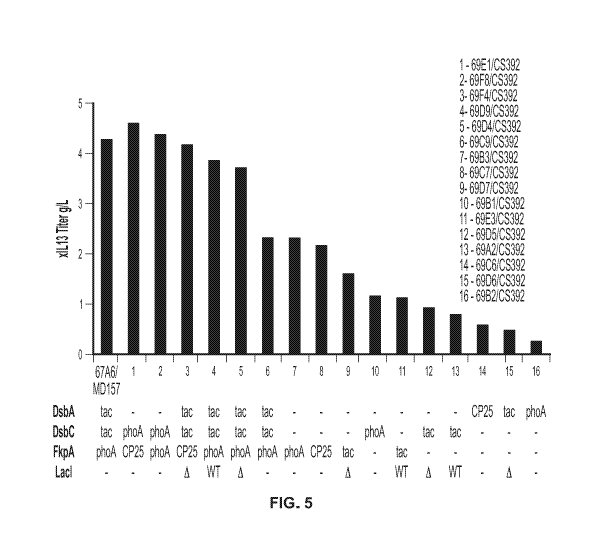
[0025] FIG. 5 shows xIL13 titer (g/L) produced by strains with the indicated
chromosomally
engineered pairs of plasmid and native chaperone locus. 67A6/MD157 refers to a
strain without
chromosomal engineering and with the MD157 plasmid expressing the indicated
chaperones
under the indicated promoter (see FIG. 1 for a diagram of the MD157 plasmid).
[0026] FIGS. 6A & 6B show the optical density (OD; FIG. 6A) and osmolality
(FIG. 6B)
over time of cultures of the indicated strain/plasmid combinations producing
xIL13.
[0027] FIGS. 7A-7C show the xIL13 titer (g/L; FIG. 7A), DsbC concentration
(FIG. 7B), and
FkpA concentration (FIG. 7C) produced by the indicated strains over time.
[0028] FIGS. 8A & 8B show the optical density (OD; FIG. 8A) and osmolality
(FIG. 8B)
over time of cultures of the indicated strain/plasmid combinations producing
AF2.
[0029] FIG. 9 shows the AF2 titer (g/L) produced by the indicated strains over
time.
[0030] FIGS. 10A & 10B show the optical density (OD; FIG. 10A) and osmolality
(FIG.
10B) over time of cultures of the indicated strain/plasmid combinations
producing MetMAb.
[0031] FIG. 11 shows the MetMAb titer (g/L) produced by the indicated strains
over time.
[0032] FIGS. 12A & 12B show the optical density (OD; FIG. 12A) and osmolality
(FIG.
12B) over time of cultures of the indicated strain/plasmid combinations
producing anti-VEGF
antibody fragment.
CA 03117051 2021-04-19
WO 2020/096959 PCT/US2019/059661
[0033] FIG. 13 shows the anti-VEGF antibody fragment titer (g/L) produced by
the indicated
strains over time.
DETAILED DESCRIPTION
[0034] The present disclosure provides host cells (e.g., prokaryotic host
cells) with integrated
non-native promoter: chaperone protein combination(s) suitable for large-scale
production of
recombinant two-chain protein products, as well as methods related thereto.
The examples
provided herein demonstrate that prokaryotic host cells expressing chaperone
proteins from the
host cell chromosome yield comparable titers to plasmid-based chaperone
expression. These
results were consistent across multiple antibody formats, such as half-
antibodies, one-armed
antibodies, and antibody fragments, and required little to no additional
process development.
Importantly, the data presented herein show that chaperone expression from the
host cell
chromosome rather than a plasmid results in lower chaperone expression levels
(potentially
obviating the need for further downstream purification to remove chaperone
proteins from the
product) but equivalent or higher product titers. These results demonstrate
that the products can
be produced at an industrial scale at least as efficiently using the host
cells and/or methods of the
present disclosure, as compared to using host cells that express the chaperone
protein(s) from a
plasmid, without requiring up-front time and cost to optimize chaperone
expression plasmids or
downstream purification steps to remove chaperone proteins.
[0035] In one aspect, provided herein are methods of producing a polypeptide
comprising two
chains in a prokaryotic host cell comprising a host cell chromosome, the
methods comprising:
culturing the host cell to express the two chains of the polypeptide in a
culture medium under
conditions suitable for expression of the two chains of the polypeptide,
whereby upon expression
the two chains fold and assemble to form a biologically active polypeptide in
the host cell; and
(b) recovering the biologically active polypeptide from the host cell; wherein
the host cell
comprises: (1) a first polynucleotide comprising a first translational unit
encoding a first chain of
the polypeptide; (2) a second polynucleotide comprising a second translational
unit encoding a
second chain of the polypeptide, wherein the first and second polynucleotides
are part of one or
more extra-chromosomal polynucleotides; and (3) a third polynucleotide
comprising a third
translational unit encoding a chaperone protein selected from the group
consisting of peptidyl-
proly1 isomerases and protein disulfide oxidoreductases, wherein the third
translational unit is
-12-
CA 03117051 2021-04-19
WO 2020/096959 PCT/US2019/059661
part of the host cell chromosome, wherein the third translational unit is in
operable combination
with a promoter that is integrated in the host cell chromosome and drives
transcription of the
third translational unit, wherein the combination of the third translational
unit and the promoter
is non-native to the host cell chromosome.
[0036] In another aspect, provided herein are methods of producing a
polypeptide comprising
two chains in a prokaryotic host cell comprising a host cell chromosome, the
methods
comprising: culturing the host cell to express the two chains of the
polypeptide in a culture
medium under conditions suitable for expression of the two chains of the
polypeptide, whereby
upon expression the two chains fold and assemble to form a biologically active
polypeptide in
the host cell; and (b) recovering the biologically active polypeptide from the
host cell; wherein
the host cell comprises: (1) a first polynucleotide comprising a first
translational unit encoding a
first chain of the polypeptide; (2) a second polynucleotide comprising a
second translational unit
encoding a second chain of the polypeptide, wherein the first and second
polynucleotides are part
of one or more extra-chromosomal polynucleotides; (3) a third polynucleotide
comprising a third
translational unit encoding a protein disulfide oxidoreductase, wherein the
third translational unit
is part of the host cell chromosome, wherein the third translational unit is
in operable
combination with a first promoter that is integrated in the host cell
chromosome and drives
transcription of the third translational unit, wherein the combination of the
third translational unit
and the first promoter is non-native to the host cell chromosome; and (4) a
fourth polynucleotide
comprising a fourth translational unit encoding a peptidyl-prolyl isomerase,
wherein the fourth
translational unit is part of the host cell chromosome, wherein the fourth
translational unit is in
operable combination with a second promoter that is integrated in the host
cell chromosome and
drives transcription of the fourth translational unit, wherein the combination
of the fourth
translational unit and the second promoter is non-native to the host cell
chromosome.
[0037] In another aspect, provided herein are prokaryotic host cells
comprising a host cell
chromosome, wherein the prokaryotic host cells comprise: (1) a first
polynucleotide comprising
a first translational unit encoding a peptidyl-prolyl isomerase, wherein the
first translational unit
is part of the host cell chromosome, wherein the first translational unit is
in operable combination
with a first promoter that is integrated in the host cell chromosome and
drives transcription of the
first translational unit, wherein the combination of the first translational
unit and the first
promoter is non-native to the host cell chromosome; and (2) a second
polynucleotide comprising
-13-
CA 03117051 2021-04-19
WO 2020/096959 PCT/US2019/059661
a second translational unit encoding a protein disulfide oxidoreductase,
wherein the second
translational unit is part of the host cell chromosome, wherein the second
translational unit is in
operable combination with a second promoter that is integrated in the host
cell chromosome and
drives transcription of the second translational unit, wherein the combination
of the second
translational unit and the second promoter is non-native to the host cell
chromosome.
I. Definitions
[0038] Before describing the disclosure in detail, it is to be understood that
this disclosure is
not limited to particular compositions or biological systems, which can, of
course, vary. It is also
to be understood that the terminology used herein is for the purpose of
describing particular
embodiments only, and is not intended to be limiting.
[0039] As used in this specification and the appended claims, the singular
forms "a", "an" and
"the" include plural referents unless the content clearly dictates otherwise.
Thus, for example,
reference to "a molecule" optionally includes a combination of two or more
such molecules, and
the like.
[0040] The term "about" as used herein refers to the usual error range for the
respective value
readily known to the skilled person in this technical field. Reference to
"about" a value or
parameter herein includes (and describes) embodiments that are directed to
that value or
parameter per se. At a maximum, the term "about" as used herein in reference
to a value,
encompasses from 90% to 110% of that value (e.g., relative translation
strength of a first and
second TIR of about 1.0 to about 3.0 refers to a relative translation strength
in the range of
between 0.9 and 3.3).
[0041] It is understood that aspects and embodiments of the disclosure
described herein
include "comprising," "consisting," and "consisting essentially of' aspects
and embodiments.
[0042] The term "polypeptide comprising two chains," (the terms "two chain
protein" and
"two chain polypeptide" may also be used interchangeably herein), as used
herein is intended to
refer to any polypeptide containing more than one distinct polypeptide chain.
In some
embodiments, a two chain protein may include a macromolecular complex of two
or more
polypeptides linked together through one or more intermolecular linkages,
including without
limitation a disulfide bond. In some embodiments, a two chain protein may
include a single
polypeptide with amino acid sequences belonging to two distinct polypeptide
chains (e.g., an
-14-
CA 03117051 2021-04-19
WO 2020/096959 PCT/US2019/059661
antibody heavy chain and an antibody light chain) linked by a polypeptide
linker. In this case, a
two chain protein may physically represent a single chain, but two or more
portions of the single
chain may functionally behave as if they are two separate protein chains. For
example, a single
chain antibody may include a functional heavy chain and a functional light
chain that, while
joined by a polypeptide linker, nonetheless fold and assemble as if they were
separate
polypeptides associated only by intermolecular linkages (e.g., one or more
disulfide bonds).
[0043] The terms "native" and "non-native," as used herein in reference to one
or more genetic
elements (e.g., a promoter, translational unit, or combination thereof), are
intended to refer to the
genomic context of the genetic element in a host cell chromosome as it occurs
in nature. For
example, a translational unit is "native" with regard to a host cell or host
cell chromosome when
the translational unit naturally occurs in the genome of the host cell, and is
"non-native" when
the translational unit does not naturally occur in the genome of the host
cell. A promoter is
"native" with regard to a host cell or host cell chromosome when the promoter
naturally occurs
in the genome of the host cell, and is "non-native" when the promoter does not
naturally occur in
the genome of the host cell. The operable combination of a promoter with a
translational unit is
"non-native" when the promoter does not naturally occur in the genome of the
host cell in the
same operable linkage with the translational unit, or vice versa. For example,
a
promoter:translational unit combination is "non-native" with respect to a host
cell or host cell
chromosome when one or both of the promoter and the translational unit is/are
not naturally
present in the host cell genome, when the promoter is present in the host cell
genome in operable
linkage with a translational unit with which it is not operably combined in
the naturally-
occurring host cell genome (even if the same promoter sequence is naturally
present elsewhere in
the host cell genome), or when the translational unit is present in the host
cell genome in
operable linkage with a promoter with which it is not operably combined in the
naturally-
occurring host cell genome (even if the same translational unit sequence is
naturally present
elsewhere in the host cell genome).
[0044] The term "vector," as used herein, is intended to refer to a nucleic
acid molecule
capable of transporting another nucleic acid to which it has been linked. One
type of vector is a
"plasmid", which refers to a circular double stranded DNA loop into which
additional DNA
segments may be ligated. Another type of vector is a phage vector. Another
type of vector is a
viral vector, wherein additional DNA segments may be ligated into the viral
genome. Certain
-15-
CA 03117051 2021-04-19
WO 2020/096959 PCT/US2019/059661
vectors are capable of autonomous replication in a host cell into which they
are introduced (e.g.,
bacterial vectors having a bacterial origin of replication and episomal
mammalian vectors). Other
vectors (e.g., non-episomal mammalian vectors) can be integrated into the
genome of a host cell
upon introduction into the host cell, and thereby are replicated along with
the host genome.
Moreover, certain vectors are capable of directing the expression of genes to
which they are
operatively linked. Such vectors are referred to herein as "recombinant
expression vectors" (or
simply, "recombinant vectors"). In general, expression vectors of utility in
recombinant DNA
techniques are often in the form of plasmids. In the present specification,
"plasmid" and "vector"
may be used interchangeably as the plasmid is the most commonly used form of
vector.
[0045] The term "cistron," as used herein, is intended to refer to a genetic
element broadly
equivalent to a translational unit comprising the nucleotide sequence coding
for a polypeptide
chain and adjacent control regions. A "cistron" may include, for example, one
or more open-
reading frames, a translational initiation region (TIR; as defined herein
below), a signal sequence
and a termination region.
[0046] A "polycistronic" expression vector refers to a single vector that
contains and expresses
multiple cistrons under the regulatory control of one single promoter. A
common example of
polycistronic vector is a "dicistronic" vector that contains and expresses two
different
polypeptides under the control of one promoter. Upon expression of a
dicistronic or polycistronic
vector, multiple genes are first transcribed as a single transcriptional unit,
and then translated
separately.
[0047] A "transcriptional unit" refers to a polynucleotide that is transcribed
as a single RNA
transcript. A "translational unit" refers to a polynucleotide that encodes
and, when translated,
produces a polypeptide. As described above, a polycistronic polynucleotide may
contain a single
transcriptional unit with multiple translational units.
[0048] A "separate cistron" expression vector according to the present
disclosure refers to a
single vector comprising at least two separate promoter-cistron pairs, wherein
each cistron is
under the control of its own promoter. Upon expression of a separate cistron
expression vector,
both transcription and translation processes of different genes are separate
and independent.
[0049] A "chaperone protein" as used herein refers to any protein that aids in
the folding or
assembly of other macromolecules, including without limitation two chain
proteins. Generally,
chaperone proteins may act by many different mechanisms to promote protein
folding or
-16-
CA 03117051 2021-04-19
WO 2020/096959 PCT/US2019/059661
assembly. For example, chaperone proteins may promote protein folding and/or
assembly,
catalyze the formation of intrachain disulfide bonds, promote protein un-
folding and/or
disassembly (e.g., of aggregated or misfolded proteins or multiprotein
complexes), prevent
aggregation, aid in protein degradation, and so forth.
[0050] "Secretion signal sequence" or "signal sequence" refers to a nucleic
acid sequence
encoding for a short signal peptide that can be used to direct a newly
synthesized protein of
interest through a cellular membrane, usually the inner membrane or both inner
and outer
membranes of prokaryotes. As such, the protein of interest such as the
immunoglobulin light or
heavy chain polypeptide is secreted into the periplasm of the prokaryotic host
cells or into the
culture medium. The signal peptide encoded by the secretion signal sequence
may be
endogenous to the host cells, or they may be non-endogenous, including signal
peptides native to
the polypeptide to be expressed. Secretion signal sequences are typically
present at the amino
terminus of a polypeptide to be expressed, and are typically removed
enzymatically between
biosynthesis and secretion of the polypeptide from the cytoplasm. Thus, the
signal peptide is
usually not present in a mature protein product.
[0051] "Operably linked" refers to a juxtaposition of two or more components,
wherein the
components so described are in a relationship permitting them to function in
their intended
manner. For example, a promoter is operably linked to a coding sequence or
translational unit if
it acts in cis to control or modulate the transcription of the linked
sequence. Generally, but not
necessarily, the DNA sequences that are "operably linked" are contiguous and,
where necessary
to join two protein coding regions or in the case of a secretory leader,
contiguous and in the
reading frame. However, although an operably linked promoter is generally
located upstream of
the coding sequence or translational unit, it is not necessarily contiguous
with it. Operably linked
enhancers can be located upstream, within or downstream of coding
sequences/translational units
and at considerable distances from the promoter. Linking is accomplished by
recombinant
methods known in the art, e.g., using PCR methodology, by annealing, or by
ligation at
convenient restriction sites. If convenient restriction sites do not exist,
then synthetic
oligonucleotide adaptors or linkers are used in accord with conventional
practice.
[0052] "Regulatory elements" as used herein, refer to nucleotide sequences
present in cis,
necessary for transcription and translation of a polynucleotide encoding a
heterologous
polypeptide into polypeptides. The transcriptional regulatory elements
normally comprise a
-17-
CA 03117051 2021-04-19
WO 2020/096959 PCT/US2019/059661
promoter 5' of the gene sequence to be expressed, transcriptional initiation
and termination sites,
and polyadenylation signal sequence. The term "transcriptional initiation
site" refers to the
nucleic acid in the construct corresponding to the first nucleic acid
incorporated into the primary
transcript, i.e., the mRNA precursor; the transcriptional initiation site may
overlap with the
promoter sequences.
[0053] A "promoter" refers to a polynucleotide sequence that controls
transcription of a gene
or sequence to which it is operably linked. A promoter includes signals for
RNA polymerase
binding and transcription initiation. The promoters used will be functional in
the cell type of the
host cell in which expression of the selected sequence is contemplated. A
large number of
promoters including constitutive, inducible and repressible promoters from a
variety of different
sources, are well known in the art (and identified in databases such as
GenBank) and are
available as or within cloned polynucleotides (from, e.g., depositories such
as ATCC as well as
other commercial or individual sources). With inducible promoters, the
activity of the promoter
increases or decreases in response to a signal, e.g., the presence of IPTG or
phosphate depletion.
[0054] The term "host cell" (or "recombinant host cell"), as used herein, is
intended to refer to
a cell that has been genetically altered, or is capable of being genetically
altered by introduction
of an exogenous or non-native polynucleotide, such as a recombinant plasmid or
vector. It should
be understood that such terms are intended to refer not only to the particular
subject cell but to
the progeny of such a cell. Because certain modifications may occur in
succeeding generations
due to either mutation or environmental influences, such progeny may not, in
fact, be identical to
the parent cell, but are still included within the scope of the term "host
cell" as used herein.
[0055] The term "pharmaceutical formulation" refers to a preparation which is
in such form as
to permit the biological activity of the active ingredient to be effective,
and which contains no
additional components which are unacceptably toxic to a subject to which the
formulation would
be administered. Such formulations are sterile. "Pharmaceutically acceptable"
excipients
(vehicles, additives) are those which can reasonably be administered to a
subject mammal to
provide an effective dose of the active ingredient employed.
[0056] A "subject" or an "individual" for purposes of treatment refers to any
animal classified
as a mammal, including humans, domestic and farm animals, and zoo, sports, or
pet animals,
such as dogs, horses, cats, cows, etc. Preferably, the mammal is human.
-18-
CA 03117051 2021-04-19
WO 2020/096959 PCT/US2019/059661
[0057] The term "antibody" herein is used in the broadest sense and
specifically covers
monoclonal antibodies (including full length monoclonal antibodies),
polyclonal antibodies,
multispecific antibodies (e.g., bispecific antibodies), and antibody fragments
so long as they
exhibit the desired biological activity.
[0058] An "isolated" antibody is one which has been identified and separated
and/or recovered
from a component of its natural environment. Contaminant components of its
natural
environment are materials which would interfere with research, diagnostic or
therapeutic uses for
the antibody, and may include enzymes, hormones, and other proteinaceous or
nonproteinaceous
solutes. In some embodiments, an antibody is purified (1) to greater than 95%
by weight of
antibody as determined by, for example, the Lowry method, and in some
embodiments, to
greater than 99% by weight; (2) to a degree sufficient to obtain at least 15
residues of N-terminal
or internal amino acid sequence by use of, for example, a spinning cup
sequenator, or (3) to
homogeneity by SDS-PAGE under reducing or nonreducing conditions using, for
example,
Coomassie blue or silver stain. Isolated antibody includes the antibody in
situ within
recombinant cells since at least one component of the antibody's natural
environment will not be
present. Ordinarily, however, isolated antibody will be prepared by at least
one purification step.
[0059] "Native antibodies" are usually heterotetrameric glycoproteins of about
150,000
daltons, composed of two identical light (L) chains and two identical heavy
(H) chains. Each
light chain is linked to a heavy chain by one covalent disulfide bond, while
the number of
disulfide linkages varies among the heavy chains of different immunoglobulin
isotypes. Each
heavy and light chain also has regularly spaced intrachain disulfide bridges.
Each heavy chain
has at one end a variable domain (VH) followed by a number of constant
domains. Each light
chain has a variable domain at one end (VI) and a constant domain at its other
end; the constant
domain of the light chain is aligned with the first constant domain of the
heavy chain, and the
light chain variable domain is aligned with the variable domain of the heavy
chain. Particular
amino acid residues are believed to form an interface between the light chain
and heavy chain
variable domains.
[0060] The term "constant domain" refers to the portion of an immunoglobulin
molecule
having a more conserved amino acid sequence relative to the other portion of
the
immunoglobulin, the variable domain, which contains the antigen binding site.
The constant
-19-
CA 03117051 2021-04-19
WO 2020/096959 PCT/US2019/059661
domain contains the CH1, CH2 and CH3 domains (collectively, CH) of the heavy
chain and the
CHL (or CL) domain of the light chain.
[0061] The "variable region" or "variable domain" of an antibody refers to the
amino-terminal
domains of the heavy or light chain of the antibody. The variable domain of
the heavy chain may
be referred to as "VH." The variable domain of the light chain may be referred
to as "VC These
domains are generally the most variable parts of an antibody and contain the
antigen-binding
sites.
[0062] The term "variable" refers to the fact that certain portions of the
variable domains differ
extensively in sequence among antibodies and are used in the binding and
specificity of each
particular antibody for its particular antigen. However, the variability is
not evenly distributed
throughout the variable domains of antibodies. It is concentrated in three
segments called
hypervariable regions (HVRs) both in the light-chain and the heavy-chain
variable domains. The
more highly conserved portions of variable domains are called the framework
regions (FR). The
variable domains of native heavy and light chains each comprise four FR
regions, largely
adopting a beta-sheet configuration, connected by three HVRs, which form loops
connecting,
and in some cases forming part of, the beta-sheet structure. The HVRs in each
chain are held
together in close proximity by the FR regions and, with the HVRs from the
other chain,
contribute to the formation of the antigen-binding site of antibodies (see
Kabat et al., Sequences
of Proteins of Immunological Interest, Fifth Edition, National Institute of
Health, Bethesda, Md.
(1991)). The constant domains are not involved directly in the binding of an
antibody to an
antigen, but exhibit various effector functions, such as participation of the
antibody in antibody-
dependent cellular toxicity.
[0063] The "light chains" of antibodies (immunoglobulins) from any mammalian
species can
be assigned to one of two clearly distinct types, called kappa ("x") and
lambda ("k"), based on
the amino acid sequences of their constant domains.
[0064] The term IgG "isotype" or "subclass" as used herein is meant any of the
subclasses of
immunoglobulins defined by the chemical and antigenic characteristics of their
constant regions.
[0065] Depending on the amino acid sequences of the constant domains of their
heavy chains,
antibodies (immunoglobulins) can be assigned to different classes. There are
five major classes
of immunoglobulins: IgA, IgD, IgE, IgG, and IgM, and several of these may be
further divided
into subclasses (isotypes), e.g., IgGi, IgG2, IgG3, IgG4, IgAi, and IgA2. The
heavy chain constant
-20-
CA 03117051 2021-04-19
WO 2020/096959 PCT/US2019/059661
domains that correspond to the different classes of immunoglobulins are called
a, y, c, y, and II.,
respectively. The subunit structures and three-dimensional configurations of
different classes of
immunoglobulins are well known and described generally in, for example, Abbas
et al. Cellular
and Mol. Immunology, 4th ed. (W.B. Saunders, Co., 2000). An antibody may be
part of a larger
fusion molecule, formed by covalent or non-covalent association of the
antibody with one or
more other proteins or peptides.
[0066] The terms "full length antibody," "intact antibody" and "whole
antibody" are used
herein interchangeably to refer to an antibody in its substantially intact
form, not antibody
fragments as defined below. The terms particularly refer to an antibody with
heavy chains that
contain an Fc region.
[0067] A "naked antibody" for the purposes herein is an antibody that is not
conjugated to a
cytotoxic moiety or radiolabel.
[0068] "Antibody fragments" comprise a portion of an intact antibody,
preferably comprising
the antigen binding region thereof. In some embodiments, the antibody fragment
described
herein is an antigen-binding fragment. Examples of antibody fragments include
Fab, Fab',
F(ab')2, and Fv fragments; diabodies; linear antibodies; single-chain antibody
molecules; and
multispecific antibodies formed from antibody fragments.
[0069] Papain digestion of antibodies produces two identical antigen-binding
fragments, called
"Fab" fragments, each with a single antigen-binding site, and a residual "Fc"
fragment, whose
name reflects its ability to crystallize readily. Pepsin treatment yields an
F(ab')2 fragment that has
two antigen-combining sites and is still capable of cross-linking antigen.
[0070] "Fv" is the minimum antibody fragment which contains a complete antigen-
binding
site. In one embodiment, a two-chain Fv species consists of a dimer of one
heavy- and one light-
chain variable domain in tight, non-covalent association. In a single-chain Fv
(scFv) species, one
heavy- and one light-chain variable domain can be covalently linked by a
flexible peptide linker
such that the light and heavy chains can associate in a "dimeric" structure
analogous to that in a
two-chain Fv species. It is in this configuration that the three HVRs of each
variable domain
interact to define an antigen-binding site on the surface of the VH-VL dimer.
Collectively, the
six HVRs confer antigen-binding specificity to the antibody. However, even a
single variable
domain (or half of an Fv comprising only three HVRs specific for an antigen)
has the ability to
recognize and bind antigen, although at a lower affinity than the entire
binding site.
-21-
CA 03117051 2021-04-19
WO 2020/096959 PCT/US2019/059661
[0071] The Fab fragment contains the heavy- and light-chain variable domains
and also
contains the constant domain of the light chain and the first constant domain
(CH1) of the heavy
chain. Fab' fragments differ from Fab fragments by the addition of a few
residues at the carboxy
terminus of the heavy chain CH1 domain including one or more cysteines from
the antibody
hinge region. Fab'-SH is the designation herein for Fab' in which the cysteine
residue(s) of the
constant domains bear a free thiol group. F(ab')2 antibody fragments
originally were produced as
pairs of Fab' fragments which have hinge cysteines between them. Other
chemical couplings of
antibody fragments are also known.
[0072] "Single-chain Fv" or "scFv" antibody fragments comprise the VH and VL
domains of
antibody, wherein these domains are present in a single polypeptide chain.
Generally, the scFv
polypeptide further comprises a polypeptide linker between the VH and VL
domains which
enables the scFv to form the desired structure for antigen binding. For a
review of scFv, see, e.g.,
Pluckthiln, in The Pharmacology of Monoclonal Antibodies, vol. 113, Rosenburg
and Moore
eds., (Springer-Verlag, New York, 1994), pp. 269-315.
[0073] The term "diabodies" refers to antibody fragments with two antigen-
binding sites,
which fragments comprise a heavy-chain variable domain (VH) connected to a
light-chain
variable domain (VL) in the same polypeptide chain (VH-VL). By using a linker
that is too short
to allow pairing between the two domains on the same chain, the domains are
forced to pair with
the complementary domains of another chain and create two antigen-binding
sites. Diabodies
may be bivalent or bispecific. Diabodies are described more fully in, for
example, EP 404,097;
WO 1993/01161; Hudson et al., Nat. Med. 9:129-134 (2003); and Hollinger et
al., Proc. Natl.
Acad. Sci. USA 90: 6444-6448 (1993). Triabodies and tetrabodies are also
described in Hudson
et al., Nat. Med. 9:129-134 (2003).
[0074] The term "monoclonal antibody" as used herein refers to an antibody
obtained from a
population of substantially homogeneous antibodies, e.g., the individual
antibodies comprising
the population are identical except for possible mutations, e.g., naturally
occurring mutations,
that may be present in minor amounts. Thus, the modifier "monoclonal"
indicates the character
of the antibody as not being a mixture of discrete antibodies. In certain
embodiments, such a
monoclonal antibody typically includes an antibody comprising a polypeptide
sequence that
binds a target, wherein the target-binding polypeptide sequence was obtained
by a process that
includes the selection of a single target binding polypeptide sequence from a
plurality of
-22-
CA 03117051 2021-04-19
WO 2020/096959 PCT/US2019/059661
polypeptide sequences. For example, the selection process can be the selection
of a unique clone
from a plurality of clones, such as a pool of hybridoma clones, phage clones,
or recombinant
DNA clones. It should be understood that a selected target binding sequence
can be further
altered, for example, to improve affinity for the target, to humanize the
target binding sequence,
to improve its production in cell culture, to reduce its immunogenicity in
vivo, to create a
multispecific antibody, etc., and that an antibody comprising the altered
target binding sequence
is also a monoclonal antibody of this disclosure. In contrast to polyclonal
antibody preparations,
which typically include different antibodies directed against different
determinants (epitopes),
each monoclonal antibody of a monoclonal antibody preparation is directed
against a single
determinant on an antigen. In addition to their specificity, monoclonal
antibody preparations are
advantageous in that they are typically uncontaminated by other
immunoglobulins.
[0075] The modifier "monoclonal" indicates the character of the antibody as
being obtained
from a substantially homogeneous population of antibodies, and is not to be
construed as
requiring production of the antibody by any particular method. For example,
the monoclonal
antibodies to be used in accordance with the disclosure may be made by a
variety of techniques,
including, for example, expression in a prokaryotic host cell, the hybridoma
method (e.g., Kohler
and Milstein, Nature, 256:495-97 (1975); Hongo et al., Hybridoma, 14 (3): 253-
260 (1995),
Harlow et at., Antibodies: A Laboratory Manual, (Cold Spring Harbor Laboratory
Press, 2nd ed.
1988); Hammerling et at., in: Monoclonal Antibodies and T-Cell Hybridomas 563-
681 (Elsevier,
N.Y., 1981)), recombinant DNA methods (see, e.g., U.S. Pat. No. 4,816,567),
phage-display
technologies (see, e.g., Clackson et al., Nature, 352: 624-628 (1991); Marks
et al., I Mot. Biol.
222: 581-597 (1992); Sidhu et al., I Mot. Biol. 338(2): 299-310 (2004); Lee et
al., I Mot. Biol.
340(5): 1073-1093 (2004); Fellouse, Proc. Natl. Acad. Sci. USA 101(34): 12467-
12472 (2004);
and Lee et at., I Immunol. Methods 284(1-2): 119-132 (2004), and technologies
for producing
human or human-like antibodies in animals that have parts or all of the human
immunoglobulin
loci or genes encoding human immunoglobulin sequences (see, e.g., WO
1998/24893; WO
1996/34096; WO 1996/33735; WO 1991/10741; Jakobovits et at., Proc. Natl. Acad.
Sci. USA
90: 2551(1993); Jakobovits et at., Nature 362: 255-258 (1993); Bruggemann et
at., Year in
Immunol. 7:33 (1993); U.S. Pat. Nos. 5,545,807; 5,545,806; 5,569,825;
5,625,126; 5,633,425;
and 5,661,016; Marks et al., Bio/Technology 10: 779-783 (1992); Lonberg et
al., Nature 368:
856-859 (1994); Morrison, Nature 368: 812-813 (1994); Fishwild et al., Nature
Biotechnol. 14:
-23-
CA 03117051 2021-04-19
WO 2020/096959 PCT/US2019/059661
845-851 (1996); Neuberger, Nature Biotechnol. 14: 826 (1996); and Lonberg and
Huszar, Intern.
Rev. Immunol. 13: 65-93 (1995).
[0076] The monoclonal antibodies herein specifically include "chimeric"
antibodies in which a
portion of the heavy and/or light chain is identical with or homologous to
corresponding
sequences in antibodies derived from a particular species or belonging to a
particular antibody
class or subclass, while the remainder of the chain(s) is identical with or
homologous to
corresponding sequences in antibodies derived from another species or
belonging to another
antibody class or subclass, as well as fragments of such antibodies, so long
as they exhibit the
desired biological activity (see, e.g.,U U.S. Pat. No. 4,816,567; and Morrison
et al., Proc. Natl.
Acad. Sci. USA 81:6851-6855 (1984)). Chimeric antibodies include PRIMATTZED
antibodies
wherein the antigen-binding region of the antibody is derived from an antibody
produced by,
e.g., immunizing macaque monkeys with the antigen of interest.
[0077] "Humanized" forms of non-human (e.g., murine) antibodies are chimeric
antibodies
that contain minimal sequence derived from non-human immunoglobulin. In one
embodiment, a
humanized antibody is a human immunoglobulin (recipient antibody) in which
residues from a
HVR of the recipient are replaced by residues from a HVR of a non-human
species (donor
antibody) such as mouse, rat, rabbit, or nonhuman primate having the desired
specificity,
affinity, and/or capacity. In some instances, FR residues of the human
immunoglobulin are
replaced by corresponding non-human residues. Furthermore, humanized
antibodies may
comprise residues that are not found in the recipient antibody or in the donor
antibody. These
modifications may be made to further refine antibody performance. In general,
a humanized
antibody will comprise substantially all of at least one, and typically two,
variable domains, in
which all or substantially all of the hypervariable loops correspond to those
of a non-human
immunoglobulin, and all or substantially all of the FRs are those of a human
immunoglobulin
sequence. The humanized antibody optionally will also comprise at least a
portion of an
immunoglobulin constant region (Fc), typically that of a human immunoglobulin.
For further
details, see, e.g., Jones et al., Nature 321:522-525 (1986); Riechmann et al.,
Nature 332:323-329
(1988); and Presta, Curr. Op. Struct. Biol. 2:593-596 (1992). See also, e.g.,
Vaswani and
Hamilton, Ann. Allergy, Asthma & Immunol. 1:105-115 (1998); Harris, Biochem.
Soc.
Transactions 23:1035-1038 (1995); Hurle and Gross, Curr. Op. Biotech. 5:428-
433 (1994); and
U.S. Pat. Nos. 6,982,321 and 7,087,409.
-24-
CA 03117051 2021-04-19
WO 2020/096959 PCT/US2019/059661
[0078] A "human antibody" is one which possesses an amino acid sequence which
corresponds to that of an antibody produced by a human and/or has been made
using any of the
techniques for making human antibodies as disclosed herein. This definition of
a human antibody
specifically excludes a humanized antibody comprising non-human antigen-
binding residues.
Human antibodies can be produced using various techniques known in the art,
including phage-
display libraries. Hoogenboom and Winter, I Mot. Biol., 227:381 (1991); Marks
et al., I Mot.
Biol., 222:581 (1991). Also available for the preparation of human monoclonal
antibodies are
methods described in Cole et at., Monoclonal Antibodies and Cancer Therapy,
Alan R. Liss, p.
77(1985); Boerneretat.,I Immunol., 147(1):86-95 (1991). See also van Dijk and
van de
Winkel, Curr. Op/n. Pharmacol., 5: 368-74 (2001). Human antibodies can be
prepared by
administering the antigen to a transgenic animal that has been modified to
produce such
antibodies in response to antigenic challenge, but whose endogenous loci have
been disabled,
e.g., immunized xenomice (see, e.g., U.S. Pat. Nos. 6,075,181 and 6,150,584
regarding
XENOMOUSETm technology). See also, for example, Li et at., Proc. Natl. Acad.
Sci. USA,
103:3557-3562 (2006) regarding human antibodies generated via a human B-cell
hybridoma
technology.
[0079] A "species-dependent antibody" is one which has a stronger binding
affinity for an
antigen from a first mammalian species than it has for a homologue of that
antigen from a second
mammalian species. Normally, the species-dependent antibody "binds
specifically" to a human
antigen (e.g., has a binding affinity (Kd) value of no more than about 1x10⁷
M, preferably no
more than about 1x10⁻⁸M and preferably no more than about 1x10⁹ M) but has a
binding
affinity for a homologue of the antigen from a second nonhuman mammalian
species which is at
least about 50 fold, or at least about 500 fold, or at least about 1000 fold,
weaker than its binding
affinity for the human antigen. The species-dependent antibody can be any of
the various types
of antibodies as defined above, but preferably is a humanized or human
antibody.
[0080] The term "hypervariable region," "HVR," or "HV," when used herein
refers to the
regions of an antibody variable domain which are hypervariable in sequence
and/or form
structurally defined loops. Generally, antibodies comprise six HVRs; three in
the VH (H1, H2,
H3), and three in the VL (L1, L2, L3). In native antibodies, H3 and L3 display
the most diversity
of the six HVRs, and H3 in particular is believed to play a unique role in
conferring fine
specificity to antibodies. See, e.g., Xu et al., Immunity 13:37-45 (2000);
Johnson and Wu, in
-25-
CA 03117051 2021-04-19
WO 2020/096959 PCT/US2019/059661
Methods in Molecular Biology 248:1-25 (Lo, ed., Human Press, Totowa, N.J.,
2003). Indeed,
naturally occurring camelid antibodies consisting of a heavy chain only are
functional and stable
in the absence of light chain. See, e.g., Hamers-Casterman et al., Nature
363:446-448 (1993);
Sheriff et al., Nature Struct. Biol. 3:733-736 (1996).
[0081] A number of HVR delineations are in use and are encompassed herein. The
Kabat
Complementarity Determining Regions (CDRs) are based on sequence variability
and are the
most commonly used (Kabat et al., Sequences of Proteins of Immunological
Interest, 5th Ed.
Public Health Service, National Institutes of Health, Bethesda, Md. (1991)).
Chothia refers
instead to the location of the structural loops (Chothia and Lesk I Mol. Biol.
196:901-917
(1987)). The AbM HVRs represent a compromise between the Kabat HVRs and
Chothia
structural loops, and are used by Oxford Molecular's AbM antibody modeling
software. The
"contact" HVRs are based on an analysis of the available complex crystal
structures. The
residues from each of these HVRs are noted below.
Table la. Antibody Hypervariable Regions
Loop Kabat AbM Chothia Contact
Li L24-L34 L24-L34 L26-L32 L30-L36
L2 L50-L56 L50-L56 L50-L52 L46-L55
L3 L89-L97 L89-L97 L91-L96 L89-L96
H1 H31-H35B H26-H35B H26-H32 H30-H35B
(Kabat Numbering)
H1 H31-H35 H26-H35 H26-H32 H30-H35
(Chothia Numbering)
H2 H50-H65 H50-H58 H53-H55 H47-H58
H3 H95-H102 H95-H102 H96-H101 H93-H101
[0082] HVRs may comprise "extended HVRs" as follows: 24-36 or 24-34 (L1), 46-
56 or 50-
56 (L2) and 89-97 or 89-96 (L3) in the VL and 26-35 (H1), 50-65 or 49-65 (H2)
and 93-102, 94-
102, or 95-102 (H3) in the VH. The variable domain residues are numbered
according to Kabat
et al., supra, for each of these definitions.
[0083] "Framework" or "FR" residues are those variable domain residues other
than the HVR
residues as herein defined.
-26-
CA 03117051 2021-04-19
WO 2020/096959 PCT/US2019/059661
[0084] The term "variable domain residue numbering as in Kabat" or "amino acid
position
numbering as in Kabat," and variations thereof, refers to the numbering system
used for heavy
chain variable domains or light chain variable domains of the compilation of
antibodies in Kabat
et al., supra. Using this numbering system, the actual linear amino acid
sequence may contain
fewer or additional amino acids corresponding to a shortening of, or insertion
into, a FR or HVR
of the variable domain. For example, a heavy chain variable domain may include
a single amino
acid insert (residue 52a according to Kabat) after residue 52 of H2 and
inserted residues (e.g.
residues 82a, 82b, and 82c, etc. according to Kabat) after heavy chain FR
residue 82. The Kabat
numbering of residues may be determined for a given antibody by alignment at
regions of
homology of the sequence of the antibody with a "standard" Kabat numbered
sequence.
[0085] The Kabat numbering system is generally used when referring to a
residue in the
variable domain (approximately residues 1-107 of the light chain and residues
1-113 of the heavy
chain) (e.g., Kabat et at., Sequences of Immunological Interest. 5th Ed.
Public Health Service,
National Institutes of Health, Bethesda, Md. (1991)). The "EU numbering
system" or "EU
index" is generally used when referring to a residue in an immunoglobulin
heavy chain constant
region (e.g., the EU index reported in Kabat et at., supra). The "EU index as
in Kabat" refers to
the residue numbering of the human IgG1 EU antibody.
[0086] The expression "linear antibodies" refers to the antibodies described
in Zapata et al.
(1995 Protein Eng, 8(10):1057-1062). Briefly, these antibodies comprise a pair
of tandem Fd
segments (VH-CH1-VH-CH1) which, together with complementary light chain
polypeptides,
form a pair of antigen binding regions. Linear antibodies can be bispecific or
monospecific.
Host Cells
[0087] Provided herein are host cells (e.g., prokaryotic host cells) with a
host cell chromosome
that comprises a translational unit encoding at least one chaperone protein
(e.g., a peptidyl-prolyl
isomerase or protein disulfide oxidoreductase) in operable combination or
linkage with a
promoter (also part of the host cell chromosome) that drives transcription of
the translational
unit, such that the combination of the promoter and the translational unit is
non-native to the host
cell or host cell chromosome.
[0088] In some embodiments, the host cell chromosome comprises: (1) a first
polynucleotide
comprising a first translational unit encoding a peptidyl-prolyl isomerase;
and (2) a second
-27-
CA 03117051 2021-04-19
WO 2020/096959 PCT/US2019/059661
polynucleotide comprising a second translational unit encoding a protein
disulfide
oxidoreductase; wherein the first and second translational units are part of
the host cell
chromosome and in operable combination or linkage with a first and a second
(respectively)
promoter (also part of the host cell chromosome) that drive transcription of
the first and the
second translation units, respectively. In some embodiments, the combination
of the first
translational unit and the first promoter and/or the combination of the second
translational unit
and the second promoter is/are non-native to the host cell chromosome. For
example, one or
both of the promoters can be non-native to the host cell chromosome, one or
both of the
translational units can be non-native to the host cell chromosome, or one or
both of the
translational units can be native to the host cell chromosome but operably
combined with a
promoter in a combination that is non-native to the host cell chromosome.
[0089] In some embodiments, the host cell further comprises one or more extra-
chromosomal
polynucleotide(s) that encode the two or more chains of a two-chain
polypeptide of the present
disclosure. For example, in some embodiments, the host cell comprises: (1) a
first
polynucleotide comprising a first translational unit encoding a first chain of
a two-chain
polypeptide of the present disclosure; (2) a second polynucleotide comprising
a second
translational unit encoding a second chain of the two-chain polypeptide of the
present disclosure;
and (3) a third polynucleotide comprising a third translational unit encoding
a chaperone protein
(e.g., a peptidyl-prolyl isomerase or protein disulfide oxidoreductase) in
operable combination
with a promoter that drives transcription of the third translational unit. In
some embodiments,
the combination of the third translational unit and the promoter is non-native
to the host cell
chromosome. In some embodiments, the first and second polynucleotides (i.e.,
encoding the first
and second translational units, respectively) are part of one or more extra-
chromosomal
polynucleotide(s) (e.g., plasmid(s)), and the third polynucleotide (and
associated promoter) is
part of the host cell chromosome.
[0090] In some embodiments, the host cell comprises: (1) a first
polynucleotide comprising a
first translational unit encoding a first chain of a two-chain polypeptide of
the present disclosure;
(2) a second polynucleotide comprising a second translational unit encoding a
second chain of
the two-chain polypeptide of the present disclosure; (3) a third
polynucleotide comprising a third
translational unit encoding a protein disulfide oxidoreductase in operable
combination with a
promoter that drives transcription of the third translational unit; and (4) a
fourth polynucleotide
-28-
CA 03117051 2021-04-19
WO 2020/096959 PCT/US2019/059661
comprising a fourth translational unit encoding a peptidyl-prolyl isomerase in
operable
combination with a promoter that drives transcription of the fourth
translational unit. In some
embodiments, the combination of the third translational unit and its
associated promoter and/or
the combination of the fourth translational unit and its associated promoter
is/are non-native to
the host cell chromosome. In some embodiments, the first and second
polynucleotides (i.e.,
encoding the first and second translational units, respectively) are part of
one or more extra-
chromosomal polynucleotide(s) (e.g., plasmid(s)), and the third and fourth
polynucleotides (and
associated promoters) are part of the host cell chromosome.
[0091] In some embodiments, the host cell comprises: (1) a first
polynucleotide comprising a
first translational unit encoding a first chain of a two-chain polypeptide of
the present disclosure;
(2) a second polynucleotide comprising a second translational unit encoding a
second chain of
the two-chain polypeptide of the present disclosure; (3) a third
polynucleotide comprising a third
translational unit encoding a protein disulfide oxidoreductase in operable
combination with a
first promoter that drives transcription of the third translational unit; and
(4) a fourth
polynucleotide comprising a fourth translational unit encoding a protein
disulfide oxidoreductase
in operable combination with a second promoter that drives transcription of
the fourth
translational unit. In some embodiments, the combination of the third
translational unit and the
first promoter and/or the combination of the fourth translational unit and the
second promoter
is/are non-native to the host cell chromosome. In some embodiments, the first
and second
polynucleotides (i.e., encoding the first and second translational units,
respectively) are part of
one or more extra-chromosomal polynucleotide(s) (e.g., plasmid(s)), and the
third and fourth
polynucleotides (and associated promoters) are part of the host cell
chromosome.
[0092] In some embodiments, the host cell comprises: (1) a first
polynucleotide comprising a
first translational unit encoding a first chain of a two-chain polypeptide of
the present disclosure;
(2) a second polynucleotide comprising a second translational unit encoding a
second chain of
the two-chain polypeptide of the present disclosure; (3) a third
polynucleotide comprising a third
translational unit encoding a protein disulfide oxidoreductase in operable
combination with a
first promoter that drives transcription of the third translational unit; (4)
a fourth polynucleotide
comprising a fourth translational unit encoding a peptidyl-prolyl isomerase in
operable
combination with a second promoter that drives transcription of the fourth
translational unit; and
(5) a fifth polynucleotide comprising a fifth translational unit encoding a
protein disulfide
-29-
CA 03117051 2021-04-19
WO 2020/096959 PCT/US2019/059661
oxidoreductase in operable combination with a third promoter that drives
transcription of the
fifth translational unit. In some embodiments, the combination of the third
translational unit and
the first promoter, the combination of the fourth translational unit and the
second promoter,
and/or the combination of the fifth translational unit and the third promoter
is/are non-native to
the host cell chromosome. In some embodiments, the first and second
polynucleotides (i.e.,
encoding the first and second translational units, respectively) are part of
one or more extra-
chromosomal polynucleotide(s) (e.g., plasmid(s)), and the third, fourth, and
fifth polynucleotides
(and associated promoters) are part of the host cell chromosome.
[0093] In some embodiments, a polynucleotide or translational unit of the
present disclosure
that encodes a chaperone protein (e.g., a peptidyl-prolyl isomerase or protein
disulfide
oxidoreductase) is native to the host cell chromosome. For example, the
polynucleotide or
translational unit encoding the chaperone protein may be a native chaperone
protein gene or
locus. In some embodiments, a promoter has been inserted into the host cell
genome (e.g., by
insertion into or replacement of one or more native regulatory sequences or
genetic elements) so
as to be in operable combination with a native chaperone protein gene or
locus, generating a
promoter:translational unit combination that is non-native to the host cell
chromosome.
[0094] In other embodiments, a polynucleotide or translational unit of the
present disclosure
that encodes a chaperone protein (e.g., a peptidyl-prolyl isomerase or protein
disulfide
oxidoreductase) is non-native to the host cell (e.g., chromosomally integrated
into a host cell).
[0095] In some embodiments, a host cell chromosome of the present disclosure
may comprise
one or more native translational unit(s) encoding a chaperone protein of the
present disclosure
and one or more non-native translational unit(s) encoding a chaperone protein
of the present
disclosure. In some embodiments, a host cell chromosome of the present
disclosure may
comprise multiple non-native translational units encoding a chaperone protein
of the present
disclosure. In addition, many of the host cells of the present disclosure are
known to contain
host cell chromosomes encoding multiple chaperone proteins (e.g., FkpA, DsbA,
and DsbC of E.
coli). In some embodiments, one or more of the native translational unit(s)
encoding a
chaperone protein of the present disclosure is operably combined with a
promoter of the present
disclosure in a combination that is non-native to the host cell or host cell
chromosome.
[0096] Methods for introducing a polynucleotide or translational unit of the
present disclosure
into a host cell (e.g., a prokaryotic host cell) are known in the art. An
exemplary method, allelic
-30-
CA 03117051 2021-04-19
WO 2020/096959 PCT/US2019/059661
exchange, is described in greater detail infra. Advantageously, the allelic
exchange method does
not leave a "scar" on the host cell genome. Other methods include, without
limitation, the
method described in Datsenko, K.A. and Wanner, B.L. (2000) Proc. Natl. Acad.
Sci. 97:6640-
6645.
[0097] In some embodiments of any of the above embodiments, a host cell
further comprises a
translational unit encoding a third chain of a two-chain polypeptide of the
present disclosure. In
some embodiments, the translational unit is part of an extra-chromosomal
polynucleotide
encoding the first and/or second chain of the two-chain polypeptide. For
example, in some
embodiments the two-chain polypeptide is a one-armed antibody comprising,
e.g., an
immunoglobulin heavy chain, an immunoglobulin light chain, and an
immunoglobulin Fc
fragment that assemble to form a biologically active monovalent antibody
(e.g., a monovalent
antibody capable of specifically binding an antigen).
Chaperone Proteins
[0098] Certain aspects of the present disclosure relate to chaperone proteins.
A chaperone
protein may refer to any protein that aids in the folding or assembly of other
macromolecules,
including without limitation two chain proteins. Examples of chaperone
proteins may include
without limitation peptidyl-prolyl isomerases, protein disulfide
oxidoreductases, and heat shock
proteins (such as Hsp60, Hsp70, Hsp90, and Hsp100 proteins). Chaperone
proteins may also aid
in transporting proteins across membranes, e.g., translocation of polypeptide
chains across the
plasma membrane or endoplasmic reticulum membrane.
[0099] In some embodiments, a chaperone protein can be a peptidyl-prolyl
isomerase.
Peptidyl-prolyl isomerase (the terms "prolyl isomerase," "rotamase," and
"PPiase" may be used
interchangeably herein) may refer to any enzyme catalyzing the interconversion
of cis and trans
isomers of proline or prolyl-iminopeptide bonds. The EC number for this
reaction is EC 5.2.1.8.
Any protein known or predicted to catalyze the reaction described by this EC
number may be a
peptidyl-prolyl isomerase of the present disclosure. Peptidyl-prolyl isomerase
activity may also
be described by the GO term ID GO:0003755. Any protein known or predicted to
possess the
molecular function described by this GO term ID may be a peptidyl-prolyl
isomerase of the
present disclosure.
[0100] Peptidyl-prolyl isomerase activity is known in the art to promote
protein folding and
assembly. In some embodiments, peptidyl-prolyl isomerases may aid in protein
folding and
-31-
CA 03117051 2021-04-19
WO 2020/096959 PCT/US2019/059661
assembly by converting trans prolyl bonds to cis prolyl bonds for proteins
whose properly folded
structure includes a cis prolyl bond. Some peptidyl-prolyl isomerases are also
known to enhance
the folding and assembly of proteins that lack cis prolyl bonds (Bothmann H
and Pluckthun A
2000 J. Biol. Chem. 275:17100). In some embodiments, peptidyl-prolyl
isomerases may aid in
protein folding and assembly of proteins that lack cis prolyl bonds. Thus,
while peptidyl-prolyl
isomerase activity may serve as a functional characteristic to identify a
chaperone protein useful
for the methods described herein, the utility of a peptidyl-prolyl isomerase
is not necessarily
limited to its catalytic activity per se.
[0101] In some embodiments, the peptidyl-prolyl isomerase is an FkpA protein.
In some
embodiments, the FkpA protein is E. coil FkpA. An E. coil FkpA may refer to
any polypeptide
encoded by an fkpA gene in any strain or isolate of bacteria belonging to the
species E. coil. In
some embodiments, E. coil FkpA refers a protein encoded by an fkpA gene
described by
EcoGene Accession Number EG12900. In some embodiments, E. coil FkpA refers a
protein
having the sequence described by the NCBI RefSeq Accession Number NP 417806.
[0102] Other FkpA proteins are known in the art. Examples of FkpA proteins may
include,
without limitation, S. boydii peptidyl-prolyl isomerase (NCBI RefSeq No. WP
000838252), C.
youngae peptidyl-prolyl isomerase (NCBI RefSeq No. WP 006687366), K oxytoca
peptidyl-
prolyl isomerase (NCBI RefSeq No. WP 004125943), S. enterica peptidyl-prolyl
isomerase
(NCBI RefSeq No. WP 000838233), K pneumoniae peptidyl-prolyl isomerase (NCBI
RefSeq
No. WPO19704642), S. cerevisiae FPR3p (NCBI RefSeq No. NP 013637), M musculus
Fkpbla (NCBI RefSeq No. NP 032045), M musculus Fkpb2 (NCBI RefSeq No. NP
032046),
H. sapiens FKBP2 (NCBI RefSeq No. NP 001128680), and D. melanogaster CG14715
(NCBI
RefSeq No. NP 650101). In some embodiments, an FkpA protein of the present
disclosure has at
least about 80%, at least about 81%, at least about 82%, at least about 83%,
at least about 84%,
at least about 85%, at least about 86%, at least about 87%, at least about
88%, at least about
89%, at least about 90%, at least about 91%, at least about 92%, at least
about 93%, at least
about 94%, at least about 95%, at least about 96%, at least about 97%, at
least about 98%, or at
least about 99% identity to E. coil FkpA.
[0103] In some embodiments, a chaperone protein may be a protein disulfide
oxidoreductase.
Protein disulfide oxidoreductase (the terms "protein disulfide isomerase" and
"thiol-disulfide
isomerase" may be used interchangeably herein) may refer to any enzyme
catalyzing the
-32-
CA 03117051 2021-04-19
WO 2020/096959 PCT/US2019/059661
rearrangement of disulfide bonds in proteins. For example, a protein disulfide
oxidoreductase
may catalyze the oxidation of cysteines to form disulfide bonds in proteins. A
protein disulfide
oxidoreductase may also catalyze the isomerization of mispaired disulfide
bonds in proteins. The
EC number for this reaction is EC 5.3.4.1. Any protein known or predicted to
catalyze the
reaction described by this EC number may be a protein disulfide oxidoreductase
of the present
disclosure. Protein disulfide oxidoreductase activity may also be described by
the GO term ID
GO:0015035. Any protein known or predicted to possess the molecular function
described by
this GO term ID may be a protein disulfide oxidoreductase of the present
disclosure.
[0104] Protein disulfide oxidoreductase activity is known in the art to
promote protein folding
and assembly. For example, protein disulfide oxidoreductase activity promotes
the formation of
proper intramolecular and intermolecular disulfide bonds during protein
folding and assembly. In
particular, protein disulfide oxidoreductase activity is important for
proteins with disulfide bonds
that are expressed in the periplasm of prokaryotic cells.
[0105] In some embodiments, the protein disulfide oxidoreductase is a DsbA
protein. In some
embodiments, the DsbA protein is E. colt DsbA. An E. colt DsbA may refer to
any polypeptide
encoded by a dsbA gene in any strain or isolate of bacteria belonging to the
species E. colt. In
some embodiments, E. colt DsbA refers a protein encoded by a dsbA gene
described by EcoGene
Accession Number EG11297. In some embodiments, E. colt DsbA refers a protein
having the
sequence described by the NCBI RefSeq Accession Number NP 418297.
[0106] Other DsbA proteins are known in the art. Examples of DsbA proteins may
include,
without limitation, S. flexneri thiol-disulfide isomerase (NCBI RefSeq No. WP
000725335), S.
dysenteriae thiol-disulfide isomerase (NCBI RefSeq No. WP 000725348), C.
youngae thiol-
disulfide isomerase (NCBI RefSeq No. WP 006686108), and S. enterica thiol-
disulfide
isomerase (NCBI RefSeq No. WP 023240584). In some embodiments, a DsbA protein
of the
present disclosure has at least about 80%, at least about 81%, at least about
82%, at least about
83%, at least about 84%, at least about 85%, at least about 86%, at least
about 87%, at least
about 88%, at least about 89%, at least about 90%, at least about 91%, at
least about 92%, at
least about 93%, at least about 94%, at least about 95%, at least about 96%,
at least about 97%,
at least about 98%, or at least about 99% identity to E. colt DsbA.
[0107] In some embodiments, the protein disulfide oxidoreductase is a DsbC
protein. In some
embodiments, the DsbC protein is E. colt DsbC. An E. colt DsbC may refer to
any polypeptide
-33-
CA 03117051 2021-04-19
WO 2020/096959 PCT/US2019/059661
encoded by a dsbC gene in any strain or isolate of bacteria belonging to the
species E. colt. In
some embodiments, E. colt DsbC refers a protein encoded by a dsbC gene
described by EcoGene
Accession Number EG11070. In some embodiments, E. colt DsbC refers a protein
having the
sequence described by the NCBI RefSeq Accession Number NP 417369.
[0108] Other DsbC proteins are known in the art. Examples of DsbC proteins may
include,
without limitation, S. sonnei protein-disulfide isomerase (NCBI RefSeq No. WP
000715206), S.
dysenteriae protein-disulfide isomerase (NCBI RefSeq No. WP 000715209), E.
fergusonii
protein-disulfide isomerase (NCBI RefSeq No. WP 000715225), S. bongori
thiol:disulfide
interchange protein DsbC (NCBI RefSeq No. WP 020845161), and S. enterica
protein disulfide
isomerase DsbC (NCBI RefSeq No. WP 023183515). In some embodiments, a DsbC
protein of
the present disclosure has at least about 80%, at least about 81%, at least
about 82%, at least
about 83%, at least about 84%, at least about 85%, at least about 86%, at
least about 87%, at
least about 88%, at least about 89%, at least about 90%, at least about 91%,
at least about 92%,
at least about 93%, at least about 94%, at least about 95%, at least about
96%, at least about
97%, at least about 98%, or at least about 99% identity to E. colt DsbC.
[0109] To determine the percent identity of two amino acid sequences, or of
two nucleic acid
sequences, the sequences are aligned for optimal comparison purposes (e.g.,
gaps can be
introduced in one or both of a first and a second amino acid or nucleic acid
sequence for optimal
alignment and non-homologous sequences can be disregarded for comparison
purposes). In one
embodiment, the length of a reference sequence aligned for comparison purposes
is at least 50%,
typically at least 75%, and even more typically at least 80%, 85%, 90%, 95% or
100% of the
length of the reference sequence. The amino acid residues or nucleotides at
corresponding amino
acid positions or nucleotide positions are then compared. When a position in
the first sequence is
occupied by the same amino acid residue or nucleotide as the corresponding
position in the
second sequence, then the molecules are identical at that position (as used
herein amino acid or
nucleic acid "identity" is equivalent to amino acid or nucleic acid
"homology").
[0110] The percent identity between the two sequences is a function of the
number of identical
positions shared by the sequences, taking into account the number of gaps, and
the length of each
gap, which need to be introduced for optimal alignment of the two sequences.
For sequence
comparison, typically one sequence acts as a reference sequence, to which test
sequences are
compared. When using a sequence comparison algorithm, test and reference
sequences are
-34-
CA 03117051 2021-04-19
WO 2020/096959 PCT/US2019/059661
entered into a computer, subsequence coordinates are designated, if necessary,
and sequence
algorithm program parameters are designated. Default program parameters can be
used, or
alternative parameters can be designated. The sequence comparison algorithm
then calculates the
percent sequence identities for the test sequences relative to the reference
sequence, based on the
program parameters. When comparing two sequences for identity, it is not
necessary that the
sequences be contiguous, but any gap would carry with it a penalty that would
reduce the overall
percent identity. For blastn, the default parameters are Gap opening penalty=5
and Gap extension
penalty=2. For blastp, the default parameters are Gap opening penalty=11 and
Gap extension
penalty=1.
[0111] A "comparison window", as used herein, includes reference to a segment
of any one of
the number of contiguous positions selected from the group consisting of from
20 to 600, usually
about 50 to about 200, more usually about 100 to about 150 in which a sequence
may be
compared to a reference sequence of the same number of contiguous positions
after the two
sequences are optimally aligned. Methods of alignment of sequences for
comparison are well-
known in the art. Optimal alignment of sequences for comparison can be
conducted using known
algorithms (e.g., by the local homology algorithm of Smith and Waterman, Adv
Appl Math,
2:482, 1981; by the homology alignment algorithm of Needleman and Wunsch, J
Mol Biol,
48:443, 1970; by the search for similarity method of Pearson and Lipman, Proc
Natl Acad Sci
USA, 85:2444, 1988; by computerized implementations of these algorithms FASTDB
(Intelligenetics), BLAST (National Center for Biomedical Information), GAP,
BESTFIT,
FASTA, and TFASTA in the Wisconsin Genetics Software Package (Genetics
Computer Group,
Madison, WI), or by manual alignment and visual inspection.
[0112] A preferred example of an algorithm that is suitable for determining
percent sequence
identity and sequence similarity is the FASTA algorithm (Pearson and Lipman,
Proc Natl Acad
Sci USA, 85:2444, 1988; and Pearson, Methods Enzymol, 266:227-258, 1996).
Preferred
parameters used in a FASTA alignment of DNA sequences to calculate percent
identity are
optimized, BL50 Matrix 15:-5, k-tuple=2; joining penalty=40, optimization=28;
gap penalty-12,
gap length penalty=-2; and width=16.
[0113] Another preferred example of algorithms suitable for determining
percent sequence
identity and sequence similarity are the BLAST and BLAST 2.0 algorithms
(Altschul et al., Nuc
Acids Res, 25:3389-3402, 1977; and Altschul et al., J Mol Biol, 215:403-410,
1990,
-35-
CA 03117051 2021-04-19
WO 2020/096959 PCT/US2019/059661
respectively). BLAST and BLAST 2.0 are used, with the parameters described
herein, to
determine percent sequence identity for the nucleic acids and proteins of the
disclosure. Software
for performing BLAST analyses is publicly available through the National
Center for
Biotechnology Information website. This algorithm involves first identifying
high scoring
sequence pairs (HSPs) by identifying short words of length W in the query
sequence, which
either match or satisfy some positive-valued threshold score T when aligned
with a word of the
same length in a database sequence. T is referred to as the neighborhood word
score threshold.
These initial neighborhood word hits act as seeds for initiating searches to
find longer HSPs
containing them. The word hits are extended in both directions along each
sequence for as far as
the cumulative alignment score can be increased. Cumulative scores are
calculated using, for
nucleotide sequences, the parameters M (reward score for a pair of matching
residues; always
>0) and N (penalty score for mismatching residues; always <0). For amino acid
sequences, a
scoring matrix is used to calculate the cumulative score. Extension of the
word hits in each
direction are halted when: the cumulative alignment score falls off by the
quantity X from its
maximum achieved value; the cumulative score goes to zero or below, due to the
accumulation
of one or more negative-scoring residue alignments; or the end of either
sequence is reached. The
BLAST algorithm parameters W, T, and X determine the sensitivity and speed of
the alignment.
The BLASTN program (for nucleotide sequences) uses as defaults a word length
(W) of 11, an
expectation (E) of 10, M=5, N=-4 and a comparison of both strands. For amino
acid sequences,
the BLASTP program uses as defaults a word length of 3, and expectation (E) of
10, and the
BLOSUM62 scoring matrix (Henikoff and Henikoff, Proc Natl Acad Sci USA,
89:10915, 1989)
alignments (B) of 50, expectation (E) of 10, M=5, N=-4, and a comparison of
both strands.
[0114] The BLAST algorithm also performs a statistical analysis of the
similarity between two
sequences (See, e.g., Karlin and Altschul, Proc Natl Acad Sci USA, 90:5873-
5787, 1993). One
measure of similarity provided by the BLAST algorithm is the smallest sum
probability (P(N)),
which provides an indication of the probability by which a match between two
nucleotide or
amino acid sequences would occur by chance. For example, a nucleic acid is
considered similar
to a reference sequence if the smallest sum probability in a comparison of the
test nucleic acid to
the reference nucleic acid is less than about 0.2, more preferably less than
about 0.01, and most
preferably less than about 0.001.
-36-
CA 03117051 2021-04-19
WO 2020/096959 PCT/US2019/059661
[0115] Another example of a useful algorithm is PILEUP. PILEUP creates a
multiple sequence
alignment from a group of related sequences using progressive, pairwise
alignments to show
relationship and percent sequence identity. It also plots a tree or dendogram
showing the
clustering relationships used to create the alignment. PILEUP uses a
simplification of the
progressive alignment method (Feng and Doolittle, J Mol Evol, 35:351-360,
1987), employing a
method similar to a published method (Higgins and Sharp, CABIOS 5:151-153,
1989). The
program can align up to 300 sequences, each of a maximum length of 5,000
nucleotides or amino
acids. The multiple alignment procedure begins with the pairwise alignment of
the two most
similar sequences, producing a cluster of two aligned sequences. This cluster
is then aligned to
the next most related sequence or cluster of aligned sequences. Two clusters
of sequences are
aligned by a simple extension of the pairwise alignment of two individual
sequences. The final
alignment is achieved by a series of progressive, pairwise alignments. The
program is run by
designating specific sequences and their amino acid or nucleotide coordinates
for regions of
sequence comparison and by designating the program parameters. Using PILEUP, a
reference
sequence is compared to other test sequences to determine the percent sequence
identity
relationship using the following parameters: default gap weight (3.00),
default gap length weight
(0.10), and weighted end gaps. PILEUP can be obtained from the GCG sequence
analysis
software package, e.g., version 7.0 (Devereaux et al., Nuc Acids Res, 12:387-
395, 1984).
[0116] Another preferred example of an algorithm that is suitable for multiple
DNA and amino
acid sequence alignments is the CLUSTALW program (Thompson et al., Nucl Acids.
Res,
22:4673-4680, 1994). ClustalW performs multiple pairwise comparisons between
groups of
sequences and assembles them into a multiple alignment based on homology. Gap
open and Gap
extension penalties were 10 and 0.05 respectively. For amino acid alignments,
the BLOSUM
algorithm can be used as a protein weight matrix (Henikoff and Henikoff, Proc
Natl Acad Sci
USA, 89:10915-10919, 1992).
Promoters
[0117] Expression and cloning vectors generally contain a promoter that is
recognized by the
host organism and is operably linked to nucleic acid encoding an antibody.
Promoters suitable
for use with prokaryotic hosts include the phoA promoter, 13-lactamase and
lactose promoter
systems, alkaline phosphatase promoter, a tryptophan (trp) promoter system,
and hybrid
-37-
CA 03117051 2021-04-19
WO 2020/096959 PCT/US2019/059661
promoters such as the tac promoter. However, other known bacterial promoters
are suitable.
Promoters for use in bacterial systems also will contain a Shine-Dalgarno
(S.D.) sequence
operably linked to the DNA encoding an antibody. As discussed above, a
promoter can be
inserted into a host cell chromosome in operable combination with a
translational unit (e.g., a
native translational unit, such as that encoding a chaperone protein of the
present disclosure) to
generate a promoter:translational unit combination that is non-native to the
host cell or host cell
chromosome.
[0118] In some embodiments, a promoter of the present disclosure is an
inducible promoter.
The activity of an inducible promoter increases or decreases in response to a
signal. For example,
an inducible promoter may promote transcription in response to the presence of
a signal, such as
IPTG. An inducible promoter may promote transcription in response to the
absence of a signal,
such as phosphate. In either of these scenarios, the amount of transcription
may or may not be
proportional to the amount of signal, or the deficiency thereof. Numerous
examples of inducible
promoters suitable for prokaryotic host cells are known in the art. These may
include, without
limitation, lac, tac, trc, trp, pho, recA, tetA, nar, phage PL, cspA, T7, and
PBAD promoters (see
Terpe K. 2006 Appl. Microbiol. Biotechnol. 72:211 for more detailed
description). In some
embodiments, multiple copies of an inducible promoter are used to drive
expression of separate
translational units, e.g., encoding chaperone proteins such as DsbC and FkpA,
in a coordinated
manner.
[0119] In some embodiments, the inducible promoter is an IPTG-inducible
promoter. An
IPTG-inducible promoter may refer to any polynucleotide sequence that promotes
transcription
in a manner responsive to isopropyl 3-D-1-thiogalactopyranoside (IPTG) or any
other lactose
derivative that is able to promote transcription from the lac operon (e.g.,
allolactose). Many
examples of IPTG-inducible promoters are known in the art, including without
limitation tac
(e.g, tacI, tacII, etc.) promoters, lac promoters, and derivatives thereof
(e.g., lacUV5, taclac, and
so forth).
[0120] In some embodiments, the inducible promoter is a pho promoter that
drives
transcription of a translational unit when phosphate in the culture medium has
been depleted. A
pho promoter may refer to any polynucleotide sequence that promotes
transcription in a manner
responsive to extracellular phosphate (for example, inorganic phosphate). For
example, the
phosphate (Pho) regulon in E. coil includes protein components that sense
extracellular
-38-
CA 03117051 2021-04-19
WO 2020/096959 PCT/US2019/059661
phosphate and, in response to phosphate levels, regulate the expression of
numerous downstream
genes through Pho promoters (see Hsieh YJ and Wanner BL 2010 Curr. Opin.
Microbiol.
13(2):198 for more detailed description). When bacteria are grown in a culture
medium,
expression of this Pho regulon is known to be repressed when phosphate (e.g.,
inorganic
phosphate, Pi) is available in the medium and induced when phosphate has been
depleted. One
non-limiting example of a pho promoter used in the methods described herein is
the E. coil phoA
promoter. This promoter is widely known and used in the art to regulate
recombinant protein
expression in prokaryotic host cells in a manner dependent upon the
concentration of phosphate
in the cell culture medium (see Lubke C et al. 1995 Enzyme Microb. Technol.
17(10):923 for
more detailed description).
[0121] In some embodiments, a promoter of the present disclosure is a
constitutive promoter.
The activity of a constitutive promoter is thought to remain at a constant
level of gene expression
regardless of variance in conditions under which the host cell is grown (e.g.,
nutrient conditions,
cell density, etc.). For example, the activity of a constitutive promoter can
be dependent on RNA
polymerase availability, rather than activity or expression of one or more
transcription factors.
In some embodiments, the promoter is a synthetic or non-naturally occurring
promoter.
Exemplary constitutive promoters suitable for a range of prokaryotic host
cells are described,
e.g., in Jensen PR, Hammer K. Appl Environ Microbiol 1998; 64: 82-87. In some
embodiments,
the constitutive promoter is a CP25 promoter.
[0122] As described herein, a host cell chromosome of the present disclosure
can comprise
multiple non-native combinations of a promoter operably linked or combined
with a translational
unit encoding a chaperone protein of the present disclosure. Different types
of promoters may be
inserted into the host cell chromosome in any number or combination. For
example, in some
embodiments, a host cell chromosome of the present disclosure comprises an
inducible promoter
(e.g., operably combined with a translational unit encoding a chaperone
protein of the present
disclosure) and a constitutive promoter (e.g., operably combined with a
different translational
unit encoding a chaperone protein of the present disclosure). For example, in
some
embodiments, a host cell chromosome of the present disclosure comprises an
inducible promoter
of the present disclosure operably combined with a translational unit encoding
a chaperone
protein of the present disclosure and a constitutive promoter of the present
disclosure operably
linked with a different translational unit encoding a chaperone protein of the
present disclosure,
-39-
CA 03117051 2021-04-19
WO 2020/096959 PCT/US2019/059661
wherein both combinations of promoter:translational unit are non-native to the
host cell or host
cell chromosome.
[0123] In some embodiments, a host cell chromosome of the present disclosure
comprises a
Pho promoter of the present disclosure operably linked with a translational
unit encoding a
chaperone protein of the present disclosure and a CP25 promoter of the present
disclosure
operably linked with a different translational unit encoding a chaperone
protein of the present
disclosure. In some embodiments, a host cell chromosome of the present
disclosure comprises a
Pho promoter of the present disclosure operably linked with a translational
unit encoding a
protein disulfide oxidoreductase of the present disclosure and a CP25 promoter
of the present
disclosure operably linked with a translational unit encoding a peptidyl-
prolyl isomerase of the
present disclosure. In some embodiments, a host cell chromosome of the present
disclosure
comprises a Pho promoter of the present disclosure operably linked with a
translational unit
encoding DsbC and a CP25 promoter of the present disclosure operably linked
with a
translational unit encoding FkpA. In some embodiments, a host cell chromosome
of the present
disclosure comprises a Pho promoter of the present disclosure operably linked
with a
translational unit encoding E. coil DsbC and a CP25 promoter of the present
disclosure operably
linked with a translational unit encoding E. coil FkpA. In some embodiments,
the host cell is E.
coil, and the translational units encoding DsbC and FkpA are native. In some
embodiments, the
host cell further comprises one or more extra-chromosome polynucleotides
comprising two or
more translational units encoding the two or more polypeptide chains of a two-
chain polypeptide
of the present disclosure.
[0124] In some embodiments, a host cell chromosome of the present disclosure
comprises a
Pho promoter of the present disclosure operably linked with a translational
unit encoding a
chaperone protein of the present disclosure and a Pho promoter of the present
disclosure
operably linked with a different translational unit encoding a chaperone
protein of the present
disclosure. In some embodiments, a host cell chromosome of the present
disclosure comprises a
Pho promoter of the present disclosure operably linked with a translational
unit encoding a
protein disulfide oxidoreductase of the present disclosure and a Pho promoter
of the present
disclosure operably linked with a translational unit encoding a peptidyl-
prolyl isomerase of the
present disclosure. In some embodiments, a host cell chromosome of the present
disclosure
comprises a Pho promoter of the present disclosure operably linked with a
translational unit
-40-
CA 03117051 2021-04-19
WO 2020/096959 PCT/US2019/059661
encoding DsbC and a Pho promoter of the present disclosure operably linked
with a translational
unit encoding FkpA. In some embodiments, a host cell chromosome of the present
disclosure
comprises a Pho promoter of the present disclosure operably linked with a
translational unit
encoding E. coil DsbC and a Pho promoter of the present disclosure operably
linked with a
translational unit encoding E. coil FkpA. In some embodiments, the host cell
is E. coil, and the
translational units encoding DsbC and FkpA are native. In some embodiments,
the host cell
further comprises one or more extra-chromosome polynucleotides comprising two
or more
translational units encoding the two or more polypeptide chains of a two-chain
polypeptide of the
present disclosure.
[0125] In some embodiments, a host cell chromosome of the present disclosure
comprises a tac
promoter of the present disclosure operably linked with a translational unit
encoding a chaperone
protein of the present disclosure, a tac promoter of the present disclosure
operably linked with a
second translational unit encoding a chaperone protein of the present
disclosure, and a CP25
promoter of the present disclosure operably linked with a third translational
unit encoding a
chaperone protein of the present disclosure. In some embodiments, a host cell
chromosome of
the present disclosure comprises a tac promoter of the present disclosure
operably linked with a
translational unit encoding a protein disulfide oxidoreductase of the present
disclosure, a tac
promoter of the present disclosure operably linked with a second translational
unit encoding a
protein disulfide oxidoreductase of the present disclosure, and a CP25
promoter of the present
disclosure operably linked with a third translational unit encoding a peptidyl-
prolyl isomerase of
the present disclosure. In some embodiments, a host cell chromosome of the
present disclosure
comprises a tac promoter of the present disclosure operably linked with a
translational unit
encoding DsbC, a tac promoter of the present disclosure operably linked with a
translational unit
encoding DsbA, and a CP25 promoter of the present disclosure operably linked
with a
translational unit encoding FkpA. In some embodiments, a host cell chromosome
of the present
disclosure comprises a tac promoter of the present disclosure operably linked
with a translational
unit encoding E. coil DsbC, a tac promoter of the present disclosure operably
linked with a
translational unit encoding E. coil DsbA, and a CP25 promoter of the present
disclosure operably
linked with a translational unit encoding E. coil FkpA. In some embodiments,
the host cell is E.
coil, and the translational units encoding DsbC, DsbA, and FkpA are native. In
some
embodiments, the host cell further comprises one or more extra-chromosome
polynucleotides
-41-
CA 03117051 2021-04-19
WO 2020/096959 PCT/US2019/059661
comprising two or more translational units encoding the two or more
polypeptide chains of a
two-chain polypeptide of the present disclosure.
Extra-Chromosomal Polynucleotides and Expression Vectors
[0126] In some embodiments, a host cell contains (1) a first polynucleotide
comprising a first
translational unit encoding a first chain of a two-chain polypeptide of the
present disclosure; and
(2) a second polynucleotide comprising a second translational unit encoding a
second chain of
the two-chain polypeptide. In some embodiments, the first and second
polynucleotides are part
of one or more extra-chromosomal polynucleotides. In some embodiments, the
first and second
polynucleotides are part of the same extra-chromosomal polynucleotide. In some
embodiments,
the extra-chromosomal polynucleotide(s) further comprises a third
translational unit encoding a
third chain of the two-chain polypeptide. In some embodiments, the extra-
chromosomal
polynucleotide(s) comprise one or more expression vectors or plasmids.
[0127] In some embodiments, the first translational unit encoding the first
chain of the two-
chain polypeptide and the second translational unit encoding the second chain
of the two-chain
polypeptide are part of a single extra-chromosomal polynucleotide (e.g., a
plasmid or other
expression vector). In some embodiments, the first translational unit encoding
the first chain of
the two-chain polypeptide and the second translational unit encoding the
second chain of the
two-chain polypeptide are expressed from separate extra-chromosomal
polynucleotides (e.g.,
plasmids or other expression vectors).
[0128] In some embodiments, the extra-chromosomal polynucleotide(s) further
contain a
selectable marker (e.g., a translational unit encoding a selectable marker
protein). A selectable
marker may refer to any polynucleotide that encodes a protein that promotes
the survival of a
host cell when the cell undergoes selection, i.e., any condition used to
preferentially increase the
abundance of cell(s) bearing a selectable marker relative to the abundance of
cell(s) lacking the
selectable marker. Typical selection markers encode proteins that (a) confer
resistance to
antibiotics or other toxins, e.g., ampicillin, neomycin, methotrexate, or
tetracycline, (b)
complement auxotrophic deficiencies, or (c) supply critical nutrients not
available from complex
media, e.g., the gene encoding D-alanine racemase for Bacilli. Numerous
selectable markers and
corresponding selection agents with single antibiotics are known in the art.
For example and
without limitation, many selectable markers and corresponding antibiotics are
described and
cited in Jang CW and Magnuson T 2013 PLoS ONE 8(2):e57075. In some
embodiments, a
-42-
CA 03117051 2021-04-19
WO 2020/096959 PCT/US2019/059661
selectable marker may refer to a gene (e.g., a gene expressed from a plasmid)
that complements a
gene deletion present within the host cell's genome. In these examples, when
the cell undergoes
selection (i.e., growth under a condition that requires the activity of the
gene deleted from the
host genome), the copy of the gene supplied by the plasmid complements the
deficiency of the
host genome, thereby selecting for cell(s) bearing the exogenous complementing
gene. Such
genes may include auxotrophic markers or genes required to produce a specific
nutrient lacking
in a cell medium, examples of which are further described herein. Several
exemplary selectable
markers and antibiotics are further described herein.
[0129] In some embodiments, the selectable marker promotes resistance to a
selection agent,
and the culture medium includes the selection agent to cause the host cell to
retain the
polynucleotide. In some embodiments, the selection agent is an antibiotic. One
example of a
selection scheme utilizes a drug to arrest growth of a host cell. Those cells
that are successfully
transformed with a heterologous gene produce a protein conferring drug
resistance and thus
survive the selection regimen. Examples of such dominant selection use the
drugs neomycin,
mycophenolic acid and hygromycin.
[0130] Another selection scheme uses a prokaryotic host cell with a
chromosomal deletion
removing a gene whose gene product is essential for growth in a particular
culture medium. In
these examples, those cells that are successfully transformed with a
heterologous gene that
complements the chromosomal deletion of the host cell will survive when grown
in the particular
culture medium. Examples of genes useful in this schemes may include
auxotrophic marker
genes or other genes that are required to generate an essential nutrient when
the host cell is
grown in a particular culture medium.
[0131] In some embodiments, the extra-chromosomal polynucleotide(s) further
contain an
origin of replication suitable for replicating the extra-chromosomal
expression vector in the
prokaryotic host cell. Generally, in cloning vectors this sequence is one that
enables the vector
to replicate independently of the host chromosomal DNA, and includes origins
of replication or
autonomously replicating sequences. Such sequences are well known for a
variety of prokaryotic
host cells. For example, the origin of replication from the plasmid pBR322 is
suitable for most
Gram-negative bacteria.
[0132] Expression vectors used in prokaryotic host cells may also contain
sequences necessary
for the termination of transcription and for stabilizing the mRNA. In
prokaryotic cells,
-43-
CA 03117051 2021-04-19
WO 2020/096959 PCT/US2019/059661
terminators may include Rho-dependent or Rho-independent terminators. One
example of a
terminator useful in prokaryotic host cells includes without limitation the
Xt0 terminator
(Scholtissek and Grosse, Nucleic Acids Res. 15:3185, 1987).
[0133] An antibody of the disclosure may be produced recombinantly not only
directly, but
also as a fusion polypeptide with a heterologous polypeptide, which is
preferably a signal
sequence or other polypeptide having a specific cleavage site at the N-
terminus of the mature
protein or polypeptide. The heterologous signal sequence selected preferably
is one that is
recognized and processed (e.g., cleaved by a signal peptidase) by the host
cell. For prokaryotic
host cells that do not recognize and process a native antibody signal
sequence, the signal
sequence is substituted by a prokaryotic signal sequence selected, for
example, from the group of
the alkaline phosphatase, penicillinase, 1pp, or heat-stable enterotoxin II
leaders.
Recombinant Polyp eptides
[0134] Certain aspects of the present disclosure relate to methods of
producing two-chain
polypeptides. Advantageously, the methods described herein may be useful for
promoting the
expression, folding and assembly of many different types of proteins,
particularly those with
disulfide bonds, such as two chain proteins as described above. Particular two
chain proteins are
described below, but the methods described herein are not limited to these
particular
embodiments. As used herein, two chain proteins may include proteins
containing more than one
distinct polypeptide chain. Although many embodiments described herein involve
two chain
proteins with two polypeptide chains, two chain proteins with more than two
polypeptide chains
(e.g., three or more polypeptides) are contemplated and may be produced by the
methods
described herein. As described above, two chain proteins made of a single
polypeptide chain that
otherwise associate as they would if they were two distinct polypeptide chains
(e.g., single chain
antibodies, single chain variable fragments, and the like) are also
contemplated and may be
produced by the methods described herein.
[0135] In some embodiments, the two chains of a two chain polypeptide of the
present
disclosure are linked to each other by at least one disulfide bond. Disulfide
bonds may refer to
any covalent bond linking two thiol groups. Disulfide bonds in polypeptides
typically form
between the thiol groups of cysteine residues. Polypeptide disulfide bonds are
known in the art to
be important for the folding and assembly of many polypeptides, such as two
chain proteins of
the present disclosure. Polypeptide disulfide bonds may include disulfide
bonds between cysteine
-44-
CA 03117051 2021-04-19
WO 2020/096959 PCT/US2019/059661
residues in a single polypeptide chain (i.e., intramolecular or intra-chain
disulfide bonds).
Polypeptide disulfide bonds may also include disulfide bonds between cysteine
residues found
on separate polypeptide chains (i.e., intermolecular or inter-chain disulfide
bonds). Therefore, in
some embodiments, two chains of a two chain polypeptide are linked to each
other by at least
one disulfide bond.
[0136] Disulfide bonds are known in the art to be important for the folding
and assembly of
antibodies and antibody fragments. Different antibody isotopes, and different
subclasses within
an isotope, are known to possess different patterns of disulfide bonds. For
example, IgG
antibodies may contain 12 intra-chain disulfide bonds, one inter-chain
disulfide bond between
each light chain and its corresponding heavy chain, and between 2 and 11 inter-
chain disulfide
bonds between heavy chains, depending upon the particular IgG subclass (see
Liu H and May K
2012 MAbs. 4(1):17 for more detailed description). IgM (see, e.g., Wiersma EJ
and Shulman MJ
1995 J. Immunol. 154(10):5265), IgE (see, e.g., Helm BA et al. 1991 Eur. J.
Immunol.
21(6):1543), IgA (see, e.g., Chintalacharuvu KR et al. 2002 J. Immunol.
169(9):5072), and IgD
(see, e.g., Shin SU et al. 1992 Hum. Antibodies Hybridomas 3(2):65) are also
known to form
disulfide bonds during folding and assembly.
[0137] In some embodiments, a two chain polypeptide of the present disclosure
is heterologous
to the host cell. As used herein, a heterologous polypeptide when used in
reference to a host cell
may refer to any polypeptide that is not natively expressed in the host cell,
i.e., when the host cell
is isolated from nature. A heterologous polypeptide may also refer to a
polypeptide that may be
expressed natively by the host cell, but is expressed under different
regulation than when the host
cell is isolated from nature. Examples of different regulation may include
without limitation a
different amount of expression, expression in response to a different
stimulus, or any other
altered context of expression, such as by use of a heterologous promoter, such
as an inducible
promoter.
[0138] In some embodiments, a two chain polypeptide of the present disclosure
is a monomer
of a heterodimer. As used herein, a heterodimer may refer to any polypeptide
complex that
contains two distinct polypeptides or polypeptide complexes in operable
linkage. A non-limiting
example of a heterodimer is a bispecific or bivalent antibody composed of two
distinct antibody
monomers (i.e., a light chain-heavy chain pair in operable linkage). In this
example, the folding
and assembly of a first heavy chain-light chain pair recognizing a first
antigen produces a first
-45-
CA 03117051 2021-04-19
WO 2020/096959 PCT/US2019/059661
antibody monomer. The folding and assembly of a second heavy chain-light chain
pair
recognizing a second antigen produces a second antibody monomer. These
monomers may be
assembled by any means known in the art (described below in more detail with
respect to
bispecific antibodies) to form a heterodimer. For more details on an
illustrative example of
heterodimeric antibody formation, see Ridgway JBB et al. 1996 Protein Eng.
9(7):617.
[0139] In some embodiments, a two chain polypeptide of the present disclosure
is a
monovalent antibody in which the first chain and the second chain represent an
immunoglobulin
heavy chain and an immunoglobulin light chain. As used herein, a monovalent
antibody may
refer to any polypeptide complex made from an antibody heavy chain and an
antibody light
chain operably linked together to form a heavy chain-light chain pair in which
the heavy chain-
light chain pair is not operably linked to a second heavy chain-light chain
pair. The term "half-
antibody (hAb)" may be used interchangeably herein.
[0140] In some embodiments, a monovalent antibody of the present disclosure is
capable of
specifically binding an antigen. As used herein, the term "binds",
"specifically binding an," or is
"specific for" refers to measurable and reproducible interactions such as
binding between a target
(i.e., and an antibody, which is determinative of the presence of the target
in the presence of a
heterogeneous population of molecules including biological molecules. For
example, an antibody
that binds to or specifically binds to a target (which can be an epitope) is
an antibody that binds
this target with greater affinity, avidity, more readily, and/or with greater
duration than it binds to
other targets. In one embodiment, the extent of binding of an antibody to an
unrelated target is
less than about 10% of the binding of the antibody to the target as measured,
e.g., by a
radioimmunoassay (RIA). In certain embodiments, an antibody that specifically
binds to a target
has a dissociation constant (Kd) of < l[iM, < 100 nM, < 10 nM, < 1 nM, or <
0.1 nM. In certain
embodiments, an antibody specifically binds to an epitope on a protein that is
conserved among
the protein from different species. In another embodiment, specific binding
can include, but does
not require, exclusive binding.
[0141] In some embodiments, a two chain polypeptide of the present disclosure
is a secretory
protein. As used herein, a secretory protein may refer to any protein that is
secreted by a host cell
into the host cell periplasm or extracellular milieu. A secretory protein may
be a protein that is
natively secreted by a host cell, or a secretory protein may be a protein that
is not natively
secreted by a host cell but is modified in such a way as to promote its
secretion. For example, the
-46-
CA 03117051 2021-04-19
WO 2020/096959 PCT/US2019/059661
presence of a signal sequence, typically found at the N-terminus of a
polypeptide, may direct a
polypeptide to the secretory pathway for secretion. Numerous signal sequences
are known in the
art and may be useful for promoting the secretion of a secretory protein or
allowing the secretion
of protein not naturally secreted by a host cell; see, e.g., Picken et al.,
Infect. Immun. 42:269-275
(1983); Simmons and Yansura, Nature Biotechnology 14:629-634 (1996); and
Humphreys DP et
al. 2000 Protein Expr. Purif. 20(2):252. One non-limiting example of a signal
sequence is a heat
stable enterotoxin II (5Th) signal sequence.
[0142] In some embodiments, a secretory protein of the present disclosure is
recovered from
the periplasm of the host cell. Periplasm is known in the art to refer to the
space between the
inner or cytoplasmic membrane and the outer membrane of a Gram-negative
bacterial cell.
Without wishing to be bound to theory, it is thought that the periplasm is an
oxidizing
environment that favors the formation of disulfide bonds. Therefore, it may be
advantageous to
localize a polypeptide with disulfide bonds as part of its properly folded and
assembled structure
(e.g., a two chain protein of the present disclosure) to the periplasm (see
Schlapschy M et al.
2006 Protein Eng. Des. Sel. 19(8):385 for more detailed description).
[0143] Numerous methods for recovering a periplasmic protein are known in the
art. One non-
limiting example of large-scale purification of periplasmic proteins is
described in European
Patent No. EP1356052 B1 (see, e.g., Example 4). Periplasmic proteins may be
recovered by
extracting a periplasmic fraction from a spheroblast preparation (see, e.g.,
Schlapschy M et al.
2006 Protein Eng. Des. Sel. 19(8):385). Once a periplasmic extract has been
generated,
periplasmic proteins may be purified by any standard protein purification
technique known in the
art, such as affinity purification, chromatography, and the like.
Host cells
[0144] Certain aspects of the present disclosure relate to prokaryotic host
cells. Suitable
prokaryotes for cloning or expressing the DNA in the vectors herein include
eubacteria, such as
Gram-negative or Gram-positive organisms, for example, Enterobacteriaceae such
as
Escherichia, e.g., E. coli, Enterobacter, , Erwin/a, Klebsiella, Proteus,
Salmonella, e.g.,
Salmonella typhimurium, Serratia, e.g., Serratia marcescans, and Shigella, as
well as Bacilli
such as B. subtilis and B. licheniformis (e.g., B. licheniformis 41P disclosed
in DD 266,710
published 12 Apr. 1989), Pseudomonas such as P. aeruginosa, and Streptomyces.
One preferred
E. coli cloning host is E. coli 294 (ATCC 31,446), although other strains such
as E. coli B, E.
-47-
CA 03117051 2021-04-19
WO 2020/096959 PCT/US2019/059661
coil X1776 (ATCC 31,537), and E. coil W3110 (ATCC 27,325) are suitable. These
examples are
illustrative rather than limiting.
[0145] In some embodiments, the prokaryotic host cell is a gram-negative
bacterium. Gram-
negative bacterium refers to any bacterium that contains an outer membrane
surrounding the
peptidoglycan layer detected by Gram staining. Many gram-negative bacterial
host cells are
known in the art. For example, gram-negative bacteria are known to include
without limitation
proteobacteria, such as Alphaproteobacteria, Betaproteobacteria,
Gammaproteobacteria,
Zetaproteobacteria, Epsilonproteobacteria, Deltaproteobacteria, and
Acidobacteria;
cyanobacteria; and spirochaetes. Well known gram-negative bacteria may include
species from
genera such as Eschericia, Salmonella, Shigella, Pseudomonas, Hehobacter,
Legionella,
Neisseria, and Klebsiella.
[0146] In some embodiments, a gram-negative bacterium of the present
disclosure is E. coil.
As used herein, E. coil may refer to any strain or isolate of bacteria
belonging to the species E.
coil. E. coil may include naturally occurring strains or strains that have
been genetically
modified, such as by mutation or transformation with a plasmid as described
herein.
[0147] In some embodiments, an E. coil of the present disclosure is of a
strain deficient in
endogenous protease activity. Without wishing to be bound to theory, it is
thought that strains
deficient in endogenous protease activity may allow for enhanced production of
recombinant
proteins, such as periplasmic proteins of the present disclosure, because some
endogenous
proteases have activity against recombinantly expressed substrates (see Baneyx
F and Georgiu G
1990 J. Bacteriol. 172(1):491 for one such example). Strains deficient in
endogenous protease
activity may include strains in which a gene encoding an endogenous protease
is mutated,
deleted, or otherwise inactivated. Examples of such genes may include, without
limitation, degP,
prc, and ompT. Methods for introducing mutations in a wide variety of
prokaryotic host cells
(e.g., for engineering strains deficient in endogenous protease activity) are
well known in the art;
see, e.g., Snyder L et al. 2013 Molecular Genetics of Bacteria 4th ed. ASM
Press). In certain
embodiments, an E. coil of the present disclosure is of a strain with a
degpS210A mutation.
[0148] In some embodiments, an E. coil of the present disclosure is of a
strain with enhanced
Lad production or activity. The sequence of an exemplary Lad protein is
represented by
UniProt KB Accession No. P03023. In certain embodiments, the E. coil is a
strain with a lacIQ
-48-
CA 03117051 2021-04-19
WO 2020/096959 PCT/US2019/059661
mutation (see, e.g., Muller-Hill, B. et at. (1968) Proc. Natl. Acad. Sci.
59:1259-1264). This
mutation is known to result in overproduction of the Lad repressor of the lac
operon.
[0149] In certain embodiments, an E. colt of the present disclosure is of the
strain AfhuA
AphoA dvG2096 (IlvG+; Valr) Aprc spr43H1 AmanA lacIQ AompT AmenE742 degPS210A.
Antibodies and antibody fragments
[0150] The two chain proteins described herein may be prepared by any suitable
techniques
known in the art. One exemplary class of two chain proteins is the antibody.
As described below,
antibodies are prepared using techniques available in the art for generating
antibodies, exemplary
methods of which are described in more detail in the following sections. One
of skill in the art
will recognize that many of the methods described below may be applied to two
chain proteins
other than antibodies.
[0151] The antibody is directed against an antigen of interest (e.g., and
without limitation, PD-
Li (such as a human PD-L1), HER2, or CD3 (such as a human CD3), IL13, IL4,
VEGFC,
VEGFA, and VEGF). Preferably, the antigen is a biologically important
polypeptide and
administration of the antibody to a mammal suffering from a disorder can
result in a therapeutic
benefit in that mammal.
[0152] In some embodiments, an antibody of the present disclosure is directed
against
interleukin-13 (referred to herein as IL-13 or IL13). For example, the
antibody may be a
monovalent antibody or "half-antibody" directed against IL13, a full antibody
comprising two
monovalent heavy chain-light chain pairs directed against IL13 (e.g., two
identical monovalent
heavy chain-light chain pairs; two monovalent heavy chain-light chain pairs,
each comprising
different HVRs or CDRs that recognize identical epitopes of IL13; or two
monovalent heavy
chain-light chain pairs, each comprising different HVRs or CDRs that recognize
non-overlapping
or partially overlapping epitopes of IL13), or a bispecific antibody
comprising a heavy chain-
light chain pair directed against IL13 and a heavy chain-light chain pair
directed against a
different antigen.
[0153] Examples of IL13 polypeptides are known in the art. In some
embodiments, the IL13
polypeptide is a human IL13 polypeptide. In some embodiments, the IL13
polypeptide is a
precursor form of IL13. A non-limiting example of a precursor form of an IL13
polypeptide is a
human IL13 precursor, as represented by Swiss-Prot Accession No. P35225.2. In
some
embodiments, the IL13 polypeptide comprises the sequence:
-49-
CA 03117051 2021-04-19
WO 2020/096959 PCT/US2019/059661
MALLLTT VIA LTCLGGFASP GPVPPSTALRELIEEL VNITQNQKAP LCNGSMVWSI
NLTAGMYCAA LESLINVSGC SAIEKTQRML SGFCPHKVSA GQFSSLHVRD
TKIEVAQFVK DLLLHLKKLF REGRFN (SEQ ID NO:1).
[0154] In other embodiments, the IL13 is a mature form of IL13 (e.g., lacking
a signal
sequence). In some embodiments, the IL13 polypeptide comprises the sequence:
SPGPVPPSTALR ELIEELVNIT QNQKAPLCNG SMVWSINLTA GMYCAALESL
INVSGCSAIE KTQRMLSGFC PHKVSAGQFS SLHVRDTKIE VAQFVKDLLL
HLKKLFREGR FN (SEQ ID NO:2).
[0155] In some embodiments, provided herein is an anti-IL13 antibody
comprising a heavy
chain variable domain and a light chain variable domain, wherein:
(a) the heavy chain variable domain comprises an HVR-H1, HVR-H2 and an HVR-
H3 sequence having at least 85% sequence identity to AYSVN(SEQ ID NO:5),
MIWGDGKIVYNSALKS (SEQ ID NO:6) and DGYYPYAMDN (SEQ ID NO:7), respectively,
and/or
(b) the light chain variable domain comprises an HVR-L1, HVR-L2 and an HVR-
L3
sequence having at least 85% sequence identity to RASKSVDSYGNSFMH (SEQ ID
NO:8),
LASNLES (SEQ ID NO:9) and QQNNEDPRT (SEQ ID NO:10), respectively.
In a specific aspect, the sequence identity is at least 86%, 87%, 88%, 89%,
90%, 91%, 92%,
93%, 94%, 95%, 96%, 97%, 98%, 99% or 100% as compared to a reference sequence.
[0156] In some embodiments, the anti-IL13 antibody comprises a heavy chain
variable domain
sequence of SEQ ID NO:3 and/or a light chain variable domain sequence of SEQ
ID NO:4. In a
still further embodiment, provided is an isolated anti-IL13 antibody
comprising a heavy chain
and/or a light chain sequence, wherein:
(a) the heavy chain variable domain sequence has at least 85%, at least
90%, at least
91%, at least 92%, at least 93%, at least 94%, at least 95%, at least 96%, at
least 97%, at least
98%, at least 99% or 100% sequence identity to the reference heavy chain
sequence:
EVTLRESGPALVKPTQTLTLTCTVSGF SLSAYSVNWIRQPPGKALEWLAMIWGDGKIVY
NSALKSRLTISKDTSKNQVVLTMTNMDPVDTATYYCAGDGYYPYAMDNWGQGSLVTV
SS (SEQ ID NO:3), and/or
(b) the light chain variable domain sequence has at least 85%, at least
90%, at least
91%, at least 92%, at least 93%, at least 94%, at least 95%, at least 96%, at
least 97%, at least
-50-
CA 03117051 2021-04-19
WO 2020/096959 PCT/US2019/059661
98%, at least 99% or 100% sequence identity to the reference light chain
sequence:
DIVLTQSPDSLSVSLGERATINCRASKSVDSYGNSFMHWYQQKPGQPPKLLIYLASNLES
GVPDRF SGSGSGTDFTLTISSLQAEDVAVYYCQQNNEDPRTFGGGTKVEIKR (SEQ ID
NO:4).
[0157] In some embodiments, an antibody of the present disclosure is directed
against
interleukin-33 (referred to herein as IL-33 or IL33). For example, the
antibody may be a
monovalent antibody or "half-antibody" directed against IL33, a full antibody
comprising two
monovalent heavy chain-light chain pairs directed against IL33 (e.g., two
identical monovalent
heavy chain-light chain pairs; two monovalent heavy chain-light chain pairs,
each comprising
different HVRs or CDRs that recognize identical epitopes of IL33; or two
monovalent heavy
chain-light chain pairs, each comprising different HVRs or CDRs that recognize
non-overlapping
or partially overlapping epitopes of IL33), or a bispecific antibody
comprising a heavy chain-
light chain pair directed against IL33 and a heavy chain-light chain pair
directed against a
different antigen.
[0158] Various isoforms of IL33 are known. For example, human IL33 isoforms
include, for
example and without limitation, those represented by NCBI RefSeq Accession
Nos. A0Z26495,
ADR77828, AAH47085, NP 254274, NP 001186569, NP 001300977, NP 001340731,
NP 001300975, NP 001300976, and XP 06870774.
[0159] In one aspect, multispecific antibodies are provided, wherein the
antibodies comprise a
first monovalent or half antibody and a second monovalent or half antibody,
wherein the first
half-antibody comprises a first VH/VL unit that binds IL-33 and the second
half antibody
comprises a second VH/VL unit that binds IL-13.
[0160] HVR and variable domain sequences for exemplary anti-IL33 antibodies
(including
anti-IL33/anti-IL13 bispecific antibodies) can be found, e.g., in
W02016077381.
[0161] In some embodiments, the CH3 and/or CH2 domains of an antibody of the
present
disclosure are from an IgG (e.g., IgG1 subtype, IgG2 subtype, IgG2A subtype,
IgG2B subtype,
IgG3, subtype, or IgG4 subtype). In some embodiments, the CH3 and/or CH2
domains of an
antibody of the present disclosure may comprise one or more knob- or hole-
forming mutations,
such as those described in Table 2 below.
[0162] In certain embodiments, the CH3 and/or CH2 domains of an antibody of
the present
disclosure are from an IgG4 subtype. In some embodiments, the IgG4 CH3 and/or
CH2 domains
-51-
CA 03117051 2021-04-19
WO 2020/096959 PCT/US2019/059661
of an antibody of the present disclosure may comprise one or more additional
mutations,
including without limitation an S228P mutation (EU numbering).
[0163] In some embodiments, an antibody of the present disclosure is an
antibody fragment, as
discussed in greater detail infra. As used herein, an antibody fragment refers
to a molecule other
than an intact antibody that comprises a portion of an intact antibody that
binds the antigen to
which the intact antibody binds. Examples of antibody fragments include but
are not limited to
Fv, Fab, Fab', Fab'-SH, F(a1302; diabodies; linear antibodies; single-chain
antibody molecules
(e.g. scFv); and multispecific antibodies formed from antibody fragments.
[0164] In some embodiments, an antibody of the present disclosure is a one-
armed antibody.
In some embodiments, a one-armed antibody comprises an immunoglobulin heavy
chain, an
immunoglobulin light chain, and an immunoglobulin Fc fragment, where the three
chains fold
and assemble to form a biologically active monovalent antibody. For
description of the
exemplary and non-limiting one-armed antibody onartuzumab (e.g., MetMAb), see,
e.g.,
Merchant, M. et at. (2013) Proc. Natl. Acad. Sci. 110:E2987-E2996.
Antibody properties
[0165] In certain embodiments, an antibody provided herein has a dissociation
constant (Kd)
of < l[tM, < 150 nM, < 100 nM, < 50 nM, < 10 nM, < 1 nM, < 0.1 nM, < 0.01 nM,
or < 0.001
nM (e.g. 10-8M or less, e.g. from 10-8M to 10-13M, e.g., from 10-9M to 10-
13M).
[0166] In one embodiment, Kd is measured by a radiolabeled antigen binding
assay (MA)
performed with the Fab version of an antibody of interest and its antigen as
described by the
following assay. Solution binding affinity of Fabs for antigen is measured by
equilibrating Fab
with a minimal concentration of (125I)-labeled antigen in the presence of a
titration series of
unlabeled antigen, then capturing bound antigen with an anti-Fab antibody-
coated plate (see,
e.g., Chen et al., I Mol. Biol. 293:865-881(1999)). To establish conditions
for the assay,
MICROTITER multi-well plates (Thermo Scientific) are coated overnight with 5
[tg/m1 of a
capturing anti-Fab antibody (Cappel Labs) in 50 mM sodium carbonate (pH 9.6),
and
subsequently blocked with 2% (w/v) bovine serum albumin in PBS for two to five
hours at room
temperature (approximately 23 C). In a non-adsorbent plate (Nunc #269620), 100
pM or 26 pM
[125I]-antigen are mixed with serial dilutions of a Fab of interest. The Fab
of interest is then
incubated overnight; however, the incubation may continue for a longer period
(e.g., about 65
hours) to ensure that equilibrium is reached. Thereafter, the mixtures are
transferred to the
-52-
CA 03117051 2021-04-19
WO 2020/096959 PCT/US2019/059661
capture plate for incubation at room temperature (e.g., for one hour). The
solution is then
removed and the plate washed eight times with 0.1% polysorbate 20 (TWEEN-20 )
in PBS.
When the plates have dried, 150111/well of scintillant (MICROSCINT-20 Tm;
Packard) is added,
and the plates are counted on a TOPCOUNT Tm gamma counter (Packard) for ten
minutes.
Concentrations of each Fab that give less than or equal to 20% of maximal
binding are chosen
for use in competitive binding assays.
[0167] According to another embodiment, Kd is measured using surface plasmon
resonance
assays using a BIACORE -2000 or a BIACORE -3000 (BIAcore, Inc., Piscataway,
NJ) at 25 C
with immobilized antigen CMS chips at ¨10 response units (RU). Briefly,
carboxymethylated
dextran biosensor chips (CMS, BIACORE, Inc.) are activated with N-ethyl-N'- (3-
dimethylaminopropy1)-carbodiimide hydrochloride (EDC) and N-hydroxysuccinimide
(NETS)
according to the supplier's instructions. Antigen is diluted with 10 mM sodium
acetate, pH 4.8,
to 51.tg/m1 (-0.2 [NI) before injection at a flow rate of 5 p1/minute to
achieve approximately 10
response units (RU) of coupled protein. Following the injection of antigen, 1
M ethanolamine is
injected to block unreacted groups. For kinetics measurements, two-fold serial
dilutions of Fab
(0.78 nM to 500 nM) are injected in PBS with 0.05% polysorbate 20 (TWEEN-20)
surfactant
(PB ST) at 25 C at a flow rate of approximately 25 pl/min. Association rates
(kon) and
dissociation rates (koff) are calculated using a simple one-to-one Langmuir
binding model
(BIACORE Evaluation Software version 3.2) by simultaneously fitting the
association and
dissociation sensorgrams. The equilibrium dissociation constant (I(d) is
calculated as the ratio
koff/kon. See, e.g., Chen et al., I Mol. Biol. 293:865-881 (1999). If the on-
rate exceeds 106 M-1
5-1 by the surface plasmon resonance assay above, then the on-rate can be
determined by using a
fluorescent quenching technique that measures the increase or decrease in
fluorescence emission
intensity (excitation = 295 nm; emission = 340 nm, 16 nm band-pass) at 250C of
a 20 nM anti-
antigen antibody (Fab form) in PBS, pH 7.2, in the presence of increasing
concentrations of
antigen as measured in a spectrometer, such as a stop-flow equipped
spectrophometer (Aviv
Instruments) or a 8000-series SLM-AMINCO TM spectrophotometer
(ThermoSpectronic) with a
stirred cuvette.
-53-
CA 03117051 2021-04-19
WO 2020/096959 PCT/US2019/059661
(i) Antigen Preparation
[0168] Soluble antigens or fragments thereof, optionally conjugated to other
molecules, can be
used as immunogens for generating antibodies. For transmembrane molecules,
such as receptors,
fragments of these (e.g. the extracellular domain of a receptor) can be used
as the immunogen.
Alternatively, cells expressing the transmembrane molecule can be used as the
immunogen. Such
cells can be derived from a natural source (e.g. cancer cell lines) or may be
cells which have
been transformed by recombinant techniques to express the transmembrane
molecule. Other
antigens and forms thereof useful for preparing antibodies will be apparent to
those in the art.
(ii) Certain Antibody-Based Methods
[0169] Polyclonal antibodies are preferably raised in animals by multiple
subcutaneous (sc) or
intraperitoneal (ip) injections of the relevant antigen and an adjuvant. It
may be useful to
conjugate the relevant antigen to a protein that is immunogenic in the species
to be immunized,
e.g., keyhole limpet hemocyanin, serum albumin, bovine thyroglobulin, or
soybean trypsin
inhibitor using a bifunctional or derivatizing agent, for example,
maleimidobenzoyl
sulfosuccinimide ester (conjugation through cysteine residues), N-
hydroxysuccinimide (through
lysine residues), glutaraldehyde, succinic anhydride, 50C12, or RiN=C=NR,
where R and R' are
different alkyl groups.
[0170] Animals are immunized against the antigen, immunogenic conjugates, or
derivatives by
combining, e.g., 100 [tg or 5 [tg of the protein or conjugate (for rabbits or
mice, respectively)
with 3 volumes of Freund's complete adjuvant and injecting the solution
intradermally at
multiple sites. One month later the animals are boosted with 1/5 to 1/10 the
original amount of
peptide or conjugate in Freund's complete adjuvant by subcutaneous injection
at multiple sites.
Seven to 14 days later the animals are bled and the serum is assayed for
antibody titer. Animals
are boosted until the titer plateaus. Preferably, the animal is boosted with
the conjugate of the
same antigen, but conjugated to a different protein and/or through a different
cross-linking
reagent. Conjugates also can be made in recombinant cell culture as protein
fusions. Also,
aggregating agents such as alum are suitably used to enhance the immune
response.
[0171] Monoclonal antibodies of the disclosure can be made using the hybridoma
method first
described by Kohler et at., Nature, 256:495 (1975), and further described,
e.g., in Hongo et at.,
Hybridoma, 14 (3): 253-260 (1995), Harlow et at., Antibodies: A Laboratory
Manual, (Cold
-54-
CA 03117051 2021-04-19
WO 2020/096959 PCT/US2019/059661
Spring Harbor Laboratory Press, 2nd ed. 1988); Hammerling et al., in:
Monoclonal Antibodies
and T-Cell Hybridomas 563-681 (Elsevier, N.Y., 1981), and Ni, Xiandai
Mianyixue, 26(4):265-
268 (2006) regarding human-human hybridomas. Additional methods include those
described,
for example, in U.S. Pat. No. 7,189,826 regarding production of monoclonal
human natural IgM
antibodies from hybridoma cell lines. Human hybridoma technology (Trioma
technology) is
described in Vollmers and Brandlein, Histology and Histopathology, 20(3):927-
937 (2005) and
Vollmers and Brandlein, Methods and Findings in Experimental and Clinical
Pharmacology,
27(3):185-91 (2005). Once desired monoclonal antibodies have been isolated
from hybridomas,
polynucleotides encoding them may be subcloned into a prokaryotic expression
vector, and
antibodies may be produced by expression in a prokaryotic host cell by any of
the methods
described herein.
(iii) Library-Derived Antibodies
[0172] Antibodies of the disclosure may be isolated by screening combinatorial
libraries for
antibodies with the desired activity or activities. For example, a variety of
methods are known in
the art for generating phage display libraries and screening such libraries
for antibodies
possessing the desired binding characteristics such as the methods described
in Example 3.
Additional methods are reviewed, e.g., in Hoogenboom et al. in Methods in
Molecular Biology
178:1-37 (O'Brien et al., ed., Human Press, Totowa, NJ, 2001) and further
described, e.g., in the
McCafferty et al., Nature 348:552-554; Clackson et al., Nature 352: 624-628
(1991); Marks et
al., I Mol. Biol. 222: 581-597 (1992); Marks and Bradbury, in Methods in
Molecular Biology
248:161-175 (Lo, ed., Human Press, Totowa, NJ, 2003); Sidhu et al., I Mol.
Biol. 338(2): 299-
310 (2004); Lee et al., I Mol. Biol. 340(5): 1073-1093 (2004); Fellouse, Proc.
Natl. Acad. Sci.
USA 101(34): 12467-12472 (2004); and Lee et al., I Immunol. Methods 284(1-2):
119-
132(2004).
[0173] In certain phage display methods, repertoires of VH and VL genes are
separately
cloned by polymerase chain reaction (PCR) and recombined randomly in phage
libraries, which
can then be screened for antigen-binding phage as described in Winter et al.,
Ann. Rev.
Immunol., 12: 433-455 (1994). Phage typically display antibody fragments,
either as single-chain
Fv (scFv) fragments or as Fab fragments. Libraries from immunized sources
provide high-
affinity antibodies to the immunogen without the requirement of constructing
hybridomas.
Alternatively, the naive repertoire can be cloned (e.g., from human) to
provide a single source of
-55-
CA 03117051 2021-04-19
WO 2020/096959 PCT/US2019/059661
antibodies to a wide range of non-self and also self-antigens without any
immunization as
described by Griffiths et al., EMBO J, 12: 725-734 (1993). Finally, naive
libraries can also be
made synthetically by cloning unrearranged V-gene segments from stem cells,
and using PCR
primers containing random sequence to encode the highly variable CDR3 regions
and to
accomplish rearrangement in vitro, as described by Hoogenboom and Winter, I
Mol. Biol., 227:
381-388 (1992). Patent publications describing human antibody phage libraries
include, for
example: US Patent No. 5,750,373, and US Patent Publication Nos. 2005/0079574,
2005/0119455, 2005/0266000, 2007/0117126, 2007/0160598, 2007/0237764,
2007/0292936,
and 2009/0002360.
[0174] Antibodies or antibody fragments isolated from human antibody libraries
are
considered human antibodies or human antibody fragments herein.
(iv) Chimeric, Humanized and Human Antibodies
[0175] In certain embodiments, an antibody provided herein is a chimeric
antibody. Certain
chimeric antibodies are described, e.g., in U.S. Patent No. 4,816,567; and
Morrison et al., Proc.
Natl. Acad. Sci. USA, 81:6851-6855 (1984)). In one example, a chimeric
antibody comprises a
non-human variable region (e.g., a variable region derived from a mouse, rat,
hamster, rabbit, or
non-human primate, such as a monkey) and a human constant region. In a further
example, a
chimeric antibody is a "class switched" antibody in which the class or
subclass has been changed
from that of the parent antibody. Chimeric antibodies include antigen-binding
fragments thereof.
[0176] In certain embodiments, a chimeric antibody is a humanized antibody.
Typically, a
non-human antibody is humanized to reduce immunogenicity to humans, while
retaining the
specificity and affinity of the parental non-human antibody. Generally, a
humanized antibody
comprises one or more variable domains in which HVRs, e.g., CDRs, (or portions
thereof) are
derived from a non-human antibody, and FRs (or portions thereof) are derived
from human
antibody sequences. A humanized antibody optionally will also comprise at
least a portion of a
human constant region. In some embodiments, some FR residues in a humanized
antibody are
substituted with corresponding residues from a non-human antibody (e.g., the
antibody from
which the HVR residues are derived), e.g., to restore or improve antibody
specificity or affinity.
[0177] Humanized antibodies and methods of making them are reviewed, e.g., in
Almagro and
Fransson, Front. Biosci. 13:1619-1633 (2008), and are further described, e.g.,
in Riechmann et
al., Nature 332:323-329 (1988); Queen et al., Proc. Nat'l Acad. Sci. USA
86:10029-10033
-56-
CA 03117051 2021-04-19
WO 2020/096959 PCT/US2019/059661
(1989); US Patent Nos. 5, 821,337, 7,527,791, 6,982,321, and 7,087,409;
Kashmiri et al.,
Methods 36:25-34 (2005) (describing SDR (a-CDR) grafting); Padlan, Mol.
Immunol. 28:489-
498 (1991) (describing "resurfacing"); Dall'Acqua et al., Methods 36:43-60
(2005) (describing
"FR shuffling"); and Osbourn et al., Methods 36:61-68 (2005) and Klimka et
al., Br. I Cancer,
83:252-260 (2000) (describing the "guided selection" approach to FR
shuffling).
[0178] Human framework regions that may be used for humanization include but
are not
limited to: framework regions selected using the "best-fit" method (see, e.g.,
Sims et al.
Immunol. 151:2296 (1993)); framework regions derived from the consensus
sequence of human
antibodies of a particular subgroup of light or heavy chain variable regions
(see, e.g., Carter et al.
Proc. Natl. Acad. Sci. USA, 89:4285 (1992); and Presta et al. I Immunol.,
151:2623 (1993));
human mature (somatically mutated) framework regions or human germline
framework regions
(see, e.g., Almagro and Fransson, Front. Biosci. 13:1619-1633 (2008)); and
framework regions
derived from screening FR libraries (see, e.g., Baca et al., I Biol. Chem.
272:10678-10684
(1997) and Rosok et al., I Biol. Chem. 271:22611-22618 (1996)).
[0179] In certain embodiments, an antibody provided herein is a human
antibody. Human
antibodies can be produced using various techniques known in the art. Human
antibodies are
described generally in van Dijk and van de Winkel, Curr. Op/n. Pharmacol. 5:
368-74 (2001)
and Lonberg, Curr. Op/n. Immunol. 20:450-459 (2008). Human antibodies can be
made, for
example and without limitation, by expression in a prokaryotic host cell from
a prokaryotic
expression vector by any of the methods described herein.
[0180] Human antibodies may also be generated by isolating Fv clone variable
domain
sequences selected from human-derived phage display libraries. Such variable
domain sequences
may then be combined with a desired human constant domain. Techniques for
selecting human
antibodies from antibody libraries are described below.
(v) Antibody Fragments
[0181] Antibody fragments may be generated by traditional means, such as
enzymatic
digestion, or by recombinant techniques. In certain circumstances there are
advantages of using
antibody fragments, rather than whole antibodies. The smaller size of the
fragments allows for
rapid clearance, and may lead to improved access to solid tumors. For a review
of certain
antibody fragments, see Hudson et al. (2003) Nat. Med. 9:129-134.
-57-
CA 03117051 2021-04-19
WO 2020/096959 PCT/US2019/059661
[0182] Various techniques have been developed for the production of antibody
fragments.
Traditionally, these fragments were derived via proteolytic digestion of
intact antibodies (see,
e.g., Morimoto et al., Journal of Biochemical and Biophysical Methods 24:107-
117 (1992); and
Brennan et al., Science, 229:81 (1985)). However, these fragments can now be
produced directly
by recombinant host cells. Fab, Fv and ScFv antibody fragments can all be
expressed in and
secreted from E. coli, thus allowing the facile production of large amounts of
these fragments.
Antibody fragments can be isolated from the antibody phage libraries discussed
above.
Alternatively, Fab'-SH fragments can be directly recovered from E. coli and
chemically coupled
to form F(ab')2 fragments (Carter et al., Bio/Technology 10:163-167 (1992)).
According to
another approach, F(ab') 2 fragments can be isolated directly from recombinant
host cell culture.
Fab and F(ab')2 fragment with increased in vivo half-life comprising salvage
receptor binding
epitope residues are described in U.S. Pat. No. 5,869,046. Other techniques
for the production of
antibody fragments will be apparent to the skilled practitioner. In certain
embodiments, an
antibody is a single chain Fv fragment (scFv). See WO 93/16185; U.S. Pat. Nos.
5,571,894; and
5,587,458. Fv and scFv are the only species with intact combining sites that
are devoid of
constant regions; thus, they may be suitable for reduced nonspecific binding
during in vivo use.
scFv fusion proteins may be constructed to yield fusion of an effector protein
at either the amino
or the carboxy terminus of an scFv. See Antibody Engineering, ed. Borrebaeck,
supra. The
antibody fragment may also be a "linear antibody", e.g., as described in U.S.
Pat. No. 5,641,870,
for example. Such linear antibodies may be monospecific or bispecific.
(vi) Multispecific Antibodies
[0183] Multispecific antibodies have binding specificities for at least two
different epitopes,
where the epitopes are usually from different antigens. While such molecules
normally will only
bind two different epitopes (i.e. bispecific antibodies, BsAbs), antibodies
with additional
specificities such as trispecific antibodies are encompassed by this
expression when used herein.
Bispecific antibodies can be prepared as full length antibodies or antibody
fragments (e.g. F(ab')2
bispecific antibodies).
[0184] Methods for making bispecific antibodies are known in the art.
Traditional production
of full length bispecific antibodies is based on the coexpression of two
immunoglobulin heavy
chain-light chain pairs, where the two chains have different specificities
(Millstein et al., Nature,
305:537-539 (1983)). Because of the random assortment of immunoglobulin heavy
and light
-58-
CA 03117051 2021-04-19
WO 2020/096959 PCT/US2019/059661
chains, these hybridomas (quadromas) produce a potential mixture of 10
different antibody
molecules, of which only one has the correct bispecific structure.
Purification of the correct
molecule, which is usually done by affinity chromatography steps, is rather
cumbersome, and the
product yields are low. Similar procedures are disclosed in WO 93/08829, and
in Traunecker et
al., EMBO 1, 10:3655-3659 (1991).
[0185] One approach known in the art for making bispecific antibodies is the
"knobs-into-
holes" or "protuberance-into-cavity" approach (see, e.g., US Pat. No.
5,731,168). In this
approach, two immunoglobulin polypeptides (e.g., heavy chain polypeptides)
each comprise an
interface. An interface of one immunoglobulin polypeptide interacts with a
corresponding
interface on the other immunoglobulin polypeptide, thereby allowing the two
immunoglobulin
polypeptides to associate. These interfaces may be engineered such that a
"knob" or
"protuberance" (these terms may be used interchangeably herein) located in the
interface of one
immunoglobulin polypeptide corresponds with a "hole" or "cavity" (these terms
may be used
interchangeably herein) located in the interface of the other immunoglobulin
polypeptide. In
some embodiments, the hole is of identical or similar size to the knob and
suitably positioned
such that when the two interfaces interact, the knob of one interface is
positionable in the
corresponding hole of the other interface. Without wishing to be bound to
theory, this is thought
to stabilize the heteromultimer and favor formation of the heteromultimer over
other species, for
example homomultimers. In some embodiments, this approach may be used to
promote the
heteromultimerization of two different immunoglobulin polypeptides, creating a
bispecific
antibody comprising two immunoglobulin polypeptides with binding specificities
for different
epitopes.
[0186] In some embodiments, a knob may be constructed by replacing a small
amino acid side
chain with a larger side chain. In some embodiments, a hole may be constructed
by replacing a
large amino acid side chain with a smaller side chain. Knobs or holes may
exist in the original
interface, or they may be introduced synthetically. For example, knobs or
holes may be
introduced synthetically by altering the nucleic acid sequence encoding the
interface to replace at
least one "original" amino acid residue with at least one "import" amino acid
residue. Methods
for altering nucleic acid sequences may include standard molecular biology
techniques well
known in the art. The side chain volumes of various amino acid residues are
shown in the
following table. In some embodiments, original residues have a small side
chain volume (e.g.,
-59-
CA 03117051 2021-04-19
WO 2020/096959
PCT/US2019/059661
alanine, asparagine, aspartic acid, glycine, serine, threonine, or valine),
and import residues for
forming a knob are naturally occurring amino acids and may include arginine,
phenylalanine,
tyrosine, and tryptophan. In some embodiments, original residues have a large
side chain volume
(e.g., arginine, phenylalanine, tyrosine, and tryptophan), and import residues
for forming a hole
are naturally occurring amino acids and may include alanine, serine,
threonine, and valine.
Table lb. Properties of amino acid residues
One-letter Mass' Volumeb
Accessible
Amino Acid abbreviation (daltons) (A3) surface area'
(A2)
Alanine (Ala) A 71.08 88.6 115
Arginine (Arg) R 156.20 173.4 225
Asparagine (Asn) N 114.11 117.7 160
Aspartic Acid (Asp) D 115.09 111.1 150
Cysteine (Cys) C 103.14 108.5 135
Glutamine (Gin) Q 128.14 143.9 180
Glutamic Acid (Glu) E 129.12 138.4 190
Glycine (Gly) G 57.06 60.1 75
Histidine (His) H 137.15 153.2 195
Isoleucine (Ile) I 113.17 166.7 175
Leucine (Leu) L 113.17 166.7 170
Lysine (Lys) K 128.18 168.6 200
Methionine (Met) M 131.21 162.9 185
Phenylalanine (Phe) F 147.18 189.9 210
Proline (Pro) P 97.12 122.7 145
Serine (Ser) S 87.08 89.0 115
Threonine (Thr) T 101.11 116.1 140
Tryptophan (Trp) W 186.21 227.8 255
Tyrosine (Tyr) Y 163.18 193.6 230
Valine (Val) V 99.14 140.0 155
'Molecular weight of amino acid minus that of water. Values from Handbook of
Chemistry and
Physics, 43rd ed. Cleveland, Chemical Rubber Publishing Co., 1961.
bValues from A.A. Zamyatnin, Prog. Biophys. Mol. Biol. 24:107-123, 1972.
'Values from C. Chothia, J. Mol. Biol. 105:1-14, 1975. The accessible surface
area is defined in
Figures 6-20 of this reference.
[0187] In some embodiments, original residues for forming a knob or hole are
identified based
on the three-dimensional structure of the heteromultimer. Techniques known in
the art for
-60-
CA 03117051 2021-04-19
WO 2020/096959 PCT/US2019/059661
obtaining a three-dimensional structure may include X-ray crystallography and
NMR. In some
embodiments, the interface is the CH3 domain of an immunoglobulin constant
domain. In these
embodiments, the CH3/CH3 interface of human IgGi involves sixteen residues on
each domain
located on four anti-parallel 13-strands. Without wishing to be bound to
theory, mutated residues
are preferably located on the two central anti-parallel 13-strands to minimize
the risk that knobs
can be accommodated by the surrounding solvent, rather than the compensatory
holes in the
partner CH3 domain. In some embodiments, the mutations forming corresponding
knobs and
holes in two immunoglobulin polypeptides correspond to one or more pairs
provided in the
following table.
Table 2. Exemplary sets of corresponding knob-and hole-forming mutations
CH3 of first immunoglobulin CH3 of second immunoglobulin
T366Y Y407T
T366W Y407A
T366W T366S: L368A: Y407V
F405A T394W
Y407T T366Y
T366Y:F405A T394W:Y407T
T366W:F405W T394S:Y407A
F405W:Y407A T366W:T394S
F405W T394S
Mutations are denoted by the original residue, followed by the position using
the Kabat
numbering system, and then the import residue (all residues are given in
single-letter amino acid
code). Multiple mutations are separated by a colon.
[0188] In some embodiments, an immunoglobulin polypeptide comprises a CH3
domain
comprising one or more amino acid substitutions listed in Table 2 above. In
some embodiments,
a bispecific antibody comprises a first immunoglobulin polypeptide comprising
a CH3 domain
comprising one or more amino acid substitutions listed in the left column of
Table 2, and a
second immunoglobulin polypeptide comprising a CH3 domain comprising one or
more
corresponding amino acid substitutions listed in the right column of Table 2.
As a non-limiting
example of a knob-and-hole-forming pair, in some embodiments, a bispecific
antibody
comprises a first immunoglobulin polypeptide comprising a CH3 domain
comprising a T366W
mutation, and a second immunoglobulin polypeptide comprising a CH3 domain
comprising
T366S, L368A, and Y407V mutations.
-61-
CA 03117051 2021-04-19
WO 2020/096959 PCT/US2019/059661
[0189] Each half-antibody can have either a knob (protuberance) or a hole
(cavity) engineered
into the heavy chain as described in U.S. Patent No. 7,642,228. Briefly, a CH3
knob mutant can
be generated first. A library of CH3 hole mutants can be then created by
randomizing residues
366, 368 and 407 that are in proximity to the knob on the partner CH3 domain.
In certain
embodiments, the knob mutation comprises T366W, and the hole mutations
comprise T366S,
L368A and Y407V in an IgGlor IgG4 backbone. Equivalent mutations in other
immunoglobulin
isotypes can be made by one skilled in the art. Further, the skilled artisan
will readily appreciate
that it is preferred that the two half-antibodies used for the bispecific
antibody be of the same
isotype.
[0190] Exemplary and non-limiting techniques for producing multispecific
(e.g., bispecific)
antibodies are provided in section III.
[0191] Antibodies with more than two valencies are contemplated. For example,
trispecific
antibodies can be prepared. Tuft et at. I Immunol. 147: 60 (1991).
[0192] In some embodiments, the two chain protein is a part of a multispecific
antibody or a
bispecific antibody. A multispecific antibody or a bispecific antibody may
contain two or more
monovalent antibodies of the present disclosure.
[0193] In some embodiments, the first antigen binding domain of the bispecific
antibody
comprises one or more heavy chain constant domains, wherein the one or more
heavy chain
constant domains are selected from a first CH1 (CH1 /) domain, a first CH2
(CH2/) domain, a
first CH3 (CH3/) domain; and the second antigen binding domain of the
bispecific antibody
comprises one or more heavy chain constant domains, wherein the one or more
heavy chain
constant domains are selected from a second CH1 (CH12) domain, second CH2
(CH22) domain,
and a second CH3 (CH32) domain. In some embodiments, at least one of the one
or more heavy
chain constant domains of the first antigen binding domain is paired with
another heavy chain
constant domain of the second antigen binding domain. In some embodiments, the
CH31 and
CH32 domains each comprise a protuberance or cavity, and wherein the
protuberance or cavity in
the CH31 domain is positionable in the cavity or protuberance, respectively,
in the CH32 domain.
In some embodiments, the CH31 and CH32 domains meet at an interface between
said
protuberance and cavity. Examplary sets of amino acid substitutions in CH31
and CH32 domains
are shown in Table 2 herein. In some embodiments, the CH21 and CH22 domains
each comprise
a protuberance or cavity, and wherein the protuberance or cavity in the CH21
domain is
-62-
CA 03117051 2021-04-19
WO 2020/096959 PCT/US2019/059661
positionable in the cavity or protuberance, respectively, in the CH22 domain.
In some
embodiments, the CH21 and CH22 domains meet at an interface between said
protuberance and
cavity. In some embodiments, the CH31 and/or CH32 domain of an IgG contain one
or more
amino acid substitutions at residues selected from the group consisting of
347, 349, 350, 351,
366, 368, 370, 392, 394, 395, 398, 399, 405, 407, and 409 according to the
amino acid
numbering as shown in FIG. 5 of the U.S. Pat. No. 8,216,805. In some
embodiments, the
protuberance comprises one or more introduced residues selected from the group
consisting of
arginine (R) residue, phenylalanine (F) residue, tyrosine (Y) residue, and
tryptophan (W) residue.
In some embodiments, the cavity comprises one or more introduced residues
selected from the
group consisting of alanine (A) residue, serine (S) residue, threonine (T)
residue, and valine (V)
residue. In some embodiments, the CH3 and/or CH2 domains are from an IgG
(e.g., IgG1
subtype, IgG2 subtype, IgG2A subtype, IgG2B subtype, IgG3, subtype, or IgG4
subtype). In
some embodiments, one CH3 domain of the bispecific antibody comprises amino
acid
substitution T366Y, and the other CH3 domain comprises amino acid substitution
Y407T. In
some embodiments, one CH3 domain comprises amino acid substitution T366W, and
the other
CH3 domain comprises amino acid substitution Y407A. In some embodiments, one
CH3 domain
comprises amino acid substitution F405A, and the other CH3 domain comprises
amino acid
substitution T394W. In some embodiments, one CH3 domain comprises amino acid
substitutions
T366Y and F405A, and the other CH3 domain comprises amino acid substitutions
T394W and
Y407T. In some embodiments, one CH3 domain comprises amino acid substitutions
T366W and
F405W, and the other CH3 domain comprises amino acid substitutions T394S and
Y407A. In
some embodiments, one CH3 domain comprises amino acid substitutions F405W and
Y407A,
and the other CH3 domain comprises amino acid substitutions T366W and T394S.
In some
embodiments, one CH3 domain comprises amino acid substitution F405W, and the
other CH3
domain comprises amino acid substitution T394S. The mutations are denoted by
the original
residue, followed by the position using the Kabat numbering system, and then
the import
residues. See also numbering in FIG. 5 of U.S. Pat. No. 8,216,805.
(vii) Single-Domain Antibodies
[0194] In some embodiments, an antibody of the disclosure is a single-domain
antibody. A
single-domain antibody is a single polypeptide chain comprising all or a
portion of the heavy
chain variable domain or all or a portion of the light chain variable domain
of an antibody. In
-63-
CA 03117051 2021-04-19
WO 2020/096959 PCT/US2019/059661
certain embodiments, a single-domain antibody is a human single-domain
antibody (Domantis,
Inc., Waltham, Mass.; see, e.g.,U U.S. Pat. No. 6,248,516 B1). In one
embodiment, a single-
domain antibody consists of all or a portion of the heavy chain variable
domain of an antibody.
(viii) Antibody Variants
[0195] In some embodiments, amino acid sequence modification(s) of the
antibodies described
herein are contemplated. For example, it may be desirable to improve the
binding affinity and/or
other biological properties of the antibody. Amino acid sequence variants of
the antibody may be
prepared by introducing appropriate changes into the nucleotide sequence
encoding the antibody,
or by peptide synthesis. Such modifications include, for example, deletions
from, and/or
insertions into and/or substitutions of, residues within the amino acid
sequences of the antibody.
Any combination of deletion, insertion, and substitution can be made to arrive
at the final
construct, provided that the final construct possesses the desired
characteristics. The amino acid
alterations may be introduced in the subject antibody amino acid sequence at
the time that
sequence is made.
(ix) Substitution, Insertion, and Deletion Variants
[0196] In certain embodiments, antibody variants having one or more amino acid
substitutions
are provided. Sites of interest for substitutional mutagenesis include the
HVRs and FRs.
Conservative substitutions are shown in Table 1 under the heading of
"conservative
substitutions." More substantial changes are provided in Table 1 under the
heading of
"exemplary substitutions," and as further described below in reference to
amino acid side chain
classes. Amino acid substitutions may be introduced into an antibody of
interest and the products
screened for a desired activity, e.g., retained/improved antigen binding,
decreased
immunogenicity, or improved ADCC or CDC.
-64-
CA 03117051 2021-04-19
WO 2020/096959 PCT/US2019/059661
Table 3. Exemplary Substitutions.
Original Residue Exemplary Substitutions Preferred
Substitutions
Ala (A) Val; Leu; Ile Val
Arg (R) Lys; Gln; Asn Lys
Asn (N) Gln; His; Asp, Lys; Arg Gln
Asp (D) Glu; Asn Glu
Cys (C) Ser; Ala Ser
Gln (Q) Asn; Glu Asn
Glu (E) Asp; Gln Asp
Gly (G) Ala Ala
His (H) Asn; Gln; Lys; Arg Arg
Ile (I) Leu; Val; Met; Ala; Phe; Norleucine Leu
Leu (L) Norleucine; Ile; Val; Met; Ala; Phe Ile
Lys (K) Arg; Gln; Asn Arg
Met (M) Leu; Phe; Ile Leu
Phe (F) Trp; Leu; Val; Ile; Ala; Tyr Tyr
Pro (P) Ala Ala
Ser (S) Thr Thr
Thr (T) Val; Ser Ser
Trp (W) Tyr; Phe Tyr
Tyr (Y) Trp; Phe; Thr; Ser Phe
Val (V) Ile; Leu; Met; Phe; Ala; Norleucine Leu
[0197] Amino acids may be grouped according to common side-chain properties:
a. hydrophobic: Norleucine, Met, Ala, Val, Leu, Ile;
b. neutral hydrophilic: Cys, Ser, Thr, Asn, Gln;
c. acidic: Asp, Glu;
d. basic: His, Lys, Arg;
e. residues that influence chain orientation: Gly, Pro;
f. aromatic: Trp, Tyr, Phe.
[0198] Non-conservative substitutions will entail exchanging a member of one
of these classes
for another class.
[0199] One type of substitutional variant involves substituting one or more
hypervariable
region residues of a parent antibody (e.g. a humanized or human antibody).
Generally, the
resulting variant(s) selected for further study will have modifications (e.g.,
improvements) in
certain biological properties (e.g., increased affinity, reduced
immunogenicity) relative to the
-65-
CA 03117051 2021-04-19
WO 2020/096959 PCT/US2019/059661
parent antibody and/or will have substantially retained certain biological
properties of the parent
antibody. An exemplary substitutional variant is an affinity matured antibody,
which may be
conveniently generated, e.g., using phage display-based affinity maturation
techniques such as
those described herein. Briefly, one or more HVR residues are mutated and the
variant antibodies
displayed on phage and screened for a particular biological activity (e.g.
binding affinity).
[0200] Alterations (e.g., substitutions) may be made in HVRs, e.g., to improve
antibody
affinity. Such alterations may be made in HVR "hotspots," i.e., residues
encoded by codons that
undergo mutation at high frequency during the somatic maturation process (see,
e.g.,
Chowdhury, Methods Mol. Biol. 207:179-196 (2008)), and/or SDRs (a-CDRs), with
the resulting
variant VH or VL being tested for binding affinity. Affinity maturation by
constructing and
reselecting from secondary libraries has been described, e.g., in Hoogenboom
et al. in Methods
in Molecular Biology 178:1-37 (O'Brien et al., ed., Human Press, Totowa, NJ,
(2001).) In some
embodiments of affinity maturation, diversity is introduced into the variable
genes chosen for
maturation by any of a variety of methods (e.g., error-prone PCR, chain
shuffling, or
oligonucleotide-directed mutagenesis). A secondary library is then created.
The library is then
screened to identify any antibody variants with the desired affinity. Another
method to introduce
diversity involves HVR-directed approaches, in which several HVR residues
(e.g., 4-6 residues
at a time) are randomized. HVR residues involved in antigen binding may be
specifically
identified, e.g., using alanine scanning mutagenesis or modeling. CDR-H3 and
CDR-L3 in
particular are often targeted.
[0201] In certain embodiments, substitutions, insertions, or deletions may
occur within one or
more HVRs so long as such alterations do not substantially reduce the ability
of the antibody to
bind antigen. For example, conservative alterations (e.g., conservative
substitutions as provided
herein) that do not substantially reduce binding affinity may be made in HVRs.
Such alterations
may be outside of HVR "hotspots" or SDRs. In certain embodiments of the
variant VH and VL
sequences provided above, each HVR either is unaltered, or contains no more
than one, two or
three amino acid substitutions.
[0202] A useful method for identification of residues or regions of an
antibody that may be
targeted for mutagenesis is called "alanine scanning mutagenesis" as described
by Cunningham
and Wells (1989) Science, 244:1081-1085. In this method, a residue or group of
target residues
(e.g., charged residues such as arg, asp, his, lys, and glu) are identified
and replaced by a neutral
-66-
CA 03117051 2021-04-19
WO 2020/096959 PCT/US2019/059661
or negatively charged amino acid (e.g., alanine or polyalanine) to determine
whether the
interaction of the antibody with antigen is affected. Further substitutions
may be introduced at
the amino acid locations demonstrating functional sensitivity to the initial
substitutions.
Alternatively, or additionally, a crystal structure of an antigen-antibody
complex to identify
contact points between the antibody and antigen. Such contact residues and
neighboring residues
may be targeted or eliminated as candidates for substitution. Variants may be
screened to
determine whether they contain the desired properties.
[0203] Amino acid sequence insertions include amino- and/or carboxyl-terminal
fusions
ranging in length from one residue to polypeptides containing a hundred or
more residues, as
well as intrasequence insertions of single or multiple amino acid residues.
Examples of terminal
insertions include an antibody with an N-terminal methionyl residue. Other
insertional variants
of the antibody molecule include the fusion to the N- or C-terminus of the
antibody to an enzyme
(e.g., for ADEPT) or a polypeptide which increases the serum half-life of the
antibody.
(x) Fc region variants
[0204] In certain embodiments, one or more amino acid modifications may be
introduced into
the Fc region of an antibody provided herein, thereby generating an Fc region
variant. The Fc
region variant may comprise a human Fc region sequence (e.g., a human IgGl,
IgG2, IgG3 or
IgG4 Fc region) comprising an amino acid modification (e.g. a substitution) at
one or more
amino acid positions.
[0205] In certain embodiments, the disclosure contemplates an antibody variant
that possesses
some but not all effector functions, which make it a desirable candidate for
applications in which
the half life of the antibody in vivo is important yet certain effector
functions (such as
complement and ADCC) are unnecessary or deleterious. In vitro and/or in vivo
cytotoxicity
assays can be conducted to confirm the reduction/depletion of CDC and/or ADCC
activities. For
example, Fc receptor (FcR) binding assays can be conducted to ensure that the
antibody lacks
FcyR binding (hence likely lacking ADCC activity), but retains FcRn binding
ability. The
primary cells for mediating ADCC, NK cells, express Fc(RIII only, whereas
monocytes express
Fc(RI, Fc(RII and Fc(RIII. FcR expression on hematopoietic cells is summarized
in Table 3 on
page 464 of Ravetch and Kinet, Annu. Rev. Immunol. 9:457-492 (1991). Non-
limiting examples
of in vitro assays to assess ADCC activity of a molecule of interest is
described in U.S. Patent
-67-
CA 03117051 2021-04-19
WO 2020/096959 PCT/US2019/059661
No. 5,500,362 (see, e.g. Hellstrom, I. etal. Proc. Nat'l Acad. Sci. USA
83:7059-7063 (1986)) and
Hellstrom, I et al., Proc. Nat'l Acad. Sci. USA 82:1499-1502 (1985); 5,821,337
(see
Bruggemann, M. etal., I Exp. Med. 166:1351-1361 (1987)). Alternatively, non-
radioactive
assays methods may be employed (see, for example, ACTITm non-radioactive
cytotoxicity assay
for flow cytometry (CellTechnology, Inc. Mountain View, CA; and CytoTox 96
non-
radioactive cytotoxicity assay (Promega, Madison, WI). Useful effector cells
for such assays
include peripheral blood mononuclear cells (PBMC) and Natural Killer (NK)
cells.
Alternatively, or additionally, ADCC activity of the molecule of interest may
be assessed in vivo,
e.g., in an animal model such as that disclosed in Clynes et al. Proc. Nat'l
Acad. Sci. USA
95:652-656 (1998). Clq binding assays may also be carried out to confirm that
the antibody is
unable to bind Clq and hence lacks CDC activity. See, e.g., Clq and C3c
binding ELISA in
WO 2006/029879 and WO 2005/100402. To assess complement activation, a CDC
assay may be
performed (see, for example, Gazzano-Santoro et at., I Immunol. Methods
202:163 (1996);
Cragg, M.S. etal., Blood 101:1045-1052 (2003); and Cragg, M.S. and M.J.
Glennie, Blood
103:2738-2743 (2004)). FcRn binding and in vivo clearance/half life
determinations can also be
performed using methods known in the art (see, e.g., Petkova, S.B. et al.,
Intl. Immunol.
18(12):1759-1769 (2006)).
[0206] Antibodies with reduced effector function include those with
substitution of one or
more of Fc region residues 238, 265, 269, 270, 297, 327 and 329 (U.S. Patent
No. 6,737,056).
Such Fc mutants include Fc mutants with substitutions at two or more of amino
acid positions
265, 269, 270, 297 and 327, including the so-called "DANA" Fc mutant with
substitution of
residues 265 and 297 to alanine (US Patent No. 7,332,581).
[0207] Certain antibody variants with improved or diminished binding to FcRs
are described.
(See, e.g., U.S. Patent No. 6,737,056; WO 2004/056312, and Shields etal., I
Biol. Chem. 9(2):
6591-6604 (2001).)
[0208] In certain embodiments, an antibody variant comprises an Fc region with
one or more
amino acid substitutions which improve ADCC, e.g., substitutions at positions
298, 333, and/or
334 of the Fc region (EU numbering of residues). In an exemplary embodiment,
the antibody
comprising the following amino acid substitutions in its Fc region: S298A,
E333A, and K334 A.
[0209] In some embodiments, alterations are made in the Fc region that result
in altered (i.e.,
either improved or diminished) Clq binding and/or Complement Dependent
Cytotoxicity (CDC),
-68-
CA 03117051 2021-04-19
WO 2020/096959 PCT/US2019/059661
e.g., as described in US Patent No. 6,194,551, WO 99/51642, and Idusogie etal.
I Immunol.
164: 4178-4184 (2000).
[0210] Antibodies with increased half lives and improved binding to the
neonatal Fc receptor
(FcRn), which is responsible for the transfer of maternal IgGs to the fetus
(Guyer et al.,
Immunol. 117:587 (1976) and Kim et al., I Immunol. 24:249 (1994)), are
described in
U52005/0014934A1 (Hinton et al.)). Those antibodies comprise an Fc region with
one or more
substitutions therein which improve binding of the Fc region to FcRn. Such Fc
variants include
those with substitutions at one or more of Fc region residues: 238, 256, 265,
272, 286, 303, 305,
307, 311, 312, 317, 340, 356, 360, 362, 376, 378, 380, 382, 413, 424 or 434,
e.g., substitution of
Fc region residue 434 (US Patent No. 7,371,826). See also Duncan & Winter,
Nature 322:738-40
(1988); U.S. Patent No. 5,648,260; U.S. Patent No. 5,624,821; and WO 94/29351
concerning
other examples of Fc region variants.
(xi) Antibody Derivatives
[0211] The antibodies of the disclosure can be further modified to contain
additional
nonproteinaceous moieties that are known in the art and readily available. In
certain
embodiments, the moieties suitable for derivatization of the antibody are
water soluble polymers.
Non-limiting examples of water soluble polymers include, but are not limited
to, polyethylene
glycol (PEG), copolymers of ethylene glycol/propylene glycol,
carboxymethylcellulose, dextran,
polyvinyl alcohol, polyvinyl pyrrolidone, poly-1,3-dioxolane, poly-1,3,6-
trioxane,
ethylene/maleic anhydride copolymer, polyaminoacids (either homopolymers or
random
copolymers), and dextran or poly(n-vinyl pyrrolidone)polyethylene glycol,
propropylene glycol
homopolymers, prolypropylene oxide/ethylene oxide co-polymers,
polyoxyethylated polyols
(e.g., glycerol), polyvinyl alcohol, and mixtures thereof. Polyethylene glycol
propionaldehyde
may have advantages in manufacturing due to its stability in water. The
polymer may be of any
molecular weight, and may be branched or unbranched. The number of polymers
attached to the
antibody may vary, and if more than one polymer are attached, they can be the
same or different
molecules. In general, the number and/or type of polymers used for
derivatization can be
determined based on considerations including, but not limited to, the
particular properties or
functions of the antibody to be improved, whether the antibody derivative will
be used in a
therapy under defined conditions, etc.
-69-
CA 03117051 2021-04-19
WO 2020/096959 PCT/US2019/059661
Methods of Production
[0212] Provided herein are methods of producing a polypeptide comprising two
chains (e.g., a
two-chain polypeptide) in a prokaryotic host cell of the present disclosure.
In some
embodiments, the methods comprise: culturing a host cell of the present
disclosure to express the
two chains of the polypeptide in a culture medium under conditions suitable
for expression of the
two chains of the polypeptide, whereby upon expression the two chains fold and
assemble to
form a biologically active polypeptide in the host cell; and recovering the
biologically active
polypeptide from the host cell.
[0213] Any of the host cells of the present disclosure (e.g., as described in
section II) may find
use in the methods of the present disclosure. For example, in some
embodiments, the host cell
comprises one or more extra-chromosomal polynucleotide(s) of the present
disclosure and one or
more translational units of the present disclosure (e.g., operably linked to a
promoter and present
on the host cell chromosome in a non-native combination). In some embodiments,
the host cell
comprises a first polynucleotide comprising a first translational unit
encoding a first chain of the
polypeptide (part of an extra-chromosomal polynucleotide); a second
polynucleotide comprising
a second translational unit encoding a second chain of the polypeptide (part
of an extra-
chromosomal polynucleotide); and a third polynucleotide comprising a third
translational unit
(part of the host cell chromosome) encoding a chaperone protein (e.g., a
peptidyl-prolyl
isomerase or protein disulfide oxidoreductase of the present disclosure) and
in operable
combination with a promoter of the present disclosure in a non-native
combination. In some
embodiments, the host cell comprises a first polynucleotide comprising a first
translational unit
encoding a first chain of the polypeptide (part of an extra-chromosomal
polynucleotide); a
second polynucleotide comprising a second translational unit encoding a second
chain of the
polypeptide (part of an extra-chromosomal polynucleotide); a third
polynucleotide comprising a
third translational unit (part of the host cell chromosome) encoding a protein
disulfide
oxidoreductase of the present disclosure and in operable combination with a
promoter of the
present disclosure in a non-native combination; and a fourth polynucleotide
comprising a fourth
translational unit (part of the host cell chromosome) encoding a peptidyl-
prolyl isomerase of the
present disclosure and in operable combination with a promoter of the present
disclosure in a
non-native combination. In some embodiments, the host cell comprises a first
polynucleotide
comprising a first translational unit encoding a first chain of the
polypeptide (part of an extra-
-70-
CA 03117051 2021-04-19
WO 2020/096959 PCT/US2019/059661
chromosomal polynucleotide); a second polynucleotide comprising a second
translational unit
encoding a second chain of the polypeptide (part of an extra-chromosomal
polynucleotide); a
third polynucleotide comprising a third translational unit (part of the host
cell chromosome)
encoding a protein disulfide oxidoreductase of the present disclosure and in
operable
combination with a promoter of the present disclosure in a non-native
combination; a fourth
polynucleotide comprising a fourth translational unit (part of the host cell
chromosome)
encoding a peptidyl-prolyl isomerase of the present disclosure and in operable
combination with
a promoter of the present disclosure in a non-native combination; and a fifth
translational unit
(part of the host cell chromosome) encoding a second protein disulfide
oxidoreductase of the
present disclosure and in operable combination with a promoter of the present
disclosure in a
non-native combination. In some embodiments of any of the above embodiments,
the host cell
further comprises a translational unit encoding a third chain of the two-chain
protein (part of an
extra-chromosomal polynucleotide).
[0214] In some embodiments, a host cell is cultured to express the two chains
of a polypeptide,
where upon expression the two chains fold and assemble to form a biologically
active
polypeptide in the host cell. As used herein, two chain folding and assembly
may refer to any or
all steps that promote the ultimate adoption of proper three-dimensional two
chain protein
conformation, two chain protein assembly, or both. Folding and assembly may
refer to the
folding and assembly of each chain into its proper conformation and folding,
or it may refer to
the folding and assembly of the complex created by the intermolecular linkage
of two protein
chains. Similarly, each chain may fold and assemble to form a biologically
active polypeptide, or
the complex created by the intermolecular linkage of two protein chains may
fold and assemble
to form, as a whole, a biologically active polypeptide.
[0215] A biologically active polypeptide may refer to any polypeptide that is
able to carry out
a function ascribed to the polypeptide. Functions of biologically active
polypeptides may
include, without limitation, proper folding or assembly, binding or other
interaction with another
macromolecule, and enzymatic activity. By way of illustration, a biologically
active antibody
may refer to an antibody that is able to carry out at least one function
ascribed to antibodies,
including without limitation binding to an epitope or possessing a property of
an antibody Fc
region, as described in further detail herein.
-71-
CA 03117051 2021-04-19
WO 2020/096959 PCT/US2019/059661
[0216] Antibodies may be produced using recombinant methods. For recombinant
production
of an anti-antigen antibody, nucleic acid encoding the antibody is isolated
and inserted into a
replicable vector for further cloning (amplification of the DNA) or for
expression. DNA
encoding the antibody may be readily isolated and sequenced using conventional
procedures
(e.g., by using oligonucleotide probes that are capable of binding
specifically to genes encoding
the heavy and light chains of the antibody). Many vectors are available. The
vector components
generally include, but are not limited to, one or more of the following: a
signal sequence, an
origin of replication, one or more marker genes, an enhancer element, a
promoter, and a
transcription termination sequence, e.g., as described supra.
Multispecific (e.g., bispecific) antibody production
[0217] Certain aspects of the present disclosure relate to methods of
producing a bispecific
antibody (e.g., comprising a first half antibody capable of binding a first
antigen and a second
half antibody capable of binding a second antigen, where the first and second
antigens are
optionally different). In some embodiments, the methods comprise producing a
first half
antibody as described herein, wherein the first half antibody comprises a
heavy chain and a light
chain encoded by a translational unit of the present disclosure (e.g., part of
one or more extra-
chromosomal polynucleotide(s)); and producing a second half antibody as
described herein,
wherein the second half antibody comprises a heavy chain and a light chain
encoded by a
translational unit of the present disclosure (e.g., part of one or more extra-
chromosomal
polynucleotide(s)). In some embodiments, one of the first and the second half-
antibodies
comprises at least one knob-forming mutation of the present disclosure, and
the other of the first
and the second half-antibodies comprises at least one hole-forming mutation of
the present
disclosure. In some embodiments, the methods further comprise combining, in a
reducing
condition, the first half antibody with the second half antibody to produce a
bispecific antibody.
Exemplary methods for half antibody production and bispecific antibody
assembly are provided
infra.
[0218] Polynucleotides encoding modified immunoglobulin polypeptides with one
or more
corresponding knob- or hole-forming mutations may be expressed and purified
using standard
recombinant techniques and cell systems known in the art. See, e.g., U.S. Pat.
Nos. 5,731,168;
5,807,706; 5,821,333; 7,642,228; 7,695,936; 8,216,805; U.S. Pub. No.
2013/0089553; and
Spiess et al., Nature Biotechnology 31: 753-758, 2013. Modified immunoglobulin
polypeptides
-72-
CA 03117051 2021-04-19
WO 2020/096959 PCT/US2019/059661
may be produced using prokaryotic host cells, such as E. coil. Corresponding
knob- and hole-
bearing immunoglobulin polypeptides may be expressed in host cells in co-
culture and purified
together as a heteromultimer, or they may be expressed in single cultures,
separately purified,
and assembled in vitro. In some embodiments, two strains of bacterial host
cells (one expressing
an immunoglobulin polypeptide with a knob, and the other expressing an
immunoglobulin
polypeptide with a hole) are co-cultured using standard bacterial culturing
techniques known in
the art. In some embodiments, the two strains may be mixed in a specific
ratio, e.g., so as to
achieve equal expression levels in culture. In some embodiments, the two
strains may be mixed
in a 50:50, 60:40, or 70:30 ratio. After polypeptide expression, the cells may
be lysed together,
and protein may be extracted. Standard techniques known in the art that allow
for measuring the
abundance of homo-multimeric vs. hetero-multimeric species may include size
exclusion
chromatography. In some embodiments, each modified immunoglobulin polypeptide
is
expressed separately using standard recombinant techniques, recovered, and
assembled together
in vitro. As described in greater detail below, assembly may be achieved, for
example, by
purifying each modified immunoglobulin polypeptide, mixing and incubating them
together in
equal mass, reducing disulfides (e.g., by treating with dithiothreitol),
concentrating, and
reoxidizing the polypeptides. Formed bispecific antibodies may be purified
using standard
techniques including cation-exchange chromatography and measured using
standard techniques
including size exclusion chromatography. For a more detailed description of
these methods, see
Speiss et al., Nat Biotechnol 31:753-8, 2013.
[0219] Half-antibodies containing either the knob or hole mutations are
generated in separate
cultures by expressing the heavy and light chains constructs in a bacterial
host cell, (e.g., E. coh).
Each half-antibody can be purified separately by Protein A affinity
chromatography. Clarified
cell extracts from the knob and hole half-antibody can be purified by a HiTrap
MAB SELECT
SURETm column. Protein A purified half antibodies with different specificity
can be assembled
to form a bispecific antibody in a redox reaction in vitro in the presence of
a reducing agent.
[0220] Any suitable methods can be used to prepare a desired reducing
condition. For
example, a desired reducing condition can be prepared by adding a
reductant/reducing agent to
the reaction (such as an assembly mixture of the invention). Suitable
reductants include without
limitation dithiothreitol (DTT), tris(2-carboxyethyl)phosphine (TCEP),
thioglycolic acid,
ascorbic acid, thiol acetic acid, glutathione (GSH), Beta-MercaptoEthylAmine,
cysteine/cystine,
-73-
CA 03117051 2021-04-19
WO 2020/096959 PCT/US2019/059661
GSH/glutathione disulfide (GSSG), cysteamine/cystamine, glycylcysteine, and
beta-
mercaptoethanol, preferably GSH. In certain particular embodiments, the
reductant is a weak
reductant including without limitation GSH, Beta-MercaptoEthylAmine,
cysteine/cystine,
GSH/GS SG, cysteamine/cystamine, glycylcysteine, and beta-mercaptoethanol,
preferably GSH.
In certain preferred embodiments, the reductant is GSH. In certain
embodiments, the reductant is
not DTT. It is within the ability of one of ordinary skill in the art to
select suitable reductants at
suitable concentrations and under suitable experimental conditions to achieve
in a reaction the
desired reducing condition. For example, 10 mM L-reduced glutathione in a
solution with a
bispecific antibody protein concentration of 10g/L at 20 C will result in a
starting redox potential
of about - 400mV. For example, glutathione added to an assembly mixture
creates a weakly
reducing condition that is advantageous for knob-into-hole bispecific
assembly. Other reductants
in a similar class such as BMEA (Beta-MercaptoEthylAmine) may have a similar
effect. See
W02013/055958, incorporated herein by reference in its entirety. The reducing
condition of the
reaction can be estimated and measured using any suitable methods known in the
art. For
example, the reducing condition can be measured using a resazurin indicator
(discolorization
from blue to colorless in reducing conditions). For more precise measurement,
a redox-potential
meter (such as an ORP Electrode made by BROADLEY JAMES ) can be used.
[0221] In certain particular embodiments, the reducing condition is a weak
reducing condition.
The term "weak reductant" or "weak reducing condition" as used herein refers
to a reducing
agent or a reducing condition prepared by the reducing agent having a negative
oxidation
potential at 25 C. The oxidation potential of the reductant is preferably
between -50 to -600 mV,
-100 to -600 mV, -200 to -600 mV, -100 to -500 mV, -150 to -300 mV, more
preferably between
about -300 to -500 mV, most preferably about -400mV, when the pH is between 7
and 9, and the
temperature is between 15 C and 39 C. One skilled in the art will be able to
select suitable
reductants for preparing a desired reducing condition. The skilled researcher
will recognize that a
strong reductant, i.e., one that has a more negative oxidation potential than
above mentioned
reductants for the same concentration, pH and temperature, may be used at a
lower
concentration. In a preferred embodiment, the proteins will be able to form
disulfide bonds in the
presence of the reductant when incubated under the above-recited conditions.
Examples of a
weak reductant include without limitation glutathione, Beta-
MercaptoEthylAmine,
cystine/cysteine, GSH/GS SG, cysteamine/cystamine, glycylcysteine, and beta-
mercaptoethanol.
-74-
CA 03117051 2021-04-19
WO 2020/096959 PCT/US2019/059661
In certain embodiments, an oxidation potential similar to that of 200X molar
ratio of
GSH:Antibody can be used as a point of reference for a weakly reducing
condition at which
efficient assembly using other reductants can be expected.
[0222] Glutathione concentrations can be expressed in terms of molarity or in
terms of molar
ratio or molar excess with respect to the amount of the half-antibodies
present in the assembly
mixture. Using a target molar ratio of reductant controls for the protein
concentration in the
assembly mixture; this prevents over reducing or under reducing as a result of
variable protein
concentrations. In certain other embodiments, the reductant is added to the
assembly mixture in
2-600X, 2-200X, 2-300X, 2-400X, 2-500X, 2-20X, 2-8X, 20-50X, 50-600X, 50-200X,
or 100-
300X molar excess, preferably 50-400X, more preferably 100-300X, and most
preferably 200X,
molar excess with respect to the total amount of the half antibodies. In
certain embodiments, the
assembly mixture has a pH of between 7 and 9, preferably pH 8.5.
[0223] In certain embodiments, the cultures of the first half antibody and
second half antibody
can be combined and subsequently lysed in the combined cultures. The released
first half
antibody and second half antibody in the combination can form a bispecific
antibody in a
reducing condition. See WO 2011/133886, incorporated herein by reference in
its entirety.
[0224] According to a different approach, antibody variable domains with the
desired binding
specificities (antibody-antigen combining sites) are fused to immunoglobulin
constant domain
sequences. The fusion preferably is with an immunoglobulin heavy chain
constant domain,
comprising at least part of the hinge, CH2, and CH3 regions. It is typical to
have the first heavy-
chain constant region (CH1) containing the site necessary for light chain
binding, present in at
least one of the fusions. DNAs encoding the immunoglobulin heavy chain fusions
and, if desired,
the immunoglobulin light chain, are inserted into separate expression vectors,
and are co-
transfected into a suitable host organism. This provides for great flexibility
in adjusting the
mutual proportions of the three polypeptide fragments in embodiments when
unequal ratios of
the three polypeptide chains used in the construction provide the optimum
yields. It is, however,
possible to insert the coding sequences for two or all three polypeptide
chains in one expression
vector when the expression of at least two polypeptide chains in equal ratios
results in high
yields or when the ratios are of no particular significance.
[0225] In one embodiment of this approach, the bispecific antibodies are
composed of a hybrid
immunoglobulin heavy chain with a first binding specificity in one arm, and a
hybrid
-75-
CA 03117051 2021-04-19
WO 2020/096959 PCT/US2019/059661
immunoglobulin heavy chain-light chain pair (providing a second binding
specificity) in the
other arm. It was found that this asymmetric structure facilitates the
separation of the desired
bispecific compound from unwanted immunoglobulin chain combinations, as the
presence of an
immunoglobulin light chain in only one half of the bispecific molecule
provides for a facile way
of separation. This approach is disclosed in WO 94/04690. For further details
of generating
bispecific antibodies see, for example, Suresh et al., Methods in Enzymology,
121:210 (1986).
[0226] According to another approach described in W096/27011, the interface
between a pair
of antibody molecules can be engineered to maximize the percentage of
heterodimers which are
recovered from recombinant cell culture. One interface comprises at least a
part of the CH 3
domain of an antibody constant domain. In this method, one or more small amino
acid side
chains from the interface of the first antibody molecule are replaced with
larger side chains (e.g.
tyrosine or tryptophan). Compensatory "cavities" of identical or similar size
to the large side
chain(s) are created on the interface of the second antibody molecule by
replacing large amino
acid side chains with smaller ones (e.g. alanine or threonine). This provides
a mechanism for
increasing the yield of the heterodimer over other unwanted end-products such
as homodimers.
[0227] Bispecific antibodies include cross-linked or "heteroconjugate"
antibodies. For
example, one of the antibodies in the heteroconjugate can be coupled to
avidin, the other to
biotin. Such antibodies have, for example, been proposed to target immune
system cells to
unwanted cells (U.S. Pat. No. 4,676,980), and for treatment of HIV infection
(WO 91/00360,
WO 92/200373, and EP 03089). Heteroconjugate antibodies may be made using any
convenient
cross-linking methods. Suitable cross-linking agents are well known in the
art, and are disclosed
in U.S. Pat. No. 4,676,980, along with a number of cross-linking techniques.
[0228] Techniques for generating bispecific antibodies from antibody fragments
have also
been described in the literature. For example, bispecific antibodies can be
prepared using
chemical linkage. Brennan et al., Science, 229: 81(1985) describe a procedure
wherein intact
antibodies are proteolytically cleaved to generate F(ab)2 fragments. These
fragments are reduced
in the presence of the dithiol complexing agent sodium arsenite to stabilize
vicinal dithiols and
prevent intermolecular disulfide formation. The Fab' fragments generated are
then converted to
thionitrobenzoate (TNB) derivatives. One of the Fab'-TNB derivatives is then
reconverted to the
Fab'-thiol by reduction with mercaptoethylamine and is mixed with an equimolar
amount of the
-76-
CA 03117051 2021-04-19
WO 2020/096959 PCT/US2019/059661
other Fab'-TNB derivative to form the bispecific antibody. The bispecific
antibodies produced
can be used as agents for the selective immobilization of enzymes.
[0229] Recent progress has facilitated the direct recovery of Fab'-SH
fragments from E. coli,
which can be chemically coupled to form bispecific antibodies. Shalaby et al.,
I Exp. Med., 175:
217-225 (1992) describe the production of a fully humanized bispecific
antibody F(ab')2
molecule. Each Fab' fragment was separately secreted from E. coli and
subjected to directed
chemical coupling in vitro to form the bispecific antibody.
[0230] Various techniques for making and isolating bispecific antibody
fragments directly
from recombinant cell culture have also been described. For example,
bispecific antibodies have
been produced using leucine zippers. Kostelny et al., I Immunol., 148(5):1547-
1553 (1992). The
leucine zipper peptides from the Fos and Jun proteins were linked to the Fab'
portions of two
different antibodies by gene fusion. The antibody homodimers were reduced at
the hinge region
to form monomers and then re-oxidized to form the antibody heterodimers. This
method can also
be utilized for the production of antibody homodimers. The "diabody"
technology described by
Hollinger et al., Proc. Natl. Acad. Sci. USA, 90:6444-6448 (1993) has provided
an alternative
mechanism for making bispecific antibody fragments. The fragments comprise a
heavy-chain
variable domain (VH) connected to a light-chain variable domain (VI) by a
linker which is too
short to allow pairing between the two domains on the same chain. Accordingly,
the VH and VL
domains of one fragment are forced to pair with the complementary VL and VH
domains of
another fragment, thereby forming two antigen-binding sites. Another strategy
for making
bispecific antibody fragments by the use of single-chain Fv (sFv) dimers has
also been reported.
See Gruber et al, I Immunol, 152:5368 (1994).
[0231] Another technique for making bispecific antibody fragments is the
"bispecific T cell
engager" or BiTE approach (see, e.g., W02004/106381, W02005/061547,
W02007/042261,
and W02008/119567). This approach utilizes two antibody variable domains
arranged on a
single polypeptide. For example, a single polypeptide chain includes two
single chain Fv (scFv)
fragments, each having a variable heavy chain (VH) and a variable light chain
(VI) domain
separated by a polypeptide linker of a length sufficient to allow
intramolecular association
between the two domains. This single polypeptide further includes a
polypeptide spacer
sequence between the two scFv fragments. Each scFv recognizes a different
epitope, and these
epitopes may be specific for different cell types, such that cells of two
different cell types are
-77-
CA 03117051 2021-04-19
WO 2020/096959 PCT/US2019/059661
brought into close proximity or tethered when each scFv is engaged with its
cognate epitope.
One particular embodiment of this approach includes a scFv recognizing a cell-
surface antigen
expressed by an immune cell, e.g., a CD3 polypeptide on a T cell, linked to
another scFv that
recognizes a cell-surface antigen expressed by a target cell, such as a
malignant or tumor cell.
[0232] As it is a single polypeptide, the bispecific T cell engager may be
expressed using any
prokaryotic cell expression system known in the art. However, specific
purification techniques
(see, e.g., EP1691833) may be necessary to separate monomeric bispecific T
cell engagers from
other multimeric species, which may have biological activities other than the
intended activity of
the monomer. In one exemplary purification scheme, a solution containing
secreted polypeptides
is first subjected to a metal affinity chromatography, and polypeptides are
eluted with a gradient
of imidazole concentrations. This eluate is further purified using anion
exchange
chromatography, and polypeptides are eluted using with a gradient of sodium
chloride
concentrations. Finally, this eluate is subjected to size exclusion
chromatography to separate
monomers from multimeric species.
Selection and Transformation of Host Cells
[0233] Two chain proteins such as full length antibodies or half antibodies,
antibody fusion
proteins, one-armed antibodies, and antibody fragments can be produced in
bacteria, in particular
when glycosylation and Fc effector function are not needed, such as when the
therapeutic
antibody is conjugated to a cytotoxic agent (e.g., a toxin) that by itself
shows effectiveness in
tumor cell destruction. Full length antibodies have greater half-life in
circulation. Production in
E. coli is faster and more cost efficient. For expression of antibody
fragments and polypeptides
in bacteria, see, e.g., U.S. Pat. No. 5,648,237 (Carter et. al.), U.S. Pat.
No. 5,789,199 (Joly et al.),
U.S. Pat. No. 5,840,523 (Simmons et al.), which describes translation
initiation region (TIR) and
signal sequences for optimizing expression and secretion. See also Charlton,
Methods in
Molecular Biology, Vol. 248 (B. K. C. Lo, ed., Humana Press, Totowa, N.J.,
2003), pp. 245-254,
describing expression of antibody fragments in E. coli. After expression, the
antibody may be
isolated from the E. coli cell paste in a soluble fraction and can be purified
through, e.g., a
protein A or G column depending on the isotype. Final purification can be
carried out similar to
the process for purifying antibody expressed e.g., in CHO cells.
[0234] Host cells are transformed with the expression or cloning vectors of
the present
disclosure for two chain protein production and cultured in conventional
nutrient media modified
-78-
CA 03117051 2021-04-19
WO 2020/096959 PCT/US2019/059661
as appropriate for inducing promoters, selecting transformants, or amplifying
the genes encoding
the desired sequences.
Culturing the Host Cells
[0235] Host cells of the present disclosure may be cultured in a variety of
media. "Culture
medium" as used herein refers to any composition or broth that supports the
growth of the
bacteria of the present disclosure. Suitable culture media may be liquid or
solid and contain any
nutrients, salts, buffers, elements, and other compounds that support the
growth and viability of
cells. Common nutrients of a culture medium may include sources of nitrogen,
carbon, amino
acids, carbohydrates, trace elements, vitamins, and minerals. These nutrients
may be added as
individual components (as in a defined culture medium) or as constituents of a
complex extract
(for example, yeast extract). A culture medium may be nutrient-rich to support
rapid growth or
minimal to support slower growth. A culture medium may also contain any agent
used to inhibit
the growth of or kill contaminating organisms (e.g., an antibiotic). A culture
medium may also
contain any compound used to control the activity of an inducible promoter or
enzyme (as one
example, IPTG may be included to induce expression of any polynucleotides
controlled by a lac
operon or functionally similar promoter). Many examples of suitable culture
media are well
known in the art and include without limitation M9 medium, Lysogeny Broth
(LB), Terrific
Broth (TB), NZY broth, SOB medium, and YT broth.
[0236] Any of these media may be supplemented as necessary with salts (such as
sodium
chloride, calcium, magnesium, and phosphate), buffers (such as HEPES),
nucleotides (such as
adenosine and thymidine), antibiotics, antimycotics, trace elements (defined
as inorganic
compounds usually present at final concentrations in the micromolar range),
glucose, and/or an
appropriate energy source. Typical ingredients found in a prokaryotic cell
culture medium
include yeast extract, salts (e.g, NaCl), tryptone, buffers (e.g., phosphate
buffer), glycerol, and so
forth. Any other necessary supplements may also be included at appropriate
concentrations that
would be known to those skilled in the art. The culture conditions, such as
temperature, pH, and
the like, are those previously used with the prokaryotic host cell selected
for expression, and will
be apparent to the ordinarily skilled artisan.
-79-
CA 03117051 2021-04-19
WO 2020/096959 PCT/US2019/059661
Purification of Biologically Active Polypeptide
[0237] Certain aspects of the present disclosure relate to recovering a
biologically active
polypeptide from a host cell. Typically recovering (the terms "purifying" or
"purification" may
be used interchangeably herein) a biologically active polypeptide of the
present disclosure
involves isolating the polypeptide from the host cell (or cell culture medium
if the polypeptide is
excreted into the medium) and purifying the polypeptide from other associated
macromolecules,
e.g., cellular debris and other polypeptides. Numerous techniques for
purifying a variety of
proteins from a variety of host cell compartments are known in the art (see,
e.g., Evans, Jr., TC
and Xu MQ (eds.) Heterologous Gene Expression in E. colt (2011) Methods in
Molecular
Biology Vol 705, Humana Press). Exemplary techniques are described below, but
these are
included for illustrative purposes only to supplement the understanding of the
skilled artisan and
are in no way meant to be limiting.
[0238] When using recombinant techniques, two chain proteins such as secretory
proteins can
be produced intracellularly, in the periplasmic space, or directly secreted
into the medium. If the
secretory protein is produced intracellularly, as a first step, the
particulate debris, either host cells
or lysed fragments, are removed, for example, by centrifugation or
ultrafiltration.
[0239] In some embodiments, the secretory protein is recovered from the
periplasm of the host
cell. Carter et al., Bio/Technology 10:163-167 (1992) describe a procedure for
isolating secretory
proteins which are secreted to the periplasmic space of E. colt. Briefly, cell
paste is thawed in the
presence of sodium acetate (pH 3.5), EDTA, and phenylmethylsulfonylfluoride
(PMSF) over
about 30 min. Cell debris can be removed by centrifugation. Where the
secretory protein is
secreted into the medium, supernatants from such expression systems are
generally first
concentrated using a commercially available protein concentration filter, for
example, an
Amicon or Millipore Pellicon ultrafiltration unit. A protease inhibitor such
as PMSF may be
included in any of the foregoing steps to inhibit proteolysis and antibiotics
may be included to
prevent the growth of adventitious contaminants.
[0240] The secretory protein composition prepared from the cells can be
purified using, for
example, hydroxylapatite chromatography, hydrophobic interaction
chromatography, gel
electrophoresis, dialysis, and affinity chromatography, with affinity
chromatography being
among one of the typically preferred purification steps. With regard to
antibodies, the suitability
of protein A as an affinity ligand depends on the species and isotype of any
immunoglobulin Fc
-80-
CA 03117051 2021-04-19
WO 2020/096959 PCT/US2019/059661
domain that is present in the antibody. Protein A can be used to purify
antibodies that are based
on human yl, y2, or y4 heavy chains (Lindmark et al., I Immunol. Meth. 62:1-13
(1983)).
Protein G is recommended for all mouse isotypes and for human y3 (Guss et at.,
EMBO
5:15671575 (1986)). The matrix to which the affinity ligand is attached is
most often agarose,
but other matrices are available. Mechanically stable matrices such as
controlled pore glass or
poly(styrenedivinyl)benzene allow for faster flow rates and shorter processing
times than can be
achieved with agarose. Where the antibody comprises a CH3 domain, the
Bakerbond ABXTm
resin (J. T. Baker, Phillipsburg, N.J.) is useful for purification. Other
techniques for protein
purification such as fractionation on an ion-exchange column, ethanol
precipitation, Reverse
Phase HPLC, chromatography on silica, chromatography on heparin SEPHAROSETM
chromatography on an anion or cation exchange resin (such as a polyaspartic
acid column),
chromatofocusing, SDS-PAGE, and ammonium sulfate precipitation are also
available
depending on the antibody to be recovered. One of skill in the art will
recognize that many of
these techniques useful for antibody recovery may readily be applied to
recover other two chain
proteins, such as secretory proteins.
EXAMPLES
[0241] The disclosure will be more fully understood by reference to the
following examples.
They should not, however, be construed as limiting the scope of the
disclosure. It is understood
that the examples and embodiments described herein are for illustrative
purposes only and that
various modifications or changes in light thereof will be suggested to persons
skilled in the art
and are to be included within the spirit and purview of this application and
scope of the appended
claims.
Example 1: Engineering E. coli strains with chromosomally integrated promoters
that
control the expression of chaperones DsbA, DsbC, and FkpA
[0242] The overexpression of the chaperones DsbA, DsbC, and FkpA from plasmids
can
improve antibody-based product production in bacterial culture. However,
expression of these
chaperones from plasmids has several disadvantages. For example, such an
approach requires
the development and tuning of expression plasmids for each new product. Large
plasmid size
can also in some cases result in lower product titer. In addition, plasmids
are typically present at
-81-
CA 03117051 2021-04-19
WO 2020/096959 PCT/US2019/059661
10-15 copies per cell, resulting in high levels of overexpression that can
necessitate downstream
purification step(s) to remove chaperone proteins (e.g., FkpA) from the
product. In some cases,
the same product titer could be achieved with lower expression levels of one
or more chaperones.
[0243] Here, the native promoters of dsbA, dsbC, andfkpA within the E. coil
genome were
exchanged with the promoters phoA, tac, and CP25 in non-native combinations to
create
engineered strains that chromosomally overexpress chaperones. These strains
were investigated
for use in producing two half-antibodies (anti-IL13 half-antibody, referred to
herein as "xIL13,"
and AF2), a one-armed antibody (MetMAb), and an antibody Fab fragment (anti-
VEGF antibody
fragment).
Methods
Vector construction
[0244] Vectors which express chaperones and either xIL13 (M1D157) or AF2
(M1D341) were
constructed as described in W02016073791 (see, e.g., paragraphs 278 and
279,281-284,285-
288).
Strain engineering
[0245] The phoA (see Wanner B.L. (1990) Colloqium Mosbach, Mol. Basis Bact.
Metab.
P.41), tac (see de Boer H.A. et al. (1983) PNAS 80, P.21-5), and CP25 (see
Jensen P.R. and
Hammer K. (1998) Appl. Environ. Microbiol. 64, P.82-87) promoters were
integrated into the E.
coil genome to replace the native promoters of dsbA, dsbC, andikpA. These
promoter
modifications were carried out through allelic exchange (see, e.g., Bass, S.
et al. (1996) J
Bacteriol. 178:1154-1161 and Innes, D. et al. (2001) Microbiology 147:1887-
1896.).
[0246] Briefly, a pS1080-based suicide vector was constructed with NEBuilderg
HiFi DNA
Assembly Master Mix (Gibson Assembly) to include the promoter of interest,
flanked on each
side by 500 base pairs of homologous sequence matching the desired region of
insertion in the E.
coil genome. After sequencing to confirm, bacteriophages M13 and P1 were used
to infect strain
48C8 with the plasmid sequence and transduce the introduced sequence to the
strain of interest
(see, e.g., Nakashima, N. and Miyazaki, K. (2014) Int. I Mol. Sci. 15:2773-
2793). The promoter
as taken from the source plasmid replaced the intergenic region upstream of
the chaperone. This
resulted in a replacement of the native promoters of dsbA, dsbC, and fkpA
within the E. coil
genome with phoA, tac, and/or CP25 promoters from the pS1080 suicide vector.
Modifications
-82-
CA 03117051 2021-04-19
WO 2020/096959 PCT/US2019/059661
to the kw/ gene were also made in order to further augment expression from the
tad promoter.
Vial lots of the resulting strains were produced and stored at -80 C.
Shake flask cultures and fermentation processes
[0247] Engineered strains (see Table B) were cultured in standard shake flask
cultures (Sh. Fl.)
and 10 liter fermentations (10 L). The 10 liter fermentations were performed
as described in
W02016073791 (see, e.g., paragraphs 289-292). Strains 67A6 and 64B4, which
only contain
chaperones expressed from their native promoters, were used as negative
controls (i.e., Sh.F1. (-)
and Ambr (-)). For positive controls, these strains were transformed with
plasmids expressing
DsbA, DsbC, and FkpA (see Table A) (i.e., Sh.F1. (+), Ambr (+), and 10 L (+)).
Electrophoresis, Western blot, and HPLC analysis
[0248] DsbA, DsbC, and FkpA relative concentrations were measured by Western
blot. xIL13,
AF2, MetMAb, and anti-VEGF antibody fragment concentrations were measured by
reverse-
phase HPLC. Electrophoresis, Western blot, and HPLC analysis methods were
performed as
described in W02016073791 (see, e.g., paragraphs 293-299).
Results
[0249] Vectors were constructed for the overexpression of the antibody-based
products xIL13,
AF2, MetMAb, and the anti-VEGF antibody fragment in E. coli. Representative
plasmid maps
for vectors that express xIL13 are shown in FIG. 1. Each vector contains a
gene encoding xIL13,
AF2, MetMAb, or the anti-VEGF antibody fragment and either (1) no chaperone
genes (e.g.,
FIG. 1, right), or (2) combinations of dsbA, dsbC, and ficpA (e.g., FIG. 1,
left), as detailed in
Table A below. Strains containing these vectors can be used as positive
controls when evaluating
protein expression in engineered strains.
-83-
CA 03117051 2021-04-19
WO 2020/096959 PCT/US2019/059661
Table A. Vectors used for strain evaluations.
Antibody Plasmids Plasmids containing chaperone genes
without
product dsbA dsbC
chaperone Name fkpA promoter
expressed genes promoter promoter
xIL13 CS392 MD157 tac tac phoA
AF2 ERD046 MD341 tac tac phoA
p0A5D5.
MetMAb p186 tac tac
3630
anti-VEGF HSK117 HSK117
- indicates the absence of the indicated chaperone gene.
[0250] In addition, seventeen strains were constructed for the overexpression
of one or more of
the periplasmic chaperones DsbC, DsbA, and FkpA by exchanging the native
promoters of these
chaperones in the E. coil genome with the tac, phoA, and CP25 promoters. The
kw/ background
of the strains was also engineered to further augment expression from the tac
promoter by either
deleting lad I (AlacI and Alack:kan), inserting a wild-type copy of lad I
(lacr or lacIWT), or
leaving the original lacIQ mutation of the parent strain intact. The kw/ gene
product represses
expression from the tac promoter, thus a deletion of kw/ leads to higher
expression from the tac
promoter. The lacIQ mutation leads to increased levels of transcription of
lad, leading to
stronger repression of the tac promoter (see Cabs (1978) Nature 274, P.762-
765). Additionally,
the tac promoter can be induced with IPTG. Table B lists the engineered
strains, indicating the
engineered promoter modifications in addition to the kw/ gene modifications.
-84-
CA 03117051 2021-04-19
WO 2020/096959
PCT/US2019/059661
Table B. Strains constructed for chromosomal expression of DsbC, DsbA, and
FkpA.
Number/Location Promoter-dsbC Promoter-dsbA Promoter-fkpA kw/
Background a
69A2 tac
69B1 phoA
69B2 phoA
69B3 phoA
69C6 CP25
69C7 CP25
69C9 tac tac phoA
69D4 tac tac phoA
Alacl. :kan
69D5 tac zilacl
69D6 tac lac/
69D7 tac zilacl
69D9 tac tac phoA lacT
69E1 phoA CP25
69E3 tac lacT
69F4 tac tac CP25
zitacl. :kan
69F8 phoA phoA
- Dashes indicate no modifications to native promoter sequence.
a The genotype of the parent strain (67A6) has the /ac/q mutation.
Example 2: Overexpression of FkpA under the control of chromosomally
integrated
promoters
[0251] Overexpression of FkpA was compared among strains engineered as
described in
Example 1 with an integrated promoter overexpressing native FkpA, as compared
with strains
harboring a plasmid expressing FkpA.
[0252] To assess the ability of FkpA to be chromosomally overexpressed, the
native promoter
of FkpA was exchanged with the tac, phoA, or CP25 promoter according to the
methods of
Example 1. Modifications to the /ac/ promoter were also made to further
augment the strength of
the tac promoter. Strains were grown in 10 liter fermentations and assessed
for FkpA production
by Western blot.
[0253] Chromosomal expression driven by the CP25 promoter showed the highest
level of
FkpA expression, followed by expression driven by the phoA promoter (FIG. 2).
The tac
promoter showed the lowest levels of FkpA expression, with higher FkpA
expression in the
Alacl background than in the tad WT background, as expected.
-85-
CA 03117051 2021-04-19
WO 2020/096959 PCT/US2019/059661
[0254] As such, a range of FkpA expression was produced through chromosomal
engineering,
as compared with a single level of high expression through use of a plasmid-
expressed fkpA
locus. These results demonstrate that expression of FkpA can be controlled and
augmented
through chromosomal overexpression.
Example 3: Chromosomal expression of chaperones under the control of tac,
phoA, and
CP25 promoters in shaker flask cultures
[0255] To evaluate chaperone expression in strains with promoter modifications
of DsbA,
DsbC, and FkpA, cultures were grown in shake flasks, and the expression of
these chaperones
was measured using Western blot.
[0256] A range of expression levels was observed (FIGS. 3A-3C). For DsbA,
expression from
the CP25 promoter was the strongest, exceeding the level of expression seen in
the positive
controls (Sh. Fl. (+) and 10 L (+); DsbA expression from plasmid) (FIG. 3A).
The next highest
DsbA levels were produced by phoA then tac, and IPTG induction of the tac
promoter raised
expression levels of DsbA, as expected. Expression from the tac promoter alone
was lower than
the negative control (Sh. Fl. (-), no exogenous chaperone expression) likely
due to the repressive
lacIQ background of this strain. Similarly, DsbC expression was higher with
the phoA promoter
as compared to the tac promoter, with IPTG induction of the tac promoter
leading to increased
DsbC expression (FIG. 3B). DsbC expression from the tac promoter alone was
also lower than
the negative control (Sh. Fl. (-)).
[0257] Interestingly, FkpA expression was slightly different among the
engineered strains
when compared to DsbA and DsbC expression results (FIG. 3C). The phoA promoter
drove the
highest expression of FkpA, followed by expression from the CP25 promoter. As
with DsbA and
DsbC, the tac promoter drove the lowest level of expression. In general,
expression levels of
FkpA were much higher compared to native levels (Sh. Fl. (-)) implying that
the native FkpA
promoter is a weaker promoter compared to native DsbA and DsbC promoters.
[0258] Together, these results demonstrate that expression of the three
chaperones DsbA,
DsbC, and FkpA can all be similarly controlled and augmented through
chromosomal
overexpression.
-86-
CA 03117051 2021-04-19
WO 2020/096959 PCT/US2019/059661
Example 4: Chromosomal expression of chaperones under the control of tac,
phoA, and
CP25 promoters in ambr250 fermentation cultures
[0259] In order to test whether chromosomal overexpression of chaperones could
be translated
from shake flask cultures to larger fermentation cultures, engineered strains
were grown in
ambr250 high cell density fermentations according to the methods in Example 1,
and chaperone
expression was measured by Western blot.
[0260] As with the shake flask cultures, a range of expression levels was
observed (FIGS. 4A-
4C). For DsbA and DsbC, only the tac promoter with different lad backgrounds
was tested. For
both DsbA and DsbC, the tad promoter with the lacIQ background yielded the
lowest level of
expression, while the IPTG-induced tad promoter with the lad l WT background
had the highest
expression, as seen in shaker flask cultures (FIGS. 4A & 4B). Unlike FkpA
expression in shake
flasks, the CP25 promoter had the highest FkpA expression in ambR250
bioreactors when
compared to phoA and tad promoters (FIG. 4C).
[0261] Together, these data indicate that expression patterns of chromosomally
expressed
chaperones can be successfully scaled up from shaker flask to a 10L high cell
density
fermentation process.
Example 5: Expression of the xIL13half-antibody in engineered strains
[0262] The effect of chaperone overexpression in host strains (see Table B) on
the production
of the half-antibody xIL13 was tested.
[0263] Host strains were transformed with plasmid CS392, which expresses
xIL13. Host strain
67A6, which does not contain chromosomally overexpressed chaperones, was
transformed with
plasmid MD157, which expresses xIL13 along with DsbA, DsbC, and FkpA (FIG. 1).
67A6/MD157 was used as a positive control. All strains were grown in 10 L
fermentations as
described in Example 1, and xIL13 production was assessed by reverse-phase
HPLC titer assay.
[0264] The top three xIL13 producing strains (69E1, 69F8, and 69F4) produced
greater or
equal titers of xIL13 as compared to the positive control, where xIL13 was
expressed from a
plasmid (FIG. 5). The highest expression from strain 69E1 contained DsbC and
FkpA driven by
the phoA and CP25 promoters, respectively. These results also indicated that
xIL13 titer was not
significantly affected by the expression of DsbA.
-87-
CA 03117051 2021-04-19
WO 2020/096959 PCT/US2019/059661
[0265] Thus, chromosomal engineering as in the three engineered strains 69E1,
69F8, and
69F4 can be used as an alternative to co-expressing chaperones on a plasmid
with xIL13,
resulting in equivalent xIL13 titer.
Example 6: Expression of chaperones and the xIL13 half-antibody in engineered
strains
69E1, 69F4, and 69F8
[0266] To further investigate the use of strains 69E1, 69F4, and 69F8 in
antibody-based
product production, these strains were more closely evaluated for expression
of xIL13 and
chaperones over the course of 72 hours. The three engineered strains described
in Example 5
were transformed with plasmid CS392 to express xIL13, as described in
Examplesl and 5. Strain
67A6 was transformed with plasmid MD157 as a positive control to express xIL13
and
chaperones, as described in Example 1. All strains were grown in 10 L
fermentations as
described in Example 1 and xIL13 concentration was assessed by reverse-phase
HPLC titer
assay.
[0267] During fermentation, optical density (FIG. 6A) and osmolality (FIG. 6B)
were
measured. No significant differences were seen between the experimental
strains and the plasmid
control strain.
[0268] For all three experimental strains, xIL13 expression was similar to or
slightly higher
than the positive control, with strains 69E1 and 69F8 having the highest
expression levels at 72
hours (FIG. 7A). The DsbC levels in engineered strain fermentations were lower
compared to
control process for a majority of the fermentations (FIG. 7B). This was
expected since the
plasmid based process has a much higher copy number than the process where
chaperones are
expressed from chromosome. The DsbC levels between 69E1 and 69F8 strain
fermentations
were similar, which was expected since both use phoA promoter for expression.
Interestingly, the
levels were similar to the plasmid expression levels towards the end of the
fermentation. DsbC
was expressed from phoA promoter in 69E1 and 69F8 strains, so the levels were
lower during the
initial part of the fermentation since the promoter was not induced until 18
hrs into the
fermentation due to the presence of phosphate. Phosphate was completely
consumed by ¨18 hrs
and after which the promoter is fully induced resulting in more DsbC
expression. The DsbC
levels in 69F4 fermentation were higher initially compared to 69E1 and 69F8
strain
fermentations since 69F4 uses a leaky tac promoter and is independent of
phosphate levels in the
-88-
CA 03117051 2021-04-19
WO 2020/096959 PCT/US2019/059661
media. These results also indicate that the phoA promoter is a stronger
promoter compared to tac
promoter similar to the expression results obtained in shake flask cultures.
[0269] Strains 69E1 and 69F4 express FkpA under a CP25 promoter whereas 69F8
expresses
FkpA under a phoA promoter. The strong constitutive CP25 promoter resulted in
high levels of
FkpA comparable to the plasmid levels (where FkpA is underphoA promoter; FIG.
7C). The
69F8 strain accumulated FkpA at lower levels compared to the plasmid-based
process, which
may be due to copy number differences (-15 for plasmid and 1 for chromosome)
even though
both cases use the phoA promoter to drive FkpA expression. However, titers
(FIG. 7A) between
Phase I plasmid based process and in 69F8 strain process were similar,
suggesting that additional
FkpA may not be necessary. Due to the burden imposed upon purification
development to reduce
FkpA levels in the final pool, reduced FkpA levels with the ability to achieve
high titers may be
considered an advantage of strain 69F8 for the xIL13 process.
[0270] These data suggest that the expression of xIL13 does not necessarily
require high levels
of DsbC and FkpA compared to the plasmid control. Equivalent or higher titers
of xIL13 could
be produced from all three strains, even though the strains generally
expressed lower levels of
chaperones (although this feature is advantageous in that it obviates the need
for further
purification to remove FkpA).
Example 7: Expression of the AF2 half-antibody in engineered strains 69E1,
69F4, and
69F8
[0271] The ability of strains 69E1, 69F4, and 69F8 to produce the half-
antibody AF2 was
assessed. 69E1, 69F4, and 69F8 strains were transformed with plasmid ERD046 to
express AF2,
and strain 67A6 was transformed with plasmid MD341 as a positive control to
express AF2 and
chaperones, as described in Example 1. Strains were grown in 10 L
fermentations as described in
Example 1 and AF2 concentration was assessed as various time points over the
course of 72
hours.
[0272] During fermentation, optical density (FIG. 8A) and osmolality (FIG. 8B)
were
measured. No significant differences were seen between the experimental
strains and the plasmid
control strain. Expression of AF2 was highest in strain 69E1, surpassing the
titer seen in the
plasmid control (FIG. 9). Strains 69F4 and 69F8 had slightly lower but
comparable titers of AF2
as compared to the control.
-89-
CA 03117051 2021-04-19
WO 2020/096959 PCT/US2019/059661
[0273] These data demonstrate that all three strains, particularly strain
69E1, can be used as an
alternative to expressing chaperones on plasmids when producing AF2.
Example 8: Expression of the MetMAb one-armed antibody in engineered strains
69E1,
69F4, and 69F8
[0274] The ability of strains 69E1, 69F4, and 69F8 to produce the one-armed
antibody
MetMAb was assessed. These three strains were transformed with plasmid p186 to
express
MetMAb, and strain 64B4 was transformed with plasmid p0A5D5.3630 as a positive
control to
express MetMAb and chaperones, as described in Example 1. Strains were grown
in 10 L
fermentations as described in Example 1, and MetMAb concentration was assessed
as various
time points over the course of 72 hours.
[0275] During fermentation, optical density (FIG. 10A) and osmolality (FIG.
10B) were
measured. No significant differences were seen between the experimental
strains and the plasmid
control strain.
[0276] In the control process, DsbA and DsbC were expressed from plasmid and
FkpA was
not used. Fermentations using all three strains had comparable or higher
titers when compared to
the control process (FIG. 11). Fermentations using the 69E1 strain had an
approximately 2-fold
increase in titers compared to control process. Surprisingly the fermentation
using the 69F4
strain did not accumulate titers similar to the 69E1 strain. Without wishing
to be bound to theory,
this could be due to sub-optimal levels of DsbC expressed under tac promoter
in this strain,
compared to phoA promoter (a stronger promoter) in the 69E1 strain.
Fermentations using the
69E8 strain had similar titers as the control process. Without wishing to be
bound to theory, this
could be due to sub-optimal levels of FkpA.
Example 9: Expression of the anti-VEGF antibody fragment in engineered strain
69E1
[0277] The ability of strain 69E1 to produce an anti-VEGF Fab fragment was
assessed. Strain
69E1 and the control strain 67A6 were transformed with plasmid HSK117 to
express the anti-
VEGF antibody fragment, as described in Example 1. Strains were grown in 10 L
fermentations
as described in Example 1 and anti-VEGF antibody fragment concentration was
assessed as
various time points over the course of 72 hours.
-90-
CA 03117051 2021-04-19
WO 2020/096959 PCT/US2019/059661
[0278] During fermentation, optical density (FIG. 12A) and osmolality (FIG.
12B) were
measured. Strain 69E1 showed slightly higher optical density at 72 hours than
the control (FIG.
12A). Expression of anti-VEGF antibody fragment in strain 69E1 also exceeded
that of the
plasmid control at all time points tested (FIG. 13).
[0279] These data demonstrate that strains with chromosomal chaperone
overexpression can
be used to produce higher titers of anti-VEGF antibody fragment, as compared
with strains using
plasmid-based chaperone overexpression.
[0280] Taken together, the results of Examples 5-9 demonstrate that
chromosomal
overexpression of chaperones has the potential to yield comparable titers to
plasmid based
chaperone expression. Several molecule formats were tested including the
bispecific half-
antibodies xIL13 and AF2, the one-armed antibody MetMAb, and an anti-VEGF Fab
fragment.
Fermentations using the three engineered strains 69E1, 68F8, and 69F4 had
comparable or
higher titers compared to the control process with little to no additional
process development.
Moreover, since no additional development was performed in the case studies
beyond a control
in one case (for AF2 with 69F8 host), it is possible that further process
development efforts
could be performed on a molecule-by-molecule basis to further drive titers
beyond the levels
observed here. In addition to achieving high titers, these strains offer a
quick and easy way for
chaperone expression to be evaluated without requiring additional plasmid
cloning work. In
some cases (e.g. 69F8 strain in xIL13 process), similar titers were obtained
with lower levels of
FkpA, which is desirable for downstream purification since additional column
purification is not
needed for clearance of FkpA.
-91-