Note: Descriptions are shown in the official language in which they were submitted.
CA 03117371 2021-04-21
WO 2020/093024
PCT/US2019/059556
METHODS OF ADMINISTERING ANTI-TIM-3 ANTIBODIES
CROSS-REFERENCE TO RELATED APPLICATIONS
[0001] This application claims the benefit of and priority to U.S. Provisional
Patent Application
No. 62/754,378, filed November 1, 2018, the entire disclosures of which are
incorporated by
reference herein.
FIELD OF THE INVENTION
[0002] The field of the invention is molecular biology, immunology and
oncology. More
particularly, the field is therapeutic antibodies.
BACKGROUND
[0003] Following the approval of Yervoy0 (ipilimumab, Bristol-Myers Squibb)
for melanoma
in 2011, immune checkpoint inhibitors have become a promising class of
molecules for
therapeutic development (for example, those targeting PD-1, PD-L1, and CTLA-
4). Several
large companies developing immune checkpoint inhibitor drugs include Bristol-
Myers Squibb,
Merck & Co., Roche, AstraZeneca and many others. The developmental strategies
and
investment in immunotherapy, together with compelling clinical efficacy have
led to several new
approvals of anti-PD(L)-1 drugs: Keytruda0 (pembrolizumab, Merck & Co.),
Opdivo0
(nivolumab, Bristol-Myers Squibb), Tecentriq0 (atezolizumab, Roche), Bavencio0
(avelumab,
EMD Serono), and ImfinziO (durvalumab, AstraZeneca).
[0004] PD-1/PD-L1 checkpoint inhibitors, with their compelling clinical
efficacy and safety
profiles, have built a solid foundation for combination immunotherapy
approaches. These
strategies include combining PD-1 pathway inhibitors with inhibitors of other
immune
checkpoint proteins expressed on T-cells. One such checkpoint protein is T
Cell
Immunoglobulin and Mucin Domain-3 (TIM-3), also known as Hepatitis A Virus
Cellular
Receptor 2 (HAVCR2).
[0005] Tim-3 was first identified as a molecule selectively expressed on IFN-
g¨producing CD4+
T helper 1(Thl) and CD8+ T cytotoxic 1 (Tcl) T cells (Monney etal. (2002)
NATURE
415(6871):536-41). TIM-3 is also expressed on the surface of many immune cell
types,
including certain subsets of T cells such as FOXP3 CD4+ T regulatory cells
(Tregs), natural
killer (NK) cells, monocytes, and tumor-associated dendritic cells (TADCs)
(Clayton et al.
(2014) J. ImmuNoL. 192(2):782-791; Jones etal. (2008) J. EXP. MED.
205(12):2763-79; Hastings
CA 03117371 2021-04-21
WO 2020/093024
PCT/US2019/059556
etal. (2009) EUR. J. ImmuNoL 39(9):2492-2501; Seki etal. (2008) CLIN IMMUNOL
127(1):78-88;
Ju etal. (2010) J HEPATOL 52(3):322-329; Anderson etal. (2007) SCIENCE
318(5853):1141-
1143; Baitsch etal. (2012) PLOS ONE 7(2):e30852; Ndhlovu etal. (2012) BLOOD
119(16):3734-
3743). Putative ligands of TIM-3 have been reported, including
phosphatidylserine (PtdSer;
Nakayama etal., (2009) BLOOD 113(16):3821-30), galectin-9 (Gal-9) (Zhu etal.
(2005) NAT
IMMUNOL 6(12):1245-52), high-mobility group protein 1 (HMGB1) (Chiba etal.
(2012) NAT
IMMUNOL 13(9):832-42), and carcinoembryonic antigen cell adhesion molecule 1
(CEACAM1)
(Huang etal. (2015) NATURE 517(7534):386-90).
[0006] Studies suggest that TIM-3 regulates various aspects of the immune
response. The
interaction of TIM-3 and its ligand galectin-9 (Gal-9) induces cell death. The
in vivo blockade
of this interaction exacerbated autoimmunity and abrogated tolerance in
experimental models,
suggesting that TIM-3/Gal-9 interaction negatively regulates immune responses
(Zhu et al.
(2005), supra; Kanzaki etal. (2012) ENDOCRINOLOGY 153(2):612-620). The
inhibition of
TIM-3 also enhanced the pathological severity in in vivo experimental
autoimmune
encephalomyelitis (Monney etal. (2002) NATURE 415:536-541; Das, etal. (2017)
IMMUNOL
REV 276(1):97-111). In studies using materials from human patients with
multiple sclerosis
(Koguchi etal. (2006) J EXP MED 203(6):1413-1418), Crohn's disease (CD)
(Morimoto etal.
(2011) SCAND J GASTROENTEROL 46(6):701-709) and rheumatoid arthritis (RA) (Liu
etal.
(2010) CLIN IMMUNOL 137(2):288-295; Li etal. (2014) PLOS ONE 9(2):e85784), the
observation
that Tim-3 expression level on T cells is inversely correlated with autoimmune
disease
progression suggests an immunosuppressive role of TIM-3 on T-cells. In
addition to the effect
on T-cells, TIM-3/Gal-9 interaction leads to antimicrobial activity by
promoting macrophage
clearance of intracellular pathogens (Sakuishi etal. (2011) TRENDS IMMUNOL
32(8):345-349),
and TIM-3 may also promote clearance of apoptotic cells by binding
phosphatidyl serine through
its unique binding cleft (DeKruyff et al. (2010) JImmuNoL 184(4):1918-1930).
[0007] Tim-3 is considered a potential candidate for cancer immunotherapy, in
part, because it is
upregulated in tumor-infiltrating lymphocytes including Foxp3+CD4+ Treg and
exhausted
CD8+ T cells, two key immune cell populations that constitute
immunosuppression in tumor
environment of many human cancers (McMahan etal. (2010) J. CLIN. INVEST.
120(12):4546-
4557; Jin etal. (2010) PROC NATL ACAD SCI USA 107(33):14733-8; Golden-Mason
etal. (2009)
J VIROL 83(18):9122-9130; Fourcade etal. (2010) J EXP MED 207(10):2175-86;
Sakuishi etal.
(2010) J EXP MED 207(10):2187-94; Zhou etal. (2011) BLOOD 117(17):4501-4510;
Ngiow et
al., (2011) CANCER RES. 71(10):3540-51, Yan, etal. (2013) PLOS ONE
8(3):e58006). The
molecular mechanism of T cell dysregulation is hypothesized to begin with the
interaction of
2
CA 03117371 2021-04-21
WO 2020/093024
PCT/US2019/059556
Tim-3 on CD8+ T cells and its ligand galectin-9 on tumor cells, which results
in the
phosphorylation of the Tim-3 cytoplasmic tail at tyrosines 256 and 263,
leading to the release of
HLA-B-associated transcript 3 (Bat3) and catalytically active lymphocyte-
specific protein
tyrosine kinase (Lck) from the Tim-3 cytoplasmic tail. The dissociation of
Bat3 and Lck from
Tim-3 leads to the accumulation of inactive phosphorylated Lck, which may
account for the
observed T cell dysfunction (Rangachari, etal. (2012) NAT MED 18(9):1394-400).
[0008] Further, intratumoral Tim-3+FoxP3+ Treg cells appear to express high
amounts of Treg
effector molecules (IL-10, perforin, and granzymes). Tim-3+ Tregs are thought
to promote the
development of a dysfunctional phenotype in CD8+ tumor infiltrating
lymphocytes (TILs) in
tumor environment (Sakuishi, etal. (2013) ONCOIMMUNOLOGY 2(4):e23849). Tim-3
has also
been reported to have effects in the myeloid compartment. T-cell expression of
Tim-3 has been
shown to promote CD11b+Gr-1+ myeloid-derived suppressor cells (MDSC) in a
galectin-9¨
dependent manner (Dardalhon, etal. (2010) J ImmuNoL 185(3):1383-92).
Furthermore, as Tim-
3 is specifically upregulated on tumor-associated dendritic cells (TADC), it
is able to interfere
with the sensing of DNA released by cells undergoing necrotic cell death. Tim-
3 binds to high
mobility group protein 1 (HMGB1), thereby prevents HMGB1 from binding to DNA
released
from dying cells and mediating delivery to innate cells via receptor for
advanced glycation end
(RAGE) products and/or Toll-like receptors (TLR) 2 and 4 pathways. Tim-3
binding to HMGB1
dampens activation of the innate immune response in tumor tissue (Chiba, etal.
(2012), supra).
Taken together, these data suggest that Tim-3 can further suppress antitumor T-
cell responses by
T-cell extrinsic mechanisms involving myeloid cells and different Tim-3/ligand
interactions.
[0009] The synergy of Tim-3/PD-1 co-blockade in inhibiting tumor growth in
preclinical mouse
tumor models suggests that the co-blockade modulates the functional phenotype
of dysfunctional
CD8+T cells and/or Tregs (Sakuishi etal. (2010), supra; Ngiow etal. (2011),
supra). Indeed,
besides in vivo co-blockade with PD(L)-1, co-blockade with many other check-
point inhibitors
enhances anti-tumor immunity and suppresses tumor growth in many preclinical
tumor models
(Dardalhon etal. (2010), supra; Nglow etal., CANCER RES 2011; Chiba etal.
(2012), supra;
Baghdadi et al., CANCER IMMUNOL IMMUNOTHER 2013; Kurtulus et al. (2015) J CLIN
INVEST
125(11):4053-62; Huang etal. (2015), supra; Sakuishi etal. (2010), supra; Jing
et al. (2015) J
IMMUNOTHER CANCER 3:2; Zhou etal. (2011), supra; Komohara etal., CANCER
IMMUNOLOGY
RES., 2015).
[0010] Despite the success of checkpoint inhibitors such as Yervoy0, Keytruda0
and Opdivo0
and others, only a fraction of the patients experience durable clinical
responses to these
therapies. Some tumor types have shown little response to anti-CTLA-4 or anti-
PD-1/PD-L1
3
CA 03117371 2021-04-21
WO 2020/093024
PCT/US2019/059556
monotherapies in clinical trials. These include prostate, colorectal, and
pancreatic cancers.
Accordingly, for these nonresponsive diseases and for the majority who are non-
responders
within responsive tumor types, there is a need for improved anti-tumor
therapies.
SUMMARY OF THE INVENTION
[0011] The invention relates in part to methods of treating cancer using a
family of antibodies
that specifically bind human T Cell Immunoglobulin and Mucin Domain-3 (TIM-3).
The
antibodies contain TIM-3 binding sites based on the complementarity
determining regions
(CDRs) of the antibodies. The antibodies can be used as therapeutic agents
alone or in
combination with other therapeutic agents, such as other immune checkpoint
inhibitors. When
used as therapeutic agents, the antibodies can be optimized, e.g., affinity-
matured, to improve
biochemical properties (e.g., affinity and/or specificity), to improve
biophysical properties (e.g.,
aggregation, stability, precipitation, and/or non-specific interactions),
and/or to reduce or
eliminate immunogenicity, when administered to a human patient.
[0012] The antibodies described herein inhibit TIM-3 from binding to TIM-3
ligands, e.g.,
galectin-9, phosphatidylserine (PtdSer), and carcinoembryonic antigen-related
cell adhesion
molecule 1 (CEACAM1). The disclosed antibodies can be used to inhibit the
proliferation of
tumor cells in vitro or in vivo. When administered to a human cancer patient
or an animal
model, the antibodies inhibit or reduce tumor growth in the human patient or
animal model.
[0013] Accordingly, in one aspect, the disclosure relates to a method of
treating cancer in a
mammal, the method comprising administering an effective amount of an anti-TIM-
3 antibody
and a second therapeutic agent to the mammal in need thereof
[0014] In another aspect, the disclosure relates to an anti-TIM-3 antibody for
use in a method of
treating cancer in a mammal, the method comprising administering an effective
amount of an
anti-TIM-3 antibody and a second therapeutic agent to the mammal in need
thereof.
[0015] In another aspect, the disclosure relates to the use of an anti-TIM-3
antibody in the
manufacture of a medicament for use in a method of treating cancer in a
mammal, the method
comprising administering an effective amount of an anti-TIM-3 antibody and a
second
therapeutic agent to the mammal in need thereof
[0016] In certain embodiments, the anti-TIM-3 antibody is administered in an
amount of from
about 0.1 mg/kg to about 100 mg/kg. In certain embodiments, the anti-TIM-3
antibody is
administered as a flat (fixed) dose of from about 5 mg to about 3500 mg.
4
CA 03117371 2021-04-21
WO 2020/093024
PCT/US2019/059556
[0017] In certain embodiments, the second therapeutic agent is an anti-PD-
Ll/TGFP Trap fusion
protein. In certain embodiments, the anti-PD-Ll/TGFP Trap fusion protein
comprises:
(a) a heavy chain comprising an CDRHI, an CDRH2, and an CDRH3, having at least
80%
overall sequence identity to SYIMM (SEQ ID NO: 78), SIYPSGGITFYADTVKG (SEQ ID
NO:
79), and IKLGTVTTVDY (SEQ ID NO: 80), respectively, and
(b) a light chain comprising an CDRLI, an CDRL2, and an CDRL3, having at least
80%
overall sequence identity to TGTSSDVGGYNYVS (SEQ ID NO: 81), DVSNRPS (SEQ ID
NO:
82), and SSYTSSSTRV (SEQ ID NO: 83), respectively.
[0018] In certain embodiments, the anti-PD-L1/TGF13 Trap fusion protein is
bintrafusp. In
certain embodiments, the anti-PD-Ll/TGFP Trap fusion protein is bintrafusp
alfa.
[0019] In certain embodiments, the anti-PD-L1/TGF13 Trap fusion protein is
administered in a
flat (fixed) dose of from about 800 mg to about 2600 mg. In certain
embodiments, the anti-PD-
Ll/TGFP Trap fusion protein is administered in a flat (fixed) dose of about
1200 mg. In certain
embodiments, the anti-PD-Ll/TGFP Trap fusion protein is administered in a flat
(fixed) dose of
about 2400 mg. In certain embodiments, the anti-TIM-3 antibody and/or the anti-
PD-Ll/TGFP
Trap fusion protein is administered every two weeks. In certain embodiments,
the anti-TIM-3
antibody and/or the anti-PD-Ll/TGFP Trap fusion protein is administered every
three weeks.
[0020] In certain embodiments, the cancer is selected from the group
consisting of diffuse large
B-cell lymphoma, renal cell carcinoma (RCC), non-small cell lung carcinoma
(NSCLC),
squamous cell carcinoma of the head and neck (SCCHN), triple negative breast
cancer (TNBC)
or gastric/stomach adenocarcinoma (STAD).
[0021] In certain embodiments, the mammal is a human.
[0022] In certain embodiments, the anti-TIM-3 antibody comprises
(i) an immunoglobulin heavy chain variable region comprising a CDRHI
comprising
the amino acid sequence of SEQ ID NO: 1, a CDRH2 comprising the amino acid
sequence of
SEQ ID NO: 2, and a CDRH3 comprising the amino acid sequence of SEQ ID NO: 3;
and
(ii) an immunoglobulin light chain variable region comprising a CDRLI
comprising
the amino acid sequence of SEQ ID NO: 4, a CDRL2 comprising the amino acid
sequence of
SEQ ID NO: 5, and a CDRL3 comprising the amino acid sequence of SEQ ID NO: 6.
[0023] In certain embodiments, the anti-TIM-3 antibody comprises an
immunoglobulin heavy
chain variable region selected from the group consisting of SEQ ID NO: 53, SEQ
ID NO: 24,
CA 03117371 2021-04-21
WO 2020/093024
PCT/US2019/059556
SEQ ID NO: 55, SEQ ID NO: 34, and an immunoglobulin light chain variable
region selected
from the group consisting of SEQ ID NO: 52, SEQ ID NO: 54, SEQ ID NO: 23 and
SEQ ID
NO: 33.
[0024] In certain embodiments, the anti-TIM-3 antibody comprises an
immunoglobulin heavy
chain variable region comprising the amino acid sequence of SEQ ID NO: 24, and
an
immunoglobulin light chain variable region comprising the amino acid sequence
of SEQ ID NO:
23.
[0025] In certain embodiments, the anti-TIM-3 antibody comprises an
immunoglobulin heavy
chain and an immunoglobulin light chain selected from the group consisting of:
(a) an immunoglobulin heavy chain comprising the amino acid sequence of SEQ
ID
NO: 22, and an immunoglobulin light chain comprising the amino acid sequence
of SEQ ID NO:
21; and
(b) an immunoglobulin heavy chain comprising the amino acid sequence of SEQ
ID
NO: 32, and an immunoglobulin light chain comprising the amino acid sequence
of SEQ ID NO:
31.
[0026] In certain embodiments, the anti-TIM-3 antibody has a KD of 9.2 nM or
lower, as
measured by surface plasmon resonance.
[0027] In certain embodiments, the anti-TIM-3 antibody competes for binding to
the galectin-9,
the PtdSer, and/or the carcinoembryonic antigen cell adhesion-related molecule
1 (CEACAM1)
binding site on human TIM-3 with an antibody comprising:
(A) (i) an immunoglobulin heavy chain variable region comprising a CDRHI
comprising
the amino acid sequence of SEQ ID NO: 1, a CDRH2 comprising the amino acid
sequence of
SEQ ID NO: 2, and a CDRH3 comprising the amino acid sequence of SEQ ID NO: 3;
and
(ii) an immunoglobulin light chain variable region comprising a CDRLI
comprising
the amino acid sequence of SEQ ID NO: 4, a CDRL2 comprising the amino acid
sequence of
SEQ ID NO: 5, and a CDRL3 comprising the amino acid sequence of SEQ ID NO: 6;
and/or
(B) an immunoglobulin heavy chain variable region selected from the group
consisting of
SEQ ID NO: 53, SEQ ID NO: 24, SEQ ID NO: 55, SEQ ID NO: 34, and an
immunoglobulin
light chain variable region selected from the group consisting of SEQ ID NO:
52, SEQ ID NO:
54, SEQ ID NO: 23 and SEQ ID NO: 33; and/or
6
CA 03117371 2021-04-21
WO 2020/093024
PCT/US2019/059556
(C) an immunoglobulin heavy chain variable region comprising the amino acid
sequence
of SEQ ID NO: 24, and an immunoglobulin light chain variable region comprising
the amino
acid sequence of SEQ ID NO: 23; and/or
(D) an immunoglobulin heavy chain and an immunoglobulin light chain selected
from
the group consisting of:
(a) an immunoglobulin heavy chain comprising the amino acid sequence of SEQ
ID
NO: 22, and an immunoglobulin light chain comprising the amino acid sequence
of SEQ ID NO:
21; and
(b) an immunoglobulin heavy chain comprising the amino acid sequence of SEQ
ID
NO: 32, and an immunoglobulin light chain comprising the amino acid sequence
of SEQ ID NO:
31.
[0028] In certain embodiments, the anti-TIME-3 antibody binds to the same
epitope on a human
TIM-3 protein as an antibody as described herein, wherein the epitope includes
P59, F61, E62,
and D120 of the human TIM-3 protein.
[0029] These and other aspects and advantages of the invention will become
apparent upon
consideration of the following figures, detailed description, and claims. As
used herein,
"including" means without limitation, and examples cited are non-limiting.
BRIEF DESCRIPTION OF THE DRAWINGS
[0030] The foregoing and other objects, features and advantages of the
invention will become
apparent from the following description of preferred embodiments, as
illustrated in the
accompanying drawings. Like referenced elements identify common features in
the
corresponding drawings. The drawings are not necessarily to scale, with
emphasis instead being
placed on illustrating the principles of the present invention, in which:
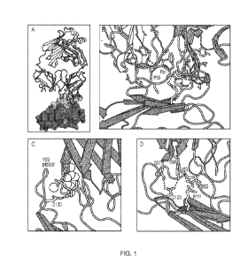
[0031] FIGURES 1A-D shows the crystal structure of human TIM-3 in complex with
M6903.
FIGURE 1A shows an overview of the Fab portion of M6903 (upper structure)
bound to TIM-3
shown as a surface representation. Extensive contacts made on TIM-3 (bottom
structure) are
shown as the lighter portion of TIM-3. FIGURE 1B shows the epitope hotspot
residues of TIM-
3 (e.g., P59 and F61 and E62). FIGURE 1C shows the polar head group of ptdSer
(light-
colored sticks) and the coordinating calcium ion (sphere) have been modeled
into the structure of
M6903-bound TIM-3 by superposition with the structure of murine TIM-3
(DeKruyff et al.
(2010), supra). The binding site of ptdSer coincides with the placement of Y59
(group of
spheres) of the heavy chain from M6903. Hydrogen bonds from D120 on TIM-3 to
ptdSer or
7
CA 03117371 2021-04-21
WO 2020/093024
PCT/US2019/059556
M6903, respectively, are shown as dotted lines. FIGURE 1D shows the polar
interactions of
M6903 with the CEACAM-1 binding residues of TIM-3 are shown with dashed lines.
[0032] FIGURE 2 depicts a model of the crystal structure of TIM-3 with an anti-
TIM-3
antibody 3903E11 (VL1.3,VH1.2) epitope map showing the P59, F61, E62, I114,
N119, and
K122 residues which reside on the face of one beta sheet of the immunoglobulin
fold.
[0033] FIGURE 3 provides a graph showing that target occupancy of anti-TIM-3
antibody
M6903 on CD14+ monocytes increased with increased concentrations of anti-TIM-
3. Serial
dilutions of anti-TIM-3 antibody 3903E11 (VL1.3,VH1.2) IgG2h (FN-AQ,322A)-delK
(M6903)
were incubated with fresh human whole blood for 1 hour. The unoccupied TIM-3
on CD14+
cells was measured by flow cytometry with anti-TIM-3 (2E2)-APC, which competes
with the
anti-TIM-3 antibody for TIM-3 binding. The average EC50 across all 10 donors
was 111.1
85.6 ng/ml. The graph shows 4 representative donors (KP46233, KP46231,
KP46315, and
KP46318) out of the 10 total donors.
[0034] FIGURE 4 provides a graph showing that M6903 efficiently blocked the
interaction of
rhTIM-3 and PtdSer on apoptotic Jurkat cells. Prior to flow cytometry
analysis, apoptosis was
induced in Jurkat cells via treatment with Staurosporine (2 [ig/mL, 18 hrs),
leading to surface
expression of a TIM-3 ligand, PtdSer. Binding of rhTIM-3-Fc PtdSer on the
surface of apoptotic
Jurkat cells was evaluated via flow cytometry by measuring the MFI of rhTIM-3-
Fc after pre-
incubation with serial dilutions of M6903 or an anti-HEL IgG2h isotype
control. While the
isotype control had no effect, M6903 blocked the interaction of rhTIM-3 and
PtdSer with an ICso
of 4.438 3.115 nM (0.666 0.467 g/ml). A nonlinear fit line was applied to
the graph using a
Sigmoid dose-response equation.
[0035] FIGURES 5A and 5B depict graphs showing M6903 increased CEF antigen
specific T
cell activation in a dose-dependent manner. The combination of M6903 and
bintrafusp further
enhanced this activation. PBMCs were treated with 40 ug/m1 CEF viral peptide
pool for (A) 6
days or (B) 4 days in the presence of M6903. In FIG. 5A, M6903 dose-
dependently enhanced T
cell activation compared to isotype control in a CEF assay as measured by IFN-
y production,
with an EC50 of 1 1.3 ug/mL, calculated from multiple experiments. Non-
linear regression
analysis was performed and mean and SD are presented. In FIG. 5B, serial
dilutions of M6903
were combined with either 10 ug/mL isotype control or bintrafusp alfa. The
combination with
bintrafusp alfa led to a further increase in IFN-y production. Mean and SD are
presented
(p<0.05).
8
CA 03117371 2021-04-21
WO 2020/093024
PCT/US2019/059556
[0036] FIGURES 6A and 6B provide graphs showing M6903 dose-dependently
enhancement
of allo-antigen specific T cell activation. T cell activation was evaluated in
an allogenic one-
way MLR assay by measuring IFN-y in the supernatant of co-cultured irradiated
Daudi cells and
human T cells after 2 days of treatment. In FIG. 6A, co-cultured cells were
treated with serial
dilutions of M6903 or isotype control. M6903 dose-dependently enhanced allo-
antigen specific
T cell activation, with an EC50 of 116 117 ng/mL. In FIG. 6B, co-cultured
cells were treated
with serial dilutions of M6903 combined with 10 pg/mL of isotype control or
bintrafusp alfa.
The combination of M6903 with bintrafusp alfa further enhanced T cell
activation. Nonlinear
regression analysis was performed and mean SD are presented for both graphs.
[0037] FIGURE 7 provides a graph demonstrating that M6903 exhibits enhanced
activity in
combination with bintrafusp in a superantigen SEB assay. Human PBMCs were
treated with
100 ng/mL SEB along with 10 mg/mL M6903 (or isotype control) either alone or
in combination
with bintrafusp alfa for 9 days. Cells were then washed once with medium and
re-stimulated
with SEB and the same antibodies for another 2 days. Supernatants were
harvested and IFN-y
was measured by IFN-y ELISA. M6903 and bintrafusp alfa both increased IFN-y
production in
SEB-stimulated T cells, and the effect was enhanced by combining M6903 with
bintrafusp alfa.
[0038] FIGURE 8 depicts the results of a CEF antigen-specific T cell assay
using M6903, anti-
PdtSer, and anti-Ga19. PBMCs were treated with 40 pg/m1 CEF viral peptide pool
for 5 days in
the presence of the antibody or antibodies indicated. The combination of anti-
Gal-9 and anti-
PtdSer had similar activity as M6903 alone, suggesting that blocking both Gal-
9 and PtdSer may
be required for anti-TIM-3 activity (compare data outlined by boxes).
[0039] FIGURES 9A-9B depict a quantitative analysis of TIM-3 expression
measured via IHC
in 12 tumor TMAs stained with anti-TIM-3 antibody. In FIG. 9A, the plot is
ordered by median
expression and in FIG. 9B, the plot is ordered by average expression following
the removal of
outliers.
[0040] FIGURE 10 depicts mIF staining of 8 tumor tissues to identify immune
cells expressing
TIM-3 in the tumor microenvironment (TME). CD3 and CD68 were used as markers
for
lymphocytes and macrophages, respectively. The percentage of TIM-3 CD3+
lymphocytes and
TIM-3 CD68+ macrophages was quantified across the tumor TMAs using mIF
analysis.
[0041] FIGURE 11 depicts TIM-3 expression in an NSCLC cohort using flow
cytometry
analysis. Within live CD3+ cells, expression of TIM-3 was observed to be
highest on CD8+ T
cells, followed by CD4+ T cells and Tregs. Each dot represents an individual
sample. Lines
represent the median value for each immune subset.
9
CA 03117371 2021-04-21
WO 2020/093024
PCT/US2019/059556
[0042] FIGURES 12A-B demonstrate that M6903 and bintrafusp, as monotherapies
or
combination, decreased MC38 tumor volume in B-huTIM-3 KI mice. B-huTIM-3 KI
mice were
inoculated with MC38 (1x10⁶ cells) s.c. in the flank and then treated with
isotype control (20
mg/kg), M6903 (10 mg/kg), bintrafusp alfa (24 mg/kg) or M6903 + bintrafusp
alfa. FIG. 12A
shows average tumor volumes with SEM and FIG. 12B shows individual tumor
volumes.
[0043] FIGURE 13 shows a dose escalation scheme in which, following a 28 day
screening
period, the subject is administered the M6903 escalation dose by IV infusion
every two weeks.
The two-week M6903 monotherapy lead-in period is followed by administration of
the M6903
escalation dose in combination with 1200 mg of bintrafusp alfa ("BFA") by IV
infusion every
two weeks.
DETAILED DESCRIPTION
[0044] The anti-TIM-3 antibodies disclosed herein are based on the antigen
binding sites of
certain monoclonal antibodies that have been selected on the basis of binding
and neutralizing
the activity of human T Cell Immunoglobulin and Mucin Domain-3 (TIM-3). The
antibodies
contain immunoglobulin variable region CDR sequences that define a binding
site for TIM-3.
[0045] In view of the neutralizing activity of these antibodies, they are
useful for inhibiting the
growth and/or proliferation of certain types of cancer cells. When used as a
therapeutic agent,
the antibodies can be optimized, e.g., affinity-matured, to improve
biochemical properties and/or
biophysical properties, and/or to reduce or eliminate immunogenicity when
administered to a
human patient. Various features and aspects of the invention are discussed in
more detail below.
[0001] As used herein, unless otherwise indicated, the term "antibody" means
an intact antibody
(e.g., an intact monoclonal antibody) or antigen-binding fragment of an
antibody, including an
intact antibody or antigen-binding fragment of an antibody (e.g., a phage
display antibody
including a fully human antibody, a semisynthetic antibody or a fully
synthetic antibody) that
has been optimized, engineered or chemically conjugated. Examples of
antibodies that have
been optimized are affinity-matured antibodies. Examples of antibodies that
have been
engineered are Fc optimized antibodies, antibody fusion proteins and
multispecific antibodies
(e.g., bispecific antibodies). Examples of antigen-binding fragments include
Fab, Fab', F(ab')2,
Fv, single chain antibodies (e.g., scFv), minibodies and diabodies. An
antibody conjugated to a
toxin moiety is an example of a chemically conjugated antibody. Antibody
fusion proteins
include, for example, an antibody genetically fused to a soluble ligand such
as a cytokine, or to
an extracellular domain of a cellular receptor protein.
I. Antibodies That Bind Human TIM-3
CA 03117371 2021-04-21
WO 2020/093024
PCT/US2019/059556
[0046] The antibodies disclosed herein comprise: (a) an immunoglobulin heavy
chain variable
region comprising a CDRHI, a CDRH2, and a CDRH3 and (b) an immunoglobulin
light chain
variable region comprising a CDRLI, a CDRL2, and a CDRL3, wherein the heavy
chain variable
region and the light chain variable region together define a single binding
site for binding TIM-3
protein.
[0047] In some embodiments, the antibody comprises: (a) an immunoglobulin
heavy chain
variable region comprising a CDRHI, a CDRH2, and a CDRH3 and (b) an
immunoglobulin light
chain variable region, wherein the heavy chain variable region and the light
chain variable region
together define a single binding site for binding TIM-3. A CDRHI comprises the
amino acid
sequence of SEQ ID NO: 1; a CDRH2 comprises the amino acid sequence of SEQ ID
NO: 2; and
a CDRH3 comprises the amino acid sequence of SEQ ID NO: 3. The CDRHi, CDRH2,
and
CDRH3 sequences are interposed between immunoglobulin FR sequences (SEQ ID NO:
7, SEQ
ID NO:8, SEQ ID NO: 9, and SEQ ID NO:10).
[0048] In some embodiments, the antibody comprises (a) an immunoglobulin light
chain
variable region comprising a CDRLI, a CDRL2, and a CDRL3, and (b) an
immunoglobulin heavy
chain variable region, wherein the IgG light chain variable region and the IgG
heavy chain
variable region together define a single binding site for binding TIM-3. A
CDRLI comprises the
amino acid sequence of SEQ ID NO: 4; a CDRL2 comprises the amino acid sequence
of SEQ ID
NO: 5; and a CDRL3 comprises the amino acid sequence of SEQ ID NO: 6. The
CDRLI, CDRL2,
and CDRL3 sequences are interposed between immunoglobulin FR sequences (SEQ ID
NO: 11,
SEQ ID NO: 12, SEQ ID NO: 13, and SEQ ID NO: 14).
[0049] In some embodiments, the antibody comprises: (a) an immunoglobulin
heavy chain
variable region comprising a CDRHI, a CDRH2, and a CDRH3 and (b) an
immunoglobulin light
chain variable region comprising a CDRLI, a CDRL2, and a CDRL3, wherein the
heavy chain
variable region and the light chain variable region together define a single
binding site for
binding TIM-3. The CDRHI is the amino acid sequence of SEQ ID NO: 1; the CDRH2
is the
amino acid sequence of SEQ ID NO: 2; and the CDRH3 is the amino acid sequence
of SEQ ID
NO: 3. The CDRLI is the amino acid sequence of SEQ ID NO: 4; the CDRL2 is the
amino acid
sequence of SEQ ID NO: 5; and the CDRL3 is the amino acid sequence of SEQ ID
NO: 6.
[0050] In other embodiments, the antibodies disclosed herein comprise an
immunoglobulin
heavy chain variable region and an immunoglobulin light chain variable region.
In some
embodiments, the antibody comprises an immunoglobulin heavy chain variable
region
comprising an amino acid sequence selected from the group consisting of SEQ ID
NO: 53, SEQ
11
CA 03117371 2021-04-21
WO 2020/093024
PCT/US2019/059556
ID NO: 24, SEQ ID NO: 55, and SEQ ID NO: 34; and an immunoglobulin light chain
variable
region.
[0051] In other embodiments, the antibody comprises an immunoglobulin light
chain variable
region selected from the group consisting of SEQ ID NO: 52, SEQ ID NO: 54, SEQ
ID NO: 23
and SEQ ID NO: 33; and an immunoglobulin heavy chain variable region.
[0052] In some embodiments, the antibody comprises an immunoglobulin heavy
chain variable
region comprising an amino acid sequence selected from the group consisting of
SEQ ID NO:
53, SEQ ID NO: 24, SEQ ID NO: 55, and SEQ ID NO: 34; and an immunoglobulin
light chain
variable region selected from the group consisting of SEQ ID NO: 52, SEQ ID
NO: 54, SEQ ID
NO: 23 and SEQ ID NO: 33.
[0053] In some embodiments, the antibody comprises an immunoglobulin heavy
chain variable
region comprising the amino acid sequence of SEQ ID NO: 24, and an
immunoglobulin light
chain variable region comprising the amino acid sequence of SEQ ID NO: 23.
[0054] In certain embodiments, the antibodies disclosed herein comprise an
immunoglobulin
heavy chain and an immunoglobulin light chain. In some embodiments, the
antibody comprises
an immunoglobulin heavy chain selected from the group consisting of SEQ ID NO:
16, SEQ ID
NO: 18, SEQ ID NO: 20, SEQ ID NO: 22, and SEQ ID NO: 32; and an immunoglobulin
light
chain.
[0055] In other embodiments, the antibody comprises an immunoglobulin light
chain selected
from the group consisting of SEQ ID NO: 15, SEQ ID NO: 17, SEQ ID NO: 19, SEQ
ID NO:
21, and SEQ ID NO: 31; and an immunoglobulin heavy chain.
[0056] In some embodiments, the antibody comprises (i) an immunoglobulin heavy
chain
comprising an amino acid sequence selected from the group consisting of SEQ ID
NO: 16, SEQ
ID NO: 18, SEQ ID NO: 20, SEQ ID NO: 22, and SEQ ID NO: 32; and (ii) an
immunoglobulin
light chain selected from the group consisting of SEQ ID NO: 15, SEQ ID NO:
17, SEQ ID NO:
19, SEQ ID NO: 21, and SEQ ID NO: 31.
[0057] In some embodiments, the antibody comprises an immunoglobulin heavy
chain
comprising the amino acid sequence of SEQ ID NO: 22 and an immunoglobulin
light chain
comprising the amino acid sequence of SEQ ID NO: 21.
[0058] In certain embodiments, an isolated antibody that binds TIM-3 comprises
an
immunoglobulin heavy chain variable region comprising an amino acid sequence
that is at least
70%, 75%, 80%, 85%, 90%, 95%, 98%, or 99% identical to the entire variable
region or the
12
CA 03117371 2021-04-21
WO 2020/093024
PCT/US2019/059556
framework region sequence of SEQ ID NO: 16, SEQ ID NO: 18, SEQ ID NO: 20, SEQ
ID NO:
22, or SEQ ID NO: 32. In certain embodiments, an isolated antibody that binds
TIM-3
comprises an immunoglobulin heavy chain variable region comprising a CDRFH
comprising the
amino acid sequence of SEQ ID NO: 1; a CDRH2 comprising the amino acid
sequence of SEQ
ID NO: 2; and a CDRH3 comprising the amino acid sequence of SEQ ID NO: 3; and
an amino
acid sequence that is at least 70%, 75%, 80%, 85%, 90%, 95%, 98%, or 99%
identical to the
entire variable region or the framework region sequence of SEQ ID NO: 16, SEQ
ID NO: 18,
SEQ ID NO: 20, SEQ ID NO: 22, or SEQ ID NO: 32.
[0059] In certain embodiments, an isolated antibody that binds TIM-3 comprises
an
immunoglobulin light chain variable region comprising an amino acid sequence
that is at least
70%, 75%, 80%, 85%, 90%, 95%, 98%, or 99% identical to the entire variable
region or the
framework region sequence of SEQ ID NO: 15, SEQ ID NO: 17, SEQ ID NO: 19, SEQ
ID NO:
21, or SEQ ID NO: 31. In certain embodiments, an isolated antibody that binds
TIM-3
comprises an immunoglobulin light chain variable region comprising a CDRLI
comprising the
amino acid sequence of SEQ ID NO: 4; a CDRL2 comprising the amino acid
sequence of SEQ
ID NO: 5; and a CDRL3 comprising the amino acid sequence of SEQ ID NO: 6; and
an amino
acid sequence that is at least 70%, 75%, 80%, 85%, 90%, 95%, 98%, or 99%
identical to the
entire variable region or the framework region sequence of SEQ ID NO: 15, SEQ
ID NO: 17,
SEQ ID NO: 19, SEQ ID NO: 21, or SEQ ID NO: 31.
[0060] Sequence identity may be determined in various ways that are within the
skill in the art,
e.g., using publicly available computer software such as BLAST, BLAST-2, ALIGN
or
Megalign (DNASTAR) software. BLAST (Basic Local Alignment Search Tool)
analysis using
the algorithm employed by the programs blastp, blastn, blastx, tblastn and
tblastx (Karlin et al.,
(1990) PROC. NATL. ACAD. So. USA 87:2264-2268; Altschul, (1993) J. MoL. EvoL.
36, 290-
300; Altschul etal., (1997) NUCLEIC ACIDS RES. 25:3389-3402, incorporated by
reference) are
tailored for sequence similarity searching. For a discussion of basic issues
in searching sequence
databases see Altschul etal., (1994) NATURE GENETICS 6:119-129, which is fully
incorporated
by reference. Those skilled in the art can determine appropriate parameters
for measuring
alignment, including any algorithms needed to achieve maximal alignment over
the full length of
the sequences being compared. The search parameters for histogram,
descriptions, alignments,
expect (i.e., the statistical significance threshold for reporting matches
against database
sequences), cutoff, matrix and filter are at the default settings. The default
scoring matrix used
by blastp, blastx, tblastn, and tblastx is the BLOSUM62 matrix (Henikoff et
al., (1992) PROC.
NATL. ACAD. So. USA 89:10915-10919, fully incorporated by reference). Four
blastn
13
CA 03117371 2021-04-21
WO 2020/093024
PCT/US2019/059556
parameters may be adjusted as follows: Q=10 (gap creation penalty); R=10 (gap
extension
penalty); wink=1 (generates word hits at every winkth position along the
query); and
gapw=16 (sets the window width within which gapped alignments are generated).
The
equivalent Blastp parameter settings may be Q=9; R=2; wink=1; and gapw=32.
Searches may
also be conducted using the NCBI (National Center for Biotechnology
Information) BLAST
Advanced Option parameter (e.g.: -G, Cost to open gap [Integer]: default = 5
for nucleotides/ 11
for proteins; -E, Cost to extend gap [Integer]: default = 2 for nucleotides/ 1
for proteins; -q,
Penalty for nucleotide mismatch [Integer]: default = -3; -r, reward for
nucleotide match
[Integer]: default = 1; -e, expect value [Real]: default = 10; -W, wordsize
[Integer]: default = 11
for nucleotides/ 28 for megablast/ 3 for proteins; -y, Dropoff (X) for blast
extensions in bits:
default = 20 for blastn/ 7 for others; -X, X dropoff value for gapped
alignment (in bits): default =
15 for all programs, not applicable to blastn; and ¨Z, final X dropoff value
for gapped alignment
(in bits): 50 for blastn, 25 for others). ClustalW for pairwise protein
alignments may also be
used (default parameters may include, e.g., Blosum62 matrix and Gap Opening
Penalty = 10 and
Gap Extension Penalty = 0.1). A Bestfit comparison between sequences,
available in the GCG
package version 10.0, uses DNA parameters GAP=50 (gap creation penalty) and
LEN=3 (gap
extension penalty) and the equivalent settings in protein comparisons are
GAP=8 and LEN=2.
[0061] In each of the foregoing embodiments, it is contemplated herein that
immunoglobulin
heavy chain variable region sequences and/or light chain variable region
sequences that together
bind TIM-3 may contain amino acid alterations (e.g., at least 1, 2, 3, 4, 5,
or 10 amino acid
substitutions, deletions, or additions) in the framework regions of the heavy
and/or light chain
variable regions. In certain embodiments, the amino acid alterations are
conservative
substitutions. As used herein, the term "conservative substitution" refers to
a substitution with a
structurally similar amino acid. For example, conservative substitutions may
include those
within the following groups: Ser and Cys; Leu, Ile, and Val; Glu and Asp; Lys
and Arg; Phe,
Tyr, and Trp; and Gln, Asn, Glu, Asp, and His. Conservative substitutions may
also be defined
by the BLAST (Basic Local Alignment Search Tool) algorithm, the BLOSUM
substitution
matrix (e.g., BLOSUM 62 matrix), or the PAM substitution:p matrix (e.g., the
PAM 250 matrix).
[0062] In certain embodiments, the antibody binds TIM-3 with a KD of 20 nM, 15
nM, 10 nM, 9
nM, 8 nM, 7 nM, 6 nM, 5 nM, 4 nM, 3 nM, 2 nM, 1 nM or lower. Unless otherwise
specified,
KD values are determined by surface plasmon resonance. For example, surface
plasmon
resonance can be measured using a GE Healthcare Biacore 4000 instrument as
follows. Goat
anti-human Fc antibody (Jackson Immunoresearch Laboratories # 109-005-098) is
immobilized
on BIAcore carboxymethylated dextran CMS chip using direct coupling to free
amino groups
14
CA 03117371 2021-04-21
WO 2020/093024
PCT/US2019/059556
following the procedure described by the manufacturer. Antibodies are captured
on the CM5
biosensor chip to achieve approximately 200 response units (RU). Binding
measurements are
performed using the running HBS-EP+ buffer. A 2-fold dilution series starting
at 100 nM of
anti-TIM-3 antibodies are injected at a flow rate of 30 ul/min at 25 C.
Association rates (kon,
M-ls-1) and dissociation rates (koff, s-1) are calculated using a simple 1:1
Langmuir binding
model (Biacore 4000 Evaluation Software). The equilibrium dissociation
constant (KD, M) is
calculated as the ratio of koff / kon.
[0063] In some embodiments, monoclonal antibodies bind to the same epitope on
TIM-3 as any
of the anti-TIM-3 antibodies disclosed herein (e.g., M6903). In some
embodiments, monoclonal
antibodies compete for binding to TIM-3 with any of the anti-TIM-3 antibodies
disclosed herein.
For example, monoclonal antibodies may compete for binding to the galectin-9
binding domain
of TIM-3 with an anti-TIM-3 antibody described herein. In another example,
monoclonal
antibodies may compete for binding to the PtdSer binding domain of TIM-3 with
an anti-TIM-3
antibody described herein. In another example, monoclonal antibodies may
compete for binding
to the CEACAM1 binding domain of TIM-3 with an anti-TIM-3 antibody described
herein. In a
further example, monoclonal antibodies may compete for binding to the galectin-
9 binding
domain and the PtdSer binding domain of TIM-3 with an anti-TIM-3 antibody
described herein.
In another example, monoclonal antibodies may compete for binding to the
galectin-9 binding
domain and the CEACAM1 binding domain of TIM-3 with an anti-TIM-3 antibody
described
herein. In another example, monoclonal antibodies may compete for binding to
the PtdSer
binding domain and the CEACAM1 binding domain of TIM-3 with an anti-TIM-3
antibody
described herein. In another example, monoclonal antibodies may compete for
binding to the
galectin-9 binding domain, the PtdSer binding domain, and the CEACAM1 binding
domain of
TIM-3 with an anti-TIM-3 antibody described herein.
[0064] Competition assays for determining whether an antibody binds to the
same epitope as an
anti-TIM-3 antibody described herein, or competes for binding with galectin-9,
PtdSer, and/or
CEACAM1 with an anti-TIM-3 antibody described herein are known in the art.
Exemplary
competition assays include immunoassays (e.g., ELISA assays, RIA assays),
BIAcore analysis,
biolayer interferometry and flow cytometry.
[0065] Typically, a competition assay involves the use of an antigen (e.g., a
TIM-3 protein or
fragment thereof) bound to a solid surface or expressed on a cell surface, a
test TIM-3-binding
antibody and a reference antibody (e.g., antibody M6903). The reference
antibody is labeled and
the test antibody is unlabeled. Competitive inhibition is measured by
determining the amount of
labeled reference antibody bound to the solid surface or cells in the presence
of the test antibody.
CA 03117371 2021-04-21
WO 2020/093024
PCT/US2019/059556
Usually the test antibody is present in excess (e.g., lx, 5x, 10x, 20x or
100x). Antibodies
identified by competition assay (i.e., competing antibodies) include
antibodies binding to the
same epitope, or similar (e.g., overlapping) epitopes, as the reference
antibody, and antibodies
binding to an adjacent epitope sufficiently proximal to the epitope bound by
the reference
antibody for steric hindrance to occur.
[0066] In an exemplary competition assay, a reference TIM-3 antibody (e.g.,
antibody M6903)
is biotinylated using commercially available reagents. The biotinylated
reference antibody is
mixed with serial dilutions of the test antibody or unlabeled reference
antibody (self-competition
control) resulting in a mixture of various molar ratios (e.g., lx, 5x, 10x,
20x or 100x) of test
antibody (or unlabeled reference antibody) to labeled reference antibody. The
antibody mixture
is added to a TIM-3 (e.g., TIM-3 extracellular domain) polypeptide coated-
ELISA plate. The
plate is then washed and HRP (horseradish peroxidase)-strepavidin is added to
the plate as the
detection reagent. The amount of labeled reference antibody bound to the
target antigen is
detected following addition of a chromogenic substrate (e.g., TMB (3,3',5,5'-
tetramethylbenzidine) or ABTS (2,2"-azino-di-(3-ethylbenzthiazoline-6-
sulfonate)), which are
well-known in the art. Optical density readings (OD units) are measured using
a SpectraMax
M2 spectrometer (Molecular Devices). OD units corresponding to zero percent
inhibition are
determined from wells without any competing antibody. OD units corresponding
to 100%
inhibition, i.e., the assay background are determined from wells without any
labeled reference
antibody or test antibody. Percent inhibition of labeled reference antibody to
TIM-3 by the test
antibody (or the unlabeled reference antibody) at each concentration is
calculated as follows: %
inhibition = (1-(OD units ¨ 100% inhibition)/(0% inhibition ¨ 100%
inhibition))*100. Persons
skilled in the art will appreciate that the competition assay can be performed
using various
detection systems well-known in the art.
[0067] A competition assay may be conducted in both directions to ensure that
the presence of
the label does not interfere or otherwise inhibit binding. For example, in the
first direction the
reference antibody is labeled and the test antibody is unlabeled, and in the
second direction, the
test antibody is labeled and the reference antibody is unlabeled.
[0068] A test antibody competes with the reference antibody for specific
binding to the antigen
if an excess of one antibody (e.g., lx, 5x, 10x, 20x or 100x) inhibits binding
of the other
antibody, e.g., by at least 50%, 75%, 90%, 95% or 99% as measured in a
competitive binding
assay.
16
CA 03117371 2021-04-21
WO 2020/093024
PCT/US2019/059556
[0069] Two antibodies may be determined to bind to the same epitope if
essentially all amino
acid mutations in the antigen that reduce or eliminate binding of one antibody
reduce or
eliminate binding of the other. Two antibodies may be determined to bind to
overlapping
epitopes if only a subset of the amino acid mutations that reduce or eliminate
binding of one
antibody reduce or eliminate binding of the other.
II. Anti-PD-Ll/TGFR Trap Fusion Proteins
[0070] The anti-TIM-3 antibodies described herein can be administered in
combination with any
anti-PD-Ll/TGFI3 Trap known in the art. "Anti-PD-Ll/TGFP Trap" refers to a
fusion molecule
comprising 1) an antibody or antigen-binding fragment thereof that is capable
of binding PD-Li
and antagonizing the interaction between PD-1 and PD-Li and 2) a TGFORII or
fragment of
TGFORII that is capable of binding TGFP and antagonizing the interaction
between TGFP and
TGFORII.
[0071] In one embodiment, the anti-PD-Ll/TGFP Trap comprises an anti-PD-Li
antibody
known in the art. Anti-PD-Li antibodies are commercially available, for
example, the 29E2A3
antibody (Biolegend, Cat. No. 329701). Antibodies can be monoclonal, chimeric,
humanized, or
human. Antibody fragments include Fab, F(ab')2, scFv and Fv fragments, which
are described in
further detail below.
[0072] Exemplary anti-PD-Li antibodies are described in PCT Publication WO
2013/079174,
which describes avelumab. These antibodies can include a heavy chain variable
region
polypeptide including a CDRHI, CDRH2, and CDRH3 sequence, where:
(a) the CDRHI sequence is XIYX2MX3(SEQ ID NO: 58);
(b) the CDRH2 sequence is SIYPSGGX4TFYADX5VKG (SEQ ID NO: 59);
(c) the CDRH3 sequence is IKLGTVTTVX6Y (SEQ ID NO: 60);
further where: Xi is K, R, T, Q, G, A, W, M, I, or S; X2 is V, R, K, L, M, or
I; X3 is H, T, N, Q,
A, V, Y, W, F, or M; X4 is F or I; X5 is S or T; X6 is E or D.
[0073] In a one embodiment, Xi is M, I, or S; X2 is R, K, L, M, or I; X3 is F
or M; X4 is F or I;
X5 is S or T; X6 is E or D.
[0074] In another embodiment Xi is M, I, or S; X2 is L, M, or I; X3 is F or M;
X4 is I; X5 is S or
T; X6 is D.
17
CA 03117371 2021-04-21
WO 2020/093024
PCT/US2019/059556
[0075] In still another embodiment, Xi is S; X2 is I; X3 is M; X4 is I; X5 is
T; X6 is D.
[0076] In another aspect, the polypeptide further includes variable region
heavy chain
framework (FR) sequences juxtaposed between the CDRs according to the formula:
(HC-FR1)-
(CDRHI)-(HC-FR2)-(CDRH2)-(HC-FR3)-(CDRH3)-(HC-FR4).
[0077] In yet another aspect, the framework sequences are derived from human
consensus
framework sequences or human germline framework sequences.
[0078] In a still further aspect, at least one of the framework sequences is
the following:
HC-FR1 is EVQLLESGGGLVQPGGSLRLSCAASGFTFS (SEQ ID NO: 61);
HC-FR2 is WVRQAPGKGLEWVS (SEQ ID NO: 62);
HC-FR3 is RFTISRDNSKNTLYLQMNSLRAEDTAVYYCAR (SEQ ID NO: 63);
HC-FR4 is WGQGTLVTVSS (SEQ ID NO: 64).
[0079] In a still further aspect, the heavy chain polypeptide is further
combined with a variable
region light chain including a CDRLI, CDRL2, and CDRL3, where:
(a) the CDRLI sequence is TGTX7X8DVGX9YNYVS (SEQ ID NO: 65);
(b) the CDRL2sequence is XioVXIIX12RPS (SEQ ID NO: 66);
(c) the CDRL3 sequence is SSX13TX14X15X16X17RV (SEQ ID NO: 67);
further where: X7 is N or S; Xs is T, R, or S; X9 is A or G; Xio is E or D;
Xii is I, N or S; X12 is
D, H or N; X13 is F or Y; X14 is N or S; X15 is R, T or S; X16 is G or S; X17
is I or T.
[0080] In another embodiment, X7 is N or S; Xs is T, R, or S; X9 is A or G;
Xio is E or D; Xii is
N or S; X12 is N; X13 is F or Y; X14 is 5; X15 is 5; X16 is G or S; X17 is T.
[0081] In still another embodiment, X7 is S; Xs is S; X9 is G; Xio is D; Xii
is S; X12 is N; X13 is
Y; X14 is S; Xis is S; X16 is S; X17 is T.
[0082] In a still further aspect, the light chain further includes variable
region light chain
framework sequences juxtaposed between the CDRs according to the formula: (LC-
CDRLI)-
(LC-FR2)-(CDRL2)-(LC-FR3)-(CDRL3)-(LC-FR4).
18
CA 03117371 2021-04-21
WO 2020/093024
PCT/US2019/059556
[0083] In a still further aspect, the light chain framework sequences are
derived from human
consensus framework sequences or human germline framework sequences.
[0084] In a still further aspect, the light chain framework sequences are
lambda light chain
sequences.
[0085] In a still further aspect, at least one of the framework sequence is
the following:
LC-FR1 is QSALTQPASVSGSPGQSITISC (SEQ ID NO: 68);
LC-FR2 is WYQQHPGKAPKLMIY (SEQ ID NO: 69);
LC-FR3 is GVSNRFSGSKSGNTASLTISGLQAEDEADYYC (SEQ ID NO: 70);
LC-FR4 is FGTGTKVTVL (SEQ ID NO: 71).
[0086] In another embodiment, the invention provides an anti-PD-Li antibody or
antigen
binding fragment including a heavy chain and a light chain variable region
sequence, where:
(a) the heavy chain includes a CDRui, CDRH2, and CDR143, wherein further: (i)
the
CDRui sequence is XIYX2MX3(SEQ ID NO: 72); (ii) the CDRH2 sequence is
SIYPSGGX4TFYADX5VKG (SEQ ID NO: 73); (iii) the CDR-13 sequence is IKLGTVTTVX6Y
(SEQ ID NO: 74), and;
(b) the light chain includes a CDRLI, CDRL2, and CDRL3, wherein further: (iv)
the
CDRLI sequence is TGTX7X8DVGX9YNYVS (SEQ ID NO: 75); (v) the CDRL2 sequence is
XioVX1IX12RPS (SEQ ID NO: 76); (vi) the CDRL3 sequence is SSX13TX14X15X16X17RV
(SEQ
ID NO: 77); wherein: Xi is K, R, T, Q, G, A, W, M, I, or S; X2 is V, R, K, L,
M, or I; X3 is H, T,
N,Q,A,V,Y,W,F,orM;X4isForI;X5isSorT;X6isEorD;X7isNorS;XsisT,R,orS;
X9 is A or G; Xio is E or D; Xii is I, N, or S; X12 is D, H, or N; X13 is F or
Y; X14 is N or S; X15
is R, T, or S; X16 is G or S; X17 is I or T.
[0087] In one embodiment, Xi is M, I, or S; X2 is R, K, L, M, or I; X3 is F or
M; X4 is F or I; X5
is S or T; X6 is E or D; X7 is N or S; X8 is T, R, or S; X9 is A or G; Xio is
E or D; N or S;
Xi2 is N; X13 is F or Y; X14 is S; Xi5 is S; X16 is G or S; Xi7 is T.
[0088] In another embodiment, Xi is M, I, or S; X2 is L, M, or I; X3 is F or
M; X4 is I; X5 is S or
T; X6 is D; X7 is N or S; X8 is T, R, or S; X9 is A or G; Xio is E or D; Xii
is N or S; Xi2 is N; X13
is F or Y; X14 is S; Xi5 is S; X16 is G or S; X17 is T.
19
CA 03117371 2021-04-21
WO 2020/093024
PCT/US2019/059556
[0089] In still another embodiment, Xi is S; X2 is I; X3 is M; X4 is I; X5 is
T; X6 is D; X7 is S;
X8 is S; X9 is G; Xio is D; Xii is S; X12 is N; X13 is Y; X14 is S; X15 is S;
X16 is S; X17 is T.
[0090] In a further aspect, the heavy chain variable region includes one or
more framework
sequences juxtaposed between the CDRs as: (HC-FR1)-(CDRH1)-(HC-FR2)-(CDRH2)-
(HC-
FR3)-(CDRH3)-(HC-FR4), and the light chain variable regions include one or
more framework
sequences juxtaposed between the CDRs as: (LC-FR1 MCDRLI)-(LC-FR2)-(CDRL2)-(LC-
FR3)-
(CDRL3)-(LC-FR4).
[0091] In a still further aspect, the framework sequences are derived from
human consensus
framework sequences or human germline sequences.
[0092] In a still further aspect, one or more of the heavy chain framework
sequences is the
following:
HC-FR1 is EVQLLESGGGLVQPGGSLRLSCAASGFTFS (SEQ ID NO: 61);
HC-FR2 is WVRQAPGKGLEWVS (SEQ ID NO: 62);
HC-FR3 is RFTISRDNSKNTLYLQMNSLRAEDTAVYYCAR (SEQ ID NO: 63);
HC-FR4 is WGQGTLVTVSS (SEQ ID NO: 64).
[0093] In a still further aspect, the light chain framework sequences are
lambda light chain
sequences.
[0094] In a still further aspect, one or more of the light chain framework
sequences is the
following:
LC-FR1 is QSALTQPASVSGSPGQSITISC (SEQ ID NO: 68);
LC-FR2 is WYQQHPGKAPKLMIY (SEQ ID NO: 69);
LC-FR3 is GVSNRFSGSKSGNTASLTISGLQAEDEADYYC (SEQ ID NO: 70);
LC-FR4 is FGTGTKVTVL (SEQ ID NO: 71).
[0095] In a still further aspect, the heavy chain variable region polypeptide,
antibody, or
antibody fragment further includes at least a CH1 domain.
CA 03117371 2021-04-21
WO 2020/093024
PCT/US2019/059556
[0096] In a more specific aspect, the heavy chain variable region polypeptide,
antibody, or
antibody fragment further includes a CH1, a CH2, and a CH3 domain.
[0097] In a still further aspect, the variable region light chain, antibody,
or antibody fragment
further includes a CL domain.
[0098] In a still further aspect, the antibody further includes a CH 1, a CH2,
a CH3, and a CL
domain.
[0099] In a still further specific aspect, the antibody further includes a
human or murine constant
region.
[00100] In a still further aspect, the human constant region is selected
from the group
consisting of IgGl, IgG2, IgG2, IgG3, IgG4.
[00101] In a still further specific aspect, the human or murine constant
region is lgGl.
[00102] In yet another embodiment, the invention features an anti-PD-Li
antibody
including a heavy chain and a light chain variable region sequence, where:
(a) the heavy chain includes a CDRHI, a CDRH2, and a CDRH3, having at least
80%
overall sequence identity to SYIMM (SEQ ID NO: 78), SIYPSGGITFYADTVKG (SEQ ID
NO:
79), and IKLGTVTTVDY (SEQ ID NO: 80), respectively, and
(b) the light chain includes a CDRLI, a CDRL2, and a CDRL3, having at least
80% overall
sequence identity to TGTSSDVGGYNYVS (SEQ ID NO: 81), DVSNRPS (SEQ ID NO: 82),
and SSYTSSSTRV (SEQ ID NO: 83), respectively.
[00103] In a specific aspect, the sequence identity is 81%, 82%, 83%, 84%,
85%, 86%,
87%, 88%, 89%, 90%, 91%, 92%, 93%, 94%, 95%, 96%, 97%, 98%, 99%, or 100%.
[00104] In yet another embodiment, the invention features an anti-PD-Li
antibody
including a heavy chain and a light chain variable region sequence, where:
(a) the heavy chain includes a CDRHI, a CDRH2, and a CDRH3, having at least
80%
overall sequence identity to MYMMM (SEQ ID NO: 84), SIYPSGGITFYADSVKG (SEQ ID
NO: 85), and IKLGTVTTVDY (SEQ ID NO: 80), respectively, and
21
CA 03117371 2021-04-21
WO 2020/093024
PCT/US2019/059556
(b) the light chain includes a CDRLI, a CDRL2, and a CDRL3, having at least
80% overall
sequence identity to TGTSSDVGAYNYVS (SEQ ID NO: 86), DVSNRPS (SEQ ID NO: 82),
and SSYTSSSTRV (SEQ ID NO: 83), respectively.
[00105] In a specific aspect, the sequence identity is 81%, 82%, 83%, 84%,
85%, 86%,
87%, 88%, 89%, 90%, 91%, 92%, 93%, 94%, 95%, 96%, 97%, 98%, 99%, or 100%.
[00106] In a still further aspect, in the antibody or antibody fragment
according to the
invention, as compared to the sequences of CDRHI, CDRH2, and CDRH3, at least
those amino
acids remain unchanged that are highlighted by underlining as follows:
(a) in CDRHI SYIMM (SEQ ID NO: 78),
(b) in CDRH2SIYPSGGITFYADTVKG (SEQ ID NO: 79),
(c) in CDRH3IKLGTVTTVDY (SEQ ID NO: 80);
and further where, as compared to the sequences of CDRLI, CDRL2, and CDRL3 at
least
those amino acids remain unchanged that are highlighted by underlining as
follows:
(a) CDRLI TGTSSDVGGYNYVS (SEQ ID NO: 81)
(b) CDRL2DVSNRPS (SEQ ID NO: 82)
(c) CDRL3 SSYTSSSTRV (SEQ ID NO: 83).
[00107] In another aspect, the heavy chain variable region includes one or
more
framework sequences juxtaposed between the CDRs as: (HC-FR1)-(CDRH1)-(HC-FR2)-
(CDRH2)-(HC-FR3)-(CDRH3)-(HC-FR4), and the light chain variable regions
include one or
more framework sequences juxtaposed between the CDRs as: (LC-FR1)-(CDRLI)-(LC-
FR2)-
(CDRL2)-(LC-FR3)-(CDRL3)-(LC-FR4).
[00108] In yet another aspect, the framework sequences are derived from
human germline
sequences.
[00109] In a still further aspect, one or more of the heavy chain framework
sequences is
the following:
HC-FR1 is EVQLLESGGGLVQPGGSLRLSCAASGFTFS (SEQ ID NO: 61);
22
CA 03117371 2021-04-21
WO 2020/093024
PCT/US2019/059556
HC-FR2 is WVRQAPGKGLEWVS (SEQ ID NO: 62);
HC-FR3 is RFTISRDNSKNTLYLQMNSLRAEDTAVYYCAR (SEQ ID NO: 63);
HC-FR4 is WGQGTLVTVSS (SEQ ID NO: 64).
[00110] In a still further aspect, the light chain framework sequences are
derived from a
lambda light chain sequence.
[00111] In a still further aspect, one or more of the light chain framework
sequences is the
following:
LC-FR1 is QSALTQPASVSGSPGQSITISC (SEQ ID NO: 68);
LC-FR2 is WYQQHPGKAPKLMIY (SEQ ID NO: 69);
LC-FR3 is GVSNRFSGSKSGNTASLTISGLQAEDEADYYC (SEQ ID NO: 70);
LC-FR4 is FGTGTKVTVL (SEQ ID NO: 71).
[00112] In a still further specific aspect, the antibody further includes a
human or murine
constant region.
[00113] In a still further aspect, the human constant region is selected
from the group
consisting of IgGl, IgG2, IgG2, IgG3, IgG4.
[00114] In a still further embodiment, the invention features an anti-PD-Li
antibody
including a heavy chain and a light chain variable region sequence, where:
(a) the heavy chain sequence has at least 85% sequence identity to the heavy
chain
sequence:
EVQLLESGGGLVQPGGSLRLSCAASGFTFSSYIMMVWRQAPGKGLEWVSSIYPSGGITF
YADWKGRFTISRDNSKNTLYLQMNSLRAEDTAVYYCARIKLGTVTTVDYWGQGTLVT
VSS (SEQ ID NO: 87), and
(b) the light chain sequence has at least 85% sequence identity to the light
chain
sequence:
23
CA 03117371 2021-04-21
WO 2020/093024
PCT/US2019/059556
QSALTQPASVSGSPGQSITISCTGTSSDVGGYNYVSWYQQHPGKAPKLMIYDVSN
RPSGVSNRFSGSKSGNTASLTISGLQAEDEADYYCSSYTSSSTRVFGTGTKVTVL (SEQ
ID NO: 88).
[00115] In a specific aspect, the sequence identity is 86%, 87%, 88%, 89%,
90%, 91%,
92%, 93%, 94%, 95%, 96%, 97%, 98%, 99%, or 100%.
[00116] In a still further embodiment, the invention provides for an anti-
PD-Li antibody
including a heavy chain and a light chain variable region sequence, where:
(a) the heavy chain sequence has at least 85% sequence identity to the heavy
chain
sequence:
EVQLLESGGGLVQPGGSLRLSCAASGFTFSMYMMMWVRQAPGKGLEVWSSIYPSGGIT
FYADSVKGRFTISRDNSKNTLYLQMNSLRAEDTAIYYCARIKLGTVTTVDYWG
QGTLVTVSS (SEQ ID NO: 89), and
(b) the light chain sequence has at least 85% sequence identity to the light
chain
sequence:
QSALTQPASVSGSPGQSMSCTGTSSDVGAYNYVSWYQQHPGKAPKLMIYDVSNR
PSGVSNRFSGSKSGNTASLTISGLQAEDEADYYCSSYTSSSTRVFGTGTKVTVL (SEQ ID
NO: 90).
[00117] In a specific aspect, the sequence identity is 86%, 87%, 88%, 89%,
90%, 91%,
92%, 93%, 94%, 95%, 96%, 97%, 98%, 99% or 100%.
[00118] In a particular embodiment, anti-PD-Ll/TGFP Trap is one of the fusion
molecules
disclosed in WO 2015/118175 or WO 2018/205985. For instance, anti-PD-Ll/TGF13
Trap may
comprise the light chains and heavy chains of SEQ ID NO: 1 and SEQ ID NO: 3 of
WO
2015/118175, respectively. In another embodiment, anti-PD-Ll/TGFP Trap is one
of the
constructs listed in Table 2 of WO 2018/205985, such as construct 9 or 15
thereof In other
embodiments, the antibody having the heavy chain sequence of SEQ ID NO: 11 and
the light
chain sequence of SEQ ID NO: 12 of WO 2018/205985 is fused via a linking
sequence
(G45)xG, wherein x is 4-5, to the TGFORII extracellular domain sequence of SEQ
ID NO: 14 or
SEQ ID NO: 15 of WO 2018/205985.
[00119] In one embodiment, the anti-PD-Ll/TGF13 Trap is a protein having the
amino acid
sequence of bintrafusp alfa, as described in International Patent Publication
WO 2015/118175
24
CA 03117371 2021-04-21
WO 2020/093024
PCT/US2019/059556
and as reflected by the amino acid sequence given by CAS Registry Number
1918149-01-5.
Bintrafusp alfa comprises a light chain that is identical to the light chain
of an anti-PD-Li
antibody (SEQ ID NO: 91). Bintrafusp alfa further comprises a fusion
polypeptide having the
sequence corresponding SEQ ID NO: 93, composed of the heavy chain of an anti-
PD-Li
antibody (SEQ ID NO: 92), wherein the C-terminal lysine residue of heavy chain
was mutated to
alanine, genetically fused to via a flexible (Gly4Ser)4Gly linker (SEQ ID NO:
97) to the N-
terminus of the soluble TGFP Receptor II (SEQ ID NO: 96). Bintrafusp alfa is
encoded by SEQ
ID NO: 94 (DNA encoding the anti-PD-Li light chain) and SEQ ID NO: 95 (DNA
encoding the
anti-PD-Ll/TGF13 Receptor II).
[00120] In one embodiment, the anti-PD-Ll/TGF13 Trap is bintrafusp alfa, a
protein having
the amino acid sequence of bintrafusp alpha and also a glycosylation form that
results from the
protein being produced in CHO cells, wherein the heavy chain is glycosylated
at Asn-300, Asn-
518, Asn-542, and Asn-602 (i.e., of SEQ ID NO: 93). (See, WHO Drug
Information, Vol. 32,
No. 2, 2018, p. 293.)
Peptide sequence of the secreted LC of anti-PD-Li
1001211 QSALTQPASVSGSPGQSITISCTGTSSDVGGYNYVSWYQQHPGKAPKLMIYDVSNRP
SGVSNRFSGSKSGNTASLTISGLQAEDEADYYCSSYTSSSTRVFGTGTKVTVLGQPKANPTVTL
FPPSSEELQANKATLVCLISDFYPGAVTVAWKADGSPVKAGVETTKPSKQSNNKYAASSYLSLT
PEQWKSHRSYSCQVTHEGSTVEKTVAPTECS (SEQUXPOD:91)
Peptide sequence of the secreted H chain of anti-PDL1
[00122] EVQLLESGGGLVQPGGSLRLSCAASGFT FS SY IMMWVRQAPGKGLEWVS S I Y PSGG I
T FYADTVKGRFT I S RDNSKNTLYLQMNSLRAEDTAVYYCARI KLGTVITVDYWGQGTLVTVS SA
ST KGP SVFPLAP SSKST SGGTAALGCLVKDY FPEPVTVSWNSGALT SGVHT FPAVLQSSGLY SL
SSVVTVP SS SLGTQTY ICNVNHKP SNTKVDKRVE PKSCDKT HTCPPCPAPELLGGP SVFL FP PK
PKDTLMI SRTPEVICVVVDVSHEDPEVKFNWYVDGVEVHNAKTKPREEQYNSTYRVVSVLTVLH
QDWLNGKEYKCKVSNKALPAP I EKT I SKAKGQPRE PQVYTL PPS RE EMTKNQVSLTCLVKGFY P
SDIAVEWESNGQ PENNY KIT P PVLDSDGS FFLYSKLTVDKSRWQQGNVFSCSVMHEALHNHYTQ
KSLSLSPGK (SEQ ID NO: 92)
Peptide sequence of the secreted H chain of anti-PDL1/TGF13 Trap
[00123] EVQLLESGGGLVQPGGSLRLSCAASGFT FS SY IMMWVRQAPGKGLEWVS S I Y PSGG I
T FYADTVKGRFT I S RDNSKNTLYLQMNSLRAEDTAVYYCARI KLGTVITVDYWGQGTLVTVS SA
ST KGP SVFPLAP SSKST SGGTAALGCLVKDY FPEPVTVSWNSGALT SGVHT FPAVLQSSGLY SL
SSVVTVP SS SLGTQTY ICNVNHKP SNTKVDKRVE PKSCDKT HTCPPCPAPELLGGP SVFL FP PK
PKDTLMI SRTPEVICVVVDVSHEDPEVKFNWYVDGVEVHNAKTKPREEQYNSTYRVVSVLTVLH
QDWLNGKEYKCKVSNKALPAP I EKT I SKAKGQPRE PQVYTL PPS RE EMTKNQVSLTCLVKGFY P
SDIAVEWESNGQ PENNY KIT P PVLDSDGS FFLYSKLTVDKSRWQQGNVFSCSVMHEALHNHYTQ
KSLSLSPGAGGGGSGGGGSGGGGSGGGGSGI PPHVQKSVNNDMIVT DNNGAVKFPQLCKFCDVR
CA 03117371 2021-04-21
WO 2020/093024
PCT/US2019/059556
FSTCDNQKSCMSNCSITSICEKPQEVCVAVTA7RKNDENITLETVCHDPKLPYHDFILEDAASPKC
IMKEKKKPGETFFMCSCSSDECNDNIIFSEEYNTSNPD (SEQ ID NO: 93)
DNA sequence from the translation initiation codon to the translation stop
codon of the anti-PD-
Li lambda light chain (the leader sequence preceding the VL is the signal
peptide from
urokinase plasminogen activator)
[00124] atgagggccctgctggctagactgctgctgtgcgtgctggtcgtgtccgacagcaag
ggcCAGTCCGCCCTGACCCAGCCTGCCTCCGTGTCTGGCTCCCCTGGCCAGTCCATCACCATCA
GCTGCACCGGCACCTCCAGCGACGTGGGCGGCTACAACTACGTGTCCTGGTATCAGCAGCACCC
CGGCAAGGCCCCCAAGCTGATGATCTACGACGTGTCCAACCGGCCCTCCGGCGTGTCCAACAGA
TTCTCCGGCTCCAAGTCCGGCAACACCGCCTCCCTGACCATCAGCGGACTGCAGGCAGAGGACG
AGGCCGACTACTACTGCTCCTCCTACACCTCCTCCAGCACCAGAGTGTTCGGCACCGGCACAAA
AGTGACCGTGCTGggccagcccaaggccaacccaaccgtgacactgttccccccatcctccgag
gaactgcaggccaacaaggccaccctggtctgcctgatctcagatttctatccaggcgccgtga
ccgtggcctggaaggctgatggctccccagtgaaggccggcgtggaaaccaccaagccctccaa
gcagtccaacaacaaatacgccgcctcctcctacctgtccctgacccccgagcagtggaagtcc
caccggtcctacagctgccaggtcacacacgagggctccaccgtggaaaagaccgtcgccccca
ccgagtgctcaTGA (SEQ ID NO: 94)
DNA sequence from the translation initiation codon to the translation stop
codon (mVK SP
leader: small underlined; VH: capitals; IgG1m3 with K to A mutation: small
letters; (G45)x4-G
linker: bold capital letters; TGFORII: bold underlined small letters; two stop
codons: bold
underlined capital letters)
[00125]
atggaaacagacaccctgctgctgtgggtgctgctgctgtgggtgcccggctcc
acaggcGAGGTGCAGCTGCTGGAATCCGGCGGAGGACTGGTGCAGCCTGGCGGCTCCCTGAGAC
TGTCTTGCGCCGCCTCCGGCTTCACCTTCTCCAGCTACATCATGATGTGGGTGCGACAGGCCCC
TGGCAAGGGCCTGGAATGGGTGTCCTCCATCTACCCCTCCGGCGGCATCACCTTCTACGCCGAC
ACCGTGAAGGGCCGGTTCACCATCTCCCGGGACAACTCCAAGAACACCCTGTACCTGCAGATGA
ACTCCCTGCGGGCCGAGGACACCGCCGTGTACTACTGCGCCCGGATCAAGCTGGGCACCGTGAC
CACCGTGGACTACTGGGGCCAGGGCACCCTGGTGACAGTGTCCTCCgctagcaccaagggccca
tcggtcttccccctggcaccctcctccaagagcacctctgggggcacagcggccctgggctgcc
tggtcaaggactacttccccgaaccggtgacggtgtcgtggaactcaggcgccctgaccagcgg
cgtgcacaccttcccggctgtcctacagtcctcaggactctactccctcagcagcgtggtgacc
gtgccctccagcagcttgggcacccagacctacatctgcaacgtgaatcacaagcccagcaaca
ccaaggtggacaagagagttgagcccaaatcttgtgacaaaactcacacatgcccaccgtgccc
agcacctgaactcctggggggaccgtcagtcttcctcttccccccaaaacccaaggacaccctc
atgatctcccggacccctgaggtcacatgcgtggtggtggacgtgagccacgaagaccctgagg
tcaagttcaactggtacgtggacggcgtggaggtgcataatgccaagacaaagccgcgggagga
gcagtacaacagcacgtaccgtgtggtcagcgtcctcaccgtcctgcaccaggactggctgaat
ggcaaggagtacaagtgcaaggtctccaacaaagccctcccagcccccatcgagaaaaccatct
ccaaagccaaagggcagccccgagaaccacaggtgtacaccctgcccccatcccgggaggagat
gaccaagaaccaggtcagcctgacctgcctggtcaaaggcttctatcccagcgacatcgccgtg
gagtgggagagcaatgggcagccggagaacaactacaagaccacgcctcccgtgctggactccg
acggctccttcttcctctatagcaagctcaccgtggacaagagcaggtggcagcaggggaacgt
cttctcatgctccgtgatgcatgaggctctgcacaaccactacacgcagaagagcctctccctg
tccccgggtgctGGCGGCGGAGGAAGCGGAGGAGGTGGCAGCGGTGGCGGTGGCTCCGGCGGAG
GTGGCTCCGGAatccctccccacgtgcagaagtccgtgaacaacgacatgatcgtgaccgacaa
caacggcgccgtgaagttccctcagctgtgcaagttctgcgacgtgaggttcagcacctgcgac
aaccagaagtcctgcatgagcaactgcagcatcacaagcatctgcgagaagccccaggaggtgt
gtgtggccgtgtggaggaagaacgacgaaaacatcaccctcgagaccgtgtgccatgaccccaa
26
CA 03117371 2021-04-21
WO 2020/093024
PCT/US2019/059556
gctgccctaccacgacttcatcctggaagacgccgcctcccccaagtgcatcatgaaggagaag
aagaagcccggcgagaccttcttcatgtgcagctgcagcagcgacgagtgcaatgacaacatca
tctttagcgaggagtacaacaccagcaaccccgacTGATAA (SEQ ID NO: 95)
A Human TGFORII Isoform B Extracellular Domain Polypeptide
[00126] IPPHVQKSVNNDMIVIDNNGAVKFPQLCKFCDVRFSTCDNQKSCMSNCS IT SICEKP
QEVCVAVTA7RKNDENITLETVCHDPKLPYHDFILEDAASPKCIMKEKKKPGETFFMCSCSSDECN
DNIIFSEEYNTSNPD (SEQ ID NO: 96)
(Gly4Ser)4Gly linker
GGGGSGGGGSGGGGSGGGGSG (SEQ ID NO: 97)
[00127] Anti-PD-L1/TGF13 Trap molecules useful in the present invention may
comprise
sequences having at least 85%, at least 90%, at least 95%, at least 96%, at
least 97%, at least
98%, or at least 99% sequence identity to any one of SEQ ID NOs: 91-96, as
described above.
[00128] In some embodiments, the anti-PD-Ll/TGF13 Trap is an anti-PD-Ll/TGF13
Trap
molecule disclosed in WO 2018/205985. For example, the anti-PD-Ll/TGF13 Trap
is one of the
constructs listed in Table 2 of WO 2018/205985, such as construct 9 or 15
thereof.
[00129] In other embodiments, anti-PD-Ll/TGFP Trap is a heterotetramer,
consisting of two
polypeptides each having the light chain sequence corresponding to SEQ ID NO:
12 of WO
2018/205985 and two fusion polypeptides each having the heavy chain sequence
corresponding
to SEQ ID NO: 11 of WO 2018/205985 fused via a linker sequence (G45)xG
(wherein x can be 4
or 5) (SEQ ID NO: 117) to the TGFORII extracellular domain sequence
corresponding to SEQ
ID NO: 14 (wherein "x" of the linker sequence is 4) or SEQ ID NO: 15 (wherein
"x" of the
linker sequence is 5) of WO 2018/205985.
[00130] In certain embodiments, an anti-PD-Ll/TGFP Trap molecule includes a
first and a
second polypeptide. The first polypeptide includes: (a) at least a variable
region of a heavy chain
of an antibody that binds to human protein Programmed Death Ligand 1 (PD-L1);
and (b) human
Transforming Growth Factor 1 Receptor II (TGFORII), or a fragment thereof,
capable of binding
Transforming Growth Factor 13 (TGFP) (e.g., a soluble fragment). The second
polypeptide
includes at least a variable region of a light chain of an antibody that binds
PD-L1, in which the
heavy chain of the first polypeptide and the light chain of the second
polypeptide, when
combined, form an antigen binding site that binds PD-Li (e.g., any of the
antibodies or antibody
fragments described herein). In certain embodiments, the anti-PD-Ll/TGFP Trap
molecule is a
heterotetramer, comprising the two immunoglobulin light chains of anti-PD-L1,
and two heavy
chains comprising the heavy chain of anti-PD-Li genetically fused via a
flexible glycine-serine
27
CA 03117371 2021-04-21
WO 2020/093024
PCT/US2019/059556
linker (e.g., (G4S)xG (wherein x can be 4 or 5) (SEQ ID NO: 117)) to the
extracellular domain of
the human TGFORII.
[00131] SEQ ID NO: 104
A Truncated Human TGFORII Isoform B Extracellular Domain Polypeptide
GAVKFPQLCKFCDVRFSTCDNQKSCMSNCSITSICEKPQEVCVAVWRKNDENITLETVC
HDPKLPYHDFILEDAASPKCIMKEKKKPGETFFMCSCS SDECNDNIIFSEEYNTSNPD
(identical to SEQ ID NO: 14 in WO 2018/205985)
[00132] SEQ ID NO: 105
A Truncated Human TGFORII Isoform B Extracellular Domain Polypeptide
VKFPQLCKFCDVRFSTCDNQKSCMSNCSITSICEKPQEVCVAVWRKNDENITLETVCHD
PKLPYHDFILEDAASPKCIMKEKKKPGETFFMCSCSSDECNDNIIFSEEYNTSNPD
(identical to SEQ ID NO: 15 in WO 2018/205985)
[00133] SEQ ID NO: 106
A Truncated Human TGFORII Isoform B Extracellular Domain Polypeptide
VTDNNGAVKFPQLCKFCDVRFSTCDNQKSCMSNCSITSICEKPQEVCVAVWRKNDENIT
LETVCHDPKLPYHDFILEDAASPKCIMKEKKKPGETFFMCSCSSDECNDNIIFSEEYNTSN
PD
[00134] SEQ ID NO: 107
A Truncated Human TGFORII Isoform B Extracellular Domain Polypeptide
LCKFCDVRFSTCDNQKSCMSNCSITSICEKPQEVCVAVWRKNDENITLETVCHDPKLPY
HDFILEDAASPKCIMKEKKKPGETFFMCSCSSDECNDNIIFSEEYNTSNPD
[00135] SEQ ID NO: 108
A Mutated Human TGFORII Isoform B Extracellular Domain Polypeptide
VTDNAGAVKFPQLCKFCDVRFSTCDNQKSCMSNCSITSICEKPQEVCVAVWRKNDENIT
LETVCHDPKLPYHDFILEDAASPKCIMKEKKKPGETFFMCSCSSDECNDNIIFSEEYNTSN
PD
[00136] SEQ ID NO: 109
Polypeptide sequence of the heavy chain variable region of anti-PD-Li antibody
28
CA 03117371 2021-04-21
WO 2020/093024
PCT/US2019/059556
QVQLQESGPGLVKP SQTL SLTCTVSGGSISNDYWTWIRQHPGKGLEYIGYISYTGSTYYN
PSLKSRVTISRDTSKNQFSLKLS SVTAADTAVYYCARSGGWLAPFDYWGRGTLVTVS S
[00137] SEQ ID NO: 110
Polypeptide sequence of the light chain variable region of anti-PD-Li antibody
DIVMTQ SPD SLAV SLGERATINCKS SQ SLFYHSNQKHSLAWYQQKPGQPPKLLIYGAST
RE S GVPDRF SGS GS GTDFTLTI S SLQAEDVAVYYCQQYYGYPYTFGGGTKVEIK
[00138] SEQ ID NO: 111
Polypeptide sequence of the heavy chain variable region of anti-PD-Li antibody
QVQLVQ SGAEVKKP GA SVKV S CKASGYTFTSYWMHWVRQAPGQGLEWMGRIGPNSG
FTSYNEKFKNRVTMTRDTS TS TVYMEL S SLRSEDTAVYYCARGGS SYDYFDYWGQGTT
VTVSS
[00139] SEQ ID NO: 112
Polypeptide sequence of the light chain variable region of anti-PD-Li antibody
DIVLTQ S PA S LAV SP GQRATITCRA SE S V SIHGTHLMHWYQ QKPGQPPKLLIYAA SNLE S
GVPARF SGSGSGTDFTLTINPVEAEDTANYYCQQ SFEDPLTFGQGTKLEIK
[00140] SEQ ID NO: 113
Polypeptide sequence of the heavy chain of anti-PD-Li antibody
QVQLQESGPGLVKP SQTL SLTCTVSGGSISNDYWTWIRQHPGKGLEYIGYISYTGSTYYN
PSLKSRVTISRDTSKNQFSLKLS SVTAADTAVYYCARSGGWLAPFDYWGRGTLVTVS SA
STKGPSVFPLAPCSRSTSESTAALGCLVKDYFPEPVTVSWNSGALTSGVHTFPAVLQ S SG
LYSL S SVVTVPS S SLGTKTYTCNVDHKPSNTKVDKRVESKYGPPCPPCPAPEAAGGPSVF
LFPPKPKDTLMISRTPEVTCVVVDVSQEDPEVQFNWYVDGVEVHNAKTKPREEQFNST
YRVVSVLTVLHQDWLNGKEYKCKVSNKGLP S SIEKTISKAKGQPREPQVYTLPP SQEEM
TKNQVSLTCLVKGFYP SDIAVEWESNGQPENNYKTTPPVLD SDGSFFLYSRLTVDKSRW
QEGNVFSC SVMHEALHNHYTQKSL SL SLGK
[00141] SEQ ID NO: 114
Polypeptide sequence of the light chain of anti-PD-Li antibody
DIVMTQ SPD SLAV SLGERATINCKS SQ SLFYHSNQKHSLAWYQQKPGQPPKLLIYGAST
RE S GVPDRF SGSGSGTDFTLTIS SLQAEDVAVYYCQQYYGYPYTFGGGTKVEIKRTVAA
29
CA 03117371 2021-04-21
WO 2020/093024
PCT/US2019/059556
PSVFIFPPSDEQLKSGTASVVCLLNNFYPREAKVQWKVDNALQSGNSQESVTEQDSKDS
TYSLSSTLTLSKADYEKHKVYACEVTHQGLSSPVTKSFNRGEC
[00142] SEQ ID NO: 115
Polypeptide sequence of the heavy chain of anti-PD-Li antibody
QVQLVQSGAEVKKPGASVKVSCKASGYTFTSYWMHWVRQAPGQGLEWMGRIGPNSG
FTSYNEKFKNRVTMTRDTSTSTVYMELSSLRSEDTAVYYCARGGSSYDYFDYWGQGTT
VTVSSASTKGPSVFPLAPCSRSTSESTAALGCLVKDYFPEPVTVSWNSGALTSGVHTFPA
VLQSSGLYSLSSVVTVPSSSLGTKTYTCNVDFIKPSNTKVDKRVESKYGPPCPPCPAPEAA
GGPSVFLFPPKPKDTLMISRTPEVTCVVVDVSQEDPEVQFNWYVDGVEVHNAKTKPRE
EQFNSTYRVVSVLTVLHQDWLNGKEYKCKVSNKGLPSSIEKTISKAKGQPREPQVYTLP
PSQEEMTKNQVSLTCLVKGFYPSDIAVEWESNGQPENNYKTTPPVLDSDGSFFLYSRLT
VDKSRWQEGNVFSCSVMHEALHNHYTQKSLSLSLGA (identical to SEQ ID NO: 11 of
WO 2018/205985)
[00143] SEQ ID NO: 116
Polypeptide sequence of the light chain of anti-PD-Li antibody
DIVLTQSPASLAVSPGQRATITCRASESVSIHGTHLMHWYQQKPGQPPKLLIYAASNLES
GVPARFSGSGSGTDFTLTINPVEAEDTANYYCQQSFEDPLTFGQGTKLEIKRTVAAPSVFI
FPPSDEQLKSGTASVVCLLNNFYPREAKVQWKVDNALQSGNSQESVTEQDSKDSTYSLS
STLTLSKADYEKHKVYACEVTHQGLSSPVTKSFNRGEC (identical to SEQ ID NO: 12 of
WO 2018/205985)
[00144] Anti-PD-Ll/TGF13 Trap molecules useful in the present invention may
comprise
sequences having at least 85%, at least 90%, at least 95%, at least 96%, at
least 97%, at least
98%, or at least 99% sequence identity to any one of SEQ ID NOs: 104-116, as
described above.
III. Production of Antibodies
[00145] Methods for producing antibodies, such as those disclosed herein, are
known in the
art. For example, DNA molecules encoding light chain variable regions and/or
heavy chain
variable regions can be chemically synthesized using the sequence information
provided herein.
Synthetic DNA molecules can be ligated to other appropriate nucleotide
sequences, including,
e.g., constant region coding sequences, and expression control sequences, to
produce
CA 03117371 2021-04-21
WO 2020/093024
PCT/US2019/059556
conventional gene expression constructs encoding the desired antibodies.
Production of defined
gene constructs is within routine skill in the art.
[00146] Nucleic acids encoding desired antibodies can be incorporated
(ligated) into
expression vectors, which can be introduced into host cells through
conventional transfection or
transformation techniques. Exemplary host cells are E.coli cells, Chinese
hamster ovary (CHO)
cells, human embryonic kidney 293 (HEK 293) cells, HeLa cells, baby hamster
kidney (BHK)
cells, monkey kidney cells (COS), human hepatocellular carcinoma cells (e.g.,
Hep G2), and
myeloma cells that do not otherwise produce IgG protein. Transformed host
cells can be grown
under conditions that permit the host cells to express the genes that encode
the immunoglobulin
light and/or heavy chain variable regions.
[00147] Specific expression and purification conditions will vary depending
upon the
expression system employed. For example, if a gene is to be expressed in E.
coil, it is first
cloned into an expression vector by positioning the engineered gene downstream
from a suitable
bacterial promoter, e.g., Trp or Toe, and a prokaryotic signal sequence. The
expressed secreted
protein accumulates in refractile or inclusion bodies, and can be harvested
after disruption of the
cells by French press or sonication. The refractile bodies then are
solubilized, and the proteins
refolded and cleaved by methods known in the art.
[00148] If the engineered gene is to be expressed in eukaryotic host cells,
e.g., CHO cells, it is
first inserted into an expression vector containing a suitable eukaryotic
promoter, a secretion
signal, a poly A sequence, and a stop codon, and, optionally, may contain
enhancers, and various
introns. This expression vector optionally contains sequences encoding all or
part of a constant
region, enabling an entire, or a part of, a heavy or light chain to be
expressed. The gene
construct can be introduced into eukaryotic host cells using conventional
techniques. The host
cells express VL or VH fragments, VL-VH heterodimers, VH-VL or VL-VH single
chain
polypeptides, complete heavy or light immunoglobulin chains, or portions
thereof, each of which
may be attached to a moiety having another function (e.g., cytotoxicity). In
some embodiments,
a host cell is transfected with a single vector expressing a polypeptide
expressing an entire, or
part of, a heavy chain (e.g., a heavy chain variable region) or a light chain
(e.g., a light chain
variable region). In other embodiments, a host cell is transfected with a
single vector encoding
(a) a polypeptide comprising a heavy chain variable region and a polypeptide
comprising a light
chain variable region, or (b) an entire immunoglobulin heavy chain and an
entire
immunoglobulin light chain. In still other embodiments, a host cell is co-
transfected with more
than one expression vector (e.g., one expression vector expressing a
polypeptide comprising an
entire, or part of, a heavy chain or heavy chain variable region, and another
expression vector
31
CA 03117371 2021-04-21
WO 2020/093024
PCT/US2019/059556
expressing a polypeptide comprising an entire, or part of a light chain or
light chain variable
region).
[00149] A polypeptide comprising an immunoglobulin heavy chain variable region
or light
chain variable region can be produced by growing (culturing) a host cell
transfected with an
expression vector encoding such variable region, under conditions that permit
expression of the
polypeptide. Following expression, the polypeptide can be harvested and
purified or isolated
using techniques well known in the art, e.g., affinity tags such as
glutathione-S-transferase
(GST) and histidine tags.
[00150] A
monoclonal antibody that binds human TIM-3, or an antigen-binding fragment
of the antibody, can be produced by growing (culturing) a host cell
transfected with: (a) an
expression vector that encodes a complete or partial immunoglobulin heavy
chain, and a separate
expression vector that encodes a complete or partial immunoglobulin light
chain; or (b) a single
expression vector that encodes both chains (e.g., complete or partial heavy
and light chains),
under conditions that permit expression of both chains. The intact antibody
(or antigen-binding
fragment) can be harvested and purified or isolated using techniques well
known in the art, e.g.,
Protein A, Protein G, affinity tags such as glutathione-S-transferase (GST)
and histidine tags. It
is within ordinary skill in the art to express the heavy chain and the light
chain from a single
expression vector or from two separate expression vectors.
IV. Antibody Modifications
[00151] Human monoclonal antibodies can be isolated or selected from phage
display
libraries including immune, naive and synthetic libraries. Antibody phage
display libraries are
known in the art, see, e.g., Hoet etal., NATURE BIOTECH. 23:344-348, 2005;
Soderlind etal.,
NATURE BIOTECH. 18:852-856, 2000; Rothe etal., J. MOL. BIOL. 376:1182-1200,
2008; Knappik
etal., J. MOL. BIOL. 296:57-86, 2000; and Krebs etal., J. IMMUNOL. METH.
254:67-84, 2001.
When used as a therapeutic, human antibodies isolated by phage display may be
optimized (e.g.,
affinity-matured) to improve biochemical characteristics including affinity
and/or specificity,
improve biophysical properties including aggregation, stability, precipitation
and/or non-specific
interactions, and/or to reduce immunogenicity. Affinity-maturation procedures
are within
ordinary skill in the art. For example, diversity can be introduced into an
immunoglobulin heavy
chain and/or an immunoglobulin light chain by DNA shuffling, chain shuffling,
CDR shuffling,
random mutagenesis and/or site-specific mutagenesis.
32
CA 03117371 2021-04-21
WO 2020/093024
PCT/US2019/059556
[00152] In some embodiments, isolated human antibodies contain one or more
somatic
mutations in a framework region. In these cases, framework regions can be
modified to a human
germline sequence to optimize the antibody (i.e., a process referred to as
germlining).
[00153] Generally, an optimized antibody has at least the same, or
substantially the same,
affinity for the antigen as the non-optimized (or parental) antibody from
which it was derived.
Preferably, an optimized antibody has a higher affinity for the antigen when
compared to the
parental antibody.
Antibody Fragments
[00154] The proteins and polypeptides of the invention can also include
antigen-binding
fragments of antibodies. Exemplary antibody fragments include scFv, Fv, Fab,
F(ab')2, and
single domain VHIH fragments such as those of camelid origin.
[00155] Single-chain antibody fragments, also known as single-chain
antibodies (scFvs),
are recombinant polypeptides which typically bind antigens or receptors; these
fragments contain
at least one fragment of an antibody variable heavy-chain amino acid sequence
(VII) tethered to
at least one fragment of an antibody variable light-chain sequence (VL) with
or without one or
more interconnecting linkers. Such a linker may be a short, flexible peptide
selected to assure
that the proper three-dimensional folding of the VL and VII domains occurs
once they are linked
so as to maintain the target molecule binding-specificity of the whole
antibody from which the
single-chain antibody fragment is derived. Generally, the carboxyl terminus of
the VL or Vti
sequence is covalently linked by such a peptide linker to the amino acid
terminus of a
complementary VL and VII sequence. Single-chain antibody fragments can be
generated by
molecular cloning, antibody phage display library or similar techniques. These
proteins can be
produced either in eukaryotic cells or prokaryotic cells, including bacteria.
[00156] Single-chain antibody fragments contain amino acid sequences having
at least
one of the variable regions or CDRs of the whole antibodies described in this
specification, but
are lacking some or all of the constant domains of those antibodies. These
constant domains are
not necessary for antigen binding, but constitute a major portion of the
structure of whole
antibodies. Single-chain antibody fragments may therefore overcome some of the
problems
associated with the use of antibodies containing part or all of a constant
domain. For example,
single-chain antibody fragments tend to be free of undesired interactions
between biological
molecules and the heavy-chain constant region, or other unwanted biological
activity.
Additionally, single-chain antibody fragments are considerably smaller than
whole antibodies
33
CA 03117371 2021-04-21
WO 2020/093024
PCT/US2019/059556
and may therefore have greater capillary permeability than whole antibodies,
allowing single-
chain antibody fragments to localize and bind to target antigen-binding sites
more efficiently.
Also, antibody fragments can be produced on a relatively large scale in
prokaryotic cells, thus
facilitating their production. Furthermore, the relatively small size of
single-chain antibody
fragments makes them less likely than whole antibodies to provoke an immune
response in a
recipient.
[00157] Fragments of antibodies that have the same or comparable binding
characteristics
to those of the whole antibody may also be present. Such fragments may contain
one or both Fab
fragments or the F(ab')2 fragment. The antibody fragments may contain all six
CDRs of the
whole antibody, although fragments containing fewer than all of such regions,
such as three, four
or five CDRs, are also functional.
Constant Regions
[00158] Unless otherwise specified, constant region antibody amino acid
residues are
numbered according to the Kabat EU index in Kabat, E.A. et al., (Sequences of
proteins of
immunological interest. 5th Edition - US Department of Health and Human
Services, NIH
publication n 91-3242, pp 662,680,689 (1991)). The antibodies and fragments
thereof (e.g.,
parental and optimized variants) as described herein can be engineered to
contain certain
constant (i.e., Fc) regions with or lacking a specified effector function
(e.g., antibody-dependent
cellular cytotoxicity (ADCC)). Human constant regions are known in the art.
[00159] The proteins and peptides (e.g., antibodies) of the invention can
include a
constant region of an immunoglobulin or a fragment, analog, variant, mutant,
or derivative of the
constant region. In preferred embodiments, the constant region is derived from
a human
immunoglobulin heavy chain, for example, IgGl, IgG2, IgG3, IgG4, or other
classes. In one
embodiment, the constant region includes a CH2 domain. In another embodiment,
the constant
region includes CH2 and CH3 domains or includes hinge-CH2-CH3. Alternatively,
the constant
region can include all or a portion of the hinge region, the CH2 domain and/or
the CH3 domain.
[00160] In one embodiment, the constant region contains a mutation that
reduces affinity
for an Fc receptor or reduces Fc effector function. For example, the constant
region can contain
a mutation that eliminates the glycosylation site within the constant region
of an IgG heavy
chain. In some embodiments, the constant region contains mutations, deletions,
or insertions at
an amino acid position corresponding to Leu234, Leu235, Gly236, Gly237,
Asn297, or Pro331
of IgG1 (amino acids are numbered according to Kabat EU index). In a
particular embodiment,
34
CA 03117371 2021-04-21
WO 2020/093024
PCT/US2019/059556
the constant region contains a mutation at an amino acid position
corresponding to Asn297 of
IgG1 . In alternative embodiments, the constant region contains mutations,
deletions, or
insertions at an amino acid position corresponding to Leu281, Leu282, Gly283,
Gly284, Asn344,
or Pro378 of IgGl.
[00161] In some embodiments, the constant region contains a CH2 domain
derived from a
human IgG2 or IgG4 heavy chain. Preferably, the CH2 domain contains a mutation
that
eliminates the glycosylation site within the CH2 domain. In one embodiment,
the mutation
alters the asparagine within the Gln-Phe-Asn-Ser (SEQ ID NO: 98) amino acid
sequence within
the CH2 domain of the IgG2 or IgG4 heavy chain. Preferably, the mutation
changes the
asparagine to a glutamine. Alternatively, the mutation alters both the
phenylalanine and the
asparagine within the Gln-Phe-Asn-Ser (SEQ ID NO: 98) amino acid sequence. In
one
embodiment, the Gln-Phe-Asn-Ser (SEQ ID NO: 98) amino acid sequence is
replaced with a
Gln-Ala-Gln-Ser (SEQ ID NO: 99) amino acid sequence. The asparagine within the
Gln-Phe-
Asn-Ser (SEQ ID NO: 98) amino acid sequence corresponds to Asn297 of IgG1
(Kabat EU
index).
[00162] In another embodiment, the constant region includes a CH2 domain
and at least a
portion of a hinge region. The hinge region can be derived from an
immunoglobulin heavy
chain, e.g., IgGl, IgG2, IgG3, IgG4, or other classes. Preferably, the hinge
region is derived
from human IgGl, IgG2, IgG3, IgG4, or other suitable classes. More preferably
the hinge
region is derived from a human IgG1 heavy chain. In one embodiment the
cysteine in the Pro-
Lys-Ser-Cys-Asp-Lys (SEQ ID NO: 100) amino acid sequence of the IgG1 hinge
region is
altered. In a preferred embodiment the Pro-Lys-Ser-Cys-Asp-Lys (SEQ ID NO:
100) amino
acid sequence is replaced with a Pro-Lys-Ser-Ser-Asp-Lys (SEQ ID NO: 101)
amino acid
sequence. In one embodiment, the constant region includes a CH2 domain derived
from a first
antibody isotype and a hinge region derived from a second antibody isotype. In
a specific
embodiment, the CH2 domain is derived from a human IgG2 or IgG4 heavy chain,
while the
hinge region is derived from an altered human IgG1 heavy chain.
[00163] The alteration of amino acids near the junction of the Fc portion
and the non-Fc
portion of an antibody or Fc fusion protein can dramatically increase the
serum half-life of the
Fc fusion protein (PCT publication WO 01/58957, the disclosure of which is
hereby
incorporated by reference). Accordingly, the junction region of a protein or
polypeptide of the
present invention can contain alterations that, relative to the naturally-
occurring sequences of an
CA 03117371 2021-04-21
WO 2020/093024
PCT/US2019/059556
immunoglobulin heavy chain, preferably lie within about 10 amino acids of the
junction point.
These amino acid changes can cause an increase in hydrophobicity. In one
embodiment, the
constant region is derived from an IgG sequence in which the C-terminal lysine
residue is
replaced. Preferably, the C-terminal lysine of an IgG sequence is replaced
with a non-lysine
amino acid, such as alanine or leucine, to further increase serum half-life.
In another
embodiment, the constant region is derived from an IgG sequence in which the
Leu-Ser-Leu-Ser
(SEQ ID NO: 102) amino acid sequence near the C-terminus of the constant
region is altered to
eliminate potential junctional T-cell epitopes. For example, in one
embodiment, the Leu-Ser-
Leu-Ser (SEQ ID NO: 102) amino acid sequence is replaced with an Ala-Thr-Ala-
Thr (SEQ ID
NO: 103) amino acid sequence. In other embodiments, the amino acids within the
Leu-Ser-Leu-
Ser (SEQ ID NO: 102) segment are replaced with other amino acids such as
glycine or proline.
Detailed methods of generating amino acid substitutions of the Leu-Ser-Leu-Ser
(SEQ ID NO:
102) segment near the C-terminus of an IgGl, IgG2, IgG3, IgG4, or other
immunoglobulin class
molecule have been described in U.S. Patent Publication No. 2003/0166877, the
disclosure of
which is hereby incorporated by reference.
[00164] Suitable hinge regions for the present invention can be derived
from IgGl, IgG2,
IgG3, IgG4, and other immunoglobulin classes. The IgG1 hinge region has three
cysteines, two
of which are involved in disulfide bonds between the two heavy chains of the
immunoglobulin.
These same cysteines permit efficient and consistent disulfide bonding
formation between Fc
portions. Therefore, a preferred hinge region of the present invention is
derived from IgGl, more
preferably from human IgG 1. In some embodiments, the first cysteine within
the human IgG1
hinge region is mutated to another amino acid, preferably serine. The IgG2
isotype hinge region
has four disulfide bonds that tend to promote oligomerization and possibly
incorrect disulfide
bonding during secretion in recombinant systems. A suitable hinge region can
be derived from
an IgG2 hinge; the first two cysteines are each preferably mutated to another
amino acid. The
hinge region of IgG4 is known to form interchain disulfide bonds
inefficiently. However, a
suitable hinge region for the present invention can be derived from the IgG4
hinge region,
preferably containing a mutation that enhances correct formation of disulfide
bonds between
heavy chain-derived moieties (Angal S, et al. (1993) Mol. Immunol., 30:105-8).
[00165] In accordance with the present invention, the constant region can
contain CH2
and/or CH3 domains and a hinge region that are derived from different antibody
isotypes, i.e., a
hybrid constant region. For example, in one embodiment, the constant region
contains CH2
and/or CH3 domains derived from IgG2 or IgG4 and a mutant hinge region derived
from IgGl.
36
CA 03117371 2021-04-21
WO 2020/093024
PCT/US2019/059556
Alternatively, a mutant hinge region from another IgG subclass is used in a
hybrid constant
region. For example, a mutant form of the IgG4 hinge that allows efficient
disulfide bonding
between the two heavy chains can be used. A mutant hinge can also be derived
from an IgG2
hinge in which the first two cysteines are each mutated to another amino acid.
Assembly of such
hybrid constant regions has been described in U.S. Patent Publication No.
2003/0044423, the
disclosure of which is hereby incorporated by reference.
[00166] In accordance with the present invention, the constant region can
contain one or
more mutations described herein. The combinations of mutations in the Fc
portion can have
additive or synergistic effects on the prolonged serum half-life and increased
in vivo potency of
the molecule. Thus, in one exemplary embodiment, the constant region can
contain (i) a region
derived from an IgG sequence in which the Leu-Ser-Leu-Ser (SEQ ID NO: 102)
amino acid
sequence is replaced with an Ala-Thr-Ala-Thr (SEQ ID NO: 103) amino acid
sequence; (ii) a C-
terminal alanine residue instead of lysine; (iii) a CH2 domain and a hinge
region that are derived
from different antibody isotypes, for example, an IgG2 CH2 domain and an
altered IgG1 hinge
region; and (iv) a mutation that eliminates the glycosylation site within the
IgG2-derived CH2
domain, for example, a Gln-Ala-Gln-Ser (SEQ ID NO: 99) amino acid sequence
instead of the
Gln-Phe-Asn-Ser (SEQ ID NO: 98) amino acid sequence within the IgG2-derived
CH2 domain.
[00167] If the antibody is for use as a therapeutic, it can be conjugated
to an effector agent
such as a small molecule toxin or a radionuclide using standard in vitro
conjugation chemistries.
If the effector agent is a polypeptide, the antibody can be chemically
conjugated to the effector
agent or joined to the effector agent as a fusion protein. Construction of
fusion proteins is within
ordinary skill in the art.
V. Use of Antibodies
[00168] The antibodies described herein can be used in a method of
downregulating at least
one exhaustion marker in a tumor microenvironment, the method comprising
exposing the tumor
microenvironment to an effective amount of an anti-TIM-3 antibody to
downregulate at least one
exhaustion marker, such as CTLA-4, LAG-3, PD-1, or TIM-3. Methods for
measuring
downregulation of exhaustion markers are known in the art, and include, for
example, measuring
an exhaustion marker on CD4+ and CD8+ T cells following treatment with an anti-
TIM-3
antibody.
37
CA 03117371 2021-04-21
WO 2020/093024
PCT/US2019/059556
[00169] In certain embodiments, the method can further include exposing the
tumor
microenvironment to an effective amount of a second therapeutic agent, such as
an immune
checkpoint inhibitor. Examples of immune checkpoint inhibitors include
inhibitors targeting
PD-1, PD-L1, or CTLA-4.
[00170] The antibodies described herein also can be used in a method of
potentiating T cell
activation. The method can include exposing the T cell to an effective amount
of an anti-TIM-3
antibody, thereby to potentiate the activation of the T cell. In certain
embodiments, the method
further includes exposing the T cell to an effective amount of a second
therapeutic agent, such as
an immune checkpoint inhibitor. Methods for measuring T cell activation are
described in
Example 2.3, and can include measuring IFN-y production from human PBMCs that
were
activated by exposure to CEF antigens. In certain embodiments, the method can
further include
exposing the tumor microenvironment to an effective amount of a second
therapeutic agent, such
as an anti-PD-Li antibody.
[00171] The antibodies disclosed herein can be used to treat various forms of
cancer. In
certain embodiments, the cancer or tumor may be selected from the group
consisting of
colorectal, breast, ovarian, pancreatic, gastric, prostate, renal, cervical,
myeloma, lymphoma,
leukemia, thyroid, endometrial, uterine, bladder, neuroendocrine, head and
neck, liver,
nasopharyngeal, testicular, small cell lung cancer, non-small cell lung
cancer, melanoma, basal
cell skin cancer, squamous cell skin cancer, dermatofibrosarcoma protuberans,
Merkel cell
carcinoma, glioblastoma, glioma, sarcoma, mesothelioma, and myelodysplastic
syndromes. In
certain embodiments, the cancer is diffuse large B-cell lymphoma, renal cell
carcinoma (RCC),
non-small cell lung carcinoma (NSCLC), squamous cell carcinoma of the head and
neck
(SCCHN), triple negative breast cancer (TNBC) or gastric/stomach
adenocarcinoma (STAD). In
certain embodiments, the cancer is metastatic or a locally advanced solid
tumor. In certain
embodiments, no standard therapy exists to treat the cancer and/or the cancer
is relapsed and/or
refractory from at least one prior treatment. The cancer cells are exposed to
a therapeutically
effective amount of the antibody so as to inhibit proliferation of the cancer
cell. In some
embodiments, the antibodies inhibit cancer cell proliferation by at least 40%,
50%, 60%, 70%,
80%, 90%, 95%, 98%, 99%, or 100%.
[00172] In some embodiments, the anti-TIM-3 antibody is used in therapy. For
example, the
antibody can be used to inhibit tumor growth in a mammal (e.g., a human
patient). In some
embodiments, use of the antibody to inhibit tumor growth in a mammal comprises
administering
to the mammal a therapeutically effective amount of the antibody. In other
embodiments, the
anti-TIM-3 antibody can be used for inhibiting proliferation of a tumor cell.
38
CA 03117371 2021-04-21
WO 2020/093024
PCT/US2019/059556
[00173] In some embodiments, the anti-TIM-3 antibody is administered in
combination with
another therapeutic agent, such as radiation (e.g., stereotactic radiation) or
an immune
checkpoint inhibitor (e.g., targeting PD-1, PD-L1, or CTLA-4). In some
embodiments, the anti-
TIM-3 antibody is administered in combination with one or more of the
following therapeutic
agents: anti-PD1/anti-PD-L1 antibodies including Keytruda0 (pembrolizumab,
Merck & Co.),
Opdivo0 (nivolumab, Bristol-Myers Squibb), Tecentriq0 (atezolizumab, Roche),
ImfinziO
(durvalumab, AstraZeneca), TGF-13 pathway targeting agents including
galunisertib (LY2157299
monohydrate, a small molecule kinase inhibitor of TGF-13R1), LY3200882 (a
small molecule
kinase inhibitor TGF-13R1 disclosed by Pei etal. (2017) CANCER RES 77(13
Suppl):Abstract
955), Metelimumab (an antibody targeting TGF-I31, see Colak etal. (2017)
TRENDS CANCER
3(1):56-71), Fresolimumab (GC-1008; an antibody targeting TGF-I31 and TGF-
I32), XOMA 089
(an antibody targeting TGF-01 and TGF-02; see Mirza etal. (2014) INVESTIGATIVE
OPHTHALMOLOGY & VISUAL. SCIENCE 55:1121), AVID200 (a TGF-131 and TGF-133 trap,
see
Thwaites etal. (2017) BLOOD 130:2532), Trabedersen/AP12009 (a TGF-I32
antisense
oligonucleotide, see Jaschinski etal. (2011) CURR PHARM BIOTECHNOL.
12(12):2203-13),
Belagen-pumatucel-L (a tumor cell vaccine targeting TGF-I32, see, e.g.,
Giaccone et al. (2015)
EUR J CANCER 51(16):2321-9); TGB-I3 pathway targeting agents described in
Colak et al.
(2017), supra, including Ki26894, 5D208, SM16, IMC-TR1, PF-03446962, TEW-7197,
and
GW788388; any of the immunomodulatory antibodies and fusion proteins described
in
International Patent Publication No. WO 2011/109789, including those with an
immunomodulatory moiety binding to TGF-I3, TGF-13R, PD-L1, PD-L2, PD-1,
Receptor
activator of nuclear factor-KB (RANK) ligand (RANKL), and Receptor activator
of nuclear
factor-KB (RANK), such as the anti-HER2./neu antibody and TGFPR.II ECD fusion
protein
comprising. SEQ ID Nos: 1 and 70 (SEQ ID Nos referenced in the following list
are the sequence
identifiers as disclosed in International Patent Publication No. WO
2011/109789), the anti-
EGFR1 antibody and TGFORII ECD fusion protein comprising SEQ ID Nos: 2 and 71,
the anti-
CD20 and TGFORH ECD fusion protein comprising SEQ ID Nos: 3 and 72, the anti-
VEGF
antibody and TG-FI3RII ECD fusion protein comprising SEQ ID Nos: 4 and 73, the
anti-CTLA-4
antibody and TGFPRII ECD fusion protein comprising SEQ ID Nos: 5 and 74, the
anti4L-2 Fc
and TGFPRII ECD fusion protein comprising SEQ ID Nos: 6 andlor 7, the anti-
CD25 antibody
and. TGWU Ecr) fusion protein comprising SEQ ID Nos: 8 and 75; the anti-CD25
(Basilbtirnab) and TGWU ECD fusion protein comprising SEQ ID Nos: 9 and 76;
the PD -1
ectodomain, Fc and "MITRE ECD fusion proteins comprising SEQ ID -Nos: 11
and/or 12, the
TG-FORII ectodomain, Fe and RANK ectodomain fusion proteins comprising SEQ ID
Nos: 13
39
CA 03117371 2021-04-21
WO 2020/093024
PCT/US2019/059556
and/or 14, the anti-HER2/neu antibody and PD-I ectodomain fusion protein
comprising SEQ ID
Nos: 15 and 70, the anti-EGFR1 antibody and PD-1 ectodomain fusion protein
comprising SEQ
ID Nos: 16 and 71, the anti-CD20 and PD-i ectodomain fusion protein comprising
SEQ ID Nos:
17 and 72, the anti-VEGF antibody and PD-1 ectodomain fusion protein
comprising SEQ ID
Nos: 18 and 73, the anti-C`FLA-4 antibody and PD-1 ectodomain fusion protein
comprising SEQ
ID Nos: 19 and 74, the anti-CD25 antibody and PD-i ectodomain fusion protein
comprising
SEQ ID Nos: 20 and 75; the anti-CD25 (Basiliximab) and PD-1 ectodomain fusion
protein
comprising SEQ ID Nos: 21 and 76; the IL-2, Fe and PD-1 ectodomain fusion
proteins
comprising SEQ ID NO: 16 and/or 23, the anti-CD4 antibody and PD-1
extracellular domain
fusion protein comprising SEQ ID Nos: 24 and 77, the PD-1 ectodom.ain, Fe,
RANK ECD
fusion proteins comprising SEQ ID NO: 16 and/or 23, the anti-HER2/neu antibody
and RANK
ECD fusion protein comprising SEQ ID Nos: 27 and 70, the anti-EGFR1 antibody
and RANK
ECD fusion protein comprising SEQ ID Nos: 28 and 71, the anti-CD20 and RANK
ECD fusion
protein comprising SEQ ID Nos: 29 and 72, the anti-VEGF antibody and RANK ECD
fusion
protein comprising SEQ ID Nos: 30 and 73, the anti-CTLA-4 antibody and RANK
ECD fusion
protein comprising SEQ ID Nos: 31 and 74, the anti-CD25 antibody and RANK ECD
fusion
protein comprising SEQ ID Nos: 32 and 75; the anti-CD25 (Basiliximab) and RANK
ECD
fusion protein comprising SEQ ID Nos: 33 and 76, the IL-2, Fc and RANK ECD
fusion proteins
comprising SEQ ID NOs: 34 and/or 35, the anti-CD4 antibody and RANK ECD fusion
protein
comprising SEQ ID Nos: 36 and 77, the anti-TNFa antibody and PD-1 ligand 1 or
PD-I ligand 2
fusion proteins comprising SEQ ID Nos: 37 and 78, the TNFR2 extracellular
biding domain, Fc
and PD-1 ligand fusion protein comprising SEQ ID NO: 38 and/or 39, the anti-
CD20 and PD-L1
fusion protein comprising SEQ ID Nos: 40 and 72, the anti-CD25 antibody and PD-
L1 fusion
protein comprising SEQ ID Nos: 41 and 75, the anti-CD25 (Basiliximab) and PD-1
ectodomain
fusion protein comprising SEQ ID Nos: 42 and 76, the IL-2, Fc and PD-Ll fusion
proteins
comprising SEQ ID Nos: 43 and/or 44, the anti-CD4 antibody and PD-L1 fusion
protein
comprising SEQ ID Nos: 45 and 77, the CTLA.-4 ECD, Fe (IeG C71) and PD-Li
fusion proteins
comprising SEQ ID Nos: 46 and/or 47, the TGF3 Fe (IgG Cyl ) and PD-L1 fusion
proteins
comprising SEQ ID Nos: 48 and/or 49, the anti-TNF-a antibody and TGF-fi fusion
protein
comprising SEQ ID Nos: 50 and 77, the T'NFR2 extracellular binding domain, Fe
and TGF-0
fusion proteins comprising SEQ ID Nos: 51 and/or 52, the anti-CD20 and TGF-fi
fusion protein
comprising SEQ ID Nos: 53 and 72, the anti-CD25 antibody and TGF-13 fusion
protein
comprising SEQ ID Nos: 54 and 75, the anti-CD25 (Basiliximab) and TGF-I3
fusion protein
comprising SEQ ID Nos: 55 and 76, the IL-2, Fe and TGF-13 fusion proteins
comprising SEQ ID
CA 03117371 2021-04-21
WO 2020/093024
PCT/US2019/059556
Nos: 56 and/or 57, the CTLA-4 ECD, Fe (IgG C71) and TGF-13 fusion proteins
comprising SEQ
ID Nos: 59 and/or 60, the anti-INF-a antibody and RANK fusion protein
comprising SEQ ID
Nos: 61 and 78, the TINFR2 extracellular binding domain , Fc and RANK fusion
proteins
comprising SEQ ID Nos: 62 and/or 63, the CTI,A-4 ECD, Fe (IgG Cyl) and RANK
fusion
proteins comprising SEQ ID Nos: 64 and/or 65, the RANK, Fe, and TGF-0 fusion
proteins
comprising SEQ ID Nos: 66 and/or 67, and the RANK, Fe, and P1)-1_,1 fusion
proteins
comprising SEQ ID Nos: 68 and/or 69.
Methods of Treatment
1001741 As used herein, "treat," "treating," and "treatment" mean the
treatment of a disease in
a mammal, e.g., in a human. This includes: (a) inhibiting the disease, i.e.,
arresting its
development; and (b) relieving the disease, i.e., causing regression of the
disease state.
1001751 Generally, a therapeutically effective amount of anti-TIM-3 antibody
or another
therapeutic agent described herein (alone or in combination with another
treatment, e.g., a
second therapeutic agent) is in the range of about 0.1 mg/kg to about 100
mg/kg, e.g., about 1
mg/kg to about 100 mg/kg, e.g., 1 mg/kg to 10 mg/kg. In certain embodiments, a
therapeutically
effective amount of an anti-TIM-3 antibody or another therapeutic agent
described herein can be
administered at a dose from about 0.1 to about 1 mg/kg, from about 0.1 to
about 5 mg/kg, from
about 0.1 to about 10 mg/kg, from about 0.1 to about 25 mg/kg, from about 0.1
to about 50
mg/kg, from about 0.1 to about 75 mg/kg, from about 0.1 to about 100 mg/kg,
from about 0.5 to
about 1 mg/kg, from about 0.5 to about 5 mg/kg, from about 0.5 to about 10
mg/kg, from about
0.5 to about 25 mg/kg, from about 0.5 to about 50 mg/kg, from about 0.5 to
about 75 mg/kg,
from about 0.5 to about 100 mg/kg, from about 1 to about 5 mg/kg, from about 1
to about 10
mg/kg, from about 1 to about 25 mg/kg, from about 1 to about 50 mg/kg, from
about 1 to about
75 mg/kg, from about 1 to about 100 mg/kg, from about 5 to about 10 mg/kg,
from about 5 to
about 25 mg/kg, from about 5 to about 50 mg/kg, from about 5 to about 75
mg/kg, from about 5
to about 100 mg/kg, from about 10 to about 25 mg/kg, from about 10 to about 50
mg/kg, from
about 10 to about 75 mg/kg, from about 10 to about 100 mg/kg, from about 25 to
about 50
mg/kg, from about 25 to about 75 mg/kg, from about 25 to about 100 mg/kg, from
about 50 to
about 75 mg/kg, from about 50 to about 100 mg/kg, from about 75 to about 100
mg/kg. The
amount administered will depend on variables such as the type and extent of
disease or
indication to be treated, the overall health of the patient, the in vivo
potency of the antibody, the
pharmaceutical formulation, and the route of administration. The initial
dosage can be increased
beyond the upper level in order to rapidly achieve the desired blood-level or
tissue level.
Alternatively, the initial dosage can be smaller than the optimum, and the
dosage may be
41
CA 03117371 2021-04-21
WO 2020/093024
PCT/US2019/059556
progressively increased during the course of treatment. Human dosage can be
optimized, e.g., in
a conventional Phase I dose escalation study designed to run from 0.5 mg/kg to
30 mg/kg.
[00176] In certain embodiments, the anti-TIM-3 antibody or another therapeutic
agent
described herein (alone or in combination with another treatment, e.g., a
second therapeutic
agent) can be administered as a flat (fixed) dose (rather than in proportion
to a mammal's body
weight, i.e., a mg/kg dosage). A therapeutically effective amount of an anti-
TIM-3 antibody can
be a flat (fixed) dose of about 5 mg to about 3500 mg. For example, the dose
can be from about
to about 250 mg, from about 5 to about 500 mg, from about 5 to about 750 mg,
from about 5 to
about 1000 mg, from about 5 to about 1250 mg, from about 5 to about 1500 mg,
from about 5 to
about 1750 mg, from about 5 to about 2000 mg, from about 5 to about 2250 mg,
from about 5 to
about 2500 mg, from about 5 to about 2750 mg, from about 5 to about 3000 mg,
from about 5 to
about 3250 mg, from about 5 to about 3500 mg, from about 250 to about 500 mg,
from about
250 to about 750 mg, from about 250 to about 1000 mg, from about 250 to about
1250 mg, from
about 250 to about 1500 mg, from about 250 to about 1750 mg, from about 250 to
about 2000
mg, from about 250 to about 2250 mg, from about 250 to about 2500 mg, from
about 250 to
about 2750 mg, from about 250 to about 3000 mg, from about 250 to about 3250
mg, from
about 250 to about 3500 mg, from about 500 to about 750 mg, from about 500 to
about 1000 mg,
from about 500 to about 1250 mg, from about 500 to about 1500 mg, from about
500 to about
1750 mg, from about 500 to about 2000 mg, from about 500 to about 2250 mg,
from about 500
to about 2500 mg, from about 500 to about 2750 mg, from about 500 to about
3000 mg, from
about 500 to about 3250 mg, from about 500 to about 3500 mg, from about 750 to
about 1000
mg, from about 750 to about 1250 mg, from about 750 to about 1500 mg, from
about 750 to
about 1750 mg, from about 750 to about 2000 mg, from about 750 to about 2250
mg, from about
750 to about 2500 mg, from about 750 to about 2750 mg, from about 750 to about
3000 mg,
from about 750 to about 3250 mg, from about 750 to about 3500 mg, from about
1000 to about
1250 mg, from about 1000 to about 1500 mg, from about 1000 to about 1750 mg,
from about
1000 to about 2000 mg, from about 1000 to about 2250 mg, from about 1000 to
about 2500 mg,
from about 1000 to about 2750 mg, from about 1000 to about 3000 mg, from about
1000 to
about 3250 mg, from about 1000 to about 3500 mg, from about 1250 to about 1500
mg, from
about 1250 to about 1750 mg, from about 1250 to about 2000 mg, from about 1250
to about
2250 mg, from about 1250 to about 2500 mg, from about 1250 to about 2750 mg,
from about
1250 to about 3000 mg, from about 1250 to about 3250 mg, from about 1250 to
about 3500 mg,
from about 1500 to about 1750 mg, from about 1500 to about 2000 mg, from about
1500 to
about 2250 mg, from about 1500 to about 2500 mg, from about 1500 to about 2750
mg, from
42
CA 03117371 2021-04-21
WO 2020/093024
PCT/US2019/059556
about 1500 to about 3000 mg, from about 1500 to about 3250 mg, from about 1500
to about
3500 mg, from about 1750 to about 2000 mg, from about 1750 to about 2250 mg,
from about
1750 to about 2500 mg, from about 1750 to about 2750 mg, from about 1750 to
about 3000 mg,
from about 1750 to about 3250 mg, from about 1750 to about 3500 mg, from about
2000 to
about 2250 mg, from about 2000 to about 2500 mg, from about 2000 to about 2750
mg, from
about 2000 to about 3000 mg, from about 2000 to about 3250 mg, from about 2000
to about
3500 mg, from about 2250 to about 2500 mg, from about 2250 to about 2750 mg,
from about
2250 to about 3000 mg, from about 2250 to about 3250 mg, from about 2250 to
about 3500 mg,
from about 2500 to about 2750 mg, from about 2500 to about 3000 mg, from about
2500 to
about 3250 mg, from about 2500 to about 3500 mg, from about 2750 to about 3000
mg, from
about 2750 to about 3250 mg, from about 2750 to about 3500 mg, from about 3000
to about
3250 mg, from about 3000 to about 3500 mg, or from about 3250 to about 3500
mg. Human
dosage can be optimized, e.g., in a conventional Phase I dose escalation study
designed to run
from a flat (fixed) dose of 5 mg to 3200 mg.
[00177] In a preferred embodiment, the anti-TIM-3 antibody is administered in
a flat (fixed)
dose of from about 20 mg to about 1600 mg. For example, the dose can be from
about 20 mg to
about 80 mg, from about 20 mg to about 240 mg, from about 20 mg to about 800
mg, from about
20 mg to about 1600 mg, from about 80 mg to about 240 mg, from about 80 mg to
about 800
mg, from about 80 mg to about 1600 mg, from about 240 mg to about 800 mg, from
about 240
mg to about 1600 mg, from about 800 mg to about 1600 mg. In certain
embodiments the anti-
TIM-3 antibody is administered in a flat (fixed) dose of about 20 mg, about 80
mg, about 240
mg, about 800 mg or about 1600 mg.
[00178] In certain embodiments, the anti-TIM-3 antibody is administered in
combination with
an anti-PD-L1/TGF13 Trap fusion protein (e.g., bintrafusp alfa), wherein the
anti-PD-L1/TGF13
Trap fusion protein is administered at a flat (fixed) dose of about 800 mg to
about 2600 mg (e.g.,
about 800 mg to about 1100 mg, about 800 mg to about 1200 mg, about 800 mg to
about 1500
mg, about 800 mg to about 2000 mg, about 800 mg to about 2300 mg, about 800 mg
to about
2400 mg, about 800 mg to about 2600 mg, about 1100 mg to about 1200 mg, about
1100 mg to
about 1500 mg, about 1100 mg to about 2000 mg, about 1100 mg to about 2300 mg,
about 1100
mg to about 2400 mg, about 1100 mg to about 2600 mg, about 1200 mg to about
1500 mg, about
1200 mg to about 2000 mg, about 1200 mg to about 2300 mg, about 1200 mg to
about 2400 mg,
about 1200 mg to about 2600 mg, about 1500 mg to about 2000 mg, about 1500 mg
to about
2300 mg, about 1500 mg to about 2400 mg, about 1500 mg to about 2600 mg, about
2000 mg to
about 2300 mg, about 2000 mg to about 2400 mg, about 2000 mg to about 2600 mg,
about 2300
43
CA 03117371 2021-04-21
WO 2020/093024
PCT/US2019/059556
mg to about 2400 mg, about 2300 mg to about 2600 mg, or about 2400 mg to about
2600 mg. In
certain embodiments, the anti-PD-Ll/TGFP Trap fusion protein is administered
at a flat (fixed)
dose of about 1200 mg. In certain further embodiments, the anti-TIM-3 antibody
is administered
in combination with an anti-PD-Ll/TGF13 Trap fusion protein (e.g., bintrafusp
alfa), wherein the
anti-PD-L1/TGF13 Trap fusion protein is administered at a flat (fixed) dose of
about 2400 mg.
[00179] Dosing frequency can vary, depending on factors such as route of
administration,
dosage amount, serum half-life of the antibody, and the disease being treated.
Exemplary dosing
frequencies are once per week, once every two weeks, once every three weeks
and once every
four weeks. In some embodiments, dosing is once every two weeks. In certain
embodiments,
the anti-TIM-3 antibody is administered in combination with an anti-PD-
Ll/TGF13 Trap fusion
protein (e.g., bintrafusp alfa) every two weeks, wherein anti-PD-L1/TGF13 Trap
fusion protein is
administered at a flat (fixed) dose of about 1200 mg. In certain embodiments,
the anti-TIM-3
antibody is administered in combination with an anti-PD-L1/TGF13 Trap fusion
protein (e.g.,
bintrafusp alfa) every three weeks, wherein anti-PD-L1/TGF13 Trap fusion
protein is
administered at a flat (fixed) dose of about 2400 mg.
[00180] A preferred route of administration is parenteral, e.g., intravenous
infusion.
Formulation of monoclonal antibody-based drugs is within ordinary skill in the
art. In some
embodiments, the antibody is lyophilized, and then reconstituted in buffered
saline, at the time of
administration.
[00181] For therapeutic use, an antibody preferably is combined with a
pharmaceutically
acceptable carrier. As used herein, "pharmaceutically acceptable carrier"
means buffers,
carriers, and excipients suitable for use in contact with the tissues of human
beings and animals
without excessive toxicity, irritation, allergic response, or other problem or
complication,
commensurate with a reasonable benefit/risk ratio. The carrier(s) should be
"acceptable" in the
sense of being compatible with the other ingredients of the formulations and
not deleterious to
the recipient. Pharmaceutically acceptable carriers include buffers, solvents,
dispersion media,
coatings, isotonic and absorption delaying agents, and the like, that are
compatible with
pharmaceutical administration. The use of such media and agents for
pharmaceutically active
substances is known in the art.
[00182] Pharmaceutical compositions containing antibodies, such as those
disclosed herein,
can be presented in a dosage unit form and can be prepared by any suitable
method. A
pharmaceutical composition should be formulated to be compatible with its
intended route of
administration. Examples of routes of administration are intravenous (IV),
intradermal,
44
CA 03117371 2021-04-21
WO 2020/093024
PCT/US2019/059556
inhalation, transdermal, topical, transmucosal, and rectal administration. A
preferred route of
administration for monoclonal antibodies is IV infusion. Useful formulations
can be prepared by
methods well known in the pharmaceutical art. For example, see Remington 's
Pharmaceutical
Sciences, 18th ed. (Mack Publishing Company, 1990). Formulation components
suitable for
parenteral administration include a sterile diluent such as water for
injection, saline solution,
fixed oils, polyethylene glycols, glycerine, propylene glycol or other
synthetic solvents;
antibacterial agents such as benzyl alcohol or methyl paraben; antioxidants
such as ascorbic acid
or sodium bisulfite; chelating agents such as EDTA; buffers such as acetates,
citrates or
phosphates; and agents for the adjustment of tonicity such as sodium chloride
or dextrose.
[00183] For intravenous administration, suitable carriers include
physiological saline,
bacteriostatic water, Cremophor ELTM (BASF, Parsippany, NJ) or phosphate
buffered saline
(PBS). The carrier should be stable under the conditions of manufacture and
storage, and should
be preserved against microorganisms. The carrier can be a solvent or
dispersion medium
containing, for example, water, ethanol, polyol (for example, glycerol,
propylene glycol, and
liquid polyethylene glycol), and suitable mixtures thereof
[00184] Pharmaceutical formulations preferably are sterile. Sterilization
can be
accomplished, for example, by filtration through sterile filtration membranes.
Where the
composition is lyophilized, filter sterilization can be conducted prior to or
following
lyophilization and reconstitution.
[00185] The intravenous drug delivery formulation of the present disclosure
for use in a
method of treating cancer or inhibiting tumor growth in a mammal may be
contained in a bag, a
pen, or a syringe. In certain embodiments, the bag may be connected to a
channel comprising a
tube and/or a needle. In certain embodiments, the formulation may be a
lyophilized formulation
or a liquid formulation. In certain embodiments, the formulation may be freeze-
dried
(lyophilized) and contained. In certain embodiments, the about 40 mg ¨ about
100 mg of freeze-
dried formulation may be contained in one vial. In certain embodiments, the
formulation may be
a liquid formulation of a protein product that includes an anti-TIM-3 antibody
as described
herein and stored as about 250 mg/vial to about 2000 mg/vial.
Liquid Formulation
[00186] This disclosure provides a liquid aqueous pharmaceutical
formulation including a
therapeutically effective amount of the protein of the present disclosure
(e.g., anti-TIM-3
CA 03117371 2021-04-21
WO 2020/093024
PCT/US2019/059556
antibody) in a buffered solution forming a formulation for use in a method of
treating cancer or
inhibiting tumor growth in a mammal.
[00187] These compositions for use in a method of treating cancer or
inhibiting tumor
growth in a mammal may be sterilized by conventional sterilization techniques,
or may be sterile
filtered. The resulting aqueous solutions may be packaged for use as-is, or
lyophilized, the
lyophilized preparation being combined with a sterile aqueous carrier prior to
administration.
The pH of the preparations typically will be between 3 and 11, more preferably
between 5 and 9
or between 6 and 8, and most preferably between 7 and 8, such as 7 to 7.5. The
resulting
compositions in solid form may be packaged in multiple single dose units, each
containing a
fixed amount of the above-mentioned agent or agents. The composition in solid
form can also be
packaged in a container for a flexible quantity.
[00188] In certain embodiments, the present disclosure provides for use in
a method of
treating cancer or inhibiting tumor growth in a mammal, a formulation with an
extended shelf
life including a protein of the present disclosure (e.g., an anti-TIM-3
antibody), in combination
with mannitol, citric acid monohydrate, sodium citrate, disodium phosphate
dihydrate, sodium
dihydrogen phosphate dihydrate, sodium chloride, polysorbate 80, water, and
sodium hydroxide.
[00189] In certain embodiments, an aqueous formulation for use in a method
of treating
cancer or inhibiting tumor growth in a mammal is prepared including a protein
of the present
disclosure (e.g., an anti-TIM-3 antibody) in a pH-buffered solution. The
buffer of this invention
may have a pH ranging from about 4 to about 8, e.g., from about 4 to about 8,
from about 4.5 to
about 8, from about 5 to about 8, from about 5.5 to about 8, from about 6 to
about 8, from about
6.5 to about 8, from about 7 to about 8, from about 7.5 to about 8, from about
4 to about 7.5,
from about 4.5 to about 7.5, from about 5 to about 7.5, from about 5.5 to
about 7.5, from about 6
to about 7.5, from about 6.5 to about 7.5, from about 4 to about 7, from about
4.5 to about 7,
from about 5 to about 7, from about 5.5 to about 7, from about 6 to about 7,
from about 4 to
about 6.5, from about 4.5 to about 6.5, from about 5 to about 6.5, from about
5.5 to about 6.5,
from about 4 to about 6.0, from about 4.5 to about 6.0, from about 5 to about
6, or from about
4.8 to about 5.5, or may have a pH of about 5.0 to about 5.2. Ranges
intermediate to the above
recited pH's are also intended to be part of this disclosure. For example,
ranges of values using a
combination of any of the above recited values as upper and/or lower limits
are intended to be
included. Examples of buffers that will control the pH within this range
include acetate (e.g.
sodium acetate), succinate (such as sodium succinate), gluconate, histidine,
citrate and other
organic acid buffers.
46
CA 03117371 2021-04-21
WO 2020/093024
PCT/US2019/059556
[00190] In certain embodiments, the formulation for use in a method of
treating cancer or
inhibiting tumor growth in a mammal includes a buffer system which contains
citrate and
phosphate to maintain the pH in a range of about 4 to about 8. In certain
embodiments the pH
range may be from about 4.5 to about 6.0, or from about pH 4.8 to about 5.5,
or in a pH range of
about 5.0 to about 5.2. In certain embodiments, the buffer system includes
citric acid
monohydrate, sodium citrate, disodium phosphate dihydrate, and/or sodium
dihydrogen
phosphate dihydrate. In certain embodiments, the buffer system includes about
1.3 mg/mL of
citric acid (e.g., 1.305 mg/mL), about 0.3 mg/mL of sodium citrate (e.g.,
0.305 mg/mL), about
1.5 mg/mL of disodium phosphate dihydrate (e.g., 1.53 mg/mL), about 0.9 mg/mL
of sodium
dihydrogen phosphate dihydrate (e.g., 0.86 mg/mL), and about 6.2 mg/mL of
sodium chloride
(e.g., 6.165 mg/mL). In certain embodiments, the buffer system includes about
1-1.5 mg/mL of
citric acid, about 0.25 to about 0.5 mg/mL of sodium citrate, about 1.25 to
about 1.75 mg/mL of
disodium phosphate dihydrate, about 0.7 to about 1.1 mg/mL of sodium
dihydrogen phosphate
dihydrate, and 6.0 to 6.4 mg/mL of sodium chloride. In certain embodiments,
the pH of the
formulation is adjusted with sodium hydroxide.
[00191] A polyol, which acts as a tonicifier and may stabilize the
antibody, may also be
included in the formulation. The polyol is added to the formulation in an
amount which may
vary with respect to the desired isotonicity of the formulation. In certain
embodiments, the
aqueous formulation may be isotonic. The amount of polyol added may also alter
with respect to
the molecular weight of the polyol. For example, a lower amount of a
monosaccharide (e.g.
mannitol) may be added, compared to a disaccharide (such as trehalose). In
certain
embodiments, the polyol which may be used in the formulation as a tonicity
agent is mannitol. In
certain embodiments, the mannitol concentration may be about 5 to about 20
mg/mL. In certain
embodiments, the concentration of mannitol may be about 7.5 to about 15 mg/mL.
In certain
embodiments, the concentration of mannitol may be about 10 ¨ about 14 mg/mL.
In certain
embodiments, the concentration of mannitol may be about 12 mg/mL. In certain
embodiments,
the polyol sorbitol may be included in the formulation.
[00192] A detergent or surfactant may also be added to the formulation.
Exemplary
detergents include nonionic detergents such as polysorbates (e.g. polysorbates
20, 80 etc.) or
poloxamers (e.g., poloxamer 188). The amount of detergent added is such that
it reduces
aggregation of the formulated antibody and/or minimizes the formation of
particulates in the
formulation and/or reduces adsorption. In certain embodiments, the formulation
may include a
surfactant which is a polysorbate. In certain embodiments, the formulation may
contain the
47
CA 03117371 2021-04-21
WO 2020/093024
PCT/US2019/059556
detergent polysorbate 80 or Tween 80. Tween 80 is a term used to describe
polyoxyethylene
(20) sorbitanmonooleate (see Fiedler, Lexikon der Hilfsstoffe, Editio Cantor
Verlag Aulendorf,
4th edi., 1996). In certain embodiments, the formulation may contain between
about 0.1 mg/mL
and about 10 mg/mL of polysorbate 80, or between about 0.5 mg/mL and about 5
mg/mL. In
certain embodiments, about 0.1% polysorbate 80 may be added in the
formulation.
[00193] In addition to aggregation, deamidation is a common product variant
of peptides
and proteins that may occur during fermentation, harvest/cell clarification,
purification, drug
substance/drug product storage and during sample analysis. Deamidation is the
loss of NH3 from
a protein forming a succinimide intermediate that can undergo hydrolysis. The
succinimide
intermediate results in a 17 u mass decrease of the parent peptide. The
subsequent hydrolysis
results in an 18 u mass increase. Isolation of the succinimide intermediate is
difficult due to
instability under aqueous conditions. As such, deamidation is typically
detectable as 1 u mass
increase. Deamidation of an asparagine results in either aspartic or
isoaspartic acid. The
parameters affecting the rate of deamidation include pH, temperature, solvent
dielectric constant,
ionic strength, primary sequence, local polypeptide conformation and tertiary
structure. The
amino acid residues adjacent to Asn in the peptide chain affect deamidation
rates. Gly and Ser
following an Asn in protein sequences results in a higher susceptibility to
deamidation.
[00194] In certain embodiments, the liquid formulation for use in a method
of treating
cancer or inhibiting tumor growth in a mammal of the present disclosure may be
preserved under
conditions of pH and humidity to prevent deamidation of the protein product.
[00195] The aqueous carrier of interest herein is one which is
pharmaceutically acceptable
(safe and non-toxic for administration to a human) and is useful for the
preparation of a liquid
formulation. Illustrative carriers include sterile water for injection (SWFI),
bacteriostatic water
for injection (BWFI), a pH buffered solution (e.g. phosphate-buffered saline),
sterile saline
solution, Ringer's solution or dextrose solution.
[00196] A preservative may be optionally added to the formulations herein
to reduce
bacterial action. The addition of a preservative may, for example, facilitate
the production of a
multi-use (multiple-dose) formulation.
[00197] Intravenous (IV) formulations may be the preferred administration
route in
particular instances, such as when a patient is in the hospital after
transplantation receiving all
drugs via the IV route. In certain embodiments, the liquid formulation is
diluted with 0.9%
48
CA 03117371 2021-04-21
WO 2020/093024
PCT/US2019/059556
Sodium Chloride solution before administration. In certain embodiments, the
diluted drug
product for injection is isotonic and suitable for administration by
intravenous infusion.
[00198] In certain embodiments, a salt or buffer components may be added in
an amount
of 10 mM - 200 mM. The salts and/or buffers are pharmaceutically acceptable
and are derived
from various known acids (inorganic and organic) with "base forming" metals or
amines. In
certain embodiments, the buffer may be phosphate buffer. In certain
embodiments, the buffer
may be glycinate, carbonate, citrate buffers, in which case, sodium, potassium
or ammonium
ions can serve as counterion.
[00199] In one embodiment, the liquid formulation contains 10 mg/mL M6903,
8 % (w/v)
Trehalose, 10 mM L-Histidine and 0.05 % Polysorbate 20, pH 5.5. Prior to
administration of
M6903 by intravenous infusion, the solution is diluted in sterile 0.9% sodium
chloride.
Lyophilized Formulation
[00200] The lyophilized formulation for use in a method of treating cancer
or inhibiting
tumor growth in a mammal of the present disclosure includes the anti-TIM-3
antibody molecule
and a lyoprotectant. The lyoprotectant may be sugar, e.g., disaccharides. In
certain
embodiments, the lycoprotectant may be sucrose or maltose. The lyophilized
formulation may
also include one or more of a buffering agent, a surfactant, a bulking agent,
and/or a
preservative.
[00201] The amount of sucrose or maltose useful for stabilization of the
lyophilized drug
product may be in a weight ratio of at least 1:2 protein to sucrose or
maltose. In certain
embodiments, the protein to sucrose or maltose weight ratio may be of from 1:2
to 1:5.
[00202] In certain embodiments, the pH of the formulation, prior to
lyophilization, may be
set by addition of a pharmaceutically acceptable acid and/or base. In certain
embodiments the
pharmaceutically acceptable acid may be hydrochloric acid. In certain
embodiments, the
pharmaceutically acceptable base may be sodium hydroxide.
[00203] Before lyophilization, the pH of the solution containing the
protein of the present
disclosure may be adjusted between about 6 to about 8. In certain embodiments,
the pH range
for the lyophilized drug product may be from about 7 to about 8.
[00204] In certain embodiments, a salt or buffer components may be added in
an amount
of about 10 mM ¨ about 200 mM. The salts and/or buffers are pharmaceutically
acceptable and
49
CA 03117371 2021-04-21
WO 2020/093024
PCT/US2019/059556
are derived from various known acids (inorganic and organic) with "base
forming" metals or
amines. In certain embodiments, the buffer may be phosphate buffer. In certain
embodiments,
the buffer may be glycinate, carbonate, citrate buffers, in which case,
sodium, potassium or
ammonium ions can serve as counterion.
[00205] In certain embodiments, a "bulking agent" may be added. A "bulking
agent" is a
compound which adds mass to a lyophilized mixture and contributes to the
physical structure of
the lyophilized cake (e.g., facilitates the production of an essentially
uniform lyophilized cake
which maintains an open pore structure). Illustrative bulking agents include
mannitol, glycine,
polyethylene glycol and sorbitol. The lyophilized formulations of the present
invention may
contain such bulking agents.
[00206] A preservative may be optionally added to the formulations herein
to reduce
bacterial action. The addition of a preservative may, for example, facilitate
the production of a
multi-use (multiple-dose) formulation.
[00207] In certain embodiments, the lyophilized drug product for use in a
method of
treating cancer or inhibiting tumor growth in a mammal may be constituted with
an aqueous
carrier. The aqueous carrier of interest herein is one which is
pharmaceutically acceptable (e.g.,
safe and non-toxic for administration to a human) and is useful for the
preparation of a liquid
formulation, after lyophilization. Illustrative diluents include sterile water
for injection (SWFI),
bacteriostatic water for injection (BWFI), a pH buffered solution (e.g.
phosphate-buffered
saline), sterile saline solution, Ringer's solution or dextrose solution.
[00208] In certain embodiments, the lyophilized drug product of the current
disclosure is
reconstituted with either Sterile Water for Injection, USP (SWFI) or 0.9%
Sodium Chloride
Injection, USP. During reconstitution, the lyophilized powder dissolves into a
solution.
[00209] In certain embodiments, the lyophilized protein product of the
instant disclosure
is constituted to about 4.5 mL water for injection and diluted with 0.9%
saline solution (sodium
chloride solution).
[00210] Practice of the invention will be more fully understood from the
foregoing
examples, which are presented herein for illustrative purposes only, and
should not be construed
as limiting the invention in any way.
EXAMPLES
CA 03117371 2021-04-21
WO 2020/093024
PCT/US2019/059556
Example 1 ¨ Epitope mapping
1.1 Co-crystallization of TIM-3 with 3903E11 (171,1.3,H-11.2) Fab
[00211] A crystal structure of the complex of TIM-3 ECD and the Fab
fragment of the
3903E11 (VL1.3,VH1.2) (heavy chain: SEQ ID NO: 47; light chain: SEQ ID NO: 48)
was
determined. Human TIM-3 (SEQ ID NO: 49 (amino acid); SEQ ID NO: 50
(nucleotide)) was
expressed in E. coli inclusion bodies, refolded, and purified by affinity and
size exclusion
chromatography. The Fab fragment of 3903E11 (VL1.3,VH1.2) was expressed as a
His-tagged
construct in Expi293F cells and purified by affinity chromatography. The
complex of TIM-3
and 3903E11 (VL1.3,VH1.2) Fab fragment was formed and purified by gel
filtration
chromatography yielding a homogenous protein with a purity greater than 95%.
[00212] Crystals of Fab 3903E11 (VL1.3,VH1.2) in complex with human TIM-3
were grown
by mixing 0.75 [d protein solution (21.8 mg/mL in 20 mM TrisHCL pH 8.0, 100 mM
NaCl) with
0.5 1 reservoirs solution (20% PEG400 (v/v), 0.1 M Tris HC1 pH 8.0) at 4 C
using hanging
drop vapor diffusion method.
[00213] Crystals were flash-frozen and measured at a temperature of 100 K.
The X-ray
diffraction data was collected at the SWISS LIGHT SOURCE (SLS, Villigen,
Switzerland)
using cryogenic conditions. The crystals belong to space group C 2 2 21. Data
were processed
using the programs XDS and XSCALE.
[00214] The phase in-formation necessary to determine and analyse the
structure was
obtained by molecular replacement. The published structures PDB-ID 5F71 and
1NLO were
used as search models for TIM3 and the Fab fragment, respectively. Subsequent
model building
and refinement was performed according to standard protocols with the software
packages CCP4
and COOT. For the calculation of the free R-factor, a measure to cross-
validate the correctness
of the final model, about 0.9 % of measured reflections were excluded from the
refinement
procedure (see TABLE 1). TLS refinement (using REFMAC5, CCP4) was carried out,
which
resulted in lower R-factors and higher quality of the electron density map.
The ligand
parameterisation and generation of the corresponding library files were
carried out with
CHEMSKETCH and LIBCHECK (CCP4), respectively. The water model was built with
the
"Find waters"-algorithm of COOT by putting water molecules in peaks of the Fo-
Fc map con-
toured at 3.0 followed by refinement with REFMAC5 and checking all waters with
the
validation tool of COOT. The criteria for the list of suspicious waters were:
B-factor greater 80
A2, 2Fo-Fc map less than 1.2 A, distance to closest contact less than 2.3 A or
more than 3.5 A.
The suspicious water molecules and those in the ligand binding site (distance
to ligand less than
51
CA 03117371 2021-04-21
WO 2020/093024
PCT/US2019/059556
The suspicious water molecules and those in the ligand binding site (distance
to ligand less than
A) were checked manually. The Ramachandran Plot of the final model shows 85.4
% of all
residues in the most favored region, 13.9 % in the additionally al-lowed
region, and 0.2 % in the
generously allowed region. The residues Arg81(A), Arg81(B), Va153(L),
Asp153(L), Va153(M),
Asp153(M), Va153(N), Va153(0), and Asp153(0) are found in the disallowed
region of the
Ramachandran plot. They are either confirmed by the electron density map or
could not be
modelled in another sensible conformation.
TABLE 1. Data collection and processing statistics for TIM3
X-ray Source PXI/X06A (51_51)
Wavelength [A] 1.0000
Detector EIGER X 16M
Temperature [K] 100
Space Group C 2 2 21
Cell: a; b; c; [A] 119.35; 270.12; 197.89
a; 13; y; [0] 90.0; 90.0; 9.0
Resolution [A] 3.06 (3.31-3.06)
Unique reflections 59975 (12146)
Multiplicity 3.8 (3.9)
Completeness [%] 99.0 (96.7)
Rsynn [%]3 7.5 (50.2)
Rnneas [%]4 8.7 (58.3)
Mean(I)/sd5 15.36 (2.95)
SWISS LIGHT SOURCE (SLS, Villigen, Switzerland)
2 values in parenthesis refer to the highest resolution
s,
3 RATIT; ¨ . with =¨ where 4.õ: is the
intensity value of the ith measurement of h
nz,
-
4 , = c I n?'
e E: = _____________ with = ¨ where 4,4
is the intensity value of the ith measurement of h
5 calculated with independent reflections
[00216] Epitope residues are defined as all residues of TIM-3 with a heavy
atom within 5
angstroms of a heavy atom of 3903E11 (VL1.3,VH1.2 Fab. Distances were measured
from the
final crystallographic coordinates using the BioPython package. Only contacts
present in 3 of
the 4 complexes of the asymmetric unit are reported (TABLE 2). TABLE 2
tabulates
interactions between TIM-3 and 3903E11 (VL1.3,VH1.2). TIM-3 residues are
numbered as in
Uniprot Code Q8TDQ0-1 (SEQ ID NO: 51). The antibody residues are numbered with
52
CA 03117371 2021-04-21
WO 2020/093024
PCT/US2019/059556
reference to SEQ ID NO:47 (heavy chain, "H") and SEQ ID NO:48 (light chain,
"L"). Residues
listed here have at least one heavy atom within 5 angstroms of a heavy atom
across the interface.
TABLE 2: Interactions between huTIM-3 and mAb 3903E11 (VL1.3,VH1.2)
huTIM-3 3903E11 (VL1.3,VH1.2)
Amino Amino
Acid Number Acid Number Chain
PRO 50 SER 54 H
LYS 55 TYR 34 L
GLY 56 TYR 34 L
ALA 57 TYR 32 L
TYR 34 L
CYS 58 TYR 34 L
ALA 94 L
PRO 59 TRP 101 H
TYR 34 L
TYR 93 L
ALA 94 L
VAL 60 TRP 47 H
TYR 59 H
TRP 101 H
GLY 102 H
TYR 93 L
ALA 94 L
ASP 95 L
SER 96 L
VAL 97 L
PHE 61 ALA 33 H
SER 35 H
TRP 47 H
ALA 50 H
TYR 59 H
ALA 99 H
ASN 100 H
TRP 101 H
GLY 102 H
PHE 104 H
VAL 97 L
GLU 62 ALA 33 H
SER 52 H
VAL 53 H
TYR 59 H
ASN 100 H
53
CA 03117371 2021-04-21
WO 2020/093024
PCT/US2019/059556
huTIM-3 3903E11 (VL1.3,VH1.2)
Amino Amino
Acid Number Acid Number Chain
TRP 101 H
CYS 63 TRP 101 H
GLY 64 TRP 101 H
TYR 34 L
ASN 65 TRP 101 H
ASP 52 L
LYS 55 L
VAL 66 TRP 101 H
ARG 69 SER 31 H
VAL 53 H
GLU 72 SER 54 H
ARG 111 TYR 59 H
GLN 113 SER 52 H
VAL 53 H
SER 54 H
SER 57 H
TYR 59 H
ILE 114 GLY 56 H
SER 57 H
PRO 115 GLY 56 H
SER 57 I
GLY 116 GLY 56 H
SER 57 H
THR 58 H
ILE 117 THR 58 H
TYR 60 H
LYS 65 H
MET 118 SER 57 H
THR 58 H
TYR 59 H
TYR 60 H
LYS 65 H
ASN 119 SER 57 H
ASP 120 SER 57 H
TYR 59 H
LYS 122 ASP 95 L
[00216] The crystal structure of human TIM-3 in complex with M6903 is shown
in FIG. 1A-
D. FIG. 1A shows an overview of the Fab portion of M6903 (upper structure)
bound to TIM-3
54
CA 03117371 2021-04-21
WO 2020/093024
PCT/US2019/059556
shown as a surface representation. Extensive contacts made on TIM-3 (bottom
structure) are
shown as the lighter portion of TIM-3. The majority of the contact occurs with
the heavy chain
and the third complementarity determining region of the light chain (CDR-L3)
of M6903. FIG.
1B shows the epitope hotspot residues of TIM-3 (e.g., P59 and F61 and E62).
The residues form
extensive hydrophobic and electrostatic interactions to M6903. FIG. 1C shows
the polar head
group of ptdSer (light-colored sticks) and the coordinating calcium ion
(sphere) have been
modeled into the structure of M6903-bound TIM-3 by superposition with the
structure of murine
TIM-3 (DeKruyff et al. (2010), supra). The binding site of ptdSer coincides
with the placement
of Y59 (group of spheres) of the heavy chain from M6903. Hydrogen bonds from
D120 on
TIM-3 to ptdSer or M6903, respectively, are shown as dotted lines. FIG. 1D
shows the polar
interactions of M6903 with the CEACAM-1 binding residues of TIM-3 are shown
with dashed
lines.
1.2 Mutagenesis
[00217] To identify residues of the epitope which contribute energetically
to binding selected
residues in human TIM-3 were mutated either to alanine (large to small) or to
glycine if the
selected residue was alanine or to switch the charge of the side-chain. In
total 11 human TIM-3
point mutants were designed, expressed and purified in HEK cells, and tested
for binding to
3903E11 (VL1.3,VH1.2)-IgG2h(FN-AQ,322A)-delK antibody (M6903) using surface
plasmon
resonance using a GE Healthcare Biacore 4000 instrument as follows. Goat anti-
human Fc
antibody (Jackson Immunoresearch Laboratories # 109-005-098) was first
immobilized on
BIAcore carboxymethylated dextran CMS chip using direct coupling to free amino
groups
following the procedure described by the manufacturer. Antibodies were then
captured on the
CMS biosensor chip to achieve approximately 200 response units (RU). Binding
measurements
were performed using the running HBS-EP+ buffer. A 2-fold dilution series
starting at 100 nM
of the anti-TIM-3 antibodies were injected at a flow rate of 30 [11/min at 25
C. Association rates
(kon, M- ls-1) and dissociation rates (koff, s-1) were calculated using a
simple 1:1 Langmuir
binding model (Biacore 4000 Evaluation Software). The equilibrium dissociation
constant (KD,
M) was calculated as the ratio of koff / kon. The affinity of the antibody for
wild-type and each
mutant was determined. Results are summarized in TABLE 3. Mutants were
compared to wild-
type TIM-3 (hu TIM-3). The temperature midpoint of fluorescently monitored
thermal
denaturation is given for the wild-type and mutant proteins. The percent
monomer as determine
by analytical SEC is given. For KD and T112, the mean and standard deviation
is given where n
> 1. It was important to confirm that the lack of binding for a particular
point mutant was indeed
due to loss of residue interaction and not to global unfolding of the antigen.
The structural
CA 03117371 2021-04-21
WO 2020/093024 PCT/US2019/059556
integrity of the mutated proteins was confirmed using a fluorescence monitored
thermal
unfolding (FMTU) assay in which the protein is incubated with a dye that is
quenched in
aqueous solution but fluoresces when bound by exposed hydrophobic residues. As
the
temperature increases, thermal denaturation of the protein exposes the
hydrophobic core residues
and this can be monitored by an increase in fluorescence of the dye. A melting
curve is fit to the
data with the Boltzmann equation outlined in Equation 1, adapted from (Bullock
et al. 1997) to
determine the temperature at the inflection point of the curve (T1/2). The
calculated T1/2 are
reported in TABLE 3.
Equation 1:
F .
Ma23:
= + 7.\=:.-
I
TABLE 3: Summary of TIM-3 Variant Binding to Antibodies
Binding Affinity KD (nM) AAGmut (kcal/mol) Stability
Ligand
M6903 27.12 E12 h03 mabl5 M6903 27.12h03 mabl5 %Monomer Ti/2 (
C)
El2
TIM3 6.2 1.5 51.6 10.8 0.3 0.5 0.7 0.2 NA NA NA NA 94 52
P59A NB 12.3 1.9 0.9 0.03 0.7 0.2 >1.6 -0.2 0.6 -0.03 83 48
V60A 3.7 0.1 23.4 1.0 0.4 0.04 0.7 0.4 -0.3 -0.2 0.2 -0.05 94 51
F61A NB 28.6 1.2 0.6 0.09 0.9
0.3 >1.6 -0.1 0.3 -0.1 100 nd
E62A 106.1 32 28.3 0.2 0.4 0.05 0.5 0.1 >1.6 -0.3 0.1 -0.3 97 51
R111A 23 nd
R111E 83 nd
1114A 29.3 0.7 26.7 2.6 0.7 0.01 0.6 0.1 0.9* -0.1 0.5 -0.1 95 nd
M118A 9.7 0.6 49.7 4.5 0.7 0.09 1.1 0.4 0.3 -0.04 0.5 0.2 99 nd
N119A 17.2 0.7 29.1 0.3 0.7 0.08 1.2 0.4 0.6 -0.3 0.4 0.3 79
nd
K122A 46.6 0.2 22.1 3.3 8.4 0.44 1.5 0.6 1.2 -0.5 1.9 0.4 90
47
F123A 79 nd
NB = No Detectable Binding; nd = not determined for data quality control; * =
potential conformational
destabilization or indirect contacts
[00219] M6903 showed a decrease or loss of binding for the TIM-3 single
point mutants
P59A, F61A, E62A, 1114A, N119A, and K122A (see TABLE 3). Residues P59A, F61A,
E62A,
Ill4A, N119A, and K122A reside on the face of one beta sheet of the
immunoglobulin fold as
shown with the model (see FIG. 2) and are present in the CC' and FG loops of
human TIM-3,
loops which have been shown to be involved in Ptd-Ser binding. Contact with
the sidechain of
Ile-114 by M6903 is not evident; the moderate deleterious effects due to
mutation are explained
56
CA 03117371 2021-04-21
WO 2020/093024 PCT/US2019/059556
as local destabilization of the loop region. The closest cross-interface
contacts for Lys-122 are
4.7A and occur with backbone carbonyls of the antibody. Water-bridging
interactions are
possible at this distance but could not be observed given the resolution of
the crystal structure.
The deleterious effects of the K122A mutation may be explained if the gap is
bridged via water
bridging.
[00219] TIM-3 mutants R111 and F123 showed low stability as assessed by
SEC, FMTU,
and any reduced binding observed for R111 and F123 mutants likely due to
destabilization of the
protein and not critical interactions with the antibody. Therefore, TABLE 3
indicates hotspot
residues for binding of M6903 to include P59A, F61A, and E62A (see also FIG.
2).
[00220] The experiment was repeated using known antibodies ABTIM3-h03
ABTIM3-mAB
15, and 27.12E12. Results are shown in TABLE 3 and TABLE 4. For known antibody
mAb
h03, residues P59A, Ill4A, M118A, and K122A are identified as residues in the
binding
interface given the effect on binding. In particular, K122 and F123 are shown
as hotspots for
mAb h03. These positions are in the reported binding footprint for mAb h03
(US20150218274A1, hum21 is Fab form of h03). Accordingly, while some mutant hu
TIM-3
proteins resulted in loss of binding to M6903 and ABTIM3-h03, other huTIM-3
mutants resulted
in loss of binding only to M6903, suggesting that the two antibodies have
partially overlapping
but distinct epitopes.
[00221] The other known antibodies, 27.12E12 and mab15, do not have
hotspots revealed
among this set of TIM-3 variant proteins despite competition observed in
epitope binning
experiments, suggesting that M6903 and ABTIM-3-mabl5 have non-overlapping
epitopes.
TABLE 4: Mutational scanning identifies hotspot residues in M6903 epitope
M6903
3903B11-IgG1 (non- AB TIM3-h03 AB TIM3-
competitive control) mabl5
Ku (nM)
Ligand
hu TIM3 5.7 31 0.3 0.8
P59A ND- - 23 0.9*0 0.6
V60A 3.7 23 0.4 0.5
F61A ND- - 27 0.6 0.8
E62A 88I 19 0.4 0.5
R111A ND*- - 28 4.1*I 3.0*0
R111E ND*- - 99*0 7.2*I 2.4*0
57
CA 03117371 2021-04-21
WO 2020/093024
PCT/US2019/059556
1114A 29*I 27 0.7 0.6
M118A 10 30 0.7 1.1
N119A 17*0 18 0.7 1.1
K122A 47*I 36 8.4 1.8
F123A ND* 1- 56 271 3.40
AAGmut >21'; >1; >0.5 kcal/mol
ND = No binding detectable
* = potential conformational destabilization or indirect contacts
Example 2 ¨Pharmacolo2v studies for anti-TIM-3 antibodies
[00222] The following studies refer to the anti-TIM3 antibody M6903. M6903
contains the
light and heavy chain variable regions of 3903E11 (VL1.3,VH1.2) in an IgG2h(FN-
AQ,322A)-
delK background (anti-TIM3-3903E11(VL1.3,VH1.2)-IgG2h(FN-AQ,322A)-delK). The
light
and heavy chains of M6903 correspond to SEQ ID NO: 21 and SEQ ID NO: 22,
respectively.
2.1 Target Occupancy of anti-TliVI-3
[00223] The ability of M6903 to bind to TIM-3 was demonstrated using anti-
TIM-3 (A16-
019-1), which is identical to M6903, but produced in Expi293F, not CHOK1SV,
cells. The
target occupancy of anti-TIM-3 (A16-019-1) on CD14+ monocytes was measured via
flow
cytometry using human whole blood samples. The samples were incubated with
serial dilutions
of anti-TIM-3 (A16-019-1) followed by anti-TIM-3(2E2)-APC, which has been
shown to
compete with anti-TIM-3 (A16-019-1) in binding to TIM-3 on CD14+ monocytes. As
expected,
target occupancy % increased with increasing concentrations of anti-TIM-3 (A16-
019-1), and
the average EC50 across all 10 donors was 111.1 85.6 ng/ml (see FIG. 3,
which shows 4
representative donors (KP46233, KP46231, KP46315, and KP46318) out of the 10
total
donors.). The highest doses were shown to saturate.
2.2 M6903 efficiently blocked the interaction of rhTliVI-3 and PtdSer on
apoptotic Jurkat
cells.
[00224] The ability of M6903 to block the interaction of TIM-3 with one if
its ligands,
PtdSer, was determined by a flow cytometry-based binding assay. Apoptotic
Jurkat cells were
used as the source for PtdSer, as the induction of apoptosis led to PtdSer
exposure on the cell
membrane of these cells. Specifically, prior to flow cytometry analysis,
apoptosis was induced
in Jurkat cells via treatment with Staurosporine (2 pg/mL, 18 hrs), leading to
surface expression
of a TIM-3 ligand, PtdSer. Binding of rhTIM-3-Fc PtdSer on the surface of
apoptotic Jurkat
cells was evaluated via flow cytometry by measuring the mean fluorescence
intensity (MFI) of
58
CA 03117371 2021-04-21
WO 2020/093024
PCT/US2019/059556
rhTIM-3-Fc AF647 after pre-incubation with serial dilutions of M6903 or an
anti-HEL IgG2h
isotype control. Pre-incubation of rhTIM-3 AF647 with M6903 led to reduced
binding of TIM-
3-Fc to apoptotic Jurkat cells, whereas pre-incubation with an isotype control
had no effect on
rhTIM-3-Fc binding (see FIG. 4). Therefore, M6903 was able to efficiently
block the
interaction between TIM-3 and PtdSer in a dose-dependent manner, with an IC50
of 4.438
3.115 nM (0.666 0.467 [tg/mL). A nonlinear fit line was applied to the graph
using a Sigmoid
dose-response equation. It is hypothesized that this blockade of TIM-3/PtdSer
interactions might
lead to suppression of the inhibitory TIM-3 signaling and, as a result,
enhanced immune cell
activation.
2.3 Effect of M6903 on T cell recall response and activation as monotherapy or
in
combination with bintrafusp alfa
[00225] M6903 treatment increased IFN-y production from human PBMCs that
were
activated by exposure to CEF antigens, which specifically elicits CEF antigen-
specific T cell
recall responses in the PBMCs from the donors who were previously infected
with CEF.
PBMCs were treated with 40 ug/m1 CEF viral peptide pool for (A) 6 days or (B)
4 days in the
presence of serial dilutions of M6903. As shown FIG. 5A, M6903 dose-
dependently enhanced
T cell activation compared to isotype control in a CEF assay as measured by
IFN-y production
using a human IFN- y ELISA kit, with an EC50 of 1 1.3 ug/mL, calculated from
multiple
experiments. Non-linear regression analysis was performed and mean and SD are
presented.
[00226] As shown in FIG. 5B, serial dilutions of M6903 were combined with
either 10
ug/mL isotype control or bintrafusp alfa. The production of IFN-y was further
enhanced in the
presence of the combination of M6903 and bintrafusp alfa, suggesting that the
combination
might lead to further enhancement in T cell activation. Mean and SD are
presented (p<0.05) in
FIG. 5B.
[00227] Irradiated Daudi tumor cells were co-cultured with human T cells
for 7 days using
IL-2 to induce allogenic reactive T cell expansion. The T cells were then
harvested and co-
cultured with freshly irradiated Daudi cells and treated with M6903 antibody
or isotype control
for 2 days. T cell activation was measured by an IFN-y ELISA, and M6903 was
shown to dose-
dependently enhance IFN-y production in these cells compared to the isotype
control, with an
EC50= 116 117 ng/mL (see FIG. 6A). The addition bintrafusp alfa further
enhanced the effect
of M6903 on T cell activation (see FIG. 6B).
59
CA 03117371 2021-04-21
WO 2020/093024
PCT/US2019/059556
[00228] M6903
treatment increased IFN-y production in human PBMCs that were activated
by exposure to superantigen SEB, which activates CD4+ T cells non-specifically
via cross-
linking T cell receptor (TCR) and MHC class II molecules. M6903 (10 iag/mL)
was incubated
with 100 ng/mL SEB either alone or in combination with bintrafusp alfa (10
iag/mL) for 9 days,
and cells were then washed once with medium and re-stimulated with 100 ng/mL
SEB and
antibody solutions with the same concentrations for an additional 2 days.
Human IFN-y in the
supernatant was measured by using a human IFN-y ELISA kit. M6903 treatment
enhanced IFN-y
production (see FIG. 7). When M6903 treatment was combined with bintrafusp
alfa, the
production of IFN-y was further enhanced (see FIG. 7).
2.4 Dual blocking of Gal-9/PtdSer is required to potentiate T-cell activity,
correlating
with M6903 activity
[00229] PBMCs
were stimulated with 40 g/ml CEF (Cytomegalovirus, Epstein Barr and
Influenza) viral peptide pool (AnaSpec, AS-61036-025) for 4 days in AIM-V
medium
(Invitrogen #12055-091) with 5% human AB serum (Valley Biomedical, HP1022) in
the
presence of 10 g/ml M6903, 10 g/ml anti-Gal-9 (9M1-3; Biolegend, 348902), or
10 g/ml
anti-PtdSer (bavituximab; Creative Biolabs, TAB-175), or with antibody
combinations 10 g/ml
M6903 and 10 g/ml anti-Gal-9, 10 g/ml M6903 and 10 g/ml anti-PtdSer, or 10
g/ml anti-
Gal-9 and 10 g/ml anti-PtdSer. Proliferation was measured by thymidine
incorporation. IFN-y
in culture supernatant was measured by ELISA (R&D Systems, DY285B) and the
results are
shown in FIG. 8 (representative of at least 3 experiments; p<0.05. As shown,
the combination
of anti-Gal-9 and anti-PtdSer, but not either antibody alone, exhibited
similar activity to M6903
in the CEF assay, suggesting that blocking the binding of both Gal-9 and
PtdSer to TIM-3 might
be required for anti-TIM-3 activity in this assay. In addition, the
combination of M6903 with
anti-Gal-9 or anti-PtdSer did not further increase IFNy production, suggesting
that M6903 fully
blocked the binding of both Gal-9 and PtdSer to TIM-3.
2.5 Profiling TIM-3 receptor and ligand expression in normal human tissue and
tumors
[00230] Expression of TIM-3 and its ligands were then explored using
chromogenic IHC and
mIF validated assays. TIM-3 expression in normal human tissues was then
evaluated using FDA
normal tissue microarrays (TMA) representing 35 distinct tissues in the human
body.
Expression of TIM-3 was observed across most tissues and was specific to
immune cells, except
in the kidney cortex, where specific TIM-3 expression was also observed on
epithelial cells.
CA 03117371 2021-04-21
WO 2020/093024
PCT/US2019/059556
Highest immune reactivity was observed in immune tissues: spleen, tonsil, and
lymph node, as
well as in immune-rich organs: lung, placenta, and liver tissues. In immune
organs, TIM-3
expression was primarily observed on macrophages (and possibly DCs) but not on
lymphocytes
(data not shown). TIM-3 expression on lymphocytes was observed only in
inflamed tissue (data
not shown).
[00231] A review of the staining patterns across 15 tumor TMAs,
representing 12 different
tumor types, showed that TIM-3 expression was observed primarily on
infiltrating immune cells
across all indications except renal cell carcinoma (RCC). Phenotypically, both
T cells and
myeloid cells stained positive for TIM-3 (data not shown). Tumor cell
expression of TIM-3 was
seen only in RCC (data not shown). When the frequency of TIM-3 cells was
quantified using
digital image analysis staining from these tumor TMAs, RCC showed the highest
frequency of
TIM-3 positivity (see FIGS. 9A and 9B), potentially due to the expression of
TIM-3 on tumor
cells in RCC, but not in other tumor types. The data were analyzed by (1)
calculating mean
expression and plotting the data by ascending median expression (FIG. 9A) and
(2) calculating
average expression following the removal of outliers and plotting the data by
descending median
expression (FIG. 9B). Other indications with high TIM-3 levels included NSCLC,
stomach
adenocarcinoma (STAD), triple negative breast cancer (TNBC) and squamous cell
head and
neck cancer (SCCHN) (see FIGS. 9A and 9B).
[00232] Tumor TMAs were then stained to identify immune cells expressing
TIM-3 in the
TME using mIF analysis. TIM-3 was found to be expressed on a subset of CD3+
lymphocytes
and CD68+ macrophages. Digital quantitation showed that, while macrophages
formed a
significant fraction of TIM-3 cells across all indications analyzed, a high
frequency of TIM-3
T cells were observed only in NSCLC and STAD tumors (see FIG. 10). These
results were
confirmed with flow cytometry analysis in a cohort of 13 NSCLC tumor samples;
within the live
CD3+ population, CD8+ T cells had the highest median percentage of TIM-3
cells (5.126
2.331%), followed by CD4+ effector cells (3.398 0.732%), and CD4+ Tregs
(1.316 0.310%)
(see FIG. 11).
[00233] Finally, correlation of TIM-3 expression with ligands, Gal-9,
CEACAM-1, and
HMGB1, was evaluated both in the TCGA RNASeq data and mIF analyses (see TABLE
5).
Pearson correlation of TIM-3 expression with expression of ligands (mRNA and
protein),
showed that Gal-9 expression was positively correlated across multiple
indications. This was not
true for CEACAM-1 and HMGB1 expression. Values approaching 1 are the most
positively
correlated and those approaching -1 are the most negatively correlated, with
values near 0
showing little to no correlation.
61
CA 03117371 2021-04-21
WO 2020/093024
PCT/US2019/059556
TABLE 5: Detection of TIM-3 and its ligands using mIF analysis
n %GAL9+_area %CEACAM+ %HMGB1+
SCLC 6 0.95 0.88 -0.91
INBC 46 0.8 -0.05 0.29
Bladder 25 0.79 0.6 0.25
Melanoma 22 0.79 0.52 0.46
Breast 46 0.68 0.7 0.42
Endometrial 28 0.59 0.43 0.53
Lung 35 0.57 -0.25 0.09
Ovarian 25 0.52 0.25 0.13
SCCHN 75 0.44 0.21 0.27
NSCLC 97 0.41 -0.03 0.07
H&N 32 0.4 -0.36 0.02
Prostate 43 0.39 -0.16 -0.08
Mouth 17 0.36 0.09 0.44
Kidney 27 0.32 -0.23 0.13
Lymphoma 43 0.32 0.17 -0.21
Gastric 19 0.28 -0.04 0.03
Thyroid 22 0.22 -0.21 0.32
Colon 24 -0.07 -0.42 -0.16
2.6 Explant platform
[00234] Due to the lack of cross-reactivity of human TIM-3 protein with
mouse TIM-3
protein, in vivo models are not readily available to interrogate the antitumor
activity of M6903.
Therefore, to determine whether M6903 had any anti-tumor efficacy, the
CANscriptTM human
tumor microenvironment (TME) platform (developed at MITRA Biotech) was used.
The
CANscriptTM platform is a functional assay that replicates a patient's
personal tumor
microenvironment, including the immune compartment. Responses to drug
treatment applied to
pieces of the tumor tissue in vitro are read out using multiple biochemical
and phenotypic
assays. These tumor responses are integrated by CANscriptTM technology's
algorithm into a
single `M'-score that can predict efficacy of the drug.
[00235] Using this platform, M6903 was tested in samples from 20 patients
with squamous
cell carcinoma of the head and neck (SCCHN) either as monotherapy or in
combination with
bintrafusp alfa. The M-Score predicts treatment outcome based on multiple
input parameters for
the given tumor specimen. A positive prediction of response correlates to an M-
Score greater
62
CA 03117371 2021-04-21
WO 2020/093024
PCT/US2019/059556
than 25 (bold numbers in TABLE 6). A negative prediction of response
correlates to an M-
Score of 25 or lower. There are no M-Scores for the Control treatment as M-
Score values are
derived from parameters relative to the control untreated samples.
[00236] Using M-score
as a readout of efficacy, positive predicted response was observed in
3/20 (15%) of tumor samples treated with M6903, 7/20 (35%) of tumor samples
treated with
bintrafusp alfa, and 9/20 (45%) of tumor samples treated with a combination of
M6903 and
bintrafusp alfa (see TABLE 6), suggesting that M6903 has anti-tumor activity
which is
increased in combination with bintrafusp.
TABLE 6: M-Score analysis for cumulative SCCHN tumors
S.No Patient ID Bintrafusp M6903 Bintrafusp
alfa alfa + M6903
1 HNS1 17 19 29
2 HNS3 26 26 13
3 HNS4 5 17 18
4 HNS5 28 9 27
HNS7 18 10 20
6 HNS9 22 11 21
7 HNS10 35 18 7
8 HNS11 9 17 27
9 HNS13 2 11 7
HNS15 30 22 29
11 HNS16 26 16 10
12 HNS18 9 13 26
13 HNS19 29 21 30
14 HNS20 26 32 26
HNS21 9 8 12
16 HN522 15 22 15
17 HN523 18 20 27
18 HN526 17 13 27
19 HN527 1 1 2
HN528 22 27 25
Example 3 ¨ In vivo anti-TIM-3 antibody studies
3.1 Animals
[00237] A human TIM-3
knock-in mouse model was obtained from Beijing Biocytogen Co.,
Ltd, in which the murine extracellular domain of TIM-3 receptor was replaced
with the human
extracellular domain of TIM-3 receptor in a mouse C57BL/6 genetic background
("B-hu-TIM-3
63
CA 03117371 2021-04-21
WO 2020/093024
PCT/US2019/059556
KT" mice). B-hu-TIM-3 KT mice were generated using CRISPR/Cas9 recombination
technology
by replacing only the IgV extracellular domain (exon 2) of mouse with the
corresponding human
domain, which kept the remaining intracellular and cytoplasmic domains of the
mouse TIM-3
receptor intact.
3.2 Anti-tumor efficacy ofM6903/bintrafusp alfa in MC38 tumor-bearing B-huTIM-
3 KI
mice
[00238] The antitumor efficacy effects of M6903 and bintrafusp alfa
combination therapy
were tested in a B-huTIM-3 KT mouse model subcutaneously implanted with MC38
tumors. 6 ¨
8 week old female mice (N= 10/group) were treated with either isotype control
(20 mg/kg; i.v;
on days 0, 3, 6), bintrafusp alfa (24 mg/kg; i.v.; on days 0, 3, 6), M6903 (10
mg/kg; i.p.;
q3dx12), or the combination of bintrafusp alfa and M6903. Significant anti-
tumor activity was
found with bintrafusp alfa monotherapy (TGI = 25.7%, P = 0.0054)) or with
M6903
monotherapy (TGI = 18.2%, P = 0.0281) relative to isotype control, 28 days
after the start of
treatment (see FIG. 12A). Combination M6903 and bintrafusp alfa further
enhanced anti-tumor
activity (TGI = 54.6%) relative to M6903 (P = 0.0011, day 28) and bintrafusp
alfa (P = 0.0018,
day 28) monotherapies (see FIG. 12A, B). No significant treatment associated
body weight loss
was observed (data not shown).
Example 4 ¨ Clinical Study of the combination of M6903 and bintrafusp alfa
4.1 Study Design
[00239] This is an exemplary single center, open-label, Phase I dose-
escalation study
investigating the safety, tolerability, pharmacokinetics, biological and
clinical activity of the
combination of M6903 and bintrafusp alfa in subjects with metastatic or
advanced solid tumors
that are relapsed/refractory or for which no standard therapy is available.
Approximately 21-24
subjects (range 15-45) may be enrolled in this study. However, the total
sample size will depend
on the number of cohorts to be evaluated and the number of participants per
cohort. The study
will involve a total of five dose levels, with three dose levels with three
subjects each and two
dose levels with six subjects each, totaling 21 subjects. A Bayesian two-
parameter logistic
regression model will be applied to assist the safety monitoring committee
(SMC) in dosing
recommendations.
[00240] The study includes a screening period, a lead-in M6903 monotherapy
and
subsequent M6903 and bintrafusp alfa combination therapy treatment period and
a follow-up
period. M6903 and bintrafusp alfa are administered at a fixed rather than
weight-based dose by
64
CA 03117371 2021-04-21
WO 2020/093024
PCT/US2019/059556
intravenous infusion (IV) every two weeks. For M6903, the escalation doses are
20 mg (DL1),
80 mg (DL2), 240 mg (DL3), 800 mg (DL4) and 1600 mg (DL5). For bintrafusp
alfa, the dose
is 1200 mg. Each subject's DLT (dose limiting toxicity) period is six weeks
(two weeks M6903
monotherapy lead-in followed by four weeks of combination therapy of M6903 and
bintrafusp
alfa). The subjects are treated until disease progression, unacceptable
toxicity or removal of
consent. Subjects will be followed for longer-term efficacy parameters such as
PFS and OS if on
active treatment or follow-up.
[00241] The dose escalation schema is presented in FIG. 13. As shown in
FIG. 13,
following a 28 day screening period, the subject is administered the M6903
escalation dose by
IV infusion every two weeks. The two-week M6903 monotherapy lead-in period is
followed by
administration of the M6903 escalation dose in combination with 1200 mg of
bintrafusp alfa
(designated "BFA" in FIG. 13) by IV infusion every two weeks.
[00242] To characterize pharmacokinetic (PK) properties and pharmacodynamic
responses to treatment, blood samples are taken at various time points during
the M6903
monotherapy lead-in and the combination treatment of M6903 and bintrafusp
alfa. M6903 PK
parameters measured on Day 1, Day 15 and Day 43 are: AUCiast, AUCo-., AUCT,
Cmax, Cpre,
Tmax, t1/2 and terminal rate constant. Further assessments are presented in
TABLE 7 (Schedule
of Assessments).
4.2 Study Objectives
[00243] The primary objectives of this study are to evaluate the safety and
tolerability of
M6903 and to determine the recommended expansion dose of M6903 for expansion
studies.
[00244] The secondary objectives are as follows:
¨ To characterize PK profile of M6903;
¨ To characterize the peripheral TIM3 target occupancy (TO) with M6903
alone and
exposure/target occupancy relationship;
¨ To characterize immunogenicity of M6903;
¨ To assess concentration-QTcF relationship using central ECG; and
¨ To evaluate preliminary efficacy parameters (PFS, BOR, DOR) using RECIST
v1.1.
[00245] The exploratory objectives are as follows:
CA 03117371 2021-04-21
WO 2020/093024
PCT/US2019/059556
¨ To evaluate overall survival;
¨ To evaluate the effect of M6903 on immune cell subsets and soluble
factors in blood;
and
¨ To evaluate the effect of M6903 in the tumor.
4.3 Study population
[00246] Subjects must meet the following key inclusion criteria for study
entry:
1. Subjects who are > 18 years of age inclusive, at the time of signing the
informed
consent;
2. Histologically or cytologically proven metastatic or locally advanced solid
tumors
that is measurable as per RECIST v1.1, for which no standard therapy exists or
relapsed / refractory from at least 1 prior treatment;
3. ECOG performance status of 0 to 1; and
4. Adequate renal, hepatic, and hematologic function.
[00247] In addition, subjects who meet any of the following exclusion
criteria are
excluded from study entry:
1. Previous malignant disease (other than the tumor disease for this trial)
within the last
2 years (except adequately treated non-melanoma skin cancers and carcinoma in
situ
located in the skin, bladder, cervix, colon/rectum, breast, or prostate)
unless a
complete remission without further recurrence was achieved at least 1 years
prior to
study entry and the subject was deemed to have been cured with no additional
therapy required or anticipated to be required.
2. Active autoimmune disease that might deteriorate when receiving an
immunostimulatory agent. Subjects with type I diabetes mellitus, vitiligo,
psoriasis,
hypo- or hyperthyroid disease not requiring immunosuppressive treatment are
eligible. Please consult with a Medical Monitor for cases of uncertainty prior
to
signing informed consent.
66
CA 03117371 2021-04-21
WO 2020/093024
PCT/US2019/059556
3. Persisting toxicity related to prior therapy (NCI-CTCAE v4.03 Grade > 1);
however,
alopecia, sensory neuropathy Grade < 2, or other Grade < 2 AEs not
constituting a
safety risk based on investigator's judgment are acceptable.
4. Current use of the following medications at the time of enrollment:
a. Immunosuppressive drugs (e.g., chemotherapy or systemic
corticosteroids) EXCEPT for the following: (i) intranasal, inhaled,
topical steroids, or local steroid injection (eg, intra-articular injection);
(ii) systemic corticosteroids at physiologic doses < 10 mg/day of
prednisone or equivalent; (iii) steroids as premedication for
hypersensitivity reactions (e.g., CT scan premedication);
b. Growth factors (granulocyte colony stimulating factor or granulocyte
macrophage colony stimulating factor);
c. Herbal remedies with immunostimulating properties (e.g., mistletoe
extract) or known to potentially interfere with major organ function
(e.g., hypericin).
5. All subjects with brain metastases, except those meeting the following
criteria:
a. Brain metastases that have been treated locally and are clinically stable
for at least 2 weeks prior to randomization;
b. No ongoing neurological symptoms that are related to the brain
localization of the disease (sequelae that are a consequence of the
treatment of the brain metastases are acceptable);
c. Subjects must be either off steroids or on a stable or decreasing dose of
<10mg daily prednisone (or equivalent).
6. Prior organ transplantation, including allogeneic stem cell
transplantation.
7. Hepatitis B virus (HBV) or hepatitis C virus (HCV) infection at screening
(positive
HBV surface antigen or HCV RNA if anti-HCV antibody screening test positive).
TABLE 7: Schedule of Assessment (week 1-2 are mono therapy only; week 3 and
beyond
are combination of M6903 plus bintrafusp alfa)
67
CA 03117371 2021-04-21
WO 2020/093024 PCT/US2019/059556
Monotherapy Combination therapy Disconti
Scree nuation Safety
Long-term
/End of Follow-up Follow-up
Treatme
nt
Upto7/28
Weeks
Until Days
Every 3
Week -4 to 0 1 2 3 4 5 7 9 11 13 PD
(Weeks) after Last after Last
months
Treatment
Treatment
Study Day -28 to1 2 5 8 15 22 29 43 57 71 85
0
Visit Window (d) 1 1 1 1 1 -3 /1 -3 /1 -3/1
-3 / 1 5 2 weeks
(days)
M6903 X X X X X X 2
Bintrafusp alfa X X X X X 2
Written informed
X
consent
Demographics,
X
height
Medical history X
Serum/urine
pregnancy test (if X X 4 X
applicable)
Physical
examination, X X X X X X 2 X X
temperature
Weight X X X X X X X 2
ECOG performance
X X X X X X 2 X X
status
Vital signs X X X X X X 2 X X
HBV, HCV, HIV
X
testing (if applicable)
AE assessment X X X X X X 2 X X
DLT assessment X
Medication history X
Concomitant X
X X X X X 2 X
medications X
X/
12-lead ECG X X X X/X X X/X X 6 X X
X
Hematology, serum
X X X X X X 2 X X
chemistry,
Coagulation X
Urinalysis dipstick, X X 12 X X
Free T4, TSH X X 6 X X
Tumor biopsy or
archived surgical X
specimen (optional)
Q8 wks
Tumor assessment for first Xs
year, then
Ql2wks
Survival X
Blood sampling
Abbreviations: AE=adverse event, ECG=electrocardiogram, ECOG=Eastern
Cooperative Oncology Group, EoT=End of
Treatment visit, EOT=End of Trial visit, FFPE=formalin fixed and paraffin
embedded, HBV=Hepatitis B virus,
HCV=Hepatitis C virus, HIV=Human immunodeficiency virus, 1MP=Investigational
medicinal product, PK=pharmacokinetic,
RECIST=Response Evaluation Criteria in Solid Tumors.
Notes:
If another antineoplastic therapy is administered before the end of the 28-day
period, the End-of-Treatment visit should be
conducted before the start of new therapy if possible
Subjects without progressive disease at End-of-Treatment visit will be
followed up for disease progression (CT / MRI scans
every 6 weeks) until PD and / or the start of a new treatment. After
completion of the Follow-up period the appropriate electronic
Case Report Form section for Trial Termination must be completed.
68
CA 03117371 2021-04-21
WO 2020/093024 PCT/US2019/059556
INCORPORATION BY REFERENCE
[00248] The entire disclosure of each of the patent and scientific
documents referred to
herein is incorporated by reference for all purposes.
EQUIVALENTS
[00249] The invention may be embodied in other specific forms without
departing from
the spirit or essential characteristics thereof The foregoing embodiments are
therefore to be
considered in all respects illustrative rather than limiting on the invention
described herein.
Scope of the invention is thus indicated by the appended claims rather than by
the foregoing
description, and all changes that come within the meaning and range of
equivalency of the
claims are intended to be embraced therein.
SEQUENCE LISTING:
Anti-TIM3 Antibodies: Optimization
SEQ ID NO:
CDRH1 3903E11 / M6903
1 GFTFSSYA
CDRH2 3903E11 / M6903
2 ISVSGGST
CDRH3 3903E11 / M6903
3 AKANWGFFDY
CDRL1 3903E11 / M6903
4 SSDVGGYNY
CDRL2 3903E11 / M6903
DVS
CDRL3 3903E11 / M6903
6 SSYADSVV
FR-1 of the heavy chain of antibody 3903E11 family, with X
being any residues selected from the group consisting of Q
(glutamine) and E (glutamic acid)
7 EVQLVXSGGGLVQPGGSLRLSCAAS
FR-2 of the heavy chain of antibody 3903E11 family, with X
being any residues selected from the group consisting of M
(methionine) and L (leucine)
8 xSWVRQAPGKGLEWVSA
69
CA 03117371 2021-04-21
WO 2020/093024 PCT/US2019/059556
FR-3 of the heavy chain of antibody 3903E11 family,
9 YYADSVKGRFTISRDNSKNTLYLQMNSLRAEDTAVYYC
FR-4 of the heavy chain of antibody 3903E11 family
WGQGTLVTVSS
FR-1 of the light chain of antibody 3903E11 family, with X1
being any residues selected from the group consisting of S
(serine) and Q (glutamine), X2
being any residues selected
from the group consisting of Y (tyrosine) and S (serine) and
X3 being E (glutamic acid) and A (alanine)
11 X1X2X3LTQPRSVSGSPGQSVTISCTGT
FR-2 of the light chain of antibody 3903E11 family, with X
being any residues selected from the group consisting of F
(phenylalanine) and Y (tyrosine)
12 VSWYQQHPGKAPKLMIX
FR-3 of the light chain of antibody 3903E11 family
13 KRPSGVPDRFSGSKSGNTASLTISGLQAEDEADYYC
FR-4 of the light chain of antibody 3903E11 family
14 FGGGTKVTVL
Light Chain Variant 1 3903E11 (VL1.1)-CL
QSALTQPRSVSGSPGQSVTISCTGTSSDVGGYNYVSWYQQHPGKAPKLMIEDVSKRPSGVPD
RFSGSKSGNTASLTISGLQAEDEADYYCSSYADSVVEGGGTKVTVLGQPKAAPSVTLFPPSS
EELQANKATLVCLISDFYPGAVTVAWKADSSPVKAGVETTTPSKQSNNKYAASSYLSLTPEQ
WKSHKSYSCQVTHEGSTVEKTVAPTECS
Light Chain Variable Region 3903E11 (VL1.1) ___________________________
52 QSALTQPRSVSGSPGQSVTISCTGTSSDVGGYNYVSWYQQHPGKAPKLMIEDVSKRPSGVPD
RFSGSKSGNTASLTISGLQAEDEADYYCSSYADSVVFGGGTKVTVL
Heavy Chain Variant 1 3903E11 (VH1.1)-gl
16 EVQLVQSGGGLVQPGGSLRLSCAASGFTFSSYALSWVRQAPGKGLEWVSAISVSGGSTYYAD
SVKGRFTISRDNSKNTLYLQMNSLRAEDTAVYYCAKANWGFFDYWGQGTLVTVSSASTKGPS
VFPLAPSSKSTSGGTAALGCLVKDYFPEPVTVSWNSGALTSGVHTFPAVLQSSGLYSLSSVV
TVPSSSLGTQTYICNVNHKPSNTKVDKRVEPKSCDKTHTCPPCPAPELLGGPSVFLFPPKPK
DTLMISRTPEVTCVVVDVSHEDPEVKFNWYVDGVEVHNAKTKPREEQYNSTYRVVSVLTVLH
QDWLNGKEYKCKVSNKALPAPIEKTISKAKGQPREPQVYTLPPSREEMTKNQVSLTCLVKGF
YPSDIAVEWESNGQPENNYKTTPPVLDSDGSFFLYSKLTVDKSRWQQGNVESCSVMHEALHN
HYTQKSLSLSPG
Heavy Chain Variable Region 3903E11 (VH1.1)
53 EVQLVQSGGGLVQPGGSLRLSCAASGFTFSSYALSWVRQAPGKGLEWVSAISVSGGSTYYAD
SVKGRFTISRDNSKNTLYLQMNSLRAEDTAVYYCAKANWGFFDYWGQGTLVTVSS
Light Chain Variant 2 3903E11 (VL1.2)-CL
17 syeLTQPRSVSGSPGQSVTISCTGTSSDVGGYNYVSWYQQHPGKAPKLMIYDVSKRPSGVPD
RFSGSKSGNTASLTISGLQAEDEADYYCSSYADSVVEGGGTKVTVLGQPKAAPSVTLEPPSS
EELQANKATLVCLISDFYPGAVTVAWKADSSPVKAGVETTTPSKQSNNKYAASSYLSLTPEQ
WKSHKSYSCQVTHEGSTVEKTVAPTECS
CA 03117371 2021-04-21
WO 2020/093024 PCT/US2019/059556
Light Chain Variable Region 3903E11 (VL1.2) ___________________________
54 syeLTQPRSVSGSPGQSVTISCTGTSSDVGGYNYVSWYQQHPGKAPKLMIYDVSKRPSGVPD
RFSGSKSGNTASLTISGLQAEDEADYYCSSYADSVVFGGGTKVTVL
Heavy Chain Variant 2 3903E11 (VH1.2)-gl
18 EVQLVESGGGLVQPGGSLRLSCAASGFTFSSYAMSWVRQAPGKGLEWVSAISVSGGSTYYAD
SVKGRFTISRDNSKNTLYLQMNSLRAEDTAVYYCAKANWGFFDYWGQGTLVTVSSASTKGPS
VFPLAPSSKSTSGGTAALGCLVKDYFPEPVTVSWNSGALTSGVHTFPAVLQSSGLYSLSSVV
TVPSSSLGTQTYICNVNHKPSNTKVDKRVEPKSCDKTHTCPPCPAPELLGGPSVFLFPPKPK
DTLMISRTPEVTCVVVDVSHEDPEVKFNWYVDGVEVHNAKTKPREEQYNSTYRVVSVLTVLH
QDWLNGKEYKCKVSNKALPAPIEKTISKAKGQPREPQVYTLPPSREEMTKNQVSLTCLVKGF
YPSDIAVEWESNGQPENNYKTTPPVLDSDGSFFLYSKLTVDKSRWQQGNVFSCSVMHEALHN
HYTQKSLSLSPG
Heavy Chain Variable Region 3903E11 (VH1.2) ___________________________
24 EVQLVESGGGLVQPGGSLRLSCAASGFTFSSYAMSWVRQAPGKGLEWVSAISVSGGSTYYAD
SVKGRFTISRDNSKNTLYLQMNSLRAEDTAVYYCAKANWGFFDYWGQGTLVTVSS
Light Chain Variant 3 3903E11 (VL1.3)-CL
19 QSALTQPRSVSGSPGQSVTISCTGTSSDVGGYNYVSWYQQHPGKAPKLMIEDVSKRPSGVPD
RFSGSKSGNTASLTISGLQAEDEADYYCSSYADSVVEGGGTKVTVLGQPKAAPSVTLEPPSS
EELQANKATLVCLISDFYPGAVTVAWKADSSPVKAGVETTTPSKQSNNKYAASSYLSLTPEQ
WKSHKSYSCQVTHEGSTVEKTVAPTECS
Light Chain Variable Region 3903E11 (VL1.3) ___________________________
23 QSALTQPRSVSGSPGQSVTISCTGTSSDVGGYNYVSWYQQHPGKAPKLMIEDVSKRPSGVPD
RFSGSKSGNTASLTISGLQAEDEADYYCSSYADSVVFGGGTKVTVL
Heavy Chain Variant 3 3903E11 (VH1.3)-gl
20 EVQLVESGGGLVQPGGSLRLSCAASGFTFSSYALSWVRQAPGKGLEWVSAISVSGGSTYYAD
SVKGRFTISRDNSKNTLYLQMNSLRAEDTAVYYCAKANWGFEDYWGQGTLVTVSSASTKGPS
VFPLAPSSKSTSGGTAALGCLVKDYFPEPVTVSWNSGALTSGVHTFPAVLQSSGLYSLSSVV
TVPSSSLGTQTYICNVNHKPSNTKVDKRVEPKSCDKTHTCPPCPAPELLGGPSVFLFPPKPK
DTLMISRTPEVTCVVVDVSHEDPEVKFNWYVDGVEVHNAKTKPREEQYNSTYRVVSVLTVLH
QDWLNGKEYKCKVSNKALPAPIEKTISKAKGQPREPQVYTLPPSREEMTKNQVSLTCLVKGF
YPSDIAVEWESNGQPENNYKTTPPVLDSDGSFFLYSKLTVDKSRWQQGNVFSCSVMHEALHN
HYTQKSLSLSPG
Heavy Chain Variable Region 3903E11 (VH1.3)
55 EVQLVESGGGLVQPGGSLRLSCAASGFTFSSYALSWVRQAPGKGLEWVSAISVSGGSTYYAD
SVKGRFTISRDNSKNTLYLQMNSLRAEDTAVYYCAKANWGFFDYWGQGTLVTVSS
M6903 (anti-TIM3-3903E11(VL1.3,VH1.2)-huIgG2h(FN-AQ,K322A)-delK): Amino Acid
SEQ ID NO: Light Chain
21 QSALTQPRSVSGSPGQSVTISCTGTSSDVGGYNYVSWYQQHPGKAPKLMIYDVSKRPSGVPDR
FSGSKSGNTASLTISGLQAEDEADYYCSSYADSVVEGGGTKVTVLGQPKAAPSVTLEPPSSEE
LQANKATLVCLISDFYPGAVTVAWKADSSPVKAGVETTTPSKQSNNKYAASSYLSLTPEQWKS
HKSYSCQVTHEGSTVEKTVAPTECS
Heavy Chain
22 EVQLVESGGGLVQPGGSLRLSCAASGFTFSSYAMSWVRQAPGKGLEWVSAISVSGGSTYYADS
VKGRFTISRDNSKNTLYLQMNSLRAEDTAVYYCAKANWGFFDYWGQGTLVTVSSASTKGPSVF
PLAPCSRSTSESTAALGCLVKDYFPEPVTVSWNSGALTSGVHTFPAVLQSSGLYSLSSVVTVP
SSNFGTQTYTCNVDHKPSNTKVDKTVEPKSSDKTHTCPPCPAPPVAGPSVFLFPPKPKDTLMI
SRTPEVTCVVVDVSHEDPEVQFNWYVDGVEVHNAKTKPREEQAQSTERVVSVLTVVHQDWLNG
KEYKCAVSNKGLPAPIEKTISKTKGQPREPQVYTLPPSREEMTKNQVSLTCLVKGFYPSDIAV
EWESNGQPENNYKTTPPMLDSDGSFFLYSKLTVDKSRWQQGNVFSCSVMHEALHNHYTQKSLS
LSPG
Light Chain Variable Region (VL1.3)
71
CA 03117371 2021-04-21
WO 2020/093024 PCT/US2019/059556
23 Q SALTQ P RSVS GS PGQSVT I SCTGT S
SDVGGYNYVSWYQQHPGKAPKLMIYDVSKRP S GVP DR
FS GSKS GNTAS LT I SGLQAEDEADYYCS SYADSVVFGGGTKVTVL
Heavy Chain Variable Region (VH1. 2 )
24 EVQLVES GGGLVQ P GGSLRL S CAAS GET FS S YAMSWVRQAP GKGLEWVSAI
SVSGGSTYYADS
VKGRFT I SRDNSKNTLYLQMNSLRAEDTAVYYCAKANWGFFDYWGQGTLVTVS S
Light Chain Constant Region
25 GQPKAAP SVTL FP P S S EELQANKAT LVCL I SDFYPGAVTVAWKADS S
PVKAGVETTT P SKQSN
NKYAAS SYLSLT P EQWKSHKS YSCQVTHEGS TVEKTVAPT EC S
Heavy Chain Constant Region
26 AS T KGP SVFPLAPCSRST S ES TAALGCLVKDYFP EPVTVSWNSGALT SGVHT
FPAVLQS SGLY
SLS SVVTVP SSNFGTQTYTCNVDHKP SNTKVDKTVEPKS S DKTHT CP PCPAP PVAGP SVFL FP
PKPKDTLMI SRT PEVTCVVVDVSHEDPEVQFNWYVDGVEVHNAKTKPREEQAQST FRVVSVLT
VVHQDWLNGKEYKCAVSNKGL PAP I EKT I SKTKGQPREPQVYTLP P S REEMT KNQVS LT CLVK
GFYPSDIAVEWESNGQPENNYKTT P PMLDSDGS FFLYS KLTVDKS RWQQGNVFS C SVMHEALH
NHYTQKSLSLSPG
M6903 (anti-TIM3-3903E11 (VL1. 3 , VH1. 2) -huIgG2h (FN-AQ , K322A) -delK) :
Nucleotide
Light Chain Variable
27 CAGAGCGCCCT GACACAGCCT CGCT CAGT GT CCGGGTCT CCT GGACAGT CAGT
CACCAT CT CC
T GCACT GGAACCAGCAGT GAT GTT GGT GGTTATAACTAT GT CTCCT GGTACCAACAGCACCCA
GGCAAAGCCCCCAAACTCAT GATTTACGATGT CAGTAAGCGGCCCT CAGGGGT CCCT GAT CGC
TT CTCT GGCTCCAAGT CT GGCAACACGGCCT CCCT GACCAT CTCT GGGCTCCAGGCT GAGGAT
GAGGCT GAT TAT TACT GCT C CT CATAT GCAGACAGC GT GGTATT C GGC GGAGGGACCAAGGT G
ACCGTCCTAGG
Heavy Chain Variable
28 GAGGT GCAGCT GGT GGAGT CT GGGGGAGGCTT GGTACAGCCT GGGGGGT CCCT
GAGACT CT CC
T GT GCAGCCTCT GGATTCACCTTTAGCAGCTAT GCCAT GAGCTGGGT CCGCCAGGCT CCAGGG
AAGGGGCT GGAGT GGGTCT CAGCTATTAGTGTTAGT GGT GGTAGCACATACTACGCAGACT CC
GT GAAGGGCCGATT CACCAT CT CCAGAGACAATT CCAAGAACACGCT GTAT CT GCAAAT GAAC
AGCCT GAGAGCCGAGGACACGGCCGTATATTACT GT GCGAAAGCCAACT GGGGGTTCTTT GAC
TACT GGGGC CAGGGAACC CT GGT CACT GT CT CTT CA
Light Chain Constant
29 GGACAGCCCAAGGCT GCCCCCT CGGT CACTCT GTT CCCGCCCTCCT CT GAGGAGCTT
CAAGCC
AACAAGGCCACACT GGTGT GT CTCATAAGTGACTT CTACCCGGGAGCCGTGACAGTGGCCT GG
AAGGCAGATAGCAGCCCCGT CAAGGCGGGAGT GGAGAC CAC CACACCCT CCAAACAAAGCAAC
AACAAGTACGCGGCCAGCAGCTACCTGAGCCTGACGCCTGAGCAGTGGAAGTCCCACAAAAGC
TACAGCT GCCAGGT CACGCAT GAAGGGAGCACCGT GGAGAAGACAGT GGCCCCTACAGAAT GT
T CA
Heavy Chain Constant
30 GCTAGCACCAAGGGCCCATCGGTCTTCCCCCTGGCGCCCTGCTCCAGGAGCACCTCCGAGAGC
ACAGCGGCCCT GGGCT GCCT GGTCAAGGACTACTT CCCCGAACCGGT GACGGT GT CGT GGAAC
T CAGGCGCT CT GACCAGCGGCGTGCACACCTT CCCAGCT GT CCTACAGT CCT CAGGACT CTAC
TCCCTCAGCAGCGTGGTGACCGTGCCCTCCAGCAACTTCGGCACCCAGACCTACACCTGCAAC
GTAGAT CACAAG C C CAGCAACAC CAAG GT GGACAAGACAGT T GAG C C CAAAT CT T CT
GACAAA
ACT CACACATGCCCACCGT GCCCAGCACCACCT GT GGCAGGACCGT CAGTCTT CCTCTT CCCC
CCAAAACCCAAGGACACCCT CATGAT CT CCCGGACCCCT GAGGT CACGT GCGT GGTGGT GGAC
GT GAGCCACGAAGACCCCGAGGTCCAGTT CAACT GGTACGT GGACGGCGTGGAGGTGCATAAT
GCCAAGACAAAGCCACGGGAGGAGCAGGCCCAGAGCACGTT CCGT GT GGTCAGCGTCCT CACC
GTT GT GCAC CAGGACT GGCT GAAC GGCAAGGAGTACAAGT GC GCT GT CT CCAACAAAGGC CT C
CCAGCCCCCATCGAGAAAACCATCTCCAAAACCAAAGGGCAGCCCCGAGAACCACAGGTGTAC
ACC CT GCCC CCAT CAC GGGAGGAGAT GAC CAAGAAC CAGGT CAGC CT GACCT GC CT GGT CAAA
GGCTTCTACCCCAGCGACATCGCCGTGGAGTGGGAGAGCAATGGGCAGCCGGAGAACAACTAC
AAGAC CACACCT CC CAT GCT GGACT CC GACGGCT C CTT CTT C CT CTACAGCAAGCT CACC GT
G
GACAAGAGCAGGT GGCAGCAGGGGAAC GT CTT CT CAT GCT CC GT GAT GCAT GAGGCT CT GCAC
72
CA 03117371 2021-04-21
WO 2020/093024
PCT/US2019/059556
AACCACTACACACAGAAGAGCCTCTCCCTGTCCCCGGGT
Parental Antibody 3903E11 (VL1.0, VH1.0): Amino Acid
Light Chain
31 syeLTQPRSVSGSPGQSVTISCTGTSSDVGGYNYVSWYQQHPGKAPKLMIFDVSKRPSGVPDR
FSGSKSGNTASLTISGLQAEDEADYYCSSYADSVVEGGGTKVTVLGQPKAAPSVTLEPPSSEE
LQANKATLVCLISDFYPGAVTVAWKADSSPVKAGVETTTPSKQSNNKYAASSYLSLTPEQWKS
HKSYSCQVTHEGSTVEKTVAPTECS
Heavy Chain
32 EVQLVQSGGGLVQPGGSLRLSCAASGFTFSSYAMSWVRQAPGKGLEWVSAISVSGGSTYYADS
VKGRFTISRDNSKNTLYLQMNSLRAEDTAVYYCAKANWGFFDYWGQGTLVTVSSASTKGPSVF
PLAPSSKSTSGGTAALGCLVKDYFPEPVTVSWNSGALTSGVHTFPAVLQSSGLYSLSSVVTVP
SSSLGTQTYICNVNHKPSNTKVDKRVEPKSCDKTHTCPPCPAPELLGGPSVFLFPPKPKDTLM
ISRTPEVTCVVVDVSHEDPEVKFNWYVDGVEVHNAKTKPREEQYNSTYRVVSVLTVLHQDWLN
GKEYKCKVSNKALPAPIEKTISKAKGQPREPQVYTLPPSREEMTKNQVSLTCLVKGFYPSDIA
VEWESNGQPENNYKTTPPVLDSDGSFFLYSKLTVDKSRWQQGNVFSCSVMHEALHNHYTQKSL
SLSPG
Light Chain Variable Region (VL1.0)
33 syeLTQPRSVSGSPGQSVTISCTGTSSDVGGYNYVSWYQQHPGKAPKLMIFDVSKRPSGVPDR
FSGSKSGNTASLTISGLQAEDEADYYCSSYADSVVFGGGTKVTVL
Heavy Chain Variable Region (VH1.0)
34 EVQLVQSGGGLVQPGGSLRLSCAASGFTFSSYAMSWVRQAPGKGLEWVSAISVSGGSTYYADS
VKGRFTI SRDNSKNTLYLQMNSLRAEDTAVYYCAKANWGFFDYWGQGTLVTVSS
Light Chain Constant Region (CL)
35 GQPKAAPSVTLEPPSSEELQANKATLVCLISDFYPGAVTVAWKADSSPVKAGVETTTPSKQSN
NKYAASSYLSLTPEQWKSHKSYSCQVTHEGSTVEKTVAPTECS
Heavy Chain Constant Region (IgG1m3)
36 ASTKGPSVFPLAPSSKSTSGGTAALGCLVKDYFPEPVTVSWNSGALTSGVHTFPAVLQSSGLY
SLSSVVTVPSSSLGTQTYICNVNHKPSNTKVDKRVEPKSCDKTHTCPPCPAPELLGGPSVFLF
PPKPKDTLMISRTPEVTCVVVDVSHEDPEVKFNWYVDGVEVHNAKTKPREEQYNSTYRVVSVL
TVLHQDWLNGKEYKCKVSNKALPAPIEKTISKAKGQPREPQVYTLPPSREEMTKNQVSLTCLV
KGFYPSDIAVEWESNGQPENNYKTTPPVLDSDGSFFLYSKLTVDKSRWQQGNVFSCSVMHEAL
HNHYTQKSLSLSPG
Antibody 3903E11 Hit: Nucleotide
Light Chain Variable Region
37 tcctatgagCTGACACAGCCTCGCTCAGTGTCCGGGTCTCCTGGACAGTCAGTCACCATCTCC
TGCACTGGAACCAGCAGTGATGTTGGTGGTTATAACTATGTCTCCTGGTACCAACAGCACCCA
GGCAAAGCCCCCAAACTCATGATTTTTGATGTCAGTAAGCGGCCCTCAGGGGTCCCTGATCGC
TTCTCTGGCTCCAAGTCTGGCAACACGGCCTCCCTGACCATCTCTGGGCTCCAGGCTGAGGAT
GAGGCTGATTATTACTGCTCCTCATATGCAGACAGCGTGGTATTCGGCGGAGGGACCAAGGTG
ACCGTCCTA
Heavy Chain Variable Region
38 GAGGTGCAGCTGGTGCAGTCTGGGGGAGGCTTGGTACAGCCTGGGGGGTCCCTGAGACTCTCC
TGTGCAGCCTCTGGATTCACCTTTAGCAGCTATGCCATGAGCTGGGTCCGCCAGGCTCCAGGG
AAGGGGCTGGAGTGGGTCTCAGCTATTAGTGTTAGTGGTGGTAGCACATACTACGCAGACTCC
GT GAAGGGCCGATT CACCAT CT CCAGAGACAATT CCAAGAACACGCT GTAT CT GCAAAT GAAC
AGCCTGAGAGCCGAGGACACGGCCGTATATTACTGTGCGAAAGCCAACTGGGGGTTCTTTGAC
TACTGGGGCCAGGGAACCCTGGTCACTGTCTCTTCA
Light Chain Constant Region
39 GGACAGCCCAAGGCTGCCCCCTCGGTCACTCTGTTCCCGCCCTCCTCTGAGGAGCTTCAAGCC
AACAAGGCCACACTGGTGTGTCTCATAAGTGACTTCTACCCGGGAGCCGTGACAGTGGCCTGG
73
CA 03117371 2021-04-21
WO 2020/093024
PCT/US2019/059556
AAGGCAGATAGCAGCCCCGTCAAGGCGGGAGTGGAGACCACCACACCCTCCAAACAAAGCAAC
AACAAGTACGCGGCCAGCAGCTACCTGAGCCTGACGCCTGAGCAGTGGAAGTCCCACAAAAGC
TACAGCTGCCAGGTCACGCATGAAGGGAGCACCGTGGAGAAGACAGTGGCCCCTACAGAATGT
TCA
Heavy Chain Constant Region (IgG1m3)
40 GCTAGCACCAAGGGCCCATCGGTCTTCCCCCTGGCACCCTCCTCCAAGAGCACCTCTGGGGGC
ACAGCGGCCCTGGGCTGCCTGGTCAAGGACTACTTCCCCGAACCGGTGACGGTGTCGTGGAAC
TCAGGCGCCCTGACCAGCGGCGTGCACACCTTCCCGGCTGTCCTACAGTCCTCAGGACTCTAC
TCCCTCAGCAGCGTGGTGACCGTGCCCTCCAGCAGCTTGGGCACCCAGACCTACATCTGCAAC
GT GAATCACAAGCCCAGCAACACCAAGGT GGACAAGAGAGTT GAGCCCAAAT CT T GT GACAAA
ACTCACACATGCCCACCGTGCCCAGCACCTGAACTCCTGGGGGGACCGTCAGTCTTCCTCTTC
CCCCCAAAACCCAAGGACACCCTCATGATCTCCCGGACCCCTGAGGTCACATGCGTGGTGGTG
GACGTGAGCCACGAAGACCCTGAGGTCAAGTTCAACTGGTACGTGGACGGCGTGGAGGTGCAT
AATGCCAAGACAAAGCCGCGGGAGGAGCAGTACAACAGCACGTACCGTGTGGTCAGCGTCCTC
ACCGTCCTGCACCAGGACTGGCTGAATGGCAAGGAGTACAAGTGCAAGGTCTCCAACAAAGCC
CTCCCAGCCCCCATCGAGAAAACCATCTCCAAAGCCAAAGGGCAGCCCCGAGAACCACAGGTG
TACACCCTGCCCCCATCACGGGAGGAGATGACCAAGAACCAGGTCAGCCTGACCTGCCTGGTC
AAAGGCTTCTATCCCAGCGACATCGCCGTGGAGTGGGAGAGCAATGGGCAGCCGGAGAACAAC
TACAAGACCACGCCTCCCGTGCTGGACTCCGACGGCTCCTTCTTCCTCTATAGCAAGCTCACC
GTGGACAAGAGCAGGTGGCAGCAGGGGAACGTCTTCTCATGCTCCGTGATGCATGAGGCTCTG
CACAACCACTACACGCAGAAGAGCCTCTCCCTGTCCCCGGGT
TIM3 Sequences (and others)
SEQ ID NO: 41: human TIM-3 extracellular domain (amino acid sequence,
NP_116171)
SEVEYRAEVGQNAYLPCFYTPAAPGNLVPVCWGKGACPVFECGNVVLRTDERDVNYWTSRYWLNGDFRKGDVSLTIE
NVTLADSGIYCCRIQIPGIMNDEKFNLKLVIKPAKVTPAPTRQRDFTAAFPRMLTTRGHGPAETQTLGSLPDINLTQ
ISTLANELRDSRLANDLRDSGATIRIG
SEQ ID NO: 42: cyno TIM-3 extracellular domain (amino acid sequence,
XP_005558438)
SEVEYIAEVGQNAYLPCSYTPAPPGNLVPVCWGKGACPVFDCSNVVLRTDNRDVNDRTSGRYWLKGDFHKGDVSLTI
ENVTLADSGVYCCRIQIPGIMNDEKHNVKLVVIKPAKVTPAPTLQRDLTSAFPRMLTTGEHGPAETQTPGSLPDVNL
TQIFTLTNELRDSGATIRTA
SEQ ID NO: 43: human TIM-3 ECD with His tag (amino acid sequence, Novoprotein
Cat# C356)
SEVEYRAEVGQNAYLPCFYTPAAPGNLVPVCWGKGACPVFECGNVVLRTDERDVNYWTSRYWLNGDFRKGDVSLTIE
NVTLADSGIYCCRIQIPGIMNDEKFNLKLVIKPAKVTPAPTLQRDFTAAFPRMLTTRGHGPAETQTLGSLPDINLTQ
ISTLANELRDSRLANDLRDSGATIRVDHHHHHH
SEQ ID NO: 44: human TIM-3 ECD with His tag (amino acid sequence, Novoprotein
Cat# CD71)
SEVEYRAEVGQNAYLPCFYTPAAPGNLVPVCWGKGACPVFECGNVVLRTDERDVNYWTSRYWLNG
DERKGDVSLTIENVTLADSGIYCCRIQIPGIMNDEKENLKLVIKPAKVTPAPTLQRDETAAFPRM
LTTRGHGPAETQTLGSLPDINLTQISTLANELRDSRLANDLRDSGATIRVDDIEGRMDEPKSCDK
THTCPPCPAPELLGGPSVFLFPPKPKDTLMISRTPEVTCVVVDVSHEDPEVKFNWYVDGVEVHNA
KTKPREEQYNSTYRVVSVLTVLHQDWLNGKEYKCKVSNKALPAPIEKTISKAKGQPREPQVYTLP
PSREEMTKNQVSLTCLVKGFYPSDIAVEWESNGQPENNYKTTPPVLDSDGSFFLYSKLTVDKSRW
QQGNVFSCSVMHEALHNHYTQKSLSLSPGKHHHHHH
SEQ ID NO: 45: marmoset TIM-3 ECD (amino acid sequence, Novoprotein Cat#
CM64)
EEYIVEVGQNAYLPCFYTLDTPGNLVPVCWGKGACPVFECGDVVLRTDERDVSYRTSSRYWLNGDFHKGNVTLAIGN
VTLEDSGIYCCRVQIPGIMNDKKFNLKLVIKPAKVTPAPTLPRDSTPAFPRMLTTEDHGPAETQTLEILHDKNLTQL
STLANELQDAGTTIRIHHHHHH
SEQ ID NO: 46: mouse TIM-3 extracellular domain (amino acid sequence,
NP 599011)
74
CA 03117371 2021-04-21
WO 2020/093024
PCT/US2019/059556
RSLENAYVFEVGKNAYLPCSYTLSTPGALVPMCWGKGFCPWSQCTNELLRTDERNVTYQKSSRYQLKGDLNKGDVSL
IIKNVTLDDHGTYCCRIQFPGLMNDKKLELKLDIKAAKVTPAQTAHGDSTTASPRTLTTERNGSETQTLVTLHNNNG
TKISTWADEIKDSGETIRTA
SEQ ID NO: 47: HC of 3903E11 Fab fragment for crystallization
EVQLVESGGGLVQPGGSLRLSCAASGFTFSSYAMSWVRQAPGKGLEWVSAISVSGGSTYYADSVKGRFTISRDNSKN
TLYLQMNSLRAEDTAVYYCAKANWGFFDYWGQGTLVTVSSASTKGPSVFPLAPSSKSTSGGTAALGCLVKDYFPEPV
TVSWNSGALTSGVHTFPAVLQSSGLYSLSSVVTVPSSSLGTQTYICNVNHKPSNTKVDKKVEPKSCAAAHHHHHH
SEQ ID NO: 48: LC of 3903E11 Fab fragment for crystallization
SYELTQPRSVSGSPGQSVTISCTGTSSDVGGYNYVSWYQQHPGKAPKLMIEDVSKRPSGVPDRFSGSKSGNTASLTI
SGLQAEDEADYYCSSYADSVVEGGGTKVTVLGQPKAAPSVTLEPPSSEELQANKATLVCLISDFYPGAVTVAWKADS
SPVKAGVETTTPSKQSNNKYAASSYLSLTPEQWKSHKSYSCQVTHEGSTVEKTVAPTECS
SEQ ID NO: 49: Human TIM-3 ECD (expressed in e. coli for crystallography)
MSEVEYRAEVGQNAYLPCFYTPAAPGNLVPVCWGKGACPVFECGNVVLRTDERDVNYWTSRYWLNGDFRKGDVSLTI
ENVTLADSGIYCCRIQIPGIMNDEKFNLKLVIK
SEQ ID NO: 50: Nucleotide sequence for Human TIM-3 ECD (expressed in e. coli
for crystallography)
ATGAGCGAGGTGGAATATCGGGCCGAAGTGGGCCAGAACGCCTACCTGCCTTGCTTCTACACACCAGCCGCCCCTGG
CAACCTGGTGCCTGTGTGTTGGGGAAAGGGCGCCTGCCCTGTGTTCGAGTGCGGCAACGTGGTGCTGAGAACCGACG
AGCGGGACGTGAACTACTGGACCAGCCGGTACTGGCTGAACGGCGACTTCAGAAAGGGCGACGTGTCCCTGACCATC
GAGAACGTGACCCTGGCCGACAGCGGCATCTACTGCTGCAGAATCCAGATCCCCGGCATCATGAACGACGAGAAGTT
CAACCTGAAGCTCGTGATCAAGTAA
SEQ ID NO: 51 Human TIM-3 Isoform 1 (Uniprot Code Q8TDQ0-1)
MFSHLP FDCVLLLLLLLLT RS S EVEYRAEVGQNAYLPCFYT
PAAPGNLVPVCWGKGACPVFECGNVVLRTDERDVNY
WT SRYWLNGD FRKGDVS LT I ENVT LAD S GI YCCRI QI PGIMNDEKFNLKLVI KPAKVT PAP
TRQRD FTAAFP RMLT T
RGHGPAETQTLGSLPDINLTQI ST LANELRD S RLANDLRD S GAT I RI GI YI GAGI CAGLALAL I
FGALI FKWYS HS K
EKI QNL S LI SLANLP PSGLANAVAEGI RS EEN I YT I EENVYEVEE PNEYYCYVS SRQQP
SQPLGCRFAMP