Note: Descriptions are shown in the official language in which they were submitted.
CA 03118528 2021-05-03
WO 2020/058534 PCT/EP2019/075576
1
A clip for fixation of a medical implant to tissue
Field of the Invention
This invention pertains in general to the field of clips being attachable to
tissue. More particularly the invention relates to clips for fixating
annuloplasty
implants to heart valve tissue, and a method therefore.
Background of the Invention
Diseased mitral and tricuspid valves frequently need replacement or
repair. The mitral and tricuspid valve leaflets or supporting chordae may
degenerate and weaken or the annulus may dilate leading to valve leak. Mitral
and tricuspid valve replacement and repair are frequently performed with aid
of
an annuloplasty ring, used to reduce the diameter of the annulus, or modify
the
geometry of the annulus in any other way, or aid as a generally supporting
structure during the valve replacement or repair procedure. The annuloplasty
ring is typically implanted around the annulus of the heart valve.
It is a challenge to achieve correct positioning of annuloplasty implants at
the heart valve and fixate the implant in the correct position. Suturing
devices
for annuloplasty implants have disadvantages that makes it difficult to suture
in
the correct position, thereby resulting insufficient suturing strength, and
also in a
very time-consuming procedure, which increases the risks for the patient.
Furthermore, suturing devices are often not sufficiently compact for catheter
based procedures.
A problem with prior art fixation systems and fixation devices, such as
clips, is also the complexity of the devices which requires a several
operating
steps in which the several movable parts must be engaged in sequence. The
procedure thus becomes more complicated and time consuming. Frequently the
target site may be of complex anatomy and there may be movement, such as
the motion of the beating heart in addition to the operator's movements, that
adds to the challenge of positioning a clip and fixating the implant
effectively. A
problem with prior art clips is limited securing strength and difficulties in
attaching the clips at the desired site with high accuracy. A further problem
in
the prior art is thus also to achieve a reliable fixation at the annulus of
the heart
CA 03118528 2021-05-03
WO 2020/058534 PCT/EP2019/075576
2
valve. An annuloplasty implant is intended to function for years and years, so
it
is critical with long term stability in this regard.
The above problems may have dire consequences for the patient and the
health care system. Patient risk is increased.
Hence, an improved clip for fixation of a medical implant to tissue would
be advantageous and in particular allowing for avoiding more of the above
mentioned problems and compromises, and in particular ensuring secure
fixation of the annuloplasty implant, during the implantation phase, and for
long-
term functioning, in addition to a less complex procedure, and increased
patient
.. safety. A related method would also be advantageous.
Summary of the Invention
Accordingly, examples of the present invention preferably seek to mitigate,
alleviate or eliminate one or more deficiencies, disadvantages or issues in
the
art, such as the above-identified, singly or in any combination by providing a
device according to the appended patent claims.
According to a first aspect a clip for fixation of a medical implant to tissue
is provided. The clip has a delivery shape and a relaxed shape and comprises a
proximal cylindrical portion to be arranged in a tubular lumen of a catheter
and
being movable in a longitudinal direction thereof when arranged therein,
whereby the cylindrical portion extend along a central longitudinal axis being
parallel with the longitudinal direction when arranged in the tubular lumen.
The
clip comprises first and second resilient legs, the first and second legs
having
respective proximal ends attached to the cylindrical portion, the proximal
ends
being separated by a width of a bridging section of the cylindrical portion,
whereby the proximal ends are connected in an inward direction, extending
from each of the proximal ends to the central longitudinal axis of the
cylindrical
portion, the bridging section together with the first and second legs forming
an
opening to receive an annuloplasty implant, each of the first and second legs
having a distal portion to pierce said tissue, and wherein in said relaxed
shape
each of the distal portions are curved to deflect from the central
longitudinal
axis.
According to a second aspect a system is provided comprising a clip
according to the first aspect and a catheter, wherein the first and second
legs
are resiliently deformable to the delivery shape when a compression force is
CA 03118528 2021-05-03
WO 2020/058534 PCT/EP2019/075576
3
applied by a distal part of the catheter on respective distal tips of the
distal
portions in the inward direction, wherein, in use, when the compression force
is
removed, the distal portions spring towards the relaxed shape to apply a force
onto the distal part having a force vector component in the longitudinal
direction
towards the cylindrical portion to pull the cylindrical portion in the
longitudinal
direction towards the distal part.
According to a third aspect a method of delivering a clip to a target site is
provided. The clip comprising first and second resilient legs having
respective
proximal ends attached to a proximal cylindrical portion, the proximal ends
being separated by a width of a bridging section of the cylindrical portion,
whereby the proximal ends are connected in an inward direction, extending
from each of the proximal ends to a central longitudinal axis of the
cylindrical
portion, each of the first and second legs having a distal portion to pierce
tissue
at the target site, wherein in a relaxed shape of the clip each of the distal
portions are curved to deflect from the central longitudinal axis. The method
comprises arranging a distal cylindrical portion of the clip in a tubular
lumen
extending in a longitudinal direction of a catheter, compressing the first and
second legs by applying a compression force on respective distal tips of the
distal portions, delivering the catheter to the target site at which an
annuloplasty
ring is positioned, pushing the cylindrical portion a distance along the
longitudinal direction towards a distal part of the catheter so that the
compression force is removed, whereby the distal portions spring towards the
relaxed shape to apply a force onto the distal part having a force vector
component in the longitudinal direction towards the cylindrical portion to
pull the
cylindrical portion in the longitudinal direction towards the distal part, and
whereby the distal portions pierce into the tissue at either side of said
annuloplasty ring to fixate the position of the annuloplasty ring at the
target site.
Further examples of the invention are defined in the dependent claims,
wherein features for the second and subsequent aspects are as for the first
aspect mutatis mutandis.
Some examples of the disclosure provide for a facilitated positioning of an
annuloplasty implant at a heart valve.
Some examples of the disclosure provide for a facilitated fixation of an
annuloplasty implant at a heart valve.
Some examples of the disclosure provide for a less time-consuming
fixation of an annuloplasty to a target site.
CA 03118528 2021-05-03
WO 2020/058534
PCT/EP2019/075576
4
Some examples of the disclosure provide for securing long-term
functioning and position of an annuloplasty implant.
Some examples of the disclosure provide for a reduced risk of damaging
the anatomy of the heart such as the annulus or the valve leaflets.
Some examples of the disclosure provide for a more secure implantation
of an annuloplasty implant in narrow anatomies.
Some embodiments of the invention provide for secure fixation of an
annuloplasty implant on either side of a heart valve.
Some embodiments of the invention provide for increased accuracy when
attaching a clip to a beating heart.
Some embodiments of the invention provide for a more secure and
stronger attachment with a clip and improved fixation of annuloplasty rings at
a
heart valve.
Some embodiments of the invention provide for reducing the risk of
damaging tissue with a clip.
It should be emphasized that the term "comprises/comprising" when used
in this specification is taken to specify the presence of stated features,
integers,
steps or components but does not preclude the presence or addition of one or
more other features, integers, steps, components or groups thereof.
Brief Description of the Drawings
These and other aspects, features and advantages of which embodiments
of the invention are capable of will be apparent and elucidated from the
following description of embodiments of the present invention, reference being
made to the accompanying drawings, in which
Fig. la is a schematic illustration of a clip in a side view;
Fig. lb is a schematic illustration of a clip in a top-down view;
Fig. lc is a schematic illustration of a clip in a side view, along axis A in
Fig. la;
Figs. 2a-f are schematic illustrations of a clip being delivered from a
catheter;
Fig. 3a is a schematic illustration of a clip fixing an annuloplasty implant
to
tissue;
CA 03118528 2021-05-03
WO 2020/058534
PCT/EP2019/075576
Fig. 3b is a schematic illustration of a plurality of clips fixing an
annuloplasty implant to tissue at a heart valve;
Fig. 4a is a schematic illustration of a clip in a side view, where the legs
of
the clip are in a straight configuration before heat setting in a curved
shape;
5 Fig. 4b is a schematic illustration of a clip in a side view, along axis
A in
Fig. 4a, where the legs of the clip are in a straight configuration before
heat
setting in a curved shape;
Fig. 4c is a schematic illustration of the clip of Fig. 4b in a top-down view;
Figs. 5a-b are schematic illustration of a clip in a side view and in a top-
down view, respectively, when attached to tissue;
Figs. 6a-b are schematic illustration of a clip in a side view and in a top-
down view, respectively, when attached to tissue;
Figs. 7a-b are schematic illustration of a clip in a side view and in a top-
down view, respectively, when attached to tissue;
Figs. 8a-b are schematic illustration of a clip in a side view and in a top-
down view, respectively, when attached to tissue;
Fig. 9 is a schematic illustration of a clip in a side view;
Fig. 10 is a schematic illustration of a clip in a top-down view;
Fig. 11 is a schematic illustration of a clip in a side view when arranged in
a catheter;
Fig. 12a is a flow chart of a method of delivering a clip to a target site;
and
Fig. 12b is another flow chart of a method of delivering a clip to a target
site.
Description of embodiments
Specific embodiments of the invention will now be described with
reference to the accompanying drawings. This invention may, however, be
embodied in many different forms and should not be construed as limited to the
embodiments set forth herein; rather, these embodiments are provided so that
this disclosure will be thorough and complete, and will fully convey the scope
of
the invention to those skilled in the art. The terminology used in the
detailed
description of the embodiments illustrated in the accompanying drawings is not
.. intended to be limiting of the invention. In the drawings, like numbers
refer to
like elements.
CA 03118528 2021-05-03
WO 2020/058534 PCT/EP2019/075576
6
The following description focuses on an embodiment of the present
invention applicable to cardiac valve implants such as annuloplasty rings.
However, it will be appreciated that the invention is not limited to this
application
but may be applied to many other annuloplasty implants and cardiac valve
implants including for example replacement valves, and other medical
implantable devices.
Figs. 1 ¨ 11 are schematic illustrations of a clip 100 for fixation of a
medical implant to tissue. The clip 100 has a delivery shape and a relaxed
shape. An example of a delivery shape is shown in Fig. 2a where the clip 100
is
restrained in a catheter 306 for delivery to a target site. Fig. 2f show an
example
of a relaxed shape of the clip 100, when the forces acting on the clip 100
from
the catheter 306 have been removed. The shape of the clip 100 in the relaxed
shape may be set in a heat treatment procedure. In one example, the clip 100
may be cut from a tubular piece of material, as schematically illustrated in
Figs.
4a-c. I.e. legs 103, 104, of the clip 100 may be formed by cutting out the
intermediate portions of such tubular piece of material. The length (11) of
the
legs 103, 104, may be in the range 3 ¨ 7 mm in one example. A particularly
advantageous length (11) of the legs 103, 104, may be in the range 4 ¨ 6 mm,
to
provide for a strong fixation of an annuloplasty implant 400 at the valve
tissue
while at the same time avoiding piercing too deep into the thin tissue at the
valve leaflets. The outer diameter (12) of the clip 100 may be in the range 1
¨ 3
mm in one example. A particularly advantageous outer diameter (12) of the clip
100 may be in the range 1.5¨ 2.5 mm, to provide a strong fixation while being
sufficiently compact in the cross-sectional dimension for facilitated delivery
to
the target site. Fig. 4b is a further schematic illustration of a clip 100 in
a side
view, along axis A in Fig. 4a, where the legs 103, 104, of the clip 100 are in
a
straight configuration before heat setting in a curved shape. Fig. 4c is a
schematic illustration of the clip of Fig. 4b in a top-down view. The
thickness (13)
of the material may be in the range 0.15 ¨ 0.35 mm in one example. A
particularly advantages thickness (13) of the material may be in the range 0.2
¨
0.3 mm, to provide for a strong fixation and also an increased resilience
providing for an associated increased force Fp (Fig. 2b) that propels the clip
100
efficiently from the catheter 300.
The legs 103, 104, may then be bent into the desired shape, e.g. as
schematically illustrated in the example of Figs. la-c, and the clip 100 may
undergo a heat treatment procedure for setting the desired shape, to obtain
the
CA 03118528 2021-05-03
WO 2020/058534 PCT/EP2019/075576
7
aforementioned relaxed shape of the clip 100, which corresponds to the shape
seen in e.g. Figs. la-c.
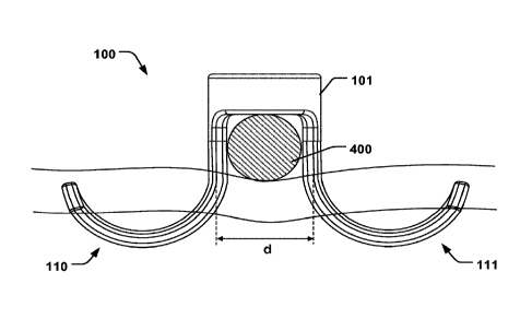
The clip 100 comprises a proximal cylindrical portion 101 to be arranged in
a tubular lumen 301 of a catheter 306, as illustrated in e.g. Fig. 2a. The
.. proximal cylindrical portion 101 is movable in a longitudinal direction 302
of the
catheter 306 when arranged therein. The cylindrical portion 101 has a central
longitudinal axis 102, along which the cylindrical portion 101 extends. The
central longitudinal axis 102 is thus parallel with the longitudinal direction
302
when arranged in the tubular lumen 301. The clip 100 comprises first and
.. second resilient legs 103, 104, having respective proximal ends 105, 106,
attached to the cylindrical portion 101, as illustrated in the example of Fig.
la.
The proximal ends 105, 106, are separated by a width (d) of a bridging section
107 of the cylindrical portion 101. The proximal ends 105, 106, are thus
connected in an inward direction 108, extending from each of the proximal ends
105, 106, to the central longitudinal axis 102 of the cylindrical portion 101.
The
bridging section 107 together with the first and second legs 103, 104, form an
opening 109 to receive an annuloplasty implant 400, as schematically
illustrated
in Fig. 3a in conjunction with Fig. la. Each of the first and second legs 103,
104, has a distal portion 110, 111, configured to pierce tissue at the heart
valve,
as schematically illustrated in Figs. 3a-b. In the relaxed shape of the clip
100,
each of the distal portions 110, 111, are curved to deflect from the central
longitudinal axis 102. As mentioned Fig. la show an example where the clip
100 is in the relaxed shape, and the distal portions are curved away in a non-
parallel orientation with respect to the longitudinal axis 102. As the clip
100 is
released from the constraint from the catheter 306, the first and second legs
103, 104, and the associated distal portions 110, 111, will progressively move
from a straightened shape, as exemplified in Fig. 2a, towards the relaxed
shape, as seen in e.g. Fig. 2f. The motion of the clip 100 between the
aforementioned shapes will be driven by the potential energy stored in the
.. deformed material of the clip 100 when confined in the catheter 306 (Fig.
2a).
The potential energy is accumulated when a force is applied onto the clip 100
to
push it inside the catheter 306. As will be described in further detail below,
the
distal portions 110, 111, have a curvature away from the longitudinal axis 102
in
the relaxed shape, such that the clip 100 will drive or propel itself out from
the
catheter 306 due to the curvature of the distal portions 110, 111, gradually
applying a force onto the distal part 303 of the catheter 306 as illustrated
in the
sequence of motion in Figs. 2b-f. Having a clip 100 shaped as specified above
CA 03118528 2021-05-03
WO 2020/058534 PCT/EP2019/075576
8
thus allows for ejecting the clip 100 from the catheter 306 by the potential
energy stored within the material of the clip 100. A small initial push with
e.g. a
wire 304, as shown in Fig. 2b, is thus sufficient for the clip 100 to propel
itself
from the catheter 306 towards the tissue and pierce the tissue. In some
examples it is not necessary to have a separate element for pushing the clip
100 as seen in Fig. 2b, but instead the catheter 306 can be tapped onto the
tissue which provides for the required amount of inertia transferred to the
clip
100 so that it moves into the position of Fig. 2b. Then, as explained above,
the
striving force towards the relaxed curved shape will drive the clip 100
forwards
as shown in Figs. 2b-f. The forward force of the clip 100 will cause the
distal
portions 110, 111, to pierce into the tissue as shown in Figs. 3a-b.
Hence, having curved distal portions 110, 111, as specified provides for
facilitating the fixation of the clip 100 into the tissue, without having to
actively
force or push the clip 100 forward into the tissue. The fixation is thus
simplified
and can be completed in less amount of time. Furthermore, by having a
proximal cylindrical portion 101 the stability of the clip 100 is improved
when
arranged in a catheter 306, since the cylindrical portion 101 will slide
inside the
tubular lumen 301 and prevent undesired tilting or tumbling of the clip 100
while
being delivered and ejected from the catheter 306. A robust clip 100 is thus
provided, which can be delivered with greater certainty of correct
positioning.
Accuracy is thus increased and the risk of dislocated clips, and thereby
insufficient fixation of an implant, can thus be minimized. Furthermore,
having a
bridging section 107 together with the first and second legs 103, 104, forming
an opening 109 to receive an annuloplasty implant 400, increases the stability
of the fixation of the implant 400. The opening 109 can be dimensioned to fit
various implants 400, which will prevent excessive relative movement between
the clip 100 and the implant 400.
In some examples the distal portions 110, 111, are curved so respective
distal tips 113, 114, thereof are oriented back towards the proximal
cylindrical
portion 101, as illustrated in the example of Fig. la and 3a. This allows for
driving the distal portions 110, 111, into the tissue from the opposite side
relative to the tissue side which is engaged by the implant 400, as seen in
Fig.
3a. This prevents damages to surrounding tissue by the distal tips 113, 114,
since these are embedded into the tissue on the opposite side, as seen in Fig.
3a. The clip 100 in Fig. 3a, i.e. when embedded into the tissue, may be
referred
to as being in its relaxed shape since it has been released from the
constraint
by the catheter 306, although it should be understood that the tissue may
CA 03118528 2021-05-03
WO 2020/058534 PCT/EP2019/075576
9
provide a counter force onto the clip 100. The shape of the clip 100 in the
implanted state is however referred to as the relaxed shape in the present
disclosure to simplify the discussion.
As seen in Fig. 3b, a plurality of clips 100 may be placed around the
.. annuloplasty implant 400 to achieve a secure fixation thereof. Although the
annuloplasty implant 400 is shown as a helix-shaped implant inserted through
the valve with first and second rings on opposite sides of the valve in the
example of Fig. 3b, it should be understood that the clip 100 may fixate
single
ring annuloplasty implants with the same advantageous benefits as described
above. The implant 400 may comprise a shape-memory material, so that the
first and second rings assume a coiled configuration after having been ejected
from a delivery catheter (not shown). While positioned in the delivery
catheter
the implant 400 may be stretched in an elongated shape. Alternatively, the
implant 400 may be arranged in the coiled configuration when being delivered
to the target site, in which case it may be implanted at the target site for
example by incision between the ribs or by opening the chest. The present
disclosure, and the associated advantages described for the various examples
of the clip 100 applies to both such variants of the implant 400.
Turning again to Figs. 2a-f, the first and second legs 103, 104, may be
resiliently deformable from the relaxed shape to the delivery shape in which
each of the distal portions extend substantially along the longitudinal axis
302
(Fig. 2a). The first and second legs 103, 104, may thus be resiliently
deformable
to the delivery shape when a compression force (Fc) is applied on respective
distal tips 113, 114, of the distal portions 110, 111. The compression force
Fc
may be applied by the distal part 303 of the catheter 306 as exemplified in
Fig.
2a. When the compression force Fc is removed, either by pushing slightly by a
wire 304 (Fig. 2b) or by tapping the catheter 306 against the tissue or
applying
an otherwise oscillating motion onto the catheter 306, so that the distal tips
113,
114, are released from the catheter 306 (Fig. 2b), the distal portions 110,
111,
.. spring towards the relaxed shape and pulls the cylindrical portion 101 in
the
longitudinal direction 302 as shown in the sequence of Figs. 2b-f. As
elucidated
above, distal portions 110, 111, have a curvature in their relaxed shape
configured to provide the following motion; i.e. the distal portions 110, 111,
are
configured to, in use, as the distal portions 110, 111, progressively move to
assume their fully curved shapes in the relaxed state, the distal portions
110,
111, will grasp around the edges of the distal part 303, and thereby push with
a
force (Fp) against the distal part 303 which pulls the entire clip 100 in the
CA 03118528 2021-05-03
WO 2020/058534 PCT/EP2019/075576
opposite direction towards the opening of the catheter 306. Previous clips
exist
having curved legs. Such clips however are only configured to have two states;
i) legs confined in a catheter, or ii) clip having deployed state requiring
the clip
to be fully pushed out from the catheter so that the legs may assume the
curved
5 state. I.e. the legs of such previous clips do not themselves push or
propel the
clip in the longitudinal direction of the catheter, as opposed to the present
clip
100 described in relation to e.g. Fig. la-f. Previous clips are thus more
cumbersome to deploy - e.g. always requiring a separate pusher - and are
unable to draw benefit from the potential energy stored in the clip for
propelling
10 in the longitudinal direction. The distal portions 110, 111, thus have a
resilience
which provides a force Fp, and corresponding vector component (Fi), that
overcomes the frictional force between the clip 100 and the inner wall of the
tubular lumen 301 of the catheter 300. The distal portions 110, 111, may thus
be formed form a shape-memory material having a Young's modulus that
provides such resilience, i.e. modulus of resilience. Typical previous clips
do not
absorb and release sufficient energy to be able to propel itself in the
longitudinal
direction.
In the relaxed shape, each of the distal portions 110, 111, may be curved
in an outward direction 112, substantially opposite the inward direction 108,
as
shown in the examples of Figs. la-c, 2a-f, 3a, 5a-b, 9. This provides for an
effective anchoring of the clip 100 into the tissue and a secure fixation of
an
annuloplasty implant 400.
Thus, the first and second legs 103, 104, may be resiliently deformable in
the inward direction 108 from the relaxed shape to the delivery shape when a
compression force (Fc) is applied on respective distal tips 113, 114, of the
distal
portions in the inward direction 108, as described above and shown in Fig. 2a.
In another example, when the clip 100 is in its relaxed shape, each of the
distal portions 110, 111, may be curved in the inward direction 108, as
schematically shown in Figs. 6a-b. This provides for reducing how far the
distal
portions 110, 111, extend from the cylindrical portion 101, which may be
advantageous in some situations and anatomies. As seen in the top-down view
of Fig. 6b, the extended distance of distal portions 110, 111, is reduced
since
the distal portions 110, 111, are curved in the inward direction 108 (Fig. 6a,
in
conjunction with Fig. la) to wrap at least partly around the implant 400. As
explained further below in relation to Fig. 11, a guide element 305 may apply
a
force onto the legs 103, 104, for separation thereof in a catheter 306.
CA 03118528 2021-05-03
WO 2020/058534 PCT/EP2019/075576
11
Each of the distal portions 110, 111, may be curved so that at least a
section 116 thereof extends in a direction 117 having a vector component 117'
parallel with the central longitudinal axis 102 and extending along the
central
longitudinal axis 102 from the opening 109 towards the cylindrical portion
101.
Figs. 2f and Fig. 9 show such examples where at least a section 106 of the
distal portions 110, 111, are curved in the direction towards the cylindrical
portion 101. I.e. the section 106 is angled so that the extension of the
curved
shape has a component 117' extending in a direction towards the cylindrical
portion 101. The distal portions 110, 11, may thus engage the tissue from the
opposite side of the cylindrical portion 101 as shown in Fig. 3a, providing
for
increasing the fixation strength while avoiding having distal tips 113, 114,
extending into the surrounding space.
The clip 100 has a transverse direction 118 which is orthogonal to the
outward direction 112 and the central longitudinal axis 102, as schematically
illustrated in Fig. 10. In one example, in the relaxed shape of the clip 100,
each
of the distal portions 110, 111, may be curved along the transverse direction
118. Each of the distal portions 110, 111, may thus extend along a direction
119
having a vector component 120 along the transverse direction 118, as further
shown in the example of Fig. 10. The angle (v) may be optimized depending on
the application and anatomy. In some examples the angle (v) may be close to
90 degrees, when it is desired to have the legs 103, 104, parallel with the
implant 400. In some examples the angle may be in the range 45-90 degrees.
Figs. 7a-b are also schematic illustrations showing the distal portions 110,
111,
being curved towards the transverse direction 118. As with the example in
Figs.
6a-b, this provides for reducing the length by which the distal portions 110,
111,
extend perpendicular to the annuloplasty implant 400. This may be
advantageous in some situations where it is desired to reduce the distance
pierced at the annulus, perpendicular to the implant 400. Further, in some
situations when the implant 400 and clip 100 is placed closer to the edges of
the
leaflets, it may be desirable to reduce the extension of the leg 103, 104,
facing
the leaflet.
The distal portions 110, 111, may be curved in opposite directions along
the transverse direction 118, as shown in the examples of Figs. 7a-b and 10.
This provides for attaining a high fixation strength while reducing the length
extended by the legs 103, 104, perpendicular to the direction of the implant
400,
i.e. in the outward direction 112.
CA 03118528 2021-05-03
WO 2020/058534 PCT/EP2019/075576
12
The clip 100 may comprise at least one secondary leg 121, 121', attached
to the cylindrical portion 101. The at least one secondary leg 121, 121',
extends
along the central longitudinal axis 102 and is curved in the inward direction
108,
as schematically illustrated in Figs. 8a-b in conjunction with Fig. la. Thus,
the
clip 100 may comprise legs 103, 104, having distal portions 110, 111, curved
in
the transverse direction 118 as explained above, as well as at least one
secondary leg 121, 121', curved in the inward direction 108 as shown in the
example of Figs. 8a-b. The at least one secondary leg 121, 121', provides for
further stabilizing the clip 100 in relation to the implant 400 due to the
additional
force that may be exerted from the at least one secondary leg 121, 121', onto
the implant 400. Resistance to rotational movement of the clip 100 may thus be
increased due to the added support from the at least one secondary leg 121,
121', and dislocation from the desired position of the clip 100 can be
prevented.
This added resistance to rotation may be particularly advantageous when the
legs 103, 104, are curved in the transverse direction 118. In the mentioned
example of Figs. 8a-b the clip 100 comprises two secondary leg 121, 121', one
on each side of the implant 400, which may provide for a further added support
and resistance to rotation or dislocation.
In one example the legs 103, 104, are curved in a circular shape, as
schematically illustrated in e.g. Fig. la. The radius of curvature may be
substantially constant in some examples. The angle (a) in Fig. la may be
in the range 150-180 degrees in some examples. In one example the angle (a)
may be 150 degrees, which may provide for a particularly advantageous
fixation.
A system 300 is provided comprising a clip 100 as described above in
relation to Figs. 1 - 11 and a catheter 306. Turning again to Figs. 2a-f, the
first
and second legs 103, 104, may be resiliently deformable to the delivery shape
when a compression force (Fc) is applied by a distal part 303 of the catheter
306
on respective distal tips 113, 114, of the distal portions 110, 111, in the
inward
direction 108. Hence, in use, when the compression force (Fc) is removed, the
distal portions 110, 111, spring towards the relaxed shape to apply a force
(Fp)
onto the distal part 303 having a force vector component (Fi) in the
longitudinal
direction 302 towards the cylindrical portion 101 to pull the cylindrical
portion
101 in the longitudinal direction 302 towards the distal part 303. Fig. 2c
illustrates an example of a direction of such vector component (Fi) that act
against the distal part 303 to pull the clip 100 out from the catheter 306 by
its
CA 03118528 2021-05-03
WO 2020/058534 PCT/EP2019/075576
13
own motion. The motion is sourced from the release of potential energy stored
in the deformed delivery state of the clip 100.
The system 300 may comprise a wire 304 being movable in the tubular
lumen 301, as seen in the example of Fig. 2b. The wire 304 is movable to
remove the compression force Fc, applied by the catheter 306 in the inward
direction 108, by pushing the cylindrical portion a distance (D) along the
longitudinal direction 302 towards the distal part 303 (Figs. 2a-b).
The system 300 may comprise an annuloplasty implant 400 comprising at
least one annuloplasty ring. The opening 109 may be configured to receive the
at least one annuloplasty ring and fixate the position of the annuloplasty
implant
400 to a heart valve 500, as exemplified in Fig. 3b.
The width (d) of the bridging section 107 of the cylindrical portion 101 may
correspond substantially to a diameter of the annuloplasty ring 400, as shown
in
the example of Fig. 3a in conjunction with Fig. la. This provides for a stable
fixation of the implant 400 to the clip 100 and tissue. The width (d) may be
in the
range 1-2 mm. A particularly advantageous range may be between 1-1.5 mm,
for a more secure and stable fixation of an annuloplasty ring 400.
The system 300 may comprise a guide element 305 in the tubular lumen
301, as schematically illustrated in the example of Fig. 11. The guide element
305 is positionable to separate the first and second legs 103, 104, in a
radial
direction 115, perpendicular to the longitudinal direction 302 when the clip
100
is arranged in the catheter 306. This provides for separating the legs 103,
104,
in a straight configuration in case the distal portions 110, 111, have a
relaxed
shape which curves in the inward direction 108 as shown in Figs. 6a-b. The
.. distal portions 110, 111, may accordingly be separated while engaging the
implant 400 so that when the guide element is 305 withdrawn, the distal
portions 110, 111, are able to move forward into position on either side of
the
implant 400 as seen in Figs. 6a-b. Thus, in the example of Fig. 11, the guide
element 305 applies a force onto the distal portions 113, 114, and when the
force is removed, e.g. by retracting the guide element 305 slightly, the
distal
portions 110, 111, will spring towards the curved relaxed shape and exert a
force onto the guide element 305 corresponding to (Fp) in the example of Figs.
2b-f. In a similar manner as described in relation to Figs. 2a-f, the clip 100
will
propel or drive itself forward out from the catheter 306 once the guide
element
305 as released the force on the distal portions 113, 114.
The guide element 305 may be positionable to extend through the
proximal cylindrical portion 101 of the clip 100. The cylindrical portion 101
may
CA 03118528 2021-05-03
WO 2020/058534 PCT/EP2019/075576
14
thus have a through opening, which may be provided by forming the clip 100
from a tubular material. The guide element 305 may thus be conveniently
advanced through the clip 100 to separate the first and second legs 103, 104,
as described above.
A method 200 of delivering a clip 100 to a target site is provided. The
method 200 is schematically illustrated in Fig. 12a, in conjunction with Figs.
1 ¨
11. The order in which the steps are described should not be construed as
limiting, and it is conceivable that the order of the steps may be varied
depending on the particular procedure.
As mentioned, the clip 100 comprises first and second resilient legs 103,
104, having respective proximal ends 105, 106, attached to a proximal
cylindrical portion 101. The proximal ends 105, 106, are separated by a width
(d) of a bridging section 107 of the cylindrical portion 101. The proximal
ends
105, 106, are connected in an inward direction 108, extending from each of the
proximal ends to a central longitudinal axis 102 of the cylindrical portion
101.
Each of the first and second legs 103, 104, has a distal portion 110, 111, to
pierce tissue at the target site. In a relaxed shape of the clip 100 each of
the
distal portions 110, 111, are curved to deflect from the central longitudinal
axis.
The method 200 comprises arranging 201 a distal cylindrical portion 101
of the clip 100 in a tubular lumen 301 extending in a longitudinal direction
302 of
a catheter 306. The method 200 comprises compressing 202 the first and
second legs 103, 104, by applying a compression force (Fc) on respective
distal
tips 113, 114, of the distal portions 110, 111. The method 200 comprises
delivering 203 the catheter to the target site at which an annuloplasty ring
400 is
positioned. The method 200 comprises pushing 204 the cylindrical portion 101 a
distance (D) along the longitudinal direction 302 towards a distal part 303 of
the
catheter 306 so that the compression force (Fc) is removed. As a result, the
distal portions 110, 111, spring towards the relaxed shape to apply a force
(Fp)
onto the distal part 303 having a force vector component (Fi) in the
longitudinal
direction 302 towards the cylindrical portion to pull the cylindrical portion
101 in
the longitudinal direction 302 towards the distal part 303. The distal
portions
110, 111, thereby pierce into the tissue at either side of the annuloplasty
ring
400 to fixate the position of the annuloplasty ring 400 at the target site.
The method 200 thus provides for the advantageous benefits as described
above in relation to the clip 100 and Figs. 1 ¨ 11.
Fig. 12b is another flowchart of a method 200 of delivering a clip 100 to a
target site. In one example, in a relaxed shape of the clip 100 each of the
distal
CA 03118528 2021-05-03
WO 2020/058534 PCT/EP2019/075576
portions 110, 111, are curved in an outward direction 112, substantially
opposite
the inward direction 108. The method 200 may thus comprise compressing 202'
the first and second legs 103, 104, in the inward direction 108 by applying a
compression force (Fc) by the distal part 303 of the catheter 306 on
respective
5 distal tips 113, 114, of the distal portions 110, 111. Subsequently
removing the
compression force (Fc), by advancing the clip 100, will cause the clip 100 to
drive itself forward as described above by pushing against the distal portion
303.
The present invention has been described above with reference to specific
10 embodiments. However, other embodiments than the above described are
equally possible within the scope of the invention. The different features and
steps of the invention may be combined in other combinations than those
described. The scope of the invention is only limited by the appended patent
claims. More generally, those skilled in the art will readily appreciate that
all
15 parameters, dimensions, materials, and configurations described herein
are
meant to be exemplary and that the actual parameters, dimensions, materials,
and/or configurations will depend upon the specific application or
applications
for which the teachings of the present invention is/are used.