Note: Descriptions are shown in the official language in which they were submitted.
CA 03118709 2021-05-03
WO 2020/093140
PCT/CA2019/051521
PULSED X-RAY IMAGING
[001] The present application claims priority from US provisional patent
application
62/755,813 filed November 5, 2018, the specification of which is hereby
incorporated by
reference.
Technical Field
[002] The present invention relates to X-ray and computed tomography (CT)
imaging
apparatus and methods.
Background
[003] X-ray imaging started with Rontgen's discovery of X-rays in 1895. X-
ray
imaging has greatly evolved towards more complex mechanisms able to obtain a
spatial
resolution as small as few microns. The principle underlying X-ray imaging
relies on
irradiating on one side a subject with X-rays produced by a source, and
collecting, on the
other side, onto a photographic plate or a digital recorder, the X-ray photons
that have
traveled through the subject.
[004] In X-ray imaging, the image is formed thanks to absorption of the X-
ray photons
in the subject. The intensity of the absorbed photons gives an indication on
the material
density between the source and the detectors. Thus, X-ray imaging gives a 2D
image of
the density of the material in the subject. The raw image data is based on the
photons
that are not absorbed as they propagate from the source to the detector. If
the object
being imaged absorbs almost all the photons, imaging is either not possible or
else most
of the X-ray energy used is left in the patient, and likewise if the object
being imaged fails
to absorb a significant quantity of the photons, no imaging is possible. Low
energy X-ray
photons, that are less harmful to tissue, are easily absorbed by a given
tissue thickness.
High energy X-ray photons, that are more harmful to tissue, are less easily
absorbed by
a given tissue thickness. The energy of the photons is thus chosen for the
object to be
1
CA 03118709 2021-05-03
WO 2020/093140
PCT/CA2019/051521
imaged to make sure that the relative loss of photons through absorption can
provide
useful imaging data.
[005] Often a 3D image is important to obtain a better understanding of an
anatomical
abnormality and to perform an accurate medical diagnosis. The principle then
relies on
irradiating the subject at different angles around it with X-rays and acquire
as many
projections as required to reconstruct a slice of the subject with an
appropriate image
reconstruction algorithm as shown in Figure 1. This principle, called computed
tomography (CT), is highly used thanks to its low cost compared to other
modalities such
as magnetic resonance imaging (MRI) and to its high acquisition speed. CT is
highly
available around the world, and many improvements have been brought to it to
optimize
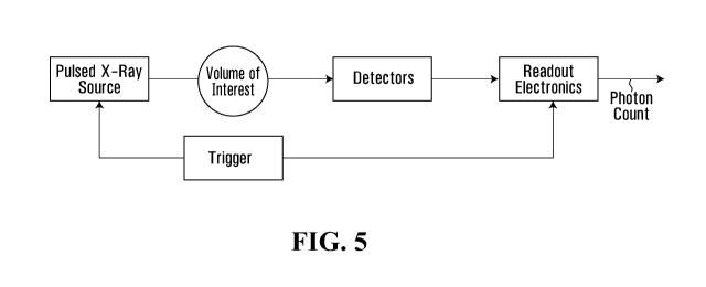
its overall performance.
[006] Owing to the better understanding of the potential harmfulness of X-
rays to
living organisms, improvements in X-ray and CT mainly address the contrast-to-
noise
ratio (CNR) to dose tradeoff. The adverse effects of X-rays become even more
important
with recent research demonstrating the possibility of developing radiation-
induced cancer
where children are more likely affected. Moreover, children are also at a
higher risk for
passing any radiation-induced genetic defects to the next generation. Imaging
protocols
on children must always consider the lowest possible dose and special
considerations
must be taken into account whenever possible depending on the part of the body
to be
imaged. Although CT scans currently represent only 10% of all ionizing
radiation-based
imaging modalities, it delivers > 50% of the collective dose for diagnostic
imaging. One
must now understand why pediatric CT imaging is made only when absolutely
necessary.
Small animal preclinical imaging is another important application of CT. At
this level, the
rat and the genetically modified mouse are chosen for their high genetic
similarity to
humans and for the ability to reproduce identical subjects at low cost. The
variations in
tissue density in these small animals is much smaller than for adult humans,
and a high
X-ray dose is mandatory to create a CNR high enough to differentiate
cartilage/bones
2
CA 03118709 2021-05-03
WO 2020/093140
PCT/CA2019/051521
from fat and muscle. Thus, the dose used must carefully be chosen to avoid any
radiation-
induced cancer, genetic radiation-induced cancer or even worse, create a
therapeutic
effect on the lesion under study in such laboratory animals.
Image contrast
[007] Globally, X-ray absorption is described by a simple equation, namely
the Beer-
Lambert law:
L = Loe¨glx)
where Lo is the initial quantity of photons, pti the linear attenuation
coefficient and x the
distance traveled in the medium by the photons. More precisely, the absorption
pti results
from 3 physical phenomena and is given by:
II = [(photoelectric + [(Compton + [(Rayleigh.
[008] Here, It
photoelectric photoelectric represents the portion of X-ray photons totally
absorbed by
atoms. A total absorption occurs when a photon collides with an electron close
to the
nucleus and ionizes the atom (hence the name photoelectric). As a result, the
photon
disappears, and an electron is then ejected and loses its energy in the
surrounding
Z3
e f f
material. This coefficient follows the relation II
r-photoelectric = E3 where Zeff is
the effective nuclear charge and E, the energy of the absorbed photon. The
photoelectric
effect is dominant at low X-ray energy. The coefficient LI
Compton accounts for Compton
scattering which occurs when a photon interacts with an electron in the outer
shells or
that is weakly bonded to the nucleus. The effect looks like an elastic
collision where the
striking X photon transfers a part of its energy to the electron but continues
its path at a
different angle (the photon is not lost; it is just redirected). From a
material point of view,
the energy transfer can be seen as a partial absorption but from an image
perspective,
3
CA 03118709 2021-05-03
WO 2020/093140
PCT/CA2019/051521
the scattered photon can hit the wrong detectors and adversely contribute to
the signal
by increasing the floor noise. The Compton scatter effect dominates for high
Zmaterials
or for high energy photons below 1 MeV. The last part of attenuation comes
from Rayleigh
scattering associated with the coefficient [t
Rayleigh. Rayleigh scattering results from the
interaction of the X-ray with an atom as a whole. The interaction modifies the
electric field
of the atom which in turn radiates at the same wavelength. Because of its low
probability
of occurring in the case of X-rays, this effect is of lesser interest here.
[009] Since human tissues have Z 7.4 , the photoelectric effect
dominates at up to
30 keV whereas Compton scattering dominates for X-ray energies > 100 keV. When
considering the subject volume, a minimal amount of X photon energy is
required to make
an image, and this is in the range 60-120 keV for humans and 30-80 keV for
small
animals. These ranges fall in the valley where the photoelectric effect (that
provides useful
information for imaging) ends and the Compton scattering increases. This
problem is
even amplified when increasing the X-ray energy over 120 keV where the
probability of
photoelectric absorption decreases while the probability of Compton scattering
stays the
same. This double effect increases the noise in integration-based X-ray
detectors since
the high energy photons contribute more to the accumulated energy while
providing poor
tissue absorption information and scattered photons increase the noise floor
in images.
Increasing the dose is the only way in integration-based X-ray imaging to
improve the
contrast in the image.
[0010] To better illustrate this phenomenon, Figure 2A shows
schematically
photoelectric absorption and Compton scattering for bones and soft tissues as
a function
of X photon energy in the range of 30 to 120 keV. One can see that absorption
is very
high for the low energy photons and decreases significantly with higher
energy. At 60
keV, the overall mass attenuation coefficient pti starts being dominated by
Compton
scattering. Figure 2B shows schematically the relative dose delivered to a
given patient
at different tissue thicknesses as a function of X photon energy to obtain an
image within
4
CA 03118709 2021-05-03
WO 2020/093140
PCT/CA2019/051521
a given acceptable CNR. As can be seen, thicker tissues require photons of
higher energy
to achieve the best possible dose-to-contrast ratio. At 140 keV, the higher
Compton
scattering contribution, compared to the photoelectric effect, decreases the
contrast, as
seen in Figure 2A, that in turn, requires a longer exposure time for the same
CNR than in
thinner tissues. Hence, to image thicker tissues, such as the abdomen of
bariatric
patients, reducing the negative effect of scatter noise on CNR is required to
reduce the
dose. Furthermore, for a given CNR, there is a photon energy that will deliver
the least
amount of ionizing radiation to the tissues to obtain the image.
[0011] Many approaches have been proposed to reduce the dose deposited
in a
subject. Among them, one can reduce the X-ray energy to the minimum according
to the
patient weight or age, make a smaller region scan, determine the level of just
acceptable
noise to enable an adequate diagnostic ("present study indication"), avoid
multiphase
imaging (imaging more than once to see different details each time), and
reduce
overlapping of slices. Other approaches are to resort to tube current
modulation, organ-
based dose modulation, iterative reconstruction, spectral imaging (two or more
X-ray
energies), use of contrast agents, gating and photon counting with energy
measurement.
Except for the photon counting technique, all other approaches are suboptimal
for dose
reduction since the aforementioned problems are still present. The photon
counting
method with or without energy measurement is a technique already used in
positron
emission tomography (PET) where each annihilation gamma ray photon is
timestamped
along with an energy measurement to eliminate Compton scattering (Compton
scattered
photons have less energy). Since the source is monochromatic in PET (511 keV),
Compton diffusion can be eliminated by applying an energy threshold. This is
not the case
in X-ray and CT since the X-ray source has a wide energy spectrum. Moreover,
PET
systems use large pixels > 0.5 x 0.5 mm2, that lead to an insufficient spatial
resolution in
X-ray CT and a limited count rate.
5
CA 03118709 2021-05-03
WO 2020/093140
PCT/CA2019/051521
[0012] Cup artefacts as shown in Figure 3 is a well-known problem in CT
imaging.
This problem occurs because X-rays crossing the central region of the subject
have more
chance to be absorbed than X-rays passing at the periphery. Similarly, X-rays
passing
through more tissue have more chance to be subjected to Compton scattering and
thus
adding noise to the image. The cup artefacts can be observed when imaging
subjects of
different thickness/diameter. At this level, bariatric patients will have more
cup artefact
than normal patients for identical imaging procedures.
Summary
[0013] Applicant has discovered that contrast to noise in an X-ray image
can be
improved using a pulsed source of X-rays coupled with a detector that is time-
sensitive
so as to be able to detect ballistic photons without the background
contribution of
scattered photons.
[0014] In some embodiments, an X-ray imager combines a pulsed X-ray
source with
a time-sensitive X-ray detector to provide a measure of ballistic photons with
a reduction
of scattered photons. The imager can provide a comparable contrast-to-noise X-
ray
image using significantly less radiation exposure than conventional X-ray
imagers, for
example less than about half of the radiation.
[0015] The time resolution of the imaging apparatus is dependent on the
sharpness of
the rise time of the X-ray source and on the time resolution of the time-
sensitive detector.
More specifically, if the time resolution is less than about 0.9 nanoseconds,
Applicant has
found that the contrast to noise ratio (CNR) can be improved over continuous X-
ray
sources for most human patient imaging. While the rising edge of the X-ray
pulse is
preferably less than 0.15 nanoseconds, it will be appreciated that
improvements in
contrast can be achieved with rising edges up to about 0.5 nanoseconds. The
pulsed X-
ray source can have a Gaussian pulse shape with a full width half maximum
(FWHM)
value in the range of less than 0.1 nanoseconds up to about 0.5 nanoseconds.
6
CA 03118709 2021-05-03
WO 2020/093140
PCT/CA2019/051521
[0016] As an example, one can measure the time resolution of an imaging
apparatus
by removing a subject or object from the apparatus and detecting one or more
pulses by
directly using the detector. This can be said to be a measure of the "impulse
response"
of the imaging apparatus which also corresponds to the response of the
ballistic photons
in a normal measurement, since there should be no scattered photons. Then, a
time point
or window parameter can be chosen to accept more or less of the
ballistic/scattered
photons according to the needs of the acquisition, either to improve the
signal-to-noise
ratio (SNR) by accepting all ballistic photons or to improve further CNR by
cutting part of
the ballistic photons to remove more scattered photons. Once a significant
portion of
scattered photons are removed from the raw image data to the benefit of
detecting a
greater percentage of ballistic photons, imaging quality can improve.
[0017] When the overall timing resolution of the system is better than
about 300
picoseconds, the CNR can be at least about doubled for the same dose of
radiation when
imaging a typical thickness of 20 cm of tissue. With time resolution better
than about 100
.. picoseconds, the amount of radiation delivered to the patient in abdominal
imaging can
be about 30% or less of the amount of radiation delivered to the patient for
continuous,
polychromatic X-ray imaging.
[0018] The innovation described herein presents a solution to both the
limited count
rate and the spatial resolution while significantly reducing the dose. The
innovation herein
described presents a solution to improve the cup artefact problem in bariatric
human
patients.
[0019] Applicant has found that an X-ray imaging apparatus can have a
pulsed X-ray
source having a control signal that determines the pulse timing and/or the
pulse rise time.
A time-sensitive X-ray detector can be included in the apparatus that has a
time-
dependent X-ray photon detection signal output. A processor, that can be
connected to
the control signal and the time-dependent X-ray photon detection signal
output, can be
7
CA 03118709 2021-05-03
WO 2020/093140
PCT/CA2019/051521
configured to provide a measure of ballistic photons with a reduction of
scattered photons
received by the time-sensitive detector.
[0020] In some embodiments, the pulsed X-ray source may include a high
voltage
source, electrodes connected to the high voltage source for accelerating
electrons, and
-- an X-ray emitting target material arranged to receive the electrons
following acceleration
by the electrodes so as to produce a pulse of X-rays.
[0021] In some embodiments, the pulsed X-ray source may comprise a
pulsed laser
source responsive to the control signal, a photoelectric material arranged to
receive a
light pulse from the pulsed laser source and to emit a burst of electrons in
response
-- thereto, wherein electrodes are arranged to accelerate the burst of
electrons. The
photoelectric material may be at least a part of a cathode of the electrodes.
[0022] In some embodiments, the pulsed X-ray source may comprise
deflection
electrodes for steering the electrons accelerated by the electrodes connected
to the high
voltage source to controllably hit the X-ray emitting target material.
[0023] In some embodiments, the electrodes connected to the high voltage
source
comprise a gated carbon nanotube cathode.
[0024] In some embodiments, the time-sensitive X-ray detector provides
for each
photon detected at each pixel element a time of detection signal.
[0025] In some embodiments, the time-sensitive X-ray detector is
responsive to a gate
-- signal controlling a time when photon detection is enabled.
[0026] In some embodiments, the time-sensitive X-ray detector is
responsive to a gate
signal controlling a time when photon detection is disabled.
[0027] In some embodiments, the processor is configured to collect X-ray
photons
detected within different time frames with respect to the control signal and
determine the
-- measure of ballistic photons with a reduction of scattered photons received
by the time-
sensitive detector through subtraction of the X-ray photons detected within
different time
frames.
8
CA 03118709 2021-05-03
WO 2020/093140
PCT/CA2019/051521
[0028] In some embodiments, the time-sensitive X-ray detector is
responsive to a
pulsed gate signal controlling a time window when photon detection is enabled.
[0029] In some embodiments, the time-sensitive X-ray detector is
arranged with
respect to the pulsed X-ray source so as to provide a different time of flight
for the ballistic
photons as a function of a pixel location within the time-sensitive X-ray
detector, wherein
the processor provides the measure of ballistic photons with the reduction of
scattered
photons received by the time-sensitive detector using different timing as a
function of
location of the pixels.
[0030] In some embodiments, the time-sensitive X-ray detector comprises
an X-ray
sensitive scintillator and a light sensor array coupled with the scintillator
for measuring X-
ray detection events in the scintillator.
[0031] In some embodiments, the time-sensitive X-ray detector comprises
an X-ray
sensitive detector based on a direct conversion of photons to electrons for
measuring X-
ray events in the detector.
[0032] In some embodiments, the processor is configured to provide a two-
dimensional image.
[0033] In some embodiments, the apparatus can further comprise a
motorized
mounting for moving the pulsed X-ray source and the time-sensitive X-ray
detector with
respecting to an object or subject to be imaged, wherein the processor is
configured to
provide a three-dimensional image.
[0034] In some embodiments, the apparatus is operative to obtain an
image with a
given contrast to noise ratio (CNR) while delivering a lower dosage of X-rays
to a typical
human abdominal region of at least 20 cm thickness than when continuous X-rays
of a
same energy are used in a similarly-structured continuous X-ray imaging
apparatus. The
lower dosage may be at least 50% lower, preferably at least 60% lower.
[0035] In some embodiments, the pulsed X-ray source produces a cone beam
and the
time-sensitive X-ray detector is arranged to detect a 2D array of pixels.
9
CA 03118709 2021-05-03
WO 2020/093140
PCT/CA2019/051521
[0036] In some embodiments, a response time of a combination of the
pulsed X-ray
source and the time-sensitive detector is less than 0.9 nanoseconds.
Preferably, the
response time is less than 0.3 nanoseconds. Preferably, a rise time of a pulse
emitted by
said pulsed X-ray source is less than 0.15 nanoseconds.
[0037] In some embodiments, the processor is configured to measure an
impulse
response time of a combination of the pulsed X-ray source and the time-
sensitive detector
to obtain a measure ballistic photons without an object or patient between the
pulsed X-
ray source and the time-sensitive detector and to derive therefrom and store
in memory
a gate parameter for the measure of ballistic photons with a reduction of
scattered
photons received by the time-sensitive detector when thereafter measuring
objects or
patients that provide scatter.
[0038] Applicant has also found a method of acquiring a medical
diagnostic image of
a human patient can comprises using an apparatus as described herein to obtain
an
image of a region of interest and having a contrast-to-noise ratio using X-
rays of a given
energy, wherein an amount of radiation delivered to the patient is about 60%
or less of
an amount of radiation delivered to a same patient for continuous,
polychromatic X-ray
imaging of the region of interest using the given energy of X-rays.
[0039] In some embodiments, the amount of radiation delivered to the
patient is about
30% or less of an amount of radiation delivered to a same patient for
continuous,
polychromatic X-ray imaging of the region of interest using the given energy
of X-rays.
Brief Description of the Drawings
[0040] The invention will be better understood by way of the following
detailed
description of embodiments of the invention with reference to the appended
drawings, in
which:
CA 03118709 2021-05-03
WO 2020/093140
PCT/CA2019/051521
[0041] Figure 1A is a schematic sectional view of a conventional
arrangement of an
X-ray source, array of X-ray detectors for a CT scanner with the object to be
imaged
shown as a patient section (Webb 1988);
[0042] Figure 1B is a schematic illustration of an image intensity
profile acquired using
the scanner of Figure 1A (Webb 1988);
[0043] Figure 2A is a plot showing Compton and photoelectric attenuation
versus
energy for bone and soft tissues (Martin 2007);
[0044] Figure 2B is a plot showing the normalized CNR obtain with
constant radiation
exposure versus tube potential and tissue thickness. (Rui et al. 2014);
[0045] Figure 3A is an image showing cup artefacts with the intensity line
profile
superposed for heavy cup artefacts (Barrett & Keat, 2004);
[0046] Figure 3B is an image showing cup artefacts with the intensity
line profile
superposed for no cup artefacts (Barrett & Keat, 2004);
[0047] Figure 4 is a schematic diagram illustrating X-ray image
acquisition showing
ballistic and scattered photons travel for computed tomography (CT);
[0048] Figure 5 is a block diagram of a time-of-flight (TOF) CT
acquisition chain;
[0049] Figure 6 is a detailed block representation of a TOF-CT
acquisition chain with
gating;
[0050] Figure 7 is a detailed block representation of a TOF-CT
acquisition chain with
timestamps;
[0051] Figure 8 is a plot showing scattered to primary radiation ratio
(SPR) after ToF
scatter rejection (TSR) as a function of SPR before TSR according to total
time resolution
for different object thicknesses (50 mm, 100 mm, 150 mm and 200 mm);
[0052] Figure 9A is a plot showing SPR after TSR as a function of SPR
before TSR
for different object thicknesses (50 mm, 100 mm, 150 mm and 200 mm) and source
to
detector distances, namely 250 mm, 500 mm, 800 mm, 1200 mm and 1650 mm;
11
CA 03118709 2021-05-03
WO 2020/093140
PCT/CA2019/051521
[0053] Figure 9B is a plot showing SPR after TSR as a function of SPR
before TSR
for different object thicknesses (50 mm, 100 mm, 150 mm and 200 mm) and X
photon
energy, namely 20 keV, 40 keV, 60 keV, 80 keV, 100 keV and 120 keV;
[0054] Figure 10 is a graph showing dose multiplication factor required
to correct the
CNR degradation caused by scatter noise as a function of SPR (Siewerdsen and
Jaffray
2001);
[0055] Figure 11A is a comparative reconstructed image of the same
object in Figure
11B without applying TSR;
[0056] Figure 11B is a simulated reconstructed image showing the effect
of TSR on
the contrast;
[0057] Figure 12 is a simulated image showing only scattered photons
removed by
TSR in the image of Figure 11A;
[0058] Figure 13 is a horizontal line profile of the reconstructed
images in Figure 11A
without TSR and with TSR, showing also the expected values based on the
phantom
structure;
[0059] Figure 14A is a histogram of the number of photons measured
experimentally
with nothing between the source and the detectors according to the time
between the
source trigger and the detection fitted with a Landau distribution.
[0060] Figure 14B is a histogram of the number of photons measured
experimentally
with a 4 cm thick beam-blocker between the source and the detectors according
to the
time between the source trigger and the detection fitted with a Landau
distribution.
[0061] Figure 15 is a schematic sectional side view of a conventional
side-window type
of X-ray tube having a water-cooled anode and a hot filament type cathode
(Wikimedia
Commons 2010);
[0062] Figure 16 is a schematic sectional side view of a laser-pulsed
cathode side-
window type of X-ray tube;
12
CA 03118709 2021-05-03
WO 2020/093140
PCT/CA2019/051521
[0063] Figure 17 is a schematic sectional side view of a carbon nanotube
(CNT) gated
cathode side-window type of pulsed X-ray tube; and
[0064] Figure 18 is a double graph showing the decrease in percentage of
primary
photons removed as a function of accepted time-of-flight difference in
nanoseconds and
the increase in SPR as a function of accepted time-of-flight difference in
nanoseconds.
Detailed Description
[0065] The present invention relates to separating diffused photons from
ballistic
photons by using photon time-of-flight (elapsed time from emission to
detection)
measurements. Currently, X-ray and CT imaging are mainly based on accumulating
the
energy of X-ray photons into pixels to create a 2D image or a slice of a 3D
image. These
approaches require a non-negligible dose to extract the useful signal from the
background. The present invention proposes to measure the time of flight of X
photons
and directly sort the scattered photons from the ballistic ones. This will
allow removing a
substantial quantity of photons participating to the background noise of the
image, as well
as significantly attenuating the cup artefacts present in CT images. A
significant dose
reduction for a similar contrast can also be obtained, which is critical in
several clinical
uses of CT.
[0066] The approach to solving the aforementioned problems consists of
measuring
the time-of-flight (TOF) of each individual photon (or of a burst of photons)
to determine
whether a photon has followed a straight line ballistic trajectory or has been
scattered.
TOF is defined as the length of time between the photon's emission by the X-
ray source
and its arrival in the detection system where it is transduced into an
electrical signal. In
the X-ray wavelength range, the distance traveled by a photon is directly
related to its
TOF since the index of refraction is close to 1 and thus all photons have a
speed close to
the speed of light in a vacuum. Measuring TOF requires knowing when X photons
leave
the source and when they arrive in the detection system. Away to create such a
condition
13
CA 03118709 2021-05-03
WO 2020/093140
PCT/CA2019/051521
can be to use ultra-short pulses of X photons to obtain a precise time of
emission, and to
measure the time of arrival of each individual photon or of the burst of
photons resolved
in time. For the types of tissue volumes encountered in human subjects (for
example
abdominal scans of children up to bariatric adults), improvement in image
contrast can
be achieved when time resolution is better than about 0.5 nanoseconds, and
because a
time resolution of at least about 0.2 nanoseconds is feasible using available
technology,
the improvement in contrast for a given dosage and/or the improvement in the
reduction
in dosage for a given contrast is significant.
[0067] Since scattered photons do not travel directly in straight line
from the X-ray
source to the detectors, their flight distance, therefore their TOF, is longer
than that of
ballistic photons as shown in Figure 4. This is true for both Compton and
Rayleigh
scattered photons. For each pixel in the detection system, a maximum TOF for a
ballistic
photon is computed by dividing the longest distance from any point in the
pixel to the X-
ray source by the speed of light in a vacuum. Then, during scan, the detection
system
decides for each individual photon if the following condition is true:
dimn dmax 6 < T detection ¨T
em _L ission "
where Tc/etection is the time of detection, 7--
=umZssionis the time of emission, dmax and dmin are
respectively the maximum and minimum distances from the X-ray source to the
detector
pixel in which the photon is detected, cis the speed of light in vacuum, Wand
c form the
limit of the accepted window of time respectively for the late and the early
photons. If the
previous condition is true, a photon is declared to be ballistic, otherwise it
is declared to
be scattered. The length of the time window (W + 6) must be chosen according
to the
spatial uncertainty of the measurements including the time resolution of the
detection
system, the time jitter of the X-ray source and the needs for the application.
To select the
optimal time window width, an acquisition can be made without any subject in
the scanner
to extract the impulse response of the system which also corresponds to the
response of
14
CA 03118709 2021-05-03
WO 2020/093140
PCT/CA2019/051521
the ballistic photons in a normal measurement. Then, the window can be chosen
to accept
more or less of the ballistic/scattered photon according to the needs of the
acquisition,
either to improve SNR by accepting all ballistic photons or CNR by cutting
part of the
ballistic photons to remove more scattered photons. Window optimization will
be
.. presented later.
[0068] One of the most promising applications of TOF-X-ray imaging
(whether 2D or
3D) is to remove photons identified as scattered photons from the measurements
to
reduce scatter noise and its adverse effect on image quality. This technique,
that we call
time-of-flight scatter rejection (TSR), can be implemented by gating the
photons in the
detection system. When the trigger, used for both the pulsed and the detection
system,
is received, the detection system timestamps and/or counts the photons
arriving in each
pixel during a period of time in the selected time window. The scattered
photons arriving
too late (outside the time window) can be discarded from the measurements.
[0069] The temporal X-ray pulse width is preferably as narrow as
possible to reduce
the dose to the minimum. However, embodiments can operate with longer pulses
to the
detriment of the injected dose. In this case, late scattered photons are being
discarded
by the TSR along with most of the ballistic photon emitted the first few
tenths of a
picosecond after the pulse. The resulting signal is therefore composed mostly
of ballistic
photons, even though most of them are removed. This allows for CNR
improvements at
the cost of a higher dose. In all cases, the rising edge of the X-ray pulse
must be as
sharp as possible to reduce the timing jitter associated to the X-ray source.
[0070] The required system for TOF- X-ray imaging (whether 2D or 3D) is
essentially
composed of four parts as shown in Figure 5: a pulsed X-ray source, a
detector, a gated
electronic readout system and a synchronisation mechanism or trigger.
[0071] Any system able to precisely detect the time of arrival of X photons
emitted by
a pulsed source synchronized with the detectors' readout electronics could
implement
CA 03118709 2021-05-03
WO 2020/093140
PCT/CA2019/051521
TOF-X-ray imaging (whether 2D or 3D). Herein, we propose an example of such a
system, detailed in Figure 6 and Figure 7, to achieve a high enough time
resolution and
data throughput to successfully scan a subject using TSR. As an example, the
system
detailed in Figure 6 and Figure 7 could be implemented with a N5084 pulsed X-
ray tube
from Hamamatsu excited by a Picosecond light pulse PLP-10 from the same
company,
acting both as a pulsed laser and a trigger. The detection system can be
implemented
using a 500 pm thick LYSO crystal and a S12571-015C SiPM also from Hamamatsu.
The
readout circuit can be designed as an application specific integrated circuit
(ASIC).
[0072] The output of the readout electronics can be subjected to
additional digital
processing such as finding centroid of events, data sorting and merging, data
correction,
dark noise filtering, etc. The output is TSR pixel data. An image processor
generates 2D
or 3D images from the pixel data using conventional techniques known in the
art,
however, without needing contrast improvement filters that might be
conventionally
employed. Such medical images can be viewed at an image viewing workstation as
is
known in the art.
[0073] Since the number of scattered photons detected in each pixel is a
function of
the scattering ability of the object outside of the ballistic photon travel
path, those photons
can thus be considered as background noise. As the reconstructed image
including
scattered photon shows (Figure 12), the contribution of those photons to the
image is
subject to a cup artifact. Moreover, the bone inserts are indistinguishable in
this image,
thus the scattered photons contribute negatively to the image contrast. The
CNR is
dependent on a statistically viable measurement of ballistic photons above the
background. Assuming that the background is not spatially dependent (which it
is),
contrast is improved by collecting more photons, both ballistic and scattered,
so that the
spatial variation in ballistic photons provides a better CNR following a
background
subtraction. As can be appreciated, removing at least a portion of scattered
photons using
16
CA 03118709 2021-05-03
WO 2020/093140
PCT/CA2019/051521
TSR reduces the need for collecting as many photons and improves image quality
due to
the non-uniform spatial distribution of scattered photons.
[0074] Multiple implementations of the X-ray source, described below,
can generate
suitably short pulses of X photons to reduce dosage while having a short
enough rise time
.. in intensity to allow for detection of ballistic photons and
correspondingly provide the
improvement in contrast. As an example, a trigger could be used to generate an
ultra-
short laser pulse directed onto the photocathode of an X-ray tube, generating
a pulse of
electrons accelerated in the tube with an electrical field. Similarly, an
electron gun or
canon could generate a continuous flow of electrons deflected or not on the X-
ray emitter
target. When the accelerated electrons hit the target then an X-ray pulse is
generated
towards the volume of interest using an aperture to form a fan or cone shaped
beam.
[0075] The X-ray detector converts the photons into electric pulses. The
detector is
composed of a material with good X photon stopping power and able to generate
low jitter
electric pulses. The detector could be of any material for which the
combination
scintillating crystal/digital silicon photomultiplier is a good candidate, or
alternatively a
direct X-photon detector that can be gated with the desired time sensitivity.
[0076] A brute force approach for the trigger could be an implementation
where all
detector pixels can individually timestamp the time of arrival of each
individual X photon
(Figure 7). A digital signal processor can then sort and compute the data to
extract the
relevant information. Another approach would be to electronically open a
programmable
or fixed time window directly within the detector. A trigger sent through a
programmable
or fixed delay line to the readout electronics of each pixel will initiate the
acquisition. The
timing window can then be adjusted to count only ballistic photons and remove
the
scattered photons (Figure 6). This can be done by counting and localizing the
photons
detected during a number of X-ray pulses where the gating is set to capture
the entire
temporal point spread function (TPSF) followed by counting and localizing the
photons
detected during a number of different X-ray pulses where the gating is set to
capture only
17
CA 03118709 2021-05-03
WO 2020/093140
PCT/CA2019/051521
the latter part of the TPSF, namely the non-ballistic photons, with the
ballistic photon
intensity being calculated by the difference between the full TPSF and the
latter part of
the TPSF. This requires a fast timing for switching on the acquisition without
the need to
have a fast response time for switching off photon detection.
[0077] The photon count in each pixel is finally sent to a centralized unit
performing, if
needed, additional digital signal processing before sending the data to a
computer to
perform image reconstruction in the case of computed tomography or to perform
any
desired image processing in the case of a 2D image.
[0078] As an example of this innovation, the Monte Carlo simulator GATE
was used
to simulate a cone-beam CT scanner with a flat panel detector system. In every
simulation, a point source emits a continuous monochromatic beam of photons in
an
isotropic circular cone pattern towards a square flat panel array of
detectors.
[0079] No energy measurements were done on individual photons, but those
below
10 keV were automatically removed from the simulations. Both the time of
emission and
the time of detection provided by the simulator were used to measure the TOF
of each
photon and mimic a pulsed X-ray source. The effect of the pulse width was
added to the
emission time by randomly generating an offset using a probability
distribution function of
emission of the source over time. GATE was set up to compute time of detection
according to the timestamp of the first event in a specific pixel associated
with a single
photon. At this point, it adds the effect of the time resolution of the
detector system, always
assumed to be Gaussian.
[0080] In Applicant's simulation, to measure performance of the TSR
algorithm, the
window was always placed within three standard deviations (3cr) of the total
timing
resolution (assumed to be Gaussian), to keep at least 99.5% of transmitted
photons. For
the single projection measurements for TSR validation, the source emits a 120
keV cone
of X photons with an 8 angle. The phantom was placed 102 cm away from the
source
and a 256 x 256 array of 1 x 1 mm2 pixels detector was placed 63 cm behind the
phantom.
18
CA 03118709 2021-05-03
WO 2020/093140
PCT/CA2019/051521
[0081] The
scattered to primary radiation ratio (SPR), is the energy of the scattered
radiation (S) divided by the energy of the primary beam (P) striking the same
point on the
imaging device). The SPR after applying TSR for increasing initial SPR,
obtained with 50,
100, 150 and 200 mm thick phantoms is presented in Figure 8. Even with a total
time
resolution of 1 ns, improvements can be seen in some systems with both high
incident
photon energy and large source-to-detector distance. However, improving total
timing
resolution below 10 ps does not lead to significantly better results. As
expected, the
achieved SPR worsens with lower energies and smaller source-to-detector
distance as
seen in Figures 9A and 9B respectively.
[0082] A
slice of a reconstructed image of a 288 mm wide water cylinder with two bone
inserts before and after applying TSR is shown in Figure 11A and Figure 11B
respectively.
The image was normalized then reconstructed by feeding the sinogram directly
to the
Reconstruction Toolkit (RTK) Feldkamp, Davis and Kress algorithm (FDK) with no
additional processing. With a perfect timing resolution, 100 keV photon energy
and 165
cm source-to-detector distance, SPR decreased from 300% to about 4% at the
center of
the detection system. The bone inserts are indistinguishable in the removed
scattered
photons image (Figure 12) and a cup artifact is present. This explains the
increase of
contrast once those photons are removed from the reconstruction data.
[0083] As
shown by the horizontal line profile in the center of the image of Figure 13,
the CNR is doubled and the cup artifact is greatly reduced. The inaccuracies
of the
reconstructed values, inherent to scatter noise, are also reduced. An image
was
reconstructed with a dose four times smaller, the CNR after TSR was the same
as the
original image with full dose and the cup artifact was still reduced, but
photon deprivation
artifacts appeared.
[0084] With TSR
implemented to remove scattered photons, the expected dosage
reduction as a function of SPR reduction is shown in Figure 10. The
degradation of CNR
can be corrected with either an increase of dose or an increase in pixel size,
shown in
19
CA 03118709 2021-05-03
WO 2020/093140
PCT/CA2019/051521
Figure 10 as az , however such increase also reduces spatial resolution.
Therefore, to
correct for a scatter noise of 100% SPR and keep the same spatial resolution,
a four-fold
increase in dose is required. If the total time resolution is short enough to
completely
mitigate the effect of scatter noise, it could be possible to divide the dose
by four. With a
.. time resolution of around 100 ps, the expected dose reduction for an
abdomen scan is
more than 50%.
[0085] An experiment was conducted to confirm the feasibility of
observing TOF
differences between scattered and transmitted photons. A 3 x 3 mm2 silicon
photomultiplier (SiPM) covered by a 500 pm thick lutetium-yttrium
oxyorthosilicate
(LYSO) crystal was placed 38 cm in front of a pulsed X-ray source (Hamamatsu
with
mean photon energy of 15 keV and pulse width of 60 ps. Two measurements were
made:
one with nothing between the source and the detector and one with a 40 mm
thick
aluminum beam blocker. Figure 14A shows the time between trigger and detection
(TPSF) without the blocker, while Figure 14B shows the time between trigger
and
detection with the blocker. 657 photons were detected after an acquisition of
36 hours.
Once fitted with a Landau distribution, the most probable value (MPV) of the
TOF with the
beam blocker is 390 ps later than the MPV without the beam blocker, which
correspond
to an increase of travel path of about 12 cm. This fits with the expected
increase of travel
path needed to circle the beam blocker by scattering on the X-ray enclosure.
No TOF
correction was made for the energy of the detected photons. Higher energy
photons were
detected earlier, however, the increased TOF of scattered photons is observed
at all
energy levels at around 400 ps. Dark counts and double detections were removed
from
both measurements.
[0086] The X-ray source is responsible for emitting X-rays. The first X-
ray sources
were vacuum tubes (so-called X-ray tubes). It is with such devices that
Roentgen
accidentally discovered X-rays. Their construction is rather simple as
illustrated
schematically in Figure 15. At one end of a vacuum tube is a filament heated
at high
CA 03118709 2021-05-03
WO 2020/093140
PCT/CA2019/051521
temperature by an electric current (voltage Uh). Through the physical process
of
thermionic emission, electrons are ejected from the filament (so-called the
hot cathode,
made out of metal, typically tungsten). At the other end of the tube is a
little plate (the
anode also often made of tungsten) onto which the electrons are directed. A
high voltage
(Ua) electric field is established between the cathode and the anode for
accelerating the
electrons from the cathode to the anode. When the electrons impinge onto the
anode,
they are rapidly decelerated as they slowdown in the metal and X-ray photons
are emitted
through the process of Bremsstrahlung (from the German literally meaning
braking
radiation). It is a consequence of the laws of electromagnetism that when a
charged
particle is accelerated (or decelerated), it emits radiation, in the present
case X-ray
radiation. For a given high voltage between the cathode and the anode, the
emitted X
photons have energies that span a given range, so-called the energy spectrum
of the
source. A source that emits X photons of only one energy is called
monochromatic,
otherwise it is called polychromatic (this terminology is in analogy with
visible photons for
which different energies correspond to different colors - chroma)
[0087] Another means for producing X-rays is by bending radially a beam
of electrons,
i.e. when the electrons accelerate perpendicular to their velocity. This
occurs for instance
in synchrotrons using bending magnets. When the electrons travel at non-
relativistic
(resp. relativistic) speeds, then the radiation is called cyclotron (resp.
synchrotron)
radiation. Because of the great control of acceleration in synchrotrons, these
are able to
produce highly monochromatic X-rays. However, synchrotrons and cyclotrons are
large
infrastructures that are not viable for integration in commercial medical
imaging devices.
[0088] In conventional X-ray tubes, the stream of X photons is
continuous, but for the
purposes of the present invention, very short pulses (or bursts) of X-rays of
the order of
at most a few tens of picoseconds are needed. Synchrotrons are able to produce
such
pulses, but these are too massive infrastructures for commercial imaging
devices as said
above. An alternative for obtaining short X-ray pulses is via X-ray emission
from
21
CA 03118709 2021-05-03
WO 2020/093140
PCT/CA2019/051521
femtosecond laser-produced plasmas on solid surfaces (Von Der Linde et al.
2001). In
this approach, an ultra-short high intensity laser pulse is focused onto a
metallic target
which rips out electrons from the metal and accelerates the electrons back
towards the
metal target where they decelerate, thus producing short X-ray bursts. Another
approach
is through high-order harmonic generation in gases, which resorts to intense
ultra-short
laser pulses. This approach can be carried out in gas-filled hollow fibers
(Von Der Linde
et al. 2001). These approaches can be foreseen to be amenable to reasonable
sizes for
integration in medical imaging devices since ultra-short pulse laser
technology is
nowadays highly compact.
[0089] Another approach to generate ultra-short X-ray pulses is one that
has been
developed for fluorescence lifetime measurements, whereby fluorescence is
induced by
X-ray excitation (Derenzo et al. 1994; Moses et al. 1995). In such a pulsed X-
ray source,
short pulses of light (<100 ps full-width at half maximum - FWHM) emitted by a
pulsed
laser diode are directed onto a light sensitive photocathode that emits short
bursts of
electrons with each light pulse impinging onto it (Figure 16). The electrons
are then
amplified or not and accelerated towards an anode as in conventional X-ray
tubes
described above. Yet another approach to generate short X-ray pulses is to use
an X-ray
tube in which the electron beam can be very rapidly deflected as in a streak
camera, with
an electric pulsed field in such a way that it strikes the anode for a very
short time interval
in which bremsstrahlung X-rays can be generated.
[0090] Another solution to generate X-ray pulses is to replace the
photocathode with
carbon nanotubes (CNT) as described in (Parmee et al. 2015). The CNTs are
plated on
top of the cathode as an electron emitter with the capability to be gated
faster than the
cathode alone directly with an electric signal (Figure 17) and operating at
lower
temperature.
[0091] Different technology configurations can support embodiments of
the invention
and are not limited to the example described below.
22
CA 03118709 2021-05-03
WO 2020/093140
PCT/CA2019/051521
[0092] The detector is among the important components to consider in the
deployment
of the technology. There are two main detection principles: Direct conversion
and indirect
conversion. While the direct conversion of X photons in materials such as
germanium or
silicon is very attractive for high energy resolution, indirect conversion can
be a preferred
avenue thanks to its lower operating voltage and its proven better timing
resolution. The
electron/hole mobility in direct conversion detectors coupled to the detector
thickness do
not currently allow obtaining timing performance in the tens of picoseconds as
can be
desired. Alternatively, the use of a thin scintillator able to stop an X
photon coupled to a
high-speed photodetector such as a silicon photomultiplier (SiPM) or all its
digital
.. derivatives is a good candidate for a complete system with timing
performance under 100
PS.
[0093] Although an indirect conversion mechanism is to be preferred, it
must be
intimately coupled to an adequate gating mechanism. The brute force approach
would be
to timestamp every individual photon and to provide the information to a
digital signal
processor able to process, in real-time, the relevant information for image
reconstruction.
This processor could be integrated in 2.5D or 3D electronics along with the
photodetector
or located remotely outside the scanner. The processing algorithms can be of
any form
from gating, filtering, up to machine learning. However, this approach will
require a large
data bandwidth and other approaches can be used.
[0094] In order to reduce the bandwidth, an adjustable and delayed trigger
can be
distributed in the scanner. This trigger can open a time window where all
photons striking
the detector in the time window are timestamped or counted. The information
can still be
sent to a local or a remote digital processor with the goal to extract the
relevant
information to be fed to the image reconstruction algorithm. The trigger can
be self-
adjusted from the center of the detector panel to the periphery or manually
adjusted with
programmable or fixed delay lines to take into account the source to flat
panel distance
variation form the center to the periphery. In the former case, each pixel has
a
23
CA 03118709 2021-05-03
WO 2020/093140
PCT/CA2019/051521
communication link with its adjacent neighbor while in the latter case, a
system calibration
is mandatory.
[0095] It would also be possible to use energy integration detectors to
implement the
discrimination between ballistic and scattered photons by either turning on
and off the
energy integration according to the time window or by reading such detectors
at the
beginning and at the end of the time window to only measure the energy of
ballistic
photons.
[0096] Optimizing TOF- X-ray imaging (whether 2D or 3D) revolves around
one central
idea: having the right gate width. Ideally the gate width is selected to be
narrow enough
to allow almost all the ballistic photons but remove the maximum number of
scattered
photons. To achieve this, the impact of every component of the system on the
gate width
must be considered. Ideally, the gate width would be almost zero, but the
source pulse
width must then be short enough to avoid removing ballistic photons.
Parameters leading
to errors in TOF measurements, such as the pulse width of the X-ray source
(and in some
cases the sharpness of the rising edge of the pulse) or the timing resolution
of the
detectors, widen the response of the system to ballistic photons. To keep
almost all
ballistic photons, the gate must be widened even if the errors increase, but
doing this
reduces the proportion of scattered photons being removed. However, since
removing
part of the ballistic photons reduces the SNR while removing more scattered
photons
increases the CNR, the gate width can be chosen more conservatively or more
aggressively according to whether SNR or CNR is driving the dose. On one hand,
increasing the SNR is particularly important in very low dose applications and
in imaging
systems with a naturally high contrast such as inorganic imaging. On the other
hand, CNR
will drive the dose up in biological tissue imaging with only small density
differences such
as in breast imaging. The quantity of ballistic photons removed and scattered
to primary
ratio according to the chosen gate in a system with 100 ps of total time
resolution is shown
in Figure 18.
24
CA 03118709 2021-05-03
WO 2020/093140
PCT/CA2019/051521
[0097] Reducing the total time resolution is one of the most important
design aspects
of TOF-X-ray imaging (whether 2D or 3D). The errors caused by each component
of the
system are added together in quadrature. Thus, reducing the pulse width and
the timing
resolution of the detectors is equally important to increase the efficiency of
discrimination.
If both effects are reduced to under 10 ps, the spatial uncertainty of the
emission (the size
of the focal spot of the source) and of the detection (size of the detector
and error on
positioning) will also have to be optimized since this affects the expected
TOF for ballistic
photons used as a comparison for the discrimination. Jitter between detectors
will also
increase the error on the measurements and will have to be reduced to a
minimum.
[0098] The present method uses the maximum possible TOF between the source
and
a pixel for discriminating ballistic and scattered X photons. However, to
further optimize
this method, it could be useful, in large pixel-size systems, to use the most
likely depth of
detection for the window. Doing this will reduce the number of ballistic
photons that are
retained, but should reduce the measured SPR.
[0099] Embodiments can be implemented in a variety of systems. The
following have
been identified as potential interesting applications of TOF- X-ray imaging
(whether 2D or
3D):
1. Pediatric (where dose of radiation can be reduced to acceptable levels);
2. Pre-clinical (where spatial resolution can be improved due to reduced CNR);
3. Dental care (where dose and form factor are important);
4. Bariatric patients (where contrast is a problem);
5. Extremities (form factor and dose);
6. Interventional radiology (form factor, dose and resolution);
7. Gating imaging.
[00100] These systems require different adjustments in their design options
(or
optimization) for dose, spatial resolution, contrast and form factor as
indicated in
parentheses above. X-ray imaging (whether 2D or 3D) is rarely used for
pediatric patients
because of the high radiation dose associated with current X-ray imaging
procedures.
CA 03118709 2021-05-03
WO 2020/093140
PCT/CA2019/051521
The method presented herein, by reducing the dose allows for use of the
technique for
this application. Scan of extremities, dental care and interventional
radiology can also be
optimised for dose to reduce the impact of the repeated use required by those
applications. For this, the system can be designed to further reduce the dose
notably by
reducing the gate size according to the CNR and SNR requirements, and increase
sensitivity. Imaging naturally high contrast volume, such as in dental care,
will generally
require optimizing for SNR while low contrast imaging, such as breast imaging,
will require
optimizing for CNR. The window can be modified according to the required
needs, for
instance looking for fractured bones requires a large window to increase SNR,
but
scanning the same area for potential tumors requires a small window to
increase CNR.
[00101] Spatial resolution is particularly important for both pre-clinical and
interventional radiology. A significantly higher spatial resolution can be
obtained by using
embodiments as described herein in conjunction with single photon avalanche
diodes
(SPADs) detectors to precisely pinpoint the location of the interaction of the
X-ray with
the detectors. The quality of the discrimination, along with the spatial
resolution, can also
be improved by using a magnification process such as increasing the distance
between
the volume of interest and the detector system.
[00102] Scanning bariatric patients requires photons of higher energies than
the
standard range of energy used in X-ray imaging (whether 2D or 3D) and yields a
lower
contrast image. TSR is particularly useful for scanning bariatric patients
since larger
volumes generate more scatter noise that is easier to remove with our approach
since
the photons generally scatter more than once in the subject in those cases.
Optimizing
for contrast, notably by choosing an aggressive gate, smaller than the total
time resolution
of the system, could even further help to scan bariatric patients.
[00103] The embodiments described herein can allow the usage of cone-beam
computed tomography with even larger volumes. Cone-beam CT has a big advantage
over standard helicoidal fan beam CT mostly used nowadays since it does not
require
26
CA 03118709 2021-05-03
WO 2020/093140
PCT/CA2019/051521
linear translation of the patient. The simplicity of the mechanical parts of
cone-beam CT
reduces the form factor of the scanner, a necessity to use such system
directly in an
operating theater.
[00104] Finally, embodiments of the invention can be well suited for gated
imaging
where the X-ray source could be turned on and off according to an external
signal such
as respiratory gating or cardiac to avoid motion artifacts and better
visualise the organ.
CNR could then be better improved in such circumstances.
References
Barrett, Julia F, and Nicholas Keat. 2004. "EDUCATION EXHIBIT Artifacts in CT:
Recog-Nition and
Avoidance 1." RadioGraphics. www.rsna.org (October 29, 2018).
Derenzo, S. E. et al. 1994. "Design of a Pulsed X-Ray System for Fluorescent
Lifetime Measurements with
a Timing Accuracy of 109 Ps." IEEE Transactions on Nuclear Science.
Von Der Linde, D et al. 2001. Generation and Application of Ultrashort X-Ray
Pulses.
http://www.ilp.physik.uni-
essen.de/vonderLinde/Publikationen/vonderLinde01LPB19_15.pdf
(October 29, 2018).
Martin, Cj. 2007. "The Importance of Radiation Quality for Optimisation in
Radiology." Biomedical imaging
and intervention journal 3(2): e38.
http://www.ncbi.nInn.nih.gov/pubnned/21614278 (October 29,
2018).
Moses, W.W. et al. 1995. "Scintillator Characterization Using the LBL Pulsed X-
Ray Facility." Radiation
Measurements 24(4):
337-41.
https://www.sciencedirect.conn/science/article/abs/pii/135044879400111D
(October 29, 2018).
Parnnee, Richard J, Clare M Collins, William I Milne, and Matthew T Cole.
2015. "X-Ray Generation Using
Carbon Nanotubes." Nano Convergence 2(1):
1.
http://www.nanoconvergencejournal.conn/content/2/1/1 (October 29, 2018).
Rui, Xue et al. 2014. "Optimal KVp Selection for Contrast CT Imaging Based on
a Projection-Domain
Method." Conference proceedings. International Conference on Image Formation
in X-Ray Computed
Tomography2014: 173-77. http://www.ncbi.nInn.nih.gov/pubnned/26413581 (October
22, 2018).
27
CA 03118709 2021-05-03
WO 2020/093140
PCT/CA2019/051521
Siewerdsen, Jeffrey H., and David A. Jaffray. 2001. "Cone-Beam Computed
Tomography with a Flat-Panel
Imager: Magnitude and Effects of x-Ray Scatter." Medical Physics 28(2): 220-
31.
http://doi.wiley.conn/10.1118/1.1339879 (April 25, 2017).
Webb, Steve. 1988. The Physics of Medical Imaging. Hi!ger.
https://www.crcpress.conn/The-Physics-of-
Medical-InnagingANebb/p/book/9781439822081 (April 2, 2017).
Wikinnedia Commons. 2010. "Coolidge Side-Window Tube (Scheme) C:
Filament/Cathode (-) A: Anode (+)
Win and Wout: Water Inlet and Outlet of the Cooling Device." X-Ray Tube:
https://en.wikipedia.org/wiki/X-ray_tube#/nnedia/Fi.
28