Note: Descriptions are shown in the official language in which they were submitted.
MEGAHERTZ COMPRESSION OF NANOSECOND PULSE BURSTS
CROSS REFERENCE TO RELATED APPLICATIONS
[0001] This patent claims priority to U.S. provisional patent application
no. 62/757,739,
titled "MEGAHERTZ COMPRESSION OF NANOSECOND PULSE BURSTS" filed on
November 8, 2018.
[0002]
STATEMENT AS TO FEDERALLY SPONSORED RESEARCH
[0003] This invention was made with Government support under Grant No.
R01HL128381
awarded by the National Institute of Health (NIH/NIHLBI), and Air Force Office
of Scientific
Research (AFOSR), Grant No. FA9550-15-1-0517. The Government has certain
rights in the
invention.
FIELD
[0004] The apparatuses and methods described herein relate to biological
treatment
apparatuses and methods for applying a train of nanosecond electrical pulses
at low voltage (e.g.,
<20 V) and high (e.g., megahertz) frequency.
BACKGROUND
[0005] Electrical treatment using pulses in the nanosecond range has many
applications in
medicine, research, and biotechnology. The applications include, for example,
clectrostimulation
and activation of cells and tissues, induction of cell differentiation and
death, tumor and tissue
ablation, defibrillation, etc. Typically, treatments using electrical pulses
in the nanosecond range
use high voltages to exceed the local electric field threshold for desired bio-
effects. For example,
depending on the desired biological effect, the threshold for single
nanosecond pulses may be on
the order of several kV/cm for a single pulse, which may be larger for shorter
pulses. For
example, 2.5 kV/cm is a typical threshold for activation of cardiomyocytes by
200-ns pulses; 1.8
kV/cm is a typical threshold for induction of calcium transients in HEK293
cells with 300-ns
pulses; 6 kV/cm and 1 kV/cm are typical thresholds for permeabilization of CHO
cells by 60-
and 600-ns pulses, respectively. Furthermore, delivering multiple nanosecond
electric pulses at
- 1 -
Date Recue/Date Received 2022-09-15
CA 03118718 2021-05-04
WO 2020/096836
PCT/US2019/058847
repetition rates of 1 Hz-1 kHz increases the effect, usually in the additive
manner, but without
decreasing the threshold electric field, or at best resulting in a modest
reduction of the threshold
(e.g., 2-3-fold).
[0006] It would be desirable to provide electrical pulses to achieve a
desired biological effect
using nanosecond pulses at low voltages, which may allow safer, and lower-cost
treatments.
SUMMARY OF THE DISCLOSURE
[0007] Traditionally, sub-microsecond electrical therapies were thought
to require a high
electric field, typically from 1 to over 50 kV/cm, in order to elicit
bioeffects. As described
herein, the inventors have found that this requirement can be overcome by
engaging temporal
summation when pulses are compressed into high-rate bursts, e.g., up to
several MHz, using
significantly lower electric fields. This technique using intense nanosecond
pulsed electric filed
(nsPEF) may be used for cell activation, nanoelectroporation and excitation of
electrically cells,
including in particular nerves such as ventricular cardiomyocytes and
peripheral nerve fibers, for
membrane electroporation, and/or for killing cells. Megahertz compression of
sub-microsecond
electrical bursts (100-1000 pulses) enables excitation at significantly lower
energy densities
(e.g., between about 0.01-0.15 kV/cm), and/or may permit electroporation at
lower energy
densities (e.g., between about 0.4-0.6 kV/cm) than previously. In some
variations, because of
the separation of excitation and electroporation thresholds, multiple
excitation cycles may be
performed without membrane disruption. The efficiency of these sub-microsecond
bursts of
energy may increase with the duty cycle, e.g., by increasing either pulse
duration or repetition
rate, and/or may increase by increasing the total time "on", e.g., by
increasing either pulse
duration or number. In some variations, the efficiency of sub-microsecond
bursts of electrical
energy may match that of single "long" pulses whose amplitude and duration are
approximately
equal to the time-average amplitude and duration of the bursts. The use of
high frequencies (e.g.,
5 kHz or more, 10 kHz or more, 100 kHz of more, 200 kHz or more, 500 kHz or
more, 1 MHz or
more, etc.) and low electric fields with sub-microsecond electrical bursts may
therefore provide
an efficient way to lower excitation thresholds and/or to facilitate
electroporation.
[0008] Thus, described herein are methods and apparatuses (e.g.,
systems, devices, etc.) that
may evoke one or more desirable biological and/or physiological effects using
pulsed electric
fields in the sub-microsecond range at very low electric field values (e.g.,
less than 1 kV/cm at
the target tissue) but at high (e.g., megahertz) frequencies.
[0009] For example, described herein are methods for treating a target
region of biological
tissue to evoke a biological effect, the method comprising: passing a sub-
microsecond pulsed
electric field through the biological tissue, wherein the pulsed electric
field has an amplitude of
- 2 -
CA 03118718 2021-05-04
WO 2020/096836
PCT/US2019/058847
less than lkV/crn at the target region of the biological tissue and the pulsed
electric field is
pulsed at equal or greater than 0.1 megahertz (e.g., 0.2 MHz or greater, 0.5
MHz or greater, 1
MHz or greater, etc.). The sub-microsecond pulsed electric field may comprise
pulses in
nanosecond range, for example, of 1000 ns or less.
[0010] The biological effect may be one of: electrical stimulation (e.g.,
evoking an action
potential, activation of voltage-sensitive ion channels, causing an influx in
charged ions,
depolarizing a cell or cells, etc.), poration, evoking an immune response,
transferring material
through a cell, etc..
[0011] Any appropriate biological tissue may be targeted, including any
one or more of:
skin, liver, kidney, neuronal, brain, spine, lung, muscle, adipose,
respiratory, gastrointestinal,
bladder, and reproductive. In particular, the tissue may be disease tissue,
including but not
limited to cancer.
[0012] Also described herein are apparatuses (e.g., systems) for
treating a biological tissue,
which are configured to perform any of the methods described herein. For
example, the system
may include: a controller; an applicator comprising a set of electrodes
adapted to be placed in
proximity to the biological tissue; and one or more pulse generator, wherein
each pulse generator
is configured to generate a sub-microsecond pulse (e.g., having a duration of
1000 ns or less);
wherein the controller is configured to apply a train of sub-microsecond
pulses at a frequency of
greater than 0.1 megahertz (e.g., 0.2 MHz or greater, 0.5 MHz or greater, 1
MHz or greater, etc.).
The system may be configured to generate an electric field strength of less
than 1 kV/cm, for
example, between 0.01 and 0.15 kV/cm, or less than 0.6 kV/cm. The amplitude of
the voltage
applied by the applicator may be equal or less than 20 V. The controller may
be configured to
coordinate the plurality of pulse generators to combine nanosecond pulses from
each of the
plurality of pulse generators.
[0013] For example, a system for treating a biological tissue may include:
a plurality of pulse
generators; and a controller comprising one or more processors, the controller
including a
machine-readable tangible medium storing instructions for causing the one or
more processors to
execute operations for: passing a nanosecond pulsed electric field through the
biological tissue,
wherein the pulsed electric field has an amplitude of less than 1 kV/cm,
wherein the pulsed
electric field is pulsed at equal or greater than 0.1 megahertz (e.g., 0.2 MHz
or greater, 0.5 MHz
or greater, 1 MHz or greater, etc.).
[0014] Also described herein are methods of evoking a biological effect
in a target region of
tissue where multiple, spatially separate source of pulse electrical energy
are concurrently driven
to apply pulsed (e.g., sub-microsecond pulses) of electrical energy to the
tissue. For example, a
method for treating a biological tissue to evoke a biological effect may
include: delivering a first
- 3 -
CA 03118718 2021-05-04
WO 2020/096836
PCT/US2019/058847
nanosecond pulsed electric field to a target region, wherein each pulse of the
first nanosecond
pulsed electric field is equal or less than 1 microsecond duration;
delivering, concurrent with the
first nanosecond pulsed electric field, a second nanosecond pulsed electric to
the target region,
wherein each pulse of the second nanosecond pulsed electric field is equal or
less than 1
microsecond duration; and forming a summed pulsed electric field in the target
region, the
summed pulsed electric field comprising a superposition of the first and
second nanosecond
pulsed electric filed and having an amplitude of less than 1 kV/cm, wherein
the summed pulsed
electric field comprises pulses having a pulsing frequency equal or greater
than 0.1 megahertz
(e.g., 0.2 MHz or greater, 0.5 MHz or greater, 1 MHz or greater, etc.).
[0015] Also described herein are methods of operation of a pulse generator.
In some
embodiments, the method comprises generating a high-frequency, sub-microsecond
pulsed
electric field, wherein the high-frequency, sub-microsecond pulsed electric
field has a field
strength of less than 1 kV/cm, a frequency of 0.1 megahertz or greater (e.g.,
0.2 MHz or greater,
0.5 MHz or greater, 1 MHz or greater, etc.) and wherein each pulse has a
duration of less than
1000 ns. In some embodiments, the method of operation of one or more pulse
generators
comprises delivering a first nanosecond pulsed electric field, wherein each
pulse is less than 1
microsecond in duration; delivering, concurrent with the first nanosecond
pulsed electric field, a
second nanosecond pulsed electric field, wherein each pulse is less than 1
microsecond in
duration; and summing the first and the second pulsed electric field to
deliver a high-frequency,
sub-microsecond pulsed electric field comprising a superposition of the first
and the second sub-
microsecond pulsed electric fields having a field strength of less than 1
kV/cm and a frequency
of 0.1 MHz or greater (e.g., 0.5 MHz or greater, 1 MHz or greater, etc.).
[0016] In any of these methods at least one of the first and second
nanosecond pulsed electric
field may comprise bipolar pulses (and preferably both), which may reduce
effects near the
sources (e.g., electrodes, antenna, etc.) of the emitted electric fields.
[0017] Also described herein are systems for treating a biological
tissue that may include: a
plurality of pulse generators; and a controller comprising one or more
processors, the controller
including a machine-readable tangible medium storing instructions for causing
the one or more
processors to execute operations for: passing a first nanosecond pulsed
electric field through the
biological tissue to a target region, wherein each pulse of the first
nanosecond pulsed electric
field is equal or less than 1000 ns duration; passing, concurrent with the
first nanosecond pulsed
electric field, a second nanosecond pulsed electric field through the
biological tissue to the target
region, wherein each pulse of the second nanosecond pulsed electric field is
equal or less than
1000 ns duration; and forming a summed pulsed electric field in the target
region, the summed
pulsed electric field comprising a superposition of the first and second
nanosecond pulsed
- 4 -
CA 03118718 2021-05-04
WO 2020/096836
PCT/US2019/058847
electric filed and having an amplitude of less than 1 kV/cm, wherein the
summed pulsed electric
field comprises monopolar pulses having a pulsing frequency equal or greater
than 0.1
megahertz, further wherein each pulse is equal or less than 1000 ns duration.
BRIEF DESCRIPTION OF THE DRAWINGS
[0018] The novel features of the invention are set forth with
particularity in the claims that
follow. A better understanding of the features and advantages of the present
invention will be
obtained by reference to the following detailed description that sets forth
illustrative
embodiments, in which the principles of the invention are utilized, and the
accompanying
drawings of which:
[0019] FIG. 1 is an example of a train of sub-microsecond (e.g.,
nanosecond) electrical
pulses as described herein, having a low peak voltage (e.g., less than 20 V),
and a high frequency
(e.g., megahertz) between pulses (e.g., having intervals between sequential
nanosecond pulses
that are typically less than 500 ns). The methods and apparatuses described
herein refer to sub-
microsecond pulses, which generally include pulses having duration of about
equal or less than 1
microsecond, but in some variations may include pulses of 10 ps or less (e.g.,
10000 ns or less).
For example, in some applications sub-microsecond pulses may each be between
0.1 ns and
1000 ns (e.g., between 50-500 ns, between 100-400 ns, etc.).
[0020] FIG. 2A is an example showing a train of 100 ns pulses (bottom)
at a 10 kHz
frequency, e.g., having a 100 vs interval between pulses. Each pulse evokes an
increase in
transmembrane potential in a cell, shown in the upper trace in arbitrary
units, and a discharge; in
this example, the cells have an 8 [is discharge time. A threshold level is
shown by the dashed line
at the top.
[0021] FIG. 2B is similar to FIG. 2A, but pulsing at a frequency of 200
kHz, or 5 is between
each pulse.
[0022] FIG. 2C is similar to FIG. 2A and 2B, but pulsing at a frequency
of 1 MHz, or 1 is
between each pulse. As shown, at this frequency the cell membrane does not
fully discharge
before the next nanosecond pulse, resulting in the membrane potential
exceeding the threshold
(threshold 1) after just a few pulses and approaching an exceeding a second
threshold (threshold
2) after additional pulsing.
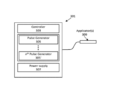
[0023] FIG. 3 is a schematic illustration of one example of an apparatus
for applying electric
pulses in the nanosecond range at low energy (e.g., low electric field) and
high frequency (e.g.,
in the megahertz range). For example, the apparatus may be configured to
include a controller
controlling a power supply and one or a plurality of nanosecond pulse
generators configured to
- 5 -
CA 03118718 2021-05-04
WO 2020/096836
PCT/US2019/058847
apply pulses having a pulse width of between about 0.1 ns and 1000 ns, an
amplitude of between
0.1 V and 20 V and a frequency of between 0.1 MHz and 30 MHz.
[0024] FIG. 4A illustrates one example of a circuit schematic for a
pulse generator for a
device such as the one shown in FIG. 3.
[0025] FIG. 4B is an example of a schematic for an apparatus for applying
nanosecond
electrical pulses at low electric field and high (e.g., megahertz) frequency,
including a plurality
of pulse generators that may be controlled by a controller to deliver a train
of nanosecond pulses
that are separated less than 1000 ns (e.g., having a frequency of greater than
or equal to 0.1
MHz,).
[0026] FIG. 4C is an example of a train of nanoseconds formed by a device
such as the one
shown in FIG. 4B; in FIG. 4C, the pulse generator (from FIG. 4B) responsible
for generating
each pulse in the train of pulses is indicated.
[0027] FIG. 5 is another example of a pulse generator apparatus
configured to generate
nanosecond pulses at low electric field and high (e.g., megahertz) frequency.
In FIG. 5, the pulse
generator may be used to generate bipolar pulses.
[0028] FIG. 6 is a schematic illustration of another variation of a
pulse generator apparatus
(e.g., system) configured to control and/or coordinate the application of
spatially separated
nanosecond pulses at low voltage in the megahertz range (e.g., from different
applicators and/or
different electrodes of a uniform applicator). This applicator may be
particularly useful with
bipolar nanosecond pulsing, as described herein.
[0029] FIG. 7 is an example of an experimental setup to apply nanosecond
pulses at low
electric field and megahertz frequency to cells. The setup in FIG. 7 also
allows imaging of the
cells.
[0030] FIG. 8A illustrates an example of excitation of a mouse
ventricular cardiomyocyte by
nanosecond pulses at low electric field (e.g., 85 V/cm) and megahertz
frequency (e.g., 3.33
MHz). In FIG. 8A a pulse train of 200 ns pulses at 3.33 MHz (separated by 100
ns between
pulses) was applied at 10 s. FIG. 8A shows a time lapse recording of calcium
activation (Fluo-4
dye emission, left column) and resulting cell contraction (DIC, right column).
Images were taken
with 0.24 s intervals, from top to bottom. Next images taken in the same
experiment (up to 48 s,
not shown) reveal no additional changes. Burst parameters and the onset of
burst delivery
(arrow) are indicated.
[0031] FIG. 8B illustrates an example of irreversible cell damage (e.g.,
poration of the cell,
causing cell death) by applying nanosecond electrical pulses at low electric
field (e.g., 150 V/cm)
and megahertz frequency (e.g., 3.33 MHz). In FIG. 8B, a train of nanosecond
electrical pulses
was applied to a mouse ventricular cell; the pulse train of 200 ns pulses was
applied at 3.33 MHz
- 6 -
CA 03118718 2021-05-04
WO 2020/096836
PCT/US2019/058847
(separated by 100 ns between pulses) at the 10 second time point. FIG. 8B
shows selected time
lapse of irreversible cell membrane damage and cell reshaping resulting from a
higher amplitude
burst. The time when each pair of images was taken is indicated at the right
side of the figure.
[0032] FIG. 9 illustrates the effect of repeated excitation of a
ventricular cardiomyocyte by a
bust of 1000 nanosecond pulses (e.g., 3.33 MHz, 200 ns width) at between 160-
340 V/cm.
Excitation was applied at the time indicated by the vertical line. Excitation
resulted in a spike in
cytosolic Calcium concentration, imaged using Fluo-4 dye. A first threshold
for excitation was
reached at 160 V/cm, resulting in the non-destructive influx of calcium. Above
400 V/cm cell
destruction occurred. Same bursts at 100 V/cm caused no effect, whereas at 400
V/cm the
excitation was followed by a prolonged Ca2+ increase (a sign of
electroporative damage to cell
membrane).
[0033] FIG. 10 is another example showing repeated excitation of
ventricular
cardiomyocytes similar to that shown in FIG. 9. Although the thresholds for
excitation are
slightly different, similar effect is seen.
[0034] FIG. 11A is a bar graph showing the threshold electric field for
excitation, possible
electroporation and definite electroporation in murine ventricular
cardiomyocytes exposed to a
train (e.g., burst) of 1000 nanosecond pulses (at 3.33 MHz, 200 ns width
pulses). Excitation was
evidenced by a brief spike in cytosolic Ca2+ concentration as imaged with Fluo-
4 dye. The
appearance of a low-amplitude spontaneous Ca2+ fluctuations following
stimulation was
regarded as a possible sign of electroporative membrane disruption ("maybe
electroporation").
Uncontrolled increase of Ca2+ without return to resting level within 40 s of
observation (such as
in bottom trace in FIG. 9) was considered evidence of electroporation.
[0035] FIGS. 11B-11E illustrate action potential thresholds for isolated
ventricular
cardiomyocytes (VCM) measured with FluoVolt dye. FIG. 11B is a graph showing
the effect of
pulse number for bursts of 100-, 200-, or 400-ns pulses, all with 100-ns
interval, showing that
increasing the number of pulses decreased the threshold similarly for all
durations. FIG. 11C is a
graph showing the thresholds plotted against the total time "on" did not
depend on pulse
duration. FIG. 11D is a graph showing bursts of shorter high frequency, sub-
microsecond, low
electric field pulse excited VCM at lower time-average electric field. For all
of FIGS. 11B-11D,
Mean s.e., n=6-10.
[0036] FIG. 11E is a graph showing the same result as in FIGS. 11D for
bursts of 1000
pulses, 50- to 600-ns duration. Interpulse intervals were varied from 0.09 to
4.8 is. Mean s.e.,
n=25-30.
[0037] FIG. 12A is a graph summarizing the effect of applying a train of
nanosecond pulses
at 2 MHz to CHO cells (in this example, nanosecond pulses are 300 ns duration
at 580 V/cm). A
- 7 -
CA 03118718 2021-05-04
WO 2020/096836
PCT/US2019/058847
line, including error bars, showing mean values for all 8 cells examined is
included. FIG. 12A
shows electroporation of CHO cells by a compressed train (e.g., a burst) of
300-ns pulses, and
specifically, the time course of cytosolic Ca2+ response in a representative
group of 8 CHO cells
following the burst (arrow). Solid black lines are the data from individual
cells, and the curve
with points is their mean value +/- s.e.
[0038] FIG. 12B illustrates electroporation of CHO cells (DIC images on
the left, Fluo-4
emission on the right) over time following application of the nanosecond
pulses based on the
same data as shown in FIG. 12A. This figure shows representative DIC (left
side) and Fluo-4
emission (right side) images of the same cells at the indicated time points.
[0039] FIG. 13 illustrates bipolar cancellation. FIG. 13 (bottom) shows
Ca2+ activation by bi
and monopolar nanosecond pulses in CHO cells (mean +/- s.e., n = 20-28). The
top of FIG. 13
shows bipolar and monophasic pulse shapes and amplitudes.
[0040] FIG. 14A illustrates megahertz compression of offset and
overlapping bipolar
nanosecond pulse trains (e.g., bursts), which may provide remote stimulation
by superposition of
two bipolar nanosecond pulse trains at high (e.g., megahertz) frequency into a
unipolar electrical
nanosecond pulse train at high (e.g., megahertz) frequency. The approach is
illustrated for a
rectangular train bipolar pulses and linear arrays of stimulating electrodes.
Two pairs of
independent, ground-isolated electrodes (a-a' and b-b') deliver two
synchronized, nanosecond
bipolar electric pulse trains as shown in the right side. Each of the
nanosecond bipolar pulses is
inherently inefficient for electrostimulation due to at least partial bipolar
cancellation, but their
superposition in the overlapping target region (c-c') font's a locally
biologically-effective
nanosecond unipolar pulse train at high frequency (e.g., in the megahertz
rate) by adjusting the
frequency of the pulse train between a-a' and b-b', as shown. The formation of
a unipolar
nanosecond pulse train in the c-c' area may be accomplished by controlling the
timing and
amplitudes of the energy applied between the electrodes a-a' and b-b'. Note
that the bipolar
cancellation is relevant at or near the electrodes, as it may reduce the
effect of the pulsed electric
field near the electrodes; in the remote target region, the monopolar field
may benefit from the
reduced thresholds resulting from the charge build up when using very rapid
(e.g., megahertz)
pulsing in the overlapping target region.
[0041] FIG. 14B is similar to FIG. 14A, but illustrates the use of pulse
trains (e.g., sub-
microsecond pulse trains) that combine in the target region c-c'.
[0042] FIG. 15A shows a schematic illustrating the CANCAN concept
according to one
embodiment. Top: A-A' and B-B' are two independent pairs of electrodes. The
lines between A
and B represent the area to which the E-field is delivered from each pair of
electrodes, which
overlap and nullify each other in the region C-C'. Bottom: Each pair of
electrodes delivers a
- 8 -
CA 03118718 2021-05-04
WO 2020/096836
PCT/US2019/058847
damped sine wave (DSW), which are per se biologically inefficient. When the
DSW from B-B'
is phase-shifted, the two DSW superpose into a biologically-effective unipolar
pulse in the C-C'
area. In this region, there is "cancellation of cancellation," or CANCAN.
[0043] FIG. 15B is similar to FIG. 15A but illustrates the use of pulse
trains (e.g., sub-
microsecond pulse trains) that combine in the target region c-c'.
[0044] FIG. 16A shows an example of an applied pulsed electrical energy,
at 60% duty
cycle.
[0045] FIG. 16B is a graph showing the effect of pulse repetition rate
on the threshold of
excitation by bursts of 340-ns pulses, 5 or 100 pulses/burst.
[0046] FIG. 16C is a graph illustrating the threshold time-average electric
field decreases
with burst duration, independently of indicated sub-microsecond pulse duration
(11-18 ns is the
duration range for the shortest setting in this example).
[0047] FIG. 16D is bar chart showing a comparison of excitation
thresholds for single pulses
of indicated duration and high frequency, sub-microsecond, low electric field
bursts. In FIGS.
16B-16D, mean s.e., n=5-9, and data collected using isolated frog sciatic
nerves.
100481 FIGS. 17A-17F illustrate one example of electroporation of CHO
and HEK cells by
high frequency, sub-microsecond, low electric field bursts evidenced by Ca2+
uptake or YO-
PRO-1 dye uptake. FIG. 17A is a graph showing that (in CHO cells) 100-pulse,
400-ns bursts
become increasingly more efficient above 0.1-0.3 MHz. Labels indicate the
electric field, in
kV/cm; 20-30 cells per data point. FIG. 17B is a graph showing the time course
of YO-PRO-1
uptake (in HEK cells) evoked by 1000 of 500-ns, 0.64 kV/cm pulses (arrow) at
indicated duty
cycle. FIG. 17C is a graph showing the last data point from FIG. 17B plotted
against the
repetition rate, showing a lack of effect below 1 MHz. FIG. 17D is a graph
showing Ca2+
transients evoked by same bursts (arrow) in HEK cells. FIG. 17E is a graph
showing similar Ca2+
transients evoked by one 500-ps pulse at indicated electric fields. FIG. 17F
shows maximum
amplitude of Ca2+ transients from the graphs of FIGS. 17D and 17E plotted
against the electric
field for a single 500-ps pulse (and against the duty cycle for high
frequency, sub-microsecond,
low electric field bursts). Mean s.e., n=11-27 for all panels. HEK cells
were stimulated in a
low-conductance solution.
[0049] FIGS. 18A-18D a graphs illustrating the viability of EL-4 cells 24 h
after exposure to
high frequency, sub-microsecond, low electric field bursts. FIG. 18A
illustrates the shape of high
frequency, sub-microsecond, low electric field pulses delivered to
electroporation cuvettes. The
dashed line denotes the time-average amplitude. FIG. 18B illustrates the
effect of pulse number
for 100-Hz and 3-MHz bursts for a low electric field of about 1.9 kV/cm. FIG.
18C is a graph
showing the same dependence for a high electric field of 5 kV/cm. FIG. 18D is
a bar chart
- 9 -
CA 03118718 2021-05-04
WO 2020/096836
PCT/US2019/058847
illustrating the high frequency, sub-microsecond, low electric field bursts at
indicated parameters
kill about 50% of cells, whereas a single pulse (whose duration equals burst
duration and the
amplitude equals the average of the burst) has smaller effect. Mean s.e.,
n=5-6. * p<0.0001, t-
test.
DETAILED DESCRIPTION
[0050] Described herein are methods and apparatuses (systems, devices,
etc.) that may
evoke one or more desirable biological and/or physiological effects using
electrical pulses in the
sub-microsecond (e.g., nanosecond) range at 0.1 megahertz and above (MHz)
frequencies, which
may permit the use of substantially lower electric fields compared to other
techniques. For
convenience, this electrical pulsing according to the present disclosure may
be referred to as
megahertz compression, megahertz compression of nanosecond pulses, or
megahertz
compression of nanosecond pulse trains (e.g., bursts). Unlike traditional
nanosecond pulsed
electric field treatments, which typically refers to very short, high
intensity pulses (e.g., high
electric field, typically much larger than 1 kV/cm), megahertz compression of
nanosecond pulses
may use much lower intensities, e.g., reduction by 5- to 10- fold or greater.
For example,
megahertz compression of nanosecond pulses may use very low electrical filed
values (e.g., at
the target site) that are less than lkV/cm (e.g., less than 900 V/cm, less
than 800 V/cm, less than
750 V/cm, less than 700 V/cm, less than 600 V/cm, less than 500 V/cm, etc.),
which is made
possible by applying the nanosecond electrical pulses at very fast rates,
e.g., in the megahertz
(MHz) range. The megahertz range may include 0.1 MHz or greater (e.g., 0.2 MHz
or greater,
0.4 MHz or great, 0.5 MHz or greater, 0.7 MHz or greater, 1 MHz or greater,
etc.) Surprisingly,
the apparatuses and methods described herein have been shown to result in
biological efficiency
of nanosecond electrical pulses to induce bio-effects at very low electric
field values;
specifically, at field values that were previously believed to have no effect.
The apparatuses
described herein are specifically configured to deliver pulse trains (e.g.,
bursts) of low-amplitude
nanosecond electrical pulses in the megahertz range, e.g., having intervals
between sequential
nanosecond electrical pulses that are typically equal or less than 1
microsecond (e.g., less than
about 900 ns, less than about 800 ns, less than about 700 ns, less than about
600 ns, less than
about 500 ns, less than 450 ns, less than 400 ns, less than 350 ns, less than
300 ns, etc.).
[0051] As mentioned above, traditional high-intensity, sub-microsecond
pulsing has been
limited to high pulse voltages in order to exceed the electric field (EF)
threshold for short pulse
durations (e.g., when applying sub-microsecond pulsing). This threshold
increases with pulse
shortening, up to tens of kV/cm. Strength-duration curves for neurostimulation
within the
nanosecond range typically require thresholds of between about 1 and 240 kV/cm
for 100- and 1-
- 10 -
CA 03118718 2021-05-04
WO 2020/096836
PCT/US2019/058847
ns pulses, respectively. For example, reported thresholds for a single high
intensity, sub-
microsecond pulsed electric field stimulus include between 1.4-2.4 kV/cm
(e.g., for activation of
cardiomyocytes by 200 ns pulses), greater than 1.8 kV/cm (e.g., for induction
of calcium
transients in HEK293 cells with 300-ns pulses), between 6 kV/cm and 1 kV/cm
(e.g., for
permeabilization of CHO cells by 60- and 600-ns pulses). Thus, it has long
been believe that
pulse voltages required to reach these thresholds were prohibitively high. For
example, achieving
kV/cm to ablate a tumor between two parallel-plate electrodes with a 2-cm gap
typically
requires 20 kV applied to the electrodes. Such voltages are beyond the
capability of most pulsed
generators and may present a high-voltage hazard. The methods and apparatuses
described
10 herein may avoid these problems.
[0052] For example, the methods an apparatuses descried herein may use
lower electric field
intensities, while providing comparable or superior effects. For example, the
methods and
apparatuses described herein may be used where high intensity, sub-microsecond
pulsed electric
fields have previously been shown to be effectively used, for example, for
neuromuscular
stimulation, including activating ion-selective nanopores (e.g., mobilization
of cytosolic Ca2+ in
both excitable and non-excitable cells, and opening inwardly rectifying and
ion-selective stable
nanopores in cell membrane), to cause non-chemical activation of diverse cell
and tissue types
and, at higher doses, to provide highly selective cell killing by necrotic
and/or apoptotic
pathways. The methods and apparatuses described herein may therefore also be
used for
defibrillation, peripheral nerve and deep brain stimulation, and tissue or
cell ablation (e.g., cancer
ablation).
[0053] Delivering multiple stimuli can result in a stepwise voltage
build-up on the membrane
of the target cell, eventually reaching the excitation or electroporation
threshold when the
interpulse interval is shorter than the relaxation of the induced
transmembrane potential.
Charging time constants in mammalian cells are typically at 0.1-1 ps. Temporal
summation can
only be expected at interpulse intervals smaller than 3-5 time constants
(which correspond to 95
and 99% discharge between sequential pulses), which translates into repetition
rates from tens of
kHz to more than 1 MHz. As described herein, delivering multiple sub-
microsecond pulsed
electrical energy at repetition rates of 1 Hz-5 kHz may cause stronger effects
than a single pulse,
but either without decreasing the threshold, or with a modest reduction of the
threshold.
[0054] For example, described herein is MHz compression of sub-
microsecond electrical
pulse bursts that facilitates excitation and electroporation at electric field
levels of, e.g., between
10-150 V/cm. As described, the efficiency may depend on the pulse and burst
parameters, which
also differ from conventional ("long") sub-microsecond electric stimuli.
- 11 -
CA 03118718 2021-05-04
WO 2020/096836
PCT/US2019/058847
[0055] FIG. 1 illustrates one example of a nanosecond pulse train (e.g.,
a nanosecond
electrical pulse train) applied at low voltage (or low electrical density, in
V/cm) and high, e.g.,
megahertz, frequency. In FIG. 1, the pulse train may include a plurality of
nanosecond pulses
101, each having a duration within the nanosecond range 108 (e.g., between
about 0.1 ns
duration and 1000 ns, e.g., between about 0.1 ns and about 950 ns, between
about 1 ns and about
900 ns, between about 1 ns and about 800 ns, between about 1 ns and about 750
ns, between
about 1 ns and about 700 ns, between about 1 ns and about 600 ns, less than
about 1000 ns, less
than about 900 ns, less than about 800 ns, less than about 750 ns, less than
about 600 ns, less
than about 500 ns, etc.). The nanosecond pulses may be separated by a pulse
interval 106 of less
than about 1000 ns, less than about 900 ns, less than about 800 ns, less than
about 750 ns, less
than about 700 ns, less than about 600 ns, less than about 500 ns, etc. Thus,
the frequency of the
nanosecond electrical pulses may be in the megahertz range, such as between
about 0.1 MHz and
about 50 MHz, between about 0.1 MHz and about 40 MHz, between about 0.1 MHz
and about
30 MHz, between about 0.1 MHz and about 25 MHz, between about 0.1 MHz and
about 20
MHz, between about 1 MHz and about 50 MHz, between about 1 MHz and about 40
MHz,
between about 1 MHz and about 30 MHz, between about 1 MHz and about 25 MHz,
between
about 1 MHz and about 20 MHz, greater than 0.1 MHz, greater than 0.2 MHz,
greater than 0.5
MHz, greater than 0.1 MHz, greater than 1.5 MHz, greater than 2 MHz, greater
than 2.5 MHz,
etc.
[0056] The energy applied by the nanosecond pulses of a pulse train such as
the one shown
in FIG. 1 may be relatively low. For example, the peak voltage applied by each
nanosecond
pulse may be less than about 500 V. less than about 100 V. less than about 50
V. less than about
40 V, less than about 30 V, less than about 25 V, less than about 20 V,
between about 0.5 V and
50 V, between about 1 V and 25 V, etc. Similarly the resulting electric field
of the applied
nanosecond pulses (e.g., at the tissue) may be, e.g., less than about 1000
V/cm, less than about
900 V/cm, less than about 800 V/cm, less than about 700 V/cm, less than about
600 V/cm, less
than about 550 V/cm, between about 1 V/cm and about 1000 V/cm, between about
10 V/cm and
900 V/cm, etc.
[0057] In general, megahertz compression of nanosecond pulses may be
applied to a
biological cell and/or tissue either directly or indirectly. For example, the
application may be
made into a patient's body via one or more tissue penetrating electrodes,
and/or via surface
electrodes. In some variations electrical stimulation using megahertz
compression of
nanosecond pulses may be applied to isolated cells and/or isolated tissues.
[0058] Without being bound by any particular theory of action, it is
possible that the
application of nanosecond pulses using megahertz compression of nanosecond
pulses may allow
- 12 -
CA 03118718 2021-05-04
WO 2020/096836
PCT/US2019/058847
the applied electrical energy to accumulate in the target cell or cell region,
allowing multiple
small-amplitude (e.g., less than about 500 V, less than about 100 V, less than
about 50 V less
than about 40 V, less than about 30 V, less than about 20 V, between about 0.1
V and 50 V,
between about 1 V and 30 V, between about 1 V and 20 V, etc.) pulses to sum to
a level that
.. exceeds the threshold for a biological effect. For example, nanosecond
electrical pulses applied
in the megahertz range at frequency that is shorter than the effective
electrical discharge time of
living cells may result in summing of the charge to above the threshold for a
desired effect. For
different mammalian cells, the discharge time constant (r) of the cell (e.g.,
of the cell membrane)
typically varies from about 100 ns for small cells, to about 1 microsecond for
larger cells (and
may be even longer for cells tightly packed in a tissue), thus the induced
membrane potential
reduces to approximately 37%, 15%, and 5%, 2% when the discharge duration
equals it, 2r, 3T,
and 4T, respectively. It is possible that when nanosecond electrical pulses
are applied in a pulse
train at a rate that is greater than the discharge rate (e.g., when the
membrane is not fully
discharged), the induced potential may add upon the remaining potential. With
multiple pulses
all applied with short enough inter-pulse intervals, the induced membrane
potential can gradually
climb up and exceed the threshold potential to induce bio-effects. See, e.g.,
Figs. 2A-2C. For
example, if one assumes that 4T is the longest time interval between the
pulses to enable
temporal summation of induced potentials, the interpulse intervals may not
exceed 400 ns for
smaller cells and 2,500 ns for large cells (perhaps up to 10,000 for still
larger cells with irregular
shape. Shorter inter-pulse intervals may allow faster accumulation of the
threshold
transmembrane potential. In practice, this means that nanosecond electrical
pulse trains may be
compressed to deliver the stimuli at repetition rates as high as 0.1-2 MHz,
with still higher rates
(up to 1,000 MHz) useful to further reduce the amplitude thresholds for
shorter nanosecond
electrical pulses and smaller cells.
[0059] In the example of FIG. 2A, nanosecond pulses are applied to a cell
or tissue in a pulse
train at a frequency of 10 kHz (e.g., separated by about 100 ps between each
pulse). Individual
pulses are 10 ns in duration, as shown on the bottom trace. The upper trace
shows the imposed
membrane potential (in arbitrary units) resulting from each pulse. For this
example tissue or cell,
the discharge time is approximately 8 his. The horizontal dashed line 203
represents the
minimum threshold for a biological effect (e.g., stimulation, poration, etc.)
that may be triggered
when the electrical potential of the membrane (which may include the outer
cell membrane
and/or one or more internal cell membranes) reaches the threshold. The tissue
or cell may have
multiple thresholds, corresponding to different or increasing effects.
[0060] FIGS. 2B and 2C illustrate megahertz compression of nanosecond
pulse bursts,
showing that megahertz compression permits the cell or tissue to reach and
exceed one or more
- 13 -
CA 03118718 2021-05-04
WO 2020/096836
PCT/US2019/058847
thresholds for bio-effects without increasing pulse amplitude. For example, in
FIG. 2B, each
100-ns pulse (bottom trace) induces a transmembrane potential (upper trace, in
arbitrary units).
Because the energy is not discharged as quickly as the inter-pulse interval,
it accumulates, until
the continued application of nanosecond pulses causes the imposed membrane
potential to
.. exceed the threshold 203, as shown, which may result in the corresponding
bio-effect. In FIG.
2B, the marginally high rate (e.g., 100 ns pulses with approximately 5 us
between each pulse)
results in a minor temporal summation which helps to reach the low threshold
203 that may
induce some bio-effect. In FIG. 2C, when a high repetition rate (e.g., 1 MHz,
with 1 us between
pulses) is applied, the imposed membrane potential may accumulate faster and
reach a much
higher threshold 205.
System for the application of megahertz compression of nanosecond pulses
[0061] FIGS. 3-5 illustrate examples of systems that may be used to
provide megahertz
compression of nanosecond pulses. For example, FIG. 3 is a schematic of one
example of a
system 301 for applying nanosecond pulsing using megahertz compression of
nanosecond
pulses. In FIG. 3, the system may include at least a controller 303, one or
more pulse generators
305, a power supply, and one or more applicators 309. The system may be
portable, and the
power source may include power conditioning circuitry that receives power from
a battery and/or
a wall source (e.g., outlet). The battery may be rechargeable. The system may
be at least
partially enclosed. In some variations the applicator may be connected via one
or more cables.
Alternatively, the applicator may be integrated with the rest of the system in
a compact, hand-
held configuration.
[0062] The controller may be configured to apply a fixed or adjustable
train of nanosecond
pulses in which the individual pulses are separated by a fixed or adjustable
inter-pulse interval so
that the frequency is in the megahertz range (e.g., between about 0.9 MHz and
100 MHz,). The
inter-pulse interval may be between, e.g., 1200 ns and 50 ns (e.g., within the
megahertz
frequency range), such as between about 1000 ns and 50 ns, between about 1000
ns and 75 ns,
between about 1000 ns and 80 ns, between about 1000 ns and 90 ns, between
about 1000 ns and
100 ns, etc. In some variations the controller is configured or adapted to
limit the applied
stimulation to within this frequency range; in some variations the apparatus
may include one or
.. more user inputs (knobs, dials, touchscreens, etc.) in communication with
the controller to allow
the user to adjust the applied frequency/inter-pulse interval within this
megahertz range.
Alternatively or additionally, the controller may be configured to allow the
user to adjust the
number of pulses, the duration that pulses will be applied, and/or voltage
applied. The voltage
applied may typically be within a predetermined range (e.g., voltage amplitude
of between about
- 14 -
CA 03118718 2021-05-04
WO 2020/096836
PCT/US2019/058847
0.1 V and about 50 V, between about 0.1 V and 40 V, between about 0.5 V and 30
V, between
about 1 V and about 20 V. less than about 50, less than about 40 V, less than
about 30 V, less
than about 25 V, less than about 20 V, etc.). In some variations, the
intensity of the electric field
may be selected (e.g., between about 1 V/cm and about 900 V/cm, between about
1 V/cm and
about 800 V/cm, between about 10 V/cm and about 750 V/cm, between about 10
V/cm and
about 700 V/cm, between about 50 V/cm and about 650 V/cm, less than 1000 V/cm,
less than
900 V/cm, less than 800 V/cm, less than 750 V/cm, less than 700 V/cm, less
than 600 V/cm, less
than 500 V/cm, etc.). Any of these ranges may be considered within the low-
voltage range. As
mentioned, in some variations the controller may allow the user (e.g., doctor,
surgeon,
technician, etc.) to select from a predetermined set of values or range,
including any of the
ranges described herein. In some variations the user may be provided with
preset values for one
or more of: number of pulses, pulse duration (within the nanosecond range),
pulse amplitude
(e.g., peak voltage within the predetermined low-voltage range), inter-pulse
interval and/or
frequency (e.g., within the megahertz range), etc. In some variation the
apparatus may be preset
or may automatically select appropriate parameters, and the user may only
select start or stop, or
may select between a limited number of parameter states.
[0063] The controller may include hardware, software and/or firmware to
allow it to control
the operation of the system and/or receive controlling input from the user.
For example, the
controller may include circuitry including one or more processors, one or more
timing circuits,
one or more memories, etc. As mentioned, the system may include one or more
inputs (e.g.,
controls) and/or may receive input from another device (e.g., via a wired or
wireless connection).
The system may include one or more outputs (e.g., monitors, displays, LEDs,
etc.), including
indicators of the device operation (e.g., ready, standby, etc.) and/or the
settings (number of pules,
frequency, voltage amplitude, etc.).
[0064] As mentioned, any of the systems described herein may include one or
more
applicators. An applicator may include two or more electrodes, including
arrays of electrodes.
The electrodes may be tissue penetrating or non-tissue penetrating. For
example, a tissue
penetrating electrode may be a needle electrode; a non-tissue penetrating may
be a surface
electrode or electrodes.
[0065] In any of the apparatuses described herein, the controller may
coordinate the
activation of one or more (e.g., a plurality) of pulse generators, as shown in
FIG. 3. Each of the n
pulse generators may be configured to apply a nanosecond pulse at a
coordinated time, and these
pulses may be combined into a single stimulation. The individual please
generators may be
configured to deliver a pulse having a pulse duration in the nanosecond range
(e.g., between
.. about 0.1 ns and about 1000 ns), at a peak voltage within a low-voltage
range as described
- 15 -
CA 03118718 2021-05-04
WO 2020/096836
PCT/US2019/058847
above. While FIG.3 illustrates a plurality of pulse generators, it should be
understood that in
various applications only one pulse generator may be used.
[0066] For example, FIG. 4A schematically illustrates an example of a
pulse generator
circuit that may be used. In FIG. 4A, the circuit 400 includes a pre-charged
capacitor (Cl)
switched by an N-type (n-channel) MOSFET switch (M1). The switch may be
triggered by one
or more low voltage MOSFET drivers. In this example, a Zener diode 403 may be
placed at the
transformer output to clamp the voltage. The output voltage may be set (e.g.,
within the low-
voltage range described above). The settings may be modified by selecting the
R, L, and C and
values, creating a critical damped mode that is favorable for ultrafast
pulsing.
[0067] As illustrated in FIG. 4B, in some embodiments a plurality of pulse
generator circuits,
such as the pulse generator circuit schematically shown in FIG. 4A, may be
coupled together and
controlled by the controller, as mentioned above. In FIG. 4B, each block (1,
2, 3, and 4)
represents a pulse generator that may be controlled by the controller and used
to form a
compound/combined nanosecond pulse train of pulses within the megahertz
pulsing frequency
having a low electric field (e.g., low voltage range). In FIG. 4B, a single
pulse generator does
not need to generate nanosecond duration pulses at high frequency (e.g.,
within the multi-MHz
range); instead several of the pulse generators are grouped to produce pulses.
When using
relatively low voltages (e.g., <1 kV, including the low voltage ranges
described above) in the
pulse regime, the RC-switch modules can be combined laterally to produce
multiphasic pulses of
arbitrary amplitudes, width and intervals. Each module can be charged with
different DC
sources, therefore offering different output voltages. Each module can be
triggered separately,
allowing various pulse widths (PWs) and delays. Alternatively, all or some of
the modules may
produce identical pulses, which may be combined as described above. FIG. 4C
illustrates one
example of a combined train of nanosecond-duration pulses originating from
four separate but
linked pulse generators, such as shown in FIG. 4B. In this example the pulses
are nonpolar (e.g.,
all positive or all negative going) and are shown as equivalent pulses; as
mentioned, in some
variations pulses having different durations and/or different voltages may be
applied within the
same pulse train. In some variations, as will be described in greater detail
below, bipolar pulses
of nanosecond duration may be delivered in the megahertz frequency range.
[0068] For example, FIG. 5 illustrate another example of a schematic for a
pulse generator
configured to provide bipolar stimulation. In FIG. 5 the pulse generator 500
combined positive
pulsing circuitry and negative pulsing circuitry to create the bipolar pulse
generator. In FIG. 5,
the switch connection in the negative pulsing circuitry is drain-to-ground
(and source-to-
negative) charging, which is opposite to that shown in the positive pulsing
circuitry. The same
load resistor may be used for both positive and negative pulsing circuitry.
The voltage and pulse
- 16 -
CA 03118718 2021-05-04
WO 2020/096836
PCT/US2019/058847
duration can be differentially adjusted. One can also trigger just one of the
positive or negative
pulsing circuitry so that either a positive or negative pulse is delivered. A
delay of any length can
be inserted in any interval. A plurality of bipolar stimulators such as those
shown in FIG. 5 may
be combined as shown in FIG. 4B, allowing megahertz frequency stimulation. The
pulse
generators shown in FIGS. 4A-5B represent merely non-limiting examples of
pulse generators
that may be used to perform the methods described herein. Other pulse
generators may be used
or configured to generate trains of nanosecond pulses at the described
frequencies.
[0069] Any of the systems described herein may also be configured as
shown in FIG. 6, in
which a single controller controls two or more sets of pulse generators that
may be used to apply
nanosecond electrical pulses from different locations (e.g., on or in a body).
In FIG. 6 the
system 601 includes a single controller 603 (which, in some variations may be
two or more
linked controllers) controls and coordinates electrical stimulation by the
application of
nanosecond pulses at low voltage in the megahertz frequency range from each of
two (or two
sets) of pulse generators 605..605' and 615..615'. As will be described in
greater detail below in
the context of bipolar pulsing, this may allow for non-invasive electrical
stimulation with
minimum near-electrode effects. The same power supply 607 or different power
supplies may
be used to provide power (e.g. to the pulse generators). One or more
applicators 609, 609' each
including electrodes for the application of the nanosecond pulses may be used.
In some
variations a single applicator having different electrode sets, which may be
separated by a known
or predetermined distance and/or geometry, may be used. In one variation of
the example shown
in FIG. 7, at about 140 urn between the tips of the electrodes, pules applying
about 16 V was
equivalent to about 570 V/cm.
[0070] The application of megahertz compression of nanosecond pulse
bursts, in which low-
voltage nanosecond pulses were applied at high (e.g., megahertz) frequencies
was examined
using an in vitro model to demonstrate the effects. For example, FIG. 7
illustrates one
arrangement of a testing set-up 700 in which one or more cultured or explanted
cells were
examined optically (e.g., using a laser 703 coupled to a microscope imaging
system 705) while
applying low-voltage nanosecond pulses at high frequency through a pair of
electrodes 707, 707'
coupled to a system such as the one shown in FIG. 5. Using a system similar to
that shown in
FIG. 7, the use of megahertz burst compression was verified in several types
of mammalian cells
in vitro (e.g., CHO, HEK 293, and enzymatically isolated murine primary
ventricular
cardiomyocytes, VCM). The effect of electrical pulses in the nanosecond range
was documented
by time-lapse recording of cytosolic Ca2+ activation with a Fluo-4 fluorescent
indicator and
transillumination recording of cell shape changes. Depending on the cell type
and other
conditions and intensity of electrical pulses in the nanosecond range, these
observations reflected
- 17 -
CA 03118718 2021-05-04
WO 2020/096836
PCT/US2019/058847
cell membrane permeabilization, opening of voltage-gated calcium channels,
contractile activity,
and/or cell-reshaping, or other types of bio-effect due to membrane
disruption.
[0071] FIGS. 8A-8B, 9, 10, 11A and 12A-12B illustrate examples of
testing of sub-
microsecond (e.g., nanosecond) pulses, e.g., having pulse durations from 50 to
300 ns, focusing
primarily on 200- and 300-ns stimuli. Pulses were delivered in bursts from 100
to 5,900 pulses,
at repetition rates of between about 1.6 and 3.33 MHz, which corresponded to
inter-pulse
intervals from about 150 to 450 ns. The pulse amplitude was varied from
between about 2 to 17
V, with the resulting electric field from about 70 to 570 V/cm, respectively,
at the cell location.
A total of about 200 pulses of electrical stimulation in the nanosecond range
treatments in 26
individual VCM and more than 80 nanosecond pulses in treatments in other cell
types (> 120
individual cells) were examined. Cell responses were consistently recorded (as
shown in FIGS.
8A-12B) despite extremely low pulse amplitudes (as compared to standard
treatments using
pulsed electric fields in the nanosecond range). The lowest electric field
that caused VCM
activation and contraction was only 85 V/cm (e.g., FIG. 8A, showing a burst of
1,000 pulses, 200
ns pulse duration, 3.33 MHz). This was a >30-fold reduction of the electric
field compared to the
published threshold of about 2.5 kV/cm for 200-ns pulses (including single 200
ns pulses). A
slightly higher electric field, about 160 V/cm, caused calcium activation
(excitation) and cell
contraction in 17 out 18 VCM which were tested and proven functional and
capable of
generating this type of response. The threshold did not show significant
dependence on whether
cells were oriented parallel or perpendicular or at any other angle to the
electric field. At this low
electric field using nanosecond pulses in the megahertz frequency range,
excitation in individual
VCM could be repeated multiple times (see, e.g., FIG. 8B) with no signs of
electroporation or
damage. Increasing the electric field about 2-fold above the excitation
threshold resulted in the
observation of modest membrane disruption, such as spontaneous sparkletts and
elevation of
resting level of cytosolic Ca2+. Further increase in the electric field, to
only about 400+/-18
V/cm caused irreversible VCM electroporation, with no recovery of cytosolic
Ca2+ and
transformation of VCM from a healthy "brick shape" to a "meatball shape", a
recognized
manifestation of permanent VCM damage. See, e.g., FIGS. 8B and 11A and the
last trace in
FIG. 9). In general, the excitation response was also observed with shorter
pulse trains, such as
400 or 500 pulses, 3.33 MHz at 0.16 kV/cm, as well as with shorter 100-ns
pulses (3900 pulses,
2 MHz, at 570 V/cm). VCM were repeatedly excited with trains of tens of
compressed
nanosecond electrical pulse bursts applied every 2 s (data not shown).
[0072] As shown in FIGS. 8A-8B, bursts of high frequency, sub-
microsecond, low electric
field electrical energy may be used for stimulation and/or electroporation of
mouse ventricular
cardiomyocytes (VCM). In some variations, high frequency, sub-microsecond, low
electric field
- 18 -
CA 03118718 2021-05-04
WO 2020/096836
PCT/US2019/058847
stimulation may be used for defibrillation. As described herein, the
previously-limiting need for
high voltage can be offset by applying high-rate bursts. For example,
compressing 1000, 200-ns
pulses into 3.33 MHz bursts enabled excitation at only 80-200 V/cm, as shown
in FIGS. 8A, 9
and 11A, which is 10-20 times lower than with a single 200-ns shock.
Excitation by such high
frequency, sub-microsecond, low electric field stimulation bursts (as shown in
FIG. 8A and 9)
was distinctly different from the persisting Ca2+ elevation and cell shrinkage
following
membrane damage by stronger electric field (as shown in FIG. 8B). VCM
excitation by a single
low frequency (e.g., < 1 MHz), sub-microsecond, high electric field (e.g.,
greater than 1 kV/cm)
stimulation pulse was damaging at the excitation threshold, causing abnormal
action potentials
and long-lasting Cal elevation. This phenomena was generally true; in contrast
to the lower-
frequency, sub-microsecond, high electric field stimulation, the high
frequency, sub-
microsecond, low electric field stimulation described herein was substantially
less damaging,
while being highly effective.
[0073] For example, as shown in FIG. 9, repeated stimulation with MHz
bursts caused no
damage. Initial signs of electroporation were observed at 350-400 V/cm (see,
e.g., FIGS. 8B and
11A), i.e., 2-3 times above the excitation threshold (p<0.001), allowing for a
large safety
window. With a fixed 100-ns interval between sub-microsecond pulsed
stimulation, thresholds
decreased as a power function for pulse widths from 100 to 400 ns (see, e.g.,
FIG. 11B). The
threshold was determined by the total time "on" within bursts, whereas the
individual high
frequency, sub-microsecond, low electric field pulse duration did not matter
(see, e.g., FIG.
11C). The threshold time-average electric field was plotted against burst
duration was smaller
for shorter pulses (FIG. 11D).
[0074] This unexpected result was verified in a separate set of
experiments where VCM were
excited by bursts of 1000 pulses; pulse duration was varied from 50 to 600 ns,
and the interpulse
intervals were changed from 90 ns to 4.8 vs. Plotting the time-average
threshold electric field
values against burst duration yielded significantly smaller values for shorter
pulses (FIG. 11E),
consistent with the previous experiment in VCM but contrasting nerve
excitation (see, e.g.,
FIGS. 16A-16D, below). This may be indicative of a specific effect of sub-
microsecond pulsing,
distinctly different from conventional pulses.
[0075] In HEK293 and CHO cells, megahertz compression of bursts of pulses
in the
nanosecond range elicited Ca2+ transients, with either complete or partial
recovery within 40-s
observation after the stimulation, as shown in FIGS. 12A-12B. In contrast to
VCM which were
subjected to pulses in the nanosecond range as one cell at a time, these
cultured cells were
exposed as small groups. The probability and the amplitude of response
increased for higher
electric fields (range tested: 130-570 V/cm), higher pulse numbers (range
tested: 100-1,000), and
- 19 -
CA 03118718 2021-05-04
WO 2020/096836
PCT/US2019/058847
shorter interpulse intervals (range tested: 150-300 ns). The pulse duration
was kept constant at
300 ns, to facilitate comparison with published work. Typical nanosecond
pulsing at lower
frequencies (including single pulsing) had a threshold of response to 300-ns
pulse(s) at about 1.8
kV/cm. CHO cells do not express any voltage gated Ca2+ channels, and thus it
is believed that
Ca2+ transients are a result of electroporation.
[0076] Based on this experimental work, megahertz compression of
nanosecond bursts (e.g.,
the application of low-voltage nanosecond pulses in the megahertz frequency
range) enabled
profound reduction of electrical energy thresholds for both excitation and
electroporation.
Surprisingly, there was also a clear separation of excitation and
electroporation thresholds (see,
e.g., FIG. 11A) and repeated excitation without damage, which has been
problematic when using
traditional electric pulsed fields in the nanosecond range.
The Application of Megahertz Compression of Nanosecond Bursts
[0077] As discussed briefly above, any of the methods and apparatuses
(e.g., devices,
systems, applicators, etc.) described herein may be used to treat tissue. Any
appropriate tissue
may be treated, including, but not limited to: skin, liver, kidney, neuronal
(brain, spine,
peripheral), lung, muscle, adipose, respiratory, gastrointestinal, bladder,
reproductive, etc. tissue,
including tumorous tissue. The nanosecond pulses at low electric field (e.g.,
low voltage) and
high (e.g., megahertz) frequency described herein may be used to manipulate
biological
functions and treat diseases. Responses to such electrical stimulation may
include a variety of
bio-effects, including but not limited to: nerve and muscle excitation,
activation of immune (or
otherwise stimulating an immune response) and endocrine cells, cell
differentiation,
electroporation, necrotic and apoptotic cell death. Thus, the use of
nanosecond pulses at low
electric field and megahertz frequency may be used in virtually any indication
in which electrical
stimulation may be applied. In general, any of the high frequency nanosecond
pulse generators
and methods of using them described herein may be used for a medical therapy.
[0078] For example, the methods and apparatuses of the present
disclosure may be used for
cardiac pacing, defibrillation, muscle training and rehabilitation, pain
control, alleviation of
Parkinson disease symptoms, psychiatric disorders, and cancer ablation. They
may also be used
in neuromuscular and psychiatric disease diagnostics and research.
[0079] For example, devices, systems and methods described herein may be
utilized in
various ablation procedures (e.g., radiation-based), dermatological procedures
(e.g., treating
various dermatological conditions, such as skin cancers), general surgery
procedures (e.g.,
pancreatectomy), cardiology (e.g., valve repair), gynecology (e.g.,
hysterectomy), neurosurgery
(e.g., tumor resection) etc.
- 20 -
CA 03118718 2021-05-04
WO 2020/096836
PCT/US2019/058847
[00801 Any of the methods described herein may be applied to excitable
tissues (including
but not limited to neuronal tissues) for either excitation and/or ablation or
other tissue treatments.
For example, described herein are methods and apparatuses for the stimulation
of excitable
tissues such as nerve and heart muscle, the treatment of neurological
disorders such as epilepsy,
Parkinson's disease and stroke. Heart disorders could include atrial
fibrillation and ventricle
fibrillation. As demonstrated above, the membrane potential of one or a group
of cells may be
excited directly using the methods described herein. The methods and
apparatuses described
herein may be used to stimulate secretion in cells such as platelets.
[0081] The methods and apparatuses described herein may find particular
use in treating the
brain, peripheral nerves, muscles, and heart. As mentioned above, these
methods may be used to
for cardiac pacing, defibrillation, deep brain stimulation in Parkinson's
disease, functional
nanosecond electrical pulses for restoring functionality of skeletal muscles,
and pain control to
the emerging applications in fibromyalgia, depression, dementia, epilepsy,
diabetic neuropathy,
and many others. In particular, the nanosecond pulses at low electric field
(e.g., low voltage) and
high (e.g., megahertz) frequency described herein may be used to treat any
indication in which it
may be beneficial to modulate or introduce action potentials (AP) in nerve and
muscle targets.
For example, the methods and apparatuses described herein may be used for
modulation (e.g.,
shifting) of resting potential, changing the synaptic efficiency. AP induction
is accomplished by
creating a transient voltage gradient at the target, either through the
inserted or implanted
electrodes, or non-invasively from the surface. Alternatively or additionally,
any of the methods
and apparatuses described herein may be used for electroporation.
[0082] Any of the tissues described herein may be selectively modulated
using the
application of megahertz compression of nanosecond pulses by applying trains
of low-voltage
nanosecond pulses in the megahertz frequency range. In some variations the
methods described
herein may modulate the cell based, at least in part, on the size of the cell
and/or the membrane
content of the cell. For example, these methods may affect cells having a high
time constant for
discharge (e.g., higher capacitance) compared to other cells, which may be a
function of the
composition and/or size of the cells.
[0083] For example, nanosecond electrical pulses at low electric field
and megahertz
frequency may be used to treat a patient's skin, including treatment of one or
more of: acne,
seborrheic keratosis, keloids, molluscum contagiosum, acrocordon, psoriasis,
papilloma, human
papilloma virus (HPV), melanoma, melasma, sebaceous hyperplasia, syringoma,
congenital
capillary malformation (port-wine stains), melasma, actinic keratosis,
dermatosis papulosa nigra,
angiofibroma, skin tumors, aging skin, wrinkled skin, cherry angioma,
epidermal/sebaceous cyst,
basal cell carcinoma, aging skin, benign tumors, precancerous tumors, cancers
and warts. These
- 21 -
CA 03118718 2021-05-04
WO 2020/096836
PCT/US2019/058847
methods and apparatuses may also be used for cosmetic skin treatments,
including tattoo
removal, hair follicle destruction, scartkeloids reduction, fat reduction, and
wrinkle reduction.
For example, the methods and apparatuses described herein may be useful for
treating
melanomas by causing them to self-destruct. In general, these methods may be
useful for in
vitro treatment of skin lesions.
[0084] The methods and apparatuses described herein for applying
nanosecond electrical
pulses at low electric field (e.g., low voltage) and high (e.g., megahertz)
frequency may be useful
for nanoelectroablation and vaccination.
[0085] Thus, the methods and devices described herein may be used in
treatment of various
diseases. A "disease" includes any abnottnal condition in or on a subject that
is associated with
abnormal, uncontrolled growths of tissue, including those that are cancerous,
precancerous, and
benign, or other diseases as known in the art. The methods and devices of the
present invention
can be used for the treatment of any type of cancer, whether characterized as
malignant, benign,
soft tissue, or solid, and cancers of all stages and grades including pre- and
post-metastatic
cancers. Examples of different types of cancer include, but are not limited
to, digestive and
gastrointestinal cancers such as gastric cancer (e.g., stomach cancer),
colorectal cancer,
gastrointestinal stromal tumors, gastrointestinal carcinoid tumors, colon
cancer, rectal cancer,
anal cancer, bile duct cancer, small intestine cancer, and esophageal cancer;
breast cancer; lung
cancer; gallbladder cancer; liver cancer; pancreatic cancer; appendix cancer;
prostate cancer,
ovarian cancer; renal cancer (e.g., renal cell carcinoma); cancer of the
central nervous system;
skin cancer (e.g., melanoma); lymphomas; gliomas; choriocarcinomas; head and
neck cancers;
osteogenic sarcomas; and blood cancers.
[0086] These methods and apparatuses may also or alternatively be useful
for ablating cancer
and generating resistance to new cancer growth, including treatment of tumors.
Examples of
tumors include a benign prostatic hyperplasia (BPH), uterine fibroid,
pancreatic carcinoma, liver
carcinoma, kidney carcinoma, colon carcinoma, pre-basal cell carcinoma, and
tissue associated
with Barrett's esophagus.
[0087] The methods and apparatuses described herein may be used for gene-
electrotransfer
or "GET". In some variations the disease, including cancer, may be treated by
transfer of genes
(e.g., in one or more plasmids coding for genes that could stimulate an immune
response) being
introduced into tumors. For example, melanoma may be treated using a plasmid
containing the
gene for interleukin 12 (IL-12), which may stimulate the differentiation of
naïve T cells into Thl
cells as well as the production of interferon-gamma and tumor necrosis factor-
alpha.
Alternatively, any of the methods and apparatuses described herein may be used
to porate cells
of a tissue, including in particular, tumor cells. This may permeabilize cells
by generating pores
- 22 -
CA 03118718 2021-05-04
WO 2020/096836
PCT/US2019/058847
large enough to allow the transport of small molecules across the plasma
membrane. As shown
in FIGS. 8A-8B, both reversible and irreversible electroporation may be
achieved. For example,
the methods and apparatuses described herein may be used for
electrochemotherapy (ECT) may
be used for the treatment of several cutaneous tumor targets, including
melanoma, basal cell
carcinoma, breast cancer and Kaposi's sarcoma (which may include the use of
bleomycin,
cisplatin, or other drugs). The methods and apparatuses described herein may
be used to cause
irreversible electroporation (IRE) which may lead to necrosis. For example,
the methods and
apparatuses described herein may be effective to treat, among other
indications, prostate, brain
tumor ablations (including gliomas), pancreatic cancer, colorectal liver
metastases, unresectable
renal tumors and rectal neoplasms.
[0088] The megahertz compression of nanosecond pulse trains described
herein may be
particularly effective in treating diseases including cancers because they may
penetrate into the
intracellular region of the cell(s). The ability to penetrate beyond the
plasma membrane (possibly
due to the pulse rise time reaching full amplitude in the nanosecond range) is
typically much
faster than the time required for intracellular and intraorganellar charges to
redistribute to cancel
the imposed field. This may allow the methods and apparatuses described herein
to permeabilize
small organelles by applying electrical pulses in the nanosecond range (e.g.,
including vesicles,
mitochondria, endoplasmic reticulum and nuclei).
[0089] The methods and apparatuses described herein may also be useful
for platelet
activation (in the absence of thrombin); for example, these methods may be
used for applying
electrical pulses in the nanosecond range of platelet-rich plasma to improve
wound healing and
enhance blood flow.
[0090] As mentioned, above, the megahertz compression of nanosecond
pulse bursts
described herein may be used to influence tumor growth; for example, to treat
tumors with
electrical pulses in the nanosecond range at low electric field and high
frequency, e.g.,
megahertz, so that the tumor disappears over days to weeks, and may exhibit
characteristics of
immunogenic cell death (ICD), e.g., releasing DAMPs such as calreticulin
translocation from the
ER to the cell surface, ATP release and HMGB 1 release. These methods may also
be used to
inhibit metastasis.
[0091] Similar to the use of pulsed electric fields in the nanosecond range
using high
voltages (e.g., 30 kV/cm) the methods described herein may also provide
treatments that are
drug-free, very fast and leave no scar, and may be treated with only one or a
few treatments.
However, unlike other pulsed regimes, the methods and apparatuses described
herein do not
require a large electric field to achieve the desired effect (including
immunogenic cell death
- 23 -
CA 03118718 2021-05-04
WO 2020/096836
PCT/US2019/058847
effects). Thus, the size of the ablation zone may be larger and may be more
easily applied and/or
tolerated by the tissue.
[0092] It should be noted that the examples given herein are for the
purposes of illustration
and example only, the description as set forth is not intended to be
exhaustive or limiting.
Megahertz Compression of Nanosecond Bursts with Bipolar Pulse Targeting
[0093] In general, the methods and apparatuses described herein for
megahertz compression
of nanosecond bursts, e.g., providing nanosecond pulses at low electric field
and high (e.g.,
megahertz) frequency, may be used with multiple sources for applied electrical
energy (e.g.,
electrodes, antenna, etc.). For example, two sets of electrodes may be used to
apply energy from
separate regions on, in, or around the tissue, and the regions in which
applied energy sums (e.g.,
by superposition) to generate a pulse train of sub-microsecond pulses at a
frequency that is in the
megahertz range (e.g., greater than or equal to about 1 megahertz), likely
resulting in
accumulation of charge at the cell membrane(s), the resulting megahertz
compression may result
in triggering a bio-effect at a substantially lower threshold as compared to
applied electric fields
that are pulsed at lower rates, including single pulses. As described above,
this may be views as
an apparent lowering of the threshold for triggering the bio-effect, in
reference to the amount of
energy that is applied.
[0094] In some embodiments of this application, the methods and
apparatuses described
herein for megahertz compression of nanosecond bursts, e.g., providing
nanosecond pulses at
low electric field and high (e.g., megahertz) frequency, may be applied in
combination with the
use of bipolar pulses in order to specifically target the treatment zone at a
distance from the
application of the electric pulses while minimizing the effect of the
electrical pulsing at or near
the electrodes (in non-target regions). U.S. Application no. 16/104,089, filed
August 16, 2018
("TARGETED REMOTE ELECTROSTIMULATION BY INTERFERENCE OF BIPOLAR
NANOSECOND PULSES") describes the use of bipolar, nanosecond pulses at high
voltages
from different but overlapping regions of a tissue to specifically target
overlapping regions
(referred to CANCAN). Interestingly, as shown in FIG. 13, nanosecond pulsed
activation using
bipolar nanosecond pulses at high voltage (e.g., high electric field
strengths) does not result in
activation of bio-effects in a cell membrane. As shown in FIG. 13, described
in greater detail
below, the use of bipolar nanosecond pulses appears to cancel out the effects
of the nanosecond
pulse on the tissue, likely because the bipolar nature of the pulse, in which
the second half (e.g.,
a negative-going portion of the pulse) undoes any charge movement due to the
first half (e.g., a
positive-going) portion of the pulse. However, when two bipolar pulses overlap
and are out of
synch, which may happen when, for example, the pulses are applied from two
different regions
- 24 -
CA 03118718 2021-05-04
WO 2020/096836
PCT/US2019/058847
of the tissue and at different times, the electric field in the overlapping
region may superimpose
and result in a monopolar pulse in the overlapping region. Thus, the
overlapping region may be
affected by the resulting monopolar nanosecond pulsing even while the rest of
the tissue is
unaffected by the bipolar nanosecond pulsing.
[0095] The megahertz compression of nanosecond bursts may be combined with
this
CANCAN effect to provide highly effective electrical pulses (e.g., including
but not limited to
electrical stimulation) of even deep tissue regions. This megahertz compressed
CANCAN
technique may be particularly effective since the energy required to evoke an
effect when using
megahertz compression of nanosecond pulsing is dramatically lower than that
required without
.. megahertz compression. This effect may be even further enhanced by the use
of antennas to
emit the nanosecond bursts at high frequency (e.g., within the megahertz
range).
[0096] Thus, also described herein are methods and apparatuses (see,
e.g., FIG. 7), that emit
multiple trains of bipolar nanosecond electrical pulses at high (e.g.,
megahertz) rates from
different locations to form a region of overlap in which the multiple bipolar
nanosecond pulsed
__ trains superimpose to form a localized train of monopolar nanosecond pulses
at low electrical
filed. The resulting superimposed monopolar nanosecond pulses at low
electrical filed may be
above threshold for evoking a bio-effect (e.g., stimulation, poration, etc.)
in the localized
overlapping region.
[0097] The use of megahertz compression of nanosecond bursts with CANCAN
may result
in sharpening of the CANCAN effect. It is somewhat counterintuitive to combine
the use of
bipolar cancellation of CANCAN with megahertz compression of nanosecond
bursts, since it is
unlikely that megahertz compression would have any effect on bipolar pulsing
from a single
source (e.g., a single train of bipolar, nanosecond pulses at low electric
field strength). Since
there is an immediate discharge at every pulse (as in traditional bipolar
nanosecond pulsing), a
__ bipolar pulse train should not be amenable to megahertz compression, which
may enhance the
voltage effect at the membrane by temporally summing the charge between
pulses. In addition,
because the use of megahertz repetition rate allows substantially lower
electric field strengths to
be applied to achieve comparable bio-effects, these methods and apparatuses
may compensate
for attenuation due to tissue thickness. In addition, there may be much
stronger (e.g., sharper)
differences between bipolar and unipolar pulses, which may allow improvements
in targeting.
This may further allow enhanced biological effects (e.g., stimulation) from
electrical pulses
without electroporative damage, for example, in excitable tissue (e.g.,
neuronal tissue). For
example, in some variations, two synchronized "CANCAN" pulse trains may be
generated from
four pairs of electrodes, which may overlap into longer pulses within the
tissue depth.
- 25 -
CA 03118718 2021-05-04
WO 2020/096836
PCT/US2019/058847
[0098] Although the megahertz compression methods and apparatuses
described herein may
be used in conjunction with bipolar cancellation to reduce the effect of the
pulsed electrical
energy at the multiple sets of electrodes emitting the pulses, it should be
understood that these
methods and apparatuses do not need to use bipolar cancellation. As mentioned
above, these
methods and apparatuses may be used with any method or apparatus that applied
pulsed sub-
microsecond energy to create a biological effect, including methods and
apparatuses that apply
pulsed electric fields from multiple locations that may combine, by
superposition within the
tissue.
[0099] Thus, described herein are methods and apparatuses for performing
nanosecond,
bipolar, electrical pulses at high (e.g., megahertz) frequency from each of a
plurality of different
locations that are offset, in order to target a region of overlap at a
distance from the plurality of
different locations in which the overlap between the plurality of offset
nanosecond, bipolar
electrical pulses at high frequency results in a nanosecond, monopolar,
electrical pulse train
having a high (e.g. megahertz) frequency that is localized to the targeted
region of overlap. For
convenience, these methods and apparatuses for performing them may be referred
to herein as
megahertz compression of offset and overlapping bipolar nanosecond bursts.
[0100] For example, described herein are megahertz compression of offset
and overlapping
bipolar nanosecond bursts to specifically target the application of nanosecond
electrical pulses at
a distance from the application of the electrical pulses to selectively evoke
a bio-effect in deep
tissues and organs without inserting electrodes. The local superposition of
bipolar stimuli of
nanosecond duration (and at megahertz repetition rates) may increase the depth
of penetration
and precision of therapeutic and diagnostic treatments that utilize nanosecond
electrical pulses.
Exemplary applications of these methods and apparatus may include any of those
mentioned
above, including, but not limited to psychiatric disorders, Parkinson's
disease, and pain control to
targeted ablation of deep tumors, among many others.
[0101] The megahertz compression of offset and overlapping bipolar
nanosecond bursts may
enable selective, non-invasive, localized electrical stimulation of deep
targets. In certain
embodiments, the disclosure relates to the use of the unique property of
nanosecond electrical
pulses to cancel their stimulatory effect following the reversal of the
stimulus polarity as well as
the high-frequency (megahertz) stimulation that may lower the threshold for
evoking a desired
biological response. In some embodiments, the second phase of a bipolar
nanosecond electrical
pulse cancels the stimulatory effect of the first phase, hence the entire
bipolar stimulus becomes
weaker than a half of it, as illustrated in FIG. 13. In turn, superposing two
bipolar stimuli into a
monopolar stimulus cancels the cancellation ("CANCAN") and restores the
stimulus efficiency.
An example in FIG. 14A shows how two bipolar pulse trains can produce a
monopolar stimulus
- 26 -
CA 03118718 2021-05-04
WO 2020/096836
PCT/US2019/058847
in the area c-c' away from the electrodes. The bipolar pulses may be repeated
in the megahertz
frequency. This technique may enable selective electrical stimulation at a
location remote from
electrodes, even where the resulting electric field is low (e.g., less than 1
kV/cm).
[0102] The methods and apparatuses for megahertz compression of offset
and overlapping
bipolar nanosecond bursts may be minimally disruptive (e.g., non-invasive).
The methods and
apparatuses disclosed herein also typically involve fewer procedural steps,
lower cost, and fewer
cells than pre-existing approaches. In addition, the methods and related
aspects disclosed herein
may also involve the use of consistent and precisely defined electric fields,
efficient media
exchange and application/removal of drugs, and addition to aseptic conditions.
[0103] In FIG. 14A, the first nanosecond pulse train between elements a-a'
and the second
nanosecond pulse train between elements b-b' may be configured as harmonics of
each other, so
that summation of the two pulse trains in region c-c' results in a
sufficiently high frequency so
that the threshold for the electric field to trigger a biological effect
(e.g., depolarization, poration,
etc.) is met by the summed pulse train. FIG. 14B illustrates a similar effect,
showing the use of
pulse trains that combine to form a monopolar pulse train of greater than
about 1 megahertz
frequency in the target c-c' region of the tissue; the electric field may be
relatively low (e.g., < 1
kV/cm) but may still trigger a bio-effect as described herein.
[0104] The methods and apparatuses for megahertz compression of offset
and overlapping
bipolar nanosecond bursts described herein may overcome the inherent
inefficiency of bipolar
nanosecond electrical pulses for targeted, non-invasive electroporation or
electrostimulation. The
megahertz compression of offset and overlapping bipolar nanosecond bursts
described herein
takes advantage of the fact that a bipolar electrical pulse on its own has a
low biological
efficiency. As illustrated in FIG. 15A, a damped wave (e.g., shown as a damped
sinusoidal wave,
DSW) applied between one pair of electrodes (A-A') is biologically
ineffective. A second DSW
that is phase-shifted (applied between electrodes B-B') is similarly
ineffective. However, the
superpositioning and synchronization of the two DSW creates a biologically
effective unipolar
pulse in a region distant from the two pairs of electrodes (C-C'). In other
words, the effect of
superpositioning the two biologically-ineffective DSW cancels the cancellation
effect of the
bipolar nanosecond electrical pulse, creating a unipolar pulse. This concept
is referred to as a
"cancellation of cancellation", or CANCAN, effect. FIG. 15B is a similar
example, showing the
use of pulse trains that combine to form a monopolar pulse train of equal or
greater than about
0.1 megahertz frequency (e.g., 0.2 MHz or greater, 0.5 MHz or greater, 1 MHz
or greater, etc.) in
the target c-c' region; the electric field may be relatively low (e.g., < 1
kV/cm) but may still
trigger a bio-effect as described herein.
- 27 -
CA 03118718 2021-05-04
WO 2020/096836
PCT/US2019/058847
[0105] The superpositioning and synchronization of two properly shaped
bipolar nanosecond
electrical pulse trains, which are per se inefficient, restores a biologically
effective unipolar pulse
train remotely. This is because, at a certain location distant from the
electrodes, the E-field
produced during each subsequent phase which coincide in time nullify one
another, so that what
remains is only the first phase as a unipolar pulse. This nullification occurs
when the E-field
components from the two independent nanosecond pulse trains are opposite in
direction,
producing anlElintensity of 0 kV/cm in that region.
[0106] One of the main goals for successful CANCAN is to have a lower
effect compared to
a unipolar pulse near the nanosecond electrical pulse-delivering electrodes,
while creating an
.. effect equal to that of unipolar remotely. E-field modeling results predict
that a second phase
amplitude of 70% may offer less E-field nullification near the electrodes more
than a 50%
second phase amplitude. This, in turn, may result in better bipolar
cancellation near the
electrodes when the two nanosecond electrical pulse trains are delivered
synchronously.
Therefore, in some variations the amplitude of only the second phase of a
first pulse train may be
70% of phase A, while the amplitude of the third phase remained at 25%. With a
second phase
amplitude of 70%, there is less E-field nullification near the electrodes,
causing the bipolar
cancellation effect to predominate. In contrast, in the center between the
electrodes, the effect is
maximally different from asynchronous delivery, indicating maximal E-field
nullification and
CANCAN.
[0107] Remote electroporation by the superpositioning of two biologically
ineffective
bipolar nanosecond pulse trains into a biologically effective unipolar pulse
train. This effect,
termed cancellation of cancellation, or CANCAN, occurs when the E-field
produced during the
coincident phases of each bipolar nanosecond pulse is opposite in direction
and nullifies each
other, leaving only a unipolar exposure in a region distant from the
electrodes, while remaining
bipolar elsewhere. Consequently, CANCAN relies on the inherent inefficiency of
bipolar
nanosecond electrical pulsing for targeted electroporation. The efficiency of
CANCAN is
expected to be directly proportional to the extent of bipolar cancellation
achieved.
[0108] The formation of a unipolar pulse remotely by megahertz
compression of offset and
overlapping bipolar nanosecond bursts presents the potential to access deep
targets non-
invasively. Megahertz compression of offset and overlapping bipolar nanosecond
bursts may
likewise extend to electrostimulation.
[0109] A similar effect, taking advantage of megahertz compression as
descried above, and
spatial summation, may be applied even without bipolar pulsing. For example,
in some
variations multiple low-electric field monopolar pulse trains that are below
the gigahertz
frequency may be emitted that, by themselves, have little, if any biological
effect, however,
- 28 -
CA 03118718 2021-05-04
WO 2020/096836
PCT/US2019/058847
when summed spatially in a target region, may result in a high-frequency
(e.g., in the megahertz
range), pulse train that is sufficient to result in megahertz compression of
nanosecond bursts,
triggering a bio-effect in the region seeing the summation.
EXAMPLES
[0110] As described above, tissue or cells (e.g., in some cases isolated
tissue or cells, e.g.,
removed from the body), were used to confirm that the methods and apparatuses
described
herein may be use to effectively treat the cell and tissue. It should be
understood that examples
of isolated tissues and cells are for illustration only, and these techniques,
methods and
apparatuses (e.g., devices, systems, etc.) described herein may be used with
intact or semi-intact
.. tissues (e.g., organs, etc.) in a living human or animal as well.
[0111] For example, as described above in reference to FIGS. 7-13, cells
were used to
illustrate the methods and apparatuses described herein. For example, cells
including adherent
cell lines: HEK 293 (human epithelial kidney), CHO-Kl (Chinese hamster ovary)
and mouse
ventricular cardiomyocytes (VCM), were used for time-lapse fluorescence
imaging assays, and
.. suspension based EL-4 cells (e.g., mouse lymphoma) were used for viability
studies. VCM were
isolated from adult DBA/2 I mice by enzymatic digestion during Langendorff
perfusion. VCM
were seeded on laminin-coated 10-mm glass cover slips and used in experiments
within 48 h.
[0112] The methods and apparatuses described herein may be used for
stimulating
electrically excitable cells. For example, nerve compound action potentials
(CAPs) were
evoked from nerves (n. ischiadicus + n. peroneus) from the bullfrog Rana
catesbiana and CAP
recording was performed. Isolated nerves were ligated at both ends and
submerged in a chilled
physiological solution containing (mM): 140 NaCl, 5.4 KC1, 1.5 MgCl2, 2 CaC12,
10 glucose,
and 10 HEPES (pH 7.3, 290-300 rnOsm/kg, 1.6 S/m). CAPs were elicited with
different
generators described below and recorded with an MP160 Data Acquisition System
(BIOPAC
Systems, Goleta, CA).
[0113] In some experiments, bursts of 5 to 1000 pulses at repetition
rates from 1 Hz to 3-4
MHz were used, with individual sub-microsecond pulsing duration ranging from
11 to 500 ns.
Single pulses of up to 1 ms in duration were used to compare bioeffects with
sub-microsecond
bursts at various timing and intensity parameters. To deliver such diverse
stimuli into different
biological loads (impedance from 8 to 200 ohm), several high-power sub-
microsecond
generators as described herein were used, and in some cases a low-power model
577 digital
delay generator (Berkley Nucleonics, San Rafael, CA) was used for comparison
(although
flexible for setting pulse parameters, the output of the model 577 pulse
amplitude was limited to
only 20 V into a 200-ohm load). The pulse generators configured as described
herein may limit
- 29 -
CA 03118718 2021-05-04
WO 2020/096836
PCT/US2019/058847
the burst duration and minimum pulse duration, but could deliver up to about 3
kV in 100-200
ohm loads (such as adherent cells on a coverslip), or up to 500 V into 8-10
ohm loads (such as an
electroporation cuvette with cell growth medium). Except for cuvette exposure,
pulses in these
experiments were typically unipolar and nearly rectangular, with rise and fall
times <15% of
pulse duration (see, e.g., FIGS. 16A). For cuvette exposure, long rise and
fall times at the
employed setting of 200 ns duration (at 50% height) resulted in a triangular
pulse shape. In some
examples, generation of single, nearly rectangular, unipolar long pulses
(e.g., pulse duration of
hundreds of microseconds) utilized either the model S88 stimulator or a custom-
built MOSFET-
based generator. In some examples, pulse shapes and amplitudes were controlled
with a
TDS3052 oscilloscope (Tektronix, Beaverton, OR).
[0114] In examples showing cell stimulation and peimeabilization, cell
response to sub-
nanosecond pulsing was monitored by time-lapse fluorescence imaging, to detect
either changes
of the membrane potential (e.g., with a FluoVolt dye), or increases in
cytosolic Ca2+ (e.g., with
Fluo-4 or Fluo-8 dye, or YO-PRO-1 dye uptake). The membrane potential and Ca2+
indicators
were pre-loaded into cells, whereas YO-PRO-1 was added to the bath solution at
1 M
throughout the experiment. In some examples, the bath solution was either the
physiological
solution defined above, or (when indicated) the same solution mixed 1:9 with
an isosmotic
sucrose solution, to decrease conductance and facilitate electroporation. A
pair of tungsten rod
electrodes (100-urn diameter, 140-170 urn gap) connected to a pulse generator
were positioned
within the microscope field of vision so that the selected cell (or a small
group of cells) was
centered between their tips; then the electrodes were lifted precisely to 50
rn above the
coverslip surface. The pulsed power system was triggered and synchronized with
image
acquisition, e.g., by a TLL pulse protocol using a Digidata 1440A board and
Clampex software
(Molecular Devices, Foster City, CA). Electric field was calculated by 3D
numerical simulations
using a finite element solver COMSOL Multiphysics (Stockholm, Sweden).
[0115] In some examples, cell viability assays were performed. EL-4
cells where re-
suspended in growth medium (DMEM with 10% FBS) at 1.2 106/ml, and 100- 1
aliquots where
placed in 1-mm gap electroporation cuvettes. Burst of sub-microsecond pulses
were applied at
room temperature; maximum (adiabatic) heating from the exposure was calculated
as described
elsewhere and did not exceed 6 degrees C. Cells were returned to the
incubator, and viability was
measured in 24 h with Presto Blue metabolic assay (ThermoFisher Scientific,
Waltham, MA)
[0116] Peripheral nerve stimulation
[01171 The methods an apparatuses described herein may be used to
stimulate excitable
cells. For example, peripheral nerves were stimulated using the methods and
apparatuses
described above. Nerve fibers can be repeatedly excited by high frequency
(e.g., megahertz),
- 30 -
CA 03118718 2021-05-04
WO 2020/096836
PCT/US2019/058847
sub-microsecond, low electric field (e.g., less than 1 kV/cm) stimulation
without damaging the
nerve. For example, as shown in FIG. 16B-16D, nerves were excited using sub-
microsecond
stimulation (similar to that shown in FIG. 16A). In FIG. 16B, 340-ns pulses
were applied in S-
and 100-pulse bursts with repetition rates from 1 Hz to 2 MHz. In this
example, there was no
summation at rates up to about 7 kHz, so thresholds for the bursts likely
equaled the threshold
for a single 340-ns pulse. Summation at rates above 10 kHz is seen by the
threshold reduction
which followed a power function and was faster for 100-pulse bursts, which
allow for a
transmembrane potential build-up from a larger number of smaller pulses. At
about 2 MHz, the
threshold for 100-pulse bursts dropped from 400-500 to 10 V/cm for these
cells. The threshold
for 5-pulse bursts drops to a theoretical minimum (1/5 of the threshold for a
single pulse) already
at 0.3 MHz, suggesting a lack of any appreciable discharge between pulses.
[0118] The membrane potential induced by a sub-microsecond burst may be
determined by
the time-average electric field during the burst. This value can be calculated
as the threshold
electric field times the duty cycle. The threshold value of the time-average
electrical plotted
against the burst duration was found to be approximately the same regardless
of the sub-
microsecond pulse duration, as shown in FIG. 16C. Thus, the threshold for a
single "long" pulse
whose duration is equal to the burst duration may be predicted. As illustrated
in Fig. 16D, a burst
of 1000, 200-ns pulses at 3.3 MHz (duty cycle 67%, burst duration 300 [is)
decreased nerve
excitation threshold from 360 4 V/cm (for a single 200-ns pulse) to 1.63 0.2
V/cm. The time-
average electric field during such a burst was approximately 1.63 x 0.67 = 1.1
V/cm. This was
the measured threshold for a single 300-iis pulse (1.04 0.2 V/cm). Measured
threshold for one
200-ps pulse (pulse duration equals total time "on" in the burst) was only
marginally higher, e.g.,
1.21 +/- 0.17 V/cm. However, a simple relation between the efficiency of high
frequency, sub-
microsecond, low electric field bursts and a single long pulse is not always
valid. In general, the
electric field required to produce the effect (e.g., in FIGS. 16B-16D, to
excite the electrically
excitable cell membrane) typically decreases as the repetition rate within the
pulse burst
increases (e.g., decreasing the delay between adjacent pulses). Although the
precise values of
the pulsing parameters (e.g., frequency, repetition rate/interpulse interval,
etc.) may vary for
different tissue types or cell types, the same general trends may apply.
[0119] As described above, the methods and apparatuses for high frequency
(e.g.,
megahertz), sub-microsecond, low electric field (e.g., less than 1 kV/cm)
pulsed stimulation may
be used for electroporation of excitable and non-excitable cells. For example,
FIGS. 17A-17F
illustrates the electroporation of non-excitable cells by high frequency, sub-
microsecond, low
electric field pulse bursts. In FIGS. 17A-17F, electropermeabilization was
measured in CHO
and HEK cells, which do not express any voltage-gated channels. In Fig. 17A, a
burst of 100,
- 31 -
CA 03118718 2021-05-04
WO 2020/096836
PCT/US2019/058847
400 ns pulses was delivered to CHO cells loaded with a Ca2+ indicator Fluo-4.
For different
electric field strengths, pulse repetition rates were tested from 1 kHz to 1
MHz, and the number
of cells which did and did not respond with a detectable Ca2 rise were
counted. Each data point
in FIG. 17A represents the responding fraction from a total of 20-30 cells
from 3 or more
independent experiments. The electric field values between about 0.96 and 1.47
kV/cm caused
no Ca2+ response at low repetition rates, but became increasingly more
efficient at about 0.3
MHz and above. This frequency was about two orders of magnitude higher than
for nerve
stimulation (e.g., compare to FIG. 16B) and was consistent with the estimated
charging time
constant of about 1 p s. A higher electric field of about 1.83 kV/cm was
efficient in 70% of cells
.. at 1 kHz; the increase of the responding fraction at just 10 and 100 kHz is
not fully understood,
and may be a result of better detection since Ca2+ transients also became
stronger.
[01201 With a low electric field of about 0.64 kV/cm, a burst of 1000,
500-ns pulses caused
no permeabilization of HEK cells to either Ca2+ ions or YO-PRO-1 below about
0.8-1 MHz (see,
e.g., FIGS. 17B-17D). Above this frequency, electropermeabilization steeply
increased in this
example. The time course and shape of Ca2+ transients evoked by MHz bursts
(FIG. 17D) and
single long pulses (FIG. 17E) were similar, suggesting the same basic
mechanism of
electroporation. The magnitude of a MHz burst-induced Ca2+ response could be
matched to that
of a single "long" pulse whose duration was made equal to the total time "on"
during the burst
(not to the total burst duration), as shown in FIG. 17F. The matching
condition to evoke the
response was found to be:
[0121] Ei = (EnsPEF) X (duty cycle) + 0.05
[1]
[0122] where Eip and EnsPEF are, respectively, the electric field values
(kV/cm) produced by a
long pulse (500 ps) and by sub-microsecond pulses (500 ns) at cell location.
Such connection
suggested that sub-microsecond bursts and matched single pulses should have
comparable
.. physiological consequences, such as the reduction of viability in severely
electroporated cells.
However, experiments did not confirm this. The viability of EL4 cells
electroporated by high
frequency, sub-microsecond, low electric field pulsing was also examined. For
example,
viability experiments of high frequency, sub-microsecond, low electric field
pulse treatments of
relatively large cell populations in electroporation cuvettes were performed;
high frequency, sub-
microsecond, low electric field pulse bursts had a triangular shape, with 200
ns width at 50%
height, and the applied voltage did not always fully drop to zero between
pulses (FIG. 18A).
Bursts of 1000 high frequency, sub-microsecond, low electric field pulses were
applied at two
different peak amplitudes (190 and 500 V. translating into 1.9 and 5 kV in
cell suspension), and
at low or high repetition rates (100 Hz and 3 MHz). The electric fields of 1.9
kV/cm was below
.. the electroporation threshold for 200-ns pulses applied at 100 Hz, so
bursts of up to 4000 pulses
- 32 -
CA 03118718 2021-05-04
WO 2020/096836
PCT/US2019/058847
did not reduce cell viability. Same pulses applied at 3 MHz were above the
threshold and
reduced viability about twofold for all tested numbers of pulses (FIG. 18B,
p<0.001 compared to
100 Hz, t-test). At about 5 kV/cm, much smaller numbers of pulses reduced
viability for both
100 Hz and 3 MHz bursts, but 3 MHz bursts were significantly more efficient
(FIG. 18C,
p<0.001).
[0123] With irregular pulse shape, time-average voltage and electric
fields during the burst
could not be calculated by multiplying high frequency, sub-microsecond, low
electric field pulse
amplitude by the duty cycle. Instead, voltages were digitized during a burst
with 0.2-ns
resolution and calculated their average, which equaled 108 V (1.08 kV/cm) when
the peak
.. voltage of high frequency, sub-microsecond, low electric field pulse in the
burst was 190 V (1.9
kV/cm). In a separate series of experiments (FIG. 18D), the viability of cells
treated by high
frequency, sub-microsecond, low electric field pulse bursts (1000 pulses, 400
ns, 3 MHz, 1.9
kV/cm) were compared side by side with single pulses whose duration equaled
burst duration
(333 s) and the electric field equaled the time-average value in the burst
(1.08 kV/cm). Viability
after high frequency, sub-microsecond, low electric field pulse bursts was
about 50%, consistent
with the previous set (FIG. 17B), whereas an "equivalent" single pulse was
significantly less
efficient and reduced the viability just to 87.6 1.4%, p<0.0001. Thus, MHz
bursts of high
frequency, sub-microsecond, low electric field pulse were more efficient than
predicted by their
time-average amplitude, potentially due to unknown high frequency, sub-
microsecond, low
electric field pulse specific effects.
[0124] These experiments illustrated that excitation and electroporation
by high frequency,
sub-microsecond, low electric field pulse bursts with up to MHz repetition
rates is both effective
and efficient. Diverse targets and endpoints all showed increased high
frequency, sub-
microsecond, low electric field pulse efficiency and decreased the threshold.
The efficiency of
high frequency, sub-microsecond, low electric field pulse bursts generally
increased with the
number of pulses per burst, their amplitude, and duty cycle. The efficiency
could be significantly
different from single long pulses whose duration and amplitude equaled the
duration and the
time-average amplitude of high frequency, sub-microsecond, low electric field
pulse bursts,
respectively.
[0125] Any of the methods (including user interfaces) described herein may
be implemented
as software, hardware or firmware, and may be described as a non-transitory
computer-readable
storage medium storing a set of instructions capable of being executed by a
processor (e.g.,
computer, tablet, smartphone, etc.), that when executed by the processor
causes the processor to
control perform any of the steps, including but not limited to: displaying,
communicating with
- 33 -
CA 03118718 2021-05-04
WO 2020/096836
PCT/US2019/058847
the user, analyzing, modifying parameters (including timing, frequency,
intensity, etc.),
determining, alerting, or the like.
[0126] When a feature or element is herein referred to as being "on"
another feature or
element, it can be directly on the other feature or element or intervening
features and/or elements
may also be present. In contrast, when a feature or element is referred to as
being "directly on"
another feature or element, there are no intervening features or elements
present. It will also be
understood that, when a feature or element is referred to as being
"connected", "attached" or
"coupled" to another feature or element, it can be directly connected,
attached or coupled to the
other feature or element or intervening features or elements may be present.
In contrast, when a
feature or element is referred to as being "directly connected", "directly
attached" or "directly
coupled" to another feature or element, there are no intervening features or
elements present.
Although described or shown with respect to one embodiment, the features and
elements so
described or shown can apply to other embodiments. It will also be appreciated
by those of skill
in the art that references to a structure or feature that is disposed
"adjacent" another feature may
have portions that overlap or underlie the adjacent feature.
[0127] Terminology used herein is for the purpose of describing particular
embodiments only
and is not intended to be limiting of the invention. For example, as used
herein, the singular
forms "a", "an" and "the" are intended to include the plural forms as well,
unless the context
clearly indicates otherwise. As used herein, the term "and/or" includes any
and all combinations
of one or more of the associated listed items and may be abbreviated as "/".
[0128] Spatially relative terms, such as "under", "below", "lower",
"over", "upper" and the
like, may be used herein for ease of description to describe one element or
feature's relationship
to another element(s) or feature(s) as illustrated in the figures. It will be
understood that the
spatially relative terms are intended to encompass different orientations of
the device in use or
operation in addition to the orientation depicted in the figures. For example,
if a device in the
figures is inverted, elements described as "under" or "beneath" other elements
or features would
then be oriented "over" the other elements or features. Thus, the exemplary
term "under" can
encompass both an orientation of over and under. The device may be otherwise
oriented (rotated
90 degrees or at other orientations) and the spatially relative descriptors
used herein interpreted
accordingly. Similarly, the terms "upwardly", "downwardly", "vertical",
"horizontal" and the like
are used herein for the purpose of explanation only unless specifically
indicated otherwise.
[0129] Although the terms "first" and "second" may be used herein to
describe various
features/elements (including steps), these features/elements should not be
limited by these terms,
unless the context indicates otherwise. These tetras may be used to
distinguish one
feature/element from another feature/element. Thus, a first feature/element
discussed below
- 34 -
CA 03118718 2021-05-04
WO 2020/096836
PCT/US2019/058847
could be termed a second feature/element, and similarly, a second
feature/element discussed
below could be termed a first feature/element without departing from the
teachings of the present
invention.
[0130] Throughout this specification and the claims which follow, unless
the context
requires otherwise, the word "comprise", and variations such as "comprises"
and "comprising"
means various components can be co-jointly employed in the methods and
articles (e.g.,
compositions and apparatuses including device and methods). For example, the
term
"comprising" will be understood to imply the inclusion of any stated elements
or steps but not
the exclusion of any other elements or steps.
[0131] In general, any of the apparatuses and methods described herein
should be understood
to be inclusive, but all or a sub-set of the components and/or steps may
alternatively be
exclusive, and may be expressed as "consisting of" or alternatively
"consisting essentially of"
the various components, steps, sub-components or sub-steps.
[0132] As used herein in the specification and claims, including as used
in the examples and
unless otherwise expressly specified, all numbers may be read as if prefaced
by the word "about"
or "approximately," even if the term does not expressly appear. The phrase
"about" or
"approximately" may be used when describing magnitude and/or position to
indicate that the
value and/or position described is within a reasonable expected range of
values and/or positions.
For example, a numeric value may have a value that is +/- 0.1% of the stated
value (or range of
values), +/- 1% of the stated value (or range of values), +/- 2% of the stated
value (or range of
values), +/- 5% of the stated value (or range of values), +/- 10% of the
stated value (or range of
values), etc. Any numerical values given herein should also be understood to
include about or
approximately that value, unless the context indicates otherwise. For example,
if the value "10"
is disclosed, then "about 10" is also disclosed. Any numerical range recited
herein is intended to
include all sub-ranges subsumed therein. It is also understood that when a
value is disclosed that
"less than or equal to" the value, "greater than or equal to the value" and
possible ranges between
values are also disclosed, as appropriately understood by the skilled artisan.
For example, if the
value "X" is disclosed the "less than or equal to X" as well as "greater than
or equal to X" (e.g.,
where X is a numerical value) is also disclosed. It is also understood that
the throughout the
application, data is provided in a number of different formats, and that this
data, represents
endpoints and starting points, and ranges for any combination of the data
points. For example, if
a particular data point "10" and a particular data point "15" are disclosed,
it is understood that
greater than, greater than or equal to, less than, less than or equal to, and
equal to 10 and 15 are
considered disclosed as well as between 10 and 15. It is also understood that
each unit between
- 35 -
CA 03118718 2021-05-04
WO 2020/096836
PCT/US2019/058847
two particular units are also disclosed. For example, if 10 and 15 are
disclosed, then 11, 12, 13,
and 14 are also disclosed.
[0133] Although various illustrative embodiments are described above,
any of a number of
changes may be made to various embodiments without departing from the scope of
the invention
as described by the claims. For example, the order in which various described
method steps are
performed may often be changed in alternative embodiments, and in other
alternative
embodiments one or more method steps may be skipped altogether. Optional
features of various
device and system embodiments may be included in some embodiments and not in
others.
Therefore, the foregoing description is provided primarily for exemplary
purposes and should
not be interpreted to limit the scope of the invention as it is set forth in
the claims.
[0134] The examples and illustrations included herein show, by way of
illustration and not of
limitation, specific embodiments in which the subject matter may be practiced.
As mentioned,
other embodiments may be utilized and derived there from, such that structural
and logical
substitutions and changes may be made without departing from the scope of this
disclosure.
Such embodiments of the inventive subject matter may be referred to herein
individually or
collectively by the term "invention" merely for convenience and without
intending to voluntarily
limit the scope of this application to any single invention or inventive
concept, if more than one
is, in fact, disclosed. Thus, although specific embodiments have been
illustrated and described
herein, any arrangement calculated to achieve the same purpose may be
substituted for the
specific embodiments shown. This disclosure is intended to cover any and all
adaptations or
variations of various embodiments. Combinations of the above embodiments, and
other
embodiments not specifically described herein, will be apparent to those of
skill in the art upon
reviewing the above description.
- 36 -