Note: Descriptions are shown in the official language in which they were submitted.
CA 03118889 2021-05-05
WO 2020/097346
PCT/US2019/060287
ANTI-GPC3 CHIMERIC ANTIGEN RECEPTORS (CARs) IN COMBINATION
WITH TRANS CO-STIMULATORY MOLECULES AND THERAPEUTIC USES
THEREOF
RELATED APPLICATIONS
This application claims the benefit of the filing date of U.S. Provisional
Application
No. 62/756,683, filed November 7, 2018. The entire content of the prior
application is
incorporated by reference herein.
BACKGROUND OF DISCLOSURE
Cancer immunotherapy, including cell-based therapy, is used to provoke immune
responses attacking tumor cells while sparing normal tissues. It is a
promising option for
treating various types of cancer because of its potential to evade genetic and
cellular
mechanisms of drug resistance, and to target tumor cells while sparing normal
tissues.
Cell-based therapy may involve cytotoxic T cells having reactivity skewed
toward
cancer cells. Eshhar et al., Proc. Natl. Acad. Sci. U. S. A.; 1993; 90(2):720-
724; Geiger et al.,
J Immunol. 1999; 162(10):5931-5939; Brentjens et al., Nat. Med. 2003; 9(3):279-
286; Cooper
et al., Blood. 2003; 101(4):1637-1644; and Imai et al., Leukemia. 2004; 18:676-
684. One
approach is to express a chimeric receptor having an antigen-binding domain
fused to one or
more T cell activation signaling domains. Binding of a cancer antigen via the
antigen-
binding domain results in T cell activation and triggers cytotwdcity. Efficacy
of chimeric
receptor-expressing autologous T lymphocytes in treating B-cell precursor
acute
lymphoblastic leukemia (ALL) has been demonstrated in clinical trials. Pule et
al., Nat. Med.
2008;14(11):1264-1270; Porter et al., N Engl J Med; 2011; 25;365(8):725-733;
Brentjens et
al., Blood. 2011;118(18):4817-4828; Till et al., Blood. 2012;119(17):3940-
3950;
Kochenderfer et al., Blood. 2012;119(12):2709-2720; and Brentjens et al., Sci
Transl Med.
2013;5(177):177ra138.
It is of great interest to develop new strategies to enhance efficacy of cell-
based
immune therapies.
SUMMARY OF DISCLOSURE
The present disclosure is based on the development of strategies to co-express
a
co-stimulatory polypeptide and an anti-GPC3 chimeric antigen receptor (CAR)
for use in
cell-based immune therapy (i.e., expressing two separate polypeptides).
Modulation of
1
CA 03118889 2021-05-05
WO 2020/097346
PCT/US2019/060287
costimulatory pathways may be achieved by expressing (e.g., over-expressing)
in
hematopoietic cells (e.g., hematopoietic stem cells, immune cells, such as T
cells or
natural killer cells) one or more co-stimulatory polypeptides such as those
described
herein. In some instances, hematopoietic cells that co-express one or more co-
stimulatory
polypeptides and an anti-GPC3 CAR would be expected to exhibit superior
bioactivities,
for example, cell proliferation, activation (e.g., increased cytokine
production, e.g., IL-2 or
IFN-y production), cytotoxicity, and/or in vivo anti-tumor activity.
Accordingly, provided herein are modified (e.g., genetically modified)
hematopoietic cells (e.g., hematopoietic stem cells, immune cells, such as T
cells or
natural killer cells) that have the capacity for modulation of costimulatory
pathways
relative to the wild-type hematopoietic cells of the same type. In some
instances, the
modified hematopoietic cells may express or overly express a co-stimulatory
polypeptide.
The co-stimulatory polypeptide may be a member of the B7/CD28 superfamily, a
member
of the tumor necrosis factor (TNF) superfamily, or a ligand thereof. Exemplary
members
of the B7/CD28 superfamily or ligands thereof include, but are not limited to,
CD28,
CD80, CD86, ICOS, ICOSL, B7-H3, B7-H4, VISTA, TMIGD2, B7-H6, B7-H7, and
variants thereof. Exemplary members of the TNF superfamily or ligands thereof
include,
but are not limited to, 4-1BB, 4-1BBL, BAFF, BAFFR, CD27, CD70, CD30, CD3OL,
CD40, CD4OL, DR3, GITR, GITRL, HVEM, LIGHT, TNF-beta, 0X40, OX4OL, RELT,
TACI, TL1A, TNF-alpha, and TNFRII. Additional examples include BCMA, EDAR2,
TROY, LTBR, EDAR, NGFR, OPG, RANK, DCR3, TNFR1, FN14 (TweakR), APRIL,
EDA-A2, TWEAK, LTb (TNF-C), NGF, EDA-Al, amyloid precursor protein (APP),
TRAIL.
In some embodiments, the member of the B7/CD28 superfamily, member of the
tumor necrosis factor (TNF) superfamily, or ligand thereof is a wild type
sequence. In
some embodiments, the member of the B7/CD28 superfamily, member of the tumor
necrosis factor (TNF) superfamily, or ligand thereof is a variant sequence
(i.e., comprising
one or more insertions, deletions, or mutations in comparison with a wild type
sequence).
For example, the 4-1BBL may be 4-1BBL Q89A, 4-1BBL L1 15A, 4-1BBL K127A, or 4-
1BBL Q227A. In some embodiments, the member of the B7/CD28 superfamily, member
of the tumor necrosis factor (TNF) superfamily, or ligand thereof may lack a
cytoplasmic
domain. In an exemplary embodiment, the 4-1BBL lacks a cytoplasmic domain. In
some
embodiments, the member of the TNF superfamily or ligand thereof is not 4-
1BBL.
2
CA 03118889 2021-05-05
WO 2020/097346
PCT/US2019/060287
In some embodiments, the co-stimulatory polypeptide co-expressed with any of
the
anti-GPC3 CARs described herein is free of any F506 binding protein (FKBP)
such as
FKBPv36. In some examples, the co-stimulatory polypeptide is free of a
signaling domain
derived from MyD88.
The modified hematopoietic cells may further express an anti-GPC3 CAR, which
may comprise (a) an extracellular antigen binding domain, wherein the
extracellular-
binding domain binds GPC3; (b) a transmembrane domain; and (c) a cytoplasmic
signaling domain. In some examples, (c) is located at the C-terminus of the
anti-GPC3
CAR. In some instances, the anti-GPC3 CAR may further comprise at least one co-
stimulatory signaling domain. In other instances, the anti-GPC3 CAR may be
free of co-
stimulatory signaling domains.
In some examples, the extracellular antigen binding domain of (a) is a single
chain
antibody fragment that is specific to (i.e., binds to) GPC3.
In some embodiments, the transmembrane domain of (b) in any of the CAR
polypeptides can be of a single-pass membrane protein, e.g., CD8oc, CD813, 4-
1BB, CD28,
CD34, CD4, FcERIy, CD16A, 0X40, CD3C, CD3E, CD3y, CD35, TCRa, CD32, CD64,
VEGFR2, FAS, and FGFR2B. Alternatively, the transmembrane domain of (b) can be
a non-
naturally occurring hydrophobic protein segment.
In some embodiments, the at least one co-stimulatory signaling domain of the
CAR
polypeptides described herein, if applicable, can be of a co-stimulatory
molecule, which can
be 4-1BB, CD28, CD28LL4GG variant, 0X40, ICOS, CD27, GITR, ICOS, HVEM, TIM1,
LFA1, and CD2. In some examples, the at least one co-stimulatory signaling
domain is a
CD28 co-stimulatory signaling domain or a 4-1BB co-stimulatory signaling
domain. In some
instances, the CAR polypeptide may comprise two co-stimulatory signaling
domains. In
some instances, one of the co-stimulatory signaling domains is a CD28 co-
stimulatory
signaling domain; and the other co-stimulatory domain can be a 4-1BB co-
stimulatory
signaling domain, an 0X40 co-stimulatory signaling domain, a CD27 co-
stimulatory
signaling domain, or an ICOS co-stimulatory signaling domain. Specific
examples include,
but are not limited to, CD28 and 4-1BB; or CD28LL4oG variant and 4-1BB.
In some embodiments, the cytoplasmic signaling domain of (c) in any of the CAR
polypeptides described herein can be a cytoplasmic domain of CD3C or FcERly.
3
CA 03118889 2021-05-05
WO 2020/097346
PCT/US2019/060287
In some embodiments, the hinge domain of any of the CAR polypeptides described
herein, when applicable, can be of CD28, CD16A, CD8oc, or IgG. In other
examples, the
hinge domain is a non-naturally occurring peptide. For example, the non-
naturally occurring
peptide may be an extended recombinant polypeptide (XTEN) or a
(Gly4Ser)npolypeptide, in
which n is an integer of 3-12, inclusive. In some examples, the hinge domain
is a short
segment, which may contain up to 60 amino acid residues.
In specific examples, the CAR polypeptide comprises (i) a CD28 co-stimulatory
domain or a 4-1BB co-stimulatory domain; and (ii) a CD28 transmembrane domain,
a CD28
hinge domain, or a combination thereof. In some embodiments, the CAR
polypeptide
comprises (i) a CD28 co-stimulatory domain or a 4-1BB co-stimulatory domain;
and (ii) a
CD8 transmembrane domain, a CD8 hinge domain, or a combination thereof. For
example,
the CAR polypeptide may comprise the amino acid sequence of SEQ ID NO: 1 or
SEQ ID
NO: 2.
In some embodiments, the genetically engineered hematopoietic cells co-express
a
CAR polypeptide and a co-stimulatory polypeptide. In some embodiments, the CAR
polypeptide comprises a co-stimulatory domain of a CD28 co-stimulatory
molecule, and the
co-stimulatory polypeptide is BAFFR or CD27. In some embodiments, the CAR
polypeptide
comprises a co-stimulatory domain of a CD28 co-stimulatory molecule, and the
co-
stimulatory polypeptide is BAFFR. In some embodiments, the CAR polypeptide
comprises a
co-stimulatory domain of a CD28 co-stimulatory molecule, and the co-
stimulatory
polypeptide is CD27. The CD28 co-stimulatory molecule may comprise the amino
acid
sequence of SEQ ID NO: 12. The BAFFR may comprise the amino acid sequence of
SEQ ID
NO: 62, and the CD27 may comprise the amino acid sequence of SEQ ID NO: 33. In
other
embodiments, the CAR polypeptide comprises a co-stimulatory domain of a 4-1BB
co-
stimulatory molecule, and the co-stimulatory polypeptide is CD70, LIGHT, or
OX4OL. The
4-1BB co-stimulatory molecule may comprise the amino acid sequence of SEQ ID
NO: 22.
The CD70 may comprise the amino acid sequence of SEQ ID NO: 34, the LIGHT may
comprise the amino acid sequence of SEQ ID NO: 43, and the OX4OL may comprise
the
amino acid sequence of SEQ ID NO: 47.
The hematopoietic cells described herein, expressing the co-stimulatory
polypeptide
and anti-GPC3 CAR, may be a hematopoietic stem cell or a progeny thereof. In
some
embodiments, the hematopoietic cells can be immune cells such as natural
killer cell,
monocyte/macrophage, neutrophil, eosinophil, or T cell. The immune cells can
be derived
4
CA 03118889 2021-05-05
WO 2020/097346
PCT/US2019/060287
from peripheral blood mononuclear cells (PBMC), hematopoietic stem cells
(HSCs), or
induced pluripotent stem cells (iPSCs). In some examples, the immune cell is a
T cell, in
which the expression of an endogenous T cell receptor, an endogenous major
histocompatibility complex, an endogenous beta-2-microglobulin, or a
combination thereof
has been inhibited or eliminated.
Any of the hematopoietic cells described herein may comprise a nucleic acid or
a
nucleic acid set, which collectively comprises: (a) a first nucleotide
sequence encoding the
co-stimulatory polypeptide; and (b) a second nucleotide sequence encoding the
CAR
polypeptide. In some embodiments, the nucleic acid or the nucleic acid set is
an RNA
molecule or a set of RNA molecules. In some instances, the immune cell
comprises the
nucleic acid, which comprises both the first nucleotide sequence and the
second nucleotide
sequence. In some embodiments, the coding sequence of the co-stimulatory
polypeptide is
upstream of that of the CAR polypeptide. In some embodiments, the coding
sequence of the
CAR polypeptide is upstream of that of the co-stimulatory polypeptide. Such a
nucleic acid
may further comprise a third nucleotide sequence located between the first
nucleotide
sequence and the second nucleotide sequence, wherein the third nucleotide
sequence encodes
a ribosomal skipping site (e.g., a P2A peptide), an internal ribosome entry
site (IRES), or a
second promoter.
In some examples, the nucleic acid or the nucleic acid set is comprised within
a vector
.. or a set of vectors, which can be an expression vector or a set of
expression vectors (e.g., viral
vectors such as a retroviral vector, which is optionally a lentiviral vector
or a
gammaretroviral vector). A nucleic acid set or a vector set refers to a group
of two or more
nucleic acid molecules or two or more vectors, each encoding one of the
polypeptides of
interest (i.e., the co-stimulatory polypeptide and the CAR polypeptide). Any
of the nucleic
acids described herein is also within the scope of the present disclosure.
In another aspect, the present disclosure provides a pharmaceutical
composition,
comprising any of the hematopoietic cells described herein, and a
pharmaceutically
acceptable carrier.
Moreover, provided herein is a method for inhibiting cells expressing GPC3
(e.g.,
.. reducing the number of such cells, blocking cell proliferation, and/or
suppressing cell
activity) in a subject, the method comprising administering to a subject in
need thereof a
population of the hematopoietic cells described herein, which may co-express
the co-
5
CA 03118889 2021-05-05
WO 2020/097346
PCT/US2019/060287
stimulatory polypeptide and the CAR polypeptide, and/or the pharmaceutical
composition
described herein.
In some examples, the hematopoietic cells are autologous. In other examples,
the
hematopoietic cells are allogeneic. In any of the methods described herein,
the hematopoietic
cells can be activated, expanded, or both ex vivo. In some instances, the
hematopoietic cells
comprise immune cells comprising T cells, which are activated in the presence
of one or
more of anti-CD3 antibody, anti-CD28 antibody, IL-2, phytohemagglutinin, and
an
engineered artificial stimulatory cell or particle. In other instances, the
immune cells
comprise natural killer cells, which are activated in the presence of one or
more of 4-1BB
ligand, anti-4-1BB antibody, IL-15, anti-IL-15 receptor antibody, IL-2, IL-12,
IL-18, IL-21,
K562 cells, and an engineered artificial stimulatory cell or particle.
In some examples, the subject to be treated by the methods described herein
may
be a human patient suffering from a cancer. Specific non-limiting examples of
cancers
which can be treated by the methods of the disclosure include, for example,
breast cancer,
gastric cancer, neuroblastoma, osteosarcoma, lung cancer, skin cancer,
prostate cancer,
colorectal cancer, renal cell carcinoma, ovarian cancer, rhabdomyosarcoma,
leukemia,
mesothelioma, pancreatic cancer, head and neck cancer, retinoblastoma, glioma,
glioblastoma, thyroid cancer, hepatocellular cancer, esophageal cancer, and
cervical
cancer. In certain embodiments, the cancer may be a solid tumor.
The details of one or more embodiments of the disclosure are set forth in the
description below. Other features or advantages of the present disclosure will
be apparent
from the detailed description of several embodiments and also from the
appended claims.
BRIEF DESCRIPTION OF THE DRAWINGS
The following drawings form part of the present specification and are included
to
further demonstrate certain aspects of the present disclosure, which can be
better
understood by reference to one or more of these drawings in combination with
the detailed
description of specific embodiments presented herein.
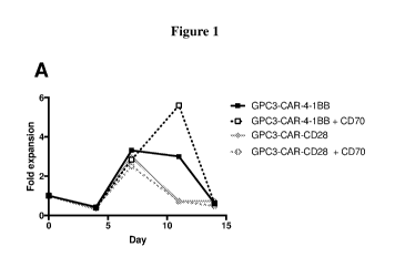
Figure] is a series of graphs showing the fold expansion of T cells relative
to the
previous time point for T cells after stimulation with GPC3-expressing Hep3B
cells. The T
cells evaluated in this experiment expressed anti-GPC3 CAR with a 4-1BB
costimulatory
domain (SEQ ID NO: 1) alone (A, B, and C) or in combination with CD70 (A; SEQ
ID
NO: 34), LIGHT (B; SEQ ID NO: 43), or OX4OL (C; SEQ ID NO: 47), or anti-GPC3
6
CA 03118889 2021-05-05
WO 2020/097346
PCT/US2019/060287
CAR with a CD28 costimulatory domain (SEQ ID NO: 2) alone (A, B, and C) or in
combination with CD70 (A; SEQ ID NO: 34), LIGHT (B; SEQ ID NO: 43), or 0X40L
(C; SEQ ID NO: 47).
Figure 2 is a series of graphs showing the fold expansion of T cells relative
to the
previous time point for T cells after stimulation with GPC3-expressing JHH7
cells as a
function of stimulation round (panel A) and cytokine production after the
second round of
stimulation for IL-2 (panel B), IFN-gamma (panel C), and IL-17A (panel D).
Data are
shown for T cells expressing anti-GPC3 CAR with a 4-1BB costimulatory domain
(SEQ
ID NO: 1) alone or in combination with CD70 (SEQ ID NO: 34), LIGHT (SEQ ID NO:
43), or 0X40L (SEQ ID NO: 47).
Figure 3 is a series of graphs showing enhanced IL-2 production (panel A) and
proliferation (panel B) for T cells expressing an anti-GPC3 CAR polypeptide
with a 4-
1BB costimulatory domain (GPC3-CAR-4-1BB; SEQ ID NO: 1) and T cells co-
expressing GPC3-CAR-4-1BB and CD70 (SEQ ID NO: 34), LIGHT (SEQ ID NO: 43), or
0X40L (SEQ ID NO: 47).
Figure 4 is a series of graphs demonstrating function of T cells expressing an
anti-
GPC3 CAR polypeptide with a 4-1BB costimulatory domain (GPC3-CAR-4-1BB; SEQ
ID NO: 1) or GPC3-CAR-4-1BB in combination with CD70 (SEQ ID NO: 34), LIGHT
(SEQ ID NO: 43), or OX4OL (SEQ ID NO: 47). T cells were evaluated for their
ability to
produce IL-17A (panel A) and proliferate (panel B) under chronic stimulation.
Additionally, T cells were evaluated for their ability to proliferate after a
single
stimulation (panel C).
Figure 5 is a series of graphs demonstrating function of T cells expressing an
anti-
GPC3 CAR polypeptide with a CD28 costimulatory domain (GPC3-CAR-CD28; SEQ ID
NO: 2) or GPC3-CAR-CD28 in combination with CD27 (SEQ ID NO: 33). T cells were
evaluated for their ability to proliferate (panels A and B) and generate
cytokines (panels C
and D).
Figure 6 is a series of graphs demonstrating function of T cells expressing an
anti-
GPC3 CAR polypeptide with a CD28 costimulatory domain (GPC3-CAR-CD28; SEQ ID
NO: 2) or GPC3-CAR-CD28 in combination with CD27 (SEQ ID NO: 33). T cells were
evaluated for their ability to proliferate in the presence of
immunosuppressive myeloid-
derived suppressor cells (MDSCs; panel A) or regulatory T cells (Tregs; panel
B).
7
CA 03118889 2021-05-05
WO 2020/097346
PCT/US2019/060287
Figure 7 is a series of graphs showing anti-tumor activity of T cells
expressing an
anti-GPC3 CAR polypeptide with a 4-1BB costimulatory domain (GPC3-CAR-4-1BB;
SEQ ID NO: 1) or GPC3-CAR-4-1BB in combination with CD70 (SEQ ID NO: 34),
LIGHT (SEQ ID NO: 43), or 0X40L (SEQ ID NO: 47). HepG2 (panel A), Hep3B (panel
B), and JHH7 (panel C) tumor xenograft models were evaluated in NSG mice.
Figure 8 is a graph showing anti-tumor activity of T cells expressing an anti-
GPC3
CAR polypeptide with a CD28 costimulatory domain (GPC3-CAR-CD28; SEQ ID NO: 2)
or GPC3-CAR-CD28 in combination with CD27 (SEQ ID NO: 33) in a JHH7 tumor
xenograft models in NSG mice.
Figure 9 is a series of graphs showing the amount of T cells in mouse blood
from
HepG2 (panel A) and Hep3B (panel B) tumor xenograft models in NSG mice. Data
are
shown for T cells expressing anti-GPC3 CAR with a 4-1BB costimulatory domain
(SEQ
ID NO: 1) alone or in combination with CD70 (SEQ ID NO: 34) (panel A) and T
cells
expressing anti-GPC3 CAR with a CD28 costimulatory domain (SEQ ID NO: 2) alone
or
in combination with CD27 (SEQ ID NO: 33) (panel B).
Figure 10 is a series of graphs showing CD70 expression on T cells expressing
anti-GPC3 CAR with a 4-1BB costimulatory domain (SEQ ID NO: 1) alone or in
combination with CD70 (SEQ ID NO: 34) (panels A and B) or CD27 expression on T
cells expressing anti-GPC3 CAR with a CD28 costimulatory domain (SEQ ID NO: 2)
alone or in combination with CD27 (SEQ ID NO: 33) (panels C and D).
DETAILED DESCRIPTION OF DISCLOSURE
Chimeric antigen receptors (CARs) are artificial cell-surface receptors that
redirect
binding specificity of immune cells (e.g., T cells) expressing such to
diseased cells such as
cancer cells, thereby eliminating the target disease cells via, e.g., the
effector activity of
the immune cells. A CAR construct often comprises an extracellular antigen
binding
domain fused to at least an intracellular signaling domain. The extracellular
antigen
binding domain (e.g., a single-chain antibody fragment) is specific to an
antigen of interest
(e.g., a tumor antigen) and the intracellular signaling domain can mediate a
cell signaling
that lead to activation of immune cells. As such, immune cells expressing a
CAR
construct can bind to diseased cells (e.g., tumor cells) expressing the target
antigen,
leading to activation of the immune cells and elimination of the diseased
cells.
8
CA 03118889 2021-05-05
WO 2020/097346
PCT/US2019/060287
The present disclosure is based, at least in part, on the development of
strategies
for enhancing activities of effector immune cells that co-express an anti-
glypican-3
(GPC3) chimeric antigen receptor (CAR) polypeptide. In particular, the present
disclosure
features methods for imparting the capacity to modulate suitable co-
stimulatory pathways
by the effector immune cells, thereby enhancing their growth and bioactivity.
For
example, T cells co-expressing an anti-GPC3 CAR comprising a 4-1BB co-
stimulatory
domain and certain co-stimulatory molecules (e.g., CD70, LIGHT, and 0X40L) and
T
cells co-expressing an anti-GPC3 CAR comprising a CD28 co-stimulatory domain
and
certain co-stimulatory molecules (e.g., CD27) showed enhanced cell
proliferation and
cytokine production. The immunosuppressive features within solid tumors may
limit the
success of engineered T cell therapies. The approach disclosed herein,
involving the co-
expression of an anti-GPC3 CAR and a co-stimulatory polypeptide (which
provides a co-
stimulation signal in trans), aims at, at least in part, overcoming this key
challenge in
tumor treatment, particularly solid tumor treatment.
In some instances, the capacity of the effector immune cells to modulate co-
stimulatory pathways may be observed in normal cellular environments. In other
instances, the capacity of the effector immune cells to modulate co-
stimulatory pathways
may be observed under conditions that may be found in a tumor
microenvironment. The
present disclosure provides various approaches to modulate (e.g., to
stimulate) co-
stimulatory pathways including by, e.g., expressing or overexpressing co-
stimulatory
polypeptides. The co-stimulatory polypeptides for use in the present
disclosure may be
members of the B7/CD28 superfamily, members of the tumor necrosis factor (TNF)
superfamily or ligands thereof that functional as a co-stimulatory factor in
one or more
types of immune cells. A co-stimulatory factor refers to a receptor or a
ligand thereof,
which enhances the primary, antigen-specific signal and fully activates immune
cells.
Accordingly, the present disclosure provides modified (e.g., genetically
engineered) hematopoietic cells (e.g., hematopoietic stem cells, immune cells,
such as T
cells or natural killer cells) that have the capacity to have modulated (e.g.,
increased) co-
stimulatory pathways. In some embodiments, such a modified hematopoietic cell
may
express one or more co-stimulatory polypeptides such as those described herein
to impart
the capacity to modulate the co-stimulatory pathways, relative to an
unmodified
hematopoietic cell. Such a genetically engineered hematopoietic cell may
further express
a CAR polypeptide (as a separate polypeptide relative to the co-stimulatory
polypeptide).
9
CA 03118889 2021-05-05
WO 2020/097346
PCT/US2019/060287
Both the CAR polypeptide and the co-stimulatory polypeptide expressed in the
genetically
engineered hematopoietic cells are encoded by nucleic acids exogenous to the
immune
cells (i.e., introduced into immune cells via recombinant technology). They
are not
encoded by endogenous genes of the hematopoietic cells absent of the involved
genetic
engineering. The present disclosure also provides pharmaceutical compositions
and kits
comprising the described genetically engineered hematopoietic cells.
The genetically engineered hematopoietic cells described herein, expressing
(e.g.,
over-expressing) a co-stimulatory peptide, may confer at least the following
advantages.
The expression of the co-stimulatory polypeptide would have the capacity to
modulate the
co-stimulatory pathways. As such, the genetically engineered hematopoietic
cells may
proliferate better, produce more cytokines, exhibit greater anti-tumor
cytotoxicity, and/or
exhibit greater T cell survival relative to hematopoietic cells that do not
express (or do not
over-express) the co-stimulatory polypeptide, leading to enhanced cytokine
production,
survival rate, cytotoxicity, and/or anti-tumor activity.
I. Co-Stimulatory Polypeptides
As used herein, a co-stimulatory polypeptide refers to a polypeptide that has
the
capacity to modulate (e.g., stimulate) a co-stimulatory pathway. Such a
polypeptide may
modulate (e.g., increase) the co-stimulatory pathway via any mechanism. In
some
examples, the co-stimulatory polypeptide may comprise a co-stimulatory
receptor or the
co-stimulatory signaling domain thereof. In other examples, the co-stimulatory
polypeptide may comprise a ligand of a co-stimulatory receptor or a signaling
domain
thereof where applicable. Such a ligand may trigger a co-stimulatory signaling
pathway
upon binding to the cognate co-stimulatory receptor. Alternatively, the co-
stimulatory
polypeptide may be a non-naturally occurring polypeptide that mimics the
activity of a
naturally-occurring ligand to any of the co-stimulatory receptors disclosed
herein. Such a
non-naturally occurring polypeptide may be a single-chain agonistic antibody
specific to a
co-stimulatory receptor, e.g., an scFv specific to 4-1BB and mimics the
activity of 4-
1BBL.
Exemplary co-stimulatory polypeptides may include, but are not limited to,
members of the B7/CD28 superfamily, members of the tumor necrosis factor (TNF)
superfamily or ligands thereof (e.g., CD28, CD80, CD86, ICOS, ICOSL, B7-H3, B7-
H4,
VISTA, TMIGD2, B7-H6, B7-H7, 4-1BB, 4-1BBL, BAFF, BAFFR, CD27, CD70, CD30,
CA 03118889 2021-05-05
WO 2020/097346
PCT/US2019/060287
CD3OL, CD40, CD4OL, DR3, GITR, GITRL, HVEM, LIGHT, TNF-beta, 0X40, OX4OL,
RELT, TACI, TL1A, TNF-alpha, or TNFRII). Additional examples include BCMA,
EDAR2, TROY, LTBR, EDAR, NGFR, OPG, RANK, DCR3, TNFR1, FN14 (TweakR),
APRIL, EDA-A2, TWEAK, LTb (TNF-C), NGF, EDA-Al , amyloid precursor protein
(APP), TRAIL. Any such polypeptide from any suitable species (e. g. , a mammal
such as
a human) may be contemplated for use with the compositions and methods
described
herein. In some embodiments, the co-stimulatory polypeptides do not comprise
the
combination of CD40 and MyD88.
As used herein, a co-stimulatory polypeptide that is a member of the B7/CD28
superfamily or a member of the TNF superfamily refers to a member of either
superfamily
that plays co-stimulatory roles in activation of any type of immune cells.
Such a member
may be a naturally-occurring receptor or ligand of either superfamily.
Alternatively, such
a member may be a variant of the naturally-occurring receptor or ligand. The
variant may
have increased or decreased activity relative to the native counterpart. In
some examples,
the variant lacks the cytoplasmic domain or a portion thereof relative to the
native
counterpart. Described below are exemplary co-stimulatory polypeptides that
can be used
in the present disclosure.
CD28 (Cluster of Differentiation 28) is a protein expressed on T cells that
provides
co-stimulatory signals required for T cell activation and survival. It is the
receptor for
CD80 and CD86 proteins, and is the only B7 receptor constitutively expressed
on naïve T
cells. The amino acid sequence of an exemplary human CD28 is provided below:
CD28 (SEQ ID NO: 12)
MLRLLLALNLFP S I QVTGNKI LVKQSPMLVAYDNAVNLS CKYSYNLF SREFRASLHKGLD
SAVEVCVVYGNYSQQLQVYSKT GFNCDGKL GNE SVTFYLQNLYVNQTD I YFCK IEVMYP PPYLDNE
KSNGT I IHVKGKHLCP SP LFPGP SKPFWVLVVVGGVLACYSLLVTVAF I IFWVRSKRSRLLHSDYM
NMTPRRPGPTRKHYQPYAPPRDFAAYRS
CD80 (Cluster of Differentiation 80; B7-1) is a protein found on dendritic
cells,
activated B cells, and monocytes. It provides a co-stimulatory signal
necessary for T cell
activation and survival. CD80 is a ligand of both CD28 and CTLA-4. The amino
acid
sequence of an exemplary human CD80 is provided below:
11
CA 03118889 2021-05-05
WO 2020/097346
PCT/US2019/060287
CD80 (SEQ ID NO: 13)
MGHTRRQGTSPSKCPYLNFFQLLVLAGLSHFCSGVIHVTKEVKEVATLSCGHNVSVEELAQTRIYWQK
EKKMVL TMMS GDMN IWPEYKNRT I FD I TNNLS IVILALRPSDEGTYECVVLKYEKDAFKREHLAEVTL
SVKADFPTPS I SDFEI PT SNIRRI I CS T SGGFPEPHL SWLENGEELNAINTTVSQDPETELYAVS SKL
DFNMTTNHSFMCL IKYGHLRVNQTFNWNTTKQEHFP DNL LP SWAT TL I SVNGIFVICCLTYCFAPRCR
ERRRNERLRRESVRPV
CD86 (Cluster of Differentiation 86; B7-2) is a type I membrane protein that
is a
member of the immunoglobulin superfamily. CD86 is expressed on antigen-
presenting
cells that provide co-stimulatory signals necessary for T cell activation and
survival.
CD86 is a ligand of both CD28 and CTLA-4. The amino acid sequence of an
exemplary
human CD86 is provided below:
CD86 (SEQ ID NO: 14)
MDPQCTMGLSNILFVMAFLLSGAAPLKIQAYFNETADLP CQFANSQNQSLSELVVFWQDQENLVLN
EVYLGKEKFDSVHSKYMGRTSFDSDSWTLRLHNLQIKDKGLYQCI IHHKKPTGMIRIHQMNSELSV
LANF SQPE IVP I SNITENVYINLTCSS IHGYPEPKKMSVLLRTKNSTIEYDGVMQKSQDNVTELYD
VS I SL SVSFP DVT SNMT IFC ILETDKTRLL SSPF S I ELEDPQPPP DHIPWI TAVLP TVI
ICVMVFC
LI LWKWKKKKRPRNSYKCGTNTMEREESEQTKKREKIHIPERSDEAQRVFKSSKTSSCDKSDTCF
ICOS (CD278; Inducible T cell co-stimulator; or CVID1) is a member of the
CD28-superfamily. ICOS is expressed on activated T cells. The amino acid
sequence of
an exemplary human ICOS is provided below:
ICOS (SEQ ID NO: 15)
MKSGLWYFFLFCLRIKVLTGEINGSANYEMFIFHNGGVQILCKYPDIVQQFKMQLLKGGQILCDLT
KTKGSGNTVS IKSLKFCHSQLSNNSVSFFLYNLDHSHANYYFCNL SIFDPPPFKVTLTGGYLHIYE
SQLCCQLKFWLP I GCAAFVVVC ILGC IL ICWLTKKKYSS SVHDPNGEYMFMRAVNTAKKSRLTDVT
L
ICOSL (ICOSLG; B7-H2; CD275) is a protein that is a ligand for T cell specific
protein ICOS. ICOSL acts as a co-stimulatory signal for T cell proliferation
and cytokine
secretion. The amino acid sequence of an exemplary human ICOSL is provided
below:
ICOSL (SEQ ID NO: 16)
MRLGSPGLLFLLFSSLRADTQEKEVRAMVGSDVELSCACPEGSRFDLNDVYVYWQTSESKTVVTYHIP
QNSSLENVDSRYRNRALMSPAGMLRGDFSLRLFNVTPQDEQKFHCLVLSQSLGFQEVLSVEVTLHVAA
NF SVPVVSAPHSP SQDEL TFTC TS INGYPRPNVYWINKTDNSLLDQALQNDTVFLNMRGLYDVVSVLR
IARTP SVN I GCC I ENVLLQQNL TVGSQTGND I GERDKI TENPVST GEKNAATWS I LAVL
CLLVVVAVA
I GWVCRDRCLQHSYAGAWAVSP ETEL TGHV
12
CA 03118889 2021-05-05
WO 2020/097346
PCT/US2019/060287
B7-H3 (CD276; Cluster of Differentiation 276) is a member of the
immunoglobulin superfamily that is thought to participate in the regulation of
T cell-
mediated immune response. The amino acid sequence of an exemplary human B7-H3
is
provided below:
B7-H3 (SEQ ID NO: 17)
MLRRRGSP GMGVHVGAAL GALWFCL TGALEVQVPEDPVVALVGTDATLCC SFS PEP GFS LAQLNL IWQ
LTDTKQLVHSFAEGQDQGSAYANRTALFPDLLAQGNASLRLQRVRVADEGSFTCFVS IRDFGSAAVSL
QVAAPYSKPSMTLEPNKDLRPGDTVT I TCS SYQGYPEAEVFWQDGQGVPLTGNVTTSQMANEQGLFDV
HS I LRVVL GANGTYSC LVRNPVLQQDAHS SVT I TPQRSP TGAVEVQVPEDPVVALVGTDATLRCSFSP
EP GF S LAQLNL IWQLTDTKQLVHSF TEGRDQGSAYANRTALFPDL LAQGNASLRLQRVRVADEGS FTC
FVS IRDFGSAAVS LQVAAP YSKP SMTLEPNKDLRPGDTVT I TC S S YRGYPEAEVFWQDGQGVP LT
GNV
TT SQMANEQGLFDVHSVLRVVL GANGTYSC LVRNPVLQQDAHGSVT I TGQPMTFPPEALWVTVGL SVC
L IALLVALAFVCWRKI KQS CEEENAGAEDQDGEGEG SKTALQP LKHSD SKEDD GQE IA
VISTA (V-domain Ig suppressor of T cell activation; B7-H5; PD-1H) is a Type I
transmembrane protein that functions as an immune checkpoint. VISTA co-
stimulates T
cells via TMIGD2 (CD28H). The amino acid sequence of an exemplary human VISTA
is
provided below:
VISTA (SEQ ID NO: 18)
MGVP TALEAGSWRWGSLLFALFLAASLGPVAAFKVATPYSLYVCPEGQNVTLTCRLLGPVDKGHDVTF
YKTWYRSSRGEVQTCSERRP IRNLTFQDLHLHHGGHQAANTSHDLAQRHGLESASDHHGNFS I TMRNL
TLLDSGLYCCLVVEIRHHHSEHRVHGAMELQVQTGKDAP SNCVVYP S S SQD SENT TAAALATGAC IVG
I L CLP L I LLLVYKQRQAASNRRAQELVRMD SN I QGI ENP GFEASP PAQG I PEAKVRHPL
SYVAQRQP S
ES GRHLL SEP STP L SP PGP GDVFFP SLDPVPDSPNFEVI
TMIGD2 (Transmembrane and immunoglobulin domain containing 2; CD28H) is
a TMIGD2 is thought to enhance T cell proliferation and cytokine production
via an AKT-
dependent signaling cascade. The amino acid sequence of an exemplary human
TMIGD2
is provided below:
TMIGD2 (SEQ ID NO: 19)
MG SP GMVL GL LVQ IWALQEAS S LSVQQGPNLLQVRQGSQATLVCQVDQATAWERLRVKWTKDGAI LCQ
PY I TNGS L SL GVCGPQGRL SWQAP SHLTLQLDPVSLNHS GAYVCWAAVE IPELEEAEGN I
TRLFVDPD
DP TQNRNRIASFPGFLFVLLGVGSMGVAAIVWGAWFWGRRSCQQRDSGNSPGNAFYSNVLYRPRGAPK
KS EDC S GEGKDQRGQS I YS T SFPQPAPRQP HLASRP CP S PRP CP S PRP GHPVSMVRVSP RP
SP TQQPR
PKGFPKVGEE
13
CA 03118889 2021-05-05
WO 2020/097346
PCT/US2019/060287
B7-H6 (NCR3LG1; Natural Killer Cell Cytotwdcity Receptor 3 Ligand 1) is a
member of the B7 family selectively expressed on tumor cells. B7-H6 interacts
with
NKp30, resulting in natural killer (NK) cell activation and cytotwdcity. The
amino acid
sequence of an exemplary human B7-H6 is provided below:
B7-H6 (SEQ ID NO: 20)
MTWRAAAS TCAALL I L LWALTTEGDLKVEMMAGGTQ I TP LNDNVT IFCN I FYS QP LN I T SMG
I TWFWK
SLTFDKEVKVFEFFGDHQEAFRPGAIVSPWRLKSGDASLRLPGIQLEEAGEYRCEVVVTPLKAQGTVQ
LEVVASPASRLLLDQVGMKENEDKYMCESSGFYPEAINI TWEKQTQKFPHP IE I SEDVI TGPTIKNMD
.. GTFNVT SCLKLNS SQEDP GTVYQCVVRHAS LHTP LRSNF TLTAARHSL SETEKTDNF S I HWWP I
SF I G
VGLVLL IVL I PWKKICNKS S SAYTP LKC ILKHWNSFDTQTLKKEHL IFFCTRAWP SYQLQDGEAWPPE
GSVNINTIQQLDVFCRQEGKWSEVPYVQAFFALRDNPDLCQCCRIDPALLTVTSGKS IDDNSTKSEKQ
TP REHSDAVP DAP I LPVSP IWEPPPATT ST TPVL SSQPP TLLLPLQ
B7-H7 (HHLA2; HERV-H LTR-Associating 2) is a protein ligand found on the
surface of monocytes. B7-H7 is thought to regulate cell-mediated immunity
through
binding a receptor on T lymphocytes and inhibiting proliferation in the same.
The amino
acid sequence of an exemplary human B7-H7 is provided below:
B7-H7 (SEQ ID NO: 21)
MKAQTAL SFF LIL I TS LSGSQG IFP LAFF I YVPMNEQIVIGRLDEDI I LP S
SFERGSEVVIHWKYQDS
YKVHSYYKGSDHLESQDPRYANRTSLFYNE IQNGNASLFFRRVSLLDEGIYTCYVGTAIQVITNKVVL
KVGVFLTPVMKYEKRNTNSFL I CSVL SVYP RP I I TWKMDNTP I SENNMEETGS LDSF S INSP LNI
TGS
NS SYECT IENSLLKQTWTGRWTMKDGLHKMQSEHVS L SCQPVNDYFSPNQDFKVTWSRMKSGTFSVLA
YYLS S SQNT I INESRFSWNKEL INQSDF SMNLMDLNL SD SGEYLCNI S SDEYT LLT IHTVHVEP
SQET
AS HNKGLWI LVP SAI LAAFLL I WSVKCCRAQLEARRSRHPADGAQQERCCVPP GERCPSAPDNGEENV
PL SGKV
4-1BB (CD137; TNFRSF9) is a tumor necrosis factor (TNF) superfamily member
that is expressed by activated T cells. Crosslinking of 4-1BB enhances T cell
proliferation, IL-2 secretion, survival, and cytolytic activity. The amino
acid sequence of
an exemplary human 4-1BB is provided below:
4-]BB (SEQ ID NO: 22)
MGNSCYNIVATLLLVLNFERTRSLQDP CSNCPAGTF CDNNRNQIC SP CPPNSF SSAGGQRTCD ICRQC
KGVFRTRKEC SS T SNAECDCTP GFHCLGAGCSMCEQDCKQGQELTKKGCKDCCFGTFNDQKRGICRPW
TNCSLDGKSVLVNGTKERDVVCGPSPADLSPGASSVTPPAPAREP GHSPQI I SFFLALT STALLF LLF
FL TLRF SVVKRGRKKL LYIFKQPFMRPVQT TQEEDGCSCRFPEEEEGGCEL
4-1BBL (TNFSF9; 4-1BB ligand) is a Type 2 transmembrane glycoprotein
receptor belonging to the TNF superfamily. 4-1BBL is expressed on activated T
14
CA 03118889 2021-05-05
WO 2020/097346
PCT/US2019/060287
Lymphocytes and binds to 4-1BB. The amino acid sequence of certain exemplary
human
4-1BBL polypeptides (including native and variants) are provided below:
4-]BBL (SEQ ID NO: 23)
ME YAS DAS LD PEAPWP PAP RARACRVLPWALVAGLL LLL LLAAACAVFLACPWAVSGARASP GSAASP
RL REGP EL SP DDPAGL LDLRQGMFAQLVAQNVLL ID GP L SWYSDP GLAGVS LT GGL
SYKEDTKELVVA
KAGVYYVFFQLELRRVVAGEGS GSVSLALHLQPLRSAAGAAALAL TVDLP PAS SEARNSAFGFQGRLL
HL SAGQRLGVHLHTEARARHAWQLTQGATVLGLFRVTPE IPAGLP SP RSE
4-]BBL-CD (lacking cytoplasmic domain; SEQ ID NO: 24)
MRVLPWALVAGLLLLL LLAAACAVFLACPWAVSGARASP GSAASP RLREGP EL SP DDPAGL LDLRQ
GMFAQLVAQNVLL I DGP L SWYS DP GLAGVS LT GGL S YKEDTKELVVAKAGVYYVFFQLE LRRVVAG
EGSGSVSLALHLQPLRSAAGAAALALTVDLPPAS SEARNSAFGFQGRLLHL SAGQRLGVHLHTEAR
ARHAWQLTQGATVLGLFRVTPE IPAGLP SP RSE
4-]BBL Q89A (SEQ ID NO: 25)
ME YAS DAS LD PEAPWP PAP RARACRVLPWALVAGLL LLL LLAAACAVFLACPWAVSGARASP GSAASP
RL REGP EL SP DDPAGL LDLRAGMFAQLVAQNVLL ID GP L SWYSDP GLAGVS LT GGL
SYKEDTKELVVA
KAGVYYVFFQLELRRVVAGEGS GSVSLALHLQPLRSAAGAAALAL TVDLP PAS SEARNSAFGFQGRLL
HL SAGQRLGVHLHTEARARHAWQLTQGATVLGLFRVTPE IPAGLP SP RSE
4-]BBL Q89A/CD (lacking cytoplasmic domain) (SEQ ID NO: 26)
MRVLPWALVAGLLLLL LLAAACAVFLACPWAVSGARASP GSAASP RLREGP EL SP DDPAGL LDLRAGM
FAQLVAQNVL L I DGP L SWYSDP GLAGVS LT GGL S YKEDTKELVVAKAGVYYVF FQLELRRVVAGE
GS G
SVSLALHLQP LRSAAGAAALAL TVDLP PAS SEARNSAFGFQGRLLHL SAGQRL GVHLHTEARARHAWQ
LT QGATVL GL FRVTPE IPAGLP SP RSE
4-]BBL L115A (SEQ ID NO: 27)
ME YAS DAS LD PEAPWP PAP RARACRVLPWALVAGLL LLL LLAAACAVFLACPWAVSGARASP GSAASP
RL REGP EL SP DDPAGL LDLRQGMFAQLVAQNVLL ID GP L SWYSDP GAAGVS LT GGL
SYKEDTKELVVA
KAGVYYVFFQLELRRVVAGEGS GSVSLALHLQPLRSAAGAAALAL TVDLP PAS SEARNSAFGFQGRLL
HL SAGQRLGVHLHTEARARHAWQLTQGATVLGLFRVTPE IPAGLP SP RSE
4-]BBL L115A/CD (SEQ ID NO: 28)
MRVLPWALVAGLLLLL LLAAACAVFLACPWAVSGARASP GSAASP RLREGP EL SP DDPAGL LDLRQ
GMFAQLVAQNVLL I DGP L SWYS DP GAAGVS LT GGL S YKE DTKE LVVAKAGVYYVFFQLE
LRRVVAG
EGSGSVSLALHLQPLRSAAGAAALALTVDLPPAS SEARNSAFGFQGRLLHL SAGQRLGVHLHTEAR
ARHAWQLTQGATVLGLFRVTPE IPAGLP SP RSE
4-]BBL K127A (SEQ ID NO: 29)
ME YAS DAS LD PEAPWP PAP RARACRVLPWALVAGLL LLL LLAAACAVFLACPWAVSGARASP GSAASP
RL REGP EL SP DDPAGL LDLRQGMFAQLVAQNVLL ID GP L SWYSDP GLAGVS LT GGL
SYAEDTKELVVA
KAGVYYVFFQLELRRVVAGEGS GSVSLALHLQPLRSAAGAAALAL TVDLP PAS SEARNSAFGFQGRLL
HL SAGQRLGVHLHTEARARHAWQLTQGATVLGLFRVTPE IPAGLP SP RSE
15
CA 03118889 2021-05-05
WO 2020/097346
PCT/US2019/060287
4-]BBL Q227A (SEQ ID NO: 30)
ME YAS DAS LD PEAPWP PAP RARACRVLPWALVAGLL L LL LLAAACAVF LACPWAVS GARAS P G
SAAS P
RLREGPEL SP DDPAGL LDLRQGMFAQLVAQNVLL ID GPL SWYSDP GLAGVS LT GGL SYKEDTKELVVA
KAGVYYVFFQLELRRVVAGEGS GSVSLALHLQPLRSAAGAAALAL TVDLPPAS SEARNSAFGFQGRLL
HL SAGQRLGVHLHTEARARHAWALTQGATVLGLFRVTPE IPAGLP SPRSE
BAFF (B-cell activating factor; TNFSF13B) is a member of the TNF ligand family
and serves as a ligand for receptors TNFRSF13B/TACI, TNFRSF17/BCMA, and
TNFRSF13C/BAFF-R. BAFF is a potent B cell activator and plays an important
role in B
cell proliferation and differentiation. The amino acid sequence of an
exemplary human
BAFF is provided below:
BAFF (SEQ ID NO: 31)
MDDS TEREQS RL T S CLKKREEMKLKECVS I LPRKESP SVRS SKDGKLLAATLL LALL SC CL TVVS
FYQ
VAALQGDLASLRAELQGHHAEKLPAGAGAPKAGLEEAPAVTAGLKIFEPPAPGEGNSSQNSRNKRAVQ
GP EETVTQDC LQL IAD SETP T I QKGSYTFVPWLL SFKRG SALEEKENKI LVKE TGYFF I
YGQVLYTDK
TYAMGHL I QRKKVHVF GDEL SLVTLFRC IQNMPETLPNNSCYSAGIAKLEEGDELQLAIPRENAQ I SL
DGDVTFFGALKLL
BAFFR (B-cell activating factor receptor; TNFRSF13C) is a membrane protein of
the TNF receptor superfamily and acts as a receptor for BAFF. BAFFR enhances B
cell
survival and is a regulator of the peripheral B-cell population. The amino
acid sequence
of an exemplary human BAFFR is provided below:
BAFFR (SEQ ID NO: 32)
MRRGP RS LRGRDAPAP TP CVPAECFDLLVRHCVACGL LRTP RP KPAGAS S PAP RTALQP QE
SVGAGAG
EAALP LP GLLFGAPAL LGLALVLALVLVGLVSWRRRQRRLRGAS SAEAPDGDKDAPEPLDKVI IL SP G
I S DATAPAWP PP GEDP GT TPPGHSVPVPATEL GS TE LVT TKTAGPEQQ
CD27 (TNFRSF7) is a member of the TNF receptor superfamily and is required
for generation and long-term maintenance of T cell immunity. CD27 binds to
CD70 and
also plays a role in regulation of B-cell activation and immunoglobulin
synthesis. The
amino acid sequence of an exemplary human CD27 is provided below:
CD27 (SEQ ID NO: 33)
MARPHPWWLCVLGTLVGLSATPAPKSCPERHYWAQGKLCCQMCEP GTFLVKDCDQHRKAAQCDPC IP G
VS FSPDHHTRPHCE SCRHCNSGLLVRNCT I TANAECACRNGWQCRDKECTECDPLPNPSLTARSSQAL
SP HPQP THLPYVSEMLEARTAGHMQTLADFRQLPARTLS THWPPQRS LC S SDF IRI LVI FS GMFLVF
T
LAGALFLHQRRKYRSNKGESPVEPAEPCHYSCPREEEGS TIP I QEDYRKPEPACSP
16
CA 03118889 2021-05-05
WO 2020/097346
PCT/US2019/060287
CD70 (CD27LG; TNFSF7) is a protein expressed on highly activated
lymphocytes. CD70 acts as a ligand for CD27. The amino acid sequence of an
exemplary
human CD70 is provided below:
CD70 (SEQ ID NO: 34)
MP EEGSGC SVRRRP YGCVLRAALVP LVAGLVI CLVVC IQRFAQAQQQLP LE SL GWDVAE LQLNHT
GPQ
QDPRLYWQGGPALGRS FLHGPE LDKGQLRI HRDGIYMVH IQVTLAIC S S T TAS RHHP TT LAVGIC
SPA
SRS I SLLRLSFHQGCT IVSQRL TP LARGDT LCTNLT GTL LP SRNTDETFFGVQWVRP
CD30 (TNFRSF8) is a member of the TNF receptor superfamily that is expressed
by activated T cells and B cells. CD30 is a cell membrane protein that has
been shown to
interact with CD3OL, TRAF1, TRAF2, TRAF3, and TRAF5. The amino acid sequence
of
an exemplary human CD30 is provided below:
CD30 (SEQ ID NO: 35)
MRVLLAALGLLFLGALRAFPQDRPFEDTCHGNP SHYYDKAVRRCCYRCPMGLFPTQQCPQRPTDCRKQ
CEPDYYLDEADRCTACVTC SRDDLVEKTPCAWNS SRVCE CRP GMF CS T SAVNS CARCFFHSVCPAGMI
VKFPGTAQKNTVCEPASPGVSPACASPENCKEP SSGT IP QAKP TPVSPAT S SASTMPVRGGTRLAQEA
ASKL TRAPDS P S SVGRP S SDPGLSP TQP CP EGS GDCRKQCEPDYYLDEAGRCTACVS CS
RDDLVEKTP
CAWNS SRTCE CRP GMI CAT SATNS CARCVP YP I CAAETVTKPQDMAEKDT TFEAPP LGTQPDCNP
TPE
NGEAPAS T SP TQSLLVDSQASKTLP I P T SAPVAL S S TGKPVLDAGPVLFWVILVLVVVVGSSAFLLCH
RRACRKRIRQKLHLCYPVQTSQPKLELVDSRPRRSS TQLRSGASVTEPVAEERGLMSQP LMETCHSVG
AAYLE S LP LQDASPAGGP SSPRDLPEPRVS TEHTNNKIEKIYIMKADTVIVGTVKAELPEGRGLAGPA
EP ELEEELEADHTPHYPEQETEPP LGS C SDVML SVEEEGKEDP LP TAASGK
CD3OL (CD3OLG; TNFSF8) is a member of the TNF receptor superfamily.
CD3OL acts as a ligand of CD30, and is expressed on induced T cells and
monocytes/macrophages. The amino acid sequence of an exemplary human CD3OL is
provided below:
CD3OL (SEQ ID NO: 36)
MDPGLQQALNGMAPPGDTAMHVPAGSVASHLGT T SRSYF YL T TAT LALCLVFTVAT IMVLVVQRTDS I
PNSPDNVPLKGGNCSEDLLC I LKRAPFKKS WAYLQVAKHLNKTKL SWNKDG I LHGVRYQDGNLVI QFP
GLYF I I CQLQFLVQCPNNSVDLKLELL INKHIKKQALVTVCESGMQTKHVYQNLSQFLLDYLQVNTT I
SVNVDTFQYI DT S TFP LENVLS IFLYSNSD
CD40 (TNFRSF5) is a cell surface receptor expressed on the surface of B cells,
monocytes, dendritic cells, endothelial cells, and epithelial cells. CD40 has
been
demonstrated to have involvement in T cell-dependent immunoglobulin class
switching,
17
CA 03118889 2021-05-05
WO 2020/097346
PCT/US2019/060287
memory B cell development, and germinal center formation. The amino acid
sequence of
an exemplary human CD40 is provided below:
CD40 (SEQ ID NO: 37)
MVRLPLQCVLWGCLLTAVHPEPPTACREKQYL INSQCCS LCQP GQKLVSDCTEFTETEC LP CGES EFL
DTWNRETHCHQHKYCDPNLGLRVQQKGT SE TDT I CT CEE GWHCTS EACE S CVLHRS C SP GFGVKQ
IAT
GVSDT I CEPCPVGFFSNVS SAFEKCHPWTS CETKDLVVQQAGTNKTDVVCGPQDRLRALVVIP II FGI
LFAILLVLVF IKKVAKKPTNKAPHPKQEPQEINFPDDLP GSNTAAPVQETLHGCQPVTQEDGKES RI S
VQERQ
CD4OL (CD4OLG; TRAP; TNFSF5) is a member of the TNF superfamily
expressed on B lymphocytes, epithelial cells, and some carcinoma cells. CD4OL
is a
transmembrane protein that is known to interact with CD40 in order to mediate
B cell
proliferation, adhesion, and differentiation. The amino acid sequence of an
exemplary
human CD4OL is provided below:
CD4OL (SEQ ID NO: 38)
MI ETYNQT SP RSAATGLP I SMKIFMYLLTVFL I TQMIGSALFAVYLHRRLDKIEDERNLHEDFVFMKT
IQRCNTGERS LS LLNCEE I KSQFEGFVKD I MLNKEE TKKENSFEMQKGDQNPQ IAAHVI SEAS SKTT
S
VLQWAEKGYYTMSNNLVTLENGKQLTVKRQGLYYIYAQVTFCSNREASSQAPF IASLCLKSPGRFERI
LLRAANTHSSAKPCGQQS IHLGGVFELQPGASVFVNVTDPSQVSHGTGFTSFGLLKL
DR3 (TNFR25; AP03; TRAMP; LARD; WSL-1,) is a TNF receptor superfamily
member expressed in lymphocytes. DR3 is thought to be the receptor responsible
for
TL1A-induced T cell co-stimulation. The amino acid sequence of an exemplary
human
DR3 is provided below:
DR3 (SEQ ID NO: 39)
MEQRPRGCAAVAAALLLVLLGARAQGGTRSPRCDCAGDFHKKIGLFCCRGCPAGHYLKAPCTEPCGNS
TCLVCPQDTFLAWENHHNSECARCQACDEQASQVALENC SAVADTRCGCKPGWFVECQVSQCVSS SPF
YCQP CLDCGALHRHTRLLC SRRDTDCGTCLPGFYEHGDGCVS CP T STLGSCPERCAAVCGWRQMFWVQ
VLLAGLVVPLLLGATL TYTYRHCWPHKP LVTADEAGMEALTPPPATHL SP LDSAHTLLAPPD S SEKI C
TVQLVGNSWTPGYPETQEALCPQVTWSWDQLP SRAL GPAAAP TLS PE SPAGSPAMMLQP GPQLYDVMD
AVPARRWKEFVRT L GL REAE IEAVEVE I GRFRDQQYEML KRWRQQQPAGL GAVYAALERMGLD GCVED
LRSRLQRGP
GITR (Glucocorticoid-induced TNFR-related protein; AITR; TNFRSF18) is a
member of the TNF receptor superfamily and is expressed in several cells and
tissues
including T lymphocytes, NK cells and antigen-presenting cells. GITR
interaction with its
18
CA 03118889 2021-05-05
WO 2020/097346
PCT/US2019/060287
ligand (GITRL) induces a co-activating signal. The amino acid sequence of an
exemplary
human GITR is provided below:
GITR (SEQ ID NO: 40)
MAQHGAMGAFRALCGLALLCAL SLGQRPTGGPGCGP GRL LLGTGTDARCCRVHTTRCCRDYP GEE CCS
EWDCMCVQPEFHCGDP CCT TCRHHP CPP GQGVQSQGKFSFGFQC I DCAS GTFS GGHEGHCKPWTD CTQ
FGFLTVFPGNKTHNAVCVPGSPPAEPLGWLTVVLLAVAACVLLLT SAQLGLHIWQLRSQCMWPRETQL
LLEVPP STEDARSCQFPEEERGERSAEEKGRLGDLW
GITRL (TNFSF18) is a cytokine belonging to the TNF ligand family and acts as a
receptor for GITR. GITR interaction with its ligand (GITRL) induces a co-
activating
signal and has been shown to modulate T lymphocyte survival in peripheral
tissues. The
amino acid sequence of an exemplary human GITRL is provided below:
GITRL (SEQ ID NO: 41)
MT LHP SP I TCEFLFSTAL I SPKMCLSHLENMPLSHSRTQGAQRSSWKLWLFCS IVMLLF LC SF SWL
IF
IF LQLETAKEPCMAKF GP LP SKWQMAS SEP PCVNKVSDWKLE I LQNGLYL I YGQVAPNANYNDVAPFE
VRLYKNKDMIQTLTNKSKIQNVGGTYELHVGDT IDL IFNSEHQVLKNNTYWGI ILLANPQF I S
HVEM (Herpesvirus entry mediator; TNFRSF14; CD270) is a cell surface receptor
and a member of the TNF receptor superfamily. HVEM provides a stimulatory
signal to T
cells following engagement with LIGHT (TNFSF14); or an inhibitory signal to T
cells
when it binds the B and T lymphocyte attenuator (BTLA), a ligand member of the
Immunoglobulin (Ig) superfamily. The amino acid sequence of an exemplary human
HVEM is provided below:
HVEM (SEQ ID NO: 42)
MEPP GDWGPP PWRS TP KTDVLRLVLYL TFL GAP CYAPALP S CKEDEYPVGSEC CPKC SP
GYRVKEACG
EL TGTVCEPCPP GTYIAHLNGL SKCLQCQMCDPAMGLRASRNC SRTENAVCGC SP GHFC IVQDGDHCA
ACRAYAT S SP GQRVQKGGTESQDTLCQNCPPGTFSPNGTLEECQHQTKCSWLVTKAGAGTSSSHWVWW
FL SGS LVIVIVC STVGL I I CVKRRKPRGDVVKVIVSVQRKRQEAE GEATVI EALQAPPDVT TVAVEET
IP SF TGRSPNH
LIGHT (TNFSF14; CD258; HVEML) is a member of the TNF ligand family that
functions as a co-stimulatory factor along with HVEM. LIGHT has been
demonstrated to
stimulate the proliferation of T cells and trigger apoptosis of various tumor
cells. The
amino acid sequence of an exemplary human LIGHT is provided below:
19
CA 03118889 2021-05-05
WO 2020/097346
PCT/US2019/060287
LIGHT (SEQ ID NO: 43)
MEESVVRP SVFVVDGQTD I PFTRLGRSHRRQS CSVARVGLGLLLL LMGAGLAVQGWFLLQLHWRL GEM
VTRLPDGPAGSWEQL I QERRSHEVNPAAHL TGANSS L TGSGGP LLWETQLGLAFLRGLS YHDGALVVT
KAGYYY I YSKVQLGGVGCP LGLAS T I THGLYKRTPRYPEELELLVSQQSP CGRAT S S SRVWWDSSFLG
GVVHLEAGEEVVVRVLDERLVRLRDGTRSYFGAFMV
TNF-alpha (TNFSF2) is a member of the TNF ligand superfamily known to be
secreted by, for example, macrophages and activated CD4-positive T cells. TNF-
alpha is
known to induce certain co-stimulatory molecules such as B7h and TNFRII. The
amino
acid sequence of an exemplary human TNF-alpha is provided below:
TNF-alpha (SEQ ID NO: 44)
MS TESMIRDVELAEEALPKKTGGPQGSRRCLFLSLF SFL IVAGATTLFCLLHFGVIGPQREEFPRDLS
LI SP LAQAVRS S SRTP SDKPVAHVVANPQAEGQLQWLNRRANALLANGVELRDNQLVVP SEGLYL IYS
QVLFKGQGCP STHVLLTHT I SR IAVSYQTKVNLL SAIKSPCQRETPEGAEAKPWYEP IYLGGVFQLEK
GDRL SAE INRPDYLDFAESGQVYFGI IAL
TNF-beta (TNFSF1; Lymphotoxin alpha) is a member of the TNF superfamily
involved in the regulation of cell survival, proliferation, differentiation,
and apoptosis.
The amino acid sequence of an exemplary human TNF-beta is provided below:
TNF-beta (SEQ ID NO: 45)
MTPPERLFLPRVCGTTLHLLLLGLLLVLLP GAQGLP GVGLTP SAAQTARQHPKMHLAHS TLKPAAHL I
GDP SKQNSLLWRANTDRAFLQDGF SL SNNS LLVP TS GIYFVYSQVVF SGKAYSPKAT SSPLYLAHEVQ
LF SSQYPFHVPLLSSQKMVYPGLQEPWLHSMYHGAAFQLTQGDQL STHTDGIP HLVL SP STVFFGAFA
L
0X40 (TNFRSF4; CD134) is a member of the TNF receptor superfamily. 0X40
binds to OX4OL and contributes to T cell expansion, survival, and cytokine
production.
The amino acid sequence of an exemplary human 0X40 is provided below:
0X40 (SEQ ID NO: 46)
MCVGARRLGRGPCAALLLLGLGLSTVTGLHCVGDTYP SNDRCCHE CRP GNGMVSRC SRS QNTVCRPCG
PGFYNDVVSSKPCKPCTWCNLRSGSERKQLCTATQDTVCRCRAGTQPLDSYKP GVDCAP CPPGHF SP G
DNQACKPWTNCTLAGKHTLQPASNS SDAICEDRDPPATQPQETQGPPARP I TVQP TEAWPRT SQGP S T
RPVEVP GGRAVAAI LGLGLVLGLLGP LAIL LALYLLRRDQRLPPDAHKPP GGGSFRTP I QEEQADAHS
TLAK I
OX4OL (TNFSF4; CD252) is a member of the TNF ligand superfamily and is
expressed, for example, on activated CD4 and CD8 T cells as well as a number
of other
lymphoid and non-lymphoid cells. OX4OL interacts with 0X40 in order to
regulate, for
CA 03118889 2021-05-05
WO 2020/097346
PCT/US2019/060287
example, T cell expansion, survival, and cytokine production. The amino acid
sequence of
an exemplary human 0X40L is provided below:
OX4OL (SEQ ID NO: 47)
MERVQP LEENVGNAARPRFERNKLLLVASVIQGLGL LLCFTY I CLHF SALQVSHRYPRI QS IKVQFTE
YKKEKGF I LT SQKEDE IMKVQNNSVI INCDGFYL I S LKGYF SQEVNI SLHYQKDEEPLFQLKKVRSVN
SLMVASLTYKDKVYLNVTTDNT SLDDFHVNGGEL IL IHQNPGEFCVL
RELT (TNFRSF19L) is a member of the TNF receptor superfamily. RELT is a
type I transmembrane glycoprotein and is thought to be capable of co-
stimulating T cell
proliferation in the presence of CD3 signaling. The amino acid sequence of an
exemplary
human RELT is provided below:
RELT (SEQ ID NO: 48)
MKPSLLCRPL SCFLML LPWP LATL T S TT LWQCPP GEEPDLDP GQGTLCRP CPP
GTFSAAWGSSPCQPH
ARCS LWRRLEAQVGMATRDTLC GDCWP GWF GPWGVP RVP CQP C SWAP LGTHGCDEWGRRARRGVEVAA
GAS S GGETRQPGNGTRAGGPEE TAAQYAVIAIVPVF CLMGLLG I LVCNLLKRKGYHCTAHKEVGP GP G
GGGSGINPAYRTEDANEDT I GVLVRL I TEKKENAAALEE LLKEYH SKQLVQTS HRPVSKLPPAPPNVP
HI CPHRHHLHTVQGLASLSGPCCSRCSQKKWPEVLL SPEAVAATTPVP SLLPNPTRVPKAGAKAGRQG
EI T I L SVGRFRVARIP EQRT SSMVSEVKT I TEAGPSWGDLPDSPQPGLPPEQQALLGSGGSRTKWLKP
PAENKAEENRYVVRLSESNLVI
TACI (Transmembrane activator and CAML interactor; TNFRSF13B; CD267) is a
TNF receptor superfamily member that is found, for example, on the surface of
B cells.
TACI is known to interact with ligands BAFF and APRIL. The amino acid sequence
of an
exemplary human TACI is provided below:
TACI (SEQ ID NO: 49)
MS GLGRSRRGGRSRVDQEERFP QGLWTGVAMRS CPEEQYWDP LLGTCMS CKT I CNHQSQRTCAAF CRS
LS CRKEQGKFYDHLLRDC I S CAS I CGQHPKQCAYFCENKLRSPVNLPPELRRQRS GEVENNSDNS GRY
QGLEHRGSEASPALPGLKLSADQVALVYSTLGLCLCAVLCCFLVAVACFLKKRGDPCSCQPRSRPRQS
PAKSSQDHAMEAGSPVSTSPEPVETCSFCFPECRAP TQESAVTPGTPDPTCAGRWGCHTRTTVLQPCP
HI PDS GLGIVCVPAQE GGP GA
TL1A (TNFSF15) is a member of the TNF ligand superfamily that is known to
bind to DR3. TL1A can act to enhance T cell proliferation and cytokine
production of T
cells. The amino acid sequence of an exemplary human TL1A is provided below:
21
CA 03118889 2021-05-05
WO 2020/097346
PCT/US2019/060287
TL1A (SEQ ID NO: 50)
MAEDLGLSFGETASVEMLPEHGSCRPKARS SSARWALTCCLVLLPFLAGLTTYLLVSQLRAQGEACVQ
FQALKGQEFAPSHQQVYAPLRADGDKPRAHLTVVRQTPTQHFKNQFPALHWEHELGLAFTKNRMNYTN
KF LL IPESGDYF IYSQVTFRGMTSECSE IRQAGRPNKPD S I TVVI TKVTDSYP EP TQLLMGTKSVCEV
GSNWFQP IYL GAMF SLQEGDKLMVNVSD I S LVDYTKEDKTFFGAF LL
TNFRII (TNFRSF1B) is a TNF receptor superfamily member that binds to TNF-
alpha. TNFRII has been shown to act as a co-stimulatory receptor for T cells
and as a
critical factor for the development of regulatory T cells (Treg) and myeloid
suppressor
cells. The amino acid sequence of an exemplary human TNFRII is provided below:
TNFRII (SEQ ID NO: 51)
MAPVAVWAALAVGLEL WAAAHALPAQVAF T PYAP EP GST CRLREYYDQTAQMC C SKC SP GQHAKVFCT
KT SDTVCDSCEDS TYTQLWNWVPECL SCGSRCS SDQVETQACTREQNRI CTCRPGWYCALSKQEGCRL
CAPLRKCRPGFGVARP GTETSDVVCKPCAP GTF SNT T SS TD I CRP HQ I CNVVAIP GNASMDAVCT
ST S
PTRSMAP GAVHLPQPVSTRSQHTQP TPEP S TAP S TSFLLPMGP SP PAEGS TGDFALPVGL IVGVTALG
LL I I GVVNCVIMTQVKKKP LCLQREAKVPHLPADKARGTQGPEQQHLL I TAP S SSSSSLESSASALDR
RAPTRNQPQAPGVEASGAGEARASTGSSDS SP GGHGTQVNVTC IVNVC S S SDH SSQCSSQAS STMGDT
DS SP SESPKDEQVPFSKEECAFRSQLETPE TLLGSTEEKPLP LGVPDAGMKP S
BCMA is a cell surface receptor of the TNF receptor superfamily, and binds to
the
tumor necrosis factor superfamily, member 13b (TNFSF13B), leading to NF-kappaB
and
MAPK8LINK activation. It is preferentially expressed on mature B lymphocytes
and plays a
pivotal role in B cell development, function, and regulation. The amino acid
sequence of an
exemplary human BCMA is provided below:
BCMA (SEQ ID NO: 52)
MLQMAGQCSQNEYFDS LLHACI PCQLRCSSNTPP LT CQRYCNASVTNSVKGTNAI LWTCLGL SL I I SL
AVFVLMFLLRKINSEPLKDEFKNTGSGLLGMANIDLEKSRTGDEI ILPRGLEYTVEECTCEDCIKSKP
KVDSDHCFPLPAMEEGAT I LVT TKTNDYCKSLPAAL SATEIEKS I SAR
EDA2R is a type III transmembrane protein of the TNFR (tumor necrosis factor
receptor) superfamily and contains 3 cysteine-rich repeats and one
transmembrane domain. It
binds to the EDA-A2 isoform of the ectodysplasin, playing an important role in
maintaining
hair and teeth. The amino acid sequence of an exemplary human EDA2R is
provided below:
22
CA 03118889 2021-05-05
WO 2020/097346
PCT/US2019/060287
EDA2R (SEQ ID NO: 53)
MD CQENEYWDQWGRCVTCQRCGPGQEL SKD CGYGEGGDAYCTACP PRRYKS SWGHHRCQ SC I TCAVIN
RVQKVNCTAT SNAVCGDCLPRFYRKTRIGGLQDQEC IPCTKQTPT SEVQCAFQLSLVEADTPTVPPQE
AT LVALVS SL LVVF TLAFLGLFFLYCKQFFNRHCQRGGL LQFEADKTAKEE SLFPVPP S KET SAE SQV
SENIFQTQPLNP I LEDDC S S TS GFP TQE SF TMAS CT SESHSHWVHSP IECTELDLQKFS
SSASYTGAE
TL GGNTVE ST GDRLEL NVP FE VP SP
TROY or TNFR (tumor necrosis factor receptor) superfamily member 19 is a type
1
cell surface receptor that is highly expressed in the embryonic and adult CNS
and developing
hair follicles. It activates the JNK signaling pathway when overexpressed in
cells, interacts
with TRAF family members, and can induce apoptosis by a caspace-independent
mechanism.
The amino acid sequence of an exemplary human TROY is provided below:
TROY (SEQ ID NO: 54)
MALKVLLEQEKTFFTLLVLLGYLSCKVTCESGDCRQQEFRDRSGNCVPCNQCGPGMELSKECGFGYGE
DAQCVTCRLHRFKEDWGFQKCKPCLDCAVVNRFQKANCSATSDAI CGDCLPGFYRKTKLVGFQDMECV
PC GDPPPP YEPHCASKVNLVKIAS TAS SPRDTALAAVI C SALATVLLALL I LCVI YCKRQFMEKKP SW
SLRSQD I QYNGSEL SCFDRPQLHEYAHRAC CQCRRD SVQTCGPVRLLP SMCCEEACSPNPATLGCGVH
SAASLQARNAGPAGEMVPTFFGSLTQS I CGEF SDAWP LMQNPMGGDNI SFCDSYPELTGEDIHSLNPE
LE SS T S LD SNS SQDLVGGAVPVQSHSENFTAATDLSRYNNTLVESAS TQDALTMRSQLDQE S GAVIHP
ATQTSLQVRQRLGSL
LTBR or tumor necrosis factor receptor superfamily member 3 (TNFRSF3) is a
cell
surface receptor that binds to the lymphotoxin membrane form (a complex of
lymphotoxin-
alpha and lymphtoxin-beta). It plays a role in apoptosis, lipid metabolism,
and the
development and organization of lymphoid tissue and transformed cells. The
amino acid
sequence of an exemplary human LTBR is provided below:
LTBR (SEQ ID NO: 55)
ML LPWAT SAP GLAWGP LVLGLF GLLAASQP QAVPPYASENQTCRDQEKEYYEP QHRI CC SRCPPGTYV
SAKCSRIRDTVCATCAENSYNEHWNYLT ICQLCRPCDPVMGLEE IAP CT SKRKTQCRCQPGMFCAAWA
LE CTHCELLS DCPP GTEAELKDEVGKGNNHCVP CKAGHFQNT S SP SARCQPHTRCENQGLVEAAP GTA
QS DT TCKNPLEP LPPEMS GTMLMLAVLLPLAFFLLLATVFS C IWKSHP SLCRKLGSLLKRRPQGEGPN
PVAGSWEPPKAHP YFP DLVQPL LP I SGDVSPVSTGLPAAPVLEAGVPQQQSPLDLTREPQLEPGEQSQ
VAHGTNGIHVTGGSMT I TGNIY IYNGPVLGGPPGPGDLPATPEPPYP IPEEGDPGPPGL STPHQEDGK
AWHLAETEHCGATP SNRGPRNQF I THD
EDAR (Ectodysplasin A receptor) is a cell surface receptor for ectodysplasin A
and
plays a pivotal role in embryonic development, as well as the development of
hair, teeth, and
other ectodermal derivatives. It can activate the nuclear factor-kappaB, JNK,
and caspase-
23
CA 03118889 2021-05-05
WO 2020/097346
PCT/US2019/060287
independent cell death pathways. The amino acid sequence of an exemplary human
EDAR is
provided below:
EDAR (SEQ ID NO: 56)
MAHVGDCTQTPWLPVLVVSLMC SARAEYSNCGENEYYNQTTGLCQECPPCGPGEEPYLS CGYGTKDED
YGCVPCPAEKFSKGGYQICRRHKDCEGFFRATVLTP GDMENDAEC GP CLP GYYMLENRP RN I YGMVCY
SC LLAPPNTKECVGAT SGASANFP GT S GS S TL SPFQHAHKEL S GQGHLATAL I TAMS T I F
IMAIAIVL
I I MFY I LKTKP SAPAC CT SHPGKSVEAQVS KDEEKKEAP DNVVMF SEKDEFEKLTATPAKP
TKSENDA
s S ENEQLL SRSVD SDEEPAPDKQGSPELCL LS LVHLAREKSAT SNKSAG I QSRRKKI LDVYANVC
GVV
EGLSP TELPFDCLEKT SRMLSS TYNSEKAVVKTWRHLAE SFGLKRDE I GGMTD GMQLFDRI STAGYS I
PE LL TKLVQI ERLDAVES LCAD I LEWAGVVPPASQP HAAS
NGFR (Nerve Growth Factor Receptor) is a low affinity cell surface receptor
for the
neurotrophins, which are protein growth factors that stimulate neuronal cell
survival and
differentiation. NGFR also binds pro-neurotrophins and functions as a co-
receptor with other
receptor partners, including SORT1 (Sortilin), LING01, and RTN4R. It has broad
expression
in the spleen, adrenal, and brain, among other tissues. The amino acid
sequence of an
exemplary human NGFR is provided below:
NGFR (SEQ ID NO: 57)
MGAGATGRAMDGPRLLLLLLLGVSLGGAKEACP TGLYTHSGECCKACNLGEGVAQPCGANQTVCEPCL
DSVTFSDVVSATEPCKPCTECVGLQSMSAP CVEADDAVCRCAYGYYQDETTGRCEACRVCEAGSGLVF
SCQDKQNTVCEECPDGTYSDEANHVDPCLP CTVCED TERQLRECTRWADAECEE IP GRW I TRS TP PEG
SD STAP STQEPEAPPEQDL IAS TVAGVVTTVMGSSQPVVTRGTTDNL I PVYCS I LAAVVVGLVAY TAF
KRWNS CKQNKQGANSRPVNQTP PPEGEKLH SD S G I SVDS QS LHDQQPHTQTAS GQALKGDGGLYS
SLP
PAKREEVEKL LNGSAGDTWRHLAGEL GYQP EH I D SF THEACPVRALLASWATQDSATLDALLAALRRI
QRADLVE S LC SE S TAT SPV
OPG (osteoprotegerin) is a cytokine receptor of tumor necrosis factor (TNF)
receptor
superfamily encoded by the TNFRSF11B gene that binds to TNF-related apoptosis-
inducing
ligand (TRAIL) and inhibits TRAIL-induced apoptosis of specific cells,
including tumor
cells. It functions as a negative regulator of bone resorption and plays an
important role in
osteoclast development, tumor growth and metastasis, heart disease, immune
system
development and signaling, mental health, diabetes, and the prevention of pre-
eclampsia and
osteoporosis during pregnancy. The amino acid sequence of an exemplary human
OPG is
provided below:
24
CA 03118889 2021-05-05
WO 2020/097346
PCT/US2019/060287
OPG (SEQ ID NO: 58)
MNNLLCCALVFLD I S I KWT TQE TFPPKYLHYDEETS HQL LCDKCP PGTYLKQHCTAKWKTVCAPCPDH
YYTDSWHTSDECLYCSPVCKELQYVKQECNRTHNRVCECKEGRYLEIEFCLKHRSCPPGFGVVQAGTP
ERNTVCKRCPDGFFSNETSSKAPCRKHTNC SVFGLL L TQKGNATHDNI C S GNS ES TQKC GIDVTL CEE
AFFRFAVP TKFTPNWL SVLVDNLP GTKVNAESVERI KRQHS SQEQ TFQLLKLWKHQNKDQD IVKK I IQ
D I DLCENSVQRHI GHANL TFEQLRS LME SLPGKKVGAED IEKT IKACKP SDQI LKLLSLWRIKNGDQD
TLKGLMHALKHSKTYHFPKTVTQSLKKT IRFLHSFTMYKLYQKLFLEMIGNQVQSVKIS CL
RANK (Receptor activator of nuclear factor lc B) is the receptor for RANK-
Ligand
(RANKL) and part of the RANK/RANKL/OPG signaling pathway that regulates
osteoclast
differentiation and activation. It is an important regulator of the
interaction between T cells
and dendritic cells and it plays an important role in bone remodeling and
repair, immune cell
function, lymph node development, thermal regulation, and mammary gland
development.
The amino acid sequence of an exemplary human RANK is provided below:
RANK (SEQ ID NO: 59)
MAPRARRRRP LFALLL LCALLARLQVALQIAPP CTS EKHYEHL GRCCNKCEPGKYMS SKCT T T SD SVC
LP CGPDEYLD SWNEEDKCLLHKVCDTGKALVAVVAGNST TPRRCACTAGYHWSQDCECCRRNTECAPG
LGAQHPLQLNKDTVCKPCLAGYFSDAFSSTDKCRPWTNCTFLGKRVEHHGTEKSDAVCS SSLPARKPP
NEPHVYLPGL I I LLLFASVALVAAI I FGVCYRKKGKALTANLWHW INEACGRL SGDKES SGDSCVSTH
TANFGQQGACEGVLLL TLEEKTFPEDMCYP DQGGVCQGT CVGGGP YAQGEDARML S LVSKTE IEEDSF
RQMP TEDEYMDRP SQP TDQLLFLTEPGSKS TPPF SEP LEVGENDS LSQCF TGTQS TVGS ES CNCTEP
L
CRTDWTPMSSENYLQKEVDSGHCPHWAASP SPNWADVCTGCRNPP GEDCEPLVGSPKRGPLPQCAYGM
GLPPEEEASRTEARDQPEDGADGRLP S SARAGAGSG S SP GGQSPASGNVTGNSNSTF IS SGQVMNFKG
DI IVVYVSQT SQEGAAAAAEPMGRPVQEET LARRDS FAGNGPRFP DP CGGPEGLREPEKASRPVQEQG
GAKA
DCR3 (Decoy receptor 3) is a soluble protein of the tumor necrosis factor
receptor
superfamily which plays a regulatory role in suppressing FasL- and LIGHT-
mediated cell
death and is a decoy receptor that competes with death receptors for ligand
binding. It is
overexpressed in gastrointestinal tract tumors. The amino acid sequence of an
exemplary
human DCR3 is provided below:
DCR3 (SEQ ID NO: 60)
MRALEGPGLSLLCLVLALPALLPVPAVRGVAETP TYPWRDAETGERLVCAQCPPGTFVQRPCRRD SP T
TC GP CPP RHY TQFWNY LERCRY CNVLC GEREEEARACHATHNRAC RCRT GFFAHAGF CL EHAS CP
P GA
GVIAPGTP SQNTQCQP CPPGTF SAS S S S SEQCQPHRNCTAL GLALNVP GS S SHDTLCTS CTGFPL
STR
VP GAEECERAVI DFVAFQD I S I KRLQRLLQALEAPE GWGP TPRAGRAALQLKLRRRL TE LL GAQD
GAL
LVRLLQALRVARMPGLERSVRERFLPVH
CA 03118889 2021-05-05
WO 2020/097346
PCT/US2019/060287
TNFR1 (Tumor necrosis factor receptor 1) is a ubiquitous membrane receptor
that
binds tumor necrosis factor-alpha (TNFa), which can activate the transcription
factor NF-KB,
mediate apoptosis, and function as a regulator of inflammation. The amino acid
sequence of
an exemplary human TNFR1 is provided below:
TNFR1 (SEQ ID NO: 61)
MGLS TVPDLL LP LVLLELLVGI YP SGVI GLVPHLGDREKRDSVCP QGKY I HPQNNS I CC
TKCHKGTYL
YNDCP GP GQD TDCRECESGSFTASENHLRHCL SC SKCRKEMGQVE I S SCTVDRDTVCGCRKNQYRHYW
SENLFQCFNC SLCLNGTVHL SCQEKQNTVC TCHAGFFLRENECVS CSNCKKSLECTKLC LPQ IENVKG
TEDSGTTVLLPLVIFFGLCLLSLLF I GLMYRYQRWKSKLYS IVCGKSTPEKEGELEGTTTKPLAPNP S
FSPTPGFTPTLGFSPVPSSTFT SS S TYTPGDCPNFAAPRREVAPP YQGADP ILATALASDP IPNP LQK
WEDSAHKPQS LDTDDPATLYAVVENVPP LRWKEFVRRLGLSDHE I DRLELQNGRCLREAQYSMLATWR
RRTPRREATLELLGRVLRDMDLLGCLEDIEEALCGPAALPPAP SLLR
FN14 (Fibroblast growth factor-inducible 14) is induced in a variety of cell
types in
situations of tissue injury and is activated by TNF-like weak inducer of
apoptosis (TWEAK),
a member of the TNF ligand family that controls many cellular activities
including
proliferation, migration, differentiation, apoptosis, angiogenesis and
inflammation. NFAT1
regulates the expression of FN14 and its ligand TWEAK with lipocalin 2 to
increase breast
cancer cell invasion. The amino acid sequence of an exemplary human FN14 is
provided
below:
FN14 (SEQ ID NO: 62)
MARGSLRRLLRLLVLGLWLALLRSVAGEQAPGTAPCSRGSSWSADLDKCMDCASCRARPHSDFCLGCA
AAPPAPFRLLWP I LGGAL SL TFVLGLL SGF LVWRRCRRREKFTTP IEETGGEGCPAVAL IQ
APRIL (A proliferation-inducing ligand) is a ligand for TNFRSF17/BCMA, a
member of the TNF receptor family. Both APRIL and its receptor are important
for B cell
development. It is expressed at low levels in lymphoid tissue and is over-
expressed by a
number of tumors. The amino acid sequence of an exemplary human APRIL is
provided
below:
APRIL (SEQ ID NO: 63)
MPASSPFLLAPKGPPGNMGGPVREPALSVALWLSWGAALGAVACAMALLTQQTELQSLRREVSRLQGT
GGP SQNGEGYPWQS LP EQS SDALEAWENGERSRKRRAVL TQKQKKQHSVLHLVP INATSKDDSDVTEV
MWQPALRRGRGLQAQGYGVRIQDAGVYLLY SQVLFQDVTFTMGQVVSREGQGRQETLFRC I RSMP SHP
DRAYNSCYSAGVFHLHQGD I LSVI IPRARAKLNL SP HGTFLGFVKL
26
CA 03118889 2021-05-05
WO 2020/097346
PCT/US2019/060287
EDA-A2 is a type II transmembrane protein that is a member of the TNF
Superfamily
(TNFSF) and acts as a homotrimer that may be involved in cell-cell signaling
during the
development of ectodermal organs. Defects in this gene are a cause of
ectodermal dysplasia,
anhidrotic, which is also known as X-linked hypohidrotic ectodermal dysplasia.
The amino
.. acid sequence of an exemplary human EDA-A2 is provided below:
EDA-A2 (SEQ ID NO: 64)
MGYPEVERRELLPAAAPRERGSQGCGCGGAPARAGEGNS CLLFLGFFGL S LALHLL TLC CYLELRSEL
RRERGAE SRL GGS GTP GT S GTL SSLGGLDPDSP I TS HLGQP SPKQQP LEP GEAALHSDS
QDGHQMALL
NFFFPDEKPY SEEE SRRVRRNKRSKSNEGADGPVKNKKKGKKAGP PGPNGPPGPP GPPGPQGPPG IP G
IP GI P GT TVMGPP GPP GPPGPQGPPGLQGP SGAADKAGTRENQPAVVHLQGQGSAIQVKNDLSGGVLN
DWSRI TMNPKVFKLHP RS GELEVLVDGT YF IYSQVYYINFTDFASYEVVVDEKPFLQCTRS IETGKTN
YNTCYTAGVC LLKARQKIAVKMVHAD I S INMSKHTTFFGAIRLGEAPAS
TWEAK (TNF-related weak inducer of apoptosis) is a cytokine that belongs to
the
tumor necrosis factor (TNF) ligand family and a ligand for the FN14/TWEAKR
receptor. It
has overlapping signaling functions with TNF, but displays a much wider tissue
distribution.
It plays an important role in apoptosis, proliferation and migration of
endothelial cells, and
.. angiogenesis. The amino acid sequence of an exemplary human TWEAK is
provided below:
TWEAK (SEQ ID NO: 65)
MAARRSQRRRGRRGEP GTALLVPLALGLGLALACLGLLLAVVSLGSRASLSAQEPAQEELVAEEDQDP
SE LNPQTEES QDPAPF LNRLVRPRRSAPKGRKTRARRAIAAHYEVHPRP GQDGAQAGVD GTVS GWEEA
RIDS S SP LRYNRQ I GEF IVTRAGLYYLYCQVHFDEGKAVYLKLDL LVDGVLALRCLEEF SATAAS SLG
PQLRLCQVSGLLALRP GS S LRI RTLPWAHLKAAPFL TYFGLFQVH
LTA (Lymphotwdn-alpha) is a cytokine produced by lymphocytes, and exists in
both
.. a membrane bound and soluble state. It forms heterotrimers with lymphotoxin-
beta which
anchor lymphotwdn-alpha to the cell surface, is involved in the formation of
secondary
lymphoid organs, and mediates a large variety of inflammatory,
immunostimulatory, and
antiviral responses. The amino acid sequence of an exemplary human LTA is
provided
below:
LTB (SEQ ID NO: 66)
MGALGLEGRGGRLQGRGSLLLAVAGATSLVTLLLAVP I TVLAVLALVPQDQGGLVTETADP GAQAQQG
LGFQKLPEEEPETDLSPGLPAAHL IGAPLKGQGLGWETTKEQAFLTSGTQFSDAEGLALPQDGLYYLY
CLVGYRGRAPPGGGDPQGRSVTLRSSLYRAGGAYGP GTPELLLEGAETVTPVLDPARRQGYGPLWYTS
VGFGGLVQLRRGERVYVN I S HP DMVDFARGKTFF GAVMVG
27
CA 03118889 2021-05-05
WO 2020/097346
PCT/US2019/060287
NGF (Nerve growth factor) is a neurotrophic factor and neuropeptide primarily
involved in the regulation of growth, maintenance, proliferation, and survival
of certain target
neurons. More specifically, NGF is critical for the survival of the
sympathetic and sensory
neurons. The amino acid sequence of an exemplary human NGF is provided below:
NGF (SEQ ID NO: 67)
MSMLFYTL I TAFL I GI QAEPHS ESNVPAGHT I PQAHWTKLQHS LD TALRRARSAPAAAIAARVAGQTR
NI TVDPRLFKKRRLRSPRVLFS TQPPREAADTQDLDFEVGGAAPFNRTHRSKRS S SHP I FHRGEF SVC
DSVSVWVGDKTTATD I KGKEVMVL GEVN INNSVFKQYFFETKCRDPNPVD S GCRG I D SKHWNSYC TT
T
HT FVKAL TMD GKQAAWRF IRIDTACVCVLSRKAVRRA
EDA-Al is a type II transmembrane protein belonging to the TNF superfamily
that
acts as a homotrimer and may be involved in cell-cell signaling during the
development of
ectodermal organs. The attachment of EDA-Al to the ectodysplasin A receptor
triggers a
series of chemical signals that affect cell activities such as division,
growth, and maturation.
The amino acid sequence of an exemplary human EDA-Al is provided below:
EDA-Al (SEQ ID NO: 68)
MGYPEVERRELLPAAAPRERGSQGCGCGGAPARAGEGNS CLLFLGFFGL S LALHLL TLC CYLELRSEL
RRERGAE SRL GGS GTP GT S GTL SSLGGLDPDSP I TS HLGQP SPKQQP LEP GEAALHSDS
QDGHQMALL
NFFFPDEKPY SEEE SRRVRRNKRSKSNEGADGPVKNKKKGKKAGP PGPNGPPGPP GPPGPQGPPG IP G
.. IP GI P GT TVMGPP GPP GPPGPQGPPGLQGP SGAADKAGTRENQPAVVHLQGQGSAIQVKNDLSGGVLN
DWSRI TMNPKVFKLHP RS GELEVLVDGTYF IYSQVEVYY INF TDFASYEVVVDEKPFLQCTRS IETGK
TNYNTCYTAGVCLLKARQKIAVKMVHAD I S INMSKHTTFFGAIRLGEAPAS
APP (amyloid precursor protein) is an integral membrane protein expressed in
many
tissues and concentrated in the synapses of neurons. It is expressed in many
tissues, including
the brain and spinal cord, and metabolized in a rapid and highly complex
fashion by a series
of sequential proteases, including the intramembranous y-secretase complex,
which also
process other key regulatory molecules. The amino acid sequence of an
exemplary human
APP is provided below:
28
CA 03118889 2021-05-05
WO 2020/097346
PCT/US2019/060287
APP (SEQ ID NO: 69)
ML P GLAL L LLAAWTARALEVP TDGNAGL LAEPQ IAMFCGRLNMHMNVQNGKWD SDP SGTKTC I
DTKEG
I L QYCQEVYP ELQ I TNVVEANQPVT I QNWCKRGRKQCKTHPHFVI PYRCLVGEFVSDAL LVPDKCKFL
HQERMDVCETHLHWHTVAKETC SEKSTNLHDYGMLLPCGIDKFRGVEFVCCPLAEESDNVDSADAEED
DS DVWWGGAD TDYADG SEDKVVEVAEEEEVAEVEEEEADDDEDDEDGDEVEEEAEEP YEEATERT TS I
AT TT T T T TESVEEVVREVC SEQAET GP CRAMI SRWYFDVTEGKCAPFFYGGCGGNRNNFDTEEYCMAV
CGSAMSQSLLKTTQEP LARDPVKLP TTAAS TPDAVDKYLETPGDENEHAHFQKAKERLEAKHRERMSQ
VMREWEEAERQAKNLP KADKKAVI QHFQEKVE S LEQEAANERQQLVE THMARVEAMLND RRRLAL ENY
I TALQAVP PRPRHVFNMLKKYVRAEQKDRQHT LKHF EHVRMVDPKKAAQ I RSQVMTHLRVI YERMNQ S
LS LLYNVPAVAEE I QDEVDELL QKEQNYSDDVLANMI SEPRI SYGNDALMP SL TETKTTVELLPVNGE
FS LDDLQPWH SF GADSVPANTENEVEPVDARPAADRGLT TRPGSGLTNIKTEE I SEVKMDAEFRHDS G
YEVHHQKLVFFAEDVGSNKGAI I GLMVGGVVIATVIVI T LVMLKKKQYT S I HHGVVEVDAAVTPEERH
LSKMQQNGYENP TYKFFEQMQN
TRAIL (TNF-related apoptosis-inducing ligand) is a cytokine that induces
apoptosis.
It binds to two death receptors DR4 (TRAIL-RI) and DRS (TRAIL-RII), and two
decoy
receptors DcR1 and DcR2. TRAIL functions by binding to the death receptors,
recruiting the
FAS-associated death domain, and activating caspases 8 and 10, which results
in apoptosis.
The amino acid sequence of an exemplary human TRAIL is provided below:
TRAIL (SEQ ID NO: 70)
MAMMEVQGGP SLGQTCVL IVIF TVLLQSLCVAVTYVYFTNELKQMQDKYSKSGIACFLKEDDSYWDPN
DEESMNSP CWQVKWQL RQLVRKMI LRT SEE T I S TVQEKQQN I SPLVRERGPQRVAAH I T
GTRGRSNTL
SSPNSKNEKALGRKINSWES SRSGHSFL SNLHLRNGELVIHEKGF YY I YSQTYFRFQEE IKENTKNDK
QMVQY I YKYT SYPDP I LLMKSARNSCWSKDAEYGLYS IYQGG I FE LKENDRIFVSVTNEHL I
DMDHEA
SFFGAFLVG
B7-H4, also known as V-set domain-containing T-cell activation inhibitor 1
(VTCN1)
is a member of the B7 family. This protein is found to be expressed on the
surface of
antigen-presenting cells and to interact with ligands such as CD28 or MIM
186760 on T cells.
The amino acid sequence of an exemplary human B7-H4 is provided below:
B7-H4 (SEQ ID NO:71)
MASLGQILFWSI ISII I ILAGAIAL I IGFGISGRHS ITVTTVASAGNIGEDGILSCTFEPDIKLSDIV
IQWLKEGVLGLVHEFKEGKDEL SEQDEMFRGRTAVFADQVIVGNASLRLKNVQLTDAGT YKCY I I TSK
GKGNANLEYKTGAFSMPEVNVDYNAS SETLRCEAPRWFPQP TVVWASQVDQGANFSEVSNTSFELNSE
NVTMKVVSVLYNVT INNTYSCMIEND IAKATGD I KVTES E I KRRS HLQL LNSKAS L CVS
SFFAISWAL
LP L SP YLMLK
In specific examples, the co-stimulatory polypeptide for use in the present
disclosures
include CD3OL, CD40, CD4OL, CD27, CD70, GITRL, ICOS, ICOSL, LIGHT, 0X40,
29
CA 03118889 2021-05-05
WO 2020/097346
PCT/US2019/060287
OX4OL, TL IA, BAFFR, 4-1BB, or 4-1BBL. In some instances, the co-stimulatory
polypeptide for use in the present disclosure is not CD80 or CD86.
The co-stimulatory polypeptide may be a naturally-occurring polypeptide from a
suitable species, for example, a mammalian co-stimulatory polypeptide such as
those derived
from human or a non-human primate. Such naturally-occurring polypeptides are
known in
the art and can be obtained, for example, using any of the above-noted amino
acid sequences
as a query to search a publicly available gene database, for example GenBank.
The co-
stimulatory polypeptide for use in the instant disclosure may share a sequence
identity of at
least 85% (e.g., 90%, 95%, 97%, 98%, 99%, or above) with any of the exemplary
proteins
noted above. In some embodiments, the member of the B7/CD28 superfamily,
member of
the tumor necrosis factor (TNF) superfamily, or ligand thereof may lack a
cytoplasmic
domain. In an exemplary embodiment, the 4-1BBL lacks a cytoplasmic domain. In
some
embodiments, the member of the TNF superfamily or ligand thereof is not 4-
1BBL.
The "percent identity" of two amino acid sequences is determined using the
algorithm
of Karlin and Altschul Proc. Natl. Acad. Sci. USA 87:2264-68, 1990, modified
as in Karlin
and Altschul Proc. Natl. Acad. Sci. USA 90:5873-77, 1993. Such an algorithm is
incorporated into the NBLAST and XBLAST programs (version 2.0) of Altschul, et
al. J.
Mol. Biol. 215:403-10, 1990. BLAST protein searches can be performed with the
XBLAST
program, score=50, wordlength=3 to obtain amino acid sequences homologous to
the protein
molecules of the invention. Where gaps exist between two sequences, Gapped
BLAST can
be utilized as described in Altschul et al., Nucleic Acids Res. 25(17):3389-
3402, 1997.
When utilizing BLAST and Gapped BLAST programs, the default parameters of the
respective programs (e.g., XBLAST and NBLAST) can be used.
Alternatively, the co-stimulatory polypeptide may be a functional variant of a
native
counterpart. Such a functional variant may contain one or more mutations
within the
functional domain(s) (e.g., within the active site of an enzyme) of the native
counterpart.
Such a functional variant may contain one or more mutations outside the
functional
domain(s) of the native counterpart. Functional domains of a native co-
stimulatory
polypeptide may be known in the art or can be predicted based on its amino
acid sequence.
Mutations outside the functional domain(s) would not be expected to
substantially affect the
biological activity of the protein. In some instances, the functional variant
may have the
capacity to modulate (i.e., stimulate) co-stimulatory pathways relative to the
native
counterpart.
CA 03118889 2021-05-05
WO 2020/097346
PCT/US2019/060287
Alternatively or in addition, the functional variant may contain a
conservative
mutation(s) at one or more positions in the native counterpart (e.g., up to 20
positions, up to
15 positions, up to 10 positions, up to 5, 4, 3, 2, 1 position(s)). As used
herein, a
"conservative amino acid substitution" refers to an amino acid substitution
that does not alter
the relative charge or size characteristics of the protein in which the amino
acid substitution is
made. Variants can be prepared according to methods for altering polypeptide
sequence
known to one of ordinary skill in the art such as are found in references
which compile such
methods, e.g., Molecular Cloning: A Laboratory Manual, J. Sambrook, et al.,
eds., Second
Edition, Cold Spring Harbor Laboratory Press, Cold Spring Harbor, New York,
1989, or
Current Protocols in Molecular Biology, F.M. Ausubel, et al., eds., John Wiley
& Sons, Inc.,
New York. Conservative substitutions of amino acids include substitutions made
amongst
amino acids within the following groups: (a) M, I, L, V; (b) F, Y, W; (c) K,
R, H; (d) A, G;
(e) S, T; (1) Q, N; and (g) E, D.
The co-stimulatory polypeptide described herein may not require chemical
induced
(e.g., rimiducid-induced) dimerization to regulate the activity of the immune
cells expressing
such. For example, the co-stimulatory polypeptide may be free of a F506
binding protein
(FKBP) or a fragment thereof (e.g., theFKBPv36 domain), which allows for
dimerization
induced by rimiducid.
II. Anti-GPC3 CAR Polypeptides
As used herein, a CAR polypeptide (a.k.a., a CAR construct) refers to a non-
naturally
occurring molecule that can be expressed on the surface of a host cell and
comprises an
extracellular antigen binding domain, a transmembrane domain, and a
cytoplasmic signaling
domain. The extracellular antigen binding domain may be any peptide or
polypeptide that
specifically binds to (i.e., is specific to) a target antigen, including
naturally occurring
antigens that are associated with a medical condition (e.g., a disease), or an
antigenic moiety
conjugated to a therapeutic agent that targets a disease-associated antigen.
In some embodiments, the CAR polypeptides described herein may further include
at
least one co-stimulatory signaling domain. The CAR polypeptides are configured
such that,
when expressed on a host cell, the extracellular antigen-binding domain is
located
extracellularly for binding to a target molecule and the cytoplasmic signaling
domain. The
optional co-stimulatory signaling domain may be located in the cytoplasm for
triggering
activation and/or effector signaling.
31
CA 03118889 2021-05-05
WO 2020/097346
PCT/US2019/060287
In some embodiments, a CAR polypeptide as described herein may comprise, from
N-
terminus to C-terminus, the extracellular antigen-binding domain, the
transmembrane
domain, and the cytoplasmic signaling domain. In some embodiments, a CAR
polypeptide as
described herein comprises, from N-terminus to C-terminus, the extracellular
antigen-binding
domain, the transmembrane domain, at least one co-stimulatory signaling
domain, and the
cytoplasmic signaling domain. In other embodiments, a CAR polypeptide as
described
herein comprises, from N-terminus to C-terminus, the extracellular antigen-
binding domain,
the transmembrane domain, the cytoplasmic signaling domains, and at least one
co-
stimulatory signaling domain.
As used herein, the phrase "a protein X transmembrane domain" (e.g., a CD8
transmembrane domain) refers to any portion of a given protein, i.e.,
transmembrane-
spanning protein X, that is thermodynamically stable in a membrane.
As used herein, the phrase "a protein X cytoplasmic signaling domain," for
example,
a CD3C cytoplasmic signaling domain, refers to any portion of a protein
(protein X) that
interacts with the interior of a cell or organelle and is capable of relaying
a primary signal as
known in the art, which lead to immune cell proliferation and/or activation.
The cytoplasmic
signaling domain as described herein differs from a co-stimulatory signaling
domain, which
relays a secondary signal for fully activating immune cells.
As used herein, the phrase "a protein X co-stimulatory signaling domain,"
e.g., a
CD28 co-stimulatory signaling domain, refers to the portion of a given co-
stimulatory protein
(protein X, such as CD28, 4-1BB, 0X40, CD27, or ICOS) that can transduce co-
stimulatory
signals (secondary signals) into immune cells (such as T cells), leading to
fully activation of
the immune cells.
In some embodiments, CAR polypeptides described herein may further comprise a
hinge domain, which may be located at the C-terminus of the antigen binding
domain and the
N-terminus of the transmembrane domain. The hinge may be of any suitable
length. In other
embodiments, the CAR polypeptide described herein may have no hinge domain at
all. In yet
other embodiments, the CAR polypeptide described herein may have a shortened
hinge
domain (e.g., including up to 25 amino acid residues).
As used in this specification and the appended claims, the singular forms "a,"
"an,"
and "the" include plural references unless the context clearly dictates
otherwise.
32
CA 03118889 2021-05-05
WO 2020/097346
PCT/US2019/060287
A. Extracellular antigen binding domains
The CAR polypeptides described herein comprise an extracellular antigen
binding
domain, which re-directs the specificity of immune cells expressing the CAR
polypeptide. As
used herein, "an extracellular antigen binding domain" refers to a peptide or
polypeptide
having binding specificity to a target antigen of interest (e.g., GPC3). The
extracellular
antigen binding domain as described herein does not comprise an extracellular
domain of an
Fc receptor, and may not bind to the Fc portion of an immunoglobulin. An
extracellular
domain that does not bind to an Fc fragment means that the binding activity
between the two
is not detectable using a conventional assay or only background or
biologically insignificant
binding activity is detected using the conventional assay.
In some instances, the extracellular antigen binding domain may be a single-
chain
antibody fragment (scFv), which may be derived from an antibody that binds the
target cell
surface antigen with a high binding affinity. The extracellular antigen
binding domain may
comprise an antigen binding fragment (e.g., a scFv) derived from a known anti-
GPC3
antibody (e.g., Codrituzumab).
In some embodiments, the scFv comprises a heavy chain variable region
comprising
the amino acid sequence of:
QVQLVQSGAEVKKPGASVKVSCKASGYTFTDYEMHWVRQAPGQGLEWMGALDPK
TGDTAYSQKFKGRVTLTADKSTSTAYMELSSLTSEDTAVYYCTRFYSYTYWGQGTL
V (SEQ ID NO: 74).
In some embodiments, the scFv comprises a light chain variable region
comprising
the amino acid sequence of:
DVVMTQSPLSLPVTPGEPASISCRSSQSLVHS NRNTYLHWYLQKPGQSPQLLIYKVSN
RFS GVPDRFSGSGS GTDFTLKISRVEAEDVGVYYCS QNTHVPPTFGQGTKLEI (SEQ
ID NO: 75).
The extracellular antigen binding domain of any of the CAR polypeptides
described
herein may have suitable binding affinity for GPC3. As used herein, "binding
affinity" refers
to the apparent association constant or KA. The KA is the reciprocal of the
dissociation
constant (KD). The extracellular antigen binding domain for use in the CAR
polypeptides
described herein may have a binding affinity (KD) of at least 10-5, 10-6, 10-
7, 10-8, 10-9, 10-19
M, or lower for the target antigen or antigenic epitope. An increased binding
affinity
corresponds to a decreased KD. Higher affinity binding of an extracellular
antigen binding
domain for a first antigen relative to a second antigen can be indicated by a
higher KA (or a
33
CA 03118889 2021-05-05
WO 2020/097346
PCT/US2019/060287
smaller numerical value KD) for binding the first antigen than the KA (or
numerical value KD)
for binding the second antigen. In such cases, the extracellular antigen
binding domain has
specificity for the first antigen (e.g., a first protein in a first
conformation or mimic thereof)
relative to the second antigen (e.g., the same first protein in a second
conformation or mimic
thereof; or a second protein). Differences in binding affinity (e.g., for
specificity or other
comparisons) can be at least 1.5, 2, 3, 4, 5, 10, 15, 20, 37.5, 50, 70, 80,
91, 100, 500, 1000,
10,000 or 105 fold.
Binding affinity (or binding specificity) can be determined by a variety of
methods
including equilibrium dialysis, equilibrium binding, gel filtration, ELISA,
surface plasmon
resonance, or spectroscopy (e.g., using a fluorescence assay). Exemplary
conditions for
evaluating binding affinity are in HBS-P buffer (10 mM HEPES pH7.4, 150 mM
NaCl,
0.005% (v/v) Surfactant P20). These techniques can be used to measure the
concentration of
bound binding protein as a function of target protein concentration. The
concentration of
bound binding protein ([Boundl) is generally related to the concentration of
free target
protein ([Freel) by the following equation:
[Bound] = [Freel/(Kd+[Freel)
It is not always necessary to make an exact determination of KA, though, since
sometimes it is sufficient to obtain a quantitative measurement of affinity,
e.g., determined
using a method such as ELISA or FACS analysis, is proportional to KA, and thus
can be used
for comparisons, such as determining whether a higher affinity is, e.g., 2-
fold higher, to
obtain a qualitative measurement of affinity, or to obtain an inference of
affinity, e.g., by
activity in a functional assay, e.g., an in vitro or in vivo assay.
B. Transmembrane domain
The transmembrane domain of the CAR polypeptides described herein can be in
any
form known in the art. As used herein, a "transmembrane domain" refers to any
protein
structure that is thermodynamically stable in a cell membrane, preferably a
eukaryotic cell
membrane. A transmembrane domain compatible for use in the CAR polypeptides
used
herein may be obtained from a naturally occurring protein. Alternatively, it
can be a
synthetic, non-naturally occurring protein segment, e.g., a hydrophobic
protein segment that
is thermodynamically stable in a cell membrane.
34
CA 03118889 2021-05-05
WO 2020/097346
PCT/US2019/060287
Transmembrane domains are classified based on the three dimensional structure
of the
transmembrane domain. For example, transmembrane domains may form an alpha
helix, a
complex of more than one alpha helix, a beta-barrel, or any other stable
structure capable of
spanning the phospholipid bilayer of a cell. Furthermore, transmembrane
domains may also
.. or alternatively be classified based on the transmembrane domain topology,
including the
number of passes that the transmembrane domain makes across the membrane and
the
orientation of the protein. For example, single-pass membrane proteins cross
the cell
membrane once, and multi-pass membrane proteins cross the cell membrane at
least twice
(e.g., 2, 3, 4, 5, 6, 7 or more times).
Membrane proteins may be defined as Type I, Type II or Type III depending upon
the
topology of their termini and membrane-passing segment(s) relative to the
inside and outside
of the cell. Type I membrane proteins have a single membrane-spanning region
and are
oriented such that the N-terminus of the protein is present on the
extracellular side of the lipid
bilayer of the cell and the C-terminus of the protein is present on the
cytoplasmic side. Type
II membrane proteins also have a single membrane-spanning region but are
oriented such that
the C-terminus of the protein is present on the extracellular side of the
lipid bilayer of the cell
and the N-terminus of the protein is present on the cytoplasmic side. Type III
membrane
proteins have multiple membrane-spanning segments and may be further sub-
classified based
on the number of transmembrane segments and the location of N- and C-termini.
In some embodiments, the transmembrane domain of the CAR polypeptide described
herein is derived from a Type I single-pass membrane protein. Single-pass
membrane
proteins include, but are not limited to, CD8a, CD813, 4-1BB/CD137, CD27,
CD28, CD34,
CD4, FcERIy, CD16, 0X40/CD134, CD3; CD3E, CD3y, CD36, TCRa, TCRO, TCK, CD32,
CD64, CD64, CD45, CD5, CD9, CD22, CD37, CD80, CD86, CD40, CD4OL/CD154,
VEGFR2, FAS, and FGFR2B. In some embodiments, the transmembrane domain is from
a
membrane protein selected from the following: CD8a, CD813, 4-1BB/CD137, CD28,
CD34,
CD4, FcERIy, CD16, 0X40/CD134, CD3; CD3E, CD3y, CD36, TCRa, CD32, CD64,
VEGFR2, FAS, and FGFR2B. In some examples, the transmembrane domain is of CD8
(e.g., the transmembrane domain is of CD8a). In some examples, the
transmembrane domain
is of 4-1BB/CD137. In other examples, the transmembrane domain is of CD28. In
some
instances, such a CAR polypeptide may be free of any hinge domain.
Alternatively or in
addition, such a CAR polypeptide may comprise two or more co-stimulatory
regions as
described herein. In other examples, the transmembrane domain is of CD34. In
yet other
CA 03118889 2021-05-05
WO 2020/097346
PCT/US2019/060287
examples, the transmembrane domain is not derived from human CD8oc. In some
embodiments, the transmembrane domain of the CAR polypeptide is a single-pass
alpha
helix.
Transmembrane domains from multi-pass membrane proteins may also be compatible
for use in the CAR polypeptides described herein. Multi-pass membrane proteins
may
comprise a complex alpha helical structure (e.g., at least 2, 3, 4, 5, 6, 7 or
more alpha helices)
or a beta sheet structure. Preferably, the N-terminus and the C-terminus of a
multi-pass
membrane protein are present on opposing sides of the lipid bilayer, e.g., the
N-terminus of
the protein is present on the cytoplasmic side of the lipid bilayer and the C-
terminus of the
protein is present on the extracellular side. Either one or multiple helix
passes from a multi-
pass membrane protein can be used for constructing the CAR polypeptide
described herein.
Transmembrane domains for use in the CAR polypeptides described herein can
also
comprise at least a portion of a synthetic, non-naturally occurring protein
segment. In some
embodiments, the transmembrane domain is a synthetic, non-naturally occurring
alpha helix
or beta sheet. In some embodiments, the protein segment is at least
approximately 20 amino
acids, e.g., at least 18, 19, 20, 21, 22, 23, 24, 25, 26, 27, 28, 29, 30, or
more amino acids.
Examples of synthetic transmembrane domains are known in the art, for example
in U.S.
Patent No. 7,052,906 B1 and PCT Publication No. WO 2000/032776 A2, the
relevant
disclosures of each of which are incorporated by reference herein.
In some embodiments, the amino acid sequence of the transmembrane domain does
not comprise cysteine residues. In some embodiments, the amino acid sequence
of the
transmembrane domain comprises one cysteine residue. In some embodiments, the
amino
acid sequence of the transmembrane domain comprises two cysteine residues. In
some
embodiments, the amino acid sequence of the transmembrane domain comprises
more than
two cysteine residues (e.g., 3, 4, 5, or more).
The transmembrane domain may comprise a transmembrane region and a cytoplasmic
region located at the C-terminal side of the transmembrane domain. The
cytoplasmic region
of the transmembrane domain may comprise three or more amino acids and, in
some
embodiments, helps to orient the transmembrane domain in the lipid bilayer. In
some
embodiments, one or more cysteine residues are present in the transmembrane
region of the
transmembrane domain. In some embodiments, one or more cysteine residues are
present in
the cytoplasmic region of the transmembrane domain. In some embodiments, the
cytoplasmic region of the transmembrane domain comprises positively charged
amino acids.
36
CA 03118889 2021-05-05
WO 2020/097346
PCT/US2019/060287
In some embodiments, the cytoplasmic region of the transmembrane domain
comprises the
amino acids arginine, serine, and lysine.
In some embodiments, the transmembrane region of the transmembrane domain
comprises hydrophobic amino acid residues. In some embodiments, the
transmembrane
region comprises mostly hydrophobic amino acid residues, such as alanine,
leucine,
isoleucine, methionine, phenylalanine, tryptophan, or valine. In some
embodiments, the
transmembrane region is hydrophobic. In some embodiments, the transmembrane
region
comprises a poly-leucine-alanine sequence.
The hydropathy, hydrophobic or hydrophilic characteristics of a protein or
protein
segment, can be assessed by any method known in the art including, for
example, the Kyte
and Doolittle hydropathy analysis.
C. Co-stimulatory signaling domains
Many immune cells require co-stimulation, in addition to stimulation of an
antigen-
specific signal, to promote cell proliferation, differentiation and survival,
as well as to
activate effector functions of the cell. In some embodiments, the CAR
polypeptides
described herein comprise at least one co-stimulatory signaling domain. In
certain
embodiments, the CAR polypeptides may contain a CD28 co-stimulatory signaling
domain or
a 4-1BB (CD137) co-stimulatory signaling domain. The term "co-stimulatory
signaling
domain," as used herein, refers to at least a fragment of a co-stimulatory
signaling protein
that mediates signal transduction within a cell to induce an immune response
such as an
effector function (a secondary signal). As known in the art, activation of
immune cells such
as T cells often requires two signals: (1) the antigen specific signal
(primary signal) triggered
by the engagement of T cell receptor (TCR) and antigenic peptide/MHC complexes
presented
by antigen presenting cells, which typically is driven by CD3C as a component
of the TCR
complex; and (ii) a co-stimulatory signal (secondary signal) triggered by the
interaction
between a co-stimulatory receptor and its ligand. A co-stimulatory receptor
transduces a co-
stimulatory signal (secondary signal) as an addition to the TCR-triggered
signaling and
modulates responses mediated by immune cells, such as T cells, NK cells,
macrophages,
neutrophils, or eosinophils.
Activation of a co-stimulatory signaling domain in a host cell (e.g., an
immune cell)
may induce the cell to increase or decrease the production and secretion of
cytokines,
phagocytic properties, proliferation, differentiation, survival, and/or
cytotoxicity. The co-
37
CA 03118889 2021-05-05
WO 2020/097346
PCT/US2019/060287
stimulatory signaling domain of any co-stimulatory molecule may be compatible
for use in
the CAR polypeptides described herein. The type(s) of co-stimulatory signaling
domain is
selected based on factors such as the type of the immune cells in which the
CAR polypeptides
would be expressed (e.g., T cells, NK cells, macrophages, neutrophils, or
eosinophils) and the
desired immune effector function. Examples of co-stimulatory signaling domains
for use in
the CAR polypeptides may be the cytoplasmic signaling domain of co-stimulatory
proteins,
including, without limitation, members of the B7/CD28 family (e.g., B7-1/CD80,
B7-
2/CD86, B7-H1/PD-L1, B7-H2, B7-H3, B7-H4, B7-H6, B7-H7, BTLA/CD272, CD28,
CTLA-4, Gi24/VISTA/B7-H5, ICOS/CD278, PD-1, PD-L2/B7-DC, and PDCD6); members
of the TNF superfamily (e.g.,4-1BB/TNFSF9/CD137, 4-1BB Ligand/TNFSF9,
BAFF/BLyS/TNFSF13B, BAFF R/TNFRSF13C, CD27/TNFRSF7, CD27 Ligand/TNFSF7,
CD30/TNFRSF8, CD30 Ligand/TNFSF8, CD40/TNFRSF5, CD40/TNFSF5, CD40
Ligand/TNFSF5, DR3/TNFRSF25, GITR/TNFRSF18, GITR Ligand/TNFSF18,
HVEM/TNFRSF14, LIGHT/TNFSF14, Lymphotoxin-alpha/TNF-beta, 0X40/TNFRSF4,
0X40 Ligand/TNFSF4, RELT/TNFRSF19L, TACl/TNFRSF13B, TL1A/TNFSF15, TNF-
alpha, and TNF RII/TNFRSF1B); members of the SLAM family (e.g.,
2B4/CD244/SLAMF4, BLAME/SLAMF8, CD2, CD2F-10/SLAMF9, CD48/SLAMF2,
CD58/LFA-3, CD84/SLAMF5, CD229/SLAMF3, CRACC/SLAMF7, NTB-A/SLAMF6, and
SLAM/CD150); and any other co-stimulatory molecules, such as CD2, CD7, CD53,
CD82/Kai-1, CD90/Thyl, CD96, CD160, CD200, CD300a/LMIR1, HLA Class I, HLA-DR,
Ikaros, Integrin alpha 4/CD49d, Integrin alpha 4 beta 1, Integrin alpha 4 beta
7/LPAM-1,
LAG-3, TCL1A, TCL1B, CRTAM, DAP12, Dectin-1/CLEC7A, DPPIV/CD26, EphB6,
TIM-1/KIM-1/HA VCR, TIM-4, TSLP, TSLP R, lymphocyte function associated
antigen-1
(LFA-1), and NKG2C. In some embodiments, the co-stimulatory signaling domain
is of 4-
1BB, CD28, 0X40, ICOS, CD27, GITR, HVEM, TIM1, LFAl(CD11 a) or CD2, or any
variant thereof.
Also within the scope of the present disclosure are variants of any of the co-
stimulatory signaling domains described herein, such that the co-stimulatory
signaling
domain is capable of modulating the immune response of the immune cell. In
some
embodiments, the co-stimulatory signaling domains comprises up to 10 amino
acid residue
mutations (e.g., 1, 2, 3, 4, 5, or 8) such as amino acid substitutions,
deletions, or additions as
compared to a wild-type counterpart. Such co-stimulatory signaling domains
comprising one
38
CA 03118889 2021-05-05
WO 2020/097346
PCT/US2019/060287
or more amino acid variations (e.g., amino acid substitutions, deletions, or
additions) may be
referred to as variants.
Mutation of amino acid residues of the co-stimulatory signaling domain may
result in
an increase in signaling transduction and enhanced stimulation of immune
responses relative
to co-stimulatory signaling domains that do not comprise the mutation.
Mutation of amino
acid residues of the co-stimulatory signaling domain may result in a decrease
in signaling
transduction and reduced stimulation of immune responses relative to co-
stimulatory
signaling domains that do not comprise the mutation. For example, mutation of
residues 186
and 187 of the native CD28 amino acid sequence may result in an increase in co-
stimulatory
activity and induction of immune responses by the co-stimulatory domain of the
CAR
polypeptide. In some embodiments, the mutations are substitution of a lysine
at each of
positions 186 and 187 with a glycine residue of the CD28 co-stimulatory
domain, referred to
as a CD28LL¨,GG variant. Additional mutations that can be made in co-
stimulatory signaling
domains that may enhance or reduce co-stimulatory activity of the domain will
be evident to
one of ordinary skill in the art. In some embodiments, the co-stimulatory
signaling domain is
of 4-1BB, CD28, 0X40, or CD28LL¨,GG variant.
In some embodiments, the CAR polypeptides may contain a single co-stimulatory
domain such as, for example, a CD27 co-stimulatory domain, a CD28 co-
stimulatory domain,
a 4-1BB co-stimulatory domain, an ICOS co-stimulatory domain, or an 0X40 co-
stimulatory
domain.
In some embodiments, the CAR polypeptides may comprise more than one co-
stimulatory signaling domain (e.g., 2, 3, or more). In some embodiments, the
CAR
polypeptide comprises two or more of the same co-stimulatory signaling
domains, for
example, two copies of the co-stimulatory signaling domain of CD28. In some
embodiments,
the CAR polypeptide comprises two or more co-stimulatory signaling domains
from different
co-stimulatory proteins, such as any two or more co-stimulatory proteins
described herein.
Selection of the type(s) of co-stimulatory signaling domains may be based on
factors such as
the type of host cells to be used with the CAR polypeptides (e.g., T cells or
NK cells) and the
desired immune effector function. In some embodiments, the CAR polypeptide
comprises
two co-stimulatory signaling domains, for example, two copies of the co-
stimulatory
signaling domain of CD28. In some embodiments, the CAR polypeptide may
comprise two
or more co-stimulatory signaling domains from different co-stimulatory
receptors, such as
any two or more co-stimulatory receptors described herein, for example, CD28
and 4-1BB,
39
CA 03118889 2021-05-05
WO 2020/097346
PCT/US2019/060287
CD28 and CD27, CD28 and ICOS, CD28LL¨GG variant and 4-1BB, CD28 and 0X40, or
CD28LL¨GG variant and 0X40. In some embodiments, the two co-stimulatory
signaling
domains are CD28 and 4-1BB. In some embodiments, the two co-stimulatory
signaling
domains are CD28LL¨,GG variant and 4-1BB. In some embodiments, the two co-
stimulatory
signaling domains are CD28 and 0X40. In some embodiments, the two co-
stimulatory
signaling domains are CD28LL¨,GG variant and 0X40. In some embodiments, the
CAR
constructs described herein may contain a combination of a CD28 and ICOSL. In
some
embodiments, the CAR construct described herein may contain a combination of
CD28 and
CD27. In certain embodiments, the 4-1BB co-stimulatory domain is located N-
terminal to
the CD28 or CD28LL¨,GG variant co-stimulatory signaling domain.
In some embodiments, the CAR polypeptides described herein do not comprise a
co-
stimulatory signaling domain.
D. Cytoplasmic signaling domain
Any cytoplasmic signaling domain can be used to create the CAR polypeptides
described herein. Such a cytoplasmic domain may be any signaling domain
involved in
triggering cell signaling (primary signaling) that leads to immune cell
proliferation and/or
activation. The cytoplasmic signaling domain as described herein is not a co-
stimulatory
signaling domain, which, as known in the art, relays a co-stimulatory or
secondary signal for
fully activating immune cells.
The cytoplasmic domain described herein may comprise an immunoreceptor
tyrosine-
based activation motif (ITAM) domain or may be ITAM free. An "ITAM," as used
herein, is
a conserved protein motif that is generally present in the tail portion of
signaling molecules
expressed in many immune cells. The motif may comprises two repeats of the
amino acid
sequence YxxL/I separated by 6-8 amino acids, wherein each x is independently
any amino
acid, producing the conserved motif YxxL/Ix(6_8)YxxL/I. ITAMs within signaling
molecules
are important for signal transduction within the cell, which is mediated at
least in part by
phosphorylation of tyrosine residues in the ITAM following activation of the
signaling
molecule. ITAMs may also function as docking sites for other proteins involved
in signaling
pathways.
In some examples, the cytoplasmic signaling domain is of CD3 or FccRly. In
other
examples, cytoplasmic signaling domain is not derived from human CD3.
CA 03118889 2021-05-05
WO 2020/097346
PCT/US2019/060287
In one specific embodiment, several signaling domains can be fused together
for
additive or synergistic effect. Non-limiting examples of useful additional
signaling domains
include part or all of one or more of TCR Zeta chain, CD28, 0X40/CD134, 4-
1BB/CD137,
FcERIy, ICOS/CD278, IL2R-beta/CD122, IL-2R-gamma/CD132, and CD40.
In other embodiments, the cytoplasmic signaling domain described herein is
free of
the ITAM motif. Examples include, but are not limited to, the cytoplasmic
signaling domain
of Jak/STAT, Toll-interleukin receptor (TIR), and tyrosine kinase.
E. Hinge domain
In some embodiments, the CAR polypeptides described herein further comprise a
hinge domain that is located between the extracellular antigen-binding domain
and the
transmembrane domain. A hinge domain is an amino acid segment that is
generally found
between two domains of a protein and may allow for flexibility of the protein
and movement
of one or both of the domains relative to one another. Any amino acid sequence
that provides
such flexibility and movement of the extracellular antigen-binding domain
relative to the
transmembrane domain of the CAR polypeptide can be used.
Hinge domains of any protein known in the art to comprise a hinge domain are
compatible for use in the CAR polypeptides described herein. In some
embodiments, the
hinge domain is at least a portion of a hinge domain of a naturally occurring
protein and
confers flexibility to the CAR polypeptide. In some embodiments, the hinge
domain is of
CD8. In some embodiments, the hinge domain is a portion of the hinge domain of
CD8, e.g.,
a fragment containing at least 15 (e.g., 20, 25, 30, 35, or 40) consecutive
amino acids of the
hinge domain of CD8. In some embodiments, the hinge domain is of CD28. In some
embodiments, the hinge domain is a portion of the hinge domain of CD28, e.g.,
a fragment
containing at least 15 (e.g., 20, 25, 30, 35, or 40) consecutive amino acids
of the hinge
domain of CD28.
Hinge domains of antibodies, such as an IgG, IgA, IgM, IgE, or IgD antibodies,
are
also compatible for use in the CAR polypeptides described herein. In some
embodiments, the
hinge domain is the hinge domain that joins the constant domains CH1 and CH2
of an
antibody. In some embodiments, the hinge domain is of an antibody and
comprises the hinge
domain of the antibody and one or more constant regions of the antibody. In
some
embodiments, the hinge domain comprises the hinge domain of an antibody and
the CH3
constant region of the antibody. In some embodiments, the hinge domain
comprises the
41
CA 03118889 2021-05-05
WO 2020/097346
PCT/US2019/060287
hinge domain of an antibody and the CH2 and CH3 constant regions of the
antibody. In
some embodiments, the antibody is an IgG, IgA, IgM, IgE, or IgD antibody. In
some
embodiments, the antibody is an IgG antibody. In some embodiments, the
antibody is an
IgGl, IgG2, IgG3, or IgG4 antibody. In some embodiments, the hinge region
comprises the
hinge region and the CH2 and CH3 constant regions of an IgG1 antibody. In some
embodiments, the hinge region comprises the hinge region and the CH3 constant
region of an
IgG1 antibody.
Non-naturally occurring peptides may also be used as hinge domains for the CAR
polypeptides described herein. In some embodiments, the hinge domain between
the C-
terminus of the extracellular antigen-binding domain and the N-terminus of the
transmembrane domain is a peptide linker, such as a (GlyxSer)n linker, wherein
x and n,
independently can be an integer between 3 and 12, including 3, 4, 5, 6, 7, 8,
9, 10, 11, 12, or
more. In some embodiments, the hinge domain is (Gly4Ser)n (SEQ ID NO:3),
wherein n can
be an integer between 3 and 60, including 3, 4, 5, 6, 7, 8, 9, 10, 11, 12, 13,
14, 15, 16, 17, 18,
19, 20, 21, 22, 23, 24, 25, 26, 27, 28, 29, 30, 31, 32, 33, 34, 35, 36, 37,
38, 39, 40, 41, 42, 43,
44, 45, 46, 47, 48, 49, 50, 51, 52, 53, 54, 55, 56, 57, 58, 59, or 60. In
certain embodiments, n
can be an integer greater than 60. In some embodiments, the hinge domain is
(Gly4Ser)3
(SEQ ID NO: 4). In some embodiments, the hinge domain is (Gly4Ser)6(SEQ ID NO:
5). In
some embodiments, the hinge domain is (Gly4Ser)9(SEQ ID NO: 6). In some
embodiments,
the hinge domain is (Gly4Ser)12 (SEQ ID NO: 7). In some embodiments, the hinge
domain is
(Gly4Ser)15 (SEQ ID NO: 8). In some embodiments, the hinge domain is
(Gly4Ser)30 (SEQ
ID NO: 9). In some embodiments, the hinge domain is (Gly4Ser)45(SEQ ID NO:
10). In
some embodiments, the hinge domain is (Gly4Ser)60(SEQ ID NO: 11).
In other embodiments, the hinge domain is an extended recombinant polypeptide
(XTEN), which is an unstructured polypeptide consisting of hydrophilic
residues of varying
lengths (e.g., 10-80 amino acid residues). Amino acid sequences of XTEN
peptides will be
evident to one of skill in the art and can be found, for example, in U.S.
Patent No. 8,673,860,
the relevant disclosures of which are incorporated by reference herein. In
some
embodiments, the hinge domain is an XTEN peptide and comprises 60 amino acids.
In some
embodiments, the hinge domain is an XTEN peptide and comprises 30 amino acids.
In some
embodiments, the hinge domain is an XTEN peptide and comprises 45 amino acids.
In some
embodiments, the hinge domain is an XTEN peptide and comprises 15 amino acids.
42
CA 03118889 2021-05-05
WO 2020/097346
PCT/US2019/060287
Any of the hinge domains used for making the CAR polypeptide as described
herein
may contain up to 250 amino acid residues. In some instances, the CAR
polypeptide may
contain a relatively long hinge domain, for example, containing 150-250 amino
acid residues
(e.g., 150-180 amino acid residues, 180-200 amino acid residues, or 200-250
amino acid
residues). In other instances, the CAR polypeptide may contain a medium sized
hinge
domain, which may contain 60-150 amino acid residues (e.g., 60-80, 80-100, 100-
120, or
120-150 amino acid residues). Alternatively, the CAR polypeptide may contain a
short hinge
domain, which may contain less than 60 amino acid residues (e.g., 1-30 amino
acids or 31-60
amino acids). In some embodiments, a CAR construct described herein contains
no hinge
domain.
F. Signal peptide
In some embodiments, the CAR polypeptide also comprises a signal peptide (also
known as a signal sequence) at the N-terminus of the polypeptide. In general,
signal
sequences are peptide sequences that target a polypeptide to the desired site
in a cell. In some
embodiments, the signal sequence targets the CAR polypeptide to the secretory
pathway of
the cell and will allow for integration and anchoring of the CAR polypeptide
into the lipid
bilayer. Signal sequences including signal sequences of naturally occurring
proteins or
synthetic, non-naturally occurring signal sequences that are compatible for
use in the CAR
polypeptides described herein will be evident to one of skill in the art. In
some embodiments,
the signal sequence from CD8a. In some embodiments, the signal sequence is
from CD28.
In other embodiments, the signal sequence is from the murine kappa chain. In
yet other
embodiments, the signal sequence is from CD16.
G. Examples of CAR polypeptides
Table 1 provides exemplary CAR polypeptides described herein. These exemplary
constructs have, from N-terminus to C-terminus in order, the signal sequence,
the antigen
binding domain (e.g., a scFv fragment specific to GPC3), the hinge domain, and
the
transmembrane, while the positions of the optional co-stimulatory domain and
the
cytoplasmic signaling domain can be switched.
43
CA 03118889 2021-05-05
WO 2020/097346
PCT/US2019/060287
Table 1: Exemplary Components of CAR polypeptides.
Extracellular
Co-
Cytoplasmic
Signal domain Transmembrane
Hinge domain stimulatory
Signaling
Sequence (antigen domain
domain domain
binding)
CD8a scFv CD8 CD8 4-1BB CD3C
anti-GPC3
scFv)
CD8a scFv CD28 CD28 CD28 CD3C
anti-GPC3
scFv)
Amino acid sequences of the example CAR polypeptides are provided below
(signal
sequence italicized).
SEQ ID NO: 1:
MALPVTALLLPLALLLHAARPDVVMTQ SP L SLPVTPGEPAS SCRS S QSLVHSNRNTYLHWYLQKPGQSPQLL
Y
KVSNRFSGVPDRF SGSGSGTDFTLKI SRVEAEDVGVYYCSQNTHVPP TFGQGTKLEIKRGGGGSGGGGSGGGGSQ
VQLVQSGAEVKKPGASVKVSCKASGYTFTDYEMHWVRQAPGQGLEWMGALDPKTGDTAYSQKFKGRVTLTADKST
S TAYMEL SSLT SEDTAVYYCTRFYS YTYWGQGTLVTVS S TT TPAP RP P TPAP T IASQPL
SLRPEACRPAAGGAVH
TRGLDFACD I Y IWAP LAGTCGVLLL SLVI
TLYCKRGRKKLLYIFKQPFMRPVQTTQEEDGCSCRFPEEEEGGCEL
RVKF S RSADAPAYQQGQNQLYNELNLGRREEYDVLDKRRGRDP EMGGKP RRKNPQEGLYNELQKDKMAEAY SE
I G
MKGERRRGKGHDGLYQGLS TATKDTYDALHMQALP PR
SEQ ID NO: 2:
MALPVTALLLPLALLLHAARPDVVMTQ SP L SLPVTPGEPAS SCRS S QSLVHSNRNTYLHWYLQKPGQSPQLL
Y
KVSNRFSGVPDRF SGSGSGTDFTLKI SRVEAEDVGVYYCSQNTHVPP TFGQGTKLEIKRGGGGSGGGGSGGGGSQ
VQLVQ SGAEVKKP GASVKVSCKASGYTFTDYEMHWVRQAPGQGLEWMGALDPKTGDTAY SQKFKGRVTL TADKS
T
S TAYMEL SSLT SEDTAVYYCTRFYSYTYWGQGTLVTVSS IEVMYPPPYLDNEKSNGTI
IHVKGKHLCPSPLFPGP
SKPFWVLVVVGGVLACYSLLVTVAF I FWVRSKRS RLLH SDYMNMTP RRPGP TRKHYQP YAPP RDFAAYRS
RVKF
S RSADAPAYQQGQNQLYNELNLGRREEYDVLDKRRGRDP EMGGKP RRKNPQEGLYNELQKDKMAEAY SE I
GMKGE
RRRGKGHDGLYQGLS TATKDTYDALHMQALP PR
III. Hematopoietic Cells Expressing Co-Stimulatory Polypeptides and Anti-GPC3
CAR Polypeptides
Provided herein are genetically engineered host cells (e.g., hematopoietic
cells
such as hematopoietic stem cells and immune cells, e.g., T cells or NK cells)
expressing
one or more of the co-stimulatory polypeptides as described herein and an anti-
GPC3
CAR polypeptides (CAR-expressing cells, e.g., CAR T cells) as also described
herein. In
some embodiments, the host cells are hematopoietic cells or a progeny thereof.
In some
embodiments, the hematopoietic cells can be hematopoietic stem cells. In other
embodiments, the host cells are immune cells, such as T cells or NK cells. In
some
embodiments, the immune cells are T cells. In some embodiments, the immune
cells are
44
CA 03118889 2021-05-05
WO 2020/097346
PCT/US2019/060287
NK cells. In other embodiments, the immune cells can be established cell
lines, for
example, NK-92 cells.
In some instances, the co-stimulatory polypeptide to be introduced into the
host
cells is identical to an endogenous protein of the host cell. Introducing
additional copies
of the coding sequences of the co-stimulatory polypeptide into the host cell
would enhance
the expression level of the polypeptide (i.e., over-express) as relative to
the native
counterpart. In some instances, the co-stimulatory polypeptide to be
introduced into the
host cells is heterologous to the host cell, i.e., does not exist or is not
expressed in the host
cell. Such a heterologous co-stimulatory polypeptide may be a naturally-
occurring protein
not expressed in the host cell in nature (e.g., from a different species).
Alternatively, the
heterologous co-stimulatory polypeptide may be a variant of a native protein,
such as
those described herein. In some examples, the exogenous (i.e., not native to
the host cells)
copy of the coding nucleic acid may exist extrachromosomally. In other
examples, the
exogenous copy of the coding sequence may be integrated into the chromosome of
the
host cell, and may be located at a site that is different from the native loci
of the
endogenous gene.
Such genetically engineered host cells have the capacity to have a modulated
co-
stimulatory pathway. Given their expected high proliferation rate,
bioactivity, and/or
survival rate, the genetically engineered cells such as T cell and NK cells
would be
expected to have higher therapeutic efficacy as relative to CAR T cells that
do not express
or express a lower level or less active form of the co-stimulatory
polypeptide.
The population of immune cells can be obtained from any source, such as
peripheral blood mononuclear cells (PBMCs), bone marrow, or tissues such as
spleen,
lymph node, thymus, stem cells, or tumor tissue. Alternatively, the immune
cell
population may be derived from stem cells, for example, hematopoietic stem
cells and
induced pluripotent stem cells (iPSCs). A source suitable for obtaining the
type of host
cells desired would be evident to one of skill in the art. In some
embodiments, the
population of immune cells is derived from PBMCs, which may be obtained from a
patient
(e.g., a human patient) who needs the treatment described herein. The type of
host cells
desired (e.g., T cells, NK cells, or T cells and NK cells) may be expanded
within the
population of cells obtained by co-incubating the cells with stimulatory
molecules. As a
non-limiting example, anti-CD3 and anti-CD28 antibodies may be used for
expansion of T
cells.
CA 03118889 2021-05-05
WO 2020/097346
PCT/US2019/060287
To construct the immune cells that express any of the co-stimulatory
polypeptides
and the anti-GPC3 polypeptide described herein, expression vectors for stable
or transient
expression of the co-stimulatory polypeptides and/or the CAR polypeptide may
be created
via conventional methods as described herein and introduced into immune host
cells. For
example, nucleic acids encoding the co-stimulatory polypeptides and/or the CAR
polypeptides may be cloned into one or two suitable expression vectors, such
as a viral
vector or a non-viral vector in operable linkage to a suitable promoter. In
some instances,
each of the coding sequences for the CAR polypeptide and the co-stimulatory
polypeptide
are on two separate nucleic acid molecules and can be cloned into two separate
vectors,
which may be introduced into suitable host cells simultaneously or
sequentially.
Alternatively, the coding sequences for the CAR polypeptide and the co-
stimulatory
polypeptide are on one nucleic acid molecule and can be cloned into one
vector. The
coding sequences of the CAR polypeptide and the co-stimulatory polypeptide may
be in
operable linkage to two distinct promoters such that the expression of the two
polypeptides
is controlled by different promoters. Alternatively, the coding sequences of
the CAR
polypeptide and the co-stimulatory polypeptide may be in operable linkage to
one
promoter such that the expression of the two polypeptides is controlled by a
single
promoter. Suitable sequences may be inserted between the coding sequences of
the two
polypeptides so that two separate polypeptides can be translated from a single
mRNA
molecule. Such sequences, for example, IRES or ribosomal skipping site, are
well known
in the art. Additional descriptions are provided below.
The nucleic acids and the vector(s) may be contacted, under suitable
conditions,
with a restriction enzyme to create complementary ends on each molecule that
can pair
with each other and be joined with a ligase. Alternatively, synthetic nucleic
acid linkers
can be ligated to the termini of the nucleic acid encoding the co-stimulatory
polypeptides
and/or the anti-GPC3 CAR polypeptides. The synthetic linkers may contain
nucleic acid
sequences that correspond to a particular restriction site in the vector. The
selection of
expression vectors/plasmids/viral vectors would depend on the type of host
cells for
expression of the co-stimulatory polypeptides and/or the CAR polypeptides, but
should be
suitable for integration and replication in eukaryotic cells.
A variety of promoters can be used for expression of the co-stimulatory
polypeptides and/or the anti-GPC3 CAR polypeptides described herein,
including, without
limitation, cytomegalovirus (CMV) intermediate early promoter, a viral LTR
such as the
46
CA 03118889 2021-05-05
WO 2020/097346
PCT/US2019/060287
Rous sarcoma virus LTR, HIV-LTR, HTLV-1 LTR, the simian virus 40 (SV40) early
promoter, the human EF1-alpha promoter, or herpes simplex tk virus promoter.
Additional promoters for expression of the co-stimulatory polypeptides and/or
the CAR
polypeptides include any constitutively active promoter in an immune cell.
Alternatively,
any regulatable promoter may be used, such that its expression can be
modulated within an
immune cell.
Additionally, the vector may contain, for example, some or all of the
following: a
selectable marker gene, such as the neomycin gene or the kanamycin gene for
selection of
stable or transient transfectants in host cells; enhancer/promoter sequences
from the
immediate early gene of human CMV for high levels of transcription; intron
sequences of
the human EF1-alpha gene; transcription termination and RNA processing signals
from
SV40 for mRNA stability; SV40 polyomavirus origins of replication and ColE1
for proper
episomal replication; internal ribosome binding sites (IRESes), versatile
multiple cloning
sites; T7 and SP6 RNA promoters for in vitro transcription of sense and
antisense RNA; a
"suicide switch" or "suicide gene" which when triggered causes cells carrying
the vector
to die (e.g., HSV thymidine kinase or an inducible caspase such as iCasp9),
and reporter
gene for assessing expression of the co-stimulatory polypeptides and/or the
anti-GPC3
CAR polypeptide.
In one specific embodiment, such vectors also include a suicide gene. As used
herein,
the term "suicide gene" refers to a gene that causes the cell expressing the
suicide gene to die.
The suicide gene can be a gene that confers sensitivity to an agent, e.g., a
drug, upon the cell
in which the gene is expressed, and causes the cell to die when the cell is
contacted with or
exposed to the agent. Suicide genes are known in the art (see, for example,
Suicide Gene
Therapy: Methods and Reviews, Springer, Caroline J. (Cancer Research UK Centre
for
Cancer Therapeutics at the Institute of Cancer Research, Sutton, Surrey, UK),
Humana Press,
2004) and include, for example, the Herpes Simplex Virus (HSV) thymidine
kinase (TK)
gene, cytosine deaminase, purine nucleoside phosphorylase, nitroreductase, and
caspases
such as caspase 8.
Suitable vectors and methods for producing vectors containing transgenes are
well
known and available in the art. Examples of the preparation of vectors for
expression of
co-stimulatory polypeptides and/or anti-GPC3 CAR polypeptides can be found,
for
example, in U52014/0106449, herein incorporated in its entirety by reference.
47
CA 03118889 2021-05-05
WO 2020/097346
PCT/US2019/060287
Any of the vectors comprising a nucleic acid sequence that encodes a co-
stimulatory polypeptide and/or an anti-GPC3 CAR polypeptide described herein
is also
within the scope of the present disclosure. Such a vector, or the sequence
encoding a co-
stimulatory polypeptide and/or a CAR polypeptide contained therein, may be
delivered
into host cells such as host immune cells by any suitable method. Methods of
delivering
vectors to immune cells are well known in the art and may include DNA
electroporation,
RNA electroporation, transfection using reagents such as liposomes, or viral
transduction
(e.g., retroviral transduction such as lentiviral transduction).
In some embodiments, the vectors for expression of the co-stimulatory
polypeptides and/or the anti-GPC3 CAR polypeptides are delivered to host cells
by viral
transduction (e.g., retroviral transduction such as lentiviral or
gammaretroviral
transduction). Exemplary viral methods for delivery include, but are not
limited to,
recombinant retroviruses (see, e.g., PCT Publication Nos. WO 90/07936; WO
94/03622;
WO 93/25698; WO 93/25234; WO 93/11230; WO 93/10218; and WO 91/02805; U.S. Pat.
Nos. 5,219,740 and 4,777,127; GB Patent No. 2,200,651; and EP Patent No. 0 345
242),
alphavirus-based vectors, and adeno-associated virus (AAV) vectors (see, e.g.,
PCT
Publication Nos. WO 94/12649, WO 93/03769; WO 93/19191; WO 94/28938; WO
95/11984; and WO 95/00655). In some embodiments, the vectors for expression of
the
co-stimulatory polypeptides and/or the CAR polypeptides are retroviruses. In
some
embodiments, the vectors for expression of the co-stimulatory polypeptides
and/or the
CAR polypeptides are lentiviruses.
Examples of references describing retroviral transduction include Anderson et
al.,
U.S. Pat. No. 5,399,346; Mann et al., Cell 33:153 (1983); Temin et al., U.S.
Pat. No.
4,650,764; Temin et al., U.S. Pat. No. 4,980,289; Markowitz et al., J. Virol.
62:1120
(1988); Temin et al., U.S. Pat. No. 5,124,263; International Patent
Publication No. WO
95/07358, published Mar. 16, 1995, by Dougherty et al.; and Kuo et al., Blood
82:845
(1993). WO 95/07358 describes high efficiency transduction of primary B
lymphocytes.
See also W02016040441A1, which is incorporated by reference herein for the
purpose
and subject matter referenced herein.
In examples in which the vectors encoding co-stimulatory polypeptides and/or
anti-GPC3 CAR polypeptides are introduced to the host cells using a viral
vector, viral
particles that are capable of infecting the immune cells and carry the vector
may be
produced by any method known in the art and can be found, for example in WO
48
CA 03118889 2021-05-05
WO 2020/097346
PCT/US2019/060287
1991/002805A2, WO 1998/009271 Al, and U.S. Patent 6,194,191. The viral
particles are
harvested from the cell culture supernatant and may be isolated and/or
purified prior to
contacting the viral particles with the immune cells.
In some embodiments, RNA molecules encoding any of the co-stimulatory
polypeptides and/or the anti-GPC3 CAR polypeptides as described herein may be
prepared
by a conventional method (e.g., in vitro transcription) and then introduced
into suitable
host cells, e.g., those described herein, via known methods, e.g., Rabinovich
et al.,
Human Gene Therapy 17:1027-1035.
In some instances, the nucleic acid encoding a co-stimulatory polypeptide and
the
nucleic acid encoding a suitable anti-GPC3 CAR polypeptide may be cloned into
separate
expression vectors, which may be introduced into suitable host cells
concurrently or
sequentially. For example, an expression vector (or an RNA molecule) for
expressing the
co-stimulatory polypeptide may be introduced into host cells first and
transfected host
cells expressing the co-stimulatory polypeptide may be isolated and cultured
in vitro. An
expression vector (or an RNA molecule) for expressing a suitable CAR
polypeptide can
then introduced into the host cells that express the co-stimulatory
polypeptide and
transfected cells expressing both polypeptides can be isolated. In another
example,
expression vectors (or RNA molecules) each for expressing the co-stimulatory
polypeptide
and the CAR polypeptide can be introduced into host cells simultaneously and
transfected
host cells expressing both polypeptides can be isolated via routine
methodology.
In other instances, the nucleic acid encoding the co-stimulatory polypeptide
and
the nucleic acid encoding the anti-GPC3 CAR polypeptide may be cloned into the
same
expression vector. Polynucleotides (including vectors in which such
polynucleotides are
operably linked to at least one regulatory element) for expression of the CAR
and co-
stimulatory polypeptide are also within the scope of the present disclosure.
Non-limiting
examples of useful vectors of the disclosure include viral vectors such as,
e.g., retroviral
vectors including gamma retroviral vectors, adeno-associated virus vectors
(AAV vectors),
and lentiviral vectors.
In some instances, the nucleic acid(s) encoding the co-stimulatory polypeptide
and/or the anti-GPC3 CAR polypeptide may be delivered into host cells via
transposons.
In some instances, the encoding nucleic acid(s) may be delivered into host
cells via gene
editing, for example, by CRISPR, TALEN, ZFN, or meganucleases.
49
CA 03118889 2021-05-05
WO 2020/097346
PCT/US2019/060287
In some instances, the nucleic acid described herein may comprise two coding
sequences, one encoding an anti-GPC3 CAR polypeptide as described herein, and
the
other encoding a polypeptide capable of modulating a co-stimulatory pathway
(i.e., a co-
stimulatory polypeptide). The nucleic acid comprising the two coding sequences
described herein may be configured such that the polypeptides encoded by the
two coding
sequences can be expressed as independent (and physically separate)
polypeptides. To
achieve this goal, the nucleic acid described herein may contain a third
nucleotide
sequence located between the first and second coding sequences. This third
nucleotide
sequence may, for example, encode a ribosomal skipping site. A ribosomal
skipping site
is a sequence that impairs normal peptide bond formation. This mechanism
results in the
translation of additional open reading frames from one messenger RNA. This
third
nucleotide sequence may, for example, encode a P2A, T2A, or F2A peptide (see,
for
example, Kim et al., PLoS One. 2011; 6(4):e18556). As a non-limiting example,
an
exemplary P2A peptide may have the amino acid sequence of
ATNFSLLKQAGDVEENPGP SEQ ID NO: 72.
In another embodiment, the third nucleotide sequence may encode an internal
ribosome entry site (IRES). An IRES is an RNA element that allows translation
initiation
in an end-independent manner, also permitting the translation of additional
open reading
frames from one messenger RNA. Alternatively, the third nucleotide sequence
may
encode a second promoter controlling the expression of the second polypeptide.
The third
nucleotide sequence may also encode more than one ribosomal skipping sequence,
IRES
sequence, additional promoter sequence, or a combination thereof.
The nucleic acid may also include additional coding sequences (including, but
not
limited to, fourth and fifth coding sequences) and may be configured such that
the
polypeptides encoded by the additional coding sequences are expressed as
further
independent and physically separate polypeptides. To this end, the additional
coding
sequences may be separated from other coding sequences by one or more
nucleotide
sequences encoding one or more ribosomal skipping sequences, IRES sequences,
or
additional promoter sequences.
In some examples, the nucleic acid (e.g., an expression vector or an RNA
molecule
as described herein) may comprise coding sequences for both the co-stimulatory
polypeptide (e.g., those described herein) and a suitable anti-GPC3 CAR
polypeptide, the
two coding sequences, in any order, being separated by a third nucleotide
sequence coding
CA 03118889 2021-05-05
WO 2020/097346
PCT/US2019/060287
for a P2A peptide (e.g., ATNFSLLKQAGDVEENPGP; SEQ ID NO: 72). As a result,
two separate polypeptides, the co-stimulatory polypeptide and the CAR, can be
produced
from such a nucleic acid, wherein the P2A portion ATNFSLLKQAGDVEENPG (SEQ ID
NO: 73) is linked to the upstream polypeptide (encoded by the upstream coding
sequence)
and residue P from the P2A peptide is linked to the downstream polypeptide
(encoded by
the downstream coding sequence). In some examples, the CAR polypeptide is the
upstream one and the co-stimulatory polypeptide is the downstream one. In
other
examples, the co-stimulatory polypeptide is the upstream one and the CAR
polypeptide is
the downstream one.
In some examples, the nucleic acid described above may further encode a linker
(e.g., a GSG linker) between two segments of the encoded sequences, for
example,
between the upstream polypeptide and the P2A peptide.
In specific examples, the nucleic acid described herein is configured such
that it
expresses two separate polypeptides in the host cell to which the nucleic acid
is
transfected: (i) the first polypeptide that contains, from the N-terminus to
the C-terminus, a
suitable anti-GPC3 CAR (e.g., SEQ ID NO:1 or SEQ ID NO:2), a peptide linker
(e.g., the
GSG linker), and the ATNFSLLKQAGDVEENPG (SEQ ID NO: 73) segment derived
from the P2A peptide; and (ii) a second polypeptide that contains, from the N-
terminus to
the C-terminus, the P residue derived from the P2A peptide and the co-
stimulatory
polypeptide (e.g., any of SEQ ID NOs: 12-71).
In some examples, the genetically engineered immune cells co-express the anti-
GPC3 CAR in combination with a co-stimulatory polypeptide such as 4-1BB, 4-
1BBL
(e.g., a variant of a native 4-1BBL such as those described herein), ICOS,
ICOSL, 0X40,
OX4OL, CD70, LIGHT, CD3OL, GITRL, CD40, CD4OL, TL1A, BAFFR, or CD27. In
other examples, the genetically engineered immune cells co-express the CAR
construct in
combination with a co-stimulatory polypeptide such as 4-1BBL (e.g., a variant
of a native
4-1BBL such as those described herein), ICOSL, OX4OL, CD70, LIGHT, GITRL,
CD4OL, or TL1A. Alternatively, the genetically engineered immune cells may co-
express
a CAR comprising a CD28 co-stimulatory domain in combination with a co-
stimulatory
polypeptide that also comprises a CD28 co-stimulatory domain.
In some embodiments, the CAR polypeptide comprises a co-stimulatory domain of
a
CD28 co-stimulatory molecule, and the co-stimulatory polypeptide is CD70,
LIGHT,
OX4OL, TL1A, BAFFR, CD40, CD4OL, CD27, 4-1BB, or ICOS. In some embodiments,
the
51
CA 03118889 2021-05-05
WO 2020/097346
PCT/US2019/060287
CAR polypeptide comprises a co-stimulatory domain of a CD28 co-stimulatory
molecule,
and the co-stimulatory polypeptide is BAFFR or CD27. The CD28 co-stimulatory
molecule
may comprises the amino acid sequence of SEQ ID NO: 12. The BAFFR may comprise
the
amino acid sequence of SEQ ID NO: 31, and the CD27 may comprise the amino acid
sequence of SEQ ID NO: 33.
In other embodiments, the CAR polypeptide comprises a co-stimulatory domain of
a
4-1BB co-stimulatory molecule, and the co-stimulatory polypeptide is CD70,
LIGHT,
0X40L, BAFFR, CD27, or 0X40. In other embodiments, the CAR polypeptide
comprises a
co-stimulatory domain of a 4-1BB co-stimulatory molecule, and the co-
stimulatory
polypeptide is CD70, LIGHT, or 0X40L. The 4-1BB co-stimulatory molecule may
comprise
the amino acid sequence of SEQ ID NO: 22. The CD70 may comprise the amino acid
sequence of SEQ ID NO: 34, the LIGHT may comprise the amino acid sequence of
SEQ ID
NO: 43, and the 0X40L may comprise the amino acid sequence of SEQ ID NO: 47.
In other embodiments, the genetically engineered immune cells co-express an
anti-
GPC3 CAR with a 4-1BB costimulatory domain such as SEQ ID NO: 1 in combination
with a co-stimulatory polypeptide such as 4-1BB, 4-1BBL (e.g., a variant of a
native 4-
1BBL such as those described herein), ICOS, ICOSL, 0X40, OX4OL, CD70, LIGHT,
CD3OL, GITRL, CD40, CD4OL, TL1A, BAFFR (e.g., a variant of a native BAFFR such
as those described herein), or CD27. In some embodiments, the genetically
engineered
immune cells co-express an anti-GPC3 CAR with a 4-1BB costimulatory domain
such as
SEQ ID NO: 1 in combination with a co-stimulatory polypeptide of ICOSL, BAFFR
(e.g.,
a variant of a native BAFFR such as those described herein), LIGHT, CD3OL, or
CD27.
In yet other embodiments, the genetically engineered immune cells co-express
an
anti-GPC3 CAR with a CD28 costimulatory domain such as SEQ ID NO: 2 in
combination of a co-stimulatory polypeptide such as 4-1BB, 4-1BBL (e.g., a
variant of a
native 4-1BBL such as those described herein), ICOS, ICOSL, 0X40, OX4OL, CD70,
LIGHT, CD3OL, GITRL, CD40, CD4OL, TL1A, BAFFR (e.g., a variant of a native
BAFFR such as those described herein), or CD27. In some embodiments, the
genetically
engineered immune cells co-express an anti-GPC3 CAR with a CD28 costimulatory
.. domain such as SEQ ID NO: 2 in combination with a co-stimulatory
polypeptide of
ICOSL, BAFFR (e.g., a variant of a native BAFFR such as those described
herein),
LIGHT, CD3OL, or CD27.
52
CA 03118889 2021-05-05
WO 2020/097346
PCT/US2019/060287
Alternatively, the genetically engineered immune cells may co-express a CAR
comprising a co-stimulatory domain such as 4-1BB or CD28 in combination with a
co-
stimulatory polypeptide that also comprises the same co-stimulatory domain. In
other
embodiments, the genetically engineered immune cells may co-express a CAR
comprising
a co-stimulatory domain such as 4-1BB or CD28 in combination with a different
co-
stimulatory polypeptide, for example, 4-1BB, 4-1BBL (e.g., a variant of a
native 4-1BBL
such as those described herein), ICOS, ICOSL, 0X40, 0X40L, CD70, LIGHT, CD3OL,
GITRL, CD40, CD4OL, TL1A, BAFFR, or CD27.
In some embodiments, the genetically engineered immune cells may co-express a
CAR comprising co-stimulatory domain such as 4-1BB or CD28 and a hinge domain
in
combination with a co-stimulatory polypeptide that also comprises a co-
stimulatory
domain. In some embodiments, the co-stimulatory domain, hinge domain, and co-
stimulatory polypeptide are from the same co-stimulatory molecule, such as 4-
1BB or
CD28. In some embodiments, the co-stimulatory domain, hinge domain, and co-
stimulatory polypeptide are from the different co-stimulatory molecules.
Alternatively or
in addition, the CAR construct disclosed herein may comprise a transmembrane
domain of
CD8 or a portion thereof.
In some embodiments, the genetically engineered immune cells may co-express a
CAR that is free of any hinge domain in combination with a co-stimulatory
polypeptide,
e.g., 4-1BB, 4-1BBL (e.g., a variant of a native 4-1BBL such as those
described herein),
ICOS, ICOSL, 0X40, OX4OL, CD70, LIGHT, CD3OL, GITRL, CD40, CD4OL, TL1A,
BAFFR (e.g., a variant of a native BAFFR such as those described herein), or
CD27. In
some embodiments, the genetically engineered immune cells co-express a CAR
that is free
of any hinge domain in combination with a co-stimulatory polypeptide of ICOSL,
BAFFR
(e.g., a variant of a native BAFFR such as those described herein), LIGHT,
CD3OL, or
CD27.
In some embodiments, the genetically engineered immune cells may co-express a
CAR (e.g., those described herein) and a co-stimulatory polypeptide, which is
4-1BBL. In
some instances, the 4-1BBL can be a functional variant of a naturally
occurring 4-1BBL
(e.g., human 4-1BBL), for example, any of the variants disclosed herein (e.g.,
4-1BBL
Q89A, 4-1BBL L115A, 4-1BBL K127A, or 4-1BBL Q227A). In some examples, the 4-
1BBL polypeptide is a truncated variant of a naturally occurring counterpart,
wherein the
truncated variant lacks the cytoplasmic fragment.
53
CA 03118889 2021-05-05
WO 2020/097346
PCT/US2019/060287
In some embodiments, the genetically engineered immune cells (e.g., T cells)
co-
express (a) a CAR construct comprising a 4-1BB co-stimulatory domain (e.g.,
SEQ ID
NO:1) or a CD28-co-stimulatory domain (e.g., SEQ ID NO:2), and (b) a co-
stimulatory
molecule (exogenous) as those disclosed herein (e.g., CD70, LIGHT, 0X40L, or
CD27),
and exhibit higher bioactivity (which may be evidenced by higher IL-2
secretion), and/or
higher proliferation activity, as relative to immune cells expressing the same
CAR but not
the exogenous co-stimulatory molecule.In some embodiments, the genetically
engineered
immune cells (e.g., T cells) co-express: (a) a CAR construct comprising anti-
GPC3 CAR
with a 4-1BB costimulatory domain (for example, a CAR construct comprising SEQ
ID
NO: 1), and (b) CD70. In some embodiments, the genetically engineered immune
cells
(e.g., T cells) co-express: (a) a CAR construct comprising anti-GPC3 CAR with
a 4-1BB
co-stimulatory domain (for example, a CAR construct comprising SEQ ID NO: 1),
and (b)
LIGHT. In some embodiments, the genetically engineered immune cells (e.g., T
cells)
co-express: (a) a CAR construct comprising anti-GPC3 CAR with a 4-1BB co-
stimulatory
domain (for example, a CAR construct comprising SEQ ID NO: 1), and (b) 0X40L.
In
some embodiments, the genetically engineered immune cells (e.g., T cells) co-
express: (a)
a CAR construct comprising a CD28 co-stimulatory domain (for example, a CAR
construct comprising SEQ ID NO: 2), and (b) CD27.
As the examples below show, when expressed with their co-stimulatory
molecules,
such CAR constructs exhibit: improved proliferation; improved cytokine
production;
improved efficacy in in vivo mouse tumor models; increased T cell persistence;
improved
resistence to MDSC suppression; and/or improved resistance to Treg suppression
relative
to their respective parental CAR constructs. In some instances, additional
polypeptides of
interest may also be introduced into the host immune cells.
Following introduction into the host cells a vector encoding any of the co-
stimulatory polypeptides and/or the anti-GPC3 CAR polypeptides provided
herein, or the
nucleic acid encoding the anti-GPC3 CAR polypeptide and/or co-stimulatory
polypeptide
(e.g., an RNA molecule), the cells may be cultured under conditions that allow
for
expression of the co-stimulatory polypeptide and/or the CAR polypeptide. In
examples in
which the nucleic acid encoding the co-stimulatory polypeptide and/or the CAR
polypeptide is regulated by a regulatable promoter, the host cells may be
cultured in
conditions wherein the regulatable promoter is activated. In some embodiments,
the
promoter is an inducible promoter and the immune cells are cultured in the
presence of the
54
CA 03118889 2021-05-05
WO 2020/097346
PCT/US2019/060287
inducing molecule or in conditions in which the inducing molecule is produced.
Determining whether the co-stimulatory polypeptide and/or the CAR polypeptide
is
expressed will be evident to one of skill in the art and may be assessed by
any known
method, for example, detection of the co-stimulatory polypeptide and/or the
CAR
polypeptide-encoding mRNA by quantitative reverse transcriptase PCR (qRT-PCR)
or
detection of the co-stimulatory polypeptide and/or the CAR polypeptide protein
by
methods including Western blotting, fluorescence microscopy, and flow
cytometry.
Alternatively, expression of the anti-GPC3 CAR polypeptide may take place in
vivo after the immune cells are administered to a subject. As used herein, the
term
"subject" refers to any mammal such as a human, monkey, mouse, rabbit, or
domestic
mammal. For example, the subject may be a primate. In a preferred embodiment,
the
subject is human.
Alternatively, expression of a co-stimulatory polypeptide and/or an anti-GPC3
polypeptide in any of the immune cells disclosed herein can be achieved by
introducing
.. RNA molecules encoding the co-stimulatory polypeptides and/or the CAR
polypeptides.
Such RNA molecules can be prepared by in vitro transcription or by chemical
synthesis.
The RNA molecules can then be introduced into suitable host cells such as
immune cells
(e.g., T cells, NK cells, or both T cells and NK cells) by, e.g.,
electroporation. For
example, RNA molecules can be synthesized and introduced into host immune
cells
.. following the methods described in Rabinovich et al., Human Gene Therapy,
17:1027-
1035 and WO W02013/040557.
In certain embodiments, a vector(s) or RNA molecule(s) comprising the co-
stimulatory polypeptide and/or the anti-GPC3 CAR polypeptide may be introduced
to the
host cells or immune cells in vivo. As a non-limiting example, this may be
accomplished
by administering a vector or RNA molecule encoding one or more co-stimulatory
polypeptides and/or one or more CAR polypeptides described herein directly to
the subject
(e.g., through intravenous administration), producing host cells comprising co-
stimulatory
polypeptides and/or CAR polypeptides in vivo.
Methods for preparing host cells expressing any of the co-stimulatory
polypeptides
and/or the anti-GPC3 CAR polypeptides described herein may also comprise
activating
the host cells ex vivo. Activating a host cell means stimulating a host cell
into an activated
state in which the cell may be able to perform effector functions (e.g.,
cytotoxicity).
Methods of activating a host cell will depend on the type of host cell used
for expression
CA 03118889 2021-05-05
WO 2020/097346
PCT/US2019/060287
of the co-stimulatory polypeptides and/or CAR polypeptides. For example, T
cells may be
activated ex vivo in the presence of one or more molecules including, but not
limited to: an
anti-CD3 antibody, an anti-CD28 antibody, IL-2, phytohemagglutinin, engineered
artificial stimulatory cells or particles, or a combination thereof. The
engineered artificial
stimulatory cells may be artificial antigen-presenting cells as known in the
art. See, e.g.,
Neal et al., J. Immunol. Res. Ther. 2017, 2(1):68-79 and Turtle et al., Cancer
J. 2010,
16(4):374-381, the relevant disclosures of each of which are hereby
incorporated by
reference for the purpose and subject matter referenced herein.
In other examples, NK cells may be activated ex vivo in the presence of one or
more molecules such as a 4-1BB ligand, an anti-4-1BB antibody, IL-15, an anti-
IL-15
receptor antibody, IL-2, IL12, IL-18, IL-21, 1(562 cells, and/or engineered
artificial
stimulatory cells or particles. In some embodiments, the host cells expressing
any of the
co-stimulatory polypeptides and/or the anti-GPC3 CAR polypeptides (CAR- and/or
co-
stimulatory polypeptide-expressing cells) described herein are activated ex
vivo prior to
administration to a subject. Determining whether a host cell is activated will
be evident to
one of skill in the art and may include assessing expression of one or more
cell surface
markers associated with cell activation, expression or secretion of cytokines,
and cell
morphology.
Methods for preparing host cells expressing any of the co-stimulatory
polypeptides
and/or the anti-GPC3 CAR polypeptides described herein may comprise expanding
the
host cells ex vivo. Expanding host cells may involve any method that results
in an increase
in the number of cells expressing co-stimulatory polypeptides and/or CAR
polypeptides,
for example, allowing the host cells to proliferate or stimulating the host
cells to
proliferate. Methods for stimulating expansion of host cells will depend on
the type of
host cell used for expression of the co-stimulatory polypeptides and/or the
CAR
polypeptides and will be evident to one of skill in the art. In some
embodiments, the host
cells expressing any of the co-stimulatory polypeptides and/or the CAR
polypeptides
described herein are expanded ex vivo prior to administration to a subject.
In some embodiments, the host cells expressing the co-stimulatory polypeptides
and/or the anti-GPC3 CAR polypeptides are expanded and activated ex vivo prior
to
administration of the cells to the subject. Host cell activation and expansion
may be used
to allow integration of a viral vector into the genome and expression of the
gene encoding
a co-stimulatory polypeptide and/or an anti-GPC3 CAR polypeptide as described
herein.
56
CA 03118889 2021-05-05
WO 2020/097346
PCT/US2019/060287
If mRNA electroporation is used, no activation and/or expansion may be
required,
although electroporation may be more effective when performed on activated
cells. In
some instances, a co-stimulatory polypeptide and/or a CAR polypeptide is
transiently
expressed in a suitable host cell (e.g., for 3-5 days). Transient expression
may be
advantageous if there is a potential toxicity and should be helpful in initial
phases of
clinical testing for possible side effects.
Any of the host cells expressing the co-stimulatory polypeptides and/or the
anti-GPC3
CAR polypeptides may be mixed with a pharmaceutically acceptable carrier to
form a
pharmaceutical composition, which is also within the scope of the present
disclosure.
The phrase "pharmaceutically acceptable", as used in connection with
compositions
of the present disclosure, refers to molecular entities and other ingredients
of such
compositions that are physiologically tolerable and do not typically produce
untoward
reactions when administered to a mammal (e.g., a human). Preferably, as used
herein, the
term "pharmaceutically acceptable" means approved by a regulatory agency of
the Federal or
a state government or listed in the U.S. Pharmacopeia or other generally
recognized
pharmacopeia for use in mammals, and more particularly in humans. "Acceptable"
means
that the carrier is compatible with the active ingredient of the composition
(e.g., the nucleic
acids, vectors, cells, or therapeutic antibodies) and does not negatively
affect the subject to
which the composition(s) are administered. Any of the pharmaceutical
compositions to be
used in the present methods can comprise pharmaceutically acceptable carriers,
excipients, or
stabilizers in the form of lyophilized formations or aqueous solutions.
Pharmaceutically acceptable carriers, including buffers, are well known in the
art,
and may comprise phosphate, citrate, and other organic acids; antioxidants
including
ascorbic acid and methionine; preservatives; low molecular weight
polypeptides; proteins,
such as serum albumin, gelatin, or immunoglobulins; amino acids; hydrophobic
polymers;
monosaccharides; disaccharides; and other carbohydrates; metal complexes;
and/or non-
ionic surfactants. See, e.g. Remington: The Science and Practice of Pharmacy
20th Ed.
(2000) Lippincott Williams and Wilkins, Ed. K. E. Hoover.
The pharmaceutical compositions of the disclosure may also contain one or more
additional active compounds as necessary for the particular indication being
treated,
preferably those with complementary activities that do not adversely affect
each other.
Non-limiting examples of possible additional active compounds include, e.g.,
IL-2 as well
57
CA 03118889 2021-05-05
WO 2020/097346
PCT/US2019/060287
as various agents known in the field and listed in the discussion of
combination treatments,
below.
IV. Immunotherapy Using the Genetically Engineered Hematopoietic Cells
Described Herein
The genetically engineered host cells such as hematopoietic cells, for
example,
immune cells described herein, co-expressing a co-stimulatory polypeptide and
an anti-GPC3
CAR polypeptide can be used in immune therapy such as T-cell therapy or NK-
cell therapy
for inhibiting diseased cells expressing an antigen to which the CAR
polypeptide targets,
directly or indirectly (e.g., via a therapeutic agent conjugated to a tag to
which the CAR
polypeptide binds). The co-stimulatory polypeptide co-expressed with a CAR
polypeptide in
immune cells would facilitate the cell-based immune therapy by allowing the
cells to grow
and/or function effectively in a low glucose, low amino acid, low pH, and/or a
hypoxic
environment, for example, in a tumor microenvironment. Clinical safety may be
further
enhanced by using mRNA electroporation to express the co-stimulatory
polypeptide and/or
the CAR polypeptide transiently, to limit any potential non-tumor specific
reactivity.
The methods described herein may comprise introducing into the subject a
therapeutically effective amount of genetically engineered host cells such as
immune cells
(e.g., T lymphocytes or NK cells), which co-express a co-stimulatory
polypeptide and a CAR
polypeptide of the disclosure. The subject (e.g., a human patient such as a
human cancer
patient) may additionally have been treated or is being treated with an anti-
cancer therapy
including, but not limited to, an anti-cancer therapeutic agent.
In the context of the present disclosure insofar as it relates to any of the
disease
conditions recited herein, the terms "treat", "treatment", and the like mean
to relieve or
alleviate at least one symptom associated with such condition, or to slow or
reverse the
progression of such condition. Within the meaning of the present disclosure,
the term "treat"
also denotes to arrest, delay the onset (i.e., the period prior to clinical
manifestation of a
disease) and/or reduce the risk of developing or worsening a disease. For
example, in
connection with cancer the term "treat" may mean eliminate or reduce a
patient's tumor
burden, or prevent, delay or inhibit metastasis, etc.
As used herein the term "therapeutically effective" applied to dose or amount
refers to
that quantity of a compound or pharmaceutical composition that is sufficient
to result in a
desired activity upon administration to a subject in need thereof. Note that
when a
58
CA 03118889 2021-05-05
WO 2020/097346
PCT/US2019/060287
combination of active ingredients is administered (e.g., a pharmaceutical
composition
comprising a population of T lymphocytes or NK cells that express a co-
stimulatory
polypeptide and/or a chimeric antigen receptor (CAR) construct and an
additional anti-cancer
therapeutic), the effective amount of the combination may or may not include
amounts of
each ingredient that would have been effective if administered individually.
Within the
context of the present disclosure, the term "therapeutically effective" refers
to that quantity of
a compound or pharmaceutical composition that is sufficient to delay the
manifestation, arrest
the progression, relieve or alleviate at least one symptom of a disorder
treated by the methods
of the present disclosure.
A. Enhancing Efficacy of Cell-Based Immune Therapy
Host cells (e.g., immune cells such as T cells and NK cells) expressing co-
stimulatory
polypeptides and anti-GPC3 CAR polypeptides described herein are useful for
inhibiting
cells expressing a target antigen and/or for enhancing growth and/or
proliferation of immune
cells in a low-glucose environment, a low amino acid environment, a low pH
environment,
and/or a hypoxic environment, for example, in a tumor microenvironment. In
some
embodiments, the subject is a mammal, such as a human, monkey, mouse, rabbit,
or domestic
mammal. In some embodiments, the subject is a human. In some embodiments, the
subject
is a human cancer patient. In some embodiments, the subject has additionally
been treated or
is being treated with any of the therapeutic antibodies described herein.
To practice the method described herein, an effective amount of the immune
cells
(NK cells and/or T lymphocytes) expressing any of the co-stimulatory
polypeptides and the
CAR polypeptides described herein, or compositions thereof may be administered
to a
subject in need of the treatment via a suitable route, such as intravenous
administration. As
used herein, an effective amount refers to the amount of the respective agent
(e.g., the NK
cells and/or T lymphocytes expressing co-stimulatory polypeptides, CAR
polypeptides, or
compositions thereof) that upon administration confers a therapeutic effect on
the subject.
Determination of whether an amount of the cells or compositions described
herein achieved
the therapeutic effect would be evident to one of skill in the art. Effective
amounts vary, as
recognized by those skilled in the art, depending on the particular condition
being treated, the
severity of the condition, the individual patient parameters including age,
physical condition,
size, gender, sex, and weight, the duration of the treatment, the nature of
concurrent therapy
(if any), the specific route of administration and like factors within the
knowledge and
59
CA 03118889 2021-05-05
WO 2020/097346
PCT/US2019/060287
expertise of the health practitioner. In some embodiments, the effective
amount alleviates,
relieves, ameliorates, improves, reduces the symptoms, or delays the
progression of any
disease or disorder in the subject associated with GPC3+ cells. In some
embodiments, the
subject is a human. In some embodiments, the subject in need of treatment is a
human cancer
patient.
The methods of the disclosure may be used for treatment of any cancer or any
pathogen. Specific non-limiting examples of cancers which can be treated by
the methods of
the disclosure include, for example, breast cancer, gastric cancer, lung
cancer, skin cancer,
prostate cancer, colorectal cancer, renal cell carcinoma, ovarian cancer,
rhabdomyosarcoma,
germ cell cancer, hepatoblastoma, mesothelioma, pancreatic cancer, head and
neck cancer,
glioma, glioblastoma, thyroid cancer, hepatocellular cancer, esophageal
cancer, and cervical
cancer. In certain embodiments, the cancer may be a solid breast cancer, lung
cancer, or
hepatocellular cancer. In certain embodiments, the cancer may be a solid
tumor.
The methods of this disclosure may also be used for treating infectious
diseases,
which may be caused by bacterial infection, viral infection, or fungus
infection. In such
instances, the genetically engineered immune cells can be co-used with an Fc-
containing
therapeutic agent (e.g., an antibody) that targets a pathogenic antigen (e.g.,
an antigen
associated with the bacterium, virus, or fungus that causes the infection).
Specific non-
limiting examples of pathogenic antigens include, but are not limited to,
bacterial, viral,
and/or fungal antigens. Some examples are provided below: influenza virus
neuraminidase,
hemagglutinin, or M2 protein, human respiratory syncytial virus (RSV) F
glycoprotein or G
glycoprotein, herpes simplex virus glycoprotein gB, gC, gD, or gE, Chlamydia
MOMP or
PorB protein, Dengue virus core protein, matrix protein, or glycoprotein E,
measles virus
hemagglutinin, herpes simplex virus type 2 glycoprotein gB, poliovirus I VP1,
envelope
glycoproteins of HIV 1, hepatitis B core antigen or surface antigen, diptheria
toxin,
Streptococcus 24M epitope, Gonococcal pilin, pseudorabies virus g50 (gpD),
pseudorabies
virus II (gpB), pseudorabies virus III (gpC), pseudorabies virus glycoprotein
H, pseudorabies
virus glycoprotein E, transmissible gastroenteritis glycoprotein 195,
transmissible
gastroenteritis matrix protein, or human hepatitis C virus glycoprotein El or
E2.
In some embodiments, the immune cells are administered to a subject in an
amount
effective in inhibiting cells expressing GPC3 by least 20% and/or by at least
2-fold, e.g.,
inhibiting cells expressing the target antigen by 50%, 80%, 100%, 2-fold, 5-
fold, 10-fold, 20-
fold, 50-fold, 100-fold, or more.
CA 03118889 2021-05-05
WO 2020/097346
PCT/US2019/060287
Additional therapeutic agents (e.g., antibody-based immunotherapeutic agents)
may
be used to treat, alleviate, or reduce the symptoms of any disease or disorder
for which the
therapeutic agent is considered useful in a subject.
The efficacy of the cell-based immunotherapy as described herein may be
assessed by
any method known in the art and would be evident to a skilled medical
professional. For
example, the efficacy of the cell-based immunotherapy may be assessed by
survival of the
subject or tumor or cancer burden in the subject or tissue or sample thereof.
In some
embodiments, the immune cells are administered to a subject in need of the
treatment in an
amount effective in enhancing the efficacy of a cell-based immunotherapy by at
least 20%
and/or by at least 2-fold, e.g., enhancing the efficacy of a cell-based
immunotherapy by 50%,
80%, 100%, 2-fold, 5-fold, 10-fold, 20-fold, 50-fold, 100-fold or more, as
compared to the
efficacy in the absence of the immune cells expressing the co-stimulatory
polypeptides and/or
the CAR polypeptide.
In any of the compositions or methods described herein, the immune cells
(e.g., NK
and/or T cells) may be autologous to the subject, i.e., the immune cells may
be obtained from
the subject in need of the treatment, genetically engineered for expression of
co-stimulatory
polypeptides and/or the CAR polypeptides, and then administered to the same
subject. In one
specific embodiment, prior to re-introduction into the subject, the autologous
immune cells
(e.g., T lymphocytes or NK cells) are activated and/or expanded ex vivo.
Administration of
autologous cells to a subject may result in reduced rejection of the host
cells as compared to
administration of non-autologous cells.
Alternatively, the host cells are allogeneic cells, i.e., the cells are
obtained from a first
subject, genetically engineered for expression of the co-stimulatory
polypeptide and/or the
CAR polypeptide, and administered to a second subject that is different from
the first subject
but of the same species. For example, allogeneic immune cells may be derived
from a human
donor and administered to a human recipient who is different from the donor.
In a specific
embodiment, the T lymphocytes are allogeneic T lymphocytes in which the
expression of the
endogenous T cell receptor has been inhibited or eliminated. In one specific
embodiment,
prior to introduction into the subject, the allogeneic T lymphocytes are
activated and/or
expanded ex vivo. T lymphocytes can be activated by any method known in the
art, e.g., in
the presence of anti-CD3/CD28, IL-2, phytohemoagglutinin, engineered
artificial stimulatory
cells or particles, or a combination thereof.
61
CA 03118889 2021-05-05
WO 2020/097346
PCT/US2019/060287
NK cells can be activated by any method known in the art, e.g., in the
presence of one
or more agents selected from the group consisting of CD137 ligand protein,
CD137 antibody,
IL-15 protein, IL-15 receptor antibody, IL-2 protein, IL-12 protein, IL-18, IL-
21 protein, and
K562 cell line, and/or engineered artificial stimulatory cells or particles.
See, e.g., U.S.
Patents Nos. 7,435,596 and 8,026,097 for the description of useful methods for
expanding
NK cells. For example, NK cells used in the compositions or methods of the
disclosure may
be preferentially expanded by exposure to cells that lack or poorly express
major
histocompatibility complex I and/or II molecules and which have been
genetically modified
to express membrane bound IL-15 and 4-1BB ligand (CDI37L). Such cell lines
include, but
are not necessarily limited to, K562 [ATCC, CCL 243; Lozzio et al., Blood
45(3): 321-334
(1975); Klein et al., mt. J. Cancer 18: 421-431 (1976)1, and the Wilms tumor
cell line HFWT
(Fehniger et al., Int Rev Immunol 20(3-4):503-534 (2001); Harada H, et al.,
Exp Hematol
32(7):614-621 (2004)), the uterine endometrium tumor cell line HHUA, the
melanoma cell
line HMV-II, the hepatoblastoma cell line HuH-6, the lung small cell carcinoma
cell lines Lu-
130 and Lu-134-A, the neuroblastoma cell lines NB 19 and N1369, the embryonal
carcinoma
cell line from testis NEC 14, the cervix carcinoma cell line TCO-2, and the
bone marrow-
metastasized neuroblastoma cell line TNB 1 ftlarada, et al., Jpn. J. Cancer
Res 93: 313-319
(2002)1. Preferably the cell line used lacks or poorly expresses both MHC I
and II molecules,
such as the K562 and HFWT cell lines. A solid support may be used instead of a
cell line.
Such support should preferably have attached on its surface at least one
molecule capable of
binding to NK cells and inducing a primary activation event and/or a
proliferative response or
capable of binding a molecule having such an affect thereby acting as a
scaffold. The support
may have attached to its surface the CD137 ligand protein, a CD137 antibody,
the IL-15
protein or an IL-15 receptor antibody. Preferably, the support will have IL-15
receptor
antibody and CD137 antibody bound on its surface.
In one embodiment of the described compositions or methods, introduction (or
re-
introduction) of T lymphocytes, NK cells, or T lymphocytes and NK cells to the
subject is
followed by administering to the subject a therapeutically effective amount of
IL-2.
In accordance with the present disclosure, patients can be treated by infusing
therapeutically effective doses of immune cells such as T lymphocytes or NK
cells
comprising a co-stimulatory polypeptide and/or a CAR polypeptide of the
disclosure in the
range of about 105 to 1010 or more cells per kilogram of body weight
(cells/Kg). The infusion
can be repeated as often and as many times as the patient can tolerate until
the desired
62
CA 03118889 2021-05-05
WO 2020/097346
PCT/US2019/060287
response is achieved. The appropriate infusion dose and schedule will vary
from patient to
patient, but can be determined by the treating physician for a particular
patient. Typically,
initial doses of approximately 106 cells/Kg will be infused, escalating to 108
or more cells/Kg.
IL-2 can be co-administered to expand infused cells. The amount of IL-2 can
about 1-5 x 10⁶
international units per square meter of body surface.
The term "about" or "approximately" means within an acceptable error range for
the
particular value as determined by one of ordinary skill in the art, which will
depend in part on
how the value is measured or determined, i.e., the limitations of the
measurement system.
For example, "about" can mean within an acceptable standard deviation, per the
practice in
the art. Alternatively, "about" can mean a range of up to 20%, preferably up
to 10%, more
preferably up to 5%, and more preferably still up to 1% of a given value.
Alternatively,
particularly with respect to biological systems or processes, the term can
mean within an
order of magnitude, preferably within 2-fold, of a value. Where particular
values are
described in the application and claims, unless otherwise stated, the term
"about" is implicit
and in this context means within an acceptable error range for the particular
value.
The efficacy of the compositions or methods described herein may be assessed
by any
method known in the art and would be evident to a skilled medical
professional. For
example, the efficacy of the compositions or methods described herein may be
assessed by
survival of the subject or cancer or pathogen burden in the subject or tissue
or sample thereof.
In some embodiments, the compositions and methods described herein may be
assessed
based on the safety or toxicity of the therapy (e.g., administration of the
immune cells
expressing the co-stimulatory polypeptides and the CAR polypeptides) in the
subject, for
example, by the overall health of the subject and/or the presence of adverse
events or severe
adverse events.
B. Combination Treatments
The compositions and methods described in the present disclosure may be
utilized in
conjunction with other types of therapy for cancer, such as chemotherapy,
surgery, radiation,
gene therapy, and so forth, or anti-infection therapy. Such therapies can be
administered
simultaneously or sequentially (in any order) with the immunotherapy according
to the
present disclosure. When co-administered with an additional therapeutic agent,
suitable
therapeutically effective dosages for each agent may be lowered due to the
additive action or
synergy.
63
CA 03118889 2021-05-05
WO 2020/097346
PCT/US2019/060287
In some instances, the immune cells (e.g., T lymphocytes and/or NK cells)
expressing
any of the co-stimulatory polypeptides and/or the anti-GPC3 CAR polypeptides
disclosed
herein may be administered to a subject who has been treated or is being
treated with an
additional therapeutic agent (e.g., an additional anti-cancer therapeutic
agent). For example,
the immune cells may be administered to a human subject simultaneously with
the additional
therapeutic agent. Alternatively, the immune cells may be administered to a
human subject
before the additional therapeutic agent. Alternatively, the immune cells may
be administered
to a human subject after the additional therapeutic agent.
Genetically engineered immune cells (e.g., T cells or NK cells) that co-
express a
co-stimulatory polypeptide and a CAR polypeptide specific to a tag can be co-
used with a
therapeutic agent conjugated to the tag. Via the therapeutic agent, which is
capable of
binding to an antigen associated with diseased cells such as tumor cells, such
genetically
engineered immune cells can be engaged with the diseased cells and inhibit
their growth.
The treatments of the disclosure can be combined with other immunomodulatory
treatments such as, e.g., therapeutic vaccines (including but not limited to
GVAX, DC-based
vaccines, etc.), checkpoint inhibitors (including but not limited to agents
that block CTLA4,
PD1, LAG3, TIM3, etc.), therapeutic antibodies (e.g., for ADCC or ADC), or
activators
(including but not limited to agents that enhance 41BB, 0X40, etc.).
Non-limiting examples of other therapeutic agents useful for combination with
the
immunotherapy of the disclosure include: (i) anti-angiogenic agents (e.g., TNP-
470, platelet
factor 4, thrombospondin-1, tissue inhibitors of metalloproteases (TIMP1 and
TIMP2),
prolactin (16-Kd fragment), angiostatin (38-Kd fragment of plasminogen),
endostatin, bFGF
soluble receptor, transforming growth factor beta, interferon alpha, soluble
KDR and FLT-1
receptors, placental proliferin-related protein, as well as those listed by
Carmeliet and Jain
(2000)); (ii) a VEGF antagonist or a VEGF receptor antagonist such as anti-
VEGF
antibodies, VEGF variants, soluble VEGF receptor fragments, aptamers capable
of blocking
VEGF or VEGFR, neutralizing anti-VEGFR antibodies, inhibitors of VEGFR
tyrosine
kinases and any combinations thereof; and (iii) chemotherapeutic compounds
such as, e.g.,
pyrimidine analogs (5-fluorouracil, floxuridine, capecitabine, gemcitabine and
cytarabine),
purine analogs, folate antagonists and related inhibitors (mercaptopurine,
thioguanine,
pentostatin and 2-chlorodeoxyadenosine (cladribine));
antiproliferative/antimitotic agents
including natural products such as vinca alkaloids (vinblastine, vincristine,
and vinorelbine),
microtubule disruptors such as taxane (paclitaxel, docetaxel), vincristine,
vinblastine,
64
CA 03118889 2021-05-05
WO 2020/097346
PCT/US2019/060287
nocodazole, epothilones, and navelbine, epidipodophyllotoxins (etoposide and
teniposide),
DNA damaging agents (actinomycin, amsacrine, anthracyclines, bleomycin,
busulfan,
camptothecin, carboplatin, chlorambucil, cisplatin, cyclophosphamide, cytoxan,
dactinomycin, daunorubicin, doxorubicin, epirubicin, hexamethylmelamine
oxaliplatin,
iphosphamide, melphalan, merchlorehtamine, mitomycin, mitoxantrone,
nitrosourea,
plicamycin, procarbazine, taxol, taxotere, teniposide,
triethylenethiophosphoramide and
etoposide (VP16)); antibiotics such as dactinomycin (actinomycin D),
daunorubicin,
doxorubicin (adriamycin), idarubicin, anthracyclines, mitoxantrone, bleomycin,
plicamycin
(mithramycin) and mitomycin; enzymes (L-asparaginase which systemically
metabolizes L-
asparagine and deprives cells which do not have the capacity to synthesize
their own
asparagine); antiplatelet agents; antiproliferative/antimitotic alkylating
agents such as
nitrogen mustards (mechlorethamine, cyclophosphamide and analogs, melphalan,
chlorambucil), ethylenimines and methylmelamines (hexamethylmelamine and
thiotepa),
alkyl sulfonates-busulfan, nitrosoureas (carmustine (BCNU) and analogs,
streptozocin),
.. trazenes-dacarbazinine (DTIC); antiproliferative/antimitotic
antimetabolites such as folic acid
analogs (methotrexate); platinum coordination complexes (cisplatin,
carboplatin),
procarbazine, hydroxyurea, mitotane, aminoglutethimide; hormones, hormone
analogs
(estrogen, tamoxifen, goserelin, bicalutamide, nilutamide) and aromatase
inhibitors
(letrozole, anastrozole); anticoagulants (heparin, synthetic heparin salts and
other inhibitors
of thrombin); fibrinolytic agents (such as tissue plasminogen activator,
streptokinase and
urokinase), aspirin, dipyridamole, ticlopidine, clopidogrel, abciximab;
antimigratory agents;
antisecretory agents (brefeldin); immunosuppressives (cyclosporine, tacrolimus
(FK-506),
sirolimus (rapamycin), azathioprine, mycophenolate mofetil); anti-angiogenic
compounds
(e.g., TNP-470, genistein, bevacizumab) and growth factor inhibitors (e.g.,
fibroblast growth
factor (FGF) inhibitors); angiotensin receptor blocker; nitric oxide donors;
anti-sense
oligonucleotides; antibodies (trastuzumab); cell cycle inhibitors and
differentiation inducers
(tretinoin); AKT inhibitors (such as MK-2206 2HC1, Perifosine (KRX-0401),
GSK690693,
Ipatasertib (GDC-0068), AZD5363, uprosertib, afuresertib, or triciribine);
mTOR inhibitors,
topoisomerase inhibitors (doxorubicin (adriamycin), amsacrine, camptothecin,
daunorubicin,
dactinomycin, eniposide, epirubicin, etoposide, idarubicin, mitoxantrone,
topotecan, and
irinotecan), corticosteroids (cortisone, dexamethasone, hydrocortisone,
methylprednisolone,
prednisone, and preclnisolone); growth factor signal transduction kinase
inhibitors;
mitochondrial dysfunction inducers and caspase activators; and chromatin
disruptors.
CA 03118889 2021-05-05
WO 2020/097346
PCT/US2019/060287
For examples of additional useful agents see also Physician's Desk Reference,
59th edition, (2005), Thomson P D R, Montvale N.J.; Gennaro et al., Eds.
Remington's
The Science and Practice of Pharmacy 20th edition, (2000), Lippincott Williams
and Wilkins,
Baltimore Md.; Braunwald et al., Eds. Harrison's Principles of Internal
Medicine, 15th
edition, (2001), McGraw Hill, NY; Berkow et al., Eds. The Merck Manual of
Diagnosis and
Therapy, (1992), Merck Research Laboratories, Rahway N.J.
The administration of an additional therapeutic agent can be performed by any
suitable route, including systemic administration as well as administration
directly to the site
of the disease (e.g., to a tumor).
In some embodiments, the method involves administering the additional
therapeutic
agent to the subject in one dose. In some embodiments, the method involves
administering
the additional therapeutic agent to the subject in multiple doses (e.g., at
least 2, 3, 4, 5, 6, 7,
or 8 doses). In some embodiments, the additional therapeutic agent is
administered to the
subject in multiple doses, with the first dose of the additional therapeutic
agent administered
to the subject about 1, 2, 3, 4, 5, 6, or 7 days prior to administration of
the immune cells
expressing the co-stimulatory polypeptide and/or the CAR polypeptide. In some
embodiments, the first dose of the additional therapeutic agent is
administered to the subject
between about 24-48 hours prior to the administration of the immune cells
expressing the co-
stimulatory polypeptide and/or the CAR polypeptide.
In some embodiments, the additional therapeutic agent is administered to the
subject
prior to administration of the immune cells expressing the co-stimulatory
polypeptide and/or
the CAR polypeptide and then subsequently about every two weeks. In some
embodiments,
the first two doses of the additional therapeutic agent are administered about
one week (e.g.,
about 6, 7, 8, or 9 days) apart. In certain embodiments, the third and
following doses are
administered about every two weeks.
In any of the embodiments described herein, the timing of the administration
of the
additional therapeutic agent is approximate and includes three days prior to
and three days
following the indicated day (e.g., administration every three weeks
encompasses
administration on day 18, day 19, day 20, day 21, day 22, day 23, or day 24).
The efficacy of the methods described herein may be assessed by any method
known
in the art and would be evident to a skilled medical professional and/or those
described
herein. For example, the efficacy of the cell-based immunotherapy may be
assessed by
survival of the subject or cancer burden in the subject or tissue or sample
thereof. In some
66
CA 03118889 2021-05-05
WO 2020/097346
PCT/US2019/060287
embodiments, the cell-based immunotherapy is assessed based on the safety or
toxicity of the
therapy (e.g., administration of the the immune cells expressing the co-
stimulatory
polypeptides and/or the CAR polypeptides) in the subject, for example by the
overall health
of the subject and/or the presence of adverse events or severe adverse events.
V. Kits for Therapeutic Use
The present disclosure also provides kits for use of the compositions
described herein.
For example, the present disclosure also provides kits comprising a population
of immune
cells (e.g., T lymphocytes or NK cells) that express co-express a co-
stimulatory polypeptide
and an anti-GPC3 CAR polypeptide for use in inhibiting the growth of diseased
cells, e.g.,
tumor cells and/or enhancing immune cell growth and/or proliferation in a low
glucose
environment, a low amino acid environment, a low-pH environment, and/or
hypoxic
environment, for example, in a tumor microenvironment. The kit may further
comprise a
therapeutic agent conjugated to a tag (e.g., those described herein), to which
the CAR
polypeptide expressed on the immune cells bind. Such kits may include one or
more
containers comprising the population of the genetically engineered immune
cells as described
herein (e.g., T lymphocytes and/or NK cells), which co-express a co-
stimulatory polypeptides
and a CAR polypeptide such as those described herein, and optionally a
therapeutic agent
conjugated to a tag.
In some embodiments, the kit described herein comprises co-stimulatory
polypeptide-expressing and CAR-expressing immune cells, which are expanded in
vitro,
and an antibody specific to a cell surface antibody that is present on
activated T cells, for
example, an anti-CD5 antibody, an anti-CD38 antibody or an anti-CD7 antibody.
The co-
stimulatory polypeptide-expressing and CAR-expressing immune cells may express
any of
the CAR constructs known in the art or disclosed herein.
Alternatively, the kit disclosed herein may comprise a nucleic acid or a
nucleic acid
set as described herein, which collectively encodes any of the CAR
polypeptides and any of
the co-stimulatory polypeptides as also described herein.
In some embodiments, the kit can additionally comprise instructions for use in
any of
the methods described herein. The included instructions may comprise a
description of
administration of the first and second pharmaceutical compositions to a
subject to achieve the
intended activity, e.g., inhibiting target cell growth in a subject, and/or
enhancing the growth
and/or proliferation of immune cells in a low-glucose environment, a low amino
acid (e.g., a
67
CA 03118889 2021-05-05
WO 2020/097346
PCT/US2019/060287
low glutamine environment) environment, a low pH environment, and/or a hypoxic
environment (e.g., a low glucose, low amino acid, low pH or hyposic tumor
microenvironment) . The kit may further comprise a description of selecting a
subject
suitable for treatment based on identifying whether the subject is in need of
the treatment. In
.. some embodiments, the instructions comprise a description of administering
the population of
genetically engineered immune cells and optionally a description of
administering the tag-
conjugated therapeutic agent.
The instructions relating to the use of the immune cells and optionally the
tag-
conjugated therapeutic agent as described herein generally include information
as to dosage,
dosing schedule, and route of administration for the intended treatment. The
containers may
be unit doses, bulk packages (e.g., multi-dose packages) or sub-unit doses.
Instructions
supplied in the kits of the disclosure are typically written instructions on a
label or package
insert. The label or package insert indicates that the pharmaceutical
compositions are used
for treating, delaying the onset, and/or alleviating a disease or disorder in
a subject.
The kits provided herein are in suitable packaging. Suitable packaging
includes, but
is not limited to, vials, bottles, jars, flexible packaging, and the like.
Also contemplated are
packages for use in combination with a specific device, such as an inhaler,
nasal
administration device, or an infusion device. A kit may have a sterile access
port (for
example, the container may be an intravenous solution bag or a vial having a
stopper
pierceable by a hypodermic injection needle). The container may also have a
sterile access
port. At least one active agent in the first pharmaceutical composition is a
population of
immune cells (e.g., T lymphocytes or NK cells) that express a CAR polypeptide
and a co-
stimulatory polypeptide as described herein.
Kits optionally may provide additional components such as buffers and
interpretive
information. Normally, the kit comprises a container and a label or package
insert(s) on or
associated with the container. In some embodiment, the disclosure provides
articles of
manufacture comprising contents of the kits described above.
General techniques
The practice of the present disclosure will employ, unless otherwise
indicated,
conventional techniques of molecular biology (including recombinant
techniques),
microbiology, cell biology, biochemistry, and immunology, which are within the
skill of
the art. Such techniques are explained fully in the literature, such as
Molecular Cloning: A
68
CA 03118889 2021-05-05
WO 2020/097346
PCT/US2019/060287
Laboratory Manual, second edition (Sambrook, et al., 1989) Cold Spring Harbor
Press;
Oligonucleotide Synthesis (M. J. Gait, ed. 1984); Methods in Molecular
Biology, Humana
Press; Cell Biology: A Laboratory Notebook (J. E. Cellis, ed., 1989) Academic
Press;
Animal Cell Culture (R. I. Freshney, ed. 1987); Introduction to Cell and
Tissue Culture (J.
P. Mather and P. E. Roberts, 1998) Plenum Press; Cell and Tissue Culture:
Laboratory
Procedures (A. Doyle, J. B. Griffiths, and D. G. Newell, eds. 1993-8) J. Wiley
and Sons;
Methods in Enzymology (Academic Press, Inc.); Handbook of Experimental
Immunology
(D. M. Weir and C. C. Blackwell, eds.): Gene Transfer Vectors for Mammalian
Cells (J.
M. Miller and M. P. Cabs, eds., 1987); Current Protocols in Molecular Biology
(F. M.
Ausubel, et al. eds. 1987); PCR: The Polymerase Chain Reaction, (Mullis, et
al., eds.
1994); Current Protocols in Immunology (J. E. Coligan et al., eds., 1991);
Short Protocols
in Molecular Biology (Wiley and Sons, 1999); Immunobiology (C. A. Janeway and
P.
Travers, 1997); Antibodies (P. Finch, 1997); Antibodies: a practice approach
(D. Catty.,
ed., IRL Press, 1988-1989); Monoclonal antibodies: a practical approach (P.
Shepherd and
C. Dean, eds., Oxford University Press, 2000); Using antibodies: a laboratory
manual (E.
Harlow and D. Lane (Cold Spring Harbor Laboratory Press, 1999); The Antibodies
(M.
Zanetti and J. D. Capra, eds. Harwood Academic Publishers, 1995); DNA Cloning:
A
practical Approach, Volumes I and II (D.N. Glover ed. 1985); Nucleic Acid
Hybridization
(B.D. Hames & S.J. Higgins eds.(1985 ; Transcription and Translation (B.D.
Hames &
S.J. Higgins, eds. (1984 ; Animal Cell Culture (R.I. Freshney, ed. (1986 ;
Immobilized
Cells and Enzymes ORL Press, (1986 ; and B. Perbal, A practical Guide To
Molecular
Cloning (1984); F.M. Ausubel et al. (eds.).
Without further elaboration, it is believed that one skilled in the art can,
based on
the above description, utilize the present disclosure to its fullest extent.
The following
specific embodiments are, therefore, to be construed as merely illustrative,
and not
limitative of the remainder of the disclosure in any way whatsoever. All
publications cited
herein are incorporated by reference for the purposes or subject matter
referenced herein.
EXAMPLES
Example 1: The activity of T cells expressing anti-GPC3 CAR variants is
enhanced by
co-expressing costimulatory polypeptides.
This example demonstrates that expressing tumor necrosis factor (TNF)
superfamily
costimulatory polypeptides or B7/CD28 superfamily costimulatory peptides in T
cells in
69
CA 03118889 2021-05-05
WO 2020/097346
PCT/US2019/060287
combination with an anti-GPC3 CAR can enhance the activity of the T cell
relative to the
anti-GPC3 CAR alone.
In these experiments, T cells were transduced with virus encoding an anti-GPC3
CAR
polypeptide with a 4-1BB costimulatory domain (GPC3-CAR-4-1BB; SEQ ID NO: 1)
alone,
an anti-GPC3 CAR polypeptide with a CD28 costimulatory domain (GPC3-CAR-CD28;
SEQ ID NO: 1) alone, or each of these CAR variants in combination with
costimulatory
polypeptides CD3OL, CD4OL, CD70, GITRL, ICOSL, LIGHT, 0X40L, TL1A, BAFFR,
CD40, CD27, 0X40, ICOS, and 4-1BB. Transduced T cells were evaluated in a
panel of
functional assays including proliferation, cytokine release, cytotwdcity, and
repeated
stimulation (see assay details in below examples). Results obtained from this
study showed
that the combination of either anti-GPC3 CAR or both with one or more of the
above-listed
co-stimulatory polypeptides enhanced T cell proliferation, increased
production of certain
cytokines, and/or enhanced cytotoxicity.
These experiments demonstrate that expressing tumor necrosis factor (TNF)
superfamily costimulatory polypeptides or B7/CD28 superfamily costimulatory
peptides in T
cells in combination with an anti-GPC3 CAR can enhance the activity of the T
cell relative to
the anti-GPC3 CAR alone in the context of both anti-GPC3 CAR polypeptide with
a 4-1BB
costimulatory domain and an anti-GPC3 CAR polypeptide with a CD28
costimulatory
domain. The costimulatory polypeptides that impart improved activity vary
depending on
which CAR variant is co-expressed in the same T cell.
Example 2: The enhanced activity of T cells expressing anti-GPC3 CAR and TNF
costimulatory polypeptides is dependent on the identity of the costimulatory
domain in
the CAR in repeated stimulation assays.
This example demonstrates that expressing tumor necrosis factor (TNF)
superfamily
costimulatory polypeptides CD70, LIGHT, and OX4OL in T cells in combination
with an
anti-GPC3 CAR enhances the activity of the T cell relative to the anti-GPC3
CAR alone in
the presence of target cells under multiple restimulation conditions and that
the level of
enhancement is dependent on the identity of the costimulatory domain in the
CAR. In these
experiments, T cells were transduced with virus encoding an anti-GPC3 CAR
polypeptide
with a 4-1BB costimulatory domain (GPC3-CAR-4-1BB; SEQ ID NO: 1) alone, an
anti-
GPC3 CAR polypeptide with a CD28 costimulatory domain (GPC3-CAR-CD28; SEQ ID
NO: 2) alone, or each of these CAR variants and CD70 (SEQ ID NO: 34), LIGHT
(SEQ ID
CA 03118889 2021-05-05
WO 2020/097346
PCT/US2019/060287
NO: 43), or 0X40L (SEQ ID NO: 47) separated by a P2A ribosomal skip sequence.
T cells
expressing GPC3-CAR-4-1BB and cells co-expressing GPC3-CAR-4-1BB and CD70 were
evaluated for CD70 expression by flow cytometry by staining with an anti-CD70
antibody. T
cells co-expressing GPC3-CAR-4-1BB and CD70 showed more CD70 surface
expression, as
evidenced by a higher mean fluorescence intensity, than T cells expressing
GPC3-CAR-4-
1BB alone (Figure 10, panels A and B).
Transduced T cells (effector) and GPC3-expressing Hep3B cells (target) were
incubated at a 2:1 effector-to-target ratio (100,000 effector cells; 50,000
target cells) in a 200-
L reaction volume in RPMI 1640 media supplemented with 10 % fetal bovine
serum.
Reactions were incubated at 37 C in 5 % CO2 incubator. Every 3 or 4 days, T
cells were
restimulated by transferring half the volume of T cells to new plates
containing 50,000 fresh
target cells (in 100 L media), and the final volume was adjusted to 200 L.
Cells were
restimulated 3 times. At each time point, the remaining cells were stained
with an anti-CD3
antibody and a live/dead stain. The number live, CD3-positive cells were
evaluated by flow
cytometry as a measure of T cell proliferation. The fold T cell expansion
relative to the
previous time point was plotted as a function of time (Figure 1).
T cells co-expressing GPC3 CAR-4-1BB and CD70 showed similar or superior
expansion relative to T cells expressing GPC3 CAR-4-1BB alone after all
stimulation rounds
(Figure 1, panel A). In contrast, T cells co-expressing GPC3 CAR-CD28 and CD70
showed
similar expansion relative to T cells expressing GPC3 CAR-CD28 alone after all
stimulation
rounds. T cells co-expressing GPC3 CAR-4-1BB and LIGHT showed similar or
superior
expansion relative to T cells expressing GPC3 CAR-4-1BB alone after all
stimulation rounds
(Figure 1, panel B). In contrast, T cells co-expressing GPC3 CAR-CD28 and
LIGHT showed
similar expansion relative to T cells expressing GPC3 CAR-CD28 alone at most
time points
and a modest improvement in expansion after the third round of simulation. T
cells co-
expressing GPC3 CAR-4-1BB and OX4OL showed similar or superior expansion
relative to
T cells expressing GPC3 CAR-4-1BB alone after all stimulation rounds (Figure
1, panel C).
In contrast, T cells co-expressing GPC3 CAR-CD28 and OX4OL showed similar or
inferior
expansion relative to T cells expressing GPC3 CAR-CD28 alone at most time
points and a
modest improvement in expansion after the third round of simulation.
These experiments demonstrate that co-expressing TNF superfamily member
polypeptides like CD70, LIGHT, and OX4OL in T cells that also express anti-
GPC3 CAR
with a 4-1BB costimulatory domain can enhance T cell activity after multiple
restimulations.
71
CA 03118889 2021-05-05
WO 2020/097346
PCT/US2019/060287
In contrast, CD70, LIGHT, and 0X40L do not enhance the activity of T cells
when co-
expressed with an anti-GPC3-CD28 CAR.
Example 3: T cells co-expressing anti-GPC3 CAR with a 4-1BB costimulatory
domain and TNF superfamily member polypeptides CD70, LIGHT, and OX4OL
show enhanced proliferation and cytokine release in a repeated stimulation
assay.
This example demonstrates that expressing tumor necrosis factor (TNF)
superfamily costimulatory polypeptides CD70, LIGHT, and OX4OL in T cells in
combination with an anti-GPC3 CAR with a 4-1BB costimulatory domain enhances
the
activity of the T cell relative to the anti-GPC3 CAR alone. In these
experiments, T cells
were transduced with virus encoding an anti-GPC3 CAR polypeptide with a 4-1BB
costimulatory domain (GPC3-CAR-4-1BB; SEQ ID NO: 1) or virus encoding GPC3-
CAR-4-1BB and CD70 (SEQ ID NO: 34), LIGHT (SEQ ID NO: 43), or 0X40L (SEQ ID
NO: 47) separated by a P2A ribosomal skip sequence.
Transduced T cells (effector) and GPC3-expressing JHH7 cells (target) were
incubated at a 2:1 effector-to-target ratio (100,000 effector cells; 50,000
target cells) in a 200-
L reaction volume in RPMI 1640 media supplemented with 10 % fetal bovine
serum.
Reactions were incubated at 37 C in 5 % CO2 incubator. At day 3 and 6, T
cells were
restimulated by transferring half the volume of T cells to new plates
containing 50,000 fresh
target cells (in 100 L media), and the final volume was adjusted to 200 L.
At each time
point, the remaining cells were stained with an anti-CD3 antibody and a
live/dead stain. The
number live, CD3-positive cells were evaluated by flow cytometry as a measure
of T cell
proliferation. The fold T cell expansion relative to the previous time point
was plotted as a
function of time (Figure 2, panel A) at each restimulation round time point.
Supernatants were removed from the reactions on day 4, 24 hr after the second
stimulation and analyzed for cytokine production. Cytokines were measured
using a U-PLEX
assay kit (Meso Scale Discovery) according to the manufacturer's instructions.
IL-2, IFN-
gamma, and IL-17A measurements were normalized based on the number of cells in
the well
measured on day 3 to give a pg/mL/cell value and plotted as a function of fold-
expansion
observed after the day 3 stimulation, as measured on day 6 measurement (Figure
2, panels B,
C, and D).
T cells co-expressing GPC3 CAR-4-1BB and CD70, LIGHT, or OX4OL showed
similar expansion relative to T cells expressing GPC3 CAR-4-1BB alone after
the first two
72
CA 03118889 2021-05-05
WO 2020/097346
PCT/US2019/060287
stimulation rounds and superior expansion after the third round of stimulation
(Figure 2,
panel A). T cells co-expressing GPC3 CAR-4-1BB and CD70, LIGHT, or OX4OL
showed
superior IL-2 (Figure 2, panel B), IFN-gamma (Figure 2, panel C), and IL17-A
(Figure 2,
panel D) relative to T cells expressing GPC3 CAR-4-1BB alone 24 hr after the
second
stimulation round.
These experiments demonstrate that co-expressing TNF superfamily member
polypeptides like CD70, LIGHT, and OX4OL in T cells that also express anti-
GPC3 CAR
with a 4-1BB costimulatory domain can enhance T cell activity.
Example 4: T cells co-expressing anti-GPC3 CAR with a 4-1BB costimulatory
domain and TNF superfamily member polypeptides CD70, LIGHT, and OX4OL
show enhanced cytokine release and proliferation.
This example demonstrates that expressing tumor necrosis factor (TNF)
superfamily costimulatory polypeptides CD70, LIGHT, and OX4OL in T cells in
combination with an anti-GPC3 CAR with a 4-1BB costimulatory domain enhances
the
activity of the T cell relative to the anti-GPC3 CAR alone. In these
experiments, T cells
were transduced with virus encoding an anti-GPC3 CAR polypeptide with a 4-1BB
costimulatory domain (GPC3-CAR-4-1BB; SEQ ID NO: 1) or virus encoding GPC3-
CAR-4-1BB and CD70 (SEQ ID NO: 34), LIGHT (SEQ ID NO: 43), or OX4OL (SEQ ID
NO: 47) separated by a P2A ribosomal skip sequence.
Transduced T cells (effector) and GPC3-expressing Hep3B cells (target) were
plated at a 2:1 effector-to-target ratio (100,000 effector cells; 50,000
target cells) and
incubated at 37 C in a 5 % CO2 incubator for 24 hr. Supernatant was removed
from the
reaction and analyzed for IL-2 using a Human IL-2 Assay Kit (Cisbio) according
to the
manufacturer's instructions. The concentration of IL-2 in the supernatant was
plotted as a
function of variant tested (Figure 3, panel A). T cells co-expressing GPC3-CAR-
4-1BB
and CD70, LIGHT, or OX4OL all demonstrated superior IL-2 production relative
to T
cells expressing GPC3-CAR-4-1BB alone.
Transduced T cells (effector) and GPC3-expressing HepG2 cells (target) were
mixed at a 1:1 effector-to-target ratio and incubated at 37 C in a 5 % CO2
incubator for 12
days. Samples were taken at day 6 and day 12 and stained with a viability dye
and an anti-
CD3 antibody and analyzed by flow cytometry. The number of live CD3+ cells,
which is a
measure of T cell proliferation, was plotted as a function of variant tested
and time point
73
CA 03118889 2021-05-05
WO 2020/097346
PCT/US2019/060287
(Figure 3, panel B). T cells co-expressing GPC3-CAR-4-1BB and CD70, LIGHT, or
OX4OL showed a similar level of proliferation at day 6 and superior
proliferation at day 12
relative to T cells expressing the GPC3-CAR-4-1BB alone.
These experiments demonstrate that co-expressing TNF superfamily member
polypeptides like CD70, LIGHT, and OX4OL in T cells that also express anti-
GPC3 CAR
with a 4-1BB costimulatory domain can enhance T cell activity.
Example 5: T cells co-expressing anti-GPC3 CAR with a 4-1BB costimulatory
domain and CD70 show superior activity relative to T cells co-expressing anti-
GPC3
CAR with a 4-1BB costimulatory domain and LIGHT or OX4OL.
This example demonstrates that, relative to other tumor necrosis factor (TNF)
superfamily members, the costimulatory polypeptide CD70 (SEQ ID NO: 34)
provides
substantial functional advantage when combined with an anti-GPC3 CAR
containing a 4-
1BB primary costimulatory domain (GPC3-CAR-4-1BB; SEQ ID NO: 1). In these
experiments, T cells were transduced with virus encoding a CAR polypeptide
(SEQ ID
NO: 1) alone or a CAR polypeptide and CD70 (SEQ ID NO: 34), LIGHT (SEQ ID NO:
43), or OX4OL (SEQ ID NO: 47) separated by a P2A ribosomal skip sequence.
For some experiments, transduced T cells T (effector) and Hep3B cells (target)
were plated at a 2:1 effector-to-target ratio (100,000 effector cells; 50,000
target cells) in
RPMI 1640 media supplemented with 10% heat inactivated fetal bovine serum
(FBS),
with re-stimulation every 3 to 4 days with 50,000 fresh target cells. Cytokine
production
(IL-17A) was measured from the culture supernatants over time using a U-PLEX
assay kit
(Meso Scale Discovery) according to the manufacturer's instructions. IL-17A
levels are
depicted throughout the course of the experiment as pg/mL (Figure 4 panel A).
T cells co-
expressing GPC3-CAR-4-1BB and CD70 showed superior IL-17A production relative
to
T cells expressing GPC3-CAR-4-1BB alone and to T cells co-expressing GPC3-CAR-
4-
1BB and LIGHT or OX4OL.
In other experiments, transduced T cells T (effector) and Hep3B cells (target)
were
plated at a 2:1 effector-to-target ratio (100,000 effector cells; 50,000
target cells) in RPMI
1640 media supplemented with 10% heat inactivated fetal bovine serum (FBS),
with re-
stimulation every 7 days with 50,000 fresh target cells; the number CD3-
positive cells
were evaluated by flow cytometry as a measure of T cell proliferation over
time.
Proliferation of CAR T cells is represented as a fold change relative to the
previous time
74
CA 03118889 2021-05-05
WO 2020/097346
PCT/US2019/060287
point (Figure 4 panel B). T cells co-expressing GPC3-CAR-4-1BB and CD70 showed
superior proliferation relative to T cells expressing GPC3-CAR-4-1BB alone and
to T
cells co-expressing GPC3-CAR-4-1BB and LIGHT or 0X40L.
In other experiments, transduced T cells (effector) were plated with HepG2
target
cells at a 1:1 effector-to-target ratio (30,000 effector cells; 30,000 target
cells) in RPMI
1640 media supplemented with 10% heat inactivated fetal bovine serum (FBS),
and the
number CD3-positive cells were evaluated by flow cytometry as a measure of T
cell
proliferation after 12 days. Proliferation of T cells is represented as to the
total CD3+ T
cell count (Figure 4 panel C). T cells co-expressing GPC3-CAR-4-1BB and CD70
showed
superior proliferation relative to T cells expressing GPC3-CAR-4-1BB alone and
to T
cells co-expressing GPC3-CAR-4-1BB and LIGHT or OX4OL.
Together experiments demonstrate that, relative to other tumor necrosis factor
(TNF) superfamily members, the costimulatory polypeptide CD70 (SEQ ID NO: 34)
provides substantial functional advantage when combined with an anti-GPC3 CAR
containing a 4-1BB primary costimulatory domain (SEQ ID NO: 1).
Example 6: T cells co-expressing anti-GPC3 CAR with a CD28 costimulatory
domain
and TNF superfamily member polypeptide CD27 show enhanced cytokine release
and proliferation.
This example demonstrates that the costimulatory polypeptide CD27 (SEQ ID NO:
33) provides substantial functional advantage to T cells when combined with an
anti-GPC3
CAR containing a CD28 primary costimulatory domain (GPC3-CAR-CD28; SEQ ID NO:
2).
In these experiments, T cells were transduced with virus encoding a CAR
polypeptide (SEQ
ID NO: 2) alone or a CAR polypeptide and CD27 (SEQ ID NO: 33), separated by a
P2A
ribosomal skip sequence. T cells expressing GPC3-CAR-CD28 and cells co-
expressing
GPC3-CAR-CD28 and CD27 were evaluated for CD27 expression by flow cytometry by
staining with an anti-CD27 antibody. T cells co-expressing GPC3-CAR-CD28 and
CD27
showed more CD27 surface expression, as evidenced by a higher mean
fluorescence
intensity, than T cells expressing GPC3-CAR-CD28 alone (Figure 10, panels C
and D).
In some experiments, T cells and Hep3B were mixed at an E:T ratio of 2:1
(60,000
effector cells; 30,000 target cells) in RPMI 1640 media supplemented with 10%
heat
inactivated fetal bovine serum (FBS), followed by incubation for 7 days. T
cell
proliferation was measured by flow cytometry. The number CD3-positive cells
were
CA 03118889 2021-05-05
WO 2020/097346
PCT/US2019/060287
plotted as a function of T cell variant (Figure 5, panel A). Similarly, T cell
proliferation
after single stimulation with HepG2 target cells at a 1:1 effector-to-target
ratio (30,000
effector cells; 30,000 target cells) in RPMI 1640 media supplemented with 10%
heat
inactivated fetal bovine serum (FBS) was also evaluated (Figure 5, panel B).
These
.. experiments demonstrate that a T cells expressing GPC3-CAR-4-1BB and CD27
sequence
has improved proliferation relative to T cells expressing GPC3-CAR-4-1BB
alone.
In some experiments, T cells (effector) and Hep3B or HepG2 cells (target) were
plated at a 4:1 effector-to-target ratio (120,000 effector cells; 30,000
target cells) in RPMI
1640 media supplemented with 10% heat inactivated fetal bovine serum (FBS),
followed
by 24 hrs incubation. IL-2 (Hep3B, Figure 5, panel C) and IFN-gamma (HepG2,
Figure 5,
panel D) were measured from reaction supernatants using Human IL-2 Assay Kit
(Cisbio)
or a Human IFN-gamma Assay Kit assay (Cisbio), respectively, according to the
manufacturer's instructions. These experiments demonstrate that a T cells
expressing
GPC3-CAR-4-1BB and CD27 sequence has improved cytokine production relative to
T
cells expressing GPC3-CAR-4-1BB alone.
This example demonstrates that the costimulatory polypeptide CD27 provides
substantial functional advantage to T cells when combined with an anti-GPC3
CAR
containing a CD28 primary costimulatory domain.
Example 7: T cells co-expressing anti-GPC3 CAR with a CD28 costimulatory
domain
and TNF superfamily member polypeptide CD27 show enhanced activity in the
presence of suppressive MDSCs and regulatory T cells.
This example demonstrates that the costimulatory polypeptide CD27 (SEQ ID NO:
33) provides substantial functional advantage to T cells when combined with an
anti-
GPC3 CAR containing a CD28 primary costimulatory domain (GPC3-CAR-CD28; SEQ
ID NO: 2) in assays containing suppressive myeloid-derived suppressor cells
(MDSCs) or
regulatory T cells (Treg). In these experiments, T cells were transduced with
virus
encoding GPC3-CAR-CD28 (SEQ ID NO: 2) alone or a GPC3-CAR-CD28 and CD27
(SEQ ID NO: 33), separated by a P2A ribosomal skip sequence.
In some experiments, MDSCs were generated from CD14+ monocytes from donor
matched PBMCs. Briefly, CD14-positive cells were isolated using the EasySep
Human
CD14 Positive Selection Kit II (Gibco) according to the manufacturer's
protocol. CD14+
cells were cultured in RPMI 1640 media supplemented with 10 % fetal bovine
serum in
76
CA 03118889 2021-05-05
WO 2020/097346
PCT/US2019/060287
the presence of GMCSF (10 ng/mL) and PGE2 (1 ng/mL). Cells were incubated in a
CO2
(5 %) incubator at 37 degrees C for 6 days. Cultures were supplemented with
GMCSF (10
ng/mL) and PGE2 (1 ng/mL) on day 2; on day 4, media was removed and
replenished with
fresh RPMI 1640 supplemented with 10 % fetal bovine serum and GMCSF (10 ng/mL)
and PGE2 (1 ng/mL). Cells were harvested for use in assays as MDSCs at day 6.
Cells
were characterized by flow cytometry to confirm that they were CD1410w/HLA-
DRiow/CD33h,gh/PDL1h,gh. T cells (effector) and Hep G2 cells (target) were
plated at a 2:1
effector-to-target ratio (100,000 effector cells; 50,000 target cells) in the
presence of 3:1
effector-to-MDSCs and incubated for 7 days at 37 C + 5 % CO2. Culture media
contained
recombinant annexin V protein (1 vg/mL) to block phagocytosis of activated T
cells by
MDSCs. The number of live CAR+ CD3+ cells were evaluated by flow cytometry and
results were expressed as percent of maximum response without MDSCs (Figure 6,
panel
A). T cells co-expressing GPC3-CAR-CD28 and CD27 showed a higher response than
T
cells expressing GPC3-CAR-CD28 alone, demonstrating a greater ability to
overcome
.. MDSC suppression.
In some experiments, inducible Tregs were generated from donor-matched PBMCs
with rapamycin and hTGF-b and isolated using the Miltenyi CD4+/CD25+/CD127d
Human Regulatory T Cell Isolation Kit II. T cells (effector) and Hep3b cells
(target) were
plated at a 2:1 effector-to-target ratio (100,000 effector cells; 50,000
target cells) in the
.. presence of Tregs at varying ratios relative to Cell Trace Violet-labeled
CAR-T cells (1:1,
1:2, 1:4 Treg to CAR-T cells) and incubated for 7 days at 37 C + 5 % CO2. The
numbers
of Cell Trace Violet-labeled CARP cells were evaluated by flow cytometry as a
measure of
proliferation (Figure 6 Panel B). T cells co-expressing GPC3-CAR-CD28 and CD27
showed more proliferation than T cells expressing GPC3-CAR-CD28 alone,
demonstrating a greater ability to overcome Treg suppression.
Together, these experiments demonstrate that a T cells co-expressing GPC3-CAR-
CD28 and CD27 show superior ability to overcome immunosuppression exerted by
either
MDSCs or Tregs relative to T cells expressing GPC3-CAR-CD28 alone.
77
CA 03118889 2021-05-05
WO 2020/097346
PCT/US2019/060287
Example 8: T cells co-expressing anti-GPC3 CAR with a 4-1BB costimulatory
domain and TNF superfamily member polypeptides CD70, LIGHT, or OX4OL show
enhanced activity in tumor xenograft models in mice.
This example demonstrates that expression of tumor necrosis factor (TNF)
superfamily costimulatory peptides CD70, LIGHT and 0X40L in GPC3-CAR-4-1BB
results in increased anti-tumor activity in GPC3-expressing xenograft models
in mice,
relative to GPC3-CAR-4-1BB alone. Subcutaneous human hepatocellular carcinoma
(HCC) xenograft models (Hep G2, Hep 3b and JHH7) were established in NSGTM
(NOD
scid gamma, NOD.Cg-Prkdcscid IL2rgtm1Wj1/SzJ, Strain 005557) mice.
Hep G2 HCC (ATCC HB-8065) xenografts were established by subcutaneous
injection with 5 x 10⁶ cells in the right flank. Treatment with GPC3 CAR-T
cells was
initiated when tumor volumes reached approximately 100 mm3 (day 19 post
inoculation).
Mice were randomized into treatment groups of 5 mice each, based on tumor
volume, and
treated with T cells expressing GPC3-CAR-4-1BB (SEQ ID NO: 1) alone or GPC3-
CAR-
4-1BB and CD70 (SEQ ID NO: 34), LIGHT (SEQ ID NO: 43) or 0X40L (SEQ ID NO:
47) at a dose of 5 x 10⁵ CAR+ cells intravenously on days 1 and 8. Tumor
volume and
body weights were measured two-to-three times weekly for the duration of the
experiment.
T cells expressing GPC3-CAR-4-1BB and T cells co-expressing GPC3-CAR-4-
1BB and LIGHT were inactive against Hep G2 xenografts at the CAR dose
evaluated;
tumor growth was comparable to untreated controls (Figure 7, panel A). T cells
co-
expressing GPC3-CAR-4-1BB and OX4OL were moderately more active than T cells
expressing GPC3-CAR-4-1BB alone, with a heterogeneous response among the 5
animals.
T cells co-expressing GPC3-CAR-4-1BB and CD70 were highly active, resulting in
complete tumor regressions in all animals by day 40, with subsequent relapse
in all
animals.
Hep 3b HCC (ATCC, HB-8064) xenografts were established by subcutaneous
injection with 5 x 10⁶ cells in the right flank. Treatment with GPC3 CAR-T
cells was
initiated when tumor volumes reached approximately 100 mm3 (day 20 post
inoculation).
Mice were randomized into treatment groups of 5 mice each, based on tumor
volume, and
treated with T cells expressing GPC3-CAR-4-1BB alone, T cells co-expressing
GPC3-
CAR-4-1BB and CD70, or T cells co-expressing GPC3-CAR-4-1BB and LIGHT at a
dose
of 1 x 10⁶ CAR+ cells intravenously on days 1 and 8. Tumor volume and body
weights
were measured two-to-three times weekly for the duration of the experiment.
78
CA 03118889 2021-05-05
WO 2020/097346
PCT/US2019/060287
T cells expressing GPC3-CAR-4-1BB were inactive against Hep 3b xenografts at
the CAR dose evaluated; tumor growth was comparable to untreated controls
(Figure 8,
panel B). T cells co-expressing GPC3-CAR-4-1BB and CD70 were highly active,
with
complete tumor regressions in 4 of 5 animals, with all tumors relapsing after
day 60. T
cells co-expressing GPC3-CAR-4-1BB and LIGHT were most active in the study,
resulting in complete tumor regressions in all animals, with relapse in 2 of 5
animals after
day 70.
JHH7 HCC (JCRB, 1031) xenografts were established by subcutaneous injection
with 5 x 10⁶ cells in the right flank. Treatment with GPC3 CAR-T cells was
initiated when
tumor volumes reached approximately 50 mm3 (day 8 post inoculation). Mice were
randomized into treatment groups of 5 mice each, based on tumor volume, and
treated
with T cells expressing GPC3-CAR-4-1BB alone, T cells co-expressing GPC3-CAR-4-
1BB and CD70, or T cells co-expressing GPC3-CAR-4-1BB and LIGHT at a dose of 5
x
106 CAR+ cells intravenously on days 1 and 8. Tumor volume and body weights
were
measured two-to-three times weekly for the duration of the experiment.
T cells expressing GPC3-CAR-4-1BB were moderately active against JHH7
xenografts at the CAR dose evaluated, with a heterogeneous response among the
treatment
group (Figure 7, panel C). In three animals, tumor growth was comparable to
untreated
controls, while 2 of 5 animals experienced complete tumor regressions. T cells
co-
expressing GPC3-CAR-4-1BB and LIGHT were highly active, with tumor regressions
in 4
of 5 animals including two complete responses. T cells co-expressing GPC3-CAR-
4-1BB
and CD70 were highly active, with complete tumor regressions in 4 of 5
animals. There
were no tumor relapses in any of the animals with complete regressions across
GPC3-
CAR-4-1BB treatment groups.
These experiments demonstrate that T cells co-expressing anti-GPC3 CAR with a
4-1BB costimulatory domain and TNF superfamily members CD70, LIGHT and OX4OL
show enhanced anti-tumor activity in xenograft models in mice relative to T
cells
expressing anti-GPC3 CAR with a 4-1BB costimulatory domain alone.
79
CA 03118889 2021-05-05
WO 2020/097346
PCT/US2019/060287
Example 9: T cells co-expressing anti-GPC3 CAR with a CD28 costimulatory
domain
and TNF superfamily member polypeptides CD27 shows enhanced activity in tumor
xenograft models in mice.
This example demonstrates that co-expression of tumor necrosis factor (TNF)
superfamily costimulatory peptides CD27 and GPC3-CAR-CD28 in T cells results
in
increased anti-tumor activity in GPC3-expressing xenograft models in mice,
relative to T
cells expressing GPC3-CAR-CD28 alone. Subcutaneous human hepatocellular
carcinoma
(HCC) xenograft models (JHH7) were established in NSGTM (NOD scid gamma,
NOD.Cg-Prkdcscid IL2rgtm1Wj1/SzJ, Strain 005557) mice.
JHH7 HCC (JCRB, 1031) xenografts were established by subcutaneous injection
with 5 x 10⁶ cells in the right flank. Treatment with GPC3 CAR-T cells was
initiated when
tumor volumes reached approximately 50 mm3 (day 8 post inoculation). Mice were
randomized into treatment groups of 5 mice each, based on tumor volume, and
treated
with T cells expressing GPC3-CAR-CD28 (SEQ ID NO: 2) alone or T cells co-
expressing
GPC3-CAR-CD28 and CD27 (SEQ ID NO: 33) at a dose of 5 x 10⁶ CAR+ cells
intravenously on days 1 and 8. Tumor volume and body weights were measured two-
to-
three times weekly for the duration of the experiment.
T cells expressing GPC3-CAR-CD28 alone were highly active against JHH7
xenografts at the CAR dose evaluated, resulting in complete tumor regressions
in 4 of 5
animals by day 15 with subsequent relapse of all tumors (Figure 8). T cells co-
expressing
GPC3-CAR-CD28 and CD27 were highly active, with tumor regressions in all
animals by
day 10 and continued tumor control throughout the remainder of the experiment
with no
tumor relapses.
These experiments demonstrate that T cells co-expressing anti-GPC3 CAR with a
CD28 costimulatory domain and TNF superfamily members CD27 shows enhanced anti-
tumor activity in xenograft models in mice relative to T cells expressing anti-
GPC3 CAR
with a CD28 costimulatory domain alone.
Example 10: Expansion of T cells expressing anti-GPC3 CAR alone or in
combination with TNF superfamily polypeptides in xenograft models in mice.
This example demonstrates that expression of tumor necrosis factor (TNF)
superfamily costimulatory peptides CD70 in GPC3-CAR-4-1BB and CD27 in GPC3-
CAR-CD28 result in enhanced in vivo expansion of CAR-T in tumor-bearing NSG
mice.
CA 03118889 2021-05-05
WO 2020/097346
PCT/US2019/060287
Animals bearing subcutaneous Hep G2 xenografts were treated with GPC3 CAR-T
cells when tumor volumes reached approximately 100 mm3 (day 19 post
inoculation).
Groups of 5 mice each were treated with T cells expressing GPC3-CAR-4-1BB (SEQ
ID
NO: 1) or T cells co-expressing GPC3-CAR-4-1BB and CD70 (SEQ ID NO: 34) at a
dose
of 5 x 10⁵ CAR+ cells intravenously on days 1 and 8. Whole blood samples (20
ul) were
collected by orbital bleed under isoflurane anesthesia on days 7, 14, 27, 42
and 56 and
frozen with BamBanker cryoprotectant until processed for flow cytometry. Red
blood
cells were lysed, and samples were stained with live/dead stain and anti-human
CD3 and
analyzed by flow cytometry. Results are expressed as number of live CD3+ cells
per u1_, of
blood (Figure 9, panel A). Each time point represents the mean of 5 animals,
with
exceptions indicated with asterisks followed by the number of samples
evaluated.
Human CD3+ cells were detected in the peripheral blood samples at all time
points, with counts ranging from approximately 1 per u1_, on day 7 (prior to
CAR dose 2)
and with increased counts over time for both T cells expressing GPC3-CAR-4-1BB
and T
cells co-expressing GPC3-CAR-4-1BB and CD70 (Figure 9, panel A). Increased
CD3+
cell counts were detected for T cells co-expressing GPC3-CAR-4-1BB and CD70,
with
counts peaking at day 27, and sustained persistence of T cell counts measured
on day 56,
whereas no CD3+ cells were detected at day 56 with T cells expressing GPC3-CAR-
4-
1BB alone. Expression of CD70 provides a benefit to GPC3-CAR-4-1BB CAR-T
expansion and persistence in vivo.
Animals bearing subcutaneous Hep 3b xenografts were treated with GPC3 CAR-T
cells
when tumor volumes reached approximately 100 mm3 (day 20 post inoculation).
Groups
of 5 mice each were treated with T cells expressing GPC3-CAR-CD28 (SEQ ID NO:
2) or
T cells co-expressing GPC3-CAR-CD28 and CD27 (SEQ ID NO: 33) at a dose of 1 x
106
CAR+ cells intravenously on days 1 and 8. Whole blood samples (20 uL) were
collected
by orbital bleed under isoflurane anesthesia on days 15, 25, 40 and 60 and
frozen with
BamBanker cryoprotectant until processed for flow cytometry. Red blood cells
were lysed,
and samples were stained with live/dead stain and anti-human CD3 and analyzed
by flow
cytometry. Results are expressed as number of live CD3+ cells per ul of blood
in Figure 9,
panel B. Each time point represents the mean of 5 animals, with exceptions
indicated with
asterisks followed by the number of samples evaluated.
Human CD3+ cells were detected in the peripheral blood samples at all time
points, with counts ranging from approximately 100 per u1_, on day 15 and with
fluctuating
81
CA 03118889 2021-05-05
WO 2020/097346
PCT/US2019/060287
counts over time for both T cells expressing GPC3-CAR-CD28 alone and T cells
co-
expressing GPC3-CAR-CD28 and CD27. Increased CD3+ cell counts were detected
for T
cells co-expressing GPC3-CAR-CD28 and CD27 on days 40 and 60, with counts
peaking
at day 40, approximately 10-fold greater levels than with T cells expressing
GPC3-CAR-
CD28 alone. Expression of CD27 provides a benefit to GPC3-CAR-CD28 CAR-T
expansion and persistence in vivo.
These experiments demonstrate that T cells co-expressing anti-GPC3 CAR
variants
and TNF superfamily polypeptides like CD70 and CD27 can show enhanced T cell
expansion and persistence in vivo in tumor models in mice relative to T cells
expressing
anti-GPC3 CAR variants alone.
Example 11: The in vitro and in vivo activity of T cells expressing anti-GPC3
CAR
variants is enhanced by co-expressing costimulatory polypeptides.
The above examples demonstrate that expressing tumor necrosis factor (TNF)
superfamily costimulatory polypeptides or B7/CD28 superfamily costimulatory
peptides in T
cells in combination with an anti-GPC3 CAR can enhance the in vitro and in
vivo activity of
the T cell relative to the anti-GPC3 CAR alone. The above data can be
summarized as
follows:
= GPC3-4-1BB CAR + CD70, GPC3-4-1BB CAR + LIGHT, and GPC3-4-1BB CAR +
OX4OL
o Improved proliferation relative to CAR-4-1BB parent in repeated
stimulation
assay relative to CAR-4-1BB parent
= Specific to CAR-4-1BB + LIGHT combination, CAR-CD28 + LIGHT
does not show improvement over CAR-CD28 parent
o Improved IL-2, IFN-gamma, and IL-17A production after repeated
stimulation
relative to CAR-4-1BB parent
o Improved proliferation relative to CAR-4-1BB parent in single stimulation
proliferation assay
o Improved IL-2 production after stimulation relative to CAR-4-1BB parent
= GPC3-4-1BB CAR + CD70, GPC3-4-1BB CAR + LIGHT
o Improved efficacy in tumor models in vivo in mice relative to CAR-4-1BB
parent
= GPC3-4-1BB CAR + CD70
o Increased T cell persistence in tumor models in vivo in mice relative to CAR-
4-1BB parent
= GPC3-CD28 CAR + CD27
o Improved proliferation relative to CAR-CD28 parent
o Improved IL-2 production relative to CAR-CD28 parent
o Improved resistance to MDSC suppression relative to CAR-CD28 parent
82
CA 03118889 2021-05-05
WO 2020/097346
PCT/US2019/060287
o Improved resistance to Treg suppression relative to CAR-CD28 parent
o Improved efficacy in tumor models in vivo in mice relative to CAR-CD28
parent
Example 12: The activity of T cells expressing anti-GPC3 CAR variants is
enhanced
by co-expressing costimulatory polypeptides.
This example demonstrates that expressing tumor necrosis factor (TNF)
superfamily
costimulatory polypeptides or B7/CD28 superfamily costimulatory peptides in T
cells in
combination with an anti-GPC3 CAR can enhance the activity of the T cell
relative to the
anti-GPC3 CAR alone.
In these experiments, T cells were transduced with virus encoding an anti-GPC3
CAR
polypeptide with a 4-1BB costimulatory domain (GPC3-CAR-4-1BB; SEQ ID NO: 1)
alone,
an anti-GPC3 CAR polypeptide with a CD28 costimulatory domain (GPC3-CAR-CD28;
SEQ ID NO: 2) alone, or each of these CAR variants in combination with the
costimulatory
polypeptides listed in Table 2 separated by a P2A ribosomal skip sequence.
Transduced T
cells were evaluated for their ability to proliferate and produce cytokines
(IL-2) upon
incubation with GPC3-expressing Hep 3B target cells. Transduced T cells T
(effector) and
Hep 3B cells (target) were plated at a 2:1 effector-to-target ratio (100,000
effector cells;
50,000 target cells) in RPMI 1640 supplemented with 10% heat inactivated fetal
bovine
serum (FBS), with re-stimulation every 3 to 4 days with 50,000 fresh target
cells over a 14-
day assay. The number of CD3-positive cells were evaluated by flow cytometry
as a measure
of T cell proliferation at each re-stimulation time point, and the area under
the curve (AUC)
of total CD3+ T cell counts was calculated from the plots of counts vs. time,
using GraphPad
Prism 7 version 7.0a for Mac OS X, GraphPad Software, La Jolla California USA.
Cytokine
production (IL-2) was measured from the culture supernatants at 24 hours using
the Meso
Scale Discovery V-Plex Human IL-2 kit according to the manufacturer's
protocol. Relative
IL-2 concentrations and proliferation AUC values were calculated as a percent
of values for
control cognate GPC3 CAR variant (parent) without an additional costimulatory
polypeptide
within each assay as indicated in Table 2. In some instances, the activity of
T cells
expressing GPC3-CAR-CD28 in combination with costimulatory polypeptides was
compared
to that of T cells expressing GPC3-CAR-4-1BB. Co-expression of a costimulatory
peptide
83
CA 03118889 2021-05-05
WO 2020/097346
PCT/US2019/060287
was determined to enhance function if the activity was >115% that of its
cognate parent for
both IL-2 production and proliferation or >140% in at least one of these
assays.
These experiments demonstrate that expressing tumor necrosis factor (TNF)
superfamily costimulatory polypeptides or B7/CD28 superfamily costimulatory
peptides in T
cells in combination with an anti-GPC3 CAR can enhance the activity of the T
cell relative to
the anti-GPC3 CAR alone in the context of both anti-GPC3 CAR polypeptide with
a 4-1BB
costimulatory domain and an anti-GPC3 CAR polypeptide with a CD28
costimulatory
domain, but not all costimulatory polypeptides enhance activity. Co-expressing
a CD27
costimulatory polypeptide enhanced activity of T cells expressing the GPC3-CAR-
4-1BB;
co-expressing CD4OL and TL1A costimulatory polypeptides enhanced activity of T
cells
expressing GPC3-CAR-CD28.
Table 2. Scoring of the in vitro proliferation and IL-2 release of variants co-
expressing
anti-GPC3 CAR and costimulatory polypeptides relative to parent 4-1BB-
containing or
parent CD28-containing CAR variant.
Primary Costimulatory polypeptide Relative Activity
costimulatory
domain IL-2
Proliferation
100% 100%
CD3OL (SEQ ID NO: 36) 62% 82%
CD4OL (SEQ ID NO: 38) 17% 66%
GITRL (SEQ ID NO: 41) 48% 76%
ICOSL (SEQ ID NO: 16) 71% 86%
4-1BB TL1A (SEQ ID NO: 50) 37% 68%
BAFFR (SEQ ID NO: 32) 157% 83%
CD40 (SEQ ID NO: 37) 77% 88%
CD27 (SEQ ID NO: 33) 130% 116%
0X40 (SEQ ID NO: 46) 103% 69%
ICOS (SEQ ID NO: 15) 77% 43%
4-1BB (SEQ ID NO: 22) 45% 52%
100% 100%
CD3OL (SEQ ID NO: 36) 100% 108%
CD28 CD4OL (SEQ ID NO: 38) 126% 118%
GITRL (SEQ ID NO: 41) 87% 109%
ICOSL (SEQ ID NO: 16) 88% 88%
TL1A (SEQ ID NO: 50) 143% 101%
84
CA 03118889 2021-05-05
WO 2020/097346
PCT/US2019/060287
Primary Costimulatory polypeptide Relative Activity
costimulatory
domain IL-2
Proliferation
BAFFR (SEQ ID NO: 32)* 465% 55%
CD40 (SEQ ID NO: 37)* 224% 97%
0X40 (SEQ ID NO: 46)* 325% 40%
ICOS (SEQ ID NO: 15)* 243% 43%
4-1BB (SEQ ID NO: 22)* 155% 47%
* When co-expressed with GPC3-CAR-CD28, these variants were scored relative to
the
GPC3-CAR-4-1BB variant alone.
OTHER EMBODIMENTS
All of the features disclosed in this specification may be combined in any
combination. Each feature disclosed in this specification may be replaced by
an
alternative feature serving the same, equivalent, or similar purpose. Thus,
unless
expressly stated otherwise, each feature disclosed is only an example of a
generic series of
equivalent or similar features.
From the above description, one of skill in the art can easily ascertain the
essential
characteristics of the present disclosure, and without departing from the
spirit and scope
thereof, can make various changes and modifications of the disclosure to adapt
it to various
usages and conditions. Thus, other embodiments are also within the claims.
EQUIVALENTS
While several inventive embodiments have been described and illustrated
herein,
those of ordinary skill in the art will readily envision a variety of other
means and/or
structures for performing the function and/or obtaining the results and/or one
or more of the
advantages described herein, and each of such variations and/or modifications
is deemed to
be within the scope of the inventive embodiments described herein. More
generally, those
skilled in the art will readily appreciate that all parameters, dimensions,
materials, and
configurations described herein are meant to be exemplary and that the actual
parameters,
dimensions, materials, and/or configurations will depend upon the specific
application or
applications for which the inventive teachings is/are used. Those skilled in
the art will
recognize, or be able to ascertain, using no more than routine
experimentation, many
equivalents to the specific inventive embodiments described herein. It is,
therefore, to be
CA 03118889 2021-05-05
WO 2020/097346
PCT/US2019/060287
understood that the foregoing embodiments are presented by way of example only
and that,
within the scope of the appended claims and equivalents thereto, inventive
embodiments may
be practiced otherwise than as specifically described and claimed. Inventive
embodiments of
the present disclosure are directed to each individual feature, system,
article, material, kit,
and/or method described herein. In addition, any combination of two or more
such features,
systems, articles, materials, kits, and/or methods, if such features, systems,
articles, materials,
kits, and/or methods are not mutually inconsistent, is included within the
inventive scope of
the present disclosure.
All definitions, as defined and used herein, should be understood to control
over
dictionary definitions, definitions in documents incorporated by reference,
and/or ordinary
meanings of the defined terms.
All references, patents and patent applications disclosed herein are
incorporated by
reference with respect to the subject matter for which each is cited, which in
some cases may
encompass the entirety of the document.
The indefinite articles "a" and "an," as used herein in the specification and
in the
claims, unless clearly indicated to the contrary, should be understood to mean
"at least one."
The phrase "and/or," as used herein in the specification and in the claims,
should be
understood to mean "either or both" of the elements so conjoined, i.e.,
elements that are
conjunctively present in some cases and disjunctively present in other cases.
Multiple
elements listed with "and/or" should be construed in the same fashion, i.e.,
"one or more" of
the elements so conjoined. Other elements may optionally be present other than
the elements
specifically identified by the "and/or" clause, whether related or unrelated
to those elements
specifically identified. Thus, as a non-limiting example, a reference to "A
and/or B", when
used in conjunction with open-ended language such as "comprising" can refer,
in one
embodiment, to A only (optionally including elements other than B); in another
embodiment,
to B only (optionally including elements other than A); in yet another
embodiment, to both A
and B (optionally including other elements); etc.
As used herein in the specification and in the claims, "or" should be
understood to
have the same meaning as "and/or" as defined above. For example, when
separating items in
a list, "or" or "and/or" shall be interpreted as being inclusive, i.e., the
inclusion of at least
one, but also including more than one, of a number or list of elements, and,
optionally,
additional unlisted items. Only terms clearly indicated to the contrary, such
as "only one of'
or "exactly one of," or, when used in the claims, "consisting of," will refer
to the inclusion of
86
CA 03118889 2021-05-05
WO 2020/097346
PCT/US2019/060287
exactly one element of a number or list of elements. In general, the term "or"
as used herein
shall only be interpreted as indicating exclusive alternatives (i.e., "one or
the other but not
both") when preceded by terms of exclusivity, such as "either," "one of,"
"only one of," or
"exactly one of." "Consisting essentially of," when used in the claims, shall
have its ordinary
meaning as used in the field of patent law.
As used herein in the specification and in the claims, the phrase "at least
one," in
reference to a list of one or more elements, should be understood to mean at
least one element
selected from any one or more of the elements in the list of elements, but not
necessarily
including at least one of each and every element specifically listed within
the list of elements
and not excluding any combinations of elements in the list of elements. This
definition also
allows that elements may optionally be present other than the elements
specifically identified
within the list of elements to which the phrase "at least one" refers, whether
related or
unrelated to those elements specifically identified. Thus, as a non-limiting
example, "at least
one of A and B" (or, equivalently, "at least one of A or B," or, equivalently
"at least one of A
and/or B") can refer, in one embodiment, to at least one, optionally including
more than one,
A, with no B present (and optionally including elements other than B); in
another
embodiment, to at least one, optionally including more than one, B, with no A
present (and
optionally including elements other than A); in yet another embodiment, to at
least one,
optionally including more than one, A, and at least one, optionally including
more than one,
B (and optionally including other elements); etc.
It should also be understood that, unless clearly indicated to the contrary,
in any
methods claimed herein that include more than one step or act, the order of
the steps or acts
of the method is not necessarily limited to the order in which the steps or
acts of the method
are recited.
87