Note: Descriptions are shown in the official language in which they were submitted.
88371812
HIP JOINT DEVICE AND METHOD
This application is a divisional of Canadian Patent Application No.
3,043,393, which in turn is a divisional of Canadian Patent Application No.
2,804,978, filed July 12, 2010.
FIELD OF INVENTION
[0001] The present invention relates generally to a medical device for
implantation in a hip joint, and a method of providing said medical device.
BACKGROUND ART
[0002] The hip joint is a synovial joint, joining the pelvis to the proximal
portion of
the femoral bone. Synovial joints are the most common types of joints in
mammals,
and are typical of nearly all limb joints. The contacting surfaces of said the
pelvic,
the acetabulum, and the contacting surface of the femoral bone, the caput
femur, are
smooth and rounded, and covered by articular cartilage. A synovial membrane,
encapsulates the joint, forming a hip joint cavity, which contains synovial
fluid.
Outside the synovial membrane is a fibrous capsule and ligaments, forming an
articular capsule.
[0003] There are both natural and pathological processes leading to a
deteriorated
joint function. With age and wear, the articular cartilage becomes less
effective as a
shock absorber and a lubricated surface. Different degenerative joint
diseases, such
as arthritis, osteoarthritis, or osteoarthrosis, accelerate the deterioration.
[0004] Hip joint Osteoarthritis is a syndrome in which low-grade inflammation
results in pain in the hip joints, caused by abnormal wearing of the cartilage
that
acts as a cushion inside of the hip joint. This abnormal wearing of the
cartilage also
results in a decrease of the joints lubricating fluid called Synovial fluid.
Hip joint
Osteoarthritis, is estimated to affect 80% of all people over 65 years of age
in more or
less serious forms.
1
Date Recue/Date Received 2021-05-14
WO 2011/005187 PCT/SE2010/050803
[0005] The present treatment for hip osteoarthritis comprises NSAID drugs,
local
injections of Hyaluronic acid or Glucocorticoid to help lubricating the hip
joint and
replacing parts of the hip joint with a prosthesis through hip joint surgery.
[0006] The replacing of parts of the hip joint is one of the most common
surgeries in
date performed at hundreds of thousands of patients in the world every year.
The most
common method comprises placing a metal prosthesis in llbmur and a plastic
bowl in
Acetabulum. This operation is done thro-ugh an incision in the hip and upper
thigh and
through Fascia lath and the lateral muscles of the thigh lb get access to the
joint, the
supporting Capsule attiched to Femur and ilium needs to be penetrated, making
it
difficuk to get a fully functional joint after the surgery. Ferrair is then
cut at the neck with
a bone saw and the prosthesis is placed in fennw either with bone cement or
without
Acetabulum is slightly enlarged using an Acetabular reamer, and the plastic
bowl is
positioned using screws or bone cement
[0007] The complications after hip joint surgery includes dislocation of the
hip joint
and loosening of the prosthesis from its fixation in the femoral bone. The
loosening
and/ or dislocation of the prosthesis could be induced by an abnormal strain
being
placed on the hip joint from e.g. the patient falling or making a rapid
movement of the
hip, or by a bodily macrophage reaction.
2
Date Recue/Date Received 2021-05-14
WO 2011/005187 PCT/SE2010/050803
SUMMARY
[0008] A medical device for implantation in a hip joint of a human patient is
provided. The natural hip joint having a ball shaped caput femur as the
proximal part
of the femoral bone with a convex hip joint surface towards the centre of the
hip joint
and a bowl shaped acetab-ulum as part of the pelvic bone with a concave hip
joint
surface towards the centre of the hip joint The caput femur has a centrally
placed
longitudinal extension, extending through the center of the caput and collum
femur,
aligned with the collum femur, defined as the caput and collum femur center
axis. The
medical device comprising; an artificial acetabulum, comprising a concave
surface
towards the centre of the hip joint The artificial concave acetabulum is
adapted to,
when implanted, be fixated to the femoral bone of the human patient and be in
movable connection with an artificial caput femur fixated to the pelvic bone
of the
patient
[0009] According to one embodiment the medical device comprises a fixating
portion adapted in be; stabilized by the cortical bone of the caput femur,
from the
inside of the caput femur or stabilized by the cortical bone of the co lhmm
femur from the
inside of the collum ferriur, when at least one of the caput and. collum femur
has been
surgically modified and opened.
[00010] According to one embodiment the medical device comprises a fixating
portion adapted in be; stabilized by the cortical bone of the caput femur,
substantially
from the proximal side of the cortical bone of the caput femur, or stabilized
by the
cortical bone of the collum femur substantially from the proximal side of the
cortical
bone of co Ilum femur, when at least one of said caput and collum femur has
been
surgically modified having a cut through corticalis edge of the caput or
collum femur
supporting said fixating portion.
[00011] According in one embodiment, the medical device comprises a fixating
portion adapted to be; stabilized by the cortical bone of the caput femur,
from the
3
Date Recue/Date Received 2021-05-14
WO 2011/005187
PCT/SE2010/050803
outside of the caput femur or stabilized by the cortical bone of the collum
femur, from
the outside of the collum femur.
[00012] According to yet another embodiment the rrcdical device comprises a
fixating portion adapted 10 be; stabilized by the cortical bone of the caput
femur or
collum femur, substantially from the proximal side of a surgically modffied
cortical
bone and from the inside of the caput femur or the collum femur, when at least
one of
the caput and collum femur has been surgically nlodified and opened.
[00013] According to yet another embodiment the medical device comprises a
fixating portion adapted to be; stabilized by the cortical bone of the caput
or collum
femur, substantially from the proximal side of a surgically modified cortical
bone and
from the outside of the caput or collum femur.
[00014] According to yet another embodiment-the nEdical device comprises a
fixating portion adapted to be stabilized by the cortical bone of the caput or
collum
femur, from the inside of caput or co llum femur and from ihe outside of the
cap ut or
collum femur.
[00015] The fixating portion could couprise atleast one cavity adapted to
receive a
mechanical fixation element
[00016] The medical device could in any of the embodiments herein further
comprise
a mechanical fixation element adapted to be placed in at least one cavity of
the
medical device and inside of the cortical bone of the caput or collum femur,
when the
medical device is implanted.
[00017] According to one embodiment, the medical device comprises a mechanical
fixation element adapted in be placed inside of the cortical bone of the
caputor
collum femur from the inside of the caput ferrnir and/ or from the outside of
the caput
femur.
4
Date Recue/Date Received 2021-05-14
CA 02804978 2013-01-10
WO 2011/005187
PCT/SE2010/050803
[0 0 0 1 8 ] The mechanical fixation element could in any of thP embodiments,
be
adapted to be placed inside of the cortical bone of the caput or collum femur,
substantially from the proximal side of the caput femur.
[00019] In any of the embodiments, the medical device could comprise a recess
.. adapted to receive a portion of the femoral bone.
[00020] According to one embodiment the mechanical fixating element could be
adapted to be placed partially inside of a first portion of said medical
device, on a
first side of said recess, partially inside of the portion of the femoral bone
placed in
said recess, and partially inside of a second portion of said medical device,
on a
second opposite side of said recess, for restraining the portion of the
femoral bone in
said recess.
[00021] According In another embodiment, the medical device further comprises
an
elongated element adapted to be placed in the collum femur from the proximal
side
thereof to stabilize the medical device.
[00022] According to yet another embodirwnt, the medical device comprises an
elongated element comprising a threaded portion. The threaded portion could be
adapted to engage atleast one of the cortical bone of the collum femur, the
cancellous bone of the collum femur, and an artificial material injected into
the collum
femur.
[00023] According to another embodiment, the elongated element could comprise
an
anchoring portion, and said anchoring portion could be adapted to engage
atleast
one of the cortical bone of the collum femur, the cancellous bone of the
collum femur,
and an artificial ukrterial injected into the collum femur.
[00024] According to yet another embodiment the anchoring portion could have a
.. first and second state, and said anchoring portion could be adapted In, in
said second
stale, Rather engage at least one of the cortical bone of the collum femur,
the
5
Date Recue/Date Received 2021-05-14
WO 2011/005187 PCT/SE2010/050803
cancellous bone of the collum femur, and an artificial material injected into
the collum
femur, for further fixating said medical device to the femoral bone.
[00025] According to yet another embodiment, the medical device comprises a
fixating portion further comprising at least one groove adapted ID stabilize a
lo op-
shaped fixating element along at least one portion thereof, when said medical
device
is implanted.
[00026] 'The loop-shaped fixating element corild be adaptd to further
stabilize the
medical device to the femoral bone. The loop shaped fixating element is could
be
elastic or the medical device could comprise an elastic portion which could be
adapted to clasp a portion of the femoral bone and thereby fixate the medical
device
to the femoral bone.
[00027] According in yet another embodiment, the medical device is adapted to
pass
beyond the equator of the artificial caput femur placed in the medical device
when
implanted, thereby clasping the artificial caput femur.
[00028] According to yet another embodiment the medical device further
comprises a
locking member adapted in lock an artificial caput femur in the medical
device.
[00029] According in yet another embodiment, the locking member could comprise
an elastic portion which could be an elastic band adapted to encircle the
artificial
caput femur.
[00030] According to yet another embodiment, the medical device has a first
and
second state, and the medical device could be adapted to, in said first state,
fixate the
artificial caput femur to the medical device, and in said second state,
release the
artificial caput femur Cann the medical device. The medical device could be
adapted to
change from said first state to said second state when a predetermined strain
is placed
on said medical device.
6
Date Recue/Date Received 2021-05-14
WO 2011/005187 PCT/SE2010/050803
[00031] The locking member of the medical device could comprise an elastic or
flexible portion, and the locking member could be adapted to change the
medical
device from the firstto the second state using the elasticity or flexibility
of the elastic or
flexible portion of the locking member.
[00032] According to yet another embodiment ihe medical device comprises a
surface adapted to be placed in contact with the cortical or cancellous bone
of the
femoral bone, when implanted, and said surface could be adapted to adhere in
the
cortical or cancellous bone using an adhesive.
[00033] According to yet another embodinent, the medical device comprises a
surface adapted to promote in-growth of bone tissue for fixating said medical
device to
the femoral bone, by means of for example a porous micro or nano structure.
[00034] The fixating portion, adapted to stabilize the medical device to the
ferrxmal
bone, could in any of the embodiments herein be elastic or flexible.
[00035] In some embodiments, the medical device comprises an elastic or
flexible
portion, which could be adapted to clasp a portion of the femoral bone fiom
the
outside of the cortical bone of caput or collum femur and thereby fixate the
medical
device to the femoral bone.
[00036] The fixating portion adapted to clasp atleast one portion of the
femoral bone
fiom the outside of the cortical bone of caput or collum femur and thereby at
least
partly fixate the medical device to the femoral bone.
[00037] In some embodiments, the fixating portion is adapted to pass proximal
beyond the equator of caput femur aligned with the caput and collum center
axis,
when implanted and engaging a surgically modified caputfemur, thereby clasping
the
surgically modified caputfemur to stabilize the medical implant
[00038] The surgically modified caput or collum femur comprises a most
proximal
portion. The fixating portion could be adapted to pass beyond the most
proximal
7
Date Recue/Date Received 2021-05-14
WO 2011/005187 PCT/SE2010/050803
portion, on the outside thereof, thus partially be placed more distal than the
most
proximal portion of the surgically modified caput or collum femur.
[00039] According to yet another embodiment a portion of the caput or collum
femur
is placed at a largest distance from the caput and collum femur center a/ds,
and
wherein a portion of said fixating portion is adapted to be placed at a
distance from
the caput and collum center axis, being shorter than the hrgest distance fThm
the caput
and collum femur center axis to the caput or corium fermi..
[00040] 'The fixating portion could according to one embodinvnt, be adapted to
clasp a portion of the caput or colhlm femur, said fixating portion-thereby
assisting in
the fixation of the medical device to the caput or collum femur. This could be
done by
the closest distance from said fixating portion to said caput or collum center
axis being
shorter than the distance between said center axis and the equator of the
caput femur.
[00041] According to another embodiment, the medical device further comprises
an
elastic layer adapted to absorb chocks from the femoral bone. The elastic
layer could
be placed between the fernoral bone and the medical device, when said medical
device is implanted, the elastic layer could be an elastic polymer layer.
[00042] The elastic polymer layer could for example be an elastic polymer
layer
selected from a group consisting of polyurethane, silicone, a combination of
polyurethane and silicone, parylene coated silicone, parylene coated
polyurethane,
and a parylene coated combination of polyurethane and silicone.
[00043] A method of replacing a natural hip joint with an artificial hip joint
is further
provided. The method comprising the steps of exposing the caput femur, opening
the
caput femur, thereby exposing the cortical and cancellous bone of the caput
femur,
placing a medical device comprises an artificial concave acettbulum surface in
the
caput femur and fixating the medical device to the caput femur or collum
femur.
8
Date Recue/Date Received 2021-05-14
WO 2011/005187 PCT/SE2010/050803
[00044] According to one embodiment, the step of fixating the medical device
to the
caput or collum femur, comprises the step of fixating the medical device to
the cortical
bone from the inside of the caput or collum femur and/ or from the outside of
the caput
or collum femur and/ or from the pro)dmal side of the caput or collum femur
and/ or
from
[00045] According to yet another embodiment, the medical device conwrises an
elastic portion, and the step of fixating the medical device could further
comprise the
step of fixating the medical device to the caput femur by the medical device
clasping
the caputfemur using the elastic portion.
[00046] According to one embodiment the medical device comprises an elongated
meniber, and the step of fixating the medical device comprises placing the
elongated
member in the collum femur, substantially aligned with the caput and co Ilurn
femur
center axis, the elongated member engaging atleast one of the cancellous bone
of
the collum femur, the cortical bone of the collum femur and an artificial
material placed
inside of the collum femur.
[00047] The elongated member could comprise a threaded portion, and the step
of
placing the elongated member in the collum femur could comprise the step of
screwing
the elongated into the collum femur.
[00048] According to yet another embodiment, the elongated member could
comprise
an anchoring portion, and the step of placing the elongated member in the
collum
femur could comprise the step of placing the anchoring portion such that the
anchoring
portion engages atleast one of the cancellous bone of the collum femur, the
cortical
bone of the collum femur and an artificial material placed inside of the
collum femur.
[00049] According to another embodiment the anchoring portion can be placed in
a
first and second slate, and said anchoring portion could be adapted to, in the
second
state, further engage at least one of the cancellous bone of the collum femur,
the
9
Date Recue/Date Received 2021-05-14
WO 2011/005187 PCT./5E2010/050803
cortical bone of the collum femur and an artificial material placed inside of
the collum
femur, for further stabilizing the medical device.
[00050] In yet another embodiment, the medical device further comprises
applying an
adhesive to a surface of the inside of the caput or collum femur and placing
the
medical device in contact with said adhesive, such that said adhesive adheres
to the
medical device.
[00051] According to yet another embodiment, the step of fixating the ndical
device
comprises the step of fixating the medical device using a mechanical fixation
element
adapted in engage the cortical bone of the caput or collum femur.
[00052] In yet another embodiment, the step of fixating the medical device
comprises
the step of fixating the medical device using a mechanical fixation element
adapted to
engage the cortical bone of the caput or collum femur.
[00053] In yet another embodiment, the step of fixating the medical device
could
comprise the step of placing a mechanical fixation element in connection with
the
medical device, clamping the medical device, and thus fixating the medical
device to
the caput femur.
[00054] h other embodiments, the step of placing the mechanical fixation
element
comprises the step of placing a loop shaped mechanical fixation element
surrounding
the medical device and caput femur.
[00055] In other embodiments, step of fixating the medical device to the caput
or
collum femur, comprises fixating the medical device to the cortical bone of
caputor
collum femur from at least one of, the outside, the inside and a proximal cut
caput or
collum femur and operating the device to adjust the fixation In clamp the
cortical bone
of the caput or collum femur.
.. [00056] According to one embodiment, the fixating portion is adapted to be
operable
In adjust the stabilization of the medical device towards the cortical bone of
the caput
11)
Date Recue/Date Received 2021-05-14
88371812
or collum femur, from at least one of; the inside of caput or collum femur,
the outside
of the caput or collum femur and a cut proximal side of caput or collum femur.
[00056a] According to another embodiment, there is provided a medical
device
for delivering an action to an area of a hip joint or its surroundings, inside
a human
body of a patient, the hip joint comprising a collum femur and a ball shaped
caput
femur, being the proximal parts of the femoral bone, and an acetabulum, being
a
bowl shaped part of the pelvic bone, said medical device comprising; an
elongated
member, having a length axis along its elongated distribution, and a first
portion
being adapted to enter the body of the patient, said first portion of said
elongated
member having a first portion cross-section area substantially perpendicular
to the
length axis of the elongated member, said first portion being adapted to pass
through a hole in a bone of the patient, the hole having a hole cross-section
area,
wherein said first portion cross-section area is smaller than said hole cross-
section
area, the first portion comprising: i. a mounting portion, and ii. a holding
member
adapted to hold a mechanical element inside the body of said patient, and a
mechanical element having a functional status ready to deliver an action
inside the
body when held by said holding member inside the body of the patient, wherein
said
mechanical element has a mechanical element cross-section area substantially
perpendicular to the length axis of the elongated member, being substantially
larger
than said first portion cross-section area and unable to pass through the hole
when
said mechanical element is in said functional status, wherein the first
portion of the
elongated member is long enough to pass through a hole in the femoral bone,
through the hip joint and through a hole in the femoral bone.
[00057] Please note that any embodiment or part of embodiment as well
as any
.. method or part of method could be combined in any way. All examples herein
should
be seen as part of the general description and therefore possible to combine
in any
way in general terms.
11
Date Recue/Date Received 2021-05-14
WO 2011/005187 PCT/SE2010/050803
BRIEF DINCRIPTION OFDRAWINGS
The invention is now described, by way of example, with reference In the
accompanying drawings, in which
fig. la shows the hip joint in section,
fig. lb shows the collumfemurin section,
fig. 2 shows the exposing of the caput femur through an incision in the thigh,
fig. 3 shows the step of removing a proximal part of the cap-ut femur,
fig. 4 shows the reaming of the collum and caputfemur,
fig. 5 shows the collum and caput femur when a medical device gas been
fixated,
fig. 6 shows the reaming of the acetabulurn,
fig. 7 shows the injecting of an adhesive in the acetabulurn,
fig. 8 shows the fixation of a medical device in the acetabulum,
fig. 9 shows an artificial hip joint, when connected,
fig. 10 shows a medical device when anchored in the femoral bone,
fig. 11 shows a frontal view of a huma.n patient when incisions have been made
ma
surgical method,
fig. 12 shows a fiuntal view of a human patient when incisions have been made
M a
aribroscopic method,
fig. 13 shows the human patient in section when a medical device for creating
a hole
in the pelvic bone is inserted,
fig. 14 shows the hip joint in section when a medical device for creating a
hole in the
pelvic bone is operating,
fig. 15 shows the step of removing a proximal part of the caputfernur,
fig.16 shows the hip joint in section when a rearrer is introduced in a hole
in the
pelvic bone,
12
Date Recue/Date Received 2021-05-14
WO 2011/005187
PCT/SE2010/050803
fig. 17 shows the hip joint in section when an injecting member injects a
fluid through
a hole in the pelvic bone.
fig 18 shows the step of providing a medical device through a hole in the
pelvic bone,
fig.19 shows the medical device in further detail,
fig. 20 shows the hip joint in section when a medical device has been
provided,
fig. 21 shows the hip joint in section when a prosthetic part is being
provided,
fig. 22 shows the hip joint in section when the medical device has been
fixated,
fig. 23 shows a second approach to reaming the caput femur,
fig. 24 shows the hip joint in section when an injecting member injects a
fluid through
a hole in the pelvic bone.
fig. 25 shows a second approach to placing the medical device in the hip
joint,
fig. 26 shows a second approach to placing the medical device in the hip joint
fig. 27 shows the hip joint in section when a prosthetic part is being
provided ma
second approach,
fig. 28 shows the hip joint in section when the medical device has been
fixated,
fig. 29 shows a schematic view of the concave hip joint surface in section,
fig. 30 shows an artificial concave acetabulum surface in section,
fig. 31 shows the step of injecting a fluid into an area of the hip joint or
its
surroundings.
13
Date Recue/Date Received 2021-05-14
WO 2011/005187 PCT/SE2010/050803
DETAILED DIWRIPTION
[00058] In the following a detailed description of preferred embodiments of
the
present invention will be given In the drawing figures, like reference
numerals
designate identical or corresponding elements throughout the several figures.
It will be
appreciated that these figures are for illustration only and are not in any
way
restricting the scope of the invention Thus, any references to direction, such
as "up" or
"down", are only referring to the directions shown in the figures. Also, any
dimensions
et. shovvn in the figures are for illustration pucrposes.
[00059] Ibnctional hip movements are to be understood as movements of the hip
that
at least partly co nespond to the natural movements of the hip. On some
occasions the
natural movements of the hip jointmight be somewhatlimited or altered after
hip joint
surgery, which makes the functional hip movements of a hip joint with
artificial surfaces
somewhat different than the functional hip movements of a natural hip joint
[00060] The functional position of an implantable 'radical device or
prosthesis is the
position in which the hip joint can perform functional hip movement. The final
position
is to be understood as a functional position in which the 'radical device
needs no
further position change.
[00061] Arthroscopy is to be understood as key hole surgery performed in a
joint
since the arthroscopic procedure could be perfomd in the abdomen of the
patient
some of the steps of this arthroscopic procedure is more laparoscopic, however
for the
purpose of this invention the two terms arthroscopy and lapamscopy is used
synonymously and for the purpose of this invention the main purpose of these
methods
are is that they are minimally invasive.
[00062] The medical device according to any of the embodiments could comprise
at
least one material selected from a group consisting of polytetrafluoroethylene
(PIFE),
perfluoroalkoxy (I1jA) and fluorinated ethylene propylene (FEE). kis
furthermore
conceivable that the material comprises a metal alloy, such as cobaltrchromium-
14
Date Recue/Date Received 2021-05-14
WO 2011/005187 PCT/SE2010/050803
molybdenum or titanium or stainless steel, or polyethylene, such as cross-
linked
polyethylene or gas sterilized polyethylene. The use of ceramic material is
also
conceivable, in the contacting surfaces or the entire medical device such as
zirconium
or zirconium dioxide ceramics or alumina ceranics. The part of the medical
device in
contact with human bone for fixation of the medical device to human bone could
comprise a poorhouse structure which could be a porous micro or nano-structure
adapted to promote the growth-in of human bone in-the medical device for
fixating the
medical device. The porous structure could be achieved by applying a hydroxy-
apatite
(HA) coating, or a rough open-pored titanium coating, which could be produced
by air
plasma spraying, a combination comprising a rough open-pored titanium coating
and
a HA top layer is also conceivable. The contacting parts could be made of a
self
lubricated material such as a waxy polymer, such as PIFE,Y14A, lig', Wand
UBMWPF4 or a powder metallurgy material which could be infused with a
lubricant,
which preferably is a biocompatible lubricant such as a Hyaluronic acid
derivate. Lis
also conceivable that the material of contacting pans or surfaces of the
medical device
herein is adapted to be constantly or intermittently lubricated. According to
some
embodiments the parts or portions of the "radical device could comprise a
combination
of metal materials and/ or carbon fibers and/ or boron, a combination of metal
and
plastic materials, a combination of metal and carbon based material, a
combination of
carbon and plastic based material, a combination of flexible and stiff
materials, a
combination of elastic and less elastic materials, Conan or acrylic polymers.
[00063] Hg. la shows the hip joint of a human patient in section. The hip
joint
comprises a caput femur 5 placed at the very top of collum femur 6 which is
the top
part of the femoral bone 7. The caput femur is in connection with the
acetabulum 8,
which is a bowl shaped part of the pelvic bone 9. Both-the caput fernor
surface 10
and the acetabulum surface 11 is covered with articular cartilage 13 which
acts as a
cushion in the hip joint In patients with hip joint osteoarthritis, this
articular cartilage
13 is abnoturally worn down due to a low grade inflamuration. The hip joint is
surrounded by the hip joint capsule 12 which provides support for the joimt
and
Date Recue/Date Received 2021-05-14
WO 2011/005187 PCT/SE2010/050803
hinders luxation. After conventional hip joint surgery, penetrating the hip
joint capsule
12, the capsule 12 is dramatically weakened due to the limited healing
possibilities of
its ligament tissue. By performing hip joint surgery without damaging the hip
joint
capsule 12 the patient can fully recover and place equal amount of strain on
an
artificial joint as is possible on a natural one.
[00064] fig. lb shows a section A ¨A of the collum femur, as shown in fig. 1.
The
section A ¨A shows the collum femur comprising cortical bone 601, the outer
mom
sclerotic bone, and cancellous bone 602, the inner porous bone located in the
bone
marrow 603. linther, fig. lb shows a section B¨ B of the caput femur,
perpendicular
to the length axis of the collum 6 and caput5 femur.
[00065] fig. 2 shows a lateral view of a human patient when an incision in the
thigh
region has been made. The femoral bone 7 comprising the collum femur 6 and the
caput femur 5 has been dislocated from its usual position in the hip joint, in
connection
with the acetabulum, which is a partof the pelvic bone 9, the caput femur 5
being a
part of the hip joint norarally being covered by the hip joint capsule.
[00066] Hg. 3 shows the proximal partof the caput femur 5 being removed e.g.
by
means of a bone saw. A surface of a section 102 is thus created
perpendicularly to a
length axis of the collum femur 6
[00067] Hg. 4a shows the reaming of the collum femur 6 and caput femur 5 using
a
reamer 40 connecting to an elongated member 21 by a connecting section 101.
The
reamer 40 creating a hemi-spherical cavity, having a concave surface 103,
centrally
placed in the caput 5 and collum femur 6.
[00068] Hg. 4b shows the step of applying an adhesive 106 to the created hemi-
spherical cavity in the femoral bone using an injecting member 104 having an
injecting nozzle 105. In ihe embodiMent shown in fig. 4b the injecting member
is
inserted into an area of the hip joint through a hole 18 in the pelvic bone 9,
however
16
Date Recue/Date Received 2021-05-14
WO 2011/005187
PCT/SE2010/050803
it is equally conceivable thatthe injecting member is inserted through the hip
joint
capsule 12 or the femoral bone 7.
[00069] Hg. 5 shows the femoral bone 7 when a medical device 109 having a
concave contacting surface has been provided ID the hemi-spherical cavity,
centrally
placed in the caput 5 and collum femur. An elastic layer 109b adapted to
absorb
shocks from-ale femoral bone has been placed between the surface 109c adapted
to
be in contact with the artificial caput femur surface, and the femoral bone 7,
6. The
elastic layer 109b could be an elastic polymer layer, such as a polyurethane
or
silicone layer. Having a layer absorbing shocks in the hip joint reduces the
risk of
fastening elements in contact with bone being affected by strains such that
the
fastening elementb are loosened from their respective fastening positions, it
also
increases the comfortfor the patient
[00070] Hg. 6a shows the femoral bone 7 when a medical device having a concave
contacting surface 110 has been provided to the hemi-spherical cavity,
centrally
placed in the caput 5 and collum femur. The medical device has been fixated to
the
femoral bone 7 using screws 121 placed aligned with the caput and collum femur
center axis and entering the cortical bone of the caput femur.
[00071] Hg. 6b shows the femoral bone 7 when a medical device having a concave
contacting surface 110 has been provided to the hemi-spherical cavity-,
centrally
placed in the caput 5 and collum femur. The medical device comprises fixating
portions 680 extending on the outside of the surface of a section 102 of the
surgically
cut caputferrnir, comprising cortical bone in the periphery thereof, thereby
stabilizing
the medical device with the artificial concave acetabulum surface 110 in the
surgically
cut caput femur.
[00072] Hg. 6c shows an alternative embodiment, in which the medical device
has
been fixated to the surgically cut caputfemur using screws 121 entering the
cortical
bone 601 of the caput femur.
17
Date Recue/Date Received 2021-05-14
WO 2011/005187 PCT/SE2010/050803
[00073] Fig. 6d shows yet another embodiment in which the medical device is
fixated to the femoral bone using fixating portions, in accordance with the
embodiment
described with reference to fig. 6b, and an elongated member 681. The
elongated
member is according to this embodiment a threaded member 681 extending along
the
collum and caput femur center axis, in the cancellous bone 602 of the collum
femur,
and entering the cortical bone 601 of the femoral bone, on the inside thereof,
in the
area of the greater. trochanter. The threaded elongated member 681 creates an
axial
force when pulled pressing the medical device towards the surface of a section
102 of
the surgically cut caputfemnr, thereby stabili7ing and fixating the medical
device in
the concave cavity created in the caput ferrnre.
[00074] Hg. 6e shows yet an alternative embodiment of the medical device in
which
the fixating portions 680 are additionally fixated using screws 121 placed
from the
outside of the surgically cut caput femur, perpendicularly to the collum and
caput femur
center axis.
[00075] Hg. 7a shows the medical device in an embodiment in which the fixating
portions 680 extends beyond the greatest circumference of the surgically cut
caput
femur and thereby clasps the medical device to the surgically cut caputfemur,
fixating
the medical device thereon. The concave contacting surface 110 is also adapted
to
travel beyond the equator of an artificial caput femur which is placed in the
artificial
acetabulum when roounttd into a functioning artificial hip joint, and clasping
the
artificial caput femur when mounted therein.
[00076] Hg. 7b shows yet another embodiment where the medical device is
additionally fixated using a fixating band 683 encircling the fixating
portions of the
medical device and thereby further clasping the medical device to the
surgically cut
caput femur.
[00077] Fig. 7c shows three different ernbodimert of medical devices
comprising
fixating portions 680 which are slightly tilted towards the collum and caput
femur
18
Date Recue/Date Received 2021-05-14
WO 2011/005187 PCT/SE2010/050803
center axis, thereby clasping a portion of the surgically cut caput femur for
fixating the
medical device to the surgically cut caput femur. The three different
embodiments
shown is first, without screws 121, second, with screws entering the cortical
bone, and
third, with screws penetrating the cortical bone and entering the medical
device on the
inside of the concave cavity, which enables the screws to squeeze a portion of
the
cortical bone for tight fixation of the medical device.
[00078] Hg. 7d shows two embodiments in which the concave contacting surface
110 only comprises the part placed inside of the concave cavity. 'The first
embodiment
shows the acetab-ulum surface 110 fixated to the concave cavity using screws
121,
whereas the second embodiment shows the artificial acetabulum surface fixated
without screws, such as using an adhesive.
[00079] fig. 7e shows two embodiments in which the artificial acetabulum
surface
extends into a portion placed on -the surface of a section created when the
caput femur
is surgically cut lathe first embodiment the medical device is fixated using
screws
entering the cortical bone, whereas in the second embodiment the artificial
contacting
surface is fixated without screws, such as using an adhesive.
[00080] fig. 7f describes an embodiment in which the medical device is further
fixated using an elongated member 681, fixating portions 680, and screws 121
placed between the fixating portions 680 and the inside of the artificial
acetabulum
contacting surface 110. 'The elongated member 681 is according to this
embodiment a
threaded member 681 and the first fig. discloses the preparation of the
cancellous
bone 602 with a curing fluid 685, such as bone cement, creating a sturdy base
for the
fixation of the threaded member 681.
[00081] fig. 8 shows the artificial acetabulum surface 110 in further detail
when the
artificial acetabulum surface comprises a fixating portion 680 extending on
the outside
of the cortical bone 601. The fixating portion 680 is further fixated using
screws 121
placed from the outside, through a hole in the medical device, penetrating the
cortical
19
Date Recue/Date Received 2021-05-14
WO 2011/005187 PCT/SE2010/050803
bone 601 of the surgically cut caput femur and entering the medical device
placed in
the concave cavity in-the caput femur.
[00082] fig. 9 shows a section of the medical device according ID the
embodiment
also described with reference to fig. 7a, in further detail 'The medical
device
according to the ernbodimentin fig. 9 comprises fixating portions 680 which
reaches
on the outside of the surgically cut cap-ut femur and clasps the cortical bone
of the
caput femur. The medical device clasps the caput femur since a distance 687,
between
the collum and caput femur center axis CA and the fixating portion in shorter
than a
distance 686 between the collum and caput femur center axis CA and a portion
of the
fixating portion placed more proximally when the medical device is implanted.
On the
inside of the artificial concave acetabulurn surface, the surface extends
beyond the
equator of the artificial caput femur adapted ID be placed therein. An
extending
portion 682 clasps the artificial caput femur placed in the artificial
acetabulum surface
110 since a distance 688, between the collum and caput femur center axis CA
and
the inside of the artificial acetabulum surface 110 is shorter than a distance
689
between the collum and caput femur center axis CA and a point on the inside of
the
artificial acetabulum contacting surface 110 being more distal when the
medical
device is implanted. In other embodinvnis, the fixating portions 680 could be
operable or adjustable for further fixating the medical device to the cortical
bone. The
fixating portions 680 could be operable for example by means of a screw for
tightening the fixating portions 680 In the cortical bone, which could squeeze
the
cortical bone between the fixating portions 680 and the part of the medical
device
placed inside of the femoral bone.
[00083] Fig. 10a shows the step of milling the periphery 690 of the cortical
bone of
the caput femur after the caput femur has been surgically cut using a milling
device
688 adapted therefor. The milling process creates a straighter edge which
facilitates
the fixation of a medical device on the outside of the caput femur.
Date Recue/Date Received 2021-05-14
WO 2011/005187 PCT/SE2010/050803
[00084] Fig. 10b shows the milling of the inside of the cortical bone of the
caput
femur after the caput femur has been surgically cut, using a milling device
689
adapted therefor, creating a straighter edge which facilitates the fixation of
a medical
device on the inside of the caputfemur.
[00085] Fig. 11 shows an artificial convex caput femur surface 112 adapted to
be
placed in an artificial acetabulum surface according to any of the embodiments
herein.
After the artificial convex caput femur surface has been placed in the
artificial
acetabulum surface it is locked using a locking member 116 comprising a
surface 117
adapted to be in contact with the artificial convex hip joint surface 112.
'Poe locking
member 116 further comprises fixating members 115 which are adapted to assist
in
the fixation of the locking member 116 to the caput femur 5 or collum femur 6,
which
in turns fixates the artificial convex hip joint surface 112. The fixating
members
comprises a fixating portion 680 which travels on the outside of the
surgically cut
caput femur for radially stabilizing and fixating the locking member to the
surgically
cut caput femur: The artificial convex hip joint surface 112 is fixated to an
attachment
nod 113 comprising a thread 114.
[00086] Hg. 12 shows the artificial convex caput femur surface 112 as
disclosed with
reference to fig. 11 when mounted in an artificial acetabulum surface 109
placed in a
concave cavity in the femoral bone. The artificial acetabulum surface is
according to
this embodimentis fixated to the femoral bone using an elongated member 681,
here
being a threaded member placed aligned with the collum and caput center axis.
[00087] fig. 13 shows the artificial convex caput femur surface 112 as
disclosed with
reference ID fig. 11 when mounted in an artificial acetabulum surface 109
placed in a
concave cavity in the femoral bone. 'The artificial acetabulum surface is
according to
this embodiment is fixated using screws 121 entering the cortical bone of the
surgically
cut caput femur.
21
Date Recue/Date Received 2021-05-14
WO 2011/005187 PCT/SE2010/050803
[00088] Fig. 14 shows the injection of an adhesive 106 in the acetabulum 8 in
the
pelvic bone 9 using an injecting member comprising an injecting nozzle 105,
which is
a preparation for the fixation of a medical device in the pelvic bone 9.
[00089] Hg. 15 shows the placing of a medical device in the reamed acetabulum
8
surface of the pelvic bone 9. The medical device comprises a convex hip joint
surface
112 fixated to a fixation element 1301, which in turn is fixated to the
acetabulum 8
using the injected fluid, which could be assisted or replaced by a mechanical
fixation
element such as screws. Ihe medical device further couvrises a pm-mounted
locking
member 116 for locking the convex hip joint surface of the concave hip joint
surface
placed in the caput 5 and collum femur 6 for hindering dislocation of the hip
joint
when the hip joint is in its functional position.
[00090] Fig. 16a shows the step of creating a hole in the pelvic bone 9 from
the
acetabulum side of the pelvic bone 9.
[00091] Hg. 16b shows the medical device according In an embodimentin which
the
medical device comprises a fixation element 1301 adapted to fixate the
artificial
convex caput femur 112 to the pelvic bone 9. 'The fixation element 1301
comprises a
fixation surface 1334 which is adapted In fit MID the acetabulum 8. The
fixation
surface 1334 could be adapted to be fixated against the acetabukim 8 using an
adhesive, such as bone cement, applied In the fixation surface 1334 and/ or
the
acetabulum surface 8. The medical device further comprises an elongated
element
1310, here being an integrated part of the fixation element 1301. The
elongated
element 1310 is inserted through the hole in the pelvic bone 9, such that said
elongated member 1310 is partially placed on the abdominal side of the pelvic
bone
9. Afier insertion of the elongated member 1310, the elongated member 1310 is
structurally changed on the abdominal side of the pelvic bone 9, such that
said
elongated member 1310 fixates the fixation element 1301 ID the pelvic bone 9.
According to the embodiment of fig. 16b the elongated member 1310 comprises an
expandable portion 1311, and the structural change comprises the expandable
22
Date Recue/Date Received 2021-05-14
WO 2011/005187 PCT/SE2010/050803
portion 1311 changing from a first, non-expanded state, in which the elongated
element 1310 is inserted into the hole in the pelvic bone 9 substantially
along a length
axis of the elongated element 1310 into an expanded state, in which the
expandable
portion 1311 is expanded in at least one away from the length axis, such that
said
elongated element 1310 is placed in an expanded state, which fixates the
fixation
elenrnt 1301 In the pelvic bone 9. The expandable portion 1311 according to
the
embodiment shown in fig. 16b comprises a plurality of expanding elements in
connection with an anvil member 1312. A threaded member 1313 is placed
centrally
in the elongated element 1310 and is in one end connected to an anvil member
1312
and in the other end connected to a threaded portion 1314 of the artificial
caput
femur 112. By the cormection with the threaded member 1313, the anvil member
1312 is adapted to press on the expanding elements following an action
perfonmd
from the acetabulura side of the pelvic bone 9, such that said expanding
elements
expand in at least one direction substantially perpendicular to the length
axis of the
elongated element 1311. The fixation element shown in fig. 16b further
couvrises a
flange 1315 adapted to extend out of the acetabulum 8 and be placed in contact
with
the pelvic bone 9.
[00092] Hg. 16c shows the expandable portion 1311 when the anvil member 1312
has pressed the expandable elements in two directions perpendicular to the
length axis
of the elongated element 1310 for fixating the elongated element 1310 and the
entire
artificial caput femur 112 In the pelvic bone 9. The threaded part 1314, being
a
portion of the artificial caput femur 112, has been partially inserted into
the artificial
caput femur 112, and thus the anvil member 1312 is pulled towards the hole in
the
pelvic bone 9.
[00093] Hg. 16d shows the elongated member 1311 in the wholly expanded state
fixating the artificial caput femur 112 to the pelvic bone 9. in this state
the threaded
member 1313 is positioned further into the artificial caput femur 112 which is
related
In tighten the expandable elongated element 1310. The locking member 116 is
23
Date Recue/Date Received 2021-05-14
WO 2011/005187 PCT/SE2010/050803
according to this embodiment pre-mounted onto the artificial caput femur 112
when
the artificial caput femur 112 is implanted, however, according to other
embodiments
itis equally conceivable that the locking member 116 is adapted to be mounted
after
the artificial caput femur 112 has been implanted in the hip joint
[00094] Hg. 16e shows the nEdical device according to an embodiment in which
the
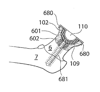
implantable medical device comprises an elongated element 1320 comprising a
movable locking portion 1321 adapted to have a first and second state, wherein
said
movable lodging portion 1321, in said first state is adapted to be inserted
into a hole
in the pelvic bone 9, and in said second state is adapted to hinder the
elongated
.. element 1320 from passing through said hole in the pelvic bone 9 by said
movable
locking portion 1321 contacting the surface of the pelvic bone 9 on the
abdominal
side. Fig. 8f shows the elongated element 1320 in its first state after having
passed
-through the hole in the pelvic bone 9.
[00095] Fig. 16f shows the movable locking portion 1321 changing from the
firstto
.. the second state at the same time as the artificial caputfemur 112,
comprising a
threaded part 1314, interacts with a corresponding threaded member 1323 being
partof the elongated element 1320. The movable locking portion 1321 is
pivotally
arranged at a pivotpoint 1322 and changes from the firstto the second slate
using
the pivotpoint 1322.
[00096] Hg. 16g shows the nuclical device according to the embodiment of figs.
16e
and 16f when the nvvable member 1321 is placed in the second state, in which
the
artificial caput femur 112 is fixated to the pelvic bone 9 by the movable
member 1321
being in contact with the abdominal side of the pekic bone 9. The artificial
caput
femur 112 has been tightened using the threaded part 1314 and corresponding
.. threaded member 1323, such that the entire medical device comprising the
artificial
caput femur 112 is securely fixated to the pelvic bone 9. Similar to the
embodiments
shown with reference to figs. 16b ¨16d the fixation element 1301 could be
further
24
Date Recue/Date Received 2021-05-14
WO 2011/005187 PCT/SE2010/050803
fixated to the acetabulum 8 using an adhesive, such as bone cement, applied to
the
fixation surface and/ or the acetabulum surface 8.
[00097] Hg. 16h shows an embodiment in which the fixation element comprises a
fixation surface 1334 comprising two holes adapted to receive two mechanical
fixation elements 1331. In the embodiment of fig. 81 the mechanical fixation
elements
1331 are expanding fixation elements 1331, such as the expanding fixation
elements
described with reference to figs. 16b ¨16d, however in other ernbodirrents it
is
equally conceivable that the mechanical fixation elements are elements adapted
to
fixate the medical device to the internal periphery of the holes, such as
screws. Similar
to the embodiments shown with reference to figs. 16b ¨16g the fixation element
1301
could be further fixated to the aceiabulum using an adhesive, such as bone
cement,
applied to the fixation surface and/ or the acetabulum surface. Hg. 16h shows
an
embodiment in which the medical device has a pre-rno-unted locking member 116,
however, in other embodiments it is equally conceivable that the locking
member 116
is adapted to be mounted after the artificial caput femur 112 has been
implanted in
the hip joint
[00098] Fig. 16i shows the artificial hip joint in section, when the medical
device
described with reference to fig. 16h has been implanted. RI/the mire an
artificial
acetabulum surface 1340 having a concave surface inwards the center of the hip
joint
has been implanted. The artificial acetabulum surface 1340 has been fixated to
the
femoral bone 7, and placed in movable contact with the artificial caput femur
surface
112, thus creating a functioning artificial hip joint The locking member 116
has been
fixated to the femoral bone 7, thus locking the artificial caput femur 112 in
the
artificial acetabulum surface 1340. The locking member 116 is according to the
embodiment shown in fig. 8j fixated using screws 121, however the screws 121
could
be assisted or replaced by an adhesive, such as bone cement
[00099] fig. 17a shows an assembled artificial hip joint with an artificial
caput femur
surface 112 fixated to the pelvic bone 9 using two fixating members adapted to
Date Recue/Date Received 2021-05-14
WO 2011/005187 PCT/SE2010/050803
expand inside of the cortical bone of ihe pelvic bone 9. The fixating members
comprises a screw 121 in connection with an anvil member 1312 affecting an
expandable portion 1311 pressing the expandable members in two directions
perpendicular to the length axis of the fixation members for fixating the
artificial caput
femur 112 to the pelvic bone 9. The artificial acetabulum 1340 is fixated to
the
femoral bone 7 using an elongated member 1310b placed in the cancellous bone
and
aligned with the caput and collum femur center axis. The elongated member
comprises
an expandable portion 1311b which is pressed by an anvil member 1312b
connected in a threaded member 1313b pressing the expandable members 1311b in
two directions perpendicular-to the length axis of the elongated member 1310b
for
fixating the artificial acetabulum surface to the femoral bone 7.
[000100] Hg. 17b shows an embodiment similar to the embodiment shown in fig.
17a with the difference thatthe artificial acetabulum surface is fixated using
an
elongated "member 1310c which penetrates the cancellous bone of the collum
femur
and the cortical bone of the femoral bone in the area of the greater
trochanier 1695.
The elongated member comprises a movable locking portion 1321b, pivotally
arranged at a pivot point 1322b. 'the movable locking portion 1321b could
change
from a firstin a second state around the pivotpoint 1322b. When the movable
locking portion 132 lb is placed in the second state itlocks the elongated
member on
the outside of the femoral bone 7 in the area of the greater trochanter 1695.
[000101] Hg. 17c shows an embodiment similar to the embodiment shown in fig.
17a with the difference that-the artificial acetabulum surface is fixated
using an
elongated member 1310d which penetrates the cancellous bone of the collum
femur
and enters the cortical bone of the femoral bone in the area of the greater
trochanter
1695 butnever exits the bone butrather is fixated inside of the bone 7.
[000102] Hg. 18a shows an embodirnPnt where the artificial acetabulum 1340 is
fixated to the femoral bone 7 using fixating portions 680 being part of the
locking
member 116. The fixating portions 680 comprises portions 680' clasping the
26
Date Recue/Date Received 2021-05-14
WO 2011/005187
PCT/SE2010/050803
surgically cut femoral bone and thereby fixating the artificial acetabulum
surface to the
femoral bone.
[000103] Hg. 18b shows an embodinEnt similqr to the embodiment described with
reference to fig. 18a with the difference that the locking member is fixated
to the
surgically cut caput femur using screws 121.
[000104] Hg. 19 shows the hip joint in section when the medical device is
assembled
and in its functional position in the hip joint The artificial caput femur
surface 45 or
convex hip joint surface 112 is fixated ID the fixation part 1301, which in
turn is
fixated to the acetabulum 8, The locking member 116 locks the artificial
convex caput
femur surface 45 in the artificial concave acetabulum surface in the caput 5
and
collum femur 6.
[000105] Hg. 20 shows a frontal view of a human patient when an incision for
reaching an area of the hip joint through the pelvic bone in a surgical method
has
been performed. According to one embodiment the incision 1 is made in the
abdominal wall of the human patient The incision 1 passes through the
abdominal
wall, preferably recius abdorninis and peritoneum, in to the abdomen of the
human
patent In a second embodirnentthe incision 2 is conducted through the rectus
abdominis and in to the pelvic area, below peritoneum. According to a third
embodiment the incision 3 is performed justbetween Mum and the surrounding
tissue,
an incision 3 which could enable the pelvic bone to be dissected with very
little
penetration of fascia and muscular tissue. According to a fourth embodiment
the
incision 4 is made in the inguinal channeL In all of the four embodiments the
tissue
surrounding the pelvic bone 9 in the area opposite to acetabulum is removed or
penetrated which enables the surgeon to reach the pelvic bone 9. It is obvious
that the
methods described may both be combined or altered reaching the same goal to
dissect the pelvic bone on the opposite side of the acetabulum.
27
Date Recue/Date Received 2021-05-14
WO 2011/005187 PCT/SE2010/050803
[000106] Hg. 21 shows a frontal view of a human patient when small incisions
for
reaching an area of the hip joint through the pelvic bone in a arihroscopic
method has
been perfornmd. According to a first embodimentthe incisions 14 is made in the
abdominal wall of the human patient The small incisions enable the surgeon to
insert
arIhroscopic (nears into the abdomen of the human patient According to the
first
embodiment the incisions 14 passes through the abdomen, preferably rectus
abdominis and peritoneum, into the abdomen of the human patent According to a
second embodiment the small incisions 15 is conducted through the rectus
abdominis
and in to the pelvic area, below peritoneum. According to a third embodiment
the
.. small incisions 16 is performed just between Ilium and the surrounding
tissue, an
incision 16 which could enable the pelvic bone ID be dissected with very
little
penetration of fascia and muscular tissue. According in a fourth embodiment1he
incision 17 is made in the inguinal channel In all of the four embodiments the
tissue
surrounding the pelvic bone 9 in the area opposite to acetabulum 8 is removed
or
penetrated which enables the suigeonto reach the pelvic bone 9.
[000107] Hg. 22 shows the human patient in section when a medical device for
creating a hole 18 in the pelvic bone 9 is inserted through an incision
according to
any of the embodiments described above. An elongated member 21, which could
comprise a part or section adapted to be benttransfers forte from an operating
device
(not shown) to the bone contacting organ 22. The bone contacting organ 22 is
placed
in contact with the pelvic bone 9 and creates a hole through a drilling,
sawing or
milling process powered by a rotating, vibrating or oscillating force
distributed from
the elongated member 21.
[000108] Hg. 23 shows the hip joint in section after the medical device for
creating a
hole 18 in the pelvic bone 9 has created said hole 18. According to this
embodiment
the hole 18 is created through the removal of a bone plug 31, however it is
equally
conceivable that said medical device comprises a bone contacting organ 22
adapted
28
Date Recue/Date Received 2021-05-14
WO 2011/005187 PCT/SE2010/050803
to create small pieces of bone, in which case the medical device could further
comprise a system for transport of said small pieces of bone.
[000109] Hg. 24 shows how the medical device adapted to create a hole is
inserted
into the hip joint and placed in contact with the caput femur 5. According to
this
embodiment the medical device for creating a hole in the pelvic bone 9 and
surgically
cutting the caput femur 5 is the same medical device, however kis equally
conceivable
that there is a second medical device particularly adapted to surgically cut
the cap-ut
femur 5.
[000110] lig. 25 shows the hip joint in section when a second medical device
604
surgically removes the most proximal portion of the caput femur 5. 'the second
medical
device 604 comprises a drilling portion in which a cutting member in a folded
position
605a is placed.
[000111] Hg. 26 shows the second medical device 604 when the drilling portion
is
positioned inside of the femoral bone, and the cutting member is placed in a
cutting
position 605b for cutting the proximal portion of the caput femur 5.
[000112] Hg. 27 shows a medical device comprising an artificial convex hip
joint
surface 112. The artificial convex hip joint surface 112 is adapted In be
fixated to the
pelvic bone 9, and is adapted to be inserted through a hole 18 in the pelvic
bone 9.
The medical device comprises a nut 120, comprising threads for securely
fixating the
medical device to the pelvic bone 9. The medical device further comprises a
prosthetic
part 118 adapted to occupy the hole 18 created in the pelvic bone 9 after the
medical
device has been implanted in the patient The prosthetic part 118 comprises
supporting members 119 adapted to be in contactwith the pelvic bone 9 and
assist in
the carrying of the load placed on the medical device from the weight of the
human
patient in nomral use. Normal use is defined as the same as a person would use
a
natural hip joint Thrther the medical device comprises a locking member 116
comprising a surface 117 adapted to be in contact with the artificial convex
hip joint
29
Date Recue/Date Received 2021-05-14
WO 2011/005187 PCT/SE2010/050803
surface 112. The locking nymber 116 further comprises fixating members 115
which
are adapted to assistin the fixation of the locking member 116 to the
caputfemur 5 or
collum femur 6, which in tams fixates the artificial convex hip joint surface
112. The
artificial convex hip joint surface 112 is fixated to an attachnnt rod 113
comprising
a thread 114 thatcorresponds to the thread of the nut 120 in connection with
the
prosthetic part 118.
[000113] Eg. 28 shows the hip joint in section when the artificial convex hip
joint
surface is fixated in the medical device 109 comprising a concave hip joint
surface
110. The convex hip joint surface 112 is secured in place by the locking
member 116
which is fixated to the caput femur using screws 121. The surface of the
locking
member 117 and the concave hip joint surface 117 is placed in connection with
the
convex hip joint surface and could be made of a friction reducing material
such as
FIFE or a self lubricating powder material However it is also conceivable that
the
connecting surfaces are lubricated using an implantable lubrication system
adapted to
lubricate the medical device after said medical device has been implanted in
the
human patient
[000114] Hg. 29 shows the placing of a prosthetic part 118 adapted in occupy
the
hole 18 created in the pelvic bone 9. The prosthetic part 118 comprises
supporting
members 119 adapted to be in contactwith the pelvic bone 9 and assist in the
carrying of the load placed on the medical device from the weight of the human
patient According to the embodiment shown in fig. 12 the supporting members
119
are located on the abdominal side of the pelvic bone 9, however itis equally
conceivable the supporting members 119 are located on the acetabulum side of
the
pelvic bone 9, in which case they are preferably displaceable for allowing
insertion of
the prosthetic part 118 through the hole 18 in the pelvic bone 9. Ruthennore
fig. 12
shows the fixation of a nut 120 In the attachmentrod 113. According In the
embodiment shown in fig. 12 the hole 18 in the pelvic bone 9 is adapted In be
larger
than the medical device allowing the medical device to be inserted in its full
functional
Date Recue/Date Received 2021-05-14
WO 2011/005187 PCT/SE2010/050803
size. According to other embodiment the hole 18 is smaller in which case the
medical
device could comprise of several parts adapted to be connected after insertion
in the
hip joint or the medical device could be expandable for insertion through a
hole
smaller than the full functional size of the medical device. The expandable
medical
device could be enabled through the elements of the medical device comprising
elastic
nraterial.
[000115] Fig. 30 shows fie hip joint in section when all the elements of the
medical
device has been fixated in the area of the hip joint or its surroundings. The
prosthetic
part 113 adapted to occupy the hole 18 in the pelvic bone 9 is here fixated
with
screws 121, however these screws 121 could be assisted or replaced by an
adhesive
which could be applied to the surface S between the prosthetic part and the
pelvic
bone 9.
[000116] Fig. 31 shows the hip joint in section when the method of supplying
the
medical device is conducted according to another embodiment The proximal part
of
the caput femur has been removed along the section created by the medical
device for
creating a hole. A reaming member 40 adapted to create a concave surface 103
in
the caputferaur 5 is here applied to a elongated member 206 which is inserted
through a hole 205 going frorn the lateral side of the thigh, penetrating the
cortical
bone of the femoral bone 7 propagating along a length axis of the co llum
femur in the
cancellous bone and entering the area of the hip joint The elongated member
206 is
operated using an operating device 207 which could be an electrically powered
operating device, a hydraulically powered operating device or a pneumatically
powered operating device. The reaming in the caput femur and part of the
collum
femur 6 is mainly perfon-ned in the cancellous bone, however that does not
exclude the
possibility the some of the reaming needs to be performed in the cortical bone
of the
caput femur 5 or the collum femur 6.
[000117] Hg. 32 shows the step of applying an adhesive 106 to the concave
surface
created by the reamer 40. 'Me adhesive 106 is applied by an injecting member
104
31
Date Recue/Date Received 2021-05-14
WO 2011/005187 PCT/SE2010/050803
comprising an injecting nozzle 105. The adhesive 106 is preferably a
biocompatible
adhesive such as bone cement The injecting member 104 is in this embodiment
adapted for introduction through a hole 18 in the pelvic bone 9, through the
injecting
member 104 being bent
[000118] Hg. 33 shows the step of providing a medical device 109 comprising an
artificial concave hip joint surface 110. The medical device is according to
this
embodimentprovided with a hole positioned in the length axis of the collum
femur 6.
The medical device is through the hole adapted to be guided by the elongated
member 206 or a guiding rod placed in the hole 205 along a length axis of the
collurn femur 6. Inserting the medical device into the hip joint while the
elongated
member 206 or guiding rod runs through the hole of the medical device
facilitates the
positioning of the medical device and ensures the different parts of the
medical device
is centered for functioning as a unit la the embodiment shown in fig. 33 the
rredical
device 109 is inserted into the hip joint as a single unit, however it is
equally
conceivable that the medical device 109 is inserted in parts (not shown) which
are
then connected to form the medical device after implantation in the patient
The
artificial concave hip joint surface 110 is fixated to the concave surface 103
created
in the caput femur 5 and collum femur 6. The medical device 109 comprises a
fixation
support 111 adapted to anchor said artificial concave hip joint surface 110,
to at least
one of the caput femur 5 and the collum femur 6. The medical device 109 is
adapted
In be introduced to the hip jointthrough a hole 18 in the pelvic bone 9 using
a
nranipulation device 122 comprising a gripping member 123. According to this
embodiment the manipulation device 122 is bent and thereby adapted to operate
through a hole 18 in the pelvic bone 9. According ID one embodimentthe medical
device 109 comprises a self lubricating material such as PIIX however it is
also
conceivable that said medical device comprises: titanium, stainless steel,
Conan, YE, or
other acrylic polymers, in which case the medical device could be adapted to
be
lubricated after insertion in the hip joint
32
Date Recue/Date Received 2021-05-14
WO 2011/005187 PCT/SE2010/050803
[000119] Hg. 34 shows the hip joirt in section when the artificial convex hip
joint
surface is fixated in the medical device 109 comprising a concave hip joint
surface
110, the medical device is guided using the elongated member 206 or a guiding
rod.
The convex hip joint surface 112 is secured in place by the locking member 116
which is fixated to the caputfemur using SC/PVis 121, the convex hip joint
surface is
guided using the elongated member 206 or a guiding red. The surface of the
locking
member 117 and the concave hip joint surface 110 is placed in connection with
the
convex hip joint surface and could be made of a friction reducing material
such as
PIFE era self lubricating powder materiaL However it is also conceivable that
the
connecting surfaces are lubricated using an implantable lubrication system
adapted to
lubricate the medical device after said medical device has been implanted in
the
human patient The elongated member or guiding rod 206 can be adapted to act as
a
centering rod for centering the atleast one artificial hip joint surface
inside of the hip
joint According to the embodiment shown the elongated member 206 is inserted
through the femoral bone, however according in other embodiments, not shown,
the
elongated member is positioned inside of the, hip jointfrom the acetab-ulum
side.
[000120] Hg. 35 shows the placing of a prosthetic part 118 adapted to occupy
the
hole 18 created in the pelvic bone 9. The prosthetic part 118 comprises
supporting
members 119 adapted to be in coninct with the pelvic bone 9 and assist in the
carrying of the load placed on the medical device from the weight of the human
patient Rwthemiore fig. 35 shows the fixation of a nut 120 to the attachment
rod 113,
which in turn is guided by the elongated member 206 or a guiding rod.
[000121] fig. 36 shows the hip joint in section when all the elements of the
medical
device has been fixabd in the area of the hip joint or its surro-undings. The
prosthetic
part 118 adapted to occupy the hole 18 in the pelvic bone 9 is here fixated
with
screws 121, however these screws 121 could be assisted or replaced by an
adhesive
which could be applied to the surface S between the prosthetic part and the
pelvic
33
Date Recue/Date Received 2021-05-14
WO 2011/005187 PCT/SE2010/050803
bone 9. The elongated member 206 or guiding rod has been refracted through the
incision in the thigh.
[000122] Hg. 37 shows an embodinnt of a locking member 116, wherein the
locking member 116 comprises a surface adapted in be in contact with the
artificial
convex hip joint surface 1353, the locking member 116 is adapted to, Ma first
slate,
lock the artificial caput femur 11210 the artificial acetabulurn surface 1340,
and in a
second state, release said artificial caput femur 112 from said artificial
acetabultun
1340. The locking member 116 is adapted to change from the first to the second
state
when a predetermined amount of strain is placed on the locking member 116. The
locking member 116 according in the embodiment shown in fig. 37, comprises
four
elastic portions 1351, and the locking member 116 is adapted to change from
the first
In the second state using the elasticity of the elastic portions 1351. The
locking
member 116 is adapted to be fixated to the femoral bone 7 using screws adapted
to
be placed in holes 1352 adapted therefor.
[000123] Hg. 38 shows the hip jointin section when a two state locking member
116
locks the artificial caput femur 112 in the artificial acetabulum 1340. The
two state
locking member 116 is fixated to the femoral bone 7 using screws 121, and is
here
shown in its first state in which the locking member 116 locks the artificial
caput femur
112 to the artificial acetabulum 1340.
[000124] fig. 39 shows the hip joint in section according to the embodiment of
fig
38, but when the two state locking member 116 is in its second state, in which
the
locking ring 116 releases the artificial caput femur 112 from the artificial
acetabulurn
surface 1340. The construction with the releasing locking ring 116 reduces the
risk of
strain placed on the artificial joint injuring the fixation points, i.e. the
contactwilh
bone; it further enables the artificial jointto be non-invasively relocated
incase of
luxation.
34
Date Recue/Date Received 2021-05-14
WO 2011/005187 PCT/SE2010/050803
[000125] According to the above mentioned embodiments the medical device is
adapted to be inserted through a hole in the pelvic bone, however it is
equally
conceivable thatthe medical device according to any of the embodiment above is
adapted to be inserted through a hole Mille hip jointcapsule or the femoral
bone of
the huma.n patient
[000126] Flease note that any embodiment or part of embo diment as well as any
method or part of method could be combined in any way. All examples herein
should
be seen as part of the general description and therefore possible to combine
in any
way in general terms.
Date Recue/Date Received 2021-05-14