Note: Descriptions are shown in the official language in which they were submitted.
CA 03118967 2021-04-22
WO 2020/096917
PCT/US2019/059583
DOSING REGIMEN OF ANTI-LAG3 ANTIBODY AND COMBINATION THERAPY WITH
ANTI-PD-1 ANTIBODY FOR TREATING CANCER
FIELD OF THE INVENTION
The present invention relates to dosing regimens of an anti-LAG3 antibody
useful for the
treatment of cancer. In particular, the invention relates to the dosing
regimen in a combination
therapy which comprises administering an antibody of a Programmed Death 1
protein (PD-1) or
Programmed Death Ligand 1 (PD-L1) and an antibody of Lymphocyte-Activation
Gene 3 (LAG3).
The invention also provides a method for treating cancer in a patient
comprising administering to the
patient an anti-LAG3 antibody and an anti-PD-1 antibody, wherein the tumor
tissue section of the
patient is PD-Li expression positive, optionally, LAG3 expression positive
BACKGROUND OF THE INVENTION
PD-1 is recognized as an important molecule in immune regulation and the
maintenance of
peripheral tolerance. PD-1 is moderately expressed on naive T, B and NKT cells
and up-regulated
by T/B cell receptor signaling on lymphocytes, monocytes and myeloid cells (1)
Two known ligands for PD-1, PD-Li (B7-H1) and PD-L2 (B7-DC), are expressed in
human
cancers arising in various tissues. In large sample sets of e.g. ovarian,
renal, colorectal, pancreatic,
liver cancers and melanoma, it was shown that PD-Li expression correlated with
poor prognosis and
reduced overall survival irrespective of subsequent treatment (2-13).
Similarly, PD-1 expression on
tumor infiltrating lymphocytes was found to mark dysfunctional T cells in
breast cancer and
melanoma (14-15) and to correlate with poor prognosis in renal cancer (16).
Thus, it has been
proposed that PD-Li expressing tumor cells interact with PD-1 expressing T
cells to attenuate T cell
activation and evasion of immune surveillance, thereby contributing to an
impaired immune
response against the tumor.
Several monoclonal antibodies that inhibit the interaction between PD-1 and
one or both of
its ligands PD-Li and PD-L2 have been approved for treating cancer.
Pembrolizumab is a potent
humanized immunoglobulin G4 (IgG4) mAb with high specificity of binding to the
programmed cell
death 1 (PD 1) receptor, thus inhibiting its interaction with programmed cell
death ligand 1 (PD-L1)
and programmed cell death ligand 2 (PD-L2). Based on preclinical in vitro
data, pembrolizumab has
- 1 -
CA 03118967 2021-04-22
WO 2020/096917
PCT/US2019/059583
high affinity and potent receptor blocking activity for PD-1. Keytrudag
(pembrolizumab) is
indicated for the treatment of patients across a number of indications.
Lymphocyte-Activation Gene 3 (LAG3) is an inhibitory immune modulatory
receptor that
regulates effector T cell homeostasis, proliferation, and activation, and has
a role in the suppressor
.. activity of regulatory T cells (Tregs). LAG3 is expressed on activated CD8+
and CD4+ T cells,
Tregs and the Trl regulatory T-cell population, as well as on natural killer
cells and a subset of
tolerogenic plasmacytoid dendritic cells. Because of its proposed role on both
effector T cells and
Tregs, LAG3 is one of several immune checkpoint molecules where simultaneous
blockade of both
cell populations has the potential to enhance antitumor immunity.
LAG3 is structurally related to cluster of differentiation (CD) 4 and a member
of the
immunoglobulin (Ig) superfamily. Like CD4, its ligand is major
histocompatibility complex (MHC)
Class II molecules. Interaction with its ligand leads to dimerization and
signal transduction resulting
in altered T-cell activation. Following T-cell activation, LAG3 is transiently
expressed on the cell
surface. A large proportion of LAG3 molecules are found in intracellular
stores and can be rapidly
.. translocated to the cell membrane upon T-cell activation. LAG3 expression
is regulated at the cell
surface by extracellular cleavage to yield a soluble form of LAG3 (sLAG 3),
which can be detected
in serum. Expression of LAG3 is tightly regulated and represents a self-
limiting mechanism to
counter uncontrolled T-cell activity. Anti-LAG3 antibodies have been described
in
W02016/028672.
Selecting a dosage regimen for an anti-LAG3 antibody monotherapy or
combination therapy
with anti-PD-1 or anti-PD-Li therapy depends on several factors, including the
serum or tissue
turnover rate of the entity, the level of symptoms, the immunogenicity of the
entity, antidrug
antibody endpoints and the accessibility of the target cells, tissue or organ
in the individual being
treated, as well as safety. Formation of antidrug antibodies can potentially
confound drug
exposures at therapeutic doses, and prime for subsequent infusion-related
toxicities. In addition,
anti-LAG3 and/or anti-PD-1/anti-PD-L1 treatment can result in immune
stimulation and the
potential for cytokine release that affects safety.
SUMMARY OF THE INVENTION
The invention provides a method for treating cancer in a patient comprising
administering 7-
1200 mg of an anti-LAG3 antibody Ab6. In one embodiment, 200-800 mg of an anti-
LAG3
- 2 -
CA 03118967 2021-04-22
WO 2020/096917
PCT/US2019/059583
antibody Ab6 is administered. In another embodiment, 800 mg of an anti-LAG3
antibody Ab6 is
administered. In one embodiment, the method optionally comprises co-
administration with an anti-
PD-1 or anti-PD-Li antibody. In one embodiment, the anti-LAG3 antibody and
anti-PD-1 antibody
are co-formulated. In another embodiment, the tumor tissue section of the
patient is PD-Li
.. expression positive. In a further embodiment, the tumor cells of the
patient is PD-Li expression
positive. In one embodiment, the anti-PD-1 antibody blocks the binding of PD-1
to PD-Li and PD-
L2.
The invention also provides a pharmaceutical composition comprising 7-1200 mg
of anti-
LAG3 antibody Ab6 or Ab6 variant, and 200 mg of pembrolizumab or pembrolizumab
variant. In
one embodiment, the pharmaceutical composition comprises 800 mg of anti-LAG3
antibody Ab6 or
Ab6 variant, and 200 mg of pembrolizumab or pembrolizumab variant.
The invention also provides a method for treating non-MSI-H colorectal cancer,
gastric
cancer or head and neck squamous cell carcinoma in a patient comprising
administering to the
patient an anti-LAG3 antibody and an anti-PD-1 antibody, wherein the tumor
tissue section of the
patient is PD-Li expression positive, and optionally LAG3 expression positive.
BRIEF DESCRIPTION OF THE DRAWINGS
FIG. 1 CT scan of patient with non-MSI-H colorectal cancer before (left)
and after (right)
treatment with 21 mg anti-LAG3 antibody Ab6 and pembrolizumab. The patient
received 5 prior
lines of chemotherapy, no prior anti¨PD-1 or anti¨PD-Li therapy. The patient
had a partial response
with 45% reduction in tumor volume. There was also tumor volume reduction in
lung lesions and
lymph nodes, and stable presacral mass. The response is ongoing at 13.5
months.
FIG. 2 CT scan of a 60-year-old male with renal cell carcinoma and
metastases to lung and bone
before (left) and after (right) treatment with 7 mg anti-LAG3 antibody Ab6 and
pembrolizumab.
The patient received 3 prior lines of therapy, including prior anti¨PD-1
therapy. The patient had a
partial response at 9 weeks with 49% reduction in tumor volume. Tumor volume
reduction was
observed at all visible disease sites including the lung and multiple lymph
nodes. The response
lasted for 15 months before disease progression.
FIG. 3 Waterfall plot of subjects with best target lesion change from baseline
based on investigator
assessment per RECIST 1.1 FAS population in the colorectal cancer expansion
cohort (Part B) using
the PD-Li IHC Combined Positive score (CPS).
Each bar represents an individual subject. Greater than a 30% decrease in
tumor size from baseline
(Y-axis) is considered a response; changes between a 30% decrease and a 20%
increase is
- 3 -
CA 03118967 2021-04-22
WO 2020/096917
PCT/US2019/059583
considered stable disease; changes greater than a 20% increase is considered
progressive disease.
Tumor samples with CPS >=1 or <1 are indicated. Tumor samples with less than
100 tumor cells
cannot be interpreted.
FIG. 4 Waterfall plot of subjects with best target lesion change from baseline
based on investigator
assessment per RECIST 1.1 FAS population in the colorectal cancer expansion
cohort (Part B) using
the LAG3 IHC CPS-like LAG3 positive cells scoring system.
Each bar represents an individual subject. Greater than a 30% decrease in
tumor size from baseline
(Y-axis) is considered a response; changes between a 30% decrease and a 20%
increase is
considered stable disease; changes greater than a 20% increase is considered
progressive disease.
Tumor samples with CPS >=1 or <1 are indicated. Tumor samples with less than
100 tumor cells
cannot be interpreted.
FIG. 5 Serum concentrations of Ab6 following intravenous doses from 7 mg
to 700 mg in cycle
1, Part A of the phase I study. Arithmetic mean serum concentration for each
dose is plotted at
nominal times.
FIG. 6 Serum concentrations of total soluble LAG-3 following intravenous
doses from 7 mg to
700 mg in cycle 1, Part A of the phase I study. Arithmetic mean of total
soluble LAG-3 plotted at
nominal times.
FIG. 7A-B shows that pembrolizumab Cmax at steady state for 400 mg Q6W lies
within the range
from 2 mg/kg and 200 mg Q3W to 10 mg/kg Q2W. 7A: pembrolizumab Cmax at steady
state for 2
mg/kg and 200 mg Q3W. 7B: pembrolizumab Cmax at steady state for 400 mg Q6W
and 10 mg/kg
Q2W.
FIG. 8 shows that pembrolizumab exposures (Cavg and Cmin) at steady
state are similar for
400 mg Q6W relative to 2 mg/kg Q3W and 200 mg Q3W.
FIG. 9A-B shows the pembrolizumab pharmacokinetic profiles at steady state for
the 400 mg Q6W
dosing regimen compared to the Q3W, 200 mg flat dosing regimen (top) and the
Q3W, 2 mg/kg
weight-based dosing regimen (bottom). 9A shows the log scale concentrations,
and 9B shows the
linear scale concentrations.
FIG. 10 Serum concentrations of Ab6 following intravenous doses from 7 mg to
700 mg in cycle
1 on linear scale with additional patient sampling compared to Figure 5. The
arithmetic mean of Ab6
serum concentrations is plotted at nominal times.
- 4 -
CA 03118967 2021-04-22
WO 2020/096917
PCT/US2019/059583
FIG. 11 Serum concentrations of Ab6 following intravenous doses from 7 mg
to 700 mg in cycle
1 on log scale with additional patient sampling compared to Figure 5. The
arithmetic mean of Ab6
serum concentrations is plotted at nominal times.
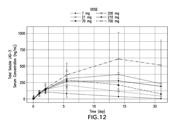
FIG. 12 Serum concentrations of total soluble LAG3 following intravenous
doses from 7 mg to
700 mg in cycle 1 with additional patient sampling compared to Figure 6. The
arithmetic mean of
total soluble LAG3 serum concentrations is plotted at nominal times.
FIG. 13 Predicted Ab6 serum concentration-time profiles in Cycle 1
corresponding to the 800
mg dose overlaid with observed concentrations for the 700 mg dose. Solid
markers represent
observed Ab6 serum concentrations at 700 mg from the Phase I study. Shaded
areas represent 2.5th
and 97.5th percentiles for predicted concentrations for the 800 mg dose. Ab6
exposures from cycle 1
are expected to be representative of subsequent treatment cycles.
FIG. 14 Predicted Ab6 exposures (AUC, Ctrough, Cmax) as a function of
dose showing
substantial overlapping exposures between the 700 mg and 800 mg doses.
Straight lines: median;
box: 25th and 75th percentile, whiskers: 5th and 95th percentiles.
FIG. 15 Box-plot of Ab6 serum Ctrough on Day 21 showing PK variability.
FIG. 16 Waterfall plot of subjects with best target lesion change from
baseline based on
investigator assessment per RECIST 1.1 FAS population in the gastric cancer
expansion cohort (Part
B) using the PD-Li MC Combined Positive score (CPS).
Each bar represents an individual subject. Greater than a 30% decrease in
tumor size from baseline
(Y-axis) is considered a response; changes between a 30% decrease and a 20%
increase is
considered stable disease; changes greater than a 20% increase is considered
progressive disease.
Tumor samples with CPS >=1 or <1 are indicated. Tumor samples with less than
100 tumor cells
cannot be interpreted.
FIG. 17 Waterfall plot of subjects with best target lesion change from
baseline based on
investigator assessment per RECIST 1.1 FAS population in the gastric cancer
expansion cohort (Part
B) using the LAG3 IHC CPS-like %LAG3 positive cells scoring method.
Each bar represents an individual subject. Greater than a 30% decrease in
tumor size from baseline
(Y-axis) is considered a response; changes between a 30% decrease and a 20%
increase is
considered stable disease; changes greater than a 20% increase is considered
progressive disease.
Tumor samples with CPS >=1 or <1 are indicated. Tumor samples with less than
100 tumor cells
cannot be interpreted.
- 5 -
CA 03118967 2021-04-22
WO 2020/096917
PCT/US2019/059583
FIG. 18 Waterfall plot of subjects with best target lesion change from
baseline based on
investigator assessment per RECIST 1.1 FAS population in the HSNCC PD-Li naive
cancer
expansion cohort (Part B) using the PD-Li IHC TPS+MIDS scoring system.
Each bar represents an individual subject. Greater than a 30% decrease in
tumor size from baseline
(Y-axis) is considered a response; changes between a 30% decrease and a 20%
increase is
considered stable disease; changes greater than a 20% increase is considered
progressive disease.
Tumor samples with CPS >=1 or <1 are indicated. Tumor samples with less than
100 tumor cells
cannot be interpreted.
FIG. 19 Waterfall plot of subjects with best target lesion change from
baseline based on
investigator assessment per RECIST 1.1 FAS population in the HSNCC PD-Li naïve
cancer
expansion cohort (Part B) using the LAG3 IHC %LAG3 positive cells scoring
system.
Each bar represents an individual subject. Greater than a 30% decrease in
tumor size from baseline
(Y-axis) is considered a response; changes between a 30% decrease and a 20%
increase is
considered stable disease; changes greater than a 20% increase is considered
progressive disease.
Tumor samples with CPS >=1 or <1 are indicated. Tumor samples with less than
100 tumor cells
cannot be interpreted.
DETAILED DESCRIPTION
Abbreviations. Throughout the detailed description and examples of the
invention the following
abbreviations will be used:
BOR Best overall response
BID One dose twice daily
CBR Clinical Benefit Rate
CDR Complementarity determining region
CHO Chinese hamster ovary
CR Complete Response
DCR Disease Control Rate
DFS Disease free survival
DLT Dose limiting toxicity
DOR Duration of Response
DSDR Durable Stable Disease Rate
FFPE Formalin-fixed, paraffin-embedded
FR Framework region
- 6 -
CA 03118967 2021-04-22
WO 2020/096917
PCT/US2019/059583
IgG Immunoglobulin G
IHC Immunohistochemistry or immunohistochemical
irRC Immune related response criteria
IV Intravenous
MTD Maximum tolerated dose
NCBI National Center for Biotechnology Information
NCI National Cancer Institute
ORR Objective response rate
OS Overall survival
PD Progressive disease
PD- I Programmed Death 1
PD-Li Programmed Cell Death 1 Ligand 1
PD-L2 Programmed Cell Death 1 Ligand 2
PFS Progression free survival
PR Partial response
Q2W One dose every two weeks
Q3W One dose every three weeks
QD One dose per day
RECIST Response Evaluation Criteria in Solid Tumors
SD Stable disease
VH Immunoglobulin heavy chain variable region
VK Immunoglobulin kappa light chain variable region
I. DEFINITIONS
So that the invention may be more readily understood, certain technical and
scientific terms
are specifically defined below. Unless specifically defined elsewhere in this
document, all other
technical and scientific terms used herein have the meaning commonly
understood by one of
ordinary skill in the art to which this invention belongs.
As used herein, including the appended claims, the singular forms of words
such as "a,"
"an," and "the," include their corresponding plural references unless the
context clearly dictates
otherwise.
- 7 -
CA 03118967 2021-04-22
WO 2020/096917
PCT/US2019/059583
As used herein, an "Ab6 variant" means a monoclonal antibody which comprises
heavy
chain and light chain sequences that are substantially identical to those in
Ab6 (as described below
and in W02016028672, incorporated by reference in its entirety), except for
having three, two or
one conservative amino acid substitutions at positions that are located
outside of the light chain
CDRs and six, five, four, three, two or one conservative amino acid
substitutions that are located
outside of the heavy chain CDRs, e.g, the variant positions are located in the
FR regions or the
constant region, and optionally has a deletion of the C-terminal lysine
residue of the heavy chain. In
other words, Ab6 and a Ab6 variant comprise identical CDR sequences, but
differ from each other
due to having a conservative amino acid substitution at no more than three or
six other positions in
their full length light and heavy chain sequences, respectively. An Ab6
variant is substantially the
same as Ab6 with respect to the following properties: binding affinity to
human LAG3 and ability to
block the binding of human LAG3 to human WIC Class II.
"Administration" as it applies to an animal, human, experimental subject,
cell, tissue, organ,
or biological fluid, refers to contact of an exogenous pharmaceutical,
therapeutic, diagnostic agent,
or composition to the animal, human, subject, cell, tissue, organ, or
biological fluid. Treatment of a
cell encompasses contact of a reagent to the cell, as well as contact of a
reagent to a fluid, where the
fluid is in contact with the cell. The term "subject" includes any organism,
preferably an animal,
more preferably a mammal (e.g., rat, mouse, dog, cat, rabbit) and most
preferably a human.
As used herein, the term "antibody" refers to any foul' of antibody that
exhibits the desired
biological or binding activity. Thus, it is used in the broadest sense and
specifically covers, but is not
limited to, monoclonal antibodies (including full length monoclonal
antibodies), polyclonal
antibodies, multispecific antibodies (e.g., bispecific antibodies), humanized,
fully human antibodies,
chimeric antibodies and camelized single domain antibodies. "Parental
antibodies" are antibodies
obtained by exposure of an immune system to an antigen prior to modification
of the antibodies for
an intended use, such as humanization of an antibody for use as a human
therapeutic.
In general, the basic antibody structural unit comprises a tetramer. Each
tetramer includes
two identical pairs of polypeptide chains, each pair having one "light" (about
25 kDa) and one
"heavy" chain (about 50-70 kDa). The amino-terminal portion of each chain
includes a variable
region of about 100 to 110 or more amino acids primarily responsible for
antigen recognition. The
carboxy-terminal portion of the heavy chain may define a constant region
primarily responsible for
effector function. Typically, human light chains are classified as kappa and
lambda light chains.
- 8 -
CA 03118967 2021-04-22
WO 2020/096917
PCT/US2019/059583
Furthermore, human heavy chains are typically classified as mu, delta, gamma,
alpha, or epsilon,
and define the antibody's isotype as IgM, IgD, IgG, IgA, and IgE,
respectively. Within light and
heavy chains, the variable and constant regions are joined by a "J" region of
about 12 or more amino
acids, with the heavy chain also including a "D" region of about 10 more amino
acids. See
.. generally, Fundamental Immunology Ch. 7 (Paul, W., ed., 2nd ed. Raven
Press, N.Y. (1989).
The variable regions of each light/heavy chain pair form the antibody binding
site. Thus, in
general, an intact antibody has two binding sites. Except in bifunctional or
bispecific antibodies, the
two binding sites are, in general, the same.
Typically, the variable domains of both the heavy and light chains comprise
three
hypervariable regions, also called complementarity determining regions (CDRs),
which are located
within relatively conserved framework regions (FR). The CDRs are usually
aligned by the
framework regions, enabling binding to a specific epitope. In general, from N-
terminal to C-
terminal, both light and heavy chains variable domains comprise FR1, CDR1,
FR2, CDR2, FR3,
CDR3 and FR4. The assignment of amino acids to each domain is, generally, in
accordance with the
.. definitions of Sequences of Proteins of Immunological Interest, Kabat, et
al.; National Institutes of
Health, Bethesda, Md. ; 5th ed.; NIH Publ. No. 91-3242 (1991); Kabat (1978)
Adv. Prot. Chem.
32:1-75; Kabat, etal., (1977) J. Biol. Chem. 252:6609-6616; Chothia, etal.,
(1987) J Mol. Biol.
196:901-917 or Chothia, etal., (1989) Nature 342:878-883.
As used herein, unless otherwise indicated, "antibody fragment" or "antigen
binding
fragment" refers to antigen binding fragments of antibodies, i.e. antibody
fragments that retain the
ability to bind specifically to the antigen bound by the full-length antibody,
e.g. fragments that retain
one or more CDR regions. Examples of antibody binding fragments include, but
are not limited to,
Fab, Fab', F(abl)2, and Fv fragments; diabodies; linear antibodies; single-
chain antibody molecules,
e.g., sc-Fv; nanobodies and multispecific antibodies formed from antibody
fragments.
An antibody that "specifically binds to" a specified target protein is an
antibody that exhibits
preferential binding to that target as compared to other proteins, but this
specificity does not require
absolute binding specificity. An antibody is considered "specific" for its
intended target if its
binding is determinative of the presence of the target protein in a sample,
e.g. without producing
undesired results such as false positives. Antibodies, or binding fragments
thereof, useful in the
present invention will bind to the target protein with an affinity that is at
least two fold greater,
preferably at least ten times greater, more preferably at least 20-times
greater, and most preferably at
- 9 -
CA 03118967 2021-04-22
WO 2020/096917
PCT/US2019/059583
least 100-times greater than the affinity with non-target proteins. As used
herein, an antibody is said
to bind specifically to a polypeptide comprising a given amino acid sequence,
e.g. the amino acid
sequence of a mature human PD-1 or human PD-Li molecule, if it binds to
polypeptides comprising
that sequence but does not bind to proteins lacking that sequence.
"Chimeric antibody" refers to an antibody in which a portion of the heavy
and/or light chain
is identical with or homologous to corresponding sequences in an antibody
derived from a particular
species (e.g., human) or belonging to a particular antibody class or subclass,
while the remainder of
the chain(s) is identical with or homologous to corresponding sequences in an
antibody derived from
another species (e.g., mouse) or belonging to another antibody class or
subclass, as well as
fragments of such antibodies, so long as they exhibit the desired biological
activity.
"Co-administration" as used herein for agents such as the PD-1 antagonist or
LAG3
antagonist means that the agents are administered so as to have overlapping
therapeutic activities,
and not necessarily that the agents are administered simultaneously to the
subject. The agents may or
may not be in physical combination prior to administration. In an embodiment,
the agents are
administered to a subject simultaneously or at about the same time. For
example, the anti-PD-1
antibody and anti-LAG3 drug products contained in separate vials, when in
liquid solution, may be
mixed into the same intravenous infusion bag or injection device, and
administered simultaneously
to the patient.
"Co-formulated" or "co-formulation" or "coformulation" or "coformulated" as
used herein
refers to at least two different antibodies or antigen binding fragments
thereof which are formulated
together and stored as a combined product in a single vial or vessel (for
example an injection device)
rather than being formulated and stored individually and then mixed before
administration or
separately administered. In one embodiment, the co-formulation contains two
different antibodies or
antigen binding fragments thereof.
Pharmacokinetic "steady state" is a period of time during which any
accumulation of drug
concentrations owing to multiple doses has been maximized and systemic drug
exposure is
considered uniform after each subsequent dose administered; in the specific
case of pembrolizumab,
steady state is achieved at and after ¨16 weeks of administration.
AUCss, Cavg,ss and Cmin,ss are pharmacokinetic measures of the systemic
exposure to the
drug (e.g. pembrolizumab) in humans after its administration, and are
typically considered drivers of
drug efficacy. AUCss and Cavg,ss represent the average exposure over a dosing
interval, but differ
- 10 -
CA 03118967 2021-04-22
WO 2020/096917
PCT/US2019/059583
in terms of units. "Cmin,ss" represents the minimum or lowest (trough) drug
concentration observed
at the end of a dosing interval, just before the next dose is administered.
"Cmax,ss" is the maximum or highest (peak) drug concentration observed soon
after its
administration. In the specific case of pembrolizumab, which is administered
as intravenous
infusion, the peak concentration occurs immediately after end of infusion.
Cmax,ss is a metric that
is typically considered a driver of safety.
"Human antibody" refers to an antibody that comprises human immunoglobulin
protein
sequences only. A human antibody may contain murine carbohydrate chains if
produced in a mouse,
in a mouse cell, or in a hybridoma derived from a mouse cell. Similarly,
"mouse antibody" or "rat
antibody" refer to an antibody that comprises only mouse or rat immunoglobulin
sequences,
respectively.
"Humanized antibody" refers to forms of antibodies that contain sequences from
non-human
(e.g., murine) antibodies as well as human antibodies. Such antibodies contain
minimal sequence
derived from non-human immunoglobulin. In general, the humanized antibody will
comprise
substantially all of at least one, and typically two, variable domains, in
which all or substantially all
of the hypervariable loops correspond to those of a non-human immunoglobulin
and all or
substantially all of the FR regions are those of a human immunoglobulin
sequence. The humanized
antibody optionally also will comprise at least a portion of an immunoglobulin
constant region (Fc),
typically that of a human immunoglobulin. The prefix "hum", "hu" or "h" is
added to antibody
clone designations when necessary to distinguish humanized antibodies from
parental rodent
antibodies. The humanized forms of rodent antibodies will generally comprise
the same CDR
sequences of the parental rodent antibodies, although certain amino acid
substitutions may be
included to increase affinity, increase stability of the humanized antibody,
or for other reasons.
"Anti-tumor response" when referring to a cancer patient treated with a
therapeutic regimen,
such as a combination therapy described herein, means at least one positive
therapeutic effect, such
as for example, reduced number of cancer cells, reduced tumor size, reduced
rate of cancer cell
infiltration into peripheral organs, reduced rate of tumor metastasis or tumor
growth, or progression
free survival. Positive therapeutic effects in cancer can be measured in a
number of ways (See, W. A.
Weber, J. Null. Med. 50:1S-10S (2009); Eisenhauer et al., supra). In some
embodiments, an anti-
tumor response to a combination therapy described herein is assessed using
RECIST 1.1 criteria,
bidimentional irRC or unidimensional irRC. In some embodiments, an anti-tumor
response is any of
SD, PR, CR, PFS, or DFS.
-11-
CA 03118967 2021-04-22
WO 2020/096917
PCT/US2019/059583
"Bidimensional irRC" refers to the set of criteria described in Wolchok JD, et
al. Guidelines
for the evaluation of immune therapy activity in solid tumors: immune-related
response criteria. Clin
Cancer Res. 2009;15(23):7412-7420. These criteria utilize bidimensional tumor
measurements of
target lesions, which are obtained by multiplying the longest diameter and the
longest perpendicular
diameter (cm2) of each lesion.
"Biotherapeutic agent" means a biological molecule, such as an antibody or
fusion protein,
that blocks ligand / receptor signaling in any biological pathway that
supports tumor maintenance
and/or growth or suppresses the anti-tumor immune response. Classes of
biotherapeutic agents
include, but are not limited to, antibodies to VEGF, EGFR, Her2/neu, other
growth factor receptors,
CD20, CD40, CD-40L, CTLA-4, OX-40, 4-1BB, and ICOS.
"CBR" or "Clinical Benefit Rate" means CR + PR + durable SD.
"CDR" or "CDRs" as used herein means complementarity determining region(s) in
a
immunoglobulin variable region, defined using the Kabat numbering system,
unless otherwise
indicated.
"Chemotherapeutic agent" is a chemical compound useful in the treatment of
cancer.
Classes of chemotherapeutic agents include, but are not limited to: alkylating
agents,
antimetabolites, kinase inhibitors, spindle poison plant alkaloids,
cytoxic/antitumor antibiotics,
topisomerase inhibitors, photosensitizers, anti-estrogens and selective
estrogen receptor modulators
(SERMs), anti-progesterones, estrogen receptor down-regulators (ERDs),
estrogen receptor
antagonists, leutinizing hormone-releasing hormone agonists, anti-androgens,
aromatase inhibitors,
EGFR inhibitors, VEGF inhibitors, and anti-sense oligonucleotides that inhibit
expression of genes
implicated in abnormal cell proliferation or tumor growth. Chemotherapeutic
agents useful in the
treatment methods of the present invention include cytostatic and/or cytotoxic
agents.
"Chothia" as used herein means an antibody numbering system described in Al-
Lazikani et
aL , ,IMB 273:927-948 (1997).
"Comprising" or variations such as "comprise", "comprises" or "comprised of'
are used
throughout the specification and claims in an inclusive sense, i.e., to
specify the presence of the
stated features but not to preclude the presence or addition of further
features that may materially
enhance the operation or utility of any of the embodiments of the invention,
unless the context
requires otherwise due to express language or necessary implication.
- 12 -
CA 03118967 2021-04-22
WO 2020/096917
PCT/US2019/059583
"Conservatively modified variants" or "conservative substitution" refers to
substitutions of
amino acids in a protein with other amino acids having similar characteristics
(e.g. charge, side-
chain size, hydrophobicity/hydrophilicity, backbone conformation and rigidity,
etc.), such that the
changes can frequently be made without altering the biological activity or
other desired property of
the protein, such as antigen affinity and/or specificity. Those of skill in
this art recognize that, in
general, single amino acid substitutions in non-essential regions of a
polypeptide do not substantially
alter biological activity (see, e.g., Watson et al. (1987) Molecular Biology
of the Gene, The
Benjamin/Cummings Pub. Co., p. 224 (4th Ed.)). In addition, substitutions of
structurally or
functionally similar amino acids are less likely to disrupt biological
activity. Exemplary
conservative substitutions are set forth in Table 1 below.
TABLE 1. Exemplary Conservative Amino Acid Substitutions
Original residue Conservative substitution
Ala (A) Gly; Ser
Arg (R) Lys; His
Asn (N) Gln; His
Asp (D) Glu; Asn
Cys (C) Ser; Ala
Gln (Q) Asn
Glu (E) Asp; Gln
Gly (G) Ala
His (H) Asn; Gln
Ile (I) Leu; Val
Leu (L) Ile; Val
Lys (K) Arg; His
Met (M) Leu; Ile; Tyr
Phe (F) Tyr; Met; Leu
Pro (P) Ala
Ser (S) Thr
Thr (T) Ser
Trp (W) Tyr; Phe
Tyr (Y) Trp; Phe
Val (V) Ile; Leu
"Consists essentially of," and variations such as "consist essentially of' or
"consisting
essentially of," as used throughout the specification and claims, indicate the
inclusion of any recited
elements or group of elements, and the optional inclusion of other elements,
of similar or different
nature than the recited elements, that do not materially change the basic or
novel properties of the
specified dosage regimen, method, or composition. As a non-limiting example, a
PD-1 antagonist
- 13 -
CA 03118967 2021-04-22
WO 2020/096917
PCT/US2019/059583
that consists essentially of a recited amino acid sequence may also include
one or more amino acids,
including substitutions of one or more amino acid residues, which do not
materially affect the
properties of the binding compound.
"DCR" or "Disease Control Rate" means CR + PR + SD.
"Diagnostic anti-PD-L monoclonal antibody" means a mAb which specifically
binds to the
mature form of the designated PD-L (PD-Li or PDL2) that is expressed on the
surface of certain
mammalian cells. A mature PD-L lacks the presecretory leader sequence, also
referred to as leader
peptide. The terms "PD-L" and "mature PD-L" are used interchangeably herein,
and shall be
understood to mean the same molecule unless otherwise indicated or readily
apparent from the
context.
As used herein, a diagnostic anti-human PD-Li mAb or an anti-hPD-L1 mAb refers
to a
monoclonal antibody that specifically binds to mature human PD-Li. A mature
human PD-L1
molecule consists of amino acids 19-290 of the following sequence:
MRIFAVFIFMTYWHLLNAFTVIVPKDLYVVEYGSNMTIECKFPVEKQLDLAALIVYWEMEDKNI I QF
VHGEE DLKVQHS S YRQRARLLKDQL S LGNAALQ I T DVKLQDAGVYRCM I S YGGADYKR I
TVKVNAPY
NKINQRILVVDPVISEHELICQAEGYPKAEVIWISSDHQVLSGKTTTTNSKREEKLFNVTSTLRINT
TINE I FYCT FRRLDPEENHTAELVI PELPLAHPPNERTHLVI LGAI LLCLGVAL T FI FRLRKGRMMD
VKKCGIQDTNSKKQSDTHLEET (SEQ ID NO:32).
Specific examples of diagnostic anti-human PD-Li mAbs useful as diagnostic
mAbs for
.. immunohistochemistry (IHC) detection of PD-Li expression in formalin-fixed,
paraffin-embedded
(FFPE) tumor tissue sections are antibody 20C3 and antibody 22C3, which are
described in
W02014/100079. Another anti-human PD-Li mAb that has been reported to be
useful for IHC
detection of PD-Li expression in FFPE tissue sections (Chen, B.J. et al., Clin
Cancer Res 19: 3462-
3473 (2013)) is a rabbit anti-human PD-Li mAb publicly available from Sino
Biological, Inc.
(Beijing, P.R. China; Catalog number 10084-R015).
Table 2. Characteristics of Monoclonal Antibody MEB037.22C3 (22C3)
SEQ ID
Antibody Feature Amino Acid Sequence
NO
Light Chain
CDRL1 KSSQSLLHTSTRKNYLA 13
CDRL2 WASTRES 14
- 14 -
CA 03118967 2021-04-22
WO 2020/096917
PCT/US2019/059583
Table 2. Characteristics of Monoclonal Antibody MEB037.22C3 (22C3)
CDRL3 KQSYDVVT 15
DIVMSQSPSSLAVSAGEKVTMTCKSSQSLLHTSTRKNYLAWYQ
Mature Variable Region
QKPGQSPKWYWASTRESGVPDRFTGSGSGTDFTLTISSVQAE 16
DLAVYYCKQSYDVVTFGAGTKLELK
Heavy Chain
CDRH1 Kabat Dern SYWIH 17
CDRH1 Chothia Defn GYTFTSYWIH 18
CDRH2 YINPSSGYHEYNQKFID 19
CDRH3 SGWLIHGDYYFDF 20
XVHLQQSGAELAKPGASVKMSCKASGYTFTSYWIHWIKQRPG
QGLEWIGYINPSSGYHEYNQKFIDKATLTADRSSSTAYMEILTSL
Mature Variable Region 21
TSEDSAVYYCARSGWLIHGDYYFDFWGQGTTLTVSS,
wherein X = Q or pE (pyro-glutamate)
"PD-Li" or "PD-L2" expression as used herein means any detectable level of
expression of
the designated PD-L protein on the cell surface or of the designated PD-L mRNA
within a cell or
tissue. PD-L protein expression may be detected with a diagnostic PD-L
antibody in an IHC assay
of a tumor tissue section or by flow cytometry. Alternatively, PD-L protein
expression by tumor
cells may be detected by PET imaging, using a binding agent (e.g., antibody
fragment, affibody and
the like) that specifically binds to the desired PD-L target, e.g., PD-Li or
PD-L2. Techniques for
detecting and measuring PD-L mRNA expression include RT-PCR, realtime
quantitative RT-PCR,
RNAseq, and the Nanostring platform (I Cl/n. Invest. 2017;127(8):2930-2940).
Several approaches have been described for quantifying PD-Li protein
expression in IHC
.. assays of tumor tissue sections. See, e.g., Thompson, R. H., et al., PNAS
101 (49), 17174-17179
(2004); Thompson, R. H. et al., Cancer Res. 66:3381-3385 (2006); Gadiot, J.,
et al., Cancer
117:2192-2201 (2011); Taube, J. M. et al., Sci Transl Med 4, 127ra37 (2012);
and Toplian, S. L. et
al., New Eng. JMed. 366 (26): 2443-2454 (2012). See US 20170285037 which
describes
Hematoxylin and Eosin staining used by the pathologist.
One approach employs a simple binary end-point of positive or negative for PD-
Li
expression, with a positive result defined in terms of the percentage of tumor
cells that exhibit
histologic evidence of cell-surface membrane staining. A tumor tissue section
is counted as positive
for PD-Li expression if it is at least 1% of total tumor cells.
In another approach, PD-Li expression in the tumor tissue section is
quantified in the tumor
cells as well as in infiltrating immune cells, which predominantly comprise
lymphocytes. The
percentage of tumor cells and infiltrating immune cells that exhibit membrane
staining are separately
- 15 -
CA 03118967 2021-04-22
WO 2020/096917
PCT/US2019/059583
quantified as < 5%, 5 to 9%, and then in 10% increments up to 100%. PD-Li
expression in the
immune infiltrate is reported as a semi-quantitative measurement called the
adjusted inflammation
score (AIS), which is determined by multiplying the percent of membrane
staining cells by the
intensity of the infiltrate, which is graded as none (0), mild (score of 1,
rare lymphocytes), moderate
(score of 2, focal infiltration of tumor by lymphohistiocytic aggregates), or
severe (score of 3,
diffuse infiltration). A tumor tissue section is counted as positive for PD-Li
expression by immune
infiltrates if the AIS is > 5.
The level of PD-L mRNA expression may be compared to the mRNA expression
levels of
one or more reference genes that are frequently used in quantitative RT-PCR.
In some embodiments, a level of PD-Li expression (protein and/or mRNA) by
malignant
cells and/or by infiltrating immune cells within a tumor is determined to be
"overexpressed" or
"elevated" based on comparison with the level of PD-Li expression (protein
and/ or mRNA) by an
appropriate control. For example, a control PD-Li protein or mRNA expression
level may be the
level quantified in nonmalignant cells of the same type or in a section from a
matched normal tissue.
In some preferred embodiments, PD-Li expression in a tumor sample is
determined to be elevated if
PD-Li protein (and/or PD-Li mRNA) in the sample is at least 10%, 20%, or 30%
greater than in the
control.
"Tumor proportion score (TPS)" refers to the percentage of tumor cells
expressing PD-L1
on the cell membrane at any intensity (weak, moderate or strong). Linear
partial or complete cell
membrane staining is interpreted as positive for PD-Li.
"Mononuclear inflammatory density score (MIDS)" refers to the ratio of the
number of PD-
Li expressing mononuclear inflammatory cells (MIC) infiltrating or adjacent to
the tumor (small and
large lymphocytes, monocytes, and macrophages within the tumor nests and the
adjacent supporting
stroma) compared to the total number of tumor cells. The MIDS is recorded at a
scale from 0 to 4
with 0=none; 1=present, but less than one MIC for every 100 tumor cells (<1%);
2=at least one MIC
for every 100 tumor cells, but less than one MIC per 10 tumor cells (1-9%);
3=at least one MIC for
every 10 tumor cells, but fewer MIC's than tumor cells (10-99%); 4=at least as
many MIC's as tumor
cells (>100%).
"Combined positive score (CPS)" refers to the ratio of the number of PD-Li
positive tumor
cells and PD-Li positive mononuclear inflammatory cells (MIC) within the tumor
nests and the
adjacent supporting stroma (numerator) compared to the total number of tumor
cells (denominator,
- 16 -
CA 03118967 2021-04-22
WO 2020/096917
PCT/US2019/059583
i.e., the number of PD-Li positive and PD-Li negative tumor cells). PD-Li
expression at any
intensity is considered positive, i.e., weak (1+), moderate (2+), or strong
(3+).
"PD-Li expression positive" refers to a Tumor Proportion Score, Mononuclear
Inflammatory
Density Score or Combined Positive Score of at least 1%; AIS is? 5; or
elevated level of PD-Li
expression (protein and/or mRNA) by malignant cells and/or by infiltrating
immune cells within a
tumor compared to an appropriate control.
LAG3 protein expression may be detected with a diagnostic anti-LAG3 antibody
in an IHC
assay of a tumor tissue section or by flow cytometry. In one embodiment, the
diagnostic anti-LAG3
antibody is clone 17B4 from LSBio. Alternatively, LAG3 protein expression by
tumor cells may be
detected by PET imaging, using a binding agent (e.g., antibody fragment,
affibody and the like) that
specifically binds to LAG3. Techniques for detecting and measuring LAG3 mRNA
expression
include RT-PCR, realtime quantitative RT-PCR, RNAseq, and the Nanostring
platform (I Cl/n.
Invest. 2017;127(8):2930-2940).
"% LAG3 positive cells" refers to LAG3 positive cells/all cells in tumor area
x100. Linear
partial or complete immune cell membrane staining in an IHC assay is
interpreted as positive for
LAG3.
"CPS-like % LAG3 positive cells" refers to LAG3 positive cells/tumor cells in
tumor area
x100. Linear partial or complete immune cell membrane staining in an IHC assay
is interpreted as
positive for LAG3.
"LAG3 expression positive" refers to the %LAG3 positive cells or CPS-like
%LAG3
positive cells >1%.
"DSDR" or "Durable Stable Disease Rate" means SD for? 23 weeks.
"Framework region" or "FR" as used herein means the immunoglobulin variable
regions
excluding the CDR regions.
"Kabat" as used herein means an immunoglobulin alignment and numbering system
pioneered by Elvin A. Kabat ((1991) Sequences of Proteins of Immunological
Interest, 5th Ed.
Public Health Service, National Institutes of Health, Bethesda, Md.).
- 17 -
CA 03118967 2021-04-22
WO 2020/096917
PCT/US2019/059583
"Anti-LAG3 antibody" means a monoclonal antibody that blocks binding of LAG3
expressed on an immune cell (T cell, Tregs, or NK cell etc.) to MHC Class II
molecules. Human
LAG3 comprises the amino acid sequence:
MWEAQFLGLL FLQPLWVAPV KPLQPGAEVP VVWAQEGAPA QLPCSPTIPL QDLSLLRRAG
VTWQHQPDSG PPAAAPGHPL APGPHPAAPS SWGPRPRRYT VLSVGPGGLR SGRLPLQPRV
QLDERGRQRG DFSLWLRPAR RADAGEYRAA VHLRDRALSC RLRLRLGQAS MTASPPGSLR
ASDWVILNCS FSRPDRPASV HWFRNRGQGR VPVRESPHHH LAESFLFLPQ VSPMDSGPWG
CILTYRDGFN VSIMYNLTVL GLEPPTPLTV YAGAGSRVGL PCRLPAGVGT RSFLTAKWTP
PGGGPDLLVT GDNGDFTLRL EDVSQAQAGT YTCHIHLQEQ QLNATVTLAI ITVTPKSFGS
PGSLGKLLCE VTPVSGQERF VWSSLDTPSQ RSFSGPWLEA QEAQLLSQPW QCQLYQGERL
LGAAVYFTEL SSPGAQRSGR APGALPAGHL LLFLILGVLS LLLLVTGAFG FHLWRRQWRP
RRFSALEQGI HPPQAQSKIE ELEQEPEPEP EPEPEPEPEP EPEQL
(SEQ ID NO: 33); see also Uniprot accession no. P18627.
"Microsatellite instability (MSI)" refers to the form of genomic instability
associated with
defective DNA mismatch repair in tumors. See Boland et al., Cancer Research
58, 5258-5257, 1998.
In one embodiment, MSI analysis can be carried out using the five National
Cancer Institute (NCI)
recommended microsatellite markers BAT25 (GenBank accession no. 9834508),
BAT26 (GenBank
accession no. 9834505), D5S346 (GenBank accession no. 181171), D2S123 (GenBank
accession no.
187953), D17S250 (GenBank accession no. 177030). Additional markers for
example, BAT40,
BAT34C4, TGF-O-RII and ACTC can be used. Commercially available kits for MSI
analysis
include, for example, the Promega MSI multiplex PCR assay.
"High frequency microsatellite instability" or "microsatellite instability-
high (MSI-H)"
refers to if two or more of the five NCI markers show instability or >30-40%
of the total markers
demonstrate instability (i.e. have insertion/deletion mutations).
"Low frequency microsatellite instability" or "microsatellite instability-low
(MSI-L)" refers
to if one of the five NCI markers show instability or <30-40% of the total
markers exhibit instability
(i.e. have insertion/deletion mutations).
"Non-MSI-H colorectal cancer" as used herein refers to microsatellite stable
(MSS) and low
frequency MSI (MSI-L) colorectal cancer.
"Microsatellite Stable (MSS)" refers to if none of the five NCI markers show
instability (i.e.
have insertion/deletion mutations)
- 18 -
CA 03118967 2021-04-22
WO 2020/096917
PCT/US2019/059583
"Proficient mismatch repair (pMMR) colorectal cancel refers to normal
expression of MMR
proteins (MLH1, PMS2, MSH2, and MSH6) in a CRC tumor specimen by IHC.
Commercially
available kits for MMR analysis include theVentana M_MR IHC assay.
"Mismatch repair deficient (dMMR) colorectal cancer" refers to low expression
of one or
more MMR protein(s) (MLH1, PMS2, MSH2, and MSH6) in a CRC tumor specimen by
IHC.
"Monoclonal antibody" or "mAb" or "Mab", as used herein, refers to a
population of
substantially homogeneous antibodies, i.e., the antibody molecules comprising
the population are
identical in amino acid sequence except for possible naturally occurring
mutations that may be
present in minor amounts. In contrast, conventional (polyclonal) antibody
preparations typically
include a multitude of different antibodies having different amino acid
sequences in their variable
domains, particularly their CDRs, which are often specific for different
epitopes. The modifier
"monoclonal" indicates the character of the antibody as being obtained from a
substantially
homogeneous population of antibodies, and is not to be construed as requiring
production of the
antibody by any particular method. For example, the monoclonal antibodies to
be used in
accordance with the present invention may be made by the hybridoma method
first described by
Kohler et al. (1975) Nature 256: 495, or may be made by recombinant DNA
methods (see, e.g., U.S.
Pat. No. 4,816,567). The "monoclonal antibodies" may also be isolated from
phage antibody
libraries using the techniques described in Clackson et al. (1991) Nature 352:
624-628 and Marks et
al. (1991)1 Mot. Biol. 222 581-597, for example. See also Presta (2005) J
Allergy Clin. Immunol.
116:731.
"Non-responder patient", when referring to a specific anti-tumor response to
treatment with a
combination therapy described herein, means the patient did not exhibit the
anti-tumor response.
"ORR" or "objective response rate" refers in some embodiments to CR + PR, and
ORR(week
24) refers to CR and PR measured using irRECIST in each patient in a cohort
after 24 weeks of anti-
cancer treatment.
"Patient" or "subject" refers to any single subject for which therapy is
desired or that is
participating in a clinical trial, epidemiological study or used as a control,
including humans and
mammalian veterinary patients such as cattle, horses, dogs, and cats.
"PD-1 antagonist" means any chemical compound or biological molecule that
blocks binding
of PD-Li expressed on a cancer cell to PD-1 expressed on an immune cell (T
cell, B cell or NKT
- 19 -
CA 03118967 2021-04-22
WO 2020/096917
PCT/US2019/059583
cell) and preferably also blocks binding of PD-L2 expressed on a cancer cell
to the immune-cell
expressed PD-1. Alternative names or synonyms for PD-1 and its ligands
include: PDCD1, PD1,
CD279 and SLEB2 for PD-1; PDCD1L1, PDL1, B7H1, B7-4, CD274 and B7-H for PD-Li;
and
PDCD1L2, PDL2, B7-DC, Btdc and CD273 for PD-L2. In any of the treatment
method,
medicaments and uses of the present invention in which a human individual is
being treated, the PD-
1 antagonist blocks binding of human PD-Li to human PD-1, and preferably
blocks binding of both
human PD-Li and PD-L2 to human PD-1. Human PD-1 amino acid sequences can be
found in
NCBI Locus No.: NP 005009. Human PD-Li and PD-L2 amino acid sequences can be
found in
NCBI Locus No.: NP 054862 and NP 079515, respectively.
As used herein, a "pembrolizumab variant" means a monoclonal antibody which
comprises
heavy chain and light chain sequences that are substantially identical to
those in pembrolizumab,
except for having three, two or one conservative amino acid substitutions at
positions that are
located outside of the light chain CDRs and six, five, four, three, two or one
conservative amino acid
substitutions that are located outside of the heavy chain CDRs, e.g, the
variant positions are located
in the FR regions or the constant region, and optionally has a deletion of the
C-terminal lysine
residue of the heavy chain. In other words, pembrolizumab and a pembrolizumab
variant comprise
identical CDR sequences, but differ from each other due to having a
conservative amino acid
substitution at no more than three or six other positions in their full length
light and heavy chain
sequences, respectively. A pembrolizumab variant is substantially the same as
pembrolizumab with
respect to the following properties: binding affinity to PD-1 and ability to
block the binding of each
of PD-Li and PD-L2 to PD-1.
"RECIST 1.1 Response Criteria" as used herein means the definitions set forth
in Eisenhauer
et al., E.A. et al., Ern-. J Cancer 45:228-247 (2009) for target lesions or
nontarget lesions, as
appropriate based on the context in which response is being measured.
"Responder patient" when referring to a specific anti-tumor response to
treatment with a
combination therapy described herein, means the patient exhibited the anti-
tumor response.
"Sustained response" means a sustained therapeutic effect after cessation of
treatment with a
therapeutic agent, or a combination therapy described herein. In some
embodiments, the sustained
response has a duration that is at least the same as the treatment duration,
or at least 1.5, 2.0, 2.5 or 3
times longer than the treatment duration.
- 20 -
CA 03118967 2021-04-22
WO 2020/096917
PCT/US2019/059583
"Tissue Section" refers to a single part or piece of a tissue sample, e.g., a
thin slice of tissue
cut from a sample of a normal tissue or of a tumor.
"Treat" or "treating" cancer as used herein means to administer therapeutic
agents of the
invention to a subject having cancer, or diagnosed with cancer, to achieve at
least one positive
therapeutic effect, such as for example, reduced number of cancer cells,
reduced tumor size, reduced
rate of cancer cell infiltration into peripheral organs, or reduced rate of
tumor metastasis or tumor
growth. Positive therapeutic effects in cancer can be measured in a number of
ways (See, W. A.
Weber, I Nucl. Med. 50:1S-10S (2009)). For example, with respect to tumor
growth inhibition,
according to NCI standards, a TIC 42% is the minimum level of anti-tumor
activity. A TIC < 10%
is considered a high anti-tumor activity level, with TIC (%) = Median tumor
volume of the
treated/Median tumor volume of the control 100. In some embodiments, response
to a combination
therapy described herein is assessed using RECIST 1.1 criteria or irRC
(bidimensional or
unidimensional) and the treatment achieved by a combination of the invention
is any of PR, CR, OR,
PFS, DFS and OS. PFS, also referred to as "Time to Tumor Progression"
indicates the length of
time during and after treatment that the cancer does not grow, and includes
the amount of time
patients have experienced a CR or PR, as well as the amount of time patients
have experienced SD.
DFS refers to the length of time during and after treatment that the patient
remains free of disease.
OS refers to a prolongation in life expectancy as compared to naive or
untreated individuals or
patients. In some embodiments, response to a combination of the invention is
any of PR, CR, PFS,
DFS, OR and OS that is assessed using RECIST 1.1 response criteria. The
treatment regimen for a
combination of the invention that is effective to treat a cancer patient may
vary according to factors
such as the disease state, age, and weight of the patient, and the ability of
the therapy to elicit an
anti-cancer response in the subject. While an embodiment of any of the aspects
of the invention may
not be effective in achieving a positive therapeutic effect in every subject,
it should do so in a
statistically significant number of subjects as determined by any statistical
test known in the art such
as the Student's t-test, the chi2-test, the U-test according to Mann and
Whitney, the Kruskal-Wallis
test (H-test), Jonckheere-Terpstra-test and the Wilcoxon-test.
The terms "treatment regimen", "dosing protocol" and "dosing regimen" are used
interchangeably to refer to the dose and timing of administration of each
therapeutic agent in a
combination of the invention.
-21 -
CA 03118967 2021-04-22
WO 2020/096917
PCT/US2019/059583
"Tumor" as it applies to a subject diagnosed with, or suspected of having,
cancer refers to a
malignant or potentially malignant neoplasm or tissue mass of any size, and
includes primary tumors
and secondary neoplasms. A solid tumor is an abnormal growth or mass of tissue
that usually does
not contain cysts or liquid areas. Different types of solid tumors are named
for the type of cells that
form them. Examples of solid tumors are sarcomas, carcinomas, and lymphomas.
Leukemias
(cancers of the blood) generally do not form solid tumors (National Cancer
Institute, Dictionary of
Cancer Terms).
"Tumor burden" also referred to as "tumor load", refers to the total amount of
tumor material
distributed throughout the body. Tumor burden refers to the total number of
cancer cells or the total
size of tumor(s), throughout the body, including lymph nodes and bone marrow.
Tumor burden can
be determined by a variety of methods known in the art, such as, e.g. by
measuring the dimensions
of tumor(s) upon removal from the subject, e.g., using calipers, or while in
the body using imaging
techniques, e.g., ultrasound, bone scan, computed tomography (CT) or magnetic
resonance imaging
(MRI) scans.
The term "tumor size" refers to the total size of the tumor which can be
measured as the
length and width of a tumor. Tumor size may be determined by a variety of
methods known in the
art, such as, e.g. by measuring the dimensions of tumor(s) upon removal from
the subject, e.g., using
calipers, or while in the body using imaging techniques, e.g., bone scan,
ultrasound, CT or MRI
scans
"Unidimensional irRC refers to the set of criteria described in Nishino M,
Giobbie-Hurder A,
Gargano M, Suda M, Ramaiya NH, Hodi FS. Developing a Common Language for Tumor
Response
to Immunotherapy: Immune-related Response Criteria using Unidimensional
measurements. Clin
Cancer Res. 2013;19(14):3936-3943). These criteria utilize the longest
diameter (cm) of each
lesion.
"Variable regions" or "V region" as used herein means the segment of IgG
chains which is
variable in sequence between different antibodies. Typically, it extends to
Kabat residue 109 in the
light chain and 113 in the heavy chain.
PD-1 ANTAGONISTS AND ANTI-LAG3 ANTIBODIES
PD-1 antagonists useful in the treatment method, medicaments and uses of the
present
invention include a monoclonal antibody (mAb), or antigen binding fragment
thereof, which
- 22 -
CA 03118967 2021-04-22
WO 2020/096917
PCT/US2019/059583
specifically binds to PD-1 or PD-L1, and preferably specifically binds to
human PD-1 or human PD-
Ll. The mAb may be a human antibody, a humanized antibody or a chimeric
antibody, and may
include a human constant region. In some embodiments the human constant region
is selected from
the group consisting of IgGl, IgG2, IgG3 and IgG4 constant regions, and in
preferred embodiments,
the human constant region is an IgG1 or IgG4 constant region. In some
embodiments, the antigen
binding fragment is selected from the group consisting of Fab, Fab'-SH,
F(a1:02, scFv and Fv
fragments. The anti-PD-1 or anti-PD-Li antibody may be produced in CHO cells
using
conventional cell culture and recovery/purification technologies.
Examples of mAbs that bind to human PD-1, and useful in the treatment method,
medicaments and uses of the present invention, are described in US7488802,
US7521051,
US8008449, US8354509, US8168757, W02004/004771, W02004/072286, W02004/056875,
and
US2011/0271358. Specific anti-human PD-1 mAbs useful as the PD-1 antagonist in
the treatment
method, medicaments and uses of the present invention include:
pembrolizumab (also known as MK-3475), a humanized IgG4 mAb with the structure
described in
WHO Drug Information, Vol. 27, No. 2, pages 161-162 (2013) and which comprises
the heavy and
light chain amino acid sequences shown in Table 3; nivolumab (BMS-936558), a
human IgG4 mAb
with the structure described in WHO Drug Information, Vol. 27, No. 1, pages 68-
69 (2013) and
which comprises the heavy and light chain amino acid sequences shown in Table
3; the humanized
antibodies h409A11, h409A16 and h409A17, which are described in W02008/156712,
and AMP-
514, which is being developed by MedImmune.
Examples of mAbs that bind to human PD-L1, and useful in the treatment method,
medicaments and uses of the present invention, are described in W02013/019906,
W02010/077634
Al and US8383796. Specific anti-human PD-Li mAbs useful as the PD-1 antagonist
in the
treatment method, medicaments and uses of the present invention include
MPDL3280A, BMS-
936559, MEDI4736, MSB0010718C and an antibody which comprises the heavy chain
and light
chain variable regions of SEQ ID NO:24 and SEQ ID NO:21, respectively, of
W02013/019906.
Other PD-1 antagonists useful in the treatment method, medicaments and uses of
the present
invention include an immunoadhesin that specifically binds to PD-1 or PD-L1,
and preferably
specifically binds to human PD-1 or human PD-L1, e.g., a fusion protein
containing the extracellular
or PD-1 binding portion of PD-Li or PD-L2 fused to a constant region such as
an Fc region of an
immunoglobulin molecule. Examples of immunoadhesion molecules that
specifically bind to PD-1
- 23 -
CA 03118967 2021-04-22
WO 2020/096917
PCT/US2019/059583
are described in W02010/027827 and W02011/066342. Specific fusion proteins
useful as the PD-1
antagonist in the treatment method, medicaments and uses of the present
invention include AMP-
224 (also known as B7-DCIg), which is a PD-L2-FC fusion protein and binds to
human PD-1.
In some preferred embodiments of the treatment method, medicaments and uses of
the
present invention, the PD-1 antagonist is a monoclonal antibody, or antigen
binding fragment
thereof, which comprises: (a) light chain CDRs SEQ ID NOs: 1, 2 and 3 and (b)
heavy chain CDRs
SEQ ID NOs: 6, 7 and 8.
In other preferred embodiments of the treatment method, medicaments and uses
of the
present invention, the PD-1 antagonist is a monoclonal antibody, or antigen
binding fragment
thereof, which specifically binds to human PD-1 and comprises (a) a heavy
chain variable region
comprising SEQ ID NO:9 or a variant thereof, and (b) a light chain variable
region comprising SEQ
ID NO :4 or a variant thereof A variant of a heavy chain variable region
sequence is identical to the
reference sequence except having up to 17 conservative amino acid
substitutions in the framework
region (i.e., outside of the CDRs), and preferably has less than ten, nine,
eight, seven, six or five
conservative amino acid substitutions in the framework region. A variant of a
light chain variable
region sequence is identical to the reference sequence except having up to
five conservative amino
acid substitutions in the framework region (i.e., outside of the CDRs), and
preferably has less than
four, three or two conservative amino acid substitution in the framework
region.
In another preferred embodiment of the treatment method, medicaments and uses
of the
present invention, the PD-1 antagonist is a monoclonal antibody which
specifically binds to human
PD-1 and comprises (a) a heavy chain comprising SEQ ID NO: 10 and (b) a light
chain comprising
SEQ ID NO:5.
In yet another preferred embodiment of the treatment method, medicaments and
uses of the
present invention, the PD-1 antagonist is a monoclonal antibody which
specifically binds to human
PD-1 and comprises (a) a heavy chain comprising SEQ ID NO: 12 and (b) a light
chain comprising
SEQ ID NO:11.
In all of the above treatment method, medicaments and uses, the PD-1
antagonist inhibits the
binding of PD-Li to PD-1, and preferably also inhibits the binding of PD-L2 to
PD-1. In some
embodiments of the above treatment method, medicaments and uses, the PD-1
antagonist is a
monoclonal antibody, or an antigen binding fragment thereof, which
specifically binds to PD-1 or to
PD-Li and blocks the binding of PD-Li to PD-1. In one embodiment, the PD-1
antagonist is an anti-
- 24 -
CA 03118967 2021-04-22
WO 2020/096917 PCT/US2019/059583
PD-1 antibody which comprises a heavy chain and a light chain, and wherein the
heavy and light
chains comprise the amino acid sequences in SEQ ID NO:10 and SEQ ID NO:5,
respectively.
Table 3 below provides a list of the amino acid sequences of exemplary anti-PD-
1 mAbs for
use in the treatment method, medicaments and uses of the present invention.
Table 3. Exemplary PD-1 Antibody Sequences
Antibody Amino Acid Sequence SEQ ID
Feature NO.
Pembrolizumab Light Chain
CDR1 RASKGVSTSGYSYLH 1
CDR2 LASYLES 2
CDR3 QHSRDLPLT 3
Variable EIVLTQSPATLSLSPGERATLSCRASKGVSTSGYSYLHWY 4
Region QQKPGQAPRLLIYLASYLESGVPARF SGSGSGTDFTLTISS
LEPEDFAVYYCQHSRDLPLTFGGGTKVEIK
Light Chain EIVLTQSPATLSLSPGERATLSCRASKGVSTSGYSYLHWY 5
QQKPGQAPRLLIYLASYLESGVPARF SGSGSGTDFTLTISS
LEPEDFAVYYCQHSRDLPLTFGGGTKVEIKRTVAAPSVFI
FPPSDEQLKSGTASVVCLLNNFYPREAKVQWKVDNALQS
GNSQESVTEQDSKDSTYSLSSTLTLSKADYEKHKVYACE
VTHQGLSSPVTKSFNRGEC
Pembrolizumab Heavy Chain
CDR1 NYYMY 6
CDR2 GINPSNGGTNFNEKFKN 7
CDR3 RDYRFDMGFDY 8
Variable QVQLVQSGVEVKKPGASVKVSCKASGYTFTNYYMYWV 9
Region RQAPGQGLEWMGGINPSNGGTNFNEKFKNRVTLTTDSST
TTAYMELKSLQFDDTAVYYCARRDYRFDMGFDYWGQG
TTVTVSS
Heavy QVQLVQSGVEVKKPGASVKVSCKASGYTFTNYYMYWV 10
Chain RQAPGQGLEWMGGINPSNGGTNFNEKFKNRVTLTTDSST
TTAYMELKSLQFDDTAVYYCARRDYRFDMGFDYWGQG
TTVTVSSASTKGPSVFPLAPCSRSTSESTAALGCLVKDYFP
EPVTVSWNSGALTSGVHTFPAVLQSSGLYSLSSVVTVPSS
SLGTKTYTCNVDHKPSNTKVDKRVESKYGPPCPPCPAPE
FLGGPSVFLFPPKPKDTLMISRTPEVTCVVVDVSQEDPEV
QFNWYVDGVEVHNAKTKPREEQFNSTYRVVSVLTVLHQ
DWLNGKEYKCKVSNKGLPSSIEKTISKAKGQPREPQVYT
LPPSQEEMTKNQVSLTCLVKGFYPSDIAVEWESNGQPEN
NYKTTPPVLDSDGSFFLYSRLTVDKSRWQEGNVFSCSVM
HEALHNHYTQKSLSLSLGK
Nivolumab Light Chain
Light Chain EIVLTQSPATLSLSPGERATLSCRASQSVSSYLAWYQQKP 11
GQ APRLLIYD A SNRATGIPARF SGS GS GTDF TLTIS SLEPE
- 25 -
CA 03118967 2021-04-22
WO 2020/096917
PCT/US2019/059583
Antibody Amino Acid Sequence SEQ ID
Feature NO.
DFAVYYCQQSSNWPRTFGQGTKVEIKRTVAAPSVFIFPPS
DEQLKSGTASVVCLLNNFYPREAKVQWKVDNALQSGNS
QESVTEQDSKDSTYSLSSTLTLSKADYEKHKVYACEVTH
QGLSSPVTKSFNRGEC
Nivolumab Heavy Chain
Heavy QVQLVESGGGVVQPGRSLRLDCKASGITFSNSGMEIWVR 12
Chain QAPGKGLEWVAVIWYDGSKRYYADSVKGRFTISRDNSK
NTLFLQMNSLRAEDTAVYYCATNDDWGQGTLVTVSSA
STKGPSVFPLAPCSRSTSESTAALGCLVKDYFPEPVTVSW
NSGALTSGVHTFPAVLQSSGLYSLSSVVTVPSSSLGTKTY
TCNVDHKPSNTKVDKRVESKYGPPCPPCPAPEFLGGPSVF
LFPPKPKDTLMISRTPEVTCVVVDVSQEDPEVQFNWYVD
GVEVHNAKTKPREEQFNSTYRVVSVLTVLHQDWLNGKE
YKCKVSNKGLPSSIEKTISKAKGQPREPQVYTLPPSQEEM
TKNQVSLTCLVKGFYPSDIAVEWESNGQPENNYKTTPPV
LD SD GSFFLY SRLTVDK SRWQEGNVF SC SVMHEALHNH
YTQKSLSLSLGK
The anti-LAG3 antibody used in the claimed invention may be a human antibody,
a
humanized antibody or a chimeric antibody, and may include a human constant
region. In some
embodiments the human constant region is selected from the group consisting of
IgGl, IgG2, IgG3
and IgG4 constant regions, and in preferred embodiments, the human constant
region is an IgG1 or
IgG4 constant region.
In one embodiment, the anti-LAG3 antibody is Ab6.
Ab6: a light chain immunoglobulin comprising the amino acid sequence:
DIVMTQTPLSLSVTPGQPASISCKASQSLDYEGDSDMNWYLQKPGQPPQLLIYGASNLESGVPDRFSGSGSGTD
FTLKISRVEAEDVGVYYCQQSTEDPRTFGGGTKVEIKRTVAAPSVFIFPPSDEQLKSGTASVVCLLNNFYPREAK
VQWKVDNALQSGNSQESVTEQDSKDSTYSLS STLTLSKADYEKHKVYACEVTHQGLSSPVTKSFNRGEC
(SEQ ID NO: 22); and
a heavy chain immunoglobulin comprising the amino acid sequence:
QMQLVQSGPEVKKPGTSVKVSCKASGYTFTDYNVDWVRQARGQRLEWIGDINPNDGGTIYAQKFQERVTITV
DKSTSTAYMELSSLRSEDTAVYYCARNYRWFGAMDHWGQGTTVTVSSASTKGPSVFPLAPCSRSTSESTAALG
CLVKDYFPEPVTVSWNSGALTSGVHTFPAVLQSSGLYSLSSVVTVPSSSLGTKTYTCNVDHKPSNTKVDKRVES
KYGPPCPPCPAPEFLGGPSVFLFPPKPKDTLMISRTPEVTCVVVDVSQEDPEVQFNWYVDGVEVHNAKTKPREE
QFNSTYRVVSVLTVLHQDWLNGKEYKCKVSNKGLPSSIEKTISKAKGQPREPQVYTLPPSQEEMTKNQVSLTC
LVKGFYPSDIAVEWESNGQPENNYKTTPPVLDSDGSFFLYSRLTVDKSRWQEGNVFSCSVMHEALHNHYTQKS
LSLSLGK
(SEQ ID NO: 23); or
- 26 -
CA 03118967 2021-04-22
WO 2020/096917
PCT/US2019/059583
a light chain immunoglobulin variable domain comprising the amino acid
sequence:
DIVMTQTPLSLSVTPGQPASISCKASQ SLDYEGDSDMNWYLQKPGQPPQLLIYGASNLESGVPDRFSGSGSGTD
FTLKISRVEAEDVGVYYCQQSTEDPRTFGGGTKVEIK
(SEQ ID NO: 24 (CDRs underscored)); and
a heavy chain immunoglobulin variable domain comprising the amino acid
sequence:
QMQLVQ S GP EVKKP GT SVKVS CKAS GYT FT DYNVDWVRQARGQRL EWI GDIN PNDGGT I
YAQKFQERVT I TVDKST STAYM
ELS S LRSEDTAVYYCARNYRWFGAMDHWGQGTTVTVS S
(SEQ ID NO: 25 (CDRs underscored)) ; or
; comprising the CDRs:
CDR-L1: KASQS LDYEGDS DMN (SEQ ID NO: 26);
CDR-L2: GASNL ES (SEQ ID NO: 27);
CDR-L3: QQSTEDPRT (SEQ ID NO: 28);
CDR-H1: DYNVD (SEQ ID NO: 29);
CDR-H2: DIN PNDGGT I YAQKFQE (SEQ ID NO: 30); and
CDR-H3: NYRWFGAMDH (SEQ ID NO: 31)
In some preferred embodiments of the treatment method, medicaments and uses of
the
present invention, the anti-LAG3 antibody comprises: (a) light chain CDRs SEQ
ID NOs: 26, 27 and
28 and (b) heavy chain CDRs SEQ ID NOs: 29, 30 and 31.
In other preferred embodiments of the treatment method, medicaments and uses
of the
present invention, the anti-LAG3 antibody comprises (a) a heavy chain variable
region comprising
SEQ ID NO:25 or a variant thereof, and (b) a light chain variable region
comprising SEQ ID NO:24
or a variant thereof A variant of a heavy chain variable region sequence is
identical to the reference
sequence except having up to 17 conservative amino acid substitutions in the
framework region (i.e.,
outside of the CDRs), and preferably has less than ten, nine, eight, seven,
six or five conservative
amino acid substitutions in the framework region. A variant of a light chain
variable region
sequence is identical to the reference sequence except having up to five
conservative amino acid
substitutions in the framework region (i.e., outside of the CDRs), and
preferably has less than four,
three or two conservative amino acid substitution in the framework region.
In another preferred embodiment of the treatment method, medicaments and uses
of the
present invention, the anti-LAG3 antibody comprises (a) a heavy chain
comprising SEQ ID NO: 23
and (b) a light chain comprising SEQ ID NO:22. In another preferred embodiment
of the treatment
method, medicaments and uses of the present invention, the anti-LAG3 antibody
comprises (a) a
- 27 -
CA 03118967 2021-04-22
WO 2020/096917
PCT/US2019/059583
heavy chain variable region comprising SEQ ID NO: 25 and (b) a light chain
variable region
comprising SEQ ID NO:24.
In one embodiment, the anti-PD-1 or anti-LAG3 antibody or antigen-binding
fragment
comprises a heavy chain constant region, e.g. a human constant region, such as
yl, y2, y3, or y4
human heavy chain constant region or a variant thereof In another embodiment,
the anti-PD-1 or
anti-LAG3 antibody or antigen-binding fragment comprises a light chain
constant region, e.g. a
human light chain constant region, such as lambda or kappa human light chain
region or variant
thereof. By way of example, and not limitation, the human heavy chain constant
region can be y4
and the human light chain constant region can be kappa. In an alternative
embodiment, the Fc
region of the antibody is y4 with a Ser228Pro mutation (Schuurman, J et. al.,
Mot Immunol. 38: 1-8,
2001).
In some embodiments, different constant domains may be appended to humanized
VL and Vx
regions derived from the CDRs provided herein. For example, if a particular
intended use of an
antibody (or fragment) of the present invention were to call for altered
effector functions, a heavy
chain constant domain other than human IgG1 may be used, or hybrid IgGl/IgG4
may be utilized.
For example, a human IgG4 constant domain, for example, may be used. The
present invention
includes the use of anti-PD-1 antibodies or anti-LAG3 antibodies and antigen-
binding fragments
thereof which comprise an IgG4 constant domain. In one embodiment, the IgG4
constant domain
can differ from the native human IgG4 constant domain (Swiss-Prot Accession
No. P01861.1) at a
position corresponding to position 228 in the EU system and position 241 in
the KABAT system,
where the native Ser108 is replaced with Pro, in order to prevent a potential
inter-chain disulfide
bond between Cys106 and Cys109 (corresponding to positions Cys 226 and Cys 229
in the EU
system and positions Cys 239 and Cys 242 in the KABAT system) that could
interfere with proper
intra-chain disulfide bond formation. See Angal et at (1993)MoL Imunot 30:105.
METHODS, USES AND MEDICAMENTS
In one aspect, the invention provides a method of treating cancer in a patient
comprising
administering an anti-LAG3 antibody at 7-1200 mg via intravenous infusion,
wherein the anti-LAG3
antibody comprises: (a) light chain CDRs of SEQ ID NOs: 26, 27 and 28 and (b)
heavy chain CDRs
of SEQ ID NOs: 29, 30 and 31. In another aspect, the invention provides a
method of treating
cancer in a patient comprising co-administering an anti-LAG3 antibody at 7-
1200 mg via
- 28 -
CA 03118967 2021-04-22
WO 2020/096917
PCT/US2019/059583
intravenous infusion with an anti-PD-1 or anti-PD-Li antibody, wherein the
anti-LAG3 antibody
comprises: (a) light chain CDRs of SEQ ID NOs: 26, 27 and 28 and (b) heavy
chain CDRs of SEQ
ID NOs: 29, 30 and 31. In one embodiment, the anti-PD-1 antibody blocks the
binding of PD-1 to
PD-Li and PD-L2. In one embodiment, 7-800 mg of the anti-LAG3 antibody is
administered. In
another embodiment, 100-800 mg of the anti-LAG3 antibody is administered. In
another
embodiment, 200 mg of the anti-LAG3 antibody is administered. In another
embodiment, 700 mg of
the anti-LAG3 antibody is administered. In another embodiment, 800 mg of the
anti-LAG3
antibody is administered. In another embodiment, 200-800 mg of the anti-LAG3
antibody is
administered. In another embodiment, 200-700 mg of the anti-LAG3 antibody is
administered. In
another embodiment, 200-700 mg of the anti-LAG3 antibody is administered. In a
further
embodiment, 200-900 mg of the anti-LAG3 antibody is administered. In a further
embodiment, 200-
1000 mg of the anti-LAG3 antibody is administered.
In a further aspect, the invention provides a method for treating cancer in a
patient
comprising administering via intravenous infusion to the individual a
composition comprising 200
mg of pembrolizumab or pembrolizumab variant and 200 mg of anti-LAG3 antibody
Ab6 or Ab6
variant. In another aspect, the invention provides a method for treating
cancer in a patient
comprising administering via intravenous infusion to the individual a
composition comprising 200
mg of pembrolizumab or pembrolizumab variant and 800 mg of anti-LAG3 antibody
Ab6 or Ab6
variant.
In one embodiment, the composition comprises 200 mg of pembrolizumab or
pembrolizumab variant and 200-800 mg of anti-LAG3 antibody Ab6 or Ab6 variant.
In one
embodiment, the composition comprises 200 mg of pembrolizumab or pembrolizumab
variant and
200-700 mg of anti-LAG3 antibody Ab6 or Ab6 variant. In one embodiment, the
composition
comprises 200 mg of pembrolizumab or pembrolizumab variant and 100-800 mg of
anti-LAG3
antibody Ab6 or Ab6 variant. In one embodiment, the composition comprises 200
mg of
pembrolizumab or pembrolizumab variant and 200-900 mg of anti-LAG3 antibody
Ab6 or Ab6
variant.
In one embodiment, the composition comprises 200 mg of pembrolizumab or
pembrolizumab variant and 200-600 mg of anti-LAG3 antibody Ab6 or Ab6 variant.
In one
- 29 -
CA 03118967 2021-04-22
WO 2020/096917
PCT/US2019/059583
embodiment, the composition comprises 200 mg of pembrolizumab or pembrolizumab
variant and
200-1000 mg of anti-LAG3 antibody Ab6 or Ab6 variant.
In another embodiment, the invention provides a medicament comprising the anti-
LAG3
antibody for use in combination with an anti-PD-1 or anti-PD-Li antibody for
treating cancer,
wherein the anti-LAG3 antibody is administered at 7-1200 mg via intravenous
infusion. In another
embodiment, the invention provides a medicament comprising the anti-LAG3
antibody and an anti-
PD-1 antibody for treating cancer. In one embodiment, the medicament comprises
200 mg of
pembrolizumab or pembrolizumab variant and 200 mg of anti-LAG3 antibody Ab6 or
Ab6 variant.
In another embodiment, the medicament comprises 200 mg of pembrolizumab or
pembrolizumab
variant and 800 mg of Ab6 or Ab6 variant. In another embodiment, the
medicament comprises 400
mg of pembrolizumab or pembrolizumab variant and 800 mg of Ab6 or Ab6 variant.
In a still further embodiment, the invention provides use of the anti-LAG3
antibody and an
anti-PD-1 or anti-PD-Li antibody in the manufacture of a medicament for
treating cancer in an
individual. In one embodiment, the medicament comprises 200 mg of
pembrolizumab or
pembrolizumab variant and 200 mg of anti-LAG3 antibody Ab6 or Ab6 variant. In
another aspect,
the medicament comprises 200 mg of pembrolizumab or pembrolizumab variant and
800 mg of Ab6
or Ab6 variant. In another embodiment, the invention provides use of the anti-
LAG3 antibody in
the manufacture of a medicament for treating cancer in an individual, wherein
the anti-LAG3
antibody is co-administered at 7-1200 mg via intravenous infusion with the
anti-PD-1 antibody at
200 mg via intravenous infusion. In a still another embodiment, the invention
provides use of the
anti-LAG3 antibody in the manufacture of a medicament for treating cancer in
an individual,
wherein the anti-LAG3 antibody is co-administered at 200 mg via intravenous
infusion with the anti-
PD-1 antibody at 200 mg via intravenous infusion. In a still another
embodiment, the invention
provides use of the anti-LAG3 antibody in the manufacture of a medicament for
treating cancer in an
individual, wherein the anti-LAG3 antibody is co-administered at 800 mg via
intravenous infusion
with the anti-PD-1 antibody at 200 mg via intravenous infusion. In yet a
further embodiment, the
invention provides use of the anti-LAG3 antibody in the manufacture of a
medicament for treating
cancer in an individual, wherein the anti-LAG3 antibody is co-administered at
7-1200 mg via
intravenous infusion with the anti-PD-1 antibody at 400 mg via intravenous
infusion. In yet a
further embodiment, the invention provides use of the anti-LAG3 antibody in
the manufacture of a
medicament for treating cancer in an individual, wherein the anti-LAG3
antibody is co-administered
- 30 -
CA 03118967 2021-04-22
WO 2020/096917
PCT/US2019/059583
at 800 mg via intravenous infusion with the anti-PD-1 antibody at 400 mg via
intravenous infusion.
In yet a further embodiment, the invention provides use of the anti-LAG3
antibody in the
manufacture of a medicament for treating cancer in an individual, wherein the
anti-LAG3 antibody
is co-administered at 200 mg via intravenous infusion with the anti-PD-1
antibody at 400 mg via
intravenous infusion.
In the foregoing methods, medicaments and uses, in one embodiment, the anti-PD-
1 antibody
and anti-LAG3 antibody are co-formulated. In one embodiment, a co-formulated
product with 200
mg pembrolizumab or pembrolizumab variant and 200 mg Ab6 or Ab6 variant is
used for
intravenous infusion. In one embodiment, a co-formulated product with 200 mg
pembrolizumab or
.. pembrolizumab variant and 300 mg Ab6 or Ab6 variant is used for intravenous
infusion. In one
embodiment, a co-formulated product with 200 mg pembrolizumab or pembrolizumab
variant and
400 mg Ab6 or Ab6 variant is used for intravenous infusion. In another
embodiment, a co-
formulated product with 200 mg pembrolizumab or pembrolizumab variant and 500
mg Ab6 or Ab6
variant is used for intravenous infusion. In another embodiment, a co-
formulated product with 200
mg pembrolizumab or pembrolizumab variant and 600 mg Ab6 or Ab6 variant is
used for
intravenous infusion. In another embodiment, a co-formulated product with 200
mg pembrolizumab
or pembrolizumab variant and 700 mg Ab6 or Ab6 variant is used for intravenous
infusion. In a
further embodiment, a co-formulated product with 200 mg pembrolizumab or
pembrolizumab
variant and 800 mg Ab6 or Ab6 variant is used for intravenous infusion. In a
further embodiment, a
co-formulated product with 200 mg pembrolizumab or pembrolizumab variant and
900 mg Ab6 or
Ab6 variant is used for intravenous infusion. In yet a further embodiment, a
co-formulated product
with 200 mg pembrolizumab or pembrolizumab variant and 1000 mg Ab6 or Ab6
variant is used for
intravenous infusion. In yet a further embodiment, a co-formulated product
with 200 mg
pembrolizumab or pembrolizumab variant and 1100 mg Ab6 or Ab6 variant is used
for intravenous
infusion. In yet a further embodiment, a co-formulated product with 200 mg
pembrolizumab or
pembrolizumab variant and 1200 mg Ab6 or Ab6 variant is used for intravenous
infusion.
The invention also provides a pharmaceutical composition comprising 200 mg
pembrolizumab or pembrolizumab variant, and 200 mg of Ab6 or Ab6 variant, and
pharmaceutically acceptable excipients. In one embodiment, the pharmaceutical
composition
comprises 200 mg pembrolizumab or pembrolizumab variant, and 300 mg of Ab6 or
Ab6 variant,
and pharmaceutically acceptable excipients. In one embodiment, the
pharmaceutical composition
comprises 200 mg pembrolizumab or pembrolizumab variant, and 400 mg of Ab6 or
Ab6 variant,
-3i -
CA 03118967 2021-04-22
WO 2020/096917
PCT/US2019/059583
and pharmaceutically acceptable excipients. In another embodiment, the
pharmaceutical
composition comprises 200 mg pembrolizumab or pembrolizumab variant, and 500
mg of Ab6 or
Ab6 variant, and pharmaceutically acceptable excipients. In a further
embodiment, the
pharmaceutical composition comprises 200 mg pembrolizumab or pembrolizumab
variant, and 600
mg of Ab6 or Ab6 variant, and pharmaceutically acceptable excipients. In a
further embodiment,
the pharmaceutical composition comprises 200 mg pembrolizumab or pembrolizumab
variant, and
700 mg of Ab6 or Ab6 variant, and pharmaceutically acceptable excipients. In a
further
embodiment, the pharmaceutical composition comprises 200 mg pembrolizumab or
pembrolizumab
variant, and 800 mg of Ab6 or Ab6 variant, and pharmaceutically acceptable
excipients. In a
further embodiment, the pharmaceutical composition comprises 200 mg
pembrolizumab or
pembrolizumab variant, and 900 mg of Ab6 or Ab6 variant, and pharmaceutically
acceptable
excipients. In yet a further embodiment, the pharmaceutical composition
comprises 200 mg
pembrolizumab or pembrolizumab variant, and 1000 mg of Ab6 or Ab6 variant, and
pharmaceutically acceptable excipients. In yet a further embodiment, the
pharmaceutical
composition comprises 200 mg pembrolizumab or pembrolizumab variant, and 1100
mg of Ab6 or
Ab6 variant, and pharmaceutically acceptable excipients. In yet a further
embodiment, the
pharmaceutical composition comprises 200 mg pembrolizumab or pembrolizumab
variant, and 1200
mg of Ab6 or Ab6 variant, and pharmaceutically acceptable excipients.
In the foregoing methods, medicaments and uses, in another embodiment, the
anti-PD-1 or
anti-PD-L1 antibody and anti-LAG3 antibody are co-administered. In one
embodiment, 200 mg
pembrolizumab or pembrolizumab variant and 200 mg Ab6 or Ab6 variant are co-
administered on
Day 1 every three weeks for intravenous infusion. In one embodiment, 200 mg
pembrolizumab or
pembrolizumab variant and 300 mg Ab6 or Ab6 variant are co-administered on Day
1 every three
weeks for intravenous infusion. In one embodiment, 200 mg pembrolizumab or
pembrolizumab
variant and 400 mg Ab6 or Ab6 variant are co-administered on Day 1 every three
weeks for
intravenous infusion. In another embodiment, 200 mg pembrolizumab or
pembrolizumab variant
and 500 mg Ab6 or Ab6 variant are co-administered on Day 1 every three weeks
for intravenous
infusion. In another embodiment, 200 mg pembrolizumab or pembrolizumab variant
and 600 mg
Ab6 or Ab6 variant are co-administered on Day 1 every three weeks for
intravenous infusion. In a
further embodiment, 200 mg pembrolizumab or pembrolizumab variant and 700 mg
Ab6 or Ab6
variant are co-administered on Day 1 every three weeks for intravenous
infusion. In a further
embodiment, 200 mg pembrolizumab or pembrolizumab variant and 800 mg Ab6 or
Ab6 variant are
- 32 -
CA 03118967 2021-04-22
WO 2020/096917
PCT/US2019/059583
co-administered on Day 1 every three weeks for intravenous infusion. In a
further embodiment,
200 mg pembrolizumab or pembrolizumab variant and 900 mg Ab6 or Ab6 variant
are co-
administered on Day 1 every three weeks for intravenous infusion. In a further
embodiment, 200
mg pembrolizumab or pembrolizumab variant and 1000 mg Ab6 or Ab6 variant are
co-administered
on Day 1 every three weeks for intravenous infusion. In yet a further
embodiment, 200 mg
pembrolizumab or pembrolizumab variant and 1100 mg Ab6 or Ab6 variant are co-
administered on
Day 1 every three weeks for intravenous infusion. In yet a further embodiment,
200 mg
pembrolizumab or pembrolizumab variant and 1200 mg Ab6 or Ab6 variant are co-
administered on
Day 1 every three weeks for intravenous infusion.
In the foregoing methods, medicaments and uses, in one embodiment, 400 mg
pembrolizumab or pembrolizumab variant is administered on Day 1 every six
weeks and 200 mg
Ab6 or Ab6 variant is administered on Day 1 every three weeks for intravenous
infusion. In one
embodiment, 400 mg pembrolizumab or pembrolizumab variant is administered on
Day 1 every six
weeks and 300 mg Ab6 or Ab6 variant is administered on Day 1 every three weeks
for intravenous
infusion. In one embodiment, 400 mg pembrolizumab or pembrolizumab variant is
administered on
Day 1 every six weeks and 400 mg Ab6 or Ab6 variant is administered on Day 1
every three weeks
for intravenous infusion. In another embodiment, 400 mg pembrolizumab or
pembrolizumab
variant is administered on Day 1 every six weeks and 500 mg Ab6 or Ab6 variant
is administered on
Day 1 every three weeks for intravenous infusion. In another embodiment, 400
mg pembrolizumab
or pembrolizumab variant is administered on Day 1 every six weeks and 600 mg
Ab6 or Ab6 variant
is administered on Day 1 every three weeks for intravenous infusion. In
another embodiment, 400
mg pembrolizumab or pembrolizumab variant is administered on Day 1 every six
weeks and 700 mg
Ab6 or Ab6 variant is administered on Day 1 every three weeks for intravenous
infusion. In a
further embodiment, 400 mg pembrolizumab or pembrolizumab variant is
administered on Day 1
every six weeks and 800 mg Ab6 or Ab6 variant is administered on Day 1 every
three weeks for
intravenous infusion. In a further embodiment, 400 mg pembrolizumab or
pembrolizumab variant
is administered on Day 1 every six weeks and 900 mg Ab6 or Ab6 variant is
administered on Day 1
every three weeks for intravenous infusion. In a further embodiment, 400 mg
pembrolizumab or
pembrolizumab variant is administered on Day 1 every six weeks and 1000 mg Ab6
or Ab6 variant
is administered on Day 1 every three weeks for intravenous infusion. In a
further embodiment, 400
mg pembrolizumab or pembrolizumab variant is administered on Day 1 every six
weeks and 1200
mg Ab6 or Ab6 variant is administered on Day 1 every three weeks for
intravenous infusion.
- 33 -
CA 03118967 2021-04-22
WO 2020/096917
PCT/US2019/059583
In the foregoing methods, medicaments and uses, in one embodiment, the cancer
is colorectal
cancer. The treatment may further comprise administration of mFOLFOX7
(Leucovorin (Calcium
Folinate), Fluorouracil, Oxaliplatin) or FOLFIRI (Leucovorin (Calcium
Folinate), Fluorouracil,
Irinotecan Hydrochloride) in the treatment of colorectal cancer. In one
embodiment, the colorectal
cancer is non-microsatellite instability-high (non-MSI-H) or proficient
mismatch repair (pM_MR)
colorectal cancer.
In one aspect, mFOLFOX7 is administered intravenously: oxaliplatin is
administered at 65 or
85 mg/m2, leucovorin (calcium folinate) is administered at 400 mg/m2,
fluorouracil (5 FU) is
administered at 2000 or 2400 mg/m2 every 2 weeks (Q2W). In one embodiment,
leucovorin can be
substituted with levofolinate calcium administered at 200 mg/m2. In one
embodiment,
pembrolizumab or pembrolizumab variant is administered at 200 mg intravenously
on Day 1 of each
21 day cycle, Ab6 or Ab6 variant is administered at 200 mg intravenously on
Day 1 of each 21 day
cycle, mFOLFOX7 is administered intravenously: oxaliplatin is administered at
65 or 85 mg/m2,
leucovorin (calcium folinate) is administered at 400 mg/m2, fluorouracil (5
FU) is administered at
2000 or 2400 mg/m2 on Day 1 or Day 8 every two weeks. In one embodiment,
pembrolizumab or
pembrolizumab variant is administered at 200 mg intravenously on Day 1 of each
21 day cycle, Ab6
or Ab6 variant is administered at 700 mg intravenously on Day 1 of each 21 day
cycle, mFOLFOX7
is administered intravenously: oxaliplatin is administered at 65 or 85 mg/m2,
leucovorin (calcium
folinate) is administered at 400 mg/m2, fluorouracil (5 FU) is administered at
2000 or 2400 mg/m2
on Day 1 or Day 8 every two weeks. In another embodiment, a pharmaceutical
composition
comprising 200 mg pembrolizumab or pembrolizumab variant and 200 mg Ab6 or Ab6
variant is
administered intravenously on Day 1 of each 21 day cycle, mFOLFOX7 is
administered
intravenously: oxaliplatin is administered at 65 or 85 mg/m2, leucovorin
(calcium folinate) is
administered at 400 mg/m2, fluorouracil (5 FU) is administered at 2000 or 2400
mg/m2 on Day 1 or
Day 8 every two weeks. In another embodiment, a pharmaceutical composition
comprising 200 mg
pembrolizumab or pembrolizumab variant and 800 mg Ab6 or Ab6 variant is
administered
intravenously on Day 1 of each 21 day cycle, mFOLFOX7 is administered
intravenously:
oxaliplatin is administered at 65 or 85 mg/m2, leucovorin (calcium folinate)
is administered at 400
mg/m2, fluorouracil (5 FU) is administered at 2000 or 2400 mg/m2 on Day 1 or
Day 8 every two
weeks.
- 34 -
CA 03118967 2021-04-22
WO 2020/096917
PCT/US2019/059583
In one embodiment, pembrolizumab or pembrolizumab variant is administered at
400 mg
intravenously on Day 1 every six weeks, Ab6 or Ab6 variant is administered at
200 mg
intravenously on Day 1 of each 21 day cycle, mFOLFOX7 is administered
intravenously:
oxaliplatin is administered at 65 or 85 mg/m2, leucovorin (calcium folinate)
is administered at 400
mg/m2, fluorouracil (5 FU) is administered at 2000 or 2400 mg/m2 on Day 1 or
Day 8 every two
weeks. In one embodiment, pembrolizumab or pembrolizumab variant is
administered at 400 mg
intravenously on Day 1 every six weeks, Ab6 or Ab6 variant is administered at
700 mg
intravenously on Day 1 of each 21 day cycle, mFOLFOX7 is administered
intravenously:
oxaliplatin is administered at 65 or 85 mg/m2, leucovorin (calcium folinate)
is administered at 400
mg/m2, fluorouracil (5 FU) is administered at 2000 or 2400 mg/m2 on Day 1 or
Day 8 every two
weeks.
In another aspect, FOLFIRI is administered intravenously: irinotecan is
administered at 150
or 180 mg/m2, leucovorin (calcium folinate) is administered at 400 mg/m2,
fluorouracil (5 FU) is
administered at 2000 or 2400 mg/m2 every 2 weeks (Q2W). In one embodiment,
pembrolizumab or
pembrolizumab variant is administered at 200 mg intravenously on Day 1 of each
21 day cycle, Ab6
or Ab6 variant is administered at 200 mg intravenously on Day 1 of each 21 day
cycle, FOLFIRI is
administered intravenously: irinotecan is administered at 150 or 180 mg/m2,
leucovorin (calcium
folinate) is administered at 400 mg/m2, fluorouracil (5 FU) is administered at
2000 or 2400 mg/m2
on Day 1 or Day 8 every two weeks. In one embodiment, pembrolizumab or
pembrolizumab variant
is administered at 200 mg intravenously on Day 1 of each 21 day cycle, Ab6 or
Ab6 variant is
administered at 700 mg intravenously on Day 1 of each 21 day cycle, FOLFIRI is
administered
intravenously: irinotecan is administered at 150 or 180 mg/m2, leucovorin
(calcium folinate) is
administered at 400 mg/m2, fluorouracil (5 FU) is administered at 2000 or 2400
mg/m2 on Day 1 or
Day 8 every two weeks. In another embodiment, pembrolizumab or pembrolizumab
variant is
administered at 200 mg intravenously on Day 1 of each 21 day cycle, Ab6 or Ab6
variant is
administered at 800 mg intravenously on Day 1 of each 21 day cycle, FOLFIRI is
administered
intravenously: irinotecan is administered at 150 or 180 mg/m2, leucovorin
(calcium folinate) is
administered at 400 mg/m2, fluorouracil (5 FU) is administered at 2000 or 2400
mg/m2 on Day 1 or
Day 8 every two weeks. In another embodiment, a pharmaceutical composition
comprising 200 mg
pembrolizumab or pembrolizumab variant and 200 mg Ab6 or Ab6 variant is
administered
intravenously on Day 1 of each 21 day cycle, FOLFIRI is administered
intravenously: irinotecan is
administered at 150 or 180 mg/m2, leucovorin (calcium folinate) is
administered at 400 mg/m2,
- 35 -
CA 03118967 2021-04-22
WO 2020/096917
PCT/US2019/059583
fluorouracil (5 FU) is administered at 2000 or 2400 mg/m2 on Day 1 or Day 8
every two weeks. In
another embodiment, a pharmaceutical composition comprising 200 mg
pembrolizumab or
pembrolizumab variant and 800 mg Ab6 or Ab6 variant is administered
intravenously on Day 1 of
each 21 day cycle, FOLFIRI is administered intravenously: irinotecan is
administered at 150 or 180
mg/m2, leucovorin (calcium folinate) is administered at 400 mg/m2,
fluorouracil (5 FU) is
administered at 2000 or 2400 mg/m2 on Day 1 or Day 8 every two weeks.
In one embodiment, pembrolizumab or pembrolizumab variant is administered at
400 mg
intravenously on Day 1 every six weeks, Ab6 or Ab6 variant is administered at
200 mg
intravenously on Day 1 of each 21 day cycle, FOLFIRI is administered
intravenously: irinotecan is
administered at 150 or 180 mg/m2, leucovorin (calcium folinate) is
administered at 400 mg/m2,
fluorouracil (5 FU) is administered at 2000 or 2400 mg/m2 on Day 1 or Day 8
every two weeks. In
one embodiment, pembrolizumab or pembrolizumab variant is administered at 400
mg intravenously
on Day 1 every six weeks, Ab6 or Ab6 variant is administered at 700 mg
intravenously on Day 1 of
each 21 day cycle, FOLFIRI is administered intravenously: irinotecan is
administered at 150 or 180
mg/m2, leucovorin (calcium folinate) is administered at 400 mg/m2,
fluorouracil (5 FU) is
administered at 2000 or 2400 mg/m2 on Day 1 or Day 8 every two weeks. In
another embodiment,
pembrolizumab or pembrolizumab variant is administered at 400 mg intravenously
on Day 1 every
six weeks, Ab6 or Ab6 variant is administered at 800 mg intravenously on Day 1
of each 21 day
cycle, FOLFIRI is administered intravenously: irinotecan is administered at
150 or 180 mg/m2,
leucovorin (calcium folinate) is administered at 400 mg/m2, fluorouracil (5
FU) is administered at
2000 or 2400 mg/m2 on Day 1 or Day 8 every two weeks.
Cancers that may be treated by the antibodies, compositions and methods of the
invention
include, but are not limited to: Cardiac: sarcoma (angiosarcoma, fibrosarcoma,
rhabdomyosarcoma,
liposarcoma), myxoma, rhabdomyoma, fibroma, lipoma and teratoma; Lung:
bronchogenic
carcinoma (squamous cell, undifferentiated small cell, undifferentiated large
cell, adenocarcinoma),
alveolar (bronchiolar) carcinoma, bronchial adenoma, sarcoma, lymphoma,
chondromatous
hamartoma, mesothelioma; Gastrointestinal: esophagus (squamous cell carcinoma,
adenocarcinoma,
leiomyosarcoma, lymphoma), stomach (carcinoma, lymphoma, leiomyosarcoma),
pancreas (ductal
adenocarcinoma, insulinoma, glucagonoma, gastrinoma, carcinoid tumors,
vipoma), small bowel
.. (adenocarcinoma, lymphoma, carcinoid tumors, Karposi's sarcoma, leiomyoma,
hemangioma,
lipoma, neurofibroma, fibroma), large bowel (adenocarcinoma, tubular adenoma,
villous adenoma,
- 36 -
CA 03118967 2021-04-22
WO 2020/096917
PCT/US2019/059583
hamartoma, leiomyoma) colorectal; Genitourinary tract: kidney (adenocarcinoma,
Wilm's tumor
[nephroblastoma], lymphoma, leukemia), bladder and urethra (squamous cell
carcinoma, transitional
cell carcinoma, adenocarcinoma), prostate (adenocarcinoma, sarcoma), testis
(seminoma, teratoma,
embryonal carcinoma, teratocarcinoma, choriocarcinoma, sarcoma, interstitial
cell carcinoma,
fibroma, fibroadenoma, adenomatoid tumors, lipoma); Liver: hepatoma
(hepatocellular carcinoma),
cholangiocarcinoma, hepatoblastoma, angiosarcoma, hepatocellular adenoma,
hemangioma; Bone:
osteogenic sarcoma (osteosarcoma), fibrosarcoma, malignant fibrous
histiocytoma, chondrosarcoma,
Ewing's sarcoma, malignant lymphoma (reticulum cell sarcoma), multiple
myeloma, malignant giant
cell tumor chordoma, osteochronfroma (osteocartilaginous exostoses), benign
chondroma,
.. chondroblastoma, chondromyxofibroma, osteoid osteoma and giant cell tumors;
Nervous system:
skull (osteoma, hemangioma, granuloma, xanthoma, osteitis deformans), meninges
(meningioma,
meningiosarcoma, gliomatosis), brain (astrocytoma, medulloblastoma, glioma,
ependymoma,
germinoma [pinealoma], glioblastoma multiform, oligodendroglioma, schwannoma,
retinoblastoma,
congenital tumors), spinal cord neurofibroma, meningioma, glioma, sarcoma);
Gynecological: uterus
(endometrial carcinoma), cervix (cervical carcinoma, pre-tumor cervical
dysplasia), ovaries (ovarian
carcinoma [serous cystadenocarcinoma, mucinous cystadenocarcinoma,
unclassified carcinoma],
granulosa-thecal cell tumors, Sertoli-Leydig cell tumors, dysgerminoma,
malignant teratoma), vulva
(squamous cell carcinoma, intraepithelial carcinoma, adenocarcinoma,
fibrosarcoma, melanoma),
vagina (clear cell carcinoma, squamous cell carcinoma, botryoid sarcoma
(embryonal
.. rhabdomyosarcoma), fallopian tubes (carcinoma), breast; Hematologic: blood
(myeloid leukemia
[acute and chronic], acute lymphoblastic leukemia, chronic lymphocytic
leukemia,
myeloproliferative diseases, multiple myeloma, myelodysplastic syndrome);
hematopoietic tumors
of the lymphoid lineage, include leukemia, acute lymphocytic leukemia, chronic
lymphocytic
leukemia, acute lymphoblastic leukemia, B-cell lymphoma, T-cell lymphoma,
Hodgkins lymphoma,
non-Hodgkins lymphoma, hairy cell lymphoma, mantle cell lymphoma, myeloma, and
Burkett's
lymphoma; hematopoetic tumors of myeloid lineage, including acute and chronic
myelogenous
leukemias, myelodysplastic syndrome and promyelocytic leukemia; tumors of
mesenchymal origin,
including fibrosarcoma and rhabdomyosarcoma; tumors of the central and
peripheral nervous
system, including astrocytoma, neuroblastoma, glioma, and schwannomas; and
other tumors,
including melanoma, skin (non-melanomal) cancer, mesothelioma (cells),
seminoma,
teratocarcinoma, osteosarcoma, xenoderoma pigmentosum, keratoctanthoma,
thyroid follicular
- 37 -
CA 03118967 2021-04-22
WO 2020/096917
PCT/US2019/059583
cancer and Kaposi's sarcoma. In one embodiment, the forgoing cancers are
advanced, unresectable
or metastatic. In one embodiment, the patients are refractory to anti-PD-1 or
anti-PD-Li therapy.
In one embodment, cancers that may be treated by the antibodies, compositions
and methods
of the invention include, but are not limited to: lung cancer, pancreatic
cancer, colon cancer,
colorectal cancer, myeloid leukemias, acute myelogenous leukemia, chronic
myelogenous leukemia,
chronic myelomonocytic leukemia, thyroid cancer, myelodysplastic syndrome,
bladder carcinoma,
epidermal carcinoma, melanoma, breast cancer, prostate cancer, head and neck
cancers, ovarian
cancer, brain cancers, cancers of mesenchymal origin, sarcomas,
tetracarcinomas, neuroblastomas,
kidney carcinomas, hepatomas, non-Hodgkin's lymphoma, multiple myeloma, and
anaplastic
thyroid carcinoma.
In another embodiment, cancers that may be treated by the antibodies,
compositions and
methods of the invention include, but are not limited to: head and neck
squamous cell cancer, gastric
cancer, adenocarcinoma of the stomach and/or gastric-esophageal junction,
renal cell cancer,
fallopian tube cancer, endometrial cancer, and colorectal cancer. In one
embodiment, the colorectal
cancer, gastric cancer, adenocarcinoma of the stomach and/or gastric-
esophageal junction (GEJ), or
endometrial cancer is non-microsatellite instability-high (non-MSI-H) or
proficient mismatch repair
(p1V[MR). In one embodiment, the cancer is gastric cancer, adenocarcinoma of
the stomach and/or
gastric-esophageal junction. In one embodiment, the cancer is renal cell
carcinoma. In one
embodiment, the patient with head and neck squamous cell cancer is anti-PD-1
or anti-PD-Li
treatment refractory. In one embodiment, the patient with head and neck
squamous cell cancer has
not received prior anti-PD-1 or anti-PD-Li treatment. In one embodiment, the
colorectal cancer is
unresectable or metastatic (Stage IV). In one embodiment, the cancer is non-
small cell lung cancer.
In another embodiment, cancers that may be treated by the antibodies,
compositions and
methods of the invention include hematological malignancies, but are not
limited to: classical
Hodgkin lymphoma (cHL), diffuse large B-cell lymphoma (DLBCL), transformed
DLBCL, gray
zone lymphoma, double hit lymphoma, Primary mediastinal B cell lymphoma
(PMBCL) or indolent
non-Hodgkin lymphoma (iNHL) (for example, follicular lymphoma, marginal zone
lymphoma,
mucosa-associated lymphoid tissue lymphoma, or small lymphocytic lymphoma). In
one
embodiment, the patient with Hodgkin lymphoma is anti-PD-1 or anti-PD-Li
treatment refractory.
In a further embodiment, cancers that may be treated by the antibodies,
compositions and
methods of the invention include cancers selected from the group consisting
of: renal cell carcinoma,
urothelial carcinoma of the renal pelvis, ureter, bladder or urethra,
melanoma, gastric, GEJ
- 38 -
CA 03118967 2021-04-22
WO 2020/096917
PCT/US2019/059583
adenocarcinoma, non-small cell lung cancer and bladder cancer. In a further
embodiment, cancers
that may be treated are selected from the group consisting of: renal cell
carcinoma, gastric, GEJ
adenocarcinoma, non-small cell lung cancer, head and neck squamous cell
cancer, fallopian tube
cancer, endometrial cancer, and colorectal cancer. In one embodiment, the
colorectal cancer is non-
.. microsatellite instability-high (non-MSI-H) or proficient mismatch repair
(pMMR). In one
embodiment, the forgoing cancers are advanced, unresectable or metastatic. In
one embodiment, the
non-small cell lung cancer is advanced or Stage IV. In another embodiment, the
melanoma is
advanced or Stage III. In one embodiment, the patients are refractory to anti-
PD-1 or anti-PD-Li
therapy.
In one embodiment, a co-formulated product with 200 mg pembrolizumab or
pembrolizumab
variant and 200 mg Ab6 or Ab6 variant is used. In another embodiment, a co-
formulated product
with 200 mg pembrolizumab or pembrolizumab variant and 600 mg Ab6 or Ab6
variant is used. In
one embodiment, a co-formulated product with 200 mg pembrolizumab or
pembrolizumab variant
and 700 mg Ab6 or Ab6 variant is used. In another embodiment, a co-formulated
product with 200
mg pembrolizumab or pembrolizumab variant and 800 mg Ab6 or Ab6 variant is
used. In another
embodiment, a co-formulated product with 200 mg pembrolizumab or pembrolizumab
variant and
1000 mg Ab6 or Ab6 variant is used
In a further embodiment, the cancer is non-small cell lung cancer, and the
patient lacks
tumor activating epidermal growth factor receptor (EGFR), or B isoform of
rapidly accelerated
fibrosarcoma (B-Raf) mutations and lacks anaplastic lymphoma kinase (ALK) or c-
ros oncogene 1
(ROS1) gene rearrangements. In a further embodiment, the cancer is non-small
cell lung cancer,
and the tumor has a squamous histology.
The combination therapy may also comprise one or more additional therapeutic
agents. The
additional therapeutic agent may be, e.g., a chemotherapeutic, a
biotherapeutic agent, an
immunogenic agent (for example, attenuated cancerous cells, tumor antigens,
antigen presenting
cells such as dendritic cells pulsed with tumor derived antigen or nucleic
acids, immune stimulating
cytokines (for example, IL-2, IFNa2, GM-C SF), and cells transfected with
genes encoding immune
stimulating cytokines such as but not limited to GM-CSF). The specific dosage
and dosage schedule
of the additional therapeutic agent can further vary, and the optimal dose,
dosing schedule and route
of administration will be determined based upon the specific therapeutic agent
that is being used.
Examples of chemotherapeutic agents include alkylating agents such as thiotepa
and
cyclosphosphamide; alkyl sulfonates such as busulfan, improsulfan and
piposulfan; aziridines such
- 39 -
CA 03118967 2021-04-22
WO 2020/096917
PCT/US2019/059583
as benzodopa, carboquone, meturedopa, and uredopa; ethylenimines and
methylamelamines
including altretamine, triethylenemelamine, trietylenephosphoramide,
triethylenethiophosphoramide
and trimethylolomelamine; acetogenins (especially bullatacin and
bullatacinone); a camptothecin
(including the synthetic analogue topotecan); bryostatin; callystatin; CC-1065
(including its
adozelesin, carzelesin and bizelesin synthetic analogues); cryptophycins
(particularly cryptophycin 1
and cryptophycin 8); dolastatin; duocarmycin (including the synthetic
analogues, KW-2189 and
CBI-TMI); eleutherobin; pancratistatin; a sarcodictyin; spongistatin; nitrogen
mustards such as
chlorambucil, chlornaphazine, cholophosphamide, estramustine, ifosfamide,
mechlorethamine,
mechlorethamine oxide hydrochloride, melphalan, novembichin, phenesterine,
prednimustine,
trofosfamide, uracil mustard; nitrosureas such as carmustine, chlorozotocin,
fotemustine, lomustine,
nimustine, ranimustine; antibiotics such as the enediyne antibiotics (e.g.
calicheamicin, especially
calicheamicin gammalI and calicheamicin phin, see, e.g., Agnew, Chem. Intl.
Ed. Engl., 33:183-
186 (1994); dynemicin, including dynemicin A; bisphosphonates, such as
clodronate; an
esperamicin; as well as neocarzinostatin chromophore and related chromoprotein
enediyne antibiotic
chromomophores), aclacinomysins, actinomycin, authramycin, azaserine,
bleomycins, cactinomycin,
carabicin, caminomycin, carzinophilin, chromomycins, dactinomycin,
daunorubicin, detorubicin, 6-
diazo-5-oxo-L-norleucine, doxorubicin (including morpholino-doxorubicin,
cyanomorpholino-
doxorubicin, 2-pyrrolino-doxorubicin and deoxydoxorubicin), epirubicin,
esorubicin, idarubicin,
marcellomycin, mitomycins such as mitomycin C, mycophenolic acid, nogalamycin,
olivomycins,
peplomycin, potfiromycin, puromycin, quelamycin, rodorubicin, streptonigrin,
streptozocin,
tubercidin, ubenimex, zinostatin, zorubicin; anti-metabolites such as
methotrexate and 5-fluorouracil
(5-FU); folic acid analogues such as denopterin, methotrexate, pteropterin,
trimetrexate; purine
analogs such as fludarabine, 6-mercaptopurine, thiamiprine, thioguanine;
pyrimidine analogs such as
ancitabine, azacitidine, 6-azauridine, carmofur, cytarabine, dideoxyuridine,
doxifluridine,
enocitabine, floxuridine; androgens such as calusterone, dromostanolone
propionate, epitiostanol,
mepitiostane, testolactone; anti-adrenals such as aminoglutethimide, mitotane,
trilostane; folic acid
replenisher such as frolinic acid; aceglatone; aldophosphamide glycoside;
aminolevulinic acid;
eniluracil; amsacrine; bestrabucil; bisantrene; edatraxate; defofamine;
demecolcine; diaziquone;
elformithine; elliptinium acetate; an epothilone; etoglucid; gallium nitrate;
hydroxyurea; lentinan;
lonidamine; maytansinoids such as maytansine and ansamitocins; mitoguazone;
mitoxantrone;
mopidamol; nitracrine; pentostatin; phenamet; pirarubicin; losoxantrone;
podophyllinic acid; 2-
ethylhydrazide; procarbazine; razoxane; rhizoxin; sizofuran; spirogermanium;
tenuazonic acid;
- 40 -
CA 03118967 2021-04-22
WO 2020/096917
PCT/US2019/059583
triaziquone; 2, 2',2"-trichlorotriethylamine; trichothecenes (especially T-2
toxin, verracurin A,
roridin A and anguidine); urethan; vindesine; dacarbazine; mannomustine;
mitobronitol; mitolactol;
pipobroman; gacytosine; arabinoside ("Ara-C"); cyclophosphamide; thiotepa;
taxoids, e.g. paclitaxel
and doxetaxel; chlorambucil; gemcitabine; 6-thioguanine; mercaptopurine;
methotrexate; platinum
analogs such as cisplatin and carboplatin; vinblastine; platinum, etoposide
(VP-16); ifosfamide;
mitoxantrone; vincristine; vinorelbine; novantrone; teniposide; edatrexate;
daunomycin;
aminopterin; xeloda; ibandronate; CPT-11; topoisomerase inhibitor RFS 2000;
difluoromethylornithine (DMF0); retinoids such as retinoic acid; capecitabine;
and
pharmaceutically acceptable salts, acids or derivatives of any of the above.
Also included are anti-
hormonal agents that act to regulate or inhibit hormone action on tumors such
as anti-estrogens and
selective estrogen receptor modulators (SERMs), including, for example,
tamoxifen, raloxifene,
droloxifene, 4-hydroxytamoxifen, trioxifene, keoxifene, LY117018, onapristone,
and toremifene
(Fareston); aromatase inhibitors that inhibit the enzyme aromatase, which
regulates estrogen
production in the adrenal glands, such as, for example, 4(5)-imidazoles,
aminoglutethimide,
megestrol acetate, exemestane, formestane, fadrozole, vorozole, letrozole, and
anastrozole, and anti-
androgens such as flutamide, nilutamide, bicalutamide, leuprolide, and
goserelin; and
pharmaceutically acceptable salts, acids or derivatives of any of the above
Each therapeutic agent in a combination therapy of the invention may be
administered either
alone or in a medicament (also referred to herein as a pharmaceutical
composition) which comprises
the therapeutic agent and one or more pharmaceutically acceptable carriers,
excipients and diluents,
according to standard pharmaceutical practice.
Each therapeutic agent in a combination therapy of the invention may be
administered
simultaneously (i.e., in the same medicament), concurrently (i.e., in separate
medicaments
administered one right after the other in any order) or sequentially in any
order. Sequential
administration is particularly useful when the therapeutic agents in the
combination therapy are in
different dosage forms (one agent is a tablet or capsule and another agent is
a sterile liquid) and/or
are administered on different dosing schedules, e.g., a chemotherapeutic that
is administered at least
daily and a biotherapeutic that is administered less frequently, such as once
weekly, once every two
weeks, or once every three weeks.
In some embodiments, the anti-LAG3 antibody is administered before
administration of the
anti-PD-1 antibody or anti-PD-Li antibody, while in other embodiments, the
anti-LAG3 antibody is
-41 -
CA 03118967 2021-04-22
WO 2020/096917
PCT/US2019/059583
administered after administration of the anti-PD-1 antibody or anti-PD-Li
antibody. In another
embodiment, the anti-LAG3 antibody is administered concurrently with the anti-
PD-1 antibody or
anti-PD-Li antibody.
In some embodiments, at least one of the therapeutic agents in the combination
therapy is
administered using the same dosage regimen (dose, frequency and duration of
treatment) that is
typically employed when the agent is used as monotherapy for treating the same
cancer. In other
embodiments, the patient receives a lower total amount of at least one of the
therapeutic agents in
the combination therapy than when the agent is used as monotherapy, e.g.,
smaller doses, less
frequent doses, and/or shorter treatment duration.
Each small molecule therapeutic agent in a combination therapy of the
invention can be
administered orally or parenterally, including the intravenous, intramuscular,
intraperitoneal,
subcutaneous, rectal, topical, and transdermal routes of administration. A
combination therapy of the
invention may be used prior to or following surgery to remove a tumor and may
be used prior to,
during or after radiation therapy.
In some embodiments, a combination therapy of the invention is administered to
a patient
who has not been previously treated with a biotherapeutic or chemotherapeutic
agent, i.e., is
treatment-naive. In other embodiments, the combination therapy is administered
to a patient who
failed to achieve a sustained response after prior therapy with a
biotherapeutic or chemotherapeutic
agent, i.e., is treatment-experienced.
A combination therapy of the invention is typically used to treat a tumor that
is large enough
to be found by palpation or by imaging techniques well known in the art, such
as MRI, ultrasound,
or CAT scan.
A combination therapy of the invention is preferably administered to a human
patient who
has a cancer that tests positive for one or both of PD-Li and PD-L2, and
preferably tests positive for
PD-Li expression. In some preferred embodiments, PD-Li expression is detected
using a diagnostic
anti-human PD-Li antibody, or antigen binding fragment thereof, in an IHC
assay on an FFPE or
frozen tissue section of a tumor sample removed from the patient. Typically,
the patient's physician
would order a diagnostic test to determine PD-Li expression in a tumor tissue
sample removed from
the patient prior to initiation of treatment with the anti-PD-1 antibody or
anti-PD-Li antibody and
anti-LAG3 antibody, but it is envisioned that the physician could order the
first or subsequent
diagnostic tests on a tunor tissue section at any time after initiation of
treatment, such as for example
- 42 -
CA 03118967 2021-04-22
WO 2020/096917
PCT/US2019/059583
after completion of a treatment cycle. In one embodiment, the PD-Li expression
is measured by the
PD-Li IHC 22C3 pharmDx assay. In another embodiment, the patient has a
Mononuclear
Inflammatory Density Score for PD-Li expression >2. In another embodiment, the
patient has a
Mononuclear Inflammatory Density Score for PD-Li expression >3. In another
embodiment, the
patient has a Mononuclear Inflammatory Density Score for PD-Li expression >4.
In another
embodiment, the patient has a Tumor Proportion Score for PD-Li expression >1%.
In another
embodiment, the patient has a Tumor Proportion Score for PD-Li expression
>10%. In another
embodiment, the patient has a Tumor Proportion Score for PD-Li expression
>20%. In another
embodiment, the patient has a Tumor Proportion Score for PD-Li expression
>30%. In another
embodiment, the patient has a Tumor Proportion Score for PD-Li expression
>50%. In a further
embodiment, the patient has a Combined Positive Score for PD-Li expression
>1%. In another
embodiment, the patient has a Mononuclear Inflammatory Density Score for PD-Li
expression >2
or a Tumor Proportion Score for PD-Li expression >1%. In a further embodiment,
the patient has a
Combined Positive Score for PD-Li expression between 1 and 20 %. In a further
embodiment, the
patient has a Combined Positive Score for PD-Li expression > 2%. In a further
embodiment, the
patient has a Combined Positive Score for PD-Li expression > 5%. In yet a
further embodiment, the
patient has a Combined Positive Score for PD-Li expression > 10%. In a further
embodiment, the
patient has a Combined Positive Score for PD-Li expression > 15%. In yet a
further embodiment,
the patient has a Combined Positive Score for PD-Li expression > 20%. In
another embodiment,
the patient has non-small cell lung cancer and a Tumor Proportion Score for PD-
Li expression
>50%.
In addition, the combination therapy of the invention can be administered to a
human patient
who has a cancer that tests positive for LAG3 expression. In some preferred
embodiments, LAG3
expression is detected using a diagnostic anti-human LAG3 antibody, or antigen
binding fragment
thereof, in an IHC assay on an FFPE or frozen tissue section of a tumor sample
removed from the
patient. Typically, the patient's physician would order a diagnostic test to
determine LAG3
expression in a tumor tissue sample removed from the patient prior to
initiation of treatment with the
anti-PD-1 antibody or anti-PD-Li antibody and anti-LAG3 antibody, but it is
envisioned that the
physician could order the first or subsequent diagnostic tests at any time
after initiation of treatment,
such as for example after completion of a treatment cycle. In one embodiment,
the patient has a
CPS-like LAG3 % positive cells of? 1%. In one embodiment, the patient has a
CPS-like LAG3 %
positive cells of? 2%. In one embodiment, the patient has a CPS-like LAG3 %
positive cells of?
- 43 -
CA 03118967 2021-04-22
WO 2020/096917
PCT/US2019/059583
5%. In one embodiment, the patient has a CPS-like LAG3 % positive cells of?
10%. In one
embodiment, the patient has a LAG3 % positive cells of? 1%. In one embodiment,
the patient has a
LAG3 % positive cells of? 2%. In one embodiment, the patient has a LAG3 %
positive cells of?
5%. In one embodiment, the patient has a LAG3 % positive cells of? 10%.
In one preferred embodiment of the invention, the anti-PD-1 antibody in the
combination
therapy is nivolumab, which is administered intravenously at a dose selected
from the group
consisting of: 1 mg/kg Q2W, 2 mg/kg Q2W, 3 mg/kg Q2W, 5 mg/kg Q2W, 10 mg Q2W,
1 mg/kg
Q3W, 2 mg/kg Q3W, 3 mg/kg Q3W, 5 mg/kg Q3W, and 10 mg/kg Q3W.
In another preferred embodiment of the invention, the anti-PD-1 antibody in
the combination
therapy is pembrolizumab, or a pembrolizumab variant, which is administered in
a liquid
medicament at a dose selected from the group consisting of 1 mg/kg Q2W, 2
mg/kg Q2W, 3 mg/kg
Q2W, 5 mg/kg Q2W, 10 mg/kg Q2W, 1 mg/kg Q3W, 2 mg/kg Q3W, 3 mg/kg Q3W, 5 mg/kg
Q3W,
10 mg/kg Q3W and flat-dose equivalents of any of these doses, i.e., such as
200 mg Q3W. In some
embodiments, pembrolizumab is provided as a liquid medicament which comprises
25 mg/ml
pembrolizumab, 7% (w/v) sucrose, 0.02% (w/v) polysorbate 80 in 10 mM histidine
buffer pH 5.5.
In other embodiments, pembrolizumab is provided as a liquid medicament which
comprises about
125 to about 200 mg/mL of pembrolizumab, or antigen binding fragment thereof;
about 10 mM
histidine buffer; about 10 mM L-methionine, or a pharmaceutically acceptable
salt thereof; about
7% w/v sucrose; and about 0.02 % w/v polysorbate 80.
In some embodiments of the invention, the anti-PD-1 antibody, or antigen
binding fragment
thereof is administered to the patient once every four or six weeks for 12
weeks or more. In other
embodiments, the anti-PD-1 antibody, or antigen binding fragment thereof is
administered to the
patient once every six weeks for 16 weeks or more, 18 weeks or more, 20 weeks
or more, 24 weeks
or more, 28 weeks or more, 30 weeks or more, 32 weeks or more, 36 weeks or
more, 40 weeks or
more, 42 weeks or more, 44 weeks or more, 48 weeks or more, 52 weeks or more,
54 weeks or more,
56 weeks or more, 60 weeks or more, 64 weeks or more, 66 weeks or more, 68
weeks or more, 72
weeks or more, 76 weeks or more, 78 weeks or more, 80 weeks or more, 84 weeks
or more, 88
weeks or more, or 90 weeks or more. In other embodiments, the anti-PD-1
antibody, or antigen
binding fragment thereof is administered at 400 mg every six weeks.
In some embodiments, the selected dose of pembrolizumab is administered by IV
infusion.
In one embodiment, the selected dose of pembrolizumab is administered by IV
infusion over a time
period of between 25 and 40 minutes, or about 30 minutes.
- 44 -
CA 03118967 2021-04-22
WO 2020/096917
PCT/US2019/059583
In some embodiments, the patient is treated with the combination therapy for
at least 24
weeks, e.g., eight 3-week cycles. In some embodiments, treatment with the
combination therapy
continues until the patient exhibits evidence of PD or a CR.
Pharmaceutically acceptable excipients of the present disclosure include for
instance,
solvents, bulking agents, buffering agents, tonicity adjusting agents, and
preservatives (see, e.g.,.
Pramanick et al., Pharma Times, 45:65-77, 2013). In some embodiments the
pharmaceutical
compositions may comprise an excipient that functions as one or more of a
solvent, a bulking agent,
a buffering agent, and a tonicity adjusting agent (e.g., sodium chloride in
saline may serve as both an
aqueous vehicle and a tonicity adjusting agent). The pharmaceutical
compositions of the present
disclosure are suitable for parenteral administration.
In some embodiments, the pharmaceutical compositions comprise an aqueous
vehicle as a
solvent. Suitable vehicles include for instance sterile water, saline
solution, phosphate buffered
saline, and Ringer's solution. In some embodiments, the composition is
isotonic.
The pharmaceutical compositions may comprise a bulking agent. Bulking agents
are
particularly useful when the pharmaceutical composition is to be lyophilized
before administration.
In some embodiments, the bulking agent is a protectant that aids in the
stabilization and prevention
of degradation of the active agents during freeze or spray drying and/or
during storage Suitable
bulking agents are sugars (mono-, di- and polysaccharides) such as sucrose,
lactose, trehalose,
mannitol, sorbital, glucose and raffinose.
The pharmaceutical compositions may comprise a buffering agent. Buffering
agents control
pH to inhibit degradation of the active agent during processing, storage and
optionally reconstitution.
Suitable buffers include for instance salts comprising acetate, citrate,
phosphate or sulfate. Other
suitable buffers include for instance amino acids such as arginine, glycine,
histidine, and lysine. The
buffering agent may further comprise hydrochloric acid or sodium hydroxide. In
some
embodiments, the buffering agent maintains the pH of the composition within a
range of 4 to 9. In
some embodiments, the pH is greater than (lower limit) 4, 5, 6, 7 or 8. In
some embodiments, the pH
is less than (upper limit) 9, 8, 7, 6 or 5. That is, the pH is in the range of
from about 4 to 9 in which
the lower limit is less than the upper limit.
The pharmaceutical compositions may comprise a tonicity adjusting agent.
Suitable tonicity
adjusting agents include for instance dextrose, glycerol, sodium chloride,
glycerin and mannitol.
The pharmaceutical compositions may comprise a preservative. Suitable
preservatives
include for instance antioxidants and antimicrobial agents. However, in
preferred embodiments, the
- 45 -
CA 03118967 2021-04-22
WO 2020/096917
PCT/US2019/059583
pharmaceutical composition is prepared under sterile conditions and is in a
single use container, and
thus does not necessitate inclusion of a preservative.
In some embodiments, a medicament comprising an anti-PD-1 antibody as the PD-1
antagonist may be provided as a liquid formulation or prepared by
reconstituting a lyophilized
powder with sterile water for injection prior to use. WO 2012/135408 describes
the preparation of
liquid and lyophilized medicaments comprising pembrolizumab that are suitable
for use in the
present invention. In some embodiments, a medicament comprising pembrolizumab
is provided in a
glass vial which contains about 100 mg of pembrolizumab in 4 ml of solution.
Each 1 mL of solution
contains 25 mg of pembrolizumab and is formulated in: L-histidine (1.55 mg),
polysorbate 80 (0.2
.. mg), sucrose (70 mg), and Water for Injection, USP. The solution requires
dilution for IV infusion.
In some embodiments, a medicament comprising the anti-LAG3 antibody may be
provided
as a liquid formulation or prepared by reconstituting a lyophilized powder
with sterile water for
injection prior to use. In one embodiment, the liquid formulation comprises
about 25 mg/mL anti-
LAG3 antibody; about 50 mg/mL sucrose; about 0.2 mg/mL polysorbate 80; about
10 mM L-
histidine buffer at about pH 5.8-6.0; about 70 mM L-Arginine-HC1 thereof; and
optionally about 10
mM L-methionine.
The medicaments described herein may be provided as a kit which comprises a
first
container and a second container and a package insert. The first container
contains at least one dose
of a medicament comprising a PD-1 antagonist, the second container contains 7-
1200 mg of a
medicament comprising the anti-LAG3 antibody, and the package insert, or
label, which comprises
instructions for treating a patient for cancer using the medicaments. The
first and second containers
may be comprised of the same or different shape (e.g., vials, syringes and
bottles) and/or material
(e.g., plastic or glass). The kit may further comprise other materials that
may be useful in
administering the medicaments, such as diluents, filters, IV bags and lines,
needles and syringes. In
some preferred embodiments of the kit, the PD-1 antagonist is an anti-PD-1
antibody and the
instructions state that the medicaments are intended for use in treating a
patient having cancer that
tests positive for PD-Li expression by an IHC assay.
In other aspects, the medicament is a co-formulation of anti-LAG3 antibodies
or antigen
binding fragments and anti-PD-1 antibodies or antigen binding fragments with
arginine or a
pharmaceutically acceptable salt thereof at a total concentration of 10-1000
mM, and a buffer at pH
about 5-8, and optionally 3-100 mM of methionine. In one embodiment, the co-
formulation
- 46 -
CA 03118967 2021-04-22
WO 2020/096917
PCT/US2019/059583
comprises about 10 to 120 mg/mL of an anti-LAG3 antibody; about 10 to 120
mg/mL of an anti-PD-
1 antibody; about 30 to 120 mg/mL sucrose or trehalose; about 0.05 to 2 mg/mL
polysorbate 80;
about 3 to 30 mM L-histidine buffer at pH about 5.0-6.5; about 40 to 150 mM L-
arginine or a
pharmaceutically acceptable salt thereof; and optionally, about 5 to 70 mM L-
methionine. WO
2018/204374 describes the preparation of liquid and lyophilized medicaments
comprising Ab6, or
Ab6 co-formulated with pembrolizumab that are suitable for use in the present
invention.
These and other aspects of the invention, including the exemplary specific
embodiments
listed below, will be apparent from the teachings contained herein.
Exemplary Specific Embodiments of the Invention
1. An anti-LAG3 antibody for use in the treatment of cancer in a patient,
wherein the anti-
LAG3 antibody comprises: (a) light chain CDRs of SEQ ID NOs: 26, 27 and 28 and
(b)
heavy chain CDRs of SEQ ID NOs: 29, 30 and 31, and is administered at 7-1200
mg via
intravenous infusion.
2. The anti-LAG3 antibody for use of embodiment 1, wherein the patient is
administered 100
mg of the anti-LAG3 antibody.
3. The anti-LAG3 antibody for use of embodiment 1, wherein the patient is
administered 200
mg of the anti-LAG3 antibody.
4. The anti-LAG3 antibody for use of embodiment 1, wherein the patient is
administered 700
mg of the anti-LAG3 antibody.
5. The anti-LAG3 antibody for use of embodiment 1, wherein the patient is
administered 800
mg of the anti-LAG3 antibody.
6. The anti-LAG3 antibody for use of embodiments 1 to 5, wherein the
patient is administered
the anti-LAG3 antibody on Day 1 once every three weeks.
7. The anti-LAG3 antibody for use of any one of embodiments 1 to 6, wherein
the anti-LAG3
antibody comprises a heavy chain and a light chain, and wherein the heavy
chain comprises a
heavy chain variable region comprising SEQ ID NO:25 and the light chain
comprises a light
chain variable region comprising SEQ ID NO: 24.
8. The anti-LAG3 antibody for use of any one of embodiments 1 to 6, wherein
the anti-LAG3
antibody comprises a heavy chain and a light chain, and wherein the heavy
chain comprises
SEQ ID NO:23 and the light chain comprises SEQ ID NO:22.
- 47 -
CA 03118967 2021-04-22
WO 2020/096917
PCT/US2019/059583
9. The anti-LAG3 antibody for use of any one of embodiments 1 to 6, wherein
the anti-LAG3
antibody is an Ab6 variant.
10. The anti-LAG3 antibody for use of any one of embodiments 1 to 9,
wherein the anti-LAG3
antibody is co-administered with an anti-PD-1 antibody or anti-PD-Li antibody,
or antigen
binding fragment thereof.
11. The anti-LAG3 antibody for use of embodiments 1 to 9, wherein the anti-
LAG3 antibody is
co-formulated with an anti-PD-1 antibody or anti-PD-Li antibody or antigen
binding
fragment thereof.
12. The anti-LAG3 antibody for use of embodiment 10 or 11, wherein the anti-
PD-1 antibody, or
antigen binding fragment thereof specifically binds to human PD-1 and blocks
the binding of
human PD-Li to human PD-1.
13. The anti-LAG3 antibody for use of embodiment 12, wherein the anti-PD-1
antibody, or
antigen binding fragment thereof also blocks binding of human PD-L2 to human
PD-1.
14. The anti-LAG3 antibody for use of embodiment 13, wherein the anti-PD-1
antibody, or
antigen binding fragment thereof comprises: (a) light chain CDRs of SEQ ID
NOs: 1, 2 and 3
and (b) heavy chain CDRs of SEQ ID NOs: 6, 7 and 8.
15. The anti-LAG3 antibody for use of embodiment 13, wherein the anti-PD-1
antibody
comprises a heavy chain and a light chain, and wherein the heavy chain
comprises a heavy
chain variable region comprising SEQ ID NO:9 and the light chain comprises a
light chain
variable region comprising SEQ ID NO: 4.
16. The anti-LAG3 antibody for use of embodiment 13, wherein the anti-PD-1
antibody
comprises a heavy chain and a light chain, and wherein the heavy chain
comprises SEQ ID
NO:10 and the light chain comprises SEQ ID NO:5.
17. The anti-LAG3 antibody for use of embodiment 13, wherein the anti-PD-1
antibody is
pembrolizumab.
18. The anti-LAG3 antibody for use of embodiment 13, wherein the anti-PD-1
antibody is a
pembrolizumab variant.
19. The anti-LAG3 antibody for use of embodiment 10, wherein the anti-PD-1
antibody is
nivolumab.
20. The anti-LAG3 antibody for use of embodiment 10, wherein the anti-PD-Li
antibody is
atezolizumab, durvalumab, or avelumab.
- 48 -
CA 03118967 2021-04-22
WO 2020/096917
PCT/US2019/059583
21. The anti-LAG3 antibody for use of any one of embodiments 14-18, wherein
the anti-PD-1
antibody is administered at 200 mg via intravenous infusion on Day 1 once
every three
weeks.
22. The anti-LAG3 antibody for use of any one of embodiments 14-18, wherein
the anti-PD-1
antibody is administered at 400 mg via intravenous infusion on Day 1 once
every six weeks.
23. The anti-LAG3 antibody for use of embodiment 10 or 11, wherein the anti-
PD-1 antibody is
a humanized anti-PD-1 antibody that comprises a heavy chain and a light chain,
and wherein
the heavy chain comprises a heavy chain variable region comprising heavy chain
CDRs of
SEQ ID NOs: 6, 7 and 8 and the light chain comprises a light chain variable
region
comprising light chain CDRs of SEQ ID NOs: 1, 2 and 3; and the anti-LAG3
antibody is a
humanized anti-LAG3 antibody which comprises a heavy chain and a light chain,
and
wherein the heavy chain comprises a heavy chain variable region comprising
heavy chain
CDRs of SEQ ID NOs: 29, 30 and 31 and the light chain comprises a light chain
variable
region comprising light chain CDRs of SEQ ID NOs: 26, 27 and 28.
24. The anti-LAG3 antibody for use of embodiment 10 or 11, wherein the anti-
PD-1 antibody
comprises a heavy chain and a light chain, and wherein the heavy chain
comprises a heavy
chain variable region comprising SEQ ID NO :9 and the light chain comprises a
light chain
variable region comprising SEQ ID NO: 4; and the anti-LAG3 antibody comprises
a heavy
chain and a light chain, and wherein the heavy chain comprises a heavy chain
variable region
comprising SEQ ID NO:25 and the light chain comprises a light chain variable
region
comprising SEQ ID NO: 24.
25. The anti-LAG3 antibody for use of embodiment 10 or 11, wherein the anti-
PD-1 antibody
comprises a heavy chain and a light chain, and wherein the heavy chain
comprises SEQ ID
NO:10 and the light chain comprises SEQ ID NO: 5; and the anti-LAG3 antibody
comprises
a heavy chain and a light chain, and wherein the heavy chain comprises SEQ ID
NO:23 and
the light chain comprises SEQ ID NO: 22.
26. The anti-LAG3 antibody for use of any one of embodiments 23-25, wherein
the anti-PD-1
antibody is administered at 200 mg via intravenous infusion on Day 1 once
every three
weeks, and the anti-LAG3 antibody is administered at 200 mg via intravenous
infusion on
Day 1 once every three weeks.
27. The anti-LAG3 antibody for use of any one of embodiments 23-25, wherein
the anti-PD-1
antibody is administered at 400 mg via intravenous infusion on Day 1 once
every six weeks,
- 49 -
CA 03118967 2021-04-22
WO 2020/096917
PCT/US2019/059583
and the anti-LAG3 antibody is administered at 200 mg via intravenous infusion
on Day 1
once every three weeks.
28. The anti-LAG3 antibody for use of of any one of embodiments 23-25,
wherein the anti-PD-1
antibody is administered at 200 mg via intravenous infusion on Day 1 once
every three
weeks, and the anti-LAG3 antibody is administered at 700 or 800 mg via
intravenous
infusion on Day 1 once every three weeks.
29. The anti-LAG3 antibody for use of any one of embodiments 23-25, wherein
the anti-PD-1
antibody is administered at 400 mg via intravenous infusion on Day 1 once
every six weeks,
and the anti-LAG3 antibody is administered at 700 or 800 mg via intravenous
infusion on
Day 1 once every three weeks.
30. The anti-LAG3 antibody for use of any one of embodiments 23-25, wherein
200 mg of anti-
PD-1 antibody is co-formulated with 200 mg anti-LAG3 antibody.
31. The anti-LAG3 antibody for use of any one of embodiments 23-25, wherein
200 mg of anti-
PD-1 antibody is co-formulated with 800 mg anti-LAG3 antibody.
32. The anti-LAG3 antibody for use of any one of embodiments 1 to 31,
wherein the cancer is
selected from the group consisting of: head and neck squamous cell cancer,
gastric cancer,
adenocarcinoma of the stomach and/or gastric-esophageal junction, renal cell
cancer,
fallopian tube cancer, endometrial cancer, and non-microsatellite instability-
high (non-MSI-
H) or proficient mismatch repair (pMMR) colorectal cancer.
33. The anti-LAG3 antibody for use of any one of embodiments 1 to 31,
wherein the cancer is
selected from the group consisting of: renal cell carcinoma, urothelial
carcinoma of the renal
pelvis, ureter, bladder or urethra, melanoma, gastric, non-small cell lung
cancer and bladder
cancer.
34. The anti-LAG3 antibody for use of any one of embodiments 1 to 31,
wherein the cancer is
classical Hodgkin lymphoma (cHL), diffuse large B-cell lymphoma (DLBCL), or
indolent
non-Hodgkin lymphoma (iNHL).
35. The anti-LAG3 antibody for use of any one of embodiments 1 to 34,
wherein the individual
has not been previously treated with anti-PD-1 or anti-PD-Li therapy or is
confirmed
progressive while receiving prior anti-PD-1 or anti-PD-Li therapy.
36. The anti-LAG3 antibody for use of any one of embodiments 1 to 35,
wherein the tumor cells
of the individual is PD-Li expression positive.
- 50 -
CA 03118967 2021-04-22
WO 2020/096917
PCT/US2019/059583
37. The anti-LAG3 antibody for use of any one of embodiments 1 to 36,
wherein the individual
has a Mononuclear Inflammatory Density Score for PD-Li expression > 2.
38. The anti-LAG3 antibody for use of any one of embodiments 1 to 37,
wherein the individual
has a Combined Positive Score for PD-Li expression >1%.
39. The anti-LAG3 antibody for use of any one of embodiments 1 to 37,
wherein the individual
has a Combined Positive Score for PD-Li expression >10%.
40. The anti-LAG3 antibody for use of any one of embodiments 37-39, wherein
the PD-Li
expression is measured by the PD-Li IHC 22C3 pharmDx assay.
41. A pharmaceutical composition comprising 200 mg pembrolizumab or
pembrolizumab
variant, and 200 mg of Ab6 or Ab6 variant, and a pharmaceutically acceptable
excipient.
42. A pharmaceutical composition comprising 200 mg pembrolizumab or
pembrolizumab
variant, and 800 mg of Ab6 or Ab6 variant, and a pharmaceutically acceptable
excipient.
43. An anti-LAG3 antibody for use in combination with an anti-PD-1 antibody
for the treatment
of gastric cancer in a patient, wherein the tumor tissue section of the
patient is PD-Li
expression positive.
44. The anti-LAG3 antibody for use of embodiment 43, wherein the gastric
cancer is
adenocarcinoma of the stomach and/or gastric-esophageal junction
adenocarcinoma
45. An anti-LAG3 antibody for use in combination with an anti-PD-1 antibody
for the treatment
of head and neck squamous cell carcinoma in a patient, wherein the tumor
tissue section of
the patient is PD-Li expression positive.
46. An anti-LAG3 antibody for use in combination with an anti-PD-1 antibody
for the treatment
of non-microsatellite instability-high (non-MSI-H) or proficient mismatch
repair (pM_MR)
colorectal cancer in a patient, wherein the tumor tissue section of the
patient is PD-Li
expression positive, and the % LAG3 positive cells or CPS-like % LAG3 positive
cells is
>1%.
47. The anti-LAG3 antibody for use of embodiments 43-46, wherein the
patient has not
previously received therapy with an anti-PD-1 antibody or an anti-PD-Li
antibody.
48. The anti-LAG3 antibody for use of embodiments 43-47, wherein the tumor
tissue section of
the patient has a Combined Positive Score (CPS) for PD-Li expression >1%.
49. The anti-LAG3 antibody for use of embodiments 43-47, wherein the tumor
tissue section of
the patient has a Combined Positive Score for PD-Li expression >5%.
- Si -
CA 03118967 2021-04-22
WO 2020/096917
PCT/US2019/059583
50. The anti-LAG3 antibody for use of embodiments 43-47, wherein the tumor
tissue section of
the patient has a Combined Positive Score for PD-Li expression >10%.
51. The anti-LAG3 antibody for use of embodiments 43-47, wherein the tumor
tissue section of
the patient has a Combined Positive Score for PD-Li expression >20%.
52. The anti-LAG3 antibody for use of embodiments 43-47, wherein the tumor
tissue section of
the patient has a Tumor Proportion Score (TPS) >1% or a Mononuclear
Inflammatory
Density Score (MID S) >2%.
53. The anti-LAG3 antibody for use of embodiments 43-52, wherein the PD-Li
expression is
measured by the PD-Li IHC 22C3 pharmDx assay.
54. The anti-LAG3 antibody for use of embodiments 43-53, wherein the % LAG3
positive cells
of the tumor tissue section is >1%.
55. The anti-LAG3 antibody for use of embodiments 43-53, wherein the CPS-
like % LAG3
positive cells of the tumor tissue section is >1%.
56. The anti-LAG3 antibody for use of embodiments 43-55, wherein the anti-
PD-1 antibody, or
antigen binding fragment thereof specifically binds to human PD-1 and blocks
the binding of
human PD-Li to human PD-1.
57. The anti-LAG3 antibody for use of embodiment 56, wherein the anti-PD-1
antibody, or
antigen binding fragment thereof also blocks binding of human PD-L2 to human
PD-1.
58. The anti-LAG3 antibody for use of embodiment 57, wherein the anti-PD-1
antibody, or
antigen binding fragment thereof comprises: (a) light chain CDRs of SEQ ID
NOs: 1, 2 and 3
and (b) heavy chain CDRs of SEQ ID NOs: 6, 7 and 8.
59. The anti-LAG3 antibody for use of embodiment 57, wherein the anti-PD-1
antibody
comprises a heavy chain and a light chain, and wherein the heavy chain
comprises a heavy
chain variable region comprising SEQ ID NO:9 and the light chain comprises a
light chain
variable region comprising SEQ ID NO: 4.
60. The anti-LAG3 antibody for use of embodiment 57, wherein the anti-PD-1
antibody
comprises a heavy chain and a light chain, and wherein the heavy chain
comprises SEQ ID
NO:10 and the light chain comprises SEQ ID NO:5.
61. The anti-LAG3 antibody for use of embodiment 57, wherein the anti-PD-1
antibody is
pembrolizumab.
62. The anti-LAG3 antibody for use of embodiment 57, wherein the anti-PD-1
antibody is a
pembrolizumab variant.
- 52 -
CA 03118967 2021-04-22
WO 2020/096917
PCT/US2019/059583
63. The anti-LAG3 antibody for use of embodiment 57, wherein the anti-PD-1
antibody is
nivolumab.
64. The anti-LAG3 antibody for use of any one of embodiments 43-63, wherein
the anti-LAG3
antibody comprises a heavy chain and a light chain, and wherein the heavy
chain comprises a
heavy chain variable region comprising SEQ ID NO:25 and the light chain
comprises a light
chain variable region comprising SEQ ID NO: 24.
65. The anti-LAG3 antibody for use of any one of embodiments 43-63, wherein
the anti-LAG3
antibody comprises a heavy chain and a light chain, and wherein the heavy
chain comprises
SEQ ID NO:23 and the light chain comprises SEQ ID NO:22.
66. The anti-LAG3 antibody for use of any one of embodiments 43-63, wherein
the anti-LAG3
antibody is an Ab6 variant.
67. The anti-LAG3 antibody for use of any one of embodiments 43-66, wherein
the anti-LAG3
antibody is co-administered with an anti-PD-1 antibody, or antigen binding
fragment thereof.
68. The anti-LAG3 antibody for use of any one of embodiments 43-66, wherein
the anti-LAG3
antibody is co-formulated with an anti-PD-1 antibody or antigen binding
fragment thereof
GENERAL METHODS
Standard methods in molecular biology are described Sambrook, Fritsch and
Maniatis (1982
& 1989 21 Edition, 2001 3rd Edition)Molecular Cloning, A Laboratory Manual,
Cold Spring
Harbor Laboratory Press, Cold Spring Harbor, NY; Sambrook and Russell (2001)
Molecular
Cloning, 3rd ed., Cold Spring Harbor Laboratory Press, Cold Spring Harbor, NY;
Wu (1993)
Recombinant DNA, Vol. 217, Academic Press, San Diego, CA). Standard methods
also appear in
Ausbel, et al. (2001) Current Protocols in Molecular Biology, Vols. 1-4, John
Wiley and Sons, Inc.
New York, NY, which describes cloning in bacterial cells and DNA mutagenesis
(Vol. 1), cloning in
mammalian cells and yeast (Vol. 2), glycoconjugates and protein expression
(Vol. 3), and
bioinformatics (Vol. 4).
Methods for protein purification including immunoprecipitation,
chromatography,
electrophoresis, centrifugation, and crystallization are described (Coligan,
et al. (2000) Current
Protocols in Protein Science, Vol. 1, John Wiley and Sons, Inc., New York).
Chemical analysis,
chemical modification, post-translational modification, production of fusion
proteins, glycosylation
of proteins are described (see, e.g., Coligan, et al. (2000) Current Protocols
in Protein Science, Vol.
2, John Wiley and Sons, Inc., New York; Ausubel, et al. (2001) Current
Protocols in Molecular
- 53 -
CA 03118967 2021-04-22
WO 2020/096917
PCT/US2019/059583
Biology, Vol. 3, John Wiley and Sons, Inc., NY, NY, pp. 16Ø5-16.22.17; Sigma-
Aldrich, Co.
(2001) Products for Life Science Research, St. Louis, MO; pp. 45-89; Amersham
Pharmacia Biotech
(2001) BioDirectory, Piscataway, N.J., pp. 384-391). Production, purification,
and fragmentation of
polyclonal and monoclonal antibodies are described (Coligan, et al. (2001)
Current Protcols in
Immunology, Vol. 1, John Wiley and Sons, Inc., New York; Harlow and Lane
(1999) Using
Antibodies, Cold Spring Harbor Laboratory Press, Cold Spring Harbor, NY;
Harlow and Lane,
supra). Standard techniques for characterizing ligand/receptor interactions
are available (see, e.g.,
Coligan, et al. (2001) Current Protocols in Immunology, Vol. 4, John Wiley,
Inc., New York).
Monoclonal, polyclonal, and humanized antibodies can be prepared (see, e.g.,
Sheperd and
.. Dean (eds.) (2000) Monoclonal Antibodies, Oxford Univ. Press, New York, NY;
Kontermann and
Dubel (eds.) (2001) Antibody Engineering, Springer-Verlag, New York; Harlow
and Lane (1988)
Antibodies A Laboratory Manual, Cold Spring Harbor Laboratory Press, Cold
Spring Harbor, NY,
pp. 139-243; Carpenter, et al. (2000) 1 Immunol. 165:6205; He, et al. (1998)1
Immunol. 160:1029;
Tang et al. (1999) J. Biol. Chem. 274:27371-27378; Baca et al. (1997)1 Biol.
Chem. 272:10678-
10684; Chothia et al. (1989) Nature 342:877-883; Foote and Winter (1992)1 Mot
Biol. 224:487-
499; U.S. Pat. No. 6,329,511).
An alternative to humanization is to use human antibody libraries displayed on
phage or
human antibody libraries in transgenic mice (Vaughan et al. (1996) Nature
Biotechnol. 14:309-314;
Barbas (1995) Nature Medicine 1:837-839; Mendez et al. (1997) Nature Genetics
15:146-156;
Hoogenboom and Chames (2000) Immunol. Today 21:371-377; Barbas et al. (2001)
Phage Display:
A Laboratory Manual, Cold Spring Harbor Laboratory Press, Cold Spring Harbor,
New York; Kay
et al. (1996) Phage Display of Peptides and Proteins: A Laboratory Manual,
Academic Press, San
Diego, CA; de Bruin et al. (1999) Nature Biotechnol. 17:397-399).
Purification of antigen is not necessary for the generation of antibodies.
Animals can be
immunized with cells bearing the antigen of interest. Splenocytes can then be
isolated from the
immunized animals, and the splenocytes can fuse with a myeloma cell line to
produce a hybridoma
(see, e.g., Meyaard et al. (1997) Immunity 7:283-290; Wright et al. (2000)
Immunity 13:233-242;
Preston et al., supra; Kaithamana et al (1999)1 Immunol. 163:5157-5164).
Antibodies can be conjugated, e.g., to small drug molecules, enzymes,
liposomes,
polyethylene glycol (PEG). Antibodies are useful for therapeutic, diagnostic,
kit or other purposes,
and include antibodies coupled, e.g., to dyes, radioisotopes, enzymes, or
metals, e.g., colloidal gold
- 54 -
CA 03118967 2021-04-22
WO 2020/096917
PCT/US2019/059583
(see, e.g., Le Doussal et al. (1991) J Immunol. 146:169-175; Gibellini et al.
(1998) J Immunol.
160:3891-3898; Hsing and Bishop (1999) 1 Immunol. 162:2804-2811; Everts et al.
(2002)1
Immunol. 168:883-889).
Methods for flow cytometry, including fluorescence activated cell sorting
(FACS), are
available (see, e.g., Owens, et at. (1994) Flow Cytometry Principles for
Clinical Laboratory
Practice, John Wiley and Sons, Hoboken, NJ; Givan (2001) Flow Cytometry, 2nd
ed.; Wiley-Liss,
Hoboken, NJ; Shapiro (2003) Practical Flow Cytometry, John Wiley and Sons,
Hoboken, NJ).
Fluorescent reagents suitable for modifying nucleic acids, including nucleic
acid primers and probes,
polypeptides, and antibodies, for use, e.g., as diagnostic reagents, are
available (Molecular Probesy
(2003) Catalogue, Molecular Probes, Inc., Eugene, OR; Sigma-Aldrich (2003)
Catalogue, St. Louis,
MO).
Standard methods of histology of the immune system are described (see, e.g.,
Muller-
Harmelink (ed.) (1986) Human Thymus: Histopathology and Pathology, Springer
Verlag, New
York, NY; Hiatt, et al. (2000) Color Atlas of Histology, Lippincott, Williams,
and Wilkins, Phila,
PA; Louis, et al. (2002) Basic Histology: Text and Atlas, McGraw-Hill, New
York, NY).
Software packages and databases for determining, e.g., antigenic fragments,
leader
sequences, protein folding, functional domains, glycosylation sites, and
sequence alignments, are
available (see, e.g., GenBank, Vector NTT Suite (Informax, Inc, Bethesda,
MD); GCG Wisconsin
Package (Accelrys, Inc., San Diego, CA); DeCypher (TimeLogic Corp., Crystal
Bay, Nevada);
Menne, et at. (2000) Bioinformatics 16: 741-742; Menne, et al. (2000)
Bioinformatics Applications
Note 16:741-742; Wren, et al. (2002) Comput. Methods Programs Biomed 68:177-
181; von Heijne
(1983) Eur. 1 Biochem. 133:17-21; von Heijne (1986) Nucleic Acids Res. 14:4683-
4690).
EXAMPLES
Example 1: Clinical Studies of anti-LAG3 antibody in advanced solid tumors
This is a multisite, open-label, dose-escalation study of anti-LAG3 antibody
Ab6
monotherapy (Part A, Arm 1) and Ab6 in combination with pembrolizumab (Part A,
Arm 2)
followed by both nonrandomized and randomized dose confirmation of Ab6 in
combination with
pembrolizumab along with efficacy evaluations of Ab6 as monotherapy and in
combination with
pembrolizumab (Part B) in subjects with a histologically or cytologically
confirmed diagnosis of
advanced solid tumors.
- 55 -
CA 03118967 2021-04-22
WO 2020/096917
PCT/US2019/059583
During Part A of the study, subjects were allocated by nonrandom assignment to
1 of 2
treatment arms:
Arm 1: Ab6 as monotherapy escalating doses 7,21, 70, 210 or 700 mg every 3
weeks (Q3W)
via intravenous infusion (IV).
Arm 2: Ab6 escalating doses 7, 21, 70, 210 or 700 mg every 3 weeks (Q3W) IV
in combination with pembrolizumab (200 mg Q3W) IV
Part B was a dose confirmation of Ab6 in combination with pembrolizumab.
Additionally,
expansion cohorts assesses the antitumor efficacy of Ab6 as monotherapy and in
combination with
pembrolizumab. Part B consists of 5 treatment arms:
Table 4 Trial Treatments
Regimen/
Dose/ Dose Route of
Drug Treatment Use
Potency Frequency Administration
Period
Part A, Arm 1
7 mg
21 mg
Intravenous (IV) Day 1 of each
Ab6 70 mg Q3W
Experimental
Infusion 21-day cycle
210 mg
700 mg
Part A, Arm 2
7 mg
21 mg
Intravenous (IV) Day 1 of each
Ab6 70 mg Q3W
Experimental
Infusion 21-day cycle
210 mg
700 mg
Day 1 of each
Pembrolizumab 200 mg Q3W IV Infusion
Experimental
21-day cycle
Part B, Arm 1
Day 1 of each
Ab6 800 mg Q3W IV infusion
Experimental
21-day cycle
Part B, Arm 2A
Ab6 200 mg Q3W IV infusion
Day 1 of each Experimental
- 56 -
CA 03118967 2021-04-22
WO 2020/096917 PCT/US2019/059583
Regimen/
Dose/ Dose Route of
Drug Treatment Use
Potency Frequency Administration
Period
21-day cycle
Day 1 of each
Pembrolizumab 200 mg Q3W IV Infusion Experimental
21-day cycle
Part B, Arm 2B
Day 1 of each
Ab6 700 mg Q3W IV infusion Experimental
21-day cycle
Day 1 of each
Pembrolizumab 200 mg Q3W IV Infusion Experimental
21-day cycle
Part B, Arm 2C
Day 1 of each
Ab6 800 mg Q3W IV infusion Experimental
21-day cycle
Day 1 of each
Pembrolizumab 200 mg Q3W IV Infusion Experimental
21-day cycle
Part B, Arm 3
Day 1 of each
Ab6 800 mg Q3W IV infusion Experimental
21-day cycle
Day 1 of each
Pembrolizumab 200 mg Q3W IV Infusion Experimental
21-day cycle
Odd Number
Cycles: Day
85 mg/m2 1, Day 15 Background
Oxaliplatin Q2W IV infusion
65 mg/m2 Therapy
Even Number
Cycles: Day 8
mFOLFOX
Leucovorin
7
c (Calcium 400 mg/m2 IV infusion
Folinate)
2400
mg/m2
5-FU IV infusion
2000
mg/m2
Part B, Arm 4
- 57 -
CA 03118967 2021-04-22
WO 2020/096917 PCT/US2019/059583
Regimen/
Dose/ Dose Route of
Drug Treatment Use
Potency Frequency Administration
Period
Day 1 of each
Ab6 800 mg Q3W IV infusion
Experimental
21-day cycle
Day 1 of each
Pembrolizumab 200 mg Q3W IV Infusion
Experimental
21-day cycle
Odd Number
Cycles: Day
180 mg/m2 1, Day 15
Background
Irinotecan Q2W IV infusion
150 mg/m2 Therapy
Even Number
Cycles: Day 8
FOLFIRI Leucovorin
c (Calcium 400 mg/m2 IV infusion
Folinate)
2400
mg/m2
5-FU IV infusion
2000
mg/m2
Part B, Arm 5
800 mg
Ab6 + 200
Day 1 of each
Ab6A mg Q3W IV infusion
Experimental
21-day cycle
pembro-
lizumab
c. Depending on local practice guidelines, levofolinate calcium (200 mg/m2
Q2W) may be substituted for
leucovorin.
This trial used an adaptive design based on the pre-specified criteria of dose
limiting toxicity
(DLT). For dose escalation (Part A, Arm 1 and Arm 2), a 3+3 dose escalation
design was utilized.
For dose confirmation (Part B), the toxicity probability interval (TPI) design
is utilized to refine the
estimate of a preliminary recommended Phase 2 dose (RPTD) from Part A, Arm 2.
Additionally,
Part B compares the safety and antitumor efficacy of 2 doses of Ab6 in
combination with
pembrolizumab.
- 58 -
CA 03118967 2021-04-22
WO 2020/096917
PCT/US2019/059583
In Part A, Arm 1 (Ab6 monotherapy), the study began with a 3+3 design to
identify a
preliminary maximum tolerated dose (MTD) or maximum administered dose (MAD).
During 3+3
dose escalation in both arms of Part A, an initial cohort of 3 subjects were
enrolled to a dose level. If
none of the 3 subjects experienced a DLT during the first 21 day cycle,
escalation to the next dose
.. occurred. If 1 of the 3 subjects experienced a DLT, another 3 subjects
enrolled at this dose level. If 1
DLT was observed among the 6 subjects, the dose escalation continued. If more
than 1 of 3 or more
than 1 of 6 subjects at a dose level developed DLTs, dose escalation was
terminated, and the study
proceeded at the previous dose level.
Treatment in Part A, Arm 2 (Ab6 in combination with pembrolizumab) began with
a 3+3
design to identify a preliminary RPTD for Part B. The starting dose of Ab6 was
at least 1 dose level
below that being tested in Part A, Arm 1. A fixed dose of 200 mg pembrolizumab
was used in
Part A, Arm 2.
Doses of Ab6 in combination with pembrolizumab was at least 1 dose level
behind the
monotherapy dose, and would not exceed the MTD or MAD of Part A, Arm 1.
However, once the
.. MTD or MAD for Part A, Arm 1 was established, the dose of Ab6 in Part A,
Arm 2 continued
escalation up to that dose. For enrollment to the last 2 dose levels of Arm 2,
all 3 (or 6) subjects in
the second highest dose level completed 1 cycle of treatment and DLT
evaluation before the highest
dose level began enrollment.
In Part B, dose confirmation and preliminary antitumor efficacy is assessed in
.. PD-1-treatment-naive head and neck squamous cell cancer (HNSCC), non-MSI-H
or pM_MR
colorectal cancer (CRC), PD-1-treatment-failure HNSCC, and PD-1/PDL-1
treatment naïve gastric
cancer. Part B also assesses the safety and antitumor efficacy of Ab6 (at the
preliminary RP2D)
administered in combination with pembrolizumab and mFOLFOX7 (up to 20
subjects) or FOLFIRI
(up to 20 subjects) in subjects with microsatellite stable (MSS) PD-1-
treatment-naive CRC that have
received < 1 prior line of therapy.
Cohort A enrolled subjects with non-MSI-H or pMMR CRC that are naïve to prior
PD-1/PD-
Li therapy and that have progressed on all available standard-of-care
therapies. Ab6 antitumor
efficacy was tested as monotherapy (Arm 1), in combination with pembrolizumab
(Arm 2A and 2C),
and as a coformulation (Ab6A, Arm 5). Monotherapy Ab6 (Arm 1) was administered
at a dose of
800 mg in up to 20 subjects. In Arm 2, up to 100 subjects are treated with the
combination of 200
mg Ab6 plus pembrolizumab (Arm 2A), and approximately 40 subjects are treated
with the
combination of 800 mg Ab6 plus pembrolizumab (Arm 2C). Forty subjects in
Cohort A are enrolled
- 59 -
CA 03118967 2021-04-22
WO 2020/096917
PCT/US2019/059583
to assess the safety, PK, and preliminary efficacy of Ab6A, a co-formulated
product of 800 mg Ab6
and pembrolizumab (Arm 5).
Cohort B enrolled subjects with non-MSI-H or pM_MR CRC naïve to prior PD-1/PD-
L1
therapy that have progressed on < 1 prior line of therapy. Cohort B tested the
antitumor efficacy of
Ab6 (800 mg) administered in combination with pembrolizumab and mFOLFOX7 (up
to 20
subjects, Arm 3) or FOLFIRI (up to 20 subjects, Arm 4).
Cohort C enrolled subjects with HNSCC that are naïve to prior PD-1/PD-L1
therapy and
have progressed after >1 prior line of chemotherapy. Subjects received 200 mg
Ab6 in combination
with pembrolizumab (Arm 2A) to evaluate antitumor efficacy.
Cohort D enrolled subjects with HNSCC that have progressed following prior
anti-PD-1/PD-L1 therapy. Subjects received 200 mg Ab6 in combination with
pembrolizumab (Arm
2A) to evaluate antitumor efficacy.
Cohort E enrolled subjects with gastric adenocarcinoma that are naïve to prior
PD-1/PD-L1
therapy and that have progressed on >1 prior line of chemotherapy. Cohort E
employed a
randomized comparison of 2 doses of Ab6 (200 mg [Arm 2A] and 700 mg [Arm 2B])
in
combination with a fixed dose of pembrolizumab. Additionally, if antitumor
activity is observed in
Arm 2 of Cohort E (>8 of 40 subjects with an objective response, irrespective
of dose) an additional
subjects with gastric cancer are enrolled to receive Ab6 (800 mg) monotherapy
(Arm 1).
Subject Inclusion Criteria
20 1. Part A - Have a histologically or cytologically confirmed metastatic
solid tumor for which
there is no available therapy that may convey clinical benefit.
Part B ¨ Have 1 of the following histologically or cytologically confirmed
tumor types:
a. Cohort A - CRC for Arm 1, Arm 2A, Arm 2C, and Arm 5: CRC originating in
either
the colon or rectum that is locally advanced unresectable or metastatic (ie,
Stage IV)
and that has received, and progressed on, all available standard-of-care
therapies
including fluoropyrimidine, oxaliplatin, and irinotecan but has not been
treated with
prior anti-PD-1/PD-L1 therapy.
b. Cohort B - CRC for Arm 3 and Arm 4: CRC originating in either the colon or
rectum
that is locally advanced unresectable or metastatic (ie, Stage IV) and has
been treated
with <1 line of systemic therapy but has not been treated with prior anti-PD-
1/PD-L1
- 60 -
CA 03118967 2021-04-22
WO 2020/096917 PCT/US2019/059583
therapy. Subjects eligible to receive EGFR-targeted therapy must have
previously
received this treatment in order to be eligible for the study.
c. Cohort C and Cohort D- HNSCC that is considered incurable by local
therapies.
Subjects should have progressed after receiving platinum-containing systemic
therapy. Systemic therapy given as part of multimodal treatment for locally
advanced
disease is allowed. The eligible primary tumor locations are oropharynx, oral
cavity,
hypopharynx, and larynx. Subjects may not have a primary tumor site of
nasopharynx
(any histology). Subjects enrolled in the PD-1-treatment-naive HNSCC cohort
(Cohort C) may not have been treated with prior anti-PD-1/PD-L1 therapy.
Subjects enrolled in the PD-1-treatment-failure HNSCC cohort (Cohort D) must
be
refractory to an FDA approved anti-PD-1/PD-L1 monoclonal antibody (mAb) as
either monotherapy or in combination with other approved checkpoint inhibitors
or
other therapies according to their label, defined as (subjects must meet all
of the
following criteria):
i. Have received at least 2 doses of anti-PD-1/PD-L1 mAb.
ii. Have progressive
disease after anti-PD-1/PD-L1 mAb defined according to
RECIST 1.1. The initial evidence of PD is to be confirmed by a second
assessment, no less than 4 weeks from the date of the first documented PD, in
the absence of rapid clinical progression.
iii. Have documented PD within 24 weeks of the last dose of anti-PD-1/PD-L1
mAb. Patients who were re-treated with anti-PD-1/PD-L1 mAb and patients
who were on maintenance with anti-PD-1/PD-L1 mAb will be allowed to
enter the trial as long as there is documented PD within 24 weeks of the last
treatment date (with anti-PD-1/PD-L1 mAb).
d. Cohort E - Adenocarcinoma of the stomach and/or gastric-esophageal junction
(GEJ)
that is considered inoperable and that has received, and progressed on, at
least 1 prior
chemotherapy regimen or HER2/neu-targeted approved therapy (if
HER2/neu-positive). In both cases, subjects must not have been treated with
prior
anti-PD-1/PD-L1 therapy. Have measurable disease by irRECIST 1.1 criteria.
- 61 -
CA 03118967 2021-04-22
WO 2020/096917
PCT/US2019/059583
In Part A of the study, Ab6 given as monotherapy and in combination with
pembrolizumab
200 mg was well tolerated and had a manageable safety profile across all doses
tested. Dose
escalation proceeded to the maximum dose of 700 mg without any DLTs.
Efficacy data is available for the subjects treated in Part A of the study,
including 18 subjects
treated with Ab6 monotherapy and 15 subjects treated with combination therapy.
In Part A, subjects
received one of 5 preselected Ab6 doses between 7 mg and 700 mg either alone
or in combination
with a 200 mg fixed dose of pembrolizumab. In subjects treated on the
monotherapy Arm 1 at all
doses, the ORR was 5.5%, with 1 subject with endometrial cancer
(microsatellite stable)
experiencing partial response. This subject received the 210 mg Ab6 dose.
Stable disease was also
observed in a patient with leiomyosarcoma and a patient with appendiceal
cancer in Arm 1.
In subjects treated on the combination Arm 2 at all Ab6 doses, the objective
response rate
(ORR) was 26%, with 4 out of 15 subjects experiencing partial responses, 3 of
which have been
radiographically confirmed with a follow-up CT scan. The responders were
diagnosed with
colorectal cancer (microsatellite proficient) in 2 subjects at doses of 21 mg
(Figure 1) and 70 mg
Ab6, renal cell cancer in 1 subject at 7 mg Ab6 (Figure 2), and fallopian tube
cancer (BRCA
negative) in 1 subject at 70 mg Ab6. Out of six colorectal cancer subjects
treated on the combination
arm in Part A, the ORR was 33%. A patient with adenocarcinoma of the GE
junction received 70 mg
Ab6 in combination with pembrolizumab and experienced a 28% reduction in
target lesion size
compared to baseline. Another patient with Amupllary cancer experienced stable
disease.
Response rates in Part B subjects have demonstrated promising activity in a
number of
cohorts. In the Part B Non-MSI-H/pMMR CRC cohort, 4 out of 39 subjects
experienced objective
response (ORR and DCR of 10.2 and 25.6%, respectively). In comparison,
pembrolizumab
monotherapy activity is poor to absent in Non-MSI-H/pMMR CRC (O'Neil BH et al.
PLoS One.
2017; 12(12), and regorafenib and TAS-102, approved agents in 3L CRC, which
have an OS benefit
of 2 months and ¨1-2% ORR. In the gastric cohort, 6 out of 46 subjects
experienced objective
response (ORR and DCR of 13 and 39%, respectively). 3 responses were at the
700 mg Ab6 dose, 3
responses were at the 200 mg Ab6 dose. This is in comparison to an ORR of
11.2% in third line
gastric subjects treated on with pembrolizumab monotherapy (Fuchs, et al.
Journal of Clinical
Oncology 35, no. 15_suppl (May 20 2017) 4003-4003). In the PD-1 naive HNSCC
cohort, 6 out of
23 subjects experienced objective response (ORR and DCR of 26.1 and 69.5%,
respectively).
Results from Keynote-055 pembrolizumab monotherapy demonstrated an objective
response rate of
- 62 -
CA 03118967 2021-04-22
WO 2020/096917
PCT/US2019/059583
18% in PD-1 naive HNSCC patients that had progressed on at least 2 prior lines
of therapy (Baumi
J. et. al . Journal of Clinical Oncology 34 no.15 suppl_(May 202016) 6011-
6011).
59 subjects with head and neck squamous-cell carcinoma (HNSCC) have been
treated in Part
B of the above Phase I study with the combination of 200 mg Q3W Ab6 and 200 mg
Q3W
pembrolizumab: 39 subjects with PD-1-treatment-naive HNSCC and 20 subjects
with PD-1-
treatment-failure HNSCC. The ORR (with confirmation) in subjects with PD-1-
treatment-naive
HNSCC was 12.8% (5 out of 39, 95% CI: 4.3, 27.4) and the DCR was 53.8% (21 out
of 39, 95% CI:
37.2, 69.9). The ORR (without confirmation) in subjects with PD-1-treatment-
naive HNSCC was
23.1% (9 out of 39, 95% CI: 11.1,39.3) and the DCR was 56.4% (22 out of 39,
95% CI: 39.6,
72.2).The ORR (with confirmation) in subjects with PD-1-treatment-failure
HNSCC was 0% (0 out
of 20) and the DCR was 20.0% (4 out of 20, 95% CI: 5.7, 43.7). The ORR
(without confirmation) in
subjects with PD-1-treatment-failure HNSCC was 5% (1 out of 20, 95% CI: 0.1,
24.9) and the DCR
was 25% (5 out of 20, 95% CI: 8.7, 49.1).
78 subjects with gastric cancer have been treated in Part B with the
combination of Ab6 with
200 mg Q3W pembrolizumab: 39 at the 200 mg dose level of Ab6 and 39 at the 700
mg dose level
of Ab6. The ORR (with confirmation) at the 200 mg dose level was 7.7% (3
out of 39, 95% CI: 1.6, 20.9) and the DCR was 23.1% (9 out of 39, 95% CI:
11.1, 39.3). The
ORR (without confirmation) at the 200 mg dose level was 7.7% (3 out of 39, 95%
CI: 1.6,
20.9) and the DCR was 25.6% (10 out of 39, 95% CI: 13.0, 42.1). The ORR (with
confirmation) at the 700 mg dose level was 10.3% (4 out of 39, 95% CI: 2.9,
24.2) and the
DCR was 33.3% (13 out of 39, 95% CI: 19.1, 50.2). The ORR (without
confirmation) at the
700 mg dose level was 15.4% (6 out of 39, 95% CI: 5.9, 30.5) and the DCR was
35.9% (14
out of 39, 95% CI: 21.2, 52.8).
Example 2: Pharmacokinetic (PK) Studies of Ab6
PK data from subjects treated during Part A of Ab6 (Ab6 alone and in
combination with
pembrolizumab) at doses from 7 mg to 700 mg showed that serum Ab6 exposures
increased in a
dose-dependent manner (Figure 5). Blood samples from patients were collected
on Days 1, 2, 8, 15
and 21 of Ab6 administration for PK analysis. PK profiles of Ab6 exposures
suggest that target
receptor mediated clearance of Ab6 is saturated at the 210 mg and 700 mg doses
(Figure 6).
Soluble (sLAG3) is a cleavage product of the membrane-bound LAG3 expressed on
immune
cells. Cleavage of LAG3 is required for optimal T- cell function (Goldberg and
Drake, LAG-3 in
- 63 -
CA 03118967 2021-04-22
WO 2020/096917
PCT/US2019/059583
Cancer Immunotherapy; Dranoff G. (eds) Cancer Immunology and Immunotherapy
(2010); Current
Topics in Microbiology and Immunology, vol 344. Springer, Berlin, Heidelberg).
sLAG is
detectable in serum in healthy patients, and to a greater extent, patients
with cancer and chronic
inflammatory disorders. sLAG3 was observed to increase in serum in a dose
dependent manner
following Ab6 administration in preclinical models. Ab6 binds both sLAG3 and
membrane LAG3.
If sLAG3 saturation is high, then membrane LAG3 saturation is also expected to
be high. Therefore
sLAG3 was chosen as a target engagement pharmacodynamic marker.
Data for the the target engagement pharmacodynamic marker sLAG3 from subjects
treated
during Part A of Ab6 (Ab6 alone and in combination with pembrolizumab) at
doses from 7 mg to
700 mg also showed a dose-dependent increase in total soluble LAG-3 in serum,
generally
approaching saturation at the 210 mg and 700 mg doses (Figure 6).
Compared to the antibody half-life predicted based on cynomolgus monkey PK
parameters,
Ab6 unexpectedly has a shorter half-life due to approximately three-fold
faster clearance than
predicted from cynomolgus monkey (predicted CLhuraan = 0.168 L/day) or faster
than anticipated
clearance compared to typical monoclonal antibodies (Ryman, J. T., & Meibohm,
B. (2017).
Pharmacokinetics of Monoclonal Antibodies. CPT: pharmacometrics & systems
pharmacology, 6(9), 576-588). Preliminary PK analysis of the above phase I
study exposures
suggest that target receptor mediated clearance (reflecting target engagement
of membrane LAG-3)
of Ab6 was more likely to remain saturated at >700 mg dose taking into account
PK (geometric CV
>100%) Ctrough variability observed in study subjects (Figures 10, 11 and 15).
Data for the target engagement pharmacodynamic marker from subjects treated
during Part A
and Part B of the above phase I study demonstrate a dose-dependent increase in
total sLAG-3 in
serum (reflecting target engagement of sLAG-3) (Figure 12). At the clinical
700 mg dose of Ab6,
[sLAG3] plateaus throughout the dosing interval, suggesting that the effect of
Ab6 is sustained
throughout the dosing interval compared to lower doses.
Furthermore, Ab6 PK exposures (Day 21 Ctrough) in 13% of patients at 200 mg
are at Below
limit of Quantitation (BLQ), and PK exposures (Day 21 Ctrough) of 0% of
patients at 700 mg are at
BLQ (See Table 6). BLQ was established according to J Pharm Biomed Anal. 2019
Jul 15;171:204-
211. The high PK variability coupled with fast clearance of Ab6 results in
patients reaching BLQ at
the lower 200 mg level. In general, the Ctrough of the 700 mg dose was higher
than the 200 mg
dose (see Figure 15).
- 64 -
CA 03118967 2021-04-22
WO 2020/096917
PCT/US2019/059583
Table 5 Ab6 Serum Ctrough and variability on Day 21
Geometric Mean Geometric
Dose
(ug/mL) CV%
14
200 mg 0.893 373
9
700 mg 28 12.300 127
N = sample size
Table 6 Percent BLQ for Ab6 Serum Ctrough on Day 21
Total
Ab6 Dose BLQ
Treated
200 mg 149 19
700 mg 28 0
Preliminary efficacy data from the dose comparison cohort in gastric cancer
(Cohort E) in
Part B also suggests a trend towards better efficacy at 700 mg Ab6 (highest
tested dose). An interim
analysis of the randomized dose-comparison in Cohort E (Ab6 200 mg vs 700 mg
plus a fixed 200
mg dose of pembrolizumab) was performed. At the time of the analysis, 39
gastric cancer subjects
per arm (78 total), had been treated with Ab6. The median follow-up time was
98 days. Though not
statistically significant, these data demonstrated trends towards improved
disease control at the
higher dose, including an ORR of 5.3% (95% CI: 0.6, 17.7) versus 8.3% (95% CI:
1.8, 22.5), and
mean change in target lesion size of 29.9 cm (95% CI: 10.2, 49.7) versus 6.4
cm (95% CI: 9.3, 22.1)
for 200 mg and 700 mg Ab6, respectively. In addition, no significant
difference in safety has been
observed at the 200 mg dose in comparison to the 700 mg dose in the randomized
dose comparison
cohort in gastric cancer.
Based on a preliminary population PK analysis, predicted median Ab6 serum
exposures at
800 mg are higher than 700 mg. However, the distribution of Ab6 serum
concentrations at the 700 mg and 800 mg doses is expected to be similar,
resulting in
substantial overlap between the exposures at these 2 doses (Figures 13 and
14). Due to the
predicted exposure overlap between 700 mg and 800 mg doses based on Ab6 drug
concentrations, a similar safety profile for the 700 mg and 800 mg doses is
anticipated.
Example 3: Measurement of PD-Li and LAG3 expression levels
Specimens from non-MSI-H colorectal cancer, gastric and HNSCC patients of Part
B were
analyzed prior to treatment. Specimens for analysis are formalin-fixed and
paraffin-embedded
- 65 -
CA 03118967 2021-04-22
WO 2020/096917
PCT/US2019/059583
(FFPE) tissue sections. The IHC staining for PD-Li expression was performed
using the Dako
Autostainer Link 48 platform (Dako AS480) and an automated staining protocol
validated for the
PD-Li IHC 22C3 pharmDx assay according to US 2017/0285037, incorporated by
reference in its
entirety. The LAG-3 IHCAssay (LSBio, clone 17B4) was developed using 0.05
ug/ml of clone
17B4 from LSBio and validated on the Dako Autostainer Link 48 platform
according to
manufacturer's protocol. Formalin-fixed, paraffin-embedded 4-micron sections
were used for the
assay. Antigen retrieval was performed with the Envision FLEX Target Retrieval
Solution, High pH
(Agilent K800221-2) on the Dako PT link. The Agilent EnVision FLEX+, High pH
(Link) (Agilent,
K800221-2) was applied for the detection system. Stained slides were
counterstained with
Hematoxylin (Agilent, K8008) and cover slipped.
IHC data was collected for the entire CRC cohort of Part B for both PD-Li
expression and
LAG3 expression. Figure 3 shows that 54% of CRC tumors in this set using the
CPS scoring system
are PD-Li positive. Of the PD-Li + tumors (CPS>=1%), 4 out of 46 are
responders (9%). Three
responders had CPS =1%, and 1 responder had CPS of 7%. Of the PD-L1- tumors
(CPS <1%), 1 out
of 35 was a responder (3%). Using a MIDS scoring system of at least 2, of the
PD-Li + tumors, 4
out of 14 are responders (28%). Of the PD-L1- tumors with a MIDS score of less
than 2, 0 out of 11
are responders (0%). Preliminary analysis of PDL1 IHC using evaluation methods
of TPS alone,
MIDS alone or TPS+MIDS indicated that enrichment of the responder population
was seen only
with the MIDS alone and TPS+MIDS methods. Using a MIDS scoring system of at
least 2, of the
PD-Li + tumors, 4 out of 14 are responders (28%). Of the PD-L1- tumors with a
MIDS score of
less than 2, 0 out of 11 are responders (0%). This suggests that PDL-1
expression in inflammatory
cells is an important component in predicting response to anti-LAG3 antibody
and anti-PD-1
antibody treatment.
Figure 4 shows LAG3 IHC results in CRC tumors. LAG 3 IBC was scored using the
CPS-
like % LAG3 positive cells. 20% of the CRC tumors in this set are LAG-3
positive. As shown in
Table 7, 75% LAG3 positive CRC tumors are PD-Li positive, while 29% PD-Li
positive CRC
tumors are LAG3 positive. In other words, most LAG3 expressing CRC tumors also
express PD-
L1, but only a small number of PD-Li expressing tumors also express LAG3.
Table 7: PD-Li expression and LAG3 expression scores for CRC tumors
PD-L1 LAG3 Prevalence ORR
- 66 -
CA 03118967 2021-04-22
WO 2020/096917
PCT/US2019/059583
<1 <1 40% 3%(i/30)
>=1 5% 0% (0/4)
>=1 <1 40% 10%(3130)
>=1 16% 0% (0/12)
PD-Li and LAG3 MC data was collected for the gastric cancer expansion cohort
administered with 700 mg Ab6 and 200 mg pembrolizumab. Figure 16 shows that
65% of gastric
tumors in this set using the CPS scoring system are PD-Li positive. Of the PD-
Li + tumors
(CPS>=1%), 7 out of 22 are responders (32%). Of the PD-L1- tumors (CPS <1%),
none were
responders. PD-Li IBC CPS has an AUROC (95% CI) of 0.90 (0.75, 1). The Area
under the
Receiver Operating Characteristic (AUROC) is a common summary statistic for
the goodness of a
predictor in a binary classification task. The ROC curve is created by
plotting the true positive
rate (TPR) against the false positive rate (FPR) at various threshold
settings. The ROC is a
probability curve and AUC represents the degree or measure of separability. An
excellent model has
an AUC close to 1.
Figure 17 shows that 42% of gastric tumors in this set using the CPS-like
scoring system are
LAG3 positive. Of the LAG3 + tumors (CPS-like>=1%), 6 out of 14 are responders
(43%). Of the
LAG3- tumors (CPS-like <1%), 1 out of 19 was a responder (5%). LAG3 IHC has an
AUROC
-- (95% CI) of 0.79 (0.62, 0.96). 93% LAG3 positive tumors are PD-Li positive,
and 65% PD-Li
positive tumors are LAG3 positive (See Table 8). In other words, almost all
LAG3 positive tumors
also express PD-L1, while only two-thirds of PD-Li positive tumors also
express LAG3.
Table 8: PD-Li expression and LAG3 expression scores for gastric tumors
PD-Li LAG3 Prevalence ORR
<1 <1 32% 0%(0/i0)
>=1 3% 0%(0/i)
>=1 <1 23% 14% (1/7)
>=1 42% 46%(6/i3)
Table 9 shows the distribution of PD-Li and LAG3 IHC scores among the
responders in this
gastric cohort. Six out of seven responders express high levels of PD-L1
suggesting a higher cutoff
- 67 -
CA 03118967 2021-04-22
WO 2020/096917
PCT/US2019/059583
(CPS? 10%) for patient selection. Similarly, for LAG3 IHC, the data suggests a
cutoff of CPS? 1%
provides enrichment in response rates.
Table 9: PD-Li expression and LAG3 expression scores for all responders in
gastric tumors
Patient Response PD-Li IHC CPS LAG3 IHC
201206 PR 1 5
201222 PR 85 5
201242 PR 80 15
201247 irPR 75 3
201256 PR 80 5
201275 PR 50 3
201276 PR 35 <1
201278 PR
Table 10 shows the clinical utility profile of the PD-Li IHC assay at
different CPS cut points
in the Gastric cohort, where PPV is positive predictive value (the percentage
of patient samples
called "positive" according to the selected CPS cut point that are responders)
and NPV is negative
predictive value (the percentage of patient samples called "positive"
according to the selected CPS
cut point that are non-responders). Sensitivity is defined as the % of
responders that are positive
according to the selected CPS cut point and specificity is defined as % of non-
responders that are
negative according to the selected CPS cut point. As the CPS cut point
increases, the prevalence
decreases but PPV increases and NPV decreases. Sensitivity is maintained at
all cut points with
CPS >,1% and specificity increases. The clinical utility profile of Table 10
also supports the CPS
>10% cutoff.
Table 10
CPS Cut-
Prevalence PPV Sensitivity NPV Specificity
point
1 63.6 33.3 100 100 46.2
5 54.5 33.3 85.7 93.3 53.8
- 68 -
CA 03118967 2021-04-22
WO 2020/096917
PCT/US2019/059583
30.3 60 85.7 95.7 84.6
27.3 66.7 85.7 95.8 88.5
24.2 75 85.7 96 92.3
PD-Li and LAG3 IHC data was collected in PD-1 naïve HNSCC patients
administered with
200 mg Ab6 and 200 mg pembrolizumab. Figure 18 shows that 86% of HNSCC tumors
in this set
using the exploratory TPS+MIDS scoring system are PD-Li positive. In this
scoring system if the
5 TPS score is >1 or the MIDS score is >2, a tumor is considered positive.
Of the PD-Li + tumors, 6
out of 30 are responders (20%). Of the PD-L1- tumors 2 out of 5 were
responders (40%) (Table 11).
Figure 19 shows that 49% of the tumors in the above HNSCC patients (using the
% LAG3
positive cells scoring system) are LAG3 positive. Of the LAG3 + tumors 5 out
of 17 are responders
(29%). Of the LAG3- tumors 4 out of 14 are responders (22%). 100% LAG3
positive tumors are
10 PD-Li positive, and 59% PD-Li positive tumors are LAG3 positive (See
Table 11). In other words,
all LAG3 positive tumors also express PD-L1, while only two-thirds of PD-Li
positive tumors also
express LAG3.
Table 11: PD-Li expression and LAG3 expression scores for PD-1 naive HNSCC
tumors
PD-Li LAG3 Prevalence ORR
<1 <1 15% 40%(2/5)
>=1 0% NA
>=1 <1 35% 8% (1/12)
>=1 50% 29% (5/17)
Example 4: Clinical Studies of anti-LAG3 antibody in advanced NSCLC
This is a group-sequential, adaptive randomization, multi-site, open-label
study of
pembrolizumab (MK-3475) at 200 mg Q3W IV infusion in combination with Ab6 at
200 mg Q3W
IV infusion in study participants with advanced NSCLC who have not received
prior systemic
therapy for advanced disease and for whom an FDA approved targeted therapy
(eg, erlotinib,
crizotinib, etc.) is not indicated as first-line (1L) therapy based on defined
oncogenic mutation
(nonsquamous NSCLC only).
Participants are eligible to be included in the study only if all of the
following criteria apply:
- 69 -
CA 03118967 2021-04-22
WO 2020/096917
PCT/US2019/059583
1. Have a histologically or cytologically confirmed diagnosis of Stage IV
(American
Joint Committee on Cancer [AJCC] v. 8) NSCLC and study participants should not
have had prior
systemic therapy for advanced disease.
2. Have confirmation that epidermal growth factor receptor¨ (EGFR),
anaplastic
lymphoma kinase¨ (ALK), c-ros oncogene 1 (ROS1), or B isoform of rapidly
accelerated
fibrosarcoma (B-Raf) directed therapy is not indicated as primary therapy
(documentation of
absence of tumor activating EGFR or B-Raf mutations and absence of ALK or ROS1
gene
rearrangements). If participant's tumor is known to have a predominantly
squamous histology,
molecular testing for EGFR mutation and ALK and ROS1 translocations will not
be required, as this
is not part of current diagnostic guidelines.
3. Have measurable disease per RECIST 1.1 as assessed by the local site
investigator/radiology. Lesions situated in a previously irradiated area are
considered measurable if
progression has been demonstrated in such lesions.
Example 5: Clinical Studies of anti-LAG3 antibody in hematological cancer
This is a nonrandomized, multi-site, open-label study of anti-LAG3 antibody
Ab6 at doses
100, 200 or 700 mg Q3W IV infusion in combination with pembrolizumab (MK-3475)
200 mg Q3W
IV infusion in participants with PD-1/L1-naive relapsed or refractory (R/R)
classical Hodgkin
lymphoma (cHL) (Cohort 1), PD-1/L1-refractory R/R cHL (Cohort 2), R/R diffuse
large B-cell
lymphoma (DLBCL) (Cohort 3), and R/R- indolent non-Hodgkin lymphoma (iNHL),
with at least
10 participants in the R/R-iNHL group having follicular lymphoma.
Patient Inclusion Criteria
Participants are eligible to be included in the study only if all of the
following criteria apply:
1. Must have measureable disease, defined as at least 1 lesion that can be
accurately measured in
2 dimensions with diagnostic quality cross sectional anatomic imaging (CT or
MRI). Minimum
measurement must be >15 mm in the longest diameter or >10 mm in the short
axis.
2. Be able to provide a core or excisional tumor biopsy for biomarker analysis
from an archival or
newly obtained biopsy (within 3 months) at Screening.
PD-1/L1-Naive R/R cHL (Cohort 1)
1. Must have histologically confirmed classical Hodgkin lymphoma.
- 70 -
CA 03118967 2021-04-22
WO 2020/096917
PCT/US2019/059583
2. Have relapsed (defined as disease progression after most recent therapy) or
refractory (defined as
failure to achieve CR or PR to most recent therapy) cHL and meet at least 1 of
the following
inclusions:
a. Have failed to achieve a response or progressed after auto-SCT.
Participants must have
relapsed after treatment with or failed to respond to brentuximab vedotin post
auto-SCT.
b. Were unable to achieve a CR or PR to salvage chemotherapy and did not
receive auto-SCT.
Participants must have relapsed after treatment with or failed to respond to
brentuximab
vedotin.
c. Participants who are ineligible for brentuximab vedotin, who discontinued
brentituximab
vedotin due to toxicity, or who reside in a region where brentuximab is not
approved or
available are eligible for the study.
3. Have not previously been treated with an anti-PD-1 or anti-PD-Li
therapy.
PD-1/L1-Refractory RJR cHL (Cohort 2)
1. Must have histologically confirmed classical Hodgkin lymphoma.
2. Have relapsed (defined as disease progression after most recent therapy) or
refractory (defined as
failure to achieve CR or PR to most recent therapy) cHL and meet 1 of the
following inclusions:
a. Have failed to achieve a response or progressed after auto-SCT.
Participants must have
relapsed after treatment with or failed to respond to brentuximab vedotin post
auto-SCT.
b. Were unable to achieve a CR or PR to salvage chemotherapy and did not
receive auto-SCT.
Participants must have relapsed after treatment with or failed to respond to
brentuximab
vedotin.
c. Participants who are ineligible for brentuximab vedotin, who discontinued
brentituximab
vedotin due to toxicity, or who reside in a region where brentuximab is not
approved or
available are eligible for the study.
3. Have progressed on treatment with an anti-PD-1/L1 mAb administered either
as monotherapy or
in combination with other checkpoint inhibitors or other therapies. PD-1
treatment progression is
defined by meeting all of the following criteria:
a. Have received at least 2 doses of an anti-PD-1 mAb that has been approved
in Hodgkin's
lymphoma, with the agent administered at the approved dose and schedule.
- 71 -
CA 03118967 2021-04-22
WO 2020/096917
PCT/US2019/059583
b. Have demonstrated disease progression after PD-1/L1 as defined by Lymphoma
Disease
Response criteria (Cheson et al. Revised Response Criteria for Malignant
Lymphoma. J
Clin Oncol. 2007; 25:579-586.).
c. Progressive disease has been documented within 12 weeks from the last
dose of anti-PD-
1/L1 mAb.
4. Have submitted pretrial imaging.
R/R DLBCL (Cohort 3)
1. Have a histologically confirmed diagnosis of DLBCL. Transformed DLBCL, Gray
zone
lymphoma, Double hit lymphoma, and Primary mediastinal B cell lymphoma (PMBCL)
are
permitted.
2. Must have progressed following at least 2 lines of previous therapy,
including progression after
an autologous SCT, have declined SCT, or are not a candidate (per
institutional criteria) for an
autologous SCT. Participants who are ineligible for standard treatment or who
have withdrawn
from standard treatment before disease progression due to unacceptable
toxicity warranting
discontinuation of that treatment and precluding retreatment with the same
agent will also be
eligible.
R/R-iNHL (Cohort 4)
1. Have histologically confirmed diagnosis of indolent (low-grade) B-cell
lymphoma, defined as
FL, marginal zone lymphoma, mucosa-associated lymphoid tissue lymphoma, or
small
lymphocytic lymphoma. Lymphoplasmacytic lymphomas, Waldenstrom's
macroglobulinema,
chronic lymphocytic leukemia (not associated with small lymphocytic lymphoma),
and T-cell
lymphomas are not eligible. At least 10 participants must have FL.
2. Participants must have progressed following at least 2 lines of previous
therapy, which may
include an autologous SCT. Participants who are ineligible for standard
treatment or who have
withdrawn from standard treatment due to unacceptable toxicity warranting
discontinuation of
that treatment and precluding retreatment with the same agent before
progression of disease are
also eligible.
Safety Lead-in Phase
a. At least 14 participants are enrolled (at least 3/cohort) in the Safety
Lead-in phase.
Participants will receive pembrolizumab (at a fixed dose of 200 mg) in
combination with
Ab6 (at a starting dose of 200 mg) Q3W.
- 72 -
CA 03118967 2021-04-22
WO 2020/096917
PCT/US2019/059583
b. A modified Toxicity Probability Interval (mTPI) design [Ji, Y. and Wang, S.-
J. 2013] is used
to establish the recommended Phase 2 dose (RP2D) of Ab6 combination with
pembrolizumab). Data from participants are monitored for the occurrence of
DLTs
beginning with the first cycle and continuously thereafter. Aggregate data are
assessed at
6-month intervals. Lower and/or higher doses of Ab6 are explored depending on
the
combined safety, PK, and pharmacodynamics data available at each dose level.
c. If required by the mTPI design, the dose of Ab6 is lowered to 100 mg,
and up to an
additional 14 participants at this dose level is evaluated. Other lower Ab6
doses may be
explored depending on the totality of the data for determination of the dose.
d. Higher Ab6 dose(s), up to 700 mg, may be explored based on the totality of
the efficacy/PK
and safety data of these Safety Lead-in phase participants. Additional
participants may be
enrolled if needed to assess efficacy.
e. The Safety Lead-in phase ends after 14 participants have been treated at
any of the selected
doses (which may include the optional doses). The pool adjacent-violators
algorithm [Ji, Y.
and Wang, S.-J. 2013] are used to estimate the DLT rates across doses in each
arm under the
assumption of monotonicity between DLT rates and dose levels. The dose with an
estimated
DLT rate closest to 30% is treated as a preliminary RP2D.
f. For participants treated at the preliminary RPTD dose in the Safety Lead-in
phase, efficacy
and safety data is combined with that of the corresponding Efficacy Expansion
cohort. For
participants treated at other doses than the confirmed dose, their data is not
to be combined
with the corresponding Efficacy Expansion phase cohort.
Efficacy Expansion Phase
a. The Efficacy Expansion phase is enrolled approximately 120 participants
overall, with
approximately 30 participants in each of the 4 cohorts (these sample sizes
include the
participants in the Safety Lead-in phase). At least 10 participants in the R/R-
iNHL cohort
(Cohort 4) in the Efficacy Expansion phase must have FL.
b. In the Efficacy Expansion phase, an interim analysis for safety and an
efficacy interim
analysis for futility is conducted for each cohort after 12 participants have
been enrolled
(counting those participants from the Safety Lead-in phase in the particular
cohort) and the
last participant has completed the first response assessment, or otherwise
discontinued study
intervention.
- 73 -
CA 03118967 2021-04-22
WO 2020/096917
PCT/US2019/059583
c. After the RP2D is established, Cohorts 1 and 2 in the Efficacy Expansion
phase is opened to
enrollment first (see Safety Lead-in phase above). After the first 12
participants are enrolled
in the respective cohort, an efficacy assessment is performed in that cohort.
Enrollment in
Cohorts 1 and 2 continue during this assessment. If a >50% ORR (>6 of 12
participants;
Cohort 1 efficacy target) is achieved in Cohort 1 or a >8.3% ORR (>1 of 12
participants;
Cohort 2 efficacy target) is achieved for Cohort 2, then enrollment is
expanded to
approximately 30 participants in the respective cohort. If either Cohort 1 or
2 achieves its
efficacy target then, Cohorts 3 and 4 is opened to enrollment.
d. After the first 12 participants are enrolled in either Cohort 3 or 4, an
efficacy analysis is
performed in that respective cohort. Enrollment continues during this
assessment. If either
cohort achieves a >16.7% ORR (>2 of 12 participants), then enrollment is
expanded to
approximately 30 participants in that cohort.
Example 6 A six-weekly (Q6W) dosing schedule for pembrolizumab across multiple
tumor types
based on an evaluation using modeling and simulation
Pembrolizumab, an anti-PD-1 checkpoint inhibitor currently approved for use in
multiple
cancer indications, has demonstrated safety and efficacy when administered at
a dose of either 200
mg or 2 mg/kg Q3W. An alternative extended dosing regimen would provide the
benefits of
convenience and flexibility to both patients and prescribers. The robust
characterization of
pembrolizumab pharmacokinetics (PK) and exposure (concentration)-response (E-
R) relationships
for both efficacy and safety allow the use of model-based approaches to
support alternative dosing
regimens for pembrolizumab.
The dose for a Q6W schedule of pembrolizumab was selected by matching
exposures with
the approved Q3W (200 mg and 2 mg/kg) regimens after PK steady state is
achieved; the efficacy
and safety between regimens were bridged based on knowledge of E-R. PK
exposures were
simulated up to 24 weeks of dosing, to ensure steady state in all subjects,
using the established
population PK model (with time dependent elimination) of pembrolizumab that
adequately described
PK across multiple tumor types. Efficacy was bridged using exposure metrics at
steady state,
AUCss or time-averaged concentration (Cavg,ss) and trough concentrations
(Cmin,ss), which were
compared between regimens. The safety profile of pembrolizumab at the Q6W
schedule was bridged
by ensuring that the predicted peak concentrations at steady state (Cmax,ss)
are below those of the
maximum clinically administered and well-tolerated dose of 10 mg/kg Q2W.
The PK of pembrolizumab after administration of 400 mg Q6W is predicted to
follow a
similar profile as the PK at the approved 200 mg Q3W and 2 mg/kg Q3W dosing
regimens (see FIG.
8). The exposure metrics as compared between regimens are summarized in Table
12. The 400 mg
- 74 -
CA 03118967 2021-04-22
WO 2020/096917
PCT/US2019/059583
Q6W dosing regimen of pembrolizumab was selected based on similar predicted
exposures (Cavg,ss
or AUCss, geometric mean (GM) ¨1% higher) compared with those achieved at 200
mg Q3W (see
FIG. 7). Less than 1% subjects were predicted to have Cmin,ss that are lower
in comparison with
those at 200 mg Q3W and 2 mg/kg Q3W (FIG. 8). The predicted Cmax,ss for 400 mg
Q6W are well
below (GM ¨65% lower) that achieved with 10 mg/kg Q2W, which has been shown to
have
acceptable safety across multiple tumor types (see FIG. 7). Given the similar
exposure profiles and
the established, flat E-R relationships for pembrolizumab at clinically tested
doses, the clinical
outcomes achieved with 400 mg Q6W are expected to be similar to those with 200
mg Q3W across
tumor types.
Based on the modeling and simulation approach used herein, it is expected that
a 400 mg
Q6W dosing regimen for pembrolizumab would lead to PK exposures that are
similar to the
approved 200 mg Q3W and 2 mg/kg dosing regimens. PK simulations demonstrate
that in terms of
pembrolizumab exposures ¨ Average concentration over the dosing interval
(Cavg) (or area under
the curve [AUC]) at 400 mg Q6W was similar to that at the approved 200 mg Q3W
dose, thus
bridging efficacy between dosing regimens. Trough concentrations (Cmin) at 400
mg Q6W were
generally within the range of those achieved with 2 mg/kg or 200 mg Q3W in the
majority (>99%)
of patients. Peak concentrations (Cmax) at 400 mg Q6W were well below the Cmax
for the highest
clinically tested dose of 10 mg/kg Q2W, supporting that the safety profile for
400 mg Q6W should
be comparable to the established safety profile of pembrolizumab. Exposure-
response (E-R) for
pembrolizumab was demonstrated to be flat across indications, and OS
predictions in melanoma and
NSCLC demonstrate that efficacy at 400 mg Q6W is expected to be similar to
that at 200 mg or 2
mg/kg Q3W, given the similar exposures; thus 400 mg Q6W is expected to be
efficacious across
indications.
Table 12. Summary of Pembrolizumab PK Exposure Metrics for the 400 mg Q6W
Dosing Regimen
Based on Simulations
Alternative Dosing Regimen Q6W 400 mg
_Cav2,ss
Relative to 200 mg Q3W, 0.7%
% difference in GM at steady state
Cmin ss
Relative to 2 mpk Q3W, -12.6%
% difference in GM at steady state
% of patients below lower limit of range for 200 mg and 2 mpk <1%
Q3W at steady state
Cmax ss
- 75 -
CA 03118967 2021-04-22
WO 2020/096917
PCT/US2019/059583
Relative to 10 mpk Q2W, -65.6%
% difference in GM at steady state
Example 7: A Phase 1 Randomized Clinical Study of Pembrolizumab to Evaluate
the Safety and
Tolerability of Intravenous Infusion of 400 mg Pembrolizumab Q6W in
Participants with Advanced
Melanoma
This study is designed to assess the pharmacokinetics (PK), safety and
tolerability of
pembrolizumab when administered every 6 weeks (Q6W). A cohort of 100
participants are given
400 mg pembrolizumab Q6W. PK, efficacy, and safety data are collected from
this cohort of
participants. Male/female participants of at least 18 years of age with
advanced melanoma are
enrolled in the study. No stratification based on age, sex, or other
characteristics is used in this
study.
Participants receive IV infusion of 400 mg pembrolizumab Q6W from cycles 1 to
18. PK,
efficacy, and safety data are collected from these participants. Results
provide preliminary PK,
efficacy, and safety data of pembrolizumab when administered Q6W. Based on the
robust
understanding of pembrolizumab clinical pharmacology and its well-established
E-R profiles, such a
.. dosing schedule change is expected to produce similar efficacy and safety
in all treatment settings
where 200 mg Q3W pembrolizumab is approved (including monotherapy and in
combination with
other agents). Thus, a 400 mg Q6W regimen would have a similar benefit-risk
profile to 200 mg
Q3W, as a less frequent dosing regimen in the clinical use of pembrolizumab
based on modeling and
simulation analyses.
Study Design
The study, which is a randomized, cross-over, multicenter, open-label, safety
study of
pembrolizumab in participants with advanced melanoma, is conducted in
conformance with Good
Clinical Practices (GCP). This Phase 1 study is conducted in participants with
unresectable or
metastatic melanoma. The treatment period continues every 42 days for up to 18
cycles
(approximately 2 years). Treatment will continue as long as participants are
receiving benefit from
treatment and have not had disease progression or met any criteria for study
withdrawal. In greater
detail, the study consists of: (1) A screening period of up to a 28-day
duration to ensure that the
participant is eligible for the study and (2) An intervention period of
approximately 104 weeks of
treatment with pembrolizumab. Participants receive pembrolizumab via IV
infusion over 30 minutes
Q6W for up to 18 cycles, and (3) A follow-up period during which participants
are monitored for
AEs for 30 days and serious adverse events (SAEs) for 90 days (30 days if the
participant initiates
new anticancer therapy). Participants with an ongoing AE at the time of
treatment discontinuation
- 76 -
CA 03118967 2021-04-22
WO 2020/096917
PCT/US2019/059583
are followed until resolution, stabilization, the event is otherwise
explained, or the participant is lost
to follow-up.
Participants who discontinue for reasons other than radiographic disease
progression have
post-treatment follow-up imaging for disease status until disease progression
is documented
radiographically per RECIST 1.1 and, when clinically appropriate, confirmed by
the site per
iRECIST, initiating a non-study cancer treatment, withdrawing consent,
becoming lost to follow-up
or the end of the study. All participants are followed by telephone for
overall survival in the Survival
follow-up period until death, participant withdrawal of consent, becoming lost
to follow-up or the
end of the study. Upon study completion, participants may be enrolled in a
pembrolizumab
extension study if available.
All participants enrolled into this study will have a diagnosis of advanced
melanoma. The
results of this study will contribute to an understanding of the PK
characteristics of pembrolizumab
when administered in a Q6W dosing regimen. Safety parameters commonly used for
evaluating
investigational systemic anticancer treatments are included as safety
endpoints including, but not
limited to, the incidence of, causality, and outcome of adverse events
(AEs)/serious adverse events
(SAEs); and changes in vital signs and laboratory values. AEs will be assessed
as defined by
National Cancer Institute Common Terminology Criteria for Adverse Events [NCI
CTCAE] Version
4.0).
An objective of this trial is to characterize the PK profile of pembrolizumab
following
administration as an IV infusion Q6W. PK data is analyzed after all
participants complete Cycle 5.
PK parameters include AUC, Cmax, and Cmin. Formation of Antidrug Antibodies
(ADA) can
potentially confound drug exposures at therapeutic doses and prime for
subsequent infusion-related
toxicity. Antidrug antibody response to pembrolizumab at the beginning of each
of Cycles 1, 2, 4,
and 5 are determined. Any impact of presence of ADAs on exposure of
pembrolizumab is explored.
This study uses ORR based on RECIST 1.1 criteria as assessed by blinded
independent
central review (BICR) as the primary endpoint. Objective response rate is an
acceptable measure of
clinical benefit for a late stage study that demonstrates superiority of a new
antineoplastic therapy,
especially if the magnitude of the effect is large and the therapy has an
acceptable risk/benefit
profile. The use of BICR and RECIST 1.1 to assess ORR is typically considered
acceptable by
regulatory authorities. Images are submitted to an imaging CRO (iCRO) and read
by independent
central review blinded to treatment assignment to minimize bias in the
response assessments.
Overall survival (OS) is a secondary endpoint and has been recognized as the
gold standard
for the demonstration of superiority of a new antineoplastic therapy in
randomized clinical studies.
RECIST 1.1 is used by the BICR when assessing images for efficacy measures and
by the local site
when determining eligibility. Modified RECIST 1.1 for immune-based
therapeutics (iRECIST)
assessment has been developed and published by the RECIST Working Group, with
input from
- 77 -
CA 03118967 2021-04-22
WO 2020/096917
PCT/US2019/059583
leading experts from industry and academia, along with participation from the
US Food and Drug
Administration and the European Medicines Agency. The unidimensional
measurement of target
lesions, qualitative assessment of nontarget lesions, and response categories
are identical to RECIST
1.1, until progression is seen by RECIST 1.1. However, if a participant is
clinically stable, additional
imaging may be performed to confirm radiographic progression. iRECIST is used
by investigators to
assess tumor response and progression and make treatment decisions as well as
for exploratory
efficacy analyses where specified.
Inclusion Criteria
Participants are eligible to be included in the study only if all of the
following criteria apply:
= Participant has histologically or cytologically confirmed diagnosis of
advanced melanoma
= Participant has unresectable Stage III or Stage IV melanoma, as per
American Joint
Committee on Cancer (AJCC) staging system not amenable to local therapy.
= Participant is untreated for advanced or metastatic disease except as
follows: BRAF V600
mutant melanoma may have received standard of care targeted therapy (eg,
BRAF/MEK
inhibitor, alone or in combination) and be eligible for this study
= Prior adjuvant or neoadjuvant melanoma therapy is permitted if it was
completed at least
4 weeks before randomization and all related AEs have either returned to
baseline or
stabilized (resolution of toxic effect(s) of the most recent prior therapy to
Grade 1 or less
[except alopecia]). If subject received major surgery or radiation therapy of
>30 Gy, they
must have recovered from the toxicity and/or complications from the
intervention.
A female participant is eligible to participate if she is not pregnant, not
breastfeeding, and
agrees to follow specific contraceptive guidance during the treatment period
and for at least 120
days or provides informed consent.
A participaent should have an Eastern Cooperative Oncology Group (ECOG)
performance
status 0 (fully active, able to carry on all pre-disease performance without
restriction) or 1 (restricted
in physically strenuous activity but ambulatory and able to carry out work of
a light or sedentary
nature, e.g., light house work, office work) and should have adequate organ
function as defined in
Table 13. Specimens are collected within 72 hours prior to the start of study
intervention.
Table 13. Adequate Organ Function Laboratory Values
System Laboratory Value
Hematological
Absolute neutrophil count (ANC) > 1500/4
Platelets > 100 000/ L
Hemoglobin > 9.0 g/dL or > 5.6 mmol/L1
- 78 -
CA 03118967 2021-04-22
WO 2020/096917
PCT/US2019/059583
Renal
Creatinine OR <1.5 >< ULN OR
Measured or calculated2 creatinine > 30 mL/min for participant with
creatinine
clearance levels >1.5 x institutional ULN
(GFR can also be used in place of
creatinine or CrC1)
Hepatic
Total bilirubin <1.5 x ULN OR direct bilirubin < ULN for
participants with total bilirubin levels >1.5 x ULN
AST (SGOT) and ALT (SGPT) <2.5 x ULN (<5 x ULN for participants
with liver
metastases)
Coagulation
International normalized ratio (INR) OR <1.5 x ULN unless participant is
receiving
prothrombin time (PT) anticoagulant therapy as long as PT or
PTT is within
Activated partial thromboplastin time (aPTT) therapeutic range of intended use
of anticoagulants
1Criteria must be met without erythropoietin dependency and without packed red
blood cell (pRBC)
transfusion within last 2 weeks.
2Creatinine clearance (CrC1) should be calculated per institutional standard.
ALT (SGPT)=alanine aminotransferase (serum glutamic pyruvic transaminase); AST
(SGOT)=aspartate
aminotransferase (serum glutamic oxaloacetic transaminase); GFR=glomemlar
filtration rate;
ULN=upper limit of normal.
Exclusion Criteria
Participants are excluded from the study if any of the following criteria
apply:
= The participant is a woman of child-bearing potential (WOCBP) who has a
positive urine
pregnancy test within 72 hours prior to randomization or treatment allocation.
If the urine
test is positive or cannot be confirmed as negative, a serum pregnancy test is
required.
= The participant has received prior systemic treatment for unresectable or
metastatic
melanoma (except as noted in inclusion criteria described above).
= The participant has received prior therapy with an anti-PD-1, anti-PD-L1, or
anti-PD-L2 or
with an agent directed to another stimulatory or co-inhibitory T-cell receptor
(eg, OX-40 and
CD137) or any other antibody or drug specifically targeting checkpoint
pathways other than
anti-CTLA-4 which is permitted in the adjuvant setting.
= The participant has received prior radiotherapy within 2 weeks of start
of study treatment.
Participants must have recovered from all radiation-related toxicities, not
require
corticosteroids, and not have had radiation pneumonitis.
= The participant has received a live vaccine within 30 days prior to the
first dose of study
drug. Examples of live vaccines include, but are not limited to, the
following: measles,
mumps, rubella, varicella/zoster (chicken pox), yellow fever, rabies, Bacillus
- 79 -
CA 03118967 2021-04-22
WO 2020/096917
PCT/US2019/059583
Calmette-Guerin (BCG), and typhoid vaccine. Seasonal influenza vaccines for
injection are
generally killed virus vaccines and are allowed; however, intranasal influenza
vaccines
(eg, FluMistg) are live attenuated vaccines and are not allowed.
= The participant is currently participating in or has participated in a
study of an investigational
agent or has used an investigational device within 4 weeks prior to the first
dose of study
intervention
= The participant has a diagnosis of immunodeficiency or is receiving
chronic systemic steroid
therapy (in dosing exceeding 10 mg daily of prednisone equivalent) or any
other form of
immunosuppressive therapy within 7 days prior the first dose of study drug.
= The participant has a known additional malignancy that is progressing or has
required active
treatment within the past 2 years. Note: Participants with basal cell
carcinoma of the skin,
squamous cell carcinoma of the skin, or carcinoma in situ (eg, breast
carcinoma, cervical
cancer in situ) that have undergone potentially curative therapy are not
excluded.
= The participant has known active CNS metastases and/or carcinomatous
meningitis.
Participants with previously treated brain metastases may participate provided
they are
radiologically stable, (ie, without evidence of progression) for at least 4
weeks by repeat
imaging (note that the repeat imaging should be performed during study
screening),
clinically stable and without requirement of steroid treatment for at least 14
days prior to first
dose of study intervention.
= The participant has severe hypersensitivity (> Grade 3) to pembrolizumab
and/or any of its
excipients.
= The participant has ocular melanoma.
= The participant has an active autoimmune disease that has required
systemic treatment in
past 2 years (ie, with use of disease modifying agents, corticosteroids or
immunosuppressive
drugs). Replacement therapy (eg, thyroxine, insulin, or physiologic
corticosteroid
replacement therapy for adrenal or pituitary insufficiency) is not considered
a form of
systemic treatment and is allowed
= The participant has a history of (non-infectious) pneumonitis that
required steroids or has
current pneumonitis.
= The participant has an active infection requiring systemic therapy.
= The participant has a known history of human immunodeficiency virus (HIV)
infection.
= The participant has a known history of Hepatitis B (defined as Hepatitis
B surface antigen
[HBsAg] reactive) or known active Hepatitis C virus (defined as HCV RNA
[qualitative] is
detected) infection.
= The participant has a history or current evidence of any condition, therapy,
or laboratory
abnormality that might confound the results of the study, interfere with the
participant's
- 80 -
CA 03118967 2021-04-22
WO 2020/096917
PCT/US2019/059583
participation for the full duration of the study, or is not in the best
interest of the participant
to participate, in the opinion of the treating investigator.
= The participant has a known psychiatric or substance abuse disorder that
would interfere with
cooperating with the requirements of the study.
= The participant is pregnant or breastfeeding or expecting to conceive or
father children
within the projected duration of the study, starting with the screening visit
through 120 days
after the last dose of study intervention.
Discontinuation of Study Intervention and Participant Withdrawal
Discontinuation of study intervention does not represent withdrawal from the
study. As
certain data on clinical events beyond study intervention discontinuation may
be important to the
study, they must be collected through the participant's last scheduled follow-
up, even if the
participant has discontinued study intervention. Therefore, all participants
who discontinue study
intervention prior to completion of the protocol-specified treatment period
will still continue to
participate in the study.
Participants may discontinue study intervention at any time for any reason or
be dropped
from the study intervention at the discretion of the investigator should any
untoward effect occur. In
addition, a participant may be discontinued from study intervention by the
investigator if study
intervention is inappropriate, the study plan is violated, or for
administrative and/or other safety
reasons.
A participant must be discontinued from study intervention but continue to be
monitored in
the study for any of the following reasons:
= The participant or participant's legally acceptable representative
requests to discontinue
study intervention.
= The participant interrupts study intervention administration for more than
12 consecutive
weeks or has 3 cumulative missed doses.
= The participant has a medical condition or personal circumstance which,
in the opinion of the
investigator, placed the participant at unnecessary risk from continued
administration of
study intervention.
= The participant has a confirmed positive serum pregnancy test.
= The participant has confirmed radiographic disease progression
= The participant has any progression or recurrence of any malignancy, or
any occurrence of
another malignancy that requires active treatment
= The participant has unacceptable adverse experiences.
= The participant has intercurrent illness other than another malignancy as
noted above that
prevents further administration of treatment.
-81 -
CA 03118967 2021-04-22
WO 2020/096917
PCT/US2019/059583
= Investigator decides to discontinue treatment.
= The participant has recurrent Grade 2 pneumonitis
= The participant has completed 35 treatments (approximately 2 years) with
pembrolizumab
A participant is withdrawn from the study if the participant or participant's
legally acceptable
.. representative withdraws consent from the study. If a participant withdraws
from the study, they
will no longer receive study treatment or be followed at scheduled protocol
visits.
Efficacy/ Assessments
Tumor assessments include all known or suspected disease sites. Imaging may
include chest,
abdomen, and pelvis computed tomography (CT) or magnetic resonance imaging
(MRI) at baseline
and when disease progression or brain metastases is suspected. Tumor imaging
is strongly preferred
to be acquired by CT. For chest, abdomen and pelvis, contrast-enhanced MRI may
be used when
CT with iodinated contrast is contraindicated, or when mandated by local
practice. For the brain,
MRI is the strongly preferred imaging modality.
The same imaging modality technique (ideally the same scanner, and consistent
use of
contrast) is used in a participant throughout the study. Consistent use of
imaging techniques will
help to optimize the reproducibility of the assessment of existing and new
tumor burden, and to
improve the accuracy of the assessment of response or progression. All
scheduled images for all
study participants are reviewed by the investigator for disease progression.
In addition, images
(including those obtained via other modalities) that are obtained at an
unscheduled time point to
determine disease progression (as well as imaging obtained for other reasons,
but that capture
radiologic progression based on investigator assessment), are also be filed at
the study site.
Confirmation of measurable disease based on RECIST 1.1 by BICR at screening
will be used
to determine participant eligibility. Confirmation by the BICR that the
participant's imaging shows
at least 1 lesion that is appropriate for selection as a target lesion per
RECIST 1.1 is required prior to
participant allocation.
Initial Tumor Imaging
Initial tumor imaging at screening is performed within 28 days prior to the
date of first dose.
Any imaging obtained after Cycle 1 Day 1 of treatment is not included in the
screening assessment.
The site study team reviews screening images to confirm the participant has
measurable disease per
RECIST 1.1. If brain imaging is performed to document the stability of
existing metastases, MRI is
used if possible. If MRI is medically contraindicated, CT with contrast is an
acceptable alternative.
Tumor Imaging During the Study
- 82 -
CA 03118967 2021-04-22
WO 2020/096917
PCT/US2019/059583
The first on-study imaging assessment is performed at 12 weeks (84 days +7
days]) from the
date of first dose. Subsequent tumor imaging is performed every 9 weeks (63
days +7 days) or more
frequently if clinically indicated. After 52 weeks (365 days 7 days),
participants who remain on
treatment will have imaging performed every 12 weeks (84 days 7 days).
Objective response is confirmed by a repeat imaging assessment. Tumor imaging
to confirm
PR or CR is performed at least 4 weeks after the first indication of a
response is observed
Participants will then return to regular scheduled imaging, starting with the
next scheduled imaging
time point. Participants who receive additional imaging for confirmation do
not need to undergo the
next scheduled tumor imaging if it is less than 4 weeks later; tumor imaging
may resume at the
subsequent scheduled imaging time point
Per modified iRECIST, disease progression is confirmed by the site 4 to 8
weeks after first
radiologic evidence of progressive disease (PD) in clinically stable
participants. Participants who
have unconfirmed disease progression may continue on treatment at the
discretion of the investigator
until progression is confirmed by the site. Participants who receive
confirmatory imaging do not
need to undergo the next scheduled tumor imaging if it is less than 4 weeks
later; tumor imaging
may resume at the subsequent scheduled imaging time point, if clinically
stable. Participants who
have confirmed disease progression by iRECIST, as assessed by the site, will
discontinue study
treatment.
End-of-Treatment and Follow-up Tumor Imaging
For participants who discontinue study intervention, tumor imaging is
performed at the time
of treatment discontinuation (+4 week window). If previous imaging was
obtained within 4 weeks
prior to the date of discontinuation, then imaging at treatment
discontinuation is not mandatory. For
participants who discontinue study intervention due to documented disease
progression, this is the
final required tumor imaging if the investigator elects not to implement
iRECIST.
For participants who discontinue study intervention without documented disease
progression,
every effort should be made to continue monitoring disease status by tumor
imaging using the same
imaging schedule used while on treatment every 12 weeks ( 7 days) until the
start of a new
anticancer treatment, disease progression, pregnancy, death, withdrawal of
consent, or the end of the
study, whichever occurs first.
RECIST 1.1 Assessment of Disease
RECIST 1.1 is used as the primary measure for assessment of tumor response,
date of disease
progression, and as a basis for all protocol guidelines related to disease
status (eg, discontinuation of
study intervention). Although RECIST 1.1 references a maximum of 5 target
lesions in total and 2
- 83 -
CA 03118967 2021-04-22
WO 2020/096917
PCT/US2019/059583
per organ, this protocol allows a maximum of 10 target lesions in total and 5
per organ, if clinically
relevant to enable a broader sampling of tumor burden.
iRECIST Assessment of Disease
iRECIST is based on RECIST 1.1, but adapted to account for the unique tumor
response seen
with immunotherapeutic drugs. iRECIST will be used by the investigator to
assess tumor response
and progression, and make treatment decisions. When clinically stable,
participants are not
discontinued until progression is confirmed by the investigator, working with
local radiology. This
allowance to continue treatment despite initial radiologic PD takes into
account the observation that
some participants can have a transient tumor flare in the first few months
after the start of
immunotherapy, and then experience subsequent disease response.
Any participant deemed clinically unstable is discontinued from study
intervention at the
time when site-assessed first radiologic evidence of PD, and is not required
to have repeat tumor
imaging for confirmation of PD by iRECIST. If the investigator decides to
continue treatment, the
participant may continue to receive study intervention and the tumor
assessment should be repeated
4 to 8 weeks later to confirm PD by iRECIST, per investigator assessment. If
repeat imaging does
not confirm PD per iRECIST, as assessed by the investigator, and the
participant continues to be
clinically stable, study intervention continues and follows the regular
imaging schedule. If PD is
confirmed, participants are discontinued from study intervention.
If a participant has confirmed radiographic progression (iCPD), study
intervention is
discontinued; however, if the participant is achieving a clinically meaningful
benefit, an exception to
continue study intervention is considered. In this case, if study intervention
is continued, tumor
imaging continues to be performed. A summary of imaging and treatment
requirements after first
radiologic evidence of progression is provided in Table 14.
Table 14 Imaging and Treatment after First Radiologic Evidence of
Progressive Disease
Clinically Stable Clinically Unstable
Imaging Treatment Imaging Treatment
First radiologic Repeat May continue Repeat imaging
Discontinue
evidence of PD by imaging at 4 study treatment at 4 to 8 weeks to
treatment
RECIST 1.1 per to 8 weeks to at the confirm PD per
investigator confirm PD assessment of investigator's
assessment the investigator discretion only.
and after the
participant's
consent
First radiologic Repeat May continue Repeat imaging
Discontinue
evidence of PD by imaging at 4 study at 4 to 8 weeks to treatment
RECIST 1.1 to 8 weeks to intervention at confirm PD per
confirm PD. the investigator's
- 84 -
CA 03118967 2021-04-22
WO 2020/096917 PCT/US2019/059583
Clinically Stable Clinically Unstable
Imaging Treatment Imaging Treatment
investigator's discretion only.
discretion while
awaiting
confirmatory
tumor imaging
by site by
iRECIST.
Repeat tumor No additional Discontinue No additional Not
applicable
imaging confirms imaging treatment. imaging
PD (iCPD) by required. required.
iRECIST per
investigator
assessment.
Repeat tumor Repeat Continue study Repeat imaging Discontinue
imaging shows imaging at 4 intervention at at 4 to 8 weeks to
treatment
iUPD by iRECIST to 8 weeks to the confirm PD per
per investigator confirm PD. investigator's investigator's
assessment. May occur at discretion. discretion only.
next regularly
scheduled
imaging visit.
Repeat tumor Continue Continue study Continue May restart
imaging shows i SD, regularly intervention at regularly
study
iPR, or iCR by scheduled the scheduled intervention if
iRECIST per imaging investigator's imaging condition has
investigator assessments. discretion. assessments. improved
and/or
assessment. clinically
stable
per
investigator's
discretion. Next
tumor imaging
should occur
according to the
regular imaging
schedule.
Abbreviations: iCPD=iRECIST confirmed progressive disease; iCR=iRECIST
complete response;
iPR=iRECIST confirmed partial response; iRECIST=modified Response Evaluation
Criteria in Solid
Tumors 1.1 for immune-based therapeutics; iSD=iRECIST stable disease;
iUPD=iRECIST unconfirmed
progressive disease; PD=progressive disease; RECIST 1.1=Response Evaluation
Criteria in Solid
Tumors 1.1; VOP=verification of progression
Safety Assessments
Safety assessments include the collection of AEs and SAEs, monitoring of vital
signs and
laboratory assessments (including pregnancy tests), performance of
electrocardiograms (ECGs) and
physical examinations, and verification of concurrent medications.
- 85 -
CA 03118967 2021-04-22
WO 2020/096917
PCT/US2019/059583
Adverse Events
The investigator or qualified designee assesses each subject to evaluate for
potential new or
worsening AEs and more frequently if clinically indicated. Assessment of AEs
includes, but is not
limited to, the type, incidence, severity (graded by the National Cancer
Institute Common
Terminology Criteria for Adverse Events [NCI CTCAE] Version 4.0), timing,
seriousness, and
relatedness to study drug. Adverse events that occur during the study,
including baseline signs and
symptoms, are recorded.
.. Full Physical Examination
The investigator or qualified designee performs a complete physical exam
during the
Screening period. Clinically significant abnormal findings are recorded as
medical history. After the
first dose of study intervention, new clinically significant abnormal findings
are recorded as AEs.
Directed Physical Examination
For cycles that do not require a full physical exam, the investigator or
qualified designee
performs a directed physical exam as clinically indicated prior to the
administration of the study
intervention. New clinically significant abnormal findings are recorded as
AEs.
Vital Signs
Vital signs are measured in a semi-supine position after 5 minutes rest and
include
temperature, systolic and diastolic blood pressure, respiratory rate, pulse
rate, and weight. Height is
collected at screening only.
Electrocardiograms
A standard 12-lead ECG is performed using local standard procedures.
Clinically significant
abnormal findings at Screening are recorded as medical history. Additional
ECG(s) are performed
on study when clinically necessary. Clinically significant findings seen on
the follow-up ECGs are
recorded as AEs.
Clinical Safety Laboratory Assessments
The tests detailed in Table 15 are performed by a local laboratory. Additional
tests may be
performed at any time during the study as determined necessary by the
investigator.
Table 15 Protocol-Required Safety Laboratory Assessments
Laboratory
Parameters
Assessments
- 86 -
CA 03118967 2021-04-22
WO 2020/096917 PCT/US2019/059583
Hematology Platelet Count RBC Indices: WBC count with
RBC Count MCV Differential:
Hemoglobin MCH Neutrophils
Hematocrit %Reticulocytes Lymphocytes
Monocytes
Eosinophils
Basophils
Chemistry Blood Urea Potassium Aspartate Total bilirubin
Nitrogen (BUN) Aminotransferase (and direct
(AST)! Serum bilirubin, if total
Glutamic- bilirubin is
Oxaloacetic elevated above
Transaminase the upper limit of
(SGOT) normal)
Albumin Bicarbonate Chloride Phosphorous
Creatinine Sodium Alanine Total Protein
Aminotransferase
(ALT)/ Serum
Glutamic-
Pyruvic
Transaminase
(SGPT)
Glucose Calcium Alkaline TSH
phosphatase Total T3 (or free
T3)
Total T4 (or free
T4)a
Routine Specific gravity
Urinalysis pH, glucose, protein, blood, ketones, [bilirubin, urobilinogen,
nitrite,
leukocyte esterase] by dipstick
Microscopic examination (if blood or protein is abnormal)
Other Follicle-stimulating hormone and estradiol (as needed in women of
Screening nonchildbearing potential only)
Tests [Serum or urine] [alcohol and drug screen (to include at minimum:
amphetamines, barbiturates, cocaine, opiates, cannabinoids and
benzodiazepines) if applicable]
[Serum or urine] 13-human chorionic gonadotropin (13-hCG) pregnancy test
(as needed for WOCBP)
[Serology [(HIV antibody, hepatitis B surface antigen RIB sAg], and
hepatitis C virus antibody)] [or specify other tests] [if applicable]
NOTES:
aT3 and T4 are preferred; if not available, free T3 and free T4 may be tested.
Abbreviations: 13-hCG=13-human chorionic gonadotropin; ALT=alanine
transaminase;
AST=aspartate transaminase; BUN=blood urea nitrogen; HiBsAg=hepatitis B
surface antigen;
HIV=human immunodeficiency virus; MCH=mean corpuscular hemoglobin; MCV=mean
corpuscular volume; RBC=red blood cell; SGOT=serum glutamic oxaloacetic
transaminase;
SGPT=serum glutamic pyruvic transaminase; TSH=thyroid stimulating hormone;
WBC=white blood cell; WOCBP=woman/women of childbearing potential.
- 87 -
CA 03118967 2021-04-22
WO 2020/096917
PCT/US2019/059583
Time Period and Frequency for Collecting AE, SAE, and Other Reportable Safety
Event Information
All AEs, SAEs, and other reportable safety events that occur after the consent
form is signed
but before treatment allocation/randomization must be reported by the
investigator if the participant
is receiving placebo run-in or other run-in treatment, if the event cause the
participant to be excluded
from the study, or is the result of a protocol-specified intervention,
including but not limited to
washout or discontinuation of usual therapy, diet, or a procedure. All AEs
from the time of
treatment allocation/randomization through 30 days following cessation of
study intervention must
be reported by the investigator.
All AEs meeting serious criteria, from the time of treatment
allocation/randomization
through 90 days following cessation of study intervention or 30 days following
cessation of study
intervention if the participant initiates new anticancer therapy, whichever is
earlier, must be reported
by the investigator. Additionally, any SAE brought to the attention of an
investigator at any time
outside of the time period specified above is reported immediately if the
event is considered drug-
related.
Statistical Methods for Efficacy Analyses
Objective Response Rate (ORR) - ORR is calculated as the ratio of the number
of
participants reported to have achieved a confirmed CR or PR verified by BICR,
divided by the
number of participants included in APaT population. Participants in the APaT
analysis population
without ORR assessments will be counted as non-responders. A 95% exact
binomial CI (based on
method Clopper and Pearson,1934) is calculated for the true ORR.
Progression-Free Survival (PFS)- The non-parametric Kaplan-Meier method is
used to
estimate the PFS distribution. 95% CIs for the median PFS and PFS point
estimates at various
.. follow-up times from first day of study treatment will be calculated. Since
disease progression is
assessed periodically, PD can occur any time in the time interval between the
last assessment where
PD was not documented and the assessment when PD is documented. The true date
of PD will be
approximated by the date of the first assessment at which PD is objectively
documented based on
RECIST 1.1 by BICR. Death is always considered as a PFS event. Participants
who do not
experience a PFS event will be censored at the last disease assessment. For
the analysis of PFS, if
the events (PD or death) are immediately after more than one missed disease
assessment, the data are
censored at the last disease assessment prior to missing visits. Also, data
after new anticancer
therapy are censored at the last disease assessment prior to the initiation of
new anticancer therapy.
If a participant meets multiple criteria for censoring, the censoring
criterion that occurs earliest will
be applied.
- 88 -
CA 03118967 2021-04-22
WO 2020/096917
PCT/US2019/059583
Overall Survival (0S)- The non-parametric Kaplan-Meier method is used to
estimate the OS
distribution. 95% CIs for the median OS and OS point estimates at various
follow-up times from
first day of study treatment is calculated.
Duration of Response (DOR) - DOR is summarized descriptively using the non-
parametric
Kaplan-Meier method. Only the subset of participants who show a CR or PR are
included in this
analysis.
Analysis Strategy for Key Efficacy Endpoint
Table 16 summarizes the primary analysis approach for key efficacy endpoints
Table 16. Analysis Strategy for Key Efficacy Endpoints
Analysis Missing Data
Endpoint Statistical Method Population Approach
Primary Endpoints
Participants without
Exact method based assessments are
on binomial considered
ORR per RECIST 1.1
distribution APaT non-responders and
by BICR
(Clopper-Pearson conservatively
method) included in the
denominator
Key Secondary Endpoint
Primary censoring
rule
Sensitivity analysis 1
Summary statistics
Sensitivity analysis 2
PFS per RECIST 1.1 (More details are
using Kaplan-Meier APaT
by BICR method provided in Table 15,
Censoring Rules
for Primary and
Sensitivity Analyses
of PFS)
Summary statistics
Censored at the last
OS using Kaplan-Meier APaT
known alive date
method
Non-responders are
excluded from
Summary statistics
analysis.
DOR per RECIST 1.1 using Kaplan-Meier
APaT Responders are
by BICR method
censored according to
the censoring rules
listed in Table 15
- 89 -
CA 03118967 2021-04-22
WO 2020/096917
PCT/US2019/059583
Analysis Missing Data
Endpoint Statistical Method Population Approach
a Statistical models are described in further detail in the text.
Abbreviations: APaT=All Participants as Treated; BICR=blinded independent
central
review; DOR=duration of response; ORR=objective response rate; OS=overall
survival; PFS=progression-free survival; RECIST=Response Evaluation Criteria
in
Solid Tumors
Statistical Methods for Safety Analyses
Safety and tolerability are assessed by clinical review of all relevant
parameters including
adverse experiences and laboratory parameters. The broad AE categories
consisting of the
percentage of participants with any AE, a drug-related AE, a serious AE, an AE
which is both drug-
related and serious, and who discontinued due to an AE are summarized via
point estimates with
95% CIs (Table 17).
Table 17. Analysis Strategy for Safety Parameters
Within
Group
Safety Endpoint 95% CI Descriptive
Statistics
Any AE X X
Any Serious AE X X
Any Drug-related AE X X
Any Serious and Drug-related AE X X
Discontinuation due to AE X X
Specific AEs, SOCs, or PDLCs X
Change from Baseline Results (Labs, Vital Signs) X
Note: 95% CIs will be calculated using the Clopper Pearson method
X = results are provided
Abbreviations: SOC=System Organ Class; PDLC=Pre-Defined Limit of Change
An AE is any untoward medical occurrence in a clinical study participant,
temporally
associated with the use of study intervention, whether or not considered
related to the study
intervention. An AE can therefore be any unfavorable and unintended sign
(including an abnormal
laboratory finding), symptom, or disease (new or exacerbated) temporally
associated with the use of
the drug. The following are included as AEs:
= Any abnormal laboratory test results (hematology, clinical chemistry, or
urinalysis) or other
safety assessments (eg, ECG, radiological scans, vital signs measurements),
including those
that worsen from baseline, or are considered clinically significant in the
medical and
scientific judgment of the investigator.
= Exacerbation of a chronic or intermittent pre-existing condition including
either an increase
in frequency and/or intensity of the condition.
- 90 -
CA 03118967 2021-04-22
WO 2020/096917
PCT/US2019/059583
= New conditions detected or diagnosed after study intervention
administration even though it
may have been present before the start of the study.
= Signs, symptoms, or the clinical sequelae of a suspected drug-drug
interaction.
= Signs, symptoms, or the clinical sequelae of a suspected overdose of
either study intervention
or a concomitant medication.
= Worsening of signs and symptoms of malignancy during the study is
reported as an AE.
Disease progression assessed by measurement of malignant lesions on
radiographs or other
methods are not be reported as an AE, unless the event results in
hospitalization or death.
The following events do not meet the AE definition for purposes of this study:
= Medical or surgical procedure (eg, endoscopy, appendectomy): the condition
that leads to the
procedure is the AE
= Situations in which an untoward medical occurrence did not occur (social
and/or
convenience admission to a hospital).
= Anticipated day-to-day fluctuations of pre-existing disease(s) or
condition(s) present or
detected at the start of the study that do not worsen.
= Surgery planned prior to informed consent to treat a pre-existing
condition that has not
worsened.
If an event is not an AE per definition above, then it cannot be an SAE even
if serious
conditions are met. An SAE is defined as any untoward medical occurrence that,
at any dose:
= Results in death
= Is life-threatening. The term "life-threatening" in the definition of
"serious" refers to an event
in which the participant was at risk of death at the time of the event. It
does not refer to an
event, which hypothetically might have caused death, if it were more severe.
= Requires inpatient hospitalization or prolongation of existing
hospitalization. Hospitalization
is defined as an inpatient admission, regardless of length of stay, even if
the hospitalization is
a precautionary measure for continued observation. Hospitalization for an
elective procedure
to treat a pre-existing condition that has not worsened is not an SAE. A pre-
existing
condition is a clinical condition that is diagnosed prior to the use of an MSD
product and is
documented in the participant's medical history.
= Results in persistent or significant disability/incapacity. The term
disability means a
substantial disruption of a person's ability to conduct normal life functions.
This definition
is not intended to include experiences of relatively minor medical
significance such as
uncomplicated headache, nausea, vomiting, diarrhea, influenza, and accidental
trauma (eg,
sprained ankle) that may interfere with or prevent everyday life functions but
do not
constitute a substantial disruption.
- 91 -
CA 03118967 2021-04-22
WO 2020/096917
PCT/US2019/059583
= Is a congenital anomaly/birth defect in offspring of participant taking
the product regardless
of time to diagnosis.
Medical or scientific judgment is exercised in deciding whether SAE reporting
is appropriate
in other situations such as important medical events that may not be
immediately life-threatening or
result in death or hospitalization but may jeopardize the participant or may
require medical or
surgical intervention to prevent 1 of the other outcomes listed in the above
definition. These events
are usually be considered serious. Examples of such events include invasive or
malignant cancers,
intensive treatment in an emergency room or at home for allergic bronchospasm,
blood dyscrasias or
convulsions that do not result in hospitalization, or development of drug
dependency or drug abuse.
Demographics and Baseline Characteristics
The number and percentage of subjects screened, allocated, the primary reasons
for screening
failure, and the primary reasons for discontinuation are displayed.
Demographic variables (e.g., age,
gender), baseline characteristics, primary and secondary diagnoses, and prior
and concomitant
therapies is summarized either by descriptive statistics or categorical tables
for all enrolled subjects.
Subgroup Analyses
To determine whether the response rate is consistent across various subgroups,
the estimate
of the response rate (with a nominal 95% CI) for the primary endpoint is
estimated within each
category of the following classification variables:
= Age category (<65 vs. >65 years)
= Sex (female vs. male)
= Race (white vs. non-white)
= Disease stage (III vs. IVM1a vs. IVM1b vs IVM1c)
= Brain metastasis (yes vs. no)
= ECOG status (0 vs. 1)
= PD-Li status (positive vs. negative)
= BRAF wild type versus BRAF mutant (no prior treatment) versus BRAF mutant
(prior
treatment)
A Forest plot is produced, which provides the estimated point estimates and
CIs for the treatment
effect across the categories of subgroups listed above. Any specified
subgroups that have less than
10 participants are excluded from analysis.
REFERENCES
1. Sharpe, A.H, Wherry, E.J., Ahmed R., and Freeman G.J. The function of
programmed cell death 1
and its ligands in regulating autoimmunity and infection. Nature Immunology
(2007); 8:239-245.
- 92 -
CA 03118967 2021-04-22
WO 2020/096917
PCT/US2019/059583
2. Dong H et at. Tumor-associated B7-H1 promotes T-cell apoptosis: a potential
mechanism of
immune evasion. Nat Med. 2002 Aug;8(8):793-800.
3. Yang et at. PD-1 interaction contributes to the functional suppression of T-
cell responses to
human uveal melanoma cells in vitro. Invest Ophthalmol Vis Sci. 2008 Jun;49(6
(2008): 49: 2518-
2525.
4. Ghebeh et at. The B7-H1 (PD-L1) T lymphocyte-inhibitory molecule is
expressed in breast cancer
patients with infiltrating ductal carcinoma: correlation with important high-
risk prognostic factors.
Neoplasia (2006) 8: 190-198.
5. Hamanishi J et at. Programmed cell death 1 ligand 1 and tumor-infiltrating
CD8+ T lymphocytes
are prognostic factors of human ovarian cancer. Proceeding of the National
Academy of Sciences
(2007): 104: 3360-3365.
6. Thompson RH et al. Significance of B7-H1 overexpression in kidney cancer.
Clinical genitourin
Cancer (2006): 5:206-211.
7. Nomi, T. Sho, M., Akahori, T., et at. Clinical significance and therapeutic
potential of the
programmed death- 1 ligand/programmed death-1 pathway in human pancreatic
cancer. Clinical
Cancer Research (2007);13:2151-2157.
8. Ohigashi Y et al. Clinical significance of programmed death-1 ligand-1 and
programmed death-1
ligand 2 expression in human esophageal cancer. Clin. Cancer Research (2005):
11: 2947-2953.
9. Inman et at. PD-Li (B7-H1) expression by urothelial carcinoma of the
bladder and BCG-induced
granulomata: associations with localized stage progression. Cancer (2007):
109: 1499-1505.
10. Shimauchi T et at. Augmented expression of programmed death-1 in both
neoplasmatic and
nonneoplastic CD4+ T-cells in adult T-cell Leukemia/ Lymphoma. Int. J. Cancer
(2007): 121:2585-
2590.
11. Gao et al. Overexpression of PD-Li significantly associates with tumor
aggressiveness and
postoperative recurrence in human hepatocellular carcinoma. Clinical Cancer
Research (2009) 15:
971-979.
12. Nakanishi J. Overexpression of B7-H1 (PD-L1) significantly associates with
tumor grade and
postoperative prognosis in human urothelial cancers. Cancer Immunol Immunother
. (2007) 56:
1173- 1182.
13. Hino et at. Tumor cell expression of programmed cell death-1 is a
prognostic factor for
malignant melanoma. Cancer (2010): 00: 1-9.
- 93 -
CA 03118967 2021-04-22
WO 2020/096917
PCT/US2019/059583
14. Ghebeh H. Foxp3+ tregs and B7-H1+/PD-1+ T lymphocytes co-infiltrate the
tumor tissues of
high-risk breast cancer patients: implication for immunotherapy. BMC Cancer.
2008 Feb 23;8:57.
15. Ahmadzadeh M et al. Tumor antigen-specific CD8 T cells infiltrating the
tumor express high
levels of PD-1 and are functionally impaired. Blood (2009) 114: 1537-1544.
16. Thompson RH et al. PD-1 is expressed by tumor infiltrating cells and is
associated with poor
outcome for patients with renal carcinoma. Clinical Cancer Research (2007) 15:
1757-1761.
All references cited herein are incorporated by reference to the same extent
as if each
individual publication, database entry (e.g. Genbank sequences or GeneID
entries), patent
application, or patent, was specifically and individually indicated to be
incorporated by reference.
U.S. provisional application 62/755,756 is incorporated by reference in its
entirety. This statement of
incorporation by reference is intended by Applicants, pursuant to 37 C.F.R.
1.57(b)(1), to relate to
each and every individual publication, database entry (e.g. Genbank sequences
or GeneID entries),
patent application, or patent, each of which is clearly identified in
compliance with 37 C.F.R.
1.57(b)(2), even if such citation is not immediately adjacent to a dedicated
statement of
incorporation by reference. The inclusion of dedicated statements of
incorporation by reference, if
any, within the specification does not in any way weaken this general
statement of incorporation by
reference. Citation of the references herein is not intended as an admission
that the reference is
pertinent prior art, nor does it constitute any admission as to the contents
or date of these
publications or documents. To the extent that the references provide a
definition for a claimed term
that conflicts with the definitions provided in the instant specification, the
definitions provided in the
instant specification shall be used to interpret the claimed invention.
- 94 -