Note: Descriptions are shown in the official language in which they were submitted.
CA 03119072 2021-05-06
WO 2020/097561 PCT/US2019/060616
ANTIBODIES RECOGNIZING TAU
CROSS REFERENCE TO RELATED APPLICATIONS
[0001] The present application claims priority to US Application 62/758,421
filed November 9,
2018 and to PCT application PCT/U52018/059895 filed November 8, 2018, each of
which is
incorporated by reference in its entirety for all purposes.
REFERENCE TO A SEQUENCE LISTING
[0002] This application includes an electronic sequence listing in a file
named 20 19-1 1-08
536322W0 ST25.TXT, created on 08 November 2019 and containing 96,408 bytes,
which is
hereby incorporated by reference in its entirety for all purposes.
BACKGROUND OF THE INVENTION
[0003] Tau is a well-known human protein that can exist in phosphorylated
forms (see, e.g.,
Goedert, Proc. Natl. Acad. Sci. U.S.A. 85:4051-4055(1988); Goedert, EMBO J.
8:393-
399(1989); Lee, Neuron 2:1615-1624(1989); Goedert, Neuron 3:519-526(1989);
Andreadis,
Biochemistry 31:10626-10633(1992). Tau has been reported to have a role in
stabilizing
microtubules, particularly in the central nervous system. Total tau (t-tau,
i.e., phosphorylated
and unphosphorylated forms) and phospho-tau (p-tau, i.e., phosphorylated tau)
are released by
the brain in response to neuronal injury and neurodegeneration and have been
reported to occur
at increased levels in the CSF of Alzheimer's patients relative to the general
population (Jack et
al., Lancet Neurol 9: 119-28 (2010)).
[0004] Tau is the principal constituent of neurofibrillary tangles, which
together with plaques
are a hallmark characteristic of Alzheimer's disease. The tangles constitute
abnormal fibrils
measuring 10 nm in diameter occurring in pairs wound in a helical fashion with
a regular
periodicity of 80 nm. The tau within neurofibrillary tangles is abnormally
phosphorylated
(hyperphosphorylated) with phosphate groups attached to specific sites on the
molecule. Severe
involvement of neurofibrillary tangles is seen in the layer II neurons of the
entorhinal cortex, the
1
CA 03119072 2021-05-06
WO 2020/097561 PCT/US2019/060616
CA1 and subicular regions of the hippocampus, the amygdala, and the deeper
layers (layers III,
V, and superficial VI) of the neocortex in Alzheimer's disease.
Hyperphosphorylated tau has
also been reported to interfere with microtubule assembly, which may promote
neuronal network
breakdown.
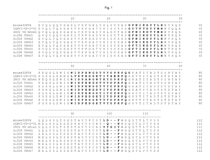
[0005] Tau inclusions are part of the defining neurophathology of several
neurodegenerative
diseases including Alzheimer's disease, frontotemporal lobar degeneration,
progressive
supranuclear palsy and Pick's disease.
BRIEF SUMMARY OF THE CLAIMED INVENTION
[0006] In one aspect, the invention provides an antibody specifically binding
to human tau,
comprising a mature heavy chain variable region comprising CDRs H1, H2 and H3
comprising
SEQ ID NOS:8, 9, and 10, respectively except that position H28 can be occupied
by N or T, H54
can be occupied by N or D, H56 can be occupied by D or E, and position H58
occupied by V or
I, and a mature light chain variable region comprising CDRs Li, L2 and L3
comprising SEQ ID
NOS: i2, 13, and 14 respectively, except that position L24 can be occupied by
K or R, wherein at
least one of the following positions is occupied by the amino acid as
specified: H1 is occupied by
Q, H5 is occupied by Q, H11 is occupied by L, H20 is occupied by L, H23 is
occupied by T, H38
is occupied by K, H75 is occupied by S, H56 is occupied by E, H58 is occupied
by I, L10 is
occupied by T, L17 is occupied by E, L24 is occupied by R, L37 is occupied by
Q, L83 is
occupied by L, L86 is occupied by H, L100 is occupied by A or Q, L106 is
occupied by L.
[0007] In some such antibodies, CDR-H1 comprises SEQ ID NO:8 or SEQ ID NO:86,
CDR-H2
comprises SEQ ID NO:9, SEQ ID NO:87, SEQ ID NO:88, or SEQ ID NO:92, CDR-H3
comprises SEQ ID NO:10, CDR-L1 comprises SEQ ID NO:12 or SEQ ID NO:89, CDR-L2
comprises SEQ ID NO:13, and CDR-L3 comprises SEQ ID NO:14. In some such
antibodies,
CDR-H1 comprises SEQ ID NO:8 or SEQ ID NO:86, CDR-H2 comprises SEQ ID NO:9,
SEQ
ID NO:87, or SEQ ID NO:88, CDR-H3 comprises SEQ ID NO:10, CDR-L1 comprises SEQ
ID
NO: i2 or SEQ ID NO:89, CDR-L2 comprises SEQ ID NO: i3, and CDR-L3 comprises
SEQ ID
NO: i4. In some such antibodies, CDR-H1 comprises SEQ ID NO:86, CDR-H2
comprises SEQ
ID NO:92, CDR-H3 comprises SEQ ID NO:10, CDR-L1 comprises SEQ ID NO:12 or SEQ
ID
NO:89, CDR-L2 comprises SEQ ID NO:13, and CDR-L3 comprises SEQ ID NO:14.
2
CA 03119072 2021-05-06
WO 2020/097561 PCT/US2019/060616
[0008] In some such antibodies, CDR-H1 has an amino acid sequence comprising
SEQ ID
NO:86. In some such antibodies, CDR-H2 has an amino acid sequence comprising
SEQ ID
NO:87. In some such antibodies, CDR-H2 has an amino acid sequence comprising
SEQ ID
NO:88. In some such antibodies, CDR-H2 has an amino acid sequence comprising
SEQ ID
NO:92. In some such antibodies, CDR-L1 has an amino acid sequence comprising
SEQ ID
NO:89.
[0009] In some such antibodies, CDR-H1 has an amino acid sequence comprising
SEQ ID
NO:86 and CDR-H2 has an amino acid sequence comprising SEQ ID NO:87. In some
such
antibodies, CDR-H1 has an amino acid sequence comprising SEQ ID NO:86 and CDR-
H2 has
an amino acid sequence comprising SEQ ID NO:88. In some such antibodies, CDR-
H1 has an
amino acid sequence comprising SEQ ID NO:86 and CDR-H2 has an amino acid
sequence
comprising SEQ ID NO:92.
[0010] In some such antibodies, the antibody is a humanized antibody, veneered
antibody, or
chimeric antibody.
[0011] In some antibodies, the humanized mature heavy chain variable region
having an amino
acid sequence at least 95% identical to any one of SEQ ID NOs:76-80 and SEQ ID
NOs:90-91
and the humanized mature light chain variable region has an amino acid
sequence at least 90%
identical to any one of SEQ ID NOs:83-85. In some antibodies, the humanized
mature heavy
chain variable region has an amino acid sequence at least 95% identical to any
one of SEQ ID
NOs:76-80 and the humanized mature light chain variable region has an amino
acid sequence at
least 90% identical to any one of SEQ ID NOs:83-85. In some antibodies, the
humanized mature
heavy chain variable region has an amino acid sequence at least 95% identical
to any one of SEQ
ID NOs:90-91 and the humanized mature light chain variable region has an amino
acid sequence
at least 90% identical to any one of SEQ ID NOs:83-85.
3
CA 03119072 2021-05-06
WO 2020/097561 PCT/US2019/060616
[0012] In some such antibodies, at least one of the following positions in the
VH region is
occupied by the amino acid as specified: H93 is occupied by S and H94 is
occupied by T. In
some such antibodies, positions H93 and H94 are occupied by S and T,
respectively.
[0013] In some such antibodies, position H91 in the VH region is occupied by
F.
[0014] In some such antibodies, at least one of the following positions in the
VH region is
occupied by the amino acid as specified: H1 is occupied by E, H5 is occupied
by V, H11 is
occupied by V, H20 is occupied I, H23 is occupied by K, H38 is occupied by R,
H42 is occupied
by G, H43 is occupied by K, H66 is occupied by R, H75 is occupied by T, H76 is
occupied by D,
H81 is occupied by E, H108 is occupied by L, H109 is occupied by V. In some
antibodies,
positions H1, H5, H11, H20, H23, H38, H42, H43, H66, H75, H76, H81, H108, and
H109 in the
VH region are occupied by E, V, V, I, K, R, G, K, R, T, D, E, L, and V,
respectively.
[0015] In some such antibodies, at least one of the following positions in the
VH region is
occupied by the amino acid as specified: H17 is occupied by T, H80 is occupied
by M, H83 is
occupied by R. In some antibodies, positions H17, H80, and H83 in the VH
region are occupied
by T, M, and R, respectively.
[0016] In some such antibodies, position H58 in the VH region is occupied by
I.
[0017] In some such antibodies, at least one of the following positions in the
VH region is
occupied by the amino acid as specified: H28 is occupied by T, H67 is occupied
by V. In some
antibodies, positions H28 and H67 in the VH region are occupied by T and V,
respectively.
[0018] In some such antibodies, at least one of the following positions in the
VH region is
occupied by the amino acid as specified: H54 is occupied by D, H56 is occupied
by E. In some
antibodies, positions H54 and H56 in the VH region are occupied by D and E,
respectively.
[0019] In some such antibodies, at least one of the following positions in the
VH region is
occupied by the amino acid as specified: H1 is occupied by Q or E, H5 is
occupied by Q or V,
H11 is occupied by L or V, H17 is occupied by S or T, H20 is occupied by L or
I, H23 is
occupied by T or K, H28 is occupied by N or T, H38 is occupied by K or R, H42
is occupied by
4
CA 03119072 2021-05-06
WO 2020/097561 PCT/US2019/060616
E or G, H43 is occupied by Q or K, H54 is occupied by N or D, H56 is occupied
by D or E, H58
is occupied by V or I, H66 is occupied by K or R, H67 is occupied by A or V,
H75 is occupied
by S or T, H76 is occupied by N or D, H80 is occupied by L or M, H81 is
occupied by Q or E,
H83 is occupied by T or R, H91 is occupied by F or Y, H93 is occupied by S,
H94 is occupied
by T, H108 is occupied by T or L, H109 is occupied by L or V.
[0020] In some antibodies, positions H91, H93, and H94 in the VH region are
occupied by F, S,
and T, respectively. In some antibodies, positions H1, H5, H11, H20, H23, H38,
H42, H43,
H66, H75, H76, H81, H91, H93, H94, H108, and H109 in the VH region are
occupied by E, V,
V, I, K, R, G, K, R, T, D, E, F, S, T,L, and V, respectively. In some
antibodies, positions H1,
H5, H11, H17, H20, H23, H38, H42, H43, H58, H66, H75, H76, H80, H81, H83, H93,
H94,
H108, and H109 in the VH region are occupied by E, V, V, T,I, K, R, G, K, I,
R, T, D, M, E, R,
S, T, L, and V, respectively. In some antibodies, positions H1, H5, H11, H17,
H20, H23, H28,
H38, H42, H43, H58, H66, H67, H75, H76, H80, H81, H83, H93, H94, H108, and
H109 in the
VH region are occupied by E, V, V, T, I, K, T, R, G, K, I, R, V, T, D, M, E,
R, S, T, L, and V,
respectively. In some antibodies, positions H1, H5, H11, H17, H20, H23, H28,
H38, H42, H43,
H54, H56, H58, H66, H67, H75, H76, H80, H81, H83, H93, H94, H108, and H109 in
the VH
region are occupied by E, V, V, T, I, K, T, R, G, K, D, E, I, R, V, T, D, M,
E, R, S, T, L, and V,
respectively. In some antibodies, positions H1, H5, H11, H17, H20, H23, H28,
H38, H42, H43,
H54, H56, H66, H67, H75, H76, H80, H81, H83, H91, H93, H94, H108, and H109 in
the VH
region are occupied by E, V, V, T, I, K, T, R, G, K, D, E, R, V, T, D, M, E,
R, F, S, T, L, and V,
respectively. In some antibodies, positions H1, H5, H11, H17, H20, H23, H28,
H38, H42, H43,
H54, H56, H66, H67, H75, H76, H80, H81, H83, H93, H94, H108, and H109 in the
VH region
are occupied by E, V, V, T, I, K, T, R, G, K, D, E, R, V, T, D, M, E, R, S, T,
L, and V,
respectively.
[0021] In some such antibodies, at least one of the following positions in the
VL region is
occupied by the amino acid as specified: L7 is occupied by S, L10 is occupied
by S, L15 is
occupied by L, L83 is occupied by V, L86 is occupied by Y, and L106 is
occupied by I. In some
antibodies, positions L7, L10, L15, L83, L86, and L106 are occupied by S, S,
L, V, Y, and Y,
respectively.
CA 03119072 2021-05-06
WO 2020/097561 PCT/US2019/060616
[0022] In some such antibodies, at least one of the following positions in the
VL region is
occupied by the amino acid as specified: L7 is T or S, L10 is T or S, L15 is I
or L, L17 is Q or E,
L24 is K or R, L37 is L or Q, L45 is K or R, L83 is L or V, L86 is H or Y,
L100 is A or Q, L106
is L or I.
[0023] In some antibodies, positions L7, L10, L15, L83, L86, and L106 in the
VL region are
occupied by S, S, L, V, Y, and I, respectively. In some antibodies, positions
L7, L10, L15, L17,
L24, L37, L45, L83, L86, L100, and L106 in the VL region are occupied by S, S,
L, E, R, Q, R,
V, Y, Q, and I, respectively.
[0024] In some antibodies, the mature heavy chain variable region has an amino
acid sequence
of any one of SEQ ID NOs:76-80 and SEQ ID NOs:90-91 and the mature light chain
variable
region has an amino acid sequence of any one of SEQ ID NO:83-85. In some
antibodies, the
mature heavy chain variable region has an amino acid sequence of any one of
SEQ ID NOs:76-
80 and the mature light chain variable region has an amino acid sequence of
any one of SEQ ID
NO:83-85. In some antibodies, the mature heavy chain variable region has an
amino acid
sequence of any one of SEQ ID NOs:90-91 and the mature light chain variable
region has an
amino acid sequence of any one of SEQ ID NO:83-85.
[0025] In some antibodies, the mature heavy chain variable region has an amino
acid sequence
of SEQ ID NO:76 and the mature light chain variable region has an amino acid
sequence of SEQ
ID NO:83. In some antibodies, the mature heavy chain variable region has an
amino acid
sequence of SEQ ID NO:76 and the mature light chain variable region has an
amino acid
sequence of SEQ ID NO:84. In some antibodies, the mature heavy chain variable
region has an
amino acid sequence of SEQ ID NO:76 and the mature light chain variable region
has an amino
acid sequence of SEQ ID NO:85.
[0026] In some antibodies, the mature heavy chain variable region has an amino
acid sequence
of SEQ ID NO:77 and the mature light chain variable region has an amino acid
sequence of SEQ
ID NO:83. In some antibodies, the mature heavy chain variable region has an
amino acid
sequence of SEQ ID NO:77 and the mature light chain variable region has an
amino acid
sequence of SEQ ID NO:84. In some antibodies, the mature heavy chain variable
region has an
6
CA 03119072 2021-05-06
WO 2020/097561 PCT/US2019/060616
amino acid sequence of SEQ ID NO:77 and the mature light chain variable region
has an amino
acid sequence of SEQ ID NO:85.
[0027] In some antibodies, the mature heavy chain variable region has an amino
acid sequence
of SEQ ID NO:78 and the mature light chain variable region has an amino acid
sequence of SEQ
ID NO:83. In some antibodies, the mature heavy chain variable region has an
amino acid
sequence of SEQ ID NO:78 and the mature light chain variable region has an
amino acid
sequence of SEQ ID NO:84. In some antibodies, the mature heavy chain variable
region has an
amino acid sequence of SEQ ID NO:78 and the mature light chain variable region
has an amino
acid sequence of SEQ ID NO:85.
[0028] In some antibodies, the mature heavy chain variable region has an amino
acid sequence
of SEQ ID NO:79 and the mature light chain variable region has an amino acid
sequence of SEQ
ID NO:83. In some antibodies, the mature heavy chain variable region has an
amino acid
sequence of SEQ ID NO:79 and the mature light chain variable region has an
amino acid
sequence of SEQ ID NO:84. In some antibodies, the mature heavy chain variable
region has an
amino acid sequence of SEQ ID NO:79 and the mature light chain variable region
has an amino
acid sequence of SEQ ID NO:85.
[0029] In some antibodies, the mature heavy chain variable region has an amino
acid sequence
of SEQ ID NO:80 and the mature light chain variable region has an amino acid
sequence of SEQ
ID NO:83. In some antibodies, he mature heavy chain variable region has an
amino acid
sequence of SEQ ID NO:80 and the mature light chain variable region has an
amino acid
sequence of SEQ ID NO:84. In some antibodies, the mature heavy chain variable
region has an
amino acid sequence of SEQ ID NO:80 and the mature light chain variable region
has an amino
acid sequence of SEQ ID NO:85.
[0030] In some antibodies, the mature heavy chain variable region has an amino
acid sequence
of SEQ ID NO:90 and the mature light chain variable region has an amino acid
sequence of SEQ
ID NO:83. In some antibodies, the mature heavy chain variable region has an
amino acid
sequence of SEQ ID NO:90 and the mature light chain variable region has an
amino acid
sequence of SEQ ID NO:84. In some antibodies, the mature heavy chain variable
region has an
7
CA 03119072 2021-05-06
WO 2020/097561 PCT/US2019/060616
amino acid sequence of SEQ ID NO:90 and the mature light chain variable region
has an amino
acid sequence of SEQ ID NO:85.
[0031] In some antibodies, the mature heavy chain variable region has an amino
acid sequence
of SEQ ID NO:91 and the mature light chain variable region has an amino acid
sequence of SEQ
ID NO:83. In some antibodies, the mature heavy chain variable region has an
amino acid
sequence of SEQ ID NO:91 and the mature light chain variable region has an
amino acid
sequence of SEQ ID NO:84. In some antibodies, the mature heavy chain variable
region has an
amino acid sequence of SEQ ID NO:91 and the mature light chain variable region
has an amino
acid sequence of SEQ ID NO:85.
[0032] For example, the antibody can be a chimeric antibody. For example, the
antibody can
be a veneered antibody.
[0033] The antibody can be an intact mouse, chimeric, veneered or humanized
antibody or a
binding fragment, single-chain antibody Fab fragment, Fab'2 fragment, or
single chain Fv.
Some of the antibodies have a human IgG1 isotype, while others may have a
human IgG2 or
IgG4 isotype. Some antibodies have the mature light chain variable region
fused to a light chain
constant region and the mature heavy chain variable region fused to a heavy
chain constant
region. The heavy chain constant region of some antibodies is a mutant form of
a natural human
heavy chain constant region which has reduced binding to a Fcy receptor
relative to the natural
human heavy chain constant region. In some antibodies, the mature heavy chain
variable region
is fused to a heavy chain constant region having the sequence of SEQ ID NO:103
with or
without the C-terminal lysine and/or the mature light chain variable region is
fused to a light
chain constant region having the sequence of SEQ ID NO:104.
[0034] Some antibodies may have at least one mutation in the constant region,
such as a
mutation that reduces complement fixation or activation by the constant
region, for example, a
mutation at one or more of positions 241, 264, 265, 270, 296, 297, 318, 320,
322, 329 and 331 by
EU numbering. Some antibodies have an alanine at positions 318, 320 and 322.
Some
antibodies can be at least 95% w/w pure. The antibody can be conjugated to a
therapeutic,
cytotoxic, cytostatic, neurotrophic, or neuroprotective agent.
8
CA 03119072 2021-05-06
WO 2020/097561 PCT/US2019/060616
[0035] In another aspect, the invention provides a pharmaceutical composition
comprising any
of the antibodies disclosed herein and a pharmaceutically-acceptable carrier.
[0036] In another aspect, the invention provides a nucleic acid encoding the
heavy chain and/or
light chain of any of the antibodies disclosed herein, a recombinant
expression vector comprising
the nucleic acid and a host cell transformed with the recombinant expression
vector. For
example, the nucleic acid can have a sequence comprising any one of SEQ ID
NOs:30-31, 93-99,
100-102, and 105-106. In another example, the nucleic acid can have a sequence
comprising any
one of SEQ ID NOs:30-31, 93-97, 100-102, and 105-106. In another example, the
nucleic acid
can have a sequence comprising any one of SEQ ID NOs:30-31, 98-99, 100-102,
and 105-106.
[0037] In another aspect, the invention provides a vector comprising a nucleic
acid encoding a
mature heavy chain variable region and a mature light chain variable region
operably linked to
one or more regulatory sequences to effect expression in a mammalian cell of
any of the
antibodies disclosed herein, a host cell comprising the nucleic acid, and a
method of expressing
an antibody in a mammalian cell comprising incorporating the nucleic acid into
the genome of a
transgenic animal, whereby the antibody is expressed.
[0038] Some vectors comprise a nucleic acid further encoding a heavy chain
constant region
fused to the mature heavy chain variable region and a light chain constant
region fused to the
mature light chain variable region. In some vectors, the encoded heavy chain
constant region has
the sequence of SEQ ID NO:103 with or without the C-terminal lysine and the
encoded light
chain constant region has the sequence of SEQ ID NO:104. In some vectors, the
heavy chain
constant region is encoded by the sequence of SEQ ID NO:105 and the light
chain constant
region is encoded by the sequence of SEQ ID NO:106.
[0039] In some vectors, the expressed antibody encoded by the nucleic acid is
a scFv. In some
vectors, the expressed antibody encoded by the nucleic acid is a Fab fragment.
[0040] In some vectors, the one or more regulatory sequences include one or
more of a
promoter, enhancer, ribosome binding site, and transcription termination
signal. In some
vectors, the nucleic acid further encodes signal peptides fused to the mature
heavy and light
chain variable regions. In some vectors, the nucleic acid is codon-optimized
for expression in a
9
CA 03119072 2021-05-06
WO 2020/097561 PCT/US2019/060616
host cell. In some vectors, the one or more regulatory sequences include a
eukaryotic promoter.
In some vectors, the nucleic acid further encodes a selectable gene.
[0041] In another aspect, the invention provides first and second vectors
respectively comprising
nucleic acids encoding a mature heavy chain variable region and a mature light
chain variable
region, each operably linked to one or more regulatory sequences to effect
expression in a
mammalian cell of any of the antibodies disclosed herein, a host cell
comprising the nucleic
acids, and a method of expressing an antibody in a mammalian cell comprising
incorporating the
nucleic acids into the genome of a transgenic animal, whereby the antibody is
expressed.
[0042] In some vectors, the nucleic acids respectively further encode a heavy
chain constant
region fused to the mature heavy chain variable region and a light chain
constant region fused to
the mature light chain variable region. In some vectors, the encoded heavy
chain constant region
has the sequence of SEQ ID NO:103 with or without the C-terminal lysine and
the encoded light
chain constant region has the sequence of SEQ ID NO:104. In some vectors, the
heavy chain
constant region is encoded by the sequence of SEQ ID NO:105 and the light
chain constant
region is encoded by the sequence of SEQ ID NO:106.
[0043] In yet another aspect, the invention provides methods of humanizing any
non-human
antibody described herein, for example, mouse antibody 3D6, wherein 3D6 is
characterized by a
mature heavy chain variable region of SEQ ID NO:7 and a mature light chain
variable region of
SEQ ID NO:11. Such methods can involve selecting one or more acceptor
antibodies,
synthesizing a nucleic acid encoding a humanized heavy chain comprising CDRs
of the mouse
heavy chain and a nucleic acid encoding a humanized light chain comprising
CDRs of the mouse
antibody light chain, and expressing the nucleic acids in a host cell to
produce a humanized
antibody.
[0044] Methods of producing antibodies, such as a humanized, chimeric or
veneered antibody,
for example humanized, chimeric or veneered forms of 3D6, are also provided.
In such methods,
cells transformed with nucleic acids encoding the heavy and light chains of
the antibody are
cultured so that the cells secrete the antibody. The antibody can then be
purified from the cell
culture media.
CA 03119072 2021-05-06
WO 2020/097561 PCT/US2019/060616
[0045] Cell lines producing any of the antibodies disclosed herein can be
produced by
introducing a vector encoding heavy and light chains of the antibody and a
selectable marker into
cells, propagating the cells under conditions to select for cells having
increased copy number of
the vector, isolating single cells from the selected cells; and banking cells
cloned from a single
cell selected based on yield of antibody.
[0046] Some cells can be propagated under selective conditions and screened
for cell lines
naturally expressing and secreting the antibody in an amount of at least 100
mg/L/106 cells/24
hours. Single cells can be isolated from the selected cells. Cells cloned from
a single cell can
then be banked. Single cells can be selected based on desirable properties,
such as the yield of
the antibody. Exemplary cell lines are cell lines expressing 3D6.
[0047] The invention also provides methods of inhibiting or reducing
aggregation of tau in a
subject having or at risk of developing a tau-mediated amyloidosis, comprising
administering to
the subject an effective regime of an antibody disclosed herein, thereby
inhibiting or reducing
aggregation of tau in the subject. Exemplary antibodies include humanized
versions of 3D6.
[0048] Also provided are methods of treating or effecting prophylaxis of a tau-
related disease
in a subject, comprising administering an effective regime of an antibody
disclosed herein and
thereby treating or effecting prophylaxis of the disease. Examples of such a
disease are
Alzheimer's disease, Down's syndrome, mild cognitive impairment, primary age-
related
tauopathy, postencephalitic parkinsonism, posttraumatic dementia or dementia
pugilistica, Pick's
disease, type C Niemann-Pick disease, supranuclear palsy, frontotemporal
dementia,
frontotemporal lobar degeneration, argyrophilic grain disease, globular glial
tauopathy,
amyotrophic lateral sclerosis/parkinsonism dementia complex of Guam,
corticobasal
degeneration (CBD), dementia with Lewy bodies, Lewy body variant of Alzheimer
disease
(LBVAD), chronic traumatic encephalopathy (CTE), globular glial tauopathy
(GGT), or
progressive supranuclear palsy (PSP). In some methods, the tau-related disease
is Alzheimer's
disease. In some methods the patient is an ApoE4 carrier.
[0049] Also provided are methods of reducing aberrant transmission of tau
comprising
administering an effective regime of an antibody disclosed herein and thereby
reducing
transmission of tau.
11
CA 03119072 2021-05-06
WO 2020/097561 PCT/US2019/060616
[0050] Also provided are methods of inducing phagocytosis of tau comprising
administering
an effective regime of an antibody disclosed herein and thereby inducing
phagocytosis of tau.
[0051] Also provided are methods of inhibiting tau aggregation or deposition
comprising
administering an effective regime of an antibody disclosed herein thereby
inhibiting tau
aggregation or deposition.
[0052] Also provided are methods of inhibiting formation of tau tangles
comprising
administering an effective regime of an antibody disclosed herein.
[0053] The invention also provides a method of detecting tau protein deposits
in a subject
having or at risk of a disease associated with tau aggregation or deposition
comprising
administering to a subject an antibody disclosed herein, and detecting the
antibody bound to tau
in the subject. Examples of such a disease are Alzheimer's disease, Down's
syndrome, mild
cognitive impairment, primary age-related tauopathy, postencephalitic
parkinsonism,
posttraumatic dementia or dementia pugilistica, Pick's disease, type C Niemann-
Pick disease,
supranuclear palsy, frontotemporal dementia, frontotemporal lobar
degeneration, argyrophilic
grain disease, globular glial tauopathy, amyotrophic lateral
sclerosis/parkinsonism dementia
complex of Guam, corticobasal degeneration (CBD), dementia with Lewy bodies,
Lewy body
variant of Alzheimer disease (LBVAD), chronic traumatic encephalopathy (CTE),
globular glial
tauopathy (GGT), or progressive supranuclear palsy (PSP). In some embodiments,
the antibody
is administered by intravenous injection into the body of the subject. In some
embodiments, the
antibody is administered directly to the brain of the subject by intracranial
injection or by drilling
a hole through the skull of the subject. In some embodiments, the antibody is
labeled. In some
embodiments, the antibody is labeled with a fluorescent label, a paramagnetic
label, or a
radioactive label. In some embodiments, the radioactive label is detected
using positron
emission tomography (PET) or single-photon emission computed tomography
(SPECT).
[0054] The invention also provides a method of measuring efficacy of treatment
in a subject
being treated for a disease associated with tau aggregation or deposition,
comprising measuring a
first level of tau protein deposits in the subject prior to treatment by
administering to a subject an
antibody disclosed herein, and detecting a first amount of the antibody bound
to tau in the
subject, administering the treatment to the subject, measuring a second level
of tau protein
12
CA 03119072 2021-05-06
WO 2020/097561 PCT/US2019/060616
deposits in the in subject after treatment by administering to a subject the
antibody, and detecting
the antibody bound to tau in the subject, wherein a decrease in the level of
tau protein deposits
indicates a positive response to treatment.
[0055] The invention also provides a method of measuring efficacy of treatment
in a subject
being treated for a disease associated with tau aggregation or deposition,
comprising measuring a
first level of tau protein deposits in the subject prior to treatment by
administering to a subject an
antibody disclosed herein, and detecting a first amount of antibody bound to
tau in the subject,
administering the treatment to the subject, measuring a second level of tau
protein deposits in the
in subject after treatment by administering to a subject the antibody, and
detecting a second
amount of antibody bound to tau in the subject, wherein no change in the level
of tau protein
deposits or a small increase in tau protein deposits indicates a positive
response to treatment.
BRIEF DESCRIPTION OF THE DRAWINGS
[0056] Figure 1 depicts an alignment of heavy chain variable regions of the
mouse 3D6
antibody (SEQ ID NO:7) and humanized versions of the 3D6 antibody (hu3D6VHvb1,
hu3D6VHvb2, hu3D6VHvb3, hu3D6VHvb4, hu3D6VHvb5, hu3D6VHvb6, and hu3D6VHvb7)
with human germline heavy chain variable region sequence IGHV1-69-2*01 (SEQ ID
NO:25)
and with human acceptor heavy chain variable region sequence 2RCS VH hFrwk
(SEQ ID
NO:75). hu3D6VHvb1 is SEQ ID NO:76, hu3D6VHvb2 is SEQ ID NO:77, hu3D6VHvb3 is
SEQ ID NO:78, hu3D6VHvb4 is SEQ ID NO:79, hu3D6VHvb5 is SEQ ID NO:80,
hu3D6VHvb6 is SEQ ID NO:90, and hu3D6VHvb7 is SEQ ID NO:91. The CDRs as
defined by
Kabat/Chothia Composite are in boldface.
[0057] Figure 2 depicts an alignment of light chain variable regions of the
mouse 3D6
antibody (SEQ ID NO:11) and humanized versions of the 3D6 antibody
(hu3D6VLvb1,
hu3D6VLvb2, and hu3D6VLvb3) with human germline light chain variable region
sequence
IGKV2-30*02 (SEQ ID NO:27) and with human acceptor ARX71335 VL hFrwk (SEQ ID
NO:82). hu3D6VLvb1 is SEQ ID NO:83, hu3D6VLvb2 is SEQ ID NO:84, and hu3D6VLvb3
is
SEQ ID NO:85. The CDRs as defined by Kabat are in boldface.
[0058] Figures 3A, 3B, and 3C depict results of ELISA screening assays for
selected mouse
monoclonal anti-tau antibodies.
13
CA 03119072 2021-05-06
WO 2020/097561 PCT/US2019/060616
[0059] Figure 4 depicts binding kinetics for selected mouse monoclonal anti-
tau antibodies to
recombinant human tau.
[0060] Figure 5 depicts results of functional blocking assays for selected
mouse monoclonal
anti-tau antibodies.
[0061] Figure 6 depicts results of disaggregation assays for selected mouse
monoclonal anti-
tau antibodies.
[0062] Figure 7 depicts results of experiments showing that 3D6 and 5G8
immunocapture tau
from human Alzheimer's disease tissue.
BRIEF DESCRIPTION OF THE SEQUENCES
[0063] SEQ ID NO:1 sets forth the amino acid sequence of an isoform of human
tau (Swiss-
Prot P10636-8).
[0064] SEQ ID NO:2 sets forth the amino acid sequence of an isoform of human
tau (Swiss-
Prot P10636-7).
[0065] SEQ ID NO:3 sets forth the amino acid sequence of an isoform of human
tau (Swiss-
Prot P10636-6), (4RON human tau).
[0066] SEQ ID NO:4 sets forth the amino acid sequence of an isoform of human
tau (Swiss-
Prot P10636-5)
[0067] SEQ ID NO:5 sets forth the amino acid sequence of an isoform of human
tau (Swiss-
Prot P10636-4).
[0068] SEQ ID NO:6 sets forth the amino acid sequence of an isoform of human
tau (Swiss-
Prot P10636-2).
[0069] SEQ ID NO:7 sets forth the amino acid sequence of the heavy chain
variable region of
the mouse 3D6 antibody.
[0070] SEQ ID NO:8 sets forth the amino acid sequence of Kabat/Chothia
composite CDR-H1
of the mouse 3D6 antibody.
[0071] SEQ ID NO:9 sets forth the amino acid sequence of Kabat CDR-H2 of the
mouse 3D6
antibody.
14
CA 03119072 2021-05-06
WO 2020/097561 PCT/US2019/060616
[0072] SEQ ID NO:10 sets forth the amino acid sequence of Kabat CDR-H3 of the
mouse 3D6
antibody.
[0073] SEQ ID NO:11 sets forth the amino acid sequence of the light chain
variable region of
the mouse 3D6 antibody and of the mouse 6A10 antibody.
[0074] SEQ ID NO:12 sets forth the amino acid sequence of Kabat CDR-L1 of the
mouse 3D6
antibody and of the mouse 6A10 antibody.
[0075] SEQ ID NO:13 sets forth the amino acid sequence of Kabat CDR-L2 of the
mouse 3D6
antibody and of the mouse 6A10 antibody.
[0076] SEQ ID NO:14 sets forth the amino acid sequence of Kabat CDR-L3 of the
mouse 3D6
antibody and of the mouse 6A10 antibody.
[0077] SEQ ID NO:15 sets forth the amino acid sequence of heavy chain variable
region of the
humanized 3D6 antibody hu3D6VHv1.
[0078] SEQ ID NO:16 sets forth the amino acid sequence of heavy chain variable
region of the
humanized 3D6 antibody hu3D6VHv2.
[0079] SEQ ID NO:17 sets forth the amino acid sequence of heavy chain variable
region of the
humanized 3D6 antibody hu3D6VHv1b.
[0080] SEQ ID NO:18 sets forth the amino acid sequence of heavy chain variable
region of the
humanized 3D6 antibody hu3D6VHv1bA1 1.
[0081] SEQ ID NO:19 sets forth the amino acid sequence of heavy chain variable
region of the
humanized 3D6 antibody hu3D6VHv5:
[0082] SEQ ID NO:20 sets forth the amino acid sequence of the light chain
variable region of
the humanized 3D6 antibody hu3D6VLv1.
[0083] SEQ ID NO:21 sets forth the amino acid sequence of the light chain
variable region of
the humanized 3D6 antibody hu3D6VLv2.
[0084] SEQ ID NO:22 sets forth the amino acid sequence of the light chain
variable region of
the humanized 3D6 antibody hu3D6VLv3.
CA 03119072 2021-05-06
WO 2020/097561 PCT/US2019/060616
[0085] SEQ ID NO:23 sets forth the amino acid sequence of the light chain
variable region of
the humanized 3D6 antibody hu3D6VLy4.
[0086] SEQ ID NO:24 sets forth the amino acid sequence of the heavy chain
variable acceptor
Acc.# BAC01986.1.
[0087] SEQ ID NO:25 sets forth the amino acid sequence of the heavy chain
variable acceptor
Acc.# IMGT# IGHV1-69-2*01.
[0088] SEQ ID NO:26 sets forth the amino acid sequence of the heavy chain
variable acceptor
Acc.# IMGT#IGKJ1*01.
[0089] SEQ ID NO:27 sets forth the amino acid sequence of the light chain
variable acceptor
Acc. # IMGT#IGKV2-30*02
[0090] SEQ ID NO:28 sets forth the amino acid sequence of the light chain
variable acceptor
Acc. # IMGT#IGKJ2*01.
[0091] SEQ ID NO:29 sets forth the amino acid sequence of the light chain
variable acceptor
Acc. # AAZ09048.1.
[0092] SEQ ID NO:30 sets forth a nucleic acid sequence encoding the heavy
chain variable
region of the mouse 3D6 antibody.
[0093] SEQ ID NO:31 sets forth a nucleic acid sequence encoding the light
chain variable
region of the mouse 3D6 antibody.
[0094] SEQ ID NO:32 sets forth the amino acid sequence of Kabat CDR-H1 of the
mouse 3D6
antibody.
[0095] SEQ ID NO:33 sets forth the amino acid sequence of Chothia CDR-H1 of
the mouse
3D6 antibody.
[0096] SEQ ID NO:34 sets forth the amino acid sequence of Chothia CDR-H2 of
the mouse
3D6 antibody.
[0097] SEQ ID NO:35 sets forth the amino acid sequence of AbM CDR-H2 of the
mouse 3D6
antibody.
16
CA 03119072 2021-05-06
WO 2020/097561 PCT/US2019/060616
[0098] SEQ ID NO:36 sets forth the amino acid sequence of Contact CDR-L1 of
the mouse
3D6 antibody.
[0099] SEQ ID NO:37 sets forth the amino acid sequence of Contact CDR-L2 of
the mouse
3D6 antibody.
[0100] SEQ ID NO:38 sets forth the amino acid sequence of Contact CDR-L3 of
the mouse
3D6 antibody.
[0101] SEQ ID NO:39 sets forth the amino acid sequence of Contact CDR-H1 of
the mouse
3D6 antibody.
[0102] SEQ ID NO:40 sets forth the amino acid sequence of Contact CDR-H2 of
the mouse
3D6 antibody.
[0103] SEQ ID NO:41 sets forth the amino acid sequence of Contact CDR-H3 of
the mouse
3D6 antibody.
[0104] SEQ ID NO:42 sets forth the amino acid sequence of an alternate Kabat-
Chothia
Composite CDR-H1 of a humanized 3D6 antibody (as in hu3D6VHv5,
hu3D6VHvlbAl1B6G2,
hu3D6VHvlbAl1B6H3, hu3D6VHv1e, and hu3D6VHv1f).
[0105] SEQ ID NO:43 sets forth the amino acid sequence of an alternate Kabat
CDR-H2 of a
humanized 3D6 antibody (as in hu3D6VHv5 and hu3D6VHvlbAl1B6H3).
[0106] SEQ ID NO:44 sets forth the consensus amino acid sequence among the
heavy chain
variable regions of the mouse 3D6 and selected humanized 3D6 antibodies (VHvl,
VHv2,
VHvlb, VHvlbAll, and VHv5) (labeled "Majority' in Figure 2 of
PCT/1132017/052544.
[0107] SEQ ID NO:45 sets forth the consensus amino acid sequence between the
light chain
variable regions of the mouse 3D6 and selected humanized 3D6 antibodies
(labeled "Majority' in
Figure 3 of PCT/1132017/052544).
[0108] SEQ ID NO:46 sets forth the amino acid sequence of heavy chain variable
region of the
humanized 3D6 antibody hu3D6VHvlbAl1B6G2.
[0109] SEQ ID NO:47 sets forth the amino acid sequence of heavy chain variable
region of the
humanized 3D6 antibody hu3D6VHvlbAl1B6H3.
17
CA 03119072 2021-05-06
WO 2020/097561 PCT/US2019/060616
[0110] SEQ ID NO:48 sets forth the amino acid sequence of heavy chain variable
region of the
humanized 3D6 antibody hu3D6VHv1c.
[0111] SEQ ID NO:49 sets forth the amino acid sequence of heavy chain variable
region of the
humanized 3D6 antibody hu3D6VHv1d.
[0112] SEQ ID NO:50 sets forth the amino acid sequence of heavy chain variable
region of the
humanized 3D6 antibody hu3D6VHv1e.
[0113] SEQ ID NO:51 sets forth the amino acid sequence of heavy chain variable
region of the
humanized 3D6 antibody hu3D6VHvlf.
[0114] SEQ ID NO:52 sets forth the amino acid sequence of heavy chain variable
region of the
humanized 3D6 antibody hu3D6VHv3.
[0115] SEQ ID NO:53 sets forth the amino acid sequence of heavy chain variable
region of the
humanized 3D6 antibody hu3D6VHv3b.
[0116] SEQ ID NO:54 sets forth the amino acid sequence of heavy chain variable
region of the
humanized 3D6 antibody hu3D6VHv3c.
[0117] SEQ ID NO:55 sets forth the amino acid sequence of heavy chain variable
region of the
humanized 3D6 antibody hu3D6VHv4.
[0118] SEQ ID NO:56 sets forth the amino acid sequence of heavy chain variable
region of the
humanized 3D6 antibody hu3D6VHv4b.
[0119] SEQ ID NO:57 sets forth the amino acid sequence of heavy chain variable
region of the
humanized 3D6 antibody hu3D6VHv4c.
[0120] SEQ ID NO:58 sets forth the amino acid sequence of an alternate Kabat-
Chothia
Composite CDR-H1 of a humanized 3D6 antibody (as in hu3D6VH1c).
[0121] SEQ ID NO:59 sets forth the amino acid sequence of an alternate Kabat-
Chothia
Composite CDR-H1 of a humanized 3D6 antibody (as in hu3D6VHvld, hu3D6VHv3c,
and
hu3D6VHv4c).
[0122] SEQ ID NO:60 sets forth the amino acid sequence of an alternate Kabat-
Chothia
Composite CDR-H1 of a humanized 3D6 antibody (as in hu3D6VHv3b and
hu3D6VHv4b).
18
CA 03119072 2021-05-06
WO 2020/097561 PCT/US2019/060616
[0123] SEQ ID NO:61 sets forth the amino acid sequence of an alternate Kabat
CDR-H2 of a
humanized 3D6 antibody (as in hu3D6VHvlbAl1B6G2).
[0124] SEQ ID NO:62 sets forth the amino acid sequence of an alternate Kabat
CDR-H2 of a
humanized 3D6 antibody (as in hu3D6VHv1c, hu3D6VHv3b, AND hu3D6VHv4b.
[0125] SEQ ID NO:63 sets forth the amino acid sequence of an alternate Kabat
CDR-H2 of a
humanized 3D6 antibody (as in hu3D6VHv1d, hu3D6VHv1f, hu3D6VHv3c, and
hu3D6VHv4c).
[0126] SEQ ID NO:64 sets forth the amino acid sequence of an alternate Kabat
CDR-H2 of a
humanized 3D6 antibody (as in hu3D6VHv1e).
[0127] SEQ ID NO:65 sets forth the amino acid sequence of an alternate Kabat
CDR-H3 of a
humanized 3D6 antibody (as in hu3D6VHv1f).
[0128] SEQ ID NO:66 sets forth the amino acid sequence of the heavy chain
variable region of
the mouse 6A10 antibody.
[0129] SEQ ID NO:67 sets forth the amino acid sequence of Kabat/Chothia
composite CDR-
H1 of the mouse 6A10 antibody.
[0130] SEQ ID NO:68 sets forth the amino acid sequence of Kabat CDR-H2 of the
mouse
6A10 antibody.
[0131] SEQ ID NO:69 sets forth the amino acid sequence of Kabat CDR-H3 of the
mouse
6A10 antibody.
[0132] SEQ ID NO:70 sets for the amino acid sequence of the VH region of mouse
antibody
(pdb code 1CR9) used as a structure template for heavy chain humanization.
[0133] SEQ ID NO:71 sets forth the consensus amino acid sequence among the
heavy chain
variable regions of the selected humanized 3D6 antibodies (VHvl, VHvlb,
VHvlbAll,
VHvlbAl1B6G2, VHvlbAl1B6H3, VHvlc, VHvld, VHvle, VHvlf, VHv2, VHv3, VHv3b,
VHv3c, VHv4, VHv4b, VHv4c, and VHv5) (labeled "Majority' in Figures 4A and 4B
of
PCT/M2017/052544).
[0134] SEQ ID NO:72 sets forth the amino acid sequence of the heavy chain of a
chimeric
3D6 antibody.
19
CA 03119072 2021-05-06
WO 2020/097561 PCT/US2019/060616
[0135] SEQ ID NO:73 sets forth the amino acid sequence of the light chain of a
chimeric 3D6
antibody.
[0136] SEQ ID NO:74 sets forth the amino acid sequence of heavy chain variable
structural
model Acc.# 5MYX-VH mSt.
[0137] SEQ ID NO:75 sets forth the amino acid sequence of heavy chain variable
acceptor
Acc.# 2RCS-VH huFrwk.
[0138] SEQ ID NO:76 sets forth the amino acid sequence of heavy chain variable
region of the
humanized 3D6 antibody hu3D6VHvb1.
[0139] SEQ ID NO:77 sets forth the amino acid sequence of heavy chain variable
region of the
humanized 3D6 antibody hu3D6VHvb2.
[0140] SEQ ID NO:78 sets forth the amino acid sequence of heavy chain variable
region of the
humanized 3D6 antibody hu3D6VHvb3.
[0141] SEQ ID NO:79 sets forth the amino acid sequence of heavy chain variable
region of the
humanized 3D6 antibody hu3D6VHvb4.
[0142] SEQ ID NO:80 sets forth the amino acid sequence of heavy chain variable
region of the
humanized 3D6 antibody hu3D6VHvb5.
[0143] SEQ ID NO:81 sets forth the amino acid sequence of light chain variable
structural
model Acc.# 5MYX-VL mSt.
[0144] SEQ ID NO:82 sets forth the amino acid sequence of light chain variable
acceptor
Acc.# ARX71335-VL huFrwk.
[0145] SEQ ID NO:83 sets forth the amino acid sequence of light chain variable
region of the
humanized 3D6 antibody hu3D6VLvb1.
[0146] SEQ ID NO:84 sets forth the amino acid sequence of light chain variable
region of the
humanized 3D6 antibody hu3D6VLvb2.
[0147] SEQ ID NO:85 sets forth the amino acid sequence of light chain variable
region of the
humanized 3D6 antibody hu3D6VLvb3.
CA 03119072 2021-05-06
WO 2020/097561 PCT/US2019/060616
[0148] SEQ ID NO:86 sets forth the amino acid sequence of an alternate Kabat-
Chothia
Composite CDR-H1 of a humanized 3D6 antibody (as in hu3D6VHvb4 and
hu3D6VHvb5).
[0149] SEQ ID NO:87 sets forth the amino acid sequence of an alternate Kabat
CDR-H2 of a
humanized 3D6 antibody (as in hu3D6VHvb3 and hu3D6VHvb4).
[0150] SEQ ID NO:88 sets forth the amino acid sequence of an alternate Kabat
CDR-H2 of a
humanized 3D6 antibody (as in hu3D6VHvb5).
[0151] SEQ ID NO:89 sets forth the amino acid sequence of an alternate Kabat
CDR-L1 of a
humanized 3D6 antibody (as in hu3D6VLvb3).
[0152] SEQ ID NO:90 sets forth the amino acid sequence of heavy chain variable
region of the
humanized 3D6 antibody hu3D6VHvb6.
[0153] SEQ ID NO:91 sets forth the amino acid sequence of heavy chain variable
region of the
humanized 3D6 antibody hu3D6VHvb7.
[0154] SEQ ID NO:92 sets forth the amino acid sequence of an alternate Kabat
CDR-H2 of a
humanized 3D6 antibody (as in hu3D6VHvb6 and hu3D6VHvb7).
[0155] SEQ ID NO:93 sets forth a nucleic acid sequence encoding the heavy
chain variable
region of the humanized 3D6 antibody hu3D6VHvb1.
[0156] SEQ ID NO:94 sets forth a nucleic acid sequence encoding the heavy
chain variable
region of the humanized 3D6 antibody hu3D6VHvb2.
[0157] SEQ ID NO:95 sets forth a nucleic acid sequence encoding the heavy
chain variable
region of the humanized 3D6 antibody hu3D6VHvb3.
[0158] SEQ ID NO:96 sets forth a nucleic acid sequence encoding the heavy
chain variable
region of the humanized 3D6 antibody hu3D6VHvb4.
[0159] SEQ ID NO:97 sets forth a nucleic acid sequence encoding the heavy
chain variable
region of the humanized 3D6 antibody hu3D6VHvb5.
[0160] SEQ ID NO:98 sets forth a nucleic acid sequence encoding the heavy
chain variable
region of the humanized 3D6 antibody hu3D6VHvb6.
21
CA 03119072 2021-05-06
WO 2020/097561 PCT/US2019/060616
[0161] SEQ ID NO:99 sets forth a nucleic acid sequence encoding the heavy
chain variable
region of the humanized 3D6 antibody hu3D6VHvb7.
[0162] SEQ ID NO:100 sets forth a nucleic acid sequence encoding the light
chain variable
region of the humanized 3D6 antibody hu3D6VLvb1.
[0163] SEQ ID NO:101 sets forth a nucleic acid sequence encoding the light
chain variable
region of the humanized 3D6 antibody hu3D6VLvb2.
[0164] SEQ ID NO:102 sets forth a nucleic acid sequence encoding the light
chain variable
region of the humanized 3D6 antibody hu3D6VLvb3.
[0165] SEQ ID NO:103 sets forth the amino acid sequence of an exemplary IgG1
heavy chain
constant region.
[0166] SEQ ID NO:104 sets forth the amino acid sequence of an exemplary kappa
light chain
constant region.
[0167] SEQ ID NO:105 sets forth a nucleic acid sequence encoding an exemplary
IgG1 heavy
chain constant region.
[0168] SEQ ID NO:106 sets forth a nucleic acid sequence encoding an exemplary
kappa light
chain constant region.
DEFINITIONS
[0169] Monoclonal antibodies or other biological entities are typically
provided in isolated
form. This means that an antibody or other biologically entity is typically at
least 50% w/w pure
of interfering proteins and other contaminants arising from its production or
purification but does
not exclude the possibility that the monoclonal antibody is combined with an
excess of
pharmaceutically acceptable carrier(s) or other vehicle intended to facilitate
its use. Sometimes
monoclonal antibodies are at least 60%, 70%, 80%, 90%, 95% or 99% w/w pure of
interfering
proteins and contaminants from production or purification. Often an isolated
monoclonal
antibody or other biological entity is the predominant macromolecular species
remaining after its
purification.
22
CA 03119072 2021-05-06
WO 2020/097561 PCT/US2019/060616
[0170] Specific binding of an antibody to its target antigen means an affinity
and/or avidity of
at least 106, 107, 108, 109, 1010, 1041,
or 1012 M-1. Specific binding is detectably higher in
magnitude and distinguishable from non-specific binding occurring to at least
one unrelated
target. Specific binding can be the result of formation of bonds between
particular functional
groups or particular spatial fit (e.g., lock and key type) whereas nonspecific
binding is usually
the result of van der Waals forces. Specific binding does not however
necessarily imply that an
antibody binds one and only one target.
[0171] The basic antibody structural unit is a tetramer of subunits. Each
tetramer includes two
identical pairs of polypeptide chains, each pair having one "light" (about 25
kDa) and one
"heavy" chain (about 50-70 kDa). The amino-terminal portion of each chain
includes a variable
region of about 100 to 110 or more amino acids primarily responsible for
antigen recognition.
This variable region is initially expressed linked to a cleavable signal
peptide. The variable
region without the signal peptide is sometimes referred to as a mature
variable region. Thus, for
example, a light chain mature variable region means a light chain variable
region without the
light chain signal peptide. The carboxy-terminal portion of each chain defines
a constant region
primarily responsible for effector function.
[0172] Light chains are classified as either kappa or lambda. Heavy chains are
classified as
gamma, mu, alpha, delta, or epsilon, and define the antibody's isotype as IgG,
IgM, IgA, IgD and
IgE, respectively. Within light and heavy chains, the variable and constant
regions are joined by
a "J" region of about 12 or more amino acids, with the heavy chain also
including a "D" region
of about 10 or more amino acids. See generally, Fundamental Immunology, Paul,
W., ed., 2nd
ed. Raven Press, N.Y., 1989, Ch. 7 (incorporated by reference in its entirety
for all purposes).
[0173] An immunoglobulin light or heavy chain variable region (also referred
to herein as a
"light chain variable domain" ("VL domain") or "heavy chain variable domain"
("VH domain"),
respectively) consists of a "framework" region interrupted by three
"complementarity
determining regions" or "CDRs." The framework regions serve to align the CDRs
for specific
binding to an epitope of an antigen. The CDRs include the amino acid residues
of an antibody
that are primarily responsible for antigen binding. From amino-terminus to
carboxyl-terminus,
both VL and VH domains comprise the following framework (FR) and CDR regions:
FR1,
23
CA 03119072 2021-05-06
WO 2020/097561 PCT/US2019/060616
CDR1, FR2, CDR2, FR3, CDR3, and FR4. CDRs 1, 2, and 3 of a VL domain are also
referred
to herein, respectively, as CDR-L1, CDR-L2, and CDR-L3; CDRs 1, 2, and 3 of a
VH domain
are also referred to herein, respectively, as CDR-H1, CDR-H2, and CDR-H3. When
the
application discloses a VL sequence with R as the C-terminal residue, the R
can alternatively be
considered as being the N-terminal residue of the light chain constant region.
Thus, the
application should also be understood as disclosing the VL sequence without
the C-terminal R.
[0174] The assignment of amino acids to each VL and VH domain is in accordance
with any
conventional definition of CDRs. Conventional definitions include, the Kabat
definition (Kabat,
Sequences of Proteins of Immunological Interest (National Institutes of
Health, Bethesda, MD,
1987 and 1991), the Chothia definition (Chothia & Lesk, I Mol. Biol. 196:901-
917, 1987;
Chothia et at., Nature 342:878-883, 1989); a composite of Chothia Kabat CDR in
which CDR-
H1 is a composite of Chothia and Kabat CDRs; the AbM definition used by Oxford
Molecular's
antibody modelling software; and, the contact definition of Martin et al
(bioinfo.org.uk/abs) (see
Table 1). Kabat provides a widely used numbering convention (Kabat numbering)
in which
corresponding residues between different heavy chains or between different
light chains are
assigned the same number. When an antibody is said to comprise CDRs by a
certain definition
of CDRs (e.g., Kabat) that definition specifies the minimum number of CDR
residues present in
the antibody (i.e., the Kabat CDRs). It does not exclude that other residues
falling within another
conventional CDR definition but outside the specified definition are also
present. For example,
an antibody comprising CDRs defined by Kabat includes among other
possibilities, an antibody
in which the CDRs contain Kabat CDR residues and no other CDR residues, and an
antibody in
which CDR H1 is a composite Chothia-Kabat CDR H1 and other CDRs contain Kabat
CDR
residues and no additional CDR residues based on other definitions.
24
CA 03119072 2021-05-06
WO 2020/097561
PCT/US2019/060616
Table 1
Conventional Definitions of CDRs Using Kabat Numbering
Composite of
Chothia
Loop Kabat Chothia AbM
Contact
Kabat
Li L24--L34 L24--L34 L24--L34 L24--L34 L30--L36
L2 L50--L56 L50--L56 L50--L56 L50--L56 L46--L55
L3 L89--L97 L89--L97 L89--L97 L89--L97 L89--L96
H1 H31--H35B H26--H32..H34* H26--H35B* H26--H35B H30--H35B
H2 H50--H65 H52--H56 H50--H65 H50--H58 H47--H58
H3 H95--H102 H95--H102 H95--H102 H95--H102 H93--H101
*CDR-H1 by Chothia can end at H32, H33, or H34 (depending on the length of
the loop). This is because the Kabat numbering scheme places insertions of
extra
residues at 35A and 35B, whereas Chothia numbering places them at 31A and
31B. If neither H35A nor H35B (Kabat numbering) is present, the Chothia CDR-
H1 loop ends at H32. If only H35A is present, it ends at H33. If both H35A and
H35B are present, it ends at H34.
[0175] The term "antibody" includes intact antibodies and binding fragments
thereof
Typically, fragments compete with the intact antibody from which they were
derived for specific
CA 03119072 2021-05-06
WO 2020/097561 PCT/US2019/060616
binding to the target including separate heavy chains, light chains Fab, Fab',
F(a1302, F(ab)c,
Dabs, nanobodies, and Fv. Fragments can be produced by recombinant DNA
techniques, or by
enzymatic or chemical separation of intact immunoglobulins. The term
"antibody" also includes
a bispecific antibody and/or a humanized antibody. A bispecific or
bifunctional antibody is an
artificial hybrid antibody having two different heavy/light chain pairs and
two different binding
sites (see, e.g., Songsivilai and Lachmann, Cl/n. Exp. Immunol., 79:315-321
(1990); Kostelny et
at., I Immunol., 148:1547-53 (1992)). In some bispecific antibodies, the two
different
heavy/light chain pairs include a humanized 3D6 heavy chain/light chain pair
and a heavy
chain/light chain pair specific for a different epitope on tau than that bound
by 3D6.
[0176] In some bispecific antibodies, one heavy chain/light chain pair is a
humanized 3D6
antibody as further disclosed below and the other heavy chain/light chain pair
is from an
antibody that binds to a receptor expressed on the blood brain barrier, such
as an insulin receptor,
an insulin-like growth factor (IGF) receptor, a leptin receptor, or a
lipoprotein receptor, or a
transferrin receptor (Friden et al., Proc. Natl. Acad. Sci. USA 88:4771-4775,
1991; Friden et al.,
Science 259:373-377, 1993). Such a bispecific antibody can be transferred
cross the blood brain
barrier by receptor-mediated transcytosis. Brain uptake of the bispecific
antibody can be further
enhanced by engineering the bi-specific antibody to reduce its affinity to the
blood brain barrier
receptor. Reduced affinity for the receptor resulted in a broader distribution
in the brain (see,
e.g., Atwal et al., Sci. Trans. Med. 3, 84ra43, 2011; Yu et al., Sci. Trans.
Med. 3, 84ra44, 2011).
[0177] Exemplary bispecific antibodies can also be: (1) a dual-variable-domain
antibody
(DVD-Ig), where each light chain and heavy chain contains two variable domains
in tandem
through a short peptide linkage (Wu et at., Generation and Characterization of
a Dual Variable
Domain Immunoglobulin (DVD-IgTM) Molecule, In: Antibody Engineering, Springer
Berlin
Heidelberg (2010)); (2) a Tandab, which is a fusion of two single chain
diabodies resulting in a
tetravalent bispecific antibody that has two binding sites for each of the
target antigens; (3) a
flexibody, which is a combination of scFvs with a diabody resulting in a
multivalent molecule;
(4) a so-called "dock and lock" molecule, based on the "dimerization and
docking domain" in
Protein Kinase A, which, when applied to Fabs, can yield a trivalent
bispecific binding protein
consisting of two identical Fab fragments linked to a different Fab fragment;
or (5) a so-called
Scorpion molecule, comprising, e.g., two scFvs fused to both termini of a
human Fc-region.
26
CA 03119072 2021-05-06
WO 2020/097561 PCT/US2019/060616
Examples of platforms useful for preparing bispecific antibodies include BiTE
(Micromet),
DART (MacroGenics), Fcab and Mab2 (F-star), Fc-engineered IgG1 (Xencor) or
DuoBody
(based on Fab arm exchange, Genmab).
[0178] The term "epitope" refers to a site on an antigen to which an antibody
binds. An
epitope can be formed from contiguous amino acids or noncontiguous amino acids
juxtaposed by
tertiary folding of one or more proteins. Epitopes formed from contiguous
amino acids (also
known as linear epitopes) are typically retained on exposure to denaturing
solvents whereas
epitopes formed by tertiary folding (also known as conformational epitopes)
are typically lost on
treatment with denaturing solvents. An epitope typically includes at least 3,
and more usually, at
least 5 or 8-10 amino acids in a unique spatial conformation. Methods of
determining spatial
conformation of epitopes include, for example, x-ray crystallography and 2-
dimensional nuclear
magnetic resonance. See, e.g., Epitope Mapping Protocols, in Methods in
Molecular Biology,
Vol. 66, Glenn E. Morris, Ed. (1996).
[0179] Antibodies that recognize the same or overlapping epitopes can be
identified in a
simple immunoassay showing the ability of one antibody to compete with the
binding of another
antibody to a target antigen. The epitope of an antibody can also be defined X-
ray
crystallography of the antibody bound to its antigen to identify contact
residues. Alternatively,
two antibodies have the same epitope if all amino acid mutations in the
antigen that reduce or
eliminate binding of one antibody reduce or eliminate binding of the other.
Two antibodies have
overlapping epitopes if some amino acid mutations that reduce or eliminate
binding of one
antibody reduce or eliminate binding of the other.
[0180] Competition between antibodies is determined by an assay in which an
antibody under
test inhibits specific binding of a reference antibody to a common antigen
(see, e.g., Junghans et
at., Cancer Res. 50:1495, 1990). A test antibody competes with a reference
antibody if an
excess of a test antibody (e.g., at least 2x, 5x, 10x, 20x or 100x) inhibits
binding of the reference
antibody by at least 50% as measured in a competitive binding assay. Some test
antibodies
inhibit binding of the reference antibody by at least 75%, 90% or 99%.
Antibodies identified by
competition assay (competing antibodies) include antibodies binding to the
same epitope as the
27
CA 03119072 2021-05-06
WO 2020/097561 PCT/US2019/060616
reference antibody and antibodies binding to an adjacent epitope sufficiently
proximal to the
epitope bound by the reference antibody for steric hindrance to occur.
[0181] The term "pharmaceutically acceptable" means that the carrier, diluent,
excipient, or
auxiliary is compatible with the other ingredients of the formulation and not
substantially
deleterious to the recipient thereof.
[0182] The term "patient" includes human and other mammalian subjects that
receive either
prophylactic or therapeutic treatment.
[0183] An individual is at increased risk of a disease if the subject has at
least one known risk-
factor (e.g., genetic, biochemical, family history, and situational exposure)
placing individuals
with that risk factor at a statistically significant greater risk of
developing the disease than
individuals without the risk factor.
[0184] The term "biological sample" refers to a sample of biological material
within or
obtainable from a biological source, for example a human or mammalian subject.
Such samples
can be organs, organelles, tissues, sections of tissues, bodily fluids,
peripheral blood, blood
plasma, blood serum, cells, molecules such as proteins and peptides, and any
parts or
combinations derived therefrom. The term biological sample can also encompass
any material
derived by processing the sample. Derived material can include cells or their
progeny.
Processing of the biological sample may involve one or more of filtration,
distillation, extraction,
concentration, fixation, inactivation of interfering components, and the like.
[0185] The term "control sample" refers to a biological sample not known or
suspected to
include tau-related disease-affected regions, or at least not known or suspect
to include diseased
regions of a given type. Control samples can be obtained from individuals not
afflicted with the
tau-related disease. Alternatively, control samples can be obtained from
patients afflicted with
the tau-related disease. Such samples can be obtained at the same time as a
biological sample
thought to comprise the tau-related disease or on a different occasion. A
biological sample and a
control sample can both be obtained from the same tissue. Preferably, control
samples consist
essentially or entirely of normal, healthy regions and can be used in
comparison to a biological
sample thought to comprise tau-related disease-affected regions. Preferably,
the tissue in the
control sample is the same type as the tissue in the biological sample.
Preferably, the tau-related
28
CA 03119072 2021-05-06
WO 2020/097561 PCT/US2019/060616
disease-affected cells thought to be in the biological sample arise from the
same cell type (e.g.,
neurons or glia ) as the type of cells in the control sample.
[0186] The term "disease" refers to any abnormal condition that impairs
physiological
function. The term is used broadly to encompass any disorder, illness,
abnormality, pathology,
sickness, condition, or syndrome in which physiological function is impaired,
irrespective of the
nature of the etiology.
[0187] The term "symptom" refers to a subjective evidence of a disease, such
as altered gait, as
perceived by the subject. A "sign" refers to objective evidence of a disease
as observed by a
physician.
[0188] The term "positive response to treatment" refers to a more favorable
response in an
individual patient or average response in a population of patients relative to
an average response
in a control population not receiving treatment.
[0189] For purposes of classifying amino acids substitutions as conservative
or
nonconservative, amino acids are grouped as follows: Group I (hydrophobic side
chains): met,
ala, val, leu, ile; Group II (neutral hydrophilic side chains): cys, ser, thr;
Group III (acidic side
chains): asp, glu; Group IV (basic side chains): asn, gln, his, lys, arg;
Group V (residues
influencing chain orientation): gly, pro; and Group VI (aromatic side chains):
trp, tyr, phe.
Conservative substitutions involve substitutions between amino acids in the
same class. Non-
conservative substitutions constitute exchanging a member of one of these
classes for a member
of another.
[0190] Percentage sequence identities are determined with antibody sequences
maximally
aligned by the Kabat numbering convention. After alignment, if a subject
antibody region (e.g.,
the entire mature variable region of a heavy or light chain) is being compared
with the same
region of a reference antibody, the percentage sequence identity between the
subject and
reference antibody regions is the number of positions occupied by the same
amino acid in both
the subject and reference antibody region divided by the total number of
aligned positions of the
two regions, with gaps not counted, multiplied by 100 to convert to
percentage.
[0191] Compositions or methods "comprising" or "including" one or more recited
elements
may include other elements not specifically recited. For example, a
composition that
29
CA 03119072 2021-05-06
WO 2020/097561 PCT/US2019/060616
"comprises" or "includes" an antibody may contain the antibody alone or in
combination with
other ingredients.
[0192] Designation of a range of values includes all integers within or
defining the range, and
all subranges defined by integers within the range.
[0193] Unless otherwise apparent from the context, the term "about"
encompasses
insubstantial variations, such as values within a standard margin of error of
measurement (e.g.,
SEM) of a stated value.
[0194] Statistical significance means ip0.05.
[0195] The singular forms of the articles "a," "an," and "the" include plural
references unless
the context clearly dictates otherwise. For example, the term "a compound" or
"at least one
compound" can include a plurality of compounds, including mixtures thereof
DETAILED DESCRIPTION
I. General
[0196] The invention provides antibodies that bind to tau. Some antibodies
specifically bind to
epitopes within the microtubule binding region (MTBR) region of human tau.
Some antibodies
bind to tau irrespective of phosphorylation state. Some antibodies of the
invention serve to
inhibit or delay tau-associated pathologies and associated symptomatic
deterioration. Although
an understanding of mechanism is not required for practice of the invention, a
reduction in
toxicity may occur as a result of the antibody inducing phagocytosis of tau,
inhibiting tau from
inter or intramolecular aggregation, or from binding to other molecules, by
stabilizing a non-
toxic conformation, by inhibiting intercellular or intracellular transmission
of pathogenic tau
forms, by blockade of tau phosphorylation, by preventing binding of tau to
cells, or by inducing
proteolytic cleavage of tau, among other mechanisms. The antibodies of the
invention or agents
that induce such antibodies can be used in methods of treating or effecting
prophylaxis of
Alzheimer's and other diseases associated with tau.
CA 03119072 2021-05-06
WO 2020/097561 PCT/US2019/060616
Target Molecules
[0197] Unless otherwise apparent from the context, reference to tau means a
natural human
form of tau including all isoforms irrespective of whether posttranslational
modification (e.g.,
phosphorylation, glycation, or acetylation) is present. There are six major
isoforms (splice
variants) of tau occurring in the human brain. The longest of these variants
has 441 amino acids,
of which the initial met residue is cleaved. Residues are numbered according
to the 441 isoform.
Thus, for example, reference to a phosphorylation at position 404 means
position 404 of the 441
isoform, or corresponding position of any other isoform when maximally aligned
with the 441
isoform. The amino acid sequences of the isoforms and Swiss-Prot numbers are
indicated below.
P10636-8 (SEQ ID NO:1)
20 30 40 50 60
MAEPRQEFEV MEDHAGTYGL GDRKDQGGYT MHQDQEGDTD AGLKESPLQT PTEDGSEEPG
70 80 90 100 110 120
SETSDAKSTP TAEDVTAPLV DEGAPGKQAA AQPHTEIPEG TTAEEAGIGD TPSLEDEAAG
130 140 150 160 170 180
HVTQARMVSK SKDGTGSDDK KAKGADGKTK IATPRGAAPP GQKGQANATR IPAKTPPAPK
190 200 210 220 230 240
TPPSSGEPPK SGDRSGYSSP GSPGTPGSRS RTPSLPTPPT REPKKVAVVR TPPKSPSSAK
250 260 270 280 290 300
SRLQTAPVPM PDLKNVKSKI GSTENLKHQP GGGKVQIINK KLDLSNVQSK CGSKDNIKHV
310 320 330 340 350 360
PGGGSVQIVY KPVDLSKVTS KCGSLGNIHH KPGGGQVEVK SEKLDFKDRV QSKIGSLDNI
370 380 390 400 410 420
THVPGGGNKK IETHKLTFRE NAKAKTDHGA EIVYKSPVVS GDTSPRHLSN VSSTGSIDMV
430 440
DSPQLATLAD EVSASLAKQG L
P10636-7 (SEQ ID NO:2)
10 20 30 40 50 60
MAEPRQEFEV MEDHAGTYGL GDRKDQGGYT MHQDQEGDTD AGLKESPLQT PTEDGSEEPG
70 80 90 100 110 120
SETSDAKSTP TAEAEEAGIG DTPSLEDEAA GHVTQARMVS KSKDGTGSDD KKAKGADGKT
130 140 150 160 170 180
KIATPRGAAP PGQKGQANAT RIPAKTPPAP KTPPSSGEPP KSGDRSGYSS PGSPGTPGSR
190 200 210 220 230 240
SRTPSLPTPP TREPKKVAVV RTPPKSPSSA KSRLQTAPVP MPDLKNVKSK IGSTENLKHQ
250 260 270 280 290 300
PGGGKVQIIN KKLDLSNVQS KCGSKDNIKH VPGGGSVQIV YKPVDLSKVT SKCGSLGNIH
310 320 330 340 350 360
HKPGGGQVEV KSEKLDFKDR VQSKIGSLDN ITHVPGGGNK KIETHKLTFR ENAKAKTDHG
370 380 390 400 410
AEIVYKSPVV SGDTSPRHLS NVSSTGSIDM VDSPQLATLA DEVSASLAKQ GL
P10636-6 (4RON human tau) (SEQ ID NO:3)
31
CA 03119072 2021-05-06
WO 2020/097561
PCT/US2019/060616
20 30 40 50 60
MAEPRQEFEV MEDHAGTYGL GDRKDQGGYT MHQDQEGDTD AGLKAEEAGI GDTPSLEDEA
70 80 90 100 110 120
AGHVTQARMV SKSKDGTGSD DKKAKGADGK TKIATPRGAA PPGQKGQANA TRIPAKTPPA
130 140 150 160 170 180
PKTPPSSGEP PKSGDRSGYS SPGSPGTPGS RSRTPSLPTP PTREPKKVAV VRTPPKSPSS
190 200 210 220 230 240
AKSRLQTAPV PMPDLKNVKS KIGSTENLKH QPGGGKVQII NKKLDLSNVQ SKCGSKDNIK
250 260 270 280 290 300
HVPGGGSVQI VYKPVDLSKV TSKCGSLGNI HHKPGGGQVE VKSEKLDFKD RVQSKIGSLD
310 320 330 340 350 360
NITHVPGGGN KKIETHKLTF RENAKAKTDH GAEIVYKSPV VSGDTSPRHL SNVSSTGSID
370 380
MVDSPQLATL ADEVSASLAK QGL
P10636-5 (SEQ ID NO:4)
10 20 30 40 50 60
MAEPRQEFEV MEDHAGTYGL GDRKDQGGYT MHQDQEGDTD AGLKESPLQT PTEDGSEEPG
70 80 90 100 110 120
SETSDAKSTP TAEDVTAPLV DEGAPGKQAA AQPHTEIPEG TTAEEAGIGD TPSLEDEAAG
130 140 150 160 170 180
HVTQARMVSK SKDGTGSDDK KAKGADGKTK IATPRGAAPP GQKGQANATR IPAKTPPAPK
190 200 210 220 230 240
TPPSSGEPPK SGDRSGYSSP GSPGTPGSRS RTPSLPTPPT REPKKVAVVR TPPKSPSSAK
250 260 270 280 290 300
SRLQTAPVPM PDLKNVKSKI GSTENLKHQP GGGKVQIVYK PVDLSKVTSK CGSLGNIHHK
310 320 330 340 350 360
PGGGQVEVKS EKLDFKDRVQ SKIGSLDNIT HVPGGGNKKI ETHKLTFREN AKAKTDHGAE
370 380 390 400 410
IVYKSPVVSG DTSPRHLSNV SSTGSIDMVD SPQLATLADE VSASLAKQGL
P10636-4 (SEQ ID NO:5)
10 20 30 40 50 60
MAEPRQEFEV MEDHAGTYGL GDRKDQGGYT MHQDQEGDTD AGLKESPLQT PTEDGSEEPG
70 80 90 100 110 120
SETSDAKSTP TAEAEEAGIG DTPSLEDEAA GHVTQARMVS KSKDGTGSDD KKAKGADGKT
130 140 150 160 170 180
KIATPRGAAP PGQKGQANAT RIPAKTPPAP KTPPSSGEPP KSGDRSGYSS PGSPGTPGSR
190 200 210 220 230 240
SRTPSLPTPP TREPKKVAVV RTPPKSPSSA KSRLQTAPVP MPDLKNVKSK IGSTENLKHQ
250 260 270 280 290 300
PGGGKVQIVY KPVDLSKVTS KCGSLGNIHH KPGGGQVEVK SEKLDFKDRV QSKIGSLDNI
310 320 330 340 350 360
THVPGGGNKK IETHKLTFRE NAKAKTDHGA EIVYKSPVVS GDTSPRHLSN VSSTGSIDMV
370 380
DSPQLATLAD EVSASLAKQG L
P10636-2 (SEQ ID NO:6)
10 20 30 40 50 60
32
CA 03119072 2021-05-06
WO 2020/097561 PCT/US2019/060616
MAEPRQEFEV MEDHAGTYGL GDRKDQGGYT MHQDQEGDTD AGLKAEEAGI GDTPSLEDEA
70 80 90 100 110 120
AGHVTQARMV SKSKDGTGSD DKKAKGADGK TKIATPRGAA PPGQKGQANA TRIPAKTPPA
130 140 150 160 170 180
PKTPPSSGEP PKSGDRSGYS SPGSPGTPGS RSRTPSLPTP PTREPKKVAV VRTPPKSPSS
190 200 210 220 230 240
AKSRLQTAPV PMPDLKNVKS KIGSTENLKH QPGGGKVQIV YKPVDLSKVT SKCGSLGNIH
250 260 270 280 290 300
HKPGGGQVEV KSEKLDFKDR VQSKIGSLDN ITHVPGGGNK KIETHKLTFR ENAKAKTDHG
310 320 330 340 350
AEIVYKSPVV SGDTSPRHLS NVSSTGSIDM VDSPQLATLA DEVSASLAKQ GL
[0198] Reference to tau includes known natural variations about 30 of which
are listed in the
Swiss-Prot database and permutations thereof, as well as mutations associated
with tau
pathologies, such as dementia, Pick's disease, supranuclear palsy, etc. (see,
e.g., Swiss-Prot
database and Poorkaj, et al. Ann Neurol. 43:815-825 (1998)). Some examples of
tau mutations
numbered by the 441 isoform are a lysine to threonine mutation at amino acid
residue 257
(K257T), an isoleucine to valine mutation at amino acid position 260 (1260V);
a glycine to valine
mutation at amino acid position 272 (G272V); an asparagine to lysine mutation
at amino acid
position 279 (N279K); an asparagine to histidine mutation at amino acid
position 296 (N296H);
a proline to serine mutation at amino acid position 301 (P301S); a proline to
leucine mutation at
amino acid 301 (P301L); a glycine to valine mutation at amino acid position
303 (G303V); a
serine to asparagine mutation at position 305 (5305N); a glycine to serine
mutation at amino acid
position 335 (G3355); a valine to methionine mutation at position 337 (V337M);
a glutamic acid
to valine mutation at position 342 (E342V); a lysine to isoleucine mutation at
amino acid
position 369 (1(3691); a glycine to arginine mutation at amino acid position
389 (G389R); and an
arginine to tryptophan mutation at amino acid position 406 (R406W).
[0199] Tau can be phosphorylated at one or more amino acid residues including
tyrosine at
amino acid positions 18, 29, 97, 310, and 394 serine at amino acid positions
184, 185, 198, 199,
202, 208, 214, 235, 237, 238, 262, 293, 324, 356, 396, 400, 404, 409, 412,
413, and 422; and
threonine at amino acids positions 175, 181, 205, 212, 217, 231, and 403.
33
CA 03119072 2021-05-06
WO 2020/097561 PCT/US2019/060616
Unless otherwise apparent from context, reference to tau, or their fragments
includes the natural
human amino acid sequences including isoforms, mutants, and allelic variants
thereof.
III. Antibodies
A. Binding Specificity and Functional Properties
[0200] The invention provides antibodies that bind to tau. Some antibodies
specifically bind to
epitopes within the microtubule binding region (MTBR) region of human tau.
Some antibodies
bind to tau irrespective of phosphorylation state. Some antibodies bind to an
epitope not
including a residue subject to phosphorylation. These antibodies can be
obtained by immunizing
with a tau polypeptide purified from a natural source or recombinantly
expressed. Antibodies
can be screened for binding tau in unphosphorylated form as well as a form in
which one or more
residues susceptible to phosphorylation are phosphorylated. Such antibodies
preferably bind
with indistinguishable affinities or at least within a factor of 1.5, 2 or 3-
fold to phosphorylated
tau compared to non-phosphorylated tau (i.e., are "pan-specific"). 3D6 is an
example of a pan-
specific monoclonal antibody. The invention also provides antibodies binding
to the same
epitope as any of the foregoing antibodies, such as, for example, the epitope
of 3D6. Also
included are antibodies competing for binding to tau with any of the foregoing
antibodies, such
as, for example, competing with 3D6.
[0201] Unless otherwise apparent from context, reference to 3D6 should be
understood as
referring to any of the mouse, chimeric, veneered, and humanized forms of this
antibody. The
antibody has been deposited as [DEPOSIT NUMBER]. This antibody specifically
binds within
the MTBR region of SEQ ID NO:1). This antibody is further characterized by its
ability to bind
both phosphorylated and unphosphorylated tau, both non-pathological and
pathological forms
and conformations of tau, and misfolded/aggregated forms of tau. An antibody
designated 6A10
is another such exemplary mouse antibody. Unless otherwise apparent from
context, reference
to 6A10 should be understood as referring to any of the mouse, chimeric,
veneered, and
humanized forms of this antibody. Kabat/Chothia Composite CDRs of the heavy
chain of 6A10
are designated SEQ ID NOs:67, 68, and 69, respectively, and Kabat CDRs of the
light chain of
6A10 are designated SEQ ID NOs:12, 13, and 14, respectively. Mouse 6A10 shares
82.1% of
34
CA 03119072 2021-05-06
WO 2020/097561 PCT/US2019/060616
VH sequence identity and 100% VL sequence identity with the VH chain and VL
chain,
respectively, of mouse 3D6.
[0202] Some antibodies of the invention bind to the same or overlapping
epitope as an
antibody designated 3D6. The sequences of the heavy and light chain mature
variable regions of
this antibody are designated SEQ ID NOs:7 and 11, respectively.
[0203] Kabat/Chothia Composite CDRs of the heavy chain of 3D6 are designated
SEQ ID
NOs:8, 9, and 10, respectively, and Kabat CDRs of the light chain of 3D6 are
designated SEQ ID
NOs:12, 13, and 14, respectively.
[0204] Table 2 indicates the 3D6 CDRs as defined by Kabat, Chothia, Composite
of Chothia
and Kabat (also referred to herein as "Kabat/Chothia Composite"), AbM, and
Contact.
CA 03119072 2021-05-06
WO 2020/097561 PCT/US2019/060616
Table 2: 3D6 CDRs as defined by Kabat, Chothia, Composite of Chothia and
Kabat, AbM, and
Contact
Composite of
Loop Kabat Chothia Chothia AbM Contact
& Kabat
L24--L34 L24--L34 L24--L34 L24--L34 L30--L36
Li
SEQ ID NO: i2 SEQ ID NO: i2 SEQ ID NO: i2 SEQ ID NO: i2 SEQ ID
NO :36
L50--L56 L50--L56 L50--L56 L50--L56 L46--L55
L2
SEQ ID NO:13 SEQ ID NO:13 SEQ ID NO:13 SEQ ID NO:13 SEQ ID NO
:37
L89--L97 L89--L97 L89--L97 L89--L97 L89--L96
L3
SEQ ID NO: i4 SEQ ID NO: i4 SEQ ID NO: i4 SEQ ID NO: i4 SEQ ID
NO :38
H31--H35B H26--H32 H26--H35B H26--H35B H30--H35B
H1
SEQ ID NO:32 SEQ ID NO:33 SEQ ID NO:8 SEQ ID NO:8 SEQ ID NO:39
H50--H65 H52--H56 H50--H65 H50--H58 H47--H58
H2
SEQ ID NO:9 SEQ ID NO:34 SEQ ID NO:9 SEQ ID NO:35 SEQ ID
NO:40
H95--H102 H95--H102 H95--H102 H95--H102 H93--H101
H3
SEQ ID NO:10 SEQ ID NO:10 SEQ ID NO:10 SEQ ID NO:10 SEQ ID
NO:41
[0205] Other antibodies can be obtained by mutagenesis of cDNA encoding the
heavy and
light chains of an exemplary antibody, such as 3D6. Monoclonal antibodies that
are at least
70%, 80%, 90%, 95%, 96%, 97%, 98%, or 99% identical to 3D6 or any other
exemplified
antibody or antibody chain in amino acid sequence of the mature heavy and/or
light chain
variable regions and maintain its functional properties, and/or which differ
from the respective
antibody by a small number of functionally inconsequential amino acid
substitutions (e.g.,
36
CA 03119072 2021-05-06
WO 2020/097561 PCT/US2019/060616
conservative substitutions), deletions, or insertions are also included in the
invention.
Monoclonal antibodies having at least one or all six CDR(s) as defined by any
conventional
definition, but preferably Kabat, that are 90%, 95%, 99% or 100% identical to
corresponding
CDRs of 3D6 are also included.
[0206] The invention also provides antibodies having some or all (e.g., 3, 4,
5, and 6) CDRs
entirely or substantially from 3D6. Such antibodies can include a heavy chain
variable region
that has at least two, and usually all three, CDRs entirely or substantially
from the heavy chain
variable region of 3D6 and/or a light chain variable region having at least
two, and usually all
three, CDRs entirely or substantially from the light chain variable region
of3D6. The antibodies
can include both heavy and light chains. A CDR is substantially from a
corresponding 3D6 CDR
when it contains no more than 4, 3, 2, or 1 substitutions, insertions, or
deletions, except that
CDR-H2 (when defined by Kabat) can have no more than 6, 5, 4, 3, 2, or 1
substitutions,
insertions, or deletions. Such antibodies can have at least 70%, 80%, 90%,
95%, 96%, 97%,
98%, or 99% identity to 3D6 in the amino acid sequence of the mature heavy
and/or light chain
variable regions and maintain their functional properties, and/or differ from
3D6 by a small
number of functionally inconsequential amino acid substitutions (e.g.,
conservative
substitutions), deletions, or insertions.
[0207] Some antibodies identified by such assays can bind to monomeric,
misfolded,
aggregated, phosphorylated, or unphosphorylated forms of tau or otherwise.
Likewise, some
antibodies are immunoreactive on non-pathological and pathological forms and
conformations of
tau.
B. Humanized Antibodies
[0208] A humanized antibody is a genetically engineered antibody in which CDRs
from a non-
human "donor" antibody are grafted into human "acceptor" antibody sequences
(see, e.g.,
Queen, US 5,530,101 and 5,585,089; Winter, US 5,225,539; Carter, US 6,407,213;
Adair, US
5,859,205; and Foote, US 6,881,557). The acceptor antibody sequences can be,
for example, a
mature human antibody sequence, a composite of such sequences, a consensus
sequence of
human antibody sequences, or a germline region sequence. Thus, a humanized
antibody is an
antibody having at least three, four, five or all CDRs entirely or
substantially from a donor
37
CA 03119072 2021-05-06
WO 2020/097561 PCT/US2019/060616
antibody and variable region framework sequences and constant regions, if
present, entirely or
substantially from human antibody sequences. Similarly a humanized heavy chain
has at least
one, two and usually all three CDRs entirely or substantially from a donor
antibody heavy chain,
and a heavy chain variable region framework sequence and heavy chain constant
region, if
present, substantially from human heavy chain variable region framework and
constant region
sequences. Similarly a humanized light chain has at least one, two and usually
all three CDRs
entirely or substantially from a donor antibody light chain, and a light chain
variable region
framework sequence and light chain constant region, if present, substantially
from human light
chain variable region framework and constant region sequences. Other than
nanobodies and
dAbs, a humanized antibody comprises a humanized heavy chain and a humanized
light chain.
A CDR in a humanized antibody is substantially from a corresponding CDR in a
non-human
antibody when at least 85%, 90%, 95% or 100% of corresponding residues (as
defined by any
conventional definition but preferably defined by Kabat) are identical between
the respective
CDRs. The variable region framework sequences of an antibody chain or the
constant region of
an antibody chain are substantially from a human variable region framework
sequence or human
constant region respectively when at least 85%, 90%, 95% or 100% of
corresponding residues
defined by Kabat are identical. To be classified as humanized under the 2014
World Health
Organization (WHO) International non-proprietary names (INN) definition of
humanized
antibodies, an antibody must have at least 85% identity to human germline
antibody sequences
(i.e., prior to somatic hypermutation). Mixed antibodies are antibodies for
which one antibody
chain (e.g., heavy chain) meets the threshold but the other chain (e.g., light
chain) does not meet
the threshold. An antibody is classified as chimeric if neither chain meets
the threshold, even
though the variable framework regions for both chains were substantially human
with some
murine backmutations. See, Jones et al. (2016) The INNs and outs of antibody
nonproprietary
names, mAbs 8:1, 1-9, DOT: 10.1080/19420862.2015.1114320. See also "WHO-INN:
International nonproprietary names (INN) for biological and biotechnological
substances (a
review)" (Internet) 2014. Available from: http://www.
who.int/medicines/services/inn/BioRev2014.pdf), incorporated herein by
reference. For the
avoidance of doubt, the term "humanized" as used herein is not intended to be
limited to the
2014 WHO INN definition of humanized antibodies. Some of the humanized
antibodies
38
CA 03119072 2021-05-06
WO 2020/097561 PCT/US2019/060616
provided herein have at least 85% sequence identity to human germline
sequences and some of
the humanized antibodies provided herein have less than 85% sequence identity
to human
germline sequences. Some of the heavy chains of the humanized antibodies
provided herein have
from about 60% to 100% sequence identity to human germ line sequences, such
as, for example,
in the range of about 60% to 69%, 70% to 79%, 80% to 84%, or 85% to 89%. Some
heavy
chains fall below the 2014 WHO INN definition and have, for example, about
64%, 65%, 66%,
67%, 68%, 69%, 70%, 71%, 72%, 73%, 74%, 75%, 76%, 77%, 78%, 79%, 80%, 81%, or
82%,
83%, or 84% sequence identity to human germ line sequences, while other heavy
chains meet
the 2014 WHO INN definition and have about 85%, 86%, 87%, 88%, 89% or greater
sequence
identity to human germ line sequences. Some of the light chains of the
humanized antibodies
provided herein have from about 60% to 100% sequence identity to human germ
line sequences,
such as, for example, in the range of about 80% to 84% or 85% to 89%. Some
light chains fall
below the 2014 WHO INN definition and have, for example, about 81%, 82%, 83%
or 84%
sequence identity to human germ line sequences, while other light chains meet
the 2014 WHO
INN definition and have about 85%, 86%, 87%, 88%, 89% or greater sequence
identity to human
germ line sequences. Some humanized antibodies provided herein that are
"chimeric" under the
2014 WHO INN definition have heavy chains with less than 85% identity to human
germ line
sequences paired with light chains having less than 85% identity to human germ
line sequences.
Some humanized antibodies provided herein are "mixed" under the 2014 WHO INN
definition,
for example, having a heavy chain with at least 85% sequence identity to human
germ line
sequences paired with a light chain having less than 85% sequence identity to
human germ line
sequences, or vice versa. Some humanized antibodies provided herein meet the
2014 WHO INN
definition of "humanized" and have a heavy chain with at least 85% sequence
identity to human
germ line sequences paired with a light chain having at least 85% sequence
identity to human
germ line sequences. Additional humanized antibodies of the invention meet the
2014 WHO
INN definition of "mixed."
[0209] Although humanized antibodies often incorporate all six CDRs (defined
by any
conventional definition but preferably as defined by Kabat) from a mouse
antibody, they can also
be made with less than all CDRs (e.g., at least 3, 4, or 5 CDRs) from a mouse
antibody (e.g.,
Pascalis et al., I Immunol. 169:3076, 2002; Vajdos et al., I ofMol. Biol.,
320: 415-428, 2002;
39
CA 03119072 2021-05-06
WO 2020/097561 PCT/US2019/060616
Iwahashi et at., Mot. Immunol. 36:1079-1091, 1999; Tamura et at, I Immunol.,
164:1432-1441,
2000).
[0210] In some antibodies only part of the CDRs, namely the subset of CDR
residues required
for binding, termed the SDRs, are needed to retain binding in a humanized
antibody. CDR
residues not contacting antigen and not in the SDRs can be identified based on
previous studies
(for example residues H60-H65 in CDR H2 are often not required), from regions
of Kabat CDRs
lying outside Chothia hypervariable loops (Chothia, I Mot. Biol. 196:901,
1987), by molecular
modeling and/or empirically, or as described in Gonzales et at., Mot. Immunol.
41: 863, 2004. In
such humanized antibodies at positions in which one or more donor CDR residues
is absent or in
which an entire donor CDR is omitted, the amino acid occupying the position
can be an amino
acid occupying the corresponding position (by Kabat numbering) in the acceptor
antibody
sequence. The number of such substitutions of acceptor for donor amino acids
in the CDRs to
include reflects a balance of competing considerations. Such substitutions are
potentially
advantageous in decreasing the number of mouse amino acids in a humanized
antibody and
consequently decreasing potential immunogenicity and/or for meeting the WHO
INN definition
of "humanized". However, substitutions can also cause changes of affinity, and
significant
reductions in affinity are preferably avoided. Positions for substitution
within CDRs and amino
acids to substitute can also be selected empirically.
[0211] The human acceptor antibody sequences can optionally be selected from
among the
many known human antibody sequences to provide a high degree of sequence
identity (e.g., 65-
85% identity) between a human acceptor sequence variable region frameworks and
corresponding variable region frameworks of a donor antibody chain.
[0212] An example of an acceptor sequence for the heavy chain is the human
mature heavy
chain variable region of humanized 48G7 Fab with PDB accession code 2RCS-VH
huFrwk
(SEQ ID NO:75). The variable domains of 3D6 and 48G7 Fab also share identical
lengths for
the CDR-H1, H2 loops. Another example of an acceptor sequence for the heavy
chain is the
human mature heavy chain variable region IMGT# IGHV1-69-2*01 (SEQ ID NO:25).
IMGT#
IGHV1-69-2*01 (SEQ ID NO:25) shares the canonical form of mouse 3D6 heavy
chain CDR-
H1 and H2. IMGT# IGHV1-69-2*01 (SEQ ID NO:25) belongs to human heavy chain
subgroup
CA 03119072 2021-05-06
WO 2020/097561 PCT/US2019/060616
1. An example of an acceptor sequence for the light chain is the human mature
light chain
variable region with PDB accession code human antibody ARX71335 VL (SEQ ID
NO:82).
The variable light domain of 3D6 and ARX71335 antibody also share identical
lengths for the
CDR-L1, L2 and L3 loops. Another example of an acceptor sequence for the light
chain is the
human mature light chain variable region with IMGT#IGKV2-30*02 (SEQ ID NO:27).
IMGT#IGKV2-30*02 (SEQ ID NO:27) has the same canonical classes for CDR-L1, CDR-
L2
and L3 as mouse 3D6. IMGT#IGKV2-30*02 (SEQ ID NO:27) belongs to human kappa
subgroup 2.
[0213] If more than one human acceptor antibody sequence is selected, a
composite or hybrid
of those acceptors can be used, and the amino acids used at different
positions in the humanized
light chain and heavy chain variable regions can be taken from any of the
human acceptor
antibody sequences used. For example, the human mature heavy chain variable
regions of
IMGT# IGHV1-69-2*01 (SEQ ID NO:25) and PDB accession code # 2RCS-VH huFrwk
(SEQ
ID NO:75) were used as acceptor sequences for humanization of the 3D6 mature
heavy chain
variable region. An example of a positions in which these two acceptors differ
is position H17
(T or S). Humanized versions of the3D6 heavy chain variable region can include
either amino
acid at this position. For example, the human mature light chain variable
regions IMGT#
IGKV2-30*02 (SEQ ID NO:27) and PDB code # ARX71335-VL huFrwk (SEQ ID NO:82)
were used as acceptor sequences for humanization of the 3D6 mature light chain
variable region.
An example of a position in which these two acceptors differ is position L100
(Q or A).
Humanized versions of the3D6 light chain variable region can include either
amino acid at this
position.
[0214] Certain amino acids from the human variable region framework residues
can be
selected for substitution based on their possible influence on CDR
conformation and/or binding
to antigen. Investigation of such possible influences is by modeling,
examination of the
characteristics of the amino acids at particular locations, or empirical
observation of the effects
of substitution or mutagenesis of particular amino acids.
[0215] For example, when an amino acid differs between a murine variable
region framework
residue and a selected human variable region framework residue, the human
framework amino
41
CA 03119072 2021-05-06
WO 2020/097561 PCT/US2019/060616
acid can be substituted by the equivalent framework amino acid from the mouse
antibody when
it is reasonably expected that the amino acid:
(1) noncovalently binds antigen directly;
(2) is adjacent to a CDR region or within a CDR as defined by Chothia but not
Kabat;
(3) otherwise interacts with a CDR region (e.g., is within about 6 A of a CDR
region),
(e.g., identified by modeling the light or heavy chain on the solved structure
of a
homologous known immunoglobulin chain); or
(4) is a residue participating in the VL-VH interface.
[0216] The invention provides humanized forms of the murine 3D6 antibody
including 7
exemplified humanized heavy chain mature variable regions (hu3D6VHvb1 (SEQ ID
NO:76),
hu3D6VHvb2 (SEQ ID NO:77), hu3D6VHvb3 (SEQ ID NO:78), hu3D6VHvb4 (SEQ ID
NO:79), hu3D6VHvb5 (SEQ ID NO:80), hu3D6VHvb6 (SEQ ID NO:90), and hu3D6VHvb7
(SEQ ID NO:91)) and 3 exemplified humanized light chain mature variable
regions
(hu3D6VLvb1 (SEQ ID NO:83), hu3D6VLvb2 (SEQ ID NO:84), and hu3D6VLvb3 (SEQ ID
NO:85)).
[0217] In an embodiment, humanized sequences are generated using a two-stage
PCR protocol
that allows introduction of multiple mutations, deletions, and insertions
using QuikChange site-
directed mutagenesis [Wang, W. and Malcolm, B.A. (1999) BioTechniques 26:680-
682)].
[0218] Framework residues from classes (1) through (3) as defined by Queen, US
5,530,101,
are sometimes alternately referred to as canonical and vernier residues.
Framework residues that
help define the conformation of a CDR loop are sometimes referred to as
canonical residues
(Chothia & Lesk, I Mol. Biol. 196:901-917 (1987); Thornton & Martin, I Mol.
Biol. 263:800-
815 (1996)). Framework residues that support antigen-binding loop
conformations and play a
role in fine-tuning the fit of an antibody to antigen are sometimes referred
to as vernier residues
(Foote & Winter, I Mol. Blot 224:487-499 (1992)).
[0219] Other framework residues that are candidates for substitution are
residues creating a
potential glycosylation site. Still other candidates for substitution are
acceptor human
framework amino acids that are unusual for a human immunoglobulin at that
position. These
42
CA 03119072 2021-05-06
WO 2020/097561 PCT/US2019/060616
amino acids can be substituted with amino acids from the equivalent position
of the mouse donor
antibody or from the equivalent positions of more typical human
immunoglobulins.
[0220] Other framework residues that are candidates for substitution are N-
terminal glutamine
residues (Q) that may be replaced with glutamic acid (E) to minimize potential
for pyroglutamate
conversion [ Y. Diana Liu, et al., 2011, J. Biol. Chem., 286: 11211-11217].
Glutamic acid (E)
conversion to pyroglutamate (pE) occurs more slowly than from glutamine (Q).
Because of the
loss of a primary amine in the glutamine to pE conversion, antibodies become
more acidic.
Incomplete conversion produces heterogeneity in the antibody that can be
observed as multiple
peaks using charge-based analytical methods. Heterogeneity differences may
indicate a lack of
process control.
[0221] Exemplary humanized antibodies are humanized forms of the mouse 3D6,
designated
Hu3D6.
[0222] The mouse antibody 3D6 comprises mature heavy and light chain variable
regions
having amino acid sequences comprising SEQ ID NO:7 and SEQ ID NO:11,
respectively. The
invention provides 7 exemplified humanized mature heavy chain variable
regions:
hu3D6VHvb1, hu3D6VHvb2, hu3D6VHvb3, hu3D6VHvb4, hu3D6VHvb5, hu3D6VHvb6, and
hu3D6VHvb7. The invention further provides 3 exemplified mature light chain
variable regions
hu3D6VLvb1, hu3D6VLvb2, and hu3D6VLvb3. Figures 1 and 2 show alignments of the
heavy
chain variable region and light chain variable region, respectively, of murine
3D6 and various
humanized antibodies.
[0223] For reasons such as possible influence on CDR conformation and/or
binding to antigen,
mediating interaction between heavy and light chains, interaction with the
constant region, being
a site for desired or undesired post-translational modification, being an
unusual residue for its
position in a human variable region sequence and therefore potentially
immunogenic, getting
aggregation potential, and other reasons, the following 31 variable region
framework positions
were considered as candidates for substitutions in the 3 exemplified human
mature light chain
variable regions and the 7 exemplified human mature heavy chain variable
regions, as further
specified in the examples: L7 (T75, from germ line), L10 (T10S, from germ
line), L15 (I15L,
from germ line), L17 (Q17E, to enhance stability), L37 (L37Q, from germ line),
L45 (K45R,
43
CA 03119072 2021-05-06
WO 2020/097561 PCT/US2019/060616
from germ line), L83 (L83V, from germ line), L86 (H86Y, from mouse 3D6), L100
(A100Q,
from germ line), L106 (L106I, from germ line), H1 (Q1E, from mouse 3D6), H5
(Q5V, from
germ line), H11 (L1 1V, from germ line), H17 (S17T, from germ line), H20
(L20I, from germ
line), H23 (T23K, from germ line), H38 (K38R, from mouse 3D6), H42 (E42G, from
germ line),
H43 (Q43K, from germ line), H66 (K66R, from germ line), H67 (A67V, from germ
line), H75
(S75T, from germ line , H76 (N76D, from germ line), H80 (L80M, from germ
line), H81 (Q81E,
from germ line), H83 (T83R, from germ line), H91 (Y91F, from mouse 3D6), H93
(A93S, from
mouse 3D6), H94 (S94T, from mouse 3D6), H108 (T108L, from germ line), and H109
(L109V,
from germ line). Here and elsewhere in describing substitutions, comments in
parentheses
indicate one rationale for a substitution. Some substitutions have multiple
rationales. The
following 5 variable region CDR positions were considered as candidates for
substitutions in the
3 exemplified human mature light chain variable regions and 7 exemplified
human mature heavy
chain variable regions, as further specified in the examples: L24 (K24R, from
germ line), H28
(N28T, from germ line), H54 (N54D, from germ line), H56 (D56E, from germline),
and H58
(V58I, from germ line). In some humanized 3D6 antibodies, Kabat CDR-H2 has an
amino acid
sequence comprising SEQ ID NO:87. In some humanized 3D6 antibodies, Kabat-
Chothia
Composite CDR-H1 has an amino acid sequence comprising SEQ ID NO:86, and Kabat
CDR-
H2 has an amino acid sequence comprising SEQ ID NO:87. In some humanized 3D6
antibodies,
Kabat-Chothia Composite CDR-H1 has an amino acid sequence comprising SEQ ID
NO:86 and
Kabat CDR-H2 has an amino acid sequence comprising SEQ ID NO:88. In some
humanized
3D6 antibodies, Kabat-Chothia Composite CDR-H1 has an amino acid sequence
comprising
SEQ ID NO:86 and Kabat CDR-H2 has an amino acid sequence comprising SEQ ID
NO:92. In
some humanized 3D6 antibodies, Kabat CDR-L1 has an amino acid sequence
comprising SEQ
ID NO:89.
[0224] Here, as elsewhere, the first-mentioned residue is the residue of a
humanized antibody
formed by grafting Kabat CDRs or a composite Chothia-Kabat CDR in the case of
CDR-H1 into
a human acceptor framework, and the second-mentioned residue is a residue
being considered
for replacing such residue. Thus, within variable region frameworks, the first
mentioned residue
is human, and within CDRs, the first mentioned residue is mouse.
44
CA 03119072 2021-05-06
WO 2020/097561 PCT/US2019/060616
[0225] Exemplified antibodies include any permutations or combinations of the
exemplified
mature heavy and light chain variable regions VHvb1/VLvb1, VHvb1/VLvb2,
VHvb1/VLvb3,
VHvb2/VLvb1, VHvb2/VLvb2, VHvb2/VLvb3, VHvb3/VLvb1, VHvb3/VLvb2,
VHvb3/VLvb3, VHvb4/VLvb1, VHvb4/VLvb2, VHvb4/VLvb3, VHvb5/VLvb1,
VHvb5/VLvb2, VHvb5/VLvb3, VHvb6/VLvb1, VHvb6/VLvb2, VHvb6/VLvb3,
VHvb7/VLvb1, VHvb7/VLvb2, VHvb7/VLvb3.
[0226] Exemplified antibodies include any permutations or combinations of the
exemplified
mature heavy chain variable regions hu3D6VHvb1 (SEQ ID NO:76), hu3D6VHvb2 (SEQ
ID
NO:77), hu3D6VHvb3 (SEQ ID NO:78), hu3D6VHvb4 (SEQ ID NO:79), hu3D6Hvb5 (SEQ
ID
NO:80), hu3D6VHvb6 (SEQ ID NO:90), and hu3D6VHvb7 (SEQ ID NO:91) with any of
the
humanized 3D6VL light chain variable regions hu3D6VLv1 (SEQ ID NO:20),
hu3D6VLv2
(SEQ ID NO:21), hu3D6VLv3 (SEQ ID NO:22), and hu3D6VLv4 (SEQ ID NO:22).
Exemplified antibodies include any permutations or combinations of the
exemplified mature
light chain variable regions hu3D6VLvb1 (SEQ ID NO:83), hu3D6VLvb2 (SEQ ID
NO:84), or
hu3D6VLvb3 (SEQ ID NO:85) with any of the humanized 3D6V6 heavy chain variable
regions
hu3D6VHv1 (SEQ ID NO:15); hu3D6VHv2 (SEQ ID NO:16); hu3D6VHv1b (SEQ ID NO:17);
hu3D6VHv1bA11 (SEQ ID NO:18); hu3D6VHv5 (SEQ ID NO:19); hu3D6VHv1bA11B6G2
(SEQ ID NO:46); hu3D6VHv1bA11B6H3 (SEQ ID NO:47); hu3D6VHv1c (SEQ ID NO:48);
hu3D6VHv1d (SEQ ID NO:49); hu3D6VHv1e (SEQ ID NO:50); hu3D6VHv1f (SEQ ID
NO:51); hu3D6VHv3 (SEQ ID NO:52); hu3D6VHv3b (SEQ ID NO:53); hu3D6VHv3c (SEQ
ID NO:54); hu3D6VHv4 (SEQ ID NO:55); hu3D6VHv4b (SEQ ID NO:56); and hu3D6VHv4c
(SEQ ID NO:57).
[0227] The invention provides variants of the 3D6 humanized antibody in which
the
humanized mature heavy chain variable region shows at least 90%, 95%, 96%,
97%, 98%, or
99% identity to hu3D6VHvb1 (SEQ ID NO:76), hu3D6VHvb2 (SEQ ID NO:77),
hu3D6VHvb3
(SEQ ID NO:78), hu3D6VHvb4 (SEQ ID NO:79), hu3D6Hvb5 (SEQ ID NO:80),
hu3D6VHvb6
(SEQ ID NO:90), or hu3D6VHvb7 (SEQ ID NO:91) and the humanized mature light
chain
variable region shows at least 90%, 95%, 96%, 97%, 98%, or 99% identity to
hu3D6VLvb1
(SEQ ID NO:83), hu3D6VLvb2 (SEQ ID NO:84), or hu3D6VLvb3 (SEQ ID NO:85). In
some
such antibodies at least 1, 2, 3, 4, 5, 6, 7, 8, 9, 10, 11, 12, 13, 14, 15,
16, 17, 18, 19, 20, 21, 22,
CA 03119072 2021-05-06
WO 2020/097561 PCT/US2019/060616
23, 24, 25, 26, 27, 28, 29, 30, 31, 32, 33, 34, 35, or all 36 of the
backmutations or other
mutations in SEQ ID NOs:76-80, SEQ ID NOs:90-91, and SEQ ID NOs:83-85) are
retained.
Some such humanized antibodies contain the same set of backmutations or other
mutations as in
the exemplified sequences defining sequence identity.
[0228] Thus for example, the invention includes humanized antibodies having a
mature heavy
chain variable region with at least 90%, 95%, 96%, 97%, 98%, 99% or 100%
sequence identity
to the mature heavy chain variable region of SEQ ID NO:77, and the same set of
mutations as
listed in Table 6 of SEQ ID NO:77, and three CDRs of SEQ ID NO:77, and a
mature light chain
region with at least 90%, 95%, 96%, 97%, 98%, 99% or 100% sequence identity to
SEQ ID 84
or 85, and the same set of mutations as listed in Table 7 for SEQ ID NO:84 or
85 respectively,
and three CDRs of SEQ ID NO:84 or 85 respectively. Some antibodies comprise a
mature heavy
chain variable region of SEQ ID NO:77 and a mature light chain variable region
of SEQ ID
NO:84 or 85.
[0229] The invention also includes humanized antibodies having a mature heavy
chain variable
region with at least 90%, 95%, 96%, 97%, 98%, 99% or 100% sequence identity to
the mature
heavy chain variable region of SEQ ID NO:90, and the same set of mutations as
listed in Table 6
for SEQ ID NO:90, and three CDRs of SEQ ID NO:90, and a mature light chain
region with at
least 90%, 95%, 96%, 97%, 98%, 99% or 100% sequence identity to SEQ ID NO:84
or 85, and
the same set of mutations as listed in Table 7 for SEQ ID NO:84 or 85
respectively, and three
CDRs of SEQ ID NO:84 or 85 respectively. Some antibodies comprise a mature
heavy chain
variable region of SEQ ID NO:90 and a mature light chain variable region of
SEQ ID NO:84 or
85.
[0230] In some humanized 3D6 antibodies, at least one of the following
positions in the VH
region is occupied by the amino acid as specified: H93 is occupied by S and
H94 is occupied by
T. In some humanized 3D6 antibodies, positions H93 and H94 (Vernier residues)
are occupied
by S and T, respectively, as is the case in, e.g., huVHvb1, huVHvb2, huVHvb3,
huVHvb4,
huVHvb5, huVHvb6, and huVHvb7.
[0231] In some humanized 3D6 antibodies, position H91 (interface residue) in
the VH region
is occupied by F, as is the case in, e.g., huVHvb1, huVHvb2, and huVHvb6.
46
CA 03119072 2021-05-06
WO 2020/097561 PCT/US2019/060616
[0232] In some humanized 3D6 antibodies, at least one of the following
positions in the VH
region is occupied by the amino acid as specified: H1 is occupied by E, H5 is
occupied by V,
H11 is occupied by V, H20 is occupied I, H23 is occupied by K, H38 is occupied
by R, H42 is
occupied by G, H43 is occupied by K, H66 is occupied by R, H75 is occupied by
T, H76 is
occupied by D, H81 is occupied by E, H108 is occupied by L, H109 is occupied
by V. In some
humanized 3D6 antibodies, positions H1, H5, H11, H20, H23, H38, H42, H43, H66,
H75, H76,
H81, H108, and H109 in the VH region are occupied by E, V, V, I, K, R, G, K,
R, T, D, E, L,
and V, respectively, as is the case in, e.g., huVHvb2, huVHvb3, huVHvb4,
huVHvb5, huVHvb6,
and huVHvb7.
[0233] In some humanized 3D6 antibodies, at least one of the following
positions in the VH
region is occupied by the amino acid as specified: H17 is occupied by T, H80
is occupied by M,
H83 is occupied by R. In some humanized 3D6 antibodies, positions H17, H80,
and H83 in the
VH region are occupied by T, M, and R, respectively, as is the case in, e.g.,
huVHvb3,
huVHvb4, huVHvb5, huVHvb6, and huVHvb7.
[0234] In some humanized 3D6 antibodies, position H58 (CDR-H2 residue) in the
VH region
is occupied by I, as is the case in, e.g., huVHvb3, huVHvb4, and huVHvb5.
[0235] In some humanized 3D6 antibodies, at least one of the following
positions in the VH
region is occupied by the amino acid as specified: H28 is occupied by T, H67
is occupied by V.
In some humanized 3D6 antibodies, positions H28 and H67 in the VH region are
occupied by T
and V, respectively, as is the case in e.g., in huVHvb4, huVHvb5, huVHvb6, and
huVHvb7.
[0236] In some humanized 3D6 antibodies, at least one of the following
positions in the VH
region is occupied by the amino acid as specified: H54 is occupied by D, H56
is occupied by E.
In some humanized 3D6 antibodies, positions H54 and H56 (CDR-H2 residues) in
the VH region
are occupied by D and E, respectively, as is the case in, e.g., huVHvb6 and
huVHvh7.
[0237] In some humanized 3D6 antibodies, at least one of the following
positions in the VH
region is occupied by the amino acid as specified: H1 is occupied by Q or E,
H5 is occupied by
Q or V, H11 is occupied by L or V, H17 is occupied by S or T, H20 is occupied
by L or I, H23
is occupied by T or K, H28 is occupied by N or T, H38 is occupied by K or R,
H42 is occupied
by E or G, H43 is occupied by Q or K, H54 is occupied by N or D, H56 is
occupied by D or E,
47
CA 03119072 2021-05-06
WO 2020/097561 PCT/US2019/060616
H58 is occupied by V or I, H66 is occupied by K or R, H67 is occupied by A or
V, H75 is
occupied by S or T, H76 is occupied by N or D, H80 is occupied by L or M, H81
is occupied by
Q or E, H83 is occupied by T or R, H91 is occupied by F or Y, H93 is occupied
by S, H94 is
occupied by T, H108 is occupied by T or L, H109 is occupied by L or V.
[0238] In some humanized 3D6 antibodies, positions H91, H93, and H94 in the VH
region are
occupied by F, S, and T, respectively, as in huVHvb1. In some humanized 3D6
antibodies,
positions H1, H5, H11, H20, H23, H38, H42, H43, H66, H75, H76, H81, H91, H93,
H94, H108,
and H109 in the VH region are occupied by E, V, V, I, K, R, G, K, R, T, D, E,
F, S, T,L, and V,
respectively, as in huVHvb2. In some humanized 3D6 antibodies, positions H1,
H5, H11, H17,
H20, H23, H38, H42, H43, H58, H66, H75, H76, H80, H81, H83, H93, H94, H108,
and H109 in
the VH region are occupied by E, V, V, T, I, K, R, G, K, I, R, T, D, M, E, R,
S, T, L, and V,
respectively, as in huVHvb3. In some humanized 3D6 antibodies, positions H1,
H5, H11, H17,
H20, H23, H28, H38, H42, H43, H58, H66, H67, H75, H76, H80, H81, H83, H93,
H94, H108,
and H109 in the VH region are occupied by E, V, V, T, I, K, T, R, G, K, I, R,
V, T, D, M, E, R,
S, T, L, and V, respectively, as in huVHvb4. In some humanized 3D6 antibodies,
positions H1,
H5, H11, H17, H20, H23, H28, H38, H42, H43, H54, H56, H58, H66, H67, H75, H76,
H80,
H81, H83, H93, H94, H108, and H109 in the VH region are occupied by E, V, V,
T, I, K, T, R,
G, K, D, E, I, R, V, T, D, M, E, R, S, T, L, and V, respectively, as in
huVHvb5. In some
humanized 3D6 antibodies, positions H1, H5, H11, H17, H20, H23, H28, H38, H42,
H43, H54,
H56, H66, H67, H75, H76, H80, H81, H83, H91, H93, H94, H108, and H109 in the
VH region
are occupied by E, V, V, T, I, K, T, R, G, K, D, E, R, V, T, D, M, E, R, F, S,
T, L, and V,
respectively, as in huVHvb6. In some humanized 3D6 antibodies, positions H1,
H5, H11, H17,
H20, H23, H28, H38, H42, H43, H54, H56, H66, H67, H75, H76, H80, H81, H83,
H93, H94,
H108, and H109 in the VH region are occupied by E, V, V, T, I, K, T, R, G, K,
D, E, R, V, T, D,
M, E, R, S, T, L, and V, respectively, as in huVHvb7.
[0239] In some humanized 3D6 antibodies, at least one of the following
positions in the VL
region is occupied by the amino acid as specified: L7 is occupied by S, L10 is
occupied by S,
L15 is occupied by L, L83 is occupied by V, L86 is occupied by Y, and L106 is
occupied by I.
In some humanized 3D6 antibodies, positions L7, L10, L15, L83, L86, and L106
are occupied by
S, S, L, V, Y, and Y, respectively, as is the case in, e.g., huVHvb2 and
huVLvb3.
48
CA 03119072 2021-05-06
WO 2020/097561 PCT/US2019/060616
[0240] In some humanized 3D6 antibodies, at least one of the following
positions in the VL
region is occupied by the amino acid as specified: L7 is T or S, L10 is T or
S, L15 is I or L, L17
is Q or E, L24 is K or R, L37 is L or Q, L45 is K or R, L83 is L or V, L86 is
H or Y, L100 is A
or Q, L106 is L or I.
[0241] In some humanized 3D6 antibodies, positions L7, L10, L15, L83, L86, and
L106 in the
VL region are occupied by S, S, L, V, Y, and I, respectively, as in huVLvb2.
In some
humanized 3D6 antibodies, positions L7, L10, L15, L17, L24, L37, L45, L83,
L86, L100, and
L106 in the VL region are occupied by S, S, L, E, R, Q, R, V, Y, Q, and I,
respectively, as in
huVLvb3.
[0242] In some humanized 3D6 antibodies, the variable heavy chain has > 85%
identity to
human sequence. In some humanized 3D6 antibodies, the variable light chain has
> 85%
identity to human sequence. In some humanized 3D6 antibodies, each of the
variable heavy
chain and variable light chain has > 85% identity to human germline sequence.
In some
humanized 3D6 antibodies, the three heavy chain CDRs are as defined by
Kabat/Chothia
Composite (SEQ ID NOs:8, 9, and 10) and the three light chain CDRs are as
defined by
Kabat/Chothia Composite (SEQ ID NOs:12, 13, and 14); provided that position
H28 is occupied
by N or T, position H54 is occupied by N or D, position H56 is occupied by D
or E, position H58
is occupied by V or I, and position L24 is occupied by K or R. In some
humanized 3D6
antibodies, Kabat/Chothia Composite CDR-H1 has an amino acid sequence
comprising SEQ ID
NO:86. In some humanized 3D6 antibodies, Kabat CDR-H2 has an amino acid
sequence
comprising SEQ ID NO:87, SEQ ID NO:88, or SEQ ID NO:92. In some humanized 3D6
antibodies, Kabat CDR-L1 has an amino acid sequence comprising SEQ ID NO:89.
[0243] The CDR regions of such humanized antibodies can be identical or
substantially
identical to the CDR regions of 3D6, The CDR regions can be defined by any
conventional
definition (e.g., Chothia, or composite of Chothia and Kabat) but are
preferably as defined by
Kabat.
[0244] Variable regions framework positions are in accordance with Kabat
numbering unless
otherwise stated. Other such variants typically differ from the sequences of
the exemplified
Hu3D6 heavy and light chains by a small number (e.g., typically no more than
1, 2, 3, 5, 10, or
49
CA 03119072 2021-05-06
WO 2020/097561 PCT/US2019/060616
15) of replacements, deletions or insertions. Such differences are usually in
the framework but
can also occur in the CDRs. ,
[0245] A possibility for additional variation in humanized 3D6 variants is
additional
backmutations in the variable region frameworks. Many of the framework
residues not in
contact with the CDRs in the humanized mAb can accommodate substitutions of
amino acids
from the corresponding positions of the donor mouse mAb or other mouse or
human antibodies,
and even many potential CDR-contact residues are also amenable to
substitution. Even amino
acids within the CDRs may be altered, for example, with residues found at the
corresponding
position of the human acceptor sequence used to supply variable region
frameworks. In addition,
alternate human acceptor sequences can be used, for example, for the heavy
and/or light chain.
If different acceptor sequences are used, one or more of the backmutations
recommended above
may not be performed because the corresponding donor and acceptor residues are
already the
same without backmutations.
[0246] Preferably, replacements or backmutations in humanized 3D6 variants
(whether or not
conservative) have no substantial effect on the binding affinity or potency of
the humanized
mAb, that is, its ability to bind to tau.
[0247] The humanized 3D6 antibodies are further characterized by their ability
to bind both
phosphorylated and unphosphorylated tau and misfolded/aggregated forms of tau.
Some
humanized antibodies are characterized by binding to human tau or possession
of other
functional property, such as inhibition of tau binding to neuronal cells or
disaggregation of tau,
the same as or more strongly than mouse 3D6 (e.g., up to 2x, 5x, 10x or 20x)
that of mouse 3D6.
Such properties can be compared by any of the assays described in the
examples.
C. Selection of Constant Region
[0248] The heavy and light chain variable regions of chimeric, veneered or
humanized
antibodies can be linked to at least a portion of a human constant region. The
choice of constant
region depends, in part, whether antibody-dependent cell-mediated
cytotoxicity, antibody
dependent cellular phagocytosis and/or complement dependent cytotoxicity are
desired. For
example, human isotypes IgG1 and IgG3 have complement-dependent cytotoxicity
and human
isotypes IgG2 and IgG4 do not. Human IgG1 and IgG3 also induce stronger cell
mediated
CA 03119072 2021-05-06
WO 2020/097561 PCT/US2019/060616
effector functions than human IgG2 and IgG4. Light chain constant regions can
be lambda or
kappa. Numbering conventions for constant regions include EU numbering
(Edelman, G.M. et
al., Proc. Natl. Acad. USA, 63, 78-85 (1969)), Kabat numbering (Kabat,
Sequences of Proteins
of Immunological Interest (National Institutes of Health, Bethesda, MD, 1991,
IMGT unique
numbering (Lefranc M.-P. et al., IMGT unique numbering for immunoglobulin and
T cell
receptor constant domains and Ig superfamily C-like domains, Dev. Comp.
Immunol., 29, 185-
203 (2005), and IMGT exon numbering (Lefranc, supra).
[0249] One or several amino acids at the amino or carboxy terminus of the
light and/or heavy
chain, such as the C-terminal lysine of the heavy chain, may be missing or
derivatized in a
proportion or all of the molecules. Substitutions can be made in the constant
regions to reduce or
increase effector function such as complement-mediated cytotoxicity or ADCC
(see, e.g., Winter
et al., US Patent No. 5,624,821; Tso et al., US Patent No. 5,834,597; and
Lazar et al., Proc. Natl.
Acad. Sci. USA 103:4005, 2006), or to prolong half-life in humans (see, e.g.,
Hinton et al., J.
Biol. Chem. 279:6213, 2004). Exemplary substitutions include a Gln at position
250 and/or a
Leu at position 428 (EU numbering is used in this paragraph for the constant
region) for
increasing the half-life of an antibody. Substitution at any or all of
positions 234, 235, 236
and/or 237 reduce affinity for Fcy receptors, particularly FcyRI receptor
(see, e.g., US
6,624,821). An alanine substitution at positions 234, 235, and 237 of human
IgG1 can be used
for reducing effector functions. Some antibodies have alanine substitution at
positions 234, 235
and 237 of human IgG1 for reducing effector functions. Optionally, positions
234, 236 and/or
237 in human IgG2 are substituted with alanine and position 235 with glutamine
(see, e.g., US
5,624,821) . In some antibodies, a mutation at one or more of positions 241,
264, 265, 270, 296,
297, 322, 329, and 331 by EU numbering of human IgG1 is used. In some
antibodies, a
mutation at one or more of positions 318, 320, and 322 by EU numbering of
human IgG1 is
used. In some antibodies, positions 234 and/or 235 are substituted with
alanine and/or position
329 is substituted with glycine. In some antibodies, positions 234 and 235 are
substituted with
alanine. In some antibodies, the isotype is human IgG2 or IgG4.
[0250] An exemplary human light chain kappa constant region has the amino acid
sequence of
SEQ ID NO: 104 (with or without the N-terminal arginine). An exemplary human
IgG1 heavy
chain constant region has the amino acid sequence of SEQ ID NO: 103 (with or
without the C-
51
CA 03119072 2021-05-06
WO 2020/097561 PCT/US2019/060616
terminal lysine). Antibodies can be expressed as tetramers containing two
light and two heavy
chains, as separate heavy chains, light chains, as Fab, Fab', F(ab')2, and Fv,
or as single chain
antibodies in which heavy and light chain mature variable domains are linked
through a spacer.
[0251] Human constant regions show allotypic variation and isoallotypic
variation between
different individuals, that is, the constant regions can differ in different
individuals at one or
more polymorphic positions. Isoallotypes differ from allotypes in that sera
recognizing an
isoallotype bind to a non-polymorphic region of a one or more other isotypes.
Thus, for
example, another heavy chain constant region is of IgG1 G1m3 with or without
the C-terminal
lysine. Reference to a human constant region includes a constant region with
any natural
allotype or any permutation of residues occupying positions in natural
allotypes.
D. Expression of Recombinant Antibodies
[0252] A number of methods are known for producing chimeric and humanized
antibodies
using an antibody-expressing cell line (e.g., hybridoma). For example, the
immunoglobulin
variable regions of antibodies can be cloned and sequenced using well known
methods. In one
method, the heavy chain variable VH region is cloned by RT-PCR using mRNA
prepared from
hybridoma cells. Consensus primers are employed to the VH region leader
peptide
encompassing the translation initiation codon as the 5' primer and a g2b
constant regions specific
3' primer. Exemplary primers are described in U.S. patent publication US
2005/0009150 by
Schenk et at. (hereinafter "Schenk"). The sequences from multiple,
independently derived
clones can be compared to ensure no changes are introduced during
amplification. The sequence
of the VH region can also be determined or confirmed by sequencing a VH
fragment obtained by
5' RACE RT-PCR methodology and the 3' g2b specific primer.
[0253] The light chain variable VL region can be cloned in an analogous
manner. In one
approach, a consensus primer set is designed for amplification of VL regions
using a 5' primer
designed to hybridize to the VL region encompassing the translation initiation
codon and a 3'
primer specific for the Ck region downstream of the V-J joining region. In a
second approach,
5'RACE RT-PCR methodology is employed to clone a VL encoding cDNA. Exemplary
primers
are described in Schenk, supra. The cloned sequences are then combined with
sequences
encoding human (or other non-human species) constant regions. Exemplary
sequences encoding
52
CA 03119072 2021-05-06
WO 2020/097561 PCT/US2019/060616
human constant regions include SEQ ID NO:105, which encodes a human IgG1
constant region
(SEQ ID NO:103), and SEQ ID NO:106, which encodes a human kappa light chain
constant
region (SEQ ID NO:104).
[0254] In one approach, the heavy and light chain variable regions are re-
engineered to encode
splice donor sequences downstream of the respective VDJ or VJ junctions and
are cloned into a
mammalian expression vector, such as pCMV-hyl for the heavy chain and pCMV-Mcl
for the
light chain. These vectors encode human yl and Ck constant regions as exonic
fragments
downstream of the inserted variable region cassette. Following sequence
verification, the heavy
chain and light chain expression vectors can be co-transfected into CHO cells
to produce
chimeric antibodies. Conditioned media is collected 48 hours post-transfection
and assayed by
western blot analysis for antibody production or ELISA for antigen binding.
The chimeric
antibodies are humanized as described above.
[0255] Chimeric, veneered, humanized, and human antibodies are typically
produced by
recombinant expression. Recombinant polynucleotide constructs typically
include an expression
control sequence operably linked to the coding sequences of antibody chains,
including naturally
associated or heterologous expression control elements, such as a promoter.
The expression
control sequences can be promoter systems in vectors capable of transforming
or transfecting
eukaryotic or prokaryotic host cells. Once the vector has been incorporated
into the appropriate
host, the host is maintained under conditions suitable for high level
expression of the nucleotide
sequences and the collection and purification of the crossreacting antibodies.
[0256] These expression vectors are typically replicable in the host organisms
either as
episomes or as an integral part of the host chromosomal DNA. Commonly,
expression vectors
contain selection markers, e.g., ampicillin resistance or hygromycin
resistance, to permit
detection of those cells transformed with the desired DNA sequences.
[0257] E. coil is one prokaryotic host useful for expressing antibodies,
particularly antibody
fragments. Microbes, such as yeast, are also useful for expression.
Saccharomyces is a yeast
host with suitable vectors having expression control sequences, an origin of
replication,
termination sequences, and the like as desired. Typical promoters include 3-
phosphoglycerate
kinase and other glycolytic enzymes. Inducible yeast promoters include, among
others,
53
CA 03119072 2021-05-06
WO 2020/097561 PCT/US2019/060616
promoters from alcohol dehydrogenase, isocytochrome C, and enzymes responsible
for maltose
and galactose utilization.
[0258] Mammalian cells can be used for expressing nucleotide segments encoding
immunoglobulins or fragments thereof See Winnacker, From Genes to Clones, (VCH
Publishers, NY, 1987). A number of suitable host cell lines capable of
secreting intact
heterologous proteins have been developed, and include CHO cell lines, various
COS cell lines,
HeLa cells, HEK293 cells, L cells, and non-antibody-producing myelomas
including Sp2/0 and
NSO. The cells can be nonhuman. Expression vectors for these cells can include
expression
control sequences, such as an origin of replication, a promoter, an enhancer
(Queen et at.,
Immunol. Rev. 89:49 (1986)), and necessary processing information sites, such
as ribosome
binding sites, RNA splice sites, polyadenylation sites, and transcriptional
terminator sequences.
Expression control sequences can include promoters derived from endogenous
genes,
cytomegalovirus, SV40, adenovirus, bovine papillomavirus, and the like. See Co
et at.,
Immunol. 148:1149 (1992).
[0259] Alternatively, antibody coding sequences can be incorporated in
transgenes for
introduction into the genome of a transgenic animal and subsequent expression
in the milk of the
transgenic animal (see, e.g.,U U.S. Pat. No. 5,741,957; U.S. Pat. No.
5,304,489; and U.S. Pat. No.
5,849,992). Suitable transgenes include coding sequences for light and/or
heavy chains operably
linked with a promoter and enhancer from a mammary gland specific gene, such
as casein or
beta lactoglobulin.
[0260] The vectors containing the DNA segments of interest can be transferred
into the host
cell by methods depending on the type of cellular host. For example, calcium
chloride
transfection is commonly utilized for prokaryotic cells, whereas calcium
phosphate treatment,
electroporation, lipofection, biolistics, or viral-based transfection can be
used for other cellular
hosts. Other methods used to transform mammalian cells include the use of
polybrene,
protoplast fusion, liposomes, electroporation, and microinjection. For
production of transgenic
animals, transgenes can be microinjected into fertilized oocytes or can be
incorporated into the
genome of embryonic stem cells, and the nuclei of such cells transferred into
enucleated oocytes.
54
CA 03119072 2021-05-06
WO 2020/097561 PCT/US2019/060616
[0261] Having introduced vector(s) encoding antibody heavy and light chains
into cell culture,
cell pools can be screened for growth productivity and product quality in
serum-free media.
Top-producing cell pools can then be subjected of FACS-based single-cell
cloning to generate
monoclonal lines. Specific productivities above 50 pg or 100 pg per cell per
day, which
correspond to product titers of greater than 7.5 g/L culture, can be used.
Antibodies produced by
single cell clones can also be tested for turbidity, filtration properties,
PAGE, IEF, UV scan, HP-
SEC, carbohydrate-oligosaccharide mapping, mass spectrometry, and binding
assay, such as
ELISA or Biacore. A selected clone can then be banked in multiple vials and
stored frozen for
subsequent use.
[0262] Once expressed, antibodies can be purified according to standard
procedures of the art,
including protein A capture, HPLC purification, column chromatography, gel
electrophoresis
and the like (see generally, Scopes, Protein Purification (Springer-Verlag,
NY, 1982)).
[0263] Methodology for commercial production of antibodies can be employed,
including
codon optimization, selection of promoters, selection of transcription
elements, selection of
terminators, serum-free single cell cloning, cell banking, use of selection
markers for
amplification of copy number, CHO terminator, or improvement of protein titers
(see, e.g., US
5,786,464; US 6,114,148; US 6,063,598; US 7,569,339; W02004/050884;
W02008/012142;
W02008/012142; W02005/019442; W02008/107388; W02009/027471; and US 5,888,809).
Antibodies can also be administered in the form of nucleic acids encoding the
antibody heavy
and/or light chains. If both heavy and light chains are present, the chains
are preferably linked as
a single chain antibody. Antibodies for passive administration can also be
prepared e.g., by
affinity chromatography from sera of patients treated with peptide immunogens.
[0264] The DNA can be delivered in naked form (i.e., without colloidal or
encapsulating
materials). Alternatively a number of viral vector systems can be used
including retroviral
systems (see, e.g., Lawrie and Tumin, Cur. Opin. Genet. Develop. 3, 102-109
(1993)) including
retrovirus derived vectors such MMLV, HIV-1, and ALV; adenoviral vectors {see,
e.g., Bett et
al, J. Virol. 67, 591 1(1993)); adeno-associated virus vectors {see, e.g.,
Zhou et al., J. Exp. Med.
179, 1867 (1994)), lentiviral vectors such as those based on HIV or FIV gag
sequences, viral
vectors from the pox family including vaccinia virus and the avian pox
viruses, viral vectors
CA 03119072 2021-05-06
WO 2020/097561 PCT/US2019/060616
from the alpha virus genus such as those derived from Sindbis and Semliki
Forest Viruses (see,
e.g., Dubensky et al., J. Virol. 70, 508-519 (1996)), Venezuelan equine
encephalitis virus (see
US 5,643,576) and rhabdoviruses, such as vesicular stomatitis virus (see WO
96/34625)and
papillomaviruses (Ohe et al., Human Gene Therapy 6, 325-333 (1995); Woo et al,
WO 94/12629
and Xiao & Brandsma, Nucleic Acids. Res. 24, 2630-2622 (1996)).
[0265] DNA encoding an immunogen, or encoding the antibody heavy and/or light
chains, or a
vector containing the same, can be packaged into liposomes. Suitable lipids
and related analogs
are described by US 5,208,036, US 5,264,618, US 5,279,833, and US 5,283,185.
Vectors and
DNA encoding an immunogen, or encoding the antibody heavy and/or light chains
can also be
adsorbed to or associated with particulate carriers, examples of which include
polymethyl
methacrylate polymers and polylactides and poly(lactide-co-glycolides), (see,
e.g., McGee et al.,
J. Micro Encap. 1996).
[0266] Vectors or segments therefrom encoding the antibody heavy and/or light
chains can be
incorporated in cells ex vivo, for example to cells explanted from an
individual patient (e.g.,
lymphocytes, bone marrow aspirates, tissue biopsy) or universal donor
hematopoietic stern cells,
followed by reimplantation of the cells into a patient, usually after
selection for cells which have
incorporated the transgenes. (see, e.g., WO 2017/091512). Exemplary patient-
derived cells
include patient derived induced pluripotent stem cells (iPSCs) or other types
of stem cells
(embryonic, hematopoietic, neural, or mesenchymal).
[0267] A vector or segment therefrom encoding the antibody heavy and/or light
chains can be
introduced into any region of interest in cells ex vivo, such as an albumin
gene or other safe
harbor gene. Cells incorporating the vector can be implanted with or without
prior
differentiation. Cells can be implanted into a specific tissue, such as a
secretory tissue or a
location of pathology, or systemically, such as by infusion into the blood.
For example, cells can
be implanted into a secretory tissue of a patient, such as the liver,
optionally with prior
differentiation to cells present in that tissue, such as hepatocytes in the
case of a liver.
Expression of the antibody in the liver results in secretion of the antibody
to the blood.
56
CA 03119072 2021-05-06
WO 2020/097561 PCT/US2019/060616
E. Antibody Screening Assays
[0268] Antibodies can be initially screened for the intended binding
specificity as described
above. Active immunogens can likewise be screened for capacity to induce
antibodies with such
binding specificity. In this case, an active immunogen is used to immunize a
laboratory animal
and the resulting sera tested for the appropriate binding specificity.
[0269] Antibodies having the desired binding specificity can then be tested in
cellular and
animal models. The cells used for such screening are preferentially neuronal
cells. A cellular
model of tau pathology has been reported in which neuroblastoma cells are
transfected with a
four-repeat domain of tau, optionally with a mutation associated with tau
pathology (e.g., delta
K280, see Khlistunova, Current Alzheimer Research 4, 544-546 (2007)). In
another model, tau
is induced in the neuroblastoma N2a cell line by the addition of doxycyclin.
The cell models
enable one to study the toxicity of tau to cells in the soluble or aggregated
state, the appearance
of tau aggregates after switching on tau gene expression, the dissolution of
tau aggregates after
switching the gene expression off again, and the efficiency of antibodies in
inhibiting formation
of tau aggregates or disaggregating them.
[0270] Antibodies can also be screened in transgenic animal models of diseases
associated
with tau. Such transgenic animals can include a tau transgene (e.g., any of
the human isoforms)
and optionally a human APP transgene among others, such as a kinase that
phosphorylates tau,
ApoE, presenilin or alpha synuclein. Such transgenic animals are disposed to
develop at least
one sign or symptom of a disease associated with tau.
[0271] An exemplary transgenic animal is the K3 line of mice (liner et al.,
Proc. Natl. Acad.
Sci. USA 105(41):15997-6002 (2008)). These mice have a human tau transgene
with a K 3691
mutation (the mutation is associated with Pick's disease) and a Thy 1.2
promoter. This model
shows a rapid course of neurodegeneration, motor deficit and degeneration of
afferent fibers and
cerebellar granule cells. Another exemplary animal is the JNPL3 line of mice.
These mice have
a human tau transgene with a P301L mutation (the mutation is associated with
frontotemporal
dementia) and a Thy 1.2 promoter (Taconic, Germantown, N.Y., Lewis, et al.,
Nat Genet.
25:402-405 (2000)). These mice have a more gradual course of
neurodegeneration. The mice
develop neurofibrillary tangles in several brain regions and spinal cord,
which is hereby
57
CA 03119072 2021-05-06
WO 2020/097561 PCT/US2019/060616
incorporated by reference in its entirety). This is an excellent model to
study the consequences of
tangle development and for screening therapy that may inhibit the generation
of these aggregates.
Another advantage of these animals is the relatively early onset of pathology.
In the homozygous
line, behavioral abnormalities associated with tau pathology can be observed
at least as early as 3
months, but the animals remain relatively healthy at least until 8 months of
age. In other words,
at 8 months, the animals ambulate, feed themselves, and can perform the
behavioral tasks
sufficiently well to allow the treatment effect to be monitored. Active
immunization of these
mice for 6-13 months with - AT wI KLH-PHF-1 generated titers of about 1,000
and showed
fewer neurofibrillary tangles, less pSer422, and reduced weight loss relative
to untreated control
mice.
[0272] The activity of antibodies can be assessed by various criteria
including reduction in
amount of total tau or phosphorylated tau, reduction in other pathological
characteristics, such as
amyloid deposits of AP, and inhibition or delay or behavioral deficits.
Antibodies can be tested
for passage of antibodies across the blood brain barrier into the brain of a
transgenic animal.
Antibodies or fragments inducing an antibody can also be tested in non-human
primates that
naturally or through induction develop symptoms of diseases characterized by
tau. Tests on an
antibody are usually performed in conjunction with a control in which a
parallel experiment is
conduct except that the antibody or active agent is absent (e.g., replaced by
vehicle). Reduction,
delay or inhibition of signs or symptoms disease attributable to an antibody
or active agent under
test can then be assessed relative to the control.
IV. Patients Amenable to Treatment
[0273] The presence of neurofibrillary tangles has been found in several
diseases including
Alzheimer's disease, Down's syndrome, mild cognitive impairment, primary age-
related
tauopathy, postencephalitic parkinsonism, posttraumatic dementia or dementia
pugilistica, Pick's
disease, type C Niemann-Pick disease, supranuclear palsy, frontotemporal
dementia,
frontotemporal lobar degeneration, argyrophilic grain disease, globular glial
tauopathy,
amyotrophic lateral sclerosis/parkinsonism dementia complex of Guam,
corticobasal
degeneration (CBD), dementia with Lewy bodies, Lewy body variant of Alzheimer
disease
(LBVAD), chronic traumatic encephalopathy (CTE), globular glial tauopathy
(GGT), and
58
CA 03119072 2021-05-06
WO 2020/097561 PCT/US2019/060616
progressive supranuclear palsy (PSP). The present regimes can also be used in
treatment or
prophylaxis of any of these diseases. Because of the widespread association
between
neurological diseases and conditions and tau, the present regimes can be used
in treatment or
prophylaxis of any subject showing elevated levels of tau or phosphorylated
tau (e.g., in the
CSF) compared with a mean value in individuals without neurological disease.
The present
regimes can also be used in treatment or prophylaxis of neurological disease
in individuals
having a mutation in tau associated with neurological disease. The present
methods are
particularly suitable for treatment or prophylaxis of Alzheimer's disease, and
especially in
patients.
[0274] Patients amenable to treatment include individuals at risk of disease
but not showing
symptoms, as well as patients presently showing symptoms. Patients at risk of
disease include
those having a known genetic risk of disease. Such individuals include those
having relatives
who have experienced this disease, and those whose risk is determined by
analysis of genetic or
biochemical markers. Genetic markers of risk include mutations in tau, such as
those discussed
above, as well as mutations in other genes associated with neurological
disease. For example,
the ApoE4 allele in heterozygous and even more so in homozygous form is
associated with risk
of Alzheimer's disease. Other markers of risk of Alzheimer's disease include
mutations in the
APP gene, particularly mutations at position 717 and positions 670 and 671
referred to as the
Hardy and Swedish mutations respectively, mutations in the presenilin genes,
PS1 and PS2, a
family history of AD, hypercholesterolemia or atherosclerosis. Individuals
presently suffering
from Alzheimer's disease can be recognized by PET imaging, from characteristic
dementia, as
well as the presence of risk factors described above. In addition, a number of
diagnostic tests are
available for identifying individuals who have AD. These include measurement
of CSF tau or
phospho-tau and Af342 levels. Elevated tau or phospho-tau and decreased Af342
levels signify
the presence of AD. Some mutations associated with Parkinson's disease.
Ala30Pro or Ala53, or
mutations in other genes associated with Parkinson's disease such as leucine-
rich repeat kinase,
PARK8. Individuals can also be diagnosed with any of the neurological diseases
mentioned
above by the criteria of the DSM IV TR.
[0275] In asymptomatic patients, treatment can begin at any age (e.g., 10, 20,
30). Usually,
however, it is not necessary to begin treatment until a patient reaches 40,
50, 60 or 70 years of
59
CA 03119072 2021-05-06
WO 2020/097561 PCT/US2019/060616
age. Treatment typically entails multiple dosages over a period of time.
Treatment can be
monitored by assaying antibody levels over time. If the response falls, a
booster dosage is
indicated. In the case of potential Down's syndrome patients, treatment can
begin antenatally by
administering therapeutic agent to the mother or shortly after birth.
V. Nucleic Acids
[0276] The invention further provides nucleic acids encoding any of the heavy
and light chains
described above (e.g., SEQ ID NO:7, SEQ ID NO:11, SEQ ID NOs:76-80, SEQ ID
NOs:90-91,
and SEQ ID NOs:83-85). Exemplary nucleic acids include SEQ ID NOs: 30-31, 93-
99, 100-102,
and 105-106. Optionally, such nucleic acids further encode a signal peptide
and can be
expressed with the signal peptide linked to the heavy chain variable region or
to the light chain
variable region. Coding sequences of nucleic acids can be operably linked with
regulatory
sequences to ensure expression of the coding sequences, such as a promoter,
enhancer, ribosome
binding site, transcription termination signal, and the like. The regulatory
sequences can include
a promoter, for example, a prokaryotic promoter or a eukaryotic promoter. The
nucleic acids
encoding heavy or light chains can be codon-optimized for expression in a host
cell. The nucleic
acids encoding heavy and light chains can encode a selectable gene. The
nucleic acids encoding
heavy and light chains can occur in isolated form or can be cloned into one or
more vectors. The
nucleic acids can be synthesized by, for example, solid state synthesis or PCR
of overlapping
oligonucleotides. Nucleic acids encoding heavy and light chains can be joined
as one contiguous
nucleic acid, e.g., within an expression vector, or can be separate, e.g.,
each cloned into its own
expression vector.
VI. Conjugated Antibodies
[0277] Conjugated antibodies that specifically bind to antigens, such as tau,
are useful in
detecting the presence of tau; monitoring and evaluating the efficacy of
therapeutic agents being
used to treat patients diagnosed with Alzheimer's disease, Down's syndrome,
mild cognitive
impairment, primary age-related tauopathy, postencephalitic parkinsonism,
posttraumatic
dementia or dementia pugilistica, Pick's disease, type C Niemann-Pick disease,
supranuclear
palsy, frontotemporal dementia, frontotemporal lobar degeneration,
argyrophilic grain disease,
globular glial tauopathy, amyotrophic lateral sclerosis/parkinsonism dementia
complex of Guam,
CA 03119072 2021-05-06
WO 2020/097561 PCT/US2019/060616
corticobasal degeneration (CBD), dementia with Lewy bodies, Lewy body variant
of Alzheimer
disease (LBVAD), chronic traumatic encephalopathy (CTE), globular glial
tauopathy (GGT), or
progressive supranuclear palsy (PSP); inhibiting or reducing aggregation of
tau; inhibiting or
reducing tau fibril formation; reducing or clearing tau deposits; stabilizing
non-toxic
conformations of tau; or treating or effecting prophylaxis of Alzheimer's
disease, Down's
syndrome, mild cognitive impairment, primary age-related tauopathy,
postencephalitic
parkinsonism, posttraumatic dementia or dementia pugilistica, Pick's disease,
type C Niemann-
Pick disease, supranuclear palsy, frontotemporal dementia, frontotemporal
lobar degeneration,
argyrophilic grain disease, globular glial tauopathy, amyotrophic lateral
sclerosis/parkinsonism
dementia complex of Guam, corticobasal degeneration (CBD), dementia with Lewy
bodies,
Lewy body variant of Alzheimer disease (LBVAD), chronic traumatic
encephalopathy (CTE),
globular glial tauopathy (GGT), or progressive supranuclear palsy (PSP) in a
patient. For
example, such antibodies can be conjugated with other therapeutic moieties,
other proteins, other
antibodies, and/or detectable labels. See WO 03/057838; US 8,455,622. Such
therapeutic
moieties can be any agent that can be used to treat, combat, ameliorate,
prevent, or improve an
unwanted condition or disease in a patient, such as Alzheimer's disease,
Down's syndrome, mild
cognitive impairment, primary age-related tauopathy, postencephalitic
parkinsonism,
posttraumatic dementia or dementia pugilistica, Pick's disease, type C Niemann-
Pick disease,
supranuclear palsy, frontotemporal dementia, frontotemporal lobar
degeneration, argyrophilic
grain disease, globular glial tauopathy, amyotrophic lateral
sclerosis/parkinsonism dementia
complex of Guam, corticobasal degeneration (CBD), dementia with Lewy bodies,
Lewy body
variant of Alzheimer disease (LBVAD), chronic traumatic encephalopathy (CTE),
globular glial
tauopathy (GGT), or progressive supranuclear palsy (PSP).
[0278] Conjugated therapeutic moieties can include cytotoxic agents,
cytostatic agents,
neurotrophic agents, neuroprotective agents, radiotherapeutic agents,
immunomodulators, or any
biologically active agents that facilitate or enhance the activity of the
antibody. A cytotoxic
agent can be any agent that is toxic to a cell. A cytostatic agent can be any
agent that inhibits
cell proliferation. A neurotrophic agent can be any agent, including chemical
or proteinaceous
agents, that promotes neuron maintenance, growth, or differentiation. A
neuroprotective agent
can be agent, including chemical or proteinaceous agents, that protects
neurons from acute insult
61
CA 03119072 2021-05-06
WO 2020/097561 PCT/US2019/060616
or degenerative processes. An immunomodulator can be any agent that stimulates
or inhibits the
development or maintenance of an immunologic response. A radiotherapeutic
agent can be any
molecule or compound that emits radiation. If such therapeutic moieties are
coupled to a tau-
specific antibody, such as the antibodies described herein, the coupled
therapeutic moieties will
have a specific affinity for tau-related disease-affected cells over normal
cells. Consequently,
administration of the conjugated antibodies directly targets cancer cells with
minimal damage to
surrounding normal, healthy tissue. This can be particularly useful for
therapeutic moieties that
are too toxic to be administered on their own. In addition, smaller quantities
of the therapeutic
moieties can be used.
[0279] Some such antibodies can be modified to act as immunotoxins. See,
e.g.,U U.S. Patent
No. 5,194,594. For example, ricin, a cellular toxin derived from plants, can
be coupled to
antibodies by using the bifunctional reagents S-acetylmercaptosuccinic
anhydride for the
antibody and succinimidyl 3-(2-pyridyldithio)propionate for ricin. See
Pietersz et at., Cancer
Res. 48(16):4469-4476 (1998). The coupling results in loss of B-chain binding
activity of ricin,
while impairing neither the toxic potential of the A-chain of ricin nor the
activity of the antibody.
Similarly, saporin, an inhibitor of ribosomal assembly, can be coupled to
antibodies via a
disulfide bond between chemically inserted sulfhydryl groups. See Polito et
at., Leukemia
18:1215-1222 (2004).
[0280] Some such antibodies can be linked to radioisotopes. Examples of
radioisotopes
include, for example, yttrium' (90Y), indium" (111In), 131J, 99mTc,
radiosilver-111,
radiosilver-199, and Bismuth'. Linkage of radioisotopes to antibodies may be
performed with
conventional bifunction chelates. For radiosilver-111 and radiosilver-199
linkage, sulfur-based
linkers may be used. See Hazra et al., Cell Biophys. 24-25:1-7 (1994). Linkage
of silver
radioisotopes may involve reducing the immunoglobulin with ascorbic acid. For
radioisotopes
such as 111In and 90Y, ibritumomab tiuxetan can be used and will react with
such isotopes to
form 111In-ibritumomab tiuxetan and 90Y-ibritumomab tiuxetan, respectively.
See Witzig,
Cancer Chemother. Pharmacol., 48 Suppl 1:S91-S95 (2001).
[0281] Some such antibodies can be linked to other therapeutic moieties. Such
therapeutic
moieties can be, for example, cytotoxic, cytostatic, neurotrophic, or
neuroprotective. For
62
CA 03119072 2021-05-06
WO 2020/097561 PCT/US2019/060616
example, antibodies can be conjugated with toxic chemotherapeutic drugs such
as maytansine,
geldanamycin, tubulin inhibitors such as tubulin binding agents (e.g.,
auristatins), or minor
groove binding agents such as calicheamicin. Other representative therapeutic
moieties include
agents known to be useful for treatment, management, or amelioration of
Alzheimer's disease,
Down's syndrome, mild cognitive impairment, primary age-related tauopathy,
postencephalitic
parkinsonism, posttraumatic dementia or dementia pugilistica, Pick's disease,
type C Niemann-
Pick disease, supranuclear palsy, frontotemporal dementia, frontotemporal
lobar degeneration,
argyrophilic grain disease, globular glial tauopathy, amyotrophic lateral
sclerosis/parkinsonism
dementia complex of Guam, corticobasal degeneration (CBD), dementia with Lewy
bodies,
Lewy body variant of Alzheimer disease (LBVAD), chronic traumatic
encephalopathy (CTE),
globular glial tauopathy (GGT), or progressive supranuclear palsy (PSP).
[0282] Antibodies can also be coupled with other proteins. For example,
antibodies can be
coupled with Fynomers. Fynomers are small binding proteins (e.g., 7 kDa)
derived from the
human Fyn SH3 domain. They can be stable and soluble, and they can lack
cysteine residues
and disulfide bonds. Fynomers can be engineered to bind to target molecules
with the same
affinity and specificity as antibodies. They are suitable for creating multi-
specific fusion
proteins based on antibodies. For example, Fynomers can be fused to N-terminal
and/or C-
terminal ends of antibodies to create bi- and tri-specific FynomAbs with
different architectures.
Fynomers can be selected using Fynomer libraries through screening
technologies using FACS,
Biacore, and cell-based assays that allow efficient selection of Fynomers with
optimal properties.
Examples of Fynomers are disclosed in Grabulovski et al., I Biol. Chem.
282:3196-3204 (2007);
Bertschinger et at., Protein Eng. Des. Set. 20:57-68 (2007); Schlatter et at.,
MAbs. 4:497-508
(2011); Banner et at., Acta. Crystallogr. D. Biol. Crystallogr. 69(Pt6):1124-
1137 (2013); and
Brack et at., Mot. Cancer Ther. 13:2030-2039 (2014).
[0283] The antibodies disclosed herein can also be coupled or conjugated to
one or more other
antibodies (e.g., to form antibody heteroconjugates). Such other antibodies
can bind to different
epitopes within tau or can bind to a different target antigen.
[0284] Antibodies can also be coupled with a detectable label. Such antibodies
can be used,
for example, for diagnosing Alzheimer's disease, Down's syndrome, mild
cognitive impairment,
63
CA 03119072 2021-05-06
WO 2020/097561 PCT/US2019/060616
primary age-related tauopathy, postencephalitic parkinsonism, posttraumatic
dementia or
dementia pugilistica, Pick's disease, type C Niemann-Pick disease,
supranuclear palsy,
frontotemporal dementia, frontotemporal lobar degeneration, argyrophilic grain
disease, globular
glial tauopathy, amyotrophic lateral sclerosis/parkinsonism dementia complex
of Guam,
corticobasal degeneration (CBD), dementia with Lewy bodies, Lewy body variant
of Alzheimer
disease (LBVAD), chronic traumatic encephalopathy (CTE), globular glial
tauopathy (GGT), or
progressive supranuclear palsy (PSP), and/or for assessing efficacy of
treatment. Such
antibodies are particularly useful for performing such determinations in
subjects having or being
susceptible to Alzheimer's disease, Down's syndrome, mild cognitive
impairment, primary age-
related tauopathy, postencephalitic parkinsonism, posttraumatic dementia or
dementia
pugilistica, Pick's disease, type C Niemann-Pick disease, supranuclear palsy,
frontotemporal
dementia, frontotemporal lobar degeneration, argyrophilic grain disease,
globular glial
tauopathy, amyotrophic lateral sclerosis/parkinsonism dementia complex of
Guam, corticobasal
degeneration (CBD), dementia with Lewy bodies, Lewy body variant of Alzheimer
disease
(LBVAD), chronic traumatic encephalopathy (CTE), globular glial tauopathy
(GGT), or
progressive supranuclear palsy (PSP), or in appropriate biological samples
obtained from such
subjects. Representative detectable labels that may be coupled or linked to an
antibody include
various enzymes, such as horseradish peroxidase, alkaline phosphatase, beta-
galactosidase, or
acetylcholinesterase; prosthetic groups, such streptavidin/biotin and
avidin/biotin; fluorescent
materials, such as umbelliferone, fluorescein, fluorescein isothiocyanate,
rhodamine,
dichlorotriazinylamine fluorescein, dansyl chloride or phycoerythrin;
luminescent materials, such
as luminol; bioluminescent materials, such as luciferase, luciferin, and
aequorin; radioactive
materials, such as radiosilver-1 11, radiosilver-1 99, Bismuth', iodine (1311,
1251, 1231, 1211,),
carbon (14C), sulfur (5S), tritium (3H), indium (n5In, 113In, 112in,
) technetium (99Tc),
thallium (201Ti), gallium ("Ga, 'Ga), palladium ,sra),
molybdenum (99Mo), xenon (133Xe),
fluorine (18F), 1535m, 177Lu, 159Gd, 149pm, 140La, 175yb, 166H0, 90y, 475c,
186Re, 188Re, 142pr, 105Rh,
97Ru, "Ge, 57co, 65zn, 855r, 32p, 153Gd, 169y1Q, 51cr, 54-,
mn755e, "35n, and 117Tin; positron
emitting metals using various positron emission tomographies; nonradioactive
paramagnetic
metal ions; and molecules that are radiolabelled or conjugated to specific
radioisotopes.
64
CA 03119072 2021-05-06
WO 2020/097561 PCT/US2019/060616
[0285] Linkage of radioisotopes to antibodies may be performed with
conventional bifunction
chelates. For radiosilver-111 and radiosilver-199 linkage, sulfur-based
linkers may be used. See
Hazra et at., Cell Biophys. 24-25:1-7 (1994). Linkage of silver radioisotopes
may involve
reducing the immunoglobulin with ascorbic acid. For radioisotopes such as
111In and 90Y,
ibritumomab tiuxetan can be used and will react with such isotopes to form
111In-ibritumomab
tiuxetan and 90Y-ibritumomab tiuxetan, respectively. See Witzig, Cancer
Chemother.
Pharmacol., 48 Suppl 1:S91-S95 (2001).
[0286] Therapeutic moieties, other proteins, other antibodies, and/or
detectable labels may be
coupled or conjugated, directly or indirectly through an intermediate (e.g., a
linker), to an
antibody of the invention. See e.g., Arnon et at., "Monoclonal Antibodies For
Immunotargeting
Of Drugs In Cancer Therapy," in Monoclonal Antibodies And Cancer Therapy,
Reisfeld et at.
(eds.), pp. 243-56 (Alan R. Liss, Inc. 1985); Hellstrom et at., "Antibodies
For Drug Delivery," in
Controlled Drug Delivery (2nd Ed.), Robinson et at. (eds.), pp. 623-53 (Marcel
Dekker, Inc.
1987); Thorpe, "Antibody Carriers Of Cytotoxic Agents In Cancer Therapy: A
Review," in
Monoclonal Antibodies 84: Biological And Clinical Applications, Pinchera et
at. (eds.), pp. 475-
506 (1985); "Analysis, Results, And Future Prospective Of The Therapeutic Use
Of
Radiolabeled Antibody In Cancer Therapy," in Monoclonal Antibodies For Cancer
Detection
And Therapy, Baldwin et al. (eds.), pp. 303-16 (Academic Press 1985); and
Thorpe et al.,
Immunol. Rev., 62:119-58 (1982). Suitable linkers include, for example,
cleavable and non-
cleavable linkers. Different linkers that release the coupled therapeutic
moieties, proteins,
antibodies, and/or detectable labels under acidic or reducing conditions, on
exposure to specific
proteases, or under other defined conditions can be employed.
VII. Pharmaceutical Compositions and Methods of Use
[0287] In prophylactic applications, an antibody or a pharmaceutical
composition the same is
administered to a patient susceptible to, or otherwise at risk of a disease
(e.g., Alzheimer's
disease) in regime (dose, frequency and route of administration) effective to
reduce the risk,
lessen the severity, or delay the onset of at least one sign or symptom of the
disease. In
particular, the regime is preferably effective to inhibit or delay tau or
phospho-tau and paired
filaments formed from it in the brain, and/or inhibit or delay its toxic
effects and/or inhibit/or
CA 03119072 2021-05-06
WO 2020/097561 PCT/US2019/060616
delay development of behavioral deficits. In therapeutic applications, an
antibody is
administered to a patient suspected of, or already suffering from a disease
(e.g., Alzheimer's
disease) in a regime (dose, frequency and route of administration) effective
to ameliorate or at
least inhibit further deterioration of at least one sign or symptom of the
disease. In particular, the
regime is preferably effective to reduce or at least inhibit further increase
of levels of tau,
phosphor-tau, or paired filaments formed from it, associated toxicities and/or
behavioral deficits.
[0288] A regime is considered therapeutically or prophylactically effective if
an individual
treated patient achieves an outcome more favorable than the mean outcome in a
control
population of comparable patients not treated by methods of the invention, or
if a more favorable
outcome is demonstrated in treated patients versus control patients in a
controlled clinical trial
(e.g., a phase II, phase II/III or phase III trial) at the p <0.05 or 0.01 or
even 0.001 level.
[0289] Effective doses of vary depending on many different factors, such as
means of
administration, target site, physiological state of the patient, whether the
patient is an ApoE
carrier, whether the patient is human or an animal, other medications
administered, and whether
treatment is prophylactic or therapeutic.
[0290] Exemplary dosage ranges for antibodies are from about 0.01 to 60 mg/kg,
or from
about 0.1 to 3 mg/kg or 0.15-2 mg/kg or 0.15-1.5 mg/kg, of patient body
weight. Antibody can
be administered such doses daily, on alternative days, weekly, fortnightly,
monthly, quarterly, or
according to any other schedule determined by empirical analysis. An exemplary
treatment
entails administration in multiple dosages over a prolonged period, for
example, of at least six
months. Additional exemplary treatment regimes entail administration once per
every two
weeks or once a month or once every 3 to 6 months.
[0291] Antibodies are preferably administered via a peripheral route (i.e.,
one in which an
administered or induced antibody crosses the blood brain barrier to reach an
intended site in the
brain. Routes of administration include topical, intravenous, oral,
subcutaneous, intraarterial,
intracranial, intrathecal, intraperitoneal, intranasal, intraocular, or
intramuscular. Preferred
routes for administration of antibodies are intravenous and subcutaneous.
Preferred routes for
active immunization are subcutaneous and intramuscular. This type of injection
is most
66
CA 03119072 2021-05-06
WO 2020/097561 PCT/US2019/060616
typically performed in the arm or leg muscles. In some methods, agents are
injected directly into
a particular tissue where deposits have accumulated, for example intracranial
injection.
[0292] Pharmaceutical compositions for parenteral administration are
preferably sterile and
substantially isotonic and manufactured under GMP conditions. Pharmaceutical
compositions
can be provided in unit dosage form (i.e., the dosage for a single
administration).
Pharmaceutical compositions can be formulated using one or more
physiologically acceptable
carriers, diluents, excipients or auxiliaries. The formulation depends on the
route of
administration chosen. For injection, antibodies can be formulated in aqueous
solutions,
preferably in physiologically compatible buffers such as Hank's solution,
Ringer's solution, or
physiological saline or acetate buffer (to reduce discomfort at the site of
injection). The solution
can contain formulatory agents such as suspending, stabilizing and/or
dispersing agents.
Alternatively antibodies can be in lyophilized form for constitution with a
suitable vehicle, e.g.,
sterile pyrogen-free water, before use.
[0293] The present regimes can be administered in combination with another
agent effective in
treatment or prophylaxis of the disease being treated. For example, in the
case of Alzheimer's
disease, the present regimes can be combined with immunotherapy against A13
(WO/2000/072880), cholinesterase inhibitors or memantine or in the case of
Parkinson's disease
immunotherapy against alpha synuclein WO/2008/103472, Levodopa, dopamine
agonists,
COMT inhibitors, MAO-B inhibitors, Amantadine, or anticholinergic agents.
[0294] Antibodies are administered in an effective regime meaning a dosage,
route of
administration and frequency of administration that delays the onset, reduces
the severity,
inhibits further deterioration, and/or ameliorates at least one sign or
symptom of a disorder being
treated. If a patient is already suffering from a disorder, the regime can be
referred to as a
therapeutically effective regime. If the patient is at elevated risk of the
disorder relative to the
general population but is not yet experiencing symptoms, the regime can be
referred to as a
prophylactically effective regime. In some instances, therapeutic or
prophylactic efficacy can be
observed in an individual patient relative to historical controls or past
experience in the same
patient. In other instances, therapeutic or prophylactic efficacy can be
demonstrated in a
67
CA 03119072 2021-05-06
WO 2020/097561 PCT/US2019/060616
preclinical or clinical trial in a population of treated patients relative to
a control population of
untreated patients.
[0295] Exemplary dosages for an antibody are 0.1-60 mg/kg (e.g., 0.5, 3, 10,
30, or 60 mg/kg),
or 0.5-5 mg/kg body weight (e.g., 0.5, 1, 2, 3, 4 or 5 mg/kg) or 10-4000 mg or
10-1500 mg as a
fixed dosage. The dosage depends on the condition of the patient and response
to prior
treatment, if any, whether the treatment is prophylactic or therapeutic and
whether the disorder is
acute or chronic, among other factors.
[0296] Administration can be parenteral, intravenous, oral, subcutaneous,
intra-arterial,
intracranial, intrathecal, intraperitoneal, topical, intranasal or
intramuscular. Some antibodies
can be administered into the systemic circulation by intravenous or
subcutaneous administration.
Intravenous administration can be, for example, by infusion over a period such
as 30-90 min.
[0297] The frequency of administration depends on the half-life of the
antibody in the
circulation, the condition of the patient and the route of administration
among other factors. The
frequency can be daily, weekly, monthly, quarterly, or at irregular intervals
in response to
changes in the patient's condition or progression of the disorder being
treated. An exemplary
frequency for intravenous administration is between weekly and quarterly over
a continuous
cause of treatment, although more or less frequent dosing is also possible.
For subcutaneous
administration, an exemplary dosing frequency is daily to monthly, although
more or less
frequent dosing is also possible.
[0298] The number of dosages administered depends on whether the disorder is
acute or
chronic and the response of the disorder to the treatment. For acute disorders
or acute
exacerbations of a chronic disorder, between 1 and 10 doses are often
sufficient. Sometimes a
single bolus dose, optionally in divided form, is sufficient for an acute
disorder or acute
exacerbation of a chronic disorder. Treatment can be repeated for recurrence
of an acute
disorder or acute exacerbation. For chronic disorders, an antibody can be
administered at regular
intervals, e.g., weekly, fortnightly, monthly, quarterly, every six months for
at least 1, 5 or 10
years, or the life of the patient.
A. Diagnostics and Monitoring Methods
In Vivo Imaging, Diagnostic Methods, and Optimizing Immunotherapy
68
CA 03119072 2021-05-06
WO 2020/097561 PCT/US2019/060616
[0299] The invention provides methods of in vivo imaging tau protein deposits
(e.g.,
neurofibrillary tangles and tau inclusions) in a patient. The methods work by
administering a a
humanized antibody of the invention to the patient and then detecting the
antibody after it has
bound. A clearing response to the administered antibodies can be avoided or
reduced by using
antibody fragments lacking a full-length constant region, such as Fabs. In
some methods, the
same antibody can serve as both a treatment and diagnostic reagent.
[0300] Diagnostic reagents can be administered by intravenous injection into
the body of the
patient, or directly into the brain by intracranial injection or by drilling a
hole through the skull.
The dosage of reagent should be within the same ranges as for treatment
methods. Typically, the
reagent is labeled, although in some methods, the primary reagent with
affinity for tau is
unlabeled and a secondary labeling agent is used to bind to the primary
reagent. The choice of
label depends on the means of detection. For example, a fluorescent label is
suitable for optical
detection. Use of paramagnetic labels is suitable for tomographic detection
without surgical
intervention. Radioactive labels can also be detected using positron emission
tomography (PET)
or single-photon emission computed tomography (SPECT).
[0301] The methods of in vivo imaging of tau protein deposits are useful to
diagnose or
confirm diagnosis of a tauopathy, such as Alzheimer's disease, frontotemporal
lobar
degeneration, progressive supranuclear palsy and Pick's disease, or
susceptibility to such a
disease. For example, the methods can be used on a patient presenting with
symptoms of
dementia. If the patient has abnormal neurofibrillary tangles, then the
patient is likely suffering
from Alzheimer's disease. Alternatively, if the patient has abnormal tau
inclusions, then
depending on the location of the inclusions, the patient may be suffering from
frontotemporal
lobar degeneration. The methods can also be used on asymptomatic patients.
Presence of
abnormal tau protein deposits indicates susceptibility to future symptomatic
disease. The
methods are also useful for monitoring disease progression and/or response to
treatment in
patients who have been previously diagnosed with a tau-related disease.
[0302] Diagnosis can be performed by comparing the number, size, and/or
intensity of labeled
loci, to corresponding baseline values. The base line values can represent the
mean levels in a
population of undiseased individuals. Baseline values can also represent
previous levels
69
CA 03119072 2021-05-06
WO 2020/097561 PCT/US2019/060616
determined in the same patient. For example, baseline values can be determined
in a patient
before beginning tau immunotherapy treatment, and measured values thereafter
compared with
the baseline values. A decrease in values relative to baseline signals a
positive response to
treatment.
[0303] In some patients, diagnosis of a tauopathy may be aided by performing a
PET scan. A
PET scan can be performed using, for example, a conventional PET imager and
auxiliary
equipment. The scan typically includes one or more regions of the brain known
in general to be
associated with tau protein deposits and one or more regions in which few if
any deposits are
generally present to serve as controls.
[0304] The signal detected in a PET scan can be represented as a
multidimensional image.
The multidimensional image can be in two dimensions representing a cross-
section through the
brain, in three dimensions, representing the three dimensional brain, or in
four dimensions
representing changes in the three dimensional brain over time. A color scale
can be used with
different colors indicating different amounts of label and, inferentially, tau
protein deposit
detected. The results of the scan can also be presented numerically, with
numbers relating to the
amount of label detected and consequently amount of tau protein deposits. The
label present in a
region of the brain known to be associated with deposits for a particular
tauopathy (e.g.,
Alzheimer's disease) can be compared with the label present in a region known
not to be
associated with deposits to provide a ratio indicative of the extent of
deposits within the former
region. For the same radiolabeled ligand, such ratios provide a comparable
measure of tau
protein deposits and changes thereof between different patients.
[0305] In some methods, a PET scan is performed concurrent with or in the same
patient visit
as an MRI or CAT scan. An MRI or CAT scan provides more anatomical detail of
the brain than
a PET scan. However, the image from a PET scan can be superimposed on an MRI
or CAT scan
image more precisely indicating the location of PET ligand and inferentially
tau deposits relative
to anatomical structures in the brain. Some machines can perform both PET
scanning and MRI
or CAT scanning without the patient changing positions between the scans
facilitating
superimposition of images.
CA 03119072 2021-05-06
WO 2020/097561 PCT/US2019/060616
[0306] Suitable PET ligands include radiolabeled antibodies of the invention
(e.g., a mouse,
humanized, chimeric or veneered 3D6 antibody). The radioisotope used can be,
for example,
N13, 015,
F'8, or 1123. The interval between administering the PET ligand and performing
the scan can depend on the PET ligand and particularly its rate of uptake and
clearing into the
brain, and the half- life of its radiolabel.
[0307] PET scans can also be performed as a prophylactic measure in
asymptomatic patients or
in patients who have symptoms of mild cognitive impairment but have not yet
been diagnosed
with a tauopathy but are at elevated risk of developing a tauopathy. For
asymptomatic patients,
scans are particularly useful for individuals considered at elevated risk of
tauopathy because of a
family history, genetic or biochemical risk factors, or mature age.
Prophylactic scans can
commence for example, at a patient age between 45 and 75 years. In some
patients, a first scan
is performed at age 50 years.
[0308] Prophylactic scans can be performed at intervals of for example,
between six months
and ten years, preferably between 1-5 years. In some patients, prophylactic
scans are performed
annually. If a PET scan performed as a prophylactic measure indicates
abnormally high levels of
tau protein deposits, immunotherapy can be commenced and subsequent PET scans
performed as
in patients diagnosed with a tauopathy. If a PET scanned performed as a
prophylactic measure
indicates levels of tau protein deposits within normal levels, further PET
scans can performed at
intervals of between six months and 10 years, and preferably 1-5 years, as
before, or in response
to appearance of signs and symptoms of a tauopathy or mild cognitive
impairment. By
combining prophylactic scans with administration of tau-directed immunotherapy
if and when an
above normal level of tau protein deposits is detected, levels of tau protein
deposits can be
reduced to, or closer to, normal levels, or at least inhibited from increasing
further, and the
patient can remain free of the tauopathy for a longer period than if not
receiving prophylactic
scans and tau-directed immunotherapy (e.g., at least 5, 10, 15 or 20 years, or
for the rest of the
patient's life).
[0309] Normal levels of tau protein deposits can be determined by the amount
of
neurofibrillary tangles or tau inclusions in the brains of a representative
sample of individuals in
the general population who have not been diagnosed with a particular tauopathy
(e.g.,
71
CA 03119072 2021-05-06
WO 2020/097561 PCT/US2019/060616
Alzheimer's disease) and are not considered at elevated risk of developing
such disease (e.g., a
representative sample of disease-free individuals under 50 years of age).
Alternatively, a normal
level can be recognized in an individual patient if the PET signal according
to the present
methods in a region of the brain in which tau protein deposits are known to
develop is not
different (within the accuracy of measurement) from the signal from a region
of the brain in
which it is known that such deposits do not normally develop. An elevated
level in an individual
can be recognized by comparison to the normal levels (e.g., outside mean and
variance of a
standard deviation) or simply from an elevated signal beyond experimental
error in a region of
the brain associated with tau protein deposits compared with a region not
known to be associated
with deposits. For purposes of comparing the levels of tau protein deposits in
an individual and
population, the tau protein deposits should preferably be determined in the
same region(s) of the
brain, these regions including at least one region in which tau protein
deposits associated with a
particular tauopathy (e.g., Alzheimer's disease) are known to form. A patient
having an elevated
level of tau protein deposits is a candidate for commencing immunotherapy.
[0310] After commencing immunotherapy, a decrease in the level of tau protein
deposits can
be first seen as an indication that the treatment is having the desired
effect. The observed
decrease can be, for example, in the range of 1-100%, 1-50%, or 1-25% of the
baseline value.
Such effects can be measured in one or more regions of the brain in which
deposits are known to
form or can be measured from an average of such regions. The total effect of
treatment can be
approximated by adding the percentage reduction relative to baseline to the
increase in tau
protein deposits that would otherwise occur in an average untreated patient.
[0311] Maintenance of tau protein deposits at an approximately constant level
or even a small
increase in tau protein deposits can also be an indication of response to
treatment albeit a
suboptimal response. Such responses can be compared with a time course of
levels of tau
protein deposits in patients with a particular tauopathy (e.g., Alzheimer's
disease) that did not
receive treatment, to determine whether the immunotherapy is having an effect
in inhibiting
further increases of tau protein deposits.
[0312] Monitoring of changes in tau protein deposits allows adjustment of the
immunotherapy
or other treatment regime in response to the treatment. PET monitoring
provides an indication of
72
CA 03119072 2021-05-06
WO 2020/097561 PCT/US2019/060616
the nature and extent of response to treatment. Then a determination can be
made whether to
adjust treatment and if desired treatment can be adjusted in response to the
PET monitoring.
PET monitoring thus allows for tau-directed immunotherapy or other treatment
regime to be
adjusted before other biomarkers, MRI or cognitive measures have detectably
responded. A
significant change means that comparison of the value of a parameter after
treatment relative to
basement provides some evidence that treatment has or has not resulted in a
beneficial effect. In
some instances, a change of values of a parameter in a patient itself provides
evidence that
treatment has or has not resulted in a beneficial effect. In other instances,
the change of values,
if any, in a patient, is compared with the change of values, if any, in a
representative control
population of patients not undergoing immunotherapy. A difference in response
in a particular
patient from the normal response in the control patient (e.g., mean plus
variance of a standard
deviation) can also provide evidence that an immunotherapy regime is or is not
achieving a
beneficial effect in a patient.
[0313] In some patients, monitoring indicates a detectable decline in tau
protein deposits but
that the level of tau protein deposits remains above normal. In such patients,
if there are no
unacceptable side effects, the treatment regime can be continued as is or even
increased in
frequency of administration and/or dose if not already at the maximum
recommended dose.
[0314] If the monitoring indicates levels of tau protein deposits in a patient
have already been
reduced to normal, or near-normal, levels of tau protein deposits, the
immunotherapy regime can
be adjusted from one of induction (i.e., that reduces the level of tau protein
deposits) to one of
maintenance (i.e. , that maintains tau protein deposits at an approximately
constant level). Such
a regime can be affected by reducing the dose and or frequency of
administering
immunotherapy.
[0315] In other patients, monitoring can indicate that immunotherapy is having
some
beneficial effect but a suboptimal effect. An optimal effect can be defined as
a percentage
reduction in the level of tau protein deposits within the top half or quartile
of the change in tau
protein deposits (measured or calculated over the whole brain or
representative region(s) thereof
in which tau protein deposits are known to form) experienced by a
representative sample of
tauopathy patients undergoing immunotherapy at a given time point after
commencing therapy.
73
CA 03119072 2021-05-06
WO 2020/097561 PCT/US2019/060616
A patient experiencing a smaller decline or a patient whose tau protein
deposits remains constant
or even increases, but to a lesser extent than expected in the absence of
immunotherapy (e.g., as
inferred from a control group of patients not administered immunotherapy) can
be classified as
experiencing a positive but suboptimal response. Such patients can optionally
be subject to an
adjustment of regime in which the dose and or frequency of administration of
an agent is
increased.
[0316] In some patients, tau protein deposits may increase in similar or
greater fashion to tau
deposits in patients not receiving immunotherapy. If such increases persist
over a period of time,
such as 18 months or 2 years, even after any increase in the frequency or dose
of agents,
immunotherapy can if desired be discontinued in favor of other treatments.
[0317] The foregoing description of diagnosing, monitoring, and adjusting
treatment for
tauopathies has been largely focused on using PET scans. However, any other
technique for
visualizing and/or measuring tau protein deposits that is amenable to the use
of tau antibodies of
the invention (e.g., a mouse, humanized, chimeric or veneered 3D6 antibody)
can be used in
place of PET scans to perform such methods.
[0318] Also provided are methods of detecting an immune response against tau
in a patient
suffering from or susceptible to diseases associated with tau. The methods can
be used to
monitor a course of therapeutic and prophylactic treatment with the agents
provided herein. The
antibody profile following passive immunization typically shows an immediate
peak in antibody
concentration followed by an exponential decay. Without a further dose, the
decay approaches
pretreatment levels within a period of days to months depending on the half-
life of the antibody
administered. For example, the half-life of some human antibodies is of the
order of 20 days.
[0319] In some methods, a baseline measurement of antibody to tau in the
subject is made
before administration, a second measurement is made soon thereafter to
determine the peak
antibody level, and one or more further measurements are made at intervals to
monitor decay of
antibody levels. When the level of antibody has declined to baseline or a
predetermined
percentage of the peak less baseline (e.g., 50%, 25% or 10%), administration
of a further dose of
antibody is administered. In some methods, peak or subsequent measured levels
less background
are compared with reference levels previously determined to constitute a
beneficial prophylactic
74
CA 03119072 2021-05-06
WO 2020/097561 PCT/US2019/060616
or therapeutic treatment regime in other subjects. If the measured antibody
level is significantly
less than a reference level (e.g., less than the mean minus one or,
preferably, two standard
deviations of the reference value in a population of subjects benefiting from
treatment)
administration of an additional dose of antibody is indicated.
[0320] Also provided are methods of detecting tau in a subject, for example,
by measuring tau
in a sample from a subject or by in vivo imaging of tau in a subject. Such
methods are useful to
diagnose or confirm diagnosis of diseases associated with tau, or
susceptibility thereto. The
methods can also be used on asymptomatic subjects. The presence of tau
indicates susceptibility
to future symptomatic disease. The methods are also useful for monitoring
disease progression
and/or response to treatment in subjects who have been previously diagnosed
with Alzheimer's
disease, Down's syndrome, mild cognitive impairment, primary age-related
tauopathy,
postencephalitic parkinsonism, posttraumatic dementia or dementia pugilistica,
Pick's disease,
type C Niemann-Pick disease, supranuclear palsy, frontotemporal dementia,
frontotemporal lobar
degeneration, argyrophilic grain disease, globular glial tauopathy,
amyotrophic lateral
sclerosis/parkinsonism dementia complex of Guam, corticobasal degeneration
(CBD), dementia
with Lewy bodies, Lewy body variant of Alzheimer disease (LBVAD), chronic
traumatic
encephalopathy (CTE), globular glial tauopathy (GGT), or progressive
supranuclear palsy (PSP).
[0321] Biological samples obtained from a subject having, suspected of having,
or at risk of
having Alzheimer's disease, Down's syndrome, mild cognitive impairment,
primary age-related
tauopathy, postencephalitic parkinsonism, posttraumatic dementia or dementia
pugilistica, Pick's
disease, type C Niemann-Pick disease, supranuclear palsy, frontotemporal
dementia,
frontotemporal lobar degeneration, argyrophilic grain disease, globular glial
tauopathy,
amyotrophic lateral sclerosis/parkinsonism dementia complex of Guam,
corticobasal
degeneration (CBD), dementia with Lewy bodies, Lewy body variant of Alzheimer
disease
(LBVAD), chronic traumatic encephalopathy (CTE), globular glial tauopathy
(GGT), or
progressive supranuclear palsy (PSP) can be contacted with the antibodies
disclosed herein to
assess the presence of tau. For example, levels of tau in such subjects may be
compared to those
present in healthy subjects. Alternatively, levels of tau in such subjects
receiving treatment for
the disease may be compared to those of subjects who have not been treated for
Alzheimer's
disease, Down's syndrome, mild cognitive impairment, primary age-related
tauopathy,
CA 03119072 2021-05-06
WO 2020/097561 PCT/US2019/060616
postencephalitic parkinsonism, posttraumatic dementia or dementia pugilistica,
Pick's disease,
type C Niemann-Pick disease, supranuclear palsy, frontotemporal dementia,
frontotemporal lobar
degeneration, argyrophilic grain disease, globular glial tauopathy,
amyotrophic lateral
sclerosis/parkinsonism dementia complex of Guam, corticobasal degeneration
(CBD), dementia
with Lewy bodies, Lewy body variant of Alzheimer disease (LBVAD), chronic
traumatic
encephalopathy (CTE), globular glial tauopathy (GGT), or progressive
supranuclear palsy (PSP).
Some such tests involve a biopsy of tissue obtained from such subjects. ELISA
assays may also
be useful methods, for example, for assessing tau in fluid samples.
VII. Kits
[0322] The invention further provides kits (e.g., containers) comprising an
antibody disclosed
herein and related materials, such as instructions for use (e.g., package
insert). The instructions
for use may contain, for example, instructions for administration of the
antibody and optionally
one or more additional agents. The containers of antibody may be unit doses,
bulk packages
(e.g., multi-dose packages), or sub-unit doses.
[0323] Package insert refers to instructions customarily included in
commercial packages of
therapeutic products that contain information about the indications, usage,
dosage,
administration, contraindications and/or warnings concerning the use of such
therapeutic
products
[0324] Kits can also include a second container comprising a pharmaceutically-
acceptable
buffer, such as bacteriostatic water for injection (BWFI), phosphate-buffered
saline, Ringer's
solution and dextrose solution. It can also include other materials desirable
from a commercial
and user standpoint, including other buffers, diluents, filters, needles, and
syringes.
VIII. Other Applications
[0325] The antibodies can be used for detecting tau, or fragments thereof, in
the context of
clinical diagnosis or treatment or in research. For example, the antibodies
can be used to detect
the presence of tau in a biological sample as an indication that the
biological sample comprises
tau deposits. Binding of the antibodies to the biological sample can be
compared to binding of
the antibodies to a control sample. The control sample and the biological
sample can comprise
cells of the same tissue origin. Control samples and biological samples can be
obtained from the
76
CA 03119072 2021-05-06
WO 2020/097561 PCT/US2019/060616
same individual or different individuals and on the same occasion or on
different occasions. If
desired, multiple biological samples and multiple control samples are
evaluated on multiple
occasions to protect against random variation independent of the differences
between the
samples. A direct comparison can then be made between the biological sample(s)
and the
control sample(s) to determine whether antibody binding (i.e., the presence of
tau) to the
biological sample(s) is increased, decreased, or the same relative to antibody
binding to the
control sample(s). Increased binding of the antibody to the biological
sample(s) relative to the
control sample(s) indicates the presence of tau in the biological sample(s).
In some instances,
the increased antibody binding is statistically significant. Optionally,
antibody binding to the
biological sample is at least 1.5-fold, 2-fold, 3-fold, 4-fold, 5-fold, 10-
fold, 20-fold, or 100-fold
higher than antibody binding to the control sample.
[0326] In addition, the antibodies can be used to detect the presence of the
tau in a biological
sample to monitor and evaluate the efficacy of a therapeutic agent being used
to treat a patient
diagnosed with Alzheimer's disease, Down's syndrome, mild cognitive
impairment, primary
age-related tauopathy, postencephalitic parkinsonism, posttraumatic dementia
or dementia
pugilistica, Pick's disease, type C Niemann-Pick disease, supranuclear palsy,
frontotemporal
dementia, frontotemporal lobar degeneration, argyrophilic grain disease,
globular glial
tauopathy, amyotrophic lateral sclerosis/parkinsonism dementia complex of
Guam, corticobasal
degeneration (CBD), dementia with Lewy bodies, Lewy body variant of Alzheimer
disease
(LBVAD), chronic traumatic encephalopathy (CTE), globular glial tauopathy
(GGT), or
progressive supranuclear palsy (PSP). A biological sample from a patient
diagnosed with
Alzheimer's disease, Down's syndrome, mild cognitive impairment, primary age-
related
tauopathy, postencephalitic parkinsonism, posttraumatic dementia or dementia
pugilistica, Pick's
disease, type C Niemann-Pick disease, supranuclear palsy, frontotemporal
dementia,
frontotemporal lobar degeneration, argyrophilic grain disease, globular glial
tauopathy,
amyotrophic lateral sclerosis/parkinsonism dementia complex of Guam,
corticobasal
degeneration (CBD), dementia with Lewy bodies, Lewy body variant of Alzheimer
disease
(LBVAD), chronic traumatic encephalopathy (CTE), globular glial tauopathy
(GGT), or
progressive supranuclear palsy (PSP) is evaluated to establish a baseline for
the binding of the
antibodies to the sample (i.e., a baseline for the presence of the tau in the
sample) before
77
CA 03119072 2021-05-06
WO 2020/097561 PCT/US2019/060616
commencing therapy with the therapeutic agent. In some instances, multiple
biological samples
from the patient are evaluated on multiple occasions to establish both a
baseline and measure of
random variation independent of treatment. A therapeutic agent is then
administered in a regime.
The regime may include multiple administrations of the agent over a period of
time. Optionally,
binding of the antibodies (i.e., presence of tau) is evaluated on multiple
occasions in multiple
biological samples from the patient, both to establish a measure of random
variation and to show
a trend in response to immunotherapy. The various assessments of antibody
binding to the
biological samples are then compared. If only two assessments are made, a
direct comparison
can be made between the two assessments to determine whether antibody binding
(i.e., presence
of tau) has increased, decreased, or remained the same between the two
assessments. If more
than two measurements are made, the measurements can be analyzed as a time
course starting
before treatment with the therapeutic agent and proceeding through the course
of therapy. In
patients for whom antibody binding to biological samples has decreased (i.e.,
the presence of
tau), it can be concluded that the therapeutic agent was effective in treating
the Alzheimer's
disease, Down's syndrome, mild cognitive impairment, primary age-related
tauopathy,
postencephalitic parkinsonism, posttraumatic dementia or dementia pugilistica,
Pick's disease,
type C Niemann-Pick disease, supranuclear palsy, frontotemporal dementia,
frontotemporal lobar
degeneration, argyrophilic grain disease, globular glial tauopathy,
amyotrophic lateral
sclerosis/parkinsonism dementia complex of Guam, corticobasal degeneration
(CBD), dementia
with Lewy bodies, Lewy body variant of Alzheimer disease (LBVAD), chronic
traumatic
encephalopathy (CTE), globular glial tauopathy (GGT), or progressive
supranuclear palsy (PSP)
in the patient. The decrease in antibody binding can be statistically
significant. Optionally,
binding decreases by at least 1%, 2%, 3%, 4%, 5%, 10%, 15%, 20%, 30%, 40%,
50%, 60%,
70%, 80%, 90%, or 100%. Assessment of antibody binding can be made in
conjunction with
assessing other signs and symptoms of Alzheimer's disease, Down's syndrome,
mild cognitive
impairment, primary age-related tauopathy, postencephalitic parkinsonism,
posttraumatic
dementia or dementia pugilistica, Pick's disease, type C Niemann-Pick disease,
supranuclear
palsy, frontotemporal dementia, frontotemporal lobar degeneration,
argyrophilic grain disease,
globular glial tauopathy, amyotrophic lateral sclerosis/parkinsonism dementia
complex of Guam,
corticobasal degeneration (CBD), dementia with Lewy bodies, Lewy body variant
of Alzheimer
78
CA 03119072 2021-05-06
WO 2020/097561 PCT/US2019/060616
disease (LBVAD), chronic traumatic encephalopathy (CTE), globular glial
tauopathy (GGT), or
progressive supranuclear palsy (PSP).
103271 The antibodies can also be used as research reagents for laboratory
research in detecting
tau, or fragments thereof In such uses, antibodies can be labeled with
fluorescent molecules,
spin-labeled molecules, enzymes, or radioisotopes, and can be provided in the
form of kit with
all the necessary reagents to perform the detection assay. The antibodies can
also be used to
purify tau, or binding partners of tau, e.g., by affinity chromatography.
[0328] All patent filings, websites, other publications, accession numbers and
the like cited
above or below are incorporated by reference in their entirety for all
purposes to the same extent
as if each individual item were specifically and individually indicated to be
so incorporated by
reference. If different versions of a sequence are associated with an
accession number at
different times, the version associated with the accession number at the
effective filing date of
this application is meant. The effective filing date means the earlier of the
actual filing date or
filing date of a priority application referring to the accession number if
applicable. Likewise if
different versions of a publication, website or the like are published at
different times, the
version most recently published at the effective filing date of the
application is meant unless
otherwise indicated. Any feature, step, element, embodiment, or aspect of the
invention can be
used in combination with any other unless specifically indicated otherwise.
Although the present
invention has been described in some detail by way of illustration and example
for purposes of
clarity and understanding, it will be apparent that certain changes and
modifications may be
practiced within the scope of the appended claims.
EXAMPLES
Example 1. Identification of tau Monoclonal Antibodies
[0329] Monoclonal antibodies against tau were generated as follows.
Immunizations were
performed with either recombinant N-terminally His-tagged 383 a.a. human tau
(4RON),
containing a P30 1S mutation [immunogen A] or recombinant 383 a.a. human tau
(4RON),
containing a P30 is mutation, lacking an N-terminal His-tag [immunogen B].
Immunogens were
emulsified in RIM adjuvant.
79
CA 03119072 2021-05-06
WO 2020/097561 PCT/US2019/060616
[0330] Five week old female Balb/c mice were intraperitoneally immunized with
251.tg of
immunogen A on day 0, and 101.tg of immunogen A each on days 7, 14, 21, 27,
34, 48, 55, and
62. Mice were immunized with 101.tg of immunogen B on days 76 and 90. On days
43 and 98,
mice were bled and titered against immunogen A; on day 101 the animals with
highest titers
were boosted with a terminal immunization of 501.tg immunogen B, which was
delivered 1/2
intraperitoneally and 1/2 intravenously. Fused hybridomas were screened via
ELISA against both
immunogens, and positives with the highest signal were epitope mapped (see
Example 2)..
Example 2. Epitope mapping of antibody 3D6
[0331] A range of overlapping biotinylated peptides spanning the entire 383aa
4RON human
tau protein were used for mapping the murine 3D6 antibody. Additional peptides
were used to
model potential post-translational modifications of the C- and N-terminal ends
of the protein.
[0332] Biotinylated peptides were bound to separate wells of a streptavidin-
coated ELISA
plate. The plate was blocked and treated with murine 3D6, followed by
incubation with a
horseradish peroxidase-conjugated anti-mouse antibody. After thorough washing,
OPD was
applied to the plate and allowed to develop. The plate was read at 450nm
absorbance.
Background subtraction was performed with absorbance values from wells
containing no
primary antibody, and a threshold for positive binding was set to 0.2
absorbance units. Binding
was mapped to sites within the MTBR region.
Example 3. Design of Humanized 3D6 Antibodies
[0333] The starting point or donor antibody for humanization was the mouse
antibody 3D6.
The heavy chain variable amino acid sequence of mature m3D6 is provided as SEQ
ID NO:7.
The light chain variable amino acid sequence of mature m3D6 is provided as SEQ
ID NO:11.
The heavy chain Kabat/Chothia Composite CDR1, CDR2, and CDR3 amino acid
sequences are
provided as SEQ ID NOs:8-10, respectively. The light chain Kabat CDR1, CDR2,
and CDR3
amino acid sequences are provided as SEQ ID NOs12-14 respectively. Kabat
numbering is used
throughout.
[0334] The variable kappa (Vk) of 3D6 belongs to mouse Kabat subgroup 2 which
corresponds
to human Kabat subgroup 2 and the variable heavy (Vh) to mouse Kabat subgroup
2c which
corresponds to human Kabat subgroup 1 [Kabat E.A., et al., (1991), Sequences
of Proteins of
CA 03119072 2021-05-06
WO 2020/097561 PCT/US2019/060616
Immunological Interest, Fifth Edition. NIH Publication No. 91-3242]. 16
residue Chothia CDR-
Li belongs to canonical class 4, 7 residue Chothia CDR-L2 to class 1, 9
residue Chothia CDR-
L3 to class 1 in Vk [Martin A.C, and Thornton J.M. (1996) J. Mol. Biol.
263:800-15. [Martin &
Thornton, 1996]. 10 residue Chothia CDR-H1 belongs to class 1, 17 residue
Chothia CDR-H2 to
class 2 [Martin & Thornton, 1996]]. CDR-H3 has no canonical classes. A search
was made over
the protein sequences in the PDB database [Deshpande N, et al., (2005) Nucleic
Acids Res. 33:
D233-7.] to find structures which would provide a rough structural model of
3D6. To build up a
Fv model of 3D6, a .structure of murine anti-pyroglutamate-Abeta antibody Fab
c#24 (pdb code
5MYX) [Piechotta, A. et al., 2017, J Biol Chem. 292: 12713-12724] with a
resolution of 1.4 A
was used. It retained the same canonical structure for the loops as 3D6.
[0335] The frameworks of 3D6 VH share a high degree of sequence similarity
with the
corresponding regions of humanized 48G7 Fab PDB: 2RCS, designed by Wedemayer,
G. J., et
al. (1997; Science 276: 1665-1669). The variable domains of 3D6 and 48G7 fab
also share
identical lengths for the CDR-H1, H2 loops. Similarly, the frameworks of 3D6
VL share a high
degree of sequence similarity with the corresponding regions of human antibody
ARX71335 VL,
cloned by Dafferner, A. J., et al. (2017; . Direct Submission). The variable
light domain of 3D6
and ARX71335 antibody also share identical lengths for the CDR-L1, L2 and L3
loops.
Accordingly, the framework regions of 48G7 VH (2RCS-VH) and ARX71335 VL were
chosen
as the acceptor sequences for the CDRs of 3D6. A model of the 3D6 CDRs grafted
onto the
respective human frame-works for VH and VL was built and used as a guidance
for further
backmutations.
[0336] Heavy and light chain variant sequences resulting from antibody
humanization process
were further aligned to human germ line sequences using IMGT Domain GapAlign
tool to assess
the humanness of the heavy and light chain as outlined by WHO INN committee
guidelines.
(WHO-INN: International nonproprietary names (INN) for biological and
biotechnological
substances (a review) (Internet) 2014. Available from: http://www.
who.int/medicines/services/inn/BioRev2014.pdf) Residues were changed to align
with
corresponding human germ line sequence, where possible, to enhance humanness
and to reduce
potential immunogenicity. For humanized VLvb2 and VLvb3 variants, mutations
were
introduced to render the sequences more similar to human germ line gene IGKV2-
30*02 (SEQ
81
CA 03119072 2021-05-06
WO 2020/097561 PCT/US2019/060616
ID NO:27) For humanized VHvb2, VHvb3, VHvb4, VHvb5, VHvb6, and VHvb6 variants,
mutations were introduced to render the sequences more similar to human germ
line gene
IGHV1-69-2*01 (SEQ ID NO:25)
[0337] Versions of hu3D6-VH and hu3D6-VL were designed to enable assessment of
various
framework residues for their contributions to antigen binding,
thermostability, and
immunogenicity, and for optimization of glycosylation, aggregation, N-term
heterogeneity,
thermostability, surface exposed charged patches, surface exposed charge
patches, deamination,
and proteinase susceptibility. The positions considered for mutation include
those that...
¨ define the canonical CDR conformations (summarized in Martin, A.C.R.
(2010) Protein
sequence and structure analysis of antibody variable domains. In: Kontermann R
and
Dithel S (eds). Antibody Engineering. Heidelberg, Germany: Springer
International
Publishing AG.),
¨ are within the Vernier zone (Foote J and Winter G. (1992) Antibody
framework residues
affecting the conformation of the hypervariable loops. J Mot Biol. 224(2):487-
99.),
¨ localize to the VH/VL domain interface (summarized in Leger OJP and
Saldanha J.
(2000) Preparation of recombinant antibodies from immune rodent spleens and
the design
of their humanisation by CDR grafting. In: Shepherd P and Dean C (eds).
Monoclonal
Antibodies: a Practical Approach. Oxford, UK: Oxford University Press.),
¨ are susceptible to post-translational modifications, such as
glycosylation or
pyroglutamination,
¨ are occupied by residues that are predicted to clash with CDRs, according
to the model of
3D6 CDRs grafted onto VH and VL frameworks, or
¨ are occupied by residues that are rare among sequenced human antibodies,
where either
the parental mouse 3D6 residue or some other residue is much more prevalent
within
human antibody repertoire.
[0338] Alignments of the murine 3D6 and various humanized antibodies are shown
for the light
chain variable regions (Table 4 and Figure 2), and heavy chain variable
regions (Table 3 and
Figure 1).
82
CA 03119072 2021-05-06
WO 2020/097561 PCT/US2019/060616
[0339] 7 humanized heavy chain variable region variants and 3 humanized light
chain variable
region variants were constructed containing different permutations of
substitutions:
hu3D6VHvb1, hu3D6VHvb2, hu3D6VHvb3, hu3D6VHvb4, hu3D6Hvb5, hu3D6VHvb6, or
hu3D6VHvb7 (SEQ ID NOs:76-80 and 90-91, respectively); and hu3D6VLvb1,
hu3D6VLvb2,
or hu3D6VLvb3 (SEQ ID NOs:83-85, respectively) (Tables 3 and 4). The exemplary
humanized
Vk and Vh designs, with backmutations and other mutations based on selected
human
frameworks, are shown in Tables 3 and 4, respectively. The bolded areas in
Tables 3 and 4
indicate the CDRs as defined by Kabat/Chothia Composite. SEQ ID NOs:76-80 and
SEQ ID
NOs:90-91 contain backmutations and other mutations as shown in Table 5. The
amino acids at
positions in hu3D6VHvb1, hu3D6VHvb2, hu3D6VHvb3, hu3D6VHvb4, hu3D6VHvb5,
hu3D6VHvb6, and hu3D6VHvb7 are listed in Table 6. The amino acids at positions
in
hu3D6VLvb1, hu3D6VLvb2, and hu3D6VLvb3 are listed in Table 7. The percentage
humanness for humanized VH chains hu3D6VHvb1, hu3D6VHvb2, hu3D6VHvb3,
hu3D6VHvb4, hu3D6Hvb5, hu3D6VHvb6, and hu3D6VHvb7 (SEQ ID NOs:76-80 and 90-91,
respectively) and humanized VL chains hu3D6VLvb1, hu3D6VLvb2, and hu3D6VLvb3
(SEQ
ID NOs:83-85, respectively) is shown in Table 8.
83
_______________________________________________________________________________
_______________________ H
A:
00 --.1 (0, u) -1. (..õ) r.) ,--,
Kabat residue #
--, c)
(44
0
n.)
o
oo -.1 cs v) -i. (..õ) r.)
--, Linear residue # t..)
--, c)
o
'a
.tD
--4
vi
c:
1-,
FR or CDR
Mouse 3D6 VH (SEQ ID NO:7)
EVIGT# IGHV1-69-2*01 (SEQ ID
NO:25)
P
.
2RCS-VH huFrwk (SEQ liD
,
,
NO:75)
00
-J
00
-1.
,,
< ci) G) '-i < tri C) ci) ,0 ,0 ,0 < ,C)
Hu3D6 VHvb1 (SEQ ID NO:76) 2'
'7
5',
,
1-1 < ci) G) < < til G) ci) C < C <
til Hu3D6 VHvb2 (SEQ ID NO:77) .
1-1 < H G) < < til G) ci) C < C <
til Hu3D6 VHvb3 (SEQ ID NO:78)
1-1 < H G) < < til G) ci) C < C <
til Hu3D6 VHvvb4 (SEQ ID NO:79)
1-1 < H G) < < til G) ci) C < C <
til Hu3D6 VHvb5 (SEQ ID NO:80) 1-d
n
1-i
1-1 < H c) 1-Lt < < tri c) ci) c < c <
tri Hu3D6 VHvb6 (SEQ ID NO:90)
cp
t..)
o
,-,
1-1 < H c) 1-Lt < < tri c) ci) ,0 < ,o
< til Hu3D6 VHvb7 (SEQ ID NO:91) ,.tD
O-
o
,-,
c:,
(44 (44
Z) 00 --I C:7 tit i,..
(../i .P. (44 IN 1¨, 0 V0 00 ---1 0\ VI -P (..,) C.) --, Kabat residue #
0
t..)
(.....)w (.....)(....) c...) c...) c...) c...) c...)
c...) ts.) ts.) ts.) ts.)
tN.) tN.) tN.) tN.) tN.) =
z) oo --I C:7 Uri .6. (44 ls.) 1¨, 0 V0 00 ---1 0\ U) -P (-0,) lN.)
--, Linear residue # t..)
=
-a
-4
QQQQQQQQQQQ;
,
E E E 't1õ, '7'1
FR or CDR
C 7:J < " 1-1 't G) ci) n
ci) Mouse 3D6 VH (SEQ ID NO:7)
EVIGT# IGHV1-69-2*01 (SEQ ID
NO:25)
P
,0 < : 4 1-c 1¨ 1-1 z )
ci) H n ci) 2RCS-VH huFrwk (SEQ liD 2
NO:75)
00
,-.,'
v,
C < : t* 1-C 1-C 't G") ci) H n
1D) Hu3D6 VHvb1 (SEQ ID NO:76) r.rõ
,0 1-1
,
,
5?,
C 7:J < i: t* 1-C 1-C 1-1 't
G") ci) n ci) Hu3D6 VHvb2 (SEQ ID NO:77)
,0 < i i: t* 1-C 1-C 1-1 't
G") ci) n ci) Hu3D6 VHvb3 (SEQ ID NO:78)
,0 < 1-1 1-3 't G") V)
() ci) Hu3D6 VHvvb4 (SEQ ID NO:79)
,0 < 1-1 1-3 't G") V)
() ci) Hu3D6 VHvb5 (SEQ ID NO:80) 1-d
n
1-i
,0 < I I: t* 1C 1-1 1- 't G") ci)
n ci) Hu3D6 VHvb6 (SEQ ID NO:90)
cp
t..)
o
,-,
,0 < I I: t* 1C 1-1 1-3 't G") ci)
n ci) Hu3D6 VHvb7 (SEQ ID NO:91) ,.tD
O-
o
,-,
c:,
fli fli fli
fli fli fli fli fli It.4 It.4 It.4 fli fli fli
-P -P -P -P -P -P -P -P -P -P
---1 0\ (../i .P. (44 (..) tit i,.. IN 1-, 0 Z, 00 --I C:7 VI -P (..,) C.) --,
Kabat residue #
0
k..)
vi c.ii c.ii c.ii c.ii c.ii c.ii c.ii c.ii -
1. -1. -1. -1. -1. -1. -1.
-1. -1. -1. =
.P. (44 00 --I C:7 VI -
P (..,) C.) --, Linear residue # k..)
-4
nnnnnnnnnnr.)
u ,
c : ,
-
FR or CDR
' ' 't 1-1 G) 1-1 til G) C til ' 7:J
Mouse 3D6 VH (SEQ ID NO:7)
EVIGT# IGHV1-69-2*01 (SEQ ID
NO:25)
P
1-1
2RCS-VH huFrwk (SEQ liD .
,
,
NO:75)
,
00
,,
,,
' *d 1-1 c) 1-1 tri c) C tri '-i
Hu3D6 VHvb1 (SEQ ID NO:76) r.,0
'7
.
u,
,
1- tt ' ' et 1-1 c) '¨' t r 1 c)
c) 'Lt Hu3D6 VHvb2 (SEQ ID NO:77) .
. . 1-d 1-1 G) 1-1 til G) G) 7:J
Hu3D6 VHvb3 (SEQ ID NO:78)
. . 1-d 1-1 G) 1-1 til G) G) 7:J
Hu3D6 VHvvb4 (SEQ ID NO:79)
. 1-d 1-1 G) 1-1 til G) G) 7:J
Hu3D6 VHvb5 (SEQ ID NO:80) 1-d
n
1-i
1-3 tt tt 1 1 ,-d 1--1 c) 1-1 tri c) c) 1-d
Hu3D6 VHvb6 (SEQ ID NO:90)
cp
k..)
o
,-,
1--1 c) 1-1 tri c) c) 1-d
Hu3D6 VHvb7 (SEQ ID NO:91)
o
,-,
c:,
C" CA CA CA CA CA CA Uri Uri
Z) 00 --I (44 IN I-,
V:; coo Kabat residue #
0
c" c" c" c" c" c" c"
z) oo c" un (44 ts.) o
vz) Linear residue #
QQQQQQQ;
Ts Ts Ts Ts Ts Ts Ts FR or CDR
ci) ci) H H H,C)=1 Mouse 3D6 VH (SEQ ID
NO:7)
EVIGT# IGHV1-69-2*01 (SEQ ID
NO:25)
1D
2RCS-VHhuFrwk(SEQID
EQ
NO:75)
00
ci) ci) H > H H >G") Hu3D6 VHvb1 (SEQ ID
NO:76)
7:J
Hu3D6 VHvb2 (SEQ ID NO:77)
ci H H H7:J G")
Hu3D6 VHvb3 (SEQ ID NO:78)
ci H H H <7:J G")
Hu3D6 VHvvb4 (SEQ ID NO:79)
ci H H H <7:J G")
Hu3D6 VHvb5 (SEQ ID NO:80) 1-d
ci H H H <
Hu3D6 VHvb6 (SEQ ID NO:90)
ci H H H <
Hu3D6 VHvb7 (SEQ ID NO:91)
00 00 00
00 00 00 00 00 00 00 (...) (...) 00 00 00
Cli (-0,) (1) Z, 00 --I U) (-0,) (N.)
Kabat residue #
0
C oo oo oo oo oo oo oo oo oo oo
vz) oo Lk.) (N.) oo c
Linear residue #
nr)
FR or CDR
c4 c4
ci) n '11 .<H ci) H <
Mouse 3D6 (SEQ ID NO:7)
EVIGT# IGHV1-69-2*01 (SEQ ID
tirri ci)tr ci)v)r tri 4 .<
NO:25)
ci)H ci) H i) ci)
2RCS-VHhuFrwk(SEQID
NO:75)
00
00
ci) > H ci) H ci) ci) <
Hu3D6 Vlivb1 (SEQ ID NO:76)
Hu3D6 VHvb2 (SEQ ID NO:77)
rri1)7:Jr¨'ci)v)r¨'
Hu3D6 VElvb3 (SEQ ID NO:78)
1)7:J t114< Hu3D6
VElvvb4 (SEQ ID NO:79)
rri1)7:Jr¨'1)1)r¨'
Hu3D6 VElvb5 (SEQ ID NO:80)
c.) Hc.) c.) c.) tri<
Hu3D6 VHvb6 (SEQ ID NO:90)
triv)pr ci)v)r tri<
Hu3D6 VHvb7 (SEQ ID NO:91)
8 8 8 8 's' 's' 's' 's' 's' 's'
Kabat residue #
0
t..)
Linear residue #
2
J) _i.. (..õ) r,...)
-4
nnnnnnnnnnnnnnnnr.)
u,
-t -t -t -t Pt Pt Pt Pt Pt Pt Pt Pt Pt Pt Pt Pt Pt Pt Pt Pt Pt
FR or CDR
_i.._1.._1.._1... . . . . . . . . . . . .
. . . .
c),0c.)1(zJ . . . . . . . . . . .
. . . . Mouse 3D6 VH (SEQ ID NO:7)
IIVIGT# IGHV1-69-2*01 (SEQ ID
NO:25)
P
1-1 . . . .
2RCS-VH huFrwk (SEQ ID .
,
,
NO:75)
00
-J
rõ
z)
rõ
' ' ' '
Hu3D6 VHvb1 (SEQ ID NO:76) r.,0
,
,
.
u,
,
' ' ' '
Hu3D6 VHvb2 (SEQ ID NO:77) .
' ' ' '
Hu3D6 VHvb3 (SEQ ID NO:78)
' ' ' '
Hu3D6 VHvvb4 (SEQ ID NO:79)
' ' ' '
Hu3D6 VHvb5 (SEQ ID NO:80) od
n
1-i
I I I I I I I . I I .
I I . I Hu3D6 VHvb6 (SEQ ID NO:90)
cp
t..)
o
,-,
I I I I I I I I I I .
I I . I Hu3D6 VHvb7 (SEQ ID NO:91)
o
,-,
c:,
I-, I-, I-, I-, , , ,
,--, ,--, ,--, ,--, c) c)
Kabat residue #
(...,.) r.) ,¨, z)
00 --.1
0
t..)
,
, , , , , , o
,--, ,--, ,--, c) c) c)
Linear residue # t..)
(N.) ,--, c) z) 00
=
O-
-4
u,
,-,
FR or CDR
ci) ci) < H H H
Mouse 3D6 VH (SEQ ID NO:7)
IIVIGT# IGHV1-69-2*01 (SEQ ID
ci) ci) < H < H
NO:25)
P
ci) ci) < H H H
2RCS-VH huFrwk (SEQ ID 2
NO:75)
z)
r.,-'
c)
,,
ci) ci) < H H H
Hu3D6 VHvb1 (SEQ ID NO:76) 2'
,
,
2
ci) ci) < H < H
Hu3D6 VHvb2 (SEQ ID NO:77)
ci) ci) < H < H
Hu3D6 VHvb3 (SEQ ID NO:78)
ci) ci) < H < H
Hu3D6 VHvvb4 (SEQ ID NO:79)
ci) ci) < H < H
Hu3D6 VHvb5 (SEQ ID NO:80) 1-d
n
1-i
ci) ci) < H < H
Hu3D6 VHvb6 (SEQ ID NO:90)
cp
t..)
o
,-,
ci) ci) < H < H
Hu3D6 VHvb7 (SEQ ID NO:91) ,.tD
O-
o
,-,
c:,
H
Kabat residue #
A:
Cr
00 ---1 CN VI -
P .. (...,.) .. t...)
(...,.) t=J C) 00 ---1 CN VI -P (..,) C.) 0
eT
.6.
0
t..)
Linear residue #
=
t...) t...)
t...) t...) N
0 00 ---1 CN VI -P (...,.) t...)
(...,.) t...) C) 00 ---1 CN VI -P (..,) C.)
0 0
'a
VD
-,1
(J1
FR or CDR
,-,
! II It ! II It ! II It ! II It ! II It ! II It ! II It !
II It ! II It ! II It ! II It ! II It ! II It ! II It !
II It ! II It ! II It ! II It ! II It ! II It ! II It !
II It ! II It
Mouse VL 3D6 (SEQ ID
n ci) . ci) ,-, ,o c) . H < ci) r H r ,-, H c H 4 < <
NO:!!)
P
.
ARX71335-VL_huFrwk
,
,
(SEQ ID NO:82)
2
z)
,,
,
,,
N)
'7
IMGT#IGKV2-30*02 0,
,
.
(SEQ ID NO:27)
Hu3D6VLvb1 (SEQ ID
NO:83)
1-d
Hu3D6VLvb2 (SEQ ID n
,-i
NO:84)
cp
t..)
o
,-,
O-
Hu3D6VLvb3 (SEQ ID
o
NO:85)
c:,
Kabat residue #
, v:) 00 ---1 CN VI .P.
0
N
Linear residue #
=
t..)
C u) 4. Lk.) (N.) ,--, v: cee ---A c" un .6. c...)---A c" vi .P.
=
VD
--4
CA
ggggggggggggggggg
FR or CDR c"
,-.
1. 1. 1. 1. 1. 1. 1. 1. 1. 1. 1. 1. 1. 1. 1. 1. 1.
Mouse VL 3D6 (SEQ ID
NO:!!)
P
ARX71335-VL huFrwk
,.
,.
c) '-d ,0 1-C 1-3 cA 1-C cA C cA cA
(SEQ ID NO:82) 2
vD
t.)
,,
N)
'7
IMGT#IGKV2-30*02
, (SEQ ID NO:27)
Hu3D6VLvb1 (SEQ ID
c) 1-d ,0 1-C 1-3 G") cA cA C cA cA
NO:83)
Hu3D6VLvb2 (SEQ ID
n
1-i
c) 1-d ,0 1-C 1-3 G") cA cA C cA cA
NO:84)
cp
t..)
o
,-.
Hu3D6VLvb3 (SEQ ID
c"
o
c"
c) ''' 7:J C C 1-C 1-3 cA cA C cA cA
7t NO:85)
c"
Kabat residue #
J) _i. (..k.) (N..) ,¨, vD 00 --.1 c" c.n .6. c...) ts.) 1¨, o vD 00 --.1 c-
,,, u) -1.
0
t..)
Linear residue #
=
t..)
c) vD 00 --.1 c-,,, u) 4. Lk.) (N.) 1¨, o v: cee --.1 c" un 4. Lk.) (N.) c)
z) oo
-a-,
-4
u,
ggggggg
FR or CDR c7,
,¨.
wits.)wits.)wits4r.)
Mouse VL 3D6 (SEQ ID
NO:!!)
P
ARX71335-VL_huFrwk
2
(SEQ ID NO:82)
vD
Lk.)
,,
N)
'7
IMGT#IGKV2-30*02
,
.
(SEQ ID NO:27)
Hu3D6VLvb1 (SEQ ID
NO:83)
Hu3D6VLvb2 (SEQ ID
n
,-i
NO:84)
cp
t..)
o
,¨.
-a-,
Hu3D6VLvb3 (SEQ ID
c7,
o
c7,
NO:85)
c7,
00 00 00 00 00 00 00 00 00 00
Kabat residue #
v: 00 Lk.) (N.) z) 00 Lk.) (N.) z) 00
0
) 00 00 00 00 00 00 00 00 00 00
Linear residue #
.6. Lk.) (N.) z) 00 C Ji 4Lk.) (N..) z) 00
FR or CDR
c7,
1-ri
Mouse VL 3D6 (SEQ ID
Hc)ci)c)
NO:!!)
ARX71335-VL_huFrwk
mn.< Hc)ci)c)
(SEQ ID NO:82)
IMGT#IGKV2-30*02
Hc)ci)c)
(SEQ ID NO:27)
Hu3D6VLvb1 (SEQ ID
titt H,-i-it)Hc)v)c)
NO:83)
1-d
Hu3D6VLvb2 (SEQ ID
(=-),-<,-<<c)< Hc)ci)c)
NO:84)
Hu3D6VLvb3 (SEQ ID
c7,
NO:85)
c7,
'8 ,c) ,c) V:) V:) VZ VZ VZ. V:)
Kabat residue #
un un un un
ci. Lil -1.= (..õ) r.)
C
w
- - - - - - - - - -.. .
Linear residue # =
--` 0
C.) --, 0 S:) 00 ----I C \ VI -P (.0,)
VD
--4
u,
nnnnnnnnnnnnnr.) FR or CDR o,
,-,
Lt Lt Lt Lt Lt Lt Lt Lt Lt Lt
r r r r r r r r r r r r r r
caw cac...)wwwwwwwc...) caw
Mouse VL 3D6 (SEQ ID
NO:!!)
P
ARX71335-VL_huFrwk
2
1 1 1 1 1
1-d : 1-3 ''') C (SEQ ID NO:82)
z)
r.,-'
v,
,,
N)
'7
IMGT#IGKV2-30*02
0,
,
g;
1... . i i
. 1-t:y ...),,c) (SEQ ID NO:27)
Hu3D6VLvb1 (SEQ ID
' ' ' ' ' *d 't : 1-3 G. C NO:83)
1-d
Hu3D6VLvb2 (SEQ ID
n
1-i
. . . . . ,-d ,t : 1-3
C NO:84)
cp
t..)
o
,-,
Hu3D6VLvb3 (SEQ ID
o,
o
o,
1... .... 1-t 1:y ...),p NO:85)
o,
' 8
Kabat residue #
---1
0
t..)
,
Linear residue # =
t..)
,
o
L..)
O-
-4
u,
FR or CDR
,Ilt
-1.
Mouse VL 3D6 (SEQ ID
NO:!!)
P
ARX71335-VL huFrwk
,
,
z)
(SEQ ID NO:82) 2
0,
,,
,,0
'7
IMGT#IGKV2-30*02
, (SEQ ID NO:27)
Hu3D6VLvb1 (SEQ ID
NO:83)
od
Hu3D6VLvb2 (SEQ ID
n
1-i
NO:84)
cp
t..)
o
O-
Hu3D6VLvb3 (SEQ ID
o
NO:85)
c:,
CA 03119072 2021-05-06
WO 2020/097561
PCT/US2019/060616
[0340] Table 5: VH, VI Backmutations and Other Mutations for Humanized 3D6
Changes from Acceptor Framework
VH or NIL Variant VH or NIL Exon Acceptor Sequence (or
CDR) Residues (based on
Kabat/Chothia Composite CDRs)
PDB ID 2RCS-VH_huFrwk (SEQ ID
Hu3D6VHvb1
NO:75) H91, H93, H94
(SEQ ID NO:76)
IMGT# IGHV1-69-2*01 (SEQ ID NO:25)
PDB ID 2RCS-VH_huFrwk (SEQ ID H1, H5, H11, H20, H23, H38,
H42,
Hu3D6VHvb2
NO:75) H43, H66, H67, H75, H76,
H81, H91,
(SEQ ID NO:??)
IMGT# IGHV1-69-2*01 (SEQ ID NO:25) H93, H94
PDB ID 2RCS-VH_huFrwk (SEQ ID H1, H5, H11, H17, H20, H23,
H38,
Hu3D6VHvb3
NO:75) H42, H43, H58, H66, H67,
H75, H76,
(SEQ ID NO:78)
IMGT# IGHV1-69-2*01 (SEQ ID NO:25) H80, H81, H83, H93, H94, H108, H109
H1, H5, H11, H17, H20, H23, H28,
PDB ID 2RCS-VH huFrwk (SEQ ID
Hu3D6VHvb4 H38, H42, H43, H58, H66, H67, H75,
NO:75)
(SEQ ID NO:79) H76, H80, H81, H83, H93,
H94, H108,
IMGT# IGHV1-69-2*01 (SEQ ID NO:25)
H109
H1, H5, H11, H17, H20, H23, H28,
PDB ID 2RCS-VH huFrwk (SEQ ID
Hu3D6VHvb5 H38, H42, H43, H54, H56, H58, H66,
NO:75)
(SEQ ID NO:80) H67, H75, H76, H80, H81,
H83, H93,
IMGT# IGHV1-69-2*01 (SEQ ID NO:25)
H94, H108, H109
H1, H5, H11, H17, H20, H23, H28,
PDB ID 2RCS-VH huFrwk (SEQ ID
Hu3D6 VHvb6 (SEQ ID H38, H42, H43, H54, H56, H66, H67,
NO:75)
NO:90) H75, H76, H80, H81, H83, H91, H93,
IMGT# IGHV1-69-2*01 (SEQ ID NO:25)
H94, H108, H109
H1, H5, H11, H17, H20, H23, H28,
PDB ID 2RCS-VH huFrwk (SEQ ID
Hu3D6 VHvb7 (SEQ ID H38, H42, H43, H54, H56, H66, H67,
NO:75)
NO:91) H75, H76, H80, H81, H83, H93, H94,
IMGT# IGHV1-69-2*01 (SEQ ID NO:25)
H108, H109
PDB ID ARX71335-VL_huFrwk (SEQ ID
Hu3D6VLvb 1
NO:82); IMGT#IGKV2-30*02 (SEQ ID
(SEQ ID NO:83)
NO:27)
PDB ID ARX71335-VL_huFrwk (SEQ ID
Hu3D6VLvb2
NO:82); IMGT#IGKV2-30*02 (SEQ ID L7, L10, L15, L83, L86, L106
(SEQ ID NO:84)
NO:27)
PDB ID ARX71335-VL huFrwk (SEQ ID
Hu3D6VLvb3
NO:82); IMGT#IGKV2-30*02 (SEQ ID L7, L10, L15, L17, L24, L37,
L45,
(SEQ ID NO:85) L83, L86, L100, L106
NO:27)
97
CA 03119072 2021-05-06
WO 2020/097561
PCT/US2019/060616
[0341] Table 6: Kabat Numbering of Framework (or CDR) Residues (based on
Kabat/Chothia Composite CDRs) for Backmutations and Other Mutations in Heavy
Chains of Humanized 3D6 Antibodies
e
6 = e e e e e e
c. E, 4 0 0 0 F 0 0 0
---.. c/) e ,c,), ,c,), ,c,), cA ,c,), ,c,),
,c,),
0' ,-1 (3' el R (.9) "O73" 'I' Cr7 un
S' .0 S> le- ',:=1
72 Ir.: =-7 el W -a IN -1:1 IN _a IN 1 IN
4 C-) S 4 4 4 4 4 4 4 4
: . A
,--1 rn vz.
'11 eu r9,) 4 4 4 4 4 4
H1 Q E E Q E E E E E E
H5 Q v Q Q v v v v v V
H11 L V L L V V V V V V
H17 S T L S S T T T T T
H20 L I L L I I I I I I
H23 T K K T K K K K K K
H28 N T N N N N T T T T
H38 K Q R K R R R R R R
H42 E G E E G G G G G G
H43 Q K Q Q K K K K K K
H54 N D NNNNND D D
H56 N E D D D D D E E E
H58 K I V V V I I I V V
H66 K R K K R R R R R R
H67 A V A A A A V V V V
98
00 00 00 Residue
O 00
0
1D2RCS-VH huFrwk (SEQ ID
ci)
NO:75)
(Heavy Chain)IMGT#
4 tiH IGHV1-69-2*01 (SEQ ID
NO:25)
ci) H ci) Mouse 3D6 (SEQ ID NO:7)
1Dci) Hu3D6 VHvb1 (SEQ ID
NO:76)
1DHu3D6 VHvb2 (SEQ ID
NO:77)
1DHu3D6 VHvb3 (SEQ ID
7:J rt 4
NO:78)
ci) <pzJ 4 Hu3D6 VHvvb4
(SEQ ID
NO:79)
1DHu3D6 VHvb5 (SEQ ID
7:J rt 4
NO:80)
1DHu3D6 VHvb6 (SEQ ID
pzJ tri 4
NO:90)
ci) <pzJ 4 Hu3D6 VHvb7
(SEQ ID
NO:91)
CA 03119072 2021-05-06
WO 2020/097561 PCT/US2019/060616
[0342] Table 7: Kabat Numbering of Framework Residues (based on Kabat/Chothia
Composite CDRs) for Backmutations and Other Mutations in Light Chains of
Humanized
3D6 Antibodies
ie so! c
4
ro
e
,? õ
'6"-:
4 zi 4
w f =' 4
e
e e e
. (4, w w w
4
a= el tn
= F. - :4 , =
, =
,
.,
.:, ..
. ;
:4 :4 :4 tn =
m C.) 'i
g Z
...
-= 1-4
L7 T S T T S S
L10 T S T T S S
L15 I L I I L L
L17 Q Q Q Q Q E
L24 K R K K K R
L37 L Q L L L Q
L45 K R K K K R
L83 L V L L V V
L86 H Y Y H Y Y
L100 A Q G A A Q
L106 L I I L I I
100
CA 03119072 2021-05-06
WO 2020/097561
PCT/US2019/060616
[0343] Table 8: Percentage Humanness of Heavy and Light Chains of Humanized
3D6
Antibodies
VH or VL Variant % Humanness
Hu3D6VHvb1 (SEQ ID NO:76) 65.3%
Hu3D6VHvb2 (SEQ ID NO:??)
Hu3D6VHvb3 (SEQ ID NO:78) 81.6%
Hu3D6VHvb4 (SEQ ID NO:79) 83.7?/0
Hu3D6VHvb5 (SEQ ID NO:80) 85.7%
Hu3D6 VHvb6 (SEQ ID NO:90) 83.7%
Hu3D6 VHvb7 (SEQ ID NO:91) 84.7%
Hu3D6VLvb1 (SEQ ID NO:83) 82.0%
Hu3D6VLvb2 (SEQ ID NO:84) 87.0%
Hu3D6VLvb3 (SEQ ID NO:85) 89,0%
101
CA 03119072 2021-05-06
WO 2020/097561 PCT/US2019/060616
[0344] Positions at which canonical, vernier, or interface residues differ
between mouse and
human acceptor sequences are candidates for substitution. Examples of
canonical/CDR
interacting residues include Kabat residues H54 and H94 in Table 3. Examples
of vernier
residues include Kabat residues H28, H67, H93, and H94 in Table 3. Examples of
interface/packing (VH+VL) residues include Kabat residues H91 and H93 in Table
3.
[0345] The rationales for selection of the positions indicated in Table 3 in
the heavy chain
variable region as candidates for substitution are as follows.
[0346] heavy chain variable regions
[0347] hu3D6VHvb1
¨ consists of the CDR-H1, H2, and H3 loops of 3D6-VH grafted onto the
framework of
48G7-VH (RCS-VH), with backmutations at positions H91 (Y91F), H93 (A93S), and
H94 (S94T).
[0348] hu3D6VHvb2
¨ reverts all framework substitutions at positions that are key for
defining the Chothia
canonical classes, are part of the Vernier zone, or localize to the VH/VL
domain interface
or contribute to structural stability. 3D6-VH vb2 incorporates backmutations
or
substitutions Q1E, Q5V, Ll1V, L20I, T23K, K38R, E42G, Q43K, K66R, S75T, N76D,
Q81E,Y91F, A93S, S94T T108L, and L109V, to enable assessment of these
positions'
contributions to antigen-binding affinity and immunogenicity.
[0349] hu3D6VHvb3, hu3D6VHvb4, hu3D6VHvb5, hu3D6VHvb6, and hu3D6VHvb 7
consists of further substitutions and either add to antibody stability and/or
for
optimization of glycosylation, aggregation, N-term heterogeneity,
thermostability, surface
exposed charged patches, surface exposed charge patches, deamination, and
proteinase
susceptibility.
[0350] Q1E: is a stability enhancing mutation to mitigate pyroglutamate
formation potential
(Liu, supra.) Q1E is a back-mutation.
[0351] Q5V: is a frequency-based and germ line-aligning mutation. Val is most
frequent in
human sequences at this position. Val is in human germ line gene IMGT# IGHV1-
69-2*01 (SEQ
ID NO:25) at this position.
[0352] Ll1V: is a germ line-aligning mutation. Val is in human germ line gene
IMGT# IGHV1-
69-2*01 (SEQ ID NO:25) at this position.
102
CA 03119072 2021-05-06
WO 2020/097561 PCT/US2019/060616
[0353] S17T: is a germ line-aligning mutation. Thr is in human germ line gene
IMGT# IGHV1-
69-2*01 (SEQ ID NO:25) at this position.
[0354] L20I: is a germ line-aligning mutation. Ile is in human germ line gene
IMGT# IGHV1-
69-2*01 (SEQ ID NO:25) at this position.
[0355] T23K: is a frequency-based and germ line-aligning mutation. Lys is more
frequent at this
position. Lys is in human germ line gene IMGT# IGHV1-69-2*01 (SEQ ID NO:25) at
this
position.
[0356] N28T: This is a CDR-H1 residue substitution to Thr. N28T is a germ line-
aligning
mutation. Thr is in human germ line gene IMGT# IGHV1-69-2*01 (SEQ ID NO:25) at
this
position.
[0357] K38R: is a frequency-based back-mutation. Arg is most frequent at this
position. Arg at
this position is predicted to make two H-bonds with Glu 46 in addition to one
H-bond each with
Asp86 and Tyr 90 in heavy chain; therefore, Arg substitution may enhance
stability over Lys at
this position.
[0358] E42G: is a frequency-based and germ line-aligning mutation. Gly is in
human germ line
gene IMGT# IGHV1-69-2*01 (SEQ ID NO:25) at this position. Gly is most frequent
at this
position. Gly substitution is predicted not to affect stability.
[0359] Q43K: Lys side chain at this position is predicted to make H-bond with
G42 besides main
chain making H-bonds with Gln 39 and Arg 40, thereby Lys substitution may
enhance stability
over Q at this position. Q43K is a germ line-aligning mutation. Lys is in
human germ line gene
IMGT# IGHV1-69-2*01 (SEQ ID NO:25) at this position.
[0360] N54D and D56E are substitutions of CDR residues and are predicted to be
non-antigen
contact positions as per homology model. N54D and D56E substitutions are
predicted to
stabilize antibody structure. N54D and D56E are germ line-aligning mutations.
Asp is at
position H54, and Glu is at position H56 in human germ line gene IMGT# IGHV1-
69-2*01
(SEQ ID NO:25).
103
CA 03119072 2021-05-06
WO 2020/097561 PCT/US2019/060616
[0361] V58I: is a substitution of a CDR-H2 residue. Germline gene IMGT# IGHV1-
69-2*01
(SEQ ID NO:25) has Ile at this position. This residue is predicted not to
contact antigen.
[0362] K66R: Arg at this position is predicted to make H-bonds with Ser 82a
and Thr 83 in
addition to making H-bond and salt-bridge with Asp 86. K66R is a germ line-
aligning mutation.
Arg is in human germ line gene IMGT# IGHV1-69-2*01 (SEQ ID NO:25) at this
position.
[0363] A67V: is a substitution of a Vernier zone residue. Germ line gene IMGT#
IGHV1-69-
2*01 (SEQ ID NO:25) has Val at this position.
[0364] 575T: Ser at this position is predicted to make H-bond with Asp 72 and
Tyr 76. Thr at
this position is predicted to also make these contacts but being surface
exposed residue Thr may
enhance antibody stability. 575T is a germ line-aligning mutation. Thr is in
human germ line
gene IMGT# IGHV1-69-2*01 (SEQ ID NO:25) at this position.
[0365] N76D: Asp is a germ line-aligning mutation. Asp is in human germ line
gene IMGT#
IGHV1-69-2*01 (SEQ ID NO:25) at this position.
[0366] L80M: Met is a germ line-aligning mutation. Met is in human germ line
gene IMGT#
IGHV1-69-2*01 (SEQ ID NO:25) at this position.
[0367] Q81E: Glu is predicted to make H-bond plus salt-bridge with K19; hence
Glu at this
position enhances antibody stability. Q81E is a germ line-aligning mutation.
Glu is in human
germ line gene IMGT# IGHV1-69-2*01 (SEQ ID NO:25) at this position.
[0368] T83R enhances thermostability and increases humanness. Arg is a germ-
line aligning
mutation. Arg is in human germ line gene IMGT# IGHV1-69-2*01 (SEQ (ID NO: 25)
at this
position. Arg is most frequent at this position.
[0369] Y91F : is mutation of an interface residue, and is a back-mutation. Tyr
at this
position,may enhance antibody stability.
[0370] A935: is a back-mutation of a Vernier zone and interface zone residue.
104
CA 03119072 2021-05-06
WO 2020/097561 PCT/US2019/060616
[0371] S94T: is a back-mutation of a Chothia defined canonical structural
residue and vernier
residue.
[0372] T108L: Leu is a germ line-aligning mutation. Leu is in human germ line
gene IMGT#
IGHV1-69-2*01 (SEQ ID NO:25) at this position. Leu at this position is
predicted to make the
antibody less immunogenic and to have no impact on antibody stability.
[0373] L109V: is a frequency-based mutation. Val is most frequent at this
position. L109V is a
germ line-aligning mutation. Val is in human germ line gene IMGT# IGHV1-69-
2*01 (SEQ ID
NO:25) at this position.
[0374] The rationales for selection of the positions indicated in Table 4 in
the light chain
variable region as candidates for substitution are as follows.
[0375] kappa light chain variable regions
[0376] hu3D6VLvb1
¨ consists of the CDR-L1, L2, and L3 loops of 3D6-VL grafted onto the
framework of
ARX71335 VL.
[0377] hu3D6VLvb2 and hu3D6VLvb3
¨ reverts all framework substitutions at positions that are key for
defining the Chothia
canonical classes, are part of the Vernier zone, or locate to the VH/VL domain
interface.
Hu3D6-VLvb2 & Hu3D6-VLvb3 also include substitutions that contribute to
structural
stability; hu3D6-VL vb2 incorporates backmutations T75, 115L, L83V, H86Y and
L1061, to enable assessment of these positions' contributions to antigen-
binding affinity
and immunogenicity.
Hu3D6-VL vb3 all substitutions mentioned for vb2 along with additional changes
at
Q17E, K24R, L37Q, K45R, and L1061
[0378] T75: is a germ line-aligning mutation. Ser is in human germ line gene
IGKV2-30*02
(SEQ ID NO:27) at this position.
[0379] TlOS: is a frequency-based and germ line-aligning mutation. Ser is
frequent at this
position. Ser is in human germ line gene IGKV2-30*02 (SEQ ID NO:27) at this
position.
[0380] 115L: is a germline-aligning mutation. Leu is in human germ line gene
IGKV2-30*02
(SEQ ID NO:27) at this position.
105
CA 03119072 2021-05-06
WO 2020/097561 PCT/US2019/060616
[0381] Q17E: Glu at this position is predicted to make H-bond with T14 and
salt-bridge with
Lys 107, both light chain residues, and to enhance antibody stability.
[0382] K24R: is a mutation of a CDR residue. Both Lys and Arg are predicted to
make H-bond
and salt-bridge with Asp 70 in the light chain. Arg is predicted to fit better
in the conformation.
Arg is also a germ line-aligning mutation. Arg is in human germ line gene
IGKV2-30*02 (SEQ
ID NO:27) at this position.
[0383] L37Q: This is predicted to be a deep buried residue, Leu is not
predicted to interact with
surrounding residues, whereas, Gln is predicted to make H-bonds with Q38 and
Asp 82 in the
light chain. Gln is also a germ line-aligning mutation. Gln is in human germ
line gene IGKV2-
30*02 (SEQ ID NO:27) at this position.
[0384] K45R: Although Lys is predicted to make H-bonds with S56 and Gly 57;
Arg's predicted
interaction with neighboring residues is much more extensive as it is
predicted to form salt-
bridges with D55, H-bond with Arg46 and double H-bonds with S56. Arg is also a
germ line-
aligning mutation. Arg is in human germ line gene IGKV2-30*02 (SEQ ID NO:27)
at this
position.
[0385] L83V: This is a frequency-based mutation of a residue predicted to be
surface exposed.
Val is also a germ line-aligning mutation. Val is in human germ line gene
IGKV2-30*02 (SEQ
ID NO:27) at this position.
[0386] H86Y: Murine 3D6 VL has Tyr at this position. Tyr is also the most
frequent residue at
this position.
[0387] A100Q: Ala is rare at this position. Ala is predicted to be surface-
exposed residue and is
not predicted to interact surrounding residues. Gln is most frequent at this
position. and is also a
germline-aligning mutation. Gln is in human germ line gene IGKV2-30*02 (SEQ ID
NO:27) at
this position. Gln is predicted to make H-bond with Ser 7 stabilizing intra-
chain.
[0388] L1061: is a frequency-based and germ line-aligning mutation. Ile is
most frequent at this
position. Ile is in human germ line gene IGKV2-30*02 (SEQ ID NO:27) at this
position.
106
CA 03119072 2021-05-06
WO 2020/097561 PCT/US2019/060616
[0389] The designs based on these human frameworks were:
heavy chain variable regions
>hu3D6VHvb1 (SEQ ID NO:76)
QVQLQQSGAELVKPGASVKLSCTASGFNIKDYYLHWVKQRPEQGLEWIGWIDPENGDT
VYDPKFQGKATITADTSSNTAYLQLSSLTSEDTAVYFCSTLDFWGQGTTLTVSS
>hu3D6VHvb2 (SEQ ID NO:77)
EVQLVQSGAEVVKPGASVKISCKASGFNIKDYYLHWVRQRPGKGLEWIGWIDPENGDT
VYDPKFQGRATITADTSTDTAYLELSSLTSEDTAVYFCSTLDFWGQGTLVTVSS
>hu3D6VHvb3 (SEQ ID NO:78)
EVQLVQSGAEVVKPGATVKISCKASGFNIKDYYLHWVRQRPGKGLEWIGWIDPENGDTI
YDPKFQGRATITADTSTDTAYMELSSLRSEDTAVYYCSTLDFWGQGTLVTVSS
hu3D6VHvb4 (SEQ ID NO:79)
EVQLVQSGAEVVKPGATVKISCKASGFTIKDYYLHWVRQRPGKGLEWIGWIDPENGDTI
YDPKFQGRVTITADTSTDTAYMELSSLRSEDTAVYYCSTLDFWGQGTLVTVSS
>hu3D6VHvb5 (SEQ ID NO:80)
EVQLVQSGAEVVKPGATVKISCKASGFTIKDYYLHWVRQRPGKGLEWIGWIDPEDGETI
YDPKFQGRVTITADTSTDTAYMELSSLRSEDTAVYYCSTLDFWGQGTLVTVSS
>hu3D6VHvb6 (SEQ ID NO:90)
EVQLVQSGAEVVKPGATVKISCKASGFTIKDYYLHWVRQRPGKGLEWIGWIDPEDGET
VYDPKFQGRVTITADTSTDTAYMELSSLRSEDTAVYFCSTLDFWGQGTLVTVSS
>hu3D6VHvb7 (SEQ ID NO:91)
EVQLVQSGAEVVKPGATVKISCKASGFTIKDYYLHWVRQRPGKGLEWIGWIDPEDGET
VYDPKFQGRVTITADTSTDTAYMELSSLRSEDTAVYYCSTLDFWGQGTLVTVSS
kappa light chain variable regions
hu3D6VLvb1 (SEQ ID NO:83)
DVVMTQTPLTLSVTIGQPASISCKSSQSLLDSDGKTYLNWLLQRPGQSPKRLIYLVSKLD
SGVPDRF SGSGSGTDFTLKISRVEAEDLGVHYCWQGTHFPYTFGAGTKLELKR
>hu3D6VLvb2 (SEQ ID NO:84)
DVVMTQSPLSLSVTLGQPASISCKSSQSLLDSDGKTYLNWLLQRPGQSPKRLIYLVSKLD
SGVPDRF SGSGSGTDFTLKISRVEAEDVGVYYCWQGTHFPYTFGAGTKLEIKR
>hu3D6VLvb3 (SEQ ID NO:85)
DVVMTQSPLSLSVTLGEPASISCRSSQSLLDSDGKTYLNWLQQRPGQSPRRLIYLVSKLD
SGVPDRF SGSGSGTDFTLKISRVEAEDVGVYYCWQGTHFPYTFGQGTKLEIKR
107
CA 03119072 2021-05-06
WO 2020/097561 PCT/US2019/060616
[0390] Humanized sequences are generated using a two-stage PCR protocol that
allows
introduction of multiple mutations, deletions, and insertions using QuikChange
site-directed
mutagenesis [Wang, W. and Malcolm, B.A. (1999) BioTechniques 26:680-682).
[0391] Example 4. Mouse monoclonal antibodies bind tau in ELISA assays
[0392] Methods: Indirect ELISA: 96-well polystyrene plates were coated with
capture
antibodies anti-6xHis (Figure 3A) or polyclonal anti-tau (Dako #A0024, Figure
3B) suspended in
1xPBS for 2 hr at RT or 16 hr at 4 C. Coating was removed, and plates were
blocked for 1 hr
with 1%BSA in 1xPBS, followed by incubation with human recombinant tau, either
with (Figure
3A) or without (Figure 3B) a polyhistidine tag at the N-terminus of the
protein. After washing,
plates were incubated with indicated antibodies, washed, and incubated with
HRP-conjugated
goat anti-mouse secondary antibody. Plates were developed with TMB, and A450
was measured
with a plate reader.
[0393] Sandwich ELISA: 96-well polystyrene plates were coated with anti-mouse
antibodies
in 1xPBS for 2 hr at RT or 16 hr at 4 C. Coating was removed, and plates were
blocked for 1 hr
with 1%BSA in 1xPBS. The plate was next incubated with the Indicated
antibodies at identical
concentrations, diluted in 0.1% BSA in 1xPBS. Plates were successively treated
with human tau,
polyclonal rabbit anti-tau (Dako #A0024), and HRP-conjugated goat anti-rabbit
antibody, all
diluted in 0.1%BSA in PBS with washes occurring between each step.
Streptavidin-HRP was
added, plates were developed with TMB, and A450 was measured with a plate
reader. See Figure
3C.
[0394] Results: A panel of hybridoma-produced antibodies were assayed for
binding to tau via
a number of different ELISA formats. Detection of tau was confirmed using an
indirect format,
using tau protein immobilized by its N-terminally fused polyhistidine tag
(Figure 3A). Binding to
the native, untagged protein was also confirmed (Figure 3B). To assess the
solution affinity of
the various antibodies, a sandwich ELISA format was used in which tested
hybridoma antibodies
were used as capture reagents (Figure 3C).
[0395] Example 5. Affinity of mouse monoclonal antibodies to tau
108
CA 03119072 2021-05-06
WO 2020/097561 PCT/US2019/060616
[0396] Methods: SPR analysis was performed using a Biacore T200 to determine
the binding
kinetics of murine antibodies to recombinant human tau. To prepare a sensor
surface, anti-mouse
antibody (GE Life Sciences) was immobilized on sensor chip CM5 via amine
coupling, and
antibody was captured at a level to ensure maximum binding of 50 RU. Various
concentrations
of recombinant tau ranging from 10-0.14 nM were passed over the captured
ligand at a flow rate
of 50 l.L/min in running buffer (HBS + 0.05% P-20, 1 mg/mL BSA), for 180 sec
association and
900 sec dissociation. Data were double-referenced to both an irrelevant sensor
not containing
antibody ligand, and 0 nM analyte concentration to account for the
dissociation of ligand from
the capture moiety. Data was then analyzed using a global 1:1 fit.
[0397] Results: Multiple murine antibodies were selected based on their
performance in a
battery of ELISA assays, and their binding affinities were assessed via SPR.
Antibodies were
tested in parallel sets, and their binding association and dissociation rates
were compared to
select the highest binder to recombinant human tau. The highest binding
affinity was observed
with antibody clone 3D6. Binding affinities are shown in Figure 4.
[0398] Example 6. Mouse monoclonal antibodies prevent binding of human tau to
the surface
of immortalized neuronal cells
[0399] Methods: Inhibition_of Tau Binding to B103 Neuroblastoma Cells with
anti-Tau
Monoclonal Antibodies
1. Resuspend B103 cells in PBS at 5 x 10⁵ cells/mL. Plate 504, of cell
suspension per
well in a MSD High Bind plate. This results in 25K cells/well. Cover the plate
and allow cells to
attach at 37 C, 5% CO2, for 2 hrs.
2. Following cell attachment, remove PBS from wells by inverting plate and
gently
tapping to remove excess buffer. Add 504, of 3% MSD Blocker A in PBS or other
suitable
blocking buffer to each well and incubate plate at RT for 1 hr without
shaking.
3. During the plate blocking step co-incubate Tau and anti-Tau antibodies
as follows:
a. Start with anti-Tau antibody at 2 mg/mL and serial dilute in PBS, 1:2,
for 7
additional dilutions.
b. Dilute Tau to 20 nM in PBS. The Tau concentration will be constant in
each well.
109
CA 03119072 2021-05-06
WO 2020/097561 PCT/US2019/060616
c. Mix the Tau and anti-Tau antibody, 1:1, for a final Tau concentration of
10 nM
and a starting concentration of anti-Tau of 1 mg/mL.
d. Incubate the mixture for approximately 1 hr at RT with shaking (600rpm).
4. After plate blocking, step 2, remove blocking buffer from wells by
inverting plate and
gently tapping and wash plate 2x with PBS using a multichannel pipette. Ensure
excess buffer is
completely removed. Cool the plated cells to 4 C prior to adding the Tau: anti-
Tau complexes.
5. Add 504, of cooled complex, step 3, to the plated cells and incubate on
ice for 30
minutes.
6. Wash plate 2x with chilled PBS as previously described.
7. Add 504, per well of the 16B5.SULFO-TAG for detection of cell surface
bound Tau.
Incubate for 30 minutes on ice.
8. Wash plate 2x with chilled PBS again as previously described.
9. Add 150 tL per well of 1X Read Buffer T Without Surfactant (diluted in
H20) and
read immediately on the MSD SECTORTm 600 instrument. Avoid introducing bubbles
when
adding read buffer.
10. Report the MSD signals vs. concentration of anti-Tau.
[0400] Antibodies tested were anti-tau antibodies 3D6, 16G7, 3H9, 4C5, and
5G8, and isotype
control.
[0401] Results:
[0402] Decreasing SulfoTag anti-tau signal occurring with increasing test
antibody indicates
functional blocking of the binding of tau to neuronal cell surfaces. No
blocking was observed
with isotype control, 16G7, or 3H9. Increasing amounts of functional blocking
activity were
observed with 4C5, 5G8, and 3D6. 3D6 demonstrated the deepest blocking
activity of the
antibodies tested. See Figure 5.
[0403] Example 7. Disaggregation activity
110
CA 03119072 2021-05-06
WO 2020/097561 PCT/US2019/060616
[0404] Methods: Aggregation of recombinant tau¨Purified recombinant tau with
an N-
terminal 6xHis tag was combined with equimolar amounts of low-molecular weight
heparin in
1xPBS (pH 7.4), and incubated at 37 C for 96 hr on a nutator. Aggregation of
the sample was
confirmed by binding to Thioflavin T.
Incubation with antibodies¨Antibodies were incubated with aggregated,
recombinant tau at the
indicated molar ratios incubated at 37 C for 96 hr without rotation or
nutation. At the end of the
experiment, aggregation was measured by incubating samples with 25 mM
Thioflavin T, and
measuring emitted fluorescence (450/482 ex/em). Signals were background
subtracted to buffer
samples.
[0405] Results: As shown in Figure 6, 3D6 preferentially disassembles intact
tau fibrils.
Varying molar ratios of 3D6 (triangles), isotype control (circles) and 16G7
(squares) were
incubated with amyloid-containing tau fibrils for 96 hours. At the end of this
period, the extent of
aggregation was assessed by binding to Thioflavin T. 3D6 preferentially
decreases the Thioflavin
T signal present in the sample, compared to both an isotype control antibody
as well as to 16G7,
an anti-tau antibody that binds to a different region of tau.
[0406] Example 8. 3D6 and 5G8 immunocapture tau from human disease tissue.
[0407] Methods: High-salt soluble protein fractions were prepared to 1 mg/ml.
For each
immunoprecipitation, 200 i.tg of sample was used. 101.tg of the indicated
antibody (either an
isotype control, 3D6, or anti-tau antibody 5G8) was added to the high-salt
sample preparations,
and incubated for 2 hr. Protein G magnetic beads were then added to the
mixtures, and incubated
for a further hour to capture antibody/antigen complexes. Samples were
thoroughly washed with
1xPBS, and beads were boiled in reducing/denaturing sample buffer to release
captured proteins.
Resulting samples were resolved by SDS-PAGE and Western blotting was performed
using a
polyclonal anti-tau antibody (Dako, #A0024).
[0408] Results: As shown in Figure 7, 3D6 and 5G8 immunoprecipitated tau from
Alzheimer
disease tissue. High-salt soluble fractions were immunoprecipitated with the
indicated antibody,
and detected with a polyclonal anti-tau antibody directed towards a separate
region of the tau
molecule from the binding sites for 3D6 and tau antibody A. 3D6 robustly
captured tau from this
fraction. The input (high-salt soluble sample) is shown at right.
111
CA 03119072 2021-05-06
WO 2020/097561 PCT/US2019/060616
[0409] Example 9. Immunohistochemistry immunoreactivity of 3D6,
[0410] Frontotemporal cortices were obtained from patients without
neurodegenerative disease
or with Alzheimer disease, which was confirmed upon post-mortem assessment.
Immunohistochemistry was performed on lightly acetone-fixed, 10um slide-
mounted
cryosections. All staining steps were performed using a Leica BOND Rx
autostainer, using Leica
consumables. Either murine or a human form of 3D6 was incubated with tissue
sections followed
by addition of species-appropriate secondary antibodies conjugated to an HRP
polymer. To
prevent non-specific binding of endogenous immunoglobulin when using humanized
antibodies
on human tissue, the antibodies were non-covalently labeled with a biotin-
conjugated anti-human
monovalent Fab fragment in vitro before incubation on tissue. Tissue labeled
with the primary
antibody-biotin Fab fragment complex was further amplified using an avidin-
biotin amplification
system (Vector Laboratories, Burlingame, CA). The staining was visualized with
a DAB
chromogen, which produced a brown deposit. Negative control consisted of
performing the
entire immunohistochemical procedure on adjacent sections with an IgG isotype
control
antibody.
[0411] Antibodies tested were murine CD6, chimeric 3D6 (which contained VH and
VL from
the murine antibody with human constant regions, heavy chain SEQ ID NO:72 and
light chain
SEQ ID NO:73), and humanized variant hu3D6VHv5/hu3D6VLv2.
[0412] Staining performed with murine, chimeric, and humanized forms of 3D6
were
qualitatively compared and assessed for the strength and intensity of
staining, as well as
localization of immunoreactivity. Intensity of staining was similar for
chimeric and humanized
forms of 3D6, and displayed similar localization patterns compared with the
murine form of the
antibody. Tau was detected in neurofibrillary tangles, fibrils, neuropil
threads, and in
degenerating axons. There was also notable somal staining detected.
[0413] Example 10. Affinity of humanized variants towards tau
[0414] Methods: Indirect ELISA 96-well polystyrene plates are coated with
human
recombinant tau suspended in 1xPBS for 2h at RT or 16h at 4 C. Coating is
removed, and plates
are blocked for lh with 1%BSA in 1xPBS. Humanized variant antibodies at 1
i.tg/mL in 0.1%
BSA in 1xPBS are added to plates for 1 hour followed by washing, and HRP-
conjugated goat
112
CA 03119072 2021-05-06
WO 2020/097561 PCT/US2019/060616
anti-human antibody are added. Plates are developed with TMB, and A450 is
measured with a
plate reader.
[0415] Sandwich ELISA 96-well polystyrene plates are coated with anti-human
antibodies in
1xPBS for 2 hr at RT or 16 hr at 4 C. Coating is removed, and plates are
blocked for 1 hr with
1%BSA in 1xPBS. Humanized variant antibodies at varying concentrations diluted
in 0.1% BSA
in 1xPBS are added to plates for 1 hour followed by washing, and biotinylated
recombinant
human tau diluted in 0.1% BSA in 1xPBS is added. After washing, Streptavidin-
HRP is added,
plates are developed with TMB, and A450 is measured with a plate reader.
[0416] Example 11. Analysis of Humanized 3D6 Variants
[0417] Humanized 3D6 variants were analyzed for several characteristics,
including target
binding affinity, activity in cell-based assays, thermostability, expression
titer, and presence of
aggregation.
[0418] Plasmids were generated containing DNA encoding heavy chains hu3D6
VHvb1,
h3D6VHvb2, hu3D6VHvb3, hu3D6VHvb4, hu3D6VHvb5, h3D6VHvb6 and h3D6VHvb7 and
light chains hu3D6 VLvb1, hu3D6VLvb2, and hu3D6VLvb3. Different combinations
of heavy
and light chains were transiently expressed as intact antibodies in HEK-293
cells, and antibodies
were purified from conditioned media using protein A chromatography. Purified
antibodies
were analyzed by size exclusion chromatography-high performance liquid
chromatography
(SEC-HPLC) to detect the presence of aggregation. Results are presented in
Table 9, in column
labeled "% Monomer."
[0419] Purified antibodies were analyzed by differential scanning calorimetry
(DSC) to
determine the thermostability of each variant. Thermostability values were
determined using
differential scanning calorimetry (DSC). All DSC scans were performed using a
VP-Capillary
DSC system (Malvern). All samples were prepared to 0.5 mg/mL inlxPBS and
referenced to
1xPBS. Approximately 0.5 mL of protein solution and buffer were introduced
into the sample
and reference cell. Calorimetric scan rates were carried out at scan rates of
60 C/hour, from
25 C to 110 C under constant pressure. Analysis was performed using origin
software.
Reported values are the temperature at which the maximal heat capacity of the
Fab peak is
recorded. Results are presented in Table 9, in column labeled "Thermostability
( C)."
113
CA 03119072 2021-05-06
WO 2020/097561 PCT/US2019/060616
[0420] Titer was determined as follows. After expression in 293 suspension
cells, antibodies
were purified using Protein A chromatography utilizing standard methods.
Following
purification, the antibodies were exchanged into 1xPBS and protein
concentrations were
determined by absorbance at 280nm. Titers were calculated by dividing the
final yield of
purified protein by the starting volume of the expression culture, and
reported in milligrams per
liter. Results are presented in Table 9, in column labeled "Expression titer
(mg/L)."
[0421] Affinity of purified humanized variants to tau were determined by
surface plasmon
resonance (SPR). SPR analysis was performed using a Biacore T200 to determine
the binding
kinetics of humanized antibodies to recombinant human tau. To prepare a sensor
surface, anti-
human antibody (GE Life Sciences) was immobilized on sensor chip CM3 via amine
coupling,
and antibody was captured at a level to ensure maximum binding of 50 RU.
Various
concentrations of recombinant tau ranging from 50-0.62 nM were passed over the
captured
ligand at a flow rate of 50 L/min in running buffer (FIBS + 0.05% P-20, 1
mg/mL BSA), in a
single cycle manner. Data were double-referenced to both an irrelevant sensor
not containing
antibody ligand, and 0 nM analyte concentration to account for the
dissociation of ligand from
the capture moiety. Data were then analyzed using a 1:1 fit. Results are
presented in Table 9, in
columns labeled "km (M-1-s-1), koff (s- ,))\ and Kd (nM)."
[0422] The ability of purified humanized variants to block internalization of
tau was
determined by cell internalization assays. An internalization assay employing
fluorescence
activated cell sorting (FACS) was performed to evaluate the ability of various
antibodies to block
neuronal internalization of tau. Antibodies that block internalization likely
block transmission
of tau. pHrodo-labeled 4RON human tau P301L soluble oligomer (1.5 g/mL final
concentration) was preincubated with humanized variants (dose titration: 80
g/mL starting
concentration followed by 4-fold serial dilutions) for 30 min at room
temperature in cell culture
media. Tau/antibody mixture was then added to B103 neuroblastoma cell lines at
500,000
cells/ml final concentration and incubated for 3-4 hrs at 37 C in a tissue
culture incubator (5%
CO2). Cells were then washed 3x with culture media, followed by 10 minutes
culture media
incubation, and washed 2x with FACS buffer (1% FBS in PBS). Cells were
resuspended in 100
tL FACS buffer and Texas Red mean fluorescence intensity measured by FACS LSR
II. Texas
114
CA 03119072 2021-05-06
WO 2020/097561
PCT/US2019/060616
red fluorescence from pHrodo is activated by low pH associated with
endolysosomal
compartments upon internalization. Because FACS detects cells and pHrodo only
fluoresces
upon internalization, only tau internalized by the cells is detected. The
lower the mean
fluorescence intensity, the lower the amount of internalized tau and higher
blocking activity of
the antibody tested. Results are presented in in Table 9.
115
CA 03119072 2021-05-06
WO 2020/097561
PCT/US2019/060616
Table 9. Biophysical/Expression Characteristics, Affinity, and Internalization
Assay Results for
3D6 Humanized Variants
Antibody Name
Biophysical/Expression
Characteristics Affinity
.2 .: =V)
.,., g
ct - = A.
l'.)4
= Ct 4
CO:
Ai
e , =
-tz =
g -
.
Z
A.
(I)
=
o
i
o et 0
cu µ.
E-1
P4 i
, . orr:11 1 13
= 0
1--1 -
Hu3D6VHvb1 Hu3D6VLvb1
(SEQ ID NO:76) (SEQ ID NO:83) 69.4 176 96 1.40E+06
9.00E-04 0.64 80.0
Hu3D6VHvb2 Hu3D6VLvb2
(SEQ ID NO:77) (SEQ ID NO:84) 80.4 478 98 1.50E+06
5.70E-05 0.04 84.9
Hu3D6VHvb2 Hu3D6VLvb3
(SEQ ID NO:77) (SEQ ID NO:85) 83.5 435 100 1.60E+06
3.00E-04 0.19 85.0
Hu3D6VHvb3 Hu3D6VLvb2
(SEQ ID NO:78) (SEQ ID NO:84) 80.8 528 100 1.40E+06
1.50E-03 1.07 71.2
Hu3D6VHvb3 Hu3D6VLvb3
(SEQ ID NO:78) (SEQ ID NO:85) 83.15 529 100 1.60E+06
1.80E-03 1.13 72.1
Hu3D6VHvb4 Hu3D6VLvb2
(SEQ ID NO:79) (SEQ ID NO:84) 81.3 557 98 6.90E+05
2.90E-03 4.20 61.0
Hu3D6VHvb4 Hu3D6VLvb3
(SEQ ID NO:79) (SEQ ID NO:85) 83.5 575 100 9.10E+05
3.40E-03 3.74 49.0
Hu3D6VHvb5 Hu3D6VLvb2
(SEQ ID NO:80) (SEQ ID NO:84) 80.6 597 100 1.80E+06 2.60E-03 1.44
67.2
Hu3D6VHvb5 Hu3D6VLvb3
(SEQ ID NO:80) (SEQ ID NO:85) 82.9 612 100 2.00E+06 2.80E-03 1.40
67.7
Hu3D6VHvb6 Hu3D6VLvb2
(SEQ ID NO:90) (SEQ ID NO:84) 79.9 395 100 3.45E+06
3.69E-04 0.107 76.3
Hu3D6VHvb6 Hu3D6VLvb3
(SEQ ID NO:90) (SEQ ID NO:85) 82.5 350 100
3.54E+06 5.38E-04 0.152 70.0
Hu3D6VHvb7 Hu3D6VLvb2
(SEQ ID NO:91) (SEQ ID NO:84) 80.6 478 100
3.09E+06 7.79E-04 0.252 74.5
116
'cn
tri
,0 w
c:s
Heavy Chain 0
0
===. o
tµ.)
cr o
..c.:": 'cn ,e vi
tri
c:
1¨,
c:s
Light Chain el)
0
.o.o Cr
cal w
,....,
=
O.
oc
.to
w
Therm ostability ( C) r) =
A: (A
.%
'"' = o
n
1--µ
r) '¨
,.- --- 1--µ
el) M O'
--, -P
--, -P
Expression titer (mg/L)
--A
eT,'' P) 2
co co ,
co ,
f:)
% Monomer S. (3
f:)
= ,
J)
f:)
lion (M-10)
tri
+
cs,
>
---.1
.---1
r:2"
koff (st) E.
tri
<5
n
cp
Kd (nM)
f:)
..c.:":
c:
Inhibition of cell based internalization (at =
c:
cs,
133 nM antibody concentration, % of c:
.c)
inhibition of maximal fluorescent signal)
CA 03119072 2021-05-06
WO 2020/097561 PCT/US2019/060616
Listing of Sequences
[0423] P10636-8 (SEQ ID NO:1)
MAEPRQEFEVMEDHAGTYGLGDRKDQGGYTMHQDQEGDTDAGLKESPLQTPTEDGSE
EPGSET SDAK S TPTAEDVTAPLVDEGAPGKQAAAQPHTEIPEGTTAEEAGIGDTP SLEDE
AAGHVTQARMVSK SKD GTGSDDKKAKGAD GKTKIATPRGAAPP GQKGQANATRIP AK
TPPAPKTPP S S GEPPK S GDR S GY S SP GSP GTPGSR SRTP SLPTPPTREPKKVAVVRTPPK SP
S S AK SRLQTAPVPNIPDLKNVK SKIGS TENLKHQPGGGKVQIINKKLDL SNVQ SKCGSKD
NIKHVPGGGSVQIVYKPVDL SKVT SKCGSLGNIHHKPGGGQVEVK SEKLDFKDRVQ SKI
GSLDNITHVPGGGNKKIETHKLTFRENAKAKTDHGAEIVYK SPVVSGDT SPRHL SNVS ST
GS IDMVD SP QLATLADEV S A S LAKQ GL
[0424] P10636-7 (SEQ ID NO:2)
MAEPRQEFEVMEDHAGTYGLGDRKDQGGYTMHQDQEGDTDAGLKESPLQTPTEDGSE
EPGSET SDAK S TPTAEAEEAGIGDTP SLEDEAAGHVTQARMVSK SKDGTGSDDKKAKG
AD GKTKIATPRGAAPP GQKGQANATRIPAK TPPAPKTPP S S GEPPK S GDR S GY S SPGSPG
TPGSRSRTPSLPTPPTREPKKVAVVRTPPKSPSSAKSRLQTAPVPMPDLKNVKSKIGSTEN
LKHQPGGGKVQIINKKLDLSNVQ SKCGSKDNIKHVPGGGSVQIVYKPVDL SKVT SKC GS
LGNIHHKPGGGQVEVK SEKLDFKDRVQ SKIGSLDNITHVPGGGNKKIETHKLTFRENAK
AK TDHGAEIVYK SPVVSGDT SPRHLSNVS S TGSIDMVD SP QLATLADEV S A SLAKQ GL
[0425] P10636-6 (4RON human tau) (SEQ ID NO:3)
MAEPRQEFEVMEDHAGTYGLGDRKD Q GGYTMHQD QEGD TDAGLKAEEAGIGD TP SLE
DEAAGHVTQARMVSK SKD GT GSDDKKAK GAD GKTKIATPRGAAPP GQKGQANATRIP
AK TPP APK TPP S SGEPPK S GDR S GY S SP GSP GTP GSR SRTP SLPTPPTREPKKVAVVRTPPK
SP S S AK SRLQ TAP VPMPDLKNVK SKIGS TENLKHQPGGGKVQIINKKLDL SNVQ SKC GS
KDNIKHVPGGGSVQIVYKPVDL SKVT SKCGSLGNIHHKPGGGQVEVK SEKLDFKDRVQ
SKIGSLDNITHVPGGGNKKIETHKLTFRENAKAKTDHGAEIVYK SPVVSGDT SPRHL SNV
S S T GS IDMVD SP QLATLADEV S A S LAKQ GL
[0426] P10636-5 (SEQ ID NO:4)
118
CA 03119072 2021-05-06
WO 2020/097561 PCT/US2019/060616
MAEPRQEFEVMEDHAGTYGLGDRKDQGGYTMHQDQEGDTDAGLKESPLQTPTEDGSE
EPGSETSDAKSTPTAEDVTAPLVDEGAPGKQAAAQPHTEIPEGTTAEEAGIGDTPSLEDE
AAGHVTQARMVSKSKDGTGSDDKKAKGADGKTKIATPRGAAPPGQKGQANATRIPAK
TPPAPKTPPS SGEPPKSGDRSGYS SP GSP GTPGSRSRTP SLP TPP TREPKKVAVVRTPPK SP
SSAKSRLQTAPVPNIPDLKNVKSKIGSTENLKHQPGGGKVQIVYKPVDLSKVTSKCGSLG
NIHHKPGGGQVEVKSEKLDFKDRVQ SKIGSLDNITHVPGGGNKKIETHKLTFRENAKAK
TDHGAEIVYKSPVVSGDTSPRHLSNVSSTGSIDMVDSPQLATLADEVSASLAKQGL
[0427] P10636-4 (SEQ ID NO:5)
MAEPRQEFEVMEDHAGTYGLGDRKDQGGYTMHQDQEGDTDAGLKESPLQTPTEDGSE
EPGSETSDAKSTPTAEAEEAGIGDTPSLEDEAAGHVTQARMVSKSKDGTGSDDKKAKG
AD GK TK IATPRGAAPP GQK GQ ANATRIP AK TPP APK TPP S SGEPPKSGDRSGYS SP GSP G
TPGSRSRTPSLPTPPTREPKKVAVVRTPPKSPSSAKSRLQTAPVPMPDLKNVKSKIGSTEN
LKHQPGGGKVQIVYKPVDLSKVT SKCGSLGNIHHKPGGGQVEVKSEKLDFKDRVQ SKI
GSLDNITHVP GGGNKK IE THKL TF RENAKAK TDHGAEIVYK SP VV S GD T SPRHLSNVS ST
GS IDMVD SP QLATLADEV S A S LAKQ GL
[0428] P10636-2 (SEQ ID NO:6)
MAEPRQEFEVMEDHAGTYGLGDRKD Q GGYTMHQD QEGD TDAGLKAEEAGIGD TP SLE
DEAAGHVTQARMVSKSKDGTGSDDKKAKGADGKTKIATPRGAAPPGQKGQANATRIP
AK TPP APK TPP S SGEPPKSGDRSGYS SP GSP GTP GSRSRTP SLPTPPTREPKKVAVVRTPPK
SPSSAKSRLQTAPVPMPDLKNVKSKIGSTENLKHQPGGGKVQIVYKPVDLSKVTSKCGS
LGNIHHKPGGGQVEVKSEKLDFKDRVQ SKIGSLDNITHVPGGGNKKIETHKLTFRENAK
AKTDHGAEIVYKSPVVSGDTSPRHLSNVSSTGSIDMVDSPQLATLADEVSASLAKQGL
[0429] SEQ ID NO:7; Murine 3D6 VH amino acid sequence:
EVQLQQ SGADLVRPGALVKLSCKASGFNIKDYYLHWVRQRPEQGLEWIGWIDPENGDT
VYDPKFQ GKAT IT ADT S SNTAYLQLGSLT SEDTAVYFC S TLDFW GQ GT TL TVS S
[0430] SEQ ID NO:8; Kabat/Chothia HCDR1:
GFNIKDYYLH
[0431] SEQ ID NO:9; Kabat HCDR2:
119
CA 03119072 2021-05-06
WO 2020/097561 PCT/US2019/060616
WIDPENGDTVYDPKFQG
[0432] SEQ ID NO:10; Kabat HCDR3:
LDF
[0433] SEQ ID NO:11; Murine 3D6 VL amino acid sequence:
DVVMTQTPLTLSVTIGQPASISCKSSQSLLDSDGKTYLNWLLQRPGQSPKRLIYLVSKLD
SGVPDRF TGSGSGTDF TLKISRVEAEDLGVYYCWQGTHFPYTFGGGTKLEIKR
[0434] SEQ ID NO:12; Murine Kabat LCDR1:
KS SQ SLLDSDGKTYLN
[0435] SEQ ID NO:13; Murine Kabat LCDR2:
LVSKLDS
[0436] SEQ ID NO:14; Murine Kabat LCDR3:
WQGTHFPYT
[0437] SEQ ID NO:15; hu3D6VHv1:
EVQLVQSGAEVVRPGALVKVSCKASGFNIKDYYLHWVRQAPEQGLEWIGWIDPENGDT
VYDPKFQGKATITADT STNTAYLQLS SLTSEDTAVYFCSTLDFWGQGTLVTVSS
[0438] SEQ ID NO:16; hu3D6VHv2:
EVQLVQSGAEVKKPGASVKVSCKVSGFNIKDYYLHWVRQAPEQGLEWMGWIDPENGD
TVYDPKFQGRVTITADTSTNTAYMELS SLTSEDTAVYYCSTLDFWGQGTLVTVSS
[0439] SEQ ID NO:17; hu3D6VHv lb:
EVQLVQSGAEVVRPGALVKISCKASGFNIKDYYLHWVRQRPEQGLEWIGWIDPENGDT
VYDPKFQGKATITADT STNTAYLQLGSLT SEDTAVYFC STLDFWGQGTLVTVSS
[0440] SEQ ID NO:18; hu3D6VHv lbAl 1:
EVQLVQSGAEVVKPGATVKISCKASGFNIKDYYLHWVRQRPGQGLEWIGWIDPENGDT
VYDPKFQGRATITADT STDTAYLQLGSLT SEDTAVYFC STLDFWGQGTLVTVS S
[0441] SEQ ID NO:19; hu3D6VHv5:
120
CA 03119072 2021-05-06
WO 2020/097561 PCT/US2019/060616
EVQLVQSGAEVVKPGATVKISCKASGFTIKDYYLHWVRQRPGQGLEWIGWIDPEDGDT
VYAPKFQGRATITADT STDTAYLQLGSLT SEDTAVYFC STLDFWGQGTLVTVS S
[0442] SEQ ID NO:20; hu3D6VLv1:
DVVMTQSPLSLSVTLGQPASISCKS SQSLLDSDGKTYLNWLLQRPGQSPKRLIYLVSKLD
SGVPDRF S GS GS GTDF TLKISRVEAEDVGVYYCWQGTHFPYTF GGGTKLEIKR
[0443] SEQ ID NO:21; hu3D6VLv2:
DVVMTQSPLSLPVTLGQPASISCKS SQSLLDSDGKTYLNWLLQRPGQSPRRLIYLVSKLD
SGVPDRF S GS GS GTDF TLKISRVEAEDVGVYYCWQGTHFPYTF GGGTKLEIKR
[0444] SEQ ID NO:22; hu3D6VLv3:
DVVMTQSPLSLPVTLGQPASISCKS SQSLLDSDGKTYLNWLLQRPGQSPRRLIYLVSKLD
SGVPSRFSGSGSGTDFTLKISRVEAEDVGVYYCWQGTHFPYTFGGGTKLEIKR
[0445] SEQ ID NO:23; hu3D6VLv4:
DIVMTQTPLSLSVTIGQPASISCKS SQSLLDSDGKTYLNWLLQKPGQSPKRLIYLVSKLDS
GVPDRF S GS GS GTDF TLKISRVEAEDVGVYYCWQGTHFPYTF GGGTKLEIKR
[0446] SEQ ID NO:24 ; heavy chain variable acceptor Acc.# BAC01986.1
QVQLQ Q S GAEVKKP GS S VKV S CKA S GGTF GS YAISWVRQ AP GQ GLEWMGRIIP ILGIAT
YAQKFQGRVTITADKSTSTAYMDLSSLRSEDTAVYYCARGKGEFEGMDVWGQGTTVT
VS S
[0447] SEQ ID NO:25 ; heavy chain variable acceptor Acc.# IIVIGT# IGHV1-69-
2*01
EVQLVQ S GAEVKKP GATVKIS CKV S GYTF TDYYMHWVQ Q AP GK GLEWMGLVDPED G
ETIYAEKFQGRVTITADTSTDTAYMELS SLRSEDTAVYYCAT
[0448] SEQ ID NO:26 ; heavy chain variable acceptor Acc.# IIVIGT#IGKJ1*01
QHWGQGTLVTVS S
[0449] SEQ ID NO:27; light chain variable acceptor Acc. # IMGT#IGKV2-30*02
Acc. #
IMGT#IGKV2-30*02
121
CA 03119072 2021-05-06
WO 2020/097561 PCT/US2019/060616
DVVMT Q SPL SLP VTL GQPA SIS CR S SQSLVHSDGNTYLNWFQQRPGQSPRRLIYKVSNRD
SGVPDRF S GS GS GTDF TLKISRVEAEDVGVYYCMQ GTHWP
[0450] SEQ ID NO:28 ; light chain variable acceptor Acc. # IMGT#IGKJ2*01
YTFGQGTKLEIK
[0451] SEQ ID NO:29; Light chain variable acceptor Acc. # AAZ09048.1
DVVMTQSPLSLTVTLGQPASISCRS SQSLVYSDGNTYLNWFQQRPGQSPRRLIYRVSHW
DSGVPDRF S GS GS GTDF TLKISRVEAEDVGVYYCMQ GTYWPLTF GQ GTKLEIK
[0452] SEQ ID NO:30 ; Murine 3D6 VH nucleic acid sequence:
GAGGTTCAGCTGCAGCAGTCTGGGGCTGACCTTGTGAGGCCAGGGGCCTTAGTCAA
GTTGTCCTGCAAAGCTTCTGGCTTCAACATTAAAGACTACTATTTGCACTGGGTGAG
GCAGAGGCCTGAACAGGGCCTGGAGTGGATTGGATGGATTGATCCTGAGAATGGTG
ATACTGTATATGACCCGAAGTTCCAGGGCAAGGCCACTATAACAGCAGACACATCC
TCCAATACAGCCTACCTGCAGCTCGGCAGCCTGACATCTGAGGACACTGCCGTCTAT
TTCTGTTCTACCCTTGACTTCTGGGGCCAAGGCACCACTCTCACAGTCTCCTCA
[0453] SEQ ID NO:31; Murine 3D6 VL nucleic acid sequence:
GATGTTGTGATGACCCAGACTCCACTCACTTTGTCGGTTACCATTGGACAACCAGCC
TCCATCTCTTGCAAGTCAAGTCAGAGCCTCTTAGATAGTGATGGAAAGACATATTTG
AATTGGTTGTTACAGAGGCCAGGCCAGTCTCCAAAGCGCCTAATCTATCTGGTGTCT
AAACTGGACTCTGGAGTCCCTGACAGGTTCACTGGCAGTGGATCAGGGACAGATTTC
ACACTGAAAATCAGCAGAGTGGAGGCTGAGGATTTGGGAGTTTATTATTGCTGGCA
AGGTACACATTTTCCGTACACGTTCGGAGGGGGGACCAAGCTGGAAATAAAACGT
[0454] SEQ ID NO:32; Murine CDR-H1 Kabat
DYYLH
[0455] SEQ ID NO:33; Murine CDR-H1 Chothia
GFNIKDY
[0456] SEQ ID NO:34; Murine CDR-H2 Chothia
DPENGD
122
CA 03119072 2021-05-06
WO 2020/097561 PCT/US2019/060616
[0457] SEQ ID NO:35; Murine CDR-H2 AbM
WIDPENGDTV
[0458] SEQ ID NO:36; Murine CDR-L1 Contact
KTYLNWL
[0459] SEQ ID NO:37; Murine CDR-L2 Contact
RLIYLVSKLD
[0460] SEQ ID NO:38; Murine CDR-L3 Contact
WQGTHFPY
[0461] SEQ ID NO:39; Murine CDR-H1 Contact
KDYYLH
[0462] SEQ ID NO:40; Murine CDR-H2 Contact
WIGWIDPENGDTV
[0463] SEQ ID NO:41; Murine CDR-H3 Contact
STLD
[0464] SEQ ID NO:42; Alternate Kabat-Chothia CDR-H1
GFTIKDYYLH
[0465] SEQ ID NO:43; Alternate Kabat CDR-H2
WIDPEDGDTVYAPKFQG
[0466] SEQ ID NO:44; consensus VH amino acid sequence from Figure 2 of
PCT/II32017/052544
EVQLVQ SGAEVVXPGALVKISCKASGFNIKDYYLHWVRQRPEQGLEWIGWIDPENGDT
VYDPKFQGXATITADT STNTAYLQLGSLT SEDTAVYFC STLDFWGQGTLVTVS S
[0467] SEQ ID NO:45; consensus VL amino acid sequence of Figure 3 of
PCT/II32017/052544
123
CA 03119072 2021-05-06
WO 2020/097561 PCT/US2019/060616
DVVMTQ SPLSLSVTLGQPASISCKS SQ SLLD SDGKTYLNWLLQRPGQ SPKRLIYLVSKLD
SGVPDRF S GS GS GTDF TLKISRVEAED VGVYYCWQGTHFP YTF GGGTKLEIKR
[0468] SEQ ID NO:46; hu3D6VHvlbAl1B6G2:
EVQLVQ S GAEVVKP GAT VKI S C KA S GF T IKD YYLHWVRQRP GK GLEW IGWVDPED GD T
VYAPKF Q GRAT IT AD T STDTAYLELGSLTSEDTAVYFC STLDFWGQGTLVTVS S
[0469] SEQ ID NO:47; hu3D6VHvlbAl1B6H3:
EVQLVQ S GAEVVKP GAT VKI S C KA S GF T IKD YYLHWVRQRP GK GLEW IGW IDPED GD T
VYAPKF Q GRAT IT AD T STDTAYLELGSLTSEDTAVYFC STLDFWGQGTLVTVS S
[0470] SEQ ID NO:48; hu3D6VHv1c:
EVQLVQ S GAEVKRP GALVKI S C KA S GFNFKD YYLHW VRQRPEQ GLEWM GW IDPENGD
T VYDEKF QGRVT IT ADT S TNTAYL QLGSLT SEDTAVYFC STLDFWGQGTLVTVS S
[0471] SEQ ID NO:49; hu3D6VHv1d:
EVQLVQ S GAEVKRP GALVKI S C KA S GYTF TD YYLHWVRQRPE Q GLEWMGW VDPED GD
T VYAEKF QGRVT IT ADT S TNTAYL QLGSLT SEDTAVYFC STLDFWGQGTLVTVS S
[0472] SEQ ID NO:50; hu3D6VHv1e:
EVQLVQ S GAD VVKP GALVK I S CKA S GF T IKD YYLHW VRQRPEQ GLEW IGW IDPENGD T
VYAEKFQGRVTITADT STNTAYLELGSLTSEDTAVYFC S TLDF WGQ GTTLT VS S
[0473] SEQ ID NO:51; hu3D6VHv1f:
EVQLVQ S GAD VVKP GALVK I S CKA S GF T IKD YYLHW VRQRP GQ GLEW IGWVDPED GD
T VYAEKF Q GRVT IT AD T S TD TAYMELGSLT SEDTAVYFC S TLD YW GQ GT TL TVS S
[0474] SEQ ID NO:52; hu3D6VHv3:
EVQLVQ S GAEVKKP GAT VKI S C KV S GFNIKD YYLHW VRQ AP GK GLEWM GWIDPENGD
TVYDPKFQGRVTITADTSTDTAYMELS S LR S ED T AVYYC STLDFWGQGTLVTVS S
[0475] SEQ ID NO:53; hu3D6VHv3b:
EVQLVQ S GAEVKKP GALVKI S C KV S GYNF KD YYLHWVRQ AP GK GLEWM GW IDPENG
DTVYDEKF QGRVT IT ADT S TNTAYMELGSLRSED TAVYYC STLDFWGQGTLVTVS S
124
CA 03119072 2021-05-06
WO 2020/097561 PCT/US2019/060616
[0476] SEQ ID NO:54; hu3D6VHv3c:
EVQLVQSGAEVKKPGALVKISCKVSGYTFTDYYLHWVRQAPGKGLEWMGWVDPEDG
DTVYAEKFQGRVTITADTSTNTAYMELGSLRSEDTAVYYCSTLDFWGQGTLVTVSS
[0477] SEQ ID NO:55; hu3D6VHv4:
EVQLVQSGAEVVKPGATVKISCKVSGFNIKDYYLHWVRQRPGKGLEWIGWIDPENGDT
VYDPKFQGKATITADTSTNTAYLELGSLTSEDTAVYYCSTLDFWGQGTLVTVSS
[0478] SEQ ID NO:56; hu3D6VHv4b:
EVQLVQSGAEVVKPGALVKISCKVSGYNFKDYYLHWVRQRPGKGLEWMGWIDPENGD
TVYDEKFQGRVTITADTSTDTAYLELGSLTSEDTAVYYCSTLDFWGQGTLVTVSS
[0479] SEQ ID NO:57; hu3D6VHv4c:
EVQLVQSGAEVVKPGALVKISCKVSGYTFTDYYLHWVRQRPGKGLEWMGWVDPEDG
DTVYAEKFQGRVTITADTSTDTAYLELGSLTSEDTAVYYCSTLDFWGQGTLVTVSS
[0480] SEQ ID NO:58; Alternate Kabat-Chothia CDR-H1 (as in hu3D6VH1c).
GFNFKDYYLH
[0481] SEQ ID NO:59; Alternate Kabat-Chothia CDR-H1, (as in hu3D6VHv1d,
hu3D6VHv3c, and hu3D6VHv4c).
GYTFTDYYLH
[0482] SEQ ID NO:60; Alternate Kabat-Chothia CDR-H1 (as in hu3D6VHv3b and
hu3D6VHv4b)
GYNFKDYYLH
[0483] SEQ ID NO:61; Alternate Kabat CDR-H2 (as in hu3D6VHv1bA11B6G2).
WVDPEDGDTVYAPKFQG
[0484] SEQ ID NO:62, Alternate Kabat CDR-H2 (as in hu3D6VHv1c, hu3D6VHv3b, AND
hu3D6VHv4b.
WIDPENGDTVYDEKFQG
125
CA 03119072 2021-05-06
WO 2020/097561 PCT/US2019/060616
[0485] SEQ ID NO:63; Alternate Kabat CDR-H2 as in hu3D6VHv1d, hu3D6VHv1f,
hu3D6VHv3c, and hu3D6VHv4c).
WVDPEDGDTVYAEKFQG
[0486] SEQ ID NO:64; Alternate Kabat CDR-H2 (as in hu3D6VHv1e).
WIDPENGDTVYAEKFQG
[0487] SEQ ID NO:65; Alternate Kabat CDR-H3 (as in hu3D6VHv1f)
LDY
[0488] SEQ ID NO:66; heavy chain variable region of the mouse 6A10 antibody.
EVQLQQSGAELVRSGASVKLSCTASGLNIKDYYIEWVKQRPEQGLEWIGWIDPENDDTE
YAPKFQGRATLTTDTSSNTAYLQLSSLTSEDTAVYYCTPLDYWGQGTSVTVSS
[0489] SEQ ID NO:67; Kabat/Chothia composite CDR-H1 of the mouse 6A10
antibody.
GLNIKDYYIE
[0490] SEQ ID NO:68; Kabat CDR-H2 of the mouse 6A10 antibody.
WIDPENDDTEYAPKFQG
[0491] SEQ ID NO:69; Kabat CDR-H3 of the mouse 6A10 antibody
LDY
[0492] SEQ ID NO:70; Mus VH structure template (PDB#1CR9 H)
KVKLQQSGAELVRSGASVKLSCTASGFNIKDYYIQWVKQRPEQGLEWIGWIDPENGNSEYAPRF
QGKATMTADTLSNTAYLQLSSLTSEDTAVYYCNADLHDYWGQGTTLTVSS
[0493] SEQ ID NO:71; consensus VH amino acid sequence from Figures 4A and 4B
of
PCT/M2017/052544
EVQLVQSGAEVVKPGALVKISCKASGFNIKDYYLHWVRQRPGQGLEWIGWIDPENGDT
VYDPKFQGRVTITADTSTNTAYLELGSLTSEDTAVYFCSTLDFWGQGTLVTVSS
[0494] SEQ ID NO:72; heavy chain of chimeric 3D6 antibody
126
CA 03119072 2021-05-06
WO 2020/097561 PCT/US2019/060616
EVQLQQ SGADLVRPGALVKLSCKASGFNIKDYYLHWVRQRPEQGLEWIGWIDPENGDT
VYDPKFQGKATITADT S SNTAYLQLGSLT SEDTAVYFC S TLDFW GQ GT TLTVS SAS TKG
PSVFPLAPSSKSTSGGTAALGCLVKDYFPEPVTVSWNSGALTSGVHTFPAVLQSSGLYSL
S SVVTVP S S SLGTQTYICNVNHKP SNTKVDKKVEPK S CDK THT CPP CP APELLGGP SVFL
FPPKPKDTLMISRTPEVTCVVVDVSHEDPEVKFNWYVDGVEVHNAKTKPREEQYNSTY
RVV S VLTVLHQDWLNGKEYKCKV SNKALPAP IEKTISKAKGQPREP QVYTLPP SREEMT
KNQVSLTCLVKGFYP SDIAVEWESNGQPENNYKTTPPVLDSDGSFFLYSKLTVDKSRWQ
QGNVF Sc SVMHEALHNHYTQK SL SL SP GK
[0495] SEQ ID NO:73; light chain of chimeric 3D6 antibody
DVVMTQTPLTLSVTIGQPASISCKS SQ SLLD SDGKTYLNWLLQRPGQ SPKRLIYLVSKLD
S GVPDRF T GS GS GTDF TLKISRVEAEDL GVYYCWQ GTHFP YTF GGGTKLEIKRTVAAP S
VFIFPP SDEQLK S GT A S VVCLLNNF YPREAKVQWKVDNALQ SGNSQESVTEQDSKDSTY
SLS STLTLSKADYEKHKVYACEVTHQGLS SPVTKSFNRGEC
[0496] SEQ ID NO:74; amino acid sequence of heavy chain variable structural
model Acc.#
5MYX-VH mSt
EVQLQQSGAELVRPGSSVKISCKASGYIFNNYWINWVKQRPGQGLEWIGQIYPGDGDTN
YNGKFKGKATLTADKSSSTAYMQLSSLTSEDSAVYFCAREGYIVYWGQGTLVTVSA
[0497] SEQ ID NO:75; amino acid sequence of heavy chain variable acceptor
Acc.# 2RCS-
VH huFrwk
QVQLQQ S GAELVKP GA S VKL S C TA S GFNIKD T YME1W VK QRPE Q GLEW IGRIDP AN GNT
KYDPKFQGKATITADT S SNTAYLQLS SLT SED TAVYYCASYYGIYW GQ GT TLTVS S
[0498] SEQ ID NO:76; amino acid sequence of heavy chain variable region of the
humanized
3D6 antibody hu3D6VHvb1
QVQLQQ S GAELVKP GA S VKL S C TA S GFNIKDYYLHWVK QRPEQ GLEWIGWIDPENGD T
VYDPKFQGKATITADT S SNTAYLQLS SLT SEDTAVYFC S TLDFW GQ GT TL TVS S
[0499] SEQ ID NO:77; amino acid sequence of heavy chain variable region of the
humanized
3D6 antibody hu3D6VHvb2
127
CA 03119072 2021-05-06
WO 2020/097561 PCT/US2019/060616
EVQLVQSGAEVVKPGASVKISCKASGFNIKDYYLHWVRQRPGKGLEWIGWIDPENGDT
VYDPKFQGRATITADTSTDTAYLELSSLTSEDTAVYFCSTLDFWGQGTLVTVSS
[0500] SEQ ID NO:78; amino acid sequence of heavy chain variable region of the
humanized
3D6 antibody hu3D6VHvb3
EVQLVQSGAEVVKPGATVKISCKASGFNIKDYYLHWVRQRPGKGLEWIGWIDPENGDTI
YDPKFQGRATITADTSTDTAYMELSSLRSEDTAVYYCSTLDFWGQGTLVTVSS
[0501] SEQ ID NO:79; amino acid sequence of heavy chain variable region of the
humanized
3D6 antibody hu3D6VHvb4
EVQLVQSGAEVVKPGATVKISCKASGFTIKDYYLHWVRQRPGKGLEWIGWIDPENGDTI
YDPKFQGRVTITADTSTDTAYMELSSLRSEDTAVYYCSTLDFWGQGTLVTVSS
[0502] SEQ ID NO:80; amino acid sequence of heavy chain variable region of the
humanized
3D6 antibody hu3D6VHvb5
EVQLVQSGAEVVKPGATVKISCKASGFTIKDYYLHWVRQRPGKGLEWIGWIDPEDGETI
YDPKFQGRVTITADTSTDTAYMELSSLRSEDTAVYYCSTLDFWGQGTLVTVSS
[0503] SEQ ID NO:81; amino acid sequence of light chain variable structural
model Acc.#
5MYX-VL mSt
DVVLTQTPLTLSVTIGQPASISCKSSQSLLYSNGKTYLNWLLQRPGQSPKRLIYVVSKLDS
GVPDRFTGSGSGTDFTLKISRVEAEDLGVYYCVQGTHFPFTFGSGTKLEIK
[0504] SEQ ID NO:82; amino acid sequence of light chain variable acceptor
Acc.#
ARX71335-VL huFrwk
DVVMTQTPLTLSVTIGQPASISCKSSQSLLYSNGKTYLNWLLQRPGQSPKRLIYLVSKLD
SGVPDRF SGSGSGTDFTLKISRVEAEDLGVHYCEQGTHFPLTFGAGTKLELK
[0505] SEQ ID NO:83; amino acid sequence of light chain variable region of the
humanized
3D6 antibody hu3D6VLvb1
DVVMTQTPLTLSVTIGQPASISCKSSQSLLDSDGKTYLNWLLQRPGQSPKRLIYLVSKLD
SGVPDRF SGSGSGTDFTLKISRVEAEDLGVHYCWQGTHFPYTFGAGTKLELKR
128
CA 03119072 2021-05-06
WO 2020/097561 PCT/US2019/060616
[0506] SEQ ID NO:84; amino acid sequence of light chain variable region of the
humanized
3D6 antibody hu3D6VLvb2
DVVMTQSPLSLSVTLGQPASISCKSSQSLLDSDGKTYLNWLLQRPGQSPKRLIYLVSKLD
SGVPDRF SGSGSGTDFTLKISRVEAEDVGVYYCWQGTHFPYTFGAGTKLEIKR
[0507] SEQ ID NO:85; amino acid sequence of light chain variable region of the
humanized
3D6 antibody hu3D6VLvb3
DVVMTQSPLSLSVTLGEPASISCRSSQSLLDSDGKTYLNWLQQRPGQSPRRLIYLVSKLD
SGVPDRF SGSGSGTDFTLKISRVEAEDVGVYYCWQGTHFPYTFGQGTKLEIKR
[0508] SEQ ID NO:86; amino acid sequence of an alternate Kabat-Chothia
Composite CDR-
H1 of a humanized 3D6 antibody (as in hu3D6VHvb4 and hu3D6VHvb5)
GFTIKDYYLH
[0509] SEQ ID NO:87; amino acid sequence of an alternate Kabat CDR-H2 of a
humanized
3D6 antibody (as in hu3D6VHvb3 and hu3D6VHvb4)
WIDPENGDTIYDPKFQG
[0510] SEQ ID NO:88; amino acid sequence of an alternate Kabat CDR-H2 of a
humanized
3D6 antibody (as in hu3D6VHvb5)
WIDPEDGETIYDPKFQG
[0511] SEQ ID NO:89; amino acid sequence of an alternate Kabat CDR-L1 of a
humanized
3D6 antibody (as in hu3D6VLvb3)
RSSQSLLDSDGKTYLN
[0512] SEQ ID NO:90; amino acid sequence of heavy chain variable region of the
humanized
3D6 antibody hu3D6VHvb6
EVQLVQSGAEVVKPGATVKISCKASGFTIKDYYLHWVRQRPGKGLEWIGWIDPEDGET
VYDPKFQGRVTITADTSTDTAYMELSSLRSEDTAVYFCSTLDFWGQGTLVTVSS
[0513] SEQ ID NO:91; amino acid sequence of heavy chain variable region of the
humanized
3D6 antibody hu3D6VHvb7
129
CA 03119072 2021-05-06
WO 2020/097561 PCT/US2019/060616
EVQLVQSGAEVVKPGATVKISCKASGFTIKDYYLHWVRQRPGKGLEWIGWIDPEDGET
VYDPKFQGRVTITADT STDTAYMELS SLR SED TAVYYC S TLDFW GQ GTLVTVS S
[0514] SEQ ID NO:92; amino acid sequence of an alternate Kabat CDR-H2 of a
humanized
3D6 antibody (as in hu3D6VHvb6 and hu3D6VHvb7)
WIDPEDGETVYDPKFQG
[0515] SEQ ID NO:93; a nucleic acid sequence encoding the heavy chain variable
region of
the humanized 3D6 antibody hu3D6VHvb1
CAAGTGCAGCTGCAGCAGAGCGGGGCAGAATTGGTCAAGCCCGGAGCGTCAGTGAA
GCTGAGCTGCACCGCCTCCGGCTTCAACATCAAAGACTACTATCTTCACTGGGTCAA
GCAACGGCCTGAACAGGGCCTGGAGTGGATTGGTTGGATCGACCCAGAAAACGGCG
ACACCGTGTACGATCCGAAGTTTCAGGGGAAGGCCACCATCACTGCTGATACGTCCT
CGAACACCGCCTACCTCCAACTGAGCTCCCTGACTTCCGAGGACACTGCCGTGTACT
TCTGTTCCACCCTGGACTTCTGGGGACAGGGAACTACCCTCACCGTGTCCTCGGCCA
GCACTAAGGGGCCTAGCGTCTTTCCGCTGGCCCCGTCCTCCAAGTCCACTTCGGGTG
GAACCGCGGCACTGGGGTGCCTCG
[0516] SEQ ID NO:94; a nucleic acid sequence encoding the heavy chain variable
region of
the humanized 3D6 antibody hu3D6VHvb2
GAAGTGCAGCTCGTGCAGTCCGGTGCCGAAGTCGTGAAACCGGGAGCCAGCGTGAA
GATTAGCTGCAAGGCCTCAGGGTTCAACATCAAGGACTATTACCTTCACTGGGTCAG
ACAGCGGCCTGGAAAGGGCTTGGAGTGGATCGGATGGATTGACCCCGAGAACGGCG
ACACCGTGTACGATCCGAAGTTTCAGGGCCGCGCAACCATCACTGCTGACACCTCCA
CCGATACCGCGTACCTGGAACTCTCGAGCCTGACTTCCGAGGATACGGCCGTGTACT
TCTGTTCCACCCTGGACTTCTGGGGACAAGGGACTCTGGTCACCGTGTCCTCGGCCA
GCACTAAGGGGCCTAGCGTCTTTCCGCTGGCCCCGTCCTCCAAGTCCACTTCGGGTG
GAACCGCGGCACTGGGGTGCCTCG
[0517] SEQ ID NO:95; a nucleic acid sequence encoding the heavy chain variable
region of
the humanized 3D6 antibody hu3D6VHvb3
130
CA 03119072 2021-05-06
WO 2020/097561 PCT/US2019/060616
GAGGTGCAACTGGTGCAGTCCGGAGCCGAAGTCGTGAAGCCGGGAGCCACCGTGAA
GATTTCGTGCAAAGCGTCAGGGTTTAACATCAAGGACTACTATCTGCACTGGGTCCG
CCAGAGGCCCGGGAAGGGCCTCGAGTGGATCGGTTGGATCGACCCTGAAAACGGCG
ACACCATCTACGATCCAAAGTTCCAGGGCAGAGCCACTATTACCGCTGACACGAGC
ACCGATACTGCATACATGGAATTGTCCTCCCTGCGGTCCGAGGATACTGCCGTGTAC
TACTGTAGCACCCTGGACTTCTGGGGACAGGGAACCCTTGTGACCGTGTCGTCCGCC
AGCACTAAGGGGCCTAGCGTCTTTCCGCTGGCCCCGTCCTCCAAGTCCACTTCGGGT
GGAACCGCGGCACTGGGGTGCCTCG
[0518] SEQ ID NO:96; a nucleic acid sequence encoding the heavy chain variable
region of
the humanized 3D6 antibody hu3D6VHvb4
GAGGTGCAGCTCGTGCAGTCCGGTGCTGAAGTCGTGAAGCCCGGCGCAACTGTGAA
GATTAGCTGCAAGGCCTCAGGGTTCACGATCAAGGACTACTATCTGCACTGGGTCCG
CCAACGGCCAGGAAAGGGACTGGAGTGGATCGGATGGATCGATCCTGAAAACGGCG
ACACCATCTACGACCCGAAATTTCAGGGGAGAGTGACCATTACCGCCGATACCTCC
ACCGACACTGCGTACATGGAACTGTCCAGCCTTCGGTCCGAGGACACCGCCGTGTAC
TACTGTTCGACCCTGGATTTCTGGGGACAGGGCACTCTCGTGACTGTGTCGTCCGCC
AGCACTAAGGGGCCTAGCGTCTTTCCGCTGGCCCCGTCCTCCAAGTCCACTTCGGGT
GGAACCGCGGCACTGGGGTGCCTCG
[0519] SEQ ID NO:97; a nucleic acid sequence encoding the heavy chain variable
region of
the humanized 3D6 antibody hu3D6VHvb5
GAAGTGCAACTGGTGCAGTCCGGCGCAGAAGTCGTGAAGCCTGGAGCCACCGTGAA
GATCAGCTGCAAGGCCTCCGGCTTCACCATCAAAGACTACTACTTGCACTGGGTCAG
ACAGCGCCCAGGAAAGGGTCTGGAATGGATTGGATGGATTGACCCCGAGGACGGGG
AGACTATCTACGATCCGAAGTTTCAGGGCCGGGTCACCATCACGGCTGATACCTCGA
CCGACACTGCGTACATGGAACTTTCCTCGCTGCGGTCCGAGGACACCGCCGTGTATT
ACTGTTCCACCCTGGATTTCTGGGGACAGGGGACTCTCGTGACTGTGTCAAGCGCCA
GCACTAAGGGGCCTAGCGTCTTTCCGCTGGCCCCGTCCTCCAAGTCCACTTCGGGTG
GAACCGCGGCACTGGGGTGCCTCG
131
CA 03119072 2021-05-06
WO 2020/097561 PCT/US2019/060616
[0520] SEQ ID NO:98; a nucleic acid sequence encoding the heavy chain variable
region of
the humanized 3D6 antibody hu3D6VHvb6
GAAGTGCAACTGGTGCAGTCCGGCGCAGAAGTCGTGAAGCCTGGAGCCACCGTGAA
GATCAGCTGCAAGGCCTCCGGCTTCACCATCAAAGACTACTACTTGCACTGGGTCAG
ACAGCGCCCAGGAAAGGGTCTGGAATGGATTGGATGGATTGACCCCGAGGACGGGG
AGACTGTGTACGATCCGAAGTTTCAGGGCCGGGTCACCATCACGGCTGATACCTCGA
CCGACACTGCGTACATGGAACTTTCCTCGCTGCGGTCCGAGGACACCGCCGTGTATT
TCTGTTCCACCCTGGATTTCTGGGGACAGGGGACTCTCGTGACTGTGTCAAGCGCCA
GCACTAAGGGGCCTAGCGTCTTTCCGCTGGCCCCGTCCTCCAAGTCCACTTCGGGTG
GAACCGCGGCACTGGGGTGCCTCG
[0521] SEQ ID NO:99; a nucleic acid sequence encoding the heavy chain variable
region of
the humanized 3D6 antibody hu3D6VHvb7
GAAGTGCAACTGGTGCAGTCCGGCGCAGAAGTCGTGAAGCCTGGAGCCACCGTGAA
GATCAGCTGCAAGGCCTCCGGCTTCACCATCAAAGACTACTACTTGCACTGGGTCAG
ACAGCGCCCAGGAAAGGGTCTGGAATGGATTGGATGGATTGACCCCGAGGACGGGG
AGACTGTGTACGATCCGAAGTTTCAGGGCCGGGTCACCATCACGGCTGATACCTCGA
CCGACACTGCGTACATGGAACTTTCCTCGCTGCGGTCCGAGGACACCGCCGTGTATT
ACTGTTCCACCCTGGATTTCTGGGGACAGGGGACTCTCGTGACTGTGTCAAGCGCCA
GCACTAAGGGGCCTAGCGTCTTTCCGCTGGCCCCGTCCTCCAAGTCCACTTCGGGTG
GAACCGCGGCACTGGGGTGCCTCG
[0522] SEQ ID NO:100; a nucleic acid sequence encoding the light chain
variable region of
the humanized 3D6 antibody hu3D6VLvb1
GATGTCGTGATGACCCAGACGCCGCTGACCCTGTCCGTGACTATCGGCCAGCCCGCG
TCCATTTCGTGCAAGAGCAGCCAGTCCCTGCTGGACTCCGACGGAAAGACCTACCTG
AACTGGCTGTTGCAACGGCCGGGACAGTCACCCAAGCGCCTCATCTATCTGGTGTCC
AAGCTCGACTCGGGAGTGCCTGATAGGTTTTCGGGATCCGGCAGCGGGACCGACTTC
ACCCTGAAAATCTCAAGAGTGGAAGCCGAGGACCTTGGTGTCCATTACTGTTGGCAG
GGTACCCACTTCCCATACACTTTCGGGGCCGGCACTAAGCTCGAACTGAAG
132
CA 03119072 2021-05-06
WO 2020/097561 PCT/US2019/060616
[0523] SEQ ID NO:101; a nucleic acid sequence encoding the light chain
variable region of
the humanized 3D6 antibody hu3D6VLvb2
GATGTCGTGATGACCCAGTCGCCGCTGTCCCTGTCCGTGACCCTGGGACAGCCAGCC
TCCATTAGCTGCAAGAGCAGCCAGTCCTTGCTGGACTCAGACGGAAAGACCTATCTG
AACTGGCTGCTGCAAAGGCCCGGCCAGTCCCCGAAGAGACTCATCTACCTCGTGTCG
AAGCTGGACTCCGGCGTGCCTGATCGCTTCTCGGGTTCCGGGTCTGGAACTGACTTC
ACCCTCAAAATCTCACGGGTCGAAGCCGAGGACGTGGGCGTGTACTACTGTTGGCA
GGGTACCCACTTTCCCTACACTTTCGGGGCGGGAACTAAGCTTGAGATCAAG
[0524] SEQ ID NO:102; a nucleic acid sequence encoding the light chain
variable region of
the humanized 3D6 antibody hu3D6VLvb3
GATGTCGTGATGACCCAGAGCCCCCTGTCCCTGAGCGTGACTCTGGGGGAACCGGC
CAGCATTTCATGCCGGTCCTCACAATCGCTGCTCGACTCCGACGGAAAGACCTATTT
GAACTGGCTGCAGCAAAGACCAGGACAGTCCCCTCGCCGGCTCATCTACCTGGTGTC
CAAGCTTGACTCGGGCGTGCCGGATAGGTTCTCCGGGTCCGGAAGCGGCACCGACT
TCACTCTGAAAATCTCGCGCGTGGAAGCCGAGGACGTGGGAGTCTACTACTGTTGGC
AGGGTACCCACTTCCCCTACACGTTTGGCCAGGGTACCAAGCTCGAGATCAAG
[0525] SEQ ID NO:103; amino acid sequence of an exemplary IgG1 heavy chain
constant
region
A STKGP SVFPL AP S SK STSG-GTAALGCLVKD YFPEPVTVSWNSGALT SGVHTFPAVLQ S S
CiLYSLS SVVTVPSSSLCiTQ7FYICNVNHKPSNTKVDKKVEPKSCDKTHTCPPCPAPELLGG
PSVFIFPPKPKDILMISRTPEVTCVVVDVSFIEDPEVKFNWYVDGVEVIINAKTKPREEQY
NSTYR VVS VLTVLHQ DWINGKEYKCKVSNKALP AP LEKTi SKAKGQPREPQ VYTLPP S R
EEMTKNQVSLTCLVKGFYPSDIAVEWESNGQPEIN-NYKTTPPVLDSDGSFFLYSKLTVDK
SIM ()WNW SC SVMHEALFINHYTQKSLS LSPGK
[0526] SEQ ID NO:104; amino acid sequence of an exemplary kappa light chain
constant
region
RP/AAP S VF IFPPSDEQLKSGTASVVCLLNNFYPREAKVQWKVDNALQSGNSQESVTEQ
DSKDsTysLs STLTLSKADYEKFIKVYACEVTHQGLSSPVIKSFNRGEC
133
CA 03119072 2021-05-06
WO 2020/097561 PCT/US2019/060616
[0527] SEQ ID NO:105; a nucleic acid sequence encoding an exemplary IgG1 heavy
chain
constant region
GCCAGCACTAAGGGGCCTAGCGTCTTTCCGCTGGCCCCGTCCTCCAAGTCCACTTCG
GGTGGAACCGCGGCACTGGGGTGCCTCGTGAAGGACTACTTCCCCGAGCCGGTCAC
CGTGTCCTGGAACTCGGGAGCCCTGACCTCCGGAGTGCATACTTTCCCTGCGGTGCT
GCAGTCCTCCGGGCTCTACTCGCTGTCAAGCGTGGTCACCGTCCCGAGCTCATCCCT
GGGTACTCAGACCTACATTTGCAACGTGAACCACAAACCTTCCAACACCAAGGTCG
ACAAGAAAGTGGAGCCTAAGAGCTGCGACAAGACCCACACCTGTCCCCCGTGTCCC
GCCCCTGAGCTGCTGGGCGGCCCCAGCGTGTTCCTCTTCCCGCCTAAGCCGAAGGAC
ACTCTGATGATCTCGAGAACCCCTGAAGTGACCTGTGTGGTGGTGGATGTGTCCCAC
GAGGATCCGGAAGTGAAGTTCAATTGGTACGTGGACGGAGTGGAAGTCCATAACGC
CAAGACCAAGCCCCGCGAGGAACAGTACAACTCAACTTACCGGGTGGTGTCAGTGC
TGACCGTGCTGCACCAAGATTGGCTGAACGGGAAGGAGTACAAGTGCAAAGTCTCC
AACAAGGCGCTGCCGGCCCCCATTGAAAAGACCATCAGCAAGGCTAAGGGCCAGCC
CCGGGAACCACAGGTCTACACCTTGCCCCCTTCCCGGGAGGAAATGACCAAGAACC
AAGTGTCGCTGACGTGCCTGGTCAAGGGCTTTTATCCATCTGACATCGCCGTGGAGT
GGGAAAGCAACGGCCAGCCGGAAAACAACTACAAGACTACCCCGCCTGTGCTGGAC
TCCGACGGCTCGTTCTTCCTGTATTCCAAGCTCACCGTGGATAAGTCCAGATGGCAG
CAGGGCAATGTGTTCAGCTGCAGCGTGATGCATGAGGCCCTGCACAACCACTACAC
TCAGAAATCACTGTCCCTTTCCCCCGGAAAGTAA
[0528] SEQ ID NO:106; a nucleic acid sequence encoding an exemplary kappa
light chain
constant region
CGAACTGTGGCTGCACCATCTGTCTTCATCTTCCCGCCATCTGATGAGCAGTTGAAA
TCTGGAACTGCCTCTGTTGTGTGCCTGCTGAATAACTTCTATCCCAGAGAGGCCAAA
GTACAGTGGAAGGTGGATAACGCCCTCCAATCGGGTAACTCCCAGGAGAGTGTCAC
AGAGCAGGACAGCAAGGACAGCACCTACAGCCTCAGCAGCACCCTGACGCTGAGCA
AAGCAGACTACGAGAAACACAAAGTCTACGCCTGCGAAGTCACCCATCAGGGCCTG
AGCTCGCCCGTCACAAAGAGCTTCAACAGGGGAGAGTGTTAA
134