Note: Descriptions are shown in the official language in which they were submitted.
CA 03120085 2021-05-14
1
Specification
Title of Invention: METHOD FOR CULTURING CORD
BLOOD-DERIVED NATURAL KILLER CELLS USING
TRANSFORMED T-CELLS
Technical field
[1] The present invention relates to a method for culturing cord blood-derived
natural killer
cells using transformed T-cells.
[2]
Background art
[3] Immunotherapy using the patient's immune function is being developed as a
treatment for
cancer patients and preventing recurrence. In particular, immunotherapy using
natural killer
cells capable of mass production and freezing is being studied. Natural killer
cells are
lymphocytic cells that account for about 15% of peripheral blood lymphocytes
and play an
important role in congenital immune responses.
[4] Specifically, natural killer cells activate dendritic cells and induce
cytotoxic T lymphocytes
(CTL) to react specifically to tumors, thereby removing tumor cells. Natural
killer cells directly
kill malignant tumors, such as sarcoma, myeloma, carcinoma, lymphomas, and
leukemia.
However, most of the natural killer cells in the body of a normal person exist
in an inactive
state, and activated natural killer cells are required to remove the tumor. In
addition, in the case
of natural killer cells in the body of cancer patients, functional defects of
natural killer cells
exist due to the immune evasion mechanism of cancer cells.
[5] Therefore, in order to use natural killer cells as a therapeutic agent, it
is very important to
activate natural killer cells. In addition, since the number of natural killer
cells present in the
body is limited, it is essential to develop a technology for proliferating and
freezing the natural
killer cells of the blood of a normal person or the blood of a patient in
large quantities.
[6] In vitro expansion method is used as a method for mass proliferation of
natural killer cells,
and a method for mass culture of natural killer cells using peripheral blood
lymphocytes
(PBMC), cord blood (CB), or human-induced pluripotent stem cells as raw
materials is being
studied.
[7] In particular, unlike bone marrow, cord blood can be obtained through a
simple procedure
from cord blood that is discarded during parturition. In addition, since the
industry for storing
cord blood has been vitalized and it is also easy to find donors, studies are
being actively carried
out on a method for culturing natural killer cells using cord blood.
[8] Specifically, methods for in vitro expansion culture of cord blood-derived
natural killer
cells include a method for proliferating using mononuclear cells (MNC) as seed
cells and a
method for proliferating using hematopoietic progenitor cells (CD34+ cells) as
seed cells. The
method using mononuclear cells as seed cells uses interleukin-2 (IL-2),
interleukin-15 (IL-15),
FLT-3L, etc. alone or in combination to help proliferate natural killer cells,
but it has a problem
Date Recue/Date Received 2021-05-14
CA 03120085 2021-05-14
2
of low proliferation rate and purity (Biossel L. et al., Biology of Blood and
Marrow
Transplantation, 14, 1031-1038, 2008). In addition, the method using
hematopoietic progenitor
cells as seed cells has a high proliferation rate and purity, but the culture
period is long and
various cytokines and growth factors must be used in combination, which
presents difficulties
in commercialization in terms of cost (Fias A.M. et al., Experimental
Hematology36(1):61-68,
2008).
[9] PBMC, CD3- cells, CD3-CD56+ cells, CD56+ cells, etc. are used as seed
cells for in vitro
expansion culture of natural killer cells, and cytokines such as IL-2, IL-12,
IL-15, and IL-21,
LPS (Goodier et al., I Immunol. 165(1):139-147, 2000), and OKT-3 antibody that
stimulates
CD3 (Condiotti et al., Experimental Hematol. 29(1):104-113, 2001) are used as
natural killer
cell proliferation factors. Said proliferation factors alone can proliferate
natural killer cells by
3 to 10 times. However, it is difficult to commercialize natural killer cells
as a therapeutic agent
with the level of proliferation rate described above.
[10] Recently, a method for mass proliferating natural killer cells using
various types of feeder
cells is being studied. Peripheral blood monocytes, EBV-LCL, and K562 cell
lines are
representative cell lines use as feeder cells. The K562 cell line is a blood
cancer-derived cell
line lacking HLA and is a representative target cell line that can be attacked
easily by natural
killer cells. For most of the feeder cells for culturing natural killer cells,
a method for
proliferating by expressing 4-1BBL and membrane-bound IL-15 in K562 cell line
(Fujisaki et
al., Cancer Res. 69(9):4010-4017, 2009), a method for proliferating by
expressing MICA, 4-
1BBL, and IL-15 (Gong et al., Tissue Antigens, 76(6):467-475, 2010), a method
for
proliferating by expressing 4-1BBL and membrane-bound IL-21, etc. are known.
[11]
Detailed description of the invention
Technical problem(s)
[12] Accordingly, in order to efficiently proliferate natural killer cells
from cord blood, the
present inventors have co-cultured cord blood-derived natural killer cells and
CD4+ T cells that
have expressed co-stimulating factors and growth factors capable of increasing
the proliferation
of natural killer cells to develop a method for in vitro proliferation.
[13] Specifically, in order to increase the efficiency of the method for
culturing natural killer
cells using the CD4(+) T cells as feeder cells, the present inventors produced
transformed
CD4(+) T cells. The present invention was completed by co-culturing the
transformed CD4(+)
T cells and cord blood-derived mononuclear cells and confirming that the
proliferation rate and
cell killing ability of natural killer cells are increased through such co-
culture.
Date Recue/Date Received 2021-05-14
CA 03120085 2021-05-14
3
[14]
Means to solve the problem(s)
[15] One aspect of the present invention provides a method for culturing
natural killer cells
comprising a step for co-culturing transformed CD4+ T cells and seed cells.
[16] Another aspect of the present invention provides natural killer cells
produced by said
culture method.
[17]
Effect of the invention
[18] The method for culturing natural killer cells using transformed T cells
of the present
invention can be produced by effectively proliferating natural killer cells
from a small amount
of cord blood-derived seed cells. In addition, the natural killer cells
produced in this manner
have improved cell killing ability. Therefore, the method for culturing
natural killer cells using
transformed T cells of the present invention can be usefully used for the
commercialization of
cell therapy agents. Furthermore, the natural killer cells produced by the
culture method of the
present invention can be usefully used as a cell therapy agent.
[19]
Brief description of drawings
[20] FIG. la is a diagram confirming the expression status of the gene in
Hut78 cell line through
FACS.
[21] FIG. lb is a diagram confirming the expression status of a single gene
introduced into
Hut78 cell line through FACS.
[22] FIG. lc is a diagram confirming the expression status of mTNF-a/OX4OL and
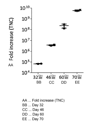
mTNF-a/4-
IBBL dual genes introduced into Hut78 cell line through FACS.
[23] FIG. Id is a diagram confirming the expression status of mbIL-21/0X4OL
and mbIL-21/4-
1 BBL dual genes introduced into Hut78 cell line through FACS.
[24] FIG. le is a diagram confirming the expression status of triple genes
introduced into Hut78
cell line through FACS.
[25] FIG. lf is a diagram confirming the expression status of quadruple genes
introduced into
Hut78 cell line through FACS.
[26] FIG. 2a is a diagram illustrating the proliferation rate of natural
killer cells produced by
co-culturing Hut78 cell line into which the gene has been introduced and cord
blood-derived
CD3(-) mononuclear cells for each transgene.
[27] FIG. 2b is a diagram illustrating the proliferation rate of natural
killer cells produced by
co-culturing H9 cell line into which the gene has been introduced and cord
blood-derived
CD3(-) mononuclear cells for each transgene.
[28] FIG. 2c is a diagram illustrating the proliferation rate of natural
killer cells produced by
Date Recue/Date Received 2021-05-14
CA 03120085 2021-05-14
4
co-culturing Jurkat cell line into which the gene has been introduced and cord
blood-derived
CD3(-) mononuclear cells for each transgene.
[29] FIG. 2d is a diagram illustrating the proliferation rate of natural
killer cells produced by
co-culturing Peer cell line into which the gene has been introduced and cord
blood-derived
CD3(-) mononuclear cells for each transgene.
[30] FIG. 2e is a diagram illustrating the proliferation rate of natural
killer cells produced by
restimulating at 14-day or 16-day interval when co-culturing Hut78 cell line
into which the
triple gene has been introduced and cord blood-derived CD3(-) mononuclear
cells.
[31] FIG. 3a is a diagram illustrating the survival rate of natural killer
cells produced by co-
culturing Hut78 cell line into which the gene has been introduced and cord
blood-derived
CD3(-) mononuclear cells for each transgene.
[32] FIG. 3b is a diagram illustrating the survival rate of natural killer
cells produced by co-
culturing H9 cell line into which the gene has been introduced and cord blood-
derived CD3(-)
mononuclear cells for each transgene.
[33] FIG. 3c is a diagram illustrating the survival rate of natural killer
cells produced by co-
culturing Jurkat cell line into which the gene has been introduced and cord
blood-derived
CD3(-) mononuclear cells for each transgene.
[34] FIG. 3d is a diagram illustrating the survival rate of natural killer
cells produced by co-
culturing Peer cell line into which the gene has been introduced and cord
blood-derived CD3(-)
mononuclear cells for each transgene.
[35] FIG. 3e is a diagram illustrating the survival rate of natural killer
cells produced by
restimulating at 14-day or 16-day interval when co-culturing Hut78 cell line
into which the
triple gene has been introduced and cord blood-derived CD3(-) mononuclear
cells.
[36] FIG. 4a is a diagram illustrating the purity (CD3-CD56+) of natural
killer cells produced
by co-culturing Hut78 cell line into which the gene has been introduced and
cord blood-derived
CD3(-) mononuclear cells for each transgene.
[37] FIG. 4b is a diagram illustrating the purity (CD3-CD56+) of natural
killer cells produced
by co-culturing H9 cell line into which the gene has been introduced and cord
blood-derived
CD3(-) mononuclear cells for each transgene.
[38] FIG. 4c is a diagram illustrating the purity (CD3-CD56+) of natural
killer cells produced
by co-culturing Jurkat cell line into which the gene has been introduced and
cord blood-derived
CD3(-) mononuclear cells for each transgene.
[39] FIG. 4d is a diagram illustrating the purity (CD3-CD56+) of natural
killer cells produced
by co-culturing Peer cell line into which the gene has been introduced and
cord blood-derived
CD3(-) mononuclear cells for each transgene.
[40] FIG. 4e is a diagram illustrating the purity (CD3-CD56+) of natural
killer cells produced
by restimulating at 14-day or 16-day interval when co-culturing Hut78 cell
line into which the
triple gene has been introduced and cord blood-derived CD3(-) mononuclear
cells.
[41] FIG. 5a is a diagram illustrating the activity (CD16+CD56+) of natural
killer cells
Date Recue/Date Received 2021-05-14
CA 03120085 2021-05-14
produced by co-culturing Hut78 cell line into which the gene has been
introduced and cord
blood-derived CD3(-) mononuclear cells for each transgene.
[42] FIG. 5b is a diagram illustrating the expression level of the NKG2D
phenotype marker of
natural killer cells produced by co-culturing Hut78 cell line into which the
gene has been
introduced and cord blood-derived CD3(-) mononuclear cells by each transgene.
[43] FIG. Sc is a diagram illustrating the expression level of the NKp30
phenotype marker of
natural killer cells produced by co-culturing Hut78 cell line into which the
gene has been
introduced and cord blood-derived CD3(-) mononuclear cells by each transgene.
[44] FIG. 5d is a diagram illustrating the expression level of the NKp44
phenotype marker of
natural killer cells produced by co-culturing Hut78 cell line into which the
gene has been
introduced and cord blood-derived CD3(-) mononuclear cells by each transgene.
[45] FIG. 5e is a diagram illustrating the expression level of the NKp46
phenotype marker of
natural killer cells produced by co-culturing Hut78 cell line into which the
gene has been
introduced and cord blood-derived CD3(-) mononuclear cells by each transgene.
[46] FIG. 5f is a diagram illustrating the expression level of the DNAM-1
phenotype marker
of natural killer cells produced by co-culturing Hut78 cell line into which
the gene has been
introduced and cord blood-derived CD3(-) mononuclear cells by each transgene.
[47] FIG. 5g is a diagram illustrating the expression level of the CXCR3
phenotype marker of
natural killer cells produced by co-culturing Hut78 cell line into which the
gene has been
introduced and cord blood-derived CD3(-) mononuclear cells by each transgene.
[48] FIG. 6a is a diagram illustrating the activity (CD16+CD56+) of natural
killer cells
produced by co-culturing H9 cell line into which the gene has been introduced
and cord blood-
derived CD3(-) mononuclear cells for each transgene.
[49] FIG. 6b a diagram illustrating the expression level of the NKG2D
phenotype marker of
natural killer cells produced by co-culturing H9 cell line into which the gene
has been
introduced and cord blood-derived CD3(-) mononuclear cells by each transgene.
[50] FIG. 6c is a diagram illustrating the expression level of the NKp30
phenotype marker of
natural killer cells produced by co-culturing H9 cell line into which the gene
has been
introduced and cord blood-derived CD3(-) mononuclear cells by each transgene.
[51] FIG. 6d is a diagram illustrating the expression level of the NKp44
phenotype marker of
natural killer cells produced by co-culturing H9 cell line into which the gene
has been
introduced and cord blood-derived CD3(-) mononuclear cells by each transgene.
[52] FIG. 6e is a diagram illustrating the expression level of the NKp46
phenotype marker of
natural killer cells produced by co-culturing H9 cell line into which the gene
has been
introduced and cord blood-derived CD3(-) mononuclear cells by each transgene.
[53] FIG. 6f is a diagram illustrating the expression level of the DNAM-1
phenotype marker
of natural killer cells produced by co-culturing H9 cell line into which the
gene has been
introduced and cord blood-derived CD3(-) mononuclear cells by each transgene.
Date Recue/Date Received 2021-05-14
CA 03120085 2021-05-14
6
[54] FIG. 6g is a diagram illustrating the expression level of the CXCR3
phenotype marker of
natural killer cells produced by co-culturing H9 cell line into which the gene
has been
introduced and cord blood-derived CD3(-) mononuclear cells by each transgene.
[55] FIG. 7a is a diagram illustrating the expression level of NKG2D phenotype
marker and
the activity (CD16+CD56+) of natural killer cells produced by restimulating at
14-day or 16-
day intervals when co-culturing Hut78 cell line into which the triple gene has
been introduced
and cord blood-derived CD3(-) mononuclear cells.
[56] FIG. 7b is a diagram illustrating the expression level of NKp30, NKp44,
NKp46, DNAM-
1, and CXCR3 phenotype markers of natural killer cells produced by
restimulating at 14-day
or 16-day interval when co-culturing Hut78 cell line into which the triple
gene has been
introduced and cord blood-derived CD3(-) mononuclear cells.
[57] FIG. 8a is a diagram illustrating the tumor cell killing ability of
natural killer cells
produced by co-culturing Hut78 cell line into which the gene has been
introduced and cord
blood-derived CD3(-) mononuclear cells by each transgene.
[58] FIG. 8b is a diagram illustrating the tumor cell killing ability of
natural killer cells
produced by co-culturing H9 cell line into which the gene has been introduced
and cord blood-
derived CD3(-) mononuclear cells by each transgene.
[59] FIG. 8c is a diagram illustrating the tumor cell killing ability of
natural killer cells
produced by co-culturing Jurkat cell line into which the gene has been
introduced and cord
blood-derived CD3(-) mononuclear cells by each transgene.
[60] FIG. 8d is a diagram illustrating the tumor cell killing ability of
natural killer cells
produced by co-culturing Peer cell line into which the gene has been
introduced and cord blood-
derived CD3(-) mononuclear cells by each transgene.
[61] FIG. 8e is a diagram illustrating the tumor cell killing ability of
natural killer cells
produced by restimulating at 14-day or 16-day interval when co-culturing Hut78
cell line into
which the triple gene has been introduced and cord blood-derived CD3(-)
mononuclear cells.
[62] FIG. 9a is a diagram illustrating an administration schedule for efficacy
evaluation using
the Raj i mouse animal model.
[63] FIG. 9b is a diagram illustrating the result of measuring the survival
rate for confirming
the efficacy of NK cells, RTX, and co-administration in the Raji animal model.
[64] FIG. 10a is a diagram illustrating an administration schedule for
efficacy evaluation using
the Ramos mouse animal model.
[65] FIG. 10b is a diagram illustrating the result of measuring the survival
rate for confirming
the efficacy of NK cells, RTX, and co-administration in the Ramos animal
model.
[66]
Best mode for carrying out the invention
[67] Hereinafter, the present invention will be described in detail.
Date Recue/Date Received 2021-05-14
CA 03120085 2021-05-14
7
[68] One aspect of the present invention provides a method for culturing
natural killer cells
comprising a step for co-culturing transformed CD4+ T cells and seed cells.
[69] The transformed CD4+ T cells may express at least one gene selected from
the group
composed of 4-1BBL gene, mbIL-21 gene, 0X40L gene, and mTNF-a gene.
[70] Specifically, when one gene is introduced into the transformed CD4+ T
cells, the gene
may be 4-1BBL, mbIL-21, 0X40L, or mTNF-a. In addition, when two genes are
introduced
into the transformed CD4+ T cells, said gene combination may be mbIL-21/4-
1BBL, 4-
IBBL/OX4OL, mTNF-a/4-1BBL, mbIL-21/0X4OL, mbIL-21/mTNF-a or mTNF-a/OX4OL.
In one embodiment of the present invention, genes of a combination of mbIL-
21/4-1BBL,
mTNF-a/OX4OL, mTNF-a/4-1BBL and mbIL-21/0X4OL were introduced into T cells.
[71] In addition, when three genes are introduced into the transformed CD4+ T
cells, said gene
combination may be 4-1BBL/mbIL-21/0X4OL, mbIL-21/0X4OL/mTNF-a, mTNF-a/ mbIL-
21 /4-1BBL or 4-1BBL/OX4OL/mTNF-a. In one embodiment of the present invention,
genes
of a combination of mTNF-a/ mbIL-21 /4-1BBL were introduced into T cells.
[72] In addition, when four genes are introduced into the transformed CD4+ T
cells, said gene
combination may be mTNF-a/mbIL-21/0X4OL/4-1BBL. In one embodiment of the
present
invention, genes of a combination of mINF-a/mbIL-21/0X4OL/4-1BBL were
introduced into
T cells.
[73] The term `4-1BBL' used in the present invention is one of TNFSF (TNF
superfamily)
called CD137L and refers to a ligand that binds to the receptor 4-1BB by
forming a trimer. The
4-1BB gene may be derived from humans.
[74] Specifically, the 4-1BBL gene may be NCBI Reference Sequence: NM_003811,
but is not
limited thereto. The 4-1BBL gene may be a base sequence coding the amino acid
sequence
represented by sequence No. 1. The base sequence coding the amino acid
sequence represented
by sequence No. I may be a base sequence represented by sequence No. 2.
[75] The term 'mbIL-21' used in the present invention may be IL-21 designed to
be bound to a
cell membrane. Here, mbIL-2 I may be a fusion protein in which IL-21 and a
transmembrane
protein are combined. The transmembrane protein may be CD8a. Specifically, it
may be a
transmembrane domain of CD8a.
[76] Specifically, the IL-21 gene may be NCBI Reference Sequence: NM_021803.3,
but is not
limited thereto. In addition, the CD8a gene may be NCBI Reference Sequence:
NM_001768,
but is not limited thereto. The mbIL-21 is expressed in the form of IL-21
bound to the cell
membrane. In addition, the mbIL-21 gene may be a base sequence coding the
amino acid
sequence represented by sequence No. 3. The base sequence coding the amino
acid sequence
represented by sequence No. 3 may be a base sequence represented by sequence
No. 4.
Date Recue/Date Received 2021-05-14
CA 03120085 2021-05-14
8
[77] The term '0X40L' used in the present invention is also called TNFSF4,
gp34, TXGP1,
CD252, and CD134L, and refers to a ligand that binds to 0X40. Specifically,
the 0X40L gene
may be NCBI Reference Sequence: NM_003326, but is not limited thereto. The
0X40L gene
may be a base sequence coding the amino acid sequence represented by sequence
No. 5. The
base sequence coding the amino acid sequence represented by sequence No. 5 may
be the base
sequence represented by sequence No. 6.
[78] The term 'mTNF-a' used in the present invention refers to the gene in
which Alanine-
Valine, which is a TACE (tumor necrosis factor-alpha-converting enzyme)
recognition site, has
undergone a point mutation in DNA in the amino acid sequence of tumor necrosis
factor-alpha
to become Proline-Valine. Mutating alanine to proline was randomly chosen.
[79] Specifically, the mTNF-a gene may be a base sequence coding the amino
acid sequence
represented by sequence No. 8. The base sequence coding the amino acid
sequence represented
by sequence No. 8 may be the base sequence represented by sequence No. 9.
[80] The 4-1BBL gene, mbIL-21 gene, OX4OL gene, or mTNF-ct gene may be
introduced
through a recombinant lentivirus, but is not limited thereto.
[81] As a method for transducing the gene into a cell, a biochemical method, a
physical method,
or a virus mediated transduction method may be used. In addition, as a
biochemical method,
FuGene6 (Roche, USA), Lipofectamine (LipofectamineTM 2000, Invitrogen, USA),
or ExGen
500 (MBI Fermentas International Inc. CANADA) may be used. In addition, a
lipid mediated
method using lipofectamine may be used.
[82] The term 'vector' used in the present invention is an expression vector
capable of
expressing a target gene in cells into which the vector has been introduced,
refers to a gene
construct comprising essential control elements operably connected so that the
gene insert
introduced into the vector can be expressed.
[83] In addition, as the expression vector comprising the gene, any expression
vector that can
be expressed in a CD4+ cell line can be used, and in a specific embodiment of
the present
invention, pCDH-CMV-MCS-EF 1 -Puro (SBI, CD510B-1) or pCDH-CMV-MCS-EF 1 -Neo
(SBI, CD514B-1) lentiviral vector was used.
[84] The lentivirus refers to a virus of the retrovirus family characterized
by a long incubation
period. Lentiviruses can carry genetic information into the DNA of host cells.
It is one of the
most effective methods of gene transfer vectors capable of replicating in non-
dividing cells.
[85]
[86] * The CD4+ T cells may be CD4+ T cells isolated in vitro, CD4+ T cells
expanded and
cultured in vitro, or CD4+ cell lines (T lymphoma cell lines). In addition,
the CD4+ T cells
may be accessory T cells, and may be hybridomas obtained by fusing CD4+ T
cells and cancer
cells. Specifically, the CD4+ T cells may be any one selected from the group
composed of
Hut78, H9, Jurkat, Loucy, Molt-3, Molt-13, Peer, RPMI8402 and TALL-01 cells.
Preferably, it
may be Hut78, H9, Jurkat or Peer cells.
Date Recue/Date Received 2021-05-14
CA 03120085 2021-05-14
9
[87] The term 'feeder cell' used in the present invention refers to a cell
that is also called a
culture support cell and does not proliferate but has the metabolic activity
to help the
proliferation of target cells by producing various metabolites. The feeder
cells may be
transformed CD4+ T cells expressing at least one gene selected from the group
composed of
4-1BBL gene, mbIL-21 gene, 0X40L gene, and mTNF-a gene.
[88] The T cells used as the feeder cells may be inactivated cells in which
divisional
proliferation is inhibited or cells that have not been inactivated, and
preferably, safety can be
ensured by inactivation. As a method for inactivation, a common method known
in the relevant
industry may be used, and for example, a method for irradiating gamma-ray may
be used. When
using T cells that have not been inactivated, since most are tumor cells, they
can be killed
during culture by activated natural killer cells.
[89] The term "seed cell" used in the present invention refers to a cell
capable of proliferating
into natural killer cells through appropriate culture. Specifically, the seed
cell may be cord
blood-derived mononuclear cells, or cord blood-derived natural killer cells.
This is not limited
thereto, and preferably, the seed cells may be CD3(-) cells from which CD3(+)
cells have been
removed.
[90] As for the method for culturing natural killer cells, they may be
cultured by mixing the
feeder cells and the seed cells with a ratio of at least 0.1. Specifically,
the ratio of the feeder
cells and the seed cells may be 0.1:1 to 50:1. More specifically, it may be
0.5:1 to 40:1. Even
more specifically, it may be 1:1 to 30:1. Most specifically, it may be 2:1 to
20:1. As a specific
example, the ratio of the feeder cell and the seed cell may be 2.5:1, but is
not particularly
limited thereto. The "ratio" refers to a ratio based on the number of cells.
[91] In the method for culturing natural killer cells, the seed cells may be
mixed once with the
feeder cells and cultured for 5 to 60 days, or mixed with the feeders cells at
least twice and
cultured for at least 60 days. Preferably, the seed cells may be mixed once
with the feeder cells
and cultured for 14 to 21 days, but it is not limited thereto.
[92] In the method for culturing natural killer cells, natural killer cells
and T lymphoma cell
lines are co-cultured in a conventional animal cell culture medium, such as
AIM-V media,
RPMI1640, CellGro SCGM, X-VIV020, IMDM, and DMEM. When co-cultured,
interleukins
and antibodies that have low affinity to T cells and stimulate T cells may be
added for culture,
but it is not limited thereto.
[93] The term 'antibody that has low affinity to T cells and stimulates T
cells' used in the
present invention refers to a protein that specifically reacts to the CD3
antigen, which is a group
of molecules that meets with the T cell receptor (TCR) to form an antigen
recognition complex.
Compared to TCR, the CD3 molecule has a longer intracellular region and plays
a role of
transmitting antigen recognition signals into the cell.
Date Recue/Date Received 2021-05-14
CA 03120085 2021-05-14
[94] Preferably, an antibody, which has a low affinity to T cells and
stimulates T cells, that can
be used in the present invention may be an anti-CD3 antibody. Specifically,
the anti-CD3
antibody may be OKT-3, UCHT1, or HIT3a.
[95] The term `interleukin' (IL) used in the present invention refers to a
group of cytokines,
and refers to a proteinaceous biological active substance produced by immune
cells, such as
lymphocytes, monocytes, and macrophages. The interleukin may be IL-2, IL-15,
IL-12, IL-18,
or IL-21.
[96] In an embodiment of the present invention, it was cultured by adding OKT-
3 antibody and
IL-2. The concentration of the OKT-3 antibody added may be 0.1 ng/mt to 1,000
ng/mt.
Preferably, the concentration of the OKT-3 antibody may be 10 ng/p,f. The
concentration of
IL-2 may be 10 U/mt to 2,000 U/mt. Prcferably, the concentration of IL-2 may
be 1,000 U/ml.
In addition, it may be cultured by adding additional growth factors that
support the proliferation
of serum or plasma and lymphocytes. The type of serum or plasma to be added to
the medium
is not particularly limited, and commercially available serum or plasma
derived from various
animals may be used. Preferably, human-derived serum or plasma derived from
the person
themselves may be used.
[97] The term 'culture' of the present invention refers to a method for
growing cells in an
environmental condition that has been appropriately artificially controlled.
The method for
culturing the transformed CD4+ T cells may be performed using a method well
known in the
relevant industry. Specifically, said culture may be carried out in a
continuous manner in a
batch process, a fed batch, or a repeated fed batch process.
[98] In addition, precursors suitable for the culture medium may be used. The
raw materials
described above may be added in a batch, fed batch, or continuous manner to
the culture during
the cultivation process, but it is not particularly limited thereto. Basic
compounds, such as
sodium hydroxide, potassium hydroxide, and ammonia, or acidic compounds, such
as
phosphoric acid or sulfuric acid, can be used in an appropriate manner to
adjust the pH of the
culture.
[99] The culture method using T cells as feeder cells selectively induces
culture of natural killer
cells in seed cells, and it can be cultured stably without differences
depending on the donor
when proliferating natural killer cells compared to when using the donor's
PBMC feeder cells.
In addition, in vitro culture of cord blood seed cells is difficult when the
donor's MNC is used
as feeder cells. Therefore, the culture method using T cells as feeder cells
can efficiently and
stably secure a large amount of therapeutic natural killer cell agents for
treatment.
[100] Another aspect of the present invention provides natural killer cells
produced by said
method for culturing natural killer cells.
[101] Natural killer cells cultured according to said method for culturing
natural killer cells can
be frozen and the function of the cells does not get damaged even when they
are thawed again.
In addition, since the expression of an activating receptor, such as NKp46, is
high, the killing
ability and secretion of cytokines against tumor cell lines are increased, and
therefore, an
excellent anticancer effect can be expected. Therefore, it is possible to
manufacture a cell
therapy product effective for tumor treatment using a large amount of
clinically applicable
activated natural killer cells.
Date Recue/Date Received 2021-05-14
CA 03120085 2021-05-14
11
[102] In addition, natural killer cells produced by the method for culturing
natural killer cells
may be comprised in an amount of 10 to 95wt% based on the total weight of the
composition
for preventing or treating infectious diseases included as an active
ingredient. In addition, the
composition for preventing or treating infectious diseases or of the present
invention may
further comprise at least one type of active ingredients exhibiting the same
or similar function
in addition to said active ingredient.
[103] The pharmaceutical composition for preventing or treating infectious
diseases may be
prepared into a pharmaceutical composition by comprising at least one type of
pharmaceutically acceptable carriers in addition to the active ingredient
described above for
administration.
[104] The dosage of the pharmaceutical composition for preventing or treating
infectious
diseases may be adjusted according to various factors including type of
disease, severity of
disease, type and content of active ingredients and other ingredients
comprised in the
composition, type of formulation, patient's age, weight, general health
condition, gender, and
diet, administration time, administration route, secretion rate of the
composition, duration of
treatment, and concurrently used drugs. However, for a desirable effect, the
dost of natural
killer cells according to the present invention may be 0.01x10⁷ cells/kg to
1.0x10⁹ cells/kg, and
may be 0.5x10⁷ cells/kg to 1.0x10⁸ cells/kg. In this case, the administration
may be carried out
once a day, or may be divided into several administrations.
[105] In addition, the pharmaceutical composition for preventing or treating
infectious diseases
may be administered to an individual by various methods known in the relevant
industry. The
administration route may be appropriately selected by a PHOSITA in
consideration of the
method of administration, volume of body fluid, viscosity, etc.
[106] Another aspect of the present invention provides a composition for
culturing natural killer
cells comprising transformed CD4+ T cells as an active ingredient. Since the
CD4+ T cells
used in the present invention and the genes introduced into said cells have
already been
described above, the corresponding descriptions will be omitted to avoid
excessive duplication.
[107]
Mode for carrying out the invention
[108] Hereinafter, the present invention will be described in detail by
embodiments. However,
the following embodiments are intended only for illustrating the present
invention, and the
present invention is not limited to the following embodiments.
[109]
[110] Embodiment 1. Production of recombinant lentivirus
[111] Embodiment 1.1. Production of recombinant lentiviral vector
[112] For the lentiviral vector, pCDH-CMV-MCS-EF1-Puro (SBI, CD510B-1) or pCDH-
CMV-MCS-EF1-Neo (SBI, CD514B-1) was used. For genes, 4-1BBL (TNF superfamily
member 9, TNFSF9), mbIL-21 (membrane bound IL-21), OX4OL (TNF superfamily
member
4(TNFSF4) transcript variant 1), and mTNF-a (membrane bound TNF alpha) were
used as
Date Recue/Date Received 2021-05-14
CA 03120085 2021-05-14
12
transgenes.
[113] Specifically, a 4-1BBL gene expression vector (Origene, RC211160) was
used for the 4-
1BBL gene (sequence No. 2). For the mbIL-21 gene (sequence No. 4), a pcDNA3.1
vector
(Genscript, US) into which the codon-optimized mbIL-21 gene sequence has been
inserted was
used. The OX4OL gene (sequence No. 6) was requested to be synthesized by
Bioneer.
[114] For mTNF-a gene (sequence No. 9), RNA was extracted from peripheral
blood
mononuclear cell (PBMC), and then CDS was obtained by RT(Reverse
transcriptase)-PCR.
TNF-a is cut by TACE (tumor necrosis factor-alpha-converting enzyme) to be
secreted, and A-
V (Alanine-Valine), which is a TACE recognition site, has undergone a point
mutation in DNA
in the TNF-a amino acid sequence to become P-V (Proline-Valine), thereby
maintaining the
state of being attached to the cell membrane. The point mutation was performed
by substituting
guanine, the 226th base, with cytosine, and adenine, the 228th base, with
guanine in the human
mTNF-a gene represented by sequence No. 7.
[115] Using primers suitable for each transgene, CDS (Coding Sequence) of the
transgene was
amplified through PCR (Table 1).
Date Recue/Date Received 2021-05-14
CA 03120085 2021-05-14
13
[116] [Table 1]
G_ene Primer Sequence information I 5'..>3' ) Sequence
number
d-11313 I-4- IBM. irci ,AGA[:;cT m:ic ( r .),ATi (, tic(' ArcAl G
Sequence
number 10
I 11 Irv,;tra 1(i.\ A I A t_ (..it :(-1 CI.GAC(.1(1:1-1
-1- I BM Ti-crg--(icirc(;(--(iciAT01-175,-Ficccorc iSequence
number 11
J jI.1.("LitiTGAMiCi
;11,11)11,- '4c1111.-::1 i \{;m3.(2.i.,...,(.;c6A.A=1-1 C(.a:CACCC;CCAC
Sequence
I number 12
21 17,u-e-eRt C . \ TiiCiCI CIT..r0:( '
I mh11.-2 I I L .1. ii: 'CiC I( .4_ '(1411. Alf(' If A VI
''\. I: -A( ;CIL ; Sequence
,
number 13
I fie'rersc 1.FGA'IGAL('
( )X44) !CI.X401 . Li.n A,( ici...),( ;cc ;Am"' ec i( ,(7. \ (,,E-
A.1.(A.i.A Sequence
number 14
IL. I Porward ACGGC.irtGCAAC.
- - = L...,_
!OX 401.
,fccic'cc icCGCGGAI CCTCM AA GACAC Sequence
1I AGAACICCO: number 15 ,
'DINE ;nil NE o ITAGAGCT A OD_ A AT ICGCCACCGCC AC :Sequence
01 number 16
I-I) I;iti-,,,,,q,i ICA.1 (1C.:;(11'11- ;(_-rt:
1 i ____________________________________________
'EICINF-, r
111I1: C. iCGOCCGCE G v. ; ( r (.14.
'.,\ r. \ 1Sequence
Inumber 17
IIC:IA '11 'ef
[117] Table 1 shows the primers used in the experiment. The transgene and
lentiviral vector
were treated with EcoRI and BamHI restriction enzymes. Then, it was ligated
using In-Fusion
HD cloning kit (Clontech, 639649). The ligated lentiviral vector was
transformed in DH5a
soluble cells (competent cells) and cultured. Plasmid DNA was obtained from
the transformed
DH5a soluble cells using a plasmid mini-prep kit (MACHEREY-NAGEL/740422.50). A
request for sequencing was made to an external company and it was confirmed
that all plasmid
DNA matches the DNA sequence. In addition, the desired transgene was inserted
into cLV-
CMV-MCS-IRES-Puro (puromycin) or cLV-CMV-MCS-IRES-Neo (neomycin), cLV-CMV-
MCS-IRES-Bsd (blasticidin) by an outsourced manufacturer by the same method as
the one
described above.
[118]
[119] Embodiment 1.2. Production of concentrated lentivirus
[120] In order to produce recombinant lentivirus, the 293T cell line was
inoculated into a 75T
flask (Nunc, 156499) with 1.5x10⁶ to 2x10⁶ cells 2 days before transfection,
and cultured in an
incubator at a temperature condition of 5% CO2, 37 C. When the cell saturation
of the 293T
cells reached about 80% to 90%, the medium was replaced with 6 mf OPTI-MEM
(Gibco,
31985-088) and incubated for 30 minutes at a temperature of 37 C and under the
condition of
5% CO2. A DNA mixture and a lipofectamine (lipofectamine 2000, Life
technologies,
11668500) mixture were prepared (Table 2).
Date Recue/Date Received 2021-05-14
CA 03120085 2021-05-14
14
[121] [Table 2]
Category Ingredients
--
DNA mixture 6 ri target DNA, ri (ac. kiN, 3 pg VV(_ I al
Lipofectamine mixture
lipolcctarmilic 2001. 1 m(' OPTI-MEM
[122] Table 2 shows the DNA mixture and the lipofectamine (lipofectamine 2000,
Life
technologies, 11668500) mixture. Each of the components of the mixtures was
mixed well
using a vortexer and left at room temperature for 3 minutes. Then, the two
mixtures were mixed
and left at room temperature for at least 20 minutes. 2 int of a mixed
solution of DNA and
lipofectamine was treated with 293T cells being cultured in 6 int OPTI-MEM.
After 4 hours,
it was replaced with DMEM (Gibco, 11995073) medium to which 10%(v/v) FBS has
been
added, and was cultured at a temperature of 37 C for 48 hours under the
condition of 5% CO2.
8mt of the culture solution of 293T cells cultured for 48 hours was collected
and filtered
through a 0.45 um filter (Millipore, SLHP033RS). The filtered culture solution
was
concentrated to 250 ut or less using an Amicon Ultra-15 Centrifugal Filter
Unit with Ultracel-
100 membrane (Merckmillipore, UFC910096). The concentrated virus was divided
into an
appropriate amount and stored at a temperature of -80 C.
[123]
[124] Embodiment 2. Production of transgenic T cells
[125] Embodiment 2.1. Lentivirus infection
[126] 0.5x10⁶ cell lines being cultured, lint OPTI-MEM, 50ut lentivirus
thawing solution,
and 10 Out polybrene (Santa Cruz, C2013) were mixed and placed in a 6-well
plate (Nunc,
140675), and spinoculation was performed for 90 minutes under 1800xg and at a
temperature
of 32 C. Then, after culturing in an incubator under a temperature condition
of 5% CO2, 37 C,
it was replaced with an existing culture medium and cultured for 48 hours.
[127] Hut78 cell line (ATCC, TIB-161Tm) was cultured in IMDM (ATCC, 30-2005)
medium
containing 20%(v/v) FBS. During subculture, the cell concentration was
maintained at 1.5x10⁵
cells/int to 2.0x10⁵ cells/mt. H9 cell line (ATCC, HTB-176Tm ) and Jurkat cell
line (ATCC,
TIB-152Tm) were cultured in RPMI1640 (ATCC, 30-2001) medium containing
10%(v/v) FBS.
During subculture, cell concentrations were maintained at 1.0x10⁵ cells/mt to
1.5x10⁵cells/mt
and 0.5x10⁵ cells/mt to 1.0x10⁵ cells/int, respectively. Peer cell line was
cultured in RPMI1640
medium containing 20%(v/v) FBS. During subculture, the cell concentration was
maintained
at 3.0x10⁵ to 5.0 x10⁵ cells/mt. The subculture of all cell lines was
performed at intervals of 2
to 3 days. A 75T flask was used as the culture vessel, and the amount of
medium was maintained
between 15 mt to 20 mt.
Date Recue/Date Received 2021-05-14
CA 03120085 2021-05-14
[128] Cell lines infected with the recombinant lentivirus were selected using
antibiotics (Table
3).
[129] [Table 3]
Transgenic Vector used Cell line Antibiotic usage
combination concentration
Single gene fraNI.-iimbil1 '[ICD311`,S)Niciii lu(78
pairrimycjih.
expression I c}seterices. I Lii ochnolop
Al I 38021
OX401. IBBL 11uI78 I I I -IA( Sigma
A idrii; h., \ 1720-5C)
I Double gene 01:1"Nl'-".10X4fIr irriM 1(SN'sit 43.5
expression tra-11..-21/0.X4kA..113ios=ciencei,.., puromycin E
ir IS13.1
-T?11313 !ci V o'Sioi 11L.117:s119 .45
I. LuPc 1-
3111.,ticidimjmilro,gc
. R21(1-01)1
(i4
Triple gene URI M.-011;11)11 -2 L TV 01-11=141S0 114117N119
ii
I expression 1/4 I 13111 Jurkat
6418
I Quadruple iiiiTHF-ahrbll NI:-: Ht. - f 11,47K
/gime
gene II0X-I!1.14=I 14- I BBL: prom yc irth
expression ON,411,_..: Ithiq it-din I
pC1I)1.1 G-318
[130] Table 3 above shows the antibiotics used in cell lines into which the
gene was introduced.
[131]
[132] Embodiment 2.2. Confirmation of transgene expression
[133] In order to confirm the expression of the transgene through flow
cytometry, the cell lines
subcultured in embodiment 2.1. were collected and centrifuged at 1,200 rpm for
5 minutes.
Then, the culture solution was removed by suction. FACS buffer was created by
adding 2%(v/v)
FBS to PBS. The number of cells was measured by diluting with 1 tut of FACS
buffer, and it
was diluted with FACS buffer to a concentration of 5x10⁶ cells/mt. 100 p.t of
diluted cell
solution was added to each of 5 mt FACS tubes (Falcon, 352052). After staining
with anti-
human TNF-a(membrane)-PE (R&D systems, FAB210P), anti-human OX40L-PE (BD,
558184), anti-human 4-1BBL-PE (BD, 559446), anti-human IL-21-PE (eBioscience,
12-7219-
42), 7-AAD (Beckman coulter, IM3630c), PE mouse IgG1 K isotype control (BD
Pharmingen,
Date Recue/Date Received 2021-05-14
CA 03120085 2021-05-14
16
555749), and PerCP-Cy5.5 mouse IgG1 ic isotype control (BD, 550795), the
expression rate of
each gene was analyzed using FACS equipment (FIGS. la to If).
[134] In addition, in order to confirm the expression of the transgene through
RT-qPCR (Real
time qPCR), the cell lines subcultured in embodiment 2.1. were collected and
centrifuged at
1,200 rpm for 5 minutes. Then, the culture solution was removed by suction.
The number of
cells was measured by diluting with PBS, and RNA was isolated and quantified
for 1x10⁶ce11s
using an RNA prep kit. In addition, cDNA was synthesized using a cDNA
synthesis kit. RT-
qPCR was performed using the synthesized cDNA. Primers used in RT-qPCR are as
shown in
Table 4 below.
[135] [Table 4]
Primer Sequence information 45,_>3, Sequence
number
4 1 BBL ForiNani primer TrTri AGA(
AGGcic ...\TGT-E. Sequence number 18
TG
Rcvcrse primer
0(.7ACCAOTIPCTIII.;GIGT(.7 Sequence number 19
(1
MTN F-(t. Forward prirncr
AACCTCCTCTCTGCCATCA Sequence number 20
A
Revers.. pMr
r AGTcGmccciATTG AT Sequence number 21
a.
CT
tubl L-2 1 Forv,..ard primcE
TGGAAAcAATGõ.xcicciAi.µ T. Sequence number 22
C A
kf2 verse primer AAC:CGCTCC
A G A ACTCI" Sequence number 23
TT
-
117170P Forward /Miller 'CEA GA
CGG A A.,GCTCGG A A. Sequence number 24
C
kcverse primer
CiTCCAOC.iAGG(.71-CTATC1 Sequence number 25
TGA/N
Date Recue/Date Received 2021-05-14
CA 03120085 2021-05-14
17
[136] Table 4 shows the primers used in the RT-qPCR experiment. The expression
level of the
transgenes in the cell lines is shown in Table 5 below.
[137] [Table 5]
Ct. value TON I NF = mbi L. 2! .4. I
BM_
1119 20.3 21 rd ,
IiQ mbli .-21-4 20..0 22.2 19 [9.4
F19-inT NI-41- 4111,-2 ?BBL 19.9 18.2 IS. 1 I [
20..1 .n.d rid
37..0 ,30
NIV--mlail -7.144 113131.. .7() 4 19 7 [9.8
PEA', r 7p J.Y 2 4 2 34..9
¨
1,,c1- 11111)1i-2F L_ 1 RBI_ 33* 29,0 6
11111 Lk:h..cw.d
[138] As shown in Table 5, it was confirmed that the expression level of the
genes introduced
into the cell lines was increased.
[139]
[140] Embodiment 3. Co-cultivation of CD3(-) PBMC and transgenic T cells
[141] Embodiment 3.1. Preparation of cord blood-derived CD3(-) PBMC seed cells
[142] Cord blood for research was placed in a 50 mt tube and centrifuged for
10 minutes at
1,500 rpm. Plasma of the upper layer was removed and PBS (phosphate buffered
saline,
LONZA, 17-516Q) was added in a 1:1 ratio. Then, after separating cord blood
mononuclear
cells (MNC) through Ficoll (Ficoll-Paque Plus, GE Healthcare, 17-1440-03)
density gradient
centrifugation method, the number of cells was measured using the ADAM cell
counter system
(Nano Entek).
[143] In order to obtain seed cells from which CD3(+) cells have been removed,
5x10⁷cord
blood mononuclear cells were moved to a new 50 mt tube, and then centrifuged
at 1,200 rpm
and a temperature of 4 C for 5 minutes. A MACS running buffer containing
2%(v/v) FBS and
EDTA with a concentration of 2 mM in PBS was prepared. After the
centrifugation, 400 p,f, of
MACS running buffer and 100 p,f, of CD3 magnetic beads (Miltenyi biotech, 130-
050-101)
were added to the pellet and reacted at a temperature of 4 C for 20 minutes.
After washing by
adding 10 mt MACS running buffer, it was centrifuged at 13,500 rpm and a
temperature of 4 C
for 8 minutes and suspended in 0.5 mt of MACS running buffer.
[144] Cells were separated by mounting a CS column (Miltenyi Biotech, 130-041-
305) on
VarioMACS (Miltenyi Biotech). Cells were recovered by washing the column until
finally
reaching 20 mt. The recovered cells were placed in a new 50 mt tube,
centrifuged at 1,200
Date Recue/Date Received 2021-05-14
CA 03120085 2021-05-14
18
rpm and a temperature of 4 C for 5 minutes, and suspended in a frozen medium.
The number
of cells was measured using the ADAM cell counter system to freeze 5x10⁶ cells
per vial in
liquid nitrogen.
[145] One vial of frozen CD3(-) cord blood mononuclear cells was thawed in a
water bath at a
temperature of 37 C and moved to a 50 int tube, suspended in PBS containing
0.6%(v/v)ACD
(Citrate-dextrose solution, Sigma-Aldrich, C3821), 0.2%(v/v) FBS (Fetal serum
bovine), and
2 mM EDTA, and centrifuged at 1,500 rpm and a temperature of 4 C for 10
minutes. CD3(-)
cord blood mononuclear cells were suspended in CellGro medium (Cellgenix,
20802-0500),
and the number of cells was measured using the ADAM cell counter system. CD3(-
) cord blood
mononuclear cells were suspended in CellGro medium at a concentration of
1x10⁶cells/mt.
[146]
[147] Embodiment 3.2. Co-cultivation of CD3(-) cord blood mononuclear cells
and
transgenic T cells
[148] The transgenic T cells prepared in embodiment 2 were recovered from the
culture flask
and centrifuged at 1,200 rpm and a temperature of 4 C for 5 minutes. Then, it
was suspended
in CellGro medium, and the number of cells was measured using the ADAM cell
counter
system. The transgenic T cells were suspended in CellGro medium at a
concentration of
2.5x10⁶ cells/int, and then prepared by inactivating it with irradiation at
20,000 cGy in a
gamma-ray irradiator.
[149] When culturing natural killer cells, 1,000 IU of IL-2 (Proleukin
Injection, Novartis Korea)
and 10 ng/mt of OKT-3 (eBioscience, 16-0037-85) were placed in a culture
plastic plate. On
day 0 of cultivation, 0.25 int of each of CD3(-) cord blood mononuclear cells
and transgenic
T cells was added at a ratio of 1:2.5, 0.25mt of CellGro medium containing
2%(v/v) human
plasma was added, and stationary culture was carried out for 4 days in an
incubator at a
temperature condition of 37 C.
[150] On the fourth day of cultivation, the same amount of CellGro medium
containing 1%(v/v)
human plasma and 1,000 IU/mt of IL-2 was added, and then stationary culture
was performed
again. Then, the number of cells was measured at intervals of 2 to 3 days, and
suspension
culture was carried out until the 21st day while adding CellGro medium
containing 1%(v/v)
human plasma and 1,000 IU/mt of IL-2 to reach a concentration of lx106
cells/mt. Proliferated
natural killer cells were obtained by performing suspension culture until the
21't day. In this
case, if the Jurkat cell lines or the Peer cell lines were used as feeder
cells, the suspension
culture was performed until the 11th day. If genes were introduced into H9 and
Hut78 and used
as feeder cells, the suspension culture was performed until the2 I 'day.
[151] The result of comparing the proliferation rate of cultured natural
killer cells showed that,
based on the total number of cells (Total nucleated cells, TNC), when co-
cultured with the
Hut78 cell lines to which the gene was not introduced, it proliferated 93
times. It was confirmed
that the proliferation rate of natural killer cells was significantly
increased when co-cultured
with the Hut78 cell lines into which one or more genes (mTNF-a, mbIL-21, 4-
1BBL) were
introduced. In particular, when co-cultured with the Hut78 cell lines into
which the gene of
mbIL-21/4-1BBL was introduced, it proliferated 957 times. In addition, when co-
cultured with
the Hut78 cell lines into which mTNF-a/mbIL-21/4-1BBL was introduced, it
proliferated 1,138
Date Recue/Date Received 2021-05-14
CA 03120085 2021-05-14
19
times (Table 6, FIG. 2a).
[152] [Table 6]
Transgene Arimage STEW V
[ILA. 7 parental 4i1-1
61,7
4b.1
10X4OL
4.113111.
1
[ 0.X1iTh .172.0 N9_2
ihiL 2 I -4. I BBL
161 ttn .21 +4. I 1.313I. 1
1 ____________________________________________________________
r.niTNIF-41..4-0X.4.1+ridillI BBL. 823.1
[153] In addition, when co-cultured with the H9 cell lines into which the gene
was not
introduced, it proliferated 13 times, but when co-cultured with the H9 cell
lines into which
mbIL-21/4-1BBL or mTNF-a/mbIL-21/4-1BBL was introduced, it proliferated 367
times and
979 times, respectively (Table 7 and FIG. 2b).
[154] [Table 7]
=
1H9 Transgene Average ,STD.E
1A4-1 parental .12.6
mbIL.21.4-4.1BBL 367_4
ITITN F.-a-HI-Ill I :21+4. I RBI 97S.8 277
[155] When co-cultured with other cell lines, such as Jurkat cell lines or
Peer cell lines,
cultivation was possible until the 11th day of culture. A relatively high
proliferation rate was
displayed in cell lines into which the mbIL-21/4.1BBL gene was introduced or
cell lines into
which the mTNF- a/mbIL-21/4-1BBL gene was introduced (Table 8 and Table 9,
FIG. 2c and
FIG. 2b).
[156] [Table 8]
jtirkat + Transgene A ve.rage (11-day culture)
Jurkat Parental
InbIL.21+4. I BBL .6.3
............
iiN
I a+rnbi I.,21 +4.1 13 BL 43.6 6.0
Date Recue/Date Received 2021-05-14
CA 03120085 2021-05-14
[157] [Table 9]
Peer + Transgene Average (11-day culture) STiWV
Peer Po renOal 16 0,7
ilibIL2 I +4 1 13111 H 4 ri
4 I
[158] The results described above showed that it is possible to culture
natural killer cells by
culturing CD3(-) cells isolated from cord blood mononuclear cells for 21 days
with feeder cells
into which the gene was introduced, and exhibited a higher proliferation that
the non-
transduced feeder cells.
[159]
[160] Embodiment 3.3. Restimulation of natural killer cell culture using Hut78
cells into
which the mTNF-a/mbIL-21/4-1BBL gene was introduced
[161] The transgenic T cells prepared in embodiment 2 were recovered from the
culture flask
and centrifuged for 5 minutes at 1,200 rpm and a temperature of 4 C. Then, it
was suspended
in CellGro medium, and the number of cells was measured using the ADAM cell
counter
system. After suspending the transgenic T cells in CellGro medium at a
concentration of
2.5x10⁶ cells/nit, it was prepared by inactivating it with irradiation at
20,000 cGy in a gamma-
ray irradiator.
[162] When culturing natural killer cells, 1,000 111 of IL-2 and 10 ng/mf of
OKT-3 were placed
in a culture plastic plate. On day 0 of cultivation, 0.25 mf to 1 mf of each
of CD3(-) cord blood
mononuclear cells and transgenic T cells were added at a ratio of 1:2.5, 0.25
mf to 1 mf of
CellGro medium containing 2%(v/v) human plasma was added, and stationary
culture was
carried out for 4 days in an incubator at a temperature condition of 37 C.
[163] On the fourth day of cultivation, the same amount of CellGro medium
containing 1%(v/v)
human plasma and 1,000 IU/mf of IL-2 was added, and then stationary culture
was performed
again. Then, the number of cells was measured at intervals of 2 to 3 days, and
cultivation was
carried out while adding CellGro medium containing 1%(v/v) human plasma and
1,000 IU/mf
of IL-2 to reach a concentration of lx106cells/mf
[164] For restimulation, on day 0 of cultivation, HuT78 cells into which the
mTNF-a/mbIL-
21/4-1BBL was introduced were used at the same ratio. On the sixteenth day of
cultivation, the
first restimulation was given. First, the number of natural killer cells in
cultivation was
measured using the ADAM cell counter system, they were diluted with CellGro
medium to
become 1.5x10⁶ cells/mf, and 0.25 mf was prepared on a culture plastic plate.
HuT78 cells
into which the mTNF-a/mbIL-21/4-1BBL was introduced were suspended in CellGro
medium
to become 2.5x10⁶ cells/mf, and then prepared by inactivating it with
irradiation at 10,000 cGy
in a gamma-ray irradiator.
[165] 0.25 mf HuT78 cells into which the inactivated mTNF-a/mbIL-21/4-1BBL
gene was
introduced were added to a culture plastic. 1,000 IU/mf of IL-2 and 10 ng/mf
of OKT-3, and
1%(v/v) human plasma were placed in a culture plastic plate, and stationary
culture was carried
out for 3 days in an incubator at a temperature of 37 C. Then, the number of
cells was measured
Date Recue/Date Received 2021-05-14
CA 03120085 2021-05-14
21
at intervals of 2 to 3 days, and cultivation was performed while adding
CellGro medium
containing 1%(v/v) human plasma and 1,000 IU/mf of IL-2 to reach a
concentration of 1x10⁶
cells/mf . After the first restimulation, restimulation through feeder cells
was performed on the
326(1, 46th, and 60th day of culture in the name manner, and culture was
continued until the 70th
day.
[166] As a result, the proliferation rate of natural killer cells on the 32nd
day of cultivation after
the first restimulation was 6.9x10⁴ times, 3.7x10⁶ times after the second
restimulation, 2.3x10⁸
times on the 60th day of cultivation after the third restimulation, and
5.9x10⁹ times on the 70th
day of cultivation after the fourth restimulation, maintaining sustained
proliferation and
showing a high proliferation rate (Table 10, FIG. 2e).
[167] [Table 10]
Culturing day Average ISTMAT
Day 32 69x 1U' [0
Day 46 3. 7x 10'' 3 klo
Day 60 I 3x 10' I .4x10'
Day 70 I o'' lx10'
[168] Through this, it was confirmed that when a periodic restimulation was
provided to HuT78
cell lines into which the mTNF-a/mbIL-21/4-1BBL was introduced, the
proliferation rate
continued to increase, making it an excellent feeder cell to beused.
[169]
[170] Experimental example 1. Confirmation of cell viability of natural killer
cells
according to transgenes
[171] In order to compare and evaluate the in-vitro cell viability, an ADAM
cell counter system,
which is one of the cell counters using PI staining solution capable of
binding with the
intracellular nucleus, was used. After calculating the number of viable cells
by subtracting the
number of dead cells from the measured total number of cells, cell viability
was calculated
using Equation I below.
[172] [Equation I]
[173] Cell viability (%) = (viable cell count / total cell count) x 100
[174] In the case of natural killer cells co-cultured with HuT78 cell lines
into which the gene
was introduced, it exhibited viability of around 90% regardless of whether the
gene was
introduced (Table 11, FIG. 3a).
Date Recue/Date Received 2021-05-14
CA 03120085 2021-05-14
22
[175] [Table 11]
HuT78 # Transgene Average S TDE V
['Lilt [-Ilia 91 , f, ___
JtaNF-, ,:,-,;.A 2.1
ral-ill. 21 v2.S L5
..........- ¨ ...
0.X401_ 00,7, 1.3
4. 1 RH II .
raINF-ft+.0X4401, 93.5 1,7
______________________________________________________________ ¨,,
riiTNF-cr+::
, ... , ..
enbIL 21 +0X401-.. 89 2,4
...¨
mb11,21+4 1 B B I_
, _____________________________________________________________
' caINF-u wflribIL-21 14 1 RBI
______________________________________________________________ ----,..,
Ql..)
, ___________________________________________________
[176] In the case of other H9, Jurkat, or Peer cell lines, the viability of
natural killer cells
cultured in the cell line into which the mbIL-21/4-1BBL gene was introduced
and the cell line
into which the mTNF-a/mbIL-21/4-1BBL gene was introduced exhibited viability
of at least
90% when cultured for 21 days (H9) and cultured for 11 days (Jurkat, Peer)
(Tables 12 to 14,
FIGS. 3b to 3d).
[177] [Table 12]
1H9 + Transgene . . ................ !Average :STDEF
______________________________________________________________ =
1Parcraal 186 6.1
11 nth 1 1 .11==i4. 113.10. !,:ii
1 _____________________________________________________________ .
I +,1 . 1 BBL.. 1c)..4
IL¨
[178] [Table 13]
iJttrkal +Transgene 11.Avcragt, !S.:IDEA,' i
- :
lrental ::-,x) 16.1
, 1 ,
m1111..21+4 'IBM. NI 0.6
InalINI---1, 4 anAl_21-4,113FiL HI
-. 10
õ
[179] [Table 14]
Peer + Transgene erage STDEV
rawmal is3.5 r,, 1
ImbIL21+4.1BBL ik.) I 0.6
_____________________________________________________________ =
Date Recue/Date Received 2021-05-14
CA 03120085 2021-05-14
23
[180] In addition, as a result of culturing while increasing the number of
restimulations with
HuT78 into which the mTNF-a/mbIL-21/4-1BBL gene was introduced, the viability
of natural
killer cells shows high viability of about 90% or higher even when the number
of restimulations
was increased (Table 15, FIG. 3e).
[181] [Table 15]
Culturing day Day 32 Day 42 Day 60 Day 70
A ve rage 9(.0 9,3. t)7_ 5 91.5
S 1 DE.11/4, L4 0.7 0.7 4 .
[182] Through this, it was confirmed that since the natural killer cells
maintain high viability
even if the cultivation is continued for a long period of time, the expanded
cultivation of natural
killer cells is possible for a long period of time.
[183]
[184] Experimental example 2. Confirmation of purity of natural killer cells
[185] Natural killer cells cultured for 21 days or natural killer cells
cultured by repeated
restimulation were collected, centrifuged at 1,200 rpm for 5 minutes, and the
culture solution
was removed by suction. The number of cells was measured by diluting with 1
int of FACS
buffer, and was diluted with FACS butter to be 5x10⁶ cells/mt. 100 ut of the
diluted cell
solution was added to each of 5 int FACS tubes (Falcon, 352052), and the
phenotype was
analyzed with the following antibodies:
[186] Tube 1: Anti-human CD3-FITC (BD Pharmingen, 555332), anti-human CD! 6-PE
(BD Pharmingen,
555407), anti-human CD56-BV421 (BD Pharmingen, 562751)
[187] Tube 2: Anti-human CD14-FITC (BD Pharmingen, 555397), anti-human CD! 9-
PE (BD Pharmingen,
555413), anti-human CD3-BV421 (BD Pharmingen, 562438)
[188] Tube 3: Anti-human CD3-FITC, anti-human NKG2D-PE (R&D system, FAB139P),
anti-human CD56-
BV421
[189] Tube 4: Anti-human CD3-FITC, anti-humanNKp30-PE (BD Pharmingen, 558407),
anti-human CD56-
BV421
[190] Tube 5: Anti-human CD3-FITC, anti-human NKp44-PE (BD Pharmingen,
558563), anti-human CD56-
BV421
[191] Tube 6: Anti-human CD3-FITC, anti-human NKp46-PE (BD Pharmingen,
557991), anti-human CD56-
BV421
[192] Tube 7: Anti-human CD3-FITC, anti-human DNAM-1-PE (BD Pharmingen,
559789), anti-human CD56-
BV421
[193] Tube 8: Anti-human CD3-FITC, anti-human CXCR3-PE (BD Pharmingen,
557185), anti-human CD56-
BV421
[194] Tube 9: Anti-human CD3-FITC, PE mouse IgG1 K isotype control (BD
Pharmingen, 555749), anti-human
CD56-BV421
[195] Tube 10: FITC mouse IgG1 K isotype control (BD Pharmingen, 555748), PE
mouse IgG1 K isotype control,
Date Recue/Date Received 2021-05-14
CA 03120085 2021-05-14
24
BV421 mouse IgG1 K isotype control (BD Pharmingen, 562438)
[196] In tube 1 described above, the anti-human CD56 was carried out by
selecting one of three
fluorescence, and accordingly, the same fluorescence was selected for CD3 of
tube 2, CD56 of
tubes 3 to 9, and isotype control of tube 10.
[197] The tubes were stained at refrigeration temperature for 30 minutes.
Then, 2 mf of FACS
buffer was added to the stained cells, and centrifuged at 1,500 rpm for 3
minutes. The
supernatant was removed, 2 mf of FACS buffer was added again, and it was
centrifuged at
2,000 rpm for 3 minutes. The supernatant was removed again, 200 p,,C, of
cytofix buffer (fixation
buffer, BD, 554655) was added and suspension was performed, and then FACS
LSRII Fortessa
(BD Biosciences) was used for confirmation of cells and investigation of
purity and various
phenotypes.
[198] After co-culturing CD3(-) cells isolated from cord blood mononuclear
cells with HuT78
cell lines into which the gene was introduced, natural killer cells were
checked and purity was
analyzed, and the result confirmed a high content of natural killer cells (CD3-
CD56+) of 90%
or higher in all conditions regardless of whether or not the gene was
introduced (Table 16, FIG.
4a).
[199] [Table 16]
titiT7F,i Transgene Averagc STDIN
Par,m 92.+ 9.0
1-61 o gqi :X 2.5
DX401 q5.7
4.113 RI 1
ct-FOX401_ 97 2
MTN I a+4. IBBI
9f$.+ 1
roh11.-,1.? 1+4,1 RBI.
triTNF-u+mt711 -2 1+4 .1 BBLi (1,9
QD
[200] In the case of other H9, Jurkat, or Peer cell lines, the natural killer
cells co-cultured with
the cell line into which the mbIL-21/4-1BBL gene or mTNF-a/mbIL-21/4-1BBL gene
was
introduced was confirmed and it was confirmed that its purity was maintained
to be higher
compared to the condition in which the gene is not introduced (Tables 17 to
19, FIGS. 4b to
4d).
Date Recue/Date Received 2021-05-14
CA 03120085 2021-05-14
[201] [Table 17]
H9 + Transgene Average STEW V
Poremal 91,5 4,
13111. 98,5 0.7
niTNF-a+mbiL21 4. BBL
[202] [Table 18]
Jurkat Transgene Average STLIEV
Parental 88.6 6.9
wubT1_,2 I-F-1 I BBL, 97.6 1.3
glITN I --(L-4-1-ribl IL2 I-1-4 I 13131 97_5 0.g
[203] [Table 19]
peer + Transgene Average 1STDEV
PLI=ontal 79.1 14 6
mh1L..21+4..1 BBL 94.9 =2.1
[204] In addition, for the natural killer cells cultured by increasing the
number of restimulations
with the cell line into which three genes, mTNF-a/mbIL-21/4-1BBL, were
introduced, a high
content of natural killer cells (CD3-CD56+) of 90% or higher up to 60 days of
cultivation was
confirmed (Table 20, FIG. 4e).
[205] [Table 20]
Culturing day Day 32 Day 60
Awl-gave 993 97,8
, ______________________________
s-r U. I s
[206] Experimental example 3. Analysis of active markers of natural killer
cells In addition,
after co-culturing CD3(-) cells isolated from cord blood mononuclear cells
with feeder cells
into which the gene was introduced for 21 days, receptor expression of
representative natural
killer cells was analyzed.
[207] When co-cultured with HuT78 cell lines, all CD16 was highly expressed,
and all of them
were highly expressed without any variation between donors under the condition
of double
transgenic feeder cells compared to the condition in which NKG2D, NKp30,
NKp44, NKp46,
and DNAM-1, which are active markers, were not introduced or the condition of
single
transgenic feeder cells (FIGS. 5a to 5g).
[208] In addition, when co-cultured with H9 cell lines, it was confirmed that
the expression
levels of CD16 and NKG2D, DNAM-1, CXCR3 were higher when co-cultured with
feeder
Date Recue/Date Received 2021-05-14
CA 03120085 2021-05-14
26
cells into which the mbIL-21/4-1BBL gene and three genes, mTNF-a/mbIL-21/4-
1BBL, were
introduced compared to the condition in which the gene was not introduced. The
expression of
other active markers, NKp30, NKp44, and NKp46, was highly expressed without
variations
between donors. Therefore, it was confirmed that the double and triple
transgene feeder cells
are useful feeder cells capable of increasing the activity of NK cells and
tumor targeting (FIGS.
6a to 6g).
[209] In addition, as a result of confirming the phenotype of co-cultured
natural killer cells by
restimulation using the Hut78 cell lines into which the three genes, mTNF-
a/mbIL-21/4-1BBL,
were introduced, the expression of active markers, such as NKG2D, NKp44,
NKp46, DNAM-
1, and CXCR3, showed a tendency to decrease when cultured under the condition
of being
restimulated 4 times rather than 1 time. Through this, it was confirmed that
as the number of
restimulation increases, the culture period lengthens and may affect the
expression level of
some active markers (FIGS. 7a to 7b).
[210]
[211] Experimental example 4. Confirmation of cell killing ability of natural
killer cells
according to the transgene and co-culture of T cells
[212] 1x10⁶ K562 cancer cell lines were placed in a 15 mf tube and
centrifuged. The cell pellet
was suspended in RPMI1640 medium to which lmf of 10%(v/v) FBS was added. Then,
30 p,f
of 1 mM Calcein-AM (Molecular probe, C34852) was added, and then the light was
blocked
with foil, and it was stained for an hour in an incubator at a temperature
condition of 37 C.
[213] Tumor cell lines after Calcein-AM staining were washed by adding 10 mt
to 15 mt of RPMI1640
medium to which 10%(v/v) FBS was added and centrifuged, and then the pellet
was suspended in 10mt
of RPM11640 medium to which 10%(v/v) FBS was added to reach a concentration of
1x10⁵ce11s/m13.
For natural killer cells, 1x10⁶ cells were placed in a 15 tut tube and
centrifuged, and the pellet was
suspended in RPMI1640 medium to which 10%(v/v) FBS was added at the desired
ratio (1:1) compared
to the K562 cancer cell line. 100 p,t of each of the prepared K562 cancer cell
line and the natural killer
cell line were mixed and divided into a round-bottom 96-well plate (96-well U-
bottom plate, Nunc,
163320), and each well was prepared in triplicate to obtain an average value.
[214] 100 p,f of the stained K562 cancer cell line was added to each Spon
(Spontaneous release)
well and 100 p,f of RPMI1640 medium to which 10%(v/v) FBS was added was
inserted to
each. 100 p,f of the stained K562 cancer cell lines was added to each Max
(Maximum release)
well and 100 p,f of triple distilled water to which 2%(v/v) Triton-X 100 was
added was inserted
to each.
[215] In order to correct auto-fluorescence present in RPMI1640 medium to
which 10%(v/v)
FBS was added and RPMI1640 medium to which 2%(v/v) Triton-X 100 was added, a
medium
value was prepared by adding 200 p,f of RPMI1640 medium to which 10%(v/v) FBS
was added,
and 100 p,f of RPMI1640 medium to which 2%(v/v) Triton-X 100 was added was
added to
100 p,f of RPMI1640 medium to which 10%(v/v) FBS was added to prepare the
value of the
mixture of the two solutions. The auto-fluorescence value was corrected by
adding the
difference (A) obtained by subtracting the value of the mixture from the
medium value to the
Max (Maximum release) value.
[216] After blocking the light and reacting for 4 hours in an incubator at a
temperature condition
Date Recue/Date Received 2021-05-14
CA 03120085 2021-05-14
27
of 37 C, the plate was centrifuged at 2,000 rpm for 3 minutes. The supernatant
was divided
into 100 ut on a 96-well black plate (Nunc, 237108). The fluorescence value
(OD 480/535I1M)
was measured using a fluorescent plate reader (Perkin Elmer, VICTOR X3), and
the tumor cell
killing ability of natural killer cells was calculated using Equation II
below.
[217] [Equation II]
[218] % of killing = (Sample well average fluorescence value ¨ Spon well
average fluorescence
value) / {(Max well average fluorescence value + A) ¨ Spon well average
fluorescence value}
x 100
[219] Natural killer cells cultured with various feeder cells were reacted
with K562 cancer cell lines to
measure the direct cell killing ability. As a result, for all feeder cells,
the cell killing ability of natural
killer cells cultured under the conditions in which the mbIL-21/4-1BBL gene
and the mTNF-a/mbIL-
21/4-1BBL gene were introduced was increased compared to the condition in
which the gene was not
introduced (FIGS. 8a to 8d).
[220] The cell killing ability of natural killer cells according to the number
of restimulations of HuT78
cell lines into which the mTNF-a/mbIL-21/4-1BBL gene was introduced exhibited
a high killing ability
up to 60 days of culture without significant difference (FIG. 8e).
[221] Through this, it was confirmed that compared to feeder cells without
genes introduced, feeder
cells into which the mbIL-21/4-1BBL gene or mTNF-a/mbIL-21/4-1BBL gene was
introduced can be
used usefully for in vitro expansion culture of high-purity natural killer
cells having high activity as
well as excellent cell killing ability.
[222]
[223] Embodiment 4. Animal experiment
[224] Embodiment 4.1. Culture of natural killer cells using transgenic T
feeder cells
[225] When culturing natural killer cells, 500 or 1000 IU/mL of IL-2 (2
(Proleukin Injection,
Novartis Korea) and 10 ng/mL of OKT-3 (eBioscience, 16-0037-85) were placed in
a culture
plastic plate, and on day 0 of cultivation, CD3(-) cord blood mononuclear
cells or peripheral
blood mononuclear cells and transgenic T cells were added at a ratio of 1:2.5,
CellGro medium
containing 2%(v/v) human plasma was added, and stationary culture was carried
out for 4 days
in an incubator at 37 C. 1000 IU/mL of IL-2 was used for cord blood
mononuclear cells and
500 IU/mL for peripheral blood mononuclear cells.
[226] Thereafter, the cultivation of cord blood-derived natural killer cells
was carried out by the
following procedure: On the 4th day of cultivation, after adding the same
amount of CellGro medium
containing 1 v/v% human plasma and 1000 IU/mL of IL-2, stationary culture was
carried out again.
Then, the number of cells was measured at intervals of 2 to 3 days, CellGro
medium containing 1 V/V%
human plasma and 1000 IU/mL of IL-2 was added to reach 1 x 10⁶ cells/mL, and
it was cultured until
the 14th day. On the 14th day of cultivation, transgenic T feeder cells were
restimulated at a ratio of 1:2.5
and cultured in CellGro medium containing 1 V/V% human plasma, OKT3, and IL-2.
Then, the number
of cells was measured at intervals of 2 to 3 days, CellGro medium containing 1
V/V% human plasma
and 1000 IU/mL of IL-2 was added to reach 1 x 10⁶ cells/mL, and it was
additionally cultured for 14
days, culturing cells for a total of 28 days.
[227] Thereafter, the cultivation of peripheral blood-derived natural killer
cells is as follows:
Date Recue/Date Received 2021-05-14
CA 03120085 2021-05-14
28
On the 4th day of cultivation, after adding the same amount of CellGro medium
containing 1
VN% human plasma and 500 IU/mL of IL-2, stationary culture was carried out
again. Then,
the number of cells was measured at intervals of 2 to 3 days, CellGro medium
containing 1
VN% human plasma and 500 IU/mL of IL-2 was added to reach 1 x 10⁶ cells/mL,
and it was
cultured until the 1 lth day. On the 1 lth day of cultivation, transgenic T
feeder cells were
restimulated at a ratio of 1:2.5 and cultured in CellGro medium containing 1
VN% human
plasma, OKT3, and IL-2. Then, the number of cells was measured at intervals of
2 to 3 days,
CellGro medium containing 1 VN% human plasma and 1000 IU/mL of 1L-2 was added
to
reach 1 x 10⁶ cells/mL, and it was additionally cultured for 8 to 10 days,
culturing cells for a
total of 19 to 21 days.
[228] The cultured cells are suspended in a freezing medium to reach 1 x 10⁶
cells/mL, frozen
using a temperature-controlled cell freezer, and stored in liquid nitrogen.
[229] As a result of comparing the proliferation rate of cultured natural
killer cells, CB-
enFeeder proliferated approximately 80,000 times and PBMC-enFeeder
proliferated 50,000
times, showing no significant difference (Table 21).
[230] [Table 21]
Average STDEV
C:13- c:n.Fecricr 79288.3
PlE3NIC-c Fccdcr 53649,3 16g27.9
[231]
[232] Embodiment 4.2. Efficacy evaluation of the Raji animal model
[233] For the Raji-luci cell lines, cancer cells were collected on the last
day of culture, the cell
concentration was adjusted to 5x 10⁵ cells/mL using PBS, and then 0.2 mL (1 x
105ce115/mouse)
per mouse was injected into the tail vein. Natural killer cells were injected
into the tail vein at
2 x 10⁷ cells/200L, and Rituxan (hereinafter RTX, Mabthera Injection, Roche
Korea) was
diluted to a concentration of 0.01 viz/ 100pL using PBS and 100pL was injected
under the skin
of the weakened area between the scapular region and the chest wall of the
mouse. NK cells
were administered a total of 6 times to the tail vein using a fixator the day
after cancer cell
transplantation, and RTX was administered once under the skin (Table 22, FIG.
9a).
[234] [Table 22]
Group Average Administration Volume ( L] )
Ntanber of Emima Is
1 Frozen culture medium -FL gC. 0.0 !ig /head 200+ 1 00 10
RTX OLl-/ I !.1 100 10
NK 2:\ 10'c-el1s/head 1., 200 JO
C I-ceder NK K10'c,211iliencl i.v 200
PB-cFc.--eckl= Nk -i-R TX .0 200+100 I 10
I < Fccdc r NK -4-R IX i C 200+ 1O 10
[235]
Date Recue/Date Received 2021-05-14
CA 03120085 2021-05-14
29
[236] The observation of all animals was carried out twice a day and general
symptoms and
dead animals were observed, and after observing the death status, the median
survival time of
the frozen medium control group, natural killer cells, and RTX-treated group
was calculated to
evaluate the effect of prolonging the viability. After transplantation of the
Raji-luci cell line,
dead animals were observed over 26 to 122 days in two types of groups, natural
killer cells and
RTX alone and co-administration, and the median survival time until the last
day (day 122)
was 48.5 days, 43 days, and 47 days in the group administered with RTX, PBMC-
enFeeder,
and CB-enFeeder alone compared to 30 days for the frozen medium control group.
The median
survival time in the PBMC-enFeeder + RTX and CB-enFeeder + RTX co-
administration group
was shown to be at least 55 days and 75.5 days (Table 23, FIG. 9b).
[237] [Table 23]
Frozen RIX P II Pv1C-rul B-c rec.' R"f RTX
model culture Feed ci IC "-on Fee +CB Ftleci
medium
dor Cr
Average 29.5 48.5 43 47 55 75 5
survival rate
[238]
[239] Embodiment 4.3. Efficacy evaluation of the Ramos animal model
[240] For the Ramos cell lines, cancer cells were collected on the last day of
cultivation, the
cell concentration was adjusted to 5 x 10⁶ cells/mL using PBS, and then 0.2 mL
(1 x 10⁶
cells/mouse) per mouse was injected into the tail vein. Natural killer cells
were injected into
the tail vein at 2 x 10⁷ cells/2004, and RTX was diluted to a concentration of
0.03[1,g/1004
using PBS and 1004 was injected under the skin of the weakened area between
the scapular
region and the chest wall of the mouse. Natural killer cells were administered
a total of 6 times
to the tail vein using a fixator from the fourth day of cancer cell
transplantation, and RTX was
administered 6 times to the tail vein from the third day of cancer cell
transplantation (Table 24,
FIG. 10a).
[241] [Table 24]
Group Amount Administration Volume idi
Number of
animals
FrOMICUllUte Mall LIM t 0. !tic ad
1.v-rs.v 200+100 to
RTX i 100 10
NK I 0 ,:01M)04:0 I , 200 10
el'ecticr N K 2 I 0' cc
=
PB.crcocli2rN K RHX i.vIiv .21.10t 31I0
CR ch2Ct.1421. NK .t.R=1 11:10it ino
[242]
[243] The observation of all animals was carried out twice a day and general
symptoms and
dead animals were observed, and after observing the death status, the median
survival time of
Date Recue/Date Received 2021-05-14
CA 03120085 2021-05-14
the frozen medium control group, natural killer cells, and RTX-treated group
was calculated to
evaluate the effect of prolonging the viability. After transplantation of the
Ramos cell line, dead
animals were observed over 34 to 110 days in two types of groups, natural
killer cells and RTX
alone and co-administration, and the median survival time until the last day
(day 124) was 49.5
days, 42 days, and 42.5 days in the group administered with RTX, PBMC-
enFeeder, and CB-
enFeeder alone compared to 31 days for the frozen medium control group. The
median survival
time in the PBMC-enFeeder + RTX and CB-enFeeder + RTX co-administration group
was
shown to be at least 63.5 days and 87.5 days (Table 25, FIG. 10b).
[244] [Table 25]
Ramos Frozen RIX CI3-crfeede R I-N RFX +
culture
model medium Icdcr +PEINIC-enh:c
dcr
Average 3 I 49.5 41 42.5 (-6
survival rate
[245]
[246] As described above, specific parts of the present invention have been
described in detail,
and it is obvious to a PHOSITA that these specific technologies are only
preferred embodiments,
and the scope of the present invention is not limited thereto. Accordingly, it
will be considered
that the substantial scope of the present invention is defined by the appended
claims and their
equivalents.
[247]
Date Recue/Date Received 2021-05-14