Note: Descriptions are shown in the official language in which they were submitted.
CA 03120859 2021-05-21
WO 2020/112622 PCT/US2019/062977
CATHETERIZATION METHOD AND APPARATUS
RELATED APPLICATION
[0001] The present application claims the benefit of United States provisional
patent
application serial no. 62/772735, filed on November 29, 2018, titled "Method
for Measuring
Atrial Pressures Intraoperatively Without Requiring A Separate Catheter",
which is
incorporated herein by reference in its entirety for all purposes.
BACKGROUND
[0002] The native heart valves (i.e., the aortic, pulmonary, tricuspid, and
mitral
valves) serve critical functions in assuring the forward flow of an adequate
supply of blood
through the cardiovascular system. These heart valves can be damaged, and thus
rendered
less effective, by congenital malformations, inflammatory processes,
infectious conditions,
disease, etc. Such damage to the valves can result in serious cardiovascular
compromise or
death. Damaged valves can be surgically repaired or replaced during open heart
surgery.
However, open heart surgeries are highly invasive, and complications may
occur.
Transvascular techniques can be used to introduce and implant prosthetic
devices in a manner
that is much less invasive than open heart surgery. As one example, a
transvascular technique
useable for accessing the native mitral and aortic valves is the trans-septal
technique. The
trans-septal technique comprises advancing a catheter into the right atrium
(e.g., inserting a
catheter into the right femoral vein, up the inferior vena cava and into the
right atrium). The
septum is then punctured, and the catheter passed into the left atrium. A
similar transvascular
technique can be used to implant a prosthetic device within the tricuspid
valve that begins
similarly to the trans-septal technique but stops short of puncturing the
septum and instead
turns the delivery catheter toward the tricuspid valve in the right atrium.
Being able to take
pressure measurements from one or more surrounding chambers of the heart might
provide
information indicative of whether the implant is effective.
[0003] A healthy heart has a generally conical shape that tapers to a lower
apex. The
heart is four-chambered and comprises the left atrium, right atrium, left
ventricle, and right
ventricle. The left and right sides of the heart are separated by a wall
generally referred to as
the septum. The native mitral valve of the human heart connects the left
atrium to the left
ventricle. The mitral valve has a very different anatomy than other native
heart valves. The
mitral valve includes an annulus portion, which is an annular portion of the
native valve
tissue surrounding the mitral valve orifice, and a pair of cusps, or leaflets,
extending
1
CA 03120859 2021-05-21
WO 2020/112622 PCT/US2019/062977
downward from the annulus into the left ventricle. The mitral valve annulus
can form a "D"-
shaped, oval, or otherwise out-of-round cross-sectional shape having major and
minor axes.
The anterior leaflet can be larger than the posterior leaflet, forming a
generally "C"-shaped
boundary between the abutting sides of the leaflets when they are closed
together.
[0004] When operating properly, the anterior leaflet and the posterior leaflet
function
together as a one-way valve to allow blood to flow only from the left atrium
to the left
ventricle. The left atrium receives oxygenated blood from the pulmonary veins.
When the
muscles of the left atrium contract and the left ventricle dilates (also
referred to as
"ventricular diastole" or "diastole"), the oxygenated blood that is collected
in the left atrium
flows into the left ventricle. When the muscles of the left atrium relax and
the muscles of the
left ventricle contract (also referred to as "ventricular systole" or
"systole"), the increased
blood pressure in the left ventricle urges the sides of the two leaflets
together, thereby closing
the one-way mitral valve so that blood cannot flow back to the left atrium and
is instead
expelled out of the left ventricle through the aortic valve. To prevent the
two leaflets from
prolapsing under pressure and folding back through the mitral annulus toward
the left atrium,
a plurality of fibrous cords called chordae tendineae tether the leaflets to
papillary muscles in
the left ventricle.
[0005] Valvular regurgitation involves the valve improperly allowing some
blood to
flow in the wrong direction through the valve. For example, mitral
regurgitation occurs when
the native mitral valve fails to close properly and blood flows into the left
atrium from the left
ventricle during the systolic phase of heart contraction. Mitral regurgitation
is one of the most
common forms of valvular heart disease. Mitral regurgitation can have many
different causes,
such as leaflet prolapse, dysfunctional papillary muscles, stretching of the
mitral valve
annulus resulting from dilation of the left ventricle, more than one of these,
etc. Mitral
regurgitation at a central portion of the leaflets can be referred to as
central jet mitral
regurgitation and mitral regurgitation nearer to one commis sure (i.e.,
location where the
leaflets meet) of the leaflets can be referred to as eccentric jet mitral
regurgitation. Central jet
regurgitation occurs when the edges of the leaflets do not meet in the middle
and thus the
valve does not close, and regurgitation is present. Monitoring pressures in
one or more
chambers might provide helpful information during procedures to address valve
issues.
SUMMARY
[0006] This summary is meant to provide some examples and is not intended to
be
limiting of the scope of the invention in any way. For example, any feature
included in an
2
CA 03120859 2021-05-21
WO 2020/112622 PCT/US2019/062977
example of this summary is not required by the claims, unless the claims
explicitly recite the
features. Also, the features, components, steps, concepts, etc. described in
examples in this
summary and elsewhere in this disclosure can be combined in a variety of ways.
Various
features and steps as described elsewhere in this disclosure may be included
in the examples
summarized here.
[0007] Catheterization methods and apparatuses are described herein. Some of
the
methods and apparatuses relate to catheter flushing and couplers for catheter
flushing. Some
of the methods and apparatuses relate to cardiac pressure measurement and
catheter
assemblies for cardiac pressure measurement. The treatment methods and steps
shown and/or
discussed herein can be performed on a living animal or on a simulation, such
as on a
cadaver, cadaver heart, simulator (e.g. with the body parts, heart, tissue,
etc. being simulated),
etc.
[0008] In one example embodiment, a catheter coupler, flush section, or flush
port
includes a housing, a cap, and one or more seals. The housing has a catheter
connection
lumen, a plurality of passages, and an outer circumferential channel. The
plurality of
passages each connect the catheter connection lumen to the outer
circumferential channel.
The cap is rotatably coupled to the housing. The cap has an outlet port in
fluid
communication with the outer circumferential passage. The one or more seals
provide seals
between the housing and the cap that direct fluid flow from the outer
circumferential passage
to the outlet port. The cap is rotatable to position the outlet port a
vertical orientation without
rotating the housing.
[0009] In one example embodiment, a method of flushing a catheter coupler
comprises rotating a cap while keeping the position of a housing stationary.
The cap rotates
an outlet port to a top-dead-center position. A vacuum is applied to the
outlet port to flush the
catheter coupler.
[0010] In one example embodiment, a delivery system includes a first catheter
coupler, a second catheter coupler, a first catheter connected to the first
catheter coupler, and
a second catheter connected to the second catheter coupler. Each of the first
and second
catheter couplers have a housing, a cap, and one or more seals. In some
implementations, the
housings each have a catheter connection lumen, a plurality of passages, and
an outer
circumferential channel. The plurality of passages each connect the catheter
connection
lumen to the outer circumferential channel. The caps are each rotatably
coupled to the
housing. The cap has an outlet port in fluid communication with the outer
circumferential
3
CA 03120859 2021-05-21
WO 2020/112622 PCT/US2019/062977
passage. The one or more seals provide seals between the housing and the cap
that direct
fluid flow from the outer circumferential passage to the outlet port. Each cap
is rotatable to
position the outlet port in a vertical orientation without rotating the
housing or the connected
catheter. The second catheter extends through the first catheter coupler and
the first catheter.
[0011] In one example embodiment, a method of measuring a fluid pressure
includes
inserting a first catheter through a second catheter. A radially inwardly
extending projection
on an inside surface of the second catheter maintains a flow space between the
first catheter
and the second catheter. A pressure of the fluid in the flow space is
measured.
[0012] In one example embodiment, a catheter coupler, flush section, or flush
port
includes a catheter connection lumen, an outer circumferential passage, a
plurality of
connecting passages, and an outlet port. Each of the connecting passages
connect the catheter
connection lumen to the outer circumferential passage. The outlet port is in
fluid
communication with the outer circumferential passage. The outer
circumferential passage
and the plurality of connecting passages are sized such that when: 1) the
outlet port is
oriented in direction not vertically upwardly facing (e.g., a downwardly
facing direction,
etc.); 2) upper ones of the connecting passages contain air; and 3) lower ones
of the
connecting passages contain liquid, the air is drawn out of the outlet port.
[0013] In one example embodiment, a delivery system includes a first catheter
coupler, a second catheter coupler, a first catheter connected to the first
catheter coupler, and
a second catheter connected to the second catheter coupler. Each of the
catheter couplers
includes a catheter connection lumen, an outer circumferential passage, a
plurality of
connecting passages, and an outlet port. Each of the connecting passages of
the couplers
connect the catheter connection lumen to the outer circumferential passage.
The outlet ports
are each in fluid communication with the corresponding outer circumferential
passage of the
coupler. The outer circumferential passage and the plurality of connecting
passages of each
coupler are sized such that when: 1) the outlet port is oriented in a
direction not vertically
upwardly facing (e.g., a downwardly facing direction, etc.); 2) upper ones of
the connecting
passages contain air; and 3) lower ones of the connecting passages contain
liquid, the air is
drawn out of the outlet port. The second catheter extends through the first
catheter coupler
and the first catheter.
[0014] In one example embodiment, a method of measuring pressure in a heart
chamber, includes inserting a valve implant or repair device delivery system
into a left
atrium. The valve implant or repair device delivery system includes a
catheter, a pusher
4
CA 03120859 2021-05-21
WO 2020/112622 PCT/US2019/062977
element, and a pressure sensor. The first catheter has a delivery lumen. The
pusher element is
positioned within the delivery lumen of the first catheter. The valve implant
or repair device
is detachably connected to the pusher element. The pressure sensor lumen is in
at least one of
the catheter and the pusher element. The pressure sensor and a fluid are
disposed in the
pressure sensor lumen. An open distal end of the pressure sensor lumen is
exposed to the left
atrium. A first blood pressure measurement is taken within the left atrium
with the pressure
sensor at a designated time during a cardiac cycle. This method can be
performed on a living
animal or on a simulation, such as on a cadaver, cadaver heart, simulator
(e.g. with the body
parts, heart, tissue, etc. being simulated), etc.
[0015] In one example embodiment, a device for measuring pressure in a heart
chamber using a valve implant or repair device delivery system includes a
catheter, a pusher
element, a pressure sensor lumen, and a pressure sensor. The catheter has a
delivery lumen.
The pusher element is positioned within the delivery lumen of the first
catheter. The valve
implant or repair device is detachably connected to the pusher element. The
pressure sensor
lumen is in at least one of the first catheter and the pusher element. The
pressure sensor and a
fluid are disposed in the pressure sensor lumen. The pressure sensor lumen
comprises an
open distal end.
[0016] Other examples from the rest of this disclosure can also be used and
features
from any described examples can be incorporated into the examples above
mutatis mutandis.
BRIEF DESCRIPTION OF THE DRAWINGS
[0017] To further clarify various aspects of embodiments of the present
disclosure, a
more particular description of the certain embodiments will be made by
reference to various
aspects of the appended drawings. It is appreciated that these drawings depict
only typical
embodiments of the present disclosure and are therefore not to be considered
limiting of the
scope of the disclosure. Moreover, while the figures can be drawn to scale for
some
embodiments, the figures are not necessarily drawn to scale for all
embodiments.
Embodiments and other features and advantages of the present disclosure will
be described
and explained with additional specificity and detail through the use of the
accompanying
drawings in which:
[0018] Figure 1 illustrates a cutaway view of the human heart in a diastolic
phase;
[0019] Figure 2 illustrates a cutaway view of the human heart in a systolic
phase;
CA 03120859 2021-05-21
WO 2020/112622 PCT/US2019/062977
[0020] Figure 3 illustrates a cutaway view of the human heart in a diastolic
phase, in
which the chordae tendineae are shown attaching the leaflets of the mitral and
tricuspid
valves to ventricle walls;
[0021] Figure 4 illustrates a healthy mitral valve with the leaflets closed as
viewed
from an atrial side of the mitral valve;
[0022] Figure 5 illustrates a dysfunctional mitral valve with a visible gap
between the
leaflets as viewed from an atrial side of the mitral valve;
[0023] Figure 6 illustrates a mitral valve having a wide gap between the
posterior
leaflet and the anterior leaflet;
[0024] Figure 7 illustrates a tricuspid valve viewed from an atrial side of
the tricuspid
valve;
[0025] Figures 8-14 illustrate an example embodiment of an implantable
prosthetic
device, in various stages of deployment;
[0026] Figures 15-20 illustrate the example implantable prosthetic device of
Figures
8-14 being delivered and implanted within a native valve;
[0027] Figure 21A illustrates a schematic of an example embodiment of a
catheter for
delivering an implant into the heart, configured with a pressure sensor;
[0028] Figure 21B illustrates a schematic of an example embodiment of a
catheter for
delivering an implant into the heart, configured with a pressure sensor;
[0029] Figure 21C illustrates a schematic of an example embodiment of a
catheter for
delivering an implant into the heart, configured with a pressure sensor;
[0030] Figure 21D illustrates a schematic of an example embodiment of a
catheter for
delivering an implant into the heart, configured with a pressure sensor;
[0031] Figure 22A illustrates a cutaway view of the human heart in a diastolic
phase,
having a delivery device of a replacement valve or valve repair device with a
pressure sensor
positioned within the left atrium;
[0032] Figure 22B illustrates a cutaway view of the human heart in a systolic
phase,
having a delivery device of a replacement valve or valve repair device with a
pressure sensor
positioned within the left atrium;
[0033] Figure 23A illustrates a schematic sectional view of an example
delivery
system configured to include a pressure sensor in accordance with an example
embodiment;
6
CA 03120859 2021-05-21
WO 2020/112622
PCT/US2019/062977
[0034] Figure 23B illustrates a schematic cross section of the delivery system
of
Figure 23A, taken along line A-A, and having a pressure sensor in accordance
with an
example embodiment;
[0035] Figure 23C illustrates a schematic cross section of the delivery system
of
Figure 23A, taken along line A-A, and having a pressure sensor in accordance
with an
example embodiment;
[0036] Figure 23D illustrates a schematic cross section of the delivery system
of
Figure 23A, taken along line A-A, and having a fluid filled lumen for
measuring pressure in
accordance with an example embodiment;
[0037] Figure 24A illustrates a schematic sectional view of an example
delivery
system configured to include a pressure sensor in accordance with an example
embodiment;
[0038] Figure 24B illustrates a schematic cross section of the delivery system
of
Figure 24A, taken along line B-B, and having a pressure sensor in accordance
with an
example embodiment;
[0039] Figure 24C illustrates a schematic cross section of the delivery system
of
Figure 24A, taken along line B-B, and having a pressure sensor in accordance
with an
example embodiment;
[0040] Figure 25A illustrates a schematic end view of an example delivery
system
configured to include a pressure sensor in accordance with an example
embodiment;
[0041] Figure 25B illustrates a schematic cross section of the delivery system
of
Figure 25A, taken along line C-C, and having a pressure sensor in accordance
with an
example embodiment;
[0042] Figure 25C illustrates a schematic cross section of the delivery system
of
Figure 25A, taken along line C-C, and having a pressure sensor in accordance
with an
example embodiment;
[0043] Figure 26A illustrates a schematic sectional view of an example
delivery
system configured to include a pressure sensor in accordance with an example
embodiment;
[0044] Figure 26B illustrates a schematic cross section of the delivery system
of
Figure 26A, taken along line D-D, and having a pressure sensor in accordance
with an
example embodiment;
[0045] Figure 26C illustrates a schematic cross section of the delivery system
of
Figure 26A, taken along line D-D, and having a pressure sensor in accordance
with an
example embodiment;
7
CA 03120859 2021-05-21
WO 2020/112622 PCT/US2019/062977
[0046] Figure 27 is a perspective view of an example embodiment of a catheter
coupler;
[0047] Figure 28 is an exploded view of various components of the catheter
coupler
of Figure 27;
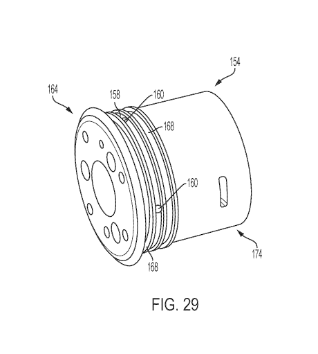
[0048] Figure 29 is a perspective view of a catheter coupler housing of the
catheter
coupler of Figure 27;
[0049] Figure 30 is a side view of the catheter coupler of Figure 27;
[0050] Figure 31A-C are cross-sectional views taken along the plane indicated
by
lines 31-31 in Figure 30 of the catheter coupler rotated to various positions;
[0051] Figure 32 is a cross-sectional view taken along the plane indicated by
lines 32-
32 in Figure 31C;
[0052] Figure 33 illustrates a cross-sectional view of the catheter coupler
shown in
Figure 32 coupled to a catheter and a pressure sensor;
[0053] Figure 34 illustrates a partial cross-sectional view of the catheter
coupler
attached to a guide sheath of a system for implanting an implantable
prosthetic device;
[0054] Figure 35 illustrates a cross-sectional view of example catheter
couplers
attached to a guide sheath and a steerable catheter of an implantable
prosthetic device;
[0055] Figure 36 is a schematic cross-sectional view of example catheters of a
delivery system for an implantable prosthetic device;
[0056] Figure 37 illustrates a cross-sectional view of an example embodiment
of a
catheter coupler;
[0057] Figure 38 is a cross-sectional view taken along the plane indicated by
lines 38-
38 in Figure 37; and
[0058] Figure 39A-D illustrate operation of the catheter coupler of Figure 38.
DETAILED DESCRIPTION
[0059] The following description refers to the accompanying drawings, which
illustrate specific embodiments of the present disclosure. Other embodiments
having different
structures and operation do not depart from the scope of the present
disclosure.
[0060] Delivery systems, apparatuses, devices, and methods for measuring
atrial
pressures during transcatheter valve repair and/or replacement procedures are
described. The
delivery system uses the same components that are used to deliver the valve
repair and/or
replacement device to measure the pressure in the atrium (or other heart
chamber). The
8
CA 03120859 2021-05-21
WO 2020/112622 PCT/US2019/062977
delivery device and method can use a pressure sensor within and/or delivered
through a
catheter and/or pusher rod or tube system that is used to administer a heart
valve therapy.
The method includes inserting the delivery system into an atria of the heart,
delivering the
valve repair and/or replacement device to the native valve (such as the mitral
valve, the
tricuspid valve, the aortic valve, or the pulmonary valve) with the delivery
system, and
measuring the pressure with the pressure sensor through an opening in one of
the catheters
and/or pushers of the delivery system. In an example embodiment, the delivery
system and
method eliminates the need to introduce a separate catheter into the heart.
For example, a
lower atrial pressure in the left atrium at the position of the delivery
device during ventricular
systole indicates less regurgitation through the mitral valve. This lower
pressure can indicate
an effectively delivered mitral valve repair device (e.g. leaflet repair or
modification device,
annulus modification device, or chordae modification or replacement device) or
replacement
mitral valve implant.
[0061] The disclosed delivery system and method does not require re-
catheterization
or other access of the left atrium to measure pressures. The disclosed
delivery system and
method also reduces the reliance on echo and other imaging to determine
whether the valve
therapy is effective.
[0062] It should be noted that various embodiments of methods for measuring
the
atrial pressure in a heart during systole and/or diastole, before, during,
and/or after the
administration of a valve repair therapy are disclosed herein, and any
combination of these
options can be made unless specifically excluded. In other words, individual
components of
the disclosed devices and methods can be combined unless mutually exclusive or
otherwise
physically impossible. Further, these methods can be performed on a living
animal or on a
simulation, such as on a cadaver, cadaver heart, simulator (e.g. with the body
parts, heart,
tissue, etc. being simulated), etc.
[0063] The example embodiments described herein rely on existing space and/or
structure within catheters and/or pusher rods or tubes used to deliver a valve
repair or
replacement device to more reliably measure atrial pressures during and/or
after procedures
for heart valve repair or replacement, including the mitral valve and the
tricuspid valve. In
some example embodiments, the delivery device for a replacement aortic valve
can include
the disclosed pressure measurement features. The valve repair therapy can be a
replacement
valve implant or a valve repair device. The atrial pressure can be in the
right atrium or the
left atrium. In some example embodiments, the delivery device can be
configured to measure
9
CA 03120859 2021-05-21
WO 2020/112622 PCT/US2019/062977
the pressure in a ventricle when the delivery device is positioned in the
ventricle. The space
can be within a catheter sheath, steering catheter, device delivery catheter,
and/or pusher rod
or tube. The space within catheters and/or pusher rod or tube can be within
the main lumen
of the catheter or pusher or can be within the wall of the catheter or pusher.
The lumen can
be reinforced with a pressure sensor catheter. The pressure sensor catheter
can be a
reinforcing sheath having its own lumen. Pressure can be measured with a
pressure sensor.
The pressure sensor can be an electric pressure sensor within a fluid-filled
lumen or in fluid
communication with the lumen. The fluid can be saline or another biocompatible
fluid. In
one example embodiment, a pressure sensor is placed in an optionally
reinforced lumen or a
port is provided for an off-the-shelf catheter to enter the body without
having to re-catheterize
the heart. The integration of the pressure sensor reduces movement and/or
vibration of the
pressure sensor to provide a more accurate pressure measurement. This more
accurate
measurement provides valuable data to the operator to determine efficacy of
the device
implanted. By providing a lumen for an off-the-shelf pressure sensor within
the main body of
the delivery catheter (for example, a steerable catheter), the noise or
vibration sensed by the
pressure sensor that occurs in the heart chamber due to the flow of blood and
the beating of
the heart is reduced and a better indication of therapy efficacy can be
obtained.
[0064] In an example embodiment, the lumen for the pressure sensor can have an
exit
opening at the tip of the outer catheter, where the implant catheter exits. In
another example
embodiment, the lumen for the pressure sensor can exit at the tip of the
steering catheter. In
an example embodiment, the lumen can exit in a flexible section of a catheter
that is
positioned in the atrium when the catheter is in the heart and positioned
towards a valve. A
steerable catheter can be used in any of the example embodiments described
herein. Other
existing catheters can also be used. The pressure sensor can be connected to a
monitoring
system through either the flush port on the handle or a separate port on the
handle designed
for the introduction of a pressure sensor, and/or direct attachment of a
pressure sensor.
During use, the pressure sensor can optionally be positioned external to the
delivery device,
such that it extends distally therefrom. In an example embodiment, the
pressure sensor can
be positioned so that a portion of the pressure sensor extends distally from
an opening. In an
example embodiment, the pressure sensor can be positioned within a lumen of
the delivery
device.
CA 03120859 2021-05-21
WO 2020/112622 PCT/US2019/062977
[0065] The pressure monitoring device can be selected to accommodate normal
positioning of a catheter, which typically includes but is not limited to,
deflection,
advancement, retraction, and/or rotation of the catheter.
[0066] As described herein, when one or more components are described as being
connected, joined, affixed, coupled, attached, or otherwise interconnected,
such
interconnection may be direct as between the components or may be indirect
such as through
the use of one or more intermediary components. Also as described herein,
reference to a
"member," "component," or "portion" shall not be limited to a single
structural member,
component, or element but can include an assembly of components, members, or
elements.
Also as described herein, the terms "substantially" and "about" are defined as
at least close to
(and includes) a given value or state (preferably within 10% of, more
preferably within 1%
of, and most preferably within 0.1% of).
[0067] Figures 1 and 2 are cutaway views of the human heart H in diastolic and
systolic phases, respectively. The right ventricle RV and left ventricle LV
are separated from
the right atrium RA and left atrium LA, respectively, by the tricuspid valve
TV and mitral
valve MV; i.e., the atrioventricular valves. Additionally, the aortic valve AV
separates the left
ventricle LV from the ascending aorta AA, and the pulmonary valve PV separates
the right
ventricle from the pulmonary artery PA. Each of these valves has flexible
leaflets (e.g.,
leaflets 20, 22 shown in Figures 4 and 5) extending inward across the
respective orifices that
come together or "coapt" in the flowstream to form the one-way, fluid-
occluding surfaces.
The methods, systems, devices, apparatuses, etc. herein are described
primarily with respect
to the mitral valve MV. Therefore, anatomical structures of the left atrium LA
and left
ventricle LV will be explained in greater detail. It should be understood that
the methods and
apparatuses described herein can also be used in repairing other native
valves, e.g., the
devices can be used in repairing the tricuspid valve TV, the aortic valve AV,
and the
pulmonary valve PV. Therefore, pressures in the right atrium RA and/or right
ventricle RV
can be measured in the same or a similar manner as the left atrium LA and/or
the left
ventricle LV.
[0068] The left atrium LA receives oxygenated blood from the lungs. During the
diastolic phase, or diastole, seen in Figure 1, the blood that was previously
collected in the
left atrium LA (during the systolic phase) moves through the mitral valve MV
and into the
left ventricle LV by expansion of the left ventricle LV. In the systolic
phase, or systole, seen
in Figure 2, the left ventricle LV contracts to force the blood through the
aortic valve AV and
11
CA 03120859 2021-05-21
WO 2020/112622 PCT/US2019/062977
ascending aorta AA into the body. During systole, the leaflets of the mitral
valve MV close to
prevent the blood from regurgitating from the left ventricle LV and back into
the left atrium
LA, and blood is collected in the left atrium from the pulmonary vein PV. In
one example
embodiment, the devices described by the present application are used to
repair the function
of a defective mitral valve MV. That is, the devices are configured to help
close the leaflets of
the mitral valve to prevent blood from regurgitating from the left ventricle
LV and back into
the left atrium LA.
[0069] Referring now to Figures 1-6, the mitral valve MV includes two
leaflets, the
anterior leaflet 20 and the posterior leaflet 22. The mitral valve MV also
includes an annulus
24, which is a variably dense fibrous ring of tissues that encircles the
leaflets 20, 22.
Referring to Figure 3, the mitral valve MV is anchored to the wall of the left
ventricle LV by
chordae tendineae 10. The chordae tendineae 10 are cord-like tendons that
connect the
papillary muscles 12 (i.e., the muscles located at the base of the chordae
tendineae and within
the walls of the left ventricle) to the leaflets 20, 22 of the mitral valve
MV. The papillary
muscles 12 serve to limit the movements of the mitral valve MV and prevent the
mitral valve
from being reverted. The mitral valve MV opens and closes in response to
pressure changes
in the left atrium LA and the left ventricle LV. The papillary muscles do not
open or close the
mitral valve MV. Rather, the papillary muscles brace the mitral valve MV
against the high
pressure needed to circulate blood throughout the body. Together the papillary
muscles and
the chordae tendineae are known as the subvalvular apparatus, which functions
to keep the
mitral valve MV from prolapsing into the left atrium LA when the mitral valve
closes.
[0070] Various disease processes can impair proper function of one or more of
the
native valves of the heart H. These disease processes include degenerative
processes (e.g.,
Barlow's Disease, fibroelastic deficiency), inflammatory processes (e.g.,
Rheumatic Heart
Disease), and infectious processes (e.g., endocarditis). In addition, damage
to the left
ventricle LV or the right ventricle RV from prior heart attacks (i.e.,
myocardial infarction
secondary to coronary artery disease) or other heart diseases (e.g.,
cardiomyopathy) can
distort a native valve's geometry, which can cause the native valve to
dysfunction. However,
the vast majority of patients undergoing valve surgery, such as surgery to the
mitral valve
MV, suffer from a degenerative disease that causes a malfunction in a leaflet
(e.g., leaflets 20,
22) of a native valve (e.g., the mitral valve MV), which results in prolapse
and regurgitation.
[0071] Generally, a native valve may malfunction in two different ways: (1)
valve
stenosis; and (2) valve regurgitation. Valve stenosis occurs when a native
valve does not open
12
CA 03120859 2021-05-21
WO 2020/112622 PCT/US2019/062977
completely and thereby causes an obstruction of blood flow. Typically, valve
stenosis results
from buildup of calcified material on the leaflets of a valve, which causes
the leaflets to
thicken and impairs the ability of the valve to fully open to permit forward
blood flow.
[0072] The second type of valve malfunction, valve regurgitation, occurs when
the
leaflets of the valve do not close completely thereby causing blood to leak
back into the prior
chamber (e.g., causing blood to leak from the left ventricle to the left
atrium). There are three
main mechanisms by which a native valve becomes regurgitant¨or
incompetent¨which
include Carpentier's type I, type II, and type III malfunctions. A Carpentier
type I
malfunction involves the dilation of the annulus such that normally
functioning leaflets are
distracted from each other and fail to form a tight seal (i.e., the leaflets
do not coapt
properly). Included in a type I mechanism malfunction are perforations of the
leaflets, as are
present in endocarditis. A Carpentier's type II malfunction involves prolapse
of one or more
leaflets of a native valve above a plane of coaptation. A Carpentier's type
III malfunction
involves restriction of the motion of one or more leaflets of a native valve
such that the
leaflets are abnormally constrained below the plane of the annulus. Leaflet
restriction can be
caused by rheumatic disease (Ma) or dilation of a ventricle (Tub).
[0073] Referring to Figure 4, when a healthy mitral valve MV is in a closed
position,
the anterior leaflet 20 and the posterior leaflet 22 coapt, which prevents
blood from leaking
from the left ventricle LV to the left atrium LA. Referring to Figure 5,
regurgitation occurs
when the anterior leaflet 20 and/or the posterior leaflet 22 of the mitral
valve MV is displaced
into the left atrium LA during systole. This failure to coapt causes a gap 26
between the
anterior leaflet 20 and the posterior leaflet 22, which allows blood to flow
back into the left
atrium LA from the left ventricle LV during systole. As set forth above, there
are several
different ways that a leaflet (e.g. leaflets 20, 22 of mitral valve MV) may
malfunction, which
can thereby lead to regurgitation.
[0074] Referring to Figure 6, in certain situations, the mitral valve MV of a
patient
can have a wide gap 26 between the anterior leaflet 20 and the posterior
leaflet 22 when the
mitral valve is in a closed position (i.e., during the systolic phase). For
example, the gap 26
can have a width W between about 2.5 mm and about 17.5 mm, such as between
about 5 mm
and about 15 mm, such as between about 7.5 mm and about 12.5 mm, such as about
10 mm.
In some situations, the gap 26 can have a width W greater than 15 mm. In any
of the above-
mentioned situations, a valve repair device is desired that is capable of
engaging the anterior
13
CA 03120859 2021-05-21
WO 2020/112622 PCT/US2019/062977
leaflet 20 and the posterior leaflet 22 to close the gap 26 and prevent
regurgitation of blood
through the mitral valve MV.
[0075] When mitral valve regurgitation occurs, blood enters the left atrium
from the
left ventricle during systole. In a healthy heart, blood should only enter the
left atrium from
the pulmonary veins, not the left ventricle. The left atrial pressure then
increases above the
pressure it should be. For example, normal left atrial pressure can range from
about 5 to about
15 mmHg. But when mitral valve regurgitation occurs, left atrial pressure
could increase to a
higher pressure, for example, 25 mmHg. With mitral valve regurgitation, the
left atrial
pressure is increased overall throughout the cardiac cycle and is most
noticeable at the end of
systole.
[0076] Although stenosis or regurgitation can affect any valve, stenosis is
predominantly found to affect either the aortic valve AV or the pulmonary
valve PV, and
regurgitation is predominantly found to affect either the mitral valve MV or
the tricuspid
valve TV. Both valve stenosis and valve regurgitation increase the workload of
the heart H
and may lead to very serious conditions if left un-treated; such as
endocarditis, congestive
heart failure, permanent heart damage, cardiac arrest, and ultimately death.
Because the left
side of the heart (i.e., the left atrium LA, the left ventricle LV, the mitral
valve MV, and the
aortic valve AV) is primarily responsible for circulating the flow of blood
throughout the
body, malfunction of the mitral valve MV or the aortic valve AV is
particularly problematic
and often life threatening. Accordingly, because of the substantially higher
pressures on the
left side of the heart, dysfunction of the mitral valve MV or the aortic valve
AV is much more
problematic.
[0077] Malfunctioning native heart valves may either be repaired or replaced.
Repair
typically involves the preservation and correction of the patient's native
valve. Replacement
typically involves replacing the patient's native valve with a biological or
mechanical
substitute. Typically, the aortic valve AV and pulmonary valve PV are more
prone to stenosis.
Because stenotic damage sustained by the leaflets is irreversible, the most
conventional
treatments for a stenotic aortic valve or stenotic pulmonary valve are removal
and
replacement of the valve with a surgically implanted heart valve, or
displacement of the valve
with a transcatheter heart valve.
[0078] Referring now to Figures 8-14, a schematically illustrated implantable
prosthetic device 100 is shown in various stages of deployment. The prosthetic
device 100
and associated systems, methods, etc. are described in more detail in
International
14
CA 03120859 2021-05-21
WO 2020/112622 PCT/US2019/062977
Application Nos. PCT/US2018/028189 and PCT/US2019/055320, the disclosures of
which
are incorporated herein by reference in their entirety. The device 100 can
include any other
features for an implantable prosthetic device discussed in the present
application, and the
device 100 can be positioned to engage valve tissue (e.g., leaflets 20, 22) as
part of any
suitable valve repair system (e.g., any valve repair system disclosed in the
present
application).
[0079] The device 100 is deployed and can include a coaptation portion or
coaption
portion 140 and an anchor portion 106. The device 100 can be deployed from a
delivery
sheath and/or can be deployed by a pusher tube or rod 81. The coaption portion
140 of the
device 100 includes a coaption element 110 that is adapted to be implanted
between the
leaflets of the native valve (e.g., native mitral valve, native tricuspid
valve, etc.) and is
slidably attached to an actuation member or actuation element 112 (e.g., a
wire, shaft, rod,
line, suture, tether, etc.). The anchor portion 106 is actuatable between open
and closed
conditions and can take a wide variety of forms, such as, for example,
paddles, gripping
elements, or the like. Actuation of the actuation element 112 opens and closes
the anchor
portion 106 of the device 100 to grasp the native valve leaflets during
implantation. The
actuation element 112 can take a wide variety of different forms. For example,
the actuation
element can be threaded such that rotation of the actuation element moves the
anchor portion
106 relative to the coaption portion 140. Or, the actuation element may be
unthreaded, such
that pushing or pulling the actuation element 112 moves the anchor portion 106
relative to the
coaption portion 140.
[0080] The anchor portion 106 of the device 100 includes outer paddles 120 and
inner
paddles 122 that are connected between a cap 114 and the coaption element 110
by portions
124, 126, 128. The portions 124, 126, 128 can be jointed and/or flexible to
move between all
of the positions described below. The interconnection of the outer paddles
120, the inner
paddles 122, the coaption element 110, and the cap 114 by the portions 124,
126, and 128 can
constrain the device to the positions and movements illustrated herein.
[0081] The actuation member or actuation element 112 extends through the
delivery
sheath and/or the pusher tube/rod and the coaption element 110 to the cap 114
at the distal
connection of the anchor portion 106. Extending and retracting the actuation
element 112
increases and decreases the spacing between the coaption element 110 and the
cap 114,
respectively. A collar 115 removably attaches the coaption element 110 to the
pusher tube or
rod 81 so that the actuation element 112 slides through the collar 115 and
coaption element
CA 03120859 2021-05-21
WO 2020/112622 PCT/US2019/062977
110 during actuation to open and close the paddles 120, 122 of the anchor
portion 106. After
the device 100 is connected to valve tissue, if the device 100 needs to be
removed from the
valve tissue, a retrieval device can be used to connect to the collar 115 such
that the actuation
wire can extend through the collar 115 and the coaption element 110 to engage
the anchor
portion 106 to open the paddles 120, 122 and remove the device 100 from the
valve tissue.
Examples of retrieval devices that could be used are shown in PCT Application
No.
PCT/US2019/062391 filed November 20, 2019, which is incorporated herein by
reference in
its entirety.
[0082] Referring now to Figure 8, the device 100 is shown in an elongated or
fully
open condition for deployment from the delivery sheath. The device 100 is
loaded in the
delivery sheath in the fully open position, because the fully open position
takes up the least
space and allows the smallest catheter to be used (or the largest implantable
device 100 to be
used for a given catheter size). In the elongated condition the cap 114 is
spaced apart from the
coaption element 110 such that the paddles 120, 122 of the anchor portion 106
are fully
extended. In some embodiments, an angle formed between the interior of the
outer and inner
paddles 120, 122 is approximately 180 degrees. The barbed clasps 130 are kept
in a closed
condition during deployment through the delivery sheath 83 so that the barbs
136 (Figs. 9 and
11) do not catch or damage the sheath or tissue in the patient's heart.
[0083] Referring now to Figure 9, the device 100 is shown in an elongated
detangling
condition, similar to Figure 8, but with the barbed clasps 130 in a fully open
position, ranging
from about 140 degrees to about 200 degrees, such as about 170 degrees to
about 190
degrees, or about 180 degrees between fixed and moveable portions of the
barbed clasps 130.
Fully opening the paddles 120, 122 and the clasps 130 has been found to
improve ease of
detanglement from anatomy of the patient during implantation of the device
100.
[0084] Referring now to Figure 10, the device 100 is shown in a shortened or
fully
closed condition. The compact size of the device 100 in the shortened
condition allows for
easier maneuvering and placement within the heart. To move the device 100 from
the
elongated condition to the shortened condition, the actuation member or
actuation element
112 is retracted to pull the cap 114 towards the coaption element 110. The
joints or flexible
connections 126 between the outer paddle 120 and inner paddle 122 are
constrained in
movement such that compression forces acting on the outer paddle 120 from the
cap 114
being retracted towards the coaption element 110 cause the paddles 120, 122 or
gripping
elements to move radially outward. During movement from the open to closed
position, the
16
CA 03120859 2021-05-21
WO 2020/112622 PCT/US2019/062977
outer paddles 120 maintain an acute angle with the actuation element 112. The
outer paddles
120 can optionally be biased toward a closed position. The inner paddles 122
during the same
motion move through a considerably larger angle as they are oriented away from
the coaption
element 110 in the open condition and collapse along the sides of the coaption
element 110 in
the closed condition. In some embodiments, the inner paddles 122 are thinner
and/or
narrower than the outer paddles 120, and the joint or flexible portions 126,
128 connected to
the inner paddles 122 can be thinner and/or more flexible. For example, this
increased
flexibility can allow more movement than the joint or flexible portion 124
connecting the
outer paddle 124 to the cap 114. In some embodiments, the outer paddles 120
are narrower
than the inner paddles 122. The joint or flexible portions 126, 128 connected
to the inner
paddles 122 can be more flexible, for example, to allow more movement than the
joint or
flexible portion 124 connecting the outer paddle 124 to the cap 114. In some
embodiments,
the inner paddles 122 can be the same width or substantially the same width as
the outer
paddles.
[0085] Referring now to Figures 11-13, the device 100 is shown in a partially
open,
grasp-ready condition. To transition from the fully closed to the partially
open condition, the
actuation member or actuation element 112 (e.g., an actuation wire, actuation
shaft, etc.) is
extended to push the cap 114 away from the coaption element 110, thereby
pulling on the
outer paddles 120, which in turn pulls on the inner paddles 122, causing the
anchor portion
106 to partially unfold. The actuation lines 116 are also retracted to open
the clasps 130 so
that the leaflets can be grasped. In the example illustrated by Figure 11, the
pair of inner and
outer paddles 122, 120 are moved in unison, rather than independently, by a
single actuation
element 112. Also, the positions of the clasps 130 are dependent on the
positions of the
paddles 122, 120. For example, referring to Figure 10 closing the paddles 122,
120 also
closes the clasps. In certain embodiments, the paddles 120, 122 can be
independently
controllable. For example, the device 100 can have two actuation elements and
two
independent caps, such that one independent wire and cap are used to control
one paddle, and
the other independent wire and cap are used to control the other paddle.
[0086] Referring now to Figure 12, one of the actuation lines 116 is extended
to allow
one of the clasps 130 to close. Referring now to Figure 13, the other
actuation line 116 is
extended to allow the other clasp 130 to close. Either or both of the
actuation lines 116 may
be repeatedly actuated to repeatedly open and close the barbed clasps 130.
17
CA 03120859 2021-05-21
WO 2020/112622 PCT/US2019/062977
[0087] Referring now to Figure 14, the device 100 is shown in a fully closed
and
deployed condition. The pusher tube or rod 81 and actuation element 112 are
retracted and
the paddles 120, 122 and clasps 130 remain in a fully closed position. Once
deployed, the
device 100 can be maintained in the fully closed position with a mechanical
latch or can be
biased to remain closed through the use of spring materials, such as steel,
other metals,
plastics, composites, etc. or shape-memory alloys such as Nitinol. For
example, the jointed or
flexible portions 124, 126, 128, 138, and/or the inner and outer paddles 122,
and/or an
additional biasing component can be formed of metals such as steel or shape-
memory alloy,
such as Nitinol¨produced in a wire, sheet, tubing, or laser sintered
powder¨and are biased
to hold the outer paddles 120 closed around the coaption element 110 and the
barbed clasps
130 pinched around native leaflets. Similarly, the fixed and moveable arms
132, 134 of the
barbed clasps 130 are biased to pinch the leaflets. In some embodiments, the
joint portions
124, 126, 128, 138, and/or the inner and outer paddles 122, and/or an
additional biasing
component can be formed of any other suitably elastic material, such as a
metal or polymer
material, to maintain the device in the closed condition after implantation.
[0088] Referring now to Figures 15-20, the implantable device 100 of Figures 8-
14
is shown being delivered and implanted within the native mitral valve MV of
the heart H.
Referring now to Figure 15, the outer catheter or sheath 83 is inserted into
the left atrium LA
through the septum and the device 100 is deployed from the outer catheter in
the fully open
condition. The actuation element 112 is then retracted to move the device 100
into the fully
closed condition shown in Figure 16. As can be seen in Figure 17, the device
100 is moved
into position within the mitral valve MV into the ventricle LV and partially
opened so that the
leaflets 20, 22 can be grasped. The steerable catheter 82 is extended out past
the distal end of
the outer catheter 83, and the pusher tube or rod 81 is extended out past the
distal end of the
steerable catheter. Referring now to Figure 18, an actuation line 116 is
extended to close one
of the clasps 130, capturing a leaflet 20. Figure 19 shows the other actuation
line 116 being
then extended to close the other clasp 130, capturing the remaining leaflet
22. Lastly, as can
be seen in Figure 20, the pusher tube or rod 81 and actuation element 112 and
actuation lines
116 are then retracted and the device 100 is fully closed and deployed in the
native mitral
valve MV.
[0089] Referring now to Figures 21A-21D, schematics of various example
embodiments of a delivery system 80 for delivering an implant into the heart
are illustrated.
The steerable catheter 82 and the outer catheter 83 each have a central lumen
which can be
18
CA 03120859 2021-05-21
WO 2020/112622 PCT/US2019/062977
considered a delivery lumen. Referring now to Figure 21A, a schematic of an
example
embodiment of a delivery system 80 for delivering an implant into the heart,
is illustrated.
This example embodiment is configured to have a pressure sensor within one of
the delivery
device's catheters or pusher. The delivery system 80 can include an implant
delivering
pusher tube or rod 81, a steerable catheter 82, and an outer catheter or
sleeve 83. The
steerable catheter 82 can extend out from the outer catheter's distal end 87,
and the pusher
tube or rod can extend outward from the steerable catheter's distal end 85. In
one example
embodiment, a valve implant, valve repair device, or other therapy is pushed
out an end 85 of
the steerable catheter 82 by a distal end 86 of the pusher tube or rod 81. In
another example
embodiment, the valve repair device or other therapy is initially positioned
distally from the
end 85 of the steerable catheter, inside the outer catheter 83 (i.e. not
inside the steerable
catheter). This allows the device to have a larger size, because it does not
need to fit inside
the steerable catheter 82. A pressure sensor P can be disposed in a lumen
within the wall of
any of the steerable catheter 82, the outer sleeve or catheter 83, the pusher
tube or rod 81, the
actuation rod 112, or in a separate catheter disposed in a space between any
two of the
steerable catheter 82, the outer sleeve or catheter 83, the pusher tube or rod
81, and the
actuation rod 112.
[0090] In the example embodiment of Figure 21A, a pressure sensor lumen 88 can
run along the length of the steerable catheter 82. The pressure sensor lumen
88 can be
formed in the steerable catheter or can be a lumen of a separate catheter
disposed in the
steerable catheter. In the embodiment illustrated in Figure 21A, the open
distal end 89 of the
lumen 88 can be along the length of the steerable catheter (i.e. not at the
end 85 of the
steerable catheter). The pressure sensor P can be at the port 89 to directly
measure pressure
of the blood. Reference character P' represents another example embodiment
where the
pressure sensor P' is upstream of the port 89, such that the pressure of the
blood in the heart is
measured indirectly through fluid in the pressure sensor lumen 88. The
pressure sensor port
89 can be anywhere along the catheter in a location that is typically
positioned in an atrium of
the heart during a valve treatment procedure.
[0091] Referring now to Figure 21B, another example embodiment of a delivery
system 80 for measuring intra-atrial pressure during delivery of a heart valve
implant or
repair without requiring a separately introduced catheter is depicted. The
example
embodiment of Figure 21B is similar to Figure 21A, except that the open distal
end or
pressure port 89 for the pressure monitoring lumen 88 is at the distal end 85
of the steerable
19
CA 03120859 2021-05-21
WO 2020/112622 PCT/US2019/062977
catheter 82. In the embodiment illustrated by Figure 21B, the pressure sensor
P is positioned
at the port 89 to directly measure the pressure of the blood.
[0092] Referring now to Figure 21C, another example embodiment of a delivery
system 80 for measuring intra-atrial pressure during delivery of a heart valve
implant or
repair without requiring a separately introduced catheter is depicted. In this
example
embodiment, the pressure sensor lumen 88 is optionally embedded in the wall of
the steerable
catheter 82. In the illustrated embodiment, the pressure sensor P is located
proximal to the
port 89 of the pressure sensor lumen. However, the pressure sensor can be at
the port 89 of
the steerable catheter as mentioned above.
[0093] Referring now to Figure 21D, the pressure sensor lumen 88 can be
embedded
in the wall or disposed inside of the pusher rod or tube 81 and can have an
open distal end or
port 89 at the distal end of the pusher tube or rod. In some embodiments the
opening or port
89 can be at any location along the pusher tube or rod 81 that extends from
the steerable
catheter 82. In example embodiments, the pressure sensor lumen can be fully
embedded
within a wall of the steerable catheter or pusher tube or rod, or it can be
partially embedded in
the wall and protrude toward a center of the catheter's lumen. In some
embodiments, the
lumens described herein can be defined by another catheter, which can be a
pressure
monitoring catheter, extending inside a lumen in the pusher 81. In some
embodiments, a
pressure measuring catheter with a lumen is in the space between the steerable
catheter and
pusher tube 81. In this embodiment, the pressure measuring catheter can
optionally be fixed
to the interior wall of the steerable catheter. In the example illustrated by
Figure 21D, the
pressure sensor is positioned at the port 89. However, in some example
embodiments, the
pressure sensor P is positioned upstream of the port.
[0094] Referring now to Figures 22A and 22B, a cutaway view of the human heart
H
is illustrated in diastole and systole, respectively. In the left atrium LA is
a delivery system
80 with a pressure sensor P within it. The pressure sensor can record a
pressure in the left
atrium during diastole, and another pressure in the left atrium during
systole. The pressure
sensor is not limited to measuring pressure in the left atrium and can also be
used in the right
atrium or one of the ventricles. Figures 8-20 illustrate an example of one of
the many
different types of valve therapies that can be delivered by the delivery
system 80. The valve
therapy is not shown in Figures 22A and 22B to simplify these two figures. A
typical valve
therapy comprises positioning and/or deploying a valve implant or valve repair
device (see
Figures 8-20) in the native valve of the heart, which can be the mitral valve
or the tricuspid
CA 03120859 2021-05-21
WO 2020/112622 PCT/US2019/062977
valve. However, the valve therapy is not limited to a replacement valve or a
valve repair
device; it can also be a docking coil with a valve implant, sutures or chordae
replacement
devices, annuloplasty devices, and/or other implants used to correct the
function of a heart
native valve. The delivery system can use a transvascular technique such as a
trans-septal
technique, but is not limited thereto; the delivery system can be delivered to
an atria of the
heart by any presently known and future-developed technique. When the valve
implant or
repair is in place, the pressure can be measured again to determine if a
therapeutic effect has
occurred, i.e., if a regurgitation or other heart valve defect has been
repaired and/or improved.
[0095] Referring now to Figures 23A-23C, schematics of example embodiments of
a
delivery system for the delivery of a valve implant or repair are illustrated.
Figure 23A
illustrates an end view, having an implant pusher or catheter 81, a steerable
catheter 82, and
an outer sheath 83. The steerable catheter can have an integrated lumen,
typically used for
steering cables (not shown) that extend along at least a length of the
steerable catheter. In
some embodiments, an additional pressure sensor catheter 102 is positioned in
the lumen 104
of the steerable catheter, adjacent to the interior surface of the steerable
catheter wall. The
pressure sensor catheter can have a lumen 105. The pressure sensor catheter
105 can have a
pressure sensor in it (Figures 23B-C) or can be filled with a biocompatible
fluid to measure
the pressure (Figure 23D). The pressure sensor catheter shown here is not
limited to this
location but can be positioned anywhere in the space between the pusher tube
and the
steerable catheter, such that sufficient flexibility of the steerable catheter
82 and delivery
system 80 as a whole can still be achieved so that the valve implant or repair
device can be
implanted in a desired location.
[0096] Figure 23B illustrates a cross section of the delivery system 80 taken
along
line A-A. In Figure 23B the opening at the end 86 of the pusher tube 81 for
the actuation rod
112 (See Figure 10) is not shown, because the opening is offset from the cross-
section plane
A-A. In Figure 23B, a pressure sensor P is positioned inside a distal end of
the pressure
sensor catheter lumen 105 and connected to a communication link 103, such as a
wire or pair
of wires. The communication link 103 can extend out a proximal end (not shown,
outside the
patient) to allow the pressure sensed at the pressure sensor P to be monitored
outside the
patient by monitoring equipment.
[0097] The pressure sensor P can be one of any of various pressure sensors.
For
example, the pressure sensor can be a piezo-electric sensor, a pressure-
sensing probe, or a
barometric pressure sensor. In the example embodiments described herein, the
pressure
21
CA 03120859 2021-05-21
WO 2020/112622 PCT/US2019/062977
sensor can be an electric pressure sensor that measures the pressure of fluid,
which can be the
blood in the left atrium of the heart or can be the pressure of the fluid,
which can be a saline
solution, in the pressure sensor lumen. The pressure sensor can be positioned
to extend
distally out of the end of the pressure sensor lumen 105 or the pressure
sensor can be fully
within the pressure lumen. The pressure sensor P can be positioned at any
position along the
length of the pressure sensor lumen 105. For example, the pressure sensor P
can be flush or
substantially flush with the port 89 or the pressure sensor P can be spaced
proximally away
from the port inside the pressure sensor lumen. In the illustrated example of
Figure 23B, the
pressure sensor lumen distal end or port 89 is flush with the distal end of
the steerable
catheter. In some embodiments, the port 89 is spaced apart from the end of the
steerable
catheter. In some embodiments, the pressure sensor can be embedded in the wall
of the
catheter having the pressure sensor lumen or pressure sensor catheter lumen.
[0098] The pressure sensor catheter 102 can be fixedly connected to the
interior wall
of the steerable catheter by any means to secure catheter tubing together. The
steerable
catheter extends distally beyond the outer catheter 83. The pusher tube or rod
extends even
farther distally from the distal end of the steerable catheter. As the pusher
tube or rod is what
delivers a valve implant or repair device to the valve, it can be extendable
from the end of the
steerable catheter. The pusher tube or rod can extend past the distal end of
the steerable
catheter so that it can reach through the mitral valve (or tricuspid valve)
towards the left
ventricle to allow an operator to properly position the valve implant or
repair device during
its deployment. The pressure sensor can provide accurate measurements of
pressure in the
atrium when the delivery system is inserted such that the steerable catheter
is still in the
atrium, and not at the valve annulus or below, in the ventricle. In the
example embodiment
illustrated in Figure 23B, the pressure sensor P is located at the distal end
of the lumen and
can detect the pressure of the fluid within the lumen at the distal end of the
lumen. Because
the distal end of the lumen can be open, the pressure exerted by the fluid in
the lumen on the
sensor will be the same as the pressure of the blood in the left atrium,
because the pressure of
the blood in the left atrium will push on the fluid in the lumen.
[0099] By having the pressure sensor in a pressure sensor lumen that fits
within
existing space in the steerable catheter main lumen, the number of
catheterizations of the
heart needed to implant a valve device and record pressures to measure its
efficacy is
reduced, thereby reducing noise that could affect pressure measurements.
22
CA 03120859 2021-05-21
WO 2020/112622 PCT/US2019/062977
[0100] Referring to Figure 23C, a schematic of a cross section taken along
line A-A
of Figure 23A, of an example embodiment of a delivery system 80 with a
pressure sensor is
illustrated. In Figure 23C the opening at the end 86 of the pusher tube 81 for
the actuation
rod 112 (See Figure 10) is not shown, because the opening is offset from the
cross-section
plane A-A. In Figure 23C, the pressure sensor is in accordance with an example
embodiment. In this embodiment, the pressure sensor is a fluid filled pressure
sensor P
located more proximal than that of Figure 23A. In Figure 23C, the pressure
sensor catheter
within the lumen of the steerable catheter is filled with a fluid. The fluid
can be saline or
another biocompatible fluid. Because this pressure sensor catheter is within a
delivery
system that is already inserted in the heart, an additional catheter is not
required to be inserted
to take a pressure measurement. The noise in the atrium is reduced by the
separate lumen
102 and the pressure measurement can provide better feedback to the operator
regarding the
efficacy of the valve implant or repair being administered. Noise reduced is
that which could
otherwise be caused by movement, increased pressure, and additional
disturbances to the
blood flow in the left atrium, that could be caused by, for example, another
catheter in the left
atrium.
[0101] Referring now to Figure 23D, a schematic of a cross-section taken along
line
A-A of Figure 23A, of another example embodiment of a delivery system 80 for
measuring
pressure in a heart chamber is illustrated. In Figure 23DF the opening at the
end 86 of the
pusher tube 81 for the actuation rod 112 (See Figure 10) is not shown, because
the opening is
offset from the cross-section plane A-A. In Figure 23D, the pressure sensor
catheter lumen
105 is filled with a fluid, which can be saline, and the pressure of the fluid
is measured by a
monitoring system. The monitoring system can be connected to the lumen 105
and/or fluid
therein through either the flush port on the handle or a separate port on the
handle designed
for the introduction of a pressure sensor, as with the other example
embodiments described
herein. In this embodiment, there is no pressure sensor P positioned within
the lumen 105 to
measure the pressure, electronically or otherwise. Instead, the pressure of
the fluid in the
lumen 105 is measured with the monitoring system. The pressure of the fluid in
the lumen
remains consistent throughout the lumen, and because the distal end of the
lumen is open to
the left atrium, the pressure exerted by the fluid in the lumen will be the
same as the pressure
of the blood in the left atrium.
[0102] The pressure can be measured using fluid instead of a pressure sensor P
in any
of the example embodiments described herein. This includes the embodiments
having a
23
CA 03120859 2021-05-21
WO 2020/112622 PCT/US2019/062977
pressure sensor lumen 88 embedded within a wall of a catheter, and also
includes some
embodiments having a pressure sensor catheter lumen 105. The pressure can be
measured in
the same way as it is measured with regard to the embodiment of Figure 23D.
[0103] An example embodiment can be to have the pressure sensor P embedded in
the
wall of any catheter that surrounds a fluid filled lumen. The catheter wall
can be that of the
pusher tube or rod 81, the steerable catheter 82, the pressure sensor catheter
102, and/or the
actuation rod 112 (See Figure 10). The fluid filled lumen can be the pressure
catheter lumen
105 or the pressure sensor lumen 88.
[0104] Referring now to Figures 24A-24C, a schematic of an example embodiment
of
a delivery system for the delivery of a valve device is illustrated. Figure
24A illustrates an
end view, having a pusher tube or rod 81, a steerable catheter 82, and an
outer sheath 83. In
this embodiment, an additional pressure sensor lumen 88 is integrated in the
wall of the
steerable catheter. The pressure sensor lumen can bump out into the central
lumen 104 of the
steerable catheter as shown in Figure 24A, or it can be flush within the wall
of the steerable
catheter. The integrated pressure sensor lumen can be adjacent to the
integrated steerable
catheter lumen as illustrated in Figure 24A, or it can be spaced apart from
it.
[0105] Figure 24B illustrates a cross section of the delivery system 80 taken
along
line B-B. In Figure 24B the opening at the end 86 of the pusher tube 81 for
the actuation rod
112 (See Figure 10) is not shown, because the opening is offset from the cross-
section plane
B-B. In Figure 24B, a pressure sensor P is positioned inside a distal end of
the pressure sensor
lumen 88 of the steerable catheter 82 and is held in place at least by a
connecting link 103.
As with the example embodiment described herein with respect to Figures 23A-
23B, the
pressure sensor can be an electric pressure sensor (or other type of pressure
sensor). The
pressure sensor can be positioned to extend distally out of the end of the
pressure sensor
lumen 88 or it can be fully within the pressure lumen. The pressure sensor
lumen distal end
89 can be flush with the distal end of the steerable catheter in this example
embodiment but is
not limited to such a length. The steerable catheter extends distally beyond
the outer catheter
83. The pusher tube or rod extends even farther distally from the distal end
of the steerable
catheter. As the pusher tube or rod is what delivers a valve implant or repair
device to the
valve, it can be extendable from the end of the steerable catheter. The pusher
tube or rod can
extend past the distal end of the steerable catheter so that it can reach
through the mitral valve
(or tricuspid valve) towards the left ventricle to allow an operator to
properly position the
valve implant or valve repair device during its deployment. The pressure
sensor can provide
24
CA 03120859 2021-05-21
WO 2020/112622 PCT/US2019/062977
accurate measurements in the atrium when the delivery system is inserted such
that the
steerable catheter is still in the atrium, and not at the valve annulus or in
the ventricle.
[0106] Referring to Figure 24C, a schematic of a cross section taken along
line B-B
of Figure 24A, of another example embodiment of a delivery system 80 with a
pressure
sensor is illustrated. In Figure 24C the opening at the end 86 of the pusher
tube 81 for the
actuation rod 112 (See Figure 10) is not shown, because the opening is offset
from the cross-
section plane B-B. In Figure 24C, the pressure sensor is in accordance with
another example
embodiment. In this embodiment, the pressure sensor is a fluid filled pressure
sensor lumen,
having a pressure sensor P at a more proximal location within the pressure
sensor lumen,
which is described in greater detail above. In Figure 24C, the lumen 104 of
the steerable
catheter is filled with a fluid, as described above. Because this example
embodiment of a
pressure sensor catheter is within a delivery system that is already implanted
in the heart, an
additional catheter is not required to be inserted to take a pressure
measurement. Therefore,
the noise in the atrium is reduced and the pressure measurement can provide
better feedback
to the operator regarding the efficacy of the valve implant or repair device
being
administered.
[0107] Referring now to Figures 25A-25C, schematics of an example embodiment
of
a delivery system for the delivery of a valve implant or repair device are
illustrated. Figure
25A illustrates an end view, having a pusher tube or rod 81, a steerable
catheter 82, and an
outer sheath 83. The steerable catheter can have an integrated lumen as
described above.
The steerable catheter can have another integrated lumen 88. In this
embodiment, an
additional pressure sensor catheter 102 is positioned in the additional lumen
88 of the
steerable catheter, adjacent to the interior surface of the steerable catheter
wall. The pressure
sensor catheter can have its own lumen 105. The pressure sensor lumen can bump
out into
the central lumen 104 of the steerable catheter as shown in Figure 25A, or it
can be flush
within the wall of the steerable catheter. The integrated pressure sensor
lumen can be
adjacent to the integrated steerable catheter lumen as illustrated in Figure
25A, or it can be
spaced apart from it. The additional lumen within the steerable catheter shown
in Figure 25A
is not limited to the locations described herein but can be positioned
anywhere at least
partially embedded in the wall of the steerable catheter, such that sufficient
flexibility of the
steerable catheter and delivery system as a whole can still be achieved so
that the valve
therapy can be implanted in a desired location.
CA 03120859 2021-05-21
WO 2020/112622 PCT/US2019/062977
[0108] Figure 25B illustrates a cross section of the delivery system 80 taken
along
line C-C. In Figure 25B the opening at the end 86 of the pusher tube 81 for
the actuation rod
112 (See Figure 10) is not shown, because the opening is offset from the cross-
section plane
C-C. In Figure 25B, a pressure sensor P is positioned inside a distal end of
the pressure sensor
lumen 105 and can be held in place by an optional connecting link 103. The
pressure sensor
can be an electric pressure sensor or other sensor. The pressure sensor can be
positioned to
extend distally out of the end of the pressure sensor catheter lumen 105 or it
can be fully
within the pressure lumen. The pressure sensor catheter lumen distal end 89 is
flush with the
distal end of the steerable catheter in this example embodiment but is not
limited to such a
length. The pressure sensor catheter 102 can be fixedly connected to the
interior wall of the
steerable catheter lumen 104 by any means to secure catheter tubing together.
In another
embodiment, the pressure sensor catheter can be slidably positioned in the
steerable catheter
lumen 104. The steerable catheter extends distally beyond the outer catheter
83. The pusher
tube or rod 86 extends even farther distally from the distal end of the
steerable catheter. As
the pusher tube or rod is what delivers a valve implant or repair to the
valve, it can be
extendable from the end of the steerable catheter. The pusher tube or rod can
extend past the
distal end of the steerable catheter so that it can reach through the mitral
valve (towards the
left ventricle) to allow an operator to properly position the valve implant or
repair during its
deployment. The pressure sensor can provide accurate measurements in the
atrium when the
delivery system is inserted such that the steerable catheter is still in the
atrium, and not at the
valve annulus or in the ventricle.
[0109] Referring to Figure 25C, a schematic of a cross section taken along
line C-C
of Figure 25A, of another example embodiment of a delivery system 80 with a
pressure
sensor is illustrated. In Figure 25C the opening at the end 86 of the pusher
tube 81 for the
actuation rod 112 (See Figure 10) is not shown, because the opening is offset
from the cross-
section plane C-C. In this example embodiment, the pressure sensor is a fluid
filled pressure
sensor lumen, having a pressure sensor P at a more proximal location within
the pressure
sensor lumen, which is described in greater detail above. Because this example
embodiment
of a pressure sensor catheter is within a delivery system that is already
implanted in the heart,
an additional catheter is not required to be inserted to take a pressure
measurement.
Therefore, the noise in the atrium is reduced and the pressure measurement can
provide more
reliable feedback to the operator regarding the efficacy of the valve implant
or repair being
administered.
26
CA 03120859 2021-05-21
WO 2020/112622 PCT/US2019/062977
[0110] Referring now to Figures 26A-26C, schematics of an example embodiment
of
a delivery system for the delivery of a valve implant or repair device are
illustrated. Figure
26A illustrates an end view, having a pusher rod or tube 81, a steerable
catheter 82, and an
outer sheath 83. In Figure 26B the opening at the end 86 of the pusher tube 81
for the
actuation rod 112 (See Figure 10) is not shown, because the opening is offset
from the cross-
section plane C-C. In this embodiment, an additional pressure sensor lumen 88
is integrated
in the wall of the pusher rod or tube 81. The pressure sensor lumen can be
flush within the
wall of the pusher rod or tube 81 as shown in Figure 26A, or it can bump out
into a central
lumen of the pusher tube or rod 81.
[0111] Figure 26B illustrates a cross section of the delivery system 80 taken
along
line D-D. In Figure 24B, a pressure sensor P is positioned inside a distal end
of the pressure
sensor lumen 88 of the pusher rod or tube 81 and can be held in place at least
by a connecting
link 103. As with the example embodiment described herein with respect to
Figures 23A-
23B, the pressure sensor can be positioned extending distally out of the end
of the pressure
sensor lumen 88 or it can be fully within the pressure sensor lumen. The
pressure sensor
lumen can be filled with a biocompatible fluid such as saline. The pressure
sensor lumen
distal end 89 is flush with the distal end of the pusher rod or tube 81 in
this example
embodiment but is not limited to such a length. The steerable catheter extends
distally
beyond the outer catheter 83. The pusher tube or rod extends even farther
distally from the
distal end of the steerable catheter. As the pusher tube or rod is what
delivers a valve implant
or repair device to the valve, it can be extendable from the end of the
steerable catheter. The
pusher tube or rod can extend past the distal end of the steerable catheter so
that it can reach
through the mitral valve (or tricuspid valve) towards the left ventricle to
allow an operator to
properly position the valve implant or repair during its deployment. As with
the other
example embodiments described herein, the pressure sensor can measure an
accurate atrial
pressure when its opening to the exterior of the delivery system is located in
the atrium. The
pressure sensor can provide accurate measurements in the atrium when the
delivery system is
inserted such that the pusher tube or rod is still in the atrium, and not at
the valve annulus or
in the ventricle.
[0112] As with the other integrated lumen embodiments, having the pressure
sensor
in a pressure sensor lumen integrated within the pusher rod or tube 81, the
number of
catheterizations of the heart needed to implant a valve implant or repair and
record pressures
27
CA 03120859 2021-05-21
WO 2020/112622 PCT/US2019/062977
to measure its efficacy is reduced, thereby reducing noise that could affect
pressure
measurements.
[0113] Referring to Figure 26C, a schematic of a cross section taken along
line D-D
of Figure 26A, of another example embodiment of a delivery system 80 with a
pressure
sensor is illustrated. In Figure 26C the opening at the end 86 of the pusher
tube 81 for the
actuation rod 112 (See Figure 10) is not shown, because the opening is offset
from the cross-
section plane C-C. In Figure 26C, the pressure sensor is in accordance with
another example
embodiment. In Figure 26C, the pressure sensor is a fluid filled pressure
sensor lumen,
having a pressure sensor P at a more proximal location within the pressure
sensor lumen,
which is described in greater detail above. Because this pressure sensor
catheter is within a
delivery system that is already implanted in the heart, an additional catheter
is not required to
be inserted to take a pressure measurement. Therefore, the noise in the atrium
is reduced and
the pressure measurement can provide better feedback to the operator regarding
the efficacy
of the valve implant or repair device.
[0114] In an example embodiment having a pressure sensor catheter within a
steerable catheter lumen as illustrated in Figures 23A-23C, the pressure can
be measured
according to the following method. In Figure 23B, the pressure sensor can be
an electric
pressure sensor. The pressure sensor in Figure 23B is accessible to the left
atrium because it
is positioned at the distal end of the pressure sensor catheter which can be
flush with the
distal end of the steerable catheter, and the distal end of at least the
pressure sensor catheter is
open to the atrium. In one embodiment, the pressure sensor can be connected to
a monitoring
system (not shown) through a flush port on the handle of the delivery system.
In another
embodiment, the pressure sensor can be connected to a monitoring system
through a separate
port on the handle, where the separate port is for the introduction of the
pressure sensor
catheter or direct attachment of the pressure monitor. As explained above, the
pressure sensor
in Figure 23B can be positioned in other locations, too.
[0115] In the example embodiment of Figure 23B, a baseline pressure
measurement
can be taken when the steerable catheter distal end 86 is in the left atrium,
before the valve
implant or repair device is delivered. The pressure of the fluid within the
pressure sensor
lumen can be electronically taken and can be recorded and/or displayed in the
monitoring
system. As explained above, this pressure is about the same or the same as the
pressure in the
left atrium of the heart. This baseline pressure measurement can be recorded
during systole
and/or diastole, and a measurement during and/or at the end of systole can be
determinative
28
CA 03120859 2021-05-21
WO 2020/112622 PCT/US2019/062977
of whether regurgitation is occurring. The valve implant or repair device can
then be
delivered, but before withdrawing the delivery system, the distal end of the
steerable catheter
can be positioned in the left atrium again, and another pressure measurement
can be taken.
This pressure measurement can determine whether the pressure has changed now
that the
valve implant or repair device has been positioned in the native valve. The
pressure
measurement should be taken at the same point(s) in the cardiac cycle (during
systole and/or
at the end of systole) as the baseline measurement. The pressure measurement
taken(s) by
the pressure sensor at this time should be lower than the baseline pressure
measurement, due
to correction of the regurgitation of blood back into the atrium. The pressure
can be
measured before the valve implant or repair device is disconnected from the
delivery catheter,
so that it can be repositioned if an operator so requires, to achieve
effective placement. If a
valve implant or repair is effectively implanted, the blood will flow from the
left ventricle to
the aorta instead of back through the mitral valve. The pressure can be
measured as many
times as needed. The pressure can also be measured continuously.
[0116] In some example embodiments of a method of measuring atrial pressure
with a
fluid-filled pressure sensor of Figure 23C, the pressure sensor lumen can be
filled with a fluid
such as saline or other biocompatible fluid known by one of ordinary skill in
the art to be
used in fluid filled atrial pressure monitors. The pressure sensor P can be at
a more proximal
location along the length of the catheter delivery system. A baseline pressure
measurement
can be taken when the steerable catheter distal end 86 is in the left atrium,
before the valve
implant or repair device is delivered. This baseline pressure measurement can
be recorded at
any time, such as during or at the end of systole, as explained above. The
valve implant or
repair device can then be delivered, but before withdrawing the delivery
system, the distal
end of the steerable catheter can be positioned in the left atrium, and
another pressure
measurement can be taken. The pressure can be measured again to determine if
it has
changed now that the valve implant or repair device has been positioned in the
native valve.
The pressure measurement can be taken at the same point in the cardiac cycle
as the baseline
measurement. The pressure measurement taken by the pressure sensor at this
time should be
lower than the baseline pressure measurement, due to correction of the
regurgitation of blood
back into the atrium, which can cause a higher than normal pressure in the
left atrium.
Conversely, a higher pressure in the left atrium will result in a higher
pressure in the fluid in
the pressure sensor lumen. The pressure can be measured before the valve
implant or repair
device is disconnected from the pusher tube or rod, so that it can be
repositioned to achieve
29
CA 03120859 2021-05-21
WO 2020/112622 PCT/US2019/062977
effective placement. If a valve implant or repair device is effectively
positioned, the blood
will flow from the left ventricle to the aorta instead of back through the
mitral valve. The
pressure can be measured as many times as needed. This method can be performed
on a
living animal or on a simulation, such as on a cadaver, cadaver heart,
simulator (e.g. with the
body parts, heart, tissue, etc. being simulated), etc.
[0117] Regarding the embodiments having a pressure sensor P embedded in a
wall,
the left atrial pressure can be measured in the same way that the pressure is
measured for the
embodiments having a pressure sensor P positioned within a fluid filled lumen
described
herein, such as the example embodiment of Figure 23C.
[0118] In some example embodiments of a method of measuring atrial pressure
with a
fluid-filled pressure sensor lumen 105 of Figure 23D, the pressure sensor
lumen can be filled
with a fluid such as saline or other biocompatible fluid known by one of
ordinary skill in the
art to be used in fluid filled atrial pressure monitors. A baseline pressure
measurement can be
taken when the steerable catheter distal end 86 is in the left atrium, before
the valve implant
or repair device is delivered. This baseline pressure measurement can be
recorded at any
time by the monitoring system, such as during or at the end of systole, as
explained above.
The valve implant or repair device can then be delivered, but before
withdrawing the delivery
system, the distal end of the steerable catheter can be positioned in the left
atrium, and
another pressure measurement can be taken. The pressure can be measured again
to
determine if it has changed now that the valve implant or repair device has
been positioned in
the native valve. The pressure measurement can be taken at the same point in
the cardiac
cycle as the baseline measurement. The pressure measurement taken at this time
should be
lower than the baseline pressure measurement, due to correction of the
regurgitation of blood
back into the atrium, which can cause a higher than normal pressure in the
left atrium.
Conversely, a higher pressure in the left atrium will result in a higher
pressure in the fluid in
the pressure sensor lumen. The pressure can be measured before the valve
implant or repair
device is disconnected from the pusher tube or rod, so that it can be
repositioned to achieve
effective placement. If a valve implant or repair device is effectively
positioned, the blood
will flow from the left ventricle to the aorta instead of back through the
mitral valve. The
pressure can be measured as many times as needed. The left atrial pressure can
be measured
in this same way for any fluid-filled lumen embodiment without a pressure
sensor P
described herein. This method can be performed on a living animal or on a
simulation, such
CA 03120859 2021-05-21
WO 2020/112622 PCT/US2019/062977
as on a cadaver, cadaver heart, simulator (e.g. with the body parts, heart,
tissue, etc. being
simulated), etc.
[0119] In an example embodiment having a pressure sensor lumen integrated
within a
steerable catheter wall as illustrated in Figures 24A-24C, or in an example
embodiment
having a pressure sensor lumen integrated within the steerable catheter wall
and a pressure
sensor catheter within the integrated lumen as illustrated in Figures 25A-25C,
a method of
measuring pressure in the left atrium can have the same steps as that
described above with
respect to Figures 23A-23C. A method of measuring pressure using the
embodiment of
Figure 24B and 25B, with a pressure sensor that is an electronic pressure
sensor, the method
using the embodiment of Figure 23B applies. The only difference is that in the
embodiment
of Figure 24B, instead of having a separate pressure sensor catheter lumen
102, there is an
integrated pressure sensor lumen 88 in the wall of the steerable catheter 82.
Figure 25B is
similar to Figure 24B but has a pressure sensor catheter 102 with its own
lumen 105 that
extends along the lumen 88 of the steerable catheter.
[0120] In an example embodiment having a pressure sensor lumen integrated in
the
wall of the valve implant or repair delivery catheter as illustrated in
Figures 26A-26C, the
method is similar to the example embodiments of the methods described above.
In Figures
26A-26C, the open distal end 89 of the pressure sensor lumen of the valve
delivery catheter
should be positioned in the left atrium to obtain a left atrium pressure.
Measuring the
pressure using an electronic pressure sensor P, can use the following steps.
The delivery
system 80 is inserted through a trans-septal procedure, so that the delivery
system 80 enters
the left atrium. The method can include taking a baseline pressure
measurement, in any of the
ways described herein. Then the valve implant or repair device can be
positioned, followed
by another pressure measurement. In any of the example embodiments described
herein, the
pressure can be measured continuously, or it can be measured at discrete
points in time. The
valve implant or repair device can be repositioned, and the pressure of the
left atrium can be
measured as described above with respect to the embodiment of Figure 23B. In
the example
embodiment of Figure 26C, the pressure can be measured as described above with
respect to
the example embodiment of Figure 23C.
[0121] The same method can be used in an example embodiment of measuring the
right atrial pressure, without the step of puncturing the septum. The
measurements would be
taken while the distal end of the delivery system is positioned in the right
side of the heart.
These various methods can be performed on a living animal or on a simulation,
such as on a
31
CA 03120859 2021-05-21
WO 2020/112622 PCT/US2019/062977
cadaver, cadaver heart, simulator (e.g. with the body parts, heart, tissue,
etc. being simulated),
etc.
[0122] As described above, the delivery system for the delivery of a valve
device can
include at least one of the outer sheath 83, the steerable catheter 82, or the
implant pusher or
rod 81. The central lumen 104 of the steerable catheter 82 (Figure 24A) and
the lumen 90 of
the outer sheath 83 (Figure 36) can be filled with a biocompatible fluid such
as saline. At
various stages before, during, and after the delivery of a valve device, the
fluid or the
presence of air in the delivery system can be detected, flushed and/or removed
in accordance
with various embodiments described herein.
[0123] Referring now to Figures 27, a schematic of an example embodiment of a
rotatable flush port or catheter coupler 150 is illustrated. In various
embodiments, the
catheter coupler 150 can be coupled to at least one of the implant pusher or
implant catheter
81, the steerable catheter 82, or the outer sheath 83. Each catheter coupler
can be connected
to a control handle (not shown) that controls operation/positioning of an
attached implant
catheter 81, steerable catheter 82, and outer sheath 83. The catheter coupler
150 can be used
to sense or monitor fluid pressure in the catheter and/or can be used to flush
the catheter, such
that no air is present in the catheter.
[0124] The catheter coupler 150 coupler can take a wide variety of different
forms.
Also, while the term catheter coupler is generally used herein, this can also
be called a flush
port and can be positioned at various locations along a catheter and/or
catheter handle. In the
embodiments disclosed below, the catheter couplers are configured to allow
flushing of the
catheter, without rotating the catheter. This can be accomplished in a variety
of different
ways. The embodiments described below are two of the ways that catheter
couplers can be
configured to allow flushing of the catheter, without rotating the catheter.
[0125] With reference to Figures 27-28 an example embodiment of a catheter
coupler
150. The illustrated coupler includes a cap 152 that is rotatably mounted to a
housing 154.
The cap 152 can be coupled to a tube 156. The tube 156 can be used for a
variety of different
purposes. For example, the tube 156 can be used to flush the catheter, measure
pressure in
the catheter, sample fluids from the catheter, deliver fluid through the
catheter, etc.
[0126] With reference to Figure 28-29, the housing 154 can include a fluid
channel
158 disposed circumferentially around the housing 154. The channel 158 is
illustrated in the
housing but can be defined or partially defined in the cap 152. The channel
158 is connected
to at least one port 160. The port 160 connects the channel 158 to a lumen or
central passage
32
CA 03120859 2021-05-21
WO 2020/112622 PCT/US2019/062977
162 of the housing 154. The lumen or central passage 162 extends between the
first end 164
and the second end 174 of the housing of the housing 154.
[0127] The cap 152 is rotatably attached to the housing 154 at the first end
164 of the
housing 154. The cap 152 is ring shaped with a central opening 176. A lumen or
passage 177
extends from the central opening 176 of the cap 152 to the tube 156. As a
result, fluid inside
the central opening 176 can flow through the cap to the tube 156.
[0128] The cap 152 can fit over top the first end 164 of the housing 154 such
that a
seal is formed between the cap 152 and the housing 154 on both sides of the
channel 158. The
seals between the cap 152 and the housing 154 can be formed in a variety of
ways. For
example, with reference to Figure 28, the catheter coupler 150 can include one
or more
sealing members. For example, catheter coupler 150 can include a first sealing
member 165
and a second sealing member 166. The sealing members can be ring-shaped and
fit between
the housing 154 and the cap 152. The first sealing member 165 and a second
sealing member
166 fit in grooves 168, which are set in the housing 154. The seals 165, 166
prevent any fluid
in the channel 158 from escaping through the rotatable coupling between the
cap 152 and the
housing 154. As a result, fluid in the passage 162 can flow through the port
160, into the
channel 158, through the passage 177, and through the tube 156 (or vice
versa), without
leakage between the cap 152 and the housing 154.
[0129] The cap 152 can rotate with respect to the housing 154. For example, in
various embodiments, the cap 152 can rotate 160 degrees about the housing 154
and can
rotate clockwise and/or counter clockwise with respect to the housing 154. The
sealing
members 165, 166 are configured to maintain seals between the housing 154 and
the cap 152
while the cap rotates relative to the housing.
[0130] Figures 31A-C illustrate a cross section of a portion of the catheter
coupler
150, taken along the plane indicated by lines 31-31 in Figure 30. In the
illustrated example,
the housing 154 includes four passages 160 that connect the lumen or passage
162 to the
circumferential channel 158. The housing 154 can have any number of passages
160
connecting the lumen or passage 162 to the channel or groove 158. For example,
the housing
154 can have any number of passages between three and twenty. The tube 156 is
in fluid
communication with the channel 158, the passages 160, and the lumen or passage
162.
[0131] With reference to Figures 31B-C, with cap 152 is rotatable
circumferentially
around housing 154 in the A' direction. This allows the tube 156 to be rotated
to the "top-
dead-center" or vertically upright position illustrated by Figure 31C. In this
position,
33
CA 03120859 2021-05-21
WO 2020/112622 PCT/US2019/062977
applying a vacuum to the tube 156 can remove all air from the tube 156,
coupler 150, and
attached catheter, leaving only fluid, such as saline solution and blood. The
process of filling
the open space in the lumens of the catheters with liquid and removing the air
is referred to as
flushing. Injecting liquid (e.g., a saline solution, etc.) through the port
should fill the lumens
with liquid. If any air remains, the air can be removed before the liquid when
the tube 156 is
in the upright position, because air is lighter than the liquids and moves in
an upward
direction.
[0132] With reference to Figure 32-33, a cross sections of the catheter
coupler 150
and a cross-section of a coupler 150 with a catheter 172 are illustrated. As
described above,
the first sealing member 165 and the second sealing member 166 are positioned
circumferentially around the housing 154, between the housing 154 and the cap
152. The first
sealing member 165 and the second sealing member 166 can be set at least
partially within
the grooves 168 of housing 154. Fluid can flow from the lumen or passage 162,
through the
passages 160, into the channel 158, through the passage 177, and through the
tube 156.
[0133] With reference to Figure 33, the catheter coupler 150 can be connected
to a
pressure sensor 170 via the tube 156. The housing 154 can be coupled to a
catheter 172 at the
second end 174 of housing 154. The catheter 172 can include at be the pusher
tube or rod 81,
the steerable catheter 82, or the outer catheter or sleeve 83. Different
catheter couplers 150
can have different sized lumens or passages 162 to mate with the differently
sized pusher tube
or rod 81, steerable catheter 82, and/or the outer catheter or sleeve 83. The
pressure sensor
170 can be used to measure pressure in the heart as described above, except
the pressure is
monitored through the main or primary lumen of the pusher tube or rod 81,
steerable catheter
82, and/or the outer catheter or sleeve 83.
[0134] With reference to Figure 34, the catheter coupler 150 is illustrated on
the
proximal end of steerable catheter 82. Fluid, which can be the blood in the
left atrium of the
heart, can travel through the central lumen 104 of the steerable catheter 82,
through flow-path
B, and into the lumen 162 of the catheter coupler 150. This flow path B is the
volume
between the pusher or implant catheter 81 and the steerable catheter 82. In
this example, the
fluid that travels into the central lumen 104 of the steerable catheter 82 is
in fluid
communication with the lumen 162, the ports 160, the channel 158, the tube 156
and the
pressure sensor 170.
[0135] The pressure sensor 170 can be a fluid-filled pressure sensor, and the
pressure
sensor lumen can be filled with a fluid such as saline or other biocompatible
fluid known by
34
CA 03120859 2021-05-21
WO 2020/112622 PCT/US2019/062977
one of ordinary skill in the art to be used in fluid filled atrial pressure
monitors. The pressure
sensor P can be at a more proximal location along the length of the catheter
delivery system.
A baseline pressure measurement can be taken when the steerable catheter 82 is
in the left
atrium, before the valve implant or repair device is delivered. This baseline
pressure
measurement can be recorded at any time, such as during or at the end of
systole, as
explained above. The valve implant or repair device can then be delivered, but
before
withdrawing the delivery system, the distal end of the steerable catheter 82
can be positioned
in the left atrium, and another pressure measurement can be taken. The
pressure can be
measured again to determine if it has changed now that the valve implant or
repair device has
been positioned in the native valve. The pressure measurement can be taken at
the same
point in the cardiac cycle as the baseline measurement. The pressure
measurement taken by
the pressure sensor at this time should be lower than the baseline pressure
measurement, due
to correction of the regurgitation of blood back into the atrium, which can
cause a higher than
normal pressure in the left atrium. Conversely, a higher pressure in the left
atrium will result
in a higher pressure in the fluid in the pressure sensor lumen. The pressure
can be measured
before the valve implant or repair device is disconnected from the pusher tube
or rod, so that
it can be repositioned to achieve effective placement. If a valve implant or
repair device is
effectively positioned, the blood will flow from the left ventricle to the
aorta instead of back
through the mitral valve. The pressure can be measured as many times as
needed.
[0136] With reference to Figures 34, the catheter coupler 150 can be used to
flush
fluids through at least one of the implant pusher or rod 81, the steerable
catheter 82, or the
outer sheath 83 to ensure that there is no air in the delivery system. As
described above, the
catheter coupler 150 can be filled with saline or other biocompatible fluid
when the catheter
coupler 150 is placed in the heart. A user may "pull back" the fluid through
the coupler
catheter 150 proximally to ensure that the system is in proper working
condition and that
there are no pockets of air or other fluids in the system. Pulling back the
fluid can pull blood
through flow-path B and towards the proximal end of catheter coupler 150.
Blood and/or
flush fluid, such as saline can fill the lumen 162, the ports 160, the channel
158, and the tube
156 when a vacuum is applied to the tube. The fluid and/or blood can be
collected through
the tube 156 or fluid or medication can be introduced through the tube.
Flushing of the
catheter 82 can be performed through the tube 156 to remove any air in the
catheter 82,
coupler 150, and/or the tube 156. A pocket or pockets of air may be present in
the lumen 162,
the ports 160, the channel 158, and/or the tube 156. When the fluid is "pulled
back" or drawn
CA 03120859 2021-05-21
WO 2020/112622 PCT/US2019/062977
out with a vacuum, the air may travel through flow-path B and towards the
proximal end of
catheter coupler 150. The air may travel into a port 160 and into the channel
158. With
reference to Figures 34 and 31 A-C, the cap 152 of the catheter coupler 150
can be rotated
with respect to the housing 154, e.g. in the A' direction (i.e. vertically in
Figure 31B) to
facilitate the pocket of air traveling through the port 160 through the
channel 158 and into
tube 156. The pocket of air or fluid may travel through tube 156 and evacuated
from the
system.
[0137] Referring to Figure 34, the implant pusher or catheter 81 of the
delivery
system and the valve repair device can be extended through the lumen or
passage 162 of the
housing 152. Although not shown, the implant pusher or catheter 81 can be
connected to a
catheter coupler 150 at the proximal end of the implant pusher or catheter 81.
[0138] In various embodiments, multiple catheter couplers or rotatable fluid
ports can
simultaneously be coupled to the various components of the delivery system for
the delivery
of a valve device. The catheter couplers can flush various components of the
delivery system
for the delivery of a valve repair device or a valve replacement device. As
described above,
the distal ends of the steerable catheter, outer catheter, and pusher tube or
rod can terminate in
different respective areas of the heart.
[0139] With reference to Figure 35, for example, the proximal end of a
steerable
catheter 82 and the proximal end of the outer catheter or guide sheath 83 are
each attached to
a catheter coupler 150. The steerable catheter 82 is disposed at least
partially within the
passage 162 of the housing 152. The implant pusher or catheter 81 is disposed
at least
partially within the lumen or passage 162 of the housing 152. The implant
pusher or catheter
81 extends through the outer catheter or guide sheath 83, as well as the
coupler that is
connected to the outer lumen or guide sheath.
[0140] Liquid, which can be the blood and/or flush liquid, can travel through
the main
or central lumen 104 of the steerable catheter 82, through flow-path B and
into the catheter
coupler 150. This flow path B is the volume between the pusher or implant
catheter 81 and
the steerable catheter 82. In this example, the liquid that travels into the
central lumen 104 of
the steerable catheter 82 is in fluid communication with the lumen 162, the
ports 160, the
channel 158, the tube 156, and the pressure sensor 170.
[0141] Liquid, which can be the blood in the left atrium of the heart and/or
flush
liquid, can travel through the lumen 90 of the outer catheter 83, through flow-
path C, and into
the lumen or passage 162 of the catheter coupler 150. This flow path C is the
volume
36
CA 03120859 2021-05-21
WO 2020/112622 PCT/US2019/062977
between the steerable catheter 82 and the guide sheath 83. In this example,
the liquid that
travels into the lumen 90 of the outer catheter 83 is in fluid communication
with the lumen or
passage 162, the ports 160, the channel 158, and the pressure sensor 170. A
catheter coupler
150 can also be provided on the proximal end of the pusher tube 81. As such, a
coupler can
be provided on one or more of any of the pusher tube 81, the steerable
catheter 82, and the
guide sheath 83.
[0142] The catheter coupler 150 connected to the outer guide sheath 83 can be
used to
flush liquids through the outer guide sheath 83 to ensure that there is no air
in the guide
sheath portion of the delivery system. When the liquid is "pulled back" or
drawn out with a
vacuum, the air may travel through flow-path C and towards the proximal end of
catheter
coupler 150. The air may travel into a port 160 and into the channel 158. With
reference to
Figures 35 and 31 A-C, the cap 152 of the catheter coupler 150 can be rotated
with respect to
the housing 154, e.g. in the A' direction (i.e. vertically in Figure 31B) to
facilitate the pocket
of air traveling through the port 160 through the channel 158 and into tube
156. The pocket
of air can travel through tube 156 and evacuated from the system. Blood can
fill the lumen or
passage 162, the ports 160, the channel 158, and/or tube 156 when drawn by the
user. The
flush fluid and blood may travel through tube 156 to a collection tube,
medication can be
introduced through the tube, and/or pressure inside the heart can be measured
through the
tube.
[0143] In various embodiments, air that is present in the outer sheath 83 can
be
detected and/or removed by the catheter coupler 150. Air can travel through
flow-path C and
towards the proximal end of the attached catheter coupler 150. The air may
travel into a port
160 and towards the channel 158. With reference to Figures 35 and 31 A-C, the
cap 152 of
the catheter coupler 150 connected to the outer sheath 83 can be rotated with
respect to the
housing 154, e.g. in the A' direction (Figure 31B) to facilitate the pocket of
air raveling
through the port 160 through the channel 158 and tube 156. The pocket of air
can be
evacuated through the tube 156.
[0144] Figure 36 is a cross sectional view of the pusher rod or tube 81, the
steerable
catheter 82, and the outer sheath 83 taken along the plane indicated by lines
36-36 in Figure
35. The space 3600 between the actuation rod 112 and the pusher tube 81, the
space or flow
path B between the pusher tube 81 and the steerable catheter 82, and/or the
space or flow path
C between the steerable catheter 82 and the outer sheath 83 can be filled with
a flush fluid
37
CA 03120859 2021-05-21
WO 2020/112622 PCT/US2019/062977
before being introduced into a patient's vasculature. These spaces can be
flushed as
described herein to remove any air from these spaces.
[0145] In the example illustrated by Figure 36, the steerable catheter 82
includes a
radially inwardly extending projection 3602 for a pull wire that is used to
steer the steerable
catheter 82. The radially inwardly extending projection 3602 maintains the
space or path B
between the outside surface 3604 of the pusher catheter 81 and the inside
surface 3605 of the
steerable catheter 82. This space or path B is maintained when the catheters
are flexed and
steered through the patient's vasculature to the implant location, such as the
mitral valve. As
a result of the clear path B, the pressure in the heart can be accurately
measured at a coupler
150 that is connected to the steerable catheter 82.
[0146] Still referring to Figure 36, the guide or outer sheath 83 includes a
radially
inwardly extending projection 3612 for a pull wire that is used to steer the
guide or outer
sheath 83. The radially inwardly extending projection 3612 maintains the space
or path C
between the outside surface 3614 of the steerable catheter 82 and the inside
surface 3615 of
the guide sheath 83. This space or path C is maintained when the catheters are
flexed and
steered through the patient's vasculature to the implant location, such as the
mitral valve. As
a result of the clear path C, the pressure in the heart can be accurately
measured at a coupler
150 that is connected to the guide sheath 83.
[0147] With reference to Figures 37-39D, an example embodiment of a flush port
or
catheter coupler 450 that includes a housing 454 and an outlet extension 452
that is fixed
relative to the housing. The housing 454 and the outlet extension 452 can be
fixed relative to
one another in a variety of different ways. For example, the flush port or
coupler can be
integrally formed as illustrated, such as by casting, molding, 3-D printing,
etc., or from
multiple pieces that are secured together. In the example embodiment
illustrated by Figures
37-39D, the tube 156 is not rotatable relative to the housing 454, like the
embodiment
illustrated by 27-35.
[0148] With reference to Figures 37 and 38, the catheter coupler 450 couples a
catheter 172, such as the guide sheath 83, the steerable catheter 82, or the
pusher tube 81 to a
port tube 156. The coupler includes a lumen or passage 462 that is configured
to connect to
the catheter 172. The lumen or passage 462 is configured for connection to the
guide sheath
83, the steerable catheter 82 or the pusher catheter 81 in the same manner as
the lumen or
passage 162. A channel 458 is disposed circumferentially within the catheter
coupler 450.
38
CA 03120859 2021-05-21
WO 2020/112622 PCT/US2019/062977
The channel 458 is connected to the lumen or passage 462 via a plurality of
ports 460. The
channel 458 is connected to the tube 156 via an outlet port 477.
[0149] With reference to Figures 39A-C, the catheter coupler or flush port 450
can be
used to flush air out of the connected catheter, such as the implant pusher or
catheter 81, the
steerable catheter 82, or the outer sheath 83, without needing to rotate the
tube 156 to the
upright or vertical position (See Figure 31C). The catheter coupler 450 and
attached catheter
can be filled with saline or other biocompatible liquid when the catheter
coupler 450 is to be
used.
[0150] The viscosity of air is less than the viscosity of a liquid, such as
water (i.e.
saline) and/or blood. Therefore, the air has lower resistance to fluid flow.
The passages with
the lowest fluid flow resistance (i.e. the upper ones of the passages 460 and
the upper portion
of the circumferential passage 458 containing air) will see the largest total
volume (air +
liquid) flow through them when a vacuum is applied to the tube 156. If the
potential created
by the vacuum applied to the tube 156 (the "net positive suction head applied"
or "NPSHA")
is greater than the height potential between the top and bottom ports 460 (the
"net positive
suction head required" or "NPSHR"), the fluid can flow first and most rapidly
through the air
exposed ports, resulting in evacuation of the air from the seal housing
regardless of the flush
tube orientation as illustrated by Figures 39A-39C.
[0151] Referring to Figure 39A, a user can draw a vacuum through the tube 156,
which pulls fluid 3900 in the catheter 172 into the passage 462. The liquid
3900, such as
flush fluid and/or blood in the passage 462 displaces air in the passage 462
through the upper
ports or passages 460 as illustrated by arrows 3902. The circumferential
passage 458 and/or
the ports or passages 460 can be sized such that air or a mixture of air and
liquid 3900 in the
upper ones of the passages 460 and the circumferential passage 458 flows to
the outlet port
477 before or faster than the liquid in the lower ports flows to the outlet
port 477. This
preferential flow is regardless of the orientation/direction of the coupler
450 and fixed outlet
port. That is, the circumferential passage 458 and/or the ports or passages
460 are small
enough or constrictive enough to allow air or a mixture of air and liquid 3900
in the upper
passages and the circumferential passage 458 to flow to the outlet port 477
before or faster
than the liquid in the lower ports flows to the outlet port 477, regardless of
the orientation of
the coupler 450 and fixed outlet port. The circumferential passage 458 and/or
the ports or
passages 460 can be sized for this preferential flow of air or air mixed with
the liquid over
liquid alone, because air or air mixed with liquid is less viscous than the
liquid alone. In one
39
CA 03120859 2021-05-21
WO 2020/112622 PCT/US2019/062977
example embodiment, a cross-section of the circumferential passage 458 is
substantially
rectangular with a cross-sectional width (i.e. left to right in Figure 38)
between 0.015 and
0.125 inches, such as between 0.030 and 0.110 inches, such as between 0.050
and 0.100
inches, such as between 0.060 and 0.080 inches, such as about 0.070 inches,
such as 0.070
inches, and a height (i.e. bottom to top in Figure 38) between 0.010 and 0.100
inches, such as
between 0.020 and 0.80 inches, such as between 0.025 and 0.050 inches, such as
between
0.030 and 0.040 inches, such as about 0.035 inches, such as 0.035 inches, and
the ports or
passages are substantially circular with a cross-sectional diameter between
0.015 and 0.125
inches, such as between 0.030 and 0.110 inches, such as between 0.050 and
0.100 inches,
such as between 0.060 and 0.080 inches, such as about 0.070 inches, such as
0.070 inches to
facilitate the preferential flow of air and air mixed with the liquid over the
liquid alone.
[0152] Referring to Figures 39A and 39B, when the circumferential passage 458
and/or the ports or passages 460 are appropriately sized, the air, can more
readily travel from
the lumen or passage 462, out the upper passages as indicated by the arrows
3902, along the
circumferential passage 458 as indicated by arrow 3904, and out through the
outlet 452 and
tube 156. Referring to Figure 39B, the liquid 3900 begins to fill the upper
ones of the lumens
or passages 460, forcing the air into the circumferential passage 458. The air
and air mixed
with liquid continues to move along the circumferential passage 458 as
indicated by arrow
3904, and out through the outlet 452 and tube 156. In Figure 39C, all of the
passages of the
coupler have been filled with liquid and all of the air has been forced out
the tube 156.
[0153] If the circumferential passage 458 and/or the ports or passages 460
were two
large, there would be less restriction on the liquid flowing through the lower
portion of the
circumferential passage 458 and/or lower ones of the ports or passages 460. As
a result, the
preferential flow of the air or the air and liquid mixture over the liquid
alone would not occur.
In some example embodiments, a larger vacuum can be applied to the tube if the
vacuum
applied for given sizes of the circumferential passage 458 and the ports or
passages 460 does
not result in the preferential flow of the air out of the catheter. However,
large sizes of the
circumferential passage 458 and/or the ports or passages can prevent any
reasonable vacuum
force from preferentially withdrawing the air out of the catheter.
[0154] Still referring to Figures 39A-39D, the tube 156 can be used for a
variety of
different purposes. For example, the tube can be used to flush the catheter,
measure pressure
in the catheter, sample fluids from the catheter, deliver fluid through the
catheter, etc.
CA 03120859 2021-05-21
WO 2020/112622 PCT/US2019/062977
[0155] Referring to Figure 39D, the example coupler 450 can be used to measure
pressure in a connected catheter, such as the guide sheath 83, the steerable
catheter 82, or the
implant catheter, without requiring rotation of the tube 156. Any combination
of the example
embodiments described above, can be used in a method for measuring pressure in
the heart,
such as atrial pressure, without requiring the entry of a new catheter for the
purpose of
measuring the atrial pressure. For example, the coupler 450 illustrated by
Figures 37-39D
can be used to measure pressure in the same manners as described with respect
to the coupler
150 shown in Figures 27-36.
[0156] The method of measuring pressure can begin with the entry of the
delivery
system delivering a valve implant or repair device. To obtain a left atrial
pressure when using
a trans-septal technique, an outer catheter having inner catheters as
described above, can be
inserted into the right femoral vein. From there the catheter is advanced up
the inferior vena
cava and into the right atrium. Once the distal end of the delivery system is
in the right
atrium, the septum is punctured and then the catheter passes into the left
atrium. Once in the
left atrium, any of the pressure sensing arrangements disclosed herein can be
used to monitor
atrial pressure. This method can be performed on a living animal or on a
simulation, such as
on a cadaver, cadaver heart, simulator (e.g. with the body parts, heart,
tissue, etc. being
simulated), etc.
[0157] While various inventive aspects, concepts and features of the
disclosures may
be described and illustrated herein as embodied in combination in the example
embodiments,
these various aspects, concepts, and features may be used in many alternative
embodiments,
either individually or in various combinations and sub-combinations thereof.
Unless
expressly excluded herein all such combinations and sub-combinations are
intended to be
within the scope of the present application. Still further, while various
alternative
embodiments as to the various aspects, concepts, and features of the
disclosures¨such as
alternative materials, structures, configurations, methods, devices, and
components,
alternatives as to form, fit, and function, and so on¨may be described herein,
such
descriptions are not intended to be a complete or exhaustive list of available
alternative
embodiments, whether presently known or later developed. Those skilled in the
art may
readily adopt one or more of the inventive aspects, concepts, or features into
additional
embodiments and uses within the scope of the present application even if such
embodiments
are not expressly disclosed herein.
41
CA 03120859 2021-05-21
WO 2020/112622 PCT/US2019/062977
[0158] Additionally, even though some features, concepts, or aspects of the
disclosures may be described herein as being a preferred arrangement or
method, such
description is not intended to suggest that such feature is required or
necessary unless
expressly so stated. Still further, example or representative values and
ranges may be
included to assist in understanding the present application, however, such
values and ranges
are not to be construed in a limiting sense and are intended to be critical
values or ranges only
if so expressly stated.
[0159] Moreover, while various aspects, features and concepts may be expressly
identified herein as being inventive or forming part of a disclosure, such
identification is not
intended to be exclusive, but rather there may be inventive aspects, concepts,
and features
that are fully described herein without being expressly identified as such or
as part of a
specific disclosure, the disclosures instead being set forth in the appended
claims.
Descriptions of example methods or processes are not limited to inclusion of
all steps as
being required in all cases, nor is the order that the steps are presented to
be construed as
required or necessary unless expressly so stated. Further, the treatment
techniques, methods,
operations, steps, etc. described or suggested herein can be performed on a
living animal or
on a non-living simulation, such as on a cadaver, cadaver heart, simulator
(e.g. with the body
parts, tissue, etc. being simulated), etc. The words used in the claims have
their full ordinary
meanings and are not limited in any way by the description of the embodiments
in the
specification.
42