Note: Descriptions are shown in the official language in which they were submitted.
CA 03121804 2021-06-01
WO 2020/132231 PCT/US2019/067455
METHODS OF PRODUCING POLYPEPTIDES USING A CELL LINE RESISTANT TO
APOPTOSIS
CROSS-REFERENCE TO RELATED APPLICATIONS
[0001] This application claims the priority benefit of U.S. Provisional
Application No.
62/784,051, filed December 21, 2018, which is hereby incorporated by reference
in its entirety.
FIELD
[0002] This disclosure relates to methods of producing a recombinant
polypeptide or viral
vector, as well as cell lines and cell cultures that may find use, e.g., in
said methods.
BACKGROUND
[0003] Monoclonal antibodies (mAbs) and other recombinant proteins have been
established
as successful therapeutics for many disease indications including immunology,
oncology,
neuroscience, and others (see, e.g., Reichert (2017) mAbs. 9:167-181; Singh et
al. (2017) Curr.
Cl/n. Pharmacol. 13:85-99). With over 300 mAbs in development in the
biotechnology industry,
the mAb market is projected to expand to 70 mAb products by the year 2020
(Ecker et at. (2015)
mAbs. 7:9-14). As the industry expands and targets become more complex, larger
antibody
discovery campaigns are needed to screen multiple mAb variants and identify
clinical candidates
with the desired characteristics.
[0004] Transient transfection of mammalian cells using the cationic polymer
polyethylenimine
(PEI) has become a prevalent method to rapidly produce recombinant proteins
for large molecule
development, including antibody discovery screening studies (Baldi et at.
(2007) Biotechnol.
Lett. 29:677-684; Hacker et at. (2013) Protein Expr. Purif. 92:67-76; Stuible
et at. (2018)1
Biotechnol. 281:39-47; Longo et al. (2013)Meth. Enzymol. 529:227-240; Rajendra
et al. (2015)
Biotechnol. Prog. 31:541-549). Human embryonic kidney 293 (HEK293) and Chinese
hamster
ovary (CHO) host cells are often used for transient transfections because they
are highly
transfectable and their transfection processes are scalable.
[0005] While HEK293 product quality may differ compared to that from CHO cells
(Ding et
at. (2017) Appl. Microbiol. Biotechnol. 101:1889-1898), HEK293 transfections
can produce
higher titers in half the time compared to CHO (Delafosse et at. (2016)1
Biotechnol. 227:103-
111; Chiou et at., (2014) Scalable transient protein expression. In: Portner
editor. Animal cell
-1-
CA 03121804 2021-06-01
WO 2020/132231 PCT/US2019/067455
biotechnology: Methods and protocols. Totowa, NJ: Humana Press. p. 35-55) and
are very
amenable to high throughput, automated small scale transfections (Vink etal.
(2014)Methods
65:5-10; Bos etal. (2014)1 Biotechnol. 180:10-16; Zhao etal. (2011)1 Struct.
Biol. 175:209-
215; Girard etal. (2001) Biochem. Eng. 1 7:117-119; Raymond etal. (2011)
Methods 55:44-51;
Nettelship etal. (2010)1 Struct. Biol. 172:55-65). While there are numerous
reports describing
CHO large scale bioreactor cultivation and transfections, fewer findings exist
for HEK293 cells
and there are currently no reports of long term cultivation of HEK293 seed
train in bioreactors to
support routine, high throughput transfections to generate large quantities of
proteins.
[0006] All references cited herein, including patent applications, patent
publications, and
UniProtKB/Swiss-Prot Accession numbers are herein incorporated by reference in
their entirety,
as if each individual reference were specifically and individually indicated
to be incorporated by
reference.
SUMMARY
[0007] There remains a need for optimal methods for culturing human cell
lines, such as
HEK293-derived cell lines, in order to produce recombinant polynucleotides
and/or
polypeptides. In particular, HEK293 cells have been found to be sensitive to
shear stress with
low viabilities when cultured in bioreactors; HEK293 culture sensitivity to
shear stress has also
been reported in spinner flasks (Mohd Zin etal. (2016) Fluid Mechanics: Open
Access 3:1-5)
and hollow fiber filters (Rockberg et al. (2018) Production of
biopharmaceuticals in an
intensified perfusion process of HEK 293 cells. Paper presented at: Cell
Culture Engineering
XVI. Tampa, Florida, USA). As such, a need exists for a HEK293 cell line with
resistance to
apoptosis in order to provide higher productivity and more robust performance
in bioreactors.
[0008] To meet these and other demands, provided herein are HEK293 cell lines
comprising a
loss-of-function mutation in each of the human Bax and Bak genes.
Advantageously, these cell
lines and cultures thereof may find use in the production of recombinant
polynucleotide and/or
polypeptide products, including without limitation antibodies (or antigen-
binding antibody
fragments), antigens, enzymes, vaccines, and viral vectors.
[0009] In one aspect, provided herein are methods of producing a recombinant
polypeptide
comprising culturing a HEK293 cell line that comprises (a) a loss-of-function
mutation in each
of the human Bax and Bak genes and (b) a polynucleotide encoding the
recombinant polypeptide
-2-
CA 03121804 2021-06-01
WO 2020/132231 PCT/US2019/067455
under conditions suitable for production of the polypeptide. In some
embodiments, the
polynucleotide that encodes the recombinant polypeptide is an extrachromosomal
polynucleotide. In some embodiments, the polynucleotide that encodes the
recombinant
polypeptide is integrated into a chromosome of the HEK293 cell line. In some
embodiments, the
recombinant polypeptide is an antibody or antigen-binding fragment thereof, an
antigen, an
enzyme, or a vaccine. In some embodiments, the recombinant polypeptide is an
antibody or
antigen-binding fragment thereof (e.g., a diagnostic or therapeutic antibody
or antigen-binding
fragment thereof). In some embodiments, the cell line produces the recombinant
polypeptide at a
titer of about 650 mg/L in 7 days. In some embodiments, the methods further
comprise isolating
the recombinant polypeptide from the cell line.
[0010] In another aspect, provided herein are methods of producing a viral
vector, comprising
culturing a HEK293 cell line that comprises (a) a loss-of-function mutation in
each of the human
Bax and Bak genes, (b) a viral genome, and (c) one or more polynucleotides
encoding a viral
capsid under conditions suitable for production of the viral vector. In some
embodiments, the
methods further comprise isolating the viral vector from the cell line.
[0011] In some embodiments of any of the above embodiments, the cell line is
cultured in a
cell culture medium. In some embodiments, the cell line is cultured at a pH of
between about 6.7
and about 7.3, between about 6.9 and about 7.1, between about 6.95 and about
7.05, or about 7Ø
In some embodiments, the cell line is cultured with a dissolved oxygen (DO)
setpoint of about
30%. In some embodiments, the cell line is cultured at an agitation rate that
imparts a power
input per volume (P/V) of about 13W/m3. In some embodiments, the cell line is
cultured in a
volume of at least about 10L. In some embodiments, the cell line is cultured
in a volume of at
least about 25L. In some embodiments, the cell line is cultured in a 35L
bioreactor. In some
embodiments, the methods comprise culturing the cell line in a 35L bioreactor
for 60 days. In
some embodiments, the cell line maintains at least 85% cell viability after
culturing the cell line
for 60 days in a 35L bioreactor. In some embodiments, the cell line is
cultured in the 35L
bioreactor at a working volume of between about 20L and about 35L. In some
embodiments, the
cell line is cultured under fed-batch culture conditions. In some embodiments,
the cell line is
cultured under perfusion culture conditions.
[0012] In another aspect, provided herein are cell cultures comprising a cell
culture medium
and a plurality of HEK293 cells, wherein each cell of the plurality comprises
a loss-of-function
-3-
CA 03121804 2021-06-01
WO 2020/132231 PCT/US2019/067455
mutation in each of the human Bax and Bak genes. In some embodiments, the
cells are at a cell
density sufficient for a 35L bioreactor culture. In some embodiments, the
cells are maintained at
a cell density sufficient for a 35L bioreactor culture for 60 days. In some
embodiments, the cell
line maintains at least 85% cell viability after culturing the cell line for
60 days in a 35L
bioreactor culture. In some embodiments, the plurality of cells maintains
greater than 75% cell
viability after exposure to a shear stress of 2.67 x 10 W/m3 energy
dissipation rate (EDR). In
some embodiments, the plurality of cells maintains greater than 75% cell
viability after exposure
to 1 staurosporine for 70 hours. In some embodiments, each cell of the
plurality comprises a
deletion in each of the Bax and Bak genes. In some embodiments, the cell line
further comprises
a polynucleotide that encodes a recombinant polypeptide. In some embodiments,
the
polynucleotide that encodes the recombinant polypeptide is an extrachromosomal
polynucleotide. In some embodiments, the polynucleotide that encodes the
recombinant
polypeptide is integrated into a chromosome of the human cell line. In some
embodiments, the
recombinant polypeptide is an antibody or antigen-binding fragment thereof, an
antigen, an
enzyme, or a vaccine. In some embodiments, the recombinant polypeptide is an
antibody or
antigen-binding fragment thereof (e.g., a diagnostic or therapeutic antibody
or antigen-binding
fragment thereof). In some embodiments, the cell culture produces the
recombinant polypeptide
at a titer of about 650 mg/L in 7 days. In some embodiments, the cell line
further comprises a
recombinant polynucleotide.
[0013] In another aspect, provided herein is a HEK293 cell line (e.g., an
isolated HEK293 cell
line) that comprises a loss-of-function mutation in each of the human Bax and
Bak genes. In
some embodiments, the cell line comprises a deletion in each of the Bax and
Bak genes. In some
embodiments, the cell line further comprises a viral genome and one or more
polynucleotides
encoding a viral capsid. In some embodiments, the cell line further comprises
a polynucleotide
encoding a recombinant polypeptide. In some embodiments, the polynucleotide
that encodes the
recombinant polypeptide is an extrachromosomal polynucleotide. In some
embodiments, the
polynucleotide that encodes the recombinant polypeptide is integrated into a
chromosome of the
human cell line. In some embodiments, the recombinant polypeptide is an
antibody or antigen-
binding fragment thereof, an antigen, an enzyme, or a vaccine. In some
embodiments, the
recombinant polypeptide is an antibody or antigen-binding fragment thereof
(e.g., a diagnostic or
therapeutic antibody or antigen-binding fragment thereof). In some
embodiments, the cell line
-4-
CA 03121804 2021-06-01
WO 2020/132231 PCT/US2019/067455
produces a higher titer of the recombinant polypeptide than a corresponding
isolated human cell
line that comprises the polynucleotide and functional copies of each of the
human Bax and Bak
genes. In some embodiments, the cell line is more resistant to shear stress
than a corresponding
isolated human cell line that comprises functional copies of each of the human
Bax and Bak
genes. In some embodiments, the cell line is more resistant to apoptosis than
a corresponding
isolated human cell line that comprises functional copies of each of the human
Bax and Bak
genes. In some embodiments, the cell line is more resistant to staurosporine
than a
corresponding isolated human cell line that comprises functional copies of
each of the human
Bax and Bak genes.
[0014] In another aspect, provided herein are cell cultures comprising the
cell line according to
any one of the above embodiments and a cell culture medium.
[0015] It is to be understood that one, some, or all of the properties of the
various
embodiments described herein may be combined to form other embodiments of the
present
disclosure. These and other aspects of the disclosure will become apparent to
one of skill in the
art. These and other embodiments of the disclosure are further described by
the detailed
description that follows.
BRIEF DESCRIPTION OF THE DRAWINGS
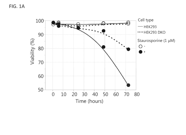
[0016] FIGS. 1A-1D compare the HEK293 DKO cell line (with Bax and Bak genes
knocked
out as described in Example 1) to the parental HEK293 cell line. FIG. 1A shows
cell viability
after exposure of the cell lines to 1 tM staurosporine to induce apoptosis.
FIGS. 1B-1D show
the effects of using a flow constriction device (FCD) to assess sensitivity of
the DKO and
parental cell lines to shear stress on total lysis after FCD (FIG. 1B), viable
cell density (VCD)
before and after FCD (FIG. 1C), and viability before and after FCD (FIG. 1D).
[0017] FIGS. 2A-2C show the optimization of N:P ratio and DNA concentration
for HEK293
DKO transient transfections. Transfections were tested across N:P ratios of 5
to 12.5 and DNA
concentrations of 0.75 to 1.5 pg/mL (FIG. 2A). FIGS. 2B & 2C show the effects
of transfecting
HEK293 and HEK293 DKO cells at the 30 mL tubespin scale with an N:P ratio of
7.5 and a
DNA concentration of 1 pg/mL on viable cell density (VCD) (FIG. 2B) and
viability over the 7
day production cultures and day 7 titers (FIG. 2C).
-5-
CA 03121804 2021-06-01
WO 2020/132231 PCT/US2019/067455
[0018] FIGS. 3A-3E show the effect of scaling up the HEK293 DKO seed train
from a 1 L
shake flask to controlled 2 L bioreactors. Bioreactor #1: pH setpoint of 7
with a deadband of
0.03 and a DO setpoint of 30%. Bioreactor #2: pH setpoint of 7 with a deadband
of 0.4 and a
DO setpoint of 60%. Passaging the 1 L shake flask and 2 L bioreactors every 3-
4 days for 25
days: (FIG. 3A) viable cell density (VCD) and viability, (FIG. 3B) glucose and
lactate, (FIG.
3C) offline pH, and (FIG. 3D) p02. (FIG. 3E) Day 7 transfection titers from 30
mL tubespins.
[0019] FIGS. 4A-4D show the scale-up of the HEK293 DKO seed train from a 1 L
shake flask
to a controlled 35 L bioreactor. Passaging the 1 L shake flask and 35 L
bioreactor every 3-4 days
for 60 days: (FIG. 4A) viable cell density (VCD) and viability, (FIG. 4B)
glucose and lactate,
and (FIG. 4C) offline pH. (FIG. 4D) Day 7 transfection titers from 30 mL
tubespins.
[0020] FIGS. 4E-4N show product quality attributes on day 7 after HEK293 DKO
cells were
transfected in 30 mL tubespins using cells sourced from the 1 L shake flask
and the 35 L
bioreactor seed train over 60 days. Glycosylation species: (FIG. 4E)
Glycosylation species
analyzed, (FIG. 4F) GOF, (FIG. 4G) G1F, (FIG. 411) G2F, (FIG. 41) GO, and
(FIG. 4L) M5.
Charge variants: (FIG. 4J) acidic, (FIG. 4M) main, and (FIG. 4K) basic. Size
variants: (FIG.
4N) high molecular weight species (HMWS).
[0021] FIGS. 5A-5E show the results of HEK293 DKO transient transfections in
controlled
ambr15 bioreactors compared to 30 mL shake flasks. (FIG. 5A) Viable cell
density (VCD) and
viability, (FIG. 5B) glucose and lactate, (FIG. 5C) osmolality and pH, (FIG.
5D) p02, and
(FIG. 5E) titers over the 7 day production cultures.
[0022] FIGS. 6A-6E show the results of scaling up HEK293 DKO transient
transfections from
a 30 mL tubespin to a 10 L wavebag. (FIG. 6A) Viable cell density (VCD) and
viability, (FIG.
6B) glucose and lactate, (FIG. 6C) osmolality and pH, and (FIG. 6D) p02 over
the 7 day
production cultures. (FIG. 6E) Day 7 titers.
DETAILED DESCRIPTION
[0023] The present disclosure provides cell lines (e.g., HEK293 cell lines)
with improved
resistance to apoptosis and shear stress. These cell lines were demonstrated
to exhibit robust
performance in a bioreactor (e.g., a seed train bioreactor) and allow for long-
term cultivation of a
human cell line at a 35L pilot scale in a stirred tank bioreactor or improved
production in a 10L
wavebag bioreactor. As such, the cell lines of the present disclosure may find
use, e.g., in cell
-6-
CA 03121804 2021-06-01
WO 2020/132231 PCT/US2019/067455
cultures and methods of cell culturing (such as methods of recombinant
polynucleotide,
recombinant polypeptide, and viral vector production).
[0024] In one aspect, provided herein are methods of producing a recombinant
polypeptide
comprising culturing a HEK293 cell line that comprises (a) a loss-of-function
mutation in each
of the human Bax and Bak genes and (b) a polynucleotide encoding the
recombinant polypeptide
under conditions suitable for production of the polypeptide.
[0025] In another aspect, provided herein are methods of producing a viral
vector, comprising
culturing a HEK293 cell line that comprises (a) a loss-of-function mutation in
each of the human
Bax and Bak genes, (b) a viral genome, and (c) one or more polynucleotides
encoding a viral
capsid under conditions suitable for production of the viral vector.
[0026] In another aspect, provided herein are cell cultures comprising a cell
culture medium
and a plurality of HEK293 cells, wherein each cell of the plurality comprises
a loss-of-function
mutation in each of the human Bax and Bak genes.
[0027] In another aspect, provided herein is a HEK293 cell line (e.g., an
isolated HEK293 cell
line) that comprises a loss-of-function mutation in each of the human Bax and
Bak genes.
I. Definitions
[0028] Before describing the disclosure in detail, it is to be understood that
this disclosure is
not limited to particular compositions or biological systems, which can, of
course, vary. It is also
to be understood that the terminology used herein is for the purpose of
describing particular
embodiments only, and is not intended to be limiting.
[0029] As used in this specification and the appended claims, the singular
forms "a", "an" and
"the" include plural referents unless the content clearly dictates otherwise.
Thus, for example,
reference to "a molecule" optionally includes a combination of two or more
such molecules, and
the like.
[0030] The term "about" as used herein refers to the usual error range for the
respective value
readily known to the skilled person in this technical field. Reference to
"about" a value or
parameter herein includes (and describes) embodiments that are directed to
that value or
parameter per se. At a maximum, the term "about" as used herein in reference
to a value,
encompasses from 90% to 110% of that value (e.g., relative translation
strength of a first and
-7-
CA 03121804 2021-06-01
WO 2020/132231 PCT/US2019/067455
second TIR of about 1.0 to about 3.0 refers to a relative translation strength
in the range of
between 0.9 and 3.3).
[0031] It is understood that aspects and embodiments of the disclosure
described herein
include "comprising," "consisting," and "consisting essentially of' aspects
and embodiments.
[0032] The term "polynucleotide," when used in singular or plural, generally
refers to any
polyribonucleotide or polydeoxyribonucleotide, which may be unmodified RNA or
DNA or
modified RNA or DNA. Thus, for instance, polynucleotides as defined herein
include, without
limitation, single- and double-stranded DNA, DNA including single- and double-
stranded
regions, single- and double-stranded RNA, and RNA including single- and double-
stranded
regions, hybrid molecules comprising DNA and RNA that may be single-stranded
or, more
typically, double-stranded or include single- and double-stranded regions. In
addition, the term
"polynucleotide" as used herein refers to triple- stranded regions comprising
RNA or DNA or
both RNA and DNA. The strands in such regions may be from the same molecule or
from
different molecules. The regions may include all of one or more of the
molecules, but more
typically involve only a region of some of the molecules. One of the molecules
of a triple-helical
region often is an oligonucleotide. The term "polynucleotide" specifically
includes cDNAs. The
term includes DNAs (including cDNAs) and RNAs that contain one or more
modified bases.
Thus, DNAs or RNAs with backbones modified for stability or for other reasons
are
"polynucleotides" as that term is intended herein. Moreover, DNAs or RNAs
comprising unusual
bases, such as inosine, or modified bases, such as tritiated bases, are
included within the term
"polynucleotides" as defined herein. In general, the term "polynucleotide"
embraces all
chemically, enzymatically and/or metabolically modified forms of unmodified
polynucleotides,
as well as the chemical forms of DNA and RNA characteristic of viruses and
cells, including
simple and complex cells.
[0033] The term "polypeptide" or "protein" are used interchangeably herein to
refer to
polymers of amino acids of any length. The polymer may be linear or branched,
it may comprise
modified amino acids, and it may be interrupted by non-amino acids. The terms
also encompass
an amino acid polymer that has been modified naturally or by intervention; for
example,
disulfide bond formation, glycosylation, lipidation, acetylation,
phosphorylation, or any other
manipulation or modification, such as conjugation with a labeling component or
toxin. Also
included within the definition are, for example, polypeptides containing one
or more analogs of
-8-
CA 03121804 2021-06-01
WO 2020/132231 PCT/US2019/067455
an amino acid (including, for example, unnatural amino acids, etc.), as well
as other
modifications known in the art. The terms "polypeptide" and "protein" as used
herein specifically
encompass antibodies.
[0034] A "loss-of-function mutation" in a gene refers to a genetic
manipulation or mutation
(e.g., a substitution, deletion, insertion, duplication, frameshift, or
translocation) in a gene that
reduces or eliminates one or more functions of the corresponding gene product.
In some
embodiments, the loss-of-function mutation is a null mutation that eliminates
one or more
functions of the corresponding gene product, e.g., a deletion that removes
some or all of the
coding sequence. In some embodiments, the loss-of-function mutation refers to
a genetic
manipulation that leads to a reduction in the expression of a gene, e.g.,
knockdown by RNAi
(e.g., siRNA or shRNA), CRISPRi, miRNA, morpholino, etc.
[0035] The term "recombinant," when used to modify a polynucleotide,
polypeptide, or viral
vector, refers to a polynucleotide/polypeptide/viral vector that has been
introduced into, or has
been produced by, a host cell that does not naturally contain or produce the
polynucleotide/polypeptide/viral vector. The polynucleotide, polypeptide, or
viral vector itself
may be non-naturally occurring (e.g., a humanized antibody), or it may exist
in nature, but not in
the context of the host cell (e.g., a human antibody produced by a human cell
type that does not
typically generate antibodies in nature).
[0036] The term "host cell" (or "recombinant host cell"), as used herein, is
intended to refer to
a cell that has been genetically altered, or is capable of being genetically
altered by introduction
of an exogenous or non-native polynucleotide, such as a recombinant plasmid or
vector. It should
be understood that such terms are intended to refer not only to the particular
subject cell but to
the progeny of such a cell. Because certain modifications may occur in
succeeding generations
due to either mutation or environmental influences, such progeny may not, in
fact, be identical to
the parent cell, but are still included within the scope of the term "host
cell" as used herein.
[0037] As used herein, a "HEK293 cell line" refers to any cell whose lineage
can ultimately be
traced back to the original HEK293 cell line, e.g., the cell line represented
by ATCC catalog
number CRL1573TM and/or generated upon transforming human embryonic kidney
cells with
fragments of adenovirus type 5 DNA as described (Graham et al. (1977)1 Gen.
Virol. 36:59-
74). The term includes HEK293 cell lines that have been genetically modified,
e.g., by
introducing mutations in the Bax and Bak genes and optionally transfected with
a recombinant
-9-
CA 03121804 2021-06-01
WO 2020/132231 PCT/US2019/067455
polynucleotide and/or infected with a viral vector (e.g., a recombinant viral
vector). The
HEK293 cell line is known to be pseudotriploid with a 4kb adenoviral genome
fragment
integrated into chromosome 19 (Louis et al. (1997) Virology 233:423-429).
Features of the
HEK293 genome and transcriptome have been described (Lin et at. (2014) Nat.
Commun. 5:4767
doi :10.1038/ncomms5767).
[0038] "Culture medium" (the term "cell culture medium" can be used
interchangeably herein)
as used herein refers to any composition or broth that supports the growth of
a cell line of the
present disclosure. Suitable culture media may be liquid or solid and contain
any nutrients, salts,
buffers, elements, and other compounds that support the growth and viability
of cells. Common
nutrients of a culture medium may include sources of nitrogen, carbon, amino
acids,
carbohydrates, trace elements, vitamins, and minerals. These nutrients may be
added as
individual components (as in a defined culture medium) or as constituents of a
complex extract
(for example, yeast extract, or plant/animal hydrolysates or peptides). A
culture medium can
include animal-derived components such as serum, or it can be animal origin-
free. A culture
medium can be chemically defined. A culture medium may be nutrient-rich to
support rapid
growth or minimal to support slower growth. A culture medium may also contain
any agent used
to inhibit the growth of or kill contaminating organisms (e.g., an antibiotic
or antimycotic). A
culture medium may also contain any compound used to control the activity of
an inducible
promoter or enzyme.
[0039] The term "antibody" herein is used in the broadest sense and
specifically covers
monoclonal antibodies (including full length monoclonal antibodies),
polyclonal antibodies,
multispecific antibodies (e.g., bispecific antibodies), and antibody fragments
so long as they
exhibit the desired biological activity.
[0040] An "isolated" antibody is one which has been identified and separated
and/or recovered
from a component of its natural environment. Contaminant components of its
natural
environment are materials which would interfere with research, diagnostic or
therapeutic uses for
the antibody, and may include enzymes, hormones, and other proteinaceous or
nonproteinaceous
solutes. In some embodiments, an antibody is purified (1) to greater than 95%
by weight of
antibody as determined by, for example, the Lowry method, and in some
embodiments, to
greater than 99% by weight; (2) to a degree sufficient to obtain at least 15
residues of N-terminal
or internal amino acid sequence by use of, for example, a spinning cup
sequenator, or (3) to
-10-
CA 03121804 2021-06-01
WO 2020/132231 PCT/US2019/067455
homogeneity by SDS-PAGE under reducing or nonreducing conditions using, for
example,
Coomassie blue or silver stain. Isolated antibody includes the antibody in
situ within
recombinant cells since at least one component of the antibody's natural
environment will not be
present. Ordinarily, however, isolated antibody will be prepared by at least
one purification step.
[0041] "Native antibodies" are usually heterotetrameric glycoproteins of about
150,000
daltons, composed of two identical light (L) chains and two identical heavy
(H) chains. Each
light chain is linked to a heavy chain by one covalent disulfide bond, while
the number of
disulfide linkages varies among the heavy chains of different immunoglobulin
isotypes. Each
heavy and light chain also has regularly spaced intrachain disulfide bridges.
Each heavy chain
has at one end a variable domain (VH) followed by a number of constant
domains. Each light
chain has a variable domain at one end (VI) and a constant domain at its other
end; the constant
domain of the light chain is aligned with the first constant domain of the
heavy chain, and the
light chain variable domain is aligned with the variable domain of the heavy
chain. Particular
amino acid residues are believed to form an interface between the light chain
and heavy chain
variable domains.
[0042] The term "constant domain" refers to the portion of an immunoglobulin
molecule
having a more conserved amino acid sequence relative to the other portion of
the
immunoglobulin, the variable domain, which contains the antigen binding site.
The constant
domain contains the CH1, CH2 and CH3 domains (collectively, CH) of the heavy
chain and the
CHL (or CL) domain of the light chain.
[0043] The "variable region" or "variable domain" of an antibody refers to the
amino-terminal
domains of the heavy or light chain of the antibody. The variable domain of
the heavy chain may
be referred to as "VH." The variable domain of the light chain may be referred
to as "VC These
domains are generally the most variable parts of an antibody and contain the
antigen-binding
sites.
[0044] The term "variable" refers to the fact that certain portions of the
variable domains differ
extensively in sequence among antibodies and are used in the binding and
specificity of each
particular antibody for its particular antigen. However, the variability is
not evenly distributed
throughout the variable domains of antibodies. It is concentrated in three
segments called
hypervariable regions (HVRs) both in the light-chain and the heavy-chain
variable domains. The
more highly conserved portions of variable domains are called the framework
regions (FR). The
CA 03121804 2021-06-01
WO 2020/132231 PCT/US2019/067455
variable domains of native heavy and light chains each comprise four FR
regions, largely
adopting a beta-sheet configuration, connected by three HVRs, which form loops
connecting,
and in some cases forming part of, the beta-sheet structure. The HVRs in each
chain are held
together in close proximity by the FR regions and, with the HVRs from the
other chain,
contribute to the formation of the antigen-binding site of antibodies (see
Kabat et at., Sequences
of Proteins of Immunological Interest, Fifth Edition, National Institute of
Health, Bethesda, Md.
(1991)). The constant domains are not involved directly in the binding of an
antibody to an
antigen, but exhibit various effector functions, such as participation of the
antibody in antibody-
dependent cellular toxicity.
[0045] The "light chains" of antibodies (immunoglobulins) from any mammalian
species can
be assigned to one of two clearly distinct types, called kappa ("x") and
lambda ("k"), based on
the amino acid sequences of their constant domains.
[0046] The term IgG "isotype" or "subclass" as used herein is meant any of the
subclasses of
immunoglobulins defined by the chemical and antigenic characteristics of their
constant regions.
[0047] Depending on the amino acid sequences of the constant domains of their
heavy chains,
antibodies (immunoglobulins) can be assigned to different classes. There are
five major classes
of immunoglobulins: IgA, IgD, IgE, IgG, and IgM, and several of these may be
further divided
into subclasses (isotypes), e.g., IgGi, IgG2, IgG3, IgG4, IgAi, and IgA2. The
heavy chain constant
domains that correspond to the different classes of immunoglobulins are called
a, y, c, y, and II.,
respectively. The subunit structures and three-dimensional configurations of
different classes of
immunoglobulins are well known and described generally in, for example, Abbas
et al. Cellular
and Mol. Immunology, 4th ed. (W.B. Saunders, Co., 2000). An antibody may be
part of a larger
fusion molecule, formed by covalent or non-covalent association of the
antibody with one or
more other proteins or peptides.
[0048] The terms "full length antibody," "intact antibody" and "whole
antibody" are used
herein interchangeably to refer to an antibody in its substantially intact
form, not antibody
fragments as defined below. The terms particularly refer to an antibody with
heavy chains that
contain an Fc region.
[0049] A "naked antibody" for the purposes herein is an antibody that is not
conjugated to a
cytotoxic moiety or radiolabel.
-12-
CA 03121804 2021-06-01
WO 2020/132231 PCT/US2019/067455
[0050] "Antibody fragments" comprise a portion of an intact antibody,
preferably comprising
the antigen binding region thereof. In some embodiments, the antibody fragment
described
herein is an antigen-binding fragment. Examples of antibody fragments include
Fab, Fab',
F(ab')2, and Fv fragments; diabodies; linear antibodies; single-chain antibody
molecules; and
multispecific antibodies formed from antibody fragments.
[0051] Papain digestion of antibodies produces two identical antigen-binding
fragments, called
"Fab" fragments, each with a single antigen-binding site, and a residual "Fc"
fragment, whose
name reflects its ability to crystallize readily. Pepsin treatment yields an
F(ab')2 fragment that has
two antigen-combining sites and is still capable of cross-linking antigen.
[0052] "Fv" is the minimum antibody fragment which contains a complete antigen-
binding
site. In one embodiment, a two-chain Fv species consists of a dimer of one
heavy- and one light-
chain variable domain in tight, non-covalent association. In a single-chain Fv
(scFv) species, one
heavy- and one light-chain variable domain can be covalently linked by a
flexible peptide linker
such that the light and heavy chains can associate in a "dimeric" structure
analogous to that in a
two-chain Fv species. It is in this configuration that the three HVRs of each
variable domain
interact to define an antigen-binding site on the surface of the VH-VL dimer.
Collectively, the
six HVRs confer antigen-binding specificity to the antibody. However, even a
single variable
domain (or half of an Fv comprising only three HVRs specific for an antigen)
has the ability to
recognize and bind antigen, although at a lower affinity than the entire
binding site.
[0053] The Fab fragment contains the heavy- and light-chain variable domains
and also
contains the constant domain of the light chain and the first constant domain
(CHI) of the heavy
chain. Fab' fragments differ from Fab fragments by the addition of a few
residues at the carboxy
terminus of the heavy chain CHI domain including one or more cysteines from
the antibody
hinge region. Fab'-SH is the designation herein for Fab' in which the cysteine
residue(s) of the
constant domains bear a free thiol group. F(ab')2 antibody fragments
originally were produced as
pairs of Fab' fragments which have hinge cysteines between them. Other
chemical couplings of
antibody fragments are also known.
[0054] "Single-chain Fv" or "scFv" antibody fragments comprise the VH and VL
domains of
antibody, wherein these domains are present in a single polypeptide chain.
Generally, the scFv
polypeptide further comprises a polypeptide linker between the VH and VL
domains which
enables the scFv to form the desired structure for antigen binding. For a
review of scFv, see, e.g.,
-13-
CA 03121804 2021-06-01
WO 2020/132231 PCT/US2019/067455
Pluckthun, in The Pharmacology of Monoclonal Antibodies, vol. 113, Rosenburg
and Moore
eds., (Springer-Verlag, New York, 1994), pp. 269-315.
[0055] The term "diabodies" refers to antibody fragments with two antigen-
binding sites,
which fragments comprise a heavy-chain variable domain (VH) connected to a
light-chain
variable domain (VL) in the same polypeptide chain (VH-VL). By using a linker
that is too short
to allow pairing between the two domains on the same chain, the domains are
forced to pair with
the complementary domains of another chain and create two antigen-binding
sites. Diabodies
may be bivalent or bispecific. Diabodies are described more fully in, for
example, EP 404,097;
WO 1993/01161; Hudson et al., Nat. Med. 9:129-134 (2003); and Hollinger et
al., Proc. Natl.
Acad. Sci. USA 90: 6444-6448 (1993). Triabodies and tetrabodies are also
described in Hudson
et al., Nat. Med. 9:129-134 (2003).
[0056] The term "monoclonal antibody" as used herein refers to an antibody
obtained from a
population of substantially homogeneous antibodies, e.g., the individual
antibodies comprising
the population are identical except for possible mutations, e.g., naturally
occurring mutations,
that may be present in minor amounts. Thus, the modifier "monoclonal"
indicates the character
of the antibody as not being a mixture of discrete antibodies. In certain
embodiments, such a
monoclonal antibody typically includes an antibody comprising a polypeptide
sequence that
binds a target, wherein the target-binding polypeptide sequence was obtained
by a process that
includes the selection of a single target binding polypeptide sequence from a
plurality of
polypeptide sequences. For example, the selection process can be the selection
of a unique clone
from a plurality of clones, such as a pool of hybridoma clones, phage clones,
or recombinant
DNA clones. It should be understood that a selected target binding sequence
can be further
altered, for example, to improve affinity for the target, to humanize the
target binding sequence,
to improve its production in cell culture, to reduce its immunogenicity in
vivo, to create a
multispecific antibody, etc., and that an antibody comprising the altered
target binding sequence
is also a monoclonal antibody of this disclosure. In contrast to polyclonal
antibody preparations,
which typically include different antibodies directed against different
determinants (epitopes),
each monoclonal antibody of a monoclonal antibody preparation is directed
against a single
determinant on an antigen. In addition to their specificity, monoclonal
antibody preparations are
advantageous in that they are typically uncontaminated by other
immunoglobulins.
-14-
CA 03121804 2021-06-01
WO 2020/132231 PCT/US2019/067455
[0057] "Humanized" forms of non-human (e.g., murine) antibodies are chimeric
antibodies
that contain minimal sequence derived from non-human immunoglobulin. In one
embodiment, a
humanized antibody is a human immunoglobulin (recipient antibody) in which
residues from a
HVR of the recipient are replaced by residues from a HVR of a non-human
species (donor
antibody) such as mouse, rat, rabbit, or nonhuman primate having the desired
specificity,
affinity, and/or capacity. In some instances, FR residues of the human
immunoglobulin are
replaced by corresponding non-human residues. Furthermore, humanized
antibodies may
comprise residues that are not found in the recipient antibody or in the donor
antibody. These
modifications may be made to further refine antibody performance. In general,
a humanized
antibody will comprise substantially all of at least one, and typically two,
variable domains, in
which all or substantially all of the hypervariable loops correspond to those
of a non-human
immunoglobulin, and all or substantially all of the FRs are those of a human
immunoglobulin
sequence. The humanized antibody optionally will also comprise at least a
portion of an
immunoglobulin constant region (Fc), typically that of a human immunoglobulin.
For further
details, see, e.g., Jones et al., Nature 321:522-525 (1986); Riechmann et al.,
Nature 332:323-329
(1988); and Presta, Curr. Op. Struct. Biol. 2:593-596 (1992). See also, e.g.,
Vaswani and
Hamilton, Ann. Allergy, Asthma & Immunol. 1:105-115 (1998); Harris, Biochem.
Soc.
Transactions 23:1035-1038 (1995); Hurle and Gross, Curr. Op. Biotech. 5:428-
433 (1994); and
U.S. Pat. Nos. 6,982,321 and 7,087,409.
[0058] A "human antibody" is one which possesses an amino acid sequence which
corresponds to that of an antibody produced by a human and/or has been made
using any of the
techniques for making human antibodies as disclosed herein. This definition of
a human antibody
specifically excludes a humanized antibody comprising non-human antigen-
binding residues.
Human antibodies can be produced using various techniques known in the art,
including phage-
display libraries. Hoogenboom and Winter, I Mot. Biol., 227:381 (1991); Marks
et al., I Mot.
Biol., 222:581 (1991). Also available for the preparation of human monoclonal
antibodies are
methods described in Cole et at., Monoclonal Antibodies and Cancer Therapy,
Alan R. Liss, p.
77(1985); Boerner et al.,' Immunol., 147(1):86-95 (1991). See also van Dijk
and van de
Winkel, Curr. Op/n. Pharmacol., 5: 368-74 (2001). Human antibodies can be
prepared by
administering the antigen to a transgenic animal that has been modified to
produce such
antibodies in response to antigenic challenge, but whose endogenous loci have
been disabled,
-15-
CA 03121804 2021-06-01
WO 2020/132231 PCT/US2019/067455
e.g., immunized xenomice (see, e.g., U.S. Pat. Nos. 6,075,181 and 6,150,584
regarding
XENOMOUSETm technology). See also, for example, Li et at., Proc. Natl. Acad.
Sci. USA,
103:3557-3562 (2006) regarding human antibodies generated via a human B-cell
hybridoma
technology.
[0059] The term "hypervariable region," "HVR," or "HV," when used herein
refers to the
regions of an antibody variable domain which are hypervariable in sequence
and/or form
structurally defined loops. Generally, antibodies comprise six HVRs; three in
the VH (H1, H2,
H3), and three in the VL (L1, L2, L3). In native antibodies, H3 and L3 display
the most diversity
of the six HVRs, and H3 in particular is believed to play a unique role in
conferring fine
specificity to antibodies. See, e.g.,Xu et al., Immunity 13:37-45 (2000);
Johnson and Wu, in
Methods in Molecular Biology 248:1-25 (Lo, ed., Human Press, Totowa, N.J.,
2003). Indeed,
naturally occurring camelid antibodies consisting of a heavy chain only are
functional and stable
in the absence of light chain. See, e.g., Hamers-Casterman et at., Nature
363:446-448 (1993);
Sheriff et at., Nature Struct. Biol. 3:733-736 (1996).
[0060] A number of HVR delineations are in use and are encompassed herein. The
Kabat
Complementarity Determining Regions (CDRs) are based on sequence variability
and are the
most commonly used (Kabat et at., Sequences of Proteins of Immunological
Interest, 5th Ed.
Public Health Service, National Institutes of Health, Bethesda, Md. (1991)).
Chothia refers
instead to the location of the structural loops (Chothia and Lesk I Mol. Biol.
196:901-917
(1987)). The AbM HVRs represent a compromise between the Kabat HVRs and
Chothia
structural loops, and are used by Oxford Molecular's AbM antibody modeling
software. The
"contact" HVRs are based on an analysis of the available complex crystal
structures. The
residues from each of these HVRs are noted below.
-16-
CA 03121804 2021-06-01
WO 2020/132231 PCT/US2019/067455
Table la. Antibody Hypervariable Regions
Loop Kabat AbM Chothia Contact
Li L24-L34 L24-L34 L26-L32 L30-L36
L2 L50-L56 L50-L56 L50-L52 L46-L55
L3 L89-L97 L89-L97 L91-L96 L89-L96
H1 H31-H35B H26-H35B H26-H32 H30-H35B
(Kabat Numbering)
H1 H31-H35 H26-H35 H26-H32 H30-H35
(Chothia Numbering)
H2 H50-H65 H50-H58 H53-H55 H47-H58
H3 H95-H102 H95-H102 H96-H101 H93-H101
[0061] HVRs may comprise "extended HVRs" as follows: 24-36 or 24-34 (L1), 46-
56 or 50-
56 (L2) and 89-97 or 89-96 (L3) in the VL and 26-35 (H1), 50-65 or 49-65 (H2)
and 93-102, 94-
102, or 95-102 (H3) in the VH. The variable domain residues are numbered
according to Kabat
et at., supra, for each of these definitions.
[0062] "Framework" or "FR" residues are those variable domain residues other
than the HVR
residues as herein defined.
[0063] The term "variable domain residue numbering as in Kabat" or "amino acid
position
numbering as in Kabat," and variations thereof, refers to the numbering system
used for heavy
chain variable domains or light chain variable domains of the compilation of
antibodies in Kabat
et at., supra. Using this numbering system, the actual linear amino acid
sequence may contain
fewer or additional amino acids corresponding to a shortening of, or insertion
into, a FR or HVR
of the variable domain. For example, a heavy chain variable domain may include
a single amino
acid insert (residue 52a according to Kabat) after residue 52 of H2 and
inserted residues (e.g.
residues 82a, 82b, and 82c, etc. according to Kabat) after heavy chain FR
residue 82. The Kabat
numbering of residues may be determined for a given antibody by alignment at
regions of
homology of the sequence of the antibody with a "standard" Kabat numbered
sequence.
[0064] The Kabat numbering system is generally used when referring to a
residue in the
variable domain (approximately residues 1-107 of the light chain and residues
1-113 of the heavy
chain) (e.g., Kabat et at., Sequences of Immunotogicat Interest. 5th Ed.
Public Health Service,
National Institutes of Health, Bethesda, Md. (1991)). The "EU numbering
system" or "EU
index" is generally used when referring to a residue in an immunoglobulin
heavy chain constant
-17-
CA 03121804 2021-06-01
WO 2020/132231 PCT/US2019/067455
region (e.g., the EU index reported in Kabat et at., supra). The "EU index as
in Kabat" refers to
the residue numbering of the human IgG1 EU antibody.
[0065] The expression "linear antibodies" refers to the antibodies described
in Zapata et al.
(1995 Protein Eng, 8(10):1057-1062). Briefly, these antibodies comprise a pair
of tandem Fd
segments (VH-CH1-VH-CH1) which, together with complementary light chain
polypeptides,
form a pair of antigen binding regions. Linear antibodies can be bispecific or
monospecific.
Cell lines
[0066] Provided herein are HEK293 cell lines (e.g., isolated HEK293 cell
lines) that comprise
a loss-of-function mutation in each of the human Bax and Bak genes.
[0067] The human Bax gene (also known as Bcl2l4) encodes a pro-apoptotic Bc1-2
family
member. During apoptosis, Bax and Bak permeate the mitochondrial membrane,
leading to loss
of membrane potential and the release of cytochrome c, which ultimately leads
to the activation
of caspase proteins that trigger programmed cell death (Taylor et at. (2008)
Nat. Rev. Mot. Cell
Biol. 9:231-241). Either Bax or Bak is required to permeabilize the
mitochondrial outer
membrane during the mitochondrial or intrinsic pathway of apoptosis. In some
embodiments,
the human Bax gene refers to the gene described by NCBI Gene ID No. 581. In
some
embodiments, the human Bax gene encodes one or more of the following human Bax
isoforms:
X1 (see, e.g., NCBI Accession No. XPO16882566.1), zeta (see, e.g., NCBI
Accession No.
NP 001278360.1), lambda (see, e.g., NCBI Accession No. NP 001278359.1), gamma
(see, e.g.,
NCBI Accession No. NP 001278358.1), 1 (see, e.g., NCBI Accession No. NP
001278357.1),
sigma (see, e.g., NCBI Accession No. NP 620119.2), delta (see, e.g., NCBI
Accession No.
NP 620118.2), alpha (see, e.g., NCBI Accession No. NP 620116.2), and beta
(see, e.g., NCBI
Accession No. NP 004315.1).
[0068] The human Bak gene (also known as BCL2 antagonist/killer 1, Bakl, Cdnl,
Bcl2l7,
and Bak-like) encodes a pro-apoptotic Bc1-2 family member. During apoptosis,
Bax and Bak
permeate the mitochondrial membrane, leading to loss of membrane potential and
the release of
cytochrome c, which ultimately leads to the activation of caspase proteins
that trigger
programmed cell death (Taylor et at. (2008) Nat. Rev. Mot. Cell Biol. 9:231-
241). In some
embodiments, the human Bak gene refers to the gene described by NCBI Gene ID
No. 578. In
some embodiments, the human Bak gene encodes one or more of the following
human Bak
-18-
CA 03121804 2021-06-01
WO 2020/132231 PCT/US2019/067455
isoforms: X1 (see, e.g., NCBI Accession No. XP 011513082.1), X2 (see, e.g.,
NCBI Accession
No. XPO11513081.1), and the standard isoform (see, e.g., NCBI Accession No. NP
001179.1).
[0069] In some embodiments, the loss-of-function mutation comprises one or
more
substitution, insertion, deletion, and/or frameshift mutations. In some
embodiments, the loss-of-
function mutation comprises a deletion. Various loss-of-function mutations in
the human Bax
and Bak genes are known. For example, Bax and Bak mutations have been
described in various
cancers; see, e.g., OMIM entries 600040 and 600516 and COSMIC (Catalogue of
Somatic
Mutations in Cancer) entries for Bax and Bak
(cancer.sanger.ac.uk/cosmic/gene/analysis?ln=BAX and
cancer.sanger.ac.uk/cosmic/gene/analysis?ln=BAK1, respectively). In some
embodiments, a
loss-of-function mutation in Bax or Bak reduces or eliminates one or more
functions of Bax or
Bak (e.g., pro-apoptotic functions), including but not limited to promotion of
apoptosis, loss of
mitochondrial membrane potential, outer mitochondrial membrane pore formation,
and release of
cytochrome c. In some embodiments, a loss-of-function mutation in Bax or Bak
reduces or
eliminates expression of Bax or Bak protein. In some embodiments, a loss-of-
function mutation
in Bax or Bak refers to a genetic manipulation that reduces or eliminates
expression of Bax or
Bak protein, e.g., by RNAi, CRISPRi, miRNA, morpholino, etc. In some
embodiments, a loss-
of-function mutation in Bax inhibits sensitivity to apoptosis and/or loss of
mitochondrial
membrane potential, outer mitochondrial membrane pore formation, or release of
cytochrome c
in a cell with a loss-of-function mutation in Bak, and/or a loss-of-function
mutation in Bak
inhibits sensitivity to apoptosis and/or loss of mitochondrial membrane
potential, outer
mitochondrial membrane pore formation, or release of cytochrome c in a cell
with a loss-of-
function mutation in Bax. For example, it has been demonstrated that reducing
Bak function
when Bax function is impaired has a much more dramatic effect on sensitivity
to apoptosis than
reducing Bak function in the context of normal Bax function (see, e.g.,
Chandra, D. et at. (2005)
Biol. Chem. 280:19051-19061).
[0070] Techniques for engineering a HEK293 cell line with a loss-of-function
mutation in each
of the human Bax and Bak genes are known. In some embodiments, as exemplified
herein, zinc
finger nuclease technology (Cost et at. (2010) Biotechnol. Bioeng. 105:330-
340) can be used to
introduce loss-of-function mutations (e.g., deletions) in Bax and Bak. Other
techniques for
-19-
CA 03121804 2021-06-01
WO 2020/132231 PCT/US2019/067455
introducing mutations in a human cell include, without limitation,
CRISPR/Cas9, TALEN, site-
directed mutagenesis by PCR, chemical mutagenesis, insertional mutagenesis,
and so forth.
[0071] In some embodiments, a HEK293 cell line that comprises a loss-of-
function mutation in
each of the human Bax and Bak genes displays one or more of the following, as
compared with a
HEK293 cell line that comprises functional copies of each of the human Bax and
Bak genes, or a
HEK293 cell line that comprises a functional copy of only one of the human Bax
and Bak genes:
increased resistance to apoptosis, increased resistance to shear stress,
increased resistance to
staurosporine, and increased production of a recombinant polypeptide (e.g., in
cell culture).
Recombinant Polynucleotides, Polyp eptides, Antigens, Enzymes, and Vaccines
[0072] In some embodiments, a cell line of the present disclosure (e.g., a
HEK293 cell line
comprising a loss-of-function mutation in each of the human Bax and Bak genes)
comprises a
recombinant polynucleotide. For example, in some embodiments, the recombinant
polynucleotide encodes a recombinant polypeptide (e.g., one or more chains of
an antibody or
antibody fragment).
[0073] In some embodiments, a recombinant polynucleotide (e.g., a recombinant
polynucleotide that encodes a recombinant polypeptide) is an extrachromosomal
polynucleotide.
In some embodiments, the recombinant polynucleotide is introduced into the
cell line without
integration of the polynucleotide into the host cell genome. In some
embodiments, the
recombinant polynucleotide is introduced into the cell line by transient
transfection. Transient
transfection is known to introduce recombinant polynucleotide(s) into a cell
line without
integration of the polynucleotide(s) into the host cell genome; as such, the
polynucleotide(s) are
not replicated along with the host cell genome and are lost after a finite
period of time (due to,
e.g., cell division, degradation, etc.). Methods for transiently transfecting
HEK293 cell lines are
known in the art (see, e.g., de Los Milagros Bassani Molinas et at. (2014)
Cytotechnology
66:493-514) and kits for transient transfection of HEK293 cells are
commercially available (see,
e.g., the Expi293TM Expression System for transient HEK293 cell transfection
sold by Thermo
Fisher Scientific).
[0074] In other embodiments, a recombinant polynucleotide (e.g., a recombinant
polynucleotide that encodes a recombinant polypeptide) is integrated into the
host cell genome,
e.g., onto a chromosome of the human cell line. In some embodiments, the
recombinant
-20-
CA 03121804 2021-06-01
WO 2020/132231 PCT/US2019/067455
polynucleotide is introduced into the cell line by stable transfection. Stable
transfection is
known to introduce recombinant polynucleotide(s) into a cell line through
stable inheritance of
non-genomic DNA or the incorporation of the recombinant polynucleotide(s) into
the host cell
genome (e.g., by integration onto a host cell chromosome). In some
embodiments, a
recombinant polynucleotide is integrated into the host cell genome by random
integration. For
example, the recombinant polynucleotide can encode a selectable marker (e.g.,
encoding a
protein that confers resistance to an antibiotic such as puromycin,
hygromycin, G418, etc.), and
cells transfected with the recombinant polynucleotide can be subjected to one
or more rounds of
selection via the selectable marker (e.g., by using a cell culture medium
comprising an antibiotic
such as puromycin, hygromycin, G418, etc. to kill cells that do not express
the selectable
marker). Techniques and kits for random integration into HEK293 cells are
known in the art;
see, e.g., www.thermofisher.com/us/en/home/references/gibco-cell-culture-
basics/transfection-
basics/transfection-methods/stable-transfection.html. In some embodiments, a
recombinant
polynucleotide is integrated into the host cell genome by site-specific or
targeted integration.
For example, a technique such as recombinase-mediated cassette exchange
(RMCE), Cre-Lox
recombination, or FLP-FRT recombination can be used to integrate a recombinant
polynucleotide into a targeted site in the host cell genome. Techniques and
kits for using site-
specific or targeted integration in HEK293 cells are known in the art (see,
e.g., Callesen et at.
(2016) PLoS One 11:e0161471) and the Flp-InTM 293 cell line (Thermo Fisher
Scientific).
[0075] In some embodiments, a recombinant polynucleotide of the present
disclosure encodes
a recombinant polypeptide. Exemplary recombinant polypeptides are listed
infra. In some
embodiments, a recombinant polypeptide produced by a HEK293 cell line of the
present
disclosure comprises one or more post-translational modifications (e.g.,
glycosylation)
characteristic of production in a human cell, e.g., as compared to the
modifications of a
comparable polypeptide produced in a prokaryotic, fungal, insect, or non-human
mammalian
cell. For example, differences in glycosylation between similar proteins
expressed in HEK293
cells vs. CHO cells have been documented (see, e.g., Croset et at. (2012)1
Biotechnol. 161:336-
348). In some embodiments, a recombinant polypeptide produced by a HEK293 cell
line of the
present disclosure comprises a glycosylation modification comprising one or
more of the
exemplary and non-limiting glycans shown in FIG. 4E.
-21-
CA 03121804 2021-06-01
WO 2020/132231 PCT/US2019/067455
[0076] In some embodiments, a recombinant polynucleotide of the present
disclosure encodes
an antigen. In some embodiments, the antigen is a polypeptide antigen. In some
embodiments,
the antigen is a peptide antigen. In some embodiments, the antigen is a
therapeutic or diagnostic
antigen.
[0077] In some embodiments, a recombinant polynucleotide of the present
disclosure encodes
an enzyme. In some embodiments, the enzyme is a therapeutic or diagnostic
enzyme.
[0078] In some embodiments, a recombinant polynucleotide of the present
disclosure encodes
a vaccine. In some embodiments, the vaccine is a peptide vaccine. In some
embodiments, the
vaccine is a live-attenuated, inactivated, toxoid, or subunit/recombinant
vaccine. In some
embodiments, the vaccine is against one or more of: measles, mumps, rubella,
rotavirus,
smallpox, chickenpox, yellow fever, hepatitis A, hepatitis B, influenza,
polio, rabies, Hib
disease, human papillomavirus, whooping cough, pneumococcal disease,
meningococcal disease,
shingles, tetanus, and diphtheria.
[0079] In some embodiments, a recombinant polynucleotide of the present
disclosure
comprises a viral genome and/or encodes a viral capsid. As described in
greater detail in section
IV infra, the cell lines of the present disclosure may find use, inter al/a,
in methods of producing
viral vectors. In some embodiments, a cell line of the present disclosure
comprises a viral
genome (e.g., of a viral vector of interest) and one or more polynucleotides
encoding a viral
capsid. For example, HEK293 cell lines have been modified to include in their
genome AAV
genes (e.g., Rep and Cap genes) to generate packaging cell lines that can then
be infected with
adenovirus for the production of adeno-associated virus (AAV) vectors (see,
e.g., Qiao et at.
(2002)1 Virol. 76:13015-13027). HEK293 cell lines have also been modified to
include in their
genome AAV genes (e.g., Rep and Cap genes) as well as adenovirus genes (e.g.,
E1A/E1B) to
generate producer cell lines for the production of adeno-associated virus
(AAV) vectors (see,
e.g., Yuan et at. (2011) Hum. Gene Ther. 22:613-624).
[0080] In some embodiments, the recombinant polynucleotide comprises an
expression vector.
An expression vector can include one or more of the following elements: a
signal sequence, an
origin of replication, one or more marker genes, an enhancer element, a
promoter, and a
transcription termination sequence.
[0081] A recombinant polypeptide of the present disclosure may be produced
recombinantly
not only directly, but also as a fusion polypeptide with a heterologous
polypeptide, which is
-22-
CA 03121804 2021-06-01
WO 2020/132231 PCT/US2019/067455
preferably a signal sequence or other polypeptide having a specific cleavage
site at the N-
terminus of the mature protein or polypeptide. The heterologous signal
sequence selected
preferably is one that is recognized and processed (e.g., cleaved by a signal
peptidase) by the
host cell. In mammalian cell expression, mammalian signal sequences as well as
viral secretory
leaders, for example, the herpes simplex gD signal, are available.
[0082] In some embodiments, an expression vector comprises an origin of
replication.
Generally, in vectors this sequence is one that enables the vector to
replicate independently of the
host chromosomal DNA, and includes origins of replication or autonomously
replicating
sequences. Generally, the origin of replication component is not needed for
mammalian
expression vectors (the SV40 origin may typically be used only because it
contains the early
promoter. In other embodiments, the vector does not include an origin of
replication (e.g., if the
recombinant polynucleotide is integrated onto a host cell chromosome).
[0083] In some embodiments, as alluded to above with regard to stable
transfections, an
expression vector comprises a selection gene or selectable marker. Typical
selection genes
encode proteins that (a) confer resistance to antibiotics or other toxinse,
(b) complement
auxotrophic deficiencies, or (c) supply critical nutrients not available from
complex media. One
example of a selection scheme utilizes a drug to arrest growth of a host cell.
Those cells that are
successfully transformed with a heterologous gene produce a protein conferring
drug resistance
and thus survive the selection regimen. Another example of suitable selectable
markers for
mammalian cells are those that enable the identification of cells competent to
take up antibody-
encoding nucleic acid, such as DHFR, glutamine synthetase (GS), thymidine
kinase,
metallothionein-I and -II, preferably primate metallothionein genes, adenosine
deaminase,
ornithine decarboxylase, etc. For example, cells transformed with the DHFR
gene are identified
by culturing the transformants in a culture medium containing methotrexate
(Mtx), a competitive
antagonist of DHFR. Under these conditions, the DHFR gene is amplified along
with any other
co-transformed nucleic acid. Alternatively, cells transformed with the GS gene
are identified by
culturing the transformants in a culture medium containing L-methionine
sulfoximine (Msx), an
inhibitor of GS. Under these conditions, the GS gene is amplified along with
any other co-
transformed nucleic acid. The GS selection/amplification system may be used in
combination
with the DHFR selection/amplification system described above. Alternatively,
host cells
(particularly wild-type hosts that contain endogenous DHFR) transformed or co-
transformed
-23-
CA 03121804 2021-06-01
WO 2020/132231 PCT/US2019/067455
with DNA sequences of interest, wild-type DHFR gene, and another selectable
marker such as
aminoglycoside 3'-phosphotransferase (APH) can be selected by cell growth in
medium
containing a selection agent for the selectable marker such as an
aminoglycosidic antibiotic, e.g.,
kanamycin, neomycin, or G418. See U.S. Pat. No. 4,965,199.
[0084] In some embodiments, an expression vector comprises a promoter.
Expression and
cloning vectors generally contain a promoter that is recognized by the host
organism and is
operably linked to the recombinant polynucleotide. Antibody transcription from
vectors in
mammalian host cells can be controlled, for example, by promoters obtained
from the genomes
of viruses such as polyoma virus, fowlpox virus, adenovirus (such as
Adenovirus 2), bovine
papilloma virus, avian sarcoma virus, cytomegalovirus, a retrovirus, hepatitis-
B virus, Simian
Virus 40 (5V40), or from heterologous mammalian promoters, e.g., the actin
promoter or an
immunoglobulin promoter, from heat-shock promoters, provided such promoters
are compatible
with the host cell systems. The early and late promoters of the 5V40 virus are
conveniently
obtained as an 5V40 restriction fragment that also contains the 5V40 viral
origin of replication.
The immediate early promoter of the human cytomegalovirus is conveniently
obtained as a
HindIII E restriction fragment. A system for expressing DNA in mammalian hosts
using the
bovine papilloma virus as a vector is disclosed in U.S. Pat. No. 4,419,446. A
modification of this
system is described in U.S. Pat. No. 4,601,978. See also Reyes et al., Nature
297:598-601 (1982)
on expression of human 13-interferon cDNA in mouse cells under the control of
a thymidine
kinase promoter from herpes simplex virus. Alternatively, the Rous Sarcoma
Virus long terminal
repeat can be used as the promoter.
[0085] In some embodiments, an expression vector comprises an enhancer
element. Many
enhancer sequences are now known from mammalian genes (globin, elastase,
albumin, a-
fetoprotein, and insulin). Typically, however, one will use an enhancer from a
eukaryotic cell
virus. Examples include the 5V40 enhancer on the late side of the replication
origin (bp 100-
270), the cytomegalovirus early promoter enhancer, the polyoma enhancer on the
late side of the
replication origin, and adenovirus enhancers. See also Yaniv, Nature 297:17-18
(1982) on
enhancing elements for activation of eukaryotic promoters. The enhancer may be
spliced into the
vector at a position 5' or 3' to the antibody-encoding sequence, but is
preferably located at a site
5' from the promoter.
-24-
CA 03121804 2021-06-01
WO 2020/132231 PCT/US2019/067455
[0086] In some embodiments, an expression vector comprises a transcription
terminator, e.g.,
sequence(s) necessary for the termination of transcription and for stabilizing
the mRNA. Such
sequences are commonly available from the 5' and, occasionally 3',
untranslated regions of
eukaryotic or viral DNAs or cDNAs. These regions contain nucleotide segments
transcribed as
polyadenylated fragments in the untranslated portion of the mRNA encoding
antibody. One
useful transcription termination component is the bovine growth hormone
polyadenylation
region. See W094/11026 and the expression vector disclosed therein.
Antibodies and antibody fragments
[0087] In some embodiments, a recombinant polynucleotide of the present
disclosure encodes
an antibody or antigen-binding fragment thereof For example, in some
embodiments, the
recombinant polynucleotide encodes the heavy and light chains for an antibody
or antibody
fragment, or a single chain antibody or antibody fragment. Exemplary methods
for using a cell
line or cell culture of the present disclosure to produce an antibody or
antibody fragment are
described in greater detail in section IV infra.
[0088] In some embodiments, the antibody (or antibody fragment) is a
diagnostic antibody.
For example, the antibody can be used to detect one or more diagnostic
antigens in a sample,
e.g., by ELISA, Western blotting, immunohistochemistry (IHC), flow cytometry,
or other
immunoassays. As non-limiting examples, diagnostic antibodies have been used
to detect HER2
(see, e.g., the HercepTest for identifying tumors that overexpress HER2 from
DAKO Corp.), the
estrogen and progesterone receptors (see, e.g., the ER/PR pharmDx kit for
identifying tumors
that overexpress ER or PR from DAKO Corp.), and PD-Li (see, e.g., the Ventana
5P263 and
SP142 assays for identifying tumors that express PD-L1) for diagnostic assays
used, e.g., in the
treatment of various cancers.
[0089] In some embodiments, the antibody (or antibody fragment) is a
therapeutic antibody.
Exemplary therapeutic antibodies include, without limitation, nivolumab
(OPDIVO , Bristol-
Myers Squibb), pembrolizumab (KEYTRUDA , Merck),avelumab (BAVENCIO , Merck),
durvalumab (IMFINZI , Astra-Zeneca/Medimmune), alemtuzumab (Campath),
bevacizumab
(AVASTIN , Genentech); cetuximab (ERBITUX , Imclone); panitumumab (VECTIBIX ,
Amgen), rituximab (RITUXAN , Genentech/Biogen Idec), pertuzumab (PERJETA ,
2C4,
Genentech), trastuzumab (HERCEPTIN , Genentech), atezolizumab (TECENTRIQ ,
-25-
CA 03121804 2021-06-01
WO 2020/132231 PCT/US2019/067455
Genentech), obinutuzumab (GAZYVA , Genentech), ocrelizumab (OCREVUS ,
Genentech),
tositumomab (Bexxar, Corixia), gemtuzumab ozogamicin (MYLOTARG , Wyeth),
apolizumab, aselizumab, atlizumab, bapineuzumab, bivatuzumab mertansine,
cantuzumab
mertansine, cedelizumab, certolizumab pegol, cidfusituzumab, cidtuzumab,
daclizumab,
eculizumab, efalizumab, epratuzumab, erlizumab, felvizumab, fontolizumab,
gemtuzumab
ozogamicin, inotuzumab ozogamicin, ipilimumab, labetuzumab, lintuzumab,
matuzumab,
mepolizumab, motavizumab, motovizumab, natalizumab, nimotuzumab, nolovizumab,
numavizumab, ocrelizumab, omalizumab, palivizumab, pascolizumab,
pecfusituzumab,
pectuzumab, pexelizumab, ralivizumab, ranibizumab, reslivizumab, reslizumab,
resyvizumab,
rovelizumab, ruplizumab, sibrotuzumab, siplizumab, sontuzumab, tacatuzumab
tetraxetan,
tadocizumab, talizumab, tefibazumab, tocilizumab, toralizumab, tucotuzumab
celmoleukin,
tucusituzumab, umavizumab, urtoxazumab, ustekinumab, visilizumab, and the
anti¨interleukin-
12 (ABT-874/J695, Wyeth Research and Abbott Laboratories) which is a
recombinant
exclusively human-sequence, full-length IgGi X. antibody genetically modified
to recognize
interleukin-12 p40 protein. A non-limiting list of monoclonal antibodies
approved by the EMA
or FDA for therapeutic use can be found at www.actip.org/products/monoclonal-
antibodies-
approved-by-the-ema-and-fda-for-therapeutic-use/.
[0090] Features of antibodies and antibody fragments are described in a non-
limiting manner
infra.
Certain Antibody-Based Methods
[0091] Monoclonal antibodies can be made using the hybridoma method first
described by
Kohler et at., Nature, 256:495 (1975), and further described, e.g., in Hongo
et at., Hybridoma, 14
(3): 253-260 (1995), Harlow et al., Antibodies: A Laboratory Manual, (Cold
Spring Harbor
Laboratory Press, 2nd ed. 1988); Hammerling et at., in: Monoclonal Antibodies
and T-Cell
Hybridomas 563-681 (Elsevier, N.Y., 1981), and Ni, Xiandai Mianyixue,
26(4):265-268 (2006)
regarding human-human hybridomas. Additional methods include those described,
for example,
in U.S. Pat. No. 7,189,826 regarding production of monoclonal human natural
IgM antibodies
from hybridoma cell lines. Human hybridoma technology (Trioma technology) is
described in
Vollmers and Brandlein, Histology and Histopathology, 20(3):927-937 (2005) and
Vollmers and
Brandlein, Methods and Findings in Experimental and Clinical Pharmacology,
27(3): 185-91
-26-
CA 03121804 2021-06-01
WO 2020/132231 PCT/US2019/067455
(2005). Once desired monoclonal antibodies have been isolated from hybridomas,
polynucleotides encoding them may be subcloned into an expression vector, and
antibodies may
be produced by expression in a HEK293 cell line by any of the methods
described herein.
Library-Derived Antibodies
[0092] Antibodies of the disclosure may be isolated by screening combinatorial
libraries for
antibodies with the desired activity or activities. For example, a variety of
methods are known in
the art for generating phage display libraries and screening such libraries
for antibodies
possessing the desired binding characteristics such as the methods described
in Example 3.
Additional methods are reviewed, e.g., in Hoogenboom et al. in Methods in
Molecular Biology
178:1-37 (O'Brien et al., ed., Human Press, Totowa, NJ, 2001) and further
described, e.g., in the
McCafferty et al., Nature 348:552-554; Clackson et al., Nature 352: 624-628
(1991); Marks et
al., I Mot. Biol. 222: 581-597 (1992); Marks and Bradbury, in Methods in
Molecular Biology
248:161-175 (Lo, ed., Human Press, Totowa, NJ, 2003); Sidhu et al., I Mot.
Biol. 338(2): 299-
310 (2004); Lee et al., I Mot Biol. 340(5): 1073-1093 (2004); Fellouse, Proc.
Natl. Acad. Sci.
USA 101(34): 12467-12472 (2004); and Lee et al., I Immunol. Methods 284(1-2):
119-
132(2004).
[0093] In certain phage display methods, repertoires of VH and VL genes are
separately
cloned by polymerase chain reaction (PCR) and recombined randomly in phage
libraries, which
can then be screened for antigen-binding phage as described in Winter et al.,
Ann. Rev.
Immunol., 12: 433-455 (1994). Phage typically display antibody fragments,
either as single-chain
Fv (scFv) fragments or as Fab fragments. Libraries from immunized sources
provide high-
affinity antibodies to the immunogen without the requirement of constructing
hybridomas.
Alternatively, the naive repertoire can be cloned (e.g., from human) to
provide a single source of
antibodies to a wide range of non-self and also self-antigens without any
immunization as
described by Griffiths et al., EMBO J, 12: 725-734 (1993). Finally, naive
libraries can also be
made synthetically by cloning unrearranged V-gene segments from stem cells,
and using PCR
primers containing random sequence to encode the highly variable CDR3 regions
and to
accomplish rearrangement in vitro, as described by Hoogenboom and Winter, I
Mol. Biol., 227:
381-388 (1992). Patent publications describing human antibody phage libraries
include, for
example: US Patent No. 5,750,373, and US Patent Publication Nos. 2005/0079574,
-27-
CA 03121804 2021-06-01
WO 2020/132231 PCT/US2019/067455
2005/0119455, 2005/0266000, 2007/0117126, 2007/0160598, 2007/0237764,
2007/0292936,
and 2009/0002360.
[0094] Antibodies or antibody fragments isolated from human antibody libraries
are
considered human antibodies or human antibody fragments herein.
Chimeric, Humanized and Human Antibodies
[0095] In certain embodiments, an antibody provided herein is a chimeric
antibody. Certain
chimeric antibodies are described, e.g., in U.S. Patent No. 4,816,567; and
Morrison et al., Proc.
Natl. Acad. Sci. USA, 81:6851-6855 (1984)). In one example, a chimeric
antibody comprises a
non-human variable region (e.g., a variable region derived from a mouse, rat,
hamster, rabbit, or
non-human primate, such as a monkey) and a human constant region. In a further
example, a
chimeric antibody is a "class switched" antibody in which the class or
subclass has been changed
from that of the parent antibody. Chimeric antibodies include antigen-binding
fragments thereof.
[0096] In certain embodiments, a chimeric antibody is a humanized antibody.
Typically, a non-
human antibody is humanized to reduce immunogenicity to humans, while
retaining the
specificity and affinity of the parental non-human antibody. Generally, a
humanized antibody
comprises one or more variable domains in which HVRs, e.g., CDRs, (or portions
thereof) are
derived from a non-human antibody, and FRs (or portions thereof) are derived
from human
antibody sequences. A humanized antibody optionally will also comprise at
least a portion of a
human constant region. In some embodiments, some FR residues in a humanized
antibody are
substituted with corresponding residues from a non-human antibody (e.g., the
antibody from
which the HVR residues are derived), e.g., to restore or improve antibody
specificity or affinity.
[0097] Humanized antibodies and methods of making them are reviewed, e.g., in
Almagro and
Fransson, Front. Biosci. 13:1619-1633 (2008), and are further described, e.g.,
in Riechmann et
at., Nature 332:323-329 (1988); Queen et al., Proc. Nat'l Acad. Sci. USA
86:10029-10033
(1989); US Patent Nos. 5, 821,337, 7,527,791, 6,982,321, and 7,087,409;
Kashmiri et al.,
Methods 36:25-34 (2005) (describing SDR (a-CDR) grafting); Padlan, Mot.
Immunol. 28:489-
498 (1991) (describing "resurfacing"); Dall'Acqua et al., Methods 36:43-60
(2005) (describing
"FR shuffling"); and Osbourn et at., Methods 36:61-68 (2005) and Klimka et
at., Br. I Cancer,
83:252-260 (2000) (describing the "guided selection" approach to FR
shuffling).
-28-
CA 03121804 2021-06-01
WO 2020/132231 PCT/US2019/067455
[0098] Human framework regions that may be used for humanization include but
are not
limited to: framework regions selected using the "best-fit" method (see, e.g.,
Sims et at. J.
Immunol. 151:2296 (1993)); framework regions derived from the consensus
sequence of human
antibodies of a particular subgroup of light or heavy chain variable regions
(see, e.g., Carter et al.
Proc. Natl. Acad. Sci. USA, 89:4285 (1992); and Presta et al. J. Immunol.,
151:2623 (1993));
human mature (somatically mutated) framework regions or human germline
framework regions
(see, e.g., Almagro and Fransson, Front. Biosci. 13:1619-1633 (2008)); and
framework regions
derived from screening FR libraries (see, e.g., Baca et al., J. Biol. Chem.
272:10678-10684
(1997) and Rosok et al., J. Biol. Chem. 271:22611-22618 (1996)).
[0099] In certain embodiments, an antibody provided herein is a human
antibody. Human
antibodies can be produced using various techniques known in the art. Human
antibodies are
described generally in van Dijk and van de Winkel, Curr. Op/n. Pharmacol. 5:
368-74 (2001)
and Lonberg, Curr. Op/n. Immunol. 20:450-459 (2008). Human antibodies can be
made, for
example and without limitation, by expression in a HEK293 cell line from an
expression vector
by any of the methods described herein.
[0100] Human antibodies may also be generated by isolating Fv clone variable
domain
sequences selected from human-derived phage display libraries. Such variable
domain sequences
may then be combined with a desired human constant domain. Techniques for
selecting human
antibodies from antibody libraries are described below.
Antibody Fragments
[0101] Antibody fragments may be generated by traditional means, such as
enzymatic
digestion, or by recombinant techniques. In certain circumstances there are
advantages of using
antibody fragments, rather than whole antibodies. The smaller size of the
fragments allows for
rapid clearance, and may lead to improved access to solid tumors. For a review
of certain
antibody fragments, see Hudson et al. (2003) Nat. Med. 9:129-134.
[0102] Various techniques have been developed for the production of antibody
fragments.
Traditionally, these fragments were derived via proteolytic digestion of
intact antibodies (see,
e.g., Morimoto et at., Journal of Biochemical and Biophysical Methods 24:107-
117 (1992); and
Brennan et at., Science, 229:81 (1985)). However, these fragments can now be
produced directly
by recombinant host cells. Fab, Fv and ScFv antibody fragments can all be
expressed in and
-29-
CA 03121804 2021-06-01
WO 2020/132231 PCT/US2019/067455
secreted from E. coli, thus allowing the facile production of large amounts of
these fragments.
Antibody fragments can be isolated from the antibody phage libraries discussed
above.
Alternatively, Fab'-SH fragments can be directly recovered from E. coli and
chemically coupled
to form F(ab')2 fragments (Carter et at., Bio/Technology 10:163-167 (1992)).
According to
another approach, F(ab') 2 fragments can be isolated directly from recombinant
host cell culture.
Fab and F(ab')2 fragment with increased in vivo half-life comprising salvage
receptor binding
epitope residues are described in U.S. Pat. No. 5,869,046. Other techniques
for the production of
antibody fragments will be apparent to the skilled practitioner. In certain
embodiments, an
antibody is a single chain Fv fragment (scFv). See WO 93/16185; U.S. Pat. Nos.
5,571,894; and
5,587,458. Fv and scFv are the only species with intact combining sites that
are devoid of
constant regions; thus, they may be suitable for reduced nonspecific binding
during in vivo use.
scFv fusion proteins may be constructed to yield fusion of an effector protein
at either the amino
or the carboxy terminus of an scFv. See Antibody Engineering, ed. Borrebaeck,
supra. The
antibody fragment may also be a "linear antibody", e.g., as described in U.S.
Pat. No. 5,641,870,
for example. Such linear antibodies may be monospecific or bispecific.
Multispecific Antibodies
[0103] Multispecific antibodies have binding specificities for at least two
different epitopes,
where the epitopes are usually from different antigens. While such molecules
normally will only
bind two different epitopes (i.e. bispecific antibodies, BsAbs), antibodies
with additional
specificities such as trispecific antibodies are encompassed by this
expression when used herein.
Bispecific antibodies can be prepared as full length antibodies or antibody
fragments (e.g. F(ab')2
bispecific antibodies).
[0104] Methods for making bispecific antibodies are known in the art.
Traditional production
of full length bispecific antibodies is based on the coexpression of two
immunoglobulin heavy
chain-light chain pairs, where the two chains have different specificities
(Millstein et at., Nature,
305:537-539 (1983)). Because of the random assortment of immunoglobulin heavy
and light
chains, these hybridomas (quadromas) produce a potential mixture of 10
different antibody
molecules, of which only one has the correct bispecific structure.
Purification of the correct
molecule, which is usually done by affinity chromatography steps, is rather
cumbersome, and the
-30-
CA 03121804 2021-06-01
WO 2020/132231 PCT/US2019/067455
product yields are low. Similar procedures are disclosed in WO 93/08829, and
in Traunecker et
at., EMBO 1, 10:3655-3659 (1991).
[0105] One approach known in the art for making bispecific antibodies is the
"knobs-into-
holes" or "protuberance-into-cavity" approach (see, e.g., US Pat. No.
5,731,168). In this
approach, two immunoglobulin polypeptides (e.g., heavy chain polypeptides)
each comprise an
interface. An interface of one immunoglobulin polypeptide interacts with a
corresponding
interface on the other immunoglobulin polypeptide, thereby allowing the two
immunoglobulin
polypeptides to associate. These interfaces may be engineered such that a
"knob" or
"protuberance" (these terms may be used interchangeably herein) located in the
interface of one
immunoglobulin polypeptide corresponds with a "hole" or "cavity" (these terms
may be used
interchangeably herein) located in the interface of the other immunoglobulin
polypeptide. In
some embodiments, the hole is of identical or similar size to the knob and
suitably positioned
such that when the two interfaces interact, the knob of one interface is
positionable in the
corresponding hole of the other interface. Without wishing to be bound to
theory, this is thought
to stabilize the heteromultimer and favor formation of the heteromultimer over
other species, for
example homomultimers. In some embodiments, this approach may be used to
promote the
heteromultimerization of two different immunoglobulin polypeptides, creating a
bispecific
antibody comprising two immunoglobulin polypeptides with binding specificities
for different
epitopes.
[0106] In some embodiments, a knob may be constructed by replacing a small
amino acid side
chain with a larger side chain. In some embodiments, a hole may be constructed
by replacing a
large amino acid side chain with a smaller side chain. Knobs or holes may
exist in the original
interface, or they may be introduced synthetically. For example, knobs or
holes may be
introduced synthetically by altering the nucleic acid sequence encoding the
interface to replace at
least one "original" amino acid residue with at least one "import" amino acid
residue. Methods
for altering nucleic acid sequences may include standard molecular biology
techniques well
known in the art. The side chain volumes of various amino acid residues are
shown in the
following table. In some embodiments, original residues have a small side
chain volume (e.g.,
alanine, asparagine, aspartic acid, glycine, serine, threonine, or valine),
and import residues for
forming a knob are naturally occurring amino acids and may include arginine,
phenylalanine,
tyrosine, and tryptophan. In some embodiments, original residues have a large
side chain volume
-31-
CA 03121804 2021-06-01
WO 2020/132231
PCT/US2019/067455
(e.g., arginine, phenylalanine, tyrosine, and tryptophan), and import residues
for forming a hole
are naturally occurring amino acids and may include alanine, serine,
threonine, and valine.
Table lb. Properties of amino acid residues
One-letter Mass' Volumeb
Accessible
Amino Acid abbreviation (daltons) (A3) surface area'
(A2)
Alanine (Ala) A 71.08 88.6 115
Arginine (Arg) R 156.20 173.4 225
Asparagine (Asn) N 114.11 117.7 160
Aspartic Acid (Asp) D 115.09 111.1 150
Cysteine (Cys) C 103.14 108.5 135
Glutamine (Gin) Q 128.14 143.9 180
Glutamic Acid (Glu) E 129.12 138.4 190
Glycine (Gly) G 57.06 60.1 75
Histidine (His) H 137.15 153.2 195
Isoleucine (Ile) I 113.17 166.7 175
Leucine (Leu) L 113.17 166.7 170
Lysine (Lys) K 128.18 168.6 200
Methionine (Met) M 131.21 162.9 185
Phenylalanine (Phe) F 147.18 189.9 210
Proline (Pro) P 97.12 122.7 145
Serine (Ser) S 87.08 89.0 115
Threonine (Thr) T 101.11 116.1 140
Tryptophan (Trp) W 186.21 227.8 255
Tyrosine (Tyr) Y 163.18 193.6 230
Valine (Val) V 99.14 140.0 155
'Molecular weight of amino acid minus that of water. Values from Handbook of
Chemistry and
Physics, 43rd ed. Cleveland, Chemical Rubber Publishing Co., 1961.
bValues from Zamyatnin, Prog. Biophys. Mol. Biol. 24:107-123, 1972.
'Values from Chothia, J. Mol. Biol. 105:1-14, 1975. The accessible surface
area is defined in
Figures 6-20 of this reference.
[0107] In some embodiments, original residues for forming a knob or hole are
identified based
on the three-dimensional structure of the heteromultimer. Techniques known in
the art for
obtaining a three-dimensional structure may include X-ray crystallography and
NMR. In some
embodiments, the interface is the CH3 domain of an immunoglobulin constant
domain. In these
embodiments, the CH3/CH3 interface of human IgGi involves sixteen residues on
each domain
-32-
CA 03121804 2021-06-01
WO 2020/132231 PCT/US2019/067455
located on four anti-parallel 13-strands. Without wishing to be bound to
theory, mutated residues
are preferably located on the two central anti-parallel 13-strands to minimize
the risk that knobs
can be accommodated by the surrounding solvent, rather than the compensatory
holes in the
partner CH3 domain. In some embodiments, the mutations forming corresponding
knobs and
holes in two immunoglobulin polypeptides correspond to one or more pairs
provided in the
following table.
Table 2. Exemplary sets of corresponding knob-and hole-forming mutations
CH3 of first immunoglobulin CH3 of second immunoglobulin
T366Y Y407T
T366W Y407A
T366W T366S: L368A: Y407V
F405A T394W
Y407T T366Y
T366Y:F405A T394W:Y407T
T366W:F405W T394S:Y407A
F405W:Y407A T366W:T394S
F405W T394S
Mutations are denoted by the original residue, followed by the position using
the Kabat
numbering system, and then the import residue (all residues are given in
single-letter amino acid
code). Multiple mutations are separated by a colon.
[0108] In some embodiments, an immunoglobulin polypeptide comprises a CH3
domain
comprising one or more amino acid substitutions listed in Table 2 above. In
some embodiments,
a bispecific antibody comprises a first immunoglobulin polypeptide comprising
a CH3 domain
comprising one or more amino acid substitutions listed in the left column of
Table 2, and a
second immunoglobulin polypeptide comprising a CH3 domain comprising one or
more
corresponding amino acid substitutions listed in the right column of Table 2.
As a non-limiting
example of a knob-and-hole-forming pair, in some embodiments, a bispecific
antibody
comprises a first immunoglobulin polypeptide comprising a CH3 domain
comprising a T366W
mutation, and a second immunoglobulin polypeptide comprising a CH3 domain
comprising
T366S, L368A, and Y407V mutations.
[0109] Each half-antibody can have either a knob (protuberance) or a hole
(cavity) engineered
into the heavy chain as described in U.S. Patent No. 7,642,228. Briefly, a CH3
knob mutant can
be generated first. A library of CH3 hole mutants can be then created by
randomizing residues
-33-
CA 03121804 2021-06-01
WO 2020/132231 PCT/US2019/067455
366, 368 and 407 that are in proximity to the knob on the partner CH3 domain.
In certain
embodiments, the knob mutation comprises T366W, and the hole mutations
comprise T366S,
L368A and Y407V in an IgGlor IgG4 backbone. Equivalent mutations in other
immunoglobulin
isotypes can be made by one skilled in the art. Further, the skilled artisan
will readily appreciate
that it is preferred that the two half-antibodies used for the bispecific
antibody be of the same
isotype.
[0110] Exemplary and non-limiting techniques for producing multispecific
(e.g., bispecific)
antibodies are provided in section IV.
[0111] Antibodies with more than two valencies are contemplated. For example,
trispecific
antibodies can be prepared. Tuft et at. I Immunol. 147: 60 (1991).
[0112] In some embodiments, the two chain protein is a part of a multispecific
antibody or a
bispecific antibody. A multispecific antibody or a bispecific antibody may
contain two or more
monovalent antibodies of the present disclosure.
[0113] In some embodiments, the first antigen binding domain of the bispecific
antibody
comprises one or more heavy chain constant domains, wherein the one or more
heavy chain
constant domains are selected from a first CH1 (CH1/) domain, a first CH2
(CH2/) domain, a
first CH3 (CH3/) domain; and the second antigen binding domain of the
bispecific antibody
comprises one or more heavy chain constant domains, wherein the one or more
heavy chain
constant domains are selected from a second CH1 (CH12) domain, second CH2
(CH22) domain,
and a second CH3 (CH32) domain. In some embodiments, at least one of the one
or more heavy
chain constant domains of the first antigen binding domain is paired with
another heavy chain
constant domain of the second antigen binding domain. In some embodiments, the
CH31 and
CH32 domains each comprise a protuberance or cavity, and wherein the
protuberance or cavity in
the CH31 domain is positionable in the cavity or protuberance, respectively,
in the CH32 domain.
In some embodiments, the CH31 and CH32 domains meet at an interface between
said
protuberance and cavity. Examplary sets of amino acid substitutions in CH31
and CH32 domains
are shown in Table 2 herein. In some embodiments, the CH21 and CH22 domains
each comprise
a protuberance or cavity, and wherein the protuberance or cavity in the CH21
domain is
positionable in the cavity or protuberance, respectively, in the CH22 domain.
In some
embodiments, the CH21 and CH22 domains meet at an interface between said
protuberance and
cavity. In some embodiments, the CH31 and/or CH32 domain of an IgG contain one
or more
-34-
CA 03121804 2021-06-01
WO 2020/132231 PCT/US2019/067455
amino acid substitutions at residues selected from the group consisting of
347, 349, 350, 351,
366, 368, 370, 392, 394, 395, 398, 399, 405, 407, and 409 according to the
amino acid
numbering as shown in FIG. 5 of the U.S. Pat. No. 8,216,805. In some
embodiments, the
protuberance comprises one or more introduced residues selected from the group
consisting of
arginine (R) residue, phenylalanine (F) residue, tyrosine (Y) residue, and
tryptophan (W) residue.
In some embodiments, the cavity comprises one or more introduced residues
selected from the
group consisting of alanine (A) residue, serine (S) residue, threonine (T)
residue, and valine (V)
residue. In some embodiments, the CH3 and/or CH2 domains are from an IgG
(e.g., IgG1
subtype, IgG2 subtype, IgG2A subtype, IgG2B subtype, IgG3, subtype, or IgG4
subtype). In
some embodiments, one CH3 domain of the bispecific antibody comprises amino
acid
substitution T366Y, and the other CH3 domain comprises amino acid substitution
Y407T. In
some embodiments, one CH3 domain comprises amino acid substitution T366W, and
the other
CH3 domain comprises amino acid substitution Y407A. In some embodiments, one
CH3 domain
comprises amino acid substitution F405A, and the other CH3 domain comprises
amino acid
substitution T394W. In some embodiments, one CH3 domain comprises amino acid
substitutions
T366Y and F405A, and the other CH3 domain comprises amino acid substitutions
T394W and
Y407T. In some embodiments, one CH3 domain comprises amino acid substitutions
T366W and
F405W, and the other CH3 domain comprises amino acid substitutions T394S and
Y407A. In
some embodiments, one CH3 domain comprises amino acid substitutions F405W and
Y407A,
and the other CH3 domain comprises amino acid substitutions T366W and T394S.
In some
embodiments, one CH3 domain comprises amino acid substitution F405W, and the
other CH3
domain comprises amino acid substitution T394S. The mutations are denoted by
the original
residue, followed by the position using the Kabat numbering system, and then
the import
residues. See also numbering in FIG. 5 of U.S. Pat. No. 8,216,805.
Single-Domain Antibodies
[0114] In some embodiments, an antibody of the disclosure is a single-domain
antibody. A
single-domain antibody is a single polypeptide chain comprising all or a
portion of the heavy
chain variable domain or all or a portion of the light chain variable domain
of an antibody. In
certain embodiments, a single-domain antibody is a human single-domain
antibody (Domantis,
-35-
CA 03121804 2021-06-01
WO 2020/132231 PCT/US2019/067455
Inc., Waltham, Mass.; see, e.g.,U U.S. Pat. No. 6,248,516 B1). In one
embodiment, a single-
domain antibody consists of all or a portion of the heavy chain variable
domain of an antibody.
Antibody Variants
[0115] In some embodiments, amino acid sequence modification(s) of the
antibodies described
herein are contemplated. For example, it may be desirable to improve the
binding affinity and/or
other biological properties of the antibody. Amino acid sequence variants of
the antibody may be
prepared by introducing appropriate changes into the nucleotide sequence
encoding the antibody,
or by peptide synthesis. Such modifications include, for example, deletions
from, and/or
insertions into and/or substitutions of, residues within the amino acid
sequences of the antibody.
Any combination of deletion, insertion, and substitution can be made to arrive
at the final
construct, provided that the final construct possesses the desired
characteristics. The amino acid
alterations may be introduced in the subject antibody amino acid sequence at
the time that
sequence is made.
Fc region variants
[0116] In certain embodiments, one or more amino acid modifications may be
introduced into
the Fc region of an antibody provided herein, thereby generating an Fc region
variant. The Fc
region variant may comprise a human Fc region sequence (e.g., a human IgGl,
IgG2, IgG3 or
IgG4 Fc region) comprising an amino acid modification (e.g. a substitution) at
one or more
amino acid positions.
[0117] In certain embodiments, the disclosure contemplates an antibody variant
that possesses
some but not all effector functions, which make it a desirable candidate for
applications in which
the half life of the antibody in vivo is important yet certain effector
functions (such as
complement and ADCC) are unnecessary or deleterious. In vitro and/or in vivo
cytotoxicity
assays can be conducted to confirm the reduction/depletion of CDC and/or ADCC
activities. For
example, Fc receptor (FcR) binding assays can be conducted to ensure that the
antibody lacks
FcyR binding (hence likely lacking ADCC activity), but retains FcRn binding
ability. The
primary cells for mediating ADCC, NK cells, express Fc(RIII only, whereas
monocytes express
Fc(RI, Fc(RII and Fc(RIII. FcR expression on hematopoietic cells is summarized
in Table 3 on
page 464 of Ravetch and Kinet, Annu. Rev. Immunol. 9:457-492 (1991). Non-
limiting examples
of in vitro assays to assess ADCC activity of a molecule of interest is
described in U.S. Patent
-36-
CA 03121804 2021-06-01
WO 2020/132231 PCT/US2019/067455
No. 5,500,362 (see, e.g. Hellstrom et al., Proc. Nat'l Acad. Sci. USA 83:7059-
7063 (1986)) and
Hellstrom et al., Proc. Nat'l Acad. Sci. USA 82:1499-1502 (1985); 5,821,337
(see Bruggemann,
et al., I Exp. Med. 166:1351-1361 (1987)). Alternatively, non-radioactive
assays methods may
be employed (see, for example, ACTITm non-radioactive cytotoxicity assay for
flow cytometry
(CellTechnology, Inc. Mountain View, CA; and CytoTox 96 non-radioactive
cytotoxicity assay
(Promega, Madison, WI). Useful effector cells for such assays include
peripheral blood
mononuclear cells (PBMC) and Natural Killer (NK) cells. Alternatively, or
additionally, ADCC
activity of the molecule of interest may be assessed in vivo, e.g., in an
animal model such as that
disclosed in Clynes et al. Proc. Nat'l Acad. Sci. USA 95:652-656 (1998). Clq
binding assays
may also be carried out to confirm that the antibody is unable to bind Clq and
hence lacks CDC
activity. See, e.g., Clq and C3c binding ELISA in WO 2006/029879 and WO
2005/100402. To
assess complement activation, a CDC assay may be performed (see, for example,
Gazzano-
Santoro et at., I Immunol. Methods 202:163 (1996); Cragg et at., Blood
101:1045-1052 (2003);
and Cragg and Glennie, Blood 103:2738-2743 (2004)). FcRn binding and in vivo
clearance/half
life determinations can also be performed using methods known in the art (see,
e.g., Petkova, et
at., Intl. Immunol. 18(12):1759-1769 (2006)).
[0118] Antibodies with reduced effector function include those with
substitution of one or
more of Fc region residues 238, 265, 269, 270, 297, 327 and 329 (U.S. Patent
No. 6,737,056).
Such Fc mutants include Fc mutants with substitutions at two or more of amino
acid positions
265, 269, 270, 297 and 327, including the so-called "DANA" Fc mutant with
substitution of
residues 265 and 297 to alanine (US Patent No. 7,332,581).
[0119] Certain antibody variants with improved or diminished binding to FcRs
are described.
(See, e.g., U.S. Patent No. 6,737,056; WO 2004/056312, and Shields et al., I
Biol. Chem. 9(2):
6591-6604 (2001).)
[0120] In certain embodiments, an antibody variant comprises an Fc region with
one or more
amino acid substitutions which improve ADCC, e.g., substitutions at positions
298, 333, and/or
334 of the Fc region (EU numbering of residues). In an exemplary embodiment,
the antibody
comprising the following amino acid substitutions in its Fc region: S298A;
E333A, and K334A.
[0121] In some embodiments, alterations are made in the Fc region that result
in altered (i.e.,
either improved or diminished) Clq binding and/or Complement Dependent
Cytotoxicity (CDC),
-37-
CA 03121804 2021-06-01
WO 2020/132231 PCT/US2019/067455
e.g., as described in US Patent No. 6,194,551, WO 99/51642, and Idusogie etal.
I Immunol.
164: 4178-4184 (2000).
[0122] Antibodies with increased half lives and improved binding to the
neonatal Fc receptor
(FcRn), which is responsible for the transfer of maternal IgGs to the fetus
(Guyer et at.,
Immunol. 117:587 (1976) and Kim et at., I Immunol. 24:249 (1994)), are
described in
U52005/0014934A1 (Hinton et al.)). Those antibodies comprise an Fc region with
one or more
substitutions therein which improve binding of the Fc region to FcRn. Such Fc
variants include
those with substitutions at one or more of Fc region residues: 238, 256, 265,
272, 286, 303, 305,
307, 311, 312, 317, 340, 356, 360, 362, 376, 378, 380, 382, 413, 424 or 434,
e.g., substitution of
Fc region residue 434 (US Patent No. 7,371,826). See also Duncan & Winter,
Nature 322:738-40
(1988); U.S. Patent No. 5,648,260; U.S. Patent No. 5,624,821; and WO 94/29351
concerning
other examples of Fc region variants.
Antibody Derivatives
[0123] The antibodies of the disclosure can be further modified to contain
additional
nonproteinaceous moieties that are known in the art and readily available. In
certain
embodiments, the moieties suitable for derivatization of the antibody are
water soluble polymers.
Non-limiting examples of water soluble polymers include, but are not limited
to, polyethylene
glycol (PEG), copolymers of ethylene glycol/propylene glycol,
carboxymethylcellulose, dextran,
polyvinyl alcohol, polyvinyl pyrrolidone, poly-1,3-dioxolane, poly-1,3,6-
trioxane,
ethylene/maleic anhydride copolymer, polyaminoacids (either homopolymers or
random
copolymers), and dextran or poly(n-vinyl pyrrolidone)polyethylene glycol,
propropylene glycol
homopolymers, prolypropylene oxide/ethylene oxide co-polymers,
polyoxyethylated polyols
(e.g., glycerol), polyvinyl alcohol, and mixtures thereof. Polyethylene glycol
propionaldehyde
may have advantages in manufacturing due to its stability in water. The
polymer may be of any
molecular weight, and may be branched or unbranched. The number of polymers
attached to the
antibody may vary, and if more than one polymer are attached, they can be the
same or different
molecules. In general, the number and/or type of polymers used for
derivatization can be
determined based on considerations including, but not limited to, the
particular properties or
functions of the antibody to be improved, whether the antibody derivative will
be used in a
therapy under defined conditions, etc.
-38-
CA 03121804 2021-06-01
WO 2020/132231 PCT/US2019/067455
Cell Cultures
[0124] Provided herein are cell cultures comprising a HEK293 cell line (e.g.,
isolated HEK293
cell lines) that comprises a loss-of-function mutation in each of the human
Bax and Bak genes.
Any of the HEK293 cell lines of the present disclosure, including the
exemplary cell lines
described in section II supra, may find use in the cell cultures of the
present disclosure.
[0125] In some embodiments, a cell culture of the present disclosure comprises
a plurality of
HEK293 cells of the present disclosure and a cell culture medium. Cell culture
media suitable
for culturing HEK293 cells are known in the art and commercially available;
see, e.g.,
FreeStyleTM 293 Expression Medium (Gibco), Expi293TM Expression Medium
(Gibco), and
ATCC-formulated Eagle's Minimum Essential Medium (ATCC Cat. No. 30-2003),
optionally
supplemented with serum. Cell culture media may be supplemented as necessary
with hormones
and/or other growth factors (such as insulin, transferrin, or epidermal growth
factor), salts (such
as sodium chloride, calcium, magnesium, and phosphate), buffers (such as
HEPES), nucleotides
(such as adenosine and thymidine), antibiotics (such as GENTAMYCINTm drug),
trace elements
(defined as inorganic compounds usually present at final concentrations in the
micromolar
range), and glucose or an equivalent energy source. Any other necessary
supplements may also
be included at appropriate concentrations.
[0126] In some embodiments, the cells of the cell culture are at a cell
density sufficient for a
35L bioreactor culture. For example, in some embodiments, the cells of the
cell culture are
maintained at a cell density sufficient for a 35L bioreactor culture for 7,
14, 21, 30, 45, or 60
days. In some embodiments, the cells of the cell culture are at a cell density
between about
0.4x10⁶ cells/mL and about 6x10⁶ cells/mL (e.g., in a fed-batch culture). In
some embodiments,
the cells of the cell culture are at a cell density between about 0.4x10⁶
cells/mL and about
1.0x10⁸ cells/mL (e.g., in a perfusion culture).
[0127] In some embodiments, a cell culture of the present disclosure maintains
greater than
50%, greater than 60%, or greater than 75% cell viability after exposure to a
shear stress of 2.67
x 107 W/m3 energy dissipation rate (EDR). In certain embodiments, a cell
culture of the present
disclosure maintains greater than 75% cell viability after exposure to a shear
stress of 2.67 x 10⁷
W/m3 energy dissipation rate (EDR). In some embodiments, cells of a cell
culture of the present
disclosure show less than 50%, less than 45%, less than 40%, less than 35%, or
less than 30%
-39-
CA 03121804 2021-06-01
WO 2020/132231 PCT/US2019/067455
total lysis after exposure to a shear stress of 2.67 x 10⁷ W/m3 energy
dissipation rate (EDR).
Exemplary assays for exposing cells to shear stress include, without
limitation, passing cells
through a flow constriction device (FCD; Ma et at. (2002) Biotechnol. Bioeng.
75:197-203; and
Mollet et at. (2007) Biotechnol. Bioeng. 98:772-788).
[0128] In some embodiments, cells of a cell culture of the present disclosure
maintain greater
than 50%, greater than 60%, greater than 75%, or greater than 80% cell
viability after exposure
to 1 tM staurosporine for 70 hours. In some embodiments, cells of a cell
culture of the present
disclosure maintain greater than 70%, greater than 75%, greater than 80%, or
greater than 85%
cell viability after exposure to 1 tM staurosporine for 60 hours. In some
embodiments, cells of a
cell culture of the present disclosure maintain greater than 80%, greater than
85%, or greater than
90% cell viability after exposure to 1 tM staurosporine for 50 hours. In
certain embodiments,
cells of a cell culture of the present disclosure maintain greater than 75%
cell viability after
exposure to 1 tM staurosporine for 70 hours.
[0129] In some embodiments, a cell culture of the present disclosure (e.g.,
comprising a
HEK293 cell line of the present disclosure that produces a recombinant
polypeptide as described
herein) produces a recombinant polypeptide (e.g., an antibody, such as a human
IgG1 antibody)
at a titer of at least about 500 mg/L, at least about 550 mg/L, at least about
600 mg/L, or a about
600 mg/L in 7 days. In certain embodiments, a cell culture of the present
disclosure (e.g.,
comprising a HEK293 cell line of the present disclosure that produces a
recombinant polypeptide
as described herein) produces a recombinant polypeptide at a titer of about
650 mg/L in 7 days.
In some embodiments, the HEK293 cell line is transiently transfected with a
recombinant
polynucleotide encoding the recombinant polypeptide. In some embodiments, the
cell culture is
a 30mL cell culture (e.g., seeded at about 2 x 10⁶ cells/mL).
IV. Methods of Production
[0130] Provided herein are methods of producing a recombinant polypeptide,
comprising
culturing a HEK293 cell line of the present disclosure that comprises a
polynucleotide encoding
the recombinant polypeptide under conditions suitable for production of the
polypeptide. Also
provided herein are methods of producing a viral vector, comprising culturing
a HEK293 cell
line of the present disclosure that comprises a viral genome and one or more
polynucleotides
encoding a viral capsid under conditions suitable for production of the viral
vector.
-40-
CA 03121804 2021-06-01
WO 2020/132231 PCT/US2019/067455
[0131] Any of the cell lines of the present disclosure (e.g., as described in
section II) or cell
cultures of the present disclosure (e.g., as described in section III) may
find use in the methods of
the present disclosure. For example, in some embodiments, the cell line is a
HEK293 cell line
comprising a loss-of-function mutation in each of the human Bax and Bak genes.
[0132] In some embodiments, a cell line of the present disclosure is cultured
in a cell culture
medium. Exemplary and non-limiting cell culture media and descriptions of cell
culture media
are provided in section III supra.
[0133] In some embodiments, a cell line of the present disclosure is cultured
at a pH of
between about 6.7 and about 7.3, between about 6.8 and about 7.2, between
about 6.9 and about
7.1, between about 6.95 and about 7.05, or about 7. In certain embodiments, a
cell line of the
present disclosure is cultured at a pH setpoint of 7.0 with a deadband of
0.03. In some
embodiments, culture pH is controlled using CO2 as acid and 1 M sodium
carbonate as base.
[0134] In some embodiments, a cell line of the present disclosure is cultured
with a dissolved
oxygen (DO) setpoint of about 30%. In some embodiments, culture DO is
controlled by sparging
with air and pure oxygen gas via an open pipe sparger.
[0135] In some embodiments, a cell line of the present disclosure is cultured
at an agitation
rate that imparts a power input per volume (P/V) of about 13W/m3. Power input
per volume can
be calculated using the formula:
P PnoN3D5P
V V
Where
Pri.0 = Power number (specific to impeller geometry)
N = Agitation rate (rotations per second)
D = Impeller diameter (m)
p = Liquid density (kg/m^3)
V = Working volume (m^3)
[0136] In some embodiments, a cell line of the present disclosure is cultured
in a volume of at
least about 10L, at least about 15L, at least about 20L, at least about 25L,
at least about 30L, at
least about 35L, at least about 50L, at least about 60L, at least about 75L,
or about 100L. In some
embodiments, a cell line of the present disclosure is cultured in a volume of
between about 10L
and about 35L, between about 15L and about 35L, between about 20L and about
35L, between
about 25L and about 35L, between about 35L and about 100L, or between about
10L and about
-41-
CA 03121804 2021-06-01
WO 2020/132231 PCT/US2019/067455
100L. In some embodiments, a cell line of the present disclosure is cultured
in a bioreactor
having a volume of at least about 10L, at least about 15L, at least about 20L,
at least about 25L,
at least about 30L, or at least about 35L. In some embodiments, a cell line of
the present
disclosure is cultured at a working volume of at least about 20L, at least
about 25L, at least about
30L, or at least about 35L. In some embodiments, a cell line of the present
disclosure is cultured
at a working volume of between about 10L and about 35L, between about 15L and
about 35L,
between about 20L and about 35L, or between about 25L and about 35L. All
combinations of
the above bioreactor volumes and working culture volumes are contemplated,
providing that the
bioreactor volume is greater than or equal to the working volume. For example,
in certain
embodiments, the cell line is cultured in a 35L bioreactor culture at a
working volume of
between about 20L and about 35L.
[0137] In some embodiments, a cell line of the present disclosure is cultured
for at least 7 days,
at least 14 days, at least 21 days, at least 28 days, at least 35 days, at
least 50 days, or at least 60
days. In some embodiments, a cell line of the present disclosure is cultured
for 7 days, 14 days,
21 days, 28 days, 35 days, 50 days, or 60 days. All combinations of the above
bioreactor
volumes, working culture volumes, and culturing durations are contemplated,
providing that the
bioreactor volume is greater than or equal to the working volume. In some
embodiments, a cell
line of the present disclosure is cultured for at least 35 days, at least 50
days, or at least 60 days
in a 35L bioreactor culture. In certain embodiments, a cell line of the
present disclosure is
cultured for 60 days in a 35L bioreactor culture. In some embodiments, a cell
culture of the
present disclosure maintains at least 50%, at least 60%, at least 70%, at
least 75%, at least 80%,
or at least 85% cell viability after culturing the cell line for 60 days in a
35L bioreactor culture.
In certain embodiments, a cell culture of the present disclosure maintains at
least 85% cell
viability after culturing the cell line for 60 days in a 35L bioreactor
culture.
[0138] In some embodiments, a cell line of the present disclosure is cultured
under fed-batch
culture conditions. Under fed-batch culturing, one or more compounds or
nutrients are added to
a cell culture during culturing. After production, the culture is harvested
and product recovered.
[0139] In some embodiments, a cell line of the present disclosure is cultured
under perfusion
culture conditions. Under perfusion culturing, fresh culture medium is fed
into the culture, and
waste/by-products are continuously removed. Perfusion cultures are known to
allow for culturing
at a greater cell density than fed-batch culturing.
-42-
CA 03121804 2021-06-01
WO 2020/132231 PCT/US2019/067455
[0140] In some embodiments, the methods of the present disclosure further
comprise isolating
a product, e.g., a recombinant polypeptide and/or viral vector, from a cell
line of the present
disclosure. Exemplary and non-limiting methods of product isolation and
purification are
described in greater detail infra.
[0141] Techniques for cell culture and recombinant polynucleotide/polypeptide
production are
described in a non-limiting manner infra.
Selection and Transfection of Host Cells
[0142] Host cells can be selected during culturing by various means, including
use of the
exemplary selectable markers described in section II supra.
[0143] Techniques suitable for transfection of human cell lines (e.g., HEK293
cell lines) are
known in the art. In some embodiments, HEK293 cells are transfected by
introducing DNA into
cells using polyethylenimine (PEI). Exemplary methods for PEI transfection of
HEK293 cells
are described herein and known in the art (see, e.g.,
www.addgene.org/protocols/transfection/).
Culturing the Host Cells
[0144] The host cells of the present disclosure may be cultured in a variety
of media.
Commercially available media such as Ham's F10 (Sigma), Minimal Essential
Medium ((MEM),
(Sigma), RPMI-1640 (Sigma), and Dulbecco's Modified Eagle's Medium ((DMEM),
Sigma) are
suitable for culturing the host cells. In addition, any of the media described
in Ham et at., Meth.
Enz. 58:44 (1979), Barnes et at., Anal. Biochem. 102:255 (1980), U.S. Pat.
Nos. 4,767,704;
4,657,866; 4,927,762; 4,560,655; or 5,122,469; WO 90/03430; WO 87/00195; or
U.S. Pat. Re.
30,985 may be used as culture media for the host cells. Any of these media may
be supplemented
as necessary with hormones and/or other growth factors (such as insulin,
transferrin, or
epidermal growth factor), salts (such as sodium chloride, calcium, magnesium,
and phosphate),
buffers (such as HEPES), nucleotides (such as adenosine and thymidine),
antibiotics (such as
GENTAMYCINTm drug), trace elements (defined as inorganic compounds usually
present at
final concentrations in the micromolar range), and glucose or an equivalent
energy source.
Purification of Biologically Active Polypeptide
[0145] A recombinant polypeptide (e.g., antibody) composition prepared from
the cells can be
purified using, for example, hydroxylapatite chromatography, hydrophobic
interaction
-43-
CA 03121804 2021-06-01
WO 2020/132231 PCT/US2019/067455
chromatography, gel electrophoresis, dialysis, and affinity chromatography,
with affinity
chromatography being among one of the typically preferred purification steps.
The suitability of
protein A as an affinity ligand depends on the species and isotype of any
immunoglobulin Fc
domain that is present in the antibody. Protein A can be used to purify
antibodies that are based
on human yl, y2, or y4 heavy chains (Lindmark et al., I Immunol. Meth. 62:1-13
(1983)).
Protein G is recommended for all mouse isotypes and for human y3 (Guss et at.,
EMBO
5:15671575 (1986)). The matrix to which the affinity ligand is attached is
most often agarose,
but other matrices are available. Mechanically stable matrices such as
controlled pore glass or
poly(styrenedivinyl)benzene allow for faster flow rates and shorter processing
times than can be
achieved with agarose. Where the antibody comprises a CH3 domain, the
Bakerbond ABXTm
resin (J. T. Baker, Phillipsburg, N.J.) is useful for purification. Other
techniques for protein
purification such as fractionation on an ion-exchange column, ethanol
precipitation, Reverse
Phase HPLC, chromatography on silica, chromatography on heparin SEPHAROSETM
chromatography on an anion or cation exchange resin (such as a polyaspartic
acid column),
chromatofocusing, SDS-PAGE, and ammonium sulfate precipitation are also
available
depending on the antibody to be recovered.
[0146] In general, various methodologies for preparing antibodies for use in
research, testing,
and clinical are well-established in the art, consistent with the above-
described methodologies
and/or as deemed appropriate by one skilled in the art for a particular
antibody of interest.
Purification of Viral Vectors
[0147] A viral vector prepared from the cells can be purified using various
means, depending,
e.g., upon the type of viral vector produced. A purified viral vector
preparation refers to a
preparation of viral vectors/particles devoid of at least some of the other
components that may
also be present where the particles naturally occur or are initially prepared
from. Thus, for
example, isolated viral vectors/particles may be prepared using a purification
technique to enrich
them from a source mixture, such as a culture lysate or production culture
supernatant.
Enrichment can be measured in a variety of ways, such as, for example, by the
proportion of
DNase-resistant particles (DRPs) or genome copies (gc) present in a solution,
or by infectivity,
or it can be measured in relation to a second, potentially interfering
substance present in the
-44-
CA 03121804 2021-06-01
WO 2020/132231 PCT/US2019/067455
source mixture, such as contaminants, including production culture
contaminants or in-process
contaminants, including helper virus, media components, and the like.
[0148] Numerous methods are known in the art for production of adenoviral
vectors/particles.
For example, for a gutted adenoviral vector, the adenoviral vector genome and
a helper
adenovirus genome can be transfected into a packaging cell line (e.g., a 293
cell line). In some
embodiments, the helper adenovirus genome may contain recombination sites
flanking its
packaging signal, and both genomes may be transfected into a packaging cell
line that expresses
a recombinase (e.g., the Cre/loxP system may be used), such that the
adenoviral vector of interest
is packaged more efficiently than the helper adenovirus (see, e.g., Alba, R.
et at. (2005) Gene
Ther. 12 Suppl 1:S18-27). AAV vectors can be generated, e.g., by the triple-
plasmid co-
transfection of human 293 cells as previously described (Xiao et at. (1998) J
Virol. 72:2224-
2232). For an exemplary isolation/purification method, AAV vectors can be
column purified as
previously described (Passini et al., (2001) J Virol. 75:12382-12392).
[0149] Numerous methods are known in the art for production of lentiviral
vectors/particles.
For example, for a third-generation lentiviral vector, a vector containing the
lentiviral genome of
interest with gag and pol genes may be co-transfected into a packaging cell
line (e.g., a 293 cell
line) along with a vector containing a rev gene. The lentiviral genome of
interest also contains a
chimeric LTR that promotes transcription in the absence of Tat (see Dull, T.
et at. (1998) J
Virol. 72:8463-71). Lentiviral vectors may be harvested and purified using
various methods
(e.g., Segura MM, et at., (2013) Expert Opin Biol Ther. 13(7):987-1011).
[0150] Numerous methods are known in the art for production of HSV particles.
HSV
vectors/particles can be harvested and purified using standard methods. For
example, for a
replication-defective HSV vector, an HSV genome of interest that lacks all of
the immediate
early (IE) genes may be transfected into a complementing cell line that
provides genes required
for virus production, such as ICP4, ICP27, and 'CPO (see, e.g., Samaniego,
L.A. et at. (1998)1
Virol. 72:3307-20). HSV vectors may be harvested and purified using various
methods (e.g.,
Goins et at., (2014) Herpes Simplex Virus Methods in Molecular Biology 1144:63-
79).
-45-
CA 03121804 2021-06-01
WO 2020/132231 PCT/US2019/067455
EXAMPLES
[0151] The disclosure will be more fully understood by reference to the
following examples.
They should not, however, be construed as limiting the scope of the
disclosure. It is understood
that the examples and embodiments described herein are for illustrative
purposes only and that
various modifications or changes in light thereof will be suggested to persons
skilled in the art
and are to be included within the spirit and purview of this application and
scope of the appended
claims.
Example 1: Creating and testing a more robust 11EK293 cell line
[0152] As noted above, HEK293 cells are sensitive to shear stress and prone to
yield low
viabilities when cultured in bioreactors. It was hypothesized that a cell line
with resistance to
apoptosis would exhibit higher productivity and more robust performance in
bioreactors. An
anti-apoptotic HEK293 cell line (HEK293 DKO) was engineered by deleting the
pro-apoptotic
genes Bax and Bak using zinc finger nuclease technology (Cost et at. (2010)
Biotechnol. Bioeng.
105:330-340). During apoptosis, Bax and Bak permeate the mitochondrial
membrane which
ultimately leads to the activation of caspase proteins that trigger programmed
cell death (Taylor
et at. (2008) Nat. Rev. Mot. Cell Biol. 9:231-241). It was previously shown
that deleting Bax
and Bak in a CHO cell line correlated with higher culture viabilities and
transfection titers
(Macaraeg et at. (2013) Biotechnol. Prog. 29:1050-1058). Improvements to
culture viabilities
and productivity have been reported with suppression or deletion of Bax and
Bak (Cost et at.
(2010) Biotechnol. Bioeng. 105:330-340; Lim et al. (2006)Metab. Eng. 8:509-
522; Gray et al.
(2015) Biotechnol. 1 10:1446-1456). These studies reported observing higher
viability (Cost et
at. (2010) Biotechnol. Bioeng. 105:330-340; Macaraeg et al. (2013) Biotechnol.
Prog. 29:1050-
1058; Lim et at. (2006)Metab. Eng. 8:509-522), higher DNA uptake levels
(Macaraeg et at.
(2013) Biotechnol. Prog. 29:1050-1058), higher transfection efficiency
(Macaraeg et at. (2013)
Biotechnol. Prog. 29:1050-1058), greater mitochondria mass (Misaghi et at.
(2013) Biotechnol.
Prog. 29:727-737), and improved mitochondria membrane potential (Misaghi et
at. (2013)
Biotehcnol. Prog. 29:727-737) in CHO DKO production cultures compared to wild-
type.
[0153] The HEK293 DKO was tested and characterized in order to create a HEK293
cell line
that exhibits higher productivity and robust performance in bioreactors.
-46-
CA 03121804 2021-06-01
WO 2020/132231 PCT/US2019/067455
Methods
Cell culture
[0154] The HEK293 DKO cell line was created by using zinc finger nuclease
technology (Cost
et al. (2010) Biotechnol. Bioeng. 105:330-340). HEK293 cells and HEK293 DKO
cells were
cultivated as a seed train in shake flasks as previously described (Bos et al.
(2015) Biotechnol.
Bioeng. 112:1832-1842) using the seed train media in Table 1.
Table 1. Seed train and production media used for HEK293 and HEK293 DKO
transient
transfections.
Cell type Seed train medium Production medium
HEK293 Expi293 Expression Medium HyCell TransFX-H Medium
(ThermoFisher, Cat# A1435101) (GE, Cat# 5H30939)
HEK293 HyCell TransFX-H Medium HyCell TransFX-H Medium
DKO (GE, Cat# 5H30939) (GE, Cat# 5H30939)
Staurosporine assay
[0155] HEK293 and HEK293 DKO seed train cultures in shake flasks were seeded
at 0.8 x 10⁶
cells/mL and either untreated or treated with 1
staurosporine (Sigma, Cat# S6942). Cultures
were sampled every day for viability.
Flow constriction device (FCD)
[0156] An FCD (Mollet et al. (2007) Biotechnol. Bioeng. 98:772-788) was used
to assess the
impact of shear stress on HEK293 and HEK293 DKO cells. Briefly, a syringe pump
(Harvard
Apparatus, Model# 33) was used to pass the cells through the FCD at a flow
rate of 70 mL/min
or an energy dissipation rate (EDR) of 2.67 x 10⁷ W/m3. Before passing through
the FCD,
whole cell samples (positive controls) were diluted 1:1 with 0.2 g/L saponin
(Amresco, Cat#
0163) in water to lyse the cells and stored at -80 C. After passing through
the FCD, the cultures
were centrifuged at 830xg and the supernatants were diluted 1:1 with 0.2 g/L
saponin and stored
at -80 C. The samples were thawed and assayed for lactate dehydrogenase (LDH)
using a Cedex
Bio HT Analyzer (Roche). Total lysis after FCD (%) was calculated using
equation 1 below.
Cultures were also sampled for viable cell density (VCD) and viability before
and after passing
through the FCD.
Total Lysis After FCD (%) = LDHLopf Hf sampleh al cell
after FCD
X 100 (1)
-47-
CA 03121804 2021-06-01
WO 2020/132231 PCT/US2019/067455
Transfection
[0157] Transient transfections were performed at a 30 mL working volume in 50
mL tubespins
or 125 mL shake flasks, at a 10 L working volume in a 22 L wavebag, or at a 12
mL working
volume in ambr15 microbioreactors.
[0158] For 30 mL transfections, cells were seeded at 2 x 10⁶ cells/mL in 25.5
mL of
production medium (see Table 1) in a 50 mL tubespin (Optimum Processing, Cat#
SV92050) or
125 mL nonbaffled shake flask (Corning, Cat# 431143) and equilibrated for 2
hours prior to
transfection at 37 C, 5% CO2 in a shaking incubator at 225 rpm with a 50 mm
orbital diameter
(Kuhner, Model# ISF1-X) or at 125 rpm with a 25 mm orbital diameter (e.g.,
Kuhner, Innova),
respectively. All transfections were performed using a DNA encoding a standard
human IgG1
(huIgG1) antibody. To transfect, indicated amounts of DNA and 25 kDa linear
PEI at 7.5 mM
(Polyplus-transfection, Cat# 101) were incubated in 3 mL of serum-free media
(e.g., Opti-MEM
I Reduced-Serum Medium (ThermoFisher, Cat# 31985062)) for 15 minutes before
addition to
the equilibrated cells. A 2.6 mL solution containing hydrolysates, amino acids
and salts, and
glucose was added 24 hours post-transfection. This process was scaled
proportionally for
smaller or larger working volumes.
Cell count, titer, and product quality measurements
[0159] Cultures were sampled every 1-4 days for viable cell densities (VCDs),
viability,
metabolites, pH, and/or gases and were measured using a Vi-CELL Cell Counter
(Beckman
Coulter), a BioProfile FLEX Analyzer (Nova Biomedical), or an ABL90 FLEX
(Radiometer).
HuIgG1 antibody titers from supernatant samples were determined using a
Protein A HPLC
assay. HuIgG1 antibody product quality attributes including level of
aggregation, acidic and
basic variants, and various glycoforms were determined using size exclusion
HPLC, imaged
capillary isoelectric focusing (icIEF), and hydrophilic interaction liquid
chromatography (HILIC)
HPLC, respectively.
N:P experiments
[0160] Transfections were performed as described above. To determine the
conditions that
produce the highest titer, a full factorial experiment tested PEI:DNA (N:P)
ratios of 5, 7.5, 10,
and 12.5 and DNA concentrations of 0.75, 1.0, 1.25, and 1.5 g/mL. An N:P
ratio of 7.5 and a
DNA concentration of 1 g/mL yielded the highest titers (FIG. 2A) and,
therefore, was used for
all subsequent transfections.
-48-
CA 03121804 2021-06-01
WO 2020/132231 PCT/US2019/067455
2L and 35L bioreactor seed trains
[0161] HEK293 DKO cells were cultivated as a seed train in two controlled 2 L
bioreactors
(Applikon) for 25 days. Bioreactor #1 used a pH setpoint of 7.0 with a
deadband of 0.03 and a
DO setpoint of 30% air saturation and bioreactor #2 used a pH setpoint of 7.0
with a deadband of
0.4 and a DO setpoint of 60% air saturation. Culture pH was controlled using
CO2 as acid and
1 M sodium carbonate as base, and DO was controlled by sparging with air and
pure oxygen gas
via an open pipe sparger. Temperature was maintained at a setpoint of 37 C,
and a pitched blade
impeller was used to agitate at 275 rpm.
[0162] Subsequently, HEK293 DKO cells were cultivated as a seed train in a 35
L bioreactor
(Chemglass) for 60 days using a pH setpoint of 7.0 with a deadband of 0.03
and a DO setpoint
of 30% air saturation. Culture pH and DO were controlled the same as in the 2
L bioreactors.
Temperature was maintained at a setpoint of 37 C and a flat blade impeller was
used to agitate at
50 rpm.
ambr15 microbioreactor system
[0163] Transfections were performed in the ambr15 microbioreactor system
(Sartorius Stedim
Biotech; see Hsu, W.T. et at. (2012) Cytotechnology 64:667-678) as described
above with a 12
mL final working volume, a temperature setpoint of 37 C, and a DO setpoint of
30% air
saturation. The full factorial experiment of 4 cases in replicate evaluated
agitation rates of 630
vs 1400 rpm using a pitched blade impeller and pH deadbands of 0.03 vs 0.3
around a setpoint
of 7Ø Culture pH was controlled using CO2 as acid and 0.5 M sodium carbonate
as base, and
DO was controlled by sparging with air and pure oxygen gas via a sparge tube.
Every 1-2 days,
antifoam (Dow Corning) was added to each ambr15 bioreactor.
Wavebag bioreactor system
[0164] The wavebag bioreactor system consisted of a heated, rocking platform
(GE
Healthcare, Model# 20/50EHT), a gas mix box (Dasgip, Model# MX4/4), and a 22 L
nominal
volume wavebag (e.g., Thermo or Meissner; custom items) with inlet and outlet
gas filters and a
sampling port. Transfections were performed as described above with a 10 L
final working
volume, a temperature setpoint of 37 C, a rock rate of 20 rpm, a rock angle of
8 , and no direct
pH control with a gas overlay of 5% CO2 in air at a flow rate of 27 standard
liters per hour
(slph).
-49-
CA 03121804 2021-06-01
WO 2020/132231 PCT/US2019/067455
Results
[0165] The susceptibility of the HEK293 DKO cell line to undergo apoptosis and
its sensitivity
to shear stress were tested. To induce apoptosis, staurosporine was added to
HEK293 and
HEK293 DKO cultures. Upon staurosporine addition, HEK293 DKO cells maintained
higher
viability compared to HEK293 cells (FIG. 1A). From this, it was inferred that
the HEK293
DKO cells are more resistant to apoptosis.
[0166] To assess the impact of shear stress, HEK293 and HEK293 DKO cultures
were passed
through a flow constriction device (FCD; Ma et at. (2002) Biotechnol. Bioeng.
75:197-203;
Mollet et at. (2007) Biotechnol. Bioeng. 98:772-788). The FCD subjected cells
to an acute
hydrodynamic force and increased shear stress equivalent to a 2.67 x 10 W/m3
energy
dissipation rate (EDR) by passing the cells at a controlled flow rate through
a narrow flow
channel. After flowing through the FCD, the HEK293 DKO cells exhibited higher
cell densities,
higher viability, and reduced lysis compared to HEK293 cells (FIGS. 1B-1D),
indicating that
HEK293 DKO cells are more resistant to shear stress than HEK293 cells. Thus,
the HEK293
DKO cell line demonstrates the desired phenotypic properties through the
deletion of Bax and
Bak.
[0167] The ratio of PEI (N) to DNA (P) and the amount of PEI and DNA can
significantly
impact transient transfection productivity (Delafosse et at. (2016)1
Biotechnol. 227:103-111;
Macaraeg et al. (2013) Biotechnol. Prog. 29:1050-1058; Choosakoonkriang et al.
(2003) Journal
of Pharmaceuticat Sciences 92:1710-1722; Bertschinger et at. (2008) Mot.
Biotechnol. 40:136-
143). To determine the HEK293 DKO transient transfection conditions that
produce the highest
titer, 30 mL tubespin production cultures were seeded at 2 x 10⁶ cells/mL, and
a full factorial
experiment was run to test a range of N:P ratios (5, 7.5, 10, and 12.5) and
DNA concentrations
(0.75, 1.0, 1.25, and 1.5 pg/mL). A N:P ratio of 7.5 and DNA concentration of
1 pg/mL yielded
the highest titers (FIG. 2A).
[0168] Using this optimized condition, HEK293 DKO transient transfection
performance was
compared to the HEK293 in 30 mL tubespins. Both cell types showed similar
growth and
viability in transfection, with final day viabilities at 68.5% and 74.9% for
HEK293 DKO and
HEK293 cultures, respectively (FIG. 2B). This suggests that the viability
decline in these
transfection cultures is induced by necrosis rather than apoptosis. With
regards to productivity,
the HEK293 DKO cultures expressed 40% higher titer than HEK293 cultures (FIG.
2C).
-50-
CA 03121804 2021-06-01
WO 2020/132231 PCT/US2019/067455
Without wishing to be bound to theory, it is thought that the difference in
productivity could be
due to sublethal effect of shear stress on HEK293 cultures, or biological
effects of deleting Bax
and Bak. The optimized N:P ratio of 7.5 and DNA concentration of 1 pg/mL were
used for all
further HEK293 DKO transfections with the 30 mL scale as the control for scale
up/down.
[0169] In summary, the HEK293 DKO cell line was more resistant to apoptosis
and shear
stress than the HEK293 parental cell line. This property renders the HEK293
DKO cell line
advantageous for high throughput transient production of recombinant proteins
and potentially
other HEK293 applications such as stable production of biopharmaceuticals,
viral vectors, and
vaccines.
Example 2: Scaling up the 11EK293 seed train
[0170] To efficiently generate cell mass to support large scale (10 L)
transfections, the
HEK293 DKO seed train was cultivated in a 35 L controlled bioreactor rather
than multiple
shake flasks. A regularly passaged (i.e. split every 3-4 days) seed train
bioreactor with a
working volume of 20-35 L would provide enough cells to start 40-70 L of
transfections seeded
at 2 x 10⁶ cells/mL twice per week. This enables routine execution of high
throughput, large
scale transient production runs.
[0171] Before testing the HEK293 DKO seed train in a 35 L bioreactor,
different pH and
dissolved oxygen (DO) controlled conditions were first evaluated in two 2 L
bioreactors.
Bioreactor #1 used a pH setpoint of 7.0 with a deadband of 0.03 and a DO
setpoint of 30%;
these are typical conditions for CHO stable cell line cultures (Li et al.
(2010) mAbs. 2:466-477;
Li et at. (2012) Biotechnol. Bioeng. 109:1173-1186; Yuk et at. (2011)
Biotechnology Progress
27:1397-1406). Bioreactor #2 used a pH setpoint of 7.0 with a deadband of 0.4
and a DO
setpoint of 60% to more closely trend with the pH and DO conditions of a shake
flask. HEK293
DKO cells were passaged in a shake flask and the 2 L bioreactors in parallel,
every 3-4 days for a
total of 25 days and monitored growth and metabolites.
[0172] Both 2 L bioreactor HEK293 DKO seed trains grew to similar peak cell
densities and
maintained similar viabilities compared to the shake flask seed train (FIG.
3A). This correlates
with similar glucose consumption and lactate production across the seed trains
(FIG. 3B).
Bioreactor #2 showed similar pH and DO trends to the shake flask (FIGS.
3C&3D). Cells from
the seed trains were used every week for 4 weeks in 30 mL tubespin
transfections. Interestingly,
-51-
CA 03121804 2021-06-01
WO 2020/132231 PCT/US2019/067455
modestly lower titers were observed from cells sourced from bioreactor #2 that
mimicked the
shake flask pH and DO conditions and similar titers from cells sourced from
bioreactor #1 with
tighter controls (FIG. 3E) compared to titers from cells sourced from the
shake flask seed train.
Different mixing in the shake flask seed train may account for the cells' high
productivity. In the
bioreactor, it is possible that the narrow pH deadband conditions impacted the
seed train cells
and/or their spent medium such that they were more amenable for transfection.
This could entail
biological modifications that (1) result in more optimal electrostatic charge
interactions of the
DNA/PEI complex with the cell surface during transient transfection, (2)
promote intracellular
trafficking of DNA/PEI complexes to the nucleus, or (3) enhance transcription,
translation, and
secretion of the recombinant protein.
[0173] Subsequently, the HEK293 DKO seed train was scaled up to a 35 L
bioreactor using
bioreactor #1 conditions (pH setpoint of 7.0 with a narrow deadband of 0.03
and DO setpoint of
30%) and matching the power input per volume of our 2 L bioreactor (13 W/m3).
The HEK293
DKO shake flask and 35 L bioreactor seed trains were passaged in parallel
every 3-4 days for a
total of 60 days and monitored regularly for growth and metabolites. While the
HEK293 DKO
cells showed comparable transfection productivity for up to 150 days after
thaw, a 60 day
duration was chosen for the bioreactor to balance the frequency of bioreactor
breakdown/set up ¨
a labor intensive operation ¨ with ensuring that cellular debris on the glass
wall of the bioreactor
at the liquid-air interface does not accumulate from continuous passaging in
the bioreactor. This
contrasts with the shake flask seed train procedure in which a new shake flask
was used for every
passage.
[0174] The bioreactor HEK293 DKO seed train achieved slightly higher peak cell
densities
and lower viabilities compared to the shake flask seed train (FIG. 4A). As
expected, due to pH
control, the bioreactor seed train consumed more glucose and produced more
lactate than the
shake flask (FIG. 4B). This glucose consumption differs from the 2 L
bioreactor seed trains
(FIG. 3B) and may be due to scale differences including sparging and mixing.
Except for pH
spikes during passaging of the bioreactor, the bioreactor seed train
maintained its pH at 7.0 with
a deadband of 0.03 (FIG. 4C). The bioreactor maintained its DO setpoint of
30% with similar
trends to the analogous 2 L bioreactor. Every week for 9 weeks, cells from the
seed trains were
used for 30 mL tubespin transfections. Despite the differences noted above
between the
bioreactor and shake flask seed train, cells sourced from the shake flask and
35 L bioreactor
-52-
CA 03121804 2021-06-01
WO 2020/132231 PCT/US2019/067455
produced similar titers (FIG. 4D) and product quality (FIGS. 4E-4N) across 9
weeks of
transfection. The HEK293 DKO cell line exhibited robust performance in a seed
train
bioreactor. The bioreactor seed trains grew to peak cell densities that enable
using <50% seed
train culture to seed the production culture (at 2 x 10⁶ cells/mL), which
minimizes the volume of
spent media in production. Carry over of >50% spent media with the seed train
into production
was shown to negatively impact transient protein expression (Tuvesson et at.
(2008)
Cytotechnol. 56:123-136).
[0175] These data demonstrate that the HEK293 DKO seed train can be cultivated
in a 35 L
bioreactor up to 60 days to source weekly transfections. This is the first
report that describes
long term cultivation of HEK293 seed train at pilot scale (35 L) in a stirred
tank, controlled
bioreactor. While there is a report of the cultivation of HEK293 cells in a
1.8 L bioreactor for 10
days (Liste-Callej a et at. (2015) Appl. Microbiol. Biotechnol. 99:9951-9960),
the seed train
strategy demonstrated herein supports 35 L of culture for up to 60 days to
supply routine, high
throughput large scale transient transfections.
[0176] Numerous manuscripts describe the achievement of high titers from
transfections
seeded at high densities (Raj endra et at. (2015) Biotechnol. Bioeng. 112:977-
986; Backliwal et
at. (2008) Biotechnol. Bioeng. 99:721-727; Rajendra et al. (2011)1 Biotechnol.
153:22-26;
Blaha et at. (2015) Protein Expr. Purif. 109:7-13; Sun et at. (2008)
Biotechnol. Bioeng. 99:108-
116; Jain et at. (2017) Protein Expr. Purif. 134:38-46). However, in these
reports, production
cultures were seeded or diluted multiple times on different days and relied on
centrifugation and
medium exchange to obtain high culture densities. This differs from the
transfection process
described herein in which production cultures were only seeded on the day of
transfection by
diluting the seed train culture with medium, an approach more conducive to
high throughput
operations.
Example 3: Optimizing and scaling up 11EK293 transfections and production
[0177] Ambr15 bioreactors have been used for CHO stable cell line process
development
(Wales and Lewis (2010) Bioprocessing 9:22-25). However, at present, there are
no reports
describing the optimization of transfection production conditions for HEK293
cultures in ambr15
bioreactors.
-53-
CA 03121804 2021-06-01
WO 2020/132231 PCT/US2019/067455
[0178] To identify optimal parameters and assess feasibility of transfecting
HEK293 DKO
cells in controlled bioreactors, transfections were performed in ambr15
microbioreactors at
varying agitation and pH conditions as described above. The full factorial
experiment of 4 cases
in replicate evaluated agitation rates of 630 vs 1400 and pH deadbands of
0.03 vs 0.3 around a
setpoint of 7Ø High and low agitations were selected based on 2 scale
up/down strategies (Hsu,
W. T. et at. (2012) Cytotechnology 64:667-678): (1) 630 rpm matches the power
input per
volume (P/V) of the 2 L bioreactor (13 W/m3) and (2) 1400 rpm matches the
maximum shear
(represented by impeller tip speed) of the 2 L bioreactor (0.26 m/s). The pH
deadbands were
chosen to mimic bioreactor and shake flask conditions. Transfected cultures
were monitored for
growth and metabolites. Because of equipment limitations, 30 mL shake flasks
were used
instead of tubespins for the control cases.
[0179] Shake flasks and tubespins produced comparable titers at the 30 mL
scale (data not
shown). Higher viable cell density and viability correlated with ambr
agitation at 630 rpm and
pH control around a tight 0.03 pH deadband (FIG. 5A). Despite similar glucose
consumption
across all vessels, the ambr bioreactor cultures had lower final lactate
levels compared to shake
flask cultures (FIG. 5B) indicating that the metabolism of HEK293 DKO cells
was different
with pH control ¨ lactate was consumed near the end of production. The wide pH
deadband of
0.3 correlated with lower osmolality levels due to fewer base additions and
showed similar pH
trends to shake flask cultures (FIG. 5C). As expected, oxygen levels were
highest and most
similar to shake flasks with an ambr agitation of 1400 rpm (FIG. 5D). Highest
yields occurred
in ambr bioreactors at an agitation of 630 rpm and a wide pH deadband of 0.30
(FIG. 5E).
Without wishing to be bound to theory, it is thought that these higher titers
may be due to (1)
lower levels of shear stress at 630 rpm allowing for more optimal interaction
of DNA/PEI
complexes with cells during transfection or more conducive conditions for
protein expression,
and (2) a wide 0.3 pH deadband leading to reduced base additions to maintain
the pH near the
end of production, which directly correlates with lower osmolality and lower
final lactate.
[0180] Next, knowing that a wide 0.3 pH deadband correlated with high yields
in ambr
bioreactors, the transfection of HEK293 DKO cells was scaled up and evaluated
without direct
pH control in 10 L wavebags and 30 mL tubespins. The wavebags and tubespins
were operated
without direct pH control with a gas overlay of 5% CO2 in air. The transfected
cultures were
monitored for growth and metabolites.
-54-
CA 03121804 2021-06-01
WO 2020/132231 PCT/US2019/067455
[0181] The transfected cells in the 30 mL tubespin reached a higher cell
density with higher
final viability compared to the 10 L wavebag (FIG. 6A). However, these cell
counts may have
been confounded by cell clumping. The HEK293 DKO 10 L wavebag transfection
showed more
significant clumping (data not shown), higher final day lactate levels (FIG.
6B), and lower
oxygen levels (FIG. 6D) compared to the 30 mL tubespin cultures. Despite
differences in cell
clumping, lactate, and oxygen, cells from both vessel types consumed glucose
at a similar rate
(FIG. 6B), exhibited similar osmolality and pH profiles (FIG. 6C), and
produced comparable
final titers (FIG. 6E). These data demonstrate HEK293 DKO transfection
methodologies that
produced similar titers in large scale 10 L wavebags and 30 mL tubespins.
Using the described
wavebag system for production, instead of a stirred-tank bioreactor,
eliminates the need for
probes and online measurements as well as cleaning and sterilization steps
between production
runs. This provides significant resource savings and operational benefits for
executing high
throughput large scale transfections to generate material for
biopharmaceutical research efforts to
identify therapeutic candidates.
[0182] Combined, the optimized HEK293 DKO 35 L bioreactor seed train and 10 L
transient
transfection processes described herein enable the high throughput generation
of recombinant
proteins to support research studies leading to the identification of
therapeutic clinical
candidates.
[0183] Transient transfection of HEK293 cells has been established as a method
to quickly
produce recombinant proteins for antibody and large molecule discovery
campaigns to identify
therapeutic candidates. An anti-apoptotic HEK293 cell line was engineered by
deleting pro-
apoptotic genes Bax and Bak. The HEK293 Bax Bak double knock out (HEK293 DKO)
cell
line was resistant to apoptosis and shear stress, and the cells were used to
optimize and
implement a 35 L bioreactor seed train and a 10 L high titer transient
production process. A
regularly passaged bioreactor seed train (i.e. split every 3-4 days) was most
productive when a
pH setpoint of 7.0, a narrow pH deadband of 0.03, and a DO setpoint of 30%
were used. A
35 L bioreactor seed train provided enough cells to start up to 70 L of
transfections twice per
week for up to 60 days. This is thought to be the first report of long term
cultivation of HEK293
seed train in a pilot scale bioreactor to source cells for routine
transfections. To optimize
transient production process, ambr15 microbioreactors were used to test pH and
agitation
parameters, and it was found that highest titers occurred when a pH setpoint
of 7.0, a wide pH
-55-
CA 03121804 2021-06-01
WO 2020/132231 PCT/US2019/067455
deadband of 0.4, and an agitation of 630 rpm were used. Targeting similar pH
to a wide pH
deadband, the transient production process was scaled up to 10 L wavebags
without direct pH
control. HEK293 DKO transient transfections at all scales tested produced high
antibody titers,
up to 650 mg/L in 7 days. Development of a HEK293 DKO 35 L bioreactor seed
train and a 10
L high titer production process enables efficient, high throughput generation
of recombinant
proteins for research and pre-clinical studies.
-56-