Note: Descriptions are shown in the official language in which they were submitted.
CA 03121923 2021-06-02
WO 2020/132144
PCT/US2019/067287
METHODS FOR DETECTING DISEASE USING ANALYSIS OF RNA
CROSS-REFERENCE
[0001] This application claims the benefit of U.S. Provisional Application
No. 62/781,512,
filed December 18, 2018, and U.S. Provisional Application No. 62/843,109,
filed May 3,2019,
which applications are incorporated herein by reference in their entireties
for all purposes.
BACKGROUND OF THE INVENTION
[0002] With a total of over 1.6 million new cases each year in the United
States as of 2017,
cancer represents a prominent worldwide public health problem. See, Siegel et
at., 2017,
"Cancer statistics," CA Cancer J Clin. 67(1):7-30. Screening programs and
early diagnosis
have an important impact in improving disease-free survival and reducing
mortality in cancer
patients. As noninvasive approaches for early diagnosis foster patient
compliance, they can be
included in screening programs.
[0003] Cell-free nucleic acids (cfNAs) can be found in serum, plasma,
urine, and other body
fluids (Chan et at., "Clinical Sciences Reviews Committee of the Association
of Clinical
Biochemists Cell-free nucleic acids in plasma, serum and urine: a new tool in
molecular
diagnosis," Ann Clin Biochem. 2003;40(Pt 2):122-130) representing a "liquid
biopsy," which
is a circulating picture of a specific disease. See, De Mattos-Arruda and
Caldas, 2016, "Cell-
free circulating tumour DNA as a liquid biopsy in breast cancer," Mol Oncol.
2016;10(3):464-
474. Similarly, cell-free RNA has been proposed as a possible analyte for
cancer detection.
See, Tzimagiorgis, et al., "Recovering circulating extracellular or cell-free
RNA from bodily
fluids," Cancer Epidemiology 2011; 35(6):580-589. These approaches represent
potential non-
invasive methods of screening for a variety of diseases, such as cancers.
[0004] Nevertheless, cancer remains a frequent cause of death worldwide.
Over the last several
decades, treatment options have improved, yet survival rates remain low. The
success of
treatment by surgical resection and drug-based approaches is strongly
dependent on
identification of early-stage tumors. However, current technologies, such as
imaging and
biomarker-based approaches, frequently cannot identify tumors until the more
advanced stages
of the disease have set in.
[0005] Non-alcoholic steatohepatitis (NASH) is a disease of the liver
characterized by
inflammation and damage to the liver cells. Typically, NASH and related
diseases, such as
NAFLD (Nonalcoholic Fatty Liver Disease), involve inflammation of the liver
related to fat
accumulation, and mimic alcoholic hepatitis but are observed in patients who
seldom or never
-1-
CA 03121923 2021-06-02
WO 2020/132144
PCT/US2019/067287
consume alcohol. NASH and NAFLD are frequently reported in both men and women,
although it most often appears in women and is especially prevalent in the
obese. Although the
disease has been observed to be accompanied by several other pathological
conditions,
including diabetes mellitus, hyperlipidemia, hyperglycemia, all part of the
"metabolic
syndrome," the cause and progression of the disease, as well as the causal or
temporal relation
to these conditions, is not well understood. However, in patients suffering
from NAFLD and
NASH in particular, certain characteristics of liver tissue and abnormalities
of function are
typical. Specifically, fatty deposits, tissue degeneration, inflammation, cell
degeneration,
cirrhosis, elevation of free fatty acids and other such abnormalities have
come to be associated
with nonalcoholic steatohepatitis and are frequently seen in patients
suffering from forms of
NAFLD.
[0006] Currently, liver biopsy is used in the clinical practice as the
primary method for
detection of liver ailments associated with NAFLD, NASH, fibrosis and
cirrhosis. However,
the use of an invasive liver biopsy as the primary means for assessing liver
disease conditions
discourages diagnosis, and thus, subsequent treatment, as biopsy tends to be
invasive, painful,
expensive, subject to sampling error and may not be possible for all patients.
[0007] Accordingly, there remains a need for new non-invasive detection
modalities that can
identify disease at the earliest stages, when therapeutic interventions have a
greater chance of
success. The current invention meets these, and other needs.
SUMMARY OF THE INVENTION
[0008] In various aspects, the present disclosure provides methods and
compositions for
detecting a disease state of a subject. In embodiments, the methods comprise
detecting one or
more markers in cell-free ribonucleic acid (cfRNA). In embodiments, detecting
cfRNA
comprises sequencing cfRNA from a biological sample from a subject to produce
cfRNA reads.
In embodiments, the method further comprises sequencing RNA from cells of a
subject to
produce cellular reads, and filtering the cfRNA reads to exclude cfRNA reads
corresponding
to one or more cellular reads. In embodiments, the cells are blood cells. In
embodiments, the
methods comprise filtering the cfRNA reads to exclude one or more ribosomal,
mitochondrial,
and/or blood-related transcripts. In embodiments, only cfRNAs reads (or read
pairs) that
overlap an exon-exon junction are measured. In embodiments, cfRNA
corresponding to one
or more markers are measured (e.g., 1, 2, 3, 4, 5, 6, 7, 8, 9, 10, 11, 12, 13,
14, 15, 20, 25, or
more markers). The one or more markers can be any of the markers disclosed
herein, in any
-2-
CA 03121923 2021-06-02
WO 2020/132144
PCT/US2019/067287
combination. In embodiments, the one or more markers are associated with the
disease state.
In embodiments, methods comprise treating the disease state of a subject.
[0009] Aspects of the invention include methods for detecting a disease
state in a subject, the
method comprising: isolating a biological test sample from the subject,
wherein the biological
test sample comprises a plurality of cell-free ribonucleic acid (cfRNA)
molecules; extracting
the plurality of cfRNA molecules from the biological test sample; performing a
sequencing
procedure on the extracted cfRNA molecules to generate a plurality of sequence
reads;
performing a filtering procedure to generate an excluded population of
sequence reads that
originate from one or more healthy cells, and a non-excluded population of
sequence reads;
performing a quantification procedure on the non-excluded sequence reads; and
detecting the
disease state in the subject when the quantification procedure produces a
value that exceeds a
threshold. In embodiments, detecting one or more non-excluded sequence reads
above a
threshold comprises (i) detection, (ii) detection above background, or (iii)
detection at a level
that is greater than a level of corresponding sequence reads in subjects that
do not have the
condition.
[0010] Aspects of the invention further include computer-implemented
methods for
identifying one or more RNA sequences indicative of a disease state, the
method comprising:
obtaining, by a computer system, a first set of sequence reads from a
plurality of RNA
molecules from a first test sample from a subject known to have the disease,
wherein the first
test sample comprises a plurality of cell-free RNA (cfRNA) molecules;
obtaining, by a
computer system, a second set of sequence reads from a plurality of RNA
molecules from a
control sample; detecting, by a computer system, one or more RNA sequences
that are present
in the first set of sequence reads, and that are not present in the second set
of sequence reads,
to identify one or more RNA sequences that are indicative of the disease
state.
[0011] In other aspects, the invention is directed to computer-implemented
methods for
detecting one or more tumor-derived RNA molecules in a subject, the method
comprising:
obtaining, by a computer system, a first set of sequence reads from a
plurality of RNA
molecules from a first test sample from a subject known to have, or suspected
of having, a
tumor, wherein the first test sample comprises a plurality of cell-free RNA
(cfRNA) molecules;
obtaining, by a computer system, a second set of sequence reads from a
plurality of RNA
molecules from a plurality of blood cells from the subject; and detecting, by
a computer system,
one or more RNA sequences that are present in the first set of sequence reads,
and that are not
present in the second set of sequence reads, to detect the one or more tumor-
derived RNA
molecules in the subject.
-3-
CA 03121923 2021-06-02
WO 2020/132144
PCT/US2019/067287
[0012] In other aspects, the invention is directed to methods for detecting
a presence of a
cancer, determining a cancer stage, monitoring a cancer progression, and/or
determining a
cancer type or cancer subtype in a subject known to have or suspected of
having a cancer, the
method comprising: (a) obtaining a biological test sample from the subject,
wherein the
biological test sample comprises a plurality of cell-free ribonucleic acid
(cfRNA) molecules;
(b) quantitatively detecting the presence of one or more nucleic acid
sequences derived from
one or more target RNA molecules in the biological test sample to determine a
tumor RNA
score, wherein the one or more target RNA molecules are selected from the
target RNA
molecules listed on any one of Tables 1-3; and (c) detecting the presence of
the cancer,
determining the cancer stage, monitoring the cancer progression, and/or
determining the cancer
type or subtype in the subject when the tumor RNA score exceeds a threshold
value.
[0013] In other aspects, the invention is directed to computer-implemented
methods for
detecting the presence of a cancer in a subject, the method comprising:
receiving a data set in
a computer comprising a processor and a computer-readable medium, wherein the
data set
comprises a plurality of sequence reads obtained from a plurality of
ribonucleic acid (RNA)
molecules in a biological test sample from the subject, and wherein the
computer-readable
medium comprises instructions that, when executed by the processor, cause the
computer to:
determine an expression level of a plurality of target RNA molecules in the
biological test
sample; compare the expression level of each of the plurality of target RNA
molecules to an
RNA tissue score matrix to determine a cancer indicator score for each of the
plurality of target
RNA molecule; aggregating the cancer indicator score for each of the plurality
of target RNA
molecule to generate a cancer indicator score for the biological test sample;
and detecting the
presence of the cancer in the subject when the cancer indicator score for the
biological test
sample exceeds a threshold value.
[0014] In other aspects, the invention is directed to methods for detecting
a presence of a liver
disease, determining a stage of a liver disease, and/or monitoring progression
of a liver disease,
the method comprising: (a) obtaining a biological test sample from the
subject, wherein the
biological test sample comprises a plurality of cell-free ribonucleic acid
(cfRNA) molecules;
(b) quantitatively detecting the presence of a nucleic acid sequence derived
from one or more
target RNA molecules in the test sample to determine an RNA score from the one
or more
target RNA molecules, wherein the one or more target RNA molecules is derived
from the
AKR1B10 gene; and (c) detecting the presence of a liver disease, determining a
stage of a liver
disease, and/or monitoring progression of a liver disease in the subject when
the tumor RNA
score exceeds a threshold value.
-4-
CA 03121923 2021-06-02
WO 2020/132144
PCT/US2019/067287
[0015] In other aspects, the invention is directed to computer-implemented
methods for
detecting the presence of a liver disease in a subject, the method comprising:
receiving a data
set in a computer comprising a processor and a computer-readable medium,
wherein the data
set comprises a plurality of sequence reads obtained from a plurality of
ribonucleic acid (RNA)
molecules in a biological test sample from the subject, and wherein the
computer-readable
medium comprises instructions that, when executed by the processor, cause the
computer to:
determining an expression level for each of a plurality of target RNA
molecules in the
biological test sample; comparing the expression level of each of the target
RNA molecules to
an RNA tissue score matrix to determine a liver disease indicator score for
each target RNA
molecule; aggregating the liver disease indicator score for each target RNA
molecule to
generate a liver indicator score for the biological test sample; and detecting
the presence of the
liver disease in the subject when the liver disease indicator score for the
biological test sample
exceeds a threshold value.
[0016] In still other aspects, the invention is directed to methods for
constructing an RNA
tissue score matrix, the method comprising: compiling a plurality of RNA
sequence reads
obtained from a plurality of subjects to generate an RNA expression matrix;
and normalizing
the RNA expression matrix with a tissue-specific RNA expression matrix to
construct the RNA
tissue score matrix. In some embodiments, the RNA sequence reads are obtained
from a
plurality of subjects having a known cancer type to construct a cancer RNA
tissue score matrix.
In other embodiments, the RNA sequence reads are obtained from a plurality of
subjects having
a known liver disease to construct a liver disease RNA tissue score matrix.
[0017] In some aspects, the present invention provides methods of measuring
a subpopulation
of cell-free RNA (cfRNA) molecules of a subject. In embodiments, the method
comprises (a)
sequencing the cfRNA molecules to produce cfRNA sequence reads; (b) sequencing
cellular
RNA extracted from cells of the subject to produce cellular sequence reads;
(c) performing a
filtering procedure to produce a non-excluded population of cfRNA sequence
reads, wherein
the filtering comprises excluding cfRNA sequence reads that match one or more
of the cellular
sequence reads; and (d) quantifying one or more of the non-excluded sequence
reads.
[0018] In some aspects, the present invention provides methods of detecting
cancer in a
subject. In embodiments, the method comprises: (a) measuring a plurality of
target cell-free
RNA (cfRNA) molecules in a sample of the subject, wherein the plurality of
target cfRNA
molecules are selected from transcripts of Tables 1-7; and (b) detecting the
cancer, wherein
detecting the cancer comprises detecting one or more of the target cfRNA
molecules above a
threshold level. In embodiments, detecting one or more non-excluded sequence
reads above a
-5-
CA 03121923 2021-06-02
WO 2020/132144
PCT/US2019/067287
threshold comprises (i) detection, (ii) detection above background, or (iii)
detection at a level
that is greater than a level of corresponding sequence reads in subjects that
do not have the
condition.
[0019] In some aspects, the present invention provides methods of
identifying cancer
biomarkers (also referred to herein as "markers") in samples collected from
one or more
subjects. In embodiments, the method comprises: (a) sequencing cfRNA of a
biological fluid
collected from subjects without cancer to produce non-cancer sequencing reads;
(b) for a
plurality of matched samples collected from one or more subjects with a
cancer: (i) sequencing
DNA and RNA collected from a cancer tissue of a matched sample to produce
sequencing
reads for the cancer tissue; (ii) sequencing cfDNA and cfRNA collected from a
matched
biological fluid of the matched sample to produce sequencing reads for the
matched biological
fluid; (iii) measuring a tumor fraction by relating counts of cfDNA sequencing
reads for the
matched biological fluid to corresponding counts of DNA sequencing reads for
the cancer
tissue; and (iv) measuring tumor content for one or more candidate biomarkers
by multiplying
a count of the RNA sequencing reads for the one or more candidate biomarkers
by the tumor
fraction, wherein the one or more candidate biomarkers are expressed at a
higher level in the
matched biological fluid than in the biological fluid collected from the
subjects without cancer;
(c) modeling expression of the one or more candidate biomarkers in cfRNA using
the tumor
content as a covariate; and (d) identifying one or more cfRNA cancer
biomarkers from among
the one or more candidate biomarkers based on the modeling.
[0020] In some aspects, the present invention provides computer systems for
implementing
one or more steps in methods of any of the various aspects disclosed herein.
[0021] In some aspects, the present invention provides non-transitory
computer-readable
media, having stored thereon computer-readable instructions for implementing
one or more
steps in methods of any of the various aspects disclosed herein.
BRIEF DESCRIPTION OF THE DRAWINGS
[0022] FIG. 1 is flowchart of a method for preparing a nucleic acid sample
for sequencing
according to one embodiment.
[0023] FIG. 2 is a flow diagram illustrating a method for identifying one
or more RNA
sequences indicative of a disease state, in accordance with one embodiment of
the present
invention.
[0024] FIG. 3 is a flow diagram illustrating a method for identifying one
or more tumor-derived
RNA sequences, in accordance with one embodiment of the present invention.
-6-
CA 03121923 2021-06-02
WO 2020/132144
PCT/US2019/067287
[0025] FIG. 4 is a flow diagram illustrating a method for detecting the
presence of cancer,
determining a state of cancer, monitoring cancer progression, and/or
determining cancer type
in a subject, in accordance with one embodiment of the present invention.
[0026] FIG. 5 is a flow diagram illustrating a method for detecting a
disease state from one or
more sequence reads derived from one or more targeted RNA molecules, in
accordance with
one embodiment of the present invention.
[0027] FIG. 6 is a flow diagram illustrating a method for detecting the
presence of cancer in a
subject based on a cancer indicator score, in accordance with one embodiment
of the present
invention.
[0028] FIG. 7 is a flow diagram illustrating a method for detecting a
presence of a liver disease,
determining a stage of a liver disease, and/or monitoring progression of a
liver disease in a
subject, in accordance with one embodiment of the present invention
[0029] FIG. 8 is a flow diagram illustrating a method for detecting the
presence of cancer in a
subject based on a liver disease indicator score, in accordance with one
embodiment of the
present invention.
[0030] FIG. 9 depicts the expression levels of 20 dark channel genes in
lung cancer with the
highest expression level ratio between cancerous and non-cancerous samples.
Reads per
million (RPM) are plotted as a function of dark channel genes. In each plot,
the columns of
dots from left to right correspond to groups indicated in the top legend from
left to right,
respectively (class, anorectal, breast, colorectal, lung, and non-cancer).
[0031] FIG. 10 is a ROC curve of the decision tree classifier using a
tissue score aggregated
from dark channel genes.
[0032] FIG. 11 is a flowchart illustrating a method in accordance with some
embodiments.
[0033] FIG. 12A is a scatter plot of an example PCA (principal component
analysis) of stage
III TCGA (The Cancer Genome Atlas) FFPE (formalin-fixed paraffin embedded)
tissue RNA-
seq data. Gene expression levels are plotted in read per million.
[0034] FIG. 12B is scatter plot showing example results of CCGA
(Circulating Cell-free
Genome Atlas) tumor tissue RNA-seq data, projected on TCGA PCA axes. Gene
expression
levels are plotted in read per million.
[0035] FIG. 12C is a scatter plot showing example results of CCGA cancer
cell-free RNA
(cfRNA) RNA-seq data projected on TCGA PCA axes. Gene expression levels are
plotted in
read per million.
[0036] FIG. 13 is a heatmap of example dark channel biomarker genes. Each
column depicts
one cfRNA sample, and each row depicts one gene. The color of the rows encodes
tissue-
-7-
CA 03121923 2021-06-02
WO 2020/132144
PCT/US2019/067287
specificity (from top to bottom, the tissues are, respectively: breast, lung,
and non-specific).
The color of the columns encodes the sample groups (from left to right, the
cancer types are,
respectively: anorectal, breast, colorectal, lung, and non-cancer).
100371 FIG. 14A shows box plots depicting cfRNA expression levels and
tissue expression
levels of two example breast dark channel biomarkers (DCB) genes (FABP7 and
SCGB2A2)
in different samples: HER2+, HR+/HER2-, triple negative breast cancer (TNBC),
or non-
cancer samples.
100381 FIG. 14B shows box plots depicting cfRNA expression levels and
tissue expression
levels of four example lung DCB genes (SLC34A2, ROS1, SFTPA2, and CXCL17) in
different
samples: adenocarcinoma, small cell lung cancer, squamous cell carcinoma, or
non-cancer
samples.
100391 FIG. 15A shows forest plots depicting the detectability of two
breast DCB genes
(FABP7 and SCGB2A2) for breast cancer samples with matched tumor tissue. The
samples
IDs are plotted based on their relative tumor fraction in cell-free DNA
(cfDNA) (95% CI).
FABP7 was detected in samples 4653, 4088, 2037, 3116, and 1202. SCGB2A2 was
detected
in samples 1656, 2419, 3911, 2367, 2037, 1039, 2139, and 3162. Tumor fraction
in cfDNA
was measured from SNV allele fractions from the cfDNA enrichment assay.
[0040] FIG. 15B shows forest plots depicting the detectability of two
breast DCB genes
(FABP7 and SCGB2A2) for breast cancer samples with matched tumor tissue.
Sample IDs are
plotted as a function of tumor content (tumor fraction * tumor tissue
expression). FABP7 was
detected in samples 4088, 1202, 3116, and 2037. SCGB2A2 was detected in
samples 1656,
2419, 2367, 3911, 1039, 2139, 3162, and 2037. Tumor fraction in cfDNA was
measured from
SNV allele fractions from the cfDNA enrichment assay. Tissue expression was
measured from
RNA-seq data of matched tumor tissue.
[0041] FIGS. 16A-D illustrate example sequencing results for DCB gene
expression in cfRNA
and matched tissue for the indicated genes for subjects with breast cancer,
lung cancer, or no
cancer (normal). The number of read counts is represented on the y-axis.
[0042] FIGS. 17A-B illustrate example classifier workflows.
[0043] FIGS. 18A-C illustrate ROC plots showing sensitivity and specificity
of example
classification schemes.
[0044] FIG. 19 illustrates a sample processing and parameter determination
method, in
accordance with one embodiment of the present invention.
-8-
CA 03121923 2021-06-02
WO 2020/132144
PCT/US2019/067287
DETAILED DESCRIPTION OF THE INVENTION
[0045] Before the present invention is described in greater detail, it is
to be understood that
this invention is not limited to particular embodiments described, as such
may, of course, vary.
It is also to be understood that the terminology used herein is for the
purpose of describing
particular embodiments only, and is not intended to be limiting, since the
scope of the present
invention will be limited only by the appended claims.
[0046] Where a range of values is provided, it is understood that each
intervening value, to the
tenth of the unit of the lower limit, unless the context clearly dictates
otherwise, between the
upper and lower limit of that range and any other stated or intervening value
in that stated range
is encompassed within the invention. The upper and lower limits of these
smaller ranges may
independently be included in the smaller ranges encompassed within the
invention, subject to
any specifically excluded limit in the stated range.
[0047] Unless defined otherwise, technical and scientific terms used herein
have the same
meaning as commonly understood by one of ordinary skill in the art to which
this invention
belongs. Singleton et al., Dictionary of Microbiology and Molecular Biology
2nd ed., J. Wiley
& Sons (New York, NY 1994), provides one skilled in the art with a general
guide to many of
the terms used in the present application, as do the following, each of which
is incorporated by
reference herein in its entirety: Kornberg and Baker, DNA Replication, Second
Edition (W.H.
Freeman, New York, 1992); Lehninger, Biochemistry, Second Edition (Worth
Publishers, New
York, 1975); Strachan and Read, Human Molecular Genetics, Second Edition
(Wiley-Liss,
New York, 1999); Abbas et al, Cellular and Molecular Immunology, 6th edition
(Saunders,
2007).
[0048] All publications mentioned herein are expressly incorporated herein
by reference to
disclose and describe the methods and/or materials in connection with which
the publications
are cited.
[0049] The terms "polynucleotide", "nucleic acid" and "oligonucleotide" are
used
interchangeably. They refer to a polymeric form of nucleotides of any length,
either
deoxyribonucleotides or ribonucleotides, or analogs thereof. Polynucleotides
may have any
three dimensional structure, and may perform any function, known or unknown.
The following
are non-limiting examples of polynucleotides: coding or non-coding regions of
a gene or gene
fragment, loci (locus) defined from linkage analysis, exons, introns,
messenger RNA (mRNA),
transfer RNA (tRNA), ribosomal RNA (rRNA), short interfering RNA (siRNA),
short-hairpin
RNA (shRNA), micro-RNA (miRNA), ribozymes, cDNA, recombinant polynucleotides,
branched polynucleotides, plasmids, vectors, isolated DNA of any sequence,
isolated RNA of
-9-
CA 03121923 2021-06-02
WO 2020/132144
PCT/US2019/067287
any sequence, nucleic acid probes, and primers. A polynucleotide may comprise
one or more
modified nucleotides, such as methylated nucleotides and nucleotide analogs.
If present,
modifications to the nucleotide structure may be imparted before or after
assembly of the
polymer. The sequence of nucleotides may be interrupted by non-nucleotide
components. A
polynucleotide may be further modified after polymerization, such as by
conjugation with a
labeling component.
100501 In general, the term "target polynucleotide" refers to a nucleic
acid molecule or
polynucleotide in a starting population of nucleic acid molecules having a
target sequence
whose presence, amount, and/or nucleotide sequence, or changes in one or more
of these, are
desired to be determined. In general, the term "target sequence" refers to a
nucleic acid
sequence on a single strand of nucleic acid. The target sequence may be a
portion of a gene, a
regulatory sequence, genomic DNA, cDNA, RNA including mRNA, miRNA, rRNA, or
others.
The target sequence may be a target sequence from a sample or a secondary
target such as a
product of an amplification reaction.
100511 The terms "marker" and "biomarker" are used interchangeably herein
to refer to a
polynucleotide (e.g., a gene or an identifiable sequence fragment thereof) the
level or
concentration of which is associated with a particular biological state (e.g.,
a disease state,
such as presence of cancer in general, or a particular cancer type and/or
stage). In
embodiments, a marker is a cfRNA of a particular gene, changes in the level of
which may be
detected by sequencing. cfRNA biomarkers may be referred to herein with
reference to the
gene from which the cfRNA derives, but does not necessitate detection of the
entire gene
transcript. In embodiments, only fragments of a particular gene transcript are
detected. In
embodiments, detecting the presence and/or level of a particular gene
comprises detecting
one or more cfRNA fragments comprising different sequence fragments
(overlapping or non-
overlapping) derived from transcripts of the same gene, which may be scored
collectively as
part of the same "biomarker." Additional information relating to recited gene
designations,
including sequence information (e.g., DNA, RNA, and amino acid sequences),
full names of
genes commonly identified by way of acronym, and the like are available in
publicly
accessible databases known to those skilled in the art, such as databases
available from the
National Center for Biotechnology Information (www.ncbi.nlm.nih.gov/),
including
GenBank (www.ncbi.nlm.nih.gov/genbank/) and the NCBI Protein database
(www.ncbi.nlm.nih.gov/protein/), and UniProt (www.uniprot.org).
[0052] The term "amplicon" as used herein means the product of a
polynucleotide
amplification reaction; that is, a clonal population of polynucleotides, which
may be single
-10-
CA 03121923 2021-06-02
WO 2020/132144
PCT/US2019/067287
stranded or double stranded, which are replicated from one or more starting
sequences. The
one or more starting sequences may be one or more copies of the same sequence,
or they may
be a mixture of different sequences. Preferably, amplicons are formed by the
amplification of
a single starting sequence. Amplicons may be produced by a variety of
amplification reactions
whose products comprise replicates of the one or more starting, or target,
nucleic acids. In one
aspect, amplification reactions producing amplicons are "template-driven" in
that base pairing
of reactants, either nucleotides or oligonucleotides, have complements in a
template
polynucleotide that are required for the creation of reaction products. In one
aspect, template-
driven reactions are primer extensions with a nucleic acid polymerase, or
oligonucleotide
ligations with a nucleic acid ligase. Such reactions include, but are not
limited to, polymerase
chain reactions (PCRs), linear polymerase reactions, nucleic acid sequence-
based amplification
(NASBAs), rolling circle amplifications, and the like, disclosed in the
following references,
each of which are incorporated herein by reference herein in their entirety:
Mullis et al, U.S.
Pat. Nos. 4,683,195; 4,965,188; 4,683,202; 4,800,159 (PCR); Gelfand et al,
U.S. Pat. No.
5,210,015 (real-time PCR with "taqman" probes); Wittwer et al, U.S. Pat. No.
6,174,670;
Kacian et al, U.S. Pat. No. 5,399,491 ("NASBA"); Lizardi, U.S. Pat. No.
5,854,033; Aono et
al, Japanese patent publ. JP 4-262799 (rolling circle amplification); and the
like. In one aspect,
amplicons of the invention are produced by PCRs. An amplification reaction may
be a "real-
time" amplification if a detection chemistry is available that permits a
reaction product to be
measured as the amplification reaction progresses, e.g., "real-time PCR", or
"real-time
NASBA" as described in Leone et al, Nucleic Acids Research, 26: 2150-2155
(1998), and like
references.
[0053] The term "amplifying" means performing an amplification reaction. A
"reaction
mixture" means a solution containing all the necessary reactants for
performing a reaction,
which may include, but is not be limited to, buffering agents to maintain pH
at a selected level
during a reaction, salts, co-factors, scavengers, and the like.
[0054] The terms "fragment" or "segment", as used interchangeably herein,
refer to a portion
of a larger polynucleotide molecule. A polynucleotide, for example, can be
broken up, or
fragmented into, a plurality of segments, either through natural processes, as
is the case with,
e.g., cfDNA fragments that can naturally occur within a biological sample, or
through in vitro
manipulation. Various methods of fragmenting nucleic acid are well known in
the art. These
methods may be, for example, either chemical or physical or enzymatic in
nature. Enzymatic
fragmentation may include partial degradation with a DNase; partial
depurination with acid;
the use of restriction enzymes; intron-encoded endonucleases; DNA-based
cleavage methods,
-11-
CA 03121923 2021-06-02
WO 2020/132144
PCT/US2019/067287
such as triplex and hybrid formation methods, that rely on the specific
hybridization of a nucleic
acid segment to localize a cleavage agent to a specific location in the
nucleic acid molecule; or
other enzymes or compounds which cleave a polynucleotide at known or unknown
locations.
Physical fragmentation methods may involve subjecting a polynucleotide to a
high shear rate.
High shear rates may be produced, for example, by moving DNA through a chamber
or channel
with pits or spikes, or forcing a DNA sample through a restricted size flow
passage, e.g., an
aperture having a cross sectional dimension in the micron or submicron range.
Other physical
methods include sonication and nebulization. Combinations of physical and
chemical
fragmentation methods may likewise be employed, such as fragmentation by heat
and ion-
mediated hydrolysis. See, e.g., Sambrook et al., "Molecular Cloning: A
Laboratory Manual,"
3rd Ed. Cold Spring Harbor Laboratory Press, Cold Spring Harbor, N. Y. (2001)
("Sambrook
et al.) which is incorporated herein by reference for all purposes. These
methods can be
optimized to digest a nucleic acid into fragments of a selected size range.
100551 The terms "polymerase chain reaction" or "PCR", as used
interchangeably herein, mean
a reaction for the in vitro amplification of specific DNA sequences by the
simultaneous primer
extension of complementary strands of DNA. In other words, PCR is a reaction
for making
multiple copies or replicates of a target nucleic acid flanked by primer
binding sites, such
reaction comprising one or more repetitions of the following steps: (i)
denaturing the target
nucleic acid, (ii) annealing primers to the primer binding sites, and (iii)
extending the primers
by a nucleic acid polymerase in the presence of nucleoside triphosphates.
Usually, the reaction
is cycled through different temperatures optimized for each step in a thermal
cycler instrument.
Particular temperatures, durations at each step, and rates of change between
steps depend on
many factors that are well-known to those of ordinary skill in the art, e.g.,
exemplified by the
following references: McPherson et al, editors, PCR: A Practical Approach and
PCR2: A
Practical Approach (IRL Press, Oxford, 1991 and 1995, respectively). For
example, in a
conventional PCR using Taq DNA polymerase, a double stranded target nucleic
acid may be
denatured at a temperature >90 C, primers annealed at a temperature in the
range 50-75 C,
and primers extended at a temperature in the range 72-78 C. The term "PCR"
encompasses
derivative forms of the reaction, including, but not limited to, RT-PCR, real-
time PCR, nested
PCR, quantitative PCR, multiplexed PCR, and the like. The particular format of
PCR being
employed is discernible by one skilled in the art from the context of an
application. Reaction
volumes can range from a few hundred nanoliters, e.g., 200 nL, to a few
hundred L, e.g., 200
L. "Reverse transcription PCR," or "RT-PCR," means a PCR that is preceded by a
reverse
transcription reaction that converts a target RNA to a complementary single
stranded DNA,
-12-
CA 03121923 2021-06-02
WO 2020/132144
PCT/US2019/067287
which is then amplified, an example of which is described in Tecott et al,
U.S. Pat. No.
5,168,038, the disclosure of which is incorporated herein by reference in its
entirety. "Real-
time PCR" means a PCR for which the amount of reaction product, i.e.,
amplicon, is monitored
as the reaction proceeds. There are many forms of real-time PCR that differ
mainly in the
detection chemistries used for monitoring the reaction product, e.g., Gelfand
et al, U.S. Pat.
No. 5,210,015 ("taqman"); Wittwer et al, U.S. Pat. Nos. 6,174,670 and
6,569,627 (intercalating
dyes); Tyagi et al, U.S. Pat. No. 5,925,517 (molecular beacons); the
disclosures of which are
hereby incorporated by reference herein in their entireties. Detection
chemistries for real-time
PCR are reviewed in Mackay et al, Nucleic Acids Research, 30: 1292-1305
(2002), which is
also incorporated herein by reference. "Nested PCR" means a two-stage PCR
wherein the
amplicon of a first PCR becomes the sample for a second PCR using a new set of
primers, at
least one of which binds to an interior location of the first amplicon. As
used herein, "initial
primers" in reference to a nested amplification reaction mean the primers used
to generate a
first amplicon, and "secondary primers" mean the one or more primers used to
generate a
second, or nested, amplicon. "Asymmetric PCR" means a PCR wherein one of the
two primers
employed is in great excess concentration so that the reaction is primarily a
linear amplification
in which one of the two strands of a target nucleic acid is preferentially
copied. The excess
concentration of asymmetric PCR primers may be expressed as a concentration
ratio. Typical
ratios are in the range of from 10 to 100. "Multiplexed PCR" means a PCR
wherein multiple
target sequences (or a single target sequence and one or more reference
sequences) are
simultaneously carried out in the same reaction mixture, e.g., Bernard et al,
Anal. Biochem.,
273: 221-228 (1999)(two-color real-time PCR). Usually, distinct sets of
primers are employed
for each sequence being amplified. Typically, the number of target sequences
in a multiplex
PCR is in the range of from 2 to 50, or from 2 to 40, or from 2 to 30.
"Quantitative PCR" means
a PCR designed to measure the abundance of one or more specific target
sequences in a sample
or specimen. Quantitative PCR includes both absolute quantitation and relative
quantitation of
such target sequences. Quantitative measurements are made using one or more
reference
sequences or internal standards that may be assayed separately or together
with a target
sequence. The reference sequence may be endogenous or exogenous to a sample or
specimen,
and in the latter case, may comprise one or more competitor templates. Typical
endogenous
reference sequences include segments of transcripts of the following genes: (3-
actin, GAPDH,
132-microglobulin, ribosomal RNA, and the like. Techniques for quantitative
PCR are well-
known to those of ordinary skill in the art, as exemplified in the following
references, which
are incorporated by reference herein in their entireties: Freeman et al,
Biotechniques, 26: 112-
-13-
CA 03121923 2021-06-02
WO 2020/132144
PCT/US2019/067287
126 (1999); Becker-Andre et al, Nucleic Acids Research, 17: 9437-9447 (1989);
Zimmerman
et al, Biotechniques, 21: 268-279 (1996); Diviacco et al, Gene, 122: 3013-3020
(1992); and
Becker-Andre et al, Nucleic Acids Research, 17: 9437-9446 (1989).
[0056] The term "primer" as used herein means an oligonucleotide, either
natural or synthetic,
that is capable, upon forming a duplex with a polynucleotide template, of
acting as a point of
initiation of nucleic acid synthesis and being extended from its 3' end along
the template so
that an extended duplex is formed. Extension of a primer is usually carried
out with a nucleic
acid polymerase, such as a DNA or RNA polymerase. The sequence of nucleotides
added in
the extension process is determined by the sequence of the template
polynucleotide. Usually,
primers are extended by a DNA polymerase. Primers usually have a length in the
range of from
14 to 40 nucleotides, or in the range of from 18 to 36 nucleotides. Primers
are employed in a
variety of nucleic amplification reactions, for example, linear amplification
reactions using a
single primer, or polymerase chain reactions, employing two or more primers.
Guidance for
selecting the lengths and sequences of primers for particular applications is
well known to those
of ordinary skill in the art, as evidenced by the following reference that is
incorporated by
reference herein in its entirety: Dieffenbach, editor, PCR Primer: A
Laboratory Manual, 2nd
Edition (Cold Spring Harbor Press, New York, 2003).
[0057] The terms "subject" and "patient" are used interchangeably herein
and refer to a human
or non-human animal who is known to have, or potentially has, a medical
condition or disorder,
such as, e.g., a cancer.
100581 The term "sequence read" as used herein refers to a string of
nucleotides from part of,
or all of, a nucleic acid molecule from a sample obtained from a subject. A
sequence read may
be a short string of nucleotides (e.g., 20-150) sequenced from a nucleic acid
fragment, a short
string of nucleotides at one or both ends of a nucleic acid fragment, Or the
sequencing of the
entire nucleic acid fragment that exists in the biological sample. Sequence
reads can be
obtained through various methods known in the art. For example, a sequence
read may be
obtained in a variety of ways, e.g., using sequencing techniques or using
probes, e.g., in
hybridization arrays or capture probes, or amplification techniques, such as
the polymerase
chain reaction (PCR) or linear amplification using a single primer or
isothermal amplification.
[0059] The term "read segment" or "read" as used herein refers to any
nucleotide sequences,
including sequence reads obtained from a subject and/or nucleotide sequences,
derived from
an initial sequence read from a sample. For example, a read segment can refer
to an aligned
sequence read, a collapsed sequence read, or a stitched read. Furthermore, a
read segment can
refer to an individual nucleotide base, such as a single nucleotide variant.
-14-
CA 03121923 2021-06-02
WO 2020/132144
PCT/US2019/067287
[0060] The term "enrich" as used herein means to increase a proportion of
one or more target
nucleic acids in a sample. An "enriched" sample or sequencing library is
therefore a sample or
sequencing library in which a proportion of one of more target nucleic acids
has been increased
with respect to non-target nucleic acids in the sample.
[0061] In general, the terms "cell-free," "circulating," and
"extracellular" as applied to
polynucleotides (e.g. "cell-free RNA" and "cell-free DNA") are used
interchangeably to refer
to polynucleotides present in a sample from a subject or portion thereof that
can be isolated or
otherwise manipulated without applying a lysis step to the sample as
originally collected (e.g.,
as in lysis for the extraction from cells or viruses). Cell-free
polynucleotides are thus
unencapsulated or "free" from the cells or viruses from which they originate,
even before a
sample of the subject is collected. Cell-free polynucleotides may be produced
as a byproduct
of cell death (e.g. apoptosis or necrosis) or cell shedding, releasing
polynucleotides into
surrounding body fluids or into circulation. Accordingly, cell-free
polynucleotides may be
isolated from a non-cellular fraction of blood (e.g. serum or plasma), from
other bodily fluids
(e.g. urine), or from non-cellular fractions of other types of samples. The
term "cell-free RNA"
or "cfRNA" refers to ribonucleic acid fragments that circulate in a subject's
body (e.g.,
bloodstream) and may originate from one or more healthy cells and/or from one
or more cancer
cells. Likewise, "cell-free DNA" or "cfDNA" refers to deoxyribonucleic acid
molecules that
circulate in a subject's body (e.g., bloodstream) and may originate from one
or more helathy
cells and/or from one or more cancer cells.
[0062] The term "circulating tumor RNA" or "ctRNA" refers to ribonucleic
acid fragments
that originate from tumor cells or other types of cancer cells, which may be
released into a
subject's body (e.g., bloodstream) as a result of biological processes, such
as apoptosis or
necrosis of dying cells, or may be actively released by viable tumor cells.
[0063] The term "dark channel RNA" or "dark channel cfRNA molecule" or
"dark channel
gene" as used herein refers to an RNA molecule or gene whose expression in
healthy cells is
very low or nonexistent. Accordingly, identification, detection, and/or
quantification of dark
channel RNA (cfRNA) molecules improves signal-to-noise, and improvements in
sensitivity
and specificity, in assessment of a disease state, such as cancer.
[0064] "Treating" or "treatment" as used herein (and as well-understood in
the art) includes
any approach for obtaining beneficial or desired results in a subject's
condition, including
clinical results. Beneficial or desired clinical results can include, but are
not limited to,
alleviation or amelioration of one or more symptoms or conditions,
diminishment of the extent
of a disease, stabilizing (i.e., not worsening) the state of disease,
prevention of a disease's
-15-
CA 03121923 2021-06-02
WO 2020/132144
PCT/US2019/067287
transmission or spread, delay or slowing of disease progression, amelioration
or palliation of
the disease state, diminishment of the reoccurrence of disease, and remission,
whether partial
or total and whether detectable or undetectable. In other words, "treatment"
as used herein
includes any cure, amelioration, or prevention of a disease. Treatment may
prevent the disease
from occurring; inhibit the disease's spread; relieve the disease's symptoms
(e.g., ocular pain,
seeing halos around lights, red eye, very high intraocular pressure), fully or
partially remove
the disease's underlying cause, shorten a disease's duration, or do a
combination of these
things.
[0065] "Treating" and "treatment" as used herein includes prophylactic
treatment. Treatment
methods include administering to a subject a therapeutically effective amount
of an active
agent. The administering step may consist of a single administration or may
include a series
of administrations. The length of the treatment period depends on a variety of
factors, such as
the severity of the condition, the age of the patient, the concentration of
active agent, the
activity of the compositions used in the treatment, or a combination thereof.
It will also be
appreciated that the effective dosage of an agent used for the treatment or
prophylaxis may
increase or decrease over the course of a particular treatment or prophylaxis
regime. Changes
in dosage may result and become apparent by standard diagnostic assays known
in the art. In
some instances, chronic administration may be required. For example, the
compositions are
administered to the subject in an amount and for a duration sufficient to
treat the patient. In
embodiments, the treating or treatment is no prophylactic treatment.
[0066] The term "prevent", as pertains to a disease or condition of a
subject, refers to a decrease
in the occurrence of one or more corresponding symptoms in the subject. As
indicated above,
the prevention may be complete (no detectable symptoms) or partial, such that
fewer symptoms
are observed, and/or with lower incidence, than would likely occur absent
treatment.
[0067] Aspects of the invention include methods for detecting a disease
state, (e.g., a presence
or absence of cancer), and/or a tissue of origin of the disease in a subject,
based on analysis of
one or more RNA molecules in a sample from the subject. In some embodiments, a
method for
detecting a disease state in a subject comprises isolating a biological test
sample from the
subject, wherein the biological test sample comprises a plurality of cell-free
ribonucleic acid
(cfRNA) molecules, extracting the cfRNA molecules from the biological test
sample,
performing a sequencing procedure on the extracted cfRNA molecules to generate
a plurality
of sequence reads, performing a filtering procedure to generate an excluded
population of
sequence reads that originate from one or more healthy cells, and a non-
excluded population
-16-
CA 03121923 2021-06-02
WO 2020/132144
PCT/US2019/067287
of sequence reads, and performing a quantification procedure on the non-
excluded sequence
reads. In embodiments, the methods comprise detecting the disease state in the
subject when
the quantification procedure produces a value that exceeds a threshold. In
embodiments,
detecting one or more non-excluded sequence reads above a threshold comprises
(i) detection,
(ii) detection above background, or (iii) detection at a level that is greater
than a level of
corresponding sequence reads in subjects that do not have the condition. In
certain
embodiments, the threshold value is an integer that ranges from about 1 to
about 10, such as
about 2, 3, 4, 5, 6, 7, 8, or about 9. In some embodiments, the threshold is a
non-integer value,
ranging from about 0.1 to about 0.9, such as about 0.2, 0.3, 0.4, 0.5, 0.6,
0.7 or about 0.8.
[0068] In some embodiments, the methods involve the use of sequencing
procedure for
detecting and quantifying the cfRNA molecules that are extracted from a
biological test sample.
For example, in certain embodiments a sequencing procedure involves performing
a reverse
transcription procedure on the cfRNA molecules to produce a plurality of
cDNA/RNA hybrid
molecules, degrading the RNA of the hybrid molecules to produce a plurality of
single-stranded
cDNA molecule templates, synthesizing a plurality of double-stranded DNA
molecules from
the single-stranded cDNA molecule templates, ligating a plurality of double-
stranded DNA
adapters to the plurality of double-stranded DNA molecules producing a
sequencing library,
and performing a sequencing procedure on at least a portion of the sequencing
library to obtain
a plurality of sequence reads. In certain embodiments, synthesizing the double-
stranded DNA
molecules involves performing a strand-displacement reverse transcriptase
procedure.
[0069] In some embodiments, the methods utilize whole transcriptome
sequencing procedures.
In other embodiments, a sequencing procedure involves a targeted sequencing
procedure,
wherein one or more of the cfRNA molecules are enriched from the biological
test sample
before preparing a sequencing library. In accordance with this embodiment, one
or more
cfRNA molecules indicative of the disease state are targeted for enrichment.
For example, in
some embodiments, the one or more targeted cfRNA molecules are derived from
one or more
genes selected from the group consisting of: AGR2, BPIFA1, CASP14, CSN1S1,
DISP2,
EIF2D, FABP7, GABRG1, GNAT3, GRHL2, HOXC10, IDI2-AS1, KRT16P2, LALBA,
LINC00163, NKX2-1, OPN1SW, PADI3, PTPRZ1, ROS1, S100A7, SCGB2A2, SERPINB5,
SFTA3, SFTPA2, SLC34A2, TFF1, VTCN1, WFDC2, MUC5B, SMIM22, CXCL17, RNU1-
1, and KLK5. In some embodiments, one or more target RNA molecules are derived
from one
or more genes selected from the group consisting of ROS1, NKX2-1, GGTLC1,
SLC34A2,
SFTPA2, BPIFA1, SFTA3, GABRG1, AGR2, GNAT3, MUC5B, SMIM22, CXCL17, and
WFDC2. In some embodiments, one or more target RNA molecules are derived from
one or
-17-
CA 03121923 2021-06-02
WO 2020/132144
PCT/US2019/067287
more genes selected from the group consisting of SCGB2A2, CSN1S1, VTCN, FABP7,
LALBA, RNU1-1, OPN1SW, CASP14, KLK5, and WFDC2. In some embodiments, one or
more target RNA molecules are derived from one or more genes selected from the
group
consisting of CASP14, CRABP2, FABP7, SCGB2A2, SERPINB5, TRGV10, VGLL1, TFF1,
and AC007563.5. In still other embodiments, the targeted RNA molecule is
derived from the
AKR1B10, C3, and/or PIEX02 gene(s).
[0070] Aspects of the invention involve analysis of one or more dark
channel RNA molecules,
whose expression in the plasma of healthy subjects is very low or nonexistent.
Due to their low
expression level in the plasma of healthy subjects, dark channel RNA molecules
provide a high
signal to noise ratio that can be used in conjunction with the present
methods.
[0071] Some aspects of the invention involve filtering procedures that are
used to generate an
excluded population of sequence reads that originate from one or more healthy
cells, and a non-
excluded population of sequence reads that are used in subsequent analyses. In
certain
embodiments, the filtering procedure involves comparing each sequence read
from the cfRNA
molecules extracted from the biological test sample to a control data set of
RNA sequences,
identifying one or more sequence reads that match one or more sequence reads
in the control
data set of RNA sequences, and placing each sequence read that matches the one
or more
sequence reads in the control data set of RNA sequences in the excluded
population of sequence
reads.
[0072] In some embodiments, a control data set of RNA sequences includes a
plurality of
sequence reads obtained from one or more healthy subjects. In some
embodiments, a control
data set of RNA sequences includes a plurality of sequence reads obtained from
a plurality of
blood cells from the subject. For example, in some embodiments, a plurality of
sequence reads
are obtained from a subject's white blood cells (WBCs).
Biological Samples
[0073] The present invention involves obtaining a test sample, e.g., a
biological test sample,
such as a tissue and/or body fluid sample, from a subject for purposes of
analyzing a plurality
of nucleic acids (e.g., a plurality of cfRNA molecules) therein. Samples in
accordance with
embodiments of the invention can be collected in any clinically-acceptable
manner. Any
sample suspected of containing a plurality of nucleic acids can be used in
conjunction with the
methods of the present invention. In some embodiments, a sample can comprise a
tissue, a
body fluid, or a combination thereof. In some embodiments, a biological sample
is collected
from a healthy subject. In some embodiments, a biological sample is collected
from a subject
-18-
CA 03121923 2021-06-02
WO 2020/132144
PCT/US2019/067287
who is known to have a particular disease or disorder (e.g., a particular
cancer or tumor). In
some embodiments, a biological sample is collected from a subject who is
suspected of having
a particular disease or disorder.
100741 As used herein, the term "tissue" refers to a mass of connected
cells and/or extracellular
matrix material(s). Non-limiting examples of tissues that are commonly used in
conjunction
with the present methods include skin, hair, finger nails, endometrial tissue,
nasal passage
tissue, central nervous system (CNS) tissue, neural tissue, eye tissue, liver
tissue, kidney tissue,
placental tissue, mammary gland tissue, gastrointestinal tissue,
musculoskeletal tissue,
genitourinary tissue, bone marrow, and the like, derived from, for example, a
human or non-
human mammal. Tissue samples in accordance with embodiments of the invention
can be
prepared and provided in the form of any tissue sample types known in the art,
such as, for
example and without limitation, formalin-fixed paraffin-embedded (FFPE),
fresh, and fresh
frozen (FF) tissue samples.
[0075] As used herein, the terms "body fluid" and "biological fluid" refer
to a liquid material
derived from a subject, e.g., a human or non-human mammal. Non-limiting
examples of body
fluids that are commonly used in conjunction with the present methods include
mucous, blood,
plasma, serum, serum derivatives, synovial fluid, lymphatic fluid, bile,
phlegm, saliva, sweat,
tears, sputum, amniotic fluid, menstrual fluid, vaginal fluid, semen, urine,
cerebrospinal fluid
(C SF), such as lumbar or ventricular CSF, gastric fluid, a liquid sample
comprising one or more
material(s) derived from a nasal, throat, or buccal swab, a liquid sample
comprising one or
more materials derived from a lavage procedure, such as a peritoneal, gastric,
thoracic, or ductal
lavage procedure, and the like.
[0076] In some embodiments, a sample can comprise a fine needle aspirate or
biopsied tissue.
In some embodiments, a sample can comprise media containing cells or
biological material. In
some embodiments, a sample can comprise a blood clot, for example, a blood
clot that has been
obtained from whole blood after the serum has been removed. In some
embodiments, a sample
can comprise stool. In one preferred embodiment, a sample is drawn whole
blood. In one
aspect, only a portion of a whole blood sample is used, such as plasma, red
blood cells, white
blood cells, and platelets. In some embodiments, a sample is separated into
two or more
component parts in conjunction with the present methods. For example, in some
embodiments,
a whole blood sample is separated into plasma, red blood cell, white blood
cell, and platelet
components.
-19-
CA 03121923 2021-06-02
WO 2020/132144
PCT/US2019/067287
[0077] In some embodiments, a sample includes a plurality of nucleic acids
not only from the
subject from which the sample was taken, but also from one or more other
organisms, such as
viral DNA/RNA that is present within the subject at the time of sampling.
[0078] Nucleic acid can be extracted from a sample according to any
suitable methods known
in the art, and the extracted nucleic acid can be utilized in conjunction with
the methods
described herein. See, e.g., Maniatis, et al., Molecular Cloning: A Laboratory
Manual, Cold
Spring Harbor, N.Y., pp. 280-281, 1982, the contents of which are incorporated
by reference
herein in their entirety. In one preferred embodiment, cell free ribonucleic
acid (e.g., cfRNA)
is extracted from a sample.
[0079] In embodiments, the sample is a "matched" or "paired" sample. In
general, the terms
"matched sample" and "paired sample" refer to a pair of samples of different
types collected
from the same subject, preferably at about the same time (e.g., as part of a
single procedure or
office visit, or on the same day). In embodiments, the different types are a
tissue sample (e.g.,
cancer tissue, as in a resection or biopsy sample) and a biological fluid
sample (e.g., blood or
a blood fraction). The terms may also be used to refer to polynucleotides
derived from the
matched sample (e.g., polynucleotides extracted from a cancer tissue, paired
with cell-free
polynucleotides from a matched biological fluid sample), or sequencing reads
thereof. In
embodiments, a plurality of paired samples are analyzed, such as in
identifying cancer
biomarkers. The plurality of paired samples may be from the same individual
collected at
different times (e.g., as in a paired sample from an early stage of cancer,
and a paired sample
from a later stage of cancer), from different individuals at the same or
different times, or a
combination of these. In embodiments, the matched samples are from different
subjects. In
embodiments, the matched samples in a plurality are from subjects with the
same cancer type,
and optionally the same cancer stage.
Example Assay Protocol
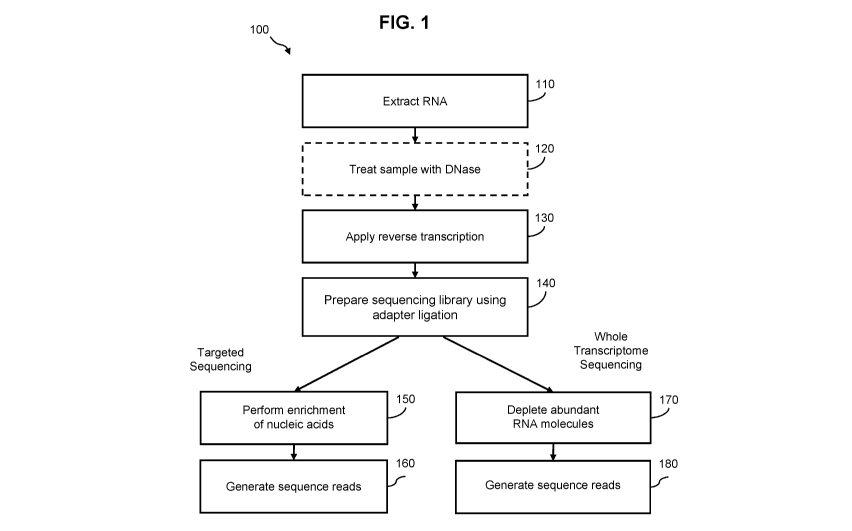
[0080] FIG. 1 is flowchart of a method 100 for preparing a nucleic acid
sample for sequencing
according to one embodiment. The method 100 includes, but is not limited to,
the following
steps. For example, any step of the method 100 may comprise a quantitation sub-
step for quality
control or other laboratory assay procedures known to one skilled in the art.
[0081] In step 110, a ribonucleic acid (RNA) sample is extracted from a
subject. The RNA
sample may comprise the whole human transcriptome, or any subset of the human
transcriptome. The sample may be extracted from a subject known to have or
suspected of
having a disease (e.g., cancer). The sample may include blood, plasma, serum,
urine, fecal,
-20-
CA 03121923 2021-06-02
WO 2020/132144
PCT/US2019/067287
saliva, other types of bodily fluids, or any combination thereof. In some
embodiments, methods
for drawing a blood sample (e.g., syringe or finger prick) may be less
invasive than procedures
for obtaining a tissue biopsy, which may require surgery. The extracted sample
may further
comprise cfDNA. If a subject has a disease (e.g., cancer), cfRNA in an
extracted sample may
be present at a detectable level for diagnosis.
[0082] In step 120, the nucleic acid sample including RNA molecules is
optionally treated with
a DNase enzyme. The DNase may remove DNA molecules from the nucleic acid
sample to
reduce DNA contamination of the RNA molecules. After RNA molecules are
converted into
DNA, it may be difficult to distinguish the RNA-converted DNA and genomic DNA
originally
found in the nucleic acid sample. Applying the DNase allows for targeted
amplification of
molecules originating from cfRNA. The DNase process may include steps for
adding a DNase
buffer, mixing the sample applied with DNase using a centrifuge, and
incubation. In some
embodiments, step 120 includes one or more processes based on the DNase
treatment protocol
described in the Qiagen QIAamp Circulating Nucleic Acid Handbook.
[0083] In step 130, a reverse transcriptase enzyme is used to convert the
RNA molecules in the
nucleic acid sample into complementary DNA (cDNA). The reverse transcriptase
process may
include a first-strand synthesis step (generation of a cDNA strand via reverse
transcription),
degradation of the RNA strand to produce a single-stranded cDNA molecule, and
synthesis of
a double-stranded DNA molecules from the single-stranded cDNA molecule using a
polymerase. During first-strand synthesis, a primer anneals to the 3' end of a
RNA molecule.
During second-strand synthesis, a different primer anneals to the 3' end of
the cDNA molecule.
[0084] In step 140, a sequencing library is prepared. For example, as is
well known in the art,
adapters can be ligated to one or both ends of a dsDNA molecule to prepare a
library for
sequencing. In one embodiment, the adapters utilized may include one or more
sequencing
oligonucleotides for use in subsequent cluster generation and/or sequencing
(e.g., known P5
and P7 sequences for used in sequencing by synthesis (SBS) (Illumina, San
Diego, CA)). In
another embodiment, the adapter includes a sample specific index sequence,
such that, after
library preparation, the library can be combined with one or more other
libraries prepared from
individual samples, thereby allowing for multiplex sequencing. The sample
specific index
sequence can comprise a short oligonucleotide sequence having a length of from
about 2 nt to
about 20 nt, from about 2 nt to about 10 nt, from about 2 to about 8 nt, or
from about 2 to about
6 nt. In another embodiment, the sample specific index sequence can comprise a
short
oligonucleotide sequence greater than about 2, 3, 4, 5, 6, 7, or 8 nucleotides
(nt) in length.
-21-
CA 03121923 2021-06-02
WO 2020/132144
PCT/US2019/067287
[0085] Optionally, during library preparation, unique molecular identifiers
(UMI) can be added
to the nucleic acid molecules in the sample through adapter ligation. The UMIs
are short nucleic
acid sequences (e.g., 4-10 base pairs) that are added to one or both ends of
nucleic acid
fragments during adapter ligation. In some embodiments, UMIs are degenerate
base pairs that
serve as a unique tag that can be used to identify sequence reads originating
from a specific
nucleic acid fragment. During PCR amplification following adapter ligation,
the UMIs are
replicated along with the attached nucleic acid fragment, which provides a way
to identify
sequence reads that came from the same original nucleic acid molecule in
downstream analysis.
[0086] For embodiments including targeted sequencing of RNA, in step 150,
targeted nucleic
acid sequences are enriched from the library. During enrichment, hybridization
probes (also
referred to herein as "probes") are used to target, and pull down, nucleic
acid fragments
informative for the presence or absence of a disease (e.g., cancer), disease
status (e.g., cancer
status), or a disease classification (e.g., cancer type or tissue of origin).
For a given workflow,
the probes may be designed to anneal (or hybridize) to a target
(complementary) nucleic acid
strand (e.g., a DNA strand converted from RNA). The probes may range in length
from 10s,
100s, or 1000s of base pairs. In one embodiment, the probes are designed based
on a gene panel
to analyze particular target regions of the genome (e.g., of the human or
another organism) that
are suspected to correspond to certain cancers or other types of diseases.
Moreover, the probes
may cover overlapping portions of a target region. In other embodiments,
targeted RNA
molecules can be enriched using hybridization probes prior to conversion of
the RNA
molecules to cDNA strands using reverse transcriptase (not shown). In general,
any known
method in the art can be used to isolate, and enrich for, probe-hybridized
target nucleic acids.
For example, as is well known in the art, a biotin moiety can be added to the
5'-end of the
probes (i.e., biotinylated) to facilitate isolation of target nucleic acids
hybridized to probes
using a streptavidin-coated surface (e.g., streptavidin-coated beads).
[0087] Additionally, for targeted sequencing, in step 160, sequence reads
are generated from
the enriched nucleic acid sample. Sequencing data may be acquired from the
enriched DNA
sequences (i.e., DNA sequences derived, or converted, from RNA sequences) by
known means
in the art. For example, the method 100 may include next generation sequencing
(NGS)
techniques including synthesis technology (Illumina), pyrosequencing (454 Life
Sciences), ion
semiconductor technology (Ion Torrent sequencing), single-molecule real-time
sequencing
(Pacific Biosciences), sequencing by ligation (SOLiD sequencing), nanopore
sequencing
(Oxford Nanopore Technologies), or paired-end sequencing. In some embodiments,
massively
-22-
CA 03121923 2021-06-02
WO 2020/132144
PCT/US2019/067287
parallel sequencing is performed using sequencing-by-synthesis with reversible
dye
terminators.
[0088] In other embodiments, for example, in a whole transcriptome
sequencing approach
(e.g., instead of targeted sequencing), in step 170, abundant RNA species are
depleted from the
nucleic acid sample. For example, in some embodiments, ribosomal RNA (rRNA)
and/or
transfer RNA (tRNA) species can be depleted. Available commercial kits, such
as
RiboMinusTm (ThermoFisher Scientific) or AnyDeplete (NuGen), can be used for
depletion of
abundant RNA species. In an embodiment, after depletion of nucleic acids
(e.g., converted
DNA) derived from abundant RNA molecules, sequence reads are generated in step
180.
[0089] In some embodiments, the sequence reads may be aligned to a
reference genome using
known methods in the art to determine alignment position information. The
alignment position
information may indicate a beginning position and an end position of a region
in the reference
genome that corresponds to a beginning nucleotide base and end nucleotide base
of a given
sequence read. Alignment position information may also include sequence read
length, which
can be determined from the beginning position and end position. A region in
the reference
genome may be associated with a gene or a segment of a gene. The reference
genome may
comprise the whole transcriptome, or any portion thereof (e.g., a plurality of
targeted
transcripts). In another embodiment, the reference genome can be the whole
genome from an
organism being tested and sequence reads derived from (or reverse transcribed
from) extracted
RNA molecules are aligned to the reference genome to determine location,
fragment length,
and/or start and end positions. For example, in one embodiment, sequence reads
are aligned to
human reference genome hg19. The sequence of the human reference genome, hg19,
is
available from Genome Reference Consortium with a reference number,
GRCh37/hg19, and
also available from Genome Browser provided by Santa Cruz Genomics Institute.
The
alignment position information may indicate a beginning position and an end
position of a
region in the reference genome that corresponds to a beginning nucleotide base
and end
nucleotide base of a given sequence read. Alignment position information may
also include
sequence read length, which can be determined from the beginning position and
end position.
A region in the reference genome may be associated with a gene or a segment of
a gene.
Identification of dark channel RNA molecules
[0090] Aspects of the invention include computer-implemented methods for
identifying one or
more RNA sequences indicative of a disease state in a subject (or "dark
channel RNA
molecules"). In some embodiments, the methods involve obtaining, by a computer
system, a
-23-
CA 03121923 2021-06-02
WO 2020/132144
PCT/US2019/067287
first set of sequence reads from a plurality of RNA molecules from a first
test sample obtained
from a subject known to have the disease, wherein the first test sample
comprises a plurality of
cell-free RNA (cfRNA) molecules, and a second set of sequence reads from a
plurality of RNA
molecules from a control sample, detecting, one or more RNA sequences that are
present in the
first set of sequence reads, and that are not present in the second set of
sequence reads, to
identify one or more RNA sequences that are indicative of the disease state.
In some
embodiments, the first test sample obtained from the patient comprises a
bodily fluid (e.g.,
blood, plasma, serum, urine, saliva, pleural fluid, pericardial fluid,
cerebrospinal fluid (C SF),
peritoneal fluid, or any combination thereof). In one preferred embodiment, a
test sample
obtained from the patient is a plasma sample. In some embodiments, the control
sample
comprises a plurality of RNA molecules obtained from healthy cells from the
subject (e.g.,
white blood cells).
[0091] FIG. 2 is a flow diagram illustrating a method for identifying one
or more RNA
sequences indicative of a disease state, in accordance with one embodiment of
the present
invention. As shown in FIG. 2, at step 210, a first set of sequence reads is
obtained from a
biological test sample comprising a plurality of cell-free RNA (cfRNA)
molecules. The cell-
free containing biological test sample can be any a bodily fluid, such as,
blood, plasma, serum,
urine, pleural fluid, cerebrospinal fluid, tears, saliva, or ascitic fluid. In
accordance with this
embodiment, the cfRNA biological test sample is obtained from a test subject
known to have,
or suspected of having a disease, the cfRNA molecules extracted from the
sample and sequence
reads determined (as described elsewhere herein). For example, in one
embodiment, a
complementary DNA strand is synthesized using a reverse transcription step
generating a
cDNA/RNA hybrid molecule, the RNA molecule degraded, a double stranded DNA
molecule
synthesized from the cDNA strand using a polymerase, a sequencing library
prepared, and
sequence reads determined using a sequencing platform. The sequencing step can
be any
carried out using any known sequencing platform in the art, such as, any
massively parallel
sequencing platform, including a sequencing-by-synthesis platform (e.g.,
Illumina's IIiSeq X)
or a sequencing-by-ligation platfon-n (e.g. the Life Technologies SOLiD
platform), the Ion
Torrent/Ion Proton, semiconductor sequencing, Roche 454, single molecular
sequencing
platforms (e.g. Helicos, Pacific Biosciences and nanopore), as previously
described.
Alternatively, other means for detecting and quantifying the sequence reads
can be used, for
example, array-based hybridization, probe-based in-solution hybridization,
ligation-based
assays, primer extension reaction assays, can be used to determine sequence
reads from DNA
-24-
CA 03121923 2021-06-02
WO 2020/132144
PCT/US2019/067287
molecules (e.g., converted from RNA molecules), as one of skill in the art
would readily
understand.
[0092] At step 220, a second set of sequence reads is obtained from a
healthy control sample.
In one embodiment, the healthy control sample is from the same subject and
comprises a
plurality of cellular RNA molecules. For example, the control sample can be
blood cells, such
as white blood cells, and the plurality of sequence reads derived from RNA
molecules extracted
from the blood cells. In accordance with this embodiment, the RNA molecules
are extracted
from the healthy control sample (e.g., blood cells), converted to DNA, a
sequencing library
prepared, and the second set of sequence reads determined (as described
elsewhere herein). In
other embodiments, the healthy control sample can be a database of sequence
data determined
for RNA sequences obtained from a healthy subject, or from healthy cells.
[0093] At step 230, sequence reads from the first set of sequence reads and
the second set of
sequence reads are compared to identifying one or more RNA molecules
indicative of a disease
state. Moreover, one or more sequence reads (derived from RNA molecules)
present in the first
set of sequence reads, and not present in the second set of sequence reads,
are identified as
derived from RNA molecules indicative of a disease state. For example, the
first set of sequence
reads can comprise sequence reads derived from cfRNA molecules from a plasma
sample
obtained from a subject known to have, or suspected of having, a disease
(e.g., cancer). And
the second set of sequence reads can comprise sequence reads derived from RNA
molecules
from healthy cells (e.g., white blood cells). By comparing, and removing, the
second set of
sequence reads derived from healthy cells from the first set of sequence reads
derived from a
cell-free RNA sample, one can identify the sequence reads derived from a
disease state (e.g.,
cancer).
[0094] In some embodiments, a control data set of RNA sequences includes a
plurality of
sequence reads obtained from one or more healthy subjects. In certain
embodiments, the second
set of sequence reads comprises RNA sequence information obtained from a
public database.
Public databases that can be used in accordance with embodiments of the
invention include the
tissue RNA-seq database GTEx (available at gtexportal.org/home). In some
embodiments, a
control data set of RNA sequences includes a plurality of sequence reads
obtained from a
plurality of blood cells from the subject. For example, in some embodiments, a
plurality of
sequence reads are obtained from a subject's white blood cells (WBCs).
-25-
CA 03121923 2021-06-02
WO 2020/132144
PCT/US2019/067287
Detection of tumor-derived RNA molecules
[0095] Aspects of the invention include computer-implemented methods for
detecting one or
more tumor-derived RNA molecules in a subject. In some embodiments, the
methods involve:
obtaining, by a computer system, a first set of sequence reads from a
plurality of RNA
molecules from a first test sample from a subject known to have a tumor,
wherein the first test
sample comprises a plurality of cell-free RNA (cfRNA) molecules; obtaining, by
a computer
system, a second set of sequence reads from a plurality of RNA molecules from
a plurality of
blood cells from the subject; and detecting, by a computer system, one or more
RNA sequences
that are present in the first set of sequence reads, and that are not present
in the second set of
sequence reads, to detect the one or more tumor-derived RNA molecules in the
subject.
[0096] In some embodiments, the first test sample obtained from the patient
comprises blood,
plasma, serum, urine, saliva, pleural fluid, pericardial fluid, cerebrospinal
fluid (C SF),
peritoneal fluid, or any combination thereof. In one preferred embodiment, a
test sample
obtained from the patient is a plasma sample. In some embodiments, the
plurality of blood cells
obtained from the subject are white blood cells (WBCs).
[0097] FIG. 3 is a flow diagram illustrating a method for identifying one
or more tumor-
derived RNA sequences, in accordance with one embodiment of the present
invention. At step
310, a first set of sequence reads is obtained from a biological test sample
comprising a plurality
of cell-free RNA (cfRNA) molecules. In accordance with this embodiment, the
cfRNA
biological test sample is obtained from a test subject known to have, or
suspected of having a
disease, the cfRNA molecules extracted from the sample and sequence reads
determined (as
described elsewhere herein). For example, in one embodiment, a complementary
DNA strand
is synthesized using a reverse transcription step generating a cDNA/RNA hybrid
molecule, the
RNA molecule degraded, a double stranded DNA molecule synthesized from the
cDNA strand
using a polymerase, a sequencing library prepared, and sequence reads
determined using a
sequencing platform. The sequencing step can be any carried out using any
known sequencing
platform in the art, as previously described. Alternatively, other means for
determining the
sequence reads can be used, for example, array-based hybridization, probe-
based in-solution
hybridization, ligation-based assays, primer extension reaction assays, can be
used to detect
and/or quantify sequence reads obtained from DNA molecules (e.g., converted
from RNA
molecules), as one of skill in the art would readily understand.
[0098] At step 315, a second set of sequence reads is obtained from blood
cells (e.g., white
blood cells or buffy coat). In one embodiment, the blood cells are obtained
from the same
subject and RNA molecules extracted therefrom. In accordance with this
embodiment, the
-26-
CA 03121923 2021-06-02
WO 2020/132144
PCT/US2019/067287
RNA molecules are extracted from the blood cells, converted to DNA, a
sequencing library
prepared, and the second set of sequence reads determined (as described
elsewhere herein). In
general, any known method in the art can be used to extract and purify cell-
free nucleic acids
from the test sample. For example, cell-free nucleic acids can be extracted
and purified using
one or more known commercially available protocols or kits, such as the QIAamp
circulating
nucleic acid kit (Qiagen).
[0099] At step 320, one or more tumor-derived RNA molecules is detected
when one or more
RNA sequences are present in the first set of sequence reads and not present
in the second set
of sequence reads. Moreover, one or more sequence reads (derived from RNA
molecules)
present in the first set of sequence reads, and not present in the second set
of sequence reads,
are identified as derived from RNA molecules indicative of a disease state.
For example, the
first set of sequence reads can comprise sequence reads derived from cfRNA
molecules from
a plasma sample obtained from a subject known to have, or suspected of having,
a disease (e.g.,
cancer). And the second set of sequence reads can comprise sequence reads
derived from RNA
molecules from blood cells (e.g., white blood cells). By comparing, and
removing, the second
set of sequence reads derived from blood cells from the first set of sequence
reads derived from
a cell-free RNA sample, one can identify the sequence reads derived from a
tumor.
Detecting a disease state using a dark channel RNA molecules
101001 FIG. 4 is a flow diagram illustrating a method for detecting the
presence of cancer,
determining a state of cancer, monitoring cancer progression, and/or
determining cancer type
in a subject, in accordance with one embodiment of the present invention. At
step 410, a
biological test sample is extracted from a subject. As previously described,
in one embodiment,
the test sample can be a bodily fluid (e.g., blood, plasma, serum, urine,
saliva, pleural fluid,
pericardial fluid, cerebrospinal fluid (C SF), peritoneal fluid, or any
combination thereof)
comprising a plurality of cell-free RNA molecules.
101011 At step 415, a plurality of cell-free RNA molecules are extracted
from the test sample
and a sequencing library prepared. In general, any known method in the art can
be used to
extract and purify cell-free nucleic acids from the test sample. For example,
cell-free nucleic
acids (cfRNA molecules) can be extracted and purified using one or more known
commercially
available protocols or kits, such as the QIAamp circulating nucleic acid kit
(Qiagen). After
extraction, the cfRNA molecules are used to prepare a sequencing library. In
one embodiment,
a reverse transcription step is used to produce a plurality of cDNA/RNA hybrid
molecules, the
RNA strand degraded to produce a single-stranded cDNA molecule, a second
strand
-27-
CA 03121923 2021-06-02
WO 2020/132144
PCT/US2019/067287
synthesized to produce a plurality of double-stranded DNA molecules from the
single-stranded
cDNA molecule templates, and DNA adapters ligated to the plurality of double-
stranded DNA
molecules to generate a sequencing library. As previously described, the DNA
adapters may
include one or more sequencing oligonucleotides for use in subsequent cluster
generation
and/or sequencing (e.g., known P5 and P7 sequences for used in sequencing by
synthesis (SBS)
(IIlumina, San Diego, CA)). In another embodiment, the adapter includes a
sample specific
index sequence, such that, after library preparation, the library can be
combined with one or
more other libraries prepared from individual samples, thereby allowing for
multiplex
sequencing. In another embodiment, unique molecular identifiers (UMI) are
added through
adapter ligation.
[0102] At step 420, a sequencing reaction is performed to generate a
plurality of sequence
reads. In general, any method known in the art can be used to obtain sequence
data or sequence
reads from the sequencing library. For example, in one embodiment, sequencing
data or
sequence reads from the sequencing library can be acquired using next
generation sequencing
(NGS). Next-generation sequencing methods include, for example, sequencing by
synthesis
technology (IIlumina), pyrosequencing (454), ion semiconductor technology (Ion
Torrent
sequencing), single-molecule real-time sequencing (Pacific Biosciences),
sequencing by
ligation (SOLiD sequencing), and nanopore sequencing (Oxford Nanopore
Technologies). In
some embodiments, sequencing is massively parallel sequencing using sequencing-
by-
synthesis with reversible dye terminators. In other embodiments, sequencing is
sequencing-by-
ligation. In yet other embodiments, sequencing is single molecule sequencing.
In still another
embodiment, sequencing is paired-end sequencing. Optionally, an amplification
step can be
performed prior to sequencing.
[0103] At step 425, sequence reads obtained from the cfRNA sample are
filtered to generate a
list of non-excluded sequence reads and the non-excluded sequence reads
quantified at step
430. For example, as described elsewhere herein, the sequence reads obtained
from the cfRNA
sample can be filtered to exclude sequence known to be present in healthy
cells. In one
embodiment, RNA molecules extracted from healthy cells (e.g., white blood
cells) are
sequenced deriving sequence reads that are excluded from the cfRNA derived
sequence reads
to obtain non-excluded sequence reads. In another embodiment, RNA sequencing
data from a
database (e.g., a public database) can be used to filter out or exclude
sequences known to be
present in healthy cells reads comprises to obtain non-excluded sequence
reads.
[0104] At step 435, a disease state is detected when the quantified non-
excluded sequence
reads exceed a threshold. In certain embodiments, the threshold value is an
integer that ranges
-28-
CA 03121923 2021-06-02
WO 2020/132144
PCT/US2019/067287
from about 1 to about 10, such as about 2, 3, 4, 5, 6, 7, 8, or about 9. In
some embodiments,
the threshold is a non-integer value, ranging from about 0.1 to about 0.9,
such as about 0.2, 0.3,
0.4, 0.5, 0.6, 0.7 or about 0.8.
[0105] Aspects of the invention relate to methods for detecting a presence
of a cancer,
determining a cancer stage, monitoring a cancer progression, and/or
determining a cancer type
in a subject known to have, or suspected of having a cancer. In some
embodiments, the methods
involve: (a) obtaining a plurality of sequence reads from a plurality of cfRNA
molecules in a
biological test sample from the subject; (b) quantitatively detecting the
presence of one or more
sequences derived from one or more RNA markers in the biological test sample
to determine a
tumor RNA score, wherein the one or more RNA markers are selected from the
group
consisting of one or more targeted RNA molecules; and (c) detecting the
presence of the cancer,
determining the cancer stage, monitoring the cancer progression, and/or
determining the cancer
type in the subject when the tumor RNA score exceeds a threshold value. In
certain
embodiments, the threshold value is an integer that ranges from about 1 to
about 10, such as
about 2, 3, 4, 5, 6, 7, 8, or about 9. In some embodiments, the threshold is a
non-integer value,
ranging from about 0.1 to about 0.9, such as about 0.2, 0.3, 0.4, 0.5, 0.6,
0.7 or about 0.8.
[0106] Quantitative detection methods in accordance with embodiments of the
invention can
include nucleic acid sequencing procedures, such as next-generation
sequencing. In certain
embodiments, sequencing can involve whole transcriptome sequencing. In certain
embodiments, sequencing can involve enriching a sample for one or more
targeted RNA
sequences of interest prior to conducting the sequencing procedure.
Alternatively, other means
for detecting and quantifying sequence reads can be used, for example, array-
based
hybridization, probe-based in-solution hybridization, ligation-based assays,
primer extension
reaction assays, can be used to determine sequence reads from DNA molecules
(e.g., converted
from RNA molecules), as one of skill in the art would readily understand.
[0107] FIG. 5 is a flow diagram illustrating a method for detecting a
disease state from one or
more sequence reads derived from one or more targeted RNA molecules, in
accordance with
another embodiment of the present invention. At step 510, a biological test
sample comprising
a plurality of cell-free RNA molecules is obtained. In one embodiment, the
biological test
sample is a bodily fluid (e.g., a blood, plasma, serum, urine, saliva, pleural
fluid, pericardial
fluid, cerebrospinal fluid (CSF), peritoneal fluid sample, or any combination
thereof).
[0108] At step 515, the presence of one or more nucleic acid sequence
derived from one or
more target RNA molecules in the biological test sample are detected, and
quantified, to
determine a tumor RNA score. As described elsewhere herein, nucleic acids
derived from RNA
-29-
CA 03121923 2021-06-02
WO 2020/132144
PCT/US2019/067287
molecules can be detected and quantified using any known means in the art. For
example, in
accordance with one embodiment, nucleic acids derived from RNA molecules are
detected and
quantified using a sequencing procedure, such as a next-generation sequencing
platform (e.g.,
HiSeq or NovaSeq, Illumina, San Diego, CA). In other embodiments, nucleic
acids derived
from RNA molecules are detected and quantified using a microarray, reverse
transcription
PCR, real-time PCR, quantitative real-time PCR, digital PCR, digital droplet
PCR, digital
emulsion PCR, multiplex PCR, hybrid capture, oligonucleotide ligation assays,
or any
combination thereof. As described elsewhere, in one embodiment, cell-free
nucleic acids
(cfRNA molecules) can be extracted and purified using one or more known
commercially
available protocols or kits, such as the QIAamp circulating nucleic acid kit
(Qiagen). After
extraction, the cfRNA molecules are used to prepare a sequencing library. In
one embodiment,
a reverse transcription step is used to produce a plurality of cDNA/RNA hybrid
molecules, the
RNA strand degraded to produce a single-stranded cDNA molecule, a second
strand
synthesized to produce a plurality of double-stranded DNA molecules from the
single-stranded
cDNA molecule templates. Optionally, in one embodiment, one or more targeted
RNA
molecules (or DNA molecules derived therefrom) are enriched prior to detection
and
quantification, as described elsewhere herein.
101091 In one embodiment, the tumor RNA score is the quantity or count of
targeted RNA
molecules (or sequence reads obtained from DNA molecules derived from the
targeted RNA
molecules) detected. In another embodiment, the tumor RNA score comprises a
mean, a mode,
or an average of the total number of targeted RNA molecules (or sequence reads
obtained from
DNA molecules derived from the targeted RNA molecules) detected divided by the
total
number of genes from which RNA molecules are targeted. In still other
embodiments, the
tumor RNA score is determined by inputting the sequence reads into a
prediction model, and
the tumor RNA score output as a likelihood or probability, as described
elsewhere herein.
[0110] At step 520, the presence of cancer is detected, a state of cancer
determined, cancer
progression monitored, and/or a cancer type determined in a subject when the
tumor RNA score
exceeds a threshold. The threshold value can be an integer that ranges from
about 1 to about
10, such as about 2, 3, 4, 5, 6, 7, 8, or about 9. In some embodiments, the
threshold is a non-
integer value, ranging from about 0.1 to about 0.9, such as about 0.2, 0.3,
0.4, 0.5, 0.6, 0.7 or
about 0.8. Alternatively, when the tumor RNA score is output from a prediction
model, the
output can simply be a likelihood or probability indicating the likelihood or
probability that the
subject has cancer, or a cancer type.
-30-
CA 03121923 2021-06-02
WO 2020/132144
PCT/US2019/067287
Cancer indicator score
[0111] Aspects of the invention are directed to computer-implemented
methods for detecting
the presence of a cancer in a patient. In some embodiments, the methods
involve: receiving a
data set in a computer comprising a processor and a computer-readable medium,
wherein the
data set comprises a plurality of sequence reads obtained by sequencing a
plurality of nucleic
acid molecules (e.g., DNA molecules) derived from a plurality of targeted
ribonucleic acid
(RNA) molecules in a biological test sample from the patient, and wherein the
computer-
readable medium comprises instructions that, when executed by the processor,
cause the
computer to: determine an expression level for the plurality of targeted RNA
molecules from
the biological test sample; comparing the expression level of each of the
targeted RNA
molecules to an RNA tissue score matrix to determine a cancer indicator score
for each targeted
RNA molecule; aggregate the cancer indicator score for each targeted RNA
molecule to
generate a cancer indicator score for the biological test sample; and
detecting the presence of
the cancer in the patient when the cancer indicator score for the biological
test sample exceeds
a threshold value.
[0112] In some embodiments, the target RNA molecules have an expression
level in patients
with a known cancer status that exceeds their expression level in healthy
patients. In certain
embodiments, an expression level of a target RNA molecule in a patient with a
known cancer
status ranges from about 2 to about 10 times greater, such as about 3, 4, 5,
6, 7, 8, or about 9
times greater, than the expression level of the target RNA molecule in a
healthy patient. In
certain embodiments, a target RNA molecule is not detectable in a biological
test sample from
a healthy patient, i.e., the target RNA molecule has an undetectable
expression level.
[0113] In some embodiments, the number of target RNA molecules in the
biological test
sample ranges from about 1 to about 2000, from about 10 to about 1000, from
about 10 to about
500, or from about 10 to about 500. In other embodiments, the number of target
RNA molecules
ranges from about 1 to about 50, from about 1 to about 40, from about 1 to
about 30, or from
about 1 to about 20, such as about 2, 3, 4, 5,6, 7, 8,9, 10, 11, 12, 13, 14,
15, 16, 17, 18, 19, or
about 20.
[0114] In some embodiments, the cancer indicator score comprises an
aggregate of the total
number of targeted RNA molecules (or sequence reads obtained from DNA
molecules derived
from the targeted RNA molecules) detected from the biological test sample. In
another
embodiment, the cancer indicator score comprises a mean, a mode, or an average
of the total
number of targeted RNA molecules (or sequence reads obtained from DNA
molecules derived
from the targeted RNA molecules) detected divided by the total number of genes
from which
-31-
CA 03121923 2021-06-02
WO 2020/132144
PCT/US2019/067287
RNA molecules are targeted. In still other embodiments, the cancer indicator
score is
determined by inputting the sequence reads into a prediction model, and the
cancer indicator
score output as a likelihood or probability, as described elsewhere herein.
[0115] In some embodiments, the threshold value is an integer that ranges
from about 1 to
about 10, such as about 2, 3, 4, 5, 6, 7, 8, or about 9. In some embodiments,
the threshold is a
non-integer value, ranging from about 0.1 to about 0.9, such as about 0.2,
0.3, 0.4, 0.5, 0.6, 0.7
or about 0.8. In other embodiments, the threshold value ranges from about 0.5
to about 5 reads
per million (RPM), such as about 1, 1.5, 2, 2.5, 3, 3.5, 4, or about 4.5 RPM.
The cancer locator
score threshold value can be determined based on the quantity of targeted RNA
molecules (or
sequence reads derived therefrom) detected in a control sample, for example a
healthy subject
or a subject with a known disease state. Alternatively, when the cancer
locator score is output
from a prediction model, the output can simply be a likelihood or probability
indicating the
likelihood or probability that the subject has cancer, or a cancer type.
[0116] FIG. 6 is a flow diagram illustrating a method for detecting the
presence of cancer in a
subject based on a cancer indicator score, in accordance with one embodiment
of the present
invention. At step 610, a data set is received comprising a plurality of
sequence reads derived
from a plurality of cfRNA molecules in a biological test sample. For example,
a plurality of
sequence reads can be determined for a plurality of cfRNA molecules extracted
from a
biological test sample, as described herein. Moreover, cfRNA molecules are
reverse
transcribed to create DNA molecules and the DNA molecules sequenced to
generate sequence
reads.
[0117] At step 615, an expression level is determined for a plurality of
target RNA molecules
in the biological test sample. For example, in one embodiment, the expression
level of targeted
RNA molecules can be determined based on quantification of detected sequence
reads derived
from a targeted RNA molecules of interest.
[0118] At step 620, the expression level of each of the target RNA
molecules is compared to
an RNA tissue score matrix to determine a cancer indicator score for each
target RNA
molecule. The RNA tissue score matrix can be determined from a training set
comprising
sequence reads derived from a plurality of cancer training samples with known
cancer status.
[0119] At step 625, the cancer indicator scores for each target RNA
molecule are aggregated
to generate a cancer indicator score. In some embodiments, the cancer
indicator score
comprises an aggregate of the total number of targeted RNA molecules (or
sequence reads
obtained from DNA molecules derived from the targeted RNA molecules) detected
from the
biological test sample. In another embodiment, the cancer indicator score
comprises a mean, a
-32-
CA 03121923 2021-06-02
WO 2020/132144
PCT/US2019/067287
mode, or an average of the total number of targeted RNA molecules (or sequence
reads
obtained from DNA molecules derived from the targeted RNA molecules) detected
divided by
the total number of genes from which RNA molecules are targeted.
[0120] At step 630, detect the presence of cancer in a subject when the
cancer indicator score
for the test sample exceeds a threshold. As described above, in one
embodiment, the threshold
value is an integer that ranges from about 1 to about 10, such as about 2, 3,
4, 5, 6, 7, 8, or
about 9. In some embodiments, the threshold is a non-integer value, ranging
from about 0.1 to
about 0.9, such as about 0.2, 0.3, 0.4, 0.5, 0.6, 0.7 or about 0.8. In other
embodiments, the
threshold value ranges from about 0.5 to about 5 reads per million (RPM), such
as about 1, 1.5,
2, 2.5, 3, 3.5, 4, or about 4.5 RPM.
[0121] Aspects of the invention include methods for determining a cancer
cell type or tissue of
origin of the cancer in the patient based on the expression level of one or
more of the target
RNA molecules, the cancer indicator score for one or more of the target RNA
molecules, the
cancer indicator score for the biological test sample, or any combination
thereof. In certain
embodiments, the methods further involve therapeutically classifying a patient
into one or more
of a plurality of treatment categories based on the expression level of one or
more of the target
RNA molecules, the cancer indicator score for one or more of the target RNA
molecules, the
cancer indicator score for the biological test sample, or any combination
thereof.
[0122] In certain embodiments, the computer is configured to generate a
report that includes
an expression level of one or more of the target RNA molecules, a cancer
indicator score for
one or more of the target RNA molecules, a cancer indicator score for the
biological test
sample, an indication of the presence or absence of the cancer in the patient,
an indication of
the cancer cell type of tissue of origin of the cancer in the patient, a
therapeutic classification
for the patient, or any combination thereof.
Liver disease
[0123] In other aspects, the present invention is directed to methods that
can be used for the
assessment of NAFLD (non-alcoholic fatty liver disease) and those conditions
associated with
NAFLD, including fatty liver disease, alcoholic steatohepatitis (ASH),
nonalcoholic
steatohepatitis (NASH), fibrosis and cirrhosis through the identification of
one or more signals
indicative of, or informative for, NAFLD, ASH, NASH, fibrosis or cirrhosis in
a test sample.
In another embodiment, the methods disclosed herein can be used for the
detection, screening,
diagnosis, and/or monitoring of hepatocellular carcinoma (HCC). The term
"NAFLD" (non-
alcohol fatty liver disease) refers to a group of conditions where there is an
accumulation of
-33-
CA 03121923 2021-06-02
WO 2020/132144
PCT/US2019/067287
excess fat in the liver of people who drink little or no alcohol. The most
common form of
NAFLD is a condition called fatty liver disease. In fatty liver disease, fat
accumulates in the
liver cells. A small group of people with NAFLD may have a more serious
condition termed
non-alcoholic steatohepatitis (NASH). In NASH, fat accumulation is associated
with liver cell
inflammation and different degrees of scarring. Cirrhosis occurs when the
liver sustains
substantial damage, and the liver cells are gradually replaced by scar tissue
which results in the
inability of the liver to work properly. The use of the term "NAFLD" is used
to include all
conditions reflecting a form of non-alcohol fatty liver disease, including,
NASH.
[0124] FIG. 7 is a flow diagram illustrating a method for detecting a
presence of a liver disease,
determining a stage of a liver disease, and/or monitoring progression of a
liver disease in a
subject, in accordance with one embodiment of the present invention. At step
710, a biological
test sample comprising a plurality of cell-free RNA molecules is obtained. In
one embodiment,
the biological test sample is a bodily fluid (e.g., a blood, plasma, serum,
urine, saliva, pleural
fluid, pericardial fluid, cerebrospinal fluid (CSF), peritoneal fluid sample,
or any combination
thereof).
[0125] At step 715, the presence of one or more nucleic acid sequences
derived from one or
more target RNA molecules in the biological test sample are detected, and
quantified, to
determine a liver disease RNA score. As described elsewhere herein, nucleic
acids derived
from RNA molecules can be detected and quantified using any known means in the
art. For
example, in accordance with one embodiment, nucleic acids derived from RNA
molecules are
detected and quantified using a sequencing procedure, such as a next-
generation sequencing
platform (e.g., HiSeq or NovaSeq, Illumina, San Diego, CA). In other
embodiments, nucleic
acids derived from RNA molecules are detected and quantified using a
microarray, reverse
transcription PCR, real-time PCR, quantitative real-time PCR, digital PCR,
digital droplet
PCR, digital emulsion PCR, multiplex PCR, hybrid capture, oligonucleotide
ligation assays, or
any combination thereof. As described elsewhere, in one embodiment, cell-free
nucleic acids
(cfRNA molecules) can be extracted and purified using one or more known
commercially
available protocols or kits, such as the QIAamp circulating nucleic acid kit
(Qiagen). After
extraction, the cfRNA molecules are used to prepare a sequencing library. In
one embodiment,
a reverse transcription step is used to produce a plurality of cDNA/RNA hybrid
molecules, the
RNA strand degraded to produce a single-stranded cDNA molecule, a second
strand
synthesized to produce a plurality of double-stranded DNA molecules from the
single-stranded
cDNA molecule templates.
-34-
CA 03121923 2021-06-02
WO 2020/132144
PCT/US2019/067287
[0126] In one embodiment, the liver disease RNA score is the quantity or
count of targeted
RNA molecules (or sequence reads obtained from DNA molecules derived from the
targeted
RNA molecules) detected. In another embodiment, the liver disease RNA score
comprises a
mean, a mode, or an average of the total number of targeted RNA molecules (or
sequence reads
obtained from DNA molecules derived from the targeted RNA molecules) detected
divided by
the total number of genes from which RNA molecules are targeted. In still
other embodiments,
the liver disease RNA score is determined by inputting the sequence reads into
a prediction
model, and the liver disease RNA score output as a likelihood or probability,
as described
elsewhere herein.
[0127] At step 720, the presence of a liver disease is detected, a stage of
liver disease
determined, and/or liver disease progression monitored in a subject when the
liver disease RNA
score exceeds a threshold. The threshold value can be an integer that ranges
from about 1 to
about 10, such as about 2, 3, 4, 5, 6, 7, 8, or about 9. In some embodiments,
the threshold is a
non-integer value, ranging from about 0.1 to about 0.9, such as about 0.2,
0.3, 0.4, 0.5, 0.6, 0.7
or about 0.8. Alternatively, when the liver disease RNA score is output from a
prediction
model, the output can simply be a likelihood or probability indicating the
likelihood or
probability that the subject has cancer, or a cancer type.
Liver disease indicator score
[0128] Aspects of the invention are directed to computer-implemented
methods for detecting
the presence of a liver disease in a patient. In some embodiments, the methods
involve:
receiving a data set in a computer comprising a processor and a computer-
readable medium,
wherein the data set comprises a plurality of sequence reads obtained by
sequencing a plurality
of ribonucleic acid (RNA) molecules in a biological test sample from the
patient, and wherein
the computer-readable medium comprises instructions that, when executed by the
processor,
cause the computer to: determine an expression level of a plurality of target
RNA molecules in
the biological test sample; compare the expression level of each of the target
RNA molecules
to an RNA tissue score matrix to determine a liver disease indicator score for
each target RNA
molecule; aggregate the liver disease indicator score for each target RNA
molecule to generate
a liver disease indicator score for the biological test sample; and detect the
presence of the liver
disease in the patient when the liver disease indicator score for the
biological test sample
exceeds a threshold value. In some embodiments, the methods involve detecting
the presence
or absence of, determining the stage of, monitoring the progression of a liver
disease selected
from the group consisting of non-alcoholic fatty liver disease (NAFLD),
alcoholic
-35-
CA 03121923 2021-06-02
WO 2020/132144
PCT/US2019/067287
steatohepatitis (ASH), nonalcoholic steatohepatitis (NASH), fibrosis and
cirrhosis. In other
embodiments, the methods involve detecting the presence or absence of,
determining the stage
of, monitoring the progression of hepatocellular carcinoma (HCC).
[0129] In some embodiments, the target RNA molecules have an expression
level in patients
with a known liver disease status that exceeds their expression level in
healthy patients. In
certain embodiments, an expression level of a target RNA molecule in a patient
with a known
liver disease status ranges from about 2 to about 10 times greater, such as
about 3, 4, 5, 6, 7, 8,
or about 9 times greater, than the expression level of the target RNA molecule
in a healthy
patient. In certain embodiments, a target RNA molecule is not detectable in a
biological test
sample from a healthy patient, i.e., the target RNA molecule has an
undetectable expression
level.
[0130] In some embodiments, the number of target RNA molecules in the
biological test
sample ranges from about 1 to about 20, such as about 2, 3, 4, 5, 6, 7, 8, 9,
10, 11, 12, 13, 14,
15, 16, 17, 18 or about 19. In other embodiments, the threshold value ranges
from about 0.5 to
about 5 reads per million (RPM), such as about 1, 1.5, 2, 2.5, 3, 3.5,4, or
about 4.5 RPM.
[0131] FIG. 8 is a flow diagram illustrating a method for detecting the
presence of a liver
disease in a subject based on a liver disease indicator score, in accordance
with one embodiment
of the present invention. At step 810, a data set is received comprising a
plurality of sequence
reads derived from a plurality of cfRNA molecules in a biological test sample.
For example, a
plurality of sequence reads can be determined for a plurality of cfRNA
molecules extracted
from a biological test sample, as described herein.
[0132] At step 815, an expression level is determined for of a plurality of
target RNA molecules
in the biological test sample. For example, in one embodiment, the expression
level of targeted
RNA molecules can be determined based on quantification of detected sequence
reads derived
from a targeted RNA molecules of interest, or that align to a targeted RNA
molecule of interest.
[0133] At step 820, the expression level of each of the target RNA
molecules is compared to
an RNA tissue score matrix to determine a liver disease indicator score for
each target RNA
molecule. The RNA tissue score matrix can be determined from a training set
comprising
sequence reads derived from a plurality of liver disease training samples with
known liver
disease status.
[0134] At step 825, the liver disease indicator scores for each target RNA
molecule are
aggregated to generate a liver disease indicator score. In some embodiments,
the liver disease
indicator score comprises an aggregate of the total number of targeted RNA
molecules (or
sequence reads obtained from DNA molecules derived from the targeted RNA
molecules)
-36-
CA 03121923 2021-06-02
WO 2020/132144
PCT/US2019/067287
detected from the biological test sample. In another embodiment, the liver
disease indicator
score comprises a mean, a mode, or an average of the total number of targeted
RNA molecules
(or sequence reads obtained from DNA molecules derived from the targeted RNA
molecules)
detected divided by the total number of genes from which RNA molecules are
targeted. In still
other embodiments, the liver disease indicator score is determined by
inputting the sequence
reads into a prediction model, and the liver disease indicator score output as
a likelihood or
probability, as described elsewhere herein.
101351 At step 830, detect the presence of a liver disease in a subject
when the liver disease
indicator score for the test sample exceeds a threshold. As described above,
in one embodiment,
the threshold value is an integer that ranges from about 1 to about 10, such
as about 2, 3, 4, 5,
6, 7, 8, or about 9. In some embodiments, the threshold is a non-integer
value, ranging from
about 0.1 to about 0.9, such as about 0.2, 0.3, 0.4, 0.5, 0.6, 0.7 or about
0.8. In other
embodiments, the threshold value ranges from about 0.5 to about 5 reads per
million (RPM),
such as about 1, 1.5, 2, 2.5, 3, 3.5, 4, or about 4.5 RPM. Alternatively, when
the liver disease
indicator score is output from a prediction model, the output can simply be a
likelihood or
probability indicating the likelihood or probability that the subject has
cancer, or a cancer type
RNA tissue matrix score
[0136] Aspects of the invention include methods for constructing an RNA
tissue score matrix.
In some embodiments, the methods involve compiling a plurality of RNA sequence
reads
obtained from a plurality of patients to generate an RNA expression matrix,
and normalizing
the RNA expression matrix with a tissue-specific RNA expression matrix to
construct the RNA
tissue score matrix. In certain embodiments, the tissue-specific RNA
expression matrix
comprises a plurality of reference human tissues. In certain embodiments, the
RNA sequence
reads are obtained from a plurality of healthy patients to construct a healthy
RNA tissue score
matrix. In certain embodiments, the RNA sequence reads are obtained from a
plurality of
patients having a known cancer type to construct a cancer RNA tissue score
matrix.
RNA markers and analysis technique
[0137] Methods in accordance with some embodiments of the invention can be
performed on
cfRNA molecules and/or ctRNA molecules. In some embodiments, RNA molecules
that are
used in the subject methods include RNA molecules from cancerous and non-
cancerous cells.
[0138] In embodiments, methods include: (a) measuring a plurality of target
cell-free RNA
(cfRNA) molecules in a sample of the subject, wherein the plurality of target
cfRNA molecules
-37-
CA 03121923 2021-06-02
WO 2020/132144
PCT/US2019/067287
are selected from transcripts of Tables 1-7; and (b) detecting the cancer,
wherein detecting the
cancer comprises detecting one or more of the target cfRNA molecules above a
threshold level.
In embodiments, the plurality of target cfRNA molecules are selected from at
least 2, 3, 4, 5,
10, 15, 20, 25, or more transcripts of genes listed in one or more of Tables 1-
7. Target cfRNA
molecules can be from genes selected from any one of these tables, or any
combination thereof.
In embodiments, the number of tables selected from among Tables 1-7 is 2, 3,
4, 5, or all tables.
In embodiments, the target cfRNA molecules that are measured are from fewer
than 500 genes
(e.g., fewer than 400, 300, 200, 100, or 50 genes).
101391 In some embodiments, one or more target cfRNA molecules are derived
from one or
more genes selected from the genes listed in Table 1. In embodiments, the one
or more target
cfRNA molecules includes at least 2, 3, 4, 5, 6, 7, 8, 9, 10, 15, 20, 25, or
30 genes from Table
1. In embodiments, the one or more target cfRNA molecules includes at least 5
genes from
Table 1. In embodiments, the one or more target cfRNA molecules includes at
least 10 genes
from Table 1. In embodiments, the one or more target cfRNA molecules includes
all of the
genes from Table 1. In embodiments, the one or more target cfRNA molecules
includes at
least one of the first 5 genes of Table 1 (AGR2, HOXC10, S100A7, BPIFA1, and
IDI2-AS1),
and optionally one or more additional genes from Table 1. In embodiments, the
one or more
target cfRNA molecules includes transcripts of the AGR2 gene. In embodiments,
the one or
more target cfRNA molecules includes transcripts of AGR2, HOXC10, Si 00A7,
BPIFA1, and
IDI2-AS1. In embodiments, the target cfRNA molecules that are measured are
from fewer
than 500 genes (e.g., fewer than 400, 300, 200, 100, or 50 genes). Table 1
below provides
examples of cancer dark channel biomarkers.
Table 1
AGR2 HOXC10 5100A7
BPIFA1 IDI2-AS1 SCGB2A2
CA5P14 KRT16P2 SERPINB5
CSN1S1 LALBA SFTA3
DISP2 LINC00163 SFTPA2
EIF2D NKX2-1 5LC34A2
FABP7 OPN1SW TFF1
GABRG1 PADI3 VTCN1
GNAT3 PTPRZ1 WFDC2
-38-
CA 03121923 2021-06-02
WO 2020/132144
PCT/US2019/067287
GRHL2 ROS1 MUC5B
SMIM22 CXCL17 RNU1-1
KLK5
101401 In some embodiments, one or more target cfRNA molecules are derived
from one or
more genes selected from the genes listed in Table 2. In embodiments, the one
or more target
cfRNA molecules includes at least 2, 3, 4, 5, 6, 7, 8, 9, or 10 genes from
Table 2. In
embodiments, the one or more target cfRNA molecules includes at least 5 genes
from Table 2.
In embodiments, the one or more target cfRNA molecules includes at least 10
genes from Table
2. In embodiments, the one or more target cfRNA molecules includes all of the
genes from
Table 2. In embodiments, the one or more target cfRNA molecules include at
least one of the
first 5 genes of Table 2 (ROS1, NKX2-1, GGTLC1, SLC34A2, and SFTPA2), and
optionally
one or more additional genes from Table 2. In embodiments, the one or more
target cfRNA
molecules include transcripts of the ROS1 gene. In embodiments, the one or
more target
cfRNA molecules include transcripts of ROS1, NKX2-1, GGTLC1, SLC34A2, and
SFTPA2.
In embodiments, the target cfRNA molecules that are measured are from fewer
than 500 genes
(e.g., fewer than 400, 300, 200, 100, or 50 genes). Table 2 below provides
examples of dark
channel lung cancer biomarkers.
Table 2
ROS1
NKX2-1
GGTLC1
SLC34A2
SFTPA2
BPIFA1
SFTA3
GABRG1
AGR2
GNAT3
MUC5B
SMIM22
CXCL17
-39-
CA 03121923 2021-06-02
WO 2020/132144
PCT/US2019/067287
WFDC2
[0141] In some embodiments, one or more target cfRNA molecules are derived
from one or
more genes selected from the genes listed in Table 3. In embodiments, the one
or more target
cfRNA molecules includes at least 2, 3, 4, 5, 6, 7, 8, 9, or 10 genes from
Table 3. In
embodiments, the one or more target cfRNA molecules includes at least 5 genes
from Table 3.
In embodiments, the one or more target cfRNA molecules includes all of the
genes from Table
3. In embodiments, the one or more target cfRNA molecules include at least one
of the first 5
genes of Table 3 (SCGB2A2, CSN1S1, VTCN1, FABP7, and LALBA), and optionally
one or
more additional genes from Table 3. In embodiments, the one or more target
cfRNA molecules
include transcripts of the SCGB2A2 gene. In embodiments, the one or more
target cfRNA
molecules include transcripts of SCGB2A2, CSN1S1, VTCN1, FABP7, and LALBA. In
embodiments, the target cfRNA molecules that are measured are from fewer than
500 genes
(e.g., fewer than 400, 300, 200, 100, or 50 genes). Table 3 below provides
examples of breast
cancer dark channel biomarkers.
Table 3
SCGB2A2
C SN1S1
VTCN1
FABP7
LALBA
RNU1 -1
CASP14
KLK5
WFDC2
OPN1 SW
[0142] In some embodiments, one or more target cfRNA molecules are derived
from one or
more genes selected from the genes listed in Table 4. In embodiments, the one
or more target
cfRNA molecules includes at least 2, 3, 4, or 5 genes from Table 4. In
embodiments, the one
or more target cfRNA molecules includes at least 5 genes from Table 4. In
embodiments, the
one or more target cfRNA molecules includes all of the genes from Table 4. In
embodiments,
-40-
CA 03121923 2021-06-02
WO 2020/132144
PCT/US2019/067287
the one or more target cfRNA molecules include at least one of the first 5
genes of Table 4
(CASP14, CRABP2, FABP7, SCGB2A2, and SERPINB5), and optionally one or more
additional genes from Table 4. In embodiments, the one or more target cfRNA
molecules
include transcripts of the CASP14 gene. In embodiments, the one or more target
cfRNA
molecules include transcripts of CASP14, CRABP2, FABP7, SCGB2A2, and SERPINB5.
In
embodiments, the target cfRNA molecules that are measured are from fewer than
500 genes
(e.g., fewer than 400, 300, 200, 100, or 50 genes). Table 4 below provides
examples of breast
cancer biomarkers identified using a heteroDE method, as described herein.
Table 4
CASP14
CRABP2
FABP7
SCGB2A2
SERPINB5
TRGV10
VGLL1
TFF1
AC007563.5
[0143] In some embodiments, one or more target cfRNA molecules are derived
from one or
more genes selected from the genes listed in Table 5. In embodiments, the one
or more target
cfRNA molecules includes at least 2, 3, 4, 5, 6, 7, 8, 9, 10, 15, 20, or 25
genes from Table 5.
In embodiments, the one or more target cfRNA molecules includes at least 5
genes from Table
5. In embodiments, the one or more target cfRNA molecules includes at least 10
genes from
Table 5. In embodiments, the one or more target cfRNA molecules includes all
of the genes
from Table 5. In embodiments, the one or more target cfRNA molecules include
at least one
of the first 5 genes of Table 5 (PTPRZ1, AGR2, SHANK1, PON1, and MY016_AS1),
and
optionally one or more additional genes from Table 5. In embodiments, the one
or more target
cfRNA molecules include transcripts of the PTPRZ1 gene. In embodiments, the
one or more
target cfRNA molecules include transcripts of PTPRZ1, AGR2, SHANK1, PON1, and
MY016 AS1. In embodiments, the target cfRNA molecules that are measured are
from fewer
than 500 genes (e.g., fewer than 400, 300, 200, 100, or 50 genes). Table 5
below provides
-41-
CA 03121923 2021-06-02
WO 2020/132144 PCT/US2019/067287
examples of lung cancer biomarkers identified using an information gain
method, as described
herein.
Table 5
PTPRZ1 AGR2 SHANK1
PON1 MY016 AS1 NPAS3
LINC00407 LMO3 KRT15
ELFN2 MUC5B SAA2
SLIT3 NALCN LUM
GDA LINC01498 TMEM178A
RCVRN XKRX ROS1
NBPF7 AC SM5 SLC10A3
SAA1 CYP3A4 LINC00643
GLP1R TRAV8_5 GNAT3
[0144] In some embodiments, one or more target cfRNA molecules are derived
from one or
more genes selected from the genes listed in Table 6. In embodiments, the one
or more target
cfRNA molecules includes at least 2, 3, 4, 5, 6, 7, 8, 9, 10, 15, 20, or 25
genes from Table 6.
In embodiments, the one or more target cfRNA molecules includes at least 5
genes from Table
6. In embodiments, the one or more target cfRNA molecules includes at least 10
genes from
Table 6. In embodiments, the one or more target cfRNA molecules includes all
of the genes
from Table 6. In embodiments, the one or more target cfRNA molecules include
at least one
of the first 5 genes of Table 6 (ADARB2, HORMAD2, SPDYE18, RPS19, and
CYP4F35P),
and optionally one or more additional genes from Table 6. In embodiments, the
one or more
target cfRNA molecules include transcripts of the ADARB2 gene. In embodiments,
the one or
more target cfRNA molecules include transcripts of ADARB2, HORMAD2, SPDYE18,
RPS19, and CYP4F35P. In embodiments, the target cfRNA molecules that are
measured are
from fewer than 500 genes (e.g., fewer than 400, 300, 200, 100, or 50 genes).
Table 6 below
provides examples of breast cancer biomarkers identified using an information
gain method,
as described herein.
-42-
CA 03121923 2021-06-02
WO 2020/132144
PCT/US2019/067287
Table 6
ADARB2 HORMAD2 SPDYE18
RPS19 CYP4F35P MIR503HG
SLC34A2 MUC5B IGKVID_16
TLX2 IDI2 PDPK2P
ACTBP2 TTPA LINC01140
RIMKLA WNT6 TRBV6_4
RANBP6 FHOD3 LINC00856
CTF1 GSTA9P FOXCl
FAM9C SMIM2 AS1 CCDC188
FAM171A2 GRIA2 GABRR2
[0145] In some embodiments, one or more target cfRNA molecules are derived
from one or
more genes selected from the genes listed in Table 7. In embodiments, the one
or more target
cfRNA molecules includes at least 2, 3, 4, 5, 6, 7, 8, 9, or 10 genes from
Table 7. In
embodiments, the one or more target cfRNA molecules includes at least 5 genes
from Table 7.
In embodiments, the one or more target cfRNA molecules includes at least 10
genes from Table
7. In embodiments, the one or more target cfRNA molecules includes all of the
genes from
Table 7. In embodiments, the one or more target cfRNA molecules include at
least one of the
first 5 genes of Table 7 (S100A7, FOXA1, BARX2, MMP7, and PLEKHG4B), and
optionally
one or more additional genes from Table 7. In embodiments, the one or more
target cfRNA
molecules include transcripts of the S100A7 gene. In embodiments, the one or
more target
cfRNA molecules include transcripts of S100A7, FOXA1, BARX2, MMP7, and
PLEKHG4B.
In embodiments, the target cfRNA molecules that are measured are from fewer
than 500 genes
(e.g., fewer than 400, 300, 200, 100, or 50 genes). Table 7 below provides
examples of dark
channel cancer biomarkers that are expressed at relatively high levels in
cancer tissue.
Table 7
S100A7 FOXA1 BARX2
MMP7 PLEKHG4B TFAP2A
TOX3 VTCN1 ANKRD3 OA
COL22A1 FDCSP LAMA1
MATN3 TFF1 VGLL1
-43-
CA 03121923 2021-06-02
WO 2020/132144
PCT/US2019/067287
[0146] In some embodiments, one or more target cfRNA molecules are derived
from one or
more genes selected from Table 1 (e.g., 2, 3, 5, or more genes) in combination
with (a) one or
more genes selected from Table 5 or Table 6 (e.g., 2, 3, 5, or more genes),
and/or (b) one or
more genes selected from Table 7 (e.g., 2, 3, 5, or more genes). In
embodiments, selection of
genes from first and second tables comprises selecting one or more genes in
both of the first
and second tables. In embodiments, selection of genes from first and second
tables comprises
selecting one or more genes from the first table that are not in the second,
and one or more
genes from the second table that are not in the first.
[0147] In embodiments, one or more target cfRNA molecules are derived from
one or more
genes selected from Table 2 (e.g., 2, 3, 5, or more genes) in combination with
(a) one or more
genes selected from Table 5 (e.g., 2, 3, 5, or more genes), and/or (b) one or
more genes selected
from Table 7 (e.g., 2, 3, 5, or more genes). In embodiments, selection of
genes from first and
second tables comprises selecting one or more genes in both of the first and
second tables. In
embodiments, selection of genes from first and second tables comprises
selecting one or more
genes from the first table that are not in the second, and one or more genes
from the second
table that are not in the first.
[0148] In embodiments, one or more target cfRNA molecules are derived from
one or more
genes selected from Table 3 (e.g., 2, 3, 5, or more genes) in combination with
(a) one or more
genes selected from Table 4 (e.g., 2, 3, 5 or more genes), (b) one or more
genes selected from
Table 6 (e.g., 2, 3, 5, or more genes), and/or (c) one or more genes selected
from Table 7 (e.g.,
2, 3, 5, or more genes). In embodiments, selection of genes from first and
second tables
comprises selecting one or more genes in both of the first and second tables.
In embodiments,
selection of genes from first and second tables comprises selecting one or
more genes from the
first table that are not in the second, and one or more genes from the second
table that are not
in the first.
[0149] In embodiments, one or more target cfRNA molecules are derived from
one or more
genes selected from Table 4 (e.g., 2, 3, 5, or more genes) in combination with
(a) one or more
genes selected from Table 3 (e.g., 2, 3, 5, or more genes), (b) one or more
genes selected from
Table 6 (e.g., 2, 3, 5, or more genes, and/or (c) one or more genes selected
from Table 7 (e.g.,
2, 3, 5, or more genes). In embodiments, selection of genes from first and
second tables
comprises selecting one or more genes in both of the first and second tables.
In embodiments,
selection of genes from first and second tables comprises selecting one or
more genes from the
-44-
CA 03121923 2021-06-02
WO 2020/132144
PCT/US2019/067287
first table that are not in the second, and one or more genes from the second
table that are not
in the first.
[0150] In some embodiments, one or more target cfRNA molecules are derived
from Table 8.
In embodiments, the one or more target cfRNA molecules includes a plurality of
genes from
Table 8. In embodiments, the one or more target cfRNA molecules includes all
of the genes
from Table 8. In embodiments, the one or more target cfRNA molecules include
transcripts of
the AKR1B10 gene. In embodiments, the target cfRNA molecules that are measured
are from
fewer than 500 genes (e.g., fewer than 400, 300, 200, 100, or 50 genes). Table
7 below provides
examples of liver disease dark channel biomarkers.
Table 8
AKR1B10
C3
PIEX02
Diseases and Disorders
[0151] Methods in accordance with embodiments of the invention can be used
for detecting
the presence or absence of any of a variety of diseases or conditions,
including, but not limited
to, cardiovascular disease, liver disease, or cancer. In some embodiments, the
methods involve
determining a cancer stage. In some embodiments, the cancer stage is stage I
cancer, stage II
cancer, stage III cancer, or stage IV cancer.
[0152] In some embodiments, the methods involve detecting the presence or
absence of,
determining the stage of, monitoring the progression of, and/or classifying a
carcinoma, a
sarcoma, a myeloma, a leukemia, a lymphoma, a blastoma, a germ cell tumor, or
any
combination thereof. In some embodiments, the carcinoma may be an
adenocarcinoma. In other
embodiments, the carcinoma may be a squamous cell carcinoma. In still other
embodiments,
the carcinoma is selected from the group consisting of small cell lung cancer,
non-small-cell
lung, nasopharyngeal, colorectal, anal, liver, urinary bladder, cervical,
testicular, ovarian,
gastric, esophageal, head-and-neck, pancreatic, prostate, renal, thyroid,
melanoma, and breast
carcinoma. In some embodiments, the breast carcinoma is hormone receptor
negative breast
carcinoma or triple negative breast carcinoma.
[0153] In some embodiments, the methods involve detecting the presence or
absence of,
determining the stage of, monitoring the progression of, and/or classifying a
sarcoma. In certain
-45-
CA 03121923 2021-06-02
WO 2020/132144
PCT/US2019/067287
embodiments, the sarcoma can be selected from the group consisting of
osteosarcoma,
chondrosarcoma, leiomyosarcoma, rhabdomyosarcoma, mesothelial sarcoma
(mesothelioma),
fibrosarcoma, angiosarcoma, liposarcoma, glioma, and astrocytoma. In still
other
embodiments, the methods involve detecting the presence or absence of,
determining the stage
of, monitoring the progression of, and/or classifying leukemia. In certain
embodiments, the
leukemia can be selected from the group consisting of: myelogenous,
granulocytic, lymphatic,
lymphocytic, and lymphoblastic leukemia. In still other embodiments, the
methods involve
detecting the presence or absence of, determining the stage of, monitoring the
progression of,
and/or classifying a lymphoma. In certain embodiments, the lymphoma can be
selected from
the group consisting of: Hodgkin's lymphoma and Non-Hodgkin's lymphoma.
[0154] Aspects of the invention include methods for determining a tissue of
origin of a disease,
wherein the tissue of origin is selected from the group consisting of
pancreatic tissue,
hepatobiliary tissue, liver tissue, lung tissue, brain tissue, neuroendocrine
tissue, uterus tissue,
renal tissue, urothelial tissue, renal tissue, cervical tissue, breast tissue,
fat, colon tissue, rectum
tissue, heart tissue, skeletal muscle tissue, prostate tissue and thyroid
tissue.
[0155] Aspects of the invention include methods for determining a cancer
cell type, wherein
the cancer cell type is selected from the group consisting of bladder cancer,
breast cancer,
cervical cancer, colorectal cancer, endometrial cancer, esophageal cancer,
gastric cancer,
head/neck cancer, hepatobiliary cancer, hematological cancer, liver cancer,
lung cancer, a
lymphoma, a melanoma, multiple myeloma, ovarian cancer, pancreatic cancer,
prostate cancer,
renal cancer, thyroid cancer, urethral cancer and uterine cancer.
Treating Conditions
[0156] Methods disclosed herein can be used in making therapeutic
decisions, guidance and
monitoring, as well as development and clinical trials of cancer therapies.
For example,
treatment efficacy can be monitored by comparing patient cfRNA in samples from
before,
during, and after treatment with particular therapies such as molecular
targeted therapies
(monoclonal drugs), chemotherapeutic drugs, radiation protocols, etc. or
combinations of
these. In some embodiments, cfRNA is monitored to see if certain cancer
biomarkers increase
or decrease after treatment, which can allow a physician to alter a treatment
(continue, stop or
change treatment, for example) in a much shorter period of time than afforded
by methods of
monitoring that track traditional patient symptoms. In some embodiments, a
method further
comprises the step of diagnosing a subject based on the RNA-derived sequences,
such as
diagnosing the subject with a particular stage or type of cancer associated
with a detected
-46-
CA 03121923 2021-06-02
WO 2020/132144
PCT/US2019/067287
cfRNA biomarker, or reporting a likelihood that the patient has or will
develop such cancer.In
embodiments, methods disclosed herein further comprise selecting a treatment
based on the
condition detected. In embodiments, the selected treatment is administered to
the subject.
Where the condition is cancer, or a particular cancer type and/or stage, an
appropriate anti-
cancer therapy may be selected. Non-limiting examples of anti-cancer therapies
include
radiation therapy, surgical resection, administration of an anti-cancer agent
(e.g., an
immunotherapy agent, a chemotherapy agent, or the like), or a combination of
one or more of
these.
Classification Model
[0157] Aspects of the invention are directed to classification models. For
example, a machine
learning or deep learning model (e.g., a disease classifier) can be used to
determine a disease
state based on values of one or more features determined from one or more RNA
molecules or
sequence reads (derived from one or more cfRNA molecules). In various
embodiments, the
output of the machine learning or deep learning model is a predictive score or
probability of a
disease state (e.g., a predictive cancer score). Therefore, the machine
learning or deep learning
model generates a disease state classification based on the predictive score
or probability.
101581 In some embodiments, the machine learned model includes a logistic
regression
classifier. In other embodiments, the machine learning or deep learning model
can be one of a
decision tree, an ensemble (e.g., bagging, boosting, random forest), gradient
boosting machine,
ion, Naïve Bayes, support vector machine, or a neural network. The disease
state model
includes learned weights for the features that are adjusted during training.
The term weights is
used generically here to represent the learned quantity associated with any
given feature of a
model, regardless of which particular machine learning technique is used. In
some
embodiments, a cancer indicator score is determined by inputting values for
features derived
from one or more RNA sequences (or DNA sequence reads thereof) into a machine
learning or
deep learning model. In other embodiments, a liver disease indicator score is
determined by
inputting values for features derived from one or more RNA sequences (or DNA
sequence
reads thereof) into a machine learning or deep learning model.
[0159] During training, training data is processed to generate values for
features that are used
to train the weights of the disease state model. As an example, training data
can include cfRNA
data and/or WBC RNA data obtained from training samples, as well as an output
label. For
example, the output label can be indication as to whether the individual is
known to have a
specific disease (e.g., known to have cancer) or known to be healthy (i.e.,
devoid of a disease).
-47-
CA 03121923 2021-06-02
WO 2020/132144
PCT/US2019/067287
In other embodiments, the model can be used to determine a disease type, or
tissue of origin
(e.g., cancer tissue of origin), or an indication of a severity of the disease
(e.g., cancer stage)
and generate an output label therefor. Depending on the particular embodiment,
the disease
state model receives the values for one or more of the features determine from
an RNA assay
used for detection and quantification of a cfRNA molecule or sequence derived
therefrom, and
computational analyses relevant to the model to be trained. In one embodiment,
the one or more
features comprise a quantity of one or more cfRNA molecules or sequence reads
derived
therefrom. Depending on the differences between the scores output by the model-
in-training
and the output labels of the training data, the weights of the predictive
cancer model are
optimized to enable the disease state model to make more accurate predictions.
In various
embodiments, a disease state model may be a non-parametric model (e.g., k-
nearest neighbors)
and therefore, the predictive cancer model can be trained to make more
accurately make
predictions without having to optimize parameters.
[0160] The trained disease state model can be stored in a computer readable
medium, and
subsequently retrieved when needed, for example, during deployment of the
model.
[0161] In some embodiments, the methods involve transforming a gene
expression matrix (G)
into a tissue score matrix (S) by multiplying the gene expression matrix (G)
with a tissue
specificity matrix (TS). Gm,n is the expression level for gene n in sample m.
TSnd is the tissue
specificity of gene n for tissue j. If gene n is not specific for tissue j,
TS" = 0. In some
embodiments, the tissue specificity matrix is calculated using the tissue RNA-
seq database
(GTEx). The tissue scores can be used as features to build models to classify,
e.g., cancer versus
non-cancer samples. In one non-limiting embodiment, the dark channel genes
identified from
lung cancer samples (SFTPA2, SLC39A4, NKX2_1, SFTPA1, BPIFA1, SLC34A2, CXCL17,
SFTA3, MUC1, AGR2, WFDC2, ABCA12, VSIG10, CRABP2) were used to build a
decision
tree classifier to distinguish lung cancer from non-cancer cfRNA samples. The
results of this
analysis are shown in FIG. 10.
Sequencing and Bioinformatics
[0162] Aspects of the invention include sequencing of nucleic acid
molecules to generate a
plurality of sequence reads, and bioinformatic manipulation of the sequence
reads to carry out
the subject methods.
[0163] In certain embodiments, a sample is collected from a subject,
followed by enrichment
for genetic regions or genetic fragments of interest. For example, in some
embodiments, a
sample can be enriched by hybridization to a nucleotide array comprising
cancer-related genes
-48-
CA 03121923 2021-06-02
WO 2020/132144
PCT/US2019/067287
or gene fragments of interest. In some embodiments, a sample can be enriched
for genes of
interest (e.g., cancer-associated genes) using other methods known in the art,
such as hybrid
capture. See, e.g., Lapidus (U.S. Patent Number 7,666,593), the contents of
which is
incorporated by reference herein in its entirety. In one hybrid capture
method, a solution-based
hybridization method is used that includes the use of biotinylated
oligonucleotides and
streptavidin coated magnetic beads. See, e.g., Duncavage et al., J Mol Diagn.
13(3): 325-333
(2011); and Newman et al., Nat Med. 20(5): 548-554 (2014). Isolation of
nucleic acid from a
sample in accordance with the methods of the invention can be done according
to any method
known in the art.
[0164] Sequencing may be by any method or combination of methods known in
the art. For
example, known nucleic acid sequencing techniques include, but are not limited
to, classic
dideoxy sequencing reactions (Sanger method) using labeled terminators or
primers and gel
separation in slab or capillary, sequencing by synthesis using reversibly
terminated labeled
nucleotides, pyrosequencing, 454 sequencing, allele specific hybridization to
a library of
labeled oligonucleotide probes, sequencing by synthesis using allele specific
hybridization to
a library of labeled clones that is followed by ligation, real time monitoring
of the incorporation
of labeled nucleotides during a polymerization step, Polony sequencing, and
SOLiD
sequencing. Sequencing of separated molecules has more recently been
demonstrated by
sequential or single extension reactions using polymerases or ligases as well
as by single or
sequential differential hybridizations with libraries of probes.
[0165] One conventional method to perform sequencing is by chain
termination and gel
separation, as described by Sanger et al., Proc Natl. Acad. Sci. U S A,
74(12): 5463 67 (1977),
the contents of which are incorporated by reference herein in their entirety.
Another
conventional sequencing method involves chemical degradation of nucleic acid
fragments. See,
Maxam et al., Proc. Natl. Acad. Sci., 74: 560 564 (1977), the contents of
which are incorporated
by reference herein in their entirety. Methods have also been developed based
upon sequencing
by hybridization. See, e.g., Harris et al., (U.S. patent application number
2009/0156412), the
contents of which are incorporated by reference herein in their entirety.
[0166] A sequencing technique that can be used in the methods of the
provided invention
includes, for example, Helicos True Single Molecule Sequencing (tSMS) (Harris
T. D. et al.
(2008) Science 320:106-109), the contents of which are incorporated by
reference herein in
their entirety. Further description of tSMS is shown, for example, in Lapidus
et al. (U.S. patent
number 7,169,560), the contents of which are incorporated by reference herein
in their entirety,
Lapidus et al. (U.S. patent application publication number 2009/0191565, the
contents of which
-49-
CA 03121923 2021-06-02
WO 2020/132144
PCT/US2019/067287
are incorporated by reference herein in their entirety), Quake et al. (U.S.
patent number
6,818,395, the contents of which are incorporated by reference herein in their
entirety), Harris
(U.S. patent number 7,282,337, the contents of which are incorporated by
reference herein in
their entirety), Quake et al. (U.S. patent application publication number
2002/0164629, the
contents of which are incorporated by reference herein in their entirety), and
Braslaysky, et al.,
PNAS (USA), 100: 3960-3964 (2003), the contents of which are incorporated by
reference
herein in their entirety.
[0167] Another example of a nucleic acid sequencing technique that can be
used in the methods
of the provided invention is 454 sequencing (Roche) (Margulies, M et al. 2005,
Nature, 437,
376-380, the contents of which are incorporated by reference herein in their
entirety). Another
example of a DNA sequencing technique that can be used in the methods of the
provided
invention is SOLiD technology (Applied Biosystems). Another example of a DNA
sequencing
technique that can be used in the methods of the provided invention is Ion
Torrent sequencing
(U.S. patent application publication numbers 2009/0026082, 2009/0127589,
2010/0035252,
2010/0137143, 2010/0188073, 2010/0197507, 2010/0282617, 2010/0300559,
2010/0300895,
2010/0301398, and 2010/0304982, the contents of each of which are incorporated
by reference
herein in their entirety).
[0168] In some embodiments, the sequencing technology is Illumina
sequencing. Illumina
sequencing is based on the amplification of DNA on a solid surface using fold-
back PCR and
anchored primers. Genomic DNA can be fragmented, or in the case of cfDNA,
fragmentation
is not needed due to the already short fragments. Adapters are ligated to the
5' and 3' ends of
the fragments. DNA fragments that are attached to the surface of flow cell
channels are
extended and bridge amplified. The fragments become double stranded, and the
double
stranded molecules are denatured. Multiple cycles of the solid-phase
amplification followed by
denaturation can create several million clusters of approximately 1,000 copies
of single-
stranded DNA molecules of the same template in each channel of the flow cell.
Primers, DNA
polymerase and four fluorophore-labeled, reversibly terminating nucleotides
are used to
perform sequential sequencing. After nucleotide incorporation, a laser is used
to excite the
fluorophores, and an image is captured and the identity of the first base is
recorded. The 3'
terminators and fluorophores from each incorporated base are removed and the
incorporation,
detection and identification steps are repeated.
[0169] Another example of a sequencing technology that can be used in the
methods of the
provided invention includes the single molecule, real-time (SMRT) technology
of Pacific
Biosciences. Yet another example of a sequencing technique that can be used in
the methods
-50-
CA 03121923 2021-06-02
WO 2020/132144
PCT/US2019/067287
of the provided invention is nanopore sequencing (Soni G V and Meller A.
(2007) Clin Chem
53: 1996-2001, the contents of which are incorporated by reference herein in
their entirety).
Another example of a sequencing technique that can be used in the methods of
the provided
invention involves using a chemical-sensitive field effect transistor
(chemFET) array to
sequence DNA (for example, as described in US Patent Application Publication
No.
20090026082, the contents of which are incorporated by reference herein in
their entirety).
Another example of a sequencing technique that can be used in the methods of
the provided
invention involves using an electron microscope (Moudrianakis E. N. and Beer
M. Proc Natl
Acad Sci USA. 1965 March; 53:564-71, the contents of which are incorporated by
reference
herein in their entirety).
[0170] If the nucleic acid from the sample is degraded or only a minimal
amount of nucleic
acid can be obtained from the sample, PCR can be performed on the nucleic acid
in order to
obtain a sufficient amount of nucleic acid for sequencing (See, e.g., Mullis
et al. U.S. patent
number 4,683,195, the contents of which are incorporated by reference herein
in its entirety)
Computer Systems and Devices
[0171] Aspects of the invention described herein can be performed using any
type of
computing device, such as a computer, that includes a processor, e.g., a
central processing unit,
or any combination of computing devices where each device performs at least
part of the
process or method. In some embodiments, systems and methods described herein
may be
performed with a handheld device, e.g., a smart tablet, or a smart phone, or a
specialty device
produced for the system.
[0172] Methods of the invention can be performed using software, hardware,
firmware,
hardwiring, or combinations of any of these. Features implementing functions
can also be
physically located at various positions, including being distributed such that
portions of
functions are implemented at different physical locations (e.g., imaging
apparatus in one room
and host workstation in another, or in separate buildings, for example, with
wireless or wired
connections).
[0173] Processors suitable for the execution of computer programs include,
by way of
example, both general and special purpose microprocessors, and any one or more
processors
of any kind of digital computer. Generally, a processor will receive
instructions and data from
a read-only memory or a random access memory, or both. The essential elements
of a computer
are a processor for executing instructions and one or more memory devices for
storing
instructions and data. Generally, a computer will also include, or be
operatively coupled to
-51-
CA 03121923 2021-06-02
WO 2020/132144
PCT/US2019/067287
receive data from or transfer data to, or both, one or more mass storage
devices for storing data,
e.g., magnetic, magneto-optical disks, or optical disks. Information carriers
suitable for
embodying computer program instructions and data include all forms of non-
volatile memory,
including, by way of example, semiconductor memory devices, (e.g., EPROM,
EEPROM,
solid state drive (SSD), and flash memory devices); magnetic disks, (e.g.,
internal hard disks
or removable disks); magneto-optical disks; and optical disks (e.g., CD and
DVD disks). The
processor and the memory can be supplemented by, or incorporated in, special
purpose logic
circuitry.
[0174] To provide for interaction with a user, the subject matter described
herein can be
implemented on a computer having an I/O device, e.g., a CRT, LCD, LED, or
projection device
for displaying information to the user and an input or output device such as a
keyboard and a
pointing device, (e.g., a mouse or a trackball), by which the user can provide
input to the
computer. Other kinds of devices can be used to provide for interaction with a
user as well. For
example, feedback provided to the user can be any form of sensory feedback,
(e.g., visual
feedback, auditory feedback, or tactile feedback), and input from the user can
be received in
any form, including acoustic, speech, or tactile input.
101751 The subject matter described herein can be implemented in a
computing system that
includes a back-end component (e.g., a data server), a middleware component
(e.g., an
application server), or a front-end component (e.g., a client computer having
a graphical user
interface or a web browser through which a user can interact with an
implementation of the
subject matter described herein), or any combination of such back-end,
middleware, and front-
end components. The components of the system can be interconnected through a
network by
any form or medium of digital data communication, e.g., a communication
network. For
example, a reference set of data may be stored at a remote location and a
computer can
communicate across a network to access the reference data set for comparison
purposes. In
other embodiments, however, a reference data set can be stored locally within
the computer,
and the computer accesses the reference data set within the CPU for comparison
purposes.
Examples of communication networks include, but are not limited to, cell
networks (e.g., 3G
or 4G), a local area network (LAN), and a wide area network (WAN), e.g., the
Internet.
[0176] The subject matter described herein can be implemented as one or
more computer
program products, such as one or more computer programs tangibly embodied in
an
information carrier (e.g., in a non-transitory computer-readable medium) for
execution by, or
to control the operation of, a data processing apparatus (e.g., a programmable
processor, a
computer, or multiple computers). A computer program (also known as a program,
software,
-52-
CA 03121923 2021-06-02
WO 2020/132144
PCT/US2019/067287
software application, app, macro, or code) can be written in any form of
programming
language, including compiled or interpreted languages (e.g., C, C++, Peri),
and it can be
deployed in any form, including as a stand-alone program or as a module,
component,
subroutine, or other unit suitable for use in a computing environment. Systems
and methods of
the invention can include instructions written in any suitable programming
language known in
the art, including, without limitation, C, C++, Perl, Java, ActiveX, HTML5,
Visual Basic, or
JavaS cript.
[0177] A computer program does not necessarily correspond to a file. A
program can be stored
in a file or a portion of a file that holds other programs or data, in a
single file dedicated to the
program in question, or in multiple coordinated files (e.g., files that store
one or more modules,
sub-programs, or portions of code). A computer program can be deployed to be
executed on
one computer or on multiple computers at one site or distributed across
multiple sites and
interconnected by a communication network.
[0178] A file can be a digital file, for example, stored on a hard drive,
SSD, CD, or other
tangible, non-transitory medium. A file can be sent from one device to another
over a network
(e.g., as packets being sent from a server to a client, for example, through a
Network Interface
Card, modem, wireless card, or similar).
[0179] Writing a file according to the invention involves transforming a
tangible, non-
transitory computer-readable medium, for example, by adding, removing, or
rearranging
particles (e.g., with a net charge or dipole moment into patterns of
magnetization by read/write
heads), the patterns then representing new collocations of information about
objective physical
phenomena desired by, and useful to, the user. In some embodiments, writing
involves a
physical transformation of material in tangible, non-transitory computer
readable media (e.g.,
with certain optical properties so that optical read/write devices can then
read the new and
useful collocation of information, e.g., burning a CD-ROM). In some
embodiments, writing a
file includes transforming a physical flash memory apparatus such as NAND
flash memory
device and storing information by transforming physical elements in an array
of memory cells
made from floating-gate transistors. Methods of writing a file are well-known
in the art and,
for example, can be invoked manually or automatically by a program or by a
save command
from software or a write command from a programming language.
[0180] Suitable computing devices typically include mass memory, at least
one graphical user
interface, at least one display device, and typically include communication
between devices.
The mass memory illustrates a type of computer-readable media, namely computer
storage
media. Computer storage media may include volatile, nonvolatile, removable,
and non-
-53-
CA 03121923 2021-06-02
WO 2020/132144
PCT/US2019/067287
removable media implemented in any method or technology for storage of
information, such
as computer readable instructions, data structures, program modules, or other
data. Examples
of computer storage media include RAM, ROM, EEPROM, flash memory, or other
memory
technology, CD-ROM, digital versatile disks (DVD) or other optical storage,
magnetic
cassettes, magnetic tape, magnetic disk storage or other magnetic storage
devices,
Radiofrequency Identification (RFID) tags or chips, or any other medium that
can be used to
store the desired information, and which can be accessed by a computing
device.
[0181] Functions described herein can be implemented using software,
hardware, firmware,
hardwiring, or combinations of any of these. Any of the software can be
physically located at
various positions, including being distributed such that portions of the
functions are
implemented at different physical locations.
[0182] As one skilled in the art would recognize as necessary or best-
suited for performance
of the methods of the invention, a computer system for implementing some or
all of the
described inventive methods can include one or more processors (e.g., a
central processing unit
(CPU) a graphics processing unit (GPU), or both), main memory and static
memory, which
communicate with each other via a bus.
[0183] A processor will generally include a chip, such as a single core or
multi-core chip, to
provide a central processing unit (CPU). A process may be provided by a chip
from Intel or
AMD.
[0184] Memory can include one or more machine-readable devices on which is
stored one or
more sets of instructions (e.g., software) which, when executed by the
processor(s) of any one
of the disclosed computers can accomplish some or all of the methodologies or
functions
described herein. The software may also reside, completely or at least
partially, within the main
memory and/or within the processor during execution thereof by the computer
system.
Preferably, each computer includes a non-transitory memory such as a solid
state drive, flash
drive, disk drive, hard drive, etc.
[0185] While the machine-readable devices can in an exemplary embodiment be
a single
medium, the term "machine-readable device" should be taken to include a single
medium or
multiple media (e.g., a centralized or distributed database, and/or associated
caches and
servers) that store the one or more sets of instructions and/or data. These
terms shall also be
taken to include any medium or media that are capable of storing, encoding, or
holding a set of
instructions for execution by the machine and that cause the machine to
perform any one or
more of the methodologies of the present invention. These terms shall
accordingly be taken to
include, but not be limited to, one or more solid-state memories (e.g.,
subscriber identity
-54-
CA 03121923 2021-06-02
WO 2020/132144
PCT/US2019/067287
module (SIM) card, secure digital card (SD card), micro SD card, or solid-
state drive (SSD)),
optical and magnetic media, and/or any other tangible storage medium or media.
[0186] A computer of the invention will generally include one or more I/O
device such as, for
example, one or more of a video display unit (e.g., a liquid crystal display
(LCD) or a cathode
ray tube (CRT)), an alphanumeric input device (e.g., a keyboard), a cursor
control device (e.g.,
a mouse), a disk drive unit, a signal generation device (e.g., a speaker), a
touchscreen, an
accelerometer, a microphone, a cellular radio frequency antenna, and a network
interface
device, which can be, for example, a network interface card (NIC), Wi-Fi card,
or cellular
modem.
[0187] Any of the software can be physically located at various positions,
including being
distributed such that portions of the functions are implemented at different
physical locations.
[0188] Additionally, systems of the invention can be provided to include
reference data. Any
suitable genomic data may be stored for use within the system. Examples
include, but are not
limited to: comprehensive, multi-dimensional maps of the key genomic changes
in major types
and subtypes of cancer from The Cancer Genome Atlas (TCGA); a catalog of
genomic
abnormalities from The International Cancer Genome Consortium (ICGC); a
catalog of
somatic mutations in cancer from COSMIC; the latest builds of the human genome
and other
popular model organisms; up-to-date reference SNPs from dbSNP; gold standard
indels from
the 1000 Genomes Project and the Broad Institute; exome capture kit
annotations from
Illumina, Agilent, Nimblegen, and Ion Torrent; transcript annotations; small
test data for
experimenting with pipelines (e.g., for new users).
[0189] In some embodiments, data is made available within the context of a
database included
in a system. Any suitable database structure may be used including relational
databases, object-
oriented databases, and others. In some embodiments, reference data is stored
in a relational
database such as a "not-only SQL" (NoSQL) database. In certain embodiments, a
graph
database is included within systems of the invention. It is also to be
understood that the term
"database" as used herein is not limited to one single database; rather,
multiple databases can
be included in a system. For example, a database can include two, three, four,
five, six, seven,
eight, nine, ten, fifteen, twenty, or more individual databases, including any
integer of
databases therein, in accordance with embodiments of the invention. For
example, one database
can contain public reference data, a second database can contain test data
from a patient, a third
database can contain data from healthy subjects, and a fourth database can
contain data from
sick subjects with a known condition or disorder. It is to be understood that
any other
-55-
CA 03121923 2021-06-02
WO 2020/132144
PCT/US2019/067287
configuration of databases with respect to the data contained therein is also
contemplated by
the methods described herein.
EXEMPLARY EMBODIMENTS (A)
[0190] The present description provides the following embodiments, with
some embodiments
of this paragraph referring to other numbered embodiments of this paragraph:
1. A method for detecting a disease state in a subject, the method
comprising:
isolating a biological test sample from the subject, wherein the biological
test sample
comprises a plurality of cell-free ribonucleic acid (cfRNA) molecules;
extracting the plurality of cfRNA molecules from the biological test sample;
performing a sequencing procedure on the extracted cfRNA molecules to generate
a
plurality of sequence reads;
performing a filtering procedure to generate an excluded population of
sequence reads
that originate from one or more healthy cells, and a non-excluded population
of sequence
reads;
performing a quantification procedure on one or more of the non-excluded
sequence
reads; and
detecting the disease state in the subject when one or more of the non-
excluded
sequence reads exceeds a threshold.
2. The method of embodiment 1, further comprising quantifying the cfRNA
molecules
that are extracted from the biological test sample.
3. The method of embodiment 1, wherein the sequencing procedure comprises:
performing a reverse transcription procedure on the cfRNA molecules to produce
a
plurality of cDNA/RNA hybrid molecules;
degrading the RNA of the hybrid molecules to produce a plurality of single-
stranded
cDNA molecule templates;
synthesizing a plurality of double-stranded DNA molecules from the single-
stranded
cDNA molecule templates;
ligating a plurality of double-stranded DNA adapters to the plurality of
double-
stranded DNA molecules, thereby producing a sequencing library; and
performing a sequencing procedure on at least a portion of the sequencing
library to
obtain a plurality of sequence reads.
-56-
CA 03121923 2021-06-02
WO 2020/132144
PCT/US2019/067287
4. The method of embodiment 3, wherein synthesizing the double-stranded DNA
molecules comprises performing a strand-displacement reverse transcriptase
procedure.
5. The method of embodiment 3, wherein the sequencing procedure comprises a
whole
transcriptome sequencing procedure.
6. The method of embodiment 3, wherein the sequencing procedure comprises a
targeted
sequencing procedure, and wherein one or more of the cfRNA molecules are
enriched from
the biological test sample before preparing the sequencing library.
7. The method of embodiment 6, wherein one or more cfRNA molecules
indicative of
the disease state are targeted for enrichment.
8. The method according to embodiment 7, wherein the targeted cfRNA
molecules are
derived from a gene selected from the group consisting of AGR2, BPIFA1,
CASP14,
CSN1S1, DISP2, EIF2D, FABP7, GABRG1, GNAT3, GRHL2, HOXC10, IDI2-AS1,
KRT16P2, LALBA, LINC00163, NKX2-1, OPN1SW, PADI3, PTPRZ1, ROS1, S100A7,
SCGB2A2, SERPINB5, SFTA3, SFTPA2, SLC34A2, TFF1, VTCN1, WFDC2, MUC5B,
SMIM22, CXCL17, RNU1-1, and KLK5.
9. The method according to embodiment 7, wherein the targeted cfRNA
molecules are
derived from a gene selected from the group consisting of ROS1, NKX2, GGTLC1,
SLC34A2, SFTPA2, BPIFA1, SFTA3, GABRG1, AGR2, GNAT3, MUC5B, SMIM22,
CXCL17, and WFDC2.
10. The method according to embodiment 7, wherein the targeted cfRNA
molecules are
derived from a gene selected from the group consisting of SCGB2A2, CSN1S1,
VTCN-1,
FABP7, LALBA, RNU1-1, OPN1SW, CA5P14, KLK5, and WFDC2.
11. The method according to embodiment 7, wherein the targeted cfRNA
molecules are
derived from the AKR1B10, C3, and/or PIEZ02 genes.
-57-
CA 03121923 2021-06-02
WO 2020/132144
PCT/US2019/067287
12. The method of embodiment 1, wherein the plurality of cfRNA molecules
comprises
one or more dark channel cfRNA molecules.
13. The method of embodiment 1, wherein the filtering procedure comprises:
comparing each sequence read from the cfRNA molecules extracted from the
biological test sample to a control data set of RNA sequences;
identifying one or more sequence reads that match one or more sequence reads
in the
control data set of RNA sequences; and
placing each sequence read that matches one or more sequence reads in the
control
data set of RNA sequences in the excluded population of sequence reads.
14. The method of embodiment 13, wherein the control data set of RNA
sequences
comprises a plurality of sequence reads obtained from one or more healthy
subjects.
15. The method of embodiment 13, wherein the control data set of RNA
sequences
comprises a plurality of sequence reads obtained from a plurality of blood
cells from the
subject.
16. The method of embodiment 15, wherein the blood cells comprise white
blood cells
(WBCs).
17. The method of any one of embodiments 1-16, wherein the disease state is
a
cardiovascular disease state.
18. The method of any one of embodiments 1-16, wherein the disease state is
a liver
disease state.
19. The method of any one of embodiments 1-16, wherein the disease state is
a cancerous
disease state.
20. The method of embodiment 19, wherein the cancerous disease state
comprises a
carcinoma, a sarcoma, a myeloma, a leukemia, a lymphoma, a blastoma, a germ
cell tumor,
or any combination thereof.
-58-
CA 03121923 2021-06-02
WO 2020/132144
PCT/US2019/067287
21. The method of embodiment 20, wherein the carcinoma is an
adenocarcinoma.
22. The method of embodiment 20, wherein the carcinoma is a squamous cell
carcinoma.
23. The method of embodiment 20, wherein the carcinoma is selected from the
group
consisting of: small cell lung cancer, non-small-cell lung, nasopharyngeal,
colorectal, anal,
liver, urinary bladder, testicular, cervical, ovarian, gastric, esophageal,
head-and-neck,
pancreatic, prostate, renal, thyroid, melanoma, and breast carcinoma.
24. The method of embodiment 23, wherein the breast carcinoma is hormone
receptor
negative breast carcinoma or triple negative breast carcinoma.
25. The method of embodiment 20, wherein the sarcoma is selected from the
group
consisting of: osteosarcoma, chondrosarcoma, leiomyosarcoma, rhabdomyosarcoma,
mesothelial sarcoma (mesothelioma), fibrosarcoma, angiosarcoma, liposarcoma,
glioma, and
astrocytoma.
26. The method of embodiment 20, wherein the leukemia is selected from the
group
consisting of: myelogenous, granulocytic, lymphatic, lymphocytic, and
lymphoblastic
leukemia.
27. The method of embodiment 20, wherein the lymphoma is selected from the
group
consisting of: Hodgkin's lymphoma and Non-Hodgkin's lymphoma.
28. The method of embodiment 1, further comprising determining a tissue of
origin of the
disease, wherein the tissue of origin is selected from the group consisting of
pancreatic tissue,
liver tissue, lung tissue, brain tissue, uterus tissue, renal tissue, breast
tissue, fat, colon tissue,
rectum tissue, heart tissue, skeletal muscle tissue, prostate tissue and
thyroid tissue.
29. The method of embodiment 1, wherein the biological test sample
comprises a
biological fluid.
-59-
CA 03121923 2021-06-02
WO 2020/132144
PCT/US2019/067287
30. The method of embodiment 29, wherein the biological fluid comprises:
blood,
plasma, serum, urine, saliva, pleural fluid, pericardial fluid, cerebrospinal
fluid (C SF),
peritoneal fluid, or any combination thereof.
31. The method of embodiment 1, wherein the disease state is determined by
inputting the
sequence reads into a machine learning or deep learning model.
32. The method of embodiment 31, wherein the machine learning or deep
learning model
comprises logistic regression, random forest, gradient boosting machine, Naïve
Bayes, neural
network, or multinomial regression.
33. The method of embodiment 31, wherein the method further comprises:
performing or having performed a computational analysis on the non-excluded
sequence reads to generate values of one or more features, wherein the
features comprise a
quantity of the one or more non-excluded sequence reads;
inputting the values of the one or more features into the machine learning or
deep
learning model to generate a disease state prediction for the subject, and
wherein the machine
learning or deep learning model transforms the values of the one or more
features to the
disease state prediction for the subject through a function comprising learned
weights; and
providing the disease state prediction for the subject.
34. A computer-implemented method for identifying one or more RNA sequences
indicative of a disease state, the method comprising:
obtaining, by a computer system, a first set of sequence reads from a
plurality of RNA
molecules from a first test sample from a subject known to have the disease,
wherein the first
test sample comprises a plurality of cell-free RNA (cfRNA) molecules;
obtaining, by a computer system, a second set of sequence reads from a
plurality of
RNA molecules from a control sample;
detecting, by a computer system, one or more RNA sequences that are present in
the
first set of sequence reads, and that are not present in the second set of
sequence reads, to
identify one or more RNA sequences that are indicative of the disease state.
35. The method of embodiment 34, wherein the control sample comprises a
plurality of
RNA molecules from one or more healthy subjects.
-60-
CA 03121923 2021-06-02
WO 2020/132144
PCT/US2019/067287
36. The method of embodiment 35, wherein the second set of sequence reads
comprises
RNA sequence information obtained from a public database.
37. The method of embodiment 36, wherein the control sample comprises a
plurality of
RNA molecules from a plurality of blood cells from the subject.
38. The method of any one of embodiments 34-37, wherein the first test
sample
comprises: blood, plasma, serum, urine, saliva, pleural fluid, pericardial
fluid, cerebrospinal
fluid (C SF), peritoneal fluid, or any combination thereof.
39. The method of embodiment 38, wherein the first test sample comprises
plasma.
40. The method of any one of embodiment 37, wherein the blood cells
comprise white
blood cells.
41. The method of any one of embodiments 34-37, wherein the disease state
is a
cardiovascular disease state.
42. The method of any one of embodiments 34-37, wherein the disease state
is a liver
disease state.
43. The method of any one of embodiments 34-37, wherein the disease state
is a
cancerous disease state.
44. The method of embodiment 36, wherein the cancerous disease state
comprises a
carcinoma, a sarcoma, a myeloma, a leukemia, a lymphoma, a blastoma, a germ
cell tumor,
or any combination thereof.
45. The method of embodiment 44, wherein the carcinoma is an
adenocarcinoma.
46. The method of embodiment 44, wherein the carcinoma is a squamous cell
carcinoma.
-61-
CA 03121923 2021-06-02
WO 2020/132144
PCT/US2019/067287
47. The method of embodiment 44, wherein the carcinoma is selected from the
group
consisting of: small cell lung cancer, non-small-cell lung, nasopharyngeal,
colorectal, anal,
liver, urinary bladder, testicular, cervical, ovarian, gastric, esophageal,
head-and-neck,
pancreatic, prostate, renal, thyroid, melanoma, and breast carcinoma.
48. The method of embodiment 47, wherein the breast carcinoma is hormone
receptor
negative breast carcinoma or triple negative breast carcinoma.
49. The method of embodiment 44, wherein the sarcoma is selected from the
group
consisting of: osteosarcoma, chondrosarcoma, leiomyosarcoma, rhabdomyosarcoma,
mesothelial sarcoma (mesothelioma), fibrosarcoma, angiosarcoma, liposarcoma,
glioma, and
astrocytoma.
50. The method of embodiment 44, wherein the leukemia is selected from the
group
consisting of: myelogenous, granulocytic, lymphatic, lymphocytic, and
lymphoblastic
leukemia.
51. The method of embodiment 44, wherein the lymphoma is selected from the
group
consisting of: Hodgkin's lymphoma and Non-Hodgkin's lymphoma.
52. The method of embodiment 34, wherein the disease state is determined by
inputting
the one or more RNA sequences into a machine learning or deep learning model.
53. The method of embodiment 52, wherein the machine learning or deep
learning model
comprises logistic regression, random forest, gradient boosting machine, Naïve
Bayes, neural
network, or multinomial regression.
54. The method of embodiment 52, wherein the method further comprises:
performing or having performed a computational analysis on the one or more RNA
sequences to generate values of one or more features, wherein the features
comprise a
quantity of the one or more RNA sequences;
inputting the values of the one or more features into the machine learning or
deep
learning model to generate a disease state prediction for the subject, and
wherein the machine
-62-
CA 03121923 2021-06-02
WO 2020/132144
PCT/US2019/067287
learning or deep learning model transforms the values of the one or more
features to the
disease state prediction for the subject through a function comprising learned
weights; and
providing the disease state prediction for the subject.
55. A computer-implemented method for detecting one or more tumor-derived
RNA
molecules in a subject, the method comprising:
obtaining, by a computer system, a first set of sequence reads from a
plurality of RNA
molecules from a first test sample from a subject known to have a tumor,
wherein the first
test sample comprises a plurality of cell-free RNA (cfRNA) molecules;
obtaining, by a computer system, a second set of sequence reads from a
plurality of
RNA molecules from a plurality of blood cells from the subject; and
detecting, by a computer system, one or more RNA sequences that are present in
the
first set of sequence reads, and that are not present in the second set of
sequence reads, to
detect the one or more tumor-derived RNA molecules in the subject.
56. The method of embodiment 55, wherein the plurality of blood cells
comprise white
blood cells.
57. The method of embodiment 55, wherein the first test sample comprises:
blood,
plasma, serum, urine, saliva, pleural fluid, pericardial fluid, cerebrospinal
fluid (C SF),
peritoneal fluid, or any combination thereof.
58. The method of embodiment 55, wherein the first test sample comprises
plasma.
59. The method of embodiment 55, wherein the tumor comprises a carcinoma, a
sarcoma,
a myeloma, a leukemia, a lymphoma, a blastoma, a germ cell tumor, or any
combination
thereof.
60. The method of embodiment 59, wherein the carcinoma is an
adenocarcinoma.
61. The method of embodiment 59, wherein the carcinoma is a squamous cell
carcinoma.
62. The method of embodiment 59, wherein the carcinoma is selected from the
group
consisting of: small cell lung cancer, non-small-cell lung, nasopharyngeal,
colorectal, anal,
-63-
CA 03121923 2021-06-02
WO 2020/132144
PCT/US2019/067287
liver, urinary bladder, testicular, cervical, ovarian, gastric, esophageal,
head-and-neck,
pancreatic, prostate, renal, thyroid, melanoma, and breast carcinoma.
63. The method of embodiment 62, wherein the breast carcinoma is hormone
receptor
negative breast carcinoma or triple negative breast carcinoma.
64. The method of embodiment 59, wherein the sarcoma is selected from the
group
consisting of: osteosarcoma, chondrosarcoma, leiomyosarcoma, rhabdomyosarcoma,
mesothelial sarcoma (mesothelioma), fibrosarcoma, angiosarcoma, liposarcoma,
glioma, and
astrocytoma.
65. The method of embodiment 59, wherein the leukemia is selected from the
group
consisting of: myelogenous, granulocytic, lymphatic, lymphocytic, and
lymphoblastic
leukemia.
66. The method of embodiment 59, wherein the lymphoma is selected from the
group
consisting of: Hodgkin's lymphoma and Non-Hodgkin's lymphoma.
67. The method of embodiment 55, wherein the method further comprises
determining
cancer status of the subject, wherein determining the cancer status is
determined by inputting
the one or more tumor-derived RNA molecules into a machine learning or deep
learning
model.
68. The method of embodiment 67, wherein the machine learning or deep
learning model
comprises logistic regression, random forest, gradient boosting machine, Naïve
Bayes, neural
network, or multinomial regression.
69. The method of embodiment 67, wherein the method further comprises:
performing or having performed a computational analysis on the one or more
tumor-
derived RNA molecules to generate values of one or more features, wherein the
features
comprise a quantity of the one or more tumor-derived RNA molecules;
inputting the values of the one or more features into the machine learning or
deep
learning model to generate a disease state prediction for the subject, and
wherein the machine
-64-
CA 03121923 2021-06-02
WO 2020/132144
PCT/US2019/067287
learning or deep learning model transforms the values of the one or more
features to the
disease state prediction for the subject through a function comprising learned
weights; and
providing the cancer status for the subject.
70. A method for detecting a presence of a cancer, determining a cancer
stage, monitoring
a cancer progression, and/or determining a cancer type in a subject known to
have or
suspected of having a cancer, the method comprising:
(a) obtaining a biological test sample from the subject, wherein the
biological test
sample comprises a plurality of cell-free ribonucleic acid (cfRNA) molecules;
(b) quantitatively detecting the presence of one or more nucleic acid
sequences
derived from one or more target RNA molecules in the biological test sample to
determine a
tumor RNA score, wherein the one or more target RNA molecules are selected
from the
target RNA molecules listed on any one of Tables 1-3; and
(c) detecting the presence of the cancer, determining the cancer stage,
monitoring the
cancer progression, and/or determining the cancer type in the subject when the
tumor RNA
score exceeds a threshold value.
71. The method of embodiment 70, wherein quantitatively detecting the
presence of the
sequences derived from the RNA markers comprises conducting a sequencing
procedure.
72. The method of embodiment 71, wherein the sequencing procedure comprises
whole
transcriptome sequencing of the cfRNA molecules in the biological test sample.
73. The method of embodiment 70, wherein quantitatively detecting the
presence of the
sequences derived from the target RNA molecules comprises sequencing, NGS
sequencing,
Sanger sequencing, microarray analysis, reverse transcription PCR, real-time
PCR,
quantitative real-time PCR, digital PCR, digital droplet PCR, digital emulsion
PCR,
multiplex PCR, hybrid capture, oligonucleotide ligation assays, or any
combination thereof.
74. The method of embodiment 70, wherein quantitatively detecting the
presence of the
sequences derived from the target RNA molecules comprises microarray analysis.
-65-
CA 03121923 2021-06-02
WO 2020/132144
PCT/US2019/067287
75. The method of embodiment 71, further comprising enriching the cfRNA
molecules in
the biological test sample for one or more RNA markers prior to quantitatively
detecting the
presence of a sequence read derived from one or more target RNA molecules.
76. The method of embodiment 70, wherein the cancer comprises a carcinoma,
a
sarcoma, a myeloma, a leukemia, a lymphoma, a blastoma, a germ cell tumor, or
any
combination thereof.
77. The method of embodiment 76, wherein the carcinoma is an
adenocarcinoma.
78. The method of embodiment 76, wherein the carcinoma is a squamous cell
carcinoma.
79. The method of embodiment 76, wherein the carcinoma is selected from the
group
consisting of: small cell lung cancer, non-small-cell lung, nasopharyngeal,
colorectal, anal,
liver, urinary bladder, testicular, cervical, ovarian, gastric, esophageal,
head-and-neck,
pancreatic, prostate, renal, thyroid, melanoma, and breast carcinoma.
80. The method of embodiment 79, wherein the breast carcinoma is hormone
receptor
negative breast carcinoma or triple negative breast carcinoma.
81. The method of embodiment 79, wherein the sarcoma is selected from the
group
consisting of: osteosarcoma, chondrosarcoma, leiomyosarcoma, rhabdomyosarcoma,
mesothelial sarcoma (mesothelioma), fibrosarcoma, angiosarcoma, liposarcoma,
glioma, and
astrocytoma.
82. The method of embodiment 76, wherein the leukemia is selected from the
group
consisting of: myelogenous, granulocytic, lymphatic, lymphocytic, and
lymphoblastic
leukemia.
83. The method of embodiment 76, wherein the lymphoma is selected from the
group
consisting of: Hodgkin's lymphoma and Non-Hodgkin's lymphoma.
84. The method of embodiment 70 wherein the cancer stage is selected from
stage I
cancer, stage II cancer, stage III cancer, and stage IV cancer.
-66-
CA 03121923 2021-06-02
WO 2020/132144
PCT/US2019/067287
85. The method of embodiment 70, wherein the tumor RNA score is determined
by
inputting the sequence reads into a machine learning or deep learning model.
86. The method of embodiment 85, wherein the machine learning or deep
learning model
comprises logistic regressionõ random forest, gradient boosting machine, Naïve
Bayes,
neural network, or multinomial regression.
87. The method of embodiment 85, wherein the method further comprises:
performing or having performed a computational analysis on the one or more
target
RNA molecules to generate values of one or more features, wherein the features
comprise a
quantity of the one or more target RNA molecules;
inputting the values of the one or more features into the machine learning or
deep
learning model to generate a cancer prediction for the subject, the cancer
prediction
comprising detecting a presence of a cancer, determining a cancer stage,
monitoring a cancer
progression, and/or determining a cancer type, and wherein the machine
learning or deep
learning model transforms the values of the one or more features to the cancer
prediction for
the subject through a function comprising learned weights; and
providing the cancer prediction for the subject.
88. The method of embodiment 70, wherein the one or more target RNA
molecules
comprise one or more of the markers listed on Table 1.
89. The method of embodiment 70, wherein the one or more target RNA
molecules
comprise one or more of the markers listed on Table 2.
90. The method of embodiment 70, wherein the one or more target RNA
molecules
include one or more of the markers listed on Table 3.
91. A computer-implemented method for detecting the presence of a cancer in
a subject,
the method comprising:
receiving a data set in a computer comprising a processor and a computer-
readable
medium, wherein the data set comprises a plurality of sequence reads obtained
from a
plurality of ribonucleic acid (RNA) molecules in a biological test sample from
the subject,
-67-
CA 03121923 2021-06-02
WO 2020/132144
PCT/US2019/067287
and wherein the computer-readable medium comprises instructions that, when
executed by
the processor, cause the computer to:
determine an expression level of a plurality of target RNA molecules in the
biological
test sample;
compare the expression level of each of the target RNA molecules to an RNA
tissue
score matrix to determine a cancer indicator score for each target RNA
molecule;
aggregate the cancer indicator score for each target RNA molecule to generate
a
cancer indicator score for the biological test sample; and
detect the presence of the cancer in the subject when the cancer indicator
score for the
biological test sample exceeds a threshold value.
92. The method according to embodiment 91, wherein the target RNA molecules
have an
expression level in subjects with a known cancer status that exceeds their
expression level in
healthy subjects.
93. The method according to embodiment 91, wherein the expression level of
a target
RNA molecule in a subject with a known cancer status is at least 10 times
greater than the
expression level of the target RNA molecule in a healthy subject.
94. The method according to embodiment 91, wherein the target RNA molecules
are not
detectable in a biological test sample from a healthy subject.
95. The method according to embodiment 91, wherein the number of target RNA
molecules ranges from 1 to 20.
96. The method according to embodiment 91, wherein the threshold value
ranges from
0.5 to 5 reads per million (RPM).
97. The method according to embodiment 91, further comprising determining a
cancer
cell type or tissue of origin of the cancer in the subject based on the
expression level of one or
more of the target RNA molecules, the cancer indicator score for one or more
of the target
RNA molecules, the cancer indicator score for the biological test sample, or
any combination
thereof.
-68-
CA 03121923 2021-06-02
WO 2020/132144
PCT/US2019/067287
98. The method according to embodiment 91, further comprising
therapeutically
classifying the subject into one or more of a plurality of treatment
categories based on the
expression level of one or more of the target RNA molecules, the cancer
indicator score for
one or more of the target RNA molecules, the cancer indicator score for the
biological test
sample, or any combination thereof.
99. The method according to any one of the preceding embodiments, wherein
the
computer is configured to generate a report that comprises: an expression
level of one or
more of the target RNA molecules, a cancer indicator score for one or more of
the target
RNA molecules, a cancer indicator score for the biological test sample, an
indication of the
presence or absence of the cancer in the subject, an indication of the cancer
cell type of tissue
of origin of the cancer in the subject, a therapeutic classification for the
subject, or any
combination thereof.
100. The method according to any one of the preceding embodiments, wherein the
plurality of RNA molecules comprise cell-free RNA (cfRNA) molecules.
101. The method according to any one of the preceding embodiments, wherein the
plurality of RNA molecules comprise circulating tumor RNA (ctRNA) molecules.
102. The method of any one of the preceding embodiments, wherein the plurality
of RNA
molecules in the biological test sample comprise RNA from cancerous and non-
cancerous
cells.
103. The method according to any one of the preceding embodiments, wherein the
biological test sample comprises a biological fluid.
104. The method according to embodiment 103, wherein the biological fluid
comprises
blood, plasma, serum, urine, saliva, pleural fluid, pericardial fluid,
cerebrospinal fluid (C SF),
peritoneal fluid, or any combination thereof.
105. The method according to any one of embodiments 91-103, wherein the
biological test
sample comprises a tissue biopsy.
-69-
CA 03121923 2021-06-02
WO 2020/132144
PCT/US2019/067287
106. The method according to embodiment 105, wherein the tissue biopsy is a
cancerous
tissue biopsy.
107. The method according to embodiment 105, wherein the tissue biopsy is a
healthy
tissue biopsy.
108. The method according to any one of the preceding embodiments, wherein the
cancer
comprises a carcinoma, a sarcoma, a myeloma, a leukemia, a lymphoma, a
blastoma, a germ
cell tumor, or any combination thereof
109. The method according to embodiment 108, wherein the carcinoma is an
adenocarcinoma.
110. The method according to embodiment 108, wherein the carcinoma is a
squamous cell
carcinoma.
111. The method according to embodiment 108, wherein the carcinoma is selected
from
the group consisting of: small cell lung cancer, non-small-cell lung,
nasopharyngeal,
colorectal, anal, liver, urinary bladder, testicular, cervical, ovarian,
gastric, esophageal, head-
and-neck, pancreatic, prostate, renal, thyroid, melanoma, and breast
carcinoma.
112. The method according to embodiment 108, wherein the breast cancer is
hormone
receptor negative breast cancer or triple negative breast cancer.
113. The method according to embodiment 108, wherein the sarcoma is selected
from the
group consisting of: osteosarcoma, chondrosarcoma, leiomyosarcoma,
rhabdomyosarcoma,
mesothelial sarcoma (mesothelioma), fibrosarcoma, angiosarcoma, liposarcoma,
glioma, and
astrocytoma.
114. The method according to embodiment 108, wherein the leukemia is selected
from the
group consisting of: myelogenous, granulocytic, lymphatic, lymphocytic, and
lymphoblastic
leukemia.
-70-
CA 03121923 2021-06-02
WO 2020/132144
PCT/US2019/067287
115. The method according to embodiment 108, wherein the lymphoma is selected
from
the group consisting of: Hodgkin's lymphoma and Non-Hodgkin's lymphoma.
116. A method for constructing an RNA tissue score matrix, the method
comprising:
compiling a plurality of RNA sequence reads obtained from a plurality of
subjects to
generate an RNA expression matrix; and
normalizing the RNA expression matrix with a tissue-specific RNA expression
matrix
to construct the RNA tissue score matrix.
117. The method according to embodiment 116, wherein the tissue-specific RNA
expression matrix comprises a plurality of reference human tissues.
118. The method according to embodiment 116, wherein the RNA sequence reads
are
obtained from a plurality of healthy subjects to construct a healthy RNA
tissue score matrix.
119. The method according to embodiment 116, wherein the RNA sequence reads
are
obtained from a plurality of subjects having a known cancer type to construct
a cancer RNA
tissue score matrix.
120. The method according to embodiment 116, wherein the RNA sequence reads
are
obtained from a plurality of subjects having a known liver disease to
construct a liver disease
RNA tissue score matrix.
121. A method for detecting a presence of a liver disease, determining a stage
of a liver
disease, and/or monitoring progression of a liver disease, the method
comprising:
(a) obtaining a biological test sample from the subject, wherein the
biological test
sample comprises a plurality of cell-free ribonucleic acid (cfRNA) molecules;
(b) quantitatively detecting the presence of a nucleic acid sequence derived
from one
or more target RNA molecules in the test sample to determine an RNA score from
the one or
more target RNA molecules, wherein the one or more target RNA molecules are
derived
from the AKR1B10 gene; and
(c) detecting a presence of a liver disease, determining a stage of a liver
disease,
and/or monitoring progression of a liver disease in the subject when the tumor
RNA score
exceeds a threshold value.
-71-
CA 03121923 2021-06-02
WO 2020/132144
PCT/US2019/067287
122. The method of embodiment 121, wherein quantitatively detecting the
presence of the
sequences derived from the target RNA molecules comprises conducting a
sequencing
procedure.
123. The method of embodiment 122, wherein the sequencing procedure comprises
whole
transcriptome sequencing of the cfRNA molecules in the biological test sample.
124. The method of embodiment 121, wherein quantitatively detecting the
presence of the
sequences derived from the target RNA molecules comprises sequencing, NGS
sequencing,
Sanger sequencing, microarray analysis, reverse transcription PCR, real-time
PCR,
quantitative real-time PCR, digital PCR, digital emulsion PCR, droplet digital
PCR,
multiplex PCR, hybrid capture, oligonucleotide ligation assays, or any
combination thereof.
125. The method of embodiment 121, wherein quantitatively detecting the
presence of the
sequences derived from the target RNA molecules comprises microarray analysis.
126. The method of embodiment 121, further comprising enriching the cfRNA
molecules
in the biological test sample for one or more RNA markers prior to
quantitatively detecting
the presence of a sequence read derived from one or more target RNA molecules.
127. The method of embodiment 121, wherein the liver disease is selected from
non-
alcoholic fatty liver disease (NAFLD), alcoholic steatohepatitis (ASH), non-
alcoholic
steatohepatitis (NASH), liver fibrosis, liver cirrhosis, hepatocellular
carcinoma (HCC), and
any combination thereof.
128. The method of embodiment 121, wherein the tumor RNA score is determined
by
inputting the sequence reads into a machine learned model.
129. The method of embodiment 121, wherein the machine learned model comprises
a
logistic regression classifier, logistic regression predictor, a random forest
predictor, a
gradient boosting machine, Naïve Bayes classifier, support vector machine, or
a neural
network.
-72-
CA 03121923 2021-06-02
WO 2020/132144
PCT/US2019/067287
130. The method of embodiment 121, wherein the one or more RNA markers
comprise one
or more of the markers listed on Table 1.
131. A computer-implemented method for detecting the presence of a liver
disease in a
subject, the method comprising:
receiving a data set in a computer comprising a processor and a computer-
readable
medium, wherein the data set comprises a plurality of sequence reads obtained
from a
plurality of ribonucleic acid (RNA) molecules in a biological test sample from
the subject,
and wherein the computer-readable medium comprises instructions that, when
executed by
the processor, cause the computer to:
determine an expression level of a plurality of target RNA molecules in the
biological
test sample;
compare the expression level of each of the target RNA molecules to an RNA
tissue
score matrix to determine a liver disease indicator score for each target RNA
molecule;
aggregate the liver disease indicator score for each target RNA molecule to
generate a
liver indicator score for the biological test sample; and
detect the presence of the liver disease in the subject when the liver disease
indicator
score for the biological test sample exceeds a threshold value.
132. The method according to embodiment 131, wherein the target RNA molecules
have
an expression level in subjects with a known liver disease status that exceeds
their expression
level in healthy subjects.
133. The method according to embodiment 132, wherein the expression level of a
target
RNA molecule in a subject with a known liver disease status is at least 10
times greater than
the expression level of the target RNA molecule in a healthy subject.
134. The method according to embodiment 132, wherein the target RNA molecules
are not
detectable in a biological test sample from a healthy subject.
135. The method according to embodiment 131, wherein the number of target RNA
molecules ranges from 1 to 20.
-73-
CA 03121923 2021-06-02
WO 2020/132144
PCT/US2019/067287
136. The method according to embodiment 131, wherein the threshold value
ranges from
0.5 to 5 reads per million (RPM).
137. The method according to any one of embodiments 121-136, wherein the
computer is
configured to generate a report that comprises: an expression level of one or
more of the
target RNA molecules, a liver disease indicator score for one or more of the
target RNA
molecules, a liver indicator score for the biological test sample, an
indication of the presence
or absence of the liver disease in the subject, a therapeutic classification
for the subject, or
any combination thereof.
138. The method according to 121-137, wherein the plurality of RNA molecules
comprise
cell-free RNA (cfRNA) molecules.
139. The method according to 121-137, wherein the plurality of RNA molecules
comprise
circulating tumor RNA (ctRNA) molecules.
140. The method of according to any one of embodiments 121-137, wherein the
plurality
of RNA molecules in the biological test sample comprise RNA from cells from
one or more
subjects known to have a liver disease and cells from a healthy subject.
141. The method according to any one of the preceding embodiments 121-137,
wherein the
biological test sample comprises a biological fluid.
142. The method according to embodiment 144, wherein the biological fluid
comprises
blood, plasma, serum, urine, saliva, pleural fluid, pericardial fluid,
cerebrospinal fluid (C SF),
peritoneal fluid, or any combination thereof.
143. The method according to any one of embodiments 121-141, wherein the
biological
test sample comprises a tissue biopsy.
144. The method according to embodiment 144, wherein the tissue biopsy is a
liver tissue
biopsy.
-74-
CA 03121923 2021-06-02
WO 2020/132144
PCT/US2019/067287
145. The method according to embodiment 144, wherein the tissue biopsy is a
healthy
tissue biopsy.
146. The method according to any one of embodiments 121-145, wherein the liver
disease
condition is selected from non-alcoholic fatty liver disease (NAFLD),
alcoholic
steatohepatitis (ASH), non-alcoholic steatohepatitis (NASH), liver fibrosis,
liver cirrhosis,
hepatocellular carcinoma (HCC), and any combination thereof.
EXEMPLARY EMBODIMENTS (B)
[0191] The present description provides the following embodiments, with
some embodiments
of this paragraph referring to other numbered embodiments of this paragraph:
1. A method of measuring a subpopulation of cell-free RNA (cfRNA) molecules
of a
subject, the method comprising:
a. sequencing the cfRNA molecules to produce cfRNA sequence reads;
b. sequencing cellular RNA extracted from cells of the subject to produce
cellular sequence reads;
c. performing a filtering procedure to produce a non-excluded population of
cfRNA sequence reads, wherein the filtering comprises excluding cfRNA sequence
reads that
match one or more of the cellular sequence reads; and
d. quantifying one or more of the non-excluded sequence reads.
2. The method of embodiment 1, wherein sequencing the cfRNA molecules
comprises
reverse transcription to produce cDNA molecules, and sequencing the cDNA
molecules to
produce the cfRNA sequence reads.
3. The method of embodiment 1, wherein sequencing the cfRNA molecules
comprises:
a. reverse transcribing the cfRNA molecules to produce a plurality of
cDNA/RNA hybrid molecules;
b. synthesizing a plurality of double-stranded cDNA molecules from the
cDNA/RNA hybrid molecules;
c. ligating a plurality of double-stranded polynucleotide adapters to the
plurality
of double-stranded cDNA molecules, thereby producing a sequencing library; and
d. sequencing at least a portion of the sequencing library to produce the
cfRNA
sequence reads.
-75-
CA 03121923 2021-06-02
WO 2020/132144
PCT/US2019/067287
4. The method of any one of embodiments 1-3, wherein sequencing the cfRNA
molecules comprises whole transcriptome sequencing.
5. The method of any one of embodiments 1-3, wherein sequencing the cfRNA
molecules comprises enriching the cfRNA molecules or cDNA molecules thereof
for one or
more target polynucleotides.
6. The method of any one of embodiments 1-5, wherein the non-excluded
sequence
reads only include reads or read pairs that overlap an exon-exon junction.
7. The method of any one of embodiments 1-6, wherein the cfRNA is from a
biological
test sample of the subject comprising a biological fluid.
8. The method of embodiment 7, wherein the biological fluid comprises
blood, plasma,
serum, urine, saliva, pleural fluid, pericardial fluid, cerebrospinal fluid (C
SF), peritoneal
fluid, or any combination thereof.
9. The method of any one of embodiments 1-8, wherein the cfRNA molecules
are
obtained from blood, a blood fraction, plasma, or serum of the subject.
10. The method of any one of embodiments 1-9, wherein the cells are
obtained from
blood or a blood fraction of the subject.
11. The method of embodiment 10, wherein the cfRNA molecules and the cells
are
obtained from a blood sample of the subject.
12. The method of any one of embodiments 1-11, wherein the cells are white
blood cells
(WBCs).
13. The method of any one of embodiments 1-12, further comprising detecting
a
condition of the subject, wherein detecting the condition comprises detecting
one or more
non-excluded sequence reads above a threshold.
-76-
CA 03121923 2021-06-02
WO 2020/132144
PCT/US2019/067287
14. The method of embodiment 13, wherein detecting one or more non-excluded
sequence reads above a threshold comprises (i) detection, (ii) detection above
background, or
(iii) detection at a level that is greater than a level of corresponding
sequence reads in
subjects that do not have the condition.
15. The method of embodiment 13, wherein detecting one or more non-excluded
sequence reads above a threshold comprises detecting the one or more sequence
reads at a
level that is at least about 10 times greater than a level of corresponding
sequence reads in
subjects that do not have the condition.
16. The method of embodiment 13, wherein detecting one or more non-excluded
sequence reads above a threshold comprises detection above a threshold value
of 0.5 to 5
reads per million (RPM).
17. The method of embodiment 13, wherein detecting one or more non-excluded
sequence reads above a threshold comprises:
(a) determining an expression level of a plurality of target cfRNA molecules;
(b) determining an indicator score for each target cfRNA molecule by comparing
the
expression level of each of the target cfRNA molecules to an RNA tissue score
matrix;
(c) aggregating the indicator scores for each target cfRNA molecule; and,
(d) detecting presence of the condition in the subject when the indicator
score exceeds
a threshold value.
18. The method of embodiment 13, wherein detecting one or more non-excluded
sequence reads above a threshold comprises inputting the sequence reads into a
machine
learning or deep learning model.
19. The method of embodiment 18, wherein the machine learning or deep
learning model
comprises logistic regression, random forest, gradient boosting machine, Naïve
Bayes, neural
network, or multinomial regression.
20. The method of embodiment 18, wherein the machine learning or deep
learning model
transforms the values of the one or more features to the disease state
prediction for the subject
through a function comprising learned weights.
-77-
CA 03121923 2021-06-02
WO 2020/132144
PCT/US2019/067287
21. The method of any one of embodiments 13-20, wherein the condition is
cardiovascular disease, liver disease, or a cancer.
22. The method of embodiment 21, wherein the condition is a liver disease
selected from
non-alcoholic fatty liver disease (NAFLD), alcoholic steatohepatitis (ASH),
non-alcoholic
steatohepatitis (NASH), liver fibrosis, liver cirrhosis, hepatocellular
carcinoma (HCC), and
any combination thereof.
23. The method of embodiment 21, wherein the condition is a cancer
comprising:
(i) a carcinoma, a sarcoma, a myeloma, a leukemia, a lymphoma, a blastoma, a
germ
cell tumor, or any combination thereof;
(ii) a carcinoma selected from the group consisting of adenocarcinoma,
squamous cell
carcinoma, small cell lung cancer, non-small-cell lung cancer, nasopharyngeal,
colorectal,
anal, liver, urinary bladder, testicular, cervical, ovarian, gastric,
esophageal, head-and-neck,
pancreatic, prostate, renal, thyroid, melanoma, and breast carcinoma;
(iii) hormone receptor negative breast carcinoma or triple negative breast
carcinoma;
(iv) a sarcoma selected from the group consisting of: osteosarcoma,
chondrosarcoma,
leiomyosarcoma, rhabdomyosarcoma, mesothelial sarcoma (mesothelioma),
fibrosarcoma,
angiosarcoma, liposarcoma, glioma, and astrocytoma;
(v) a leukemia selected from the group consisting of myelogenous,
granulocytic,
lymphatic, lymphocytic, and lymphoblastic leukemia; or
(vi) a lymphoma selected from the group consisting of: Hodgkin's lymphoma and
Non-Hodgkin's lymphoma.
24. The method of any one of embodiments 13-23, wherein the one or more non-
excluded
sequence reads detected above a threshold originate from a transcript of a
diseased cell, and
the method further comprises the step of identifying the tissue origin of the
diseased cell.
25. The method of embodiment 24, wherein the tissue origin is selected from
the group
consisting of pancreatic tissue, liver tissue, lung tissue, brain tissue,
uterus tissue, renal tissue,
breast tissue, fat, colon tissue, rectum tissue, heart tissue, skeletal muscle
tissue, prostate
tissue and thyroid tissue.
-78-
CA 03121923 2021-06-02
WO 2020/132144
PCT/US2019/067287
26. The method of any one of embodiments 13-23, wherein the one or more non-
excluded
sequence reads detected above a threshold originate from a transcript of a
cancer cell, and the
method further comprises determining a cancer cell type or tissue of origin of
the cancer in
the subject.
27. The method of any one of embodiments 13-26, wherein the one or more non-
excluded
sequence reads detected above a threshold are sequence reads of one or more
target
polynucleotides enriched from the cfRNA molecules or amplicons thereof.
28. The method of any one of embodiments 13-27, wherein the one or more non-
excluded
sequence reads detected above a threshold are sequence reads for cfRNA
molecules derived
from 1 to 20 target genes.
29. The method of any one of embodiments 13-28, wherein (i) the condition
is cancer,
and (ii) the one or more non-excluded sequence reads detected above a
threshold are
sequence reads for cfRNA molecules derived from one or more genes selected
from the
group consisting of: AGR2, BPIFA1, CASP14, CSN1S1, DISP2, EIF2D, FABP7,
GABRG1,
GNAT3, GRHL2, HOXC10, IDI2-AS1, KRT16P2, LALBA, LINC00163, NKX2-1,
OPN1SW, PADI3, PTPRZ1, ROS1, S100A7, SCGB2A2, SERPINB5, SFTA3, SFTPA2,
SLC34A2, TFF1, VTCN1, WFDC2, MUC5B, SMIM22, CXCL17, RNU1-1, and KLK5.
30. The method of any one of embodiments 13-28, wherein (i) the condition
is lung
cancer, and (ii) the one or more non-excluded sequence reads detected above a
threshold are
sequence reads for cfRNA molecules derived from one or more genes selected
from the
group consisting of: ROS1, NKX2-1, GGTLC1, SLC34A2, SFTPA2, BPIFA1, SFTA3,
GABRG1, AGR2, GNAT3, MUC5B, SMIM22, CXCL17, and WFDC2.
31. The method of any one of embodiments 13-28, wherein (i) the condition
is breast
cancer, and (ii) the one or more non-excluded sequence reads detected above a
threshold are
sequence reads for cfRNA molecules derived from one or more genes selected
from the
group consisting of: SCGB2A2, CSN1S1, VTCN1, FABP7, LALBA, RNU1-1, OPN1SW,
CASP14, KLK5, and WFDC2.
-79-
CA 03121923 2021-06-02
WO 2020/132144
PCT/US2019/067287
32. The method of any one of embodiments 13-28, wherein (i) the condition
is breast
cancer, and (ii) the one or more non-excluded sequence reads detected above a
threshold are
sequence reads for cfRNA molecules derived from one or more genes selected
from the
group consisting of: CASP14, CRABP2, FABP7, SCGB2A2, SERPINB5, TRGV10,
VGLL1, TFF1, and AC007563.5.
33. The method of any one of embodiments 13-28, wherein (i) the condition
is liver
disease, and (ii) the one or more non-excluded sequence reads detected above a
threshold are
sequence reads for cfRNA molecules derived from one or more genes selected
from the
group consisting of: AKR1B10, C3, and PIEX02.
34. The method of any one of embodiments 13-33, further comprising
selecting a
treatment based on the condition detected.
35. The method of embodiment 34, wherein the condition is cancer, and the
treatment
comprises surgical resection, radiation therapy, or administering an anti-
cancer agent.
36. The method of embodiment 34 or 35, wherein the method further comprises
treating
the subject with the selected treatment.
37. A method of detecting cancer in a subject, the method comprising:
(a) measuring a plurality of target cell-free RNA (cfRNA) molecules in a
sample of
the subject, wherein the plurality of target cfRNA molecules are selected from
one or more
transcripts of Tables 1-7; and
(b) detecting the cancer, wherein detecting the cancer comprises detecting one
or
more of the target cfRNA molecules above a threshold level.
38. The method of embodiment 37, wherein the plurality of target cfRNA
molecules are
selected from at least 5, 10, 15, or 20 transcripts of Tables 1-7.
39. The method of embodiment 37, wherein the plurality of target cfRNA
molecules
comprise a plurality of transcripts from Table 1, from each of Table 2 and 5,
or from each of
Tables 3-4 and 6.
-80-
CA 03121923 2021-06-02
WO 2020/132144
PCT/US2019/067287
40. The method of any one of embodiments 37-39, wherein the plurality of
target cfRNA
molecules comprise all of the transcripts of one or more of Tables 1, 2, 3, 4,
5, or 6.
41. The method of embodiment 37, wherein the plurality of target cfRNA
molecules
comprise transcripts from one or more of Tables 1-6 and one or more
transcripts from Table
7.
42. The method of embodiment 37, wherein the plurality of target cfRNA
molecules
detected above a threshold are cfRNA molecules derived from a plurality of
genes selected
from the group consisting of: AGR2, BPIFA1, CASP14, CSN1S1, DISP2, EIF2D,
FABP7,
GABRG1, GNAT3, GRHL2, HOXC10, IDI2-AS1, KRT16P2, LALBA, LINC00163, NKX2-
1, OPN1SW, PADI3, PTPRZ1, ROS1, S100A7, SCGB2A2, SERPINB5, SFTA3, SFTPA2,
SLC34A2, TFF1, VTCN1, WFDC2, MUC5B, SMIM22, CXCL17, RNU1-1, and KLK5.
43. The method of embodiment 37, wherein (i) the cancer is lung cancer, and
(ii) the
plurality of target cfRNA molecules detected above a threshold are cfRNA
molecules derived
from a plurality of genes selected from the group consisting of: ROS1, NKX2-1,
GGTLC1,
SLC34A2, SFTPA2, BPIFA1, SFTA3, GABRG1, AGR2, GNAT3, MUC5B, SMIM22,
CXCL17, and WFDC2.
44. The method of embodiment 37, wherein (i) the cancer is breast cancer,
and (ii) the
plurality of target cfRNA molecules detected above a threshold are cfRNA
molecules derived
from a plurality of genes selected from the group consisting of: SCGB2A2,
CSN1S1,
VTCN1, FABP7, LALBA, RNU1-1, OPN1SW, CASP14, KLK5, and WFDC2.
45. The method of embodiment 37, wherein (i) the cancer is breast cancer,
and (ii) the
plurality of target cfRNA molecules detected above a threshold are cfRNA
molecules derived
from a plurality of genes selected from the group consisting of: CASP14,
CRABP2, FABP7,
SCGB2A2, SERPINB5, TRGV10, VGLL1, TFF1, and AC007563.5.
46. The method of any one of embodiments 37-45, wherein the measuring
comprises
sequencing, microarray analysis, reverse transcription PCR, real-time PCR,
quantitative real-
time PCR, digital PCR, digital droplet PCR, digital emulsion PCR, multiplex
PCR, hybrid
capture, oligonucleotide ligation assays, or any combination thereof.
-81-
CA 03121923 2021-06-02
WO 2020/132144
PCT/US2019/067287
47. The method of any one of embodiments 37-46, wherein the measuring
comprises
sequencing cfRNA molecules to produce cfRNA sequence reads.
48. The method of embodiment 47, wherein sequencing the cfRNA molecules
comprises
whole transcriptome sequencing.
49. The method of embodiment 47 or 48, wherein sequencing the cfRNA
molecules
comprises reverse transcription to produce cDNA molecules, and sequencing the
cDNA
molecules to produce the cfRNA sequence reads.
50. The method of embodiment 47, wherein sequencing the cfRNA molecules
comprises
enriching for the target cfRNA molecules or cDNA molecules thereof.
51. The method of any one of embodiments 37-50, wherein the sample
comprises a
biological fluid.
52. The method of embodiment 51, wherein the biological comprises blood,
plasma,
serum, urine, saliva, pleural fluid, pericardial fluid, cerebrospinal fluid (C
SF), peritoneal
fluid, or any combination thereof.
53. The method of embodiment 51, wherein the biological comprises blood, a
blood
fraction, plasma, or serum of the subject.
54. The method of any one of embodiments 37-53, wherein detecting one or
more of the
target cfRNA molecules above a threshold level comprises (i) detection, (ii)
detection above
background, or (iii) detection at a level that is greater than a level of the
target cfRNA
molecules in subjects that do not have the condition.
55. The method of any one of embodiments 37-53, wherein detecting one or
more of the
target cfRNA molecules above a threshold level comprises detecting the one or
more target
cfRNA molecules at a level that is at least about 10 times greater than a
level in subjects that
do not have the condition.
-82-
CA 03121923 2021-06-02
WO 2020/132144
PCT/US2019/067287
56. The method of any one of embodiments 47-53, wherein detecting one or
more of the
target cfRNA molecules above a threshold level comprises detection above a
threshold value
of 0.5 to 5 reads per million (RPM).
57. .. The method of any one of embodiments 37-53, wherein detecting one or
more of the
target cfRNA molecules above a threshold level comprises:
(a) determining an indicator score for each target cfRNA molecule by comparing
the
expression level of each of the target cfRNA molecules to an RNA tissue score
matrix;
(b) aggregating the indicator scores for each target cfRNA molecule; and,
(c) detecting the cancer when the indicator score exceeds a threshold value.
58. The method of any one of embodiments 47-57, wherein detecting one or
more of the
target cfRNA molecules above a threshold level comprises inputting the
sequence reads into
a machine learning or deep learning model.
59. The method of embodiment 58, wherein the machine learning or deep
learning model
comprises logistic regression, random forest, gradient boosting machine, Naïve
Bayes, neural
network, or multinomial regression.
60. The method of embodiment 58, wherein the machine learning or deep
learning model
transforms the values of the one or more features to the disease state
prediction for the subject
through a function comprising learned weights.
61. The method of any one of embodiments 37-60, wherein the cancer
comprises:
(i) a carcinoma, a sarcoma, a myeloma, a leukemia, a lymphoma, a blastoma, a
germ
cell tumor, or any combination thereof;
(ii) a carcinoma selected from the group consisting of adenocarcinoma,
squamous cell
carcinoma, small cell lung cancer, non-small-cell lung cancer, nasopharyngeal,
colorectal,
anal, liver, urinary bladder, testicular, cervical, ovarian, gastric,
esophageal, head-and-neck,
pancreatic, prostate, renal, thyroid, melanoma, and breast carcinoma;
(iii) hormone receptor negative breast carcinoma or triple negative breast
carcinoma;
(iv) a sarcoma selected from the group consisting of: osteosarcoma,
chondrosarcoma,
leiomyosarcoma, rhabdomyosarcoma, mesothelial sarcoma (mesothelioma),
fibrosarcoma,
angiosarcoma, liposarcoma, glioma, and astrocytoma;
-83-
CA 03121923 2021-06-02
WO 2020/132144
PCT/US2019/067287
(v) a leukemia selected from the group consisting of myelogenous,
granulocytic,
lymphatic, lymphocytic, and lymphoblastic leukemia; or
(vi) a lymphoma selected from the group consisting of: Hodgkin's lymphoma and
Non-Hodgkin's lymphoma.
62. The method of any one of embodiments 37-61, wherein detecting the
cancer
comprises determining a cancer stage, determining cancer progression,
determining a cancer
type, determining cancer tissue of origin, or a combination thereof.
63. The method of any one of embodiments 37-62, further comprising
selecting a
treatment based on the cancer detected.
64. The method of embodiment 63, wherein the treatment comprises surgical
resection,
radiation therapy, or administering an anti-cancer agent.
65. The method of embodiment 63 or 64, wherein the method further comprises
treating
the subject with the selected treatment.
66. A method of identifying cancer biomarkers in samples collected from one
or more
subjects, the method comprising:
(a) sequencing cfRNA of a biological fluid collected from subjects without
cancer to
produce non-cancer sequencing reads;
(b) for a plurality of matched samples collected from one or more subjects
with a
cancer:
(i) sequencing DNA and RNA collected from a cancer tissue of a matched
sample to produce sequencing reads for the cancer tissue;
(ii) sequencing cfDNA and cfRNA collected from a matched biological fluid
of the matched sample to produce sequencing reads for the matched biological
fluid;
(iii) measuring a tumor fraction by relating counts of cfDNA sequencing reads
for the matched biological fluid to corresponding counts of DNA sequencing
reads for the
cancer tissue; and
(iv) measuring tumor content for one or more candidate biomarkers by
multiplying a count of the RNA sequencing reads for the one or more candidate
biomarkers
by the tumor fraction, wherein the one or more candidate biomarkers are
expressed at a
-84-
CA 03121923 2021-06-02
WO 2020/132144
PCT/US2019/067287
higher level in the matched biological fluid than in the biological fluid
collected from the
subjects without cancer;
(c) modeling expression of the one or more candidate biomarkers in cfRNA using
the
tumor content as a covariate; and
(d) identifying one or more cfRNA cancer biomarkers from among the one or more
candidate biomarkers based on the modeling.
67. The method of embodiment 66, wherein the method further comprises:
selectively
measuring expression of the one or more cancer biomarkers in a biological
fluid of a test
subject.
68. The method of embodiment 66, wherein the method further comprises:
sequencing
cfRNA from a biological fluid of a test subject, and generating an output for
the test subject
based on levels of the one or more cancer biomarkers, wherein the output
indicates: a
presence of cancer, determines a cancer stage, monitors a cancer progression,
or determines a
cancer type.
69. The method of embodiment 68, wherein a machine learning or deep
learning model
transforms values for sequencing reads of the cfRNA of the test subject to the
output for the
test subject through a function comprising learned weights.
70. The method of embodiment 68, further comprising selecting a cancer
treatment for
the test subject, and optionally administering the treatment to the test
subject.
71. The method of any one of embodiments 66-70, wherein the modeling
comprises
negative binomial general linear model analysis (NB-GLM).
72. The method of any one of embodiments 66-70, wherein the modeling is
performed
using a computer-implemented classification model which applies at least one
of a leave-one-
out (L00) or k-fold cross validation classification to classify different
cancer features,
wherein k-fold is at least 5-fold.
-85-
CA 03121923 2021-06-02
WO 2020/132144
PCT/US2019/067287
73. The method of any one of embodiments 66-71, wherein the modeling
comprises
inputting the one or more DNA, RNA cfDNA or cfRNA sequences into a machine
learning
or deep learning model.
74. The method of embodiment 73, wherein the machine learning or deep
learning model
comprises logistic regression, random forest, gradient boosting machine, Naïve
Bayes, neural
network, or multinomial regression.
75. A computer system for implementing one or more steps in the method of
any one of
embodiments 1-74.
76. A non-transitory, computer-readable medium, having stored thereon
computer-
readable instructions for implementing one or more steps in the method of any
one of
embodiments 1-74.
EXAMPLES
[0192] It is understood that the examples and embodiments described herein
are for illustrative
purposes only and that various modifications or changes in light thereof will
be suggested to
persons skilled in the art and are to be included within the spirit and
purview of this application
and scope of the appended claims.
[0193] Example 1: Detection of Tissue-Specific RNA in the Plasma of Cancer
Patients
[0194] Cell-free RNA (cfRNA) is a promising analyte for cancer detection,
but a
comprehensive assessment of cfRNA is lacking. To characterize tumor-derived
RNA in
plasma, we performed an exploratory analysis from a Circulating Cell-free
Genome Atlas
(CCGA) substudy to examine cfRNA expression in participants with and without
cancer. This
analysis focused on breast, lung, and colorectal cancers due to their high
incidence in the
general population and in CCGA.
[0195] We selected 210 participants from the CCGA training set (Klein et
at., ASCO, 2018).
A total of 98 participants were diagnosed with stage III cancer at the time of
blood draw (breast
(47 patients), lung (32 patients), colorectal (15 patients), and anorectal (4
patients)). Stage III
samples were selected to maximize signal in the blood and avoid confounding
signal from
potential secondary metastases. 112 non-cancer participants frequency-age-
matched to the
-86-
CA 03121923 2021-06-02
WO 2020/132144
PCT/US2019/067287
cancer group were also included. For each participant, whole transcriptome
libraries from
buffy coat, cfRNA, and FFPE of tumor tissue biopsies were generated.
[0196] Nucleic acids were extracted from participant plasma, samples were
DNAse-treated to
remove cell-free DNA (cfDNA) and genomic DNA, and reverse transcription was
performed
using random hexamer primers to capture the whole transcriptome for each study
participant.
The resulting cDNA was converted into DNA libraries, amplified, and depleted
of abundant
sequences arising from ribosomal, mitochondrial, and blood-related
transcripts, such as
globins. The resulting whole-transcriptome RNA-seq libraries were sequenced at
a depth of
¨750M paired-end reads per sample and analyzed using a custom bioinformatics
pipeline that
generated UMI-collapsed counts for each gene on a sample-by-sample basis. This
same
procedure was used to create and analyze RNA-seq libraries from matched buffy
coat and tissue
RNA when available. Due to the presence of residual DNA contamination, all
downstream
analyses relied on the use of strict RNA reads, defined in this example as
read pairs where at
least one read overlapped an exon-exon junction. FIG. 11 shows a summary of
the end-to-end
workflow. Table 9 provides a summary of participant samples:
Table 9
Disease Status Passed QC cfRNA WBC Tissue
Breast Fail 1 0 0
Lung Fail 2 1 0
Non-cancer Fail 4 0 0
Anorectal Pass 4 1 4
Breast Pass 46 32 40
Colorectal Pass 15 11 10
Lung Pass 30 26 12
Non-cancer Pass 89 93 0
Young Healthy Pass 19 19 0
Total NA 210 183 66
[0197] We compared our data to RNA samples from TCGA (FIG. 12A). When we
projected
CCGA tumor tissue RNA-seq data onto the principal components derived from TCGA
tumor
tissue RNA-seq data, the CCGA tumor tissue samples were separable by cancer
type (FIG.
-87-
CA 03121923 2021-06-02
WO 2020/132144
PCT/US2019/067287
12B). These results suggest that the expression profiles of CCGA and TCGA
tumors were very
similar in spite of differences in sample collection/handling/library
preparation, and validate
the analytical approach. A projection of cancer cfRNA samples from the CCGA
cohort onto
the principal components derived from TCGA tumor tissue RNA-seq data showed no
separation of the sample by cancer type (FIG. 12C), implying that cancer type
was not the
dominant source of variance in cfRNA.
[0198] The majority of cfRNA in plasma is thought to originate from healthy
immune cells.
As such, we treated these transcripts as background noise and focused on tumor-
derived cfRNA
as a source of cancer signal. Our analysis identified two classes of genes in
cfRNA data: "dark
channels" and "dark channel biomarkers". Dark channels are genes that were not
detected in
the cfRNA of non-cancer participants. Of 57,783 annotated genes, 39,564 (68%)
were
identified as dark channels. Dark channel biomarker (DCB) genes met three
criteria: 1) median
expression of the gene in the non-cancer cohort was zero, 2) gene expression
was detected in
more than one participant in the cancer cohort, and 3) gene expression was up-
regulated in the
cancer group.
[0199] 14 DCB genes were identified for lung cancer: SLC34A2, GABRG1, ROS1,
AGR2,
GNAT3, SFTPA2, MUC5B, SFTA3, SMIM22, CXCL17, BPIFA1, WFDC2, NKX2-1, and
GGTLC1 (see Table 2). 10 DCB genes were identified for breast cancer: RNU1-1,
CSN1S1,
FABP7, OPN1SW, SCGB2A2, LALBA, CASP14, KLK5, WFDC2, and VTCN1 (see Table
3). No DCB genes were identified for colorectal cancer.
[0200] DCB genes exhibited several distinct characteristics. First, DCB
genes were enriched
for tissue-specific genes (FIG. 13). Among the 57,783 annotated genes, 0.3%
were lung-
specific and 0.2% were breast-specific. In comparison, 50% of the lung DCB
genes were lung-
specific, and 44% of the breast DCB genes were breast-specific (as defined by
the protein atlas
database (Uhlen et at., Science, 2015)).
[0201] Moreover, some DCB genes were subtype-specific biomarkers that were
only detected
in certain cancer subtypes (FIGS. 14A and 14B). FABP7 was only detected in
triple negative
breast cancer (TNBC) samples. Conversely, SCGB2A2 was not detected in TNBC,
but was
detected in HER2+ and HR+/HER- breast cancer samples. 5LC34A2, ROS1, SFTPA2
and
CXCL17 genes were detected in cfRNA of lung adenocarcinoma patient samples but
not in
squamous cell carcinoma patient samples. These subtype-specific genes also had
higher
expression in tumor tissue compared to other subtypes of cancer originating
from the same
organ.
-88-
CA 03121923 2021-06-02
WO 2020/132144
PCT/US2019/067287
[0202] In order to determine the source of tumor-associated transcripts in
the blood,
concordance between cfRNA and tumor tissue RNA for dark channel biomarker
genes was
assessed. High concordance between cfRNA and tumor tissue expression was
observed (FIG.
15A). Genes not detected in the tumor tissue were unlikely to be detected in
the matched
cfRNA sample, and genes detected in the tumor tissue were more likely to be
detected in the
matched cfRNA sample. Additionally, tumor content, measured as the product of
cfDNA tumor
fraction for a given patient and the gene expression in matched tumor tissue,
was a strong
predictor of the detectability of a DCB gene in the cfRNA of breast cancer
patients (FIG. 15B).
[0203] Dark channel biomarkers (DCBs), transcripts that were not found in
cfRNA from non-
cancer subjects, exhibited the potential for high signal-to-noise in cancer
patients. DCB signal
was correlated with tumor content (measured as the product of tumor fraction
in the blood and
RNA expression in the tissue). cfRNA DCBs were identified in cancer
participants in a tissue-
and subtype-specific manner. We observed cases where high tumor tissue
expression led to
DCB signal amplification and enabled detection of cancer in patients with low
cfDNA tumor
fraction. Taken together, these data suggest that tissue-specific transcripts
have potential for
use in blood-based multi-cancer detection.
[0204] Example 2: Identifying Biomarkers in Heterogeneous Samples
[0205] We observed two common sources of false-positives in biomarker
discovery on
heterogeneous samples using standard differential expression (DE) analysis.
First, the gene
expression follows bimodal distribution due to genetic heterogeneity or gene
amplification
drop-out in both control and cancer groups. Second, a single influential
outlier inflated the
slope and p-value of the generalized linear model (GLM).
[0206] A method was developed to identify differentially expressed genes in
highly
heterogeneous samples, such as cfRNA based on tissue expression, referred to
as heteroDE.
The heteroDE model uses a negative binomial generalized linear moodel (NB-
GLM). To
reduce the false-positives, heteroDE includes two additional functionalities:
(1) it checks if the
gene expression in the non-cancer group follows bimodal distribution due to
genetic
heterogeneity or gene amplification drop-out; and (2) it checks if only a
single outlier sample
is influencing the p-value of the NB-GLM. The outlier sample is identified
using Cook's
distance. The NB-GLM is performed for a second time without the sample with
the largest
Cook's distance.
[0207] In contrast to prior differential expression (DE) methods, heteroDE
uses the tumor
content as a covariate in the NG-GLM. The tumor content for the non-cancer
samples was set
-89-
CA 03121923 2021-06-02
WO 2020/132144
PCT/US2019/067287
to zero. The hypothesis for a cfRNA tumor biomarker gene was that the higher
of the gene's
expression in the tissue and the larger the tumor fraction in the cfDNA, the
more likely it is to
detect that gene in cfRNA. When we applied this method to breast cancer
samples, we
identified 9 cfRNA biomarkers: TRGV10, SCGB2A2, CASP14, FABP7, CRABP2, VGLL1,
SERPINB5, TFF1, and AC007563.5 (see Table 4). Three of these biomarkers
(FABP7,
SCGB2A2, CASP14) overlap with the genes identified as DCB genes.
[0208] An example workflow illustrating the sample processing and parameter
determination
in accordance with heteroDE is shown in FIG. 19. Tumor content was constrained
to zero for
non-cancer subjects, due to a lack of tissue sample. An example implementation
of the
workflow is given by:
K14: read counts for gene i in the cfRNA of patient j;
,u,,,: mean read counts for gene i in the cfRNA of patient j;
a,: dispersion for gene i;
the mean reads count when no tumor contents in plasma for gene i;
tumor contents, log10 (tumor fraction in matched cfDNA * gene expression in
matched tumor tissue)
flu: the coefficient for tumor contents;
log(p1) = Of, +
[0209] Feature selection using an information gain method was also tested.
Information gain
is a method to select genes with high mutual information between the binarized
cfRNA gene
expression and the cancer/non-cancer label. The gene expression RPM matrix was
converted
to a binary matrix. If the gene had an RPM > 0, it was converted to 1. If the
gene had an RPM
= 0, it was set to 0. The information gain was computed for each gene given
the cancer type
(e.g., lung cancer) and non-cancer label using the binary expression value.
The non-cancer
group for the breast cancer group was balanced with gender¨only the female
subjects in the
non-cancer group were selected. The top 100 genes with the highest information
gain were
selected as the feature for modeling. The value of each gene was converted to
binary value in
the modeling process. These procedures were repeated for breast cancer vs. non-
cancer, and
colorectal cancer vs. non-cancer. The top 30 genes with the highest
information gain forlung
cancer are shown in Table 5, and the top 30 genes with the highest information
gain for breast
cancer are shown in Table 6..
-90-
CA 03121923 2021-06-02
WO 2020/132144
PCT/US2019/067287
[0210] In another embodiment, feature selection was carried out from cancer
tissue samples to
identify genes expressed in cancer tissues samples but not expressed in non-
cancer participants.
Libraries were prepared and sequenced as described above in Example 1. For
each cancer
tissue sample, we identified genes that were expressed at relatively high
levels in cancer tissue
(tissue RPM > 10) from Dark Channels. These genes were classified as "tissue
bright channel
genes." The top 15 tissue bright channel genes identified are shown in Table
7.
Example 3: Validation of DCB's in a Separate Cohort
[0211] We set out to validate the DCBs identified in our CCGA cohort in an
orthogonal set of
breast (38) and lung (18) cancer samples obtained from a commercial vendor
(Discovery Life
Sciences). Stage I-TV patients were selected to assess the prevalence of DCBs
across disease
progression, and 38 age-matched non-cancer samples were included as controls
of DCB
expression in patients without cancer. In order to improve sensitivity and
reduce sequencing
requirements, we developed a targeted enrichment approach to select for 23
DCBs identified
in our CCGA cohort. We also enriched for 33 positive control genes that are
normally present
in non-cancer plasma. These transcripts act as carrier material in the
enrichment step, since the
majority of non-cancer samples will not contain DCB transcripts. The resulting
targeted RNA-
seq libraries were sequenced and subsampled to a depth of 100M paired-end
reads per sample,
and the number of strict RNA reads quantified for both target and off-target
genes. When
compared to the whole transcriptome assay, we found that the targeted approach
increased
conversion efficiency for targeted cfRNA transcripts by 2- to 3-fold.
[0212] Of the 23 DCBs identified in our CCGA cohort, all but one (CRABP2)
had a median
expression (in RPM) of 0 in the non-cancer group. 19 DCBs in our panel were
expressed in at
least 1 cancer sample in the validation cohort (>2 unique fragments), and 16
of these DCBs
were differentially expressed in at least one cancer type compared to non-
cancer samples. With
the increased assay efficiency and stage, we noticed that some tissue-specific
markers are
present in both breast and lung cancer, though they remain differentially
expressed between
the two groups. There are also some DCBs that are exclusively expressed in one
cancer type,
like SCGB2A2 in breast cancer, and ROS1, SFTA3, and SFTPA2 in lung cancer. For
all of the
DCBs observed in this validation cohort, the level of DCB expression in cancer
samples
increased with stage, with the highest expression seen for stage IV samples in
our cohort,
supporting the validity of these features as specific markers of cancer.
Despite this trend, we
also observed DCB expression in early stage cancers within our cohort,
suggesting an
-91-
CA 03121923 2021-06-02
WO 2020/132144
PCT/US2019/067287
opportunity to detect early stage cancers using an approach that enriches for
DCBs. Illustrative
results are shown in FIGS. 16A-D, with the number of read counts along the y-
axis.
Example 4: Classification Results
[0213] We applied leave-one-out (L00) and 5-fold cross validation
classification using
different feature selection methods, including dark-channel biomarkers (DCB),
heteroDE, and
information gain (IG). Illustrative workflows are shown in FIGS. 17A-B.
Because heteroDE
utilized matched tumor tissue, this feature selection method was not applied
to lung cancer/non-
cancer classification due to limited number of lung tissue samples. Overall,
LOO had
significantly better classification performance in LOO compared to 5-fold
cross validation in
breast cancer/non-cancer classification, implying that the breast cancer
classifier is under
trained in 5-fold classification due to smaller sample sizes in each training
set. DCB had the
best performance (sensitivity at 98% specificity: 0.2 0.037) for lung
cancer/non-cancer
classifier and heteroDE had the best performance (sensitivity at 98%
specificity: 0.303 0.046)
for breast cancer/non-cancer classifier (Table 10).
Table 10:
Cancer Type Feature Selection Cross-Validation
Sens95spec
Lung DCB LOO 0.3 0.042
Lung IG LOO 0.333 0.043
Breast heteroDE LOO 0.394 0.049
Breast DCB LOO 0.212 0.041
Breast IG LOO 0.303 0.046
Lung DCB 5-fold 0.261 0.146
Breast heteroDE 5-fold 0.177 0.142
[0214] Illustrative results are also plotted in FIGS. 18A-C, which were
generated using leave-
one-out cross validation. FIG. 18A shows a receiver operating characteristic
(ROC) plot and
a variable importance plot from leave-one-out (L00) cross-validation
classification for breast
vs non-cancer using the heteroDE feature selection method and a random forest
classifier. The
input data was counts per gene which was normalized using size factor
normalization (using
the estimateSizeFactors) function from the DESeq2 R package). As shown in
Table 10, the
sensitivity at 95% was 0.394 +/- 0.049.
-92-
CA 03121923 2021-06-02
WO 2020/132144
PCT/US2019/067287
102151 FIG. 18B shows a ROC plot from leave-one-out (L00) cross-validation
classification
for lung vs non-cancer labels using the dark channel feature selection method
and a random
forest classifier. The input data was normalized counts per gene in reads per
million (rpm). As
shown in Table 10, the sensitivity at 95% specificity was 0.3 +/- 0.042.
[0216] FIG. 18C shows a ROC plot and variable importance plot from leave-
one-out (L00)
cross-validation classification for breast vs non-cancer labels using the dark
channel feature
selection method and a random forest classifier. The input data was normalized
counts per gene
in reads per million (rpm). As shown in Table 10, the sensitivity at 95%
specificity was 0.212
+/- 0.041.
Example 5: Materials and Methods
[0217] Sequencing data processing
[0218] Raw reads were aligned to gencode v19 primary assembly with all
transcripts using
STAR version 2.5.3a. Duplicate sequence reads were detected and removed based
on genomic
alignment position and non-random UMI sequences. A majority of paired-end
reads had UMI
sequences exactly matching expected sequences. A subset of reads contained
errors in the UMI
sequence and a heuristic error correction was applied. If the UMI was within a
hamming
distance of 1 from an expected UMI, it was assigned to that UMI sequence. In
the case where
hamming distance exceeded 1, or multiple known sequences were within a hamming
distance
of 1, the read with the UMI error was discarded. Sets of reads sharing
alignment position and
corrected UMIs were error corrected via multiple sequence alignment of member
reads and a
single consensus sequence/alignment was generated. Read alignments were
compared to
annotated transcripts in gencode v19. Only reads spanning annotated exon-exon
junctions were
counted to the remove false counts resulting from DNA contaminating reads.
[0219] Sample collection
[0220] Whole blood was collected in Streck Cell-free DNA BCT tubes, which
were shipped
and stored at ambient temperature prior to plasma separation. Whole blood was
spun at 1600g
for 10 min at 4 C in a swing-bucket rotor to separate plasma. The plasma layer
was transferred
to a separate tube and spun at 15000g for 12 min at 4 C to further remove
cellular contaminants.
Double-spun plasma was stored at -80 C and thawed at room temperature prior to
extraction
to avoid the formation of cryoprecipitates.
[0221] Sample selection criteria
[0222] We selected a subset of stage III breast, lung, and colorectal
cancer samples from the
Circulatin2. Cell-free Genotne Atlas study (CCGA_ NCT02889978). We required
that the
-93-
CA 03121923 2021-06-02
WO 2020/132144
PCT/US2019/067287
selected patients had at least two tubes of unprocessed grade 1-2 plasma (no
hemolysis), with
6-8 mL of plasma per patient. We further required that selected patients had
matched cfDNA
sequencing data from previous studies. Once the cancer patients were selected,
we selected an
equal number of non-cancer samples matched for age, gender, and ethnicity to
the cancer
samples. Based on this criteria, we selected 210 samples. These samples were
randomized into
batches of 14 using a randomization function in R that ensured a random
mixture of cancer
types (cancer and non-cancer samples) within each batch.
[0223] Sample processing
[0224] Cell-free nucleic acids were extracted from up to 8 mL of frozen
plasma using the
circulating miRNA protocol from the QIAamp Circulating Nucleic Acids kit
(Qiagen, 55114).
The extracted material was DNase treated using the RNase-free DNase Set
(Qiagen, 79254)
according to the manufacturer's instructions and quantified using the High
Sensitivity RNA
Fragment Analyzer kit (Agilent, DNF-472). Reverse transcription and adapter
ligation was
performed using the TruSeq RNA Exome kit (Illumina, 20020189 The resulting
libraries were
depleted of abundant sequences using the AnyDeplete for Human rRNA and
Mitochondrial
Kit (Tecan, 9132), supplemented with a custom set of depletion targets.
[0225] Sequenced samples were screened and those exhibiting low quality
control metrics
were excluded from subsequent analysis. One assay metric and three pipeline
metrics were
chosen as "red flags" and were used to exclude samples with poor metrics. The
assay metric
measured whether samples had sufficient material for sequencing, and the
pipeline metrics
were sequencing depth, RNA purity, and cross-sample contamination.
[0226] Gene expression quantification
[0227] Initial inspection of the data revealed varying levels of residual
DNA in cfRNA samples
despite the DNase digestion step during library preparation. The level of
contamination was
minimal (<6 haploid genome equivalents per sample), and was not correlated
with the amount
of cfDNA prior to digestion or batch-specific issues. Rather, it appears to be
stochastic, in line
with previous reports.
[0228] A QC metric, "quantile 95 strand specificity" defined as the strand
specificity of genes
at or below the 95th quantile of expression, was used to assess the level of
DNA contamination
in each sample. UHR positive control samples exhibited high quantile 95 strand
specificity (>
0.85). cfRNA quantile 95 strand specificity values were spread across a wide
range (0.52 -
0.89). For reference, cfDNA samples have a quantile 95 strand specificity of
¨0.5, suggesting
that some cfRNA samples are dominated by signal from residual DNA. The read
strand colors
show even distribution of sense and anti-sense reads in NC67 versus only sense
reads in NC3.
-94-
CA 03121923 2021-06-02
WO 2020/132144
PCT/US2019/067287
Additionally, there is abundant coverage across both introns and exons in
NC67, as would be
expected with presence of DNA. The distribution of fragment length in samples
with high
levels of DNA contamination shows that they mimic the length distribution of
cfDNA (median
¨ 160), strongly suggesting that undigested cfDNA is the major contaminant.
[0229] Samples with quantile 95 strand specificity below 0.84 were flagged
and removed from
subsequent analysis. To further guard against the inflation of RNA counts due
to DNA
contamination, the gene counts presented here are generated using strict
counts, defined as read
pairs where at least one of the two reads maps across an exon-exon junction.
An experiment
performed using varying levels of cfDNA spiked into a cfRNA sample showed that
the
estimation of RNA levels using strict counts remains unchanged, supporting the
use of strict
counts in the pilot study samples for quantifying and comparing gene
expression.
[0230] Dark-channel features election
[0231] The dark channel genes were identified by the following criteria: 1)
The median
expression (in RPM) of this gene in the non-cancer group is 0, and the
standard deviation of
this gene is less than 0.1 RPM. The dark channel biomarkers (DCB) for each
cancer type were
identified using the following criteria: 1) There are at least two samples in
the specified cancer
group for which the gene is expressed, 2) the RPM of the second highest
expressed sample is
greater than 0.1, and 3) the gene is differentially expressed in the specified
cancer group
compared to the non-cancer group (p-value < 2e-02 for lung cancer and p-value
<2e-01 for
breast cancer). The p-value of two-group differential expression was
calculated by the edgeR
package. There are 816 genes with FDR < 0.05 between lung cancer and non-
cancer groups.
There are 28 genes with FDR < 0.05 between breast cancer and non-cancer
groups. There are
4 genes with FDR < 0.05 between colorectal cancer and non-cancer groups. For
the boxplot
and heatmap, we only displayed the most significant differentially expressed
genes (FDR < 2e-
06 for lung and breast cancer and FDR < 2e-02 for colorectal cancer).
[0232] Annotation of tissue-specific genes was performed as follows. The
tissue-specific gene
files for lung, breast, and colon cancers were downloaded from the Human
Protein Atlas
website (www.proteinatlas.org/). Tissue-specific genes are divided into three
categories: 1)
Tissue Enriched: At least 4-fold higher mRNA levels in a particular tissue as
compared to all
other tissues, 2) Group Enriched: At least 4-fold higher mRNA levels in a
group of 2-5 tissues,
3) Tissue Enhanced: At least 4-fold higher mRNA levels in a particular tissue
as compared to
average levels in all tissues. All three categories were included in our
definition of tissue-
specific genes.
-95-
CA 03121923 2021-06-02
WO 2020/132144
PCT/US2019/067287
[0233] In order to test enrichment of the tissue-specific genes. 1)
Fisher's exact test was applied
to test the independence between lung DCB and lung-specific genes for all the
annotated
human genes. 2) Fisher's exact test was applied to test the independence
between breast DCB
and breast-specific genes for all the annotated human genes.
References
[0234] Klein et at. Development of a comprehensive cell-free DNA (cfDNA)
assay for early
detection of multiple tumor types: The Circulating Cell-free Genome Atlas
(CCGA) study.
ASCO (2018).
[0235] Uhlen et at. Tissue-based map of the human proteome
(www.proteinatlas.org). Science
doi:10.1126/science.1260419 (2015).
[0236] A. M. Newman, et at., An ultrasensitive method for quantitating
circulating tumor DNA
with broad patient coverage. Nat. Med. 20, 548-554 (2014).
[0237] E. Kirkizlar, et at., Detection of Clonal and Subclonal Copy-Number
Variants in Cell-
Free DNA from Patients with Breast Cancer Using a Massively Multiplexed PCR
Methodology. Transl. Oncol. 8, 407-416 (2015).
[0238] S. Y. Shen, et at., Sensitive tumour detection and classification
using plasma cell-free
DNA methylomes. Nature 563, 579-583 (2018).
[0239] C. Bettegowda, et at., Detection of circulating tumor DNA in early-
and late-stage
human malignancies. Sci. Transl. Med. 6, 224ra24 (2014).
[0240] K. C. A. Chan, et at., Noninvasive detection of cancer-associated
genome-wide
hypomethylation and copy number aberrations by plasma DNA bisulfite
sequencing. Proc.
Natl. Acad. Sci. U. S. A. 110, 18761-18768 (2013).
[0241] I. S. Hague, 0. Elemento, Challenges in Using ctDNA to Achieve Early
Detection of
Cancer. bioRxiv, 237578 (2017).
[0242] K. C. A. Chan, et at., Cancer genome scanning in plasma: detection
of tumor-associated
copy number aberrations, single-nucleotide variants, and tumoral heterogeneity
by massively
parallel sequencing. Clin. Chem. 59, 211-224 (2013).
[0243] C. Abbosh, et at., Phylogenetic ctDNA analysis depicts early-stage
lung cancer
evolution. Nature 545, 446-451 (2017).
[0244] K.-W. Lo, et at., Analysis of Cell-free Epstein-Barr Virus-
associated RNA in the
Plasma of Patients with Nasopharyngeal Carcinoma. Clin. Chem. 45, 1292-1294
(1999).
-96-
CA 03121923 2021-06-02
WO 2020/132144
PCT/US2019/067287
[0245] M. S.
Kopreski, F. A. Benko, L. W. Kwak, C. D. Gocke, Detection of tumor messenger
RNA in the serum of patients with malignant melanoma. Clin. Cancer Res. Off J.
Am. Assoc.
Cancer Res. 5, 1961-1965 (1999).
[0246] J. D.
Arroyo, et at., Argonaute2 complexes carry a population of circulating
microRNAs independent of vesicles in human plasma. Proc. Natl. Acad. Sci. U.
S. A. 108,
5003-5008 (2011).
[0247] P. M.
Godoy, et at., Large Differences in Small RNA Composition Between Human
Biofluids. Cell Rep. 25, 1346-1358 (2018).
[0248] M. F.
de Souza, et at., Circulating mRNAs and miRNAs as candidate markers for the
diagnosis and prognosis of prostate cancer. PLoS ONE 12 (2017).
[0249] G. Y.
F. Ho, et at., Differential expression of circulating microRNAs according to
severity of colorectal neoplasia. Transl. Res. 166, 225-232 (2015).
[0250] I. Lee,
D. Baxter, M. Y. Lee, K. Scherler, K. Wang, The importance of standardization
on analyzing circulating RNA. Mot. Diagn. Ther. 21, 259-268 (2017).
[0251] X. Q.
Chen, et at., Telomerase RNA as a detection marker in the serum of breast
cancer
patients. Clin. Cancer Res. Off J. Am. Assoc. Cancer Res. 6, 3823-3826 (2000).
[0252] 17. R. C.
Kamm, A. G. Smith, Ribonuclease activity in human plasma. Clin.
Biochem. 5, 198-200 (1972).
[0253] T. El-
Hefnawy, et at., Characterization of amplifiable, circulating RNA in plasma
and
its potential as a tool for cancer diagnostics. Clin. Chem. 50, 564-573
(2004).
[0254] N. B.
Y. Tsui, E. K. 0. Ng, Y. M. D. Lo, Stability of endogenous and added RNA in
blood specimens, serum, and plasma. Clin. Chem. 48, 1647-1653 (2002).
[0255] J. D.
Arroyo, et at., Argonaute2 complexes carry a population of circulating
microRNAs independent of vesicles in human plasma. Proc. Natl. Acad. Sci. U.
S. A. 108,
5003-5008 (2011).
[0256] G. J.
S. Talhouarne, J. G. Gall, 75L RNA in vertebrate red blood cells. RNA 24, 908-
914 (2018).
[0257] L. A.
Hancock, et at., Muc5b overexpression causes mucociliary dysfunction and
enhances lung fibrosis in mice. Nat. Commun. 9, 1-10 (2018).
[0258] T.
Handa, et at., Caspasel4 expression is associated with triple negative
phenotypes
and cancer stem cell marker expression in breast cancer patients. J. Slug.
Oncol. 116, 706-715
(2017).
[0259] R.
Hrstka, et at., The pro-metastatic protein anterior gradient-2 predicts poor
prognosis
in tamoxifen-treated breast cancers. Oncogene 29, 4838-4847 (2010).
-97-
CA 03121923 2021-06-02
WO 2020/132144
PCT/US2019/067287
[0260] M. Pizzi, et at., Anterior gradient 2 overexpression in lung
adenocarcinoma. Appl.
Immunohistochem. Mot. Morphol. AIMM 20, 31-36 (2012).
[0261] H. Cho, A. B. Mariotto, L. M. Schwartz, J. Luo, S. Woloshin, When do
changes in
cancer survival mean progress? The insight from population incidence and
mortality. J. Natl.
Cancer Inst. Monogr. 2014, 187-197 (2014).
[0262] Y. M. Lo, et at., Rapid clearance of fetal DNA from maternal plasma.
Am. J. Hum.
Genet. 64, 218-224 (1999).
[0263] M. A. Watson, T. P. Fleming, Mammaglobin, a mammary-specific member
of the
uteroglobin gene family, is overexpressed in human breast cancer. Cancer Res.
56, 860-865
(1996).
[0264] G. H. Lewis, et at., Relationship between molecular subtype of
invasive breast
carcinoma and expression of gross cystic disease fluid protein 15 and
mammaglobin. Am. J.
Clin. Pathol. 135, 587-591 (2011).
[0265] R.-Z. Liu, et at., A fatty acid-binding protein 7/RXRp pathway
enhances survival and
proliferation in triple-negative breast cancer. J. Pathol. 228, 310-321(2012).
[0266] A. Cordero, et at., FABP7 is a key metabolic regulator in HER2+
breast cancer brain
metastasis. Oncogene 38, 6445-6460 (2019).
[0267] H. Zhang, et at., The proteins FABP7 and OATP2 are associated with
the basal
phenotype and patient outcome in human breast cancer. Breast Cancer Res.
Treat. 121, 41-51
(2010).
[0268] J. Xiao, et at., Eight potential biomarkers for distinguishing
between lung
adenocarcinoma and squamous cell carcinoma. Oncotarget 8, 71759-71771 (2017).
[0269] M. Grageda, P. Silveyra, N. J. Thomas, S. L. DiAngelo, J. Floros,
DNA methylation
profile and expression of surfactant protein A2 gene in lung cancer. Exp. Lung
Res. 41, 93-102
(2015).
[0270] Z. Zhang, et at., High expression of 5LC34A2 is a favorable
prognostic marker in lung
adenocarcinoma patients. Tumour Biol. J. Int. Soc. Oncodevelopmental Biol.
Med. 39,
1010428317720212 (2017).
[0271] F. Diehl, et at., Circulating mutant DNA to assess tumor dynamics.
Nat. Med. 14, 985-
990 (2008).
[0272] References and citations to other documents, such as patents, patent
applications, patent
publications, journals, books, papers, web contents, have been made throughout
this disclosure.
-98-
CA 03121923 2021-06-02
WO 2020/132144
PCT/US2019/067287
All such documents are hereby incorporated herein by reference in their
entirety for all
purposes.
[0273] Various modifications of the invention and many further embodiments
thereof, in
addition to those shown and described herein, will become apparent to those
skilled in the art
from the full contents of this document, including references to the
scientific and patent
literature cited herein. The subject matter herein contains important
information,
exemplification and guidance that can be adapted to the practice of this
invention in its various
embodiments and equivalents thereof. All references cited throughout the
specification are
expressly incorporated by reference herein.
[0274] The foregoing detailed description of embodiments refers to the
accompanying
drawings, which illustrate specific embodiments of the present disclosure.
Other embodiments
having different structures and operations do not depart from the scope of the
present
disclosure. The term "the invention" or the like is used with reference to
certain specific
examples of the many alternative aspects or embodiments of the applicants'
invention set forth
in this specification, and neither its use nor its absence is intended to
limit the scope of the
applicants' invention or the scope of the claims. This specification is
divided into sections for
the convenience of the reader only. Headings should not be construed as
limiting of the scope
of the invention. The definitions are intended as a part of the description of
the invention. It
will be understood that various details of the present invention may be
changed without
departing from the scope of the present invention. Furthermore, the foregoing
description is for
the purpose of illustration only, and not for the purpose of limitation.
[0275] While the present invention has been described with reference to the
specific
embodiments thereof, it should be understood by those skilled in the art that
various changes
may be made and equivalents may be substituted without departing from the true
spirit and
scope of the invention. In addition, many modifications may be made to adapt
to a particular
situation, material, composition of matter, process, process step or steps, to
the objective, spirit
and scope of the present invention. All such modifications are intended to be
within the scope
of the claims appended hereto.
-99-