Note: Descriptions are shown in the official language in which they were submitted.
CA 03122235 2021-06-04
WO 2020/118018
PCT/US2019/064623
SOLUTION COLLECTION DEVICE WITH EVALUATION ELEMENT
[001] The subject application claims benefit under 35 USC 119(e) of US
provisional Application No. 62/776,825, filed December 7, 2018. The entire
contents
of the above-referenced patent application are hereby expressly incorporated
herein
by reference.
Statement reciardind Federally Sponsored Research and Development
[002] Not Applicable.
Backdround
[003] Point-of-care testing refers generally to medical testing at or near
the
site of patient care, such as in an emergency room. A desired outcome of such
tests
is often rapid and accurate lab results to determine a next course of action
in the
patient care. A number of such point of care tests involve analysis of a blood
sample
from the patient. The ideal blood sample is pure plasma separated from the
source
whole blood sample. However, even in such plasma samples, there are often
residual broken blood cells as a result of hemolysis due to imperfections in
obtaining
the sample from the subject, pre-analytical blood sample handling, and the
whole
blood separation process. In certain cases, these hemolysed cells can
interfere with
the integrity of analytical test results.
[004] For example, if hemolysis occurs, resulting free hemoglobin in the
sample may cause interference in a number of tests, thereby leading to a
signal
reduction, reduced measurement accuracy and precision, or to false positive
results
at the other end of the spectrum. For one, it has been found that the
potassium
1
CA 03122235 2021-06-04
WO 2020/118018
PCT/US2019/064623
concentration in a corresponding sample may increase significantly and cause a
high
risk of misdiagnosis in a diagnostic test for potassium levels.
[005] To determine whether hemolysis has occurred, a number of tests have
been developed to determine hemoglobin (Hb) levels in a blood sample. One
common reagent used for determining Hb levels or hemolysis in a blood sample
is
referred to as Drabkin's Reagent. Drabkin's Reagent comprises a mixture that
works
by lysing red blood cells and quantitatively converting all Hb in a sample
into one
form, cyanomethaemoglobin, which is then be measured on a spectrometer using a
single wavelength. As such, Drabkin's Reagent measures intracellular
hemoglobin
as well as free hemoglobin. For at least this reason, Drabkin's Reagent does
not
provide a realistic picture of the extent of free Hb present at a particular
point in time
in a sample, which is indicative of hemolysis.
[006] A need exists, therefore, for rapid, point-of-care testing of a blood
sample to determine whether hemolysis has occurred that overcomes the
shortcomings of the present testing regimes.
BRIEF DESCRIPTION OF THE DRAWINGS
[007] The accompanying drawings, which are incorporated in and constitute
a part of this specification, illustrate one or more implementations described
herein
and, together with the description, explain these implementations. In the
drawings:
[008] Figure 1 is a cross sectional view of a portion of a blood testing
device
constructed in accordance with one embodiment of the present disclosure
containing
a blood sample.
2
CA 03122235 2021-06-04
WO 2020/118018
PCT/US2019/064623
[009] Figure 2
is a cross sectional view of a portion of the blood testing
device of Figure 1 showing a sample of acoustically treated blood in
accordance with
one embodiment of the present disclosure in which blood cells and plasma
within a
blood sample are separated into a first zone containing plasma and blood
cells, and
a second zone containing plasma and being substantially devoid of blood cells.
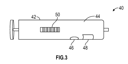
[0010] Figure 3
is an orthogonal view of a fluid collection device having an
integrated blood testing device constructed in accordance with the present
disclosure.
[0011] Figures
4A-40 illustrate views of portions of another version of a fluid
collection device constructed in accordance with the present disclosure.
[0012] Figure
5A illustrates a fluid housing of Figure 4 being inserted into a
slot of a reader for acoustic treatment and colorimetric analysis in
accordance with
one embodiment of the present disclosure.
[0013] Figure
5B illustrates the fluid housing of Figure 4 being inserted into a
port of another version of a reader for acoustic treatment and colorimetric
analysis in
accordance with one embodiment of the present disclosure.
[0014] Figure 6
illustrates an exploded, perspective view of a blood testing
device removably attachable to a fluid housing such as a syringe constructed
in
accordance with one embodiment of the present disclosure.
[0015] Figure 7
illustrates a perspective view of another version of a blood
testing device constructed in accordance with one embodiment of the present
disclosure.
[0016] Figure 8
illustrates top plan view of a blood testing device constructed
in accordance with one embodiment of the present disclosure.
3
CA 03122235 2021-06-04
WO 2020/118018
PCT/US2019/064623
[0017] Figure 9
illustrates an exploded, perspective view of a blood testing
device removably attachable to a fluid housing such as a syringe constructed
in
accordance with one embodiment of the present disclosure.
[0018] Figure
10 illustrates a perspective view of another version of a blood
testing device constructed in accordance with one embodiment of the present
disclosure.
[0019] Figure
11 illustrates a perspective view of a blood testing device having
a closeable gate constructed in accordance with one embodiment of the present
disclosure.
[0020] Figure
12 illustrates a cross-sectional view of the blood testing device
of Figure 11 having the closeable gate in a first position, and positioned
within a slot
of a reader in accordance with the present disclosure.
[0021] Figure
13 illustrates another cross-sectional view of the blood testing
device of Figure 11 having the closeable gate in a second position, and
positioned
within the slot of the reader in accordance with the present disclosure.
[0022] Figure
14 illustrates a perspective view of another version of a blood
testing device having a moveable gate constructed in accordance with one
embodiment of the present disclosure.
[0023] Figure
15 illustrates a cross-sectional view of the blood testing device
of Figure 14 having the moveable gate in a first position, and positioned
within a slot
of the reader in accordance with the present disclosure.
[0024] Figure
16 illustrates another cross-sectional view of the blood testing
device of Figure 14 having the moveable gate in a second position, and
positioned
within the slot of the reader in accordance with the present disclosure.
4
CA 03122235 2021-06-04
WO 2020/118018
PCT/US2019/064623
DETAILED DESCRIPTION
[0025] The
following detailed description refers to the accompanying
drawings. The same reference numbers in different drawings may identify the
same
or similar elements.
[0026] As used
herein, the terms "comprises," "comprising," "includes,"
"including," "has," "having" or any other variation thereof, are intended to
cover a
non-exclusive inclusion. For example, a process, method, article, or apparatus
that
comprises a list of elements is not necessarily limited to only those
elements, but
may include other elements not expressly listed or inherent to such process,
method,
article, or apparatus. Further, unless expressly stated to the contrary, "or"
refers to
an inclusive or and not to an exclusive or. For example, a condition A or B is
satisfied
by any one of the following: A is true (or present) and B is false (or not
present), A is
false (or not present) and B is true (or present), and both A and B are true
(or
present).
[0027] In
addition, use of the "a" or "an" are employed to describe elements
and components of the embodiments herein. This is done merely for convenience
and to give a general sense of the inventive concept. This description should
be read
to include one or more and the singular also includes the plural unless it is
obvious
that it is meant otherwise.
[0028] Further,
use of the term "plurality" is meant to convey "more than one"
unless expressly stated to the contrary.
[0029] As used
herein any reference to "one embodiment" or "an
embodiment" means that a particular element, feature, structure, or
characteristic
CA 03122235 2021-06-04
WO 2020/118018
PCT/US2019/064623
described in connection with the embodiment is included in at least one
embodiment.
The appearances of the phrase "in one embodiment" in various places in the
specification are not necessarily all referring to the same embodiment.
[0030]
Circuitry, as used herein, may be analog and/or digital, components, or
one or more suitably programmed microprocessors and associated hardware and
software, or hardwired logic. Also, "components" may perform one or more
functions. The term "component," may include hardware, such as a processor, an
application specific integrated circuit (ASIC), or a field programmable gate
array
(FPGA), or a combination of hardware and software. Software includes one or
more
computer executable instructions that when executed by one or more component
cause the component to perform a specified function. It should be understood
that
the algorithms described herein are stored on one or more non-transitory
memory.
Exemplary non-transitory memory includes random access memory, read only
memory, flash memory or the like. Such non-transitory memory may be
electrically
based or optically based.
[0031] As used
herein, the term "substantially" means that the subsequently
described parameter, event, or circumstance completely occurs or that the
subsequently described parameter, event, or circumstance occurs to a great
extent
or degree. For example, the term "substantially" means that the subsequently
described parameter, event, or circumstance occurs at least 90% of the time,
or at
least 91%, or at least 92%, or at least 93%, or at least 94%, or at least 95%,
or at
least 96%, or at least 97%, or at least 98%, or at least 99%, of the time, or
means
that the dimension or measurement is within at least 90%, or at least 91%, or
at least
92%, or at least 93%, or at least 94%, or at least 95%, or at least 96%, or at
least
97%, or at least 98%, or at least 99%, of the referenced dimension or
measurement.
6
CA 03122235 2021-06-04
WO 2020/118018
PCT/US2019/064623
[0032] In
accordance with one aspect, there are provided devices, systems,
and processes for determining a presence of hemolysis in a sample.
Advantageously, devices, systems, and processes described herein determine
whether hemolysis has occurred in a sample based upon a colorimetry assessment
of a portion of the sample.
[0033] In
accordance with another aspect, there are provided devices,
systems, and processes for a blood collection container having a hemolysis
indicating feature.
[0034] In
accordance with another aspect, there are provided blood collection
devices, systems, accessories and processes having a plasma separating
feature.
[0035] In
accordance with another aspect, there are provided blood collection
devices, systems, accessories, and processes having a hemolysis indicating
feature.
[0036]
Referring now to the Figures and in particular to Figure 1, shown
therein is a diagramatic view of a blood testing device 10 constructed in
accordance
with the present disclosure. In general, the blood testing device 10 includes
a
housing 12, an acoustic transducer 16, a reader 18, and a control unit 20
connected
to the transducer 16 and the reader 18. The housing 12 is constructed of a
fluid
impermeable material so that the housing 12 can hold and contain a sample of
blood
containing blood cells, suspended within plasma. The housing 12 can be a
syringe or
a vacutainer, for example that can be used for collecting blood and
transporting the
blood for purposes of testing. The blood may be collected from an animal, such
as a
human, or a non-human (such as a cat, dog, cow, horse, fish, or the like). The
acoustic transducer 16, the reader 18, and the control unit 20 may be located
outside
of the housing 12 as shown in FIG. 1 or inside the housing 12 (not shown). The
7
CA 03122235 2021-06-04
WO 2020/118018
PCT/US2019/064623
acoustic transducer 16 selectively generates acoustic forces that are directed
to the
housing 12. In some embodiments, the acoustic transducer 16 can be tuned so as
to provide a magnitude and/or frequency of acoustic forces so as to facilitate
separation of the undamaged blood cells from the plasma and damaged blood
cells.
The magnitude and/or frequency of the acoustic forces generated by the
acoustic
transducer 16 can be selected depending upon a size and/or construction of the
housing 12, or composition of the blood sample within the housing 12. In one
embodiment, the acoustic transducer 16 can be a piezoelectric element. At
least a
portion of the housing 12, adjacent to the transducer 16, is constructed of a
material
that functions to pass the acoustic forces generated by the acoustic
transducer 16
into the sample contained within the housing 12. Exemplary materials that can
be
used to form the housing 12 include glass, crystal, and the like. Parts of the
housing
12 away from the transducer 16 can be made of other materials such as plastic.
The
application of the acoustic forces into the sample by the acoustic transducer
16
causes the blood cells within the blood to move within the plasma to form a
first zone
22 having an increased density or concentration of the blood cells than the
blood
contained prior to the application of the acoustic forces, and at least one
second
zone 24 being substantially only plasma, i.e., substantially devoid of any
undamaged
blood cells. The reader 18 is positioned adjacent to the second zone 24 and
functions to read at least one parameter of the plasma. In one embodiment, the
reader 18 is an optical reader, such as a camera or photospectrometer having a
field
of view overlapping with the housing 12 such that the plasma within the second
zone
is visible to the reader 18. The optical reader 18 is positioned such that the
second
zone 24 is within the field of view. The control unit 20 selectively actuates
/
deactuates the acoustic transducer 16 to cause separation of the blood cells
and
8
CA 03122235 2021-06-04
WO 2020/118018
PCT/US2019/064623
plasma into the first zone 22 and the second zone 24. Then, in some
embodiments,
the control unit 20 actuates the reader 18 to capture information indicative
of at least
one parameter of the plasma. The information captured by the reader 18 is then
transferred to the control unit 20 to determine a degree of hemolysis within
the
sample of blood. The control unit 20 can be constructed of circuitry and/or a
combination of circuitry and software.
[0037] When the
reader 18 is the optical reader, the degree of hemolysis can
be determined by the control unit 20 based upon a colorimetric analysis of the
sample. That is, when the sample is devoid of hemolysis and is illuminated
with
white light, the plasma will be substantially devoid of any color, i.e., the
sample will
be transparent. When hemolysis has occurred within the sample, the plasma will
be
pink when the plasma is illuminated with white light. By correlating the color
of the
plasma with predetermined colors indicative of an extent of hemolysis
occurring
within other samples, the extent of hemolysis within the sample can be
determined.
Depending upon a color of a backdrop, and/or color of illumination of the
plasma,
colors detected by the reader 18 indicative of an extent of hemolysis may
differ.
[0038]
Information indicative of an extent of hemolysis within the sample can
be used to determine whether the blood has hemolysis.
[0039] FIG. 3
illustrates a blood testing device 40 constructed in accordance
with one embodiment of the present disclosure. The blood testing device 40 is
provided with a fluid container such as a syringe 42 or vacutainer having a
fluid
reservoir 44 for containing blood. The blood testing device 40 may also be
provided
with an acoustic transducer 46, an optical zone 48, and a bar code 50 which
identifies the contents of the syringe 42 and can be correlated to specific
patients.
9
CA 03122235 2021-06-04
WO 2020/118018
PCT/US2019/064623
The acoustic transducer 46 may be provided with any suitable shape, such as
planar, arcuate, or the like. In some embodiments, the acoustic transducer 46
may
be provided with a shape to match a shape of the optical zone 48, or other
section of
the blood testing device 40 to be stimulated by the acoustic transducer 46. In
such
an embodiment, the blood testing device 40 allows the blood to be acoustically
treated using the acoustic transducer 46 after which the blood may be analyzed
using an optical reader or the human eye through the optical zone 48. Once a
degree of hemolysis within the sample of blood has been determined, a decision
can
be made whether or not to continue with further testing of the sample of
blood.
[0040]
Referring now to Figures 4A-40, shown therein is an embodiment of a
blood testing device 70 and reader 71. The blood testing device 70 has a fluid
housing 72, a fluid reservoir 74, a fluid treatment area 75, an optical zone
76, a
treatment window 78, and a flow port 82. The reader 71 has a reading device
80, a
flow path 82, and an analysis unit 84. In this embodiment of the blood testing
device
70, a blood sample is contained in the fluid housing 72 and directed along the
flow
path 82 into the fluid treatment area 75 where the blood sample is directed to
flow
past the treatment window 78. The treatment window 78 is constructed of a
material
that functions to pass acoustic forces generated by an acoustic transducer
into the
blood sample contained within the fluid housing 72.
[0041] The
reading device 80 is part of the analysis unit 84 and is provided
with an acoustic transducer (not shown) and an optical reader (not shown)
which
operate as described above to acoustically treat the blood sample. The
acoustic
transducer may be provided with a planar shape so as to mate with the
treatment
window 78 of the blood testing device 70. The optical reader of the reading
device 80
has a field of view directed to the optical zone 76 where the acoustically
treated
CA 03122235 2021-06-04
WO 2020/118018
PCT/US2019/064623
blood sample may be read. The analysis unit 84 actuates the acoustic
transducer to
acoustically treat the blood sample and move the blood cells away from the
optical
zone 76 such that only the plasma is visible in the optical zone 76. Then the
optical
reader captures an image of the plasma and any backdrop and sends the image to
the analysis unit 84 for colorimetric analysis as discussed above. The
analysis unit
84 may be provided with further blood analysis features (not shown) such as
blood
gas analysis which may further analyze the blood sample after it passes
through the
flow path 82. The reader 71 may be portable and have a housing 88 that
includes a
slot 90 sized and dimensioned to receive the blood testing device 70 such that
the
optical zone 76 is in the field of view of the optical reader and the
treatment window
overlaps with the acoustic transducer. The housing 88 can be provided in a
variety of
shapes such as in a shape of a hot dog bun, for instance. The analysis unit 84
can
be supported in the housing 88 or be separate therefrom. For example, the
reading
device 80 can be provided with a wireless transceiver to communicate with the
analysis unit 84. The analysis unit 84 may be constructed and function in a
similar
manner as the control unit 20 discussed above.
[0042] Figure 5
illustrates another variation of the reader 71 in which the fluid
housing 72 of Figure 4 may be inserted into the housing 88 so that the blood
sample
may be acoustically treated and read by the reading device 80 as described
above.
[0043]
Referring now to Figure 6, a lateral flow blood testing device 100
constructed in accordance with the present disclosure is shown having a fluid
housing 102, a first fluid reservoir 104, and a fluid treatment module 106.
The fluid
treatment module 106 of the lateral flow blood testing device 100 is provided
with a
lower portion 108, an upper portion 110, a second fluid reservoir 111, a
lateral flow
11
CA 03122235 2021-06-04
WO 2020/118018
PCT/US2019/064623
strip 112, a fluid channel 114, a first fluid port 116, a second fluid port
118, an optical
zone 120, and a bar code 122.
[0044] The
lower portion 108 and the upper portion 110 of the fluid treatment
module 106 are sealably connected to form the second fluid reservoir 111. When
the
fluid treatment module 106 is connected to the fluid housing 102, a blood
sample
may be transferred from the first fluid reservoir 104 to the second fluid
reservoir 111.
Once in the second fluid reservoir 111, a portion of the blood sample may be
directed through the second fluid port 118 into the fluid channel 114 where
the blood
sample passes through the lateral flow strip 112. Through capillary action
(which
may also be referred to as capillary flow), the lateral flow strip 112 causes
the
separation of undamaged blood cells and plasma in the blood sample as
described
more fully in U.S. Patent Application No. 15/317,748, the entirety of which is
incorporated herein by reference. The plasma that has passed through the
lateral
flow strip 112 may then be analyzed in the optical zone 120 to determine a
degree of
hemolysis using an optical reader as described above or human eyes.
[0045] To
facilitate directing the blood sample into the second fluid port 118,
the first fluid port 116 may be temporarily sealed using a removable cap (not
shown),
for instance, that temporarily prevents movement of the blood sample through
the
first fluid port 116. When the blood sample has been analyzed using the
lateral flow
blood testing device 100, the cap may be removed and the blood sample may be
allowed to pass through the first fluid port 116 to be used for further
testing, for
instance, as desired.
12
CA 03122235 2021-06-04
WO 2020/118018
PCT/US2019/064623
[0046] As
described above, the bar code 122 may be used to identify the
blood sample, the patient the blood sample belongs too, the test to be
performed,
and the like.
[0047] Figure 7
illustrates a lateral flow blood testing device 140 having a fluid
housing 142, a fluid treatment module 144, a first fluid reservoir 146, and a
second
fluid reservoir 148. The lateral flow blood testing device 140 is similar to
the lateral
flow blood testing device 100 described above, therefore, in the interest of
brevity
only the differences will be described herein. In the embodiment shown in
Figure 7,
the fluid treatment module 144 and the fluid housing 142 are integrated to
form the
lateral flow blood testing device 140.
[0048] Figure 8
illustrates a top plan view of a fluid treatment module 160
similar to fluid treatment modules 106 and 144. In this embodiment, the fluid
treatment module 160 is provided with a fluid channel 162 that connects a
first fluid
port 164 with a second fluid port 166 to direct a flow of a blood sample into
a fluid
channel 168 for separation of the blood sample into at least two constituent
parts as
described above. The fluid channel 168 houses the lateral flow strip 112 that
functions to separate the undamaged blood cells from the plasma so that the
plasma
and any color caused by damaged blood cells is visible in the optical zone
120.
[0049]
Referring now to Figure 9, shown therein is another embodiment of a
lateral flow blood testing device 180 having a fluid housing 182, a fluid
reservoir 184,
and a fluid treatment module 186. The fluid treatment module 186 of the
lateral flow
blood testing device 180 is provided with a lower portion 188, an upper
portion 190,
a second fluid reservoir 192, and a lateral flow membrane 194.
13
CA 03122235 2021-06-04
WO 2020/118018
PCT/US2019/064623
[0050] The
lower portion 188 and the upper portion 190 of the fluid treatment
module 186 are sealably connected to form the second fluid reservoir 192. When
the
fluid treatment module 186 is connected to the fluid housing 182, a blood
sample
may be transferred from the first fluid reservoir 184 to the second fluid
reservoir 192.
As the blood sample is transferred from the first fluid reservoir 184 to the
second
fluid reservoir 192 the blood sample passes through the lateral flow membrane
194
and the blood sample is separated into at least two constituent parts, i.e.,
undamaged blood cells remain in the first fluid reservoir 184 and plasma with
any
damaged blood cells pass through the lateral flow membrane 194 and into the
second fluid reservoir 192.
[0051] At least
the lower portion 188 of the fluid treatment module 186 is
constructed of an optically clear material which allows the plasma that has
passed
through the lateral flow membrane 194 to be colorimetrically analyzed in the
second
fluid reservoir 192 using an optical reader as described above or human eyes.
[0052] Also
shown in Figure 9 is a probe 196 which may be attached to or part
of a blood analysis machine (not shown) such as a blood gas analyzer. Where
whole
blood is needed for analysis, the probe 196 may be passed through a fluid port
198
in the fluid treatment module 186, through the second fluid reservoir 192, and
through the lateral flow membrane 194 into the first fluid reservoir 184 where
the
blood sample has not been separated.
[0053] Figure
10 illustrates another version of a lateral flow blood testing
device 200 having a fluid housing 202, a fluid treatment module 204, a first
fluid
reservoir 206, a second fluid reservoir 208, a lateral flow membrane 210, and
a fluid
port 214. The lateral flow blood testing device 200 is similar to the lateral
flow blood
14
CA 03122235 2021-06-04
WO 2020/118018
PCT/US2019/064623
testing device 180 described above, therefore, in the interest of brevity only
the
differences will be described herein. In the embodiment shown in Figure 10,
the fluid
treatment module 204 and the fluid housing 202 are integrated into a unitary
structure, rather than removably connected to form the lateral flow blood
testing
device 200.
[0054] A probe
212 is also shown which may be attached to or part of a blood
analysis machine (not shown) such as a blood gas analyzer. Where whole
(unseparated) blood is needed for analysis, the probe 212 may be passed
through
the fluid port 214 in the fluid treatment module 204, through the second fluid
reservoir 206, and through the lateral flow membrane 210 into the first fluid
reservoir
206 where the blood sample has not been separated.
[0055]
Referring now to Figures 11-13, shown therein is yet another version of
a blood testing device 240 similar to the blood testing device 70 shown in
Figure 4
that can be read by the reader 71. In the interest of brevity, only the
differences will
be described in detail herein. The blood testing device 240 is provided with a
fluid
housing 242 a fluid reservoir 244, a fluid treatment area 245, an optical zone
246, a
treatment window 248, a flow path 252, a gate 254 having a port 256, and a
gate
guide channel 258.
[0056] When the
gate 254 is in a first position (shown in Figure 12), the flow
path 252 is restricted such that a blood sample in the flow path 252 stops in
the
optical zone 246 where the sample may be acoustically treated to move
undamaged
blood cells away from the plasma adjacent to the optical zone 246 and read
with the
reader 71 as described above.
CA 03122235 2021-06-04
WO 2020/118018
PCT/US2019/064623
[0057] Once the
blood sample has been analyzed the gate 254 may be
moved to a second position (shown in Figure 13), which moves the port 256 into
the
flow path 252 allowing the blood sample to pass.
[0058]
Referring now to Figures 14-16, shown therein is yet another version of
a blood testing device 280 similar to the blood testing devices 70 and 240
shown in
Figures 4 and 11-13, respectively that can be read by the reader 71. In the
interest of
brevity, only the differences will be described in detail herein. The blood
testing
device 280 is provided with a fluid housing 282 a fluid reservoir 284, a fluid
treatment
area 285, an optical zone 286, a treatment window 288, a first flow path 292,
a
second flow path 294, a gate 296 having a port 298, and a gate guide channel
300.
[0059] When the
gate 296 is in a first position (shown in Figure 15), a blood
sample is directed into the first flow path 292 such that the blood sample
stops in the
optical zone 286 where the blood sample may be acoustically treated and read
with
the reading device 80 as described above.
[0060] Once the
blood sample has been analyzed the gate 296 may be
moved to a second position (shown in Figure 16), which moves the port 298 into
the
second flow path 294 allowing the blood sample to pass through the second flow
path 294.
[0061] From the
above description, it is clear that the inventive concepts
disclosed herein is well adapted to carry out the objects and to attain the
advantages
mentioned herein as well as those inherent in the inventive concepts disclosed
herein. While presently preferred embodiments of the inventive concepts
disclosed
herein have been described for purposes of this disclosure, it will be
understood that
numerous changes may be made which will readily suggest themselves to those
16
CA 03122235 2021-06-04
WO 2020/118018
PCT/US2019/064623
skilled in the art and which are accomplished within the scope and coverage of
the
inventive concepts disclosed and claimed herein.
17