Note: Descriptions are shown in the official language in which they were submitted.
System, Method, and Computer Algorithm for Characterization and Classification
of Electrophysiological Evoked Potentials
Background of the Invention
1. Field of the Invention
[0001] The present invention relates generally to detecting changes in evoked
potentials (EPs;
and more particularly to detecting changes in EPs automatically with a
computer algorithm.
2. Related Art
[0002] Somatosensory evoked potentials are summated electrical potentials
usually recorded
from the head or neck area after repeatedly stimulating a peripheral nerve.
Monitoring patients
using somatosensory evoked potentials during surgery has been shown to allow
early
identification of impending positioning effect injury that can then be avoided
by repositioning
the patient to alleviate pressure or tension.
[0003] For example, as described in Hickey, C.; Gugino, L. D.; Aglio, L. S.;
Mark, J. B.; Son,
L. & Maddi, R. (1993), "Intraoperative somatosensory evoked potential
monitoring predicts
peripheral nerve injury during cardiac surgery," Anesthesiology 78(1), 29-35,
Kamel, I. R.;
Drum, E. T.; Koch, S. A.; Whitten, J. A.; Gaughan, J. P.; Barnette, R. E. &
Wendling, W. W.
(2006), "The use of somatosensory evoked potentials to determine the
relationship between
patient positioning and impending upper extremity nerve injury during spine
surgery: a
retrospective analysis," Anesth Analg 102(5), 1538 _______________________
1542, and Labrom, R. D.; Hoskins, M.;
Reilly, C. W.; Tredwell, S. J. & Wong, P. K. H. (2005), and "Clinical
usefulness of
somatosensory evoked potentials for detection of brachial plexopathy secondary
to
tnalpositioning in scoliosis surgery." Spine 30(18), 2089-2093.
[0004] Such monitoring generally requires highly trained technologists under
physician
supervision with sophisticated, multichannel amplifier and display equipment.
Unfortunately,
such personnel and equipment are limited in their availability, require pre-
booking, and are
costly. In addition, such monitoring is not traditionally done in many of the
areas in which
positioning effects occur outside of the operating room where unresponsive,
weak or confined
patients may incur positioning effect.
-1 -
Date Recue/Date Received 2021-06-14
WO 2013/166157 PCT/US2013/039078
Typically, the technologist reviews the waveforms while a neurologist
contemporaneously
reviews the EP waveforms either on site or remotely through the interne. The
technologist and
neurologist are trained and are experts in determining whether the changes in
the EP waveforms
are significant and are indicative of pending nerve injury. The cost of having
professionals fully
engaged in interpreting these waveforms results in rationing of the service to
all but the most
high risk surgeries.
[0006] U.S. Patent Application Publication No. 2008/0167574 describes a
semiautomated
device available for automatically measuring biometric signals during surgery
to avoid nerve
injury. However, the device focuses on muscle or motor recordings to measure
nerve proximity
to surgical instruments and does not address positioning effect.
[0007] The difficulty with analyzing and classifying waveforms to identify
positioning effect
lies in the wide variation in the amplitude, frequency and shape of the
waveforms. These
variations are caused by many factors including anesthesia, electrical
interference from other
devices and any preexisting abnormalities of the nerves.
[0008] Accordingly, there is a need for a system and method that can overcome
the
disadvantages of previous systems and methods.
Summary of the Invention
[0009] In an exemplary embodiment of the present invention, a system, method,
and computer
algorithm for characterization and classification of electrophysiological EPs
is disclosed. An EP
may be defined as a voltage versus time signal obtained by a neural system
using suitable
electrodes. For example when obtaining an EP from a somatosensory system a
signal may be
obtained by ensemble averaging the electrophysiological responses to
repetitive stimulation of
the somatosensory system detected using suitable electrodes. Examples of EPs
are
somatosensory, auditory or visual EPs. The algorithms are applied to a time
sequence of EPs
acquired over the course of an ongoing clinical procedure. The algorithms
establish the
characteristics of a baseline/normal EP and then characterize subsequent EPs
relative to the
baseline EP as well as to previous EPs to determine if the functioning of the
underlying sensory
neural system has been significantly affected by the ongoing clinical
procedure. The algorithms
communicate with ancillary hardware and algorithms developed to acquire the
sequence of EPs
and provide suitable feedback to ensure an effective clinical workflow. The
algorithms provide
-2-
Date Recue/Date Received 2021-06-14
the basis for a clinically effective application such that false positives and
false negatives are
minimized.
[00010] Various embodiments of the claimed invention relate to an automated EP
analysis
apparatus for automatic baseline acquisition and subsequent monitoring,
detecting and
identifying changes (adverse or recovering) to a physiological system
generating EP
waveforms, wherein the apparatus is adapted to characterize and classify the
EP waveforms
and create alerts of the changes (adverse or recovering) to the physiological
system
generating the EP waveforms if the acquired EP waveforms change significantly
in latency,
amplitude or morphology.
[00010A] In one embodiment, there is provided an automated evoked potential
(EP) analysis
apparatus for automatic baseline acquisition and subsequent monitoring,
detecting and
identifying changes (adverse or recovering) to a physiological system
generating a sequence
of EP waveforms. The apparatus is adapted to: compare a number of Good EP
responses
received so far in the sequence of EP waveforms to a number of initial EP
response required
to create a baseline response (Ni); if there are not NI Good EP responses,
analyze previous EP
responses in the sequence of EP waveforms to estimate the baseline response
and establish an
analysis range; and if there are Ni Good EP responses, update the baseline
response based on
a current EP response. The apparatus is further adapted to: characterize the
current EP
response relative to the baseline response and the previous EP responses;
classify the current
EP response as Good, Bad, Undetermined or Unreliable based on the
characterization; and
create alerts of the changes (adverse or recovering) to the physiological
system generating the
sequence of EP waveforms if acquired EP waveforms change in latency, amplitude
or
morphology.
[00010B] In another embodiment, there is provided an automated evoked
potential (EP)
analysis apparatus for automatic baseline acquisition and subsequent
monitoring, detecting
and identifying changes (adverse or recovering) to a physiological system
generating EP
waveforms. The apparatus is adapted to characterize and classify acquired EP
waveforms and
create alerts of the changes (adverse or recovering) to the physiological
system generating the
EP waveforms if the acquired EP waveforms change in latency, amplitude or
morphology.
-3-
Date recue/Date received 2023-02-17
The apparatus comprises memory and a processor. The processor is configured to
execute
instructions stored on the memory, that, when executed by the processor, cause
the processor
to perform operations comprising: stimulating, via electrical pulse
electrodes, a peripheral
nerve structure; recording, via electrodes placed at a neck or head, signals
forming an EP
waveform generated by a nervous system in response to the stimulation; and
measuring a
waveform change in the recorded EP waveform relative to a baseline response.
The
waveform change comprises one or more of a Euclidean distance, a pseudo-
correlation, a
cross-correlation, and an energy ratio between the recorded EP waveform and
the baseline
response. The operations further comprise: comparing the waveform change with
one or
more threshold change values to classify the recorded EP waveform; and
determining
whether a current state of the peripheral nerve structure has changed based on
the
classification of the recorded EP waveform by determining whether a count of
EP waveforms
in a classification exceeds a threshold count value.
-3a-
Date recue/Date received 2023-02-17
Brief Description of the Drawings
[00011] The foregoing and other features and advantages of the invention will
be apparent
from the following, more particular description of a preferred embodiment of
the invention,
as illustrated in the accompanying drawings wherein like reference numbers
generally
indicate identical, functionally similar, and/or structurally similar
elements.
[00012] FIG. 1 illustrates an exemplary depiction of stimulation of a
physiological system of
interest with a context relevant stimulus according to an exemplary embodiment
of the
present invention.
[00013] FIG. 2 illustrates an exemplary depiction of a sequence of suitable
stimuli applied to
a physiological system of interest and the sequence of corresponding responses
according to
an exemplary embodiment of the present invention.
[00014] FIG. 3 illustrates an exemplary depiction of the creation of an
ensemble averaged EP
based on a number of responses according to an exemplary embodiment of the
present
invention.
[00015] FIG. 4A illustrates an exemplary flowchart process for acquiring and
classifying EP
responses according to an exemplary embodiment of the present invention.
[00016] FIG. 4B illustrates an exemplary flowchart process for determining
whether a
change has occurred in a sequence of Eps according to an exemplary embodiment
of the
present invention.
[00017] FIG. 5 illustrates an exemplary flowchart process for calculating a
baseline response
according to an exemplary embodiment of the present invention.
[00018] FIG. 6 illustrates an exemplary flowchart process for determining the
analysis range
according to an exemplary embodiment of the present invention.
[00019] FIG. 7 illustrates an exemplary flowchart process for updating a
baseline response
according to an exemplary embodiment of the present invention.
-4-
Date recue/Date received 2023-02-17
[00020] FIG. 8 illustrates an exemplary embodiment of a relationship diagram
in metric
calculation for characterizing Eps according to an exemplary embodiment of the
present
invention.
[00021] FIG. 9 illustrates an exemplary flowchart process for a good state
according to an
exemplary embodiment of the present invention.
[00022] FIG. 10 illustrates an exemplary flowchart process for a bad state
according to an
exemplary embodiment of the present invention.
Detailed Description of the Invention
[00023] Various exemplary embodiments of the invention including preferred
embodiments
are discussed in detail below. While specific exemplary embodiments are
discussed, it should
be understood that this is done for illustration purposes only. A person
skilled in the relevant
art will recognize that other components and configurations can be used
without parting from
the spirit and scope of the invention.
[00024] An embodiment of the present invention relates to the computer signal
processing
and pattern recognition algorithms for the characterization and classification
of Eps in real-
time. This algorithm may substitute for the expert analysis typically provided
by the
technologist and physician. The computer algorithm running in software
installed on an EP
machine may be used in any surgery or situation where a patient is at risk to
detect, alert and
ameliorate positioning effect or any abnormality.
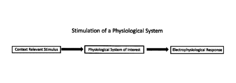
[00025] FIG. 1 illustrates an exemplary stimulation of a physiological system
of interest with
a context relevant stimulus according to an exemplary embodiment of the
present invention.
For the somatosensory system, for example, a stimulus may be the application
of an
appropriate sized and shaped current pulse over a superficial nerve. The
stimulus may be a
context relevant stimulus applied to the physiological system of interest to
elicit an
-5-
Date recue/Date received 2023-02-17
electrophysiological response. Given that a suitable stimulus is applied, the
corresponding
electrophysiological response may then be a volley of action potentials along
the axons
excited by the applied stimulus.
[00026] FIG. 2 illustrates an exemplary depiction of a sequence of suitable
stimuli applied to
a physiological system of interest and the sequence of corresponding
responses. The
physiological system of interest may be stimulated with a series/sequence of
stimuli as shown
at 202 in FIG. 2. The series/sequence of stimuli may cause a corresponding
series/sequence
of electrophysiological responses that can be detected at specific sites as
shown at 204 in
FIG. 2. The series/sequence of corresponding responses 204 may be detected
using suitable
electrodes suitably configured at a suitable recording site according to an
exemplary
embodiment of the present invention. These responses are comprised of time
sampled and
digitized measurements of the volume conducted voltage fields created by the
electrophysiological response (and may be referred to as a series/sequence of
evoked
potentials (EPs)) of the physiological system of interest when evoked by the
applied stimuli.
[00027] FIG. 3 illustrates an exemplary depiction of the creation of an
ensemble averaged EP
based on a number of responses according to an exemplary embodiment of the
present
invention. Individual responses may be contaminated by voltage contributions
from the
electrophysiological activity of other physiological systems as well as
ambient electrical
noise. As such, in order to obtain a suitable signal to noise ratio, a number
of responses may
be ensemble averaged to create a resulting evoked potential (EP) (which may
also be referred
to as an ensemble averaged EP). The signal to noise ratio of the resulting
ensemble averaged
EP improves as N, the number of responses averaged, increases. In an
embodiment, N may
range from 10 to 1000 depending on the physiological system of interest.
[00028] EPs may be processed to assess the state of the physiological system
of interest. A
physiological system in a normal operating mode may be considered to be in a
"Good" state.
If the physiological system is stressed, fatigued, or injured, the system may
be considered to
be in a "Bad" state. Starting with the physiological system in a Good state,
detected changes
in the characteristics of the EPs in a sequence of EPs can be used to predict
if the
physiological system is in a Good or Bad state.
-5a-
Date recue/Date received 2023-02-17
[00029] FIG. 4A illustrates an exemplary flowchart process for acquiring and
classifying EP
responses according to an exemplary embodiment of the present invention. Each
EP may be
initially filtered to remove unwanted instrumentation noise to better present
the
electrophysiological response of the system of interest. The EPs may be
filtered based on
likelihood estimation.
[00030] If a baseline response does not exist, acquired responses may be
analyzed to estimate
a baseline response and to establish an analysis range. For example, if there
is not Ar1 Good
responses received, where Ni is a number of initial EP responses required to
create a baseline
response, then a baseline response may not exist. The analysis to estimate a
baseline response
and to establish an analysis range is further described below.
[00031] If a baseline response exists, the baseline may be updated based on
the current
response. Updating the baseline is further described below.
[00032] Once the current baseline response is determined, the current response
is then
characterized relative to the current baseline and previous response. For
example,
characterization may at least one of a Euclidean distance, a pseudo-
correlation, a cross-
correlation, or an energy ratio between the current response and current
baseline. Energy ratio
-5b-
Date recue/Date received 2023-02-17
WO 2013/166157 PCT/US2013/039078
may be the ratio of the energy between the current response and the current
baseline. The
energy ratio may represent a change in size of the EP response. The current
response may be
then be classified based on the current response's characterization.
[00033] EPs may be classified into four possible categories: Good, Bad,
Undetermined and
Unreliable based on the characterization. A Good classification may indicate
the EP
characterization corresponds with no significant waveform change. For example,
when there is
no positioning effect. A Bad classification may indicate the EP
characterization corresponds
with a signification waveform change. For example, when there is positioning
effect. An
Undetermined classification may indicate that the EP characterization may be
of indeterminate
significance. For example, the EP characterization may be insufficient for a
Good classification
but also insufficient for a Bad classification. For example, the EP may
possibly correspond with
either positioning effect or no positioning effect. An unreliable
classification may indicate the
EP includes too much noise to be properly characterized and classified.
[00034] Each classification may correspond with a particular threshold. The
threshold may
indicate how similar an EP response should be to a baseline to be considered a
Good response or
how dissimilar an EP response should be to a baseline to be considered a Bad
response. The
thresholds may be based on the characterizations of the EP responses. For
example, thresholds
may be based on at least one of Euclidean distance, a pseudo-correlation, a
cross-correlation, or
an energy ratio between an EP response and a baseline. A threshold may also
indicate how
much noise may be included in an EP response before the EP response is
considered unreliable.
[00035] The thresholds used for classification may be determined by analyzing
training data.
Training data may include a plurality of EP responses known to correspond to
particular
classifications. Using multiple sets of thresholds determined from the
analysis of training data,
the current response may be classified as belonging to a category of interest
based on the values
of its calculated metrics.
[00036] FIG. 4B illustrates an exemplary flowchart process for determining
whether a change
has occurred in a sequence of EPs according to an exemplary embodiment of the
present
invention. FIG. 4B continues from FIG. 4A. Given the sequence of classified
EPs, it may be
determined whether the state of the physiological system of interest has
changed (either from
Good to Bad or vice versa) or if the state of the physiological system of
interest has not changed.
If the state has changed, the system may create an alert.
-6-
Date Recue/Date Received 2021-06-14
[00037] FIG. 5 illustrates an exemplary flowchart process for calculating a
baseline response
according to an exemplary embodiment of the present invention. Currently
loaded responses
may be iteratively represented as nodes within a minimum spanning tree (MST)
created using
the Euclidean distances between response pairs (each response represented as
nodes 1, 2, 3,
4, 5 and 6) as shown at 502 in FIG. 5. Each line in the MST that links pairs
of responses may
represent a Euclidean distance value. The currently loaded responses may be
initially
acquired responses. Response pairs may be combinations of any two currently
loaded
responses. For example, three responses may result in three response pairs.
The Euclidean
distance may be based on the sum of the squares of the differences between
responses in each
response pair or the sum of the absolute value of the differences between
responses in each
response pair.
[00038] The MST may be separated into clusters based on cutting lines that are
greater than a
threshold as shown at 504 in FIG. 5. The threshold may be based on the mean of
the line
lengths and standard deviations of the line lengths. The clusters may be
sorted based on the
sizes of the clusters. The size of a cluster may be the number of responses
within the cluster.
The cluster with the largest size may be selected so that a temporary baseline
is calculated
based on the responses within the cluster. All the responses within the
largest cluster may be
aligned using a default analysis range and pseudo-correlation. The response
members of the
cluster with the largest number of members may be averaged to estimate the
baseline
response.
[00039] FIG. 6 illustrates an exemplary flowchart process for determining the
analysis range
according to an exemplary embodiment of the present invention. Initial
responses are
characterized and classified using initial baseline response estimates and a
default analysis
range. First, initial Good responses are used to locate a default width
analysis range by
adjusting the location of the range until a minimum congruity value is
obtained. Using the
initial Good responses, the width of the analysis range is then adjusted by
increasing it to the
left or right until a minimum congruity value is obtained. In some
embodiments, if analysis
range location is adjusted by more than 5ms, responses may be re-clustered and
the analysis
-7-
Date recue/Date received 2023-02-17
range may be recalculated. For both analysis range location and sizing, the
congruity measure
may be:
1 I
1 :
h- 2* 'annED +
CC
where NormED is a normalized Euclidean distance and CC is the cross-
correlation. The
above congruity equation may be used for locating the analysis range as shown
at 602 in FIG.
6 and for widening the analysis range as shown at 604 in FIG. 6. While not
shown in FIG. 6,
the calculated new baseline response may be used to re-calculate the analysis
range.
[00040] FIG. 7 illustrates an exemplary flowchart process for updating a
baseline response
according to an exemplary embodiment of the present invention. As shown in
FIG. 7, if a
previous response is classified as good, the current baseline may be
recalculated based on the
previous response and the previous baseline. For example, the current baseline
may be set to
25% of the previous response and 75% of the previous baseline. If the previous
response is
not classified as good, the current baseline may be set to the previous
baseline.
[00041] Regardless of how the new current baseline is determined, the new
current baseline
may be used to re-align the current response relative to the new current
baseline. Metric
calculation may then be performed on the re-aligned response.
[00042] FIG. 8 illustrates an exemplary embodiment of a relationship diagram
in metric
calculation for characterizing EPs according to an exemplary embodiment of the
present
invention. As shown in FIG. 8, a current response may be compared with a
previous response
to give a Euclidean distance between the responses, a pseudo-correlation, and
a cross-
correlation. A current response may be compared with a current baseline to
give a Euclidean
distance between the response and baseline, a pseudo-correlation, a cross-
correlation, and an
energy ratio. The current response may be classified based on these various
results.
-8-
Date recue/Date received 2023-02-17
[00043] After a next response is acquired, the current response may also be
used to give a
Euclidean distance between the current response and next response, a pseudo-
correlation, and
a cross-correlation.
[00044] FIG. 9 illustrates an exemplary flowchart process for a good state
according to an
exemplary embodiment of the present invention. If a Bad response is received
while in the
Good state, the system may check to see if a bad counter is greater than or
equal to a bad
counter threshold, NB. The bad counter may indicate a number of Bad responses.
The bad
counter threshold NB may indicate the number of Bad responses or undetermined
responses
to receive before the next Bad response changes the state to a bad state. The
bad counter
threshold NB may be set for each state depending on the physiological system
of interest.
[00045] If the bad counter is greater than the bad counter threshold NB, then
the current state
may be changed to the Bad state and an alert may be created. The alert may be
conveyed to a
user of the system in a variety of ways, e.g., with displaying visualizations,
generating
sounds, creating vibrations, etc. If the bad counter is not greater than bad
counter threshold
NB, then the bad counter may be incremented and the Bad response added to a
bad tracker.
The bad tracker may track the Bad responses and Undetermined responses
received. In FIG.
9, "counter" refers to a number, such as a number of Bad responses for
example; while
"tracker" refers to a response identifier, such as identifying a response as a
Bad response for
example.
-8a-
Date recue/Date received 2023-02-17
WO 2013/166157 PCT/US2013/039078
63
[00046] If the response received is not a bad response, the system may check
if the response
received is an undetermined response. If the response received is an
undetermined response,
then the bad counter is also incremented and the undetermined response is
added to the bad
tracker.
[00047] If the response received is also not an undetermined response, the
system may check if
the response received is a good response. If the response received is a good
response, then if the
bad counter is less than or equal to the bad counter threshold NB, then the
bad counter is reset to
zero and the bad tracker is emptied. If the bad counter is greater than bad
counter threshold NB,
then the good counter may be incremented and the Good response added to the
Good tracker.
[00048] If the response received is also not a good response, then the system
may determine that
the response is an unreliable response and may ignore the response.
[00049] Based on the bad counter, the bad tracker, the good counter, and the
good tracker, the
system may provide different indications to a user. The system may change the
color of an icon
displayed so that the icon appears green when the bad counter is zero and
gradually becomes
redder with increasing values for the bad tracker.
[00050] FIG. 10 illustrates an exemplary flowchart process for a bad state
according to an
exemplary embodiment of the present invention. If a good response is received
while in the bad
state, the system may increment a good counter, and, if the bad counter is
less than the bad
counter threshold NB, clear the bad tracker check.
[00051] The system may check to see if a good counter is greater than or equal
to a good counter
threshold, NG. The good counter may indicate a number of good responses. The
good counter
threshold Numay indicate the number of good responses needed to be received to
change the
state to a good state. The good counter threshold NGmay be set for each state
depending on the
physiological system of interest. If the good counter is greater than the good
counter threshold
NG, then the current state may be changed to the good state and an alert may
be created. If the
good counter is not greater than good counter threshold NG, then the good
response may be
added to a good tracker. The good tracker may track the good responses
received.
[00052] If the response received is not a good response, the system may check
if the response
received is an undetermined response. If the response received is an
undetermined response,
then the bad counter is incremented and the undetermined response is added to
the bad tracker.
[00053] If the response received is also not an undetermined response, the
system may check if
the response received is a bad response. If the response received is a bad
response, then if the
-9-
Date Recue/Date Received 2021-06-14
WO 2013/166157 PCT/US2013/039078
good counter is less than or equal to the good counter threshold NG, then the
good counter is
reset to zero and the good tracker is emptied. If the good counter is greater
than good counter
threshold NG, then the bad counter may be incremented and the bad response
added to the bad
tracker.
[00054] If the response received is also not a bad response, then the system
may determine that
the response is an unreliable response and may ignore the response.
[00055] The signal processing routines may be applied to reduce the noise in
the acquired EPs
and to detect when EPs with inadequate signal to noise ratio (SNR) are
acquired so that these
EPs may be excluded from further analysis and the poor signal quality
reported. The number of
unreliable signals received may be tracked and compared with a threshold to
determine when to
create an alert regarding poor signal quality.
[00056] The filtering techniques applied may use likelihood-estimation based
averaging to
decrease instrumentation and context-based noise and increase the SNR of the
acquired EPs
such that baseline EPs can be more clearly defined and that changes in
subsequent EPs can be
better characterized and compared to the baseline and previous EPs.
[00057] Pattern recognition algorithms may be used to characterize the EPs, to
measure changes
in latter acquired EPs relative to the baseline and previous EPs and to detect
when changes to the
EPs, indicative of a changed functioning of the underlying sensory neural
system, have
occurred. EPs may be characterized using their energy, Euclidean distance and
pseudo and cross
correlations relative to a defined baseline template response as well as to
previous EPs. Using
these metrics, classification rules may be applied to determine if the current
response indicates
significant (adverse or recovering) changes to the underlying physiological
system generating
the EPs.
[00058] In an embodiment, a component may be added to allow medical or other
attending
personnel to reset the baseline response when the changes in the acquired EPs
are not related to
any underlying physiological change (e.g., changes related to stimulation or
electrode factors).
[00059] In an embodiment, the system may be an automated EP analysis apparatus
for
monitoring, detecting and identifying changes (adverse or recovering) to a
physiological system
generating the analyzed EPs, wherein the apparatus is adapted to characterize
and classify EPs
and create alerts of changes (adverse or recovering) to the physiological
systems generating the
EPs if the acquired EP waveforms change significantly in latency, amplitude or
morphology.
-10-
Date Recue/Date Received 2021-06-14
WO 2013/166157 PCT/US2013/039078
The system may further include a system to integrate such apparatus into other
devices in a
surgical environment.
[00060] The apparatus may also feed information to other devices in the
surgical environment
that allows these devices to manually or automatically ameliorate or mitigate
the physiological
changes and improve subsequently acquired EP waveforms.
[000611 The apparatus may also obtain information from an anesthesia or blood
pressure
machine to calculate when changes in EP waveforms are due to anesthesia or
blood pressure
changes.
[00062] The apparatus may perform a method of automatically identifying
potential injury to
peripheral nerve structures including stimulating peripheral nerves with
electrical pulses,
recording resultant electrical waveforms generated by the nervous system
through electrodes
placed at the neck or head, measuring changes or trends in the acquired EP
waveforms, alerting
the user to the changes, allowing the user the option to decide if the data is
accurate, passing that
information to an automated operating room table, and automatically or semi
automatically
readjusting patient position through adjustment of the table to ameliorate or
avoid injury.
[00063] While various embodiments of the present invention have been described
above, it
should be understood that they have been presented by way of example only, and
not limitation.
Thus, the breadth and scope of the present invention should not be limited by
any of the above-
described exemplary embodiments, but should instead be defined only in
accordance with the
following claims and their equivalents.
-11 -
Date Recue/Date Received 2021-06-14