Note: Descriptions are shown in the official language in which they were submitted.
CA 03123868 2021-06-16
WO 2020/132577 PCT/US2019/068069
IN THE UNITED STATE PATENT AND TRADEMARK OFFICE
METHODS, APPARATUSES, SYSTEMS AND DEVICES FOR MOBILE
DIGITAL SPATIAL PROFILING OF PATHOLOGICAL SPECIMENS
RELATED APPLICATIONS
[0001] This application claims benefit of and priority to U.S. provisional
patent application no.
62/783,735, filed December 21, 2018, the entire disclosure of which is herein
incorporated by
reference.
FIELD OF THE DISCLOSURE
[0002] Embodiments of the present disclosure relate to mobile digital spatial
profiling for
biochemical characterization of pathological specimens.
BACKGROUND
[0003] In biological research and clinical pathology, information of the
spatial arrangement of
biomolecules in tissues is critical to determining disease state and etiology.
However, current
methods are either "low-plex", that is, not quantitative, destructive, or
lacking spatial information.
To meet this need, digital spatial profiling (DSP) methods have been developed
to quantify relative
amounts of biological species in fixed tissue samples. Such methods target
DNA, RNA, and
proteins, and is, "high-plex," that is, the collection of an adequate (or
greater) amount of
information for determining a disease state and/or etiology, due to the use of
a DNA-based
fluorescent barcode. Each barcode is associated with an oligonucleotide bound
to a molecular
recognition moiety which can be cleaved using UV light and recovered in
solution. The barcodes
1
CA 03123868 2021-06-16
WO 2020/132577 PCT/US2019/068069
are then used to determine relative quantities of the molecules in the sample.
While this method
has many advantages, there is room in the market for a lightweight
alternative.
SUMMARY OF SOME OF THE EMBODIMENTS
[0004] Embodiments of the present disclosure are directed to a reduced size,
digital spatial
profiling (DSP) system, and associated apparatuses, devices and methods. All
of the preceding can
be configured to image one or more regions-of-interest (ROIs) of a tissue, use
UV light to cleave
oligos (i.e., oligomer) off antibodies in one or more ROIs ("photo-cleaving"),
and collect the
photo-cleaved oligos, which can later be hybridized and counted (using, for
example Nanostring
nCounter technology). In some embodiments, such functionality can also be
provided in a mobile,
and moreover (in some embodiments), a compact, form.
[0005] Accordingly, in some embodiments, such a compact, mobile DSP system can
comprise, a
housing, or other structure for containing at least one component of the DSP
system, including, for
example, a power source, a processor, a UV source (UVS), a visible light
source (VLS) for bright
field imaging such as, for example, an LED, LED array, fluorescence bulb,
incandescent bulb, arc
lamp, metal halide lamp, photomasking means configured to selectively
illuminate a tissue sample
with UV light from the UV source and/or visible light from the visible light
source, a chamber
configured to receive at least a portion of the slide having the tissue
thereon, where the chamber
can be configured with a liquid environment for tissue, and optic means (which
in some
embodiments could be provided outside the chamber) configured to at least one
of direct and/or
focus the UVS and/or VLS onto at least one of the tissue, slide, the chamber,
the photomasking
means, and a camera sensor operably linked to a personal mobile computing
device (PMD). A
PMD can include a phone, tablet, laptop and desktop. The operably linked
camera sensor may be
internal or integral to the PMD or external to the PMD. At least one of the
housing and chamber
is configured for removable attachment to the PMD such that the camera sensor
can image the
tissue.
[0006] Such embodiments may additionally include at least one or more of the
following features,
structures, functionality, steps, and/or clarifications (in some embodiments,
a plurality thereof, an
in further embodiments, all of), yielding yet further embodiments:
2
CA 03123868 2021-06-16
WO 2020/132577 PCT/US2019/068069
- the photomasking means can comprise an LCD optionally having a backlight
- the VLS can comprise the LCD backlight or a separate external visible
light source;
- the optics means can comprise a first set for the UVS, which can include
at least one of, a
plurality of, or all of: a condenser lens, a scan lens, a dichroic mirror, and
a second set of
optics which can comprise an objective lens;
o the dichroic mirror can be configured to redirect light from multiple
sources into
one optical axis;
- the photomasking means can comprise an LCD configured as a programmable
aperture so
as to structure at least one of UV and visible light to reach the tissue only
in a regions-of-
interest (ROT);
- the chamber can include a slot configured for receiving the/a slide;
- the photomask can comprise at least one of: a digital micro-mirror device
(DMD), a liquid
crystal on silicon (LCoS) display, an organic light-emitting diode (OLED), a
micro light-
emitting diode ( LED) array, a fiber optic bundle, a liquid crystal displays
(LCD), a
scanning laser, and, a physical barrier;
- the photomasking means can comprise an LCD including a pixel grid, and
wherein the
LCD is arranged at a predetermined distance from the tissue, where:
o the predetermined distance can be configured such that the tissue is not
obscured
by the pixel grid;
o the predetermined distance can be between approximately 0.01 to 5 mm;
o the predetermined distance can be between approximately 0.50 to 2.5 mm;
o the predetermined distance can be between approximately 0.75 to 2.25 mm;
or
o the predetermined distance can be between approximately 1 to 2 mm; and/or
o the predetermined distance can be configured to at least provide clear
visualization
of tissue, or to minimize diffusion of UV light;
- the photomasking means can be configured to provide at least one of: an
illumination
resolution of between approximately 50 and 300 nm, a field of view between
3
CA 03123868 2021-06-16
WO 2020/132577 PCT/US2019/068069
approximately 1-12.5 cm2 or 5-12.5 cm2, and/or a magnification of between
approximately
1-5x or 1-3x;
- at least one of the housing, chamber, and slot are configured to enable
the slide to move
relative thereto, where:
o relative movement of the slide can be configured for tissue imaging;
- at least one of the housing, the chamber and the slot is configured to
receive and/or retrieve
at least one solution, where receiving and/or retrieving of the at least one
solution can be
via fluid transport, where fluid transport can comprise at least one of
pipetting and capillary
action, and pipetting may be either manual or automatic via robotic means;
- the housing can comprise or include at least one of, in some embodiments,
a plurality of,
and in some embodiment, all of: a plurality of scaffolds, a PMD frame, at
least one
objective lens frame, at least one slide frame, a photomasking frame, at least
one condenser
frame, and at least one thermal management means;
- the thermal management means can comprise at least one of a heat sink, a
heat pump, a
fan, a liquid cooling system, and a Peltier device;
- the housing can be configured to removably receive a single objective
lens frame of a
plurality of objective lens frames, where each has a different objective lens
and
corresponding magnification, where:
o each objective lens frame can be configured so as to provide a different
spacing
from the camera sensor; and/or
o the at least thermal management means can comprise a plurality of
heatsink clips;
- further may include the PMD;
o the PMD can include at least one of, and in some embodiments, a plurality
of, and
in some embodiments, all of: a PMD processor, a display, the camera sensor for
imaging the tissue arranged on the slide, and first wireless communication
means
for communicating information to a remote device either directly or via a
network,
and optionally second wireless communications means for communication with a
local device; and/or
4
CA 03123868 2021-06-16
WO 2020/132577 PCT/US2019/068069
o the second wireless communications means can comprise at least one of
Bluetooth,
Wi-Fi or infra-red;
- a software application, which can be configured to operate on the
processor, which can be
configured to cause the mobile device to display a graphical-user-interface
(GUI), the GUI
can be configured to receive user input to select a/the region-of-interest
(ROT) of a tissue
image obtained via the camera sensor of the tissue slide and presented on
a/the display of
the PMD;
- the system can be further configured for at least one of dark-field
microscopy, bright-field
microscopy, phase-contrast microscopy, fluorescent microscopy and microscopy
with
ultraviolet surface excitation;
- a pump system configured to provide a flow of a solution to the slide,
where the solution
can be a buffer and/or tissue stain;
- a temperature sensor which can be configured to determine the temperature
in at least one
of the housing and chamber;
- a/the processor can be configured to:
o receive input from the temperature sensor corresponding to a sensed
temperature,
and/or
o to at least one of: turn off the UVS upon the sensed temperature being
greater than
a predetermined temperature; and provide at least one of a visual and audible
warning upon the sensed temperature being greater than a predetermined amount;
- sealing means which can be configured to maintain a liquid environment
over the tissue;
and
- manual fluid collection guiding means which can:
o be arranged proximate the issue,
o be configured to enable pipetting solution from the tissue, and
o comprise a grid barrier where the grid barrier can be configured within
or proximate
to the sealing means or can be configured as or with a flow cell and/or the
grid
barrier can be arranged within or proximate to the chamber.
CA 03123868 2021-06-16
WO 2020/132577 PCT/US2019/068069
[0007] In some embodiments, the manual fluid collection guiding means can
comprise a
microarray where the microarray can be configured as or with a flow cell
and/or the microarray
can be arranged within or proximate to the chamber.
[0008] In some embodiments, a digital spatial profiling system is provided and
comprises at least
one of, and in some embodiments, a plurality of, and in some embodiments, all
of: a personal
mobile device (PMD) having a processor, a display, a camera sensor for imaging
a tissue arranged
on a slide, and communication means for communicating information to a remote
device either
directly or via a network, a software application operating on the processor
and configured to cause
the mobile device to display a graphical-user-interface (GUI) configured to
receive user input to
select a region-of-interest (ROT) of a tissue image obtained via the camera
sensor of the tissue slide
and presented on the display, and a housing or other structure for containing
at least one component
of the DSP system including, which can include at least one of, and in some
embodiments, a
plurality of, and in some embodiments, all of: a UV source (UVS), a visible
light source (VLS) for
bright field imaging, photomasking means configured to selectively illuminate
the tissue with UV
light from the UV source or visible light from the visible light source, a
slot configured to receive
the slide, and a chamber configured to receive at least a portion of the slide
having tissue thereon
via the slot. The chamber can be configured with aqueous environment for
tissue. The system may
also include optic means configured to at least one of direct and/or focus the
UVS and/or VLS
onto at least one of the tissue, the chamber, the photomasking means, and the
camera sensor. The
housing, slot, and/or chamber can be configured for removable attachment to
the PMD such that
the camera sensor can image the tissue, and the communication means can be a
wireless
communication means.
[0009] In some embodiments, a digital spatial profiling (DSP) method is
provided and includes at
least one of, and in some embodiments, a plurality of, and in some
embodiments, all of: optionally
providing a system, apparatus, and/or device according to of such disclosed
systems, apparatuses
and devices, initiating the software application on the/a personal mobile
device (PMD), inserting
a slide with a tissue sample, the tissue having previously been conjugated
with an antibody solution
and prior to insertion, covered in a buffer solution, such that it is received
by the chamber for
imaging and aligned with the photomask, providing illuminating light to the
tissue, imaging the
tissue sample with the camera sensor of the PMD and displaying the image via
the PMD display,
selecting a plurality of markers of the photomask displayed via the GUI, such
selection forming
6
CA 03123868 2021-06-16
WO 2020/132577 PCT/US2019/068069
an outline of a rectangle, selecting a ROT via the GUI, wirelessly connecting
the PMD to the D SP
system, ceasing illuminating light, exposing the tissue to UV illumination for
a predetermined
period of time sufficient to cleave oligos in the tissue, and collecting the
solution from the tissue
containing cleaved oligos.
[0010] Such embodiments, may additionally include at least one or more of the
following features,
structures, functionality, steps, and/or clarifications (in some embodiments,
a plurality thereof, an
in further embodiments, all of), yielding yet further embodiments:
- imaging the photomask prior to inserting the slide so as to calibrate the
photomask, and
- changing the size of the rectangle outlined by the selected markers,
where the changed
sizes can correspond to one of a plurality of designated sizes.
[0011] In some embodiments, a non-transitory computer readable medium is
provided, having
stored thereon instructions for enabling one or more computer processors to
conduct one or more
steps of any of the method embodiments presented by the present disclosure.
[0012] In some embodiments, the first wireless communication means for
communicating
information to a remote device either directly or via a network allows for the
remote selection of
ROIs and/or the delivery of healthcare services, such as health assessments or
consultations, over
the telecommunications infrastructure.
[0013] These and other embodiments of the present disclosure will become even
clearer with
reference to the figures, a brief description of which is provided below, and
additional details of at
least some embodiments of the disclosure which follows.
7
CA 03123868 2021-06-16
WO 2020/132577 PCT/US2019/068069
BRIEF DESCRIPTION OF THE FIGURES
[0014] Figure 1A illustrates a schematic of an overview of some of the steps
performed by a
compact, mobile, digital spatial profiling (DSP) system, according to at least
some embodiments
of the present disclosure;
[0015] Figure 1B-1 illustrates a schematic of a process for imaging one or
more regions-of-
interest (ROIs), using a DSP system, according to some embodiments of the
present disclosure;
[0016] Figure 1B-2 illustrates a schematic of a DSP system according to some
embodiments of
the present disclosure;
[0017] Figure 1C is a chart of design considerations for DSP systems,
according to some
embodiments;
[0018] Figure 1D is a perspective view of some components of a DSP system
according some
embodiments of the present disclosure,
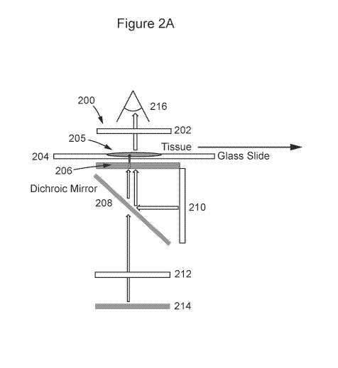
[0019] Figure 2A is a schematic of at least a portion of a DSP system
according to some
embodiments of the present disclosure;
[0020] Figure 2B is a schematic of an LCD masking component representing a
portion of a DSP
system according to some embodiments of the present disclosure;
[0021] Figure 2C-1 is a schematic of at least a portion of a DSP system
according to some
embodiments of the present disclosure;
[0022] Figure 2C-2 is a schematic of at least a portion of a DSP system
according to some
embodiments of the present disclosure;
[0023] Figure 2C-3 is a schematic of at least a portion of a DSP system
according to some
embodiments of the present disclosure, illustrating use of a UV LED component
of the system;
[0024] Figure 2D illustrates an attenuation of UV illumination by a UV LED
component of a DSP
system according to some embodiments of the present disclosure;
[0025] Figures 2E-1 through 2E-5 illustrate sealing functionality, and
pipetting fluid in/out of a
slide for a DSP system according to some embodiments of the present
disclosure;
8
CA 03123868 2021-06-16
WO 2020/132577 PCT/US2019/068069
[0026] Figure 3A illustrates a schematic of a DSP system according to some
embodiments of the
present disclosure, which is similar to that which is illustrated in Figure
2A;
[0027] Figure 3B illustrates exemplary display patterns for achieving
different microscopy
modalities, according to some embodiments of the present disclosure;
[0028] Figures 4A and 4B, are schematics which illustrate a digital micro-
mirror device
(DMD/liquid crystal on silicon (LCoS)), reflective mask structure and
operation, according to
some embodiments of the present disclosure;
[0029] Figure 4C,is a schematic of a DSP system, according to some embodiments
of the present
disclosure; illustrating the structure and operation of a scanning laser;
[0030] Figure 4D illustrates calibration schemes for a DSP system according to
some
embodiments of the present disclosure;
[0031] Figures 5A-E illustrate exemplary scaffolds and frames for a DSP system
according to
some embodiments of the present disclosure;
[0032] Figures 5F illustrate an exemplary DSP system housing structure
according to some
embodiments of the present disclosure;
[0033] Figure 6 illustrates an exemplary circuit for thermal management of a
DSP system,
according to some embodiments of the present disclosure;
[0034] Figures 7A through 7D-7 illustrate screenshots of the graphical user
interface (GUI) for a
DSP/PMD system/device, according to some embodiments of the present
disclosure;
[0035] Figure 8 illustrates a means for communicating fluid to/from tissue on
a slide, in the DSP
system according to some embodiments of the disclosure.
[0036] Figures 9A-C illustrates example patterns of openings, other structure,
configurations
and/or related data, for a DSP system according to some embodiments of the
present disclosure.
[0037] Figures 10A-10E-4 illustrates examples of fluid transport to/from a
slide and/or an assay
(e.g., 96 well plate), according to some embodiments of the present
disclosure.
9
CA 03123868 2021-06-16
WO 2020/132577 PCT/US2019/068069
DETAILED DESCRIPTION FOR AT LEAST
SOME OF THE DISCLOSED EMBODIMENTS
[0038] Some embodiments of the present disclosure provide for a compact,
mobile, digital spatial
profiling (DSP) systems (as well as associated apparatuses, devices, and
methods) are provided,
and are configured to image one or more regions-of-interest (ROIs), use UV
light to cleave oligos
off antibodies in each ROT ("photo-cleaving"), and collect the photo-cleaved
oligos (for later
hybridization and counting using, for example nanostring nCounter
technology). Some such
embodiments of the present disclosure are further to design considerations for
DSP systems as
illustrated in the chart of Figure 1C.
[0039] A high-level overview of steps performed by at least some embodiments
of the present
disclosure are shown in Figure 1A (which is a portion of the process outlined
in Figure 1B-1).
Specifically, tissue is imaged by the DSP system to find fluorescently tagged
antibodies 101, ROIs
are determined, illuminated with UV and collect DNA tags via capillary means
103, and then the
collected DNA is then hybridized to barcodes in plate and index counts to the
specific ROIs 105.
[0040] More specifically, according to some embodiments, and as shown in
Figure 1B-1, the
process begins by staining a slide with tissue thereon having oligo-conjugated
antibodies 102, the
slide is then imaged with the DSP system (according to some embodiments), and
one or more
ROIs are selected 104. The one or more ROIs are then exposed to UV light 106,
so as to cleave
off oligonucleotides ("oligos") off antibodies in the one or more ROIs. From
there, the cleaved off
oligos are aspirated 108 from the slide, via, for example, a micro-capillary
device. The collected
oligos may then be placed into an assay 110 (e.g., 96-well plate). This
process is repeated 112 for
each ROT selected. After the oligos are dispensed into the assay, the oligos
are hybridized to
barcodes, and then quantified 114 via a quantification system (e.g.,
NanoString nCounter
platform system).
[0041] Figure 1B-2 illustrates an exemplary block diagram of a DSP system
according to some
embodiments. As shown, a processor (e.g., "Raspberry Pi", "Arduino Uno", and
the like) 120 is
in communication (e.g., digital) with a camera 122, an LCD 124 with UV
polarizers (for example),
UV LED 124a and/or UV LED diver 124b, and a visible LED array 126 (for
example), used for
bright field imaging. Supplied power can be either AC or DC, which supplies,
for example an
appropriate amount of power to power the system (e.g., 25 watts or less), for
powering, for
CA 03123868 2021-06-16
WO 2020/132577 PCT/US2019/068069
example, the photomasking means, the UV and visible light sources, as well as
any processor and
communication means that may be provided. Thus, components (e.g., processor,
UV LED, UV
LED driver, visible LED array, etc.) can be configured to receive power from a
typical, standard
AC power supply 128 (e.g., wall outlet or dedicated power supply), and/or a dc
power source (e.g.,
12V power supply 130). The processor can include or have access to computer
instructions
operable on the processor to cause the processor to control one or more of
such components, and
can also include instructions (and associated hardware, if needed, e.g., wifi,
Bluetooth, cellular,
wired) to communicate information obtained or needed to/from a mobile device
132 or other
remote computing device/system (e.g., desktop, laptop, server). The remote
device can be
accessible by a pathologist 134 to review results and/or directed processes
carried out by the
system (according to some embodiments). Figure 1B-2 also includes a legend 136
regarding the
different processes being illustrated according to some embodiments (e.g.,
power, control,
interfacing, and input/output). Again, the processor can be configured to
provide graphics support
enabling the creation of photomasks with adjustable aperture sizes and
location, as well as a
calibration grid as illustrated in Figure 4D (e.g., for a personal mobile
device application),
including, for example, four-white dots 480 or corners 482 on a black or dark
colored background,
a cyan rectangle 484 on a white or light background, or single pixel
illumination 486 on black or
dark background. In some embodiments, upon startup, the processor can be
configured to cause a
calibration grid to be displayed onto the LCD, and wait for coordinates to be
sent to a processor in
the DSP (e.g., pairing via Bluetooth). Once received, an appropriate photomask
is displayed on
the LCD to highlight a user selected ROT, the backlight is turned off, and the
UV source is turned
on for a predetermined period of time such as, for example, three minutes, two
minutes, one
minute or 30 seconds).
[0042] Accordingly, in some embodiments, an example of which is shown in
Figure 1D, a digital
spatial profiling (DSP) system 140 is provided and comprises, at least one of,
and in some
embodiments, a plurality of, and in still further embodiments, all of, a
housing, and a power source,
a processor, a UV source (UVS), e.g., a UV LED(s), a visible light source
(VLS) for bright field
imaging (e.g., LCD backlight), photomasking means (e.g., LCD) configured to
selectively
illuminate a tissue sample with UV light from the UV source and/or visible
light from the visible
light source, as well as a chamber (not shown) within the DSP system (e.g., a
chamber within a
11
CA 03123868 2021-06-16
WO 2020/132577 PCT/US2019/068069
housing) configured to receive at least a portion of the slide having the
tissue thereon, via, e.g., a
slot, (not shown). A mobile device 142 is also part of the system (according
to some embodiments).
[0043] Figure 2A is a schematic of at least a portion of a DSP system
according to some
embodiments of the present disclosure. As shown, system 200 includes an
objective lens 202, an
LCD 206, a dichroic mirror 208, an LCD backlight 210, a UV LED 214, and a
condenser lens 212.
A glass slide 204 containing a tissue sample 205 is placed in a portion of the
system arranged for
imaging and exposure to light, UV or otherwise. In the illustrated example,
the slide is placed
adjacent or near to the LCD photomasking means. The imager, i.e., a mobile
device including a
camera 216, is arranged within the system so as to image the tissue on the
slide. Figure 2C-1 is
similar to Figure 2A, but includes an example of the distances certain
components are placed
among other components of the system, according to some embodiments.
Accordingly, in some
embodiments, the photomasking means is preferably arranged at a predetermined
distance from
the tissue, the distance of which can be configured such that the tissue is
not obscured by the pixel
grid. The predetermined distance can be between approximately 0.01 to 5 mm,
between
approximately 0.50 to 2.5 mm, between approximately 0.75 to 2.25 mm, or
between approximately
1 to 2 mm. Additionally, the predetermined distance can be configured to at
least one of provide
clear visualize of tissue, and to minimize diffusion of UV light. In some
embodiments, the
photomasking means is configured to provide, for example, an illumination
resolution of between
approximately 50 and 300 nm, a field of view between approximately 1-5 cm2,
and/or a
magnification of between approximately 1-3x.
[0044] Figure 2B illustrates a schematic of an exploded view of the LCD
photomask/functionality
219, with voltage "on" 221, and voltage "off' 223. Accordingly, in some
embodiments, the LCD
219 includes, a polarizing filter 220, a transparent electrode 222, a liquid
crystal 224, a second
transparent electrode 226, a second polarizing filter 228, and a screen 230.
When the voltage is on
221, the screen is dark and initially received un-polarized light (visible
and/or UV) is blocked from
passing through the LCD. When the voltage is off 223, received un-polarized
light is allowed to
pass through to the screen. Thus, illustrating the structure of the LCD and
how it performs as a
photomask via the "ON-OFF" of one or more pixels, to pass and block light.
[0045] Similar to Figure 2A, Figures 2C-2 and 2C-3 illustrate operation of the
DSP system,
according to some embodiments, illustrating how the LCD 206 and accompanying
polarizing
12
CA 03123868 2021-06-16
WO 2020/132577 PCT/US2019/068069
filters 220, 228 enable only one or more selected ROIs to be exposed to UV by
action of LCD
masking. In Figure 2C-2, the system is shown prior to exposing any of the
tissue (ROT or
otherwise) to UV light, an allowing the tissue to be illuminated by white LED
light (thus, allowing
a user to select one or more ROIs), and Figure 2C-3, illustrates exposure of
selected ROIs, to UV
light by action of the LCD mask. Specifically, it can be seen that the LCD 206
can block passage
of UV to all but ROIs of the tissue sample. It is worth noting that in some
embodiments, a diffuser
232 can be included to diffuse the white LED light. In some embodiments, UV
light can be
attenuated (see, e.g., Figure 2D), using, for example the photomasking means
for example (in
some embodiments).
[0046] A slide can be received into the chamber (and/or housing) of the DSP
system according to
some embodiments, via a "box" configuration, such that a top or side of the
box opens (via, e.g.,
hinges). The slide can be movable relative to the chamber (or housing
containing the chamber,
optics, and/or UV/light sources), where the chamber can be configured with a
liquid environment
for tissue, and sealed from liquid escaping, by any sealing means known in the
art; e.g., gasket,
see Figures 2E-1 through Figure 2E-5, which illustrate a slide (Figure 2E-1,
and gasket
configurations which may be used therewith; Figures 2E-2 and 2E-3). Such slide
and gasket
configurations can utilize magnetic means 240A on the slide, for example, and
240B on the gasket
239 (within a housing and/or frame), where, e.g., the magnetic means may be
permanent and
electromagnetic on one and/or another of the slide and gasket/housing/frame,
for mated attachment
between the slide and the gasket. The slide and gasket configurations can
include a guide means
such as a grid barrier 242 (can also be referred to as a guide, in some
embodiments), as illustrated
in Figure 2E-3, which can be configured within or proximate to a slide and
gasket configuration
to allow for guided pipetting of cleaved oligos manually or via
machine/robotically. Figures 2E-
4 and 2E-5 illustrate use of a grid 242 of capillary tubes 243 configured to
collect fluid through
capillary action (as well as dispense fluid). As shown, the grid can be
configured to fit within
components of the system, e.g., gasket 239. As shown, the grid 243 and tubes
243 can be inserted
into the gasket 239.
[0047] The system may also include optic means (e.g., lenses and like,
including an objective lens)
configured to at least one of direct and/or focus the UVS and/or VLS onto at
least one of the tissue,
the chamber, the photomasking means, and a camera sensor (e.g., "phone
camera") operably linked
to a personal mobile computing device (PMD). At least one of the housing and
chamber is
13
CA 03123868 2021-06-16
WO 2020/132577 PCT/US2019/068069
configured for removable attachment to the PMD such that the camera sensor can
image the tissue.
Figure 3A illustrates a high-level overview of the system according to some
embodiments (similar
to Figure 2A). Figure 3B illustrates different patterns which can be displayed
for achieving
different microscopy modalities for the DSP system according to some
embodiments, including
bright-field 302, dark-field 304, phase-gradient 306A, 306B, 3D 308 and super-
resolution 310.
[0048] The optic means, according to some embodiments, may include the UV
source and VLS
(though, in some embodiments, such structure can be also considered separate
from the optic
means), one or more of any of: condenser lenses, scan lenses, dichroic
mirrors, photomasking
means (see below, and elsewhere herein), objective lenses, cameras (e.g., a
personal mobile device
with camera, and the like). The optic mean, in some embodiments, is configured
to illuminate a
tissue sample with UV light from the UV source, visible light from VLS, or
visible or white light
from the LCD backlight. The dichroic mirror is configured to allow the re-
direction of light from
multiple sources (e.g., two (2) sources), into an optical axis (in some
embodiments, a single optical
axis), so it reaches the sample only in user-determined locations.
[0049] As noted according to some embodiments above, the photomasking means
can comprise
at least one of: an LCD (which can include a backlight, e.g., as shown in
Figure 2A), an LCD
configured as a programmable aperture, so as to structure at least one of UV
and visible or white
light to reach the tissue only in a regions-of-interest (ROT), a digital micro-
mirror device (DMD),
a liquid crystal on silicon (LCoS) display, an organic light-emitting diode
(OLED), a micro light-
emitting diode ( LED) array, a fiber optic bundle, a liquid crystal displays
(LCD), a scanning
laser, and, a physical barrier. In some embodiments, where the photomasking
means comprises an
LCD, the LCD may include a pixel grid.
[0050] Figures 4A and 4B, which correspond to a DSP system according to some
embodiments,
similar to that of Figures 2A and 2B, but illustrating a digital micro-mirror
device (DMD / liquid
crystal on silicon (LCoS)), reflective mask structure and operation,
corresponding to a form of the
photomasking means. A DMD is typically a chip having on its surface a
multitude (e.g., several
hundred thousand) microscopic mirrors arranged in an array (e.g., rectangular)
which correspond
to "pixels" used for photomasking. The mirrors can be individually rotated
(e.g., 10-12 ), to an
on or off state. In the on state, light from a light source is reflected into
a lens (making the pixel
appear bright), and in the off state, the light is directed elsewhere (e.g., a
heatsink), making the
14
CA 03123868 2021-06-16
WO 2020/132577 PCT/US2019/068069
pixel appear dark. Accordingly, as shown, a tissue slide 403 with tissue 405
is placed in the system,
where it can be illuminated with white light 410, via lens 407 and dichromatic
mirror 407. The
DMD/LCoS 420 performs masking to direct the UV light 414 onto specific ROIs
403b within the
tissue sample 405a on the slide 403. Figure 4B-1, and 4B-2, illustrates the
functionality of a
DMD/LCoS components (e.g., independently movable micro-mirrors 450, secondary
mirror 452
and silicon chip 454).
[0051] Figure 4C, which correspond to a DSP system according to some
embodiments, are similar
to that of Figures 4A and 4B, but make use of a scanning laser system 438,
corresponding to a yet
another form of the photomasking means. The scanning laser typically includes
moveable mirrors,
such as an XY galvanometer mirror 444 for example, capable of directing a
laser beam from laser
442 in at least two dimensions via scan lens 440 (and then via the other noted
components of the
dichroic mirror 408, camera 416, lens 402, slide 403, tissue 405a, while LED
410 and lens 407).
Scanning can be in the form of raster scanning or vector scanning. When
scanning, the scanning
laser is directed only to that part of the tissue to be illuminated which
correspond to "pixels" used
for photomasking.
[0052] Housing/frame structure for the DSP, according to some embodiments, can
comprise a
plurality of components, including, for example, one or more of any of:
scaffolds, PMD frames,
objective lens frames, slide frames, photomasking frames, condenser frame,
and, in some
embodiments, at least one thermal management means. Figures 5A-E illustrate
the various
scaffolds and frames for the DSP (e.g., which can form or together be the
housing): Figure 5A-1,
5A-2 ¨ condenser frame, for housing the condenser lens; Figure 5B-1 and 5B-2 ¨
a LCD, dichroic,
backlight frame, for holding the photomasking means (e.g., LCD); Figure 5B-3 ¨
a housing/frame
for holding the LCD and a lens (e.g., dichroic), Figure 5B-4 ¨ a housing/frame
for holding an
LCD with a controller, and a lens (e.g., dichroic); Figure 5C ¨ PMD and/or
objective frame, for
holding the PMD relative to the housing/chamber; Figure 5D ¨ a scaffold for
various uses (e.g.,
support for housing/chamber); and Figure 5E ¨ slide frame, for holding a slide
to be received by
the chamber, via a slot. The housing can be configured to removably receive a
single objective
lens frame of a plurality of objective lens frames, where each has a different
objective lens and
corresponding magnification. Each objective lens frame can be configured so as
to provide a
different spacing from the camera sensor, and can easily be swapped out for
another. Such
CA 03123868 2021-06-16
WO 2020/132577 PCT/US2019/068069
examples of frames, supports, scaffolds, and the like. Figure 5F is a
perspective view of an
assembled DSP system using various frames and scaffolds.
[0053] A thermal management means can be included in some embodiments of the
DSP system,
which can comprise at least one of a heat sink, a heat pump, a fan, a liquid
cooling system, and a
Peltier device. Figure 6 illustrates an exemplary circuit for thermal
management of the DSP,
according to some embodiments, which can be operably connected to the
processor, via an analog-
to-digital convert (e.g., Arduino ADC). The thermal management means can also
comprise a
plurality of heatsink clips.
[0054] In some embodiments, a software application (e.g., mobile application)
is included, which
can be configured to operate on a/the processor, which can be configured to
cause the PMD to
display a graphical-user-interface (GUI), the GUI can be configured to receive
user input to select
a/the region-of-interest (ROT) of a tissue image obtained via the camera
sensor of the tissue slide
and presented on a/the display of the PMD. Figures 7A-B illustrate example
screenshots of the
GUI according to some embodiments. The mobile application, in some
embodiments, is
configured to provide, for example, functional calibration of the LCD. For
example, a plurality
(e.g., 4 corners of square/rectangle) of pixels of illumination shown on the
LCD can be selected
by a user (using, e.g., the GUI), to establish a ROT as a position within the
four corners (ratio of x
and y); see e.g., left hand images on Figures 7A-B (see, e.g., also Figure 4D
for calibration
schemes of LCD). The application can also display the selected ROT, including
recording the
location on the image, and the ROT may also be changed, re-selected
immediately in case of
mistakes, or increased or decreased in size.
[0055] Figure 7C illustrates example screenshots of an exemplary software
application according
to some embodiments. Beginning at the uppermost left-hand side of the figure,
and proceeding left
to right, then down, left to right, the GUI of the mobile application allows
the process for imaging
the tissue (after the slide having the tissue thereon is received in the
chamber), by pressing (via
touchscreen), and selecting the region of interests, the "start" button.
Thereafter, the tissue is
imaged, the likes of which includes controlling the VLS to provide visible
light to the tissue sample
during image capture. Thereafter, the image is calibrated using at least one
(and preferably a
plurality) marker on the LCD for example (i.e., the photomasking means).
16
CA 03123868 2021-06-16
WO 2020/132577 PCT/US2019/068069
[0056] Next, for example, a ROT is selected by the user via the touchscreen,
and the PMD
operating the application is paired/connected to the DSP (e.g., Bluetooth).
Thereafter, the
coordinates of the ROT(s) are sent to the DSP, and UV illumination is begun,
to cleave off the
oligos bound to antibodies via a photocleavable linker.
[0057] Figures 7D-1 through 7D-7, correspond to example screenshots for the
GUI/software
application according to some embodiments:
Figure 7D-1: start GUI screenshot;
Figure 7D-2: confirmation of corner selection screenshot;
Figure 7D-3: confirmation of ROT screenshot;
Figure 7D-4: selection of new coordinates and sending of coordinates
screenshot;
Figure 7D-5: initiation of illumination of selected ROT screenshot;
Figure 7D-6: confirmation of correct ROT position screenshot; and/or
(depending
upon the embodiment)
Figure 7D-7: completion of ROT imaging and continuation onto a next ROT
screenshot.
[0058] In some embodiments, structure and associated structure is provided to
communicate fluid
to and from the tissue on the slide. For example, as shown in Figure 8, the
cleaved oligos are
aspirated, which can be done manually or via machine/robotically, via
pipetting. Such can be
conducted via openings/holes provided above the tissue sample/slide (and/or as
part of the
chamber, e.g., at least a portion thereof), or via, e.g., a flowcell. Guide
means can be configured
within or proximate to a slide and gasket configuration including a capillary
means for communic
ating fluid from the tissue on the slide (see, e.g., Figures 2E-4, 2E-5).
Figures 9A-C
illustrates example patterns of such openings, other structure, configurations
and/or related data
pertaining thereto. Such fluid related structure and/or functionality can also
include a pump system
configured to provide a flow of a solution to and/or from the slide (e.g.,
supplying buffer solution).
The cleaved oligos can be aspirated manually or via machine/robotically, via
pipetting through a
guide means such as a grid barrier as illustrated in Figure 8. The guide means
can be configured
within or proximate to a slide and gasket configuration as illustrated in
Figure 2E-3.
17
CA 03123868 2021-06-16
WO 2020/132577 PCT/US2019/068069
[0059] Figures 10A-10E-4 illustrates examples of fluid transport to/from a
slide and/or an assay
(e.g., 96 well plate), according to some embodiments of the present
disclosure. For example Figure
10A, illustrates a pipette guide 1005 for retrieving fluid (e.g., oligos) off
a slide (for example). A
perspective view of the guide is shown in Figure 10B, as well as a top view in
Figure 10C. Points
1010 illustrate backlight from an LCD "opening", which illuminates
corresponding array holes
above a ROT, for guiding the pipette to a precise location. For example,
Figures 10D-1 through
10D-3, show exemplary steps for collecting samples, including exposing the
sample to
white/visible light to visualize determined ROIs (Figure 10D-1), inserting the
guide and
identifying the ROT location (Figure 10D-2), and then inserting a pipette to
retrieve the sample
(Figure 10D-3). This repeated for each ROT.
[0060] Figure 10E-1 illustrate a micro-capillary array (e.g., 96 well format)
1010, with a guide
1020, over a sample 1030. Figure 10E-2 illustrate the number of openings/holes
to which the
micro-capillary array can be used with above the sample. Figure 10E-3
illustrates use of an airtight
cap (for example) 1040, on the top of one or more capillary tubes, which can
be a thin parafilm
layer which can be removed by heat, a plug of photo degradable material,
and/or a microfluidic
valve. Figure 10E-4 illustrates use of plugs 1050 on the bottom of one or more
capillary tubes,
which can be used in some embodiments to delay capillary action by a
relatively short time period
(e.g., several seconds or less), which can function so that no aspiration of
fluid/sample occurs
during UV illumination, but then can initiate immediately thereafter. Such a
plug can comprise at
least one of a layer of soluble material (e.g., salt, sugar), and a photo
degradable layer (e.g., UV
degradable).
[0061] While various inventive embodiments have been described and illustrated
herein, those of
ordinary skill in the art will readily envision a variety of other means,
functionality, steps, and/or
structures (including software code) for performing the functionality
disclosed and/or obtaining
the results and/or one or more of the advantages described herein, and each of
such variations
and/or modifications is deemed to be within the scope of the inventive
embodiments described
herein. More generally, those skilled in the art will readily appreciate that
all parameters, and
configurations described herein are meant to be exemplary and that the actual
parameters, and
configurations will depend upon the specific application or applications for
which the inventive
teachings is/are used. Those skilled in the art will recognize, or be able to
ascertain using no more
than routine experimentation, many equivalents to the specific inventive
embodiments described
18
CA 03123868 2021-06-16
WO 2020/132577 PCT/US2019/068069
herein. It is therefore to be understood that the foregoing embodiments are
presented by way of
example only and that, within the scope of any claims supported by this
disclosure and equivalents
thereto, inventive embodiments may be practiced otherwise than as specifically
described and
claimed. Inventive embodiments of the present disclosure are directed to each
individual feature,
system, apparatus, device, step, code, functionality and/or method described
herein. In addition,
any combination of two or more such features, systems, apparatuses, devices,
steps, code,
functionalities, and/or methods, if such features, systems, apparatuses,
devices, steps, code,
functionalities, and/or methods are not mutually inconsistent, is included
within the inventive
scope of the present disclosure. Further embodiments may be patentable over
prior art by
specifically lacking one or more features/functionality/steps (i.e., claims
directed to such
embodiments may include one or more negative limitations to distinguish such
claims from prior
art).
[0062] The above-described embodiments of the present disclosure can be
implemented in any of
numerous ways. For example, some embodiments may be implemented (e.g., as
noted) using
hardware, software or a combination thereof When any aspect of an embodiment
is implemented
at least in part in software, the software code can be executed on any
suitable processor or
collection of processors, servers, and the like, whether provided in a single
computer or distributed
among multiple computers.
[0063] In this respect, various embodiments disclosed herein may be embodied
at least in part as
a computer readable storage medium (or multiple computer readable storage
media) (e.g., a
computer memory, one or more floppy discs, compact discs, optical discs,
magnetic tapes, flash
memories, circuit configurations in Field Programmable Gate Arrays or other
semiconductor
devices, or other tangible computer storage medium or non-transitory medium)
encoded with one
or more programs that, when executed on one or more computers or other
processors, perform
methods that implement the various embodiments of the technology discussed
above. The
computer readable medium or media can be transportable, such that the program
or programs
stored thereon can be loaded onto one or more different computers or other
processors to
implement various aspects of the present technology as discussed above.
[0064] The terms "program," "software," "code," or "software code" are used
herein in a generic
sense to refer to any type of computer code or set of computer-executable
instructions that can be
19
CA 03123868 2021-06-16
WO 2020/132577 PCT/US2019/068069
employed to program a computer or other processor to implement various aspects
of the present
technology as discussed above. Additionally, it should be appreciated that
according to one aspect
of this embodiment, one or more computer programs that when executed perform
methods of the
present technology need not reside on a single computer or processor, but may
be distributed in a
modular fashion amongst a number of different computers or processors to
implement various
aspects of the present technology, on and/or over a network.
[0065] Computer-executable instructions may be in many forms, such as program
modules, or
containers, executed by one or more computers or other devices. Generally,
program modules
include routines, programs, objects, components, data structures, etc. that
perform particular tasks
or implement particular abstract data types. Typically the functionality of
the program modules
may be combined or distributed as desired in various embodiments.
[0066] Any and all references to publications or other documents, including
but not limited to,
patents, patent applications, articles, webpages, books, etc., presented
anywhere in the present
application, are herein incorporated by reference in their entirety. Moreover,
all definitions, as
defined and used herein, should be understood to control over dictionary
definitions, definitions in
documents incorporated by reference, and/or ordinary meanings of the defined
terms.
[0067] The indefinite articles "a" and "an," as used herein in the
specification and in the claims,
unless clearly indicated to the contrary, should be understood to mean "at
least one."
[0068] The phrase "and/or," as used herein in the specification and in the
claims, should be
understood to mean "either or both" of the elements so conjoined, i.e.,
elements that are
conjunctively present in some cases and disjunctively present in other cases.
Multiple elements
listed with "and/or" should be construed in the same fashion, i.e., "one or
more" of the elements
so conjoined. Other elements may optionally be present other than the elements
specifically
identified by the "and/or" clause, whether related or unrelated to those
elements specifically
identified. Thus, as a non-limiting example, a reference to "A and/or B", when
used in conjunction
with open-ended language such as "comprising" can refer, in one embodiment, to
A only
(optionally including elements other than B); in another embodiment, to B only
(optionally
including elements other than A); in yet another embodiment, to both A and B
(optionally
including other elements); etc.
CA 03123868 2021-06-16
WO 2020/132577 PCT/US2019/068069
[0069] As used herein in the specification and in the claims, "or" should be
understood to have
the same meaning as "and/or" as defined above. For example, when separating
items in a list, "or"
or "and/or" shall be interpreted as being inclusive, i.e., the inclusion of at
least one, but also
including more than one, of a number or list of elements, and, optionally,
additional unlisted items.
Only terms clearly indicated to the contrary, such as "only one of' or
"exactly one of," or, when
used in the claims, "consisting of," will refer to the inclusion of exactly
one element of a number
or list of elements. In general, the term "or" as used herein shall only be
interpreted as indicating
exclusive alternatives (i.e. "one or the other but not both") when preceded by
terms of exclusivity,
such as "either," "one of" "only one of" or "exactly one of." "Consisting
essentially of" when
used in the claims, shall have its ordinary meaning as used in the field of
patent law.
[0070] As used herein in the specification and in the claims, the phrase "at
least one," in reference
to a list of one or more elements, should be understood to mean at least one
element selected from
any one or more of the elements in the list of elements, but not necessarily
including at least one
of each and every element specifically listed within the list of elements and
not excluding any
combinations of elements in the list of elements. This definition also allows
that elements may
optionally be present other than the elements specifically identified within
the list of elements to
which the phrase "at least one" refers, whether related or unrelated to those
elements specifically
identified. Thus, as a non-limiting example, "at least one of A and B" (or,
equivalently, "at least
one of A or B," or, equivalently "at least one of A and/or B") can refer, in
one embodiment, to at
least one, optionally including more than one, A, with no B present (and
optionally including
elements other than B); in another embodiment, to at least one, optionally
including more than
one, B, with no A present (and optionally including elements other than A); in
yet another
embodiment, to at least one, optionally including more than one, A, and at
least one, optionally
including more than one, B (and optionally including other elements); etc.
[0071] In the claims, as well as in the specification above, all transitional
phrases such as
"comprising," "including," "carrying," "having," "containing," "involving,"
"holding,"
"composed of," and the like are to be understood to be open-ended, i.e., to
mean including but not
limited to. Only the transitional phrases "consisting of' and "consisting
essentially of' shall be
closed or semi-closed transitional phrases, respectively, as set forth in the
United States Patent
Office Manual of Patent Examining Procedures, Section 2111.03.
21