Note: Descriptions are shown in the official language in which they were submitted.
CA 03124168 2021-06-17
WO 2020/132066 PCT/US2019/067173
BISPECIFIC ANTI-CD28 X ANTI-CD22 ANTIBODIES AND USES THEREOF
RELATED APPLICATIONS
[0001] This application claims the benefit of priority to U.S. Provisional
Application No.
62/781,689, filed on December 19, 2018, the entire contents of which are
incorporated
herein by reference.
SEQUENCE LISTING
[0002] The instant application contains a Sequence Listing which has been
submitted
electronically in ASCII format and is hereby incorporated by reference in its
entirety. Said
ASCII copy, created on December 16, 2019, is named 118003 49220 SL.txt and is
104,353
bytes in size.
FIELD OF THE INVENTION
[0003] The present invention relates to bispecific antigen-binding molecules
that bind
CD28 and a target molecule, such as CD22, and methods of use thereof.
BACKGROUND
[0004] CD28 is a type I transmembrane protein expressed on the surface of T
cells, which
has a single extracellular Ig-V-like domain assembled as a homodimer. CD28 is
the
receptor for the CD80 (B7.1) and CD86 (B7.2) proteins and is activated by CD80
or CD86
expressed on antigen-presenting cells (APCs). The binding of CD28 to CD80 or
CD86
provides co-stimulatory signals important for T cell activation and survival.
T cell stimulation
through CD28, in addition to the T-cell receptor (TCR), provides a potent
signal for the
production of various interleukins. CD28 also potentiates cellular signals
such as pathways
controlled by the NFKB transcription factor after TCR activation. The CD28 co-
signal is
important for effective T-cell activation such as T cell differentiation,
proliferation, cytokine
release and cell-death.
[0005] Anti-CD28 antibodies have been proposed for therapeutic purposes
involving the
activation of T cells. One particular anti-CD28 antibody, TGN1412 (anti-CD28
superagonist),
was used in a clinical trial in 2006. Six healthy volunteers were dosed
intravenously with
TGN1412 (anti-CD28 superagonist) at a dose of 0.1 mg/kg. Within two hours, all
six patients
had significant inflammatory responses (cytokine storm), and all patients were
in multi-organ
failure within sixteen hours. Subjects were treated with corticosteriods, and
cytokine levels
returned to normal levels within 2-3 days. The starting dose of 0.1 mg/kg in a
Phase 1 study
(associated with CRS) was based on 500-fold multiple of cynomolgus "NOAEL" of
50 mg/kg
1
CA 03124168 2021-06-17
WO 2020/132066 PCT/US2019/067173
(Suntharalingam, etal., Cytokine Storm in a Phase 1 Trial of the Anti-0D28
Monoclonal
Antibody TGN1412, NEJM 355:1018-1028 (2006)). Unfortunately, TGN1412 induced a
cytokine storm, which was not predicted by toxicology studies in cynomolgus
macaques or
ex vivo human PBMC studies.
[0006] 0D22 (also known as Siglec-2), a member of Siglec family, specifically
recognizes
a2,6 sialic acid, and is a transmembrane protein preferentially expressed on B
lymphocytes
(B cells).
[0007] 0D22 has a number of ascribed functions including, for example, B cell
homeostasis, B cell survival and migration, dampening TLR and CD40 signaling,
and
inhibiting B cell receptor (BCR) signaling via recruitment of 5H2 domain-
containing
phosphatases by phosphorylation of immunoreceptor tyrosine-based inhibition
motifs (ITIMs)
in the cytoplasmic region, as well as facilitation of adhesion between B cells
and other cell
types.
[0008] 0D22 is not found on the surface of B cells during the early stages of
development,
nor is it expressed in stem cells. However, 60-70% of all B-cell lymphomas and
leukemias
express 0D22.
[0009] An anti-0D22 antibody for treating B-cell lymphomas and leukemias has
been
investigated. However, the monoclonal antibody, Epratuzumab, had limited
success. (Grant,
etal. (2013) Cancer 119(21): 10.1002/cncr.28299)
[0010] Accordingly, there is a need in the art for improved anti-0D22-
antibodies. There is
also a need for anti-0D28 antibody that is safe for use in a pharmaceutical
composition.
Furthermore, bispecific antigen-binding molecules that bind both 0D28 and a
target antigen
(such as 0D22) would be useful in therapeutic settings in which specific
targeting and T cell-
mediated killing of cells that express the target antigen is desired.
BRIEF SUMMARY OF THE INVENTION
[0011] In a first aspect, the present invention provides bispecific antigen-
binding molecules
that bind 0D28 and a target antigen. According to certain exemplary
embodiments, the
bispecific antigen-binding molecules bind 0D28 and 0D22; such bispecific
antigen-binding
molecules are also referred to herein as "anti-0D28/anti-0D22 bispecific
molecules." The
anti-0D22 portion of the anti-0D28/anti-0D22 bispecific molecule is useful for
targeting
cancer cells that express 0D22 (e.g., a cancerous B cell), and the anti-0D28
portion of the
bispecific molecule is useful for activating T-cells. The simultaneous binding
of 0D22 on a
cancer cell and 0D28 on a T-cell facilitates directed killing (cell lysis) of
the targeted cancer
cell by the activated T-cell, e.g., after TCR activation of the T cell. The
anti-0D28/anti-0D22
bispecific molecules of the invention are therefore useful, inter alia, for
treating diseases and
disorders related to or caused by 0D22-expressing tumors (e.g., a B cell
proliferative
2
CA 03124168 2021-06-17
WO 2020/132066 PCT/US2019/067173
disorder, e.g., a B cell lymphoma, e.g., diffuse large B-cell lymphoma (DLBCL,
follicular
lymphoma (FL), a marginal zone lymphoma).
[0012] The bispecific antigen-binding molecules according to this aspect of
the present
invention comprise a first antigen-binding domain that specifically binds
human 0D28, and a
second antigen-binding domain that specifically binds 0D22. The present
invention includes
anti-0D28/anti-0D22 bispecific molecules (e.g., bispecific antibodies) wherein
each antigen-
binding domain comprises a heavy chain variable region (HCVR) paired with a
light chain
variable region (LCVR). In certain exemplary embodiments of the invention, the
anti-0D28
antigen-binding domain and the anti-0D22 antigen binding domain each comprise
different,
distinct HCVRs paired with a common LCVR.
[0013] The present invention provides anti-0D28/anti-0D22 bispecific
molecules, wherein
the first antigen-binding domain that specifically binds 0D28 comprises any of
the HCVR
amino acid sequences as set forth in Table 6. The first antigen-binding domain
that
specifically binds 0D28 may also comprise any of the LCVR amino acid sequences
as set
forth in Table 6. According to certain embodiments, the first antigen-binding
domain that
specifically binds 0D28 comprises any of the HCVR/LCVR amino acid sequence
pairs as set
forth in Table 6. The present invention also provides anti-0D28/anti-0D22
bispecific
molecules, wherein the first antigen-binding domain that specifically binds
0D28 comprises
any of the heavy chain CDR1-CDR2-CDR3 amino acid sequences as set forth in
Table 6,
and/or any of the light chain CDR1-CDR2-CDR3 amino acid sequences as set forth
in Table
6.
[0014] According to certain embodiments, the present invention provides anti-
0D28/anti-
0D22 bispecific molecules, wherein the first antigen-binding domain that
specifically binds
0D28 comprises a heavy chain variable region (HCVR) having an amino acid
sequence
selected from the group consisting of SEQ ID NOs: 28 and 26 or a substantially
similar
sequence thereof having at least 90%, at least 95%, at least 98% or at least
99% sequence
identity.
[0015] The present invention also provides anti-0D28/anti-0D22 bispecific
molecules,
wherein the first antigen-binding domain that specifically binds 0D28
comprises a light chain
variable region (LCVR) having the amino acid sequence of SEQ ID NO: 10, or a
substantially similar sequence thereof having at least 90%, at least 95%, at
least 98% or at
least 99% sequence identity.
[0016] The present invention also provides anti-0D28/anti-0D22 bispecific
molecules,
wherein the first antigen-binding domain that specifically binds 0D28
comprises a HCVR and
LCVR (HCVR/LCVR) amino acid sequence pair selected from the group consisting
of SEQ
ID NOs: 28/10 and 26/10.
3
CA 03124168 2021-06-17
WO 2020/132066 PCT/US2019/067173
[0017] The present invention also provides anti-0D28/anti-0D22 bispecific
molecules,
wherein the first antigen-binding domain that specifically binds 0D28
comprises a heavy
chain CDR3 (HCDR3) domain having the amino acid sequence of SEQ ID NO: 32, or
a
substantially similar sequence thereto having at least 90%, at least 95%, at
least 98% or at
least 99% sequence identity; and a light chain CDR3 (LCDR3) domain having the
amino
acid sequence of SEQ ID NO: 16, or a substantially similar sequence thereof
having at least
90%, at least 95%, at least 98% or at least 99% sequence identity.
[0018] In certain embodiments, the first antigen-binding domain that
specifically binds
0D28 comprises the HCDR3/LCDR3 amino acid sequence pair of SEQ ID NOs: 32/16.
[0019] The present invention also provides anti-0D28/anti-0D22 bispecific
antigen-binding
molecules, wherein the first antigen-binding domain that specifically binds
0D28 comprises a
heavy chain CDR1 (HCDR1) domain having the amino acid sequence of SEQ ID NO:
28, or
a substantially similar sequence thereof having at least 90%, at least 95%, at
least 98% or at
least 99% sequence identity; a heavy chain CDR2 (HCDR2) domain having the
amino acid
sequence of SEQ ID NO: 30, or a substantially similar sequence thereof having
at least 90%,
at least 95%, at least 98% or at least 99% sequence identity; a light chain
CDR1 (LCDR1)
domain having the amino acid sequence of SEQ ID NO: 12, or a substantially
similar
sequence thereof having at least 90%, at least 95%, at least 98% or at least
99% sequence
identity; and a light chain CDR2 (LCDR2) domain having the amino acid sequence
of SEQ
ID NO: 14, or a substantially similar sequence thereof having at least 90%, at
least 95%, at
least 98% or at least 99% sequence identity.
[0020] Certain non-limiting, exemplary anti-CD28/anti-CD22 bispecific antigen-
binding
molecules of the invention include a first antigen-binding domain that
specifically binds CD28
comprising HCDR1-HCDR2-HCDR3-LCDR1-LCDR2-LCDR3 domains, respectively, having
the amino acid sequence of: SEQ ID NOs: 28-30-32-12-14-16.
[0021] The present invention also provides anti-CD28/anti-CD22 bispecific
molecules,
wherein the second antigen-binding domain that specifically binds CD22
comprises a heavy
chain variable region (HCVR) having the amino acid sequence selected from the
group
consisting SEQ ID NOs: 2 and 18, or a substantially similar sequence thereof
having at least
90%, at least 95%, at least 98% or at least 99% sequence identity.
[0022] The present invention also provides anti-CD28/anti-CD22 bispecific
molecules,
wherein the second antigen-binding domain that specifically binds CD22
comprises a light
chain variable region (LCVR) having the amino acid sequence selected of SEQ ID
NO: 10,
or a substantially similar sequence thereof having at least 90%, at least 95%,
at least 98% or
at least 99% sequence identity.
[0023] The present invention also provides anti-CD28/anti-CD22 bispecific
molecules,
wherein the second antigen-binding domain that specifically binds CD22
comprises a HCVR
4
CA 03124168 2021-06-17
WO 2020/132066 PCT/US2019/067173
and LCVR (HCVR/LCVR) amino acid sequence pair selected from the group
consisting of
SEQ ID NOs: 2/10 and 18/10.
[0024] The present invention also provides anti-0D28/anti-0D22 bispecific
molecules,
wherein the second antigen-binding domain that specifically binds 0D22
comprises a heavy
chain CDR3 (HCDR3) domain having the amino acid sequence selected from the
group
consisting of SEQ ID NOs: 8 and 24, or a substantially similar sequence
thereto having at
least 90%, at least 95%, at least 98% or at least 99% sequence identity; and a
light chain
CDR3 (LCDR3) domain having the amino acid sequence selected of SEQ ID NO:16,
or a
substantially similar sequence thereof having at least 90%, at least 95%, at
least 98% or at
least 99% sequence identity.
[0025] In certain embodiments, the second antigen-binding domain that
specifically binds
0D22 comprises a HCDR3/LCDR3 amino acid sequence pair selected from the group
consisting of SEQ ID NOs: 8/16 and 24/16.
[0026] The present invention also provides anti-0D28/anti-0D22 bispecific
antigen-binding
molecules, wherein the second antigen-binding domain that specifically binds
0D22
comprises a heavy chain CDR1 (HCDR1) domain having the amino acid sequence
selected
from the group consisting of SEQ ID NOs: 4 and 20, or a substantially similar
sequence
thereof having at least 90%, at least 95%, at least 98% or at least 99%
sequence identity; a
heavy chain CDR2 (HCDR2) domain having the amino acid sequence selected from
the
group consisting of SEQ ID NOs: 6 and 22, or a substantially similar sequence
thereof
having at least 90%, at least 95%, at least 98% or at least 99% sequence
identity; a light
chain CDR1 (LCDR1) domain having the amino acid sequence of SEQ ID NO: 12, or
a
substantially similar sequence thereof having at least 90%, at least 95%, at
least 98% or at
least 99% sequence identity; and a light chain CDR2 (LCDR2) domain having the
amino
acid sequence of SEQ ID NO: 14, or a substantially similar sequence thereof
having at least
90%, at least 95%, at least 98% or at least 99% sequence identity.
[0027] Certain non-limiting, exemplary anti-CD28/anti-CD22 bispecific antigen-
binding
molecules of the invention include a second antigen-binding domain that
specifically binds
CD22 comprising HCDR1-HCDR2-HCDR3-LCDR1-LCDR2-LCDR3 domains, respectively,
having the amino acid sequences selected from the group consisting of: SEQ ID
NOs: 4-6-8-
12-14-16 and 20-22-24-12-14-16
[0028] In a related embodiment, the invention includes anti-CD28/anti-CD22
bispecific
antigen binding molecules wherein the second antigen-binding domain that
specifically binds
CD22 comprises the heavy and light chain CDR domains contained within heavy
and light
chain variable region (HCVR/LCVR) sequences selected from the group consisting
of SEQ
ID NOs: 2/10 and 18/10.
CA 03124168 2021-06-17
WO 2020/132066 PCT/US2019/067173
[0029] In another aspect, the present invention provides nucleic acid
molecules encoding
any of the HCVR, LCVR or CDR sequences of the anti-0D28/anti-0D22 bispecific
antigen-
binding molecules disclosed herein, including nucleic acid molecules
comprising the
polynucleotide sequences as set forth in Table 7 herein, as well as nucleic
acid molecules
comprising two or more of the polynucleotide sequences as set forth in Table 7
in any
functional combination or arrangement thereof. Recombinant expression vectors
carrying the
nucleic acids of the invention, and host cells into which such vectors have
been introduced,
are also encompassed by the invention, as are methods of producing the
antibodies by
culturing the host cells under conditions permitting production of the
antibodies, and
recovering the antibodies produced.
[0030] The present invention includes anti-0D28/anti-0D22 bispecific antigen-
binding
molecules wherein any of the aforementioned antigen-binding domains that
specifically bind
0D28 is combined, connected or otherwise associated with any of the
aforementioned
antigen binding domains that specifically bind 0D22 to form a bispecific
antigen-binding
molecule that binds 0D28 and 0D22.
[0031] The present invention includes anti-0D28/anti-0D22 bispecific antigen-
binding
molecules having a modified glycosylation pattern. In some applications,
modification to
remove undesirable glycosylation sites may be useful, or an antibody lacking a
fucose
moiety present on the oligosaccharide chain, for example, to increase antibody
dependent
cellular cytotoxicity (ADCC) function (see Shield etal. (2002) JBC 277:26733).
In other
applications, modification of galactosylation can be made in order to modify
complement
dependent cytotoxicity (CDC).
[0032] In another aspect, the invention provides a pharmaceutical composition
comprising
an anti-0D28/anti-0D22 bispecific antigen-binding molecule as disclosed herein
and a
pharmaceutically acceptable carrier. In a related aspect, the invention
features a
composition which is a combination of an anti-0D28/anti-0D22 bispecific
antigen-binding
molecule and a second therapeutic agent. In one embodiment, the second
therapeutic
agent is any agent that is advantageously combined with an anti-0D28/anti-0D22
bispecific
antigen-binding molecule. Exemplary agents that may be advantageously combined
with an
anti-0D28/anti-0D22 bispecific antigen-binding molecule are discussed in
detail elsewhere
herein.
[0033] In yet another aspect, the invention provides therapeutic methods for
targeting/killing cancer cells expressing 0D22 using an anti-0D28/anti-0D22
bispecific
antigen-binding molecule of the invention, wherein the therapeutic methods
comprise
administering a therapeutically effective amount of a pharmaceutical
composition comprising
an anti-0D28/anti-0D22 bispecific antigen-binding molecule of the invention to
a subject in
need thereof.
6
CA 03124168 2021-06-17
WO 2020/132066 PCT/US2019/067173
[0034] The present invention also includes the use of an anti-0D28/anti-0D22
bispecific
antigen-binding molecule of the invention in the manufacture of a medicament
for the
treatment of a disease or disorder related to or caused by 0D22 expression.
[0035] In yet another aspect, the invention provides therapeutic methods for
targeting/killing cancer cells expressing 0D22 using an anti-0D28/anti-0D22
bispecific
antigen-binding molecule of the invention, wherein the anti-0D28/anti-0D22
bispecific
antigen-binding molecule is combined with other anti-tumor bispecific antigen-
binding
molecules that bind to CD3 (e.g., anti-0D28/anti-0D22 combined with anti-
CD3/anti-0D20
antibodies).
[0036] In still another aspect, the invention provides therapeutic methods for
targeting/killing cancer cells expressing 0D22 using an anti-0D28/anti-0D22
bispecific
antigen-binding molecule of the invention, wherein the anti-0D28/anti-0D22
bispecific
antigen-binding molecule is combined with a checkpoint inhibitor targeting PD-
1, PD-L1 or
CTLA-4 (e.g., anti-0D28/anti-CD-22 combined with anti-PD-1 antibodies). For
example, in
certain embodiments, the anti-0D28/anti-0D22 antibodies of the invention may
be combined
with agents that target PD-1, such as Pembrolizumab (Keytrudae). Nivolumab
(Opdivoe). or
Cemiplimab (Libtayo6). In certain embodiments, the anti-0D28/anti-0D22
antibodies of the
invention may be combined with agents that target PD-L1, such as Atezolizumab
(iecentrige), Avelumab (Bavencioe), or Durvalumab (Imfinzi ). In certain
embodiments, the
anti-0D28/anti-0D22 antibodies of the invention may be combined with agents
that target
CTLA-4, such as Ipilimumab (Yervoy(9).
[0037] In still another aspect, the invention provides therapeutic methods for
targeting/killing cancer cells expressing 0D22 using an anti-0D28/anti-0D22
bispecific
antigen-binding molecule of the invention, wherein the anti-0D28/anti-0D22
bispecific
antigen-binding molecule is combined with other anti-tumor bispecific antigen-
binding
molecules that binds to CD3 (e.g., anti-0D28/anti-0D22 combined with anti-
CD3/anti-0D20
bispecific antibodies, for example, REGN1979 (See U59,657,102, wherein the
anti-0D20
arm comprises the HCVR/LCVR amino acid pair of SEQ ID NOs: 1242/1258 and the
anti-
CD3 arm comprises the amino acid pair of SEQ ID NOs: 1250/1258)) and/or a
checkpoint
inhibitor targeting PD-1, PD-L1 or CTLA-4 (e.g., anti-0D28/anti-0D22 combined
with anti-PD-
1 antibodies). For example, in certain embodiments, the anti-0D28/anti-0D22
antibodies of
the invention may be combined with agents that target PD-1, such as
Pembrolizumab
(Keytrudae), Nivolumab (Opdivoe), or Cemiplimab (Libtayoe, see for example,
US9,987,500, wherein cemiplimab comprises the HCVIR/LCVR amino acid pair of
SEQ ID
NOs: 162/170)). In certain embodiments, the anti-0D28/anti-0D22 antibodies of
the
invention may be combined with agents that target PD-L1, such as Atezolizumab
(Tecentride), Avelumab (Bavencio8), or Durvalumab (Imfinzie). In certain
embodiments, the
7
CA 03124168 2021-06-17
WO 2020/132066 PCT/US2019/067173
anti-0D28/anti-0D22 antibodies of the invention may be combined with agents
that target
CTLA-4, such as ipilimurnab (Yervoye).
[0038] Other embodiments will become apparent from a review of the ensuing
detailed
description.
BRIEF DESCRIPTION OF THE FIGURES
[0039] Figure 1 is a set of graphs depicting the binding of anti-0D28/anti-
0D22 bispecific
antibodies to human CD4+ T-cells expressing 0D28 and target cells expressing
human
0D22 on the cell surface.
[0040] Figures 2A and 2B are a set of graphs depicting that anti-0D28/anti-
0D22
bispecific antibodies show increased Luciferase production in the presence of
primary T-cell
stimulation and 0D22 target expression. Figure 2A is a set of graphs depicting
the
activation of engineered reporter T-cells co-incubated with HEK293/hCD20,
HEK293/hCD20/hCD22, or Raji/CD80 and 0D86 negative cells in addition to 200pM
constant REGN1945 (a negative hIgG4 isotype control), as assessed by Lucif
erase
production. Figure 2B is a set of graphs depicting the activation of
engineered reporter T-
cells co-incubated with HEK293/hCD20, HEK293/hCD20/hCD22, or Raji/0D80 and
0D86
negative cells in addition to 200pM constant REGN2281 (anti-CD20 x anti-CD3),
as
assessed by Lucif erase production.
[0041] Figure 3A and 3B are a set of graphs depicting that anti-0D28/anti-0D22
bispecific
antibodies increase IL-2 production in the presence of primary T-cell
stimulation and 0D22
target expression. More specifically, Figure 3A is a set of graphs depicting
the activation of
CD4+ T-cells co-incubated with HEK293/hCD20, HEK293/hCD20/hCD22, or Raji/CD80
and
0D86 negative cells in the presence of 2nM constant REGN1945 (h IgG4 isotype
control), as
assessed by IL-2 production. Figure 3B is a set of graphs depicting the
activation of CD4+
T-cells co-incubated with HEK293/hCD20, HEK293/hCD20/hCD22, or Raji/CD80 and
0D86
negative cells in the presence of 2nM constant REGN2281 (anti-CD20 x anti-
CD3), as
assessed by IL-2 production.
[0042] Figure 4 is a set of graphs showing that a combination of REGN5837 with
cemiplimab enhances IL-2 release above REGN5837 treatment alone in cells
engineered to
express PD-L1.
[0043] Figure 5A is a set of graphs showing that a combination of REGN5837
with
cemiplimab enhances IL-2 release in the presence of NALM6 cells engineered to
express
PD-L1.
[0044] Figure 5B is a set of graphs showing that a combination of REGN5837
with
cemiplimab enhances IL-2 release above REGN5837 treatment alone in RAJI cells
engineered to express PD-L1.
8
CA 03124168 2021-06-17
WO 2020/132066 PCT/US2019/067173
[0045] Figure 6 is a graph showing that treatment of NSG mice bearing NALM-6-
Luc
tumors with REGN5837 in the presence of REGN1979 (anti-CD20 x anti-CD3) is
associated
with significant tumor suppression. Briefly, NSG mice (n=6 to 9 per group)
were engrafted
with human PBMC, then implanted with NALM-6-luc B-cell leukemia cells 12 days
post-
engraftment (day 0). Mice were administered 4 mg/kg REGN5837 + 0.04 mg/kg
REGN1979
(hashed circles), 0.4 mg/kg REGN5837 + 0.04 mg/kg REGN1979 (closed upright
triangles),
0.04 mg/kg REGN5837 + 0.04 mg/kg REGN1979 (diamonds), 4 mg/kg non-TAAxCD28 +
0.04 mg/kg REGN1979 (squares), 4 mg/kg REGN5837 + 0.4 mg/kg non-TAAxCD3 (open
circles), or 4 mg/kg non-TAAxCD28 + 0.4 mg/kg non-TAAxCD3 (closed inverted
triangles)
on days 8, 15, and 22 post-implantation (arrows). Tumor growth was monitored
by
bioluminescent imaging of tumor volume on days 6, 10, 14, 17, 20, and 23 post-
implantation.
Combined data are expressed as the group mean SEM. Statistical significance
was
determined using two-way ANOVA with Tukey's post hoc test. The following
symbols were
used to indicate statistically significant differences relative to non-
TAAxCD28 + non-
TAAxCD3 control: *, p<0.05; **, p<0.01; ***, p<0.001.
[0046] Figures 7A-7C are graphs showing that REGN1979 activated and directed
human
T cells to kill Nalm6 cells in a dose dependent manner. More specifically,
Figure 7A is a
graph depicting the percent survival of Nalm6 cells in the presence of the
indicated
antibodies. Figure 7B is a graph depicting the the percent of CD8+ cells
expressing CD25
(CD25+) in the presence of the indicated antibodies. Figure 7C is a graph
depicting the
proliferation of CD25+CD8+ cells as assessed by CellTrace violet dilution in
the presence of
the indicated antibodies.
[0047] Figure 8A, 8B and 8C are graphs showing that REGN1979 activated and
directed
human T cells to kill WSU-DLCL2 cells in a dose dependent manner. More
specifically,
Figure 8A is a graph depicting the percent survival of WSU-DLCL2 cells in the
presence of
the indicated antibodies. Figure 8B is a graph depicting the the percent of
CD8+ cells
expressing CD25 (CD25+) in the presence of the indicated antibodies. Figure 8C
is a graph
depicting the proliferation of CD8+ cells, expressed as % divided, in the
presence of the
indicated antibodies.
[0048] Figure 9 is a set of graphs showing that in assays with human PBMC and
WSU-
DLCL2 cells, REGN1979 induced the release of human cytokines, IL-2, IL-4, IL-
6, and IL-10.
Cytokine release observed with REGN1979 was enhanced in the presence of a
fixed
concentration of CD22 X CD28 compared to cytokine release induced by REGN1979
alone.
[0049] Figures 10A-10E are graphs showing that REGN1979 activated and directed
human T cells to deplete NHL in a dose-dependent manner. The addition of a
fixed
concentration of CD22xCD28 bispecific antibodies to REGN1979 enhanced the
cytotoxic
efficacy (EC50) of REGN1979 2.3 and 3.5 fold when compared to REGN1979 with 1-
arm
9
CA 03124168 2021-06-17
WO 2020/132066 PCT/US2019/067173
0D28 control antibody or no costimulatory control. The observed target-cell
lysis mediated
by REGN1979 was associated with T cell activation and proliferation, as
measured by 0D25
upregulation on CD8+ and CD4+ cells or CellTrace violet dilution respectively.
More
specifically, Figure 10A is a graph depicting the percent survival of NHL
cells from patient
bone marrow in the presence of the indicated antibodies. Figure 10B is a graph
depicting
the the percent of CD8+ cells expressing 0D25 (0D25+) in the presence of the
indicated
antibodies. Figure 10C is a graph depicting the proliferation of CD8+ cells as
assessed by
CellTrace violet dilution in the presence of the indicated antibodies. Figure
10D is a graph
depicting the the percent of CD4+ cells expressing 0D25 (0D25+) in the
presence of the
indicated antibodies. Figure 10E is a graph depicting the proliferation of
CD4+ cells as
assessed by CellTrace violet dilution in the presence of the indicated
antibodies.
[0050] Figures 11A-11E are graphs showing that REGN5837 Enhances the potency
of
REGN1979-mediated cytotoxicity, cell-surface expression of 0D25, and T-Cell
proliferation
in a concentration-dependent manner. Briefly, WSU-DLCL2 cells were incubated
with
lymphocyte-enriched human PBMC at a target cell to PBMC ratio of 1:5 and with
anti-
CD20xCD3 (REGN1979) at a range of concentrations (4.8fM to 10nM) as a single
agent (ie,
no REGN5837) or in the presence of fixed concentrations of REGN5837 (ranging
from 0.01
to 15 pg/mL) for 72 hours at 37 C. A condition lacking REGN1979 contains
REGN5837
alone at the concentration indicated, and is plotted as 0.04 pM. Viable cells
were detected by
flow cytometry using LIVE/DEAD cell stain (11A). Violet Cell Tracker dye and a
phenotyping
cocktail of fluorophore-labeled antibodies to CD2, CD4, CD8, and CD25 was used
to detect
T-cell activation (measured as CD25 expression; 11B, 11D) and CD4+ and CD8+ T-
cell
proliferation by flow cytometry (11C, 11E).
[0051] More specifically, Figure 11A is a graph depicting the % of dead cells
with the
indicated concentrations of REGN5837. Figure 11B is graph depicting the
percent of
CD25+ CD4+ cells with the indicated concentrations of REGN5837. Figure 11C is
a graph
depicting the proliferation of CD4+ cells as assessed by CellTrace violet
dilution with the
indicated concentrations of REGN5837. Figure 11D is graph depicting the
percent of CD25+
CD8+ cells. Figure 11E is a graph depicting the proliferation of CD8+ cells as
assessed by
CellTrace violet dilution with the indicated concentrations of REGN5837.
[0052] Figures 12A-12G are graphs showing that REGN5837 enhances the potency
and
maximal levels of REGN1979-mediated cytokine release from human T cells in a
concentration-dependent manner in the presence of WSU-DLCL2 B-cell lymphoma
cells.
Briefly, WSU-DLCL2 cells were incubated with lymphocyte-enriched human PBMC at
a
target cell to PBMC ratio of 1:5 and with anti-CD20xCD3 (REGN1979) at a range
of
concentrations (4.8fM to 10nM) as a single agent (i.e., no REGN5837) or in the
presence of
fixed concentrations of REGN5837 (ranging from 0.01 to 15 pg/mL) for 72 hours
at 37 C. A
CA 03124168 2021-06-17
WO 2020/132066 PCT/US2019/067173
condition lacking REGN1979 contains REGN5837 alone at the concentration
indicated, and
is plotted as 0.04pM. Supernatants were assessed for cytokine release of (12A)
IL-2, (12B)
IL-4, (12C) IL-6, (12D) IL-10, (12E) TNF-a, (12F) IFN-y, and (12G) IL-17a
using a BD
Cytometric Bead Array Human Th1/Th2/Th17 Cytokine Kit.
[0053] More specifically, Figure 12A is a graph depicting the level of IL-2
released from
human T cells in the presence of WSU-DLCL2 cells with the indicated
concentrations of
REGN5837. Figure 12B is a graph depicting the level of IL-4 released from
human T cells in
the presence of WSU-DLCL2 cells WSU-DLCL2 cells with the indicated
concentrations of
REGN5837. Figure 12C is a graph depicting the level of IL-6 released from
human T cells in
the presence of WSU-DLCL2 cells WSU-DLCL2 cells with the indicated
concentrations of
REGN5837. Figure 12D is a graph depicting the level of IL-10 released from
human T cells
in the presence of WSU-DLCL2 cells WSU-DLCL2 cells with the indicated
concentrations of
REGN5837. Figure 12E is a graph depicting the level of TNF-a released from
human T cells
in the presence of WSU-DLCL2 cells WSU-DLCL2 cells with the indicated
concentrations of
REGN5837. Figure 12F is a graph depicting the level of IFN-y released from
human T cells
in the presence of WSU-DLCL2 cells WSU-DLCL2 cells with the indicated
concentrations of
REGN5837. Figure 12G is a graph depicting the level of IL-17a released from
human T
cells in the presence of WSU-DLCL2 cells WSU-DLCL2 cells with the indicated
concentrations of REGN5837.
[0054] Figures 13A and 13B are graphs showing that treatment of NSG mice
bearing
WSU-DLCL2 tumors with REGN5837 in the presence of 0.4 or 4 mg/kg of REGN1979
is
associated with significant tumor suppression. Briefly, Female NSG mice (n=6
to 7 per
group) were implanted with a 1:1 mixture of WSU-DLCL2 B-cell lymphoma cells
and human
PBMC (day 0). Mice were administered combinations of 1 mg/kg REGN5837 and 0.4
mg/kg
(13A) or 4 mg/kg (13B) REGN1979 (or non-bridging controls) on days 1, 8, and
15 post-
implantation (arrows). Tumor growth was monitored by caliper measurement on
days 7, 10,
14, 16, 28, 31, 35, 38, 43, 46, 49, 53, 57, and 63 post-implantation. Combined
data are
expressed as the group mean SEM. Statistical significance was determined
using two-way
ANOVA with Tukey's post hoc test. The following symbols were used to indicate
statistically
significant differences between groups: *, p<0.05; **, p<0.01; ***, p<0.001;
****, p<0.0001.
Asterisks indicate statistical significance between REGN1979 monotherapy and
isotype
control, hash marks indicate significance between the combination of REGN5837
with
REGN1979 and isotype control, and carets indicate significance between
REGN1979
monotherapy and the combination of REGN5837 with REGN1979 .
[0055] More specifically, Figure 13A is a graph depicting tumor growth in mice
administered 1 mg/kg REGN5837 and 0.4 mg/kg REGN1979 (or non-bridging
controls, non-
11
CA 03124168 2021-06-17
WO 2020/132066 PCT/US2019/067173
TAAxCD3). Figure 13B is a graph depicting tumor growth in mice administered 1
mg/kg
REGN5837 and 4 mg/kg (or non-bridging controls, non-TAAxCD3).
[0056] Figure 14 is a graph showing that treatment of NSG mice bearing WSU-
DLCL2
tumors with REGN5837 in the presence of sub-efficacious doses of REGN1979 is
associated with significantly greater survival than REGN5837 or REGN1979
monotherapy.
Briefly, female NSG mice (n=6 to 7 per group) were implanted with a 1:1
mixture of WSU-
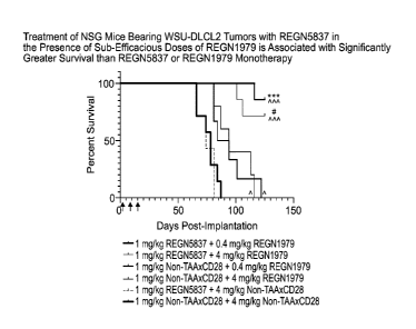
DLCL2 B-cell lymphoma cells and human PBMC (day 0). Mice were administered
combinations of REGN5837 and REGN1979 or controls on days 1,8, and 15 post-
implantation (arrows). Statistical significance was determined using a Mantel-
Cox test. The
following symbols were used to indicate statistically significant differences
between groups: *,
p<0.05; ***, p<0.001. Carets indicate statistical significance compared with
isotype control,
astericks indicate significance compared with 0.4 mg/kg REGN1979 monotherapy,
and hash
marks indicate significance compared with 4 mg/kg REGN1979 monotherapy.
DETAILED DESCRIPTION
[0057] Before the present invention is described, it is to be understood that
this invention is
not limited to particular methods and experimental conditions described, as
such methods
and conditions may vary. It is also to be understood that the terminology used
herein is for
the purpose of describing particular embodiments only, and is not intended to
be limiting,
since the scope of the present invention will be limited only by the appended
claims.
[0058] Unless defined otherwise, all technical and scientific terms used
herein have the
same meaning as commonly understood by one of ordinary skill in the art to
which this
invention belongs. As used herein, the term "about," when used in reference to
a particular
recited numerical value, means that the value may vary from the recited value
by no more
than 1 %. For example, as used herein, the expression "about 100" includes 99
and 1 01
and all values in between (e.g., 99.1, 99.2, 99.3, 99.4, etc.).
[0059] Although any methods and materials similar or equivalent to those
described herein
can be used in the practice or testing of the present invention, the preferred
methods and
materials are now described. All patents, applications and non-patent
publications
mentioned in this specification are incorporated herein by reference in their
entireties.
Definitions
[0060] The expression "CD28," as used herein, refers to an antigen which is
expressed on
T cells as a costimulatory receptor. Human CD28 comprises the amino acid
sequence as
set forth in SEQ ID NO: 74, and/or having the amino acid sequence as set forth
in NCB!
accession No. NP 006130.1. All references to proteins, polypeptides and
protein fragments
herein are intended to refer to the human version of the respective protein,
polypeptide or
12
CA 03124168 2021-06-17
WO 2020/132066 PCT/US2019/067173
protein fragment unless explicitly specified as being from a non-human
species. Thus, the
expression "0D28" means human 0D28 unless specified as being from a non-human
species, e.g., "mouse 0D28," "monkey 0D28," etc.
[0061] As used herein, "an antibody that binds 0D28" or an "anti-0D28
antibody" includes
antibodies and antigen-binding fragments thereof that specifically recognize a
monomeric
0D28, as well as antibodies and antigen-binding fragments thereof that
specifically
recognize a dimeric 0D28. The antibodies and antigen-binding fragments of the
present
invention may bind soluble 0D28 and/or cell surface expressed 0D28. Soluble
0D28
includes natural 0D28 proteins as well as recombinant 0D28 protein variants
such as, e.g.,
monomeric and dimeric 0D28 constructs, that lack a transmembrane domain or are
otherwise unassociated with a cell membrane.
[0062] As used herein, the expression "cell surface-expressed 0D28" means one
or more
0D28 protein(s) that is/are expressed on the surface of a cell in vitro or in
vivo, such that at
least a portion of a 0D28 protein is exposed to the extracellular side of the
cell membrane
and is accessible to an antigen-binding portion of an antibody. "Cell surface-
expressed
CD28" includes CD28 proteins contained within the context of a functional T
cell
costimulatory receptor in the membrane of a cell. The expression "cell surface-
expressed
CD28" includes CD28 protein expressed as part of a homodimer on the surface of
a cell. A
"cell surface-expressed CD28" can comprise or consist of a CD28 protein
expressed on the
surface of a cell which normally expresses CD28 protein. Alternatively, "cell
surface-
expressed CD28" can comprise or consist of CD28 protein expressed on the
surface of a
cell that normally does not express human CD28 on its surface but has been
artificially
engineered to express CD28 on its surface.
[0063] As used herein, the expression "anti-CD28 antibody" includes both
monovalent
antibodies with a single specificity, as well as bispecific antibodies
comprising a first arm that
binds CD28 and a second arm that binds a second (target) antigen, wherein the
anti-CD28
arm comprises any of the HCVR/LCVR or CDR sequences as set forth in Table 1
herein.
Examples of anti-CD28 bispecific antibodies are described elsewhere herein.
The term
"antigen-binding molecule" includes antibodies and antigen-binding fragments
of antibodies,
including, e.g., bispecific antibodies.
[0064] The term "CD22," as used herein, refers to the human CD22 protein
unless
specified as being from a non-human species (e.g., "mouse CD22," "monkey
CD22," etc.).
The human CD22 protein has the amino acid sequence as set forth in accession
number
CAA42006. The sequence of recombinant human CD22 ecto (D20-R687) with a myc
myc
hexahistidine tag ("hexahistidine" disclosed as SEQ ID NO: 60) is shown in
accession
number NP 001762.2 and also as SEQ ID NO: 50. The hCD22 ectodomain (D20-
R687).hFc, can also be purchased from R&D Systems, Catalog# 1968-SL-050.
13
CA 03124168 2021-06-17
WO 2020/132066 PCT/US2019/067173
[0065] As used herein, "an antibody that binds 0D22" or an "anti-0D22
antibody" includes
antibodies and antigen-binding fragments thereof that may bind soluble 0D22
and/or cell
surface expressed 0D22. Soluble 0D22 includes natural 0D22 proteins as well as
recombinant 0D22 protein variants such as, e.g., 0D22 constructs, that lack a
transmembrane domain or are otherwise unassociated with a cell membrane.
[0066] As used herein, the expression "anti-0D22 antibody" includes both
monovalent
antibodies with a single specificity, as well as bispecific antibodies
comprising a first arm that
binds 0D22 and a second arm that binds a second (target) antigen, wherein the
anti-0D22
arm comprises any of the HCVR/LCVR or CDR sequences as set forth in Table 1
herein.
Examples of anti-0D22 bispecific antibodies are described elsewhere herein.
The term
"antigen-binding molecule" includes antibodies and antigen-binding fragments
of antibodies,
including, e.g., bispecific antibodies.
[0067] The term "antigen-binding molecule" includes antibodies and antigen-
binding
fragments of antibodies, including, e.g., bispecific antibodies.
[0068] The term "antibody", as used herein, means any antigen-binding molecule
or
molecular complex comprising at least one complementarity determining region
(CDR) that
specifically binds to or interacts with a particular antigen (e.g., 0D28). The
term "antibody"
includes immunoglobulin molecules comprising four polypeptide chains, two
heavy (H)
chains and two light (L) chains inter-connected by disulfide bonds, as well as
multimers
thereof (e.g., IgM). Each heavy chain comprises a heavy chain variable region
(abbreviated
herein as HCVR or VH) and a heavy chain constant region. The heavy chain
constant
region comprises three domains, CH1, CH2 and CH3. Each light chain comprises a
light
chain variable region (abbreviated herein as LCVR or VL) and a light chain
constant region.
The light chain constant region comprises one domain (CO). The VH and VI_
regions can be
further subdivided into regions of hypervariability, termed complementarity
determining
regions (CDRs), interspersed with regions that are more conserved, termed
framework
regions (FR). Each VH and VI_ is composed of three CDRs and four FRs, arranged
from
amino-terminus to carboxy-terminus in the following order: FR1, CDR1, FR2,
CDR2, FR3,
CDR3, FR4. In different embodiments of the invention, the FRs of the anti-0D28
antibody
and/or anti-0D22 antibody (or antigen-binding portion thereof) may be
identical to the human
germ line sequences, or may be naturally or artificially modified. An amino
acid consensus
sequence may be defined based on a side-by-side analysis of two or more CDRs.
[0069] The term "antibody", as used herein, also includes antigen-binding
fragments of full
antibody molecules. The terms "antigen-binding portion" of an antibody,
"antigen-binding
fragment" of an antibody, and the like, as used herein, include any naturally
occurring,
enzymatically obtainable, synthetic, or genetically engineered polypeptide or
glycoprotein
that specifically binds an antigen to form a complex. Antigen-binding
fragments of an
14
CA 03124168 2021-06-17
WO 2020/132066 PCT/US2019/067173
antibody may be derived, e.g., from full antibody molecules using any suitable
standard
techniques such as proteolytic digestion or recombinant genetic engineering
techniques
involving the manipulation and expression of DNA encoding antibody variable
and optionally
constant domains. Such DNA is known and/or is readily available from, e.g.,
commercial
sources, DNA libraries (including, e.g., phage-antibody libraries), or can be
synthesized.
The DNA may be sequenced and manipulated chemically or by using molecular
biology
techniques, for example, to arrange one or more variable and/or constant
domains into a
suitable configuration, or to introduce codons, create cysteine residues,
modify, add or
delete amino acids, etc.
[0070] Non-limiting examples of antigen-binding fragments include: (i) Fab
fragments;
(ii) F(ab')2 fragments; (iii) Fd fragments; (iv) Fv fragments; (v) single-
chain Fv (scFv)
molecules; (vi) dAb fragments; and (vii) minimal recognition units consisting
of the amino
acid residues that mimic the hypervariable region of an antibody (e.g., an
isolated
complementarity determining region (CDR) such as a CDR3 peptide), or a
constrained
FR3-CDR3-FR4 peptide. Other engineered molecules, such as domain-specific
antibodies,
single domain antibodies, domain-deleted antibodies, chimeric antibodies, CDR-
grafted
antibodies, diabodies, triabodies, tetrabodies, minibodies, nanobodies (e.g.
monovalent
nanobodies, bivalent nanobodies, etc.), small modular immunopharmaceuticals
(SMIPs),
and shark variable IgNAR domains, are also encompassed within the expression
"antigen-
binding fragment," as used herein.
[0071] An antigen-binding fragment of an antibody will typically comprise at
least one
variable domain. The variable domain may be of any size or amino acid
composition and
will generally comprise at least one CDR which is adjacent to or in frame with
one or more
framework sequences. In antigen-binding fragments having a VH domain
associated with a
VI_ domain, the VH and VI_ domains may be situated relative to one another in
any suitable
arrangement. For example, the variable region may be dimeric and contain VH-
VH, VH-VL or
VL-VL dimers. Alternatively, the antigen-binding fragment of an antibody may
contain a
monomeric VH or VI_ domain.
[0072] In certain embodiments, an antigen-binding fragment of an antibody may
contain at
least one variable domain covalently linked to at least one constant domain.
Non-limiting,
exemplary configurations of variable and constant domains that may be found
within an
antigen-binding fragment of an antibody of the present invention include: (i)
VH-CH1; (ii) VH-
CH2; (iii) VH-CH3; (iv) VH-CH1 -CH2; (V) VH-CH1-CH2-CH3; (Vi) VH-CH2-CH3;
VH-CL; VL-
CH1 ; (ix) VL-CH2, (X) VL-CH3, (xi) VL-CH1 -CH2; (Xii) VL-CH1-CH2-CH3; (Xiii)
VL-CH2-CH3; and
(xiv) VL-CL. In any configuration of variable and constant domains, including
any of the
exemplary configurations listed above, the variable and constant domains may
be either
directly linked to one another or may be linked by a full or partial hinge or
linker region. A
CA 03124168 2021-06-17
WO 2020/132066 PCT/US2019/067173
hinge region may consist of at least 2 (e.g., 5, 10, 15, 20, 40, 60 or more)
amino acids which
result in a flexible or semi-flexible linkage between adjacent variable and/or
constant
domains in a single polypeptide molecule. Moreover, an antigen-binding
fragment may
comprise a homo-dimer or hetero-dimer (or other multimer) of any of the
variable and
constant domain configurations listed above in non-covalent association with
one another
and/or with one or more monomeric VH or VI_ domain (e.g., by disulfide
bond(s)).
[0073] As with full antibody molecules, antigen-binding fragments may be
monospecific or
multispecific (e.g., bispecific). A multispecific antigen-binding fragment of
an antibody will
typically comprise at least two different variable domains, wherein each
variable domain is
capable of specifically binding to a separate antigen or to a different
epitope on the same
antigen. Any multispecific antibody format, including the exemplary bispecific
antibody
formats disclosed herein, may be adapted for use in the context of an antigen-
binding
fragment of an antibody of the present invention using routine techniques
available in the art.
[0074] The antibodies of the present invention may function through complement-
dependent cytotoxicity (CDC) or antibody-dependent cell-mediated cytotoxicity
(ADCC).
"Complement dependent cytotoxicity" (CDC) refers to lysis of antigen-
expressing cells by an
antibody of the invention in the presence of complement. "Antibody-dependent
cell-
mediated cytotoxicity" (ADCC) refers to a cell-mediated reaction in which
nonspecific
cytotoxic cells that express Fc receptors (FcRs) (e.g., Natural Killer (NK)
cells, neutrophils,
and macrophages) recognize bound antibody on a target cell and thereby lead to
lysis of the
target cell. CDC and ADCC can be measured using assays that are well known and
available in the art. (See, e.g., U.S. Patent Nos. 5,500,362 and 5,821,337,
and Clynes etal.
(1998) Proc. Natl. Acad. Sci. (USA) 95:652- 656). The constant region of an
antibody is
important in the ability of an antibody to fix complement and mediate cell-
dependent
cytotoxicity. Thus, the isotype of an antibody may be selected on the basis of
whether it is
desirable for the antibody to mediate cytotoxicity.
[0075] In certain embodiments of the invention, the anti-CD28 antibodies
and/or anti-CD22
antibodies of the invention (monospecific or bispecific) are human antibodies.
The term
"human antibody", as used herein, is intended to include antibodies having
variable and
constant regions derived from human germ line immunoglobulin sequences. The
human
antibodies of the invention may include amino acid residues not encoded by
human germline
immunoglobulin sequences (e.g., mutations introduced by random or site-
specific
mutagenesis in vitro or by somatic mutation in vivo), for example in the CDRs
and in
particular CDR3. However, the term "human antibody", as used herein, is not
intended to
include antibodies in which CDR sequences derived from the germ line of
another
mammalian species, such as a mouse, have been grafted onto human framework
sequences.
16
CA 03124168 2021-06-17
WO 2020/132066 PCT/US2019/067173
[0076] The antibodies of the invention may, in some embodiments, be
recombinant human
antibodies. The term "recombinant human antibody", as used herein, is intended
to include
all human antibodies that are prepared, expressed, created or isolated by
recombinant
means, such as antibodies expressed using a recombinant expression vector
transfected
into a host cell (described further below), antibodies isolated from a
recombinant,
combinatorial human antibody library (described further below), antibodies
isolated from an
animal (e.g., a mouse) that is transgenic for human immunoglobulin genes (see
e.g., Taylor
etal. (1992) Nucl. Acids Res. 20:6287-6295) or antibodies prepared, expressed,
created or
isolated by any other means that involves splicing of human immunoglobulin
gene
sequences to other DNA sequences. Such recombinant human antibodies have
variable
and constant regions derived from human germline immunoglobulin sequences. In
certain
embodiments, however, such recombinant human antibodies are subjected to in
vitro
mutagenesis (or, when an animal transgenic for human Ig sequences is used, in
vivo
somatic mutagenesis) and thus the amino acid sequences of the VH and VI_
regions of the
recombinant antibodies are sequences that, while derived from and related to
human germ
line VH and VI_ sequences, may not naturally exist within the human antibody
germ line
repertoire in vivo.
[0077] Human antibodies can exist in two forms that are associated with hinge
heterogeneity. In one form, an immunoglobulin molecule comprises a stable four
chain
construct of approximately 150-160 kDa in which the dimers are held together
by an
interchain heavy chain disulfide bond. In a second form, the dimers are not
linked via inter-
chain disulfide bonds and a molecule of about 75-80 kDa is formed composed of
a
covalently coupled light and heavy chain (half-antibody). These forms have
been extremely
difficult to separate, even after affinity purification.
[0078] The frequency of appearance of the second form in various intact IgG
isotypes is
due to, but not limited to, structural differences associated with the hinge
region isotype of
the antibody. A single amino acid substitution in the hinge region of the
human IgG4 hinge
can significantly reduce the appearance of the second form (Angal etal. (1993)
Molecular
Immunology 30:105) to levels typically observed using a human IgG1 hinge. The
instant
invention encompasses antibodies having one or more mutations in the hinge,
CH2 or CH3
region which may be desirable, for example, in production, to improve the
yield of the
desired antibody form.
[0079] The antibodies of the invention may be isolated antibodies. An
"isolated antibody,"
as used herein, means an antibody that has been identified and separated
and/or recovered
from at least one component of its natural environment. For example, an
antibody that has
been separated or removed from at least one component of an organism, or from
a tissue or
cell in which the antibody naturally exists or is naturally produced, is an
"isolated antibody"
17
CA 03124168 2021-06-17
WO 2020/132066 PCT/US2019/067173
for purposes of the present invention. An isolated antibody also includes an
antibody in situ
within a recombinant cell. Isolated antibodies are antibodies that have been
subjected to at
least one purification or isolation step. According to certain embodiments, an
isolated
antibody may be substantially free of other cellular material and/or
chemicals.
[0080] The present invention also includes one-arm antibodies that bind 0D28
and/or
0D22. As used herein, a "one-arm antibody" means an antigen-binding molecule
comprising a single antibody heavy chain and a single antibody light chain.
The one-arm
antibodies of the present invention may comprise any of the HCVR/LCVR or CDR
amino
acid sequences as set forth in Table 1.
[0081] The anti-0D28 antibodies and/or anti-0D22 antibodies herein, or the
antigen-
binding domains thereof, may comprise one or more amino acid substitutions,
insertions
and/or deletions in the framework and/or CDR regions of the heavy and light
chain variable
domains as compared to the corresponding germline sequences from which the
antigen-
binding proteins or antigen-binding domains were derived. Such mutations can
be readily
ascertained by comparing the amino acid sequences disclosed herein to germline
sequences available from, for example, public antibody sequence databases. The
present
invention includes antibodies, and the antigen-binding domains thereof, which
are derived
from any of the amino acid sequences disclosed herein, wherein one or more
amino acids
within one or more framework and/or CDR regions are mutated to the
corresponding
residue(s) of the germline sequence from which the antibody was derived, or to
the
corresponding residue(s) of another human germline sequence, or to a
conservative amino
acid substitution of the corresponding germline residue(s) (such sequence
changes are
referred to herein collectively as "germline mutations"). A person of ordinary
skill in the art,
starting with the heavy and light chain variable region sequences disclosed
herein, can
easily produce numerous antibodies and antigen-binding fragments, which
comprise one or
more individual germline mutations or combinations thereof. In certain
embodiments, all of
the framework and/or CDR residues within the VH and/or VL domains are mutated
back to
the residues found in the original germline sequence from which the antibody
was derived.
In other embodiments, only certain residues are mutated back to the original
germline
sequence, e.g., only the mutated residues found within the first 8 amino acids
of FR1 or
within the last 8 amino acids of FR4, or only the mutated residues found
within CDR1, CDR2
or CDR3. In other embodiments, one or more of the framework and/or CDR
residue(s) are
mutated to the corresponding residue(s) of a different germline sequence
(i.e., a germline
sequence that is different from the germline sequence from which the antibody
was originally
derived). Furthermore, the antibodies, or the antigen-binding domains thereof,
of the
present invention may contain any combination of two or more germline
mutations within the
framework and/or CDR regions, e.g., wherein certain individual residues are
mutated to the
18
CA 03124168 2021-06-17
WO 2020/132066 PCT/US2019/067173
corresponding residue of a particular germline sequence while certain other
residues that
differ from the original germline sequence are maintained or are mutated to
the
corresponding residue of a different germline sequence. Once obtained,
antibodies, or the
antigen-binding fragments thereof, that contain one or more germline mutations
can be
easily tested for one or more desired property such as, improved binding
specificity,
increased binding affinity, improved or enhanced antagonistic or agonistic
biological
properties (as the case may be), reduced immunogenicity, etc. Antibodies, or
the antigen-
binding fragments thereof, obtained in this general manner are encompassed
within the
present invention.
[0082] The present invention also includes anti-0D28 antibodies and/or anti-
0D22
antibodies and antigen-binding molecules comprising variants of any of the
HCVR, LCVR,
and/or CDR amino acid sequences disclosed herein. Exemplary variants included
within this
aspect of the invention include variants of any of the HCVR, LCVR, and/or CDR
amino acid
sequences disclosed herein having one or more conservative substitutions. For
example,
the present invention includes anti-0D28 antibodies and antigen-binding
molecules having
HCVR, LCVR, and/or CDR amino acid sequences with, e.g., 10 or fewer, 8 or
fewer, 6 or
fewer, 4 or fewer, etc. conservative amino acid substitutions relative to any
of the HCVR,
LCVR, and/or CDR amino acid sequences set forth in Table 6 herein.
[0083] The term "epitope" refers to an antigenic determinant that interacts
with a specific
antigen binding site in the variable region of an antibody molecule known as a
paratope. A
single antigen may have more than one epitope. Thus, different antibodies may
bind to
different areas on an antigen and may have different biological effects.
Epitopes may be
either conformational or linear. A conformational epitope is produced by
spatially juxtaposed
amino acids from different segments of the linear polypeptide chain. A linear
epitope is one
produced by adjacent amino acid residues in a polypeptide chain. In certain
circumstance,
an epitope may include moieties of saccharides, phosphoryl groups, or sulfonyl
groups on
the antigen.
[0084] The term "substantial identity" or "substantially identical," when
referring to a nucleic
acid or fragment thereof, indicates that, when optimally aligned with
appropriate nucleotide
insertions or deletions with another nucleic acid (or its complementary
strand), there is
nucleotide sequence identity in at least about 95%, and more preferably at
least about 96%,
97%, 98% or 99% of the nucleotide bases, as measured by any well-known
algorithm of
sequence identity, such as FASTA, BLAST or Gap, as discussed below. A nucleic
acid
molecule having substantial identity to a reference nucleic acid molecule may,
in certain
instances, encode a polypeptide having the same or substantially similar amino
acid
sequence as the polypeptide encoded by the reference nucleic acid molecule.
19
CA 03124168 2021-06-17
WO 2020/132066 PCT/US2019/067173
[0085] As applied to polypeptides, the term "substantial similarity" or
"substantially similar"
means that two peptide sequences, when optimally aligned, such as by the
programs GAP
or BESTFIT using default gap weights, share at least 95% sequence identity,
even more
preferably at least 98% or 99% sequence identity. Preferably, residue
positions which are
not identical differ by conservative amino acid substitutions. A "conservative
amino acid
substitution" is one in which an amino acid residue is substituted by another
amino acid
residue having a side chain (R group) with similar chemical properties (e.g.,
charge or
hydrophobicity). In general, a conservative amino acid substitution will not
substantially
change the functional properties of a protein. In cases where two or more
amino acid
sequences differ from each other by conservative substitutions, the percent
sequence
identity or degree of similarity may be adjusted upwards to correct for the
conservative
nature of the substitution. Means for making this adjustment are well-known to
those of skill
in the art. See, e.g., Pearson (1994) Methods Mol. Biol. 24: 307-331. Examples
of groups of
amino acids that have side chains with similar chemical properties include (1)
aliphatic side
chains: glycine, alanine, valine, leucine and isoleucine; (2) aliphatic-
hydroxyl side chains:
serine and threonine; (3) amide-containing side chains: asparagine and
glutamine; (4)
aromatic side chains: phenylalanine, tyrosine, and tryptophan; (5) basic side
chains: lysine,
arginine, and histidine; (6) acidic side chains: aspartate and glutamate, and
(7) sulfur-
containing side chains are cysteine and methionine. Preferred conservative
amino acids
substitution groups are: valine-leucine-isoleucine, phenylalanine-tyrosine,
lysine-arginine,
alanine-valine, glutamate-aspartate, and asparagine-glutamine. Alternatively,
a conservative
replacement is any change having a positive value in the PAM250 log-likelihood
matrix
disclosed in Gonnet et al (1992) Science 256: 1443-1445. A "moderately
conservative"
replacement is any change having a nonnegative value in the PAM250 log-
likelihood matrix.
[0086] Sequence similarity for polypeptides, which is also referred to as
sequence identity,
is typically measured using sequence analysis software. Protein analysis
software matches
similar sequences using measures of similarity assigned to various
substitutions, deletions
and other modifications, including conservative amino acid substitutions. For
instance, GCG
software contains programs such as Gap and Bestf it which can be used with
default
parameters to determine sequence homology or sequence identity between closely
related
polypeptides, such as homologous polypeptides from different species of
organisms or
between a wild type protein and a mutein thereof. See, e.g., GCG Version 6.1.
Polypeptide
sequences also can be compared using FASTA using default or recommended
parameters,
a program in GCG Version 6.1. FASTA (e.g., FASTA2 and FASTA3) provides
alignments
and percent sequence identity of the regions of the best overlap between the
query and
search sequences (Pearson (2000) supra). Another preferred algorithm when
comparing a
sequence of the invention to a database containing a large number of sequences
from
CA 03124168 2021-06-17
WO 2020/132066 PCT/US2019/067173
different organisms is the computer program BLAST, especially BLASTP or
TBLASTN, using
default parameters. See, e.g., Altschul etal. (1990) J. Mol. Biol. 215:403-410
and Altschul et
al. (1997) Nucleic Acids Res. 25:3389-402.
[0087] The terms "cell proliferative disorder" and "proliferative disorder"
refer to disorders
that are associated with some degree of abnormal cell proliferation that would
benefit from
treatment with anti-0D28/anti-0D22 bispecific antigen-binding molecules or
method of the
invention. This includes chronic and acute disorders including those
pathological conditions
which predispose the mammal to the disorder in question. In one embodiment,
the cell
proliferative disorder is cancer, the physiological condition in mammals that
is typically
characterized by unregulated cell growth/proliferation.
[0088] "Tumor," as used herein, refers to all neoplastic cell growth and
proliferation,
whether malignant or benign, and all pre-cancerous and cancerous cells and
tissues. The
terms "cancer," "cancerous," "cell proliferative disorder," "proliferative
disorder" and "tumor"
are not mutually exclusive as referred to herein.
[0089] A "B-cell proliferative disorder" includes Hodgkin's lymphoma, non-
Hodgkin's
lymphoma (NHL), such as aggressive NHL, relapsed aggressive NHL, low
grade/follicular
NHL, small lymphocytic (SL) NHL, intermediate grade/follicular NHL,
intermediate grade
diffuse NHL, high grade immunoblastic NHL, high grade lymphoblastic NHL, high
grade
small non-cleaved cell NHL, bulky disease NHL, indolent NHL including relapsed
indolent
NHL and rituximab-refractory indolent NHL; refractory NHL, refractory indolent
NHL, mantle
cell lymphoma, AIDS-related lymphoma, and Waldenstrom's Macroglobulinemia,
lymphocyte
predominant Hodgkin's disease (LPHD), small lymphocytic lymphoma (SLL),
chronic
lymphocytic leukemia (CLL); leukemia, including acute lymphoblastic leukemia
(ALL),
chronic lymphocytic leukemia (CLL), Hairy cell leukemia, chronic myeloblastic
leukemia;
and other hematologic malignancies.
[0090] The term "non-Hodgkin's lymphoma" or "NHL", as used herein, refers to a
cancer of
the lymphatic system other than Hodgkin's lymphomas. Hodgkin's lymphomas can
generally
be distinguished from non-Hodgkin's lymphomas by the presence of Reed-
Sternberg cells in
Hodgkin's lymphomas and the absence of said cells in non-Hodgkin's lymphomas.
Examples
of non-Hodgkin's lymphomas encompassed by the term as used herein include any
that
would be identified as such by one skilled in the art (e.g., an oncologist or
pathologist) in
accordance with classification schemes known in the art, such as the Revised
European-
American Lymphoma (REAL) scheme as described in Color Atlas of Clinical
Hematology
(3rd edition), A. Victor Hoffbrand and John E. Pettit (eds.) (Harcourt
Publishers Ltd., 2000).
See, in particular, the lists in FIGS. 11.57, 11.58 and 11.59. More specific
examples include,
but are not limited to, relapsed or refractory NHL, front line low grade NHL,
Stage III/IV NHL,
chemotherapy resistant NHL, precursor B lymphoblastic leukemia and/or
lymphoma, small
21
CA 03124168 2021-06-17
WO 2020/132066 PCT/US2019/067173
lymphocytic lymphoma, B cell chronic lymphocytic leukemia and/or
prolymphocytic leukemia
and/or small lymphocytic lymphoma, B-cell prolymphocytic lymphoma,
immunocytoma
and/or lymphoplasmacytic lymphoma, lymphoplasmacytic lymphoma, marginal zone B
cell
lymphoma, splenic marginal zone lymphoma, extranodal marginal zone¨MALT
lymphoma,
nodal marginal zone lymphoma, hairy cell leukemia, plasmacytoma and/or plasma
cell
myeloma, low grade/follicular lymphoma, intermediate grade/follicular NHL,
mantle cell
lymphoma, follicle center lymphoma (follicular), intermediate grade diffuse
NHL, diffuse large
B-cell lymphoma, aggressive NHL (including aggressive front-line NHL and
aggressive
relapsed NHL), NHL relapsing after or refractory to autologous stem cell
transplantation,
primary mediastinal large B-cell lymphoma, primary effusion lymphoma, high
grade
immunoblastic NHL, high grade lymphoblastic NHL, high grade small non-cleaved
cell NHL,
bulky disease NHL, Burkitt's lymphoma, precursor (peripheral) large granular
lymphocytic
leukemia, mycosis fungoides and/or Sezary syndrome, skin (cutaneous)
lymphomas,
anaplastic large cell lymphoma, angiocentric lymphoma.
Bispecific Antigen-Binding Molecules
[0091] The antibodies of the present invention may be monospecific, bi-
specific, or
multispecific. Multispecific antibodies may be specific for different epitopes
of one target
polypeptide or may contain antigen-binding domains specific for more than one
target
polypeptide. See, e.g., Tutt etal., 1991, J. lmmunol. 147:60-69; Kufer etal.,
2004, Trends
Biotechnol. 22:238-244. The anti-0D28 antibodies and/or anti-0D22 antibodies
of the
present invention can be linked to or co-expressed with another functional
molecule, e.g.,
another peptide or protein. For example, an antibody or fragment thereof can
be functionally
linked (e.g., by chemical coupling, genetic fusion, noncovalent association or
otherwise) to
one or more other molecular entities, such as another antibody or antibody
fragment to
produce a bi-specific or a multispecific antibody with a second binding
specificity.
[0092] Use of the expressions "anti-0D28 antibody" and/or "anti-CD-22
antibody" herein is
intended to include both monospecific anti-0D28 antibodies and/or monospecific
anti-0D22
antibodies as well as bispecific antibodies comprising a 0D28-binding arm or
0D22-binding
arm and an arm that binds a target antigen. Thus, the present invention
includes bispecific
antibodies wherein one arm of an immunoglobulin binds human 0D28 or 0D22, and
the
other arm of the immunoglobulin is specific for a target antigen. The target
antigen that the
other arm of the 0D28 or 0D22 bispecific antibody binds can be any antigen
expressed on
or in the vicinity of a cell, tissue, organ, microorganism or virus, against
which a targeted
immune response is desired. The 0D28-binding arm can comprise any of the
HCVR/LCVR
or CDR amino acid sequences as set forth in Table 1 herein. The 0D22-binding
arm can
comprise any of the HCVR/LCVR or CDR amino acid sequences as set forth in
Table 1
22
CA 03124168 2021-06-17
WO 2020/132066 PCT/US2019/067173
herein. In certain embodiments, the 0D28-binding arm binds human 0D28 and
induces
human T cell proliferation.
[0093] In the context of bispecific antibodies of the present invention
wherein one arm of
the antibody binds 0D28 and the other arm binds a target antigen, the target
antigen can be
a tumor-associated antigen, such as 0D22.
[0094] According to certain exemplary embodiments, the present invention
includes
bispecific antigen-binding molecules that specifically bind 0D28 and 0D22.
Such molecules
may be referred to herein as, e.g., "anti-0D28/anti-0D22," or "anti-
CD28xCD22," or
"CD28xCD22" or "anti-0D22/anti-0D28," or "anti-CD22xCD28," or "CD22xCD28"
bispecific
molecules, or "aCD22 x aCD28", or "aCD28 x aCD22", or other similar
terminology.
[0095] According to certain exemplary embodiments, the bispecific antigen-
binding
molecules (e.g., bispecific antibody) may have an effector arm and a targeting
arm. The
effector arm may be the first antigen-binding domain (e.g., anti-0D28
antibody) that binds to
the antigens on effector cells (e.g., T cells). The targeting arm may be the
second antigen
binding domain (e.g., anti-0D22 antibody) that binds to the antigens on target
cells (e.g.,
tumor cells). According to certain exemplary embodiments, the effector arm
binds to 0D28
and the targeting arm binds to 0D22. The bispecific anti-0D28/0D22 may provide
co-
stimulatory signal to effector cells (e.g., T cells). The effector arm has no
effect to stimulate
T cells without clustering. The effector arm alone has little effect to
stimulate T cells unless
in combination with the targeting arm. The tumor targeting arm may have
imperfect tumor
specificity. The antigen that is the target of the targeting arm (e.g., 0D22)
may be expressed
on a fraction of tumor cells. The specificity of the tumor targeting arm may
be increased by
overlapping with combination with anti-CD3 bispecific antigen-binding
molecules (e.g., anti-
CD3/CD20 bispecific antibody).
[0096] As used herein, the expression "antigen-binding molecule" means a
protein,
polypeptide or molecular complex comprising or consisting of at least one
complementarity
determining region (CDR) that alone, or in combination with one or more
additional CDRs
and/or framework regions (FRs), specifically binds to a particular antigen. In
certain
embodiments, an antigen-binding molecule is an antibody or a fragment of an
antibody, as
those terms are defined elsewhere herein.
[0097] As used herein, the expression "bispecific antigen-binding molecule"
means a
protein, polypeptide or molecular complex comprising at least a first antigen-
binding domain
and a second antigen-binding domain. Each antigen-binding domain within the
bispecific
antigen-binding molecule comprises at least one CDR that alone, or in
combination with one
or more additional CDRs and/or FRs, specifically binds to a particular
antigen. In the context
of the present invention, the first antigen-binding domain specifically binds
a first antigen
23
CA 03124168 2021-06-17
WO 2020/132066 PCT/US2019/067173
(e.g., 0D28), and the second antigen-binding domain specifically binds a
second, distinct
antigen (e.g., 0D22).
[0098] In certain exemplary embodiments of the present invention, the
bispecific antigen-
binding molecule is a bispecific antibody. Each antigen-binding domain of a
bispecific
antibody comprises a heavy chain variable domain (HCVR) and a light chain
variable
domain (LCVR). In the context of a bispecific antigen-binding molecule
comprising a first and
a second antigen binding domain (e.g., a bispecific antibody), the CDRs of the
first antigen-
binding domain may be designated with the prefix "Dl" and the CDRs of the
second antigen-
binding domain may be designated with the prefix "D2". Thus, the CDRs of the
first antigen-
binding domain may be referred to herein as Dl-HODR1, Dl-HODR2, and Dl-HODR3;
and
the CDRs of the second antigen-binding domain may be referred to herein as D2-
HCDR1,
D2-HCDR2, and D2-HCDR3.
[0099] The first antigen-binding domain and the second antigen-binding domain
may be
directly or indirectly connected to one another to form a bispecific antigen-
binding molecule
of the present invention. Alternatively, the first antigen-binding domain and
the second
antigen binding domain may each be connected to a separate multimerizing
domain. The
association of one multimerizing domain with another multimerizing domain
facilitates the
association between the two antigen-binding domains, thereby forming a
bispecific antigen-
binding molecule. As used herein, a "multimerizing domain" is any
macromolecule, protein,
polypeptide, peptide, or amino acid that has the ability to associate with a
second
multimerizing domain of the same or similar structure or constitution. For
example, a
multimerizing domain may be a polypeptide comprising an immunoglobulin CH3
domain. A
non-limiting example of a multimerizing component is an Fc portion of an
immunoglobulin
(comprising a CH2-CH3 domain), e.g., an Fc domain of an IgG selected from the
isotypes
IgG1, IgG2, IgG3, and IgG4, as well as any allotype within each isotype group.
[00100] Bispecific antigen-binding molecules of the present invention will
typically comprise
two multimerizing domains, e.g., two Fc domains that are each individually
part of a separate
antibody heavy chain. The first and second multimerizing domains may be of the
same IgG
isotype such as, e.g., IgG1/IgG1, IgG2/IgG2, IgG4/IgG4. Alternatively, the
first and second
multimerizing domains may be of different IgG isotypes such as, e.g.,
IgG1/IgG2, IgG1/IgG4,
IgG2/IgG4, etc.
[00101] In certain embodiments, the multimerizing domain is an Fc fragment or
an amino
acid sequence of 1 to about 200 amino acids in length containing at least one
cysteine
residue. In other embodiments, the multimerizing domain is a cysteine residue,
or a short
cysteine containing peptide. Other multimerizing domains include peptides or
polypeptides
comprising or consisting of a leucine zipper, a helix-loop motif, or a coiled-
coil motif.
24
CA 03124168 2021-06-17
WO 2020/132066 PCT/US2019/067173
[00102] Any bispecific antibody format or technology may be used to make the
bispecific
antigen-binding molecules of the present invention. For example, an antibody
or fragment
thereof having a first antigen binding specificity can be functionally linked
(e.g., by chemical
coupling, genetic fusion, noncovalent association or otherwise) to one or more
other
molecular entities, such as another antibody or antibody fragment having a
second antigen-
binding specificity to produce a bispecific antigen-binding molecule. Specific
exemplary
bispecific formats that can be used in the context of the present invention
include, without
limitation, e.g., seFv-based or diabody bispecific formats, IgG-seFv fusions,
dual variable
domain (0V0)-Ig, Quadroma, knobs-into-holes, common light chain (e.g., common
light
chain with knobs-intoholes, etc.), CrossMab, CrossFab, (SEE0)body, leucine
zipper,
Ouobody, IgG1/IgG2, dual acting Fab (OAF)-IgG, and Mab2 bispecific formats
(see, e.g.,
Klein etal. 2012, mAbs 4:6, 1-11, and references cited therein, for a review
of the foregoing
formats).
[00103] In the context of bispecific antigen-binding molecules of the present
invention, the
multimerizing domains, e.g., Fc domains, may comprise one or more amino acid
changes
(e.g., insertions, deletions or substitutions) as compared to the wild-type,
naturally occurring
version of the Fc domain. For example, the invention includes bispecific
antigen-binding
molecules comprising one or more modifications in the Fc domain that results
in a modified
Fc domain having a modified binding interaction (e.g., enhanced or diminished)
between Fc
and FcRn. In one embodiment, the bispecific antigen-binding molecule comprises
a
modification in a CH2 or a CH3 region, wherein the modification increases the
affinity of the
Fc domain to FcRn in an acidic environment (e.g., in an endosome where pH
ranges from
about 5.5 to about 6.0). Non-limiting examples of such Fe modifications
include, e.g., a
modification at position 250 (e.g., E or Q); 250 and 428 (e.g., L or F); 252
(e.g., LN/FIW or T),
254 (e.g., S or T), and 256 (e.g., S/R/Q/EID or T); or a modification at
position 428 and/or
433 (e.g., UR/S/P/Q or K) and/or 434 (e.g., H/F or V); or a modification at
position 250
and/or 428; or a modification at position 307 or 308 (e.g., 308F, V308F), and
434. In one
embodiment, the modification comprises a 428L (e.g., M428L) and 434S (e.g.,
N4345)
modification; a 428L, 2591 (e.g., V2591), and 308F (e.g., V308F) modification;
a 433K (e.g.,
H433K) and a 434 (e.g., 434Y) modification; a 252,254, and 256 (e.g., 252Y,
2541, and
256E) modification; a 2500 and 428L modification (e.g., 12500 and M428L); and
a 307
and/or 308 modification (e.g., 308F or 308P).
[00104] The present invention also includes bispecific antigen-binding
molecules comprising
a first CH3 domain and a second Ig CH3 domain, wherein the first and second Ig
CH3
domains differ from one another by at least one amino acid, and wherein at
least one amino
acid difference reduces binding of the bispecific antibody to Protein A as
compared to a bi-
specific antibody lacking the amino acid difference. In one embodiment, the
first Ig CH3
CA 03124168 2021-06-17
WO 2020/132066 PCT/US2019/067173
domain binds Protein A and the second Ig CH3 domain contains a mutation that
reduces or
abolishes Protein A binding such as an H95R modification (by IMGT exon
numbering;
H435R by EU numbering). The second CH3 may further comprise a Y96F
modification (by
IMGT; Y436F by EU). Further modifications that may be found within the second
CH3
include: D16E, L 18M, N44S, K52N, V57M, and V821 (by IMGT; D356E, L358M,
N384S,
K392N, V397M, and V4221 by EU) in the case of IgG1 antibodies; N44S, K52N, and
V821
(IMGT; N384S, K392N, and V4221 by EU) in the case of IgG2 antibodies; and
Q15R, N44S,
K52N, V57M, R69K, E790, and V821 (by IMGT; 0355R, N384S, K392N, V397M, R409K,
E4190, and V4221 by EU) in the case of IgG4 antibodies.
[00105] In certain embodiments, the Fc domain may be chimeric, combining Fc
sequences
derived from more than one immunoglobulin isotype. For example, a chimeric Fc
domain
can comprise part or all of a CH2 sequence derived from a human lgGl, human
IgG2 or
human IgG4 CH2 region, and part or all of a CH3 sequence derived from a human
lgGl,
human IgG2 or human IgG4. A chimeric Fc domain can also contain a chimeric
hinge region.
For example, a chimeric hinge may comprise an "upper hinge" sequence, derived
from a
human lgGl, a human IgG2 or a human IgG4 hinge region, combined with a "lower
hinge"
sequence, derived from a human lgGl, a human IgG2 or a human IgG4 hinge
region. A
particular example of a chimeric Fc domain that can be included in any of the
antigen-
binding molecules set forth herein comprises, from N- to 0-terminus: [IgG4
CH1] - [IgG4
upper hinge] - [IgG2 lower hinge] - [IgG4 0H2] - [IgG4 CH3]. Another example
of a chimeric
Fc domain that can be included in any of the antigen-binding molecules set
forth herein
comprises, from N- to 0-terminus: [lgGl CH1] - [lgGl upper hinge] - [IgG2
lower hinge] -
[IgG4 CH2] - [lgGl CH3]. These and other examples of chimeric Fc domains that
can be
included in any of the antigen-binding molecules of the present invention are
described in
W02014/022540A1, the entire contents of which are incorporated herein by
reference.
Chimeric Fc domains having these general structural arrangements, and variants
thereof,
can have altered Fe receptor binding, which in turn affects Fc effector
function.
Sequence Variants
[00106] The antibodies and bispecific antigen-binding molecules of the present
invention
may comprise one or more amino acid substitutions, insertions and/or deletions
in the
framework and/or CDR regions of the heavy and light chain variable domains as
compared
to the corresponding germline sequences from which the individual antigen-
binding domains
were derived. Such mutations can be readily ascertained by comparing the amino
acid
sequences disclosed herein to germ line sequences available from, for example,
public
antibody sequence databases. The antigen-binding molecules of the present
invention may
comprise antigen binding fragments which are derived from any of the exemplary
amino acid
26
CA 03124168 2021-06-17
WO 2020/132066 PCT/US2019/067173
sequences disclosed herein, wherein one or more amino acids within one or more
framework and/or CDR regions are mutated to the corresponding residue(s) of
the germline
sequence from which the antibody was derived, or to the corresponding
residue(s) of
another human germline sequence, or to a conservative amino acid substitution
of the
corresponding germline residue(s) (such sequence changes are referred to
herein
collectively as "germline mutations"). A person of ordinary skill in the art,
starting with the
heavy and light chain variable region sequences disclosed herein, can easily
produce
numerous antibodies and antigen-binding fragments which comprise one or more
individual
germline mutations or combinations thereof. In certain embodiments, all of the
framework
and/or CDR residues within the VH and/or VI_ domains are mutated back to the
residues
found in the original germline sequence from which the antigen-binding domain
was
originally derived. In other embodiments, only certain residues are mutated
back to the
original germline sequence, e.g., only the mutated residues found within the
first 8 amino
acids of FR1 or within the last 8 amino acids of FR4, or only the mutated
residues found
within CDR1, CDR2 or CDR3. In other embodiments, one or more of the framework
and/or
CDR residue(s) are mutated to the corresponding residue(s) of a different
germline
sequence (i.e., a germline sequence that is different from the germ line
sequence from which
the antigen-binding domain was originally derived). Furthermore, the antigen-
binding
domains may contain any combination of two or more germline mutations within
the
framework and/or CDR regions, e.g., wherein certain individual residues are
mutated to the
corresponding residue of a particular germ line sequence while certain other
residues that
differ from the original germ line sequence are maintained or are mutated to
the
corresponding residue of a different germline sequence. Once obtained, antigen-
binding
domains that contain one or more germline mutations can be easily tested for
one or more
desired property such as, improved binding specificity, increased binding
affinity, improved
or enhanced antagonistic or agonistic biological properties (as the case may
be), reduced
immunogenicity, etc. Bispecific antigen-binding molecules comprising one or
more antigen-
binding domains obtained in this general manner are encompassed within the
present
invention.
[00107] The present invention also includes antigen-binding molecules wherein
one or both
antigen-binding domains comprise variants of any of the HCVR, LCVR, and/or CDR
amino
acid sequences disclosed herein having one or more conservative substitutions.
For
example, the present invention includes antigen-binding molecules comprising
an antigen-
binding domain having HCVR, LCVR, and/or CDR amino acid sequences with, e.g.,
10 or
fewer, 8 or fewer, 6 or fewer, 4 or fewer, etc. conservative amino acid
substitutions relative
to any of the HCVR, LCVR, and/or CDR amino acid sequences disclosed herein. A
"conservative amino acid substitution" is one in which an amino acid residue
is substituted
27
CA 03124168 2021-06-17
WO 2020/132066 PCT/US2019/067173
by another amino acid residue having a side chain (R group) with similar
chemical properties
(e.g., charge or hydrophobicity). In general, a conservative amino acid
substitution will not
substantially change the functional properties of a protein. Examples of
groups of amino
acids that have side chains with similar chemical properties include (1)
aliphatic side chains:
glycine, alanine, valine, leucine and isoleucine; (2) aliphatic-hydroxyl side
chains: serine and
threonine; (3) amide-containing side chains: asparagine and glutamine; (4)
aromatic side
chains: phenylalanine, tyrosine, and tryptophan; (5) basic side chains:
lysine, arginine, and
histidine; (6) acidic side chains: aspartate and glutamate, and (7) sulfur-
containing side
chains are cysteine and methionine. Preferred conservative amino acids
substitution groups
are: valine-leucine-isoleucine, phenylalanine-tyrosine, lysine-arginine,
alanine-valine,
glutamate-aspartate, and asparagine-glutamine. Alternatively, a conservative
replacement
is any change having a positive value in the PAM250 log-likelihood matrix
disclosed in
Gonnet etal. (1992) Science 256: 1443-1445. A "moderately conservative"
replacement is
any change having a nonnegative value in the PAM250 log-likelihood matrix.
[00108] The present invention also includes antigen-binding molecules
comprising an
antigen binding domain with an HCVR, LCVR, and/or CDR amino acid sequence that
is
substantially identical to any of the HCVR, LCVR, and/or CDR amino acid
sequences
disclosed herein. The term "substantial identity" or "substantially
identical," when referring to
an amino acid sequence means that two amino acid sequences, when optimally
aligned,
such as by the programs GAP or BESTFIT using default gap weights, share at
least 95%
sequence identity, even more preferably at least 98% or 99% sequence identity.
Preferably,
residue positions which are not identical differ by conservative amino acid
substitutions. In
cases where two or more amino acid sequences differ from each other by
conservative
substitutions, the percent sequence identity or degree of similarity may be
adjusted upwards
to correct for the conservative nature of the substitution. Means for making
this adjustment
are well-known to those of skill in the art. See, e.g., Pearson (1994) Methods
Mol. Biol. 24:
307-331.
[00109] Sequence similarity for polypeptides, which is also referred to as
sequence identity,
is typically measured using sequence analysis software. Protein analysis
software matches
similar sequences using measures of similarity assigned to various
substitutions, deletions
and other modifications, including conservative amino acid substitutions. For
instance, GCG
software contains programs such as Gap and Bestf it which can be used with
default
parameters to determine sequence homology or sequence identity between closely
related
polypeptides, such as homologous polypeptides from different species of
organisms or
between a wild type protein and a mutein thereof. See, e.g., GCG Version 6.1.
Polypeptide
sequences also can be compared using FASTA using default or recommended
parameters,
a program in GCG Version 6.1. FASTA (e.g., FASTA2 and FASTA3) provides
alignments
28
CA 03124168 2021-06-17
WO 2020/132066 PCT/US2019/067173
and percent sequence identity of the regions of the best overlap between the
query and
search sequences (Pearson (2000) supra). Another preferred algorithm when
comparing a
sequence of the invention to a database containing a large number of sequences
from
different organisms is the computer program BLAST, especially BLASTP or
TBLASTN, using
default parameters. See, e.g., Altschul etal. (1990) J. Mol. Biol. 215:403-410
and Altschul et
al. (1997) Nucleic Acids Res. 25:3389-402.
pH-Dependent Binding
[00110] The present invention includes anti-0D28/anti-0D22 bispecific antigen-
binding
molecules, with pH-dependent binding characteristics. For example, an anti-
0D28 antibody
of the present invention may exhibit reduced binding to 0D28 at acidic pH as
compared to
neutral pH. Alternatively, anti-0D22 antibodies of the invention may exhibit
enhanced
binding to 0D22 at acidic pH as compared to neutral pH. The expression "acidic
pH"
includes pH values less than about 6.2, e.g., about 6.0, 5.95, 5.9, 5.85, 5.8,
5.75, 5.7, 5.65,
5.6, 5.55, 5.5, 5.45, 5.4, 5.35, 5.3, 5.25, 5.2, 5.15, 5.1, 5.05, 5.0, or
less. As used herein, the
expression "neutral pH" means a pH of about 7.0 to about 7.4. The expression
"neutral pH"
includes pH values of about 7.0, 7.05, 7.1, 7.15, 7.2, 7.25, 7.3, 7.35, and
7.4.
[00111] In certain instances, "reduced binding ... at acidic pH as compared to
neutral pH" is
expressed in terms of a ratio of the KD value of the antibody binding to its
antigen at acidic
pH to the KD value of the antibody binding to its antigen at neutral pH (or
vice versa). For
example, an antibody or antigen-binding fragment thereof may be regarded as
exhibiting
"reduced binding to 0D28 at acidic pH as compared to neutral pH" for purposes
of the
present invention if the antibody or antigen-binding fragment thereof exhibits
an
acidic/neutral KD ratio of about 3.0 or greater. In certain exemplary
embodiments, the
acidic/neutral KD ratio for an antibody or antigen-binding fragment of the
present invention
can be about 3.0, 3.5, 4.0, 4.5, 5.0, 5.5, 6.0, 6.5, 7.0, 7.5, 8.0, 8.5, 9.0,
9.5, 10.0, 10.5, 11.0,
11.5, 12.0, 12.5, 13.0, 13.5, 14.0, 14.5, 15.0, 20Ø 25.0, 30.0, 40.0, 50.0,
60.0, 70.0, 100.0
or greater.
[00112] Antibodies with pH-dependent binding characteristics may be obtained,
e.g., by
screening a population of antibodies for reduced (or enhanced) binding to a
particular
antigen at acidic pH as compared to neutral pH. Additionally, modifications of
the antigen-
binding domain at the amino acid level may yield antibodies with pH-dependent
characteristics. For example, by substituting one or more amino acids of an
antigen-binding
domain (e.g., within a CDR) with a histidine residue, an antibody with reduced
antigen-
binding at acidic pH relative to neutral pH may be obtained.
29
CA 03124168 2021-06-17
WO 2020/132066 PCT/US2019/067173
Antibodies Comprising Fc Variants
[00113] According to certain embodiments of the present invention, anti-
0D28/anti-0D22
bispecific antigen binding molecules are provided comprising an Fc domain
comprising one
or more mutations which enhance or diminish antibody binding to the FcRn
receptor, e.g., at
acidic pH as compared to neutral pH. For example, the present invention
includes
antibodies and antigen binding molecules comprising a mutation in the CH2 or a
CH3 region
of the Fc domain, wherein the mutation(s) increases the affinity of the Fc
domain to FcRn in
an acidic environment (e.g., in an endosome where pH ranges from about 5.5 to
about 6.0).
Such mutations may result in an increase in serum half-life of the antibody
when
administered to an animal. Non-limiting examples of such Fc modifications
include, e.g., a
modification at position 250 (e.g., E or Q); 250 and 428 (e.g., L or F); 252
(e.g., L/Y/F/W or
T), 254 (e.g., S or T), and 256 (e.g., S/R/Q/E/D or T); or a modification at
position 428 and/or
433 (e.g., H/L/R/S/P/Q or K) and/or 434 (e.g., H/F or Y); or a modification at
position 250
and/or 428; or a modification at position 307 or 308 (e.g., 308F, V308F), and
434. In one
embodiment, the modification comprises a 428L (e.g., M428L) and 434S (e.g.,
N4345)
modification; a 428L, 2591 (e.g., V2591), and 308F (e.g., V308F) modification;
a 433K (e.g.,
H433K) and a 434 (e.g., 434Y) modification; a 252, 254, and 256 (e.g., 252Y,
2541, and
256E) modification; a 2500 and 428L modification (e.g., 12500 and M428L); and
a 307
and/or 308 modification (e.g., 308F or 308P).
[00114] For example, the present invention includes anti-0D28/anti-0D22
bispecific antigen
binding molecules comprising an Fe domain comprising one or more pairs or
groups of
mutations selected from the group consisting of: 2500 and 248L (e.g., 12500
and M248L);
252Y, 2541 and 256E (e.g., M252Y, S2541 and 1256E); 428L and 434S (e.g., M428L
and
N4345); and 433K and 434F (e.g., H433K and N434F). All possible combinations
of the
foregoing Fc domain mutations, and other mutations within the antibody
variable domains
disclosed herein, are contemplated within the scope of the present invention.
Biological Characteristics of the Antibodies and Antigen-Binding Molecules
[00115] The present invention includes antibodies and antigen-binding
fragments thereof
that bind human 0D28 and/or 0D22 with high affinity. The present invention
also includes
antibodies and antigen binding fragments thereof that bind human 0D28 and/or
0D22 with
medium or low affinity, depending on the therapeutic context and particular
targeting
properties that are desired. For example, in the context of a bispecific
antigen-binding
molecule, wherein one arm binds 0D28 and another arm binds a target antigen
(e.g., 0D22),
it may be desirable for the target antigen-binding arm to bind the target
antigen with high
affinity while the anti-0D28 arm binds 0D28 with only moderate or low
affinity. In this
manner, preferential targeting of the antigen-binding molecule to cells
expressing the target
CA 03124168 2021-06-17
WO 2020/132066 PCT/US2019/067173
antigen may be achieved while avoiding general/untargeted 0D28 binding and the
consequent adverse side effects associated therewith.
[00116] According to certain embodiments, the present invention includes
antibodies and
antigen-binding fragments of antibodies that bind human 0D22 (e.g., at 25 C)
with a KD of
less than about 15 nM as measured by surface plasmon resonance, e.g., using an
assay
format as defined in Example 5 herein. In certain embodiments, the antibodies
or antigen-
binding fragments of the present invention bind human 0D22 with a KD of less
than about 15
nM, less than about 14 nM, less than about 13 nM, less than about 12 nM, less
than about
11 nM, less than about 10 nM, less than about 9 nM, less than about 8 nM, less
than about 7
nM, less than about 6 nM, less than about 5 nM, less than about 4 nM, less
than about 3 nM,
less than about 2 nM, or less than about 1 nM, as measured by surface plasmon
resonance,
e.g., using an assay format as defined in Example 5 herein, or a substantially
similar assay.
[00117] According to certain embodiments, the present invention includes
antibodies and
antigen-binding fragments of antibodies that bind monkey CD22 (e.g., at 25 C)
with a KD of
less than about 601aM as measured by surface plasmon resonance, e.g., using an
assay
format as defined in Example 5 herein. In certain embodiments, the antibodies
or antigen-
binding fragments of the present invention bind monkey CD22 with a KD of less
than about
60 iaM, less than about 59 iaM, less than about 58 iaM, less than about 57
iaM, less than
about 56 iaM, less than about 55 iaM, less than about 54 iaM, less than about
53 iaM, less
than about 52 iaM, less than about 51 iaM, less than about 50 iaM, less than
about 49 iaM,
less than about 48 iaM, less than about 47 iaM, less than about 46 iaM, less
than about 45
iaM, less than about 44 iaM, less than about 43 iaM, less than about 42 iaM,
less than about
41 iaM, less than about 40 iaM, less than about 39 iaM, less than about 38
iaM, less than
about 37 iaM, less than about 36 iaM, less than about 35 iaM, less than about
34 iaM, less
than about 33 iaM, less than about 32 iaM, less than about 31 iaM, less than
about 30 iaM,
less than about 25 iaM, less than about 20 iaM, less than about 15 iaM, or
less than about 10
iaM, as measured by surface plasmon resonance, e.g., using an assay format as
defined in
Example 5 herein, or a substantially similar assay.
[00118] According to certain embodiments, the present invention includes
antibodies and
antigen-binding fragments of antibodies that bind human CD28 (e.g., at 25 C)
with a KD of
less than about 45iaM as measured by surface plasmon resonance, e.g., using an
assay
format as defined in Example 5 herein. In certain embodiments, the antibodies
or antigen-
binding fragments of the present invention bind human CD28 with a KD of less
than about 45
iaM, less than about 44 iaM, less than about 43 iaM, less than about 42 iaM,
less than about
41 iaM, less than about 40 iaM, less than about 39 iaM, less than about 38
iaM, less than
about 37 iaM, less than about 36 iaM, less than about 35 iaM, less than about
34 iaM, less
31
CA 03124168 2021-06-17
WO 2020/132066 PCT/US2019/067173
than about 33 iaM, less than about 32 iaM, less than about 31 iaM, less than
about 30 iaM,
less than about 25 iaM, less than about 20 iaM, less than about 15 iaM, less
than about 10
iaM, as measured by surface plasmon resonance, e.g., using an assay format as
defined in
Example 5 herein, or a substantially similar assay.
[00119] The present invention also includes antibodies and antigen-binding
fragments
thereof that bind human 0D22 with a dissociative half-life (t1/2) of greater
than about 7.5
minutes as measured by surface plasmon resonance at 25 C, e.g., using an assay
format as
defined in Example 5 herein, or a substantially similar assay. In certain
embodiments, the
antibodies or antigen-binding fragments of the present invention bind human
0D22 with a t1/2
of greater than about 7 minutes, greater than about 10 minutes, greater than
about 15
minutes, greater than about 20 minutes, greater than about 25 minutes, greater
than about
30 minutes, greater than about 35 minutes, greater than about 40 minutes,
greater than
about 45 minutes, greater than about 50 minutes, greater than about 55
minutes, greater
than about 60 minutes, greater than about 65 minutes, greater than about 70
minutes,
greater than about 75 minutes, or greater than about 100 minutes, as measured
by surface
plasmon resonance at 25 C, e.g., using an assay format as defined in Example 5
herein, or
a substantially similar assay.
[00120] The present invention also includes antibodies and antigen-binding
fragments
thereof that bind monkey CD22 with a dissociative half-life (t1/2) of greater
than about 4.3
minutes as measured by surface plasmon resonance at 37 C, e.g., using an assay
format as
defined in the examples herein, or a substantially similar assay. In certain
embodiments, the
antibodies or antigen-binding fragments of the present invention bind CD28
with a t1/2 of
greater than about 4 minutes, greater than about 5 minutes, greater than about
6 minutes,
greater than about 7 minutes, greater than about 8 minutes, greater than about
9 minutes,
greater than about 10 minutes, greater than about 15 minutes, greater than
about 20
minutes, greater than about 25 minutes, greater than about 30 minutes, greater
than about
35 minutes, greater than about 40 minutes, greater than about 45 minutes, or
greater than
about 50 minutes, as measured by surface plasmon resonance at 25 C, e.g.,
using an assay
format as defined in Example 5 herein, or a substantially similar assay.
[00121] The present invention also includes antibodies and antigen-binding
fragments
thereof that bind human CD28 with a dissociative half-life (t1/2) of greater
than about 2.3
minutes as measured by surface plasmon resonance at 25 C, e.g., using an assay
format as
defined in Example 5 herein, or a substantially similar assay. In certain
embodiments, the
antibodies or antigen-binding fragments of the present invention bind CD28
with a t1/2 of
greater than about 2 minutes, greater than about 5 minutes, greater than about
10 minutes,
greater than about 20 minutes, greater than about 30 minutes, greater than
about 40
32
CA 03124168 2021-06-17
WO 2020/132066 PCT/US2019/067173
minutes, greater than about 50 minutes, greater than about 60 minutes, greater
than about
70 minutes, greater than about 80 minutes, greater than about 90 minutes,
greater than
about 100 minutes, greater than about 200 minutes, greater than about 300
minutes, greater
than about 400 minutes, greater than about 500 minutes, greater than about 600
minutes,
greater than about 700 minutes, greater than about 800 minutes, greater than
about 900
minutes, greater than about 1000 minutes, or greater than about 1200 minutes,
as measured
by surface plasmon resonance at 25 C or 37 C, e.g., using an assay format as
defined in
the examples herein, or a substantially similar assay.
[00122] The present invention also includes bispecific antigen-binding
molecules (e.g.,
bispecific antibodies) which are capable of binding to human 0D28 and human
and monkey
0D22. According to certain embodiments, the bispecific antigen-binding
molecules of the
invention specifically interact with cells that express 0D28 and/or 0D22. The
extent to which
a bispecific antigen-binding molecule binds cells that express CD28 and/or
CD22 can be
assessed by fluorescence activated cell sorting (FACS), as illustrated in
Example 6 herein.
For example, the present invention includes bispecific antigen-binding
molecules which
specifically bind human cell lines or cynomolgus cells which express CD28 but
not CD22
(e.g., T cells), and human cell lines or cynomolgus cells which express CD22
but not CD28
(e.g., B cells or Nalm6 cells). The present invention includes bispecific
antigen-binding
molecules which bind any of the aforementioned cells and cell lines with an
EC50 value of
from about 1.3x10⁻⁶ to about 2.3 x10⁻⁸ M, or less, as determined using a FACS
assay as set
forth in Example 6 or a substantially similar assay.
[00123] The present invention includes bispecific antigen-binding molecules
(e.g., bispecific
antibodies) which are capable of binding to human CD28 and/or human CD22.
According to
certain embodiments, the bispecific antigen-binding molecules of the invention
specifically
interact with cells that express CD28 and/or CD22. The extent to which a
bispecific antigen-
binding molecule binds cells that express CD28 and/or CD22 can be assessed by
flow
cytometry, as illustrated in Example 7 herein. For example, the present
invention includes
bispecific antigen-binding molecules which specifically bind human cells which
express
CD28 but not CD22 (e.g., T cells), and human cell lines which express CD22 but
not CD28
(e.g., HEK293 cells transduced with human CD22 and Raji B cells genetically
modified to
delete CD80 and CD86). The present invention includes bispecific antigen-
binding
molecules which bind any of the aforementioned cells and cell lines with an
EC50 value of
from about 1.14x10⁻⁸ to about 9.76 x10⁻⁹ M, or less, as determined by flow
cytometry as set
forth in Example 7 or a substantially similar assay.
[00124] The present invention also provides anti-CD28/anti-CD22 bispecific
antigen-binding
molecules that induce or enhance the potency of CD20xCD3 T cell-mediated
killing of tumor
33
CA 03124168 2021-06-17
WO 2020/132066 PCT/US2019/067173
cells. For example, the present invention includes anti-CD28xCD22 antibodies
that induce
or increase the potency of CD20xCD3 T cell-mediated killing of tumor cells
with an E050 of
less than about 1.48x10⁻¹⁰M, as measured in an in vitro T cell-mediated tumor
cell killing
assay, e.g., using the assay format as defined in Example 8 herein (e.g.,
assessing the
extent of Raji cell killing by human PBMCs in the presence of anti-CD20xCD3
and anti-
CD28xCD22 antibodies), or a substantially similar assay. In certain
embodiments, the
antibodies or antigen-binding fragments of the present invention induce T cell-
mediated
tumor cell killing (e.g., PBMC mediated killing of Raji cells) with an E050
value of less than
about 150 pM, less than about 100 pM, less than about 75 pM, less than about
50 pM, less
than about 25 pM, less than about 10 pM, less than about 5.0 pM, less than
about 4.0 pM,
less than about 3.0 pM, less than about 2.5 pM, less than about 2.0 pM, or
less than about
1.5 pM, as measured by an in vitro T cell mediated tumor cell killing assay,
e.g., using the
assay format as defined in Example 8 herein, or a substantially similar assay.
[00125] The present invention also includes anti-0D28/anti-0D22 bispecific
antigen-binding
molecules which exhibit one or more characteristics selected from the group
consisting of:
activating T-cells, inducing IL-2 release, inducing 0D25+ up-regulation in
human PBMCs;
and increasing human T-cell mediated cytotoxicity on 0D22 expressing cell
lines (see, e.g.,
Example 9 herein). The present invention also includes anti-0D28/anti-0D22
bispecific
antigen-binding molecules which enhance killing of tumor cells expressing 0D22
when
combined with a bispecific antibody that binds CD20 and CD3, such as, but not
limited to,
REGN1979. The present invention also includes anti-0D28/anti-0D22 bispecific
antigen-
binding molecules which enhance killing of tumor cells expressing 0D22 when
combined
with an antibody that binds PD-1, such as, but not limited to, cemiplimab.
(See Examples 10-
15).
Epitope Mapping and Related Technologies
[00126] The epitope on 0D28 or 0D22 to which the antigen-binding molecules of
the
present invention bind may consist of a single contiguous sequence of 3 or
more (e.g., 3, 4,
5, 6, 7, 8, 9, 10, 11, 12, 13, 14, 15, 16, 17, 18, 19, 20 or more) amino acids
of a CD28
protein or a 0D22 protein. Alternatively, the epitope may consist of a
plurality of non-
contiguous amino acids (or amino acid sequences) of 0D28 or 0D22. The
antibodies of the
invention may interact with amino acids contained within a 0D28 monomer, or
may interact
with amino acids on two different 0D28 chains of a 0D28 dimer. The term
"epitope," as
used herein, refers to an antigenic determinant that interacts with a specific
antigen binding
site in the variable region of an antibody molecule known as a paratope. A
single antigen
may have more than one epitope. Thus, different antibodies may bind to
different areas on
an antigen and may have different biological effects. Epitopes may be either
conformational
34
CA 03124168 2021-06-17
WO 2020/132066 PCT/US2019/067173
or linear. A conformational epitope is produced by spatially juxtaposed amino
acids from
different segments of the linear polypeptide chain. A linear epitope is one
produced by
adjacent amino acid residues in a polypeptide chain. In certain circumstance,
an epitope
may include moieties of saccharides, phosphoryl groups, or sulfonyl groups on
the antigen.
[00127] Various techniques known to persons of ordinary skill in the art can
be used to
determine whether an antigen-binding domain of an antibody "interacts with one
or more
amino acids" within a polypeptide or protein. Exemplary techniques that can be
used to
determine an epitope or binding domain of a particular antibody or antigen-
binding domain
include, e.g., routine crossblocking assay such as that described in
Antibodies, Harlow and
Lane (Cold Spring Harbor Press, Cold Spring Harb., NY), point mutagenesis
(e.g., alanine
scanning mutagenesis, arginine scanning mutagenesis, etc.), peptide blots
analysis
(Reineke, 2004, Methods Mol Bio/248:443-463), protease protection, and peptide
cleavage
analysis. In addition, methods such as epitope excision, epitope extraction
and chemical
modification of antigens can be employed (Tomer, 2000, Protein Science 9:487-
496).
Another method that can be used to identify the amino acids within a
polypeptide with which
an antibody interacts is hydrogen/deuterium exchange detected by mass
spectrometry. In
general terms, the hydrogen/deuterium exchange method involves deuterium-
labeling the
protein of interest, followed by binding the antibody to the deuterium-labeled
protein. Next,
the protein/antibody complex is transferred to water to allow hydrogen-
deuterium exchange
to occur at all residues except for the residues protected by the antibody
(which remain
deuterium-labeled). After dissociation of the antibody, the target protein is
subjected to
protease cleavage and mass spectrometry analysis, thereby revealing the
deuterium-labeled
residues which correspond to the specific amino acids with which the antibody
interacts.
See, e.g., Ehring (1999) Analytical Biochemistry 267(2):252-259; Engen and
Smith (2001)
AnaL Chem. 73:256A-265A. X-ray crystal structure analysis can also be used to
identify the
amino acids within a polypeptide with which an antibody interacts.
[00128] The present invention further includes anti-CD28 and anti-CD22
antibodies that
bind to the same epitope as any of the specific exemplary antibodies described
herein (e.g.
antibodies comprising any of the amino acid sequences as set forth in Table 6
herein).
[00129] According to certain embodiments, the present invention provides
antibodies and
antigen binding fragments of antibodies that bind an epitope on human CD22
comprising
one or more amino acids of SEQ ID NO:34, SEQ ID NO:35, and/or SEQ ID NO:36 as
determined by hydrogen/deuterium exchange detected by mass spectrometry as set
forth in
Examples 3 and 4.
[00130] Likewise, the present invention also includes anti-CD28 and/or anti-
CD22
antibodies that compete for binding to CD28 and/or CD22 with any of the
specific exemplary
CA 03124168 2021-06-17
WO 2020/132066 PCT/US2019/067173
antibodies described herein (e.g. antibodies comprising any of the amino acid
sequences as
set forth in Table 6 herein).
[00131] The present invention also includes bispecific antigen-binding
molecules comprising
a first antigen-binding domain that specifically binds human 0D28, and a
second antigen
binding fragment that specifically binds human 0D22, wherein the first antigen-
binding
domain binds to the same epitope on 0D28 as any of the specific exemplary 0D28-
specific
antigen-binding domains described herein, and/or wherein the second antigen-
binding
domain binds to the same epitope on 0D22 as any of the specific exemplary 0D22-
specific
antigen-binding domains described herein.
[00132] Likewise, the present invention also includes bispecific antigen-
binding molecules
comprising a first antigen-binding domain that specifically binds human 0D28,
and a second
antigen binding fragment that specifically binds human 0D22, wherein the first
antigen-
binding domain competes for binding to 0D28 with any of the specific exemplary
0D28-
specific antigen binding domains described herein, and/or wherein the second
antigen-
binding domain competes for binding to 0D22 with any of the specific exemplary
0D22-
specific antigen-binding domains described herein.
[00133] One can easily determine whether a particular antigen-binding molecule
(e.g.,
antibody) or antigen-binding domain thereof binds to the same epitope as, or
competes for
binding with, a reference antigen-binding molecule of the present invention by
using routine
methods known in the art. For example, to determine if a test antibody binds
to the same
epitope on 0D28 (or 0D22) as a reference bispecific antigen-binding molecule
of the present
invention, the reference bispecific molecule is first allowed to bind to a
0D28 protein (or
0D22 protein). Next, the ability of a test antibody to bind to the 0D28 (or
0D22) molecule is
assessed. If the test antibody is able to bind to 0D28 (or 0D22) following
saturation binding
with the reference bispecific antigen-binding molecule, it can be concluded
that the test
antibody binds to a different epitope of 0D28 (or 0D22) than the reference
bispecific
antigen-binding molecule. On the other hand, if the test antibody is not able
to bind to the
0D28 (or 0D22) molecule following saturation binding with the reference
bispecific antigen-
binding molecule, then the test antibody may bind to the same epitope of 0D28
(or 0D22) as
the epitope bound by the reference bispecific antigen-binding molecule of the
invention.
Additional routine experimentation (e.g., peptide mutation and binding
analyses) can then be
carried out to confirm whether the observed lack of binding of the test
antibody is in fact due
to binding to the same epitope as the reference bispecific antigen-binding
molecule or if
steric blocking (or another phenomenon) is responsible for the lack of
observed binding.
Experiments of this sort can be performed using ELISA, RIA, Biacore, flow
cytometry or any
other quantitative or qualitative antibody-binding assay available in the art.
In accordance
with certain embodiments of the present invention, two antigen-binding
proteins bind to the
36
CA 03124168 2021-06-17
WO 2020/132066 PCT/US2019/067173
same (or overlapping) epitope if, e.g., a 1-, 5-, 10-, 20- or 100-fold excess
of one antigen-
binding protein inhibits binding of the other by at least 50% but preferably
75%, 90% or even
99% as measured in a competitive binding assay (see, e.g., Junghans et al.,
Cancer Res.
1990:50:1495-1502). Alternatively, two antigen-binding proteins are deemed to
bind to the
same epitope if essentially all amino acid mutations in the antigen that
reduce or eliminate
binding of one antigen-binding protein reduce or eliminate binding of the
other. Two antigen-
binding proteins are deemed to have "overlapping epitopes" if only a subset of
the amino
acid mutations that reduce or eliminate binding of one antigen-binding protein
reduce or
eliminate binding of the other.
[00134] To determine if an antibody or antigen-binding domain thereof competes
for binding
with a reference antigen-binding molecule, the above-described binding
methodology is
performed in two orientations: In a first orientation, the reference antigen-
binding molecule is
allowed to bind to a 0D28 protein (or 0D22 protein) under saturating
conditions followed by
assessment of binding of the test antibody to the 0D28 (or 0D22) molecule. In
a second
orientation, the test antibody is allowed to bind to a 0D28 (or 0D22) molecule
under
saturating conditions followed by assessment of binding of the reference
antigen-binding
molecule to the 0D28 (or 0D22) molecule. If, in both orientations, only the
first (saturating)
antigen-binding molecule is capable of binding to the 0D28 (or 0D22) molecule,
then it is
concluded that the test antibody and the reference antigen-binding molecule
compete for
binding to 0D28 (or 0D22). As will be appreciated by a person of ordinary
skill in the art, an
antibody that competes for binding with a reference antigen-binding molecule
may not
necessarily bind to the same epitope as the reference antibody, but may
sterically block
binding of the reference antibody by binding an overlapping or adjacent
epitope.
Preparation of Antigen-Binding Domains and Construction of Bispecific
Molecules
[00135] Antigen-binding domains specific for particular antigens can be
prepared by any
antibody generating technology known in the art. Once obtained, two different
antigen-
binding domains, specific for two different antigens (e.g., 0D28 and 0D22),
can be
appropriately arranged relative to one another to produce a bispecific antigen-
binding
molecule of the present invention using routine methods. (A discussion of
exemplary
bispecific antibody formats that can be used to construct the bispecific
antigen-binding
molecules of the present invention is provided elsewhere herein). In certain
embodiments,
one or more of the individual components (e.g., heavy and light chains) of the
multispecific
antigen-binding molecules of the invention are derived from chimeric,
humanized or fully
human antibodies. Methods for making such antibodies are well known in the
art. For
example, one or more of the heavy and/or light chains of the bispecific
antigen-binding
molecules of the present invention can be prepared using VELOCIMMUNETm
technology.
37
CA 03124168 2021-06-17
WO 2020/132066 PCT/US2019/067173
Using VELOCIMMUNETm technology (or any other human antibody generating
technology),
high affinity chimeric antibodies to a particular antigen (e.g., 0D28 or 0D22)
are initially
isolated having a human variable region and a mouse constant region. The
antibodies are
characterized and selected for desirable characteristics, including affinity,
selectivity, epitope,
etc. The mouse constant regions are replaced with a desired human constant
region to
generate fully human heavy and/or light chains that can be incorporated into
the bispecific
antigen-binding molecules of the present invention.
[00136] Genetically engineered animals may be used to make human bispecific
antigen
binding molecules. For example, a genetically modified mouse can be used which
is
incapable of rearranging and expressing an endogenous mouse immunoglobulin
light chain
variable sequence, wherein the mouse expresses only one or two human light
chain variable
domains encoded by human immunoglobulin sequences operably linked to the mouse
kappa
constant gene at the endogenous mouse kappa locus. Such genetically modified
mice can
be used to produce fully human bispecific antigen-binding molecules comprising
two
different heavy chains that associate with an identical light chain that
comprises a variable
domain derived from one of two different human light chain variable region
gene segments.
(See, e.g., US 2011/0195454, the entire contents of which are incorporated
herein by
reference, for a detailed discussion of such engineered mice and the use
thereof to produce
bispecific antigen-binding molecules).
Bioequivalents
[00137] The present invention encompasses antigen-binding molecules having
amino acid
sequences that vary from those of the described antibodies but that retain the
ability to bind
0D28 and/or 0D22. Such variant molecules comprise one or more additions,
deletions, or
substitutions of amino acids when compared to parent sequence, but exhibit
biological
activity that is essentially equivalent to that of the described antigen-
binding molecules.
Likewise, the antigen binding molecules-encoding DNA sequences of the present
invention
encompass sequences that comprise one or more additions, deletions, or
substitutions of
nucleotides when compared to the disclosed sequence, but that encode an
antigen binding
molecule that is essentially bioequivalent to the described antigen-binding
molecules of the
invention. Examples of such variant amino acid and DNA sequences are discussed
above.
[00138] The present invention includes antigen-binding molecules that are
bioequivalent to
any of the exemplary antigen-binding molecules set forth herein. Two antigen-
binding
proteins, or antibodies, are considered bioequivalent if, for example, they
are pharmaceutical
equivalents or pharmaceutical alternatives whose rate and extent of absorption
do not show
a significant difference when administered at the same molar dose under
similar
experimental conditions, either single does or multiple dose. Some antibodies
will be
38
CA 03124168 2021-06-17
WO 2020/132066 PCT/US2019/067173
considered equivalents or pharmaceutical alternatives if they are equivalent
in the extent of
their absorption but not in their rate of absorption and yet may be considered
bioequivalent
because such differences in the rate of absorption are intentional and are
reflected in the
labeling, are not essential to the attainment of effective body drug
concentrations on, e.g.,
chronic use, and are considered medically insignificant for the particular
drug product
studied.
[00139] In one embodiment, two antigen-binding proteins are bioequivalent if
there are no
clinically meaningful differences in their safety, purity, and potency.
[00140] In one embodiment, two antigen-binding proteins are bioequivalent if a
patient can
be switched one or more times between the reference product and the biological
product
without an expected increase in the risk of adverse effects, including a
clinically significant
change in immunogenicity, or diminished effectiveness, as compared to
continued therapy
without such switching.
[00141] In one embodiment, two antigen-binding proteins are bioequivalent if
they both act
by a common mechanism or mechanisms of action for the condition or conditions
of use, to
the extent that such mechanisms are known.
[00142] Bioequivalence may be demonstrated by in vivo and in vitro methods.
Bioequivalence measures include, e.g., (a) an in vivo test in humans or other
mammals, in
which the concentration of the antibody or its metabolites is measured in
blood, plasma,
serum, or other biological fluid as a function of time; (b) an in vitro test
that has been
correlated with and is reasonably predictive of human in vivo bioavailability
data; (c) an in
vivo test in humans or other mammals in which the appropriate acute
pharmacological effect
of the antibody (or its target) is measured as a function of time; and (d) in
a well-controlled
clinical trial that establishes safety, efficacy, or bioavailability or
bioequivalence of an
antibody.
[00143] Bioequivalent variants of the exemplary bispecific antigen-binding
molecules set
forth herein may be constructed by, for example, making various substitutions
of residues or
sequences or deleting terminal or internal residues or sequences not needed
for biological
activity. For example, cysteine residues not essential for biological activity
can be deleted or
replaced with other amino acids to prevent formation of unnecessary or
incorrect
intramolecular disulfide bridges upon renaturation. In other contexts,
bioequivalent
antibodies may include the exemplary bispecific antigen-binding molecules set
forth herein
comprising amino acid changes which modify the glycosylation characteristics
of the
antibodies, e.g., mutations which eliminate or remove glycosylation.
39
CA 03124168 2021-06-17
WO 2020/132066 PCT/US2019/067173
Species Selectivity and Species Cross-Reactivity
[00144] The present invention, according to certain embodiments, provides
antigen-binding
molecules that bind to human 0D28 but not to 0D28 from other species. The
present
invention also provides antigen-binding molecules that bind to human 0D22 but
not to 0D22
from other species. The present invention also includes antigen-binding
molecules that bind
to human 0D28 and to 0D28 from one or more non-human species; and/or antigen-
binding
molecules that bind to human 0D22 and to 0D22 from one or more non-human
species.
[00145] According to certain exemplary embodiments of the invention, antigen-
binding
molecules are provide which bind to human 0D28 and/or human 0D22 and may bind
or not
bind, as the case may be, to one or more of mouse, rat, guinea pig, hamster,
gerbil, pig, cat,
dog, rabbit, goat, sheep, cow, horse, camel, cynomologous, marmoset, rhesus or
chimpanzee 0D28 and or 0D22. For example, in a particular exemplary embodiment
of the
present invention, bispecific antigen-binding molecules are provided
comprising a first
antigen-binding domain that binds human 0D28 and cynomolgous 0D28, and a
second
antigen-binding domain that specifically binds human 0D22.
Immunoconjugates
[00146] The present invention encompasses antigen-binding molecules conjugated
to a
therapeutic moiety ("immunoconjugate"), such as a cytotoxin, a
chemotherapeutic drug, an
immunosuppressant or a radioisotope. Cytotoxic agents include any agent that
is
detrimental to cells. Examples of suitable cytotoxic agents and
chemotherapeutic agents for
forming immunoconjugates are known in the art, (see, for example, WO
05/103081, the
entire contents of which are incorporated herein by reference).
Therapeutic Formulation and Administration
[00147] The present invention provides pharmaceutical compositions comprising
the
antigen binding molecules of the present invention. The pharmaceutical
compositions of the
invention are formulated with suitable carriers, excipients, and other agents
that provide
improved transfer, delivery, tolerance, and the like. A multitude of
appropriate formulations
can be found in the formulary known to all pharmaceutical chemists:
Remington's
Pharmaceutical Sciences, Mack Publishing Company, Easton, PA. These
formulations
include, for example, powders, pastes, ointments, jellies, waxes, oils,
lipids, lipid (cationic or
anionic) containing vesicles (such as LIPOFECTINTm, Life Technologies,
Carlsbad, CA),
DNA conjugates, anhydrous absorption pastes, oil-in-water and water-in-oil
emulsions,
emulsions carbowax (polyethylene glycols of various molecular weights), semi-
solid gels,
and semi-solid mixtures containing carbowax. See also Powell etal. "Compendium
of
excipients for parenteral formulations" PDA (1998) J Pharm Sci Technol 52:238-
311.
CA 03124168 2021-06-17
WO 2020/132066 PCT/US2019/067173
[00148] The dose of antigen-binding molecule administered to a patient may
vary
depending upon the age and the size of the patient, target disease,
conditions, route of
administration, and the like. The preferred dose is typically calculated
according to body
weight or body surface area. When a bispecific antigen-binding molecule of the
present
invention is used for therapeutic purposes in an adult patient, it may be
advantageous to
intravenously administer the bispecific antigen-binding molecule of the
present invention
normally at a single dose of about 0.01 to about 20 mg/kg body weight, more
preferably
about 0.02 to about 7, about 0.03 to about 5, or about 0.05 to about 3 mg/kg
body weight.
Depending on the severity of the condition, the frequency and the duration of
the treatment
can be adjusted. Effective dosages and schedules for administering a
bispecific antigen-
binding molecule may be determined empirically; for example, patient progress
can be
monitored by periodic assessment, and the dose adjusted accordingly. Moreover,
interspecies scaling of dosages can be performed using well-known methods in
the art (e.g.,
Mordenti etal., 1991, Pharmaceut. Res. 8:1351).
[00149] Various delivery systems are known and can be used to administer the
pharmaceutical composition of the invention, e.g., encapsulation in liposomes,
microparticles,
microcapsules, recombinant cells capable of expressing the mutant viruses,
receptor
mediated endocytosis (see, e.g., Wu etal., 1987, J. Biol. Chem. 262:4429-
4432). Methods
of introduction include, but are not limited to, intradermal, intramuscular,
intraperitoneal,
intravenous, subcutaneous, intranasal, epidural, and oral routes. The
composition may be
administered by any convenient route, for example by infusion or bolus
injection, by
absorption through epithelial or mucocutaneous linings (e.g., oral mucosa,
rectal and
intestinal mucosa, etc.) and may be administered together with other
biologically active
agents. Administration can be systemic or local.
[00150] A pharmaceutical composition of the present invention can be delivered
subcutaneously or intravenously with a standard needle and syringe. In
addition, with
respect to subcutaneous delivery, a pen delivery device readily has
applications in delivering
a pharmaceutical composition of the present invention. Such a pen delivery
device can be
reusable or disposable. A reusable pen delivery device generally utilizes a
replaceable
cartridge that contains a pharmaceutical composition. Once all of the
pharmaceutical
composition within the cartridge has been administered and the cartridge is
empty, the
empty cartridge can readily be discarded and replaced with a new cartridge
that contains the
pharmaceutical composition. The pen delivery device can then be reused. In a
disposable
pen delivery device, there is no replaceable cartridge. Rather, the disposable
pen delivery
device comes prefilled with the pharmaceutical composition held in a reservoir
within the
device. Once the reservoir is emptied of the pharmaceutical composition, the
entire device
is discarded.
41
CA 03124168 2021-06-17
WO 2020/132066 PCT/US2019/067173
[00151] Numerous reusable pen and autoinjector delivery devices have
applications in the
subcutaneous delivery of a pharmaceutical composition of the present
invention. Examples
include, but are not limited to AUTOPENTm (Owen Mumford, Inc., Woodstock, UK),
DISETRONICTm pen (Disetronic Medical Systems, Bergdorf, Switzerland), HUMALOG
MIX
75/25TM pen, HUMALOGTm pen, HUMALIN 70/3OTM pen (Eli Lilly and Co.,
Indianapolis, IN),
NOVOPENTM I, ll and III (Novo Nordisk, Copenhagen, Denmark), NOVOPEN JUNIORTM
(Novo Nordisk, Copenhagen, Denmark), BDTM pen (Becton Dickinson, Franklin
Lakes, NJ),
OPTIPENTm, OPTIPEN PROTM, OPTIPEN STARLETTm, and OPTICLIKTm (Sanofi-Aventis,
Frankfurt, Germany), to name only a few. Examples of disposable pen delivery
devices
having applications in subcutaneous delivery of a pharmaceutical composition
of the present
invention include, but are not limited to the SOLOSTARTm pen (Sanofi-Aventis),
the
FLEXPENTM (Novo Nordisk), and the KW IKPENTm (Eli Lilly), the SURECLICKTM
Autoinjector
(Amgen, Thousand Oaks, CA), the PENLETTm (Haselmeier, Stuttgart, Germany), the
EPIPEN (Dey, L.P.), and the HUMIRATm Pen (Abbott Labs, Abbott Park IL), to
name only a
few.
[00152] In certain situations, the pharmaceutical composition can be delivered
in a
controlled release system. In one embodiment, a pump may be used (see Langer,
supra;
Sefton, 1987, CRC Crit. Ref. Biomed. Eng. 14:201). In another embodiment,
polymeric
materials can be used; see, Medical Applications of Controlled Release, Langer
and Wise
(eds.), 1974, CRC Pres., Boca Raton, Florida. In yet another embodiment, a
controlled
release system can be placed in proximity of the composition's target, thus
requiring only a
fraction of the systemic dose (see, e.g., Goodson, 1984, in Medical
Applications of
Controlled Release, supra, vol. 2, pp. 115-138). Other controlled release
systems are
discussed in the review by Langer, 1990, Science 249:1527-1533.
[00153] The injectable preparations may include dosage forms for intravenous,
subcutaneous, intracutaneous and intramuscular injections, drip infusions,
etc. These
injectable preparations may be prepared by methods publicly known. For
example, the
injectable preparations may be prepared, e.g., by dissolving, suspending or
emulsifying the
antibody or its salt described above in a sterile aqueous medium or an oily
medium
conventionally used for injections. As the aqueous medium for injections,
there are, for
example, physiological saline, an isotonic solution containing glucose and
other auxiliary
agents, etc., which may be used in combination with an appropriate
solubilizing agent such
as an alcohol (e.g., ethanol), a polyalcohol (e.g., propylene glycol,
polyethylene glycol), a
nonionic surfactant [e.g., polysorbate 80, HCO-50 (polyoxyethylene (50 mol)
adduct of
hydrogenated castor oil)], etc. As the oily medium, there are employed, e.g.,
sesame oil,
soybean oil, etc., which may be used in combination with a solubilizing agent
such as benzyl
42
CA 03124168 2021-06-17
WO 2020/132066 PCT/US2019/067173
benzoate, benzyl alcohol, etc. The injection thus prepared is preferably
filled in an
appropriate ampoule.
[00154] Advantageously, the pharmaceutical compositions for oral or parenteral
use
described above are prepared into dosage forms in a unit dose suited to fit a
dose of the
active ingredients. Such dosage forms in a unit dose include, for example,
tablets, pills,
capsules, injections (ampoules), suppositories, etc. The amount of the
aforesaid antibody
contained is generally about 5 to about 500 mg per dosage form in a unit dose;
especially in
the form of injection, it is preferred that the aforesaid antibody is
contained in about 5 to
about 100 mg and in about 10 to about 250 mg for the other dosage forms.
Therapeutic Uses of the Antigen-Binding Molecules
[00155] The present invention includes methods comprising administering to a
subject in
need thereof a therapeutic composition comprising an anti-0D28 antibody or a
bispecific
antigen binding molecule that specifically binds 0D28 and a target antigen
(e.g., 0D22).
The therapeutic composition can comprise any of the antibodies or bispecific
antigen-binding
molecules as disclosed herein and a pharmaceutically acceptable carrier or
diluent. As used
herein, the expression "a subject in need thereof" means a human or non-human
animal that
exhibits one or more symptoms or indicia of cancer (e.g., a subject expressing
a tumor or
suffering from any of the cancers mentioned herein below), or who otherwise
would benefit
from an inhibition or reduction in 0D22 activity or a depletion of 0D22+
cells.
[00156] The antibodies and bispecific antigen-binding molecules of the
invention (and
therapeutic compositions comprising the same) are useful, inter alia, for
treating any disease
or disorder in which stimulation, activation and/or targeting of an immune
response would be
beneficial. In particular, the anti-0D28/anti-0D22 bispecific antigen-binding
molecules of the
present invention may be used for the treatment, prevention and/or
amelioration of any
disease or disorder associated with or mediated by 0D22 expression or activity
or the
proliferation of 0D22+ cells. The mechanism of action by which the therapeutic
methods of
the invention are achieved include killing of the cells expressing 0D22 in the
presence of
effector cells, for example, T cells. Cells expressing CD22 which can be
inhibited or killed
using the bispecific antigen-binding molecules of the invention include, for
example,
cancerous B cells.
[00157] The antigen-binding molecules of the present invention may be used to
treat, e.g.,
primary and/or metastatic tumors arising in the blood, bone marrow, lymph
nodes (e.g.,
thymus, spleen), colon, liver, lung, breast, renal cancer, central nervous
system, and bladder
cancer. According to certain exemplary embodiments, the bispecific antigen
binding
molecules of the present invention are used to treat a B cell proliferative
disorder.
43
CA 03124168 2021-06-17
WO 2020/132066 PCT/US2019/067173
[00158] The present invention also includes methods for treating residual
cancer in a
subject. As used herein, the term "residual cancer" means the existence or
persistence of
one or more cancerous cells in a subject following treatment with an anti-
cancer therapy.
[00159] According to certain aspects, the present invention provides methods
for treating a
disease or disorder associated with 0D22 expression (e.g., a B cell
proliferative disorder)
comprising administering one or more of the bispecific antigen-binding
molecules described
elsewhere herein to a subject after the subject has been shown to be non-
responsive to
other types of anti-cancer therapies. For example, the present invention
includes methods
for treating a B cell proliferative disorder comprising administering an anti-
0D28/anti-0D22
bispecific antigen-binding molecule to a patient 1 day, 2 days, 3 days, 4
days, 5 days, 6 days,
1 week, 2 weeks, 3 weeks or 4 weeks, 2 months, 4 months, 6 months, 8 months, 1
year, or
more after the subject has received the standard of care for patients
suffering from cancer,
e.g., a B cell proliferative disorder. In other aspects, a bispecific antigen-
binding molecule of
the invention (an anti-0D28/anti-0D22 bispecific antigen binding molecule)
comprising an
IgG4 Fc domain is initially administered to a subject at one or more time
points (e.g., to
provide robust initial depletion of prostate cancer cells), followed by
administration of an
equivalent bispecific antigen-binding molecule comprising a different IgG
domain, such as an
IgG1 Fc domain, at subsequent time points. It is envisioned that the anti-
0D28/anti-0D22
antibodies of the invention may be used in conjunction with other bispecific
antigen binding
molecules, such as with an anti-CD20/anti-CD3 bispecific antibody. It is also
envisioned that
the bispecific antibodies of the invention will be used in conjunction with
checkpoint inhibitors,
for example, those that target PD-1 and CTLA-4, and other targets. It may be
advantageous
to combine two bispecific antibodies that target the same tumor antigen (e.g.,
0D22), but
with one of the bispecifics targeting the CD3 on T cells and the other
bispecific targeting a
co-stimulator molecule like 0D28. This combination may be used alone to
enhance tumor
cell killing, or may be used in combination with a checkpoint inhibitor.
Combination Therapies and Formulations
[00160] The present invention includes compositions and therapeutic
formulations
comprising any of the exemplary antibodies and bispecific antigen-binding
molecules
described herein in combination with one or more additional therapeutically
active
components, and methods of treatment comprising administering such
combinations to
subjects in need thereof.
[00161] Exemplary additional therapeutic agents that may be combined with or
administered in combination with an antigen-binding molecule of the present
invention
include, e.g., chemotherapy, radiation therapy, checkpoint inhibitors that
target PD-1 (e.g.,
an anti-PD-1 antibody such as pembrolizumab, nivolumab, or cemiplimab, see
US9,987,500,
44
CA 03124168 2021-06-17
WO 2020/132066 PCT/US2019/067173
HCVR/LCVR of SEQ ID NOs 162/170), CTLA-4, LAG3, 1IM3, and others,
costimulatory
agonist bivalent antibodies that target molecules such as GITR, 0X40, 4-1 BB,
and others,
CD3x bispecific antibodies (See for example U59,657,102 (REGN1979),
W02017/053856A1, W02014/047231A1 , W02018/067331A1 and W02018/058001A1),
other antibodies that target 0D22 X CD3, 0D22 X 0D28, or that target CD20 X
CD3 and
other costimulatory CD28x bispecific antibodies.
[00162] Other agents that may be beneficially administered in combination with
antibodies
of the invention include, e.g., tamoxifen, aromatase inhibitors, and cytokine
inhibitors,
including small-molecule cytokine inhibitors and antibodies that bind to
cytokines such as IL-
1, IL-2, IL-3, IL-4, IL-5, IL-6, IL-8, IL-9, IL-11, IL-12, IL-13, IL-17, IL-
18, or to their respective
receptors. The pharmaceutical compositions of the present invention (e.g.,
pharmaceutical
compositions comprising an anti-0D28/anti-0D22 bispecific antigen-binding
molecule as
disclosed herein) may also be administered as part of a therapeutic regimen
comprising one
or more therapeutic combinations selected from "ICE": ifosfamide (e.g.,
Ifexe), carboplatin
(e.g., Paraplatine), etoposide (e.g., Etopophos , Toposar , VePeside, VP-16);
"DHAP":
dexamethasone (e.g., Decadrone), cytarabine (e.g., Cytosar-U , cytosine
arabinoside, ara-
C), cisplatin (e.g., Platinole-AQ); and "ESHAP": etoposide (e.g., Etopophos ,
Toposar ,
VePeside, VP-16), methylprednisolone (e.g., Medrole), high-dose cytarabine,
cisplatin (e.g.,
Platinole-AQ).
[00163] The present invention also includes therapeutic combinations
comprising any of the
antigen-binding molecules mentioned herein and an inhibitor of one or more of
VEGF, Ang2,
DLL4, EGFR, ErbB2, ErbB3, ErbB4, EGFRvIll, cMet, IGF1 R, B-raf, PDGFR-o, PDGFR-
I3,
FOLH1, PRLR, STEAP1, STEAP2, TMPRSS2, MSLN, CA9, uroplakin, or any of the
aforementioned cytokines, wherein the inhibitor is an aptamer, an antisense
molecule, a
ribozyme, an siRNA, a peptibody, a nanobody or an antibody fragment (e.g., Fab
fragment;
F(ab')2 fragment; Fd fragment; Fv fragment; scFv; dAb fragment; or other
engineered
molecules, such as diabodies, triabodies, tetrabodies, minibodies and minimal
recognition
units). The antigen-binding molecules of the invention may also be
administered and/or co-
formulated in combination with antivirals, antibiotics, analgesics,
corticosteroids and/or
NSAIDs. The antigen-binding molecules of the invention may also be
administered as part of
a treatment regimen that also includes radiation treatment and/or conventional
chemotherapy, or treatment with a biologic, including checkpoint inhibitors or
other bispecific
antibodies.
[00164] The present invention includes compositions and therapeutic
formulations
comprising any of the antigen-binding molecules described herein in
combination with one or
more chemotherapeutic agents. Examples of chemotherapeutic agents include
alkylating
agents such as thiotepa and cyclosphosphamide (CytoxanTm); alkyl sulfonates
such as
CA 03124168 2021-06-17
WO 2020/132066 PCT/US2019/067173
busulfan, improsulfan and piposulfan; aziridines such as benzodopa,
carboquone,
meturedopa, and uredopa; ethylenimines and methylamelamines including
altretamine,
triethylenemelamine, trietylenephosphoramide, triethylenethiophosphaoramide
and
trimethylolomelamine; nitrogen mustards such as chlorambucil, chlornaphazine,
cholophosphamide, estramustine, ifosfamide, mechlorethamine, mechlorethamine
oxide
hydrochloride, melphalan, novembichin, phenesterine, prednimustine,
trofosfamide, uracil
mustard; nitrosureas such as carmustine, chlorozotocin, fotemustine,
lomustine, nimustine,
ranimustine; antibiotics such as aclacinomysins, actinomycin, authramycin,
azaserine,
bleomycins, cactinomycin, calicheamicin, carabicin, carminomycin,
carzinophilin,
chromomycins, dactinomycin, daunorubicin, detorubicin, 6-diazo-5-oxo-L-
norleucine,
doxorubicin, epirubicin, esorubicin, idarubicin, marcellomycin, mitomycins,
mycophenolic
acid, nogalamycin, olivomycins, peplomycin, potfiromycin, puromycin,
quelamycin,
rodorubicin, streptonigrin, streptozocin, tubercidin, ubenimex, zinostatin,
zorubicin; anti-
metabolites such as methotrexate and 5-fluorouracil (5-FU); folic acid
analogues such as
denopterin, methotrexate, pteropterin, trimetrexate; purine analogs such as
fludarabine, 6-
mercaptopurine, thiamiprine, thioguanine; pyrimidine analogs such as
ancitabine, azacitidine,
6-azauridine, carmofur, cytarabine, dideoxyuridine, doxifluridine,
enocitabine, floxuridine;
androgens such as calusterone, dromostanolone propionate, epitiostanol,
mepitiostane,
testolactone; anti-adrenals such as aminoglutethimide, mitotane, trilostane;
folic acid
replenisher such as frolinic acid; aceglatone; aldophosphamide glycoside;
aminolevulinic
acid; amsacrine; bestrabucil; bisantrene; edatraxate; defofamine; demecolcine;
diaziquone;
elfornithine; elliptinium acetate; etoglucid; gallium nitrate; hydroxyurea;
lentinan; lonidamine;
mitoguazone; mitoxantrone; mopidamol; nitracrine; pentostatin; phenamet;
pirarubicin;
podophyllinic acid; 2-ethylhydrazide; procarbazine; PSKTM; razoxane;
sizofiran;
spirogermanium; tenuazonic acid; triaziquone; 2,2',2"-trichlorotriethylamine;
urethan;
vindesine; dacarbazine; mannomustine; mitobronitol; mitolactol; pipobroman;
gacytosine;
arabinoside ("Ara-C"); cyclophosphamide; thiotepa; taxanes, e.g. paclitaxel
(TaxolTm, Bristol-
Myers Squibb Oncology, Princeton, N.J.) and docetaxel (TaxotereTM, Aventis
Antony,
France); chlorambucil; gemcitabine; 6-thioguanine; mercaptopurine;
methotrexate; platinum
analogs such as cisplatin and carboplatin; vinblastine; platinum; etoposide
(VP-16);
ifosfamide; mitomycin C; mitoxantrone; vincristine; vinorelbine; navelbine;
novantrone;
teniposide; daunomycin; aminopterin; xeloda; ibandronate; CPT-11;
topoisomerase inhibitor
RFS 2000; difluoromethylornithine (DMF0); retinoic acid; esperamicins;
capecitabine; and
pharmaceutically acceptable salts, acids or derivatives of any of the above.
Also included in
this definition are anti-hormonal agents that act to regulate or inhibit
hormone action on
tumors such as anti-estrogens including for example tamoxifen, raloxifene,
aromatase
inhibiting 4(5)-imidazoles, 4-hydroxytamoxifen, trioxifene, keoxifene, LY
117018, onapristone,
46
CA 03124168 2021-06-17
WO 2020/132066 PCT/US2019/067173
and toremifene (Fareston); and anti-androgens such as flutamide, nilutamide,
bicalutamide,
leuprolide, and goserelin; and pharmaceutically acceptable salts, acids or
derivatives of any
of the above.
[00165] The additional therapeutically active component(s) may be administered
just prior to,
concurrent with, or shortly after the administration of an antigen-binding
molecule of the
present invention; (for purposes of the present disclosure, such
administration regimens are
considered the administration of an antigen-binding molecule "in combination
with" an
additional therapeutically active component).
[00166] The present invention includes pharmaceutical compositions in which an
antigen
binding molecule of the present invention is co-formulated with one or more of
the additional
therapeutically active component(s) as described elsewhere herein.
Administration Regimens
[00167] According to certain embodiments of the present invention, multiple
doses of an
antigen-binding molecule (e.g., an anti-0D28 antibody or a bispecific antigen-
binding
molecule that specifically binds 0D22 and 0D28) may be administered to a
subject over a
defined time course. The methods according to this aspect of the invention
comprise
sequentially administering to a subject multiple doses of an antigen-binding
molecule of the
invention. As used herein, "sequentially administering" means that each dose
of an antigen-
binding molecule is administered to the subject at a different point in time,
e.g., on different
days separated by a predetermined interval (e.g., hours, days, weeks or
months). The
present invention includes methods which comprise sequentially administering
to the patient
a single initial dose of an antigen-binding molecule, followed by one or more
secondary
doses of the antigen-binding molecule, and optionally followed by one or more
tertiary doses
of the antigen-binding molecule.
[00168] The terms "initial dose," "secondary doses," and "tertiary doses,"
refer to the
temporal sequence of administration of the antigen-binding molecule of the
invention. Thus,
the "initial dose" is the dose which is administered at the beginning of the
treatment regimen
(also referred to as the "baseline dose"); the "secondary doses" are the doses
which are
administered after the initial dose; and the "tertiary doses" are the doses
which are
administered after the secondary doses. The initial, secondary, and tertiary
doses may all
contain the same amount of the antigen-binding molecule, but generally may
differ from one
another in terms of frequency of administration. In certain embodiments,
however, the
amount of an antigen-binding molecule contained in the initial, secondary
and/or tertiary
doses varies from one another (e.g., adjusted up or down as appropriate)
during the course
of treatment. In certain embodiments, two or more (e.g., 2, 3, 4, or 5) doses
are administered
47
CA 03124168 2021-06-17
WO 2020/132066 PCT/US2019/067173
at the beginning of the treatment regimen as "loading doses" followed by
subsequent doses
that are administered on a less frequent basis (e.g., "maintenance doses").
[00169] In one exemplary embodiment of the present invention, each secondary
and/or
tertiary dose is administered 1 to 26 (e.g., 1, 11/2, 2, 21/2, 3, 31/2, 4,
41/2, 5, 51/2, 6, 61/2, 7, 71/2, 8,
81/2, 9, 91/2, 10, 101/2, 11, 111/2, 12, 121/2, 13, 131/2, 14, 141/2, 15,
151/2, 16, 161/2, 17, 171/2, 18,
181/2, 19, 191/2, 20, 201/2, 21, 211/2, 22, 221/2, 23, 231/2, 24, 241/2, 25,
251/2, 26, 261/2, or more)
weeks after the immediately preceding dose. The phrase "the immediately
preceding dose,"
as used herein, means, in a sequence of multiple administrations, the dose of
antigen-
binding molecule which is administered to a patient prior to the
administration of the very
next dose in the sequence with no intervening doses.
[00170] The methods according to this aspect of the invention may comprise
administering
to a patient any number of secondary and/or tertiary doses of an antigen-
binding molecule
(e.g., an anti-0D28 antibody or a bispecific antigen-binding molecule that
specifically binds
0D22 and 0D28). For example, in certain embodiments, only a single secondary
dose is
administered to the patient. In other embodiments, two or more (e.g., 2, 3, 4,
5, 6, 7, 8, or
more) secondary doses are administered to the patient. Likewise, in certain
embodiments,
only a single tertiary dose is administered to the patient. In other
embodiments, two or more
(e.g., 2, 3, 4, 5, 6, 7, 8, or more) tertiary doses are administered to the
patient.
[00171] In embodiments involving multiple secondary doses, each secondary dose
may be
administered at the same frequency as the other secondary doses. For example,
each
secondary dose may be administered to the patient 1 to 2 weeks after the
immediately
preceding dose. Similarly, in embodiments involving multiple tertiary doses,
each tertiary
dose may be administered at the same frequency as the other tertiary doses.
For example,
each tertiary dose may be administered to the patient 2 to 4 weeks after the
immediately
preceding dose. Alternatively, the frequency at which the secondary and/or
tertiary doses
are administered to a patient can vary over the course of the treatment
regimen. The
frequency of administration may also be adjusted during the course of
treatment by a
physician depending on the needs of the individual patient following clinical
examination.
[00172] In one embodiment, the antigen-binding molecule (e.g., a bispecific
antigen-binding
molecule that specifically binds 0D22 and 0D28) is administered to a subject
as a weight-
based dose. A "weight-based dose" (e.g., a dose in mg/kg) is a dose of the
antibody or the
antigen-binding fragment thereof or the bispecific antigen-binding molecule
that will change
depending on the subject's weight.
[00173] In another embodiment, an antibody or the antigen-binding fragment
thereof or a
bispecific antigen-binding molecule is administered to a subject as a fixed
dose. A "fixed
dose" (e.g., a dose in mg) means that one dose of the antibody or the antigen-
binding
fragment thereof or the bispecific antigen-binding molecule is used for all
subjects regardless
48
CA 03124168 2021-06-17
WO 2020/132066 PCT/US2019/067173
of any specific subject-related factors, such as weight. In one particular
embodiment, a fixed
dose of an antibody or the antigen-binding fragment thereof or a bispecific
antigen-binding
molecule of the invention is based on a predetermined weight or age.
[00174] In general, a suitable dose of the antigen binding molecule the
invention can be in
the range of about 0.001 to about 200.0 milligram per kilogram body weight of
the recipient,
generally in the range of about 1 to 50 mg per kilogram body weight. For
example, the
antibody or the antigen-binding fragment thereof or the bispecific antigen-
binding molecule
can be administered at about 0.1 mg/kg, about 0.2 mg/kg, about 0.5 mg/kg,
about 1 mg/kg,
about 1.5 mg/kg, about 2 mg/kg, about 3 mg/kg, about 5 mg/kg, about 10 mg/kg,
about 15
mg/kg, about 20 mg/kg, about 25 mg/kg, about 30 mg/kg, about 40 mg/kg, about
50 mg/kg
per single dose. Values and ranges intermediate to the recited values are also
intended to
be part of this invention.
[00175] In some embodiments, the antigen binding molecule of the invention is
administered as a fixed dose of between about 25 mg to about 2500 mg. In some
embodiments, the antigen binding molecule of the invention is administered as
a fixed dose
of about 25 mg, about 30 mg, about 50 mg, about 75 mg, about 100 mg, about 125
mg,
about 150 mg, about 175 mg, 200 mg, about 225 mg, about 250 mg, about 275 mg,
about
300 mg, about 325 mg, about 350 mg, about 375 mg, about 400 mg, about 425 mg,
about
450 mg, about 475 mg, about 500 mg, about 525 mg, about 550 mg, about 575 mg,
about
600 mg, about 625 mg, about 650 mg, about 675 mg, about 700 mg, about 725 mg,
about
750 mg, about 775 mg, about 800 mg, about 825 mg, about 850 mg, about 875 mg,
about
900 mg, about 925 mg, about 950 mg, about 975 mg, about 1000 mg, about 1500
mg, about
2000 mg, or about 2500 mg. Values and ranges intermediate to the recited
values are also
intended to be part of this invention.
Diagnostic Uses of the Antibodies
[00176] The bispecific antibodies of the present invention may also be used to
detect and/or
measure 0D28 or 0D22, or 0D28-expressing or 0D22-expressing cells in a sample,
e.g., for
diagnostic purposes. For example, an anti-Anti-0D28 x anti-0D22 antibody, or
fragment
thereof, may be used to diagnose a condition or disease characterized by
aberrant
expression (e.g., over-expression, under-expression, lack of expression, etc.)
of 0D28 or
0D22. Exemplary diagnostic assays for 0D28 or 0D22 may comprise, e.g.,
contacting a
sample, obtained from a patient, with an antibody of the invention, wherein
the antibody is
labeled with a detectable label or reporter molecule. Alternatively, an
unlabeled antibody
can be used in diagnostic applications in combination with a secondary
antibody which is
itself detectably labeled. The detectable label or reporter molecule can be a
radioisotope,
such as 3H, 140, 32p, 35,,,
or 1251; a fluorescent or chemiluminescent moiety such as
49
CA 03124168 2021-06-17
WO 2020/132066 PCT/US2019/067173
fluorescein isothiocyanate, or rhodamine; or an enzyme such as alkaline
phosphatase,
betagalactosidase, horseradish peroxidase, or luciferase. Specific exemplary
assays that
can be used to detect or measure 0D28 or 0D22 in a sample include enzyme-
linked
immunosorbent assay (ELISA), radioimmunoassay (RIA), and fluorescence-
activated cell
sorting (FACS). Samples that can be used in 0D28 or 0D22 diagnostic assays
according to
the present invention include any tissue or fluid sample obtainable from a
patient which
contains detectable quantities of 0D28 or 0D22 protein, or fragments thereof,
under normal
or pathological conditions. Generally, levels of 0D28 or 0D22 in a particular
sample
obtained from a healthy patient (e.g., a patient not afflicted with a disease
or condition
associated with abnormal 0D28 or 0D22 levels or activity) will be measured to
initially
establish a baseline, or standard, level of 0D28 or 0D22. This baseline level
of 0D28 or
0D22 can then be compared against the levels of 0D28 or 0D22 measured in
samples
obtained from individuals suspected of having a 0D28 or 0D22 related disease
or condition.
EXAMPLES
[00177] The following examples are put forth so as to provide those of
ordinary skill in the
art with a complete disclosure and description of how to make and use the
methods and
compositions of the invention and are not intended to limit the scope of what
the inventors
regard as their invention. Efforts have been made to ensure accuracy with
respect to
numbers used (e.g., amounts, temperature, etc.) but some experimental errors
and
deviations should be accounted for. Unless indicated otherwise, parts are
parts by weight,
molecular weight is average molecular weight, temperature is in degrees
Centigrade, and
pressure is at or near atmospheric.
Example 1. Construction of anti-CD22xCD28 Antibodies
Generation of Anti-CD28 Antibodies
[00178] Anti-CD28 antibodies were obtained by immunizing a VELOCIMMUNE mouse
(i.e., an engineered mouse comprising DNA encoding human lmmunoglobulin heavy
and
kappa light chain variable regions) with with human CD28 protein fused to the
Fc portion of
mouse IgG2a, or with cells expressing CD28 or with DNA encoding CD28. The
antibody
immune response was monitored by a CD28-specific immunoassay. When a desired
immune response was achieved splenocytes were harvested and fused with mouse
myeloma cells to preserve their viability and form hybridoma cell lines. The
hybridoma cell
lines were screened and selected to identify cell lines that produce CD28-
specific antibodies.
Using this technique several anti-CD28 chimeric antibodies (i.e., antibodies
possessing
human variable domains and mouse constant domains) were obtained. In addition,
several
CA 03124168 2021-06-17
WO 2020/132066 PCT/US2019/067173
fully human anti-0D28 antibodies were isolated directly from antigen-positive
B cells without
fusion to myeloma cells, as described in US 2007/0280945A1.
[00179] Certain biological properties of the exemplary anti-CD28 antibodies
generated in
accordance with the methods of this Example are described in detail in the
Examples set
forth below.
Generation of Anti-CD22 Antibodies
[00180] Anti-CD22 antibodies were obtained by immunizing a genetically
modified mouse (a
VELOCIMMUNE mouse, see above) with a human CD22 antigen (e.g., See hCD22 ecto
(D20-R687).hFc, R&D Systems, Catalog# 1968-SL-050; Accession# CAA42006 (See
also,
Figure 3), or by immunizing an engineered mouse comprising DNA encoding human
immunoglobulin heavy and kappa light chain variable regions with a human CD22
antigen.
[00181] Following immunization, splenocytes were harvested from each mouse and
either
(1) fused with mouse myeloma cells to preserve their viability and form
hybridoma cells and
screened for CD22 specificity, or (2) B-cell sorted (as described in US
2007/0280945A1)
using a human CD22 fragment as the sorting reagent that binds and identifies
reactive
antibodies (antigen-positive B cells).
[00182] Chimeric antibodies to CD22 were initially isolated having a human
variable region
and a mouse constant region. The antibodies were characterized and selected
for desirable
characteristics, including affinity, selectivity, etc. If necessary, mouse
constant regions were
replaced with a desired human constant region, for example wild-type or
modified IgG1 or
IgG4 constant region, to generate a fully human anti-CD22 antibody. While the
constant
region selected may vary according to specific use, high affinity antigen-
binding and target
specificity characteristics reside in the variable region.
Generation of Bispecific Antibodies that Bind CD28 and CD22
[00183] Bispecific antibodies comprising an anti-CD22-specific binding domain
and an anti-
CD28-specific binding domain were constructed using standard methodologies,
wherein the
anti-CD22 antigen binding domain and the anti-CD28 antigen binding domain each
comprise
different, distinct HCVRs paired with a common LCVR. In some instances the
bispecific
antibodies were constructed utilizing a heavy chain from an anti-CD28
antibody, a heavy
chain from an anti-CD22 antibody and a common light chain (See Table 1).
[00184] The bispecific antibodies created in accordance with the present
Example comprise
two separate antigen-binding domains (i.e., binding arms). The first antigen-
binding domain
comprises a heavy chain variable region derived from an anti-CD28 antibody
("CD28-VH"),
and the second antigen-binding domain comprises a heavy chain variable region
derived
from an anti-CD22 antibody ("CD22-VH"). Both the anti-CD22 and the anti-CD28
share a
51
CA 03124168 2021-06-17
WO 2020/132066
PCT/US2019/067173
common light chain. The 0D28-VH/0D22-VH pairing creates antigen-binding
domains that
specifically recognize 0D28 on T cells and 0D22 on tumor cells.
Example 2. Heavy and Light Chain Variable Region Amino Acid and Nucleic Acid
Sequences
[00185] Table 1 sets forth the amino acid sequence identifiers of the heavy
and light chain
variable regions and CDRs of selected anti-0D22 antibodies of the invention.
The
corresponding nucleic acid sequence identifiers are set forth in Table 2.
Table 1: Amino Acid Sequence Identifiers of CD22 Antibodies
SEQ ID NOs:
Antibody
Designation HCVR HCDR1 HCDR2 HCDR3 LCVR LCDR1 LCDR2 LCDR3
mAb33037P2 2 4 6 8 10 12 14 16
mAb33041P2 18 20 22 24 10 12 14 16
Table 2: Nucleic Acid Sequence Identifiers of CD22 Antibodies
SEQ ID NOs:
Antibody
Designation HCVR HCDR1 HCDR2 HCDR3 LCVR LCDR1 LCDR2 LCDR3
mAb33037P2 1 3 5 7 9 11 13 15
mAb33041P2 17 19 21 23 9 11 13 15
52
CA 03124168 2021-06-17
WO 2020/132066 PCT/US2019/067173
[00186] Table 3 sets forth the amino acid sequence identifiers of the heavy
and light chain
variable regions (HCVR and LCVR), CDRs of selected anti-0D28 antibodies of the
invention.
The corresponding nucleic acid sequence identifiers are set forth in Table 4.
Table 3: Amino Acid Sequence Identifiers of CD28 Antibody
SEQ ID NOs:
Antibody
Designation HCVR HCDR1 HCDR2 HCDR3 LCVR LCDR1 LCDR2 LCDR3
mAb14226P2 26 28 30 32 10 12 14 16
Table 4: Nucleic Acid Sequence Identifiers of CD28 Antibody
SEQ ID NOs:
Antibody
Designation HCVR HCDR1 HCDR2 HCDR3 LCVR LCDR1 LCDR2 LCDR3
mAb14226P2 25 27 29 31 9 11 13 15
[00187] A summary of the component parts of the various anti-CD22xanti-0D28
bispecific
antibodies constructed is set forth in Table 5. Tables 6 and 7 list the HCVR,
LCVR, CDRs
and heavy chain and light chain sequence identifiers of the bispecific
antibodies.
Table 5: Summary of Component Parts of Anti-CD22 x Anti-CD28 Bispecific
Antibodies
Anti-CD22 Anti-CD28
Antigen-Binding Antigen-Binding Common
Bispecific
Domain Domain Light Chain
Antibody Identifier
Heavy Chain Heavy Chain Variable Region
Variable Region Variable Region
REGN5837 mAb33037P2 mAb14226P2 ULC3-20
REGN5838 mAb33041P2 mAb14226P2 ULC3-20
53
CA 03124168 2021-06-17
WO 2020/132066
PCT/US2019/067173
[00188] Table 6 shows the amino acid sequence identifiers for the bispecific
anti-0D22 x
anti-0D28 antibodies exemplified herein. The corresponding nucleic acid
sequence
identifiers are set forth in Table 7.
54
Table 6: Amino Acid Sequences of Anti-CD22 x Anti-CD28 Bispecific Antibodies
Anti-CD28 Anti-CD22
0
Common
t..)
Bispecific First Antigen-Binding Domain Second Antigen-
Binding Domain
t..)
Light Chain Variable Region
Antibody (D-1) (D2)
(...)
t..)
o
Identifier D1- D1- D1- D1- D2- D2-
D2- D2- o,
o,
LCVR LCDR1 LCDR2 LCDR3
HCVR HCDR1 HCDR2 HCDR3 HCVR HCDR1 HCDR2 HCDR3
REGN5837 26 28 30 32 2 4
6 8 10 12 14 16
26 28 30 32 18 20
22 24 10 12 14 16
REGN5838
p
.
,
,,
,
(a, Table 7: Nucleic Acid Sequences of Anti-CD22 x
Anti-CD28 Bispecific Antibodies 2
0
Anti-CD28 Anti-CD22
,
,
0
Common
.
,
Bispecific First Antigen-Binding Domain Second Antigen-
Binding Domain ,
,
Light Chain Variable Region
Antibody (D-1) (D2)
Identifier D1- D1- D1- D1- D2- D2-
D2- D2-
LCVR LCDR1 LCDR2 LCDR3
HCVR HCDR1 HCDR2 HCDR3 HCVR HCDR1 HCDR2 HCDR3
REGN5837 25 27 29 31 1 3
5 7 9 11 13 15 od
n
1-i
cp
t..)
25 27 29 31 17 19
21 23 9 11 13 15 o
,-,
REGN5838
,o
O-
o,
-4
,-,
-4
(...)
ME1 32199355v.1
CA 03124168 2021-06-17
WO 2020/132066 PCT/US2019/067173
Example 3: Epitope Mapping of REGN5837 binding to CD22 by Hydrogen Deuterium
Exchange
[00189] H/D exchange epitope mapping with mass spectrometry (HDX-MS) was
performed
to determine the amino acid residues of 0D22 (recombinant human 0D22, SEQ ID
NO:50)
interacting with H4sH33037P2 (See Table 1, HCVR/LCVR pair of SEQ ID NO: 2/10)
(anti-
hCD22 monoclonal antibody; parent anti-hCD22 antibody of REGN5837). A general
description of the HID exchange method is set forth in e.g., Ehring (1999)
Analytical
Biochemistry 267(2):252-259; and Engen and Smith (2001) Anal. Chem. 73:256A-
265A.
[00190] The HDX-MS experiments were performed on an integrated HDX/MS
platform,
consisting of a Leaptec HDX PAL system for the deuterium labeling and
quenching, a
Waters Acquity M-Class (Auxiliary solvent manager) for the sample digestion
and loading, a
Waters Acquity M-Class (pBinary solvent manager) for the analytical gradient,
and Thermo
Q Exactive HF mass spectrometer for peptide mass measurement.
[00191] The labeling solution was prepared as PBS buffer in D20 at pD 7.0 (10
mM
phosphate buffer, 140 mM NaCI, and 3 mM KCI, equivalent to pH 7.4 at 25 C).
For
deuterium labeling, 11 pL of 0D22.mmH (REGN5140 (SEQ ID NO:50), 56.7 pM) or
0D22.mmH premixed with H4sH33037P2 (See above) in 1:0.6 molar ratio (Ag-Ab
complex)
was incubated at 20 C with 44 pL D20 labeling solution for various time-points
in duplicates
(e.g., Undeuterated control = 0 second; deuterium-labeled for 5 minutes and 10
minutes).
The deuteration reaction was quenched by adding 55 pL of pre-chilled quench
buffer (0.5 M
TCEP-HCI, 8 M urea and 1% formic acid) to each sample for a 5-minute
incubation at 20 C.
The quenched sample was then injected into a Waters HDX Manager for online
pepsin/protease XIII digestion. The digested peptides were separated by a C8
column (1.0
mm x 50 mm, NovaBioassays) with a 13-minute gradient from 10%-32% B (mobile
phase A:
0.5% formic acid in water, mobile phase B: 0.1% formic acid in acetonitrile).
The eluted
peptides were analyzed by Q Exactive HF mass spectrometry in LC-MS/MS or LC-MS
mode.
[00192] The LC-MS/MS data of undeuterated CD22 sample were searched against a
database including CD22 and its randomized sequence using Byonic search engine
(Protein
Metrics). The search parameters (in ELN) were set as default using non-
specific enzymatic
digestion and human glycosylation as common variable modification. The list of
identified
peptides was then imported into the HDX Workbench software (version 3.3) to
calculate the
deuterium uptake of each peptide detected by LC-MS from all deuterated
samples. For a
given peptide, the centroid mass (intensity-weighted average mass) at each
time point was
used to calculate the deuterium uptake (D) and percentage of deuterium uptake
(%D) (see
below).
56
CA 03124168 2021-06-17
WO 2020/132066 PCT/US2019/067173
Average Mass (deuterated)- Average Mass
Deuterium Uptake (D-uptake) =
(undeuterated)
D-uptake for peptide at each time point X 100%
Percentage of deuterium uptake
(%D) = Maximum D-uptake of the peptide
(defined in
ELN)
[00193] A total of 427 peptides from hCD22.mmH (SEQ ID NO: 50) were identified
from
both hCD22.mmH alone and hCD22.mmH in complex with H4sH33037P2 (HCVR/LCVR pair
of SEQ ID NOs: 2/10) samples, representing 92.0% sequence coverage of hCD22.
Any
peptide which exhibited a differential percent D-uptake value above 5% was
defined as
significantly protected Table 8). For hCD22.mmH, peptides corresponding to
amino acids
481-505 (NVQYAPRDVRVRKIKPLSEIHSGNS; SEQ ID NO:57) and 523-537
(FWEKNGRLLGKESQLNF; SEQ ID NO:58) were significantly protected by H4sH33037P2.
Table 8: Selected CD22.mmH peptides with significant protection upon binding
to
H4sH33037P2
5 min 10 min
REGN5140 REGN5140
REGN514
REGN5140
0 + +
CD22 H4sH33037P2 H4sH33037P2
Residues Charge Centroid
Centroid Mil+ AD Centroid
Centroid MK AD
A''ADD
481-492 2 1477.27 1476.94 -0.33 1477.51
1477.01 -0.50 -5.7
481-497 4 2059.98 2059.38 -0.60 2060.24
2059.62 -0.62 -5.9
481-499 4 2277.09 2276.48 -0.61 2277.33
2276.65 -0.68 -5.4
482-490 3 1108.33 1108.02 -0.32 1108.46
1108.20 -0.26 -5.99
484-492 3 1136.43 1136.22 -0.22 1136.56
1136.29 -0.27 -5.08
484-499 4 1935.26 1934.65 -0.61 1935.40
1934.69 -0.70 -6.82
488-505 3 2043.24 2042.65 -0.60 2043.42
2042.69 -0.73 -5.52
489-497 3 1113.62 1113.13 -0.49 1113.71
1113.17 -0.53 -10.63
489-499 3 1330.85 1330.31 -0.54 1330.93
1330.32 -0.61 -8.96
489-505 2 1927.04 1926.45 -0.59 1927.17
1926.46 -0.70 -5.76
489-505 3 1928.14 1927.55 -0.59 1928.26
1927.61 -0.65 -5.56
491-499 2 1074.18 1073.78 -0.41 1074.19
1073.74 -0.45 -8.94
491-499 3 1075.28 1074.83 -0.45 1075.31
1074.80 -0.51 -9.97
57
CA 03124168 2021-06-17
WO 2020/132066 PCT/US2019/067173
491-505 2 1671.53 1671.05 -0.48 1671.59 1671.11 -
0.48 -5.02
491-505 3 1672.53 1672.04 -0.48 1672.59 1672.10 -
0.49 -5.09
491-505 4 1673.57 1673.08 -0.50 1673.64 1673.14 -
0.50 -5.19
493-505 2 1415.81 1415.33 -0.48 1415.87 1415.40 -
0.47 -5.91
523-531 2 1167.25 1166.91 -0.34 1167.40 1166.97 -
0.43 -6.87
523-531 3 1168.23 1167.88 -0.36 1168.41 1167.93 -
0.48 -7.48
523-534 4 1484.62 1484.25 -0.37 1484.68 1484.12 -
0.56 -5.80
523-536 3 1699.57 1699.11 -0.46 1699.77 1699.24 -
0.53 -5.16
524-534 3 1336.24 1335.83 -0.41 1336.31 1335.93 -
0.38 -5.50
526-537 2 1348.48 1348.17 -0.32 1348.76 1348.27 -
0.49 -5.03
527-537 2 1220.04 1219.62 -0.42 1220.31 1219.81 -
0.50 -6.44
528-534 2 776.80 776.55 -0.26 776.92 776.60 -
0.32 -7.19
528-536 2 992.76 992.44 -0.32 992.95 992.57 -
0.38 -6.30
528-537 2 1105.90 1105.60 -0.30 1106.06 1105.71 -
0.35 -- -5.11
528-537 3 1106.65 1106.35 -0.30 1106.83 1106.47 -
0.37 -5.20
Example 4: Epitope Mapping of H4sH33041P2 binding to CD22 by Hydrogen
Deuterium Exchange
[00194] H/D exchange epitope mapping with mass spectrometry (HDX-MS) was
performed
to determine the amino acid residues of 0D22 (recombinant human 0D22, SEQ ID
NO:50)
interacting with H4sH33041P2 (anti-hCD22 monoclonal antibody having a
HCVR/LCVR pair
of SEQ ID NOs: 18/10), the parent anti-hCD22 of REGN5838). A general
description of the
HID exchange method is set forth in e.g., Ehring (1999) Analytical
Biochemistry 267(2):252-
259; and Engen and Smith (2001) Anal. Chem. 73:256A-265A.
[00195] The HDX-MS experiments were performed on an integrated HDX/MS
platform,
consisting of a Leaptec HDX PAL system for the deuterium labeling and
quenching, a
Waters Acquity M-Class (Auxiliary solvent manager) for the sample digestion
and loading, a
Waters Acquity M-Class (pBinary solvent manager) for the analytical gradient,
and Thermo
Q Exactive HF mass spectrometer for peptide mass measurement.
[00196] The labeling solution was prepared as PBS buffer in D20 at pD 7.0 (10
mM
phosphate buffer, 140 mM NaCI, and 3 mM KCI, equivalent to pH 7.4 at 25 C).
For
deuterium labeling, 11 pL of 0D22.mmH (REGN5140 (SEQ ID NO:50), 56.7 pM) or
0D22.mmH premixed with H4sH33041P2 in 1:0.6 molar ratio (Ag-Ab complex) was
incubated at 20 C with 44 pL D20 labeling solution for various time-points in
duplicates (e.g.,
Undeuterated control = 0 second; deuterium-labeled for 5 minutes and 10
minutes). The
deuteration reaction was quenched by adding 55 pL of pre-chilled quench buffer
(0.5 M
TCEP-HCI, 8 M urea and 1% formic acid) to each sample for a 5-minute
incubation at 20 C.
The quenched sample was then injected into a Waters HDX Manager for online
58
CA 03124168 2021-06-17
WO 2020/132066 PCT/US2019/067173
pepsin/protease XIII digestion. The digested peptides were separated by a 08
column (1.0
mm x 50 mm, NovaBioassays) with a 13-minute gradient from 10%-32% B (mobile
phase A:
0.5% formic acid in water, mobile phase B: 0.1% formic acid in acetonitrile).
The eluted
peptides were analyzed by Q Exactive HF mass spectrometry in LC-MS/MS or LC-MS
mode.
[00197] The LC-MS/MS data of undeuterated 0D22 sample were searched against a
database including 0D22 and its randomized sequence using Byonic search engine
(Protein
Metrics). The search parameters (in ELN) were set as default using non-
specific enzymatic
digestion and human glycosylation as common variable modification. The list of
identified
peptides was then imported into the HDX Workbench software (version 3.3) to
calculate the
deuterium uptake of each peptide detected by LC-MS from all deuterated
samples. For a
given peptide, the centroid mass (intensity-weighted average mass) at each
time point was
used to calculate the deuterium uptake (D) and percentage of deuterium uptake
(%D) as set
forth below.
Average Mass (deuterated)- Average Mass
Deuterium Uptake (D-uptake) =
(undeuterated)
D-uptake for peptide at each time point X 100%
Percentage of deuterium uptake
(%D) . Maximum D-uptake of the peptide (defined
in
ELN)
[00198] A total of 454 peptides from hCD22.mmH (SEQ ID NO: 50) were identified
from
both hCD22.mmH alone and hCD22.mmH in complex with H4sH33041P2 samples,
representing 90.5% sequence coverage of hCD22. Any peptide which exhibited a
differential
percent D-uptake value above 5% was defined as significantly protected. For
hCD22.mmH,
peptides corresponding to amino acids 246-277
(CEVSSSNPEYTTVSWLKDGTSLKKQNTFTLNL; SEQ ID NO:59) were significantly
protected by H4sH33041P2. Table 9 provides the results from selected peptides
with
significant protection upon binding to H4sH33041P2.
59
CA 03124168 2021-06-17
WO 2020/132066
PCT/US2019/067173
Table 9: Selected CD22.mmH peptides with significant protection upon binding
to
H4sH33041P2
min 10 min
REGN5140 REGN5140
REGN5140 + REGN5140 +
CD22 H4sH33041P H4sH33041P
2 2
Centroid Centroid Centroid Centroid
Residues Charge (+) AD AD
WAD
Mil+ Mil+ Mil+ Mil+
246-260 2 1695.69 1693.75 -1.94 1695.81
1693.88 -1.94 -20.2
247-255 1 1014.95 1013.86 -1.09 1015.09
1013.90 -1.19 -23.7
248-255 1 885.67 884.67 -1.00 885.72 884.68 -
1.03 -25.4
248-257 1 1088.87 1087.06 -1.81 1088.95
1087.08 -1.86 -32.8
248-258 1 1188.86 1186.62 -2.24 1188.80
1186.76 -2.03 -33.4
248-260 1 1462.34 1460.14 -2.20 1462.56
1460.37 -2.19 -27.5
248-260 2 1462.38 1460.87 -1.51 1462.50
1461.05 -1.45 -18.5
248-267 2 2179.41 2177.73 -1.68 2179.48
2177.78 -1.70 -12.4
250-255 1 698.73 698.06 -0.68 698.74 698.07 -
0.67 -28.2
256-260 1 595.67 595.38 -0.29 595.77 595.43 -
0.34 -13.2
256-277 3 2506.39 2505.08 -1.31 2506.57
2505.28 -1.28 -8.1
258-277 3 2303.55 2302.37 -1.18 2303.70
2302.61 -1.09 -7.9
258-277 4 2304.39 2303.05 -1.34 2304.54
2303.17 -1.37 -9.4
259-274 3 1863.23 1862.24 -1.00 1863.34
1862.39 -0.95 -8.7
259-276 3 2090.99 2089.88 -1.11 2091.03
2090.10 -0.93 -8.0
259-277 2 2202.69 2201.88 -0.82 2202.82
2201.99 -0.83 -6.1
259-277 3 2204.00 2202.93 -1.07 2204.07
2203.10 -0.97 -7.5
260-267 2 923.71 923.41 -0.30 923.83 923.53 -
0.30 -6.2
261-267 1 736.43 736.18 -0.25 736.55 736.26 -
0.29 -6.7
261-267 2 737.43 737.18 -0.26 737.55 737.28 -
0.28 -6.7
261-272 2 1339.48 1338.86 -0.62 1339.58
1338.99 -0.59 -7.5
261-272 3 1340.56 1339.91 -0.65 1340.66
1340.07 -0.59 -7.8
261-273 2 1487.02 1486.00 -1.02 1487.11
1486.15 -0.96 -11.2
261-273 3 1488.10 1487.01 -1.09 1488.13
1487.19 -0.94 -11.5
261-274 2 1588.74 1587.70 -1.04 1588.85
1587.90 -0.94 -10.3
261-276 2 1816.39 1815.41 -0.98 1816.48
1815.57 -0.92 -8.5
261-277 2 1929.27 1929.31 -1.04 1928.23
1928.38 -0.93 -8.2
261-277 3 1930.39 1929.37 -1.01 1930.45
1929.55 -0.89 -7.9
261-277 4 1931.34 1930.30 -1.05 1931.40
1930.46 -0.94 -8.3
262-267 1 622.98 622.71 -0.27 623.08 622.80 -
0.27 -8.4
262-274 3 1475.17 1474.20 -0.97 1475.28
1473.08 -2.20 -18.0
262-275 4 1590.80 1589.66 -1.13 1590.89
1589.77 -1.12 -11.7
CA 03124168 2021-06-17
WO 2020/132066
PCT/US2019/067173
262-276 3 1704.03 1701.62 -2.41 1704.09 1701.66 -
2.43 -23.3
262-277 2 1816.39 1815.41 -0.98 1816.48 1815.57 -
0.92 -8.5
262-277 3 1817.40 1816.38 -1.02 1817.50 1816.57 -
0.93 -8.7
264-273 2 1129.77 1128.49 -1.28 1129.74 1128.78 -
0.96 -17.5
264-274 2 1231.69 1231.40 -0.29 1231.77 1231.25 -
0.52 -5.6
267-276 2 1212.11 1211.38 -0.73 1212.10 1211.47 -
0.63 -10.6
267-277 2 1325.86 1325.12 -0.74 1325.83 1325.22 -
0.61 -9.4
268-273 2 769.65 768.82 -0.83 769.64 768.88 -
0.76 -24.9
268-274 1 870.35 869.46 -0.89 870.28 869.56 -
0.73 -20.2
268-274 2 871.36 870.53 -0.83 871.34 870.61 -
0.73 -19.4
268-276 2 1099.07 1098.29 -0.79 1099.07 1098.36 -
0.70 -13.3
268-277 1 1211.10 1210.45 -0.66 1211.10 1210.45 -
0.65 -10.2
268-277 2 1212.11 1211.38 -0.73 1212.10 1211.47 -
0.63 -10.6
268-277 3 1212.84 1212.11 -0.73 1212.81 1212.16 -
0.64 -10.7
Example 5: Surface Plasmon Resonance Derived Binding Affinities and Kinetic
Constants of CD22 xCD28 Bispecific Antibodies
[00199] Equilibrium dissociation constants (KD values) for hCD22.mmH (SEQ ID
NO: 50)
and mfCD22.mmH (SEQ ID NO: 51) binding to purified anti-CD22xCD28 bispecific
mAb or
anti-0D22 bivalent parental mAb (See Table 1, mAB33037P2; HCVR/LCVR: SEQ ID
NOs:
2/10) and mAb33041P2; HCVR/LCVR: SEQ ID NOs: 18/10) were determined using a
real-
time surface plasmon resonance biosensor using a Biacore T-200 or Biacore 4000
instrument. The CMS Biacore sensor surface was derivatized by amine coupling
with a
monoclonal mouse anti-human Fc antibody (REGN2567: HCVR/LCVR: SEQ ID NOs:
33/34)
to capture purified anti-CD22xCD28 bispecific or anti-0D22 parental mAbs (See
Table 1 and
2 for mAb33037P2 and mAb33041P2). This Biacore binding study was performed in
a
buffer composed of 0.01M HEPES pH 7.4, 0.15M NaCI, 3mM EDTA, 0.05% v/v
Surfactant
P20 (HBS-EP running buffer). Different concentrations of hCD22 (SEQ ID NO: 50)
and
mfCD22 (SEQ ID NO: 51) with an C-terminal myc.myc hexahistidine tag
("hexahistidine"
disclosed as SEQ ID NO: 60) prepared in HBS-EP running buffer (ranging from 90
nM to
3.33 or 0.37nM, 3-fold dilutions) were injected over the mAb captured surface
at a flow rate
of 304/minute. Association of CD22.mmH (SEQ ID NO: 50) to the captured
monoclonal
antibody was monitored for 5 minutes and the dissociation of CD22.mmH in HBS-
EP running
buffer was monitored for 10 minutes. All of the binding kinetics experiments
were performed
at 25 C. Kinetic association (ka) and dissociation (kd) rate constants were
determined by
fitting the real-time sensorgrams to a 1:1 binding model using Scrubber 2.0c
curve fitting
software. Binding dissociation equilibrium constants (KD) and dissociative
half-lives (t1/2) were
calculated from the kinetic rate constants as:
61
CA 03124168 2021-06-17
WO 2020/132066 PCT/US2019/067173
KD (M) = kd Ica, and t1/2 (min) = 0.693/kd/60
[00200] Binding kinetic parameters for human and cyno 0D22 binding to purified
mAbs at
25 C are shown below in Tables 10-12.
[00201] Equilibrium dissociation constants (KD values) for hCD28.mmH (SEQ ID
NO: 54)
purified anti-CD22xCD28 bispecific mAb or anti-CD28 bivalent parental mAb (See
Tables 3
and 4 for mAb14226P2) were determined using a real-time surface plasmon
resonance
biosensor using a Biacore T-200 instrument. The CM4 Biacore sensor surface was
derivatized by amine coupling with a monoclonal mouse anti-human Fc antibody
(REGN2567; HCVR/LCVR SEQ ID NOs: 33/34) to capture purified anti-CD22xCD28
bispecific or anti-CD28 parental mAb (See above). This Biacore binding study
was
performed in a buffer composed of 0.01M HEPES pH 7.4, 0.15M NaCI, 3mM EDTA,
0.05%
v/v Surfactant P20 (HBS-EP running buffer). Different concentrations of hCD28
with a C-
terminal myc.myc hexahistidine tag ("hexahistidine" disclosed as SEQ ID NO:
60) prepared
in HBS-EP running buffer (ranging from 600 nM to 2.47 nM, 3-fold dilutions)
were injected
over the mAb captured surface at a flow rate of 50 L/minute. Association of
CD28.mmH
(SEQ ID NO: 54) to the captured monoclonal antibody was monitored for 5
minutes and the
dissociation of CD28.mmH in HBS-EP running buffer was monitored for 10
minutes. All of
the binding kinetics experiments were performed at 25 C. Kinetic association (
k a) and
dissociation (kd) rate constants were determined by fitting the real-time
sensorg rams to a 1:1
binding model using Scrubber 2.0c curve fitting software. Binding dissociation
equilibrium
constants (KD) and dissociative half-lives (t1/2) were calculated from the
kinetic rate constants
as:
KD (M) = kd Ica, and t1/2 (min) = 0.693/kd/60
[00202] Binding kinetic parameters for human CD28 binding to purified mAbs at
25 C are
shown below in Table 13.
Table 10: Human CD22.mmH Binding Kinetics to anti-CD22xCD28 bispecific mAb at
25 C
mAk OM
NENNEgmmgmmgmmgmEmmgmmmm
ggREGNOW: CommNEMME:::::NEEME: btim
niftw
CD22xCD28 379.1 1.70E+ 1.49E- 8.75E-
REGN5837 REGN5837-L3 100.5 7.8
mAb 2.4 05 03 09
H4sH33037 H4sH33037P2 358.3 1.95E+ 1.48E- 7.60E-
P2 m Ab 192.6 7.8
P2 -L2 2.0 05 03 09
CD22xCD28 475.3 2.11E+ 3.02E- 1.43E-
REG N5838 REG N5838-L4 26.7
38.2
mAb 5.3 04 04 08
H4sH33041 H4sH33041P2 0D22 Ab 61 5 581.7 2.05E+
1.93E- 9.43E-
m . 59.8
P2 -L2 1.9 04 04 09
62
CA 03124168 2021-06-17
WO 2020/132066 PCT/US2019/067173
Table 11: Monkey CD22.mmH (XP_005588899.1) Binding Kinetics to anti-CD22xCD28
bispecific mAb at 25 C
mA
HM:Kgf:M
PID Name 80 8lnd 1/MS ''
...............................................................................
..........................................................
...............................................................................
...............
...............................................................................
........................................................ .......
...............................................................................
...............
H4sH33037 H4sH33037P2- 0D22 Ab 423.8 1128 6.55E+ 2.66E-
4.06E-
P2 . 4.3
P2 L2 3.6 04 03 08
H4sH33041 H4sH33041 P2- 500.9 6.83E+ 2.91E- 4.26E-
P2 L2
CD22 mAb 0.9 13.2 03 04 08
39.8
Table 12: Monkey CD22.mmH (EHH59463.1 ) Binding Kinetics to anti-CD22xCD28
bispecific mAb at 25 C
ftga NOVAIC
...............................................................................
.......................... ............ ............ ........ ....... ........
...............................................................................
...
............................... .....................................
...............................................................................
........ ...................... ..................... .....................
...............
H4sH33037 H4sH33037P2- 426.4 8.07E+ 2.87E- 3.56E-
P2 L2
CD22 mAb 1.6 127.5 04 03 08 4.0
H4sH33041 H4sH33041 P2- 0D22 Ab 10.9 499.7 6.59E+ 3.90E-
5.92E-
m 29.6
P2 L2 3.5 03 04 08
Table 13: Human CD28.mmH Binding Kinetics to anti-CD22xCD28 bispecific mAbs at
25 C
REG N58 REG N5837- CD22xCD28 1060.7 1.39E+0 4.73E-
3.41E-
72.9 2.4
37 L3 mAb 7.0 4 03 07
REG N58 REG N5838- CD22xCD28 1289.2 1.23E+0 4.96E-
4.04E-
77.0 2.3
38 L4 mAb 10.5 4 03 07
REG N57 REG N5705- 1.31E+0 4.80E-
3.65E-
05 L2
CD28 mAb 564.5 5.2 88.4 4 03 07
2.4
Example 6. Binding Specificity of of Anti-CD28 and Anti-CD22xCD28 Bispecific
Antibodies to Target Cell Lines (Nalm6), Effector Cell Lines (Jurkat), and
Cynomolgus
Monkey T and B Cells Using Flow Cytometry
[00203] Flow cytometric analysis was utilized to determine binding of
CD22xCD28 bispecific
antibodies to human 0D22 expressing Nalm6 cells and human 0D28 expressing
Jurkat cells
and to cynomolgus monkey T (0D28+) and B (0D22+) cells. Briefly, 1 x 10⁵
cells/well were
incubated for 30 minutes at 4 C with a serial dilution of CD22xCD28 bispecific
antibodies or
H4sH15260P (an isotype control human IgG4 antibody that binds a human antigen
with no
cross-reactivity to human or cynomolgus 0D28 or 0D22), ranging from 133 nM to
61 pM for
Jurkat and Nalm6 cells. Cynomolgus monkey PBMCs were incubated with a single
67 nM
concentration of antibody. After incubation, the cells were washed twice with
cold PBS
containing 1% filtered FBS and a PE-conjugated anti-human secondary antibody
was added
to the cells and incubated for an additional 30 minutes. An additional
phenotyping antibody
63
CA 03124168 2021-06-17
WO 2020/132066 PCT/US2019/067173
cocktail (anti-CD2, anti-0D20, anti-CD16, anti-CD14) was added to wells with
cynomolgus
monkey PBCMs. Wells containing no antibody or secondary only were used as a
control.
[00204] After incubation with secondary antibody, cells were washed, re-
suspended in 200
pL cold PBS containing 1% filtered FBS and analyzed by flow cytometry on a BD
LSR Fortessa. Cynomolgus monkey T cells were identified as CD2+/CD16- and B
cells as
0D20+. E050 values for FACS binding were calculated using 4-parameter non-
linear
regression analysis in Prism software.
[00205] Table 14 provides the binding data of CD22xCD28 bispecific antibodies
to the
surface of cell lines expressing 0D22 as determined by flow cytometry. Table
14 also
provides the binding data of CD22xCD28 bispecific antibodies to the surface of
cell lines
expressing human 0D28 as determined by flow cytometry.
[00206] REGN5837 bound to Nalm6 cells E050 value of 1.3E-08M. REGN5838 bound
to
Nalm6 cells EC50 value of 1.8E-08M. The isotype control antibody did not
exhibit any
binding to cell lines expressing 0D22.
[00207] REGN5837 bound to Jurkat cells E050 value of 2.1E-08M. REGN5838 bound
to
Jurkat cells E050 value of 2.3E-08M. The isotype control antibody did not
exhibit any binding
to cell lines expressing 0D28.
[00208] Table 15 provides the binding of data of CD22xCD28 bispecific
antibodies to the
surface of Cynomolgus monkey (Cambodian origin) T and B cells as determined by
flow
cytometry.
[00209] REGN5837 bound B cells of 12 of 12 and T cells of 11 of 12 cynomolgus
monkeys
tested. Binding to CD20+ B cells ranged from 12.6-30.3-fold over secondary,
with a median
of 15.7 fold. Binding to CD2+/CD16- T cells ranged from 1.2-5.2-fold over
secondary, with a
median of 3.5-fold. Positive binding was defined as greater than 1.2 fold over
secondary.
REGN5838 bound B cells of 12 of 12 and T cells of 11 of 12 cynomolgus monkeys
tested.
Binding to CD20+ B cells ranged from 6.5-13.5-fold over secondary, with a
median of 9.3
fold. Binding to CD2+/CD16- T cells ranged from 1.2-4.7-fold over secondary,
with a median
of 3.8-fold. Positive binding was defined as greater than 1.2-fold over
secondary. The
isotype control antibody did not exhibit any binding to cynomolgus T or B
cells.
Table 14: Binding and fold binding results for flow cytometric experiments on
engineered target and effector cells.
Antibody Pi D Jurkat Jurkat Nalm6 Nalm6
FACS [M] FACS Fold FACS [M] FACS Fold
REGN5837 2.1E-08 198 1.3E-08 12.2
REGN5838 2.3E-08 203 1.8E-08 11.7
Isotype Control No binding 1 No binding 1
64
CA 03124168 2021-06-17
WO 2020/132066 PCT/US2019/067173
Table 15: Fold binding results for flow cytometric experiments on cynomolgus
(Cambodian origin) T and B cells.
B cell binding (Fold over secondary) T cell binding (Fold over secondary)
!so !so
REGN5837 REGN5838 Control REGN5837 REGN5838 Control
Cyno 1 22.4 12.2 1.3 4.3 4.3 1.2
Cyno 2 28 13.4 1.1 5.2 3.9 1.2
Cyno 3 22.5 13.5 1 5.2 4.7 1
Cyno 4 15.4 7.9 1 1.5 1.4 0.9
Cyno 5 13.2 7.4 1 2.9 2.8 1.1
Cyno 6 30.3 17 1 3.5 3.7 1.2
Cyno 7 19.8 11.9 1 4.1 4 1
Cyno 8 10.6 6.5 1.2 3.5 4 1.2
Cyno 9 12.8 7.2 1.1 2.6 3 1.1
Cyno
16 10.2 1.4 3.5 3.8 1.1
Cyno
11 14.5 8.4 1.2 1.5 1.4 1
Cyno
12 12.6 7.5 1 1.2 1.2 0.8
Median 15.7 9.3 1.05 3.5 3.75 1.1
Example 7. Binding Specificity of of Anti-CD28 and Anti-CD22xCD28 Bispecific
Antibodies to Human CD4+ T-cells and Engineered Target Cells Using Flow
Cytometry
[00210] Flow cytometric analysis was used to investigate the binding of 0D22 x
0D28
bispecific (REGN5837; REGN5838) and control antibodies to effector cells
expressing
human 0D28 (human CD4+ T-cells) and target cells expressing human 0D22
(HEK293/hCD20/hCD22 and Raji/0D80 and 0D86 negative B-cells). HEK293/hCD20
cells
were included as a negative cell line for 0D28 and 0D22.
[00211] Human CD4+ T-cells were isolated from human peripheral blood
mononuclear cells
(PBMCs) obtained from a healthy donor leukocyte packs. PBMC isolation was
accomplished by density gradient centrifugation using 50 mL SepMate TM tubes
following the
manufacturer's recommended protocol. Briefly, 15 mL of Ficoll-Paque PLUS was
layered
into 50 mL SepMate tubes, followed by addition of 30 mL of leukocytes diluted
1:2 with D-
PBS+2`)/0 FBS. Subsequent steps were followed according to SepMate
manufacturer's
protocol. CD4+ T-cells were subsequently isolated from PBMC's using human CD4
Microbead kits from Miltenyi Biotec following the manufacturer's instructions.
Isolated CD4+
T-cells were frozen in FBS containing 10% DMSO at a concentration of 5 x 10⁶
cells per vial.
CA 03124168 2021-06-17
WO 2020/132066 PCT/US2019/067173
[00212] Target cells, including a HEK293 cell line and human Raji B-cells,
were prepared as
follows.
[00213] A stable HEK293 cell line (ATCC, # CRL-1573) expressing human 0D20
(amino
acids M1 to P297 of accession number NP 068769.2) was transduced with human
0D22
(amino acids M1 to A847 of accession number NP 001762.2). Human 0D22 positive
cells
were isolated by fluorescence-activated cell sorting (FACS) and single cloned.
The resulting
clonal cell line (HEK293/hCD20/hCD22 clone E4) was maintained in DMEM + 10% +
P/S/G
+ NEAA supplemented with 500 g/mL G418.
[00214] Human Raji B-cells (ATCC # CCL-86), which endogenously express CD20,
0D22,
Fc gamma receptors (Fc7R), CD80 and 0D86 on the cell surface were genetically
modified
by deleting CD80 and 0D86 using the CRIPSR technology. CD80 and 0D86 are known
ligands for 0D28. Engineered Raji/CD80 and 0D86 negative cells were maintained
in RPM!
+ 10% FBS + penicillin + streptomycin + glutamine supplemented with HEPES and
sodium
pyruvate.
[00215] Cells were stained as follows.
[00216] Briefly, human CD4+ T-cells, HEK293/hCD20, HEK293/hCD20/hCD22 and
Raji/CD80 and CD86 negative cells were resuspended in staining buffer
containing D-
PBS+2% FBS. Raji cells were incubated with mouse IgG (final concentration of
625mg/mL)
to block endogenous Fc Gamma receptors). Briefly, in a 96 well plate, 2x10⁵
cells/well were
incubated for 30-60 minutes at 4 C with serial dilutions of antibodies,
ranging from 6.1pM to
100nM. A negative control sample was included containing no antibody. Cells
were washed
once with cold staining buffer and incubated for 30-45 minutes with
Allophycocyanin (APC)
labeled anti-human secondary antibody. After incubation, cells were washed
once with cold
D-PBS buffer without FBS and incubated with LIVE/DEAD Fixable Green Dead Cell
Stain
(lnvitrogen) according to manufacturer's instructions to discriminate between
live and dead
cells. Cells were then fixed in BD Cytofix Buffer according to manufacturer's
instructions,
washed, re-suspended in staining buffer, and analyzed by flow cytometry on an
iQue
Screener flow cytometer. For EC50 determinations, geometric mean fluorescence
intensity
(MFI) values were analyzed using a four parameter logistic equation over a 9-
point response
curve using Graph Pad Prism. Fold binding was calculated using the following
equation:
Fold binding = Maximum Geometric MFI value within tested dose-range
Geometric MFI value of background [0n1V1]
66
CA 03124168 2021-06-17
WO 2020/132066
PCT/US2019/067173
[00217] The ability of 0D22 x 0D28 bispecific antibodies to bind to human 0D22
and 0D28
was assessed on primary human CD4+ T-cells and engineered cells either
overexpressing
0D22 (HEK293/hCD20/hCD22) or endogenously (Raji/0D80 and 0D86 negative) by
flow
cytometry. A negative cell line was included as a control (HEK293/hCD20).
[00218] E050/fold binding values are summarized in Figure 1 and Table 16.
Table 16: EC50 and Fold binding results for flow cytometric experiments on
human CD4+ T-cells and engineered target cells:
Antibodie HEK293/hCD20
HEK293/hCD20 Raji/CD80 and Human CD44r-iii
/hC D22 CD86 negative T-
cells
E050 Fold EC50 [M] Fold EC50 Fold E050 Fold
1
[M] binding bindin [M]
bindin [M] bindin
g g g
REGN5837 ND 1.07 NC 16.44 9.76E- 38.35 NC 9.25
09
REGN5838 ND 1.11 1.14E- 34.90 1.49E- 81.74 NC 10.63
08 08
REGN5705 ND 1.11 ND 1.00 ND 1.04 4.13E- 37.48
09
One-arm 0D28 ND 1.05 ND 1.11 ND 1.07 n/c
10.97
control
Isotype Control NC 1.82 ND 1.15 ND 1.08 ND 1.11
Abbreviations: NC = not-calculable (denoted for curves in which the binding
did not reach saturation); ND = not determined
Table 16. Tabulated EC50 and fold binding values of antibodies to human CD4+ T-
cells and
engineered cell lines such as HEK293/hCD20, HEK293/hCD20/hCD22 or Raji/CD80
and CD86
negative B-cells.
[00219] As expected none of the 0D28 antibodies, parental (REGN5705; HCVR/LCVR
SEQ
ID NOs: 35/36) or its bispecific formats (REGN5837, REGN5838 and one-armed
0D28
control (SEQ ID NO: 48) bound to negative HEK293/hCD20 cells. A weak binding,
approximately 1.8x at the highest concentration, was detected with the isotype
control
antibody due to non-specific binding (Figure 1 and Table 16).
[00220] Binding of anti-0D22 x anti-0D28 antibodies was observed on
HEK293/hCD20/hCD22, (16.44x for REGN5837 and 34.9x for REGN5838 with an E050
of
approximately 11.4nM) and on Raji/CD80 and 0D86 negative cells (38.35x for
REGN5837
with an E050 of approximately 9.76nM and 81.74x for REGN5838 with an E050 of
approximately 14.9nM). No significant binding was detected with the one-armed
0D28 and
isotype control (Figure 1 and Table 16).
[00221] Binding of antibodies targeting human 0D28 was detected on primary
human CD4+
T-cells. Parental 0D28 antibody, REGN5705, bound 37.48x with an E050 of
approximately
4.13nM over background, whereas the bispecific antibodies, REGN5837, REGN5838
and
67
CA 03124168 2021-06-17
WO 2020/132066 PCT/US2019/067173
the one-armed control showed a binding of 9.25x, 10.63x and 10.97x,
respectively. As
expected, the isotype control did not bind to cells (Figure 1 and Table 16).
Example 8. Anti-CD22xCD28 Bispecific Antibody Co-Stimulation Enhances Targeted
Cytotoxicity, T Cell Activation, and Cytokine Release by Anti-CD20xCD3
Bispecific
Antibodies
[00222] CD22xCD28 enhancement of CD20xCD3 targeted killing was evaluated in a
96-
hour cytotoxicity assay targeting Raji cells engineered to lack expression of
0D80 and 0D86
(Raji-80/86DK0). Briefly, human PBMCs were plated in supplemented RPM! media
at 1x10⁶
cells/mL and incubated overnight at 37 C in order to enrich for lymphocytes by
depleting
adherent macrophages, dendritic cells, and some monocytes. The following day,
Raji-
80/86DK0 cells were labeled with luM of the fluorescent tracking dye CFDA-SE
and the
adherent cell-depleted naïve PBMC were labeled with luM of the fluorescent
tracking dye
CellTrace Violet. Labeled target cells and PBMC (Effector/Target cell 10:1
ratio) were co-
incubated a serial dilution of CD20xCD3 bispecific antibody, REGN1979, having
one heavy
chain arm comprised of SEQ ID NO: 42, the other heavy chain arm comprised of
SEQ ID
NO: 43 and the light chain of SEQ ID NO: 44), (concentration range: 5 nM to
0.64pM) and a
fixed concentration of CD22xCD28 costimulatory molecules REGN5837 or REGN5838,
1-
arm control CD28 bispecific (REGN5678), or IgG4s isotype control (H4sH10154P3,
an
isotype control having an HCVR/LCVR pair of SEQ ID NOs: 37/38) at
2.5ug/m1(16.7nM) for
96 hours at 37 C. Cells were harvested from the plates and analyzed by FACS on
a FACS
BD LSRFortessa-X20. For FACS analysis, cells were stained with a Fixable
Live/Dead Far
Red reactive (lnvitrogen) dye. 20,000 counting beads were added to each well
immediately
before FACS analysis and 10,000 beads were collected for each sample. For the
assessment of specificity of killing, cells were gated on live CFDA-SE labeled
populations.
Percent of live population was recorded and used for the calculation of
survival.
[00223] T cell activation was assessed by incubating cells with directly
conjugated
antibodies to CD2, CD4, CD8, and CD25. The percentage of CD8+ cells expressing
CD25
was reported as the measure of T cell activation. Additionally, as T cells
proliferate,
CellTraceViolet is diluted, leading to lower MFI as measured by FACS. T cell
proliferation
was thus reported as a decrease in the MFI of CellTraceViolet on CD8+ T cells.
EC50 values
for target Raji cells lacking CD80 and CD86 expression and binding were
calculated using 4-
parameter non-linear regression analysis in Prism software.
[00224] Supernatants from this assay were collected for analysis of cytokine
levels.
Concentrations of IL 17a, IFNy, TNFa, IL-10, IL-6, IL-4, and IL-2 were
analyzed using a
Cytometric Bead Array (CBA) kit following the manufacturer's instructions.
Cytokine levels
were interpolated from the curves generated by the kit standards and reported
as pg/mL.
68
CA 03124168 2021-06-17
WO 2020/132066
PCT/US2019/067173
Maximum cytokine levels were calculated using 4-parameter non-linear
regression analysis
in Prism software.
[00225] The results of the assays to assess the ability of the anti-CD20xCD3
bispecific
antibody REGN1979 (see above) to induce unstimulated human T cells to kill
target cells
expressing human CD20 and 0D22 in combination with a costimulatory CD22xCD28
antibody or 1-arm 0D28 or isotype control antibodies was tested.
[00226] REGN1979 activated and directed human T cells to deplete Raji cells
lacking CD80
and 0D86 expression in a dose-dependent manner. The addition of a fixed
concentration of
CD22xCD28 bispecific antibodies to REGN1979 enhanced the cytotoxic efficacy
(E050) of
REGN1979 3.5-6.4-fold when compared to REGN1979 with 1-arm 0D28 or isotype
control
antibodies (Table 17).
[00227] The observed target-cell lysis mediated by REGN1979 was associated
with T cell
activation and proliferation, as measured by 0D25 upregulation on CD8+ cells
or CellTrace
violet dilution respectively. The addition of a fixed concentration of
CD22xCD28 bispecific
antibodies to REGN1979 enhanced the potency of REGN1979 induced T cell
activation and
proliferation 2.1 to 2.6 fold and 7.4-8.4 fold respectively when compared to
REGN1979 with
1-arm 0D28 or isotype control antibodies (Table 17).
[00228] REGN1979 induced the release of human cytokines. Cytokine released
observed
with REGN1979 in combination with CD22xCD28 bispecific antibodies was enhanced
in the
presence of a fixed concentration of a CD22xCD28 costimulatory molecules with
a fixed
concentration of 1-arm 0D28 or isotype control antibodies (Table 18).
[00229] In summary, co-stimulation increased the potency of targeted
cytotoxicity, T cell
activation, and cytokine release when compared to what was observed with
CD20xCD3 in
combination with control antibodies.
Table 17: EC50 values for cytotoxicity and T cell activation (average of 3
experiments)
T cell activation T cell division
(CellTrace
Cell Kill (CD8+/CD25+) MFI
of CD8+ cells)
Fold EC50 Fold EC50
Fold EC50
compared to compared to
compared to
Antibody EC50 [M] IgG4s EC50 [M] IgG4s EC50 [M] IgG4s
REGN5837 1.48E-10 3.5 1.58E-11 2.1 4.81E-12 7.4
REGN5838 8.12E-11 6.4 1.28E-11 2.6 4.23E-12 8.4
1-arm CD28 6.37E-10 0.8 3.46E-11 1.0 3.03E-11 1.2
IgG4s !so 5.22E-10 1.0 3.32E-11 1.0 3.55E-11 1.0
69
CA 03124168 2021-06-17
WO 2020/132066 PCT/US2019/067173
Table 18: Cytokine release (pg/ml)
1-arm
REGN5837 REGN5838 0D28 IgG4s Iso
IL-4 46 50 33 29
IL-6 907 810 248 283
IL-2 531 270 36 39
IL-10 1917 2555 739 375
TNFa 277 339 100 66
IFNg 1847 1956 267 160
IL-17A 154 172 41 35
Example 9: Bioassays for CD22 Bispecific Antibodies
[00230] T-cell activation is achieved by stimulating T-cell receptors (TCR)
that recognize
specific peptides presented by major histocompatibility complex class I or II
(MHCI or MHCII)
proteins on antigen-presenting cells (APC) (Goldrath etal. 1999). An activated
TCR, in turn,
initiates a cascade of signaling events which can be monitored by reporter
genes driven by
various transcription factors such as, activator-protein 1 (AP-1), Nuclear
Factor of Activated
T-cells (NFAT) or Nuclear factor kappa-light-chain-enhancer of activated B
cells (NFKB). The
T-cell response is then further refined via engagement of co-receptors
expressed either
constitutively or inducibly on T-cells, such as 0D28, CTLA-4 (Cytotoxic T-
Lymphocyte-
Associated Protein 4), PD-1 (Programmed Cell Death Protein 1), LAG-3
(Lymphocyte-
Activation Gene 3) or other molecules (Sharpe eta,'. 2002). The co-stimulatory
molecule,
CD28, is activated by its endogenous ligands, CD80 or CD86 expressed on APCs.
CD28
potentiates cellular signals, such as pathways controlled by the NFKB
transcription factor
after TCR activation. The CD28 co-signal is important for effective T-cell
activation such as T
cell differentiation, proliferation, cytokine release and cell-death (Smeets
eta,'. 2012).
[00231] Anti-CD22xCD28 bispecific antibodies were characterized in a lucif
erase-based
reporter bioassay and an IL-2 functional assay using primary human CD4+ T-
cells.
Luciferase based reporter assay:
[00232] A T-cell/APC (antigen-presenting cell) luciferase-based reporter assay
was
developed to evaluate the effect of CD28 activation on NFKB activity upon
engagement with
anti-CD28 x anti-CD22 bispecific antibodies.
Engineering of reporter T-cells:
[00233] A clonal reporter T cell line was engineered by transducing immortal
human Jurkat
T-cells (ATCC # TIB-152) with a NFKB-Luciferase (NFKB-Luc) lentivirus reporter
(from
Qiagen) as per the manufacturer's instructions. The clonal reporter line
(Jurkat/NFKB-Luc
clone 1C11) was maintained in RPM! + 10% FBS + penicillin + streptomycin +
glutamine
supplemented with liag/mL puromycin.
CA 03124168 2021-06-17
WO 2020/132066 PCT/US2019/067173
Engineering of APCs:
[00234] A stable HEK293 cell line (AT, # CRL-1573) expressing human 0D20
(amino
acids M1 to P297 of accession number NP 068769.2) was transduced with human
0D22
(amino acids M1 to A847 of accession number NP 001762.2). Human 0D22 positive
cells
were isolated by fluorescence-activated cell sorting (FACS) and single cloned.
The resulting
clonal cell line (HEK293/hCD20/hCD22 clone E4) was maintained in DMEM + 10% +
P/S/G
+ NEAA supplemented with 500iag/mL G418.
[00235] Human Raji B-cells (ATCC # CCL-86), which express endogenously CD20,
0D22,
Fc gamma receptors (Fc7R), CD80 and 0D86 on the cell surface were genetically
modified
by deleting CD80 and 0D86 using the CRIPSR technology. CD80 and 0D86 are known
ligands for 0D28. Engineered Raji/CD80 and 0D86 negative cells were maintained
in RPM!
+ 10% FBS + penicillin + streptomycin + glutamine supplemented with HEPES and
sodium
pyruvate.
T-cell/APC stimulation:
[00236] In this experiment, engineered reporter T-cells were stimulated via
two bispecific
antibodies. The first stimulation is delivered by a T-cell activating
bispecific antibody
REGN2281, an anti-CD20 X anti-CD3 antibody with one heavy chain arm comprised
of SEQ
ID NO: 39, one heavy chain arm comprised of SEQ ID NO: 40 and a light chain
arm of SEQ
ID NO: 41), targeting CD3 molecules on engineered reporter T-cells and CD20 on
HEK293
or on Raji/CD80 and 0D86 negative B-cells. The first stimulation bypasses the
need of
activation of TCRs by their natural ligands, which are specific peptides
displayed on MHC
molecules. The second stimulation is driven by a 0D28 bispecific antibody
(i.e., an anti-
0D28 x anti-0D22 bispecific antibody). This antibody mimics the 0D28
activation on T-cells
by its ligands, CD80/0D86, expressed on APCs. It interacts with 0D28 on T-
cells and 0D22
on HEK293 cells or on Raji/CD80 and 0D86 negative B-cells and drives the
activation of
0D28 on engineered reporter T-cells. The simultaneous TCR and 0D28 activation
leads to
enhanced transcriptional activity of NH(13, which increases the production of
the reporter
gene, luciferase.
Luciferase Assay set up:
[00237] RPMI1640 supplemented with 10% FBS and P/S/G was used as the assay
medium
to prepare cell suspensions and antibody dilutions for screening of anti-0D22
x anti-0D28
bispecific antibodies.
[00238] A day prior to screening, engineered reporter T-cells were cultured to
0.5 x 10⁶
cells/mL in cell culture media. 1:3 serially diluted anti-0D28 x anti-0D22
bispecific antibodies
and controls were tested in the presence of constant 200pM REGN2281 (anti-CD20
x anti-
CD3, see above) or REGN1945 (an hIgG4 isotype control having an HCVR/LCVR pair
of
71
CA 03124168 2021-06-17
WO 2020/132066 PCT/US2019/067173
SEQ ID NOs: 45/46). The 10-point dilution ranged between 15 pM to 100 nM with
the final
dilution containing no 0D28 antibodies.
[00239] Reagents were added in following order: 1) A fixed concentration of
final 200pM
REGN2281 (anti-CD20 x anti-CD3, see above) or REGN1945 (h IgG4 isotype
control, see
above) were added to each well in 96 well white flat bottom plates; 2) HEK293
cells re-
suspended to 4 x 10⁵ cells/mL (final cell concentration 1 x 10⁴ cells/well) or
Raji/CD80 and
0D86 negative B-cells to 2 x 10⁶ cells/mL (final cell concentration 5 x 10⁴
cells/well) were
added to corresponding plates; 3) Serially diluted antibodies were added to
corresponding
wells; 4) Overnight cultured reporter T-cells were re-suspended to 2 x 10⁶/mL
and added to
plates with a final concentration 5 x 10⁴ cells/well. Plates were incubated
for 4-6 hours at
37 C/5% 002, before the addition of 1004 ONE-Glo TM (Promega) reagent to lyse
cells and
detect lucif erase activity. The emitted light was captured in relative light
units (RLU) on a
multi-label plate reader Envision (PerkinElmer). All serial dilutions were
tested in duplicates.
[00240] The E050 values of the antibodies were determined by fitting the data
to a four-
parameter logistic equation over a 10-point dose-response curve using GraphPad
PrismTm
software. Fold induction was calculated using the following equation:
Maximum Mean RLU value within tested dose-range
Fold induction =
Mean RLU value of background [On M]
IL-2 Functional assay using primary human CD4+ T-cells:
[00241] A primary CD4+ T-cell/APC functional assay was developed to evaluate
the effect of
0D28 activation on IL-2 production upon engagement with anti-0D22 x anti-0D28
bispecific
antibodies.
Human Primary CD4+ T-Cell Isolation:
[00242] Human peripheral blood mononuclear cells (PBMCs) were isolated from a
healthy
donor leukocyte packs. PBMC isolation was accomplished by density gradient
centrifugation
using 50 mL SepMateTm tubes following the manufacturer's recommended protocol.
Briefly,
15 mL of FicollPaque PLUS was layered into 50 mL SepMate tubes, followed by
addition of
30 mL of leukocytes diluted 1:2 with D-PBS+2`)/0 FBS. Subsequent steps were
followed
according to SepMate manufacturer's protocol. CD4+ T-cells were subsequently
isolated
from PBMC's using human 0D4 Microbead kits from Miltenyi Biotec following the
manufacturer's instructions. Isolated CD4+ T-cells were frozen in FBS
containing 10%
DMSO at a concentration of 5 x 10⁶ cells per vial.
IL-2 release from primary CD4+ T-cells treated with CD28 antibodies:
[00243] In this assay, primary CD4+ T-cells are activated via the crosslinking
of 0D3 on their
surface using anti-0D20 x anti-0D3 bispecific antibody (REGN2281, see above)
in
72
CA 03124168 2021-06-17
WO 2020/132066 PCT/US2019/067173
combination with either HEK293 cells engineered to express human 0D20 or with
endogenous 0D20-expressing Raji cells, where 0D80 and 0D86 have been silenced
using
CRISPR technology (Raji/0D80 and 0D86 negative cells). Binding of the 0D20 arm
of
REGN2281 to 0D20 expressing cells drives the clustering of the CD3 receptor,
providing the
first signal necessary for T-cell stimulation. Importantly, in some instances
co-culturing of
primary leukocytes with genetically distinct cells leads to incompatibility of
allogeneic
determinants and results in T-cell activation. This can provide a sufficient
primary stimulus in
the absence of exogenous addition of anti-0D20 x anti-CD3 bispecific antibody.
Regardless
of the primary stimulus, in order to detect quantifiable IL-2 release, co-
stimulation, which can
be provided by cross-linking 0D28 molecules, is necessary. Here, a bispecific
0D28
antibody (i.e., an anti-0D28 x anti-0D22 bispecific antibody) interacts with
0D28 on CD4+ T-
cells and 0D22 on HEK293/hCD20 or RAJI/0D80 and 0D86 negative cells and drives
the
clustering-activation of 0D28. The combined TCR and 0D28 engagement leads to
enhanced IL-2 production which is released into cell culture media. IL-2 is
detected and
quantified from the cell supernatant using a homogenous, no wash, AlphaLisa
kit from
PerkinElmer.
[00244] Previously isolated and frozen human CD4+ T-cells were thawed the day
of the
assay in stimulation media (X-VIVO 15 cell culture media supplemented with 10%
FBS,
HEPES, NaPyr, NEAA, and 0.01 mM BME containing 50 U/mlbenzonase nuclease).
Cells
were centrifuged at 1200 rpm for 10 minutes, resuspended in stimulation media
and plated
into 96-well round bottom plates at a concentration of 1 x 10⁵ cells/well.
HEK293 cells
engineered to express human CD20 alone or in combination with human CD22, were
treated
with 1511g/mL of Mitomycin C in primary stimulation media at a concentration
of 10 X
106 cells/mL. Raji/CD80 and CD86 negative cells were treated with 20iag/mL of
Mitomycin C
in primary stimulation media at a concentration of 10 x 106 cells/mL. After
incubation for 1
hour at 37 C, 5% CO2, HEK293 and Raji cells were washed 3 times with D-PBS
containing
2% FBS and added to the wells containing CD4+ T-cells at a final concentration
of 1 x
104 cells per well HEK293 cells or 5 x 10⁴ cells per well for Raji/CD80 and
CD86 negative
cells. Subsequently, 1:3 serially diluted Anti-CD28 x anti-CD22 bispecific or
control
antibodies, ranging from 15pM to 100nM, were added to wells in the presence of
2nM
REGN2281 (anti-CD20 x anti-CD3) or REGN1945 (a negative hIgG4 isotype control,
see
above). The final point of the 10-point dilution contained no CD28 antibody.
After plates were
incubated for 72 hours at 37 C, 5% CO2 they were centrifuged to pellet the
cells and 40[11_
of media supernatant was collected. From this, 54 was tested in a human IL-2
AlphaLISA
assay according to the manufacturer's protocol. The measurements were acquired
on the
73
CA 03124168 2021-06-17
WO 2020/132066 PCT/US2019/067173
multi-label plate reader Envision and raw RFU (Relative Flourescence Units)
values plotted.
All serial dilutions were tested in duplicate.
[00245] The E050 values of the antibodies were determined by fitting data to a
four-
parameter logistic equation over a 10-point dose-response curve using GraphPad
PrismTm
software. Fold induction was calculated using following equation:
Maximum Mean RFU value within the tested dose-range
Fold induction =
Mean RFU values of background [0nM]
Results summary and conclusions:
Luciferase based reporter assay:
[00246] The ability of anti-0D22 x anti-0D28 bispecific antibodies to provide
co-stimulation
through 0D28 on T-cells in the absence or presence of 0D22 target expression
was
assessed in a reporter cell-based bioassay using luciferase activity as a read-
out.
[00247] Activation curves are shown in Figure 2 (A and B), E050 and fold
induction values
are summarized in Table 19 and 20 for engineered reporter T-cells co-incubated
with
HEK293/hCD20 or HEK293/hCD20/hCD22 cells in addition to 200pM constant
REGN1945
(h IgG4 isotype control) or REGN2281 (anti-CD20 x anti-CD3).
[00248] When reporter T-cells and HEK293 derived APCs were treated with 200pM
REGN1945, none of the 0D28 bispecific antibodies showed an increase in
luciferase activity
in the absence of TCR stimulation, irrespective of the HEK293 line used in the
assay.
Increased luciferase activation was observed only for the parental 0D28
antibody
(REGN5705) with HEK293/hCD20 cells (2.18x) and HEK293/hCD20/hCD22 cells
(2.05x).
The one-armed 0D28 and isotype control antibody did not give rise in
luciferase response in
this setting (Table 19 and Figure 2).
[00249] When reporter T-cells and HEK293 derived APCs were treated with 200pM
REGN2281, both anti-0D22 x anti-0D28 bispecific antibodies (REGN5837 and
REGN5838)
induced a strong luciferase activity with 0D22 expressing HEK293 cells,
indicated by
increasing E050 and fold induction values. The one-armed 0D28 control antibody
and the
parental 0D28 antibody (REGN5705; see HCVR/LCVR SEQ ID NOs: 35/36) showed
similar
activities on both HEK293 lines. The isotype control antibody did not give
rise to a luciferase
response in this setting (Table 20 and Figure 2).
[00250] When reporter T-cells and Raji/CD80 and 0D86 negative were treated
with 200pM
REGN1945, both anti-0D22 x anti-0D28 bispecific antibodies (REGN5837 and
REGN5838)
and the parental 0D28 antibody induced luciferase activity, while the one-
armed 0D28
control antibody and isotype control showed no activity (Table 19 and Figure
2).
[00251] When reporter T-cells and Raji/CD80 and 0D86 negative were treated
with 200pM
REGN2281, all 0D28 bispecific antibodies (REGN5837 and REGN5838) and the one-
armed
74
CA 03124168 2021-06-17
WO 2020/132066 PCT/US2019/067173
0D28 control antibody, including the parental 0D28 antibody, induced
luciferase activity.
E050 values could be determined only for anti-0D22 x anti-0D28 and the
parental 0D28
antibody but not for the one-armed 0D28 control due to a failure to reach
saturation levels.
No activation was detected with the isotype control (Table 20 and Figure 2).
IL-2 Functional assay using primary human CD4+ T-cells:
[00252] The ability of anti-0D22 x anti-0D28 bispecific antibodies to provide
co-stimulation
through 0D28 on T-cells in the absence or presence of 0D22 target expression
was
assessed in a functional primary CD4+ T-cell assay measuring IL-2 cytokine
production.
[00253] Activation curves are shown in Figure 3 (A and B), E050 and fold
induction values
are summarized in Table 21 for CD4+ T-cells co-incubated with HEK293/hCD20,
HEK293/hCD20/hCD22, or Raji/CD80 and 0D86 negative cells in the presence of
either 2nM
constant REGN1945 (h IgG4 isotype control) or REGN2281 (anti-CD20 x anti-CD3).
[00254] No measurable IL-2 release was observed in wells containing
HEK293/hCD20 or
HEK293/hCD20/0D22 cells with constant amounts of REGN1945, due to the absence
of
sufficient allogeneic primary T-cell stimulation (Figure 3). IL-2 release was,
however,
detected in wells containing Raji/CD80 and 0D86 negative cells with constant
amounts of
REGN1945, due to a significant allogeneic response providing sufficient
primary stimulus,
even in the absence of antibody-mediated CD3 clustering (Figure 3 and Table
21).
[00255] Measurable IL-2 levels were detected in samples containing
HEK293/hCD20 or
HEK293/hCD20/0D22 cells when a constant 2nM concentration of REGN2281 and
parental
0D28 mab (REGN5705, see above) was added. In contrast to the bivalent 0D28
mAb, IL2
release was not dramatically enhanced when anti-0D22 x anti-0D28 bispecific
mAbs are
added to wells containing HEK293/hCD20 cells and REGN2281. It was only in the
presence
of HEK293/hCD20/0D22 cells and REGN2281 that anti-0D22 x anti-0D28 bispecific
mAbs
significantly enhance IL-2 release (Figure 3 and Table 22).
[00256] Tables 19-22 are set forth, below.
[00257] Table 19 presentes the tabulated E050, maximum and fold induction
values of
lucif erase activity in engineered T-cells co-incubated with HEK293/hCD20,
HEK293/hCD20/hCD22 or RAJI/CD80 and 0D86 negative cells and 200pM constant
REGN1945 (isotype control).
[00258] Table 20 presents the tabulated E050, maximum and fold induction
values of
lucif erase activity in engineered T-cells co-incubated with HEK293/hCD20,
HEK293/hCD20/hCD22 or Raji/CD80 and 0D86 negative cells and 200pM constant
REGN2281 (anti-CD20 x anti-CD3).
CA 03124168 2021-06-17
WO 2020/132066
PCT/US2019/067173
[00259] Table 21 presents the tabulated E050, maximum and fold induction
values of IL-2
release from CD4+ T-cells co-incubated with HEK293/hCD20, HEK293/hCD20/hCD22,
or
RAJI/CD80 and 0D86 negative cells and 2nM constant REGN1945 (isotype control).
[00260] Table 22. Presents the tabulated E050, maximum and fold induction
values of IL-2
release from CD4+ T-cells co-incubated with HEK293/hCD20, HEK293/hCD20/hCD22,
or
Raji/CD80 and 0D86 negative cells and 2nM constant REGN2281 (anti-CD20 x anti-
CD3).
76
Table 19: EC50, Maximum and Fold induction values of luciferase activity in
engineered reporter T-cells in absence of TCR stimulation
with 200pM REGN1945 (isotype control):
0
w
Antibodies ''' """"" HEK293/hCD20 --""" HEK293/hCD20/hCD22 """
Raji/CD80 and CD86 negative =
w
.
EC50 [M] Fold Max E050 [M] Fold Max
EC50 [M] Fold Max RLU
(...,
, induction RLU
induction RLU induction w
:
REGN5837 ND 1.01 11860 ND 1.11
13280 1.66E-09 1.29 16020 c,
c,
REGN5838 ND 1.04 11980 ND 1.05
12480 2.55E-09 2.06 26500
REGN5705 6.23E-09 2.18 24980 8.45E-09 2.05
25060 7.88E-09 2.57 33180
One-arm ND 1.00 11700 ND 1.05
12880 ND 1.05 13720
CD28
lsotype ND 1.04 11180 ND 1.04
12160 ND 1.04 12540
Control
Abbreviations: ND = not determined
P
.
,
Table 20: EC50, Maximum and Fold induction values of luciferase activity in
engineered reporter T-cells in presence of TCR .
,
-1
-1 stimulation with 200pM REGN2281 (aCD20 x aCD3):
0
,
ntibodies "" HEK293/hCD20 ":"
HEK293/hCD20/hCD22 Raji/CD80 and CD86 negative
,
E050 [M] Fold Max EC50 [M] Fold Max
EC50 [M] Fold Max RLU
,
induction RLU induction RLU induction l:
REGN5837 NC 1.40 250140 6.44E-
10 8.46 799220 1.43E-09 4.46 ' 754820
REGN5838 NC
1.65 256380 1.89E-09 4.53 398720 1.33E-09 11.02 1725640
REGN5705 2.63E-10 3.55 408680 7.7E-11 2.06
171300 4.33E-10 4.15 568740
One-arm NC 3.56 454560 1.29E-
08 2.82 228840 NC 4.46 642220
CD28
lsotype ND 1.09 212580 ND 1.08
103940 ND 1.05 175540 oo
n
Control
Abbreviations: NC = not-calculable (denoted for curves in which the response
did not reach saturation); ND = not determined. cp
w
=
,-,
'a
c,
-4
,-,
-4
(...,
Table 21: EC50, Maximum and Fold induction values of IL-2 release from primary
human CD4+ Tells in presence of 2nM REGN1945
(Isotype Control).
0
w
i!!!AntibodieS'---iir- HEK293/hCD20 --------- HEK293/hCD20/hCD22 "-- Raji/CD80
and CD86 negative
w
II!! EC50 Fold Max RFU EC50 [M] Fold Max RFU
EC50 [M] Fold Max RFU 1 o
,-,
(...,
....,.,.,.,.,.,.,.,.,.,.,.iiiii [M]
induction induction induction w
_
-4 0
REGN5837 ND 1.07 2.01E+03 ND 1.00 1.74E+03
6.31E-11 5.07 6.18E+04 o
o
REGN5838 ND 1.24 2.22E+03 ND 1.00 1.89E+03
4.23E-10 3.05 5.38E+04
REGN5705 ND 1.11 2.29E+03 ND 1.71 2.95E+03
1.11E-10 3.56 4.95E+04
One-arm 0D28 ND 1.32 1.76E+03 ND 1.91 2.27E+03
NC 2.43 4.35E+04
lsotype Control ND 1.19 2.29E+03 ND 1.47 1.87E+03
ND 1.00 1.51E+04
Abbreviations: NC = not-calculable (denoted for curves in which the response
did not reach saturation); ND = not determined.
P
Table 22: EC50, Maximum and Fold induction values of IL-2 release from primary
human CD4+ T-cells in presence of 2nM REGN2281
,
(aCD20 x aCD3).
.
,
-1
.3
oc
!$iAntibodies ---v----- HEK293/hCD20 -------- HEK293/hCD20/hCD22 ----
Raji/CD80 and C086 negative
-
,
' EC50 [M] Fold Max RFU EC50 [M] Fold Max RFU EC50 [M] Fold Max
RFU: 0
,
inducti inductio
induction
,
onn
..............õ...........................................
......,,,,, ..
REGN5837 NC 4.68 1.21E+04 2.35E-10
55.61 1.01E+05 4.66E-11 5.11 2.40E+05
REGN5838 NC 6.82 1.31E+04 6.85E-10
53.73 9.63E+04 8.93E-11 6.32 2.54E+05
REGN5705 2.23E-09 30.80 6.89E+04 4.07E-09
32.24 5.74E+04 4.51E-11 6.15 2.49E+05
One-arm 0D28 NC 19.45 3.99E+04 NC 6.85 1.89E+04
NC 4.45 1.79E+05
lsotype Control ND 1.22 2.22E+03 ND 1.00 3.20E+03
ND 1.00 4.54E+04
oo
n
Abbreviations: NC = not-calculable (denoted for curves in which the response
did not reach saturation); ND = not determined.
cp
w
=
,-,
'a
c,
-4
,-,
-4
(...,
CA 03124168 2021-06-17
WO 2020/132066 PCT/US2019/067173
Example 10: The Effect of a Combination of Anti-CD22 X Anti-CD28 Antibody Plus
Cemiplimab on IL-2 Release from Cells Engineered to Express PD-L1
Materials and Methods
Engineering of APCs:
RAJI Cells
[00261] RAJI is a B-lymphocyte cell line isolated from an 11-year old male in
(ATCC COL-
86Tm). RAJI are maintained in RPM! + 10%FBS + P/S/G + NaPyr + HEPES.
RAJI CD80 and CD86 negative
[00262] Expression of 0D80 and 0D86 in RAJI cells were eliminated using
CRISPR/Cas9
system.
NALM6 clone G5
[00263] NALM6 clone is an acute lymphoblastic leukemia (ALL) cell line
isolated from a 19-
year old male in [NALM6 clone G5 (AT, # CRL-3273)]. NALM6 cells are maintained
in
RPM! + 10% FBS + P/S/G.
WSU-DLCL2
[00264] WSU-DLCL2 is a human DLBCL cell line isolated from the pleural
effusion of a 41-
year-old Caucasian male (Leibnitz lnstitute-DSMZ, Cat. # ACC 575).
PD-L1 Engineered Cell Lines
[00265] NALM-6, RAJI CD80 and CD86 negative (RAJI/CD8O-CD86-), and WSU-DLCL2
cell lines were genetically engineered to stably express human PD-L1 (amino
acids M1-1290
of accession number NP 054862.1). The resulting cell lines NALM6/PD-L1,
RAJI/CD80-
CD86-/PD-L1, and WSU-DLCL2/PD-L1 were maintained in their respective media,
supplemented with 0.5iag/mL puromycin for RAJI/CD8O-CD86-, and liag/mL
puromycin for
NALM-6/PD-L1 and WSU-DLCL2/PD-L1 cells.
T-Cell Activation Assays for T-Cell Proliferation and IL-2 Release
[00266] The effect of REGN5837 on IL-2 release was assessed using human
primary T
cells and allogeneic human B-cell lymphoma cell lines [NALM-6, NALM-6/PD-L1,
RAJI/CD8O-CD86-, RAJI/CD8O-CD86-/PD-L1, WSU-DLCL2, WSU-DLCL2/PD-L1]) in the
presence of a fixed concentration of cemiplimab. Co-culturing of primary
leukocytes with
genetically distinct cells leads to incompatibility of allogeneic determinants
and can result in
1-cell activation. For assays using NALM-6 and RAJI/CD8O-CD86- (+/- PD-L1)
cells, 1-cell
activation assays were performed with enriched human primary T cells from 3
donors, while
assays utilizing WSU-DLCL2 (+/- PD-L1) cells used 1-cells from 1 donor.
Isolation of T-cells used in T-Cell activation Assays for testing REGN5837 +
REGN2810 combination treatment.
79
CA 03124168 2021-06-17
WO 2020/132066 PCT/US2019/067173
[00267] For experiments utilizing NALM-6 and RAJI/0D80-0D86- cells CD3+ T-
cells were
isolated from 3 donor PBMC's (555109, 555130, and 555131), while PBMC's from
one
donor (555175) were used for assays with WSU-DLCL2 cells. For Donor 555109,
PBMC's
were isolated from peripheral blood using density gradient centrifugation.
Briefly, 15m1of
Ficoll-Paque PLUS is added to 50m1 conical tubes, and subsequently 30m1 of
blood diluted
1:1 with PBS containing 2% FBS is layered on top. After a 30-minute
centrifugation at 400 x
g, with the brake off, the mononuclear cell layer is transferred to a fresh
tube, diluted 5x with
PBS containing 2% FBS and centrifuged for 8 minutes at 300 x g. For Donor
555130,
555131, and 555175, PBMCs were isolated from peripheral blood from healthy
donors using
EasySep Direct Human PBMC Isolation Kit from Stem Cell Technologies and
following the
manufacturers protocol. Isolated PBMC's were frozen in FBS containing 10%
DMSO. For
CD3+ T-cell isolation, frozen vials of PBMC's were thawed in a 37C water bath
and diluted in
stimulation media (X-VIVO 15 cell culture media supplemented with 10% FBS,
HEPES,
NaPyr, NEAA, and 0.01 mM BME) containing 50 U/mlbenzonase nuclease. Cells were
centrifuged at 1200 rpm for 10 minutes, resuspended in EasySep buffer and
isolated using
StemCell Technologies EasySep T-Cell Isolation kit, following the
manufacturers protocol.
IL-2 release from primary CDT T-cells treated with CD28 antibodies:
T-Cell Activation Assay with Human OVCAR-3, PE01, NALM-6, RAJI/CD8O-CD86-, and
WSU-DLCL2 cells (+/- PD-L1)
[00268] CD3+ T cells, resuspended in stimulation media (X-VIVO 15 cell culture
media
supplemented with 10% FBS, HEPES, NaPyr, NEAA, and 0.01mM BME), were plated
out
into 96-well round bottom plates at a concentration of 1x10⁵ cells/well. NALM-
6, RAJI/CD8O-
CD86-, WSU-DLCL2 cells with or without PD-L1 (+/- PD-L1), were treated with
either 20
pg/mL (RAJI) or 15 pg/mL (NALM-6 and WSU-DLCL2), mitomycin C to arrest
proliferation.
After incubation for 1 hour at 37 C, 5% CO2, mitomycin C-treated cells were
washed 3 times
with D-PBS containing 2% FBS, followed by resuspension in stimulation media.
NALM-6,
RAJI/CD8O-CD86-, and WSU-DLCL2 cells (+/- PD-L1) cells were added to wells
containing
CD3+ T cells at a final concentration of 2.5x10⁴ cells/well for RAJI and WSU-
DLCL2 cells
and 5x10⁴for NALM-6 cells. A constant concentration of cemiplimab or non-
binding IgG4P
control (20nM) was added to wells. In assays using WSU-DLCL2 (+/- PD-L1) cells
a
constant concentration of belatacept (hCTLA4.h IgG1) or non-binding IgG1
control (50nM)
was added to wells. Subsequently, REGN5837 or a non-TAAxCD28 control antibody
was
titrated from 3.1pM to 200nM in a 1:4 dilution series for NALM-6 (+/-PD-L1)
cells and from
0.6pM to 1000nM in a 1:6 dilution series for WSU-DLCL2 and RAJI/CD8O-CD86- (+/-
PD-L1)
cells and added to wells. The final point of the 10-point concentration curve
contained no
REGN5837 or non-TAAxCD28 control antibody. After incubating plates for 72h
(WSU-
CA 03124168 2021-06-17
WO 2020/132066 PCT/US2019/067173
DLCL2 (+/- PD-L1)) or 96h (NALM-6 and RAJI/0D80-0D86- (+/-PD-L1)) at 37 C, 5%
002,
504 of media supernatant was collected to measure IL-2 release.
[00269] For IL-2 release, 5[11_ of supernatant was tested using the human IL-2
AlphaLISA
kit according the manufacturer's protocol. The IL-2 measurements were acquired
on Perkin
Elmer's multilabel plate reader Envision. A standard curve of know IL-2
concentrations was
included and was used to derive pg/ml values.
[00270] All serial dilutions were tested in triplicate for IL-2 release. The
E050 values for the
antibodies were determined from a 4-parameter logistic equation over a 10-
point dose-
response curve using GraphPad Prism TM software. Maximal levels of IL-2
release are given
as the mean maximal response detected within the tested dose range.
Additionally, data
reported for assays using WSU-DLCL2 cells include the IL-2 values generated in
the
absence or presence of 1000nM titrated antibody, in order to capture the
decrease in IL-2
observed with increasing concentration of non-TAAxCD28 antibody.
Results summary and conclusions:
IL-2 Functional assay using primary human CD3+ T-cells:
[00271] The ability of anti-0D22 x anti-0D28 bispecific antibodies to provide
co-stimulation
through 0D28 on T-cells in the presence of B-cell lymphocyte cell lines, which
endogenously
express 0D22, was assessed in a functional primary 0D3+ T-cell assay measuring
IL-2
cytokine production.
[00272] Activation curves are shown in Figures 5A and 5B for T-cells incubated
with NALM-
6 (+/- PD-L1) (Figure 5A) or RAJI/0D80-0D86- (+/- PD-L1) (Figure 5B). EC50 and
Max IL-2
values are summarized in Table 23A for 0D3+ T-cells incubated with NALM-6 (+/-
PD-L1)
cells and Table 23B for 0D3+ T-cells incubated with RAJI/0D80-0D86- (+/- PD-
L1) in the
presence of either 20nM constant hIgG4P isotype control or cemiplimab. For
0D3+ T-cells
incubated with WSU-DLCL2 (+/- PD-L1) cells, activation curves are shown in
Figure 4.
EC50 and IL-2 values (reported for OnM or 1000nM REGN5837 or non-TAAxCD28) are
summarized in Table 24 for T-cells incubated with WSU-DLCL2 (+/- PD-L1) cells
in the
presence of either 20nM constant hIgG4P isotype control or cemiplimab and in
the presence
of either 50nM constant hIgG1 isotype control or the CTLA-4 receptor,
belatacept.
[00273] In the presence of human primary T cells and the allogeneic B-cell
lymphocyte cell
lines RAJI/0D80-0D86- and NALM-6, REGN5837 mediated concentration-dependent
increases in IL-2 release. The non-TAAxCD28 control antibody slightly
increases IL-2 at high
antibody concentrations. In the absence of PD-L1 on RAJI/0D80-0D86- or NALM-6
cells,
the addition of 20nM cemiplimab has no impact on IL-2 release. In the presence
of
RAJI/0D80-0D86- or NALM-6 cells expressing PD-L1, the maximum IL-2 released in
response to treatment with REGN5837 alone is reduced, in comparison to T-cells
incubated
with non-PD-L1 expressing cells. Addition of cemiplimab minimally enhances
REGN5837-
81
CA 03124168 2021-06-17
WO 2020/132066 PCT/US2019/067173
mediated IL-2 release in conditions with NALM-6/PD-L1 cells, whereas it
significantly
enhances IL-2 release in in the presence of RAJI/CD8O-0D86-/PD-L1 cells, to
levels
observed in conditions with RAJI/CD8O-0D86- cells, lacking PD-L1.
[00274] In the presence of human primary T-cells and the allogeneic B-cell
lymphocyte cell
line WSU-DLCL2, no concentration-dependent increase in IL-2 release was
observed. The
non-TAAxCD28 control antibody, conversely, was observed to decrease IL-2
release in a
concentration-dependent manner. Unlike NALM-6 and RAJI/CD8O-0D86- cells, which
express little to no 0D28 ligands, the WSU-DLCL2 cell line is known to express
0D28
ligands. As the 0D28 binding arm of REGN5837, and thus the 0D28 arm of the non-
TAAxCD28 antibody, is known to compete with 0D28 ligands for binding to 0D28,
the non-
TAAxCD28 control antibody blocks 0D28 activation by 0D28 ligand expressed on
the WSU-
DLCL2 cells, leading to decreased IL-2 release. Unlike the non-TAAxCD28
control, IL-2 is
not decreased by REGN5837, due to its ability to anchor to WSU-DLCL2 cells via
it's 0D22
binding arm allowing it to behave similarly to 0D28 ligands, essentially
replacing them. In the
presence of WSU-DLCL2/PD-L1 cells, basal IL-2 release is decreased in
comparison to
WSU-DLCL2 cells not expressing PD-L1. Addition of REGN5837 alone, in the
absence of
cemiplimab leads to a slight enhancement of IL-2 release. Upon addition of
20nM
cemiplimab, basal activity is enhanced and can be enhanced slightly further
with a dose
titration of REGN5837, making the max IL-2 release higher for the combination
of
REGN5837 and cemiplimab, compared to either treatment alone. As observed with
the
WSU-DLCL2 cells, incubation of WSU-DLCL2/PD-L1 cells in the presence of Non-
TAAxCD28 leads to decreased IL-2 levels, irrespective if cemiplimab or the
hIgG4P isotype
control is present. To further explore the impact 0D28 ligand expression has
on masking the
impact of REGN5837, the soluble CTLA-4 receptor belatacept, or a hIgG1 matched
isotype
control, was added at 50nM. Belatacept binds with high affinity to 0D28
ligands, CD80 and
0D86, and blocks their interaction and therefore activation of 0D28. In the
presence of 50nM
Belatacept, basal IL-2 release is dramatically reduced, due to the inability
of 0D28 ligands to
bind 0D28 and provide costimulatory signaling. Under these conditions REGN5837
is still
able to engage 0D28 and provide costimulation, noted by the dose dependent
enhancement
of IL-2 release. While addition of 20nM cemiplimab, by itself, does not
enhance IL-2 release
in the presence of belatacept, combination of cemiplimab with increasing doses
of
REGN5837 increases max IL-2 release, compared to REGN5837 alone, in the
presence of
cells engineered to over-express PD-L1.
82
CA 03124168 2021-06-17
WO 2020/132066 PCT/US2019/067173
Table 23A: Combination of REGN5837 with Cemiplimab Enhances IL-2 Release Above
REGN5837 Treatment Alone in NALM-6 Cells Engineered to Express PD-L1
Primary T-Celis + NALM-6 (+/- PD-L1) Max [Pginli] EC50 [MI
Cemiplimab 199,50 8.22E-10
REGN54537 P
h IgG4 217,80 7.54E-10
NALM-6
Cerniplimab 21.71 ND
Donor Non-TAA x 0028
h IgG4F,
24.74 ND
555109
Cemiplimab 68,34 7,42E-10
T-Ceiis REGN5837
NALM-6/ h IgG4P 4a26 6,70E-10
hPD- Ll Cemiplimab 10 07 ND
Non-TAA x CD28
h IgG4P 11.80 ND
Cerniplimab 281.00 8.31E-10
REGN5837
h IgG4P 261,30 7.18E-10
N ALM-6 ___________________________________________________________
Cerniphmab 27,51 ND
Donor Non-TA4 x CD28
h IgG4P 25A8 ND
555130
Cemiplimab 123.20 7,50E-10
T-Cefis REGN5837
NALM-6/ hIgG4P 75.29 8.72E-10
hPD-L1 Cemiplimab 10.29 ND
Non-TM x CO28
h IgG4' 331 ND
Cerniplimab 294.20 9,98E-10
REGN5831
h IgG4P
284.90 1,05E-09
NALNI-6
Cemiplimab 2 t 13 ND
Do F1OF Non-TAA x 0D28
h IgG4P 25,45 ND
555131
Cerniplimab 90.17 8.57E-10
T CcHs REGN5837
NALM-G/ h IgG4P 50.99 8.95E-10
hPD- Ll CerniOmab 1 t 35 ND
Non-TA4 x CD28
h IgG4P 6.57 ND
ND: Not determined because a concentration-dependent response was not
observed.
83
CA 03124168 2021-06-17
WO 2020/132066 PCT/US2019/067173
Table 23B: Combination of REGN5837 with Cemiplimab Enhances IL-2 Release Above
REGN5837 Treatment Alone in RAJI/CD80-CD86" Cells Engineered to Express PD-L1
Pi may 1-Cells -I- RANCD8O-CD86- (*1- PD-L1) Max [pg/m1,1 EC so [MI
Cemiplimab 1258.00 1,41E-10
REGN5837
RAJ1/CD80 P
hg-4 1149.00 1,28E-10
CD86 Cemiplirnab 503.70 NC
Donor Non-TAA x CO28
hIgG4P 454.90 9.49E-08
5551,09
Cemiplimab 910.70 1.97E-10
T-Cells REGN5837
RAJI/CD80 higG4P 230.60 3,86E-11
CD85/PD-L1 Cemiplimab 518.80 NC
Non-TAA x 0D28
higG4 P
84.41 8.20E-08
Cemiplimab 791.90 9,31E-11
REGN5837
RAJ1/CD80 P
higG4 711.60 5.04E-11
C086 Cemiplimab 489.70 NC
Donor Non-TAA x CO28
hIgG4P 370.00 NC
555130
Cemiplimab 664.30 6,15E-11
T-Cells REGN5837
RAJVCD80 higG4P 182.10 5.24E-11
CD86-/PD-L1 Cemiplimab 426.80 NC
Non-TAA x 0D28 7
higG4 85.29 7.24E-08
Cemiplimab 437.40 1,28E-10
REGN5837
RAJ1/CD80 P
higG4 426.10 9.31E-11
CD86 Cemiplimab 162.20 5.97E-08
Donor Non-TAA x CO28
h IgG4 r 156.60 7.92E-08
555131
Cemiplimab 406.00 3,16E-10
T-Cells REGN5837
RAJVCD80 higG4P 132.40 2.58E-10
CD86-/PD-L1 Cemiplimab 94.94 NC
Non-TAA x 0D28
higG4P 37.24 NC
NC: Not calculated because the data did not fit a 4-parameter logistic
equation.
84
CA 03124168 2021-06-17
WO 2020/132066 PCT/US2019/067173
Table 24: Combination of REGN5837 with Cemiplimab Enhances IL-2 Release Above
REGN5837 Treatment Alone in WSU-DLCL2 Cells Expressing PD-L1
IL-2 at 100nM IL-2 at OnM
Primary T-Cells + WSU-DICL2 (+1- PD-11) [Pei
ECso [MI IC50 [M]
Cemiplimab 2276.35 3217.69 ND
REGN5837
higG4P 2284.28 2482.54 ND
WSU-DLCL2
Cemiplimab 609.01 3299.38
4.81E-09
Donor Non-TAA x CD28
higG4P 616.95 3552.86
8.54E-09
555175T-
Cemiplimab 723.06 642.64 NC
Cells REGN5837
WSLI- higG4P 209.84 124.81 1.39E-12
DLCL2IPD-L1 Cemiplimab 66.17 444.51 2.87E-09
Non-TAA x CD28
higG4P 15.47 82.91 NC
Cemiplimab 2061.29 112.64 6.95E-13
REGN5837
RAJVCD80- higG4P 1809.89 71.65 1.45E-12
Donor
CD86 Cemiplimab 502.33 164.55 NC
555175T- Non-TAA x CD28
higG4P 402.66 109.02 NC
Cells +
Cemiplimab 792.06 22.40 1.59E-12
50nM REGN5837
RAJI/CD80- higG4P 238.23 5.19 3.39E-12
Belatacept
CD867PD-L1 Cemiplimab 128.69 24.44 NC
Non-TAA x CD28
higG4P 14.49 1.72 NC
ND: Not determined because a concentration-dependent response was not
observed.
NC: Not calculated because the data did not fit a 4-parameter logistic
equation.
Example 11. Anti-Tumor Efficacy of Administration of REGN5837 in the Presence
and
Absence of REGN1979
Introduction
[00275] REGN5837 is a human IgG4-based bispecific antibody (bsAb) designed to
target B
cell NHLs (e.g., DLBCL) by bridging CD22+ B cells with CD28 + T cells. The
"signal 2"
provided by REGN5837, in combination with other agents providing "signal 1" (,
e.g.,
delivering a signal via primary T-cell stimulation via the TCR or CD3
clustering), such as the
CD20xCD3 bispecific antibody (bsAb) REGN1979, may provide amplified T cell
activation
and T cell-mediated killing of B cell NHLs, deepening the response to
CD20xCD3.
Furthermore, REGN5837 may provide increased efficacy in patients unresponsive
to
CD20xCD3 monotherapy.
[00276] The studies described below evaluated the anti-tumor efficacy of the
CD22xCD28
bsAb REGN5837, in the presence or absence of a sub-efficacious dose of
CD20xCD3 bsAb
(REGN1979), administered to immunodeficient NSG mice bearing 8-day,
established B-cell
leukemia tumors.
[00277] Briefly, mice (n=6 to 9 per group) were intraperitoneally (IP)
engrafted with human
peripheral blood mononuclear cells (PBMC) and intravenously (IV) implanted 12
days later
with human NALM-6 B-cell leukemia cells, which were engineered to express
luciferase to
enable bioluminescence imaging (NALM-6-luc). The anti-tumor efficacy of
REGN5837 at
CA 03124168 2021-06-17
WO 2020/132066 PCT/US2019/067173
0.04, 0.4, and 4 mg/kg, in combination with a fixed 0.04 mg/kg dose of
REGN1979, was
compared to REGN5837 and REGN1979 monotherapies and to non-bridging IgG4P-PvA
control bsAbs. Mice received doses of antibodies by intraperitoneal (IP)
injection 8, 15, and
22 days after implantation of NALM-6-luc cells. Tumor burden was assessed
twice a week
throughout the duration of the experiment.
Materials and Methods
Human-Derived Cell Lines
[00278] NALM-6-luc: The NALM-6 cell line is an acute lymphoblastic leukemia
cell line
isolated from a 19-year old male patient (DSMZ, cat # ACC 128); this line was
modified with
the EF1a-Luciferase-2A-GFP-Puro lentivirus (GenTarget) to facilitate imaging
of tumor cell
growth in vivo.
[00279] PBMC: Human PBMC were obtained from ReachBio, Cat. # 0500-401, donor #
0180905 (tumor growth experiment) and 0180621 (serum antibody experiment)
Experimental Design
Test System
[00280] Female NSG mice (age 8-9 weeks old) were used in all experiments. All
mice were
IP engrafted with human PBMC, and then IV implanted with NALM-6-luc B-cell
leukemia
cells 12 days after engraftment. The experimental design is detailed in Table
25. Tumor
growth was monitored by bioluminescence imaging twice a week throughout the
duration of
the study. For all experiments, mice were housed in the Regeneron animal
facility under
standard conditions. All experiments were performed in accordance with the
guidelines for
the Institutional Animal Care and Use Committee at Regeneron.
Engraftment of NSG mice
[00281] Female immunodeficient NSG mice were IP engrafted with 4x10⁷ human
PBMC. T
cell levels were checked 11 days after engraftment by retro-orbital collection
of blood and
evaluation of the percent of human CD45+ cells in all live cells in whole
blood by flow
cytometry; the engraftment levels ranged from 0.16 to 16% hCD45+ cells. PBMC-
engrafted
NSG mice were subsequently implanted with NALM-6-luc cells.
NALM-6-Luc Culture Conditions and Tumor Implantation
[00282] The NALM-6 cell line was modified with the EF1a-Luciferase-2A-GFP-Puro
lentivirus (GenTarget) to facilitate imaging of tumor cell growth in vivo. The
cell line was
maintained in RPM! with 10% FBS supplemented with PSG (penicillin,
streptomycin, and
glutamine) and under puromycin selection.
[00283] NALM-6-luc cells were collected by centrifugation and re-suspended in
PBS at
2.5x10⁷cells/mL. NSG mice were injected IV with 200 pl (5x10⁶ cells) of NALM-6-
luc cells on
day 12 post-engraftment with PBMC.
86
CA 03124168 2021-06-17
WO 2020/132066 PCT/US2019/067173
Antibody Dosing for Tumor Measurement
[00284] Prior to dosing with test articles or controls, mice were assigned to
groups, stratified
according to tumor burden and T-cell engraftment levels. Antibodies (REGN5837,
REGN1979, REGN5671 [Non-TAAxCD28 non-bridging control bsAb], or H4sH17664D
[Non-
TAAxCD3 non-bridging control bsAb]) were administered as monotherapy or in
combination
by IP injection on days 8, 15, and 22 post-implantation (for in vivo efficacy)
at the doses
stated in Table 25.
Table 25: Experimental Design for Assessing Tumor Growth
Groups N per Dose of Dose of Ab
Dosing Days Tumor
Group REGN5837 REGN1979 or Schedule (IP
Volumes
or Non- Non- Injection) Measured
TAAxCD28 TAAxCD3
REGN5837
+ REGN1979 8 4 mg/kg
REGN5837
4 k
+ REGN1979 0. mg/ g
REGN5837 Days 8, 15, and
Days 6, 10,
+ REGN1979
9 0.04 mg/kg 22 post-
14, 17,
0.04 mg/kg implantation of
and 23, po20st-
REGN5837 + Non- 4 mg/kg NALM-6-luc
implantation
TAAxCD3 cells
Non-TAAxCD28 +
8 4 mg /kg
REGN1979
Non-TAAxCD28 + 9a 4 mg /kg
Non-TAAxCD3
a One mouse from this group died early during the experiment and was excluded.
These deaths were not due to
tumor burdon and were unlikely to be related to dosing with test articles as
one mouse died in the control
group
Tumor Measurement and Designated Endpoint
[00285] Mice implanted with NALM-6-luc tumors were imaged twice a week using
an IVIS
Spectrum instrument, and the data were analyzed using Living Image software.
Prior to
imaging, mice were IP injected with luciferin substrate. After ten minutes,
mice were
anesthetized with isoflurane and the bioluminescence (total flux, expressed as
photons per
second [p/s]) quantified. The experiment was ended when mice began exhibiting
signs of
graft versus host disease (GVHD) (assessed as weight loss 20%) in accordance
with
IACUC standards.
Statistical Analysis of Tumor Growth
[00286] Results of tumor volume over time were analyzed using a 2-way analysis
of
variance (ANOVA) followed by Tukey's post hoc test for multiple comparisons.
Differences
87
CA 03124168 2021-06-17
WO 2020/132066 PCT/US2019/067173
were considered statistically significant when p<0.05. Statistical analyses
were performed
using GraphPad Prism software (Version 8).
RESULTS
Anti-Tumor Efficacy of Administration of REGN5837 in the Presence and Absence
of
REGN1979
Immunodeficient NSG mice bearing NALM-6-luc tumors received IP injections of
antibodies or non-bridging controls as described above.
[00287] In tumor-bearing mice, treatment with 0.04, 0.4, and 4 mg/kg REGN5837
in the
presence of 0.04 mg/kg REGN1979 resulted in statistically significant
suppression of tumor
growth compared with non-bridging control bsAbs (non-TAAxCD28 and non-TAAxCD3
bsAbs) at day 23 post-implantation (p<0.05, p<0.01, and p<0.001, respectively)
(Figure 6).
On day 20 post-implantation, significant suppression of tumor growth was
observed for the
0.4 and 4 mg/kg groups (p<0.05 for both groups). Neither REGN5837 (4 mg/kg)
nor
REGN1979 (0.04 mg/kg) monotherapy significantly reduced tumor growth compared
with
non-bridging control bsAbs. No difference between any REGN5837 + REGN1979
combination dose and either bsAb monotherapy reached statistical significance.
Rapid tumor
growth was observed upon dosing with non-bridging control bsAbs throughout the
dosing
period, and all mice were euthanized on day 23. In all groups, GVHD was
observed in at
least one mouse at the end of the experiment (assessed as 20`)/0 reduction in
weight).
[00288] In an independent experiment using a different set of mice, blood was
collected at
the following timepoints to determine serum antibody concentrations: 1 and 4
hours post-
dose on day 7, 1 hour pre-dose and 4 hours post-dose on days 14 and 21, and
once on day
29. Trough concentrations of antibodies in serum during the dosing period were
determined
1 hour prior to dosing on days 14 and 21. Administration of REGN5837 doses of
0.04, 0.4,
and 4 mg/kg in the presence of 0.04 mg/kg REGN1979 was associated with trough
concentrations of REGN5837 in serum ranging from below the limit of
quantitation (BLQ) to
0.1, 1.6 to 2.3, and 16.5 to 21.1 pg/mL, respectively. Trough concentrations
of REGN1979 in
serum were BLQ in all cases (Data not shown).
CONCLUSIONS
[00289] Doses of 0.04, 0.4, and 4 mg/kg REGN5837 in the presence of 0.04 mg/kg
REGN1979 were effective at suppressing NALM-6 B-cell leukemia tumor growth in
mice. No
significant tumor suppression was observed with either 4 mg/kg REGN5837 or
0.04 mg/kg
REGN1979 monotherapy relative to control.
88
CA 03124168 2021-06-17
WO 2020/132066 PCT/US2019/067173
Example 12: FACS based cytotoxicity on CD22 cells + human PBMC +/- CD22xCD28
costimulatory bispecific antibody (fixed CD22xCD28, titrated CD20xCD3)
Materials and Methods
[00290] CD22xCD28 enhancement of CD20xCD3 targeted killing was evaluated in a
96-
hour cytotoxicity assay targeting Nalm6 or WSU-DLCL2 cells. Briefly, human
PBMCs were
plated in supplemented RPM! media at 1x10⁶ cells/mL and incubated overnight at
37 C in
order to enrich for lymphocytes by depleting adherent macrophages, dendritic
cells, and
some monocytes. The following day, Nalm6 or WSU-DLCL2 cells were labeled with
luM of
the fluorescent tracking dye CFDA-SE and the adherent cell-depleted naïve PBMC
were
labeled with luM of the fluorescent tracking dye CellTrace Violet. Labeled
target cells and
PBMC (Effector/Target cell 4:1 ratio for Nalm6, 5:1 for WSU-DLCL2) were co-
incubated a
serial dilution of CD20xCD3 bispecific antibody REGN1979 (concentration range:
5 nM to
0.64pM) with or without a fixed concentration of CD22xCD28 REGN5837 at
2.5ug/m1
(16.7nM). In the assay targeting Nalm6 cells, a constant amount of CD22xCD28
REGN5838,
1-arm control CD28 bispecific (REGN5678) or IgG4s isotype control
(H4sH10154P3) at
2.5ug/m1(16.7nM) was added. After incubation for 96 hours at 37 C, cells were
harvested
from the plates and analyzed by FACS on a FACS BD LSRFortessa-X20. For FACS
analysis, cells were stained with a Fixable Live/Dead Far Red reactive
(lnvitrogen) dye.
20,000 counting beads were added to each well immediately before FACS analysis
and
10,000 beads were collected for each sample. For the assessment of specificity
of killing,
cells were gated on live CFDA-SE labeled populations. The percent or number of
live target
cells was recorded and used for the calculation of survival.
[00291] T cell activation was assessed by incubating cells with directly
conjugated
antibodies to CD2, CD4, CD8, and CD25. The percentage of CD8+ cells expressing
CD25
was reported as the measure of T cell activation. Additionally, as T cells
proliferate,
CellTraceViolet is diluted, leading to lower MFI as measured by FACS. T cell
proliferation
was thus reported as a decrease in the MFI of CellTraceViolet on CD8+ T cells,
or as the
percentage of CD8+ cells that had decreased CellTraceViolet MFI.
[00292] Supernatants from this assay were collected for analysis of cytokine
levels.
Concentrations of IL 17a, IFNy, TNFa, IL-10, IL-6, IL-4, and IL-2 were
analyzed using a
Cytometric Bead Array (CBA) kit following the manufacturer's instructions.
Cytokine levels
were interpolated from the curves generated by the kit standards and reported
as pg/mL.
EC50 values for target cell killing, T cell activation, proliferation, and
cytokine levels, and
maximum cytokine levels were calculated using 4-parameter non-linear
regression analysis
in Prism software.
Results, summary and conclusions:
89
CA 03124168 2021-06-17
WO 2020/132066
PCT/US2019/067173
[00293] The anti-CD20xCD3 bispecific antibody REGN1979 was tested for its
ability to
induce naïve human T cells to kill target cells expressing human CD20 and 0D22
in
combination with a costimulatory CD22xCD28 antibody or 1-arm 0D28 or isotype
control
antibodies.
[00294] REGN1979 activated and directed human T cells to kill Nalm6 (Figure 7)
or WSU-
DLCL2 (Figure 8) cells in a dose-dependent manner. The addition of a fixed
concentration of
CD22xCD28 bispecific antibodies to REGN1979 enhanced the cytotoxic efficacy
(EC50) of
REGN1979 against Nalm6 cells 4.7-5.2 fold when compared to REGN1979 with 1-arm
0D28
or isotype control antibodies (Table 26) or 17.5 fold against WSU-DLCL2 cells
when
compared to REGN1979 alone (Table 27).
[00295] The observed target-cell lysis mediated by REGN1979 was associated
with T cell
activation and proliferation, as measured by 0D25 upregulation on CD8+ cells
or CellTrace
violet dilution respectively (Figure 7, Figure 8). The addition of a fixed
concentration of
CD22xCD28 bispecific antibodies to REGN1979 enhanced the potency of REGN1979
induced T cell activation and proliferation in the presence of Nalm6 cells 2.3-
2.6 fold and 5.4-
7.1 fold respectively when compared to REGN1979 with 1-arm 0D28 or isotype
control
antibodies (Table 26), or 8.2 and 16.1 fold in the presence of WSU-DLCL2 cells
when
compared to REGN1979 alone (Table 27).
[00296] In assays with human PBMC and WSU-DLCL2 cells, REGN1979 induced the
release of human cytokines. Cytokine released observed with REGN1979 was
enhanced in
the presence of a fixed concentration of a CD22xCD28 compared to cytokine
release
induced by REGN1979 alone (Table 28, Figure 9).
[00297] In summary, co-stimulation increased the potency of targeted
cytotoxicity, T cell
activation, and cytokine release when compared to what was observed with
CD20xCD3 in
combination with control antibodies or alone.
Tabulated Data Summary:
Table 26: EC50 values for cytotoxicity and T cell activation with Nalm6
targets (1
experiment)
T cell proliferatiom
T cell activation (CellTrace MFI of CD8+
Cell Kill (CD8+/0D25+) cells)
Fold EC50 Fold EC50
Fold EC50
compared compared
compared to
Ab EC50 [M] to IgG4s EC50 [M] to IgG4s EC50 [M]
IgG4s
REGN5837 3.89E-11 4.7 4.05E-11 2.3 3.83E-11 5.4
REGN5838 3.57E-11 5.2 3.60E-11 2.6 2.87E-11 7.1
1-arm CD28 1.88E-10 1.0 9.20E-11 1.0 1.97E-10 1.0
IgG4s Iso 1.84E-10 1.0 9.28E-11 1.0
2.05E-10 1.0
CA 03124168 2021-06-17
WO 2020/132066 PCT/US2019/067173
Table 27: EC50 values for cytotoxicity and T cell activation with WSU-DLCL2
targets (Average of 2 experiments)
T cell activation T cell Proliferation ( /0
Cell Kill (CD8+/CD25+) Divided of CD8)
Fold EC50 Fold EC50
Fold EC50
compared to compared to
compared to
EC50 REGN1979 EC50 REGN1979 EC50 REGN1979
Ab [M] Only [M] Only [M] Only
REGN583 1.39E- 1.28E- 7.10E-
7 13
17.5 12 8.2 13 16.1
2.33E- 1.22E- 1.23E-
1.0 1.0 1.0
No Ab 12 11 11
Table 28: Cytokine release from WSU-DLCL2 cytotoxicity assay (Average of 2
experiments)
REGN5837 No Ab
EC50 1.82E-11 5.85E-10
IL-2 Max (pg/ml) 3022 564
Fold (max) 6.4 1.0
EC50 1.24E-11 8.69E-11
IL-4 Max (pg/ml) 65.035 34.855
Fold (max) 1.9 1.0
EC50 2.31E-11 8.66E-11
IL-6 Max (pg/ml) 194.36 116.085
Fold (max) 1.8 1.0
EC50 1.35E-10 1.30E-10
IL-10 Max (pg/ml) 686.15 520.7
Fold (max) 1.3 1.0
EC50 3.84E-11 2.09E-10
TNFa Max (pg/ml) 327.85 84.55
Fold (max) 4.1 1.0
EC50 1.06E-10 2.78E-10
IFNg Max (pg/ml) 470.7 195.5
Fold (max) 2.4 1.0
EC50 2.56E-10 2.74E-10
IL-17a Max (pg/ml) 19.775 15.091
Fold (max) 1.4 1.0
Example 13.: FACS based cytotoxicity on NHL + human PBMC +/- CD22xCD28 stim
(fixed CD22xCD28, titrated CD20xCD3)
Experimental Procedure
[00298] CD22xCD28 enhancement of CD20xCD3 targeted killing was evaluated in a
96-
hour cytotoxicity assay targeting NHL cells isolated from primary NHL patient
biopsy with
autologous PBMC in the presence of human stromal cells (HS-5). Briefly, HS-5
cells were
plated 5000 cells per well in a flat-bottom 96 well plate and were incubated
overnight. The
91
CA 03124168 2021-06-17
WO 2020/132066 PCT/US2019/067173
next day, PBMC from NHL patient were labeled with luM of the fluorescent
tracking dye
CellTrace Violet. Bone marrow and labeled PBMC (Effector/Target cell 10:1
ratio) were
plated in the wells with stromal cells and co-incubated with a serial dilution
of CD20xCD3
bispecific antibody REGN1979 (concentration range: 6.7 nM to 10.7 pM) and a
fixed
concentration of CD22xCD28 costimulatory molecules REGN5837 or 1-arm control
0D28
bispecific (REGN5678) at 2.5ug/m1(16.7nM) for 96 hours at 37 C. Cells were
harvested
from the plates and analyzed by FACS on a FACS BD LSRFortessa-X20. For FACS
analysis, cells were stained with an antibody cocktail (CD45, CD19, CD4, CD8,
CD25) and
Fixable Live/Dead near IR reactive dye (lnvitrogen). 20,000 counting beads
were added to
each well immediately before FACS analysis and 10,000 beads were collected for
each
sample. For the assessment of specificity of killing, target cells were gated
on live CD45+
violet negative CD19+ population. Survival was calculated based on number of
target cells in
treated well normalized to number of target cells in untreated wells.
[00299] T cells were gated as live CD45+ violet labeled CD4+ or CD8+
populations. The
percentage of CD8+ and CD4+ cells expressing CD25 was reported as the measure
of T cell
activation. Additionally, as T cells proliferate, CellTraceViolet is diluted,
leading to lower MFI
as measured by FACS. T cell proliferation was thus reported as a decrease in
the MFI of
CellTraceViolet on CD8+ and CD4+ T cells.
[00300] EC50 values for target killing and T cell activation and proliferation
were calculated
using 4-parameter non-linear regression analysis in Prism software.
Results summary and conclusions:
[00301] The anti-CD20xCD3 bispecific antibody REGN1979 was tested for its
ability to
induce naïve autologous T cells to kill NHL cells from patient bone marrow in
combination
with a costimulatory CD22xCD28 antibody or 1-arm CD28 control antibodies.
[00302] REGN1979 activated and directed human T cells to deplete NHL in a dose-
dependent manner. The addition of a fixed concentration of CD22xCD28
bispecific
antibodies to REGN1979 enhanced the cytotoxic efficacy (EC50) of REGN1979 2.3
and 3.5
fold when compared to REGN1979 with 1-arm CD28 control antibody or no costim
control
(Table 29).
[00303] The observed target-cell lysis mediated by REGN1979 was associated
with T cell
activation and proliferation, as measured by CD25 upregulation on CD8+ and
CD4+ cells or
CellTrace violet dilution respectively. The addition of a fixed concentration
of CD22xCD28
bispecific antibodies to REGN1979 enhanced the potency of REGN1979 induced T
cell
activation and proliferation 2.8 to 4.2 fold and 2.8-4.8 fold respectively
when compared to
REGN1979 with 1-arm CD28 or no costim control (Table 29 and Figure 10).
92
CA 03124168 2021-06-17
WO 2020/132066 PCT/US2019/067173
[00304] In summary, co-stimulation increased the potency of targeted
cytotoxicity and T cell
activation when compared to what was observed with CD20xCD3 in combination
with control
antibodies.
Tabulated Data Summary:
Table 29: EC50 values for cytotoxicity and T cell activation
T cell division
T cell activation
Cell Kill (CD25+) E050 [M] (CellTrace
MFI of T cells)
Ab EC50IM]
EC50 [M]
CD8 T CD4 T CD4 T
CD8 T cells
cells cells cells
REGN5837 7.81E-12 1.35E-11 6.96E-12 1.52E-11 8.54E-12
1-arm CD28 1.80E-11 4.01E-11 2.95E-11 4.71E-11
4.13E-11
no costim 2.72E-11 3.77E-11 2.74E-11 4.27E-11
3.93E-11
Example 14. In Vitro Characterization and In Vivo Evaluation of the Anti-Tumor
Efficacy of REGN5837 Alone and in Combination with REGN1979 in a Model of
Diffuse
Large B-Cell Lymphoma (DLBCL)
Materials and Methods-Introduction to Studies and Summary of Results
In vitro and in vivo studies were conducted to evaluate:
[00305] (1) the ability of REGN5837 to enhance activation of primary T cells
by bridging
CD28+ T cells with CD22+ target cells. T-cell activation was assessed using
cytotoxicity
against target cells, expression of cell-surface markers of T-cell activation,
T-cell
proliferation, and levels of cytokine release as readouts. Experiments were
performed in the
presence or absence of REGN1979, a CD20xCD3 bsAb that bridges CD3 molecules on
T
cells and CD20+ target cells and leads to T-cell activation.
[00306] (2) the anti-tumor efficacy of the CD22xCD28 bsAb REGN5837, in the
presence or
absence of 0.4 or 4 mg/kg of of REGN1979, administered to immunodeficient NSG
mice
bearing DLBCL tumors.
[00307] REGN5837 and REGN1979 were tested in combination at a range of
concentrations to evaluate the effect of REGN5837 on REGN1979-mediated T-cell
cytotoxicity against a human DLBCL cell line (WSU-DLCL2), upregulation of a
marker of late
T-cell activation (CD25), T-cell proliferation, and cytokine release from
primary human T
cells. REGN5837 enhanced the potency of REGN1979 to mediate T-cell
cytotoxicity, CD25
cell-surface expression on CD4 + and CD8 + T cells, and proliferation of CD4 +
and CD8 + T
cells in a concentration-dependent manner. Similarly, REGN5837 enhanced the
potency of
REGN1979 to mediate cytokine release in a concentration-dependent manner. At
concentrations ranging from 77.2pM to 100nM, REGN5837 increased the potency of
REGN1979-mediated T-cell cytotoxicity against target cells; at concentrations
ranging from
93
CA 03124168 2021-06-17
WO 2020/132066 PCT/US2019/067173
77.2pM to 2.78nM, REGN5837 increased the potency of REGN1979-mediated T-cell
activation and proliferation, but higher concentrations of REGN5837 did not
further increase
the potency of REGN1979 (Table 30). The maximal amount of REGN1979-mediated
target
cell killing and T-cell proliferation was not substantially increased upon
addition of
REGN5837, whereas REGN5837 enhanced the maximal levels of REGN1979-mediated
release of IL-2, IL-4, IL-6, IL-10, TNF-a, IFN-y, and IL-17a in a
concentration-dependent
manner.
[00308] lmmunodeficient NSG mice (n=6 to 7 per group) were subcutaneously (SC)
implanted with a 1:1 ratio of WSU-DLCL2 cells and human PBMC. The anti-tumor
efficacy of
REGN5837 at 1 mg/kg, in combination with a 0.4 or 4 mg/kg dose of REGN1979,
was
compared to REGN5837 and REGN1979 monotherapies and to non-bridging IgG4P-PvA
control bsAbs. Mice received doses of antibodies by intraperitoneal (IP)
injection 1, 8, and 15
days after implantation of WSU-DLCL2 cells. Treatment with 1 mg/kg REGN5837 in
the
presence of 0.4 or 4 mg/kg REGN1979 resulted in statistically significant
suppression of
WSU-DLCL2 tumor growth compared with REGN5837 or REGN1979 monotherapies and
non-bridging control bsAbs by day 28 post-implantation. REGN1979 monotherapy
resulted
in modest suppression of tumor growth, whereas REGN5837 monotherapy had no
effect
relative to non-bridging control.
[00309] In summary, when REGN5837 and REGN1979 were tested in combination at a
range of concentrations in vitro, REGN5837 enhanced the potency of REGN1979 to
mediate
human T-cell activation in the presence of CD22+ WSU-DLCL2 cells. The maximal
levels of
REGN1979-mediated cytokine release, but not cytotoxicity, T-cell activation,
or proliferation,
were increased in the presence of REGN5837. In vivo, 1 mg/kg REGN5837 in the
presence
of either 0.4 or 4 mg/kg REGN1979 was effective at suppressing WSU-DLCL2 B-
cell
lymphoma tumor growth in mice relative to either REGN5837 or REGN1979
monotherapy
alone.
Table 30: Summary of the Effect of REGN5837 on REGN1979-Mediated T-cell
Activation (Measured by Cytotoxicity Against Target Cells, CD25 Expression,
and T-
Cell Proliferation) Using Human PBMC
Fixed Concentration of REGN5837
1.00x1 1.67x 2.78x 4.63x 7.72x 0
0-7 10-8 10-9 10-10 10-11
WSU- REGN1979 8.53x10 1.85x1
3.98x1 3.46x1 8.37x1 2.89x10⁻¹²
DLCL2 EC50 (M)a -14 0-13 0-13 0-13 0-13
Cell
Max % 81.5 79.8 80.6 87.5 82.1
83.2
Killing
Toxicity
Fold Change 33.9 15.7 7.3 8.3 3.5 1.0
(EC50)b
94
CA 03124168 2021-06-17
WO 2020/132066
PCT/US2019/067173
Fixed Concentration of REGN5837
CD4+ T REGN1979 1.16x10 8.70x1 7.11x1 1.46x1 2.08x1 9.17x10⁻¹³
cell EC50 (M) -13 0-14 0-14 0-13 0-13
activat
Max % CD25+ 94.1 95.6 94.2 91.9 91.7 92.4
ion (%
CD25) Cells
Fold Change 7.9 10.5 12.9 6.3 4.4 1.0
(EC50)
CD8+ T REGN1979 6.32x10⁻¹³
5.88x1 2.68x1 9.01x1 1.64x1 3.38x1
cell EC50 (M) 0-13 0-13 0-13 0-12 0-12
activat
Max % CD25+ 89 90.9 89.7 86.2 88.4 87.1
ion (%
CD25),ells
Fold Change 5.3 5.7 12.6 3.8 2.1 1.0
(EC50)
CD4+ REGN1979 8.42x10⁻¹³
9.31x1 4.25x1 1.25x1 2.36x1 1.63x1
T-cell EC50 (M) 0-13 0-13 0-12 0-12 0-11
Prolife
Max % 54.72 54.09
54.79 52.06 44.73 38.67
ration
Proliferation
Fold Change 19.3 17.5 38.3 13.1 6.9 1.0
(EC5o)a
CD8+ REGN1979 1.55x10⁻¹²
8.31x1 7.09x1 3.43x1 8.36x1 1.97x1
T-cell EC50 (M) 0-13 0-13 0-12 0-12 0-11
Prolife
Max % 59.1 58.9 60.2 56.4 53.2 51.1
ration
Proliferation
Fold Change 12.7 23.7 27.8 5.7 2.4 1.0
(EC5o)a
a REGN1979 was tested at a concentration range of 4.8fM to lOnM
b Fold change in EC50 was calculated as the EC50(no REGN5837)/EC50([M]
REGN5837)
EXAMPLE 15: EVALUATION OF THE EFFECT OF REGN5837 ON REGN1979-
MEDIATED HUMAN T-CELL ACTIVATION AND TESTING REGN5837 AND REGN1979 IN
COMBINATION AT A RANGE OF CONCENTRATIONS IN THE PRESENCE OF WSU-
DLCL2 CELLS.
[00310] Both in vitro and in vivo studies were done to evaluate the anti-tumor
efficacy of the
human CD22xCD28 bsAb REGN5837, in the presence or absence of a sub-efficacious
dose
of a human CD20xCD3 bsAb (REGN1979), in NSG mice after implantation with human
PBMC and WSU-DLCL2 cells by measuring the following parameters: a) T-cell
cytotoxicity
against CD22+ target cells;
b) upregulation of 0D25 on the cell surface of CD4+ and CD8+ T cells, a marker
of T-cell
activation; c) T-cell proliferation; d) cytokine release (IL-4, IL-2, IL-6, IL-
10, TNF-a, IFN-y,
and IL-17A)
CA 03124168 2021-06-17
WO 2020/132066 PCT/US2019/067173
Materials and Methods
Cell Lines
[00311] WSU-DLCL2: WSU-DLCL2 is a human DLBCL cell line isolated from the
pleural
effusion of a 41-year-old Caucasian male (Leibnitz lnstitute-DSMZ, Cat. # ACC
575).
Human PBMC
[00312] For cytotoxicity, T-cell activation, T-cell proliferation, and
cytokine release assays
leukopaks from human donors were obtained from the New York Blood Center
(donor #
1500A).
[00313] For in vivo mouse experiments Human PBMC were obtained from ReachBio
(Cat. #
0500-401).
Experimental Design
[00314] REGN5837 and REGN1979 were tested in combination at a range of
concentrations to evaluate the effect of REGN5837 on REGN1979-mediated T-cell
cytotoxicity against WSU-DLCL2 cells, T-cell proliferation, cell-surface
expression of a
marker of late T-cell activation (CD25), and cytokine release from human T
cells. The
percentage of target cell killing, T-cell activation, and and T-cell
proliferation were
determined as described herein.
[00315] The anti-tumor efficacy of REGN5837 alone and in combination with
REGN1979 in
a model of DLBCL using WSU-DLCL2 cells and PBMC was evaluated as described
herein.
See Table 31.
In Vitro Assessment of the Effect of REGN5837 on the Potency of REGN1979 to
Mediate T-Cell Activation
Human Primary T Cell Isolation
[00316] Human peripheral blood mononuclear cells (PBMC) were isolated from a
healthy
donor leukocyte pack via density gradient centrifugation using 50 mL SepMate
TM tubes
following the manufacturer's recommended protocol. Briefly, 15 mL of
FicollPaque PLUS
was layered into 50 mL SepMate tubes, followed by addition of 30 mL of whole
blood diluted
1:2 with D-PBS. Tubes were centrifuged at room temperature at 1200 x g for 10
minutes with
the brake off. The top layer, containing plasma and PBMC was decanted into a
fresh tube.
Subsequent steps were followed according to SepMate manufacturer's protocol.
Isolated
PBMC were frozen in FBS containing 10% DMSO at a concentration of 250x106
cells/mL in
mL cryovials. PBMC were thawed in a 37 water bath and resuspended in
stimulation
media (X-VIVO 15 cell culture media supplemented with 10% FBS, HEPES, NaPyr,
NEAA,
and 0.01mM BME) containing 50 U/mL benzonase nuclease at 10mL per 100 million
PBMC
and centrifuged at 300 x g for 10 minutes. CD3+ T cells were isolated from
pelleted PBMC's
using an EasySepTM Human CD3+ T Cell Isolation Kit from StemCell Technologies
following
the manufacturer's recommended instructions.
96
CA 03124168 2021-06-17
WO 2020/132066 PCT/US2019/067173
Flow Cytometry-based T-Cell Activation Assays Using PBMC
[00317] The capacity of REGN5837 to enhance T-cell activation mediated by
either
allogeneic primary stimulus, or with "signal 1" provided by REGN1979, was
evaluated using
WSU-DLCL2 target cells and human PBMC as effector cells. PBMC were enriched
for
lymphocytes as described herein. Target and effector cells were incubated with
test article
and control antibodies as described herein. Flow cytometry was performed to
assess T-cell
cytotoxicity, proliferation, and upregulation of markers of T-cell activation
as described
herein. Additionally, the effect of REGN5837 on REGN1979-mediated cytokine
release was
evaluated as described herein. A non-TAAxCD28 bsAb (containing a 0D28-binding
arm
identical to REGN5837, and a non-binding arm) was tested as a non-bridging
control for
REGN5837.
Lymphocyte Enrichment of PBMC
[00318] Human PBMC were plated in complete media (RPM! cell culture media
supplemented with 10% FBS, penicillin-streptomycin-glutamine) at 1x10⁶
cells/mL and
incubated overnight at 37 C to enrich for lymphocytes by depleting adherent
cells such as
macrophages, dendritic cells, and some monocytes.
Incubation of PBMC and Target Cells with Test Articles
[00319] Lymphocyte-enriched PBMC were harvested and labeled with 1pM of Violet
Cell
Tracker fluorescent tracking dye. WSU-DLCL2 target cells were labeled with 1pM
of the
fluorescent dye Vybrant CFDA-SE.
[00320] Subsequently, dye-labeled PBMC were plated in round-bottom 96-well
plates with
dye-labeled target cells at a ratio of 5:1 (WSU-DLCL2 at 5x10³ target
cells/well).
[00321] Plated PBMC and target cells were incubated for 72 hours at 37 C with
test articles
or their respective controls at final concentrations ranging from 12.9pM to
100nM
(REGN5837 or non-TAAxCD28) and 4.8fM to 10nM (REGN1979 or non-TAAxCD3).
Flow Cytometry Analysis
[00322] Following incubation with test articles and controls, dye-labeled
cells were stained
with LIVE/DEAD stain and with a cocktail of fluorophore-labeled antibodies to
CD2, CD4,
CD8, and CD25. Counting beads (20 pL per well) were added immediately before
sample
analysis on a BD Celesta flow cytometer. Flow cytometry data were used to
determine target
cell survival, T-cell proliferation, and upregulation of markers of T-cell
activation. EC50 values
were determined from a four-parameter logistic equation over a 9-point dose-
response curve
using GraphPad Prism software. Maximum responses for cytotoxicity, T-cell
activation
(CD25 upregulation), and proliferation were determined as the maximum response
plateau
generated by the Prism curve fit. The relative change in EC50 compared with
control was
calculated as EC5ON0 REGN5837/EC50[M] REGN5837) and the relative change in
maximum cytokine
release was calculated as Max[M] REGN5837/MaXNo REGN5837
97
CA 03124168 2021-06-17
WO 2020/132066 PCT/US2019/067173
Target Cell Survival
[00323] The percentage of viable target cells in each experimental condition
was calculated
as the number of live, CFDA-SE-labeled target cells/well normalized to the
number of beads
collected/well. The percentage of target cell survival was determined as the
ratio of the
number of viable target cells in any experimental condition over the number of
viable target
cells in the no antibody control condition (target cells in the presence of
PBMC only).
[00324] The percentage of cytotoxicity against target cells in each
experimental condition,
where reported in this manner, was determined as 100 minus the percent
survival
(calculated as described above).
CD25 Expression on CD4+ and CD8+ T Cells
[00325] Upregulation of 0D25 (a marker of late-activated T cells) was assessed
by gating
on live, CD2+, and either CD4+ or CD8+ cells. The percentage of activated T
cells expressing
0D25 out of total T cells expressing either CD4 or CD8 was reported.
Proliferation of CD4+ and CD8+ T Cells
[00326] Primary CD4+ and CD8+ T-cell proliferation was assessed using flow
cytometry by
calculating the percentage of divided cells out of total CD4+ and CD8+ T
cells. The
fluorescence intensity of Violet Cell Tracker-stained cells was used as a
readout of cell
division, as the fluorescence intensity of each cell decreases by a factor of
2 with each round
of division.
Cytokine Release Analysis
[00327] The levels of cytokines (IL-4, IL-2, IL-6, IL-10, TNF-a, IFN-y, and IL-
17A) in cell-
culture supernatants were quantified using a BD Cytometric Bead Array Human
Th1/Th2/Th17 Cytokine Kit according to manufacturer's instructions.
In Vivo Model of DLBCL Using WSU-DLCL2 Cell Xenografts
[00328] Female NSG mice were used in all experiments. All mice were SC
implanted with
WSU-DLCL2 B-cell lymphoma cells and dosed IP with antibodies. Tumor growth was
measured using calipers several times per week throughout the duration of the
study. For all
experiments, mice were housed in the Regeneron animal facility under standard
conditions.
All experiments were performed in accordance with the guidelines for the
Institutional Animal
Care and Use Committee (IACUC) at Regeneron.
WSU-DLCL2 Cell Culture Conditions and Tumor Implantation
[00329] The WSU-DLCL2 cell line was obtained from the Leibnitz lnstitute-DSMZ
and
maintained in RPMI-1640 media with 10% FBS supplemented with penicillin,
streptomycin,
glutamine, and 1mM HEPES.
[00330] WSU-DLCL2 cells (3x10⁶ cells) were collected and mixed with 5x1ob
PBMCs and
resuspended in a 1:1 mixture of PBS and GFR Matrigel. Female NSG mice were
injected SC
with 100 pL of the cell mixture in the right flank.
98
CA 03124168 2021-06-17
WO 2020/132066
PCT/US2019/067173
Antibody Dosing for Tumor Measurement
[00331] Prior to dosing with test articles or controls, mice were assigned to
groups, stratified
according to tumor burden.
[00332] Antibodies (REGN5837, REGN1979, REGN5671 [Non-TAAxCD28 non-bridging
control bsAb], or H4sH17664D [Non-TAAxCD3 non-bridging control bsAb]) were
administered as monotherapy or in combination by IP injection on days 1, 8,
and 15 post-
implantation at the doses stated in Table 31.
Table 31: Experimental Design for Assessing Tumor Growth and Survival
Groups N per Dose of Dose of BsAb Dosing Days Tumor
Group REGN5837 REGN1979 Schedule (IP
Volumes
or Non- or Non- Injection)
Measured
TAAxCD28 TAAxCD3
REGN5837
+ REGN1979 7 0.4 mg/kg
REGN5837
7 4 mg/kg
+ REGN1979 Days
7, 10,
Days 1, 8, and 14,
16, 28,
REGN5837 + Non- TAAxCD3 7 4 mg/kg 15
post- 31, 35, 38,
1 mg/kg implantation of 43,
46, 49,
Non-TAAxCD28 + 6 0 . 4 m g/kg WSU-
DLCL2 53, 57, and
REGN1979 cells 63
post-
implantation
Non-TAAxCD28 +
6a 4 mg/kg
REGN1979
Non-TAAxCD28 + 7 4 mg/kg
Non-TAAxCD3
a One mouse died during the experiment and were excluded.
Tumor Measurement and Designated Endpoint
[00333] Tumor growth was monitored over time using caliper measurements of the
tumor X
and Y diameter (perpendicular measurements of length and width). Tumor volume
was
calculated (X*Y*[X/2], where X is the shorter diameter). Mice were euthanized
when the
tumor reached the designated tumor endpoint (tumor diameter >20 mm or tumor
ulceration).
This endpoint was in accordance with IACUC standards.
Statistical Analysis of Tumor Growth and Survival
[00334] Results of tumor volume over time were analyzed using a 2-way analysis
of
variance (ANOVA) followed by Tukey's post hoc test for multiple comparisons.
Results of
survival over time were analyzed using a Mantel-Cox test across all groups,
and further
Mantel-Cox tests were run for individual group-wise comparisons. Differences
were
considered statistically significant when p<0.05. Statistical analyses were
performed using
GraphPad Prism 8 software.
99
CA 03124168 2021-06-17
WO 2020/132066 PCT/US2019/067173
RESULTS
Effect of REGN5837 on the Capacity of REGN1979 to Mediate T-Cell Cytotoxicity
Against WSU-DLCL2 Target Cells and Cytokine Release from Human PBMC
[00335] REGN5837 and REGN1979 were tested in combination at a range of
concentrations to evaluate the effect of REGN5837 on REGN1979-mediated
cytotoxicity
against WSU-DLCL2 cells, T-cell activation, T-cell proliferation, and cytokine
release from T
cells from human PBMC as described previously. Table 30 shows the effect of
REGN5837
on REGN1979-mediated cytotoxicity against WSU-DLCL2 cells, T-cell
activation,and T-cell
proliferation. Numerical results from 2 human donors demonstrating the effect
of
REGN5837 on cytokine release are summarized in Table 32.
Effect of REGN5837 on REGN1979-Mediated Cytotoxicity and Human T-cell
Proliferation
[00336] The effect of increasing concentrations of REGN5837 on the potency
(E050) and
efficacy (maximal response) of REGN1979 was assessed by evaluating REGN1979-
mediated cytotoxicity against WSU-DLCL2 target cells, REGN1979-mediated T-cell
activation, and REGN1979-mediated proliferation of human CD4+ and CD8+ T cells
from
human PBMC. REGN5837 enhanced the potency of REGN1979 to mediate cytotoxicity
against WSU-DLCL2 cells, T-cell activation (measured as 0D25 expression on
CD4+ and
CD8+ T cells), and CD4+ and CD8+ T-cell proliferation in a concentration-
dependent manner.
At concentrations ranging from 77.2pM to 100nM, REGN5837 increased the potency
of
REGN1979-mediated T-cell cytotoxicity against target cells; at concentrations
ranging from
77.2pM to 2.78nM, REGN5837 increased the potency of REGN1979-mediated T-cell
activation and proliferation, but higher concentrations of REGN5837 did not
further increase
the potency of REGN1979. These data are represented graphically in Figure 11
and in Table
30.
REGN1979-Mediated Cytokine Release from Human PBMC in the Presence of
REGN5837
[00337] The effect of increasing concentrations of REGN5837 on the potency and
maximal
response of REGN1979-mediated cytokine release from human PBMC was assessed.
In the
presence of human PBMC and WSU-DLCL2 cells, increasing concentrations of
REGN5837
enhanced the maximal levels of REGN1979-mediated release of IL-2, IL-4, IL-6,
IL-10, TNF-
a, IFN-y, and IL-17A in a concentration-dependent manner (Figure 12).
Furthermore,
increasing concentrations of REGN5837 showed a trend of enhancing the potency
of
REGN1979 to mediate cytokine release. The E050 values, maximum cytokine
levels, and
100
CA 03124168 2021-06-17
WO 2020/132066 PCT/US2019/067173
relative increase above background cytokine levels (without REGN5837) mediated
by
REGN1979 are summarized in Figure 12 and Table 32.
Table 32
Fixed Concentration of REGN5387
1.0 x10⁻⁷ 1.67 X 10⁸ 2.78 X lir 4.63 X 1040 7.72 X 10"
0
11
REGN1979 1.66 X10 " 1.53 X10 " 1.28 X10 " 1,89 X10 " 3.77X10
9.99X10
IL-4 EC50 (M)a 10 10
Max CKR 2,637 2,793 2,705 1,560 935 775
(PWIn)b
Fold change 3.4 3.6 3.5 2.0 1.2 1.0
(CKR)C
REGN1979 2.81 X 10 11 1.96 X 10 11 5.18 X 10 12 5.36 X 10 11 3.89
X 108 1.25 X 10
IL-2 ECso (M) 10
Max CKR 79.3 75.4 63.2 70.3 116 36.2
(Pg/mL)
Fold change 2.2 2.1 1.7 1.9 3.2 1.0
(CKR)
REGN1979 5.13 X 10¹¹ 7.89 X 10 12 2.68 X 10 11 1.45 X 10 12 1.61 X
10 7.42 X 10
IL-6 ECso (M) 10 11
Max CKR 366 304 367 246 210 190
(Pg/mL)
Fold change 1.9 1.6 1.9 1.3 1.1 1.0
(CKR)
REGN1979 3.54 X 10 " 1.04 X 10 m 2.43 X 10 m 5.29 X 10 " 3.34 X 10
9.95 X 10
IL-10 ECso (M) 11 11
Max CKR 528 683 835 633 450 512
(Pg/mL)
Fold change 1.0 1.3 1.6 1.2 0.9 1.0
(CKR)
REGN1979 5.03 X 10 12 8.69 X 10 13 3.45 X 10 12 2.08 X 10 3 1.62
X 10 2.43 X 10
TNF ECso (M) 10 10
alpha
Max CKR 236 129 170 NC 35.4 29.6
(Pg/mL)
Fold change 8.0 4.4 5.8 NC 1.2 1.0
(CKR)
REGN1979 1.17 X 10' 5.34 X 10 " 2.00 X 10' 6.72 X 10¹¹ 6.57 X 10
1.82 X 10
IFN- ECso (M) 11 10
gamma
Max CKR 525 537 693 348 167 2.06
(Pg/mL)
Fold change 2.6 2.6 3.4 1.7 0.8 1.0
(CKR)
REGN1979 3.22 X 10- 1.11 X 10' 7.60 X 10¹¹ 4.63 X 10' 1.86 X
10 2.50 X 10
IL-17A EC50(M) 10 10 10
Max CKR 26.6 25.2 20.6 33.3 21.9 20.5
(Pg/mL)
101
CA 03124168 2021-06-17
WO 2020/132066 PCT/US2019/067173
Fold change 1.3 1.2 1.0 1.6 1.1 1.0
(CKR)
a REGN1979 was tested at a concentration ange of 4.8fM to 10nM.
b Maximum CKR was reported as the maximum plateau determined by the PRISM
curve fit.
Fold changes in maximum cytokine level in serum was calculated as the Max
CKR([M]
REGN5837)/MAX CKR(no REGN5837)
Abbrev: CKR, cytokine release; NC, Not Calculated because the cytokine release
levels did not reach
saturation at the range of REGN1979 concentrations tested, or a dose-response
curve could not be
fitted.
Anti-Tumor Efficacy of Administration of REGN5837 in the Presence and Absence
of
Sub-Efficacious Doses of REGN1979
[00338] lmmunodeficient NSG mice bearing WSU-DLCL2 tumors received IP
injections of
antibodies or non-bridging controls as described previously herein.
[00339] In tumor-bearing mice, treatment with 1 mg/kg REGN5837 in the presence
of either
0.4 or 4 mg/kg REGN1979 resulted in statistically significant suppression of
tumor growth
compared with non-bridging control bsAbs (non-TAAxCD28 and non-TAAxCD3 bsAbs)
by
day 28 (6 days following the final antibody dose (Figure 13A and 13B). The
combination of 1
mg/kg REGN5837 and 0.4 mg/kg REGN1979 resulted in a significant reduction in
tumor
volume relative to REGN1979 monotherapy by day 46.
[00340] Both 0.4 and 4 mg/kg REGN1979 monotherapy resulted in modest tumor
suppression relative to non-bridging controls by day 28, whereas REGN5837
monotherapy
had no effect. Rapid tumor growth was observed upon dosing with non-bridging
control
bsAbs throughout the dosing period, and all mice were euthanized on day 125.
[00341] A Mantel-Cox test detected statistically significant differences in
survival across all
groups (p=0.0001), and additional Mantel-Cox tests were performed for group-
wise
comparisons. A significant increase in survival was observed for mice dosed
with 1 mg/kg
REGN5837 in combination with either 0.4 or 4 mg/kg REGN1979 (85% and 70%
survival,
respectively) compared with mice dosed with non-bridging control antibodies
(no survival)
(Figure 14).
[00342] Furthermore, a significant increase in survival was observed for mice
dosed with 1
mg/kg REGN5837 in combination with either 0.4 or 4 mg/kg REGN1979 compared
with mice
dosed with either REGN5837 or REGN1979 monotherapy.
[00343] The present invention is not to be limited in scope by the specific
embodiments
described herein. Indeed, various modifications of the invention in addition
to those
described herein will become apparent to those skilled in the art from the
foregoing
description and the accompanying figures. Such modifications are intended to
fall within the
scope of the appended claims.
102