Note: Descriptions are shown in the official language in which they were submitted.
CA 03124283 2021-06-18
WO 2020/128067 PCT/EP2019/086834
COATED OCULAR IMPLANTS
Description
FIELD OF THE INVENTION
The present invention relates to coated ocular implants for the controlled
release of a
therapeutic agent or drug.
BACKGROUND OF THE INVENTION
Chronic retinal diseases are the leading contributor to visual impairment and
blindness worldwide. Loss of sight has a major personal impact on people's
daily lives and
has a profound economic impact on individuals, families, public health and
society. The World
Health Organization estimates that globally about 285 million people are
visually impaired, of
which 39 million are blind and 246 million have low vision. Diseases that
originate in the
posterior segment (PS) or back of the eye lead to permanent loss of vision if
left untreated
and account for the majority of blindness, such as in age-related macular
degeneration
(AMD), diabetic retinopathy (DR), diabetic macular edema (DME),
cytomegalovirus (CMV)
retinitis, retinitis pigmentosa, uveitis and glaucoma. The PS of the eye,
which includes the
retina, choroid, and vitreous body, is difficult to access due to the recessed
location within the
orbital cavity. Therefore, delivery of therapeutic agents to the PS of the eye
has remained one
of the most challenging tasks for pharmaceutical scientists and retina
specialists.
Multiple approaches have been used to deliver therapeutic agents to the PS of
the eye
such as systemic, topical, periocular (or transscleral) and intravitreal.
Topical (e.g. eye drops)
and systemic (e.g. oral tablets) routes result in low or sub-therapeutic agent
levels due to
multiple ocular barriers, requiring administration of unnecessarily high
concentrations of
therapeutic agent that causes therapeutic agent-related toxicity and producing
low treatment
efficacy.
W02017081154A1 discloses ocular compositions that can be administered to the
eye
in various forms to achieve controlled release of a therapeutic agent. These
compositions can
be used to form ocular implants by crosslinking the formulation either in situ
after injecting it
into the eye of a patient or can be preformed prior to injecting in the eye.
There is a need for alternative systems for ocular delivery of therapeutic
agents.
1
CA 03124283 2021-06-18
WO 2020/128067 PCT/EP2019/086834
SUMMARY OF THE INVENTION
In a first aspect, the invention relates to a coated ocular implant that can
be
administered to the eye in various forms to achieve controlled release of a
therapeutic agent
or drug. Such ocular composition comprises:
a) at least 0.1% w/w of a therapeutic agent;
b) 5 to 95% w/w of a crosslinked polymer matrix;
c) and 0.1 to 40% w/w of a biodegradable polymer selected from the group
consisting of lactide/glycolide copolymer (including poly(lactide-co-
glycolide)
(PLGA)), poly (L-lactide) (PLA), polyhydroxyalkanoates, including
polyhydroxybutyrate, polyglycolic acid (PGA), polycaprolactone (PCL), poly
(DL-lactide) (PDL), poly (D-lactide), lactide/caprolactone copolymer, poly-L-
lactide-co-caprolactone (PLC) and mixtures, copolymers, and block
copolymers thereof;
wherein the crosslinked polymer matrix is obtained by crosslinking a
photopolymerizable composition selected from the group consisting of fragments
or
monomers of polyalkylene glycol mono-acrylate, polyalkylene glycol diacrylate,
polyalkylene
glycol mono-methacrylate and polyalkylene glycol dimethacrylate, and mixtures,
copolymers,
and block copolymers thereof,
characterized in that the ocular implant is at least partially coated on its
external
surface with at least one coating layer selected from the group consisting of
lactide/glycolide
copolymer (including poly(lactide-co-glycolide) (PLGA)), poly (L-lactide)
(PLA),
polyhydroxyalkanoates, including polyhydroxybutyrate, polyglycolic acid (PGA),
polycaprolactone (PCL), lactide/caprolactone copolymer, poly (DL-lactide)
(PDL), poly (D-
lactide), poly-L-lactide-co-caprolactone (PLC) and mixtures, copolymers, and
block
copolymers thereof; crosslinked fragments or monomers of polyalkylene glycol
mono-
acrylate, polyalkylene glycol diacrylate, polyalkylene glycol methacrylate and
polyalkylene
glycol dimethacrylate, and mixtures, copolymers, and block copolymers thereof.
In a further aspect, the invention relates to a method of making the above
ocular
implant.
The present invention provides ocular implants that can be administered to the
eye in
various forms to achieve controlled release of a therapeutic agent The
invention allows the
2
CA 03124283 2021-06-18
WO 2020/128067 PCT/EP2019/086834
flexibility to administer a range of small and large therapeutic molecules
including proteins,
peptides and gene therapeutics, and maintain their activity for a controlled
period of time.
The ocular implants of the present invention enable to achieve long-term
release by
customizing and controlling the profile is function of the specific
therapeutic agent(s) used
and in accordance with the needs of the patient
The ocular implants of the present invention enable to suppress the so called
"burst
release" or "rapid initial release" effect, thus preventing that most of the
therapeutic agent is
released on the first day of the treatment. The patient is therefore never
exposed to
therapeutic agent doses which may exceed the maximum acceptable amount and, at
the same
time, the efficacy of the therapy is guaranteed by a sustainable release of
the agent(s) over
the entire period of treatment.
DESCRIPTION OF THE FIGURES:
Fig. 1 Shows the Scanning Electronic Microscopy (SEM) images of the implants
DEX 1
and comparative example DEX 2.
Fig. 2 Shows the in vitro release of DEX from implants DEX1 and DEX2,
expressed as
percentage cumulative release (Mean SD, n = 3).
Fig. 3 Shows the in vitro release of TM from implants TM1 and TM2, expressed
as
percentage cumulative release (Mean SD, n = 3).
Fig. 4 Shows the in vitro drug release profile of FITC-dextran from implants
D1 and
CD1, expressed as percentage cumulative release (Mean SD, n = 3).
Fig. 5 Shows the in vitro drug release profile of LP from implants LP1, LP2,
LPC1 and
LPC2, expressed as percentage cumulative release (Mean SD, n = 3)..
Fig. 6 Shows the in vitro drug release profile of LP from implants LPC1 and
LPC3,
expressed as percentage cumulative release (Mean SD, n = 3).
Fig. 7 Shows the in vitro drug release profile of LP from implants LPC1 and
LPC4,
expressed as percentage cumulative release (Mean SD, n = 3).
3
CA 03124283 2021-06-18
WO 2020/128067 PCT/EP2019/086834
Fig. 8 Shows the in vitro drug release profile of LP from implants LP40 and
LPC40,
expressed as percentage cumulative release (Mean SD, n = 3).
DETAILED DESCRIPTION OF THE INVENTION
As used herein, the term "% w/w" means the weight percentage of a given
component
over the total weight of the copolymer, the composition or the implant
including such
component, as the case may be.
As used herein, "biodegradable" is the chemical degradation by biological
means. In
some embodiments, the biodegradation is 100%, 98%, 90%, 85%, 80%, 60%, 50%, or
45%
degradation of one or more of the compositions, monomers, oligomers,
fragments, polymers,
photoinitiators, solvents, co-solvents, or co-initiators.
As used herein "copolymer" is a mixture of two or more different types of
monomer
units. As used herein "block copolymer" is a mixture of two or more
homopolymer subunits.
The therapeutic agent of the composition of the present invention can be
selected
from a wide range of small and large molecules. Exemplary therapeutic agents
include, but
are not limited to, polypeptides, nucleic acids, such as DNA, RNA, and siRNA,
growth factors,
steroid agents, antibody therapies, antimicrobial agents, antibiotics,
antiretroviral
therapeutic agents, anti-inflammatory compounds, antitumor agents, anti-
angiogeneic agents,
anti-VEGF (Vascular endothelial growth factor) agents, and chemotherapeutic
agents.
In one embodiment, the therapeutic agent of the present invention includes,
but is not
limited to, ketorolac, naphazoline, lidocaine, bevacizumab, aflibercept,
pegaptanib,
brimonidine tartrate, dorzolamide, bromfenac sodium, azithromycin, rapamycin,
bepotastine
besilate, diclofenac, besifloxacin, cysteamine hydrochloride, fluocinolone
acetonide,
difluprednate, tasimelteon, ocriplasmin, enoxaparin sodium, ranibizumab,
latanoprost,
timolol maleate, bimatoprost, ofloxacin, cephazolin, phenylephrine,
dexamethasone,
triamcinolone acetonide, levofloxacin, cyclophosphamide, melphalan
cyclosporine,
methotrexate, azathioprine, travoprost, verteporfin, tafluprost, ketotifen
fumarate, foscarnet,
amphotericin B, fluconazole, voriconazole, ganciclovir, acyclovir,
gatifloxacin, mitomycin-C ,
prednisolone, prednisone, vitamin (vitamin A, vitamin C, and vitamin E), zinc,
copper, lutein,
zeaxanthin or combinations thereof.
4
CA 03124283 2021-06-18
WO 2020/128067 PCT/EP2019/086834
In another embodiment, the therapeutic agent of the present invention is
dexamethasone, timolol maleate, brimonidine tartrate, triamcinolone acetonide,
bromfenac
sodium, latanoprost or mixtures thereof.
In one embodiment, the implants of the present invention can deliver bioactive
agents, a large molecular weight therapeutic agent, such as, aflibercept,
pegaptanib, or an
antibody therapeutic, such as ranibizumab, bevacizumab, trastuzumab,
rituximab,
gentuzumab, ozagamicin, brolucizumab or cetuximab.
In some embodiments, the molecular weight of the therapeutic agent is greater
than
200 Da, 500 Da, 1000 Da, 10 kDa, 30 kDa, 50 kDa, 75 kDa, 100 kDa, 150 kDa, 200
kDa.
According to other embodiments of the present invention, the therapeutic agent
is
present in an amount between 0.5 and 70% w/w, between 10 and 70% w/w, between
20 and
70% w/w, between 30 and 70% w/w, between 40 and 70%, between 5 and 50%,
between 10
and 50% w/w, between 20 and 50% w/w, between 30 and 50% and between 40 and 50%
of
the total weight of the ocular implant.
The therapeutic agent can be used as such or in form of a solution wherein an
amount
of therapeutic agent is dissolved in a suitable solvent The therapeutic agent
can also be
freeze-dried or spray-dried before being used in the preparation of the ocular
composition of
the present invention in order to facilitate the incorporation of high
concentrations of the
therapeutic agent into the implant. The amount of the therapeutic agent to be
dissolved
depends on the final loading that the ocular composition or implant has to
have. The choice of
the solvent depends on the polarity of the therapeutic agent
According to an embodiment of the present invention, the solvent can be
selected
from water, dimethyl sulfoxide, decylmethyl sulfoxide, 2-pyrrolidone, 1-methyl-
2-
pyrrolidone, N-vinyl-pyrrolidine, N-Methyl-2- pyrrolidone, N-ethyl-
pyrrolidone, glycerol
formal, glycerol, polyethylene glycol, propylene glycol, benzyl alcohol,
benzyl benzoate, ethyl
benzoate, triacetin, triethyl citrate, dimethylformamide, dimethylacetamide
and
tetrahydrofuran.
In one embodiment, co-solvents may be used, and they can be selected from
dichloromethane, tetrahydrofuran, ethyl acetate, acetone, dimethylformamide,
acetonitrile,
acetic acid, methanol, ethanol, isopropanol, glycofurol or butanol.
5
CA 03124283 2021-06-18
WO 2020/128067 PCT/EP2019/086834
In case of hydrophilic therapeutic agents, the solvent may be an aqueous based
solvent such as water or a phosphate buffered saline (PBS) solution.
According to another embodiment, the solvent may be selected from dimethyl
sulfoxide, decylmethyl sulfoxide, 2-pyrrolidone, 1-methyl-2-pyrrolidne, N-
methyl-2-
pyrrolidone and glycerol formal.
Furthermore, the above described solvents and co-solvents can be used in the
preparation of any of the implants of the invention, in combination with any
of the other
photopolymerizable compositions, biodegradable polymers, photoinitiators, pore
forming
agents, and co-initiators described herein.
In one embodiment, a solvent is used when the biodegradable polymer is PLGA,
PCL,
PLC, and/or PLA. In one embodiment the solvent is N-Methyl-2-pyrrolidone and N-
Vinyl-2-
pyrrolidine when the biodegradable polymer is PLGA, PCL, PLC, and/or PLA. In
another
embodiment, a solvent is used when the photopolymerizable composition is
PEGDA.
The photopolymerizable fragments or monomers of the present invention can be
used in any of the compositions and implants of the invention in combination
with any of the
other biodegradable polymers, therapeutic agents, photoinitiators, solvents,
co-solvents, drug
modulating agents and co-initiators described herein or known in the common
general
knowledge.
In one embodiment, the photopolymerizable composition of the invention can be
biodegradable. In some embodiments the biodegradation takes place over 1
minute, 10
minutes, 20 minutes, 2 hours, 6 hours, 12 hours, 24 hours, 2 days, 5 days, 1
week, 1
month, 2 months, 5 months, 6 months, 8 months or 12 months. In some
embodiments
the biodegradation takes place between 1 month and 12 months, between 6 months
and
12 months, or between 8 months and 12 months.
As used herein, the term "photopolymerizable composition" is a composition
which
can form a crosslinked polymer network upon exposure to light, in particular
UV light. As
used herein, photopolymerizable compositions include photopolymerizable
monomers and
oligomers (such as, dimers, trimers, and tetramers). The terms "oligomers" and
"fragments"
can be used interchangeably to mean between two and twenty monomers,
optionally
between two and ten monomers, further optionally between two and five monomers
or
between two and four monomers. A "photopolymerizable monomer" is a single unit
of a
photopolymerizable polymer that can bind chemically to other monomers to form
a polymer.
6
CA 03124283 2021-06-18
WO 2020/128067 PCT/EP2019/086834
Photopolymerizable compositions of the present invention can be crosslinked
with
UV radiation to form the crosslinked polymer matrix of the ocular implant of
the present
invention.
In one embodiment, the photopolymerizable composition is selected from the
group
consisting of fragments or monomers of polyalkylene glycol mono-acrylate,
polyalkylene
glycol diacrylate, polyalkylene glycol methacrylate, polyalkylene glycol
dimethacrylate, and
mixtures, copolymers, and block copolymers thereof.
In one embodiment, the photopolymerizable compositions are polyalkylene glycol
diacrylate fragments or monomers incorporating diacrylate end units selected
from the
group comprising polyether fragments or monomers, polyester fragments or
monomers,
polycarbonate fragments or monomers or mixtures, copolymers, or block
copolymers
thereof.
In one embodiment, the photopolymerizable composition comprises monomers
incorporating diacrylate end units, such as 4-arm or 8-arm PEG acrylate.
In another embodiment, the photopolymerizable composition is polyethylene
glycol
diacrylate, diethylene glycol diacrylate, polyethylene glycol dimethacrylate,
diethylene glycol
dimethacrylate, polypropylene glycol diacrylate, dipropylene glycol
diacrylate, dipropylene
glycol dimethacrylate, and polypropylene glycol dimethacrylate or mixtures,
copolymers, or
block copolymers thereof.
In another embodiment, the photopolymerizable composition is polyethylene
glycol
diacrylate (PEGDA), polyethylene glycol mono-acrylate (PEGMoA) or polyethylene
glycol
dimethacrylate (PEGDMA).
In yet another embodiment, the photopolymerizable composition is polyethylene
glycol diacrylate (PEGDA).
In yet another embodiment, the photopolymerizable composition is polyethylene
glycol methacrylate (PEGMA) or mixtures of PEGMA with other polyalkylene
glycol mono-
acrylates, diacrylates, methacrylates and/or dimethacrylates. In an
embodiment, the
polymerizable composition is a mixture of PEGDA, PEGMoA and/or PEGMA.
7
CA 03124283 2021-06-18
WO 2020/128067 PCT/EP2019/086834
PEGDA is a synthetic polymer, available in different molecular weights. PEGDA
is
extremely amenable to mechanical, structural and chemical alteration and so
resulting in
hydrogels with variable properties in terms of drug delivery and other
biomedical
applications. PEGDA is formed through the functionalization of the end of each
PEG molecule
with an acrylate group. PEGDA is also non-toxic, eliciting only a minimal
immunogenic
response. PEGDA has double-bond containing acrylate end groups which show
rapid
polymerization when exposed to light in the presence of an appropriate
initiator to produce a
hydrogel network.
The average molecular weight of the photopolymerizable compositions of the
present
invention is typically between 100 and 300,000 Da, between 200 to 100,000 Da,
between
200 to 50,000 Da, between 200 to 20,000 Da, between 200 to 10,000 Da, between
200
and 8,000 Da, between 200 and 5,000 Da, or between 200 and 1 ,000 Da.
It has been found, for the compositions and implants of the present invention,
that an
increase in molecular weight of the photopolymerizable compositions results in
an increase
in therapeutic agent release rate. Without wishing to be bound by theory, it
is believed that
photopolymerizable compositions with lower molecular weights have higher
crosslinking
density and therefore slower therapeutic agent release rates.
The photopolymerizable compositions of the present invention typically have
viscosities between 0.1 to 7 dL/g, between 0.2 to 5 dL/g, or between 0.5 to 2
dL/g.
In an embodiment, the photopolymerizable composition is present in an amount
between 10 and 90 % w/w, between 10 and 75% w/w, between 20 and 75% w/w,
between
and 75% w/w and between 30 and 60% w/w, between 40 and 60% w/w.
The biodegradable polymers of the present invention can be used in any of the
25 compositions and implants of the invention in combination with any of
the other
photopolymerizable compositions, therapeutic agents, photoinitiators,
solvents, co-solvents,
therapeutic agent release modulating agents and co-initiators described herein
or known in
the common general knowledge.
In one embodiment of the present invention, the biodegradable polymers are
30 aliphatic polyester- based polyurethanes, polylactides,
polycaprolactones, polyorthoesters or
mixtures, copolymers, or block copolymers thereof.
8
CA 03124283 2021-06-18
WO 2020/128067 PCT/EP2019/086834
In another embodiment of the present invention, the biodegradable polymer is
chitosan, poly(propylene fumarate), lactide/glycolide copolymer (including
poly(lactide-co-
glycolide) (PLGA)), poly (L-lactide) (PLA), polyglycolic acid (PGA),
polycaprolactone (PCL),
lactide/caprolactone copolymer (PLC), polyhydroxybutyrate, natural
biodegradable
polymers, such as collagen and hyaluronic acid, or mixtures, copolymers, or
block copolymers
thereof.
In another embodiment, the biodegradable polymer is selected from the group
consisting of lactide/glycolide copolymer (including poly(lactide-co-
glycolide) (PLGA)), poly
(L-lactide) (PLA), polyhydroxyalkanoates, including polyhydroxybutyrate,
polyglycolic acid
(PGA), polycaprolactone (PCL), poly (DL-lactide) (PDL), poly (D-lactide),
lactide/caprolactone
copolymer, poly-L-lactide-co-caprolactone (PLC) and mixtures, copolymers, and
block
copolymers thereof.
In one embodiment, the biodegradable polymer is lactide/glycolide copolymer
(including poly(lactide-co-glycolide) (PLGA)), poly (L-lactide) (PLA), poly(DL-
lactide) (PDL),
and lactide/caprolactone copolymer (PLC).
In a particular embodiment, the biodegradable polymer is poly(lactide-co-
glycolide)
(PLGA).
PLGA is typically prepared by polymerization of lactic acid and glycolic acid
monomers. The glass transition temperatures (Tg) of PLGA copolymers are above
physiological temperatures of 37 C, which imparts a moderately rigid chain
configuration
and therefore the mechanical strength at ambient temperatures. The use of PLGA
in different
lactide (LA) to glycolide (GA) ratio and molecular weight allows different
drug release
profiles. An increase in GA content will result in an increased water uptake
of PLGA and
therefore more rapid degradation. The degradation of PLGA with LA/GA 50/50 is
typically
between one and three months. In one embodiment, the molar ratio of lactic
acid to glycolic
acid in the PLGA is 90% lactic acid to 10% glycolic acid, 85% lactic acid to
15% glycolic acid,
75% lactic acid to 25% glycolic acid, 65% lactic acid to 35% glycolic acid,
50% lactic acid to
50% glycolic acid, 35% lactic acid to 65% glycolic acid, 25% lactic acid to
75% glycolic acid,
15% lactic acid to 85% glycolic acid, and 10% lactic acid to 90% glycolic
acid.
In another embodiment, the biodegradable polymer is PCL, PLC, PLA, or
mixtures,
copolymers, or block copolymers thereof.
9
CA 03124283 2021-06-18
WO 2020/128067 PCT/EP2019/086834
In an embodiment, the biodegradable polymer is present in an amount between 1
and
40% w/w, between 1 and 30% w/w, between 1 and 20% w/w, between 2 and 10% w/w
and
between 5 and 10% w/w.
In one embodiment of the present invention, the at least one coating layer
comprises
actide/glycolide copolymer (including poly(lactide-co-glycolide) (PLGA)), poly
(DL-lactide)
(PDL), poly (L-lactide) (PLA) and poly (D-lactide), and lactide/caprolactone
copolymer,
including poly-L-lactide-co-caprolactone (PLC) or combinations thereof.
In another embodiment, the at least one coating layer is poly (L-lactide)
(PLA), poly
(DL-lactide) (PDL) and lactide/caprolactone copolymer, including poly-L-
lactide-co-
caprolactone (PLC) or combinations thereof.
In another embodiment, the at least one coating layer is poly-L-lactide-co-
caprolactone (PLC), poly (L-lactide) (PLA) or mixtures thereof.
In an embodiment, the at least one coating layer is a crosslinked
photopolymerizable
composition selected from the group consisting of polyethylene glycol
diacrylate, diethylene
glycol diacrylate, polyethylene glycol dimethacrylate, diethylene glycol
dimethacrylate,
polypropylene glycol diacrylate, dipropylene glycol diacrylate, dipropylene
glycol
dimethacrylate, and polypropylene glycol dimethacrylate.
In another embodiment, the at least one coating layer is crosslinked
polyethylene
glycol diacrylate (PEGDA).
In one embodiment, the ocular implant of the invention is at least partially
coated on
its external surface with at least two coating layers. In another embodiment,
the ocular
implant is at least partially coated on its external surface with at least
three coating layers.
According to another embodiment, the ocular implant has a first and a second
portion
of external surface, wherein the first and second portion of the external
surface are each
coated with at least one coating layer independently selected from the group
consisting of
lactide/glycolide copolymer (including poly(lactide-co-glycolide) (PLGA)),
poly (L-lactide)
(PLA), polyhydroxyalkanoates, including polyhydroxybutyrate, polyglycolic acid
(PGA),
polycaprolactone (PCL), lactide/caprolactone copolymer, poly (DL-lactide)
(PDL), poly (D-
lactide), poly-L-lactide-co-caprolactone (PLC) and mixtures, copolymers, and
block
copolymers thereof; crosslinked fragments or monomers of polyalkylene glycol
mono-
CA 03124283 2021-06-18
WO 2020/128067 PCT/EP2019/086834
acrylate, polyalkylene glycol diacrylate, polyalkylene glycol methacrylate and
polyalkylene
glycol dimethacrylate, and mixtures, copolymers, and block copolymers thereof.
In another embodiment, the ocular implant of the invention is coated on the
totality of
its external surface with at least one coating layer, at least two coating
layers or at least three
coating layers. The number of coating layers which are necessary depends on
the viscosity of
the solution of the coating material and, accordingly, the layer thickness
that such solution
can provide. The viscosity of the coating solution can be modified by
changing, among others,
the polymer concentration and the polymer molecular weight in order to
optimize the release
profile for each specific therapeutic agent
In one embodiment, the implant of the present invention comprises a release
modulating agent A suitable release modulating agent may be selected in view
of the specific
therapeutic agent and composition of the implant, as well as the desired
elution profile or
release rate. The release modulating agent may be a naturally occurring agent
or polymer or
a synthetic agent or polymer.
All release modulating agents described herein can be used in any of the
implants and
compositions of the invention in combination with any of the other
photopolymerizable
compositions, biodegradable polymers, therapeutic agents, photoinitiators,
solvents, co-
solvents, and co-initiators described herein.
The release modulating agents may be present in amounts between 0.1 and 40%
w/w, between 1 and 30% w/w, between 1 and 20% w/w, between 1 and 10% w/w,
between
5 and 10% w/w.
Optionally, the release modulating agent alters water absorption into the
implant
matrix, thus controlling the release rate of the therapeutic agents and the
implant
degradation. In an embodiment, a suitable water absorption modulating agent is
one or more
polysaccharide like for example chitosan and cellulose based materials
including
hydroxypropyl methylcellulose (HPMC); hyaluronic acid; poloxamer; polyether
like for
example polyethylene glycol; gelatin; polyvinylpyrrolidone; polyvinyl alcohol
and mixtures
thereof. In one embodiment, suitable water absorption modulating agents are
hydroxypropyl
methylcellulose (HPMC) and polyethylene glycol (PEG).
In one embodiment, the release modulating agent is a pore-forming agent
Optionally,
it is lactose, maltose, glucose, mannitol, sodium chloride, magnesium
carbonate, magnesium
11
CA 03124283 2021-06-18
WO 2020/128067 PCT/EP2019/086834
hydroxide, potassium chloride, sodium bicarbonate, ammonium bicarbonate,
potassium
bicarbonate, agarose or sucrose.
In another embodiment, the release modulating agent is a mixture of two or
more
modulating agents described above in order to provide more than one
functionality to the
ocular composition or implant of the present invention. Optionally, the
release modulating
agent is polyethylene glycol, hydroxypropyl methylcellulose (HPMC) or mixtures
thereof.
Optionally, the at least one coating layer may be prepared in the presence of
porosinogens so as to adjust coating porosity and thereby affect drug release.
The pore size of
the coating layer prepared by this porosinogen technique depends on the size
of the
porosinogens.
In another embodiment of the present invention, the ocular implant does not
contain
any release modulating agent
According to another embodiment, the at least one coating layer of the implant
is
porous.
According to another embodiment, the at least one coating layer has a
thickness
between 1 and 150 um. In another embodiment, the at least one coating layer
has a thickness
between 15 and 40 um.
In another embodiment, the at least one layer of the ocular implant of the
present
invention comprises at least some of the therapeutic agent. This can be the
case, for example,
if a second therapeutic agent has to be delivered from the same ocular
implant. The second
therapeutic agent may be present only in the coating while the first
therapeutic agent only in
the core of the implant, thus creating a differentiated release profile for
the two agents. In
another embodiment, the same therapeutic agent may be present both in the at
least one
coating layer and in the core of the implant, wherein the at least one coating
layer is photo
crosslinked to a different extent than the core of the implant. Accordingly, a
differentiated
release profile of the same therapeutic agent from the core and from the at
least one coating
layer of the implant is obtained.
The implants of the present invention can be of any desired shape such as but
not
limited to, rectangular, square, spherical cylindrical, circular, oval, films,
dumbbell, rods and
beads.
12
CA 03124283 2021-06-18
WO 2020/128067 PCT/EP2019/086834
The implants of the present invention can have any desired size and can be,
for
example, in the macro, micro or nano particle size range.
In one embodiment of the present invention, the ocular implant is an implant
which is
less than 10 mm or less than 5 mm or less than 3 mm in one of the dimensions.
In one
embodiment, the implant is a rectangular implant of dimensions 10 x 5 x 0.5
mm. In one
embodiment of the present invention, the ocular implant is a nanoparticle or a
microparticle.
In one embodiment, the nanoparticle ocular implant is less than 1 ,000 nm,
less than
900 nm, less than 750 nm, less than 500 nm, or less than 100 nm.
In one embodiment, the microparticle ocular implant is less than 1 ,000 um,
less than
900 um, less than 750 um, less than 500 um, or less than 25 um.
In one embodiment, the ocular implants of the present invention comprise the
therapeutic agent in a concentration between 200 ug and 2000 ug per um3,
between 1000 ug
and 2000 ug per um3, between 1200 ug and 1800 ug per um3, between 1200 ug and
1500 ug
per um3.
Another aspect of the present invention is a method of making an ocular
implant as
described above. The method comprises the subsequent steps of a) providing the
therapeutic
agent; b) obtaining an ocular composition by mixing the therapeutic agent with
the
polymerizable composition, the biodegradable polymer, a photoinitiator and
optionally the
release modulating agent; c) irradiating the ocular composition obtained under
step b) with
light at a wavelength between 200 and 550 nm for a period of time between 1
second and 60
minutes to form an uncoated ocular implant and d) coating at least a portion
of the uncoated
ocular implant external surface with at least one coating layer.
Optionally, under step b), the therapeutic agent is first mixed with the
photopolymerizable composition and the so obtained mixture is mixed, in any
order of
addition, with the biodegradable polymer, the photoinitiator and optionally
the release
modulating agent Alternatively, the therapeutic agent is first mixed with a
portion of the
photopolymerizable composition and another portion of photopolymerizable
composition is
mixed with the biodegradable polymer, the photoinitiator and optionally the
release
modulating agent.
The photoinitiators described herein can be used in any of the compositions
and
implants of the present invention in combination with any of the other
photopolymerizable
13
CA 03124283 2021-06-18
WO 2020/128067 PCT/EP2019/086834
compositions, biodegradable polymers, therapeutic agents, photoinitiators,
solvents, co-
solvents, and co-initiators described herein.
In certain embodiments, the photoinitiator is designed to work using light
from 200
to 550 nm. In some embodiments, a photoinitiator is designed to work using UV
light from
200 to 500 nm. In other embodiments, a photoinitiator is designed to work
using UV light
from 200 to 425 nm.
In certain embodiments, the light source may allow variation of the wavelength
of
light and/or the intensity of the light Light sources useful in the present
invention include,
but are not limited to, lamps and fiber optics devices.
In one embodiment, the photoinitiator is a ketone (i.e. RCOR'). In one
embodiment,
the compound is an azo compound (i.e. compounds with a¨ N=N¨ group). In one
embodiment, the photoinitiator is an acylphosphineoxide. In one embodiment,
the
photoinitiator is a sulfur containing compound. In one embodiment, the
initiator is a quinone.
In certain embodiments, a combination of photoinitiators is used.
In another embodiment, the photoinitiator may be selected from a hydroxyketone
photoinitiator, an amino ketone photoinitiator, a hydroxy ketone/benzophenone
photoinitiator, a benzyldimethyl ketal photoinitiator, a phenylglyoxylate
photoinitiator, an
acyl phosphine oxide photoinitiator, an acyl phosphine oxide/alpha hydroxy
ketone
photoinitiator, a benzophenone photoinitiator, a ribityl isoalloxazine
photoinitiator, a
peroxide photoinitiator, a persulfate photoinitiator or a phenylglyoxylate
photoinitiator or
any combination thereof. Optionally, the photoinitiator is 2-Hydroxy-4'-(2-
hydroxyethoxy)-2-
methylpropiophenone, 1-[4-(2- hydroxyethoxy)-phenyl]-2-hydroxy-2-methy1-1-
propanone,
2,2-dimethoxy-2-phenylacetophenone (DMPA), dipheny1(2,4,6-trimethylbenzoyl)
phosphine
oxide (DPPO), or riboflavin. In another embodiment, the photoinitiator is
benzoyl peroxide,
2,2"-azobisisobutyronitrile, dicumyl peroxide, lauroyl peroxide and/or
camphorquinone.
In one embodiment, the compositions of the present invention further comprise
a co-
initiator. In one embodiment, the co-initiator is eosin Y, triethanolamine,
camphorquinone, 1-
vinyl-2 pyrrolidinone (NVP), eosin, dimethylaminobenzoate (DMAB), Irgacure D-
2959
(Sigma Aldrich, Basingstoke, UK), Irgacure 907 (Sigma Aldrich, Basingstoke,
UK), Irgacure
651 (Sigma Aldrich, Basingstoke, UK), diphenyl (2,4,6-trimethylbenzoyl)
phosphine oxide
(DPPO/Darocur TPO) (Sigma Aldrich, Basingstoke, UK) or ethy1-4-N,N-
14
CA 03124283 2021-06-18
WO 2020/128067 PCT/EP2019/086834
dimethylaminobenzoate (4EDMAB). Optionally, the photoinitiator is riboflavin
and the co-
initiator is L-arginine.
In another embodiment, the therapeutic agent is first dissolved into a solvent
to
obtain a solution before the so obtained solution is mixed, under step b, with
the
polymerizable composition, the biodegradable polymer, the photoinitiator and
optionally the
release modulating agent.
The choice of the solvent which can be used according to the present invention
depends on the polarity of the therapeutic agent
Optionally, the solvent can be selected from water, dimethyl sulfoxide,
decylmethyl
sulfoxide, 2-pyrrolidone, 1-methyl-2-pyrrolidne, N-vinyl-pyrrolidine, N-Methyl-
2-
pyrrolidone, N-ethyl-pyrrolidone, glycerol formal, glycerol, polyethylene
glycol, propylene
glycol, benzyl alcohol, benzyl benzoate, ethyl benzoate, triacetin, triethyl
citrate,
dimethylformamide, dimethylacetamide, acetonitrile, dichloromethane and
tetrahydrofuran.
In one embodiment, co-solvents may be used and they can be selected from
dichloromethane, tetrahydrofuran, ethyl acetate, acetone, dimethylformamide,
acetonitrile,
acetic acid, methanol, ethanol, isopropanol, glycofurol or butanol.
In case of hydrophilic therapeutic agents, the solvent may be an aqueous based
solvent such as water or phosphate buffered saline (PBS) solution.
According to another embodiment, the solvent may be selected from dimethyl
sulfoxide, decylmethyl sulfoxide, acetonitrile, 2-pyrrolidone, 1-methyl-2-
pyrrolidne, N-
methyl-2-pyrrolidone and glycerol formal.
Alternatively, the therapeutic agent is not dissolved into a solvent prior to
mixing it
with the other components. Accordingly, the therapeutic agent, the
polymerizable
composition, the biodegradable polymer, the photoinitiator and optionally the
release
modulating agent are mixed together in any order of addition. Alternatively,
the therapeutic
agent is first mixed with a portion of the photopolymerizable composition and
another
portion of photopolymerizable composition is mixed with the biodegradable
polymer, the
photoinitiator and optionally the release controlling agent.
In an embodiment, the ocular composition obtained under step b) is irradiated
with
light at a wavelength between 200 and 500 nm, between 200 and 490 nm, or
between 200
CA 03124283 2021-06-18
WO 2020/128067 PCT/EP2019/086834
to 425 nm, for a period of time between 1 second and 60 minutes, between 30
seconds and
30 minutes, between 2.5 minutes and 20 minutes, between 5 minutes and 10
minutes. In one
embodiment, the crosslinking is for 3 seconds, 6 seconds, 9 seconds, 15
seconds, 30 seconds,
1, 2.5, 5, 10, 20 or 30 minutes.
In another embodiment, the uncoated ocular implant is coated under step d) on
the
totality of its external surface with at least one coating layer.
In one embodiment, the step d) of coating is performed by manual dipping,
controlled
dip-coating ultrasound coating, spray coating or 3D printing.
A further aspect of the present invention is an ocular implant obtainable by
the
method mentioned above.
In an embodiment of the present invention, the coated implant may be obtained
by
.. injecting an ocular composition comprising the therapeutic agent, the
photopolymerizable
composition, the biodegradable polymer, the photoinitiator and optionally a
release
modulating agent, into a preformed hollow tube of required dimensions made of
the material
of the at least one coating layer as described above. Accordingly, the coated
implant of this
embodiment has surface coating but not side coating.
In one embodiment, polymer molecular weight, type and copolymer ratio, drug
type
and loading implant size, time and extent of UV crosslinking, amount and type
of
photoinitiator, release modulating agent, solvent and/or co-solvent can be
altered to control
the rate and extent of drug release. The alteration of these factors provides
compositions of
the invention that can be easily tailored to produce desired period of drug
release to address
.. specific clinical/patient needs in treating various ocular diseases.
The implants of the invention can be crosslinked prior to application in the
eye to
form an implant of desired shape and size (e.g. film, rod or
nano/microparticles) that can be
administered intraocularly to provide desired period of drug delivery, termed
as Preformed
Photocrosslinked Implants (PPcI).
The PPcIs of the present invention can be inserted in the eye, for example in
the
fornix, subconjunctively, intracameral, intrastromal/intracorneal,
transsclerally/periocular,
intrasclerally or intravitreally, subretinal, to treat the front of the eye or
back of the eye
diseases. The PPcIs can be fabricated in a variety of shapes including but not
limited to, rods,
films, cylindrical or circular and sizes, including in the form of micro or
nanoparticles.
16
CA 03124283 2021-06-18
WO 2020/128067 PCT/EP2019/086834
In one embodiment, PPcI nano and microparticles are obtained by sonicating the
mixture of therapeutic agent, photopolymerizable composition, biodegradable
polymer,
photoinitiator and, optionally, release modulating agent in an aqueous medium.
In one
embodiment, the aqueous medium is a combination of water and phosphate
buffered saline
(PBS). Irradiation can be applied during sonication i.e. sonicating the
mixture under UV light
or it can alternatively occur after the sonication step.
The PPcIs of the present invention have the advantage of high crosslink
density
and/or a tight polymer network structure which can be configured to control
drug release
and/or eliminate any burst release.
The PPcIs of the present invention can be fabricated to have a single and/or
multiple
layer which will enable loading of more than one drug or the same drug with
different release
profiles or rates.
The PPcIs of the invention comprise photopolymerizable polymers having a
molecular weight typically between 100 and 300,000 Da, between 200 to 100,000
Da,
between 200 to 50,000 Da, between 200 to 20,000 Da, or between 200 to 10,000
Da.
In one embodiment, the present invention is a PLGA/PEGDA PPcI.
In one embodiment, the biodegradable polymer is essentially contained within a
matrix of the photopolymerizable composition. Optionally, the biodegradable
polymer is
essentially contained within a matrix of the photopolymerizable composition
that forms a gel
upon mixing. In one embodiment the photopolymerizable polymer is crosslinked
in presence
of a photoinitiator and the biodegradable polymer and therapeutic agent(s). In
one
embodiment, the biodegradable polymer is hydrophobic in nature and the
photopolymerizable polymer is hydrophilic in nature. In one embodiment, the
degree of
crosslinking of the composite implant will govern the rate and extent of
release of the
therapeutic agent(s).
In the implants of the present invention, varying the UV crosslinking time can
control
the rate of and duration of drug release. In some embodiments, an increase in
UV crosslinking
times causes a decrease in drug release. Additionally, varying the
concentration of the
photoinitiator can control the rate and duration of drug release. Furthermore,
varying both
the UV crosslinking time and the concentration of photoinitiator can control
the rate and
duration of drug release. In one embodiment, decreasing the concentration of
the
biodegradable polymer (such as PLGA) increases the drug release rate. In one
embodiment,
17
CA 03124283 2021-06-18
WO 2020/128067 PCT/EP2019/086834
adding a pore-forming agent (e.g. MgCO3), increases the drug release rate. In
one
embodiment, higher UV crosslinking time and higher concentration of
photoinitiator can
sustain the drug release for longer periods of time. In one embodiment, the
drug release can
be sustained for a period of greater than 1 day, 2 days, 1 week, 1 month, 2
months, 3 months,
or 6 months.
In some embodiments, the slow degradation rate of the PPcIs of the present
invention
provide protection of the sensitive molecules such as peptides and proteins.
In one embodiment, the present invention is a PPcI with high crosslinking
density
that significantly slows drug diffusion.
Any of the implants and compositions described herein are suitable for use in
any of
the methods of the invention described herein.
In one embodiment, the present invention is a method of treating a disease or
disorder of the eye in a subject in need thereof, comprising administering a
composition or
implant of the present invention to an ocular area of the subject
In one embodiment, the present invention is a composition or implant of the
present
invention for use in treating a disease or disorder of the eye in a subject in
need thereof.
As used herein, an "ocular area is an area inside, outside or adjacent to the
eye of the
subject In one embodiment, the ocular area is the sclera (intrascleral),
outside the sclera
(transscleral), the vitreous body, the choroid, the cornea, the stroma,
intracameral, the
aqueous humor, the lens, the fornix, or the optic nerve.
In one embodiment, the compositions and implants can be administered by
injection,
including, intravitreal, subconjunctival, peribulbar, subtenon or retrobulbar
injections and
cornea.
In some embodiments, the implants are administered via a surgical procedure.
In
some embodiments, the implants are secured in place, after surgical
implantation, via an
adhesive or sutures.
The term "subject refers to an animal (e.g., a bird such as a chicken, quail
or turkey,
or a mammal), specifically a "mammal" including a non-primate (e.g., a cow,
pig, horse, sheep,
rabbit, guinea pig rat, cat, dog, and mouse) and a primate (e.g., a monkey,
chimpanzee and a
human), and more specifically a human. In one embodiment, the subject is a non-
human
18
CA 03124283 2021-06-18
WO 2020/128067 PCT/EP2019/086834
animal such as a farm animal (e.g., a horse, cow, pig or sheep), or a pet
(e.g., a dog, cat, guinea
pig or rabbit). In another embodiment, the subject is a "human".
As used herein, the terms "treat", "treatment and "treating" refer to
therapeutic
treatments includes the reduction or amelioration of the progression, severity
and/or
duration of a disease, disorder or condition, or the amelioration of one or
more symptoms
(specifically, one or more discernible symptoms) of a disease, disorder or
condition, resulting
from the administration of the compositions or implant of the invention. In
specific
embodiments, the therapeutic treatment includes the amelioration of at least
one measurable
physical parameter of a disease, disorder or condition. In other embodiments
the therapeutic
treatment includes the inhibition of the progression of a condition, either
physically by, e.g.,
stabilization of a discernible symptom, physiologically by, e.g.,
stabilization of a physical
parameter, or both. In other embodiments the therapeutic treatment includes
the reduction
or stabilization of a disease, disorder or condition.
In one embodiment, the disease, or disorder is pain, inflammation, cataracts,
allergies,
age-related macular degeneration (AMD), diabetic retinopathy (DR), macular
edema, diabetic
macular edema (DME), cytomegalovirus (CMV), retinitis, retinitis pigmentosa,
uveitis, dry-eye
syndrome, keratitis, glaucoma, blepharitis, blephariconjunctivtis, ocular
hypertension,
conjunctivitis, cystinosis, vitreomacular adhesion, corneal
neovascularisation, corneal ulcers
and post-surgical ocular inflammations/wound healing.
The following list of numbered items are embodiments comprised by the present
invention:
1. An ocular implant comprising:
a) at least 0.1% w/w of a therapeutic agent;
b) 5 to 95% w/w of a crosslinked polymer matrix;
and 0.1 to 40% w/w of a biodegradable polymer selected from the group
consisting of lactide/glycolide copolymer (including poly(lactide-co-
glycolide)
(PLGA)), poly (L-lactide) (PLA), polyhydroxyalkanoates, including
polyhydroxybutyrate, polyglycolic acid (PGA), polycaprolactone (PCL), poly
(DL-lactide) (PDL), poly (D-lactide), lactide/caprolactone copolymer, poly-L-
lactide-co-caprolactone (PLC) and mixtures, copolymers, and block
copolymers thereof;
19
CA 03124283 2021-06-18
WO 2020/128067
PCT/EP2019/086834
wherein the crosslinked polymer matrix is obtained by crosslinking a
photopolymerizable composition selected from the group consisting of fragments
or
monomers of polyalkylene glycol mono-acrylate, polyalkylene glycol diacrylate,
polyalkylene glycol mono-methacrylate and polyalkylene glycol dimethacrylate,
and
mixtures, copolymers, and block copolymers thereof,
characterized in that the ocular implant is at least partially coated on its
external
surface with at least one coating layer selected from the group consisting of
lactide/glycolide copolymer (including poly(lactide-co-glycolide) (PLGA)),
poly (L-
lactide) (PLA), polyhydroxyalkanoates, including polyhydroxybutyrate,
polyglycolic
acid (PGA), polycaprolactone (PCL), lactide/caprolactone copolymer, poly (DL-
lactide) (PDL), poly (D-lactide), poly-L-lactide-co-caprolactone (PLC) and
mixtures,
copolymers, and block copolymers thereof; crosslinked fragments or monomers of
polyalkylene glycol mono-acrylate, polyalkylene glycol diacrylate,
polyalkylene glycol
methacrylate and polyalkylene glycol dimethacrylate, and mixtures, copolymers,
and
block copolymers thereof.
2. The ocular implant according to embodiment 1 or 2, wherein the
therapeutic agent is
present in an amount between 0.5 and 70% w/w.
3. The ocular implant according to embodiment 2, wherein the therapeutic
agent is
present in an amount between 10 and 50% w/w.
4. The ocular implant according to any preceding embodiment, wherein the
therapeutic
agent is present in an amount between 20 and 50% w/w.
5. The ocular implant according to any preceding embodiment, wherein the
photopolymerizable composition is selected from the group consisting of
fragments
or monomers of polyalkylene glycol diacrylate, polyalkylene glycol
dimethacrylate,
and mixtures, copolymers, and block copolymers thereof.
6. The ocular implant according to any preceding embodiment, wherein the
photopolymerizable composition is selected from the group consisting of
polyethylene glycol diacrylate, diethylene glycol diacrylate, polyethylene
glycol
CA 03124283 2021-06-18
WO 2020/128067
PCT/EP2019/086834
dimethacrylate, diethylene glycol dimethacrylate, polypropylene glycol
diacrylate,
dipropylene glycol diacrylate, dipropylene glycol dimethacrylate, and
polypropylene
glycol dimethacrylate.
7. The ocular implant according to embodiment 6, wherein the
photopolymerizable
composition is polyethylene glycol diacrylate (PEGDA).
8. The ocular implant according to any preceding embodiment, wherein the
biodegradable polymer is present in an amount between 1 and 30% (w/w).
9. The ocular implant according to any preceding embodiment, wherein the
biodegradable polymer is lactide/glycolide copolymer (including poly(lactide-
co-
glycolide) (PLGA)), poly (L-lactide) (PLA), poly(DL-lactide) (PDL), and
lactide/caprolactone copolymer (PLC).
10. The ocular implant according to embodiment 9, wherein the biodegradable
polymer
is lactide/glycolide copolymer, including poly(lactide-co-glycolide) (PLGA).
11. The ocular implant according to any preceding embodiment, wherein the at
least one
coating layer is poly (L-lactide) (PLA), poly (DL-lactide) (PDL), poly-L-
lactide-co-
caprolactone (PLC) and combinations thereof.
12. The ocular implant according to embodiment 11, wherein the at least one
coating
layer is poly-L-lactide-co-caprolactone (PLC), poly(L-lactide) (PLA) or
mixtures
thereof.
13. The ocular implant according to any embodiment 1 to 10, wherein the at
least one
coating layer is a crosslinked photopolymerizable composition selected from
the
group consisting of polyethylene glycol mono-/di-acrylate, diethylene glycol
diacrylate, polyethylene glycol dimethacrylate, diethylene glycol
dimethacrylate,
polypropylene glycol diacrylate, dipropylene glycol diacrylate, dipropylene
glycol
dimethacrylate, and polypropylene glycol dimethacrylate.
14. The ocular implant according to embodiment 13, wherein the at least one
coating
layer is crosslinked polyethylene glycol diacrylate (PEGDA).
21
CA 03124283 2021-06-18
WO 2020/128067 PCT/EP2019/086834
15. The ocular implant according to any preceding embodiment, wherein it is at
least
partially coated on its external surface with at least two coating layers.
16. The ocular implant according to any preceding embodiment, wherein it is at
least
partially coated on its external surface with at least three coating layers.
17. The ocular implant according to any preceding embodiment, wherein the
implant is
coated on the totality of its external surface with at least one coating
layer.
18. The ocular implant according to embodiment 17, wherein the implant is
coated on the
totality of its external surface with at least three coating layers.
19. The ocular implant according to any preceding embodiment, haying a first
and a
second portion of external surface, wherein the first and second portion of
the
external surface are each coated with at least one coating layer independently
selected from the group consisting of lactide/glycolide copolymer (including
poly(lactide-co-glycolide) (PLGA)), poly (L-lactide) (PLA),
polyhydroxyalkanoates,
including polyhydroxybutyrate, polyglycolic acid (PGA), polycaprolactone
(PCL),
lactide/caprolactone copolymer, poly (DL-lactide) (PDL), poly (D-lactide),
poly-L-
lactide-co-caprolactone (PLC) and mixtures, copolymers, and block copolymers
thereof; crosslinked fragments or monomers of polyalkylene glycol mono-
acrylate,
polyalkylene glycol diacrylate, polyalkylene glycol methacrylate and
polyalkylene
glycol dimethacrylate, and mixtures, copolymers, and block copolymers thereof.
20. The ocular implant according to any preceding embodiment, further
comprising a
release modulating agent.
21. The ocular implant according to embodiment 20, wherein the release
modulating
agent is selected from polyethylene glycol, hydroxypropyl methylcellulose
(HPMC),
maltose, glucose, agarose, mannitol, gelatin, sodium chloride, magnesium
carbonate,
magnesium hydroxide, potassium chloride, sodium bicarbonate, potassium
bicarbonate and sucrose.
22
CA 03124283 2021-06-18
WO 2020/128067
PCT/EP2019/086834
22. The ocular implant according to embodiment 21, wherein the release
modulating
agent is polyethylene glycol, hydroxypropyl methylcellulose (HPMC) or mixtures
thereof.
23. The ocular implant according to any embodiment 1 to 19 wherein the
composition
does not contain any release modulating agent
24. The ocular implant according to any preceding embodiment, wherein the at
least one
coating layer is porous.
25. The ocular implant according to any preceding embodiment, wherein the at
least one
coating layer has a thickness between 1 and 150 um.
26. The ocular implant according to any preceding embodiment, wherein the at
least one
layer further comprises the therapeutic ingredient or an additional
therapeutic
ingredient.
27. The ocular implant according to any preceding embodiment, which is a
macro, micro
or nanoparticle.
28. The ocular implant according to any preceding embodiment, wherein the
therapeutic
agent is present in a concentration of 200 ug and 2000 ug per um3 of ocular
implant.
29. A method of making an ocular implant of any embodiment 1 to 28, comprising
the
steps of:
a) Providing the therapeutic agent;
b) Obtaining an ocular composition by mixing the therapeutic agent with the
polymerizable composition, the biodegradable polymer, a photoinitiator
and optionally the release modulating agent;
c) Irradiating
the ocular composition obtained under step b) with light at a
wavelength between 200 and 550 nm for a period of time between 1
second and 60 minutes to form an uncoated ocular implant;
23
CA 03124283 2021-06-18
WO 2020/128067 PCT/EP2019/086834
d) Coating at least a portion of the uncoated ocular implant
external surface
with at least one coating layer.
30. The method of embodiment 29, wherein the therapeutic agent is first
dissolved into a
solvent to obtain a solution before the so obtained solution is mixed with the
polymerizable composition, the biodegradable polymer, the photoinitiator and
optionally the release modulating agent.
31. The method of embodiment 29 or 30, wherein the photoinitiator is a
hydroxyketone
photoinitiator, an amino ketone photoinitiator, a hydroxy ketone/benzophenone
photoinitiator, a benzyldimethyl ketal photoinitiator, a phenylglyoxylate
photoinitiator, an acylphosphine oxide photoinitiator, an acyl phosphine
oxide/alpha
hydroxy ketone photoinitiator, a benzophenone photoinitiator, a ribityl
isoalloxazine
photoinitiator, or a phenyglyoxylate photoinitiator or any combination
thereof.
32. The method of embodiment 31, wherein the photoinitiator is 144-(2-
hydroxyethoxy)-phenyl]-2-hydroxy-2-methyl-1-propanone, 2,2-dimethoxy-2-
phenylacetophenone (DMPA) or 2-Hydroxy-1-[4-(2-hydroxyethoxy) phenyl]-2-
methyl-1-propanone (Irgacure 2959) or riboflavin.
33. The method of any embodiment 29 to 32, wherein under step d) the uncoated
ocular
implant is coated on the totality of its external surface with at least one
coating layer.
34. The method of any embodiment 29 to 33, wherein the step d) of coating is
performed
by manual dipping, controlled dip-coating ultrasound coating spray coating or
3D
printing.
35. A method of making an ocular implant of any embodiment 1 to 28, comprising
the
steps of:
a) Providing the therapeutic agent;
b) Obtaining an ocular composition by mixing the therapeutic agent with
the
polymerizable composition, the biodegradable polymer, a photoinitiator
and optionally the release modulating agent;
c) Injecting the ocular composition obtained under step b)
into a preformed
hollow coating layer;
24
CA 03124283 2021-06-18
WO 2020/128067
PCT/EP2019/086834
d)
Irradiating the ocular composition within the hollow coating layer withl
ight at a wavelength between 200 and 550 nm for a period of time between
1 second and 60 minutes.
36. The method of embodiment 35, wherein the hollow coating layer is a hollow
tube.
The following examples serve to illustrate the invention, however, should not
to be
understood as restricting the scope of the invention.
EXAMPLES
Example 1. Dexamethasone (DEX) and Timolol Maleate (TM) with or without
poly(L-lactide) PLA coating
1.1. Materials
Poly(ethylene glycol) diacrylate (Mn = 700, PEGDA 700), poly(ethylene glycol)
diacrylate (Mn = 250, PEGDA 250), dichloromethane, sodium hydroxide (NaOH),
Irgacure
2959, N-Methyl-2-pyrrolidone (NMP) and acetonitrile were purchased from Sigma
(Dorset,
UK). Dexamethasone (DEX) was bought from Bufa (Hilversum, the Netherlands).
Poly(lactide-
co-glycolide) (PURASORB PDLG 5002, 50:50, PLGA 50/50), poly(lactide-co-
glycolide)
(PURASORB PDLG 7502, 75:25, PLGA 75/25) and poly(L-lactide) (PURASORB PL 65,
PLA) were obtained from Purac Biochem (Gorinchem, The Netherlands), Timolol
Maleate
from Gangwal Chemicals Pvt Ltd (Maharashtra, India).
1.2. Preparation of rod shape implants for DEX (DEX 10% w/w, PLGA 20 % w/w,
PEGDA 700
70 % w/w)
PEGDA 700 (280 mg), PLGA 50/50 (80 mg) and DEX (40 mg) were mixed and stirred
overnight. 90 tL of photoinitiator solution (40 mg/mL solution of Irgacure
2959 in pure
ethanol) was added and the mixture was stirred for 10 min. The resultant
mixture was
injected into silicone tubes and photo-crosslinked using a light hammer (Light
Hammer 6,
Heraeus Noblelight Fusion UV Inc., Gaithersburg MD, USA). The intensity of the
UV light was
set as 100% and the silicone tubes were exposed to the UV light for 30 sec (10
runs, 5 runs on
each side). Then the rod shape implants were removed from the tubes. To
prepare coated
implants, the uncoated implants were dipped into PLA solution (2.5% PLA in
dichloromethane) for 3 sec and then left dry in the fume hood for 48 h.
CA 03124283 2021-06-18
WO 2020/128067 PCT/EP2019/086834
1.3. Preparation of rod shape implants for TM (TM 10% w/w, PLGA 75/25 20% w/w,
PEGDA
250 70% w/w)
TM (20 mg) was first dissolved in NMP (30 it), and then mixed with PEGDA 250
(140
mg) and PLGA 75/25 (40 mg). The mixture was stirred overnight 45 i.iL of
photoinitiator
solution (40 mg/mL solution of Irgacure 2959 in ethanol) was added and the
mixture was
stirred for 10 min. The resultant mixture was injected into silicone tubes and
photo-
crosslinked using a light hammer (Light Hammer 6, Heraeus Noblelight Fusion
UV Inc.,
Gaithersburg, MD, USA). The intensity of the UV light was set as 100% and the
silicone tubes
were exposed to the UV light for 30 sec (10 runs). Then the rod shape implants
were removed
from the tubes. To prepare coated implants, the uncoated implants were dipped
into PLA
solution (2.5% PLA in dichloromethane) for 3 sec and then left dry in the fume
hood for 48 h.
1.4. Determination of DEX using high-performance liquid chromatography (HPLC)
DEX was determined by reverse-phase HPLC. The HPLC instrument consisted of
Agilent 1260 Infinity pump equipped with a sample injection port fitted with
20 ul sample
loop, a UV-VIS detector and a Chromato-Integrator (Agilent Technologies,
Germany). The
mobile phase consisted of acetonitrile and water in the ratio 40:60. The flow
rate of mobile
phase was 0.8 mL/min and the eluted drug was detected at 245 nm wavelength.
Chromatographic separation of the DEX was achieved at ambient room temperature
(24 2 C)
using Poroshell 120 EC-C18 4i_tm (250 x 4.60 mm) analytical column fitted with
a refillable
guard column. The mobile phase was filtered by passing through 0.45 i_tm
membrane filter
(Whatman International, UK) under vacuum and degassed before use.
1.5. Determination of TM using high-performance liquid chromatography (HPLC)
TM content was determined by reverse-phase HPLC. The HPLC instrument consisted
of Agilent 1260 Infinity pump equipped with a sample injection port fitted
with 20 ul sample
loop, a UV-VIS detector and a Chromato-Integrator (Agilent Technologies,
Germany). The
mobile phase consisted of acetonitrile (0.05% v/v TFA) and water (0.05% v/v
TFA) in the
ratio 40:60. The flow rate of mobile phase was 0.8 mL/min and the eluted drug
was detected
at 295 nm wavelength. Chromatographic separation of the TM was achieved at
ambient room
temperature (24 2 C) using Poroshell 120 EC-C18 4i_tm (250 x 4.60 mm)
analytical column
fitted with a refillable guard column. The mobile phase was filtered by
passing through 0.45
26
CA 03124283 2021-06-18
WO 2020/128067 PCT/EP2019/086834
i_tm membrane filter (Whatman International, UK) under vacuum and degassed
before use.
1.6. In vitro drug release studies
The drug-loaded implants (4 mg, diameter 0.635 mm, length 10 mm) were immersed
in 20 mL PBS (pH = 7.4) and kept in a horizontal shaking incubator at 37 C and
40 rpm. The
drug release supernatant (1.7 mL) was collected periodically (24, 48, 72h,
etc.) and replaced
with fresh medium. The drug content in the aliquots was determined by HPLC.
All release
experiments were carried out in 3-fold, and all data were averages of three
determinations.
Table 1 Summarizes the parameters for Implants DEX 1, DEX 2, TM 1, TM 2
Formulation Therapeutic PLGA PEGDA PLA
Agent (w/w %) (w/w0/0) Coating
(w/w %)
DEX 1 10% DEX 20%,50/50 70%700 -
DEX 2 10% DEX 20%,50/50 70%700 1 layer
TM 1 10% TM 20%,75/25 70%250 -
TM 2 10% TM 20%, 75/25 70% 250 3 layers
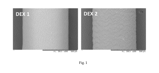
Surface morphology of the implants were characterized by SEM, as shown in
Figure 1.
DEX 2 has a slightly rough surface while DEX 1 appears to have a smooth
surface. The
diameter of the rod shape implants is approximately 0.635 mm. The thickness of
the coating
is approximately 0.029 mm, i.e. 29 i_tm.
As can be seen from Figures 2 and 3, comparative implants DEX 1 and TM 1 show
a
considerable burst release on the first day. This effect is greatly suppressed
in DEX 2 and
TM 2. The implants according to the invention can provide a sustainable
release of the
therapeutic agent over a prolonged period of time.
27
CA 03124283 2021-06-18
WO 2020/128067 PCT/EP2019/086834
Example 2. Fluorescein isothiocyanate (FITC)-dextran implants with & without
poly-L-
lactide-co-caprolactone (PLC) and poly (DL-lactidel fPDL) coating
FITC-Dextran
PLGA 75/25 PEGDA 700 (w/w Dimensions (D * L)
Formulation (4 kDa) loading
(w/w Vo) Vo) mm
(w/w %)
D1 10 5 85 0.5 *7.5
CD1 (Coated) 10 5 85 0.5 * 7.5
2.1. Preparation of D1
mg of PLGA 75/25 (Purac Biochem, Gorinchem, The Netherlands) was dissolved
in 190 mg of PEGDA of molecular weight (MW) 700 Da (Sigma Aldrich,
Basingstoke, England)
to prepare Solution A. 5 mg Irgacure 2559 (Sigma Aldrich, Basingstoke,
England) was
dissolved in 1 ml PBS to prepare Solution B. 10 mg of FITC dextran (average MW
4000 Da,
10 Sigma Aldrich. Basingstoke, England) was dissolved into a 60 ul of
Solution B in an
Eppendorf tube to prepare Solution C. 85 mg of Solution A was weighed in an
empty
Eppendorf tube and 60 ul of Solution C was added to the mixture slowly through
Eppendorf
tube wall with continuous stirring at 900 rpm for 15 minutes. The mixtures
finally obtained
was withdrawn into silicon tubes with ID of 0.635 mm (Polymer System
Technology,
England) and cross-linked using a UV light (Light Hammer 6, Heraeus
Noblelight Fusion UV
Inc., Gaithersburg, MD, USA). The intensity of the UV light was set as 50% and
the silicone
tubes were exposed to the UV light for 15 seconds (i.e. a total of 5 runs).
The implants were
then removed from the silicon tubes and left to dry in vacuum at 25 C for 4
hours. The rod-
shaped implants were cut at each 7.5 mm length.
2.2. Preparation of CD1 (coated with PLC)
Implants CD1 were manufactured according to Section 2.1 as described above
(except
last sentence). They were cut into 20 mm length and coated with 17% w/v
solution of poly-L-
lactide-co-caprolactone (PLC 8516) (Purac Biochem, Gorinchem, The Netherlands)
in
dichloromethane (DCM) using a texture analyser instrument (TA-XT plus; Stable
Micro
Systems, US). The implant was dipped at speed of 10 mm/s, held for 1 s inside
the coating
solution, then withdrawn at speed of 10 mm/s. A single coat layer was applied
with thickness
of about 20-25 i_tm. The implants were then cut into 7.5 mm length and the
sides of these
28
CA 03124283 2021-06-18
WO 2020/128067 PCT/EP2019/086834
surface-coated implants were coated with 15% w/v poly-DL-lactide (PDL)
solution in
acetonitrile (ACN) by using a 29G needle syringe under digital microscope.
2.3 In vitro drug release set up
Two implants of D1 and two implants of CD1 (of 7.5 mm length) were placed into
two glass
vials containing 2 mL of PBS (Phosphate buffered saline) with 0.01% w/v Sodium
azide (NaN2)
(pH 7.4 0.2) as release media. All the experiments were carried out in
triplicate. The glass
vials containing the implants were placed in a shaking orbital incubator at a
speed of 40 rpm
and at 37 C (GFL Orbital Shaking Incubator; Gesellschaft fin- Labortechnik
mbH, Germany).
Sampling followed by complete replacement of the PBS medium was performed on
Day 1 and
weekly thereafter, i.e. Day 7, Day 14, Day 21, Day 28 and so on. The
concentration of released
drug molecule in the PBS samples was analyzed as described in the following
section. The vials
were then incubated at 37 C and at predetermined time intervals the entire
medium was
removed and replaced with fresh medium.
2.4 Sample analysis
Analysis of FITC-dextran in vitro drug release samples were performed using
the
fluorescence spectrophotometry method. Detection was carried out by micro 96
well plate
spectrophotometer (BMG Labtech FLUOstar Optima fluorescence plate reader (BMG
Labtech
GmbH, Ortenberg, Germany). Excitation was set to 485 nm, emission was set to
520 nm, and
gain was set to 750.
Fig. 4 shows the in vitro release of D1 and CD1 expressed as percentage
cumulative release. As
it can be seen from this figure, the presence of the coating polymer layer on
the implant matrix
significantly reduces the burst effect and the overall release of FITC-dextran
is controlled over
the entire period of time.
Example 3 - Latanoprost (LP) implants with different diameter size and with or
without poly-L-lactide-co-caprolactone (PLC) coating.
Latanoprost PLGA 75/25 PEGDA 250
Dimensions (D * L)
Formulation
loading (w/w %) (w/w %) (w/w %) mm
LP1 20 30 50 0.3 *2
LP2 20 30 50 0.6 *2
29
CA 03124283 2021-06-18
WO 2020/128067 PCT/EP2019/086834
LPC1 20 30 50 0.3 *2
(1 layer coated)
LPC2 20 30 50 0.6 *2
(1 layer coated)
3.1. Preparation of LP1 and LP2
20 mg Irgacure 2959 (Sigma Aldrich, Basingstoke, England) was dissolved in
acetonitrile to prepare Solution A. 50 mg Latanoprost (LP) (Alfa Chemistry,
New York, USA)
was dissolved in 2.5 mL acetonitrile to prepare Solution B. 75 mg PEGDA 250
and 15 mg of
PLGA 75/25 (Purac Biochem, Gorinchem, The Netherlands) were put into a 2 mL
Eppendorf
tube, and dissolved in 250 IA acetonitrile to prepare Solution C.
37.5 IA of Solution A and 1 mL of Solution B were then added to Solution C,
and subsequently
stirred at 250 rpm for 30 minutes (Multistirrer, Velp ScientificaTM, Italy).
Acetonitrile was then
evaporated under gauge pressure of -0.1 MPa at room temperature for 6 h (OV-12
vacuum
oven; JeioTech, Korea). The mixture finally obtained was withdrawn into a
silicon tube of
internal diameter 0.32 or 0.63 mm (HelixMark Standard Silicone Tubing;
Freudenberg,
Germany) by using a 25G needle attached to 10 mL syringe. Photocrosslinking
was
performed for 10 runs under UV D-lamp operated at 100% intensity with a belt
speed of 11.5
m/min (Light Hammer 6; Heraeus Noblelight Fusion UV, USA). The solidified rod-
shaped
implant was removed from the silicone tubing and cut into a 2 mm length. The
implants had a
weight of about 0.2 mg (for 0.3 mm diameter, LP1) and 0.9 mg (for 0.6 mm
diameter, LP2).
3.2 Preparation of LPC1 and LPC2 (LP1 and LP2 coated with PLC)
Implants LP1 and LP2 were coated by automated dip coating method to obtain
LPC1
and LPC2, respectively. A single coat layer was applied with thickness of
about 20-25 i_tm.
They were coated on surface with 17% w/v solution of poly-L-lactide-co-
caprolactone (PLC)
(Purac Biochem, Gorinchem, The Netherlands) polymer solution in
dichloromethane (DCM)
using Texture analyser instrument and on sides with 15% w/v poly-DL-lactide
(PDL) (Purac
Biochem, Gorinchem, The Netherlands) solution in acetonitrile (ACN) by using a
29G needle
syringe under digital microscope.
3.3 In vitro drug release set up
Implants LP1, LP2, LPC1 and LPC2 were each placed in a centrifuge tube
containing 2
mL of PBS (Phosphate buffered saline) with 0.01% w/v Sodium azide (NaN2) (pH
7.4 0.2) as
release media. All the experiments were carried out in triplicate. The
centrifuge tubes
CA 03124283 2021-06-18
WO 2020/128067 PCT/EP2019/086834
containing implants were placed in a shaking orbital incubator at a speed of
40 rpm and at 37 C
(GFL Orbital Shaking Incubator; Gesellschaft fin- Labortechnik mbH, Germany).
Sampling
followed by complete replacement of the PBS medium was performed on Day 1, Day
3, Day 7
and weekly thereafter. The concentration of released drug was analysed using a
developed
HPLC method for Latanoprost
3.4 Sample analysis
Analysis of LP1, LP2, LPC1 and LPC2 samples was performed using HPLC system
with
fluorescence detection (Agilent 1260 Infinity II Quaternary System) using a
Poroshell 120 EC-
C18 column (250 mm length, 4.6 mm internal diameter and 4 um particle size).
The samples
were analyzed in an isocratic mode using a mobile phase of acetonitrile: 0.1%
v/v formic acid
(60:40), with an injection volume of 50 uL and a flow rate of 1 mL/min. The
column
temperature was maintained at 40 C. The fluorescence detector was set at an
excitation
wavelength of 265 nm and an emission wavelength of 285 nm.
Fig. 5 shows the in vitro release of LP1, LP2, LPC1 and LPC2 expressed as
percentage
cumulative. As it can be seen from the figure, the presence of the coating
polymer layer on the
implant matrix significantly reduces the burst effect and the overall release
of LP is controlled
over the entire period of time.
Example 4 - Latanoprost (LP) implants with one or more layers of poly-L-
lactide-co-
caprolactone (PLC) - Effect of the layers.
Latanoprost PLGA 75/25 PEGDA 250
Dimensions (D * L)
Formulation
loading (w/w %) (w/w %) (w/w %) mm
LPC1 20 30 50 0.3 *2
(1 layer coated)
LPC3 20 30 50 0.3 *2
(2 layers coated)
4.1. Preparation of LP3
LPC3 implants were prepared from LP1 implants using the coating method
described under
Section 3.2, whereby the automated dip coating was repeated a second time on
dried LPC1
implants to achieve 2 layers of PLC coating.
4.2 In vitro drug release set up and sample analysis
31
CA 03124283 2021-06-18
WO 2020/128067 PCT/EP2019/086834
A LPC1 and a LPC3 implant, 2 mm long and having a weight of about 0.2 mg were
each
placed in a centrifuge tube containing 2 mL of PBS (Phosphate buffered saline)
with 0.01% w/v
Sodium azide (NaN2) (pH 7.4 0.2) as release media. All the experiments were
carried out in
triplicate. The centrifuge tubes containing implants were placed in a shaking
orbital incubator
at a speed of 40 rpm and at 37 C (GFL Orbital Shaking Incubator; Gesellschaft
fin- Labortechnik
mbH, Germany). Sampling followed by complete replacement of the PBS medium was
performed on Day 1, Day 3, Day 7 and weekly thereafter. The concentration of
released drug
was analyzed using a developed HPLC method for latanoprost.
Fig. 6 shows the in vitro release of LPC1 and LPC3 expressed as percentage
cumulative. As
it can be seen from the figure, an additional coating polymer layer on the
implant matrix further
reduces the burst effect and the overall release of latanoprost is controlled
over the entire
period of time.
Example 5 - Latanoprost (LP) implants coated with layers of poly-L-lactide-co-
caprolactone (PLC) and poly(L-lactide) (PLA) - effect of the composition of
the coating
material.
Latanoprost PLGA 75/25 PEGDA 250
Dimensions (D * L)
Formulation
loading (w/w %) (w/w %) (w/w %) mm
LPC1 20 30 50 0.3 *2
(PLC coated)
LPC4 20 30 50 0.3 *2
(PLA coated)
5.1. Preparation of LPC4
LPC4 implants were prepared by coating LP1 implants by automated dip coating
method.
The LPC4 implants were coated on surface with 2.5% w/v solution of poly(L-
lactide) (PLA)
polymer solution in dichloromethane (D CM) using Texture analyzer instrument
and on sides
with 15% w/v poly(DL-lactide) (PDL) (Purac Biochem, Gorinchem, The
Netherlands) solution
in acetonitrile (ACN) by using a 29G needle syringe under digital microscope.
5.2 In vitro drug release set up and sample analysis
A LPC1 and a LPC4 implant, 2 mm long and having a weight of about 0.2 mg were
each
placed in a centrifuge tube containing 2 mL of PBS (Phosphate buffered saline)
with 0.01% w/v
Sodium azide (NaN2) (pH 7.4 0.2) as release media. All the experiments were
carried out in
triplicate. The centrifuge tubes containing implants were placed in a shaking
orbital incubator
at a speed of 40 rpm and at 37 C (GFL Orbital Shaking Incubator; Gesellschaft
fur Labortechnik
32
CA 03124283 2021-06-18
WO 2020/128067 PCT/EP2019/086834
mbH, Germany). Sampling followed by complete replacement of the PBS medium was
performed on Day 1, Day 3, Day 7 and weekly thereafter. The concentration of
released drug
was analyzed using a developed HPLC method for latanoprost
Fig. 4 shows the in vitro release of LPC1 and LPC4 expressed as percentage
cumulative. As
it can be seen from these figures, both coating polymer materials reduce the
burst effect
(compared to LP1) and the overall release of latanoprost is controlled over
the entire period
of time.
Example 6 -High loading Latanoprost (LP) implants with or without PLC coating.
Latanoprost PLGA 75/25 PEGDA 250
Dimensions (D * L)
Formulation
loading (w/w %) (w/w %) (w/w %) mm
LP40 40 2 58 0.3*2
LPC40 40 2 58 0.3*2
(1 layer coated
PLC)
6.1. Preparation of LP40
mg Irgacure 2959 (Sigma Aldrich, Basingstoke, England) was dissolved in
acetonitrile to
prepare Solution A. 50 mg Latanoprost (LP) (Alfa Chemistry, New York, USA) was
dissolved
in 2.5 mL acetonitrile to prepare Solution B. 29 mg PEGDA 250 and 1 mg PLGA
75/25 (Purac
15 Biochem, Gorinchem, The Netherlands) were put into a 2 mL Eppendorf
tube, and dissolved
in 250 IA acetonitrile to prepare Solution C.
14.5 uL of Solution A and 1000 IA of Solution B were then added to Solution C,
and
subsequently stirred at 250 rpm for 30 minutes (Multistirrer, Velp
ScientificaTM, Italy).
Acetonitrile was then evaporated under gauge pressure of -0.1 MPa at room
temperature for 6
20 h (OV-12 vacuum oven; JeioTech, Korea). The mixture finally obtained was
withdrawn into a
silicon tube of internal diameter 0.32 (HelixMark Standard Silicone Tubing;
Freudenberg,
Germany) by using a 25G needle attached to 10 mL syringe. Photocrosslinking
was performed
for 5 runs under UV D-lamp operated at 50% intensity with a belt speed of 11.5
m/min (Light
Hammer 6; Heraeus Noblelight Fusion UV, USA). The solidified rod-shaped
implant was
removed from the silicone tubing and cut into a 2 mm length. The implants had
a weight of
about 0.2 mg.
33
CA 03124283 2021-06-18
WO 2020/128067 PCT/EP2019/086834
6.2 Preparation of LPC40
Implants LP40 were coated by automated dip coating method to obtain LPC40. A
single coat
layer was applied with thickness of around 20-25 i_tm. They were coated on
surface with 17%
w/v solution of poly-L-lactide-co-caprolactone (PLC) (Purac Biochem,
Gorinchem, The
Netherlands) polymer solution in dichloromethane (DCM) using Texture analyser
instrument
and on sides with 15% w/v poly-DL-lactide (PDL) (Purac Biochem, Gorinchem, The
Netherlands) solution in acetonitrile (ACN) by using a 29G needle syringe
under digital
microscope.
6.3 In vitro drug release set up and sample analysis
.. A LP40 and a LPC40 implant of 2 mm length and having a weight of about 0.2
mg were each
placed in a centrifuge tube containing 2 mL of PBS (Phosphate buffered saline)
with 0.01%
w/v Sodium azide (NaN2) (pH 7.4 0.2) as release media. All the experiments
were carried
out in triplicate. The centrifuge tubes containing implants were placed in a
shaking orbital
incubator at a speed of 40 rpm and at 37 C (GFL Orbital Shaking Incubator;
Gesellschaft fin-
Labortechnik mbH, Germany). Sampling followed by complete replacement of the
PBS
medium was performed on Day 1, Day 3, Day 7 and weekly thereafter. The
concentration of
released drug was analyzed using a developed HPLC method for latanoprost
Fig. 8 shows the in vitro release of LP40 & LPC40 expressed as percentage
cumulative release.
As it can be seen from this figure, coating on the surface of implants
significantly reduced initial
burst release and sustained the release over longer period as compared to
uncoated implants.
The coated LPC40 implants maintained near zero-order release for 180 days (6-
months) while
uncoated LP40 implants could achieve sustained release for 20 days.
34