Note: Descriptions are shown in the official language in which they were submitted.
CA 03124489 2021-06-21
WO 2020/142153
PCT/US2019/064041
METHODS OF DETECTING DNA AND RNA IN THE SAME SAMPLE
CROSS-REFERENCE TO RELATED APPLICATION
This application claims priority to U.S. Provisional Application No.
62/787,114 filed
December 31, 2018, herein incorporated by reference in its entirety.
FIELD
The present disclosure provides quantitative nuclease protection sequencing
(qNPS)
methods that allow sequencing of nucleic acid targets (for example by co-
amplifying DNA and an
RNA surrogate in the same sample). Such methods can be used to determine if
one or more nucleic
acid targets are present in a sample, and in some examples is quantitative.
BACKGROUND
Although methods of sequencing nucleic acid molecules are known, there is
still a need for
methods that permit sequencing of RNA and DNA co-amplified in the sample
mixture. Methods of
multiplexing nucleic acid molecule sequencing reactions that utilize DNA and
RNA co-amplified
in the sample mixture have not been realized at the most desired performance
or simplicity levels.
SUMMARY
Methods are provided that improve prior quantitative nuclease protection
sequencing
(qNPS) methods (such as those disclosed in U.S. Publication No. US 2011-
0104693 and U.S.
Patent No. 8,741,564) and represent an improvement to current nucleic acid
sequencing methods.
In some examples, the disclosed methods sequence or detect at least one target
DNA and at least
one target RNA in the same sample (such as the same biopsy sample or the same
tissue sample), by
co-amplifying both molecules from the same sample, by use of an RNA surrogate
molecule. In
some examples, a plurality of different (e.g., unique) samples are analyzed
simultaneously. In
some examples, the target RNA and DNA molecules have a point mutation, a
deletion, insertion, or
combinations thereof. In some examples, the method determines the abundance
(e.g.,
quantitatively or qualitatively) of one or more target RNAs and determines if
genomic mutations
are present in one or more target DNA sequences.
The disclosed methods of determining a sequence of a target DNA molecule
(e.g., a target
genomic molecule) and a target RNA molecule (e.g., a target mRNA or target
miRNA molecule) in
a sample (e.g., a fixed sample, such as a formalin-fixed sample) can include
lysing the sample with
-1-
CA 03124489 2021-06-21
WO 2020/142153
PCT/US2019/064041
a lysis buffer (e.g., a lysis buffer that includes a detergent and/or a
chaotropic agent), thereby
generating a lysate comprising the target DNA molecule and the target RNA
molecule. The lysate
is divided into at least two different portions, for example of equal volume
or of equal nucleic acid
content.
The target DNA is amplified from a first portion of the lysate using at least
one primer (e.g.,
a target DNA primer, such as a first forward primer and first reverse primer),
thereby generating
flanked amplicon regions (FARs). In some examples, amplifying the target DNA
from the first
portion of the cell lysate uses at least two primers (e.g., at least two
target DNA primers), each
DNA primer having a flanking sequence at its 5' end. For example, the first
target DNA primer
(such as a forward primer) can have at its 5'-end a flanking sequence that is
the reverse-
complement sequence of the 3'-flanking sequence of the nuclease protection
probe that includes a
flanking sequence (NPPF - see below), while the second target DNA primer (such
as a reverse
primer) can have at its 5'-end a flanking sequence identical to the 5'-
flanking sequence of the
NPPF. These flanking sequences on the DNA primers allow flanking sequences to
be added to the
DNA amplicons, thereby generating flanked amplicon regions (FARs). In some
examples, the
flanking sequences added are about 10 to 50 nucleotides (nt) each, such as 25
nt each. In some
examples, the DNA amplified from the target is about 40 to 150 nt in length,
such as 40 to 125 nt or
40 to 100 nt. In some examples, the FAR generated is about 100 to 200 nt in
length, such as 160 to
200 nt.
A second (i.e., different) portion of the lysate is incubated with at least
one nuclease
protection probe that includes a flanking sequence (NPPF) under conditions
sufficient for the NPPF
to specifically bind to the target RNA molecule present in the second portion
of the lysate. In some
examples the NPPF is a DNA molecule about 50 to 200 nt in length, such as 60
to 200 nt, 75 to
150, or 65 to 100 nt. The NPPF includes (1) a 5'-end, (2) a 3'-end, (3) a
sequence (e.g., about 10-60
nt in length, such as 16 to 50 nt) that is complementary to all or a portion
of the target RNA
molecule, thus permitting specific binding or hybridization between the target
RNA molecule and
the NPPF, and (4) a flanking sequence. For example, the region of the NPPF
that is
complementary to a region of the target RNA molecule binds to or hybridizes to
that region of the
target RNA molecule with high specificity. In some examples, the flanking
sequence is located 5',
3', or both to the sequence complementary to the target RNA molecule, such as
a 5'-flanking
sequence 5' of the sequence complementary to the target RNA molecule and a 3'-
flanking sequence
3' of the sequence complementary to the target RNA molecule. In some examples,
the flanking
- 2 -
CA 03124489 2021-06-21
WO 2020/142153
PCT/US2019/064041
sequence includes at least 12 contiguous nucleotides not found in a nucleic
acid molecule present in
the sample.
In some examples, the NPPF includes a 5'-flanking sequence, and the methods
further
include contacting the second portion of the lysate with a nucleic acid
molecule (e.g., DNA or
RNA) that includes a sequence complementary to the 5'-flanking sequence (5CFS)
under
conditions sufficient for the 5'-flanking sequence to specifically hybridize
to the 5CF S. In some
examples, the NPPF includes a 3'-flanking sequence, and the method further
includes contacting
the second portion of the lysate with a nucleic acid molecule (e.g., DNA or
RNA) that includes a
sequence complementary to the 3'-flanking sequence (3CFS) under conditions
sufficient for the 3'-
flanking sequence to specifically hybridize to the 3CFS. In some examples, the
NPPF includes a
3'- and a 5'-flanking sequence, and the method further includes contacting the
second portion of the
lysate with a 3CFS and 5CFS under conditions sufficient for the 3'-flanking
sequence to
specifically hybridize to the 3CFS and the 5'-flanking sequence to
specifically hybridize to the
5CFS. Hybridization results in the generation of a double-stranded (ds)
nucleic acid molecule,
namely NPPF hybridized to (1) the target RNA molecule, and (2) the 5CFS and/or
3CFS. In some
examples, at least one nucleotide in the NPPF does not have complementarity to
the corresponding
nucleotide in the target RNA molecule or does not have complementarity to the
corresponding
nucleotide in the 5CFS or 3CFS.
The resulting double-stranded (ds) nucleic acid molecule, namely NPPF
hybridized to (1)
the target RNA molecule, and (2) the 5CFS and/or 3CFS present in the second
portion of the lysate
is contacted with a nuclease specific for single-stranded (ss) nucleic acid
molecules (e.g., an
exonuclease, an endonuclease, or a combination thereof, such as Si nuclease)
under conditions
sufficient to degrade (hydrolyze) or remove unbound ss nucleic acid molecules
in the second
portion of the lysate. Thus for example, NPPFs that have not bound target RNA
or CF Ss, unbound
RNA molecules, unbound portions of target RNA molecules, unbound CFSs, and
other ss nucleic
acid molecules in the second portion of the lysate, are degraded. This results
in a second portion of
the lysate containing a digested sample that includes an NPPF hybridized to
its target RNA
molecule, hybridized to its corresponding 3CFS, hybridized to its
corresponding 5CFS, or
hybridized to both its corresponding 3CFS and its corresponding 5CFS.
This ds nucleic acid molecule (NPPF: target RNA molecule:CFS) in the second
portion of
the lysate can be separated into its corresponding ss nucleic acid molecules
(for example by
heating, for example heating to 95 C to 100 C), thereby generating a mixture
of ssNPPFs, ssCFSs,
and ss target RNA molecules. In some examples, this separation occurs as the
first step of the
- 3 -
CA 03124489 2021-06-21
WO 2020/142153
PCT/US2019/064041
second amplification (amplification of the FARs and ssNPPFs) described below.
In one example,
the RNA strand of the NPPF:RNA target can be selectively removed by treating
the complex with
RNase H, which selectively removes the RNA moiety of a DNA:RNA complex (for
example, if the
if the target molecule is RNA, the NPPF is DNA, and the 3CFS and 5 CFS are
DNA). Alternative
nucleases can be used to optionally degrade RNA separately from DNA.
The methods include mixing or combining the FARs generated in the first
portion of the
sample lysate with the second portion of the sample lysate containing the
ssNPPFs, thereby
generating a DNA amplicons/ssNPPF mixture. In some examples, the first portion
of the cell
lysate containing the DNA amplicons is added to the second portion of the cell
lysate containing
the ssNPPFs (or vice versa). In some examples, a1:1, 1:2, 1:3, 1:4, 1:5, or
1:10 ratio of
ssNPPFs:FARs is used in the subsequent amplification step.
The resulting FARs/ssNPPF mixture is incubated with appropriate primers (such
as forward
and reverse primers), under conditions that co-amplify the FARs and the
ssNPPFs in the same
reaction vessel (e.g., same microfuge tube or same well of a multi-well
plate). In some examples,
different primers are used to amplify the FARs, and to amplify the ssNPPFs. In
some examples the
same forward and reverse primers are used to amplify the FARs, and to amplify
the ss NPPFs, for
example due to the presence of identical 5'- and 3- flanking sequences on the
FARs and the
ssNPPFs (e.g., the NPPF includes a 5'-flanking sequence and a 3'-flanking
sequence, and the FARs
include the same 5'-flanking sequence and same 3'-flanking sequence as that in
the NPPF). For
example, the amplification can use a first amplification primer having a
region identical to the 5'-
flanking sequence and a second amplification primer having a region
complementary to the 3'-
flanking sequence. Such primers can further include one or more sequences that
permit attachment
of an experimental tag, sequencing adaptor, or both, to the FAR amplicons or
NPPF amplicons (for
example to the 5'-end, 3'-end, or both of the resulting amplicons) during the
amplification of the
FARs and the single stranded NPPFs. In some examples, the methods further
include removing the
amplification primers after amplifying the FARs and the ssNPPFs but before
sequencing the FAR
amplicons and the NPPF amplicons.
In some examples, the NPPF includes both a 5'-flanking sequence and a 3'-
flanking
sequence (such as a flanking sequence at the 5'-end that differs from the
flanking sequence at the
3'-end), and the FARs include the same 5'-flanking sequence and same 3'-
flanking sequence as
those in the NPPF. Thus, after separating the ds NPPF:RNA target:CFS molecule
into a ss NPPF
molecule, but before sequencing, the methods can include contacting the ssNPPF
(and in some
examples also the FAR with the same 5'- and 3'-end flanking sequences) with a
first amplification
- 4 -
CA 03124489 2021-06-21
WO 2020/142153
PCT/US2019/064041
primer that includes a region complementary to the 3'-flanking sequence and
with a second
amplification primer that includes a region complementary to the 5'-flanking
sequence. For
example, the first and second amplification primers can permit attachment of
an experimental tag
(e.g., a nucleic acid sequence that permits identification of a sample,
subject, treatment or target
RNA or DNA molecule) and/or sequencing adaptor (e.g., a nucleic acid sequence
that permits
capture onto a sequencing platform) to the resulting NPPF amplicons (and FAR
amplicons) (such
as an experiment tag or sequence adaptor on the 5'-end or 3'-end of the NPPF
amplicons and FAR
amplicons), such as a first amplification primer that permits attachment of a
first experimental tag
and/or first sequencing adaptor to the NPPF amplicons and FAR amplicons and a
second
amplification primer that permits attachment of a second experimental tag
and/or second
sequencing adaptor to the NPPF amplicons and FAR amplicons. In some examples,
the methods
further include removing the first and second amplification primers after
amplifying but before
sequencing (such as removing amplification primers after the amplifying the
target DNA from a
first portion of the lysate using at least one target DNA primer, removing the
first and second
amplification primers after the amplifying of the FARs and the single stranded
NPPF, or removing
both sets of amplification primers, before the sequencing step).
The methods can further include sequencing (e.g., next generation sequencing
or single
molecule sequencing) at least a portion of the resulting NPPF amplicons and at
least a portion of
the FAR amplicons, thereby determining the sequence of the target DNA molecule
(via the FAR
amplicons), the sequence of (and/or abundance of) the target RNA molecule (via
the NPPF
amplicons) in the sample.
In some examples, the methods sequence or detect at least two different target
RNA
molecules (e.g., where the sample is contacted with at least two different
NPPFs, such as where
each NPPF is specific for a different target RNA molecule, or where the sample
is contacted with at
least one NPPF specific for the at least two different target RNA molecules,
such as separate RNA
molecules transcribed from different loci, or more than one alternative
transcript or splice isoform
transcribed from the same locus). In some examples, the methods sequence or
detect at least two
different target DNA molecules (e.g., where the at least two different target
DNA molecules
include a wild type gene sequence and at least one mutation in the gene
sequence). In specific
examples, the methods can be performed on a plurality of samples with, for
example, at least two
different target RNA molecules and at least two different target DNA molecules
detected in each of
the plurality of samples. In specific examples, at least one NPPF is specific
for a miRNA target
nucleic acid molecule and at least one NPPF is specific for an mRNA target
nucleic acid molecule.
- 5 -
CA 03124489 2021-06-21
WO 2020/142153
PCT/US2019/064041
Also provided are isolated nucleic acid molecules, such as one comprising or
consisting of
the nucleic acid sequence of any one of SEQ ID NO: 4, 5, 6, 7, 8,9, 10, 11,
12, 13, 17, 18, 19, 20,
21, 22, 23, 24, 25, 26, 27, 28, 29, 30, 31 or 32. Also provided are sets of
nucleic acid primers, for
example as part of a kit. In some examples, the set includes the nucleic acid
sequence of SEQ ID
.. NOs: 4 and 5; SEQ ID NOs: 6 and 7; SEQ ID NOs: 8 and 9; SEQ ID NOs: 10 and
11; SEQ ID
NOs: 12 and 13; SEQ ID NOs: 17 and 18; SEQ ID NOs: 19 and 20; SEQ ID NOs: 21
and 22; SEQ
ID NOs: 23 and 24; SEQ ID NOs: 25 and 26; SEQ ID NOs: 27 and 28;SEQ ID NOs: 29
and 30;
SEQ ID NOs: 31 and 32; or combinations of these sets (such as at least two or
at least three of these
sets).
The foregoing and other objects and features of the disclosure will become
more apparent
from the following detailed description, which proceeds with reference to the
accompanying
figures.
BRIEF DESCRIPTION OF THE DRAWINGS
FIG. 1A is a schematic diagram showing an exemplary nuclease protection probe
having
flanking sequences (NPPF), 100. The NPPF 100 includes a region 102 having a
sequence that
specifically binds to/hybridizes to a target nucleic acid sequence (e.g.,
target RNA sequence). The
NPPF also includes a 5'-flanking sequence 104, a 3'-flanking sequence 106, or
both (the
embodiment with both is shown).
FIG. 1B is a schematic diagram showing an exemplary nuclease protection probe
having
flanking sequences (NPPF), 120. In this example, the NPPF 120 is composed of
two separate
nucleic acid molecules 128, 130, instead of a single nucleic acid molecule as
shown in FIG. 1A.
The NPPF 120 includes a region 122 having a sequence that specifically binds
to/hybridizes to a
target nucleic acid sequence. The NPPF also includes a 5'-flanking sequence
124, a 3'-flanking
sequence 126, or both (the embodiment with both is shown).
FIG. 2 is a schematic diagram showing an overview of the steps of an
illustrative method
for lysing the sample 10, dividing the lysed sample into at least two
portions, wherein target DNA
is amplified in a first portion 12, generating FARs specific for the target
DNA, and target RNA is
hybridized to NPPFs, nuclease digested, and ds nucleic acid molecules
denatured, generating
ssNPPFs specific for the target RNA in second portion 14, then at least a
portion of first and second
portions combined and the FARs and NPPFs co-amplified in the mixture 16, prior
to sequencing
and data extraction 18.
- 6 -
CA 03124489 2021-06-21
WO 2020/142153
PCT/US2019/064041
FIG. 3 is a schematic diagram showing an overview of the steps of an
illustrative method
for sequencing of at least one target DNA molecule and at least one target RNA
molecule, wherein
the DNA and a surrogate of the RNA are amplified in the sample mixture. Step 1
shows a sample
(such as cells or FFPE tissue), which is contacted with sample disruption
buffer (for example to
permit lysis of cells and tissues in the sample) and then separated into at
least two portions (first
and second portion). Step 2A shows that a first portion of the cell lysate is
incubated with two
amplification primers (e.g., target DNA primers), such as a first primer
containing a 5' extension
234 and a second primer containing a 5' extension 232 under conditions that
allow for
hybridization of the primers to the target DNA 230. Step 2B shows that the
target DNA molecule
230 is amplified using the primers 234, 232, generating a flanked amplicon
region (FAR) 236 with
5' and 3' extensions from the primers (in some examples the 5'- and 3'-
extensions of the FAR
(shown as 238, 239, respectively) are identical to the 5'- and 3'-flanking
sequences of the NPPF
(204, 206). Step 2AA shows that a second portion of the cell lysate is
incubated with at least one
NPPF 202 and its complementary 5CFS 208 and 3CFS 210 under conditions that
allow specific
hybridization of the NPPF 202 to a target RNA 200, and to the CF Ss 208, 210.
Step 2BB shows
that the resulting ds nucleic acid molecule generated in Step 2AA, is
incubated with a nuclease
specific for ss nucleic acid molecules (such as 51 nuclease, mung bean
nuclease, BAL 31 nuclease,
or P1 nuclease), resulting in a ds NPPF/RNA/CF Ss target complex 212. Step 2CC
shows that the
ds NPPF/RNA target complex 212 is then separated or denature into its single
nucleic acid strands,
generating a mixture of ssRNA 200, ss CFSs 208, 210, and ssNPPF 202. In Step
3, the mixture of
ssRNA 200, ss CFSs 208, 210 and ssNPPF 202 is combined with the DNA amplicons
236. In Step
4, the combined ssNPPF 202 and FARs 236 are co- amplified in the same
reaction, for example, by
using PCR with appropriate primers, and then sequenced.
FIG. 4 is a schematic diagram showing amplification of ssNPPF 200 (RNA target
surrogate) and FAR 236 using forward and reverse primers (arrows), resulting
in NPPF amplicons
226 and FAR amplicons 246, respectively. The primers can include sequences
that allow
sequencing adaptors 218, 220, 248, 240 and/or experiment tags 222, 224, 242,
244 to be added to
the NPPF amplicons 226 and FAR amplicons 246, respectively. The resulting NPPF
amplicons
226 are used to detect target RNA (and can be used to determine a target RNA
sequence and/or its
abundance), and FAR amplicons 246 are used to detect target DNA (and can be
used to determine a
target DNA sequence). In some examples, the primer sequences are used to
identify amplicons
(such as NPPF amplicons 226 and FAR amplicons 246) as a product of the same
sample, in which,
some examples of the methods include primers where the adaptor and/or tag
sequences are the
- 7 -
CA 03124489 2021-06-21
WO 2020/142153
PCT/US2019/064041
same (e.g., in such examples, sequences 218, 222 are the same as 248, 242, and
sequences 224, 220
are the same as 244, 240).
FIGS. 5A-5B show scatterplots with Pearson correlations for raw data from
triplicate
experiments for a formalin-fixed, paraffin-embedded (FFPE) sample (FIG. 5A)
and a cell line
mixture sample (FIG. 5B).
FIG. 6 shows expression of the indicated RNA measured in a cell line titration
series from
triplicate experiments.
FIG. 7 shows DNA mutations detected in cell line samples as a percentage of
the total
counts for the indicated region (BRAF left, KRAS right) from triplicate
experiments performed on
three different days.
FIG. 8 shows the average of raw counts in cell line titration from triplicate
experiments
performed on three different days (BRAF V600E left, KRAS G12D right)
FIG. 9 shows the percentage of total reads consumed by NPPFs/RNA (grey) and by
FARs/DNA (hatched grey) for one sample under the different conditions used.
FIG. 10 shows the results for a single set of conditions (14 cycles and 4 ul
added) for all
seven FFPE samples. The graph shows the percentage of total reads consumed by
NPPFs or RNA
(grey) and by FARs or DNA (hatched grey).
FIG. 11 shows DNA mutation information and BRAF mutation detection in eight
FFPE
samples as a percentage of total BRAF signal (SEQ ID NOS: 14-16, from top to
bottom).
FIGS. 12A-12B show scatterplots of RNA expression data generated using a set
of 470
NPPS for two of the eight FFPE samples (FFPE1 (lung, FIG. 12A) and FFPE7591
(melanoma,
FIG. 12B)). Pearson correlations (r) for triplicate measurements are displayed
on the scatterplots.
FIG. 13 shows a principal component analysis (PCA) plot of RNA expression data
from
nine replicates of samples from cell lines HD300, HD301, and HD789. The three
different cell
lines are strongly separated, demonstrating the differences in expression
profiles. The replicates
are tightly clustered together, demonstrating excellent repeatbility between
technical replicates and
replicates run on different days.
FIG. 14 is a table showing observed and expected allelic frequencies for each
of the three
reference standards and the three mixture samples.
FIG. 15 shows a bar graph and table demonstrating the repeatability of
individual
measurements of DNA variants.
SEQUENCE LISTING
- 8 -
CA 03124489 2021-06-21
WO 2020/142153
PCT/US2019/064041
The nucleic acid and protein sequences are shown using standard letter
abbreviations for
nucleotide bases as defined in 37 C.F.R. 1.822. Only one strand of each
nucleic acid sequence is
shown, but the complementary strand is understood as included by any reference
to the displayed
strand. The contents of the text file named "seq listing", which was created
on December 2, 2019
and is about 4KB in size, are hereby incorporated by reference in their
entirety.
SEQ ID NO: 1 shows an exemplary 5'-flanking sequence.
SEQ ID NO: 2 shows an exemplary 3'-flanking sequence.
SEQ ID NO: 3 shows an exemplary reverse-complement of a 3'- flanking sequence.
SEQ ID NOs: 4 and 5 show exemplary forward and reverse primers, respectively,
for
amplifying BRAF.
SEQ ID NOs: 6 and 7 show exemplary forward and reverse primers, respectively,
for
amplifying KRAS.
SEQ ID NOs: 8 and 9 show exemplary forward and reverse primers, respectively,
for
amplifying EGFR.
SEQ ID NOs: 10 and 11 show exemplary forward and reverse primers,
respectively, for
amplifying EGFR.
SEQ ID NOs: 12 and 13 show exemplary primers that can be used to add an
experiment tag
to the resulting amplicon.
SEQ ID NOs: 14-16 show three BRAF sequences: Wild type, nt mutation giving
rise to
V600E mutation, and another nt mutation giving rise to V600E2 mutation.
SEQ ID NOs: 17 and 18 show exemplary forward and reverse primers,
respectively, for
amplifying BRAF to detect a V600 mutation.
SEQ ID NOs: 19 and 20 show exemplary forward and reverse primers,
respectively, for
amplifying EGFR to detect a G719 mutation.
SEQ ID NOs: 21 and 22 show exemplary forward and reverse primers,
respectively, for
amplifying EGFR to detect mutations within exon 19.
SEQ ID NOs: 23 and 24 show exemplary forward and reverse primers,
respectively, for
amplifying EGFR to detect mutations within exon 20.
SEQ ID NOs: 25 and 26 show exemplary forward and reverse primers,
respectively, for
amplifying EGFR to detect a L858F or L858-L861 mutation.
SEQ ID NOs: 27 and 28 show exemplary forward and reverse primers,
respectively, for
amplifying KRAS to detect a G12 mutation.
- 9 -
CA 03124489 2021-06-21
WO 2020/142153
PCT/US2019/064041
SEQ ID NOs: 29 and 30 show exemplary forward and reverse primers,
respectively, for
amplifying KRAS to detect a Q61 mutation.
SEQ ID NOs: 31 and 32 show exemplary forward and reverse primers,
respectively, for
amplifying PIK3CA.
DETAILED DESCRIPTION
Unless otherwise noted, technical terms are used according to conventional
usage.
Definitions of common terms in molecular biology may be found in Benjamin
Lewin, Genes VII,
published by Oxford University Press, 2000 (ISBN 019879276X); Kendrew et al.
(eds.), The
Encyclopedia of Molecular Biology, published by Blackwell Publishers, 1994
(ISBN 0632021829);
Robert A. Meyers (ed.), Molecular Biology and Biotechnology: a Comprehensive
Desk Reference,
published by Wiley, John & Sons, Inc., 1995 (ISBN 0471186341); and George P.
Redei,
Encyclopedic Dictionary of Genetics, Genomics, and Proteomics, 2nd Edition,
2003 (ISBN: 0-471-
26821-6).
The singular forms "a," "an," and "the" refer to one or more than one, unless
the context
clearly dictates otherwise. For example, the term "comprising an NPPF"
includes single or plural
NPPFs and is considered equivalent to the phrase "comprising at least one
NPPF." The term "or"
refers to a single element of stated alternative elements or a combination of
two or more elements,
unless the context clearly indicates otherwise. As used herein, "comprises"
means "includes."
Thus, "comprising A or B," means "including A, B, or A and B," without
excluding additional
elements.
It is further to be understood that all base sizes or amino acid sizes, and
all molecular weight
or molecular mass values, given for nucleic acids or polypeptides are
approximate, and are provided
for description. Although methods and materials similar or equivalent to those
described herein can
be used in the practice or testing of the present disclosure, suitable methods
and materials are
described below. All publications, patent applications, patents, and other
references mentioned
herein are incorporated by reference in their entirety, as are the GenBankg
Accession numbers (for
the sequence present on December 31, 2018). In case of conflict, the present
specification,
including explanations of terms, will control. In addition, the materials,
methods, and examples are
illustrative only and not intended to be limiting.
Except as otherwise noted, the methods and techniques of the present
disclosure are
generally performed according to conventional methods well known in the art
and as described in
various general and more specific references that are cited and discussed
throughout the present
-10 -
CA 03124489 2021-06-21
WO 2020/142153
PCT/US2019/064041
specification. See, e.g., Sambrook et al., Molecular Cloning: A Laboratory
Manual, 2d ed., Cold
Spring Harbor Laboratory Press, 1989; Sambrook et al., Molecular Cloning: A
Laboratory Manual,
3d ed., Cold Spring Harbor Press, 2001; Ausubel et al., Current Protocols in
Molecular Biology,
Greene Publishing Associates, 1992 (and Supplements to 2000); Ausubel et al.,
Short Protocols in
Molecular Biology: A Compendium of Methods from Current Protocols in Molecular
Biology, 4th
ed., Wiley & Sons, 1999; Harlow and Lane, Antibodies: A Laboratory Manual,
Cold Spring Harbor
Laboratory Press, 1990; and Harlow and Lane, Using Antibodies: A Laboratory
Manual, Cold
Spring Harbor Laboratory Press, 1999.
I. Overview
The present disclosure provides methods that allow for sequencing of target
nucleic acid
molecules, such as target DNA and target RNA (using a NPPF surrogate) co-
amplified in the
sample mixture, which methods further can be multiplexed (e.g., detecting a
plurality of DNA and
RNA targets in a single sample) or are amenable to high-throughput (e.g.,
detecting DNA and RNA
targets in a plurality of samples, e.g., different samples) or are multiplexed
and high-throughput
(e.g., detecting a plurality of DNA and RNA targets in a plurality of sample,
e.g., different
samples). The disclosed methods provide several improvements over currently
available
sequencing methods. For example, because the methods co-amplify target DNA
(generating
amplicons referred to herein as FAR amplicons) and NPPFs (generating NPPF
amplicons, which
serve as surrogates of target RNA) in the same reaction vessel, these allow
for analysis of DNA and
RNA from the same sample, instead of from two different samples (i.e., one
sample for DNA
analysis and another/different sample for RNA analysis). In addition, the
disclosed methods
eliminate the requirement of extracting nucleic acid molecules from the
samples, prior to analysis.
Instead, the sample is simply lysed. The disclosed methods allow for the use
of a very small input
size compared to standard methods. For example, when RNA and DNA are extracted
from an
FFPE sample, for example, to perform DNA and RNA sequencing, this normally
requires 10-12
tissue sections from the FFPE sample. In contrast, the disclosed methods can
use less than 1 FFPE
section for analysis of both RNA and DNA. Similarly, the disclosed methods can
use only a few
thousand cells for analysis of both RNA and DNA (such as lysing only 1000 to
10,000 cells for the
analysis, such as 1000 to 5000 cells or 1000 to 2000 cells). Because the
methods require less
processing of the target nucleic acid molecules, bias, or loss of material
(especially loss of small
fragments) introduced by such processing can be reduced or eliminated. For
example, in some
current methods, when the target is both DNA and RNA (such as mRNA and/or
miRNA), methods
-11 -
CA 03124489 2021-06-21
WO 2020/142153
PCT/US2019/064041
typically employ steps to isolate or extract the nucleic acids from the
sample. For example, in prior
methods, RNA is typically isolated from a sample, subjected to reverse
transcription, amplification,
ligation of the RNA, or combinations thereof. Prior methods may also require a
depletion or a
separation step to remove undesired nucleic acid molecules or undesired
library molecules. In
some embodiments of the disclosed methods, such steps are not required. As a
result, the methods
permit one to analyze a range of sample types not otherwise amenable to
detection by sequencing.
In addition, this results in less loss of the targets from the sample,
providing a more accurate result.
The methods can be used to detect DNA and RNA (e.g., sequence, determine the
amount
of) in the same sample (such as the same individual FFPE tissue
section/slice). For example, the
methods can be used to detect a mutation, such as one or more
nucleotide/ribonucleotide insertions,
substitutions, deletions, or combinations thereof, for example gene fusions,
insertions, or deletions;
tandem repeats, single nucleotide polymorphisms (SNPs); single nucleotide (or
ribonucleotide)
variants (SNVs); microsatellite repeats; and DNA methylation status. In one
example, the methods
are used to detect a point mutation in a target nucleic acid molecule. Such a
mutation can be a
known mutation or a mutation that is newly discovered using the disclosed
methods. For example,
the methods can be used to detect one or more point mutations (such as at
least 2, at least 3, at least
4, at least 5, at least 6, at least 7, at least 8, at least 9, at least 10 or
more point mutations, such as 1,
2, 3, 4, 5, 6, 7, 8, 9, or 10 different point mutations) in a single target
nucleic acid molecule or in
multiple target nucleic acid molecules. The methods can be used to detect an
insertion and/or a
deletion, such as both an insertation and a deletion (indel, such as one that
is less than about 10kb,
less than about lkb, less than 100 bases, or less than 50 bases) in a single
target nucleic acid
molecule or in multiple target nucleic acid molecules. In some examples, each
different point
mutation is considered a different target nucleic acid molecule. In some
examples, the methods can
be used to detect one or more point mutations in two or more different target
nucleic acid
molecules. The method amplifies DNA to generate FAR amplicons to detect target
DNA, and uses
a nucleic acid probe, referred to herein as a nuclease protection probe
comprising a flanking
sequence (NPPF), which binds to the target RNA, thereby serving as a surrogate
for the target
RNA. The method amplifies the ssNPPF to generate NPPF amplicons to detect
target RNA.
Amplification of the FAR and ssNPPF occurs at the same time, in the same
reaction vessel,
eliminating the requirement of two separate samples for DNA and RNA analysis.
The methods can
be multiplexed and, in some examples, roughly conserve the stoichiometry of
the sequenced target
DNA and RNA molecules.
- 12 -
CA 03124489 2021-06-21
WO 2020/142153
PCT/US2019/064041
The primers used to amplify target DNA in the first amplification reaction
permit addition
of flanking sequences to the resulting FARs, wherein the flanking sequences
can be the same as
those on the NPPF. The NPPF includes flanking sequences. During the second
amplification
reaction, sequencing adaptors and/or experiment tags can be added to the FARs
and ssNPPFs using
the same amplification primers due to the presence of the same flanking
sequences. The presence
of the experiment tags on the resulting sequencing library (composed of FAR
amplicons and NPPF
amplicons) permit the identification of the target without necessitating the
sequencing of the entire
target itself or to permit samples from different patients or different
experiments or otherwise to be
combined into a single sequencing run. Experiment tags may be included at
either the 3'- or the 5'-
end or at both ends, for example, to increase multiplexing. Sequencing
adaptors permit attachment
of a sequence needed for a particular sequencing platform and formation of
clusters for some
sequencing platforms. The sequencing library composed of FAR amplicons and
NPPF amplicons
also simplifies the complexity of the sequencer input that is analyzed (e.g.,
sequenced), as the
sequencing library contains a known portion of the target DNA(s) and RNA(s) of
interest rather
than whole targets, many fragments of whole targets, or unknown targets. The
sequencing of FAR
amplicons and NPPF amplicons simplifies data analysis compared to that
required for other
sequencing methods, reducing the algorithm to simply count the amplicons and
NPPF amplicons
sequenced, rather than having to match sequences to the genome and deconvolute
the multiple
sequences per gene that are obtained from standard methods of sequencing.
In one example, the disclosure provides methods for sequencing at least one
target DNA
molecule (by sequencing a FAR amplicon) and at least one RNA molecule (by
sequencing an
NPPF amplicon) in a sample (such as at least 3, at least 4, at least 5, at
least 10, at least 20, at least
30, at least 40, at least 50, at least 100, at least 500, at least 1000, at
least 2000, or at least 3000
different target nucleic acid molecules In one example, about 2-100, 2-50, 5-
50, 5-100, 50-100, 50-
500, 100-1000, 100-2000, 500-3000, 2-40,0000, 2-30,000, 2-20,000, 2 - 10,000,
100-40,0000, or
30,000 - 40,000 different target DNA and RNA molecules are analyzed. The
sample (e.g., single
slice of an FFPE tissue) is lysed and separated or divided into at least two
portions (e.g., having the
same or a different volume or amount of nucleic acids, such as a volume ratio
of the DNA:RNA
reaction of at at least about 1:1, 1:2, 1:3, 1:4, 1:5, 1:6, 1:7, 1:8, 1:9,
1:10, 1:11, 1:12, 1:13, 1:14,
1:15, 1:16, 1:17, 1:18, 1:19, 1:20, 1:21, 1:22, 1:23, 1:24, 1:25, 1:30, 1:35,
1:40, 1:45, or 1:50 or 1:1-
1:5, 1:1-1:10, 1:10-1:15, 1:15-1:20, 1:10-1:25, 1:10-1:50, or about 1:14; in
some examples the
DNA reaction has fewer nucleic acid molecules than the RNA reaction or may
need more or fewer
reads per amplicon of sequencing depth). In some examples, the sample is a
fixed sample (such as
- 13 -
CA 03124489 2021-06-21
WO 2020/142153
PCT/US2019/064041
a paraffin-embedded formalin-fixed (FFPE) sample, hematoxylin and eosin
stained tissues, or
glutaraldehyde fixed tissues). In some examples, the sample is isolated
genomic DNA and isolated
RNA obtained from the same sample (e.g., from an individual slice of FFPE
tissue section),. In
some examples, the sample is a single FFPE tissue section (e.g., individual
slice), or part of a single
(e.g., individual slice) FFPE tissue section. In some examples, the sample
contains fewer than
10,000 cells, fewer than 5000 cells, or fewer than 1000 cells, such as 1000-
10,000, 1000-5000,
1000-3000, 1000-2000, or 100-1000 cells. For example, the target nucleic acid
molecules (e.g.,
DNA, RNA, or both) can be fixed, cross-linked, or insoluble.
In some examples, the sample (or a portion thereof), such as a sample
including nucleic
acids (such as DNA and RNA), is heated to denature nucleic acid molecules in
the sample, for
example to permit subsequent hybridization between target DNA molecules in the
sample and at
least one target DNA amplification primer (such as a forward and a reverse
target DNA
amplification primer), and between the NPPF and target RNA molecules in the
sample, and
hybridization between the NPPF and its corresponding CFS(s).
In some examples, the disclosed methods include sequencing at least one target
RNA
molecule (via an NPPF surrogate) and at least one target DNA molecule in a
plurality of samples
simultaneously or contemporaneously. Simultaneous sequencing refers to
sequencing that occurs
at the same time or substantially the same time and/or occurring in the same
sequencing library or
the same sequencing reaction or performed on the same sequencing flowcell or
semiconductor chip
(for example, contemporaneous). In some examples, the events occur within 1
microsecond to 120
seconds of one another (for example within 0.5 to 120 seconds, 1 to 60
seconds, or 1 to 30 seconds,
or 1 to 10 seconds). In some examples, the disclosed methods sequence two or
more target DNA
molecules in a sample (e.g., single slice of an FFPE tissue) (for example
simultaneously or
contemporaneously), for example using (1) at least two different sets of
amplification primers in
the first amplification step of the target DNA, each set specific for a
different target DNA molecule,
(2) by using one set of amplification primers specific for a plurality of
different target DNA
molecules. In one example, at least one portion of the lysed sample is
contacted with a plurality of
amplification primer sets (such as at least 2, 3, 4, 5, 10, 15, 20, 25, 50,
75, 100, 200, 300, 500,
1000, 2000, 3000, 4000, 5000, or more amplification primer sets), wherein each
amplification
primer set specifically binds to a particular target DNA molecule. For
example, if there are 10
target DNA molecules, at least one portion of the lysed sample can be
contacted with 10 different
amplification primer sets, each specific for one of the 10 DNA targets.
However, in some
examples, at least one portion of the lysed sample is contacted with at least
one amplification
- 14 -
CA 03124489 2021-06-21
WO 2020/142153
PCT/US2019/064041
primer set (such as at least 2, 3, 4, 5, 10, 15, 20, 25, 50, 75, 100, 200,
300, 500, 1000, 2000, 3000,
4000, 5000, 10,000, 15,000, 20,000, 25,000, 30,000 or more amplification
primer sets), wherein
each amplification primer set specifically binds to at least two (such as at
least 2, 3, 4, 5, 6, 7, 8, 9,
10, or more) different target DNA molecules (such as a wild type gene and one
or more mutations
of the wild type gene, such as EGFR, BRAF, PIK3CA, or KRAS). In some examples,
at least one
portion of the lysed sample is contacted with one or more amplification primer
sets that each
specifically bind to a particular target DNA molecule and is contacted with
one or more
amplification primer sets that each specifically bind to at least two
different target DNA molecules
(such as a wild type gene and one or more mutations of the wild type gene,
such as wt EGFR,
EGFR with a L861Q mutation, EGFR with a G719S mutation, EGFR with a T790M
mutation, and
EGFR with an L858R mutation; e.g., see FIG. 14). In some examples, at least 10
different
amplification primer sets are incubated with one portion of the lysed sample.
However, it is
appreciated that in some examples, more than one amplification primer set
(such as 2, 3, 4, 5, 10,
20, or more amplification primer sets) specific for a single target DNA
molecule can be used, such
as a population of amplification primers that are specific for different
regions of the same target
DNA, or a population of amplification primers that can bind to the target DNA
and variations
thereof (such as those having mutations or polymorphisms) (for example SEQ ID
NOS: 36-43 to
detect different EGFR mutations). For example, a particular DNA target known
to have multiple
polymorphisms of interest across its sequence may have more amplification
primers that hybridize
to it relative to a DNA target known to have one polymorphism of interest
(specific examples
provided in Tables 1 and 7). Thus, a population of amplification primer sets
can include at least
two different amplification primer set populations (such as 2, 3, 4, 5, 10,
20, or 50 different
amplification primer sets), wherein each amplification primer population (or
sequence) specifically
binds to a different target DNA molecule.
In some examples, the disclosed methods sequence two or more target RNA
molecules in a
sample (e.g., same or individual sample) (for example simultaneously or
contemporaneously), for
example using (1) at least two different NPPFs, each NPPF specific for a
different target RNA
molecule, (2) by using one NPPF specific for a plurality of different target
RNA molecules. In one
example, at least one portion of the lysed sample is contacted with a
plurality of NPPFs (such as at
least 2, 3, 4, 5, 10, 15, 20, 25, 50, 75, 100, 200, 300, 500, 1000, 2000,
3000, 4000, 5000, 6000,
7000, 8000, 9000, 10,000, 15,000, 20,000, 25,000, 30,000, 35,000, 40,000,
45,000, 50,000 or more
NPPFs), wherein each NPPF specifically binds to a particular target RNA
molecule. For example,
if there are 10 target RNA molecules, at least one portion of the lysed sample
can be contacted with
- 15 -
CA 03124489 2021-06-21
WO 2020/142153
PCT/US2019/064041
different NPPFs each specific for one of the 10 RNA targets. However, in some
examples, at
least one portion of the lysed sample is contacted with at least one NPPF
(such as at least 2, 3, 4, 5,
10, 15, 20, 25, 50, 75, 100, 200, 300, 500, 1000, 2000, 3000, 4000, 5000,
6000, 7000, 8000, 9000,
10,000, 15,000, 20,000, 25,000, 30,000, 35,000, 40,000, 45,000, 50,000 or more
NPPFs), wherein
5 each NPPF specifically binds to at least two (such as at least 2, 3, 4,
5, 6, 7, 8, 9, 10, or more)
different target RNA molecules (such as separate RNA molecules transcribed
from different loci, or
more than one alternative transcript or splice isoform transcribed from the
same locus). In some
examples, the at least one portion of the lysed sample is contacted with one
or more NPPFs that
each specifically bind to a particular target RNA molecule and is contacted
with one or more
10 NPPFs that each specifically bind to at least two different target RNA
molecules (such as a wild
type RNA and one or more mutations of the wild type RNA). In one example, at
least one NPPF is
specific for a miRNA target nucleic acid molecule and at least one NPPF is
specific for an mRNA
target nucleic acid molecule. In some examples, at least 10 different NPPFs
are incubated with the
sample. However, it is appreciated that in some examples, more than one NPPF
(such as 2, 3, 4, 5,
10, 20, or more NPPFs) specific for a single target RNA molecule can be used,
such as a population
of NPPFs that are specific for different regions of the same target RNA or a
population of NPPFs
that can bind to the target RNA and variations thereof (such as those with
alternative splicing of
exons, alternative transcription start sites, tissue-specific isoforms, or
structural changes such as
insertions, deletions, or fusion transcripts). For example, a low expressed
RNA target may have
more NPPFs that hybridize to it relative to a RNA target expressed at a higher
level, such as four
NPPFs hybridizing to a low expressed RNA target and a single NPPF hybridizing
to a high
expressed RNA target. Thus, a population of NPPFs can include at least two
different NPPF
populations (such as 2, 3, 4, 5, 10, 20, or 50 different NPPF sequences),
wherein each NPPF
population (or sequence) specifically binds to a different target RNA
molecule.
The methods also include contacting at least one portion of a lysed sample
(such as a first
portion of a lysed sample) with at least one target DNA amplification primer
(such as a set
composed of two target DNA amplification primers, such as a forward and
reverse primer set)
under conditions sufficient for the primer(s) to specifically bind to or
hybridize to the target DNA
molecule in the lysed sample. In some examples, the target DNA amplification
primers include a
sequence that allows addition of 5'- and 3'-flanking sequences to the
resulting amplicons, wherein
the added 5'- and 3'-flanking sequences are identical to the 5'- and 3'-
flanking sequences of the
NPPF. The methods include contacting at least one portion of a (e.g., single
or individual) lysed
sample (such as a second portion of a lysed sample) with at least one nuclease
protection probe
- 16 -
CA 03124489 2021-06-21
WO 2020/142153
PCT/US2019/064041
comprising a flanking sequence (NPPF) under conditions sufficient for the NPPF
to specifically
bind to or hybridize to the target RNA molecule in the lysed sample.
Hybridization is the process
that occurs wherein there is a sufficient degree of complementarity between
two nucleic acid
molecules such that stable and specific binding (e.g., base pairing) occurs
between the first (e.g., an
NPPF or primer) and the second nucleic acid molecule (e.g., a RNA target and
CF Ss, or DNA
target).
The NPPF molecule includes a 5'-end and a 3'-end, as well as a sequence in
between that is
complementary to all or a part of the target RNA molecule. The 5'-end of a
nucleic acid sequence
is where the 5' position of the terminal residue is not bound by a nucleotide.
The 3'-end of a nucleic
acid molecule is the end that does not have a nucleotide bound to it 3' of the
terminal residue. This
permits specific binding or hybridization between the NPPF and the target RNA
molecule. For
example, the region of the NPPF that is complementary to a region of the
target RNA molecule
binds to or hybridizes to that region of the target RNA molecule with high
specificity. In some
examples, the region of the NPPF that is complementary to a region of the
target RNA molecule is
about 40-150 nt, such as 40-100 nt, 45-60 nt, such as 50 nt (e.g., if the
target is mRNA), or about
15-27 nt (e.g., if the target is miRNA). The NPPF molecule further includes
one or more flanking
sequences, which are at the 5'-end and/or 3'-end of the NPPF. Thus, the one or
more flanking
sequences are located 5', 3', or both, to the sequence complementary to the
target nucleic acid
molecule. Each flanking sequence includes several contiguous nucleotides,
generating a sequence
that is not found in a nucleic acid molecule otherwise present in the sample
(such as a sequence of
at least about 8, 10, 12, 14, 16, 18, 20, 25, 30, or 35 contiguous
nucleotides, or about 8-30, 8-25, 8-
20, or 10-15 contiguous nucleotides, or at least about 25 contiguous
nucleotides). If the NPPF
includes a flanking sequence at both the 5'-end and the 3'-end, in some
examples the sequence of
each NPPF is different and not complementary to each other.
The flanking sequence(s) are complementary to complementary flanking sequences
(CFSs)
and provide a universal hybridization/amplification sequence, which is
complementary to at least a
portion of an amplification primer. In some examples, the flanking sequence(s)
are identical to the
flanking sequence(s) of the FARs. In some examples, the flanking sequence(s)
can include (or
permit addition of) an experimental tag, sequencing adaptor, or combinations
thereof The methods
further include contacting at least one portion of the sample (such as a
second portion of the
sample) with at least one nucleic acid molecule having complementarity to the
flanking sequence
(CFS) under conditions sufficient for the CFS to specifically bind or
hybridize to the flanking
sequence of the NPPF. For example, if the NPPF has a 5'-flanking sequence, at
least one portion of
- 17 -
CA 03124489 2021-06-21
WO 2020/142153
PCT/US2019/064041
the sample is contacted with a nucleic acid molecule having sequence
complementarity to the 5'-
flanking sequence (5CFS) under conditions sufficient for the 5'-flanking
sequence to specifically
bind to the 5CFS. Similarly, if the NPPF has a 3'-flanking sequence, at least
one portion of the
sample is contacted with a nucleic acid molecule having sequence
complementarity to the 3'-
flanking sequence (3CFS) under conditions sufficient for the 3'-flanking
sequence to specifically
bind to the 3CFS. One skilled in the art will appreciate that instead of using
a single CFS to protect
a flanking sequence, multiple CFSs can be used to protect a flanking sequence
(e.g., multiple
5CFSs can be used to protect a 5'-flanking sequence). The 5CFS and the 3CFS
can be DNA or
RNA. In some examples, the 5CFS and/or the 3CFS is an RNA-DNA hybrid oligo,
for example
wherein the 5' base or bases of the 5CFS and/or the 3' base or bases of the
3CFS are RNA, and the
remainder of the 5CFS and 3CFS are DNA. In some examples, one or more CFSs
contain
modifications to a base, or a modification to the 3' or 5' end of the CFS,
such as a phosphorothioate
linkage, a nucleotide that will result in a locked nucleic acid (LNA) (e.g., a
ribose s modified with
an extra bridge connecting the 2' oxygen and 4' carbon), or a chain-terminator
(e.g., ddCTP or
inverted-T base).
This results in the generation of NPPF molecules that have bound thereto a
target RNA
molecule (or portion thereof), as well as the CFS(s), thereby generating a
double-stranded molecule
that includes bases of the NPPF engaged in hybridization to complementary
ribobases or bases on
the target RNA and CFS. The CFS(s) hybridizes to and, thus, protects its
corresponding flanking
sequence from digestion with the nuclease in subsequent steps. In some
examples, each CFS is the
exact length of its corresponding flanking sequence. In some examples, the CFS
is completely
complementary to its corresponding flanking sequence. However, one skilled in
the art will
appreciate that the 3'-end of a 5CFS that protects a 5'-end flanking sequence
or the 5'- end of a
3CFS that protects the 3'-end flanking sequence can have a difference, such as
a nucleotide
mismatch, a modification discussed above, or combinations thereof, at each of
these positions.
After allowing a target RNA molecule and the CFS(s) to bind to the NPPFs, the
method
further includes contacting the at least one portion of the sample with a
nuclease specific for single-
stranded (ss) nucleic acid molecules or ss regions of a nucleic acid molecule,
such as 51 nuclease,
under conditions sufficient to remove nucleic acid bases (or ribobases) that
are not hybridized to
complementary bases. Thus for example, NPPFs that have not bound to target RNA
molecules or
CFSs, as well as unbound single-stranded target RNA molecules, other ss
nucleic acid molecules in
the sample, and unbound CFSs, are degraded. This generates a digested sample
that includes intact
NPPFs present as double stranded adducts hybridized to 5CFSs, 3CFSs, or both,
and at least a
- 18 -
CA 03124489 2021-06-21
WO 2020/142153
PCT/US2019/064041
portion of the target RNA. In some examples, the NPPF is composed of DNA and
the nuclease
includes an exonuclease, an endonuclease, or a combination thereof
In some examples, the double-stranded (ds) NPPF:target RNA:CFS(s) molecule is
separated
into its component ss nucleic acid molecules (for example by creating an
environment that
encourages denaturation, such as heating (e.g., about 95 C to 100 C in a
buffer or dH20),
increasing the pH of the sample (e.g. treatment with NaOH), or treatment with
50%
formamide/0.02% Tweeng detergent), or a combination of such treatments,
thereby generating a
mixture of ssNPPFs, ss CF Ss, and ss target RNA. Such methods allow the
liberated NPPF to be
further analyzed (such as amplified, sequenced, or both). In some examples,
separation of the ds
NPPF:target RNA:CFS molecule into its corresponding ss nucleic acid molecules
includes
treatment with a RNase. Thus, the RNA target is degraded, cleaved, digested,
or separated from
the NPPF, or combinations thereof, thereby allowing the liberated ssNPPF to be
further analyzed
(such as amplified, sequenced, or both), thus allowing the ssNPPF to serve as
a surrogate of the
target RNA. As the ssNPPF is composed of DNA, it can be co-amplified with the
DNA amplicons
generated in the other portion of the lysed sample. One skilled in the art
will appreciate that
amplification of the ds NPPF:target RNA:CFS (i.e., the second amplification
step) will start with a
denaturation step, which may also serve as the method for generating ssNPPFs
prior to or during
amplification and sequencing.
Thus, the amplicons generated in a first portion of the lysed sample (FARs),
and the
liberated ssNPPF generated in the second portion of the lysed sample, are
combined, and amplified.
In some examples, the first portion of the lysed sample and the second portion
of the lysed sample
are simply combined once the DNA amplicons and liberated ssNPPF are generated,
amplification
primers added, and the mixture subjected to nucleic acid amplification
conditions, such as PCR
amplification. In some examples, the volumetric ratio of the second portion of
the lysed sample
containing liberated ssNPPF to the first portion of the lysed sample
containing FARs is 1:1, 1:2,
1:3, 1:4, 1:5, 1:15, 1:10 or 1:20. Such amplification can be used to add an
experiment tag and/or
sequence adaptor to resulting amplicons, and/or to increase the number of
copies of the FARs and
the ssNPPFs. At least a portion of the resulting FAR amplicons and NPPF
amplicons are
sequenced, thereby determining the sequence of the at least one target DNA
molecule and the at
least one target RNA molecule, respectively in the sample.
The FARs generated in a first portion of the lysed sample, and the liberated
ssNPPF
generated in the second portion of the lysed sample can be amplified using one
or more
amplification primers, thereby generating FAR amplicons and NPPF amplicons.
One or more of
- 19 -
CA 03124489 2021-06-21
WO 2020/142153
PCT/US2019/064041
the amplification primers can include a sequence or sequences that act as an
experiment tag and/or
sequencing adaptor to the FAR amplicons and to the NPPF amplicons. In some
examples, one or
more of the amplification primers are labeled, such as with a biotin moiety,
to permit labeling of
the resulting FAR amplicons and NPPF amplicons. In some examples, the FARs and
NPPFs have
the same flanking sequences, allowing them to be amplified using the same
primer or primers.
In one example, at least one of the primers used to amplify the ssNPPF
includes a region
that is complementary to a flanking sequence of the NPPF. In some examples,
two amplification
primers are used to amplify the ssNPPF, wherein one amplification primer has a
region that has
identity to a region of the 5' flanking sequence and the other amplification
primer has a region that
is has complementarity to a region of the 3' flanking sequence, wherein the
complementarity is
sufficient to allow hybridization of the primers to the ssNPPF. In some
examples, the FARs and
NPPFs have the same flanking sequences, allowing them to be amplified using
the same primers.
In some examples, one amplification primer is used (for example to perform
linear amplification),
wherein the amplification primer has a region that has complementarity to a
region of the 3'
flanking sequence.
In some examples, during the co-amplification, both an experiment tag and a
sequencing
adaptor are added to the FAR and the ssNPPF, for example, at opposite ends of
the resulting
amplicon(s). For example, the use of such primers can generate an experiment
tag and/or sequence
adaptor extending from the 5'-end or 3'-end of the amplicons or from both the
3'-end and 5'-end to
increase the degree of multiplexing possible. The experiment tag can include a
unique nucleic acid
sequence that permits identification of a sample, subject, or target nucleic
acid sequence. The
sequencing adaptor can include a nucleic acid sequence that permits capture of
the resulting
amplicons onto a sequencing platform. In some examples, primers are removed
from the mixture
prior to sequencing.
The FAR amplicons and NPPF amplicons are sequenced. Any sequencing method can
be
used, and the disclosure is not limited to particular sequencing methods. In
some examples, the
sequencing method used is chain termination sequencing, dye termination
sequencing,
pyrosequencing, nanopore sequencing, or massively parallel sequencing (also
called next-
generation sequencing (or NGS)), which is exemplified by ThermoFisher Ion
Torrent sequencers
(e.g. Ion Torrent Personal Genome Machine (PGMTm, S5Tm, or GenexusTm systems),
Illumina-
branded NGS sequencers (e.g., MiSeem, NextSeem) (or as otherwise derived from
SolexaTm
sequencing) and 454 sequencing from Roche Life Sciences. In some examples,
single molecule
sequencing is used. In some examples, the method also includes comparing at
least one of the
- 20 -
CA 03124489 2021-06-21
WO 2020/142153
PCT/US2019/064041
obtained sequences of the FAR amplicons or NPPF amplicons to a sequence or
mutations database,
for example, to determine if a target mutation is present or absent. In some
examples, the method
includes determining the number of (e.g., counting) each of the FAR amplicons
and NPPF
amplicons obtained (e.g., wild type, SNPs, newly identified variant, etc.),
for example using
bowtie, bowtie2, TMAP or other sequence aligners. In some examples, the method
includes
aligning the sequencing results to an appropriate genome (e.g., if the target
nucleic acid molecule(s)
are human, then the appropriate genome is the human genome) or portions
thereof. In one
example, the method includes aligning to only the expected target sequences
but enumerating the
matches to the expected sequence and any changes within the expected sequence.
Methods of Sequencing
Disclosed herein are methods of sequencing at least one target DNA molecule
and at least
one target RNA molecule (indirectly via an NPPF surrogate for the RNA) present
in a sample, such
as a single or individual sample (e.g., a single FFPE slice from a FFPE
tissue). In some examples,
.. the at least one target DNA molecule and at least one target RNA molecule
(indirectly via an NPPF
surrogate) are amplified in the same mixture. In some examples, the same
target nucleic acid
molecules are detected in at least two different samples or assays (for
example, in samples from
different patients). Thus, the disclosed methods can be multiplexed (e.g.,
detecting a plurality of
targets in a single sample), high-throughput (e.g., detecting a target in a
plurality of samples), or
multiplexed and high-throughput (e.g., detecting a plurality of targets in a
plurality of samples).
In the disclosed methods, following lysing, the sample (such as a single or
individual
sample, such as a single FFPE slice from a FFPE tissue) is separated into at
least two portions. At
least a first portion of the lysed sample is contacted with target DNA-
specific primers (such as
primers containing flanking sequences), under conditions sufficient for
amplification of one or
more DNA targets, thus generating FARs. At least a second portion of the lysed
sample is
contacted with NPPFs and corresponding CFSs under conditions sufficient for
hybridization of
NPPFs to one or more RNA targets (and CFSs to NPPFs), thus generating an
NPPF:target
RNA:CFSs complex. The NPPF:target RNA:CFSs complex is then contacted with at
least one
nuclease specific for ss nucleic acid (such as Si nuclease) under conditions
sufficient for nuclease
digestion of ss nucleic acid molecules in the second portion of the lysed
sample. The hybridized
NPPF:target RNA:CFSs complex is then separated, thus generating ssNPPFs,
ssCFSs, and ssRNA.
The FARs generated in the first portion of the lysed sample are combined with
the ssNPPFs
generated in the second portion of the lysed sample, thus generating a mixture
of FARs
- 21 -
CA 03124489 2021-06-21
WO 2020/142153
PCT/US2019/064041
(representing DNA in the sample), ssNPPFs (serving as surrogates of RNA in the
sample). The
resulting mixture is then incubated with primers under conditions sufficient
for amplification of the
FARs and ssNPPFs (which can be composed of DNA), thus generating FAR amplicons
and NPPF
amplicons, which can be sequenced.
In some examples, the ssNPPFs and FARs can be co-amplified in the same
reaction
mixture, for example, by using primers having a region that is complementary
to the flanking
sequence(s) of the NPPFs (and can include sequences that allow the
incorporation of an experiment
tag and/or sequence adaptor to the target) and primers having a region that is
complementary to a
region of the DNA amplicons (such as flanking sequence(s) added during the
first amplification
reaction, which is/are, in some examples, identical to the flanking sequences
of the NPPFs). In
some examples, the disclosed methods provide sequenced nucleic acid molecules
that have similar
relative quantities of the nucleic acid molecules as in the test sample, such
as a variation of no more
than 20%, no more than 15%, no more than 10%, no more than 9%, no more than
8%, no more
than 7%, no more than 6%, no more than 5%, no more than 4%, no more than 3%,
no more than
2%, no more than 1%, no more than 0.5%, or no more than 0.1%, such as 0.001% -
5%, 0.01% -
5%, 0.1% - 5%, or 0.1% - 1%.
FIGS. 1A and 1B are schematic diagrams showing exemplary NPPFs, which can be
used as
a "surrogate" for a target RNA. The NPPF functions as a "surrogate" or
representative of the target
RNA. Thus, if multiple target RNAs are to be detected or sequenced, multiple
NPPFs can be used
in the disclosed assays. As shown in FIG. 1A, the nuclease protection probe
having at least one
flanking sequence (NPPF) 100 includes a region 102 that includes a sequence
that specifically
binds to (e.g., hybridizes to) the target RNA sequence (e.g., at least a
portion of the target RNA
sequence). The target RNA can be mRNA, miRNA, tRNA, siRNA, rRNA, lncRNA,
snRNA, other
non-coding RNAs, or combinations thereof. The NPPF includes one or more
flanking sequences
104 and 106. FIG. 1A shows an NPPF 100 with both a 5'-flanking sequence 104
and a 3'-flanking
sequence 106. However, NPPFs in some examples have only one flanking sequence
(e.g., only one
of 104 or 106). FIG. 1A shows an exemplary NPPF 100 that is a single nucleic
acid molecule.
FIG. 1B shows an exemplary NPPF 120 that is composed of two separate nucleic
acid molecules
128, 130. For example, if NPPF 100 is a 100-mer, 128, 130 of NPPF 120 could
each be a 50-mer.
Like the NPPF 100 shown in FIG. 1A, the NPPF 120 includes a region 122 that
includes a
sequence that specifically binds to (e.g., hybridizes to) the target RNA
sequence (e.g., at least a
portion of the target RNA sequence), and one or more flanking sequences 124
and 126.
- 22 -
CA 03124489 2021-06-21
WO 2020/142153
PCT/US2019/064041
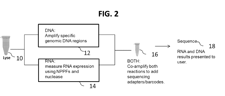
FIG. 2 is a schematic diagram showing an overview of an embodiment of the
disclosed
methods for nucleic acid amplification of DNA and RNA surrogates in the sample
mixture. First,
the sample 10 is lysed with a lysis buffer, thereby generating a lysate
comprising the target DNA
molecule and the target RNA molecule. The resulting lysate is divided or split
into at least two
fractions or portions, 12, 14. Target DNA in portion 12 is amplified, thereby
generating FARs.
Target RNA in portion 14 is incubated with NPPFs specific for the target RNA,
under conditions
that allow the NPPF to specifically bind or hybridize to the target RNA,
thereby forming a double
stranded (ds) nucleic acid molecule, composed of the NPPF hybridized to the
target RNA molecule.
The NPPF hybridized to the target RNA molecule complex is incubated with a
nuclease specific for
single stranded nucleic acid molecules, thereby generating a digested second
portion of the lysate
comprising NPPF hybridized to the target RNA molecule, and then separating the
NPPF from the
target RNA. This resulting mixture containing ss NPPF (comprised of DNA) and
ss target RNA
obtained in portion 14 is mixed with FARs obtained in portion 12, and the
mixture 16 subjected to
nucleic acid amplification (e.g., PCR), allowing amplification of the FARs and
the ss NPPF
simultaneously in the same reaction mixture. The resulting amplicons can then
be sequenced 18,
wherein the NPPF-generated amplicons serve as surrogates for RNA in the
sample. A specific
example is shown in FIG. 3.
FIG. 3 is a more detailed schematic diagram showing an overview of an
embodiment of the
disclosed methods for performing amplification with DNA and RNA surrogate in
the sample
mixture. As shown in Step 1, a sample (such as one known or suspected of
containing target RNA
200 and DNA 230) is treated with a sample disruption buffer (e.g., lysed or
otherwise treated to
make nucleic acids accessible) and then separated into at least two portions.
As shown in Step 2A,
one portion is used to amplify target DNA, thereby generating FARs 236 (the
FARs are double
stranded, though only one strand is shown here for simplicity). For example,
at least one target
DNA 230 is contacted with or incubated with at least one primer (e.g., target
DNA primers, such as
at least two target DNA primers 234, 232), such as target DNA primers with
extensions (for
example, to add the same flanking sequences as on the NPPF to the FAR). Target
specific primers
(e.g., primer pairs) can be used for each target DNA of interest. Thus in some
examples, the
reaction includes at least two different sets of primers, each set specific
for a target DNA (though
one will recognize that in some examples a single primer set can amplify
multiple DNA targets of
interest). As shown in Step 2B of FIG. 3, the target DNA(s) are incubated or
contacted with the
primers (e.g., target DNA primers) under conditions sufficient for
amplification (such as by PCR),
thus generating flanked amplicon regions 236. In some examples, amplification
of the target DNA
- 23 -
CA 03124489 2021-06-21
WO 2020/142153
PCT/US2019/064041
in this step utilizes primers that add a 5'- and a 3'-flanking sequence to the
FARs, wherein the 5'-
end flanking sequence 238 added is the same as the 5'-end flanking sequence of
the NPPF 204, and
the 3'-end flanking sequence 239 added is the same as the 3'-end flanking
sequence of the NPPF
206.
As shown in Step 2AA of FIG. 3, at least a second portion of the sample (i.e.,
different from
the first portion, but still from the same sample) is used to obtain single
stranded NPPFs, which
serves as a surrogate of the target RNA. For example, the second portion of
the lysed sample is
contacted with or incubated with a nuclease protection probe having one or
more flanking
sequences (NPPF) 202 (shown here with both a 5'- and a 3'-flanking sequence,
204 and 206,
respectively), which specifically binds to a first target RNA 200. In some
examples, the NPPF 202
can bind to a plurality of target RNA molecules, such as different splice
isoforms of a particular
RNA. The reaction can include additional NPPFs that specifically bind to a
second target RNA (or
to a plurality of additional target RNA molecules), and so on. In one example,
the method uses one
or more different NPPFs designed to be specific for each unique target RNA
molecule. Thus, the
measurement of 100 different RNA targets (e.g., gene expression product(s))
can use at least 100
different NPPFs with at least one NPPF specific per RNA target (such as
several different
NPPFs/target). In another example, the method uses one or more different NPPFs
designed to be
specific for a plurality of target RNA molecules, such as different splice
isoforms or a wild type
RNA and variations thereof. Thus, the measurement of multiple different RNA
targets can use a
single NPPF. In some examples, combinations of these two types of NPPFs are
used in a single
reaction. Thus, the method can use at least 2 different NPPFs, at least 3, at
least 4, at least 5, at
least 10, at least 25, at least 50, at least 75, at least 100, at least 200,
at least 500, at least 1000, at
least 2000, or at least 2000 different NPPFs (such as 2 to 500, 2 to 100, 2 to
40,000, 2 to 30,000, 2
to 20,000, 2 to 10,000, 2 to 1000, 5 to 10, 2 to 10, 2 to 20, 100 to 500, 100
to 1000, 500 to 5000,
.. 1000 to 3000, 30,000 to 40,000 or 1000 to 30,000 different NPPFs). In
addition, one will
appreciate that in some examples, a plurality of NPPFs can include more than
one (such as 2, 3, 4,
5, 10, 20, 50 or more) NPPFs specific for a single target nucleic acid
molecule (which is referred to
as a tiled set of NPPFs). The reaction also includes nucleic acid molecules
that are complementary
to the flanking sequences (CF S) 208, 210. Thus, if the NPPF has a 5'-flanking
sequence 204, the
.. reaction will include a sequence complementary to the 5'-flanking sequence
(5CFS) 208 and if the
NPPF has a 3'-flanking sequence 206, the reaction will include a sequence
complementary to the
3'-flanking sequence (3CFS) 210. One skilled in the art will appreciate that
the sequence of the
CFSs will vary depending on the flanking sequence present. In addition, more
than one CFS can be
- 24 -
CA 03124489 2021-06-21
WO 2020/142153
PCT/US2019/064041
used to ensure a flanking region is protected (e.g., at least two CFSs can use
that bind to different
regions of a single flanking sequence). The CFS can include natural or
unnatural bases and may be
RNA or DNA.
In the second portion of the sample (Step 2AA), NPPF(s), and CFS(s) are
incubated under
conditions sufficient for the NPPFs to specifically bind to (e.g., hybridize
to) its respective target
RNA molecule, and for CF Ss to bind to (e.g., hybridize to) their
complementary sequence on the
NPPF flanking sequence. In some examples, the CFSs 208, 210 are added in
excess of the NPPFs
202, for example at least 2-fold more CFSs than NPPFs (molar excess), such as
at least 3-fold, at
least 4-fold, at least 5-fold, at least 6-fold, at least 7-fold, at least 8-
fold, at least 9-fold, at least 10-
fold, at least 20-fold, at least 40-fold, at least 50-fold, or at least 100-
fold more CFSs than the
NPPFs. In some examples, the NPPFs 202 are added in excess of the total
nucleic acid molecules
in the sample, for example at least 50-fold more NPPF than total nucleic acid
molecules in the
sample (molar excess), such as at least 75-fold, at least 100-fold, at least
200-fold, at least 500-fold,
or at least 1000-fold more NPPF than the total nucleic acid molecules in the
sample. For
experimental convenience, a similar concentration of each NPPF can be included
to make a
cocktail, such that for the most abundant RNA target measured there will be at
least 50-fold more
NPPF for that RNA target, such as an at least 100-fold excess. The actual
excess and total amount
of all NPPFs used is limited only by the capacity of the nuclease (e.g., 51
nuclease) to destroy all
NPPFs that are not hybridized to target RNA targets. In some examples the
reaction at Step 2AA is
heated, for example incubated for overnight (such as for 16 hours) at 50 C.
This results in the
generation of an NPPF hybridized to (1) its target RNA molecule and (2) the
3CFS, 5CF S, or both
the 3CFS and the 5CFS.
Following hybridization of the NPPF to its target RNA (and hybridization of
the CF Ss to
their flanking sequence), as shown in Step 2BB in FIG. 3 the sample is
contacted with a reagent
(such as a nuclease) specific for single-stranded (ss) nucleic acid molecules
under conditions
sufficient to remove (or hydrolyze or digest) ss nucleic acid molecules, such
as unbound nucleic
acid molecules (such as unbound NPPFs, unbound CFSs, and unbound target RNA
molecules, or
portions of such molecules that remain single stranded, such as portions of a
target RNA molecule
not bound to the NPPF). This results in the generation of a ds NPPF/target
RNA/CFSs complex (or
duplex) 212. Incubation of the sample with a nuclease specific for ss nucleic
acid molecules
results in degradation of any ss nucleic acid molecules present, leaving
intact double-stranded
nucleic acid molecules, including NPPFs that have bound to CFSs and a target
RNA molecule. For
example, the reaction can be incubated at 50 C for 1.5 hours with 51 nuclease
(though hydrolysis
- 25 -
CA 03124489 2021-06-21
WO 2020/142153
PCT/US2019/064041
can occur at other temperatures and be carried out for other periods of time,
and, in part, the time
and temperature required will be a function of the amount of nuclease, and on
the amount of
nucleic acid required to be hydrolyzed, as well as the Tm of the double-
stranded region being
protected).
As shown in Step 2CC of FIG. 3, the ds NPPF/RNA target/CFSs complex 212 is
exposed to
conditions that allow the target RNA sequence 200 and the CFSs (e.g., 5CFS
208, 3CFS 210, or
both) to be separated from the NPPF, thereby generating ssRNA 200, ssNPPF 202,
and ssCFSs
(such as 208 and 210). Although two CFSs are shown, single CFS embodiments are
also
contemplated by this disclosure. If only one flanking sequence is present on
the NPPF, only one
CFS will have been bound in the NPPF/target complex. The CFSs can be DNA or
RNA (or a
mixture of both nucleotide types). In one example, 5CFS 208 and/or 3CFS 210
are DNA. In some
examples, the reaction can be heated or the pH altered (e.g., to result in the
reaction having a basic
pH) under conditions that allow the NPPF 202 to dissociate from the hybridized
RNA target 200,
resulting in a mixed population of ssNPPFs 202 and ss target nucleic acids
(e.g., ssRNA targets)
200. In some examples, Step 2CC of FIG. 3 is performed as the first step of
Step 4 of FIG.3, that is
instead of performing a separate denaturation step, the ds NPPF/RNA
target/CFSs complex 212 is
dissociated into ss nucleic acid molecules as the first step in the second
amplification reaction.
As shown in Step 3 of FIG. 3, the mixture obtained after Step 2CC containing
ssNPPFs 202
(or the ds NPPF/RNA target/CFSs complex 212 obtained after step 2BB of FIG.
3), and the mixture
obtained after Step 2B containing FARs (which are double stranded) 236 are
combined into a
single mixture. As shown in Step 4 of FIG. 3, the resulting mixture is
subjected to nucleic acid
amplification conditions (e.g., using PCR) to generate amplicons, prior to the
sequencing. Thus,
FARs and the ssNPPF (or RNA ds NPPF/RNA target/CFSs complex 212) surrogates
are amplified
in the same reaction, generating amplicons, which can then be sequenced. FIG.
4 shows exemplary
PCR primers or probes as arrows, which can be used in the amplification
reaction shown in Step 4.
The PCR primers or probes can include one or more experiment tags 222, 224,
242, 244 (e.g., that
allow for the identification of a sample or patient) and/or sequencing
adaptors 218, 220, 248, 240
(e.g., that allow the targets to be sequenced by a particular sequencing
platform, and, thus, such
adaptors are complementary to capture sequences on e.g. a sequencing chip or
flowcell). At least a
portion of the PCR primers/probes are specific for the flanking sequences 204,
206 (and in some
examples also 238, 239). In some examples, the concentration of the primers
are in excess of the
ssNPPFs 200 and/or the FARs 236, for example, in excess by at least 10,000-
fold, at least 50,000-
fold, at least 100,000-fold, at least 150,000-fold, at least 200,000-fold, at
least 400,000-fold, at least
- 26 -
CA 03124489 2021-06-21
WO 2020/142153
PCT/US2019/064041
500,000-fold, at least 600,000-fold, at least 800,000-fold, or at least
1,000,000-fold. In some
examples, the concentration of primers 208 in the reaction is at least 200 nM
(such as at least 400
nM, at least 500 nM, at least 600 nM, at least 750 nM, or at least 1000 nM).
In Step 4 of FIG. 3, amplicons generated in Step 3 can be sequenced. In some
examples, a
plurality of FAR amplicons and NPPF amplicons are sequenced in parallel, for
example,
simultaneously or contemporaneously. Thus, this method can be used to sequence
a plurality of
target nucleic acid sequences.
A. Exemplary Hybridization Conditions
Disclosed herein are conditions sufficient for (1) amplification primers to
specifically
hybridize to their complementary nucleic acid molecules (e.g., to DNA target
molecules in a lysed
sample, to FARs, and to ssNPPFs), and (2) an NPPF or a plurality of NPPFs to
specifically
hybridize to target RNA molecule(s), such RNAs present in a at least one
portion of a lysed sample,
as well as specifically hybridize to CFS complementary to the flanking
sequence(s). In some
examples, a plurality of NPPFs include at least 2, at least 5, at least 10, at
least 20, at least 100, at
least 500, at least 1000, at least 3000, at least 10,000, at least 15,000, at
least 20,000, at least
25,000, at least 30,000, at least 35,000, at least 40,000, at least 45,000, or
at least 50,000 (such as 2
to 5000, 2 to 3000, 10 to 1000, 50 to 500, 25 to 300, 50 to 300, 10 to 100, 50
to 100, 500 to 1000,
1000 to 5000, 2000 to 10,000, 100 to 50,000, 100 to 40,000, 100 to 30,000, 100
to 20,000, 100 to
10,000, 10 to 50,000, 10 to 40,000, 10 to 30,000, 10 to 20,000, 10 to 10,000,
or 30,000 to 40,000)
unique NPPF sequences.
Hybridization is the ability of complementary single-stranded DNA, RNA, or
DNA/RNA
hybrids, to form a duplex molecule (also referred to as a hybridization
complex). For example, the
features (such as length, base composition, and degree of complementarity)
that will enable a
nucleic acid (e.g., an NPPF) to hybridize to another nucleic acid (e.g.,
target RNA or CFS) under
conditions of selected stringency, while minimizing non-specific hybridization
to other substances
or molecules can be determined based on the present disclosure. "Specifically
hybridize" and
"specifically complementary" are terms that indicate a sufficient degree of
complementarity such
that stable and specific binding occurs between a first nucleic acid molecule
(e.g., an NPPF or
primer) and a second nucleic acid molecule (such as a nucleic acid target, for
example, a DNA or
RNA target, or a CFS). The first and second nucleic acid molecules need not be
100%
complementary to be specifically hybridizable. Specific hybridization is also
referred to herein as
"specific binding." Hybridization conditions resulting in particular degrees
of stringency will vary
- 27 -
CA 03124489 2021-06-21
WO 2020/142153
PCT/US2019/064041
depending upon the nature of the hybridization method and the composition and
length of the
hybridizing nucleic acid sequences. Generally, the temperature of
hybridization and the ionic
strength (such as the Na + concentration) of the hybridization buffer will
determine the stringency of
hybridization. Calculations regarding hybridization conditions for attaining
particular degrees of
stringency are discussed in Sambrook et at., (1989) Molecular Cloning, second
edition, Cold
Spring Harbor Laboratory, Plainview, NY (chapters 9 and 11).
Characteristics of the NPPFs are discussed in more detail in Section III,
below. Typically, a
region of an NPPF will have a nucleic acid sequence (e.g., FIG. 1A, 102) that
is of sufficient
complementarity to its corresponding target RNA molecule(s) to enable it to
hybridize under
selected stringent hybridization conditions, as well as a region (e.g., FIG.
1A, 104, 106) that is of
sufficient complementarity to its corresponding CFS(s) to enable it to
hybridize under selected
stringent hybridization conditions. In some examples, an NPPF shares at least
90%, at least 92%,
at least 95%, at least 98%, at least 99% or 100% complementarity to its target
RNA sequence(s).
Exemplary hybridization conditions include hybridization at about 37 C or
higher (such as about
37 C, 42 C, 50 C, 55 C, 60 C, 65 C, 70 C, 75 C, or higher, such as 45-55 C or
48-52 C).
Among the hybridization reaction parameters which can be varied are salt
concentration, buffer,
pH, temperature, time of incubation, amount and type of denaturant such as
formamide. For
example, nucleic acid (e.g., a plurality of NPPFs) can be added to at least
one portion of a sample at
a concentration ranging from about 10 pM to about 10 nM (such as about 30 pM
to 5 nM, about
100 pM to about 1 nM, such as 1 nM NPPFs), in a buffer (such as one containing
NaCl, KC1,
H2PO4, EDTA, 0.05% Triton X-100, or combinations thereof) such as a lysis
buffer.
In some examples, the NPPFs are added in excess of the corresponding target
RNA
molecules in at least one portion of the sample, such as an at least 10-fold,
at least 50-fold, at least
75-fold, at least 100-fold, at least 250-fold, at least 1,000 fold, at least
10,000 fold, at least 100,000
fold, at least 1,000,000 fold, or at least 10,000,000 fold molar excess or
more of NPPF to
corresponding target RNA molecules in the at least one portion of the sample.
In one example,
each NPPF is added to the at least one portion of the sample at a final
concentration of at least 10
pM, such as at least 20 pM, at least 30 pM, at least 50 pM, at least 100 pM,
at least 150 pM, at least
200 pM, at least 500 pM, at least 1 nM, or at least 10 nM. In one example,
each NPPF is added to
the at least one portion of the sample at a final concentration of about 125
pM. In another example,
each NPPF is added to the at least one portion of the sample at a final
concentration of about 167
pM. In a further example, each NPPF is added to the at least one portion of
the sample at a final
concentration of about 1 nM. In a further example, each NPPF is added to the
at least one portion
- 28 -
CA 03124489 2021-06-21
WO 2020/142153
PCT/US2019/064041
of the sample at least about 100,000,000, at least 300,000,000, or at least
about 3,000,000,000
copies per 11.1. In some examples, the CFSs are added in excess of the NPPFs,
such as an at least 2-
fold, at least 3-fold, at least 4-fold, at least 5-fold or at least 10-fold
molar excess of CFS to NPPF.
In one example, each CFS is added to the at least one portion of the sample at
a final concentration
of about at least 6 times the amount of probe, such as at least 10 times or at
least 20 times the
amount of probe (such as 6 to 20 times the amount of probe). In one example,
each CFS (e.g.,
5CFS and 3CFS) is added at least at 1 nM, at least 5 nM, at least 10 nM, at
least 50 nM, at least 100
nM, or at least 200 nm, such as 1 to 100, 5 to 100 or 5 to 50 nM. For example
if there are six
probes, each at 166 pM, each CFSs can be added at 5 to 50 nM.
Prior to hybridization with NPPFs and CFS(s), the nucleic acids in at least
one portion of
the sample are denatured, rendering them single stranded and available for
hybridization (for
example at about 85 C to about 105 C for about 5-15 minutes, such as 85 C for
10 minutes). By
using different denaturation solutions, this denaturation temperature can be
modified, so long as the
combination of temperature and buffer composition leads to formation of single
stranded target
DNA or RNA or both.
In the portion of the lysed sample used to obtain NPPFs for surrogates of RNA
present in
the sample, the nucleic acids in the at least one portion of the lysed sample
and the 5CFS, 3CFS, or
both, are hybridized to the plurality of NPPFs for between about 10 minutes
and about 72 hours
(for example, at least about 1 hour to 48 hours, about 2 to 16 hours, about 6
hours to 24 hours,
about 12 hours to 18 hours, about 16 hours, or overnight, such as 2 to 20
hours) at a temperature
ranging from about 4 C to about 70 C (for example, about 37 C to about 65 C,
about 42 C to
about 60 C, or about 50 C to about 60 C, such as 50 C). In one example,
hybridization is
performed at 50 C for 2 to 20 hours. Hybridization conditions will vary
depending on the
particular NPPFs and CF Ss used, but are set to ensure hybridization of NPPFs
to the target RNA
molecules and the CFSs. In some examples, the plurality of NPPFs and CFSs are
incubated with
the at least one portion of the lysed sample at a temperature of at least
about 37 C, at least about
40 C, at least about 45 C, at least about 50 C, at least about 55 C, at least
about 60 C, at least
about 65 C, or at least about 70 C. In one example, the plurality of NPPFs and
CFSs are incubated
with the sample at about 37 C, at about 42 C, or at about 50 C.
In some embodiments, the methods do not include nucleic acid purification (for
example,
nucleic acid purification is not performed before or after lysis of the
sample, such as not prior to
contacting a portion of the lysed sample with NPPFs and CF Ss or with nucleic
acid primers for
target DNA amplification, and/or nucleic acid purification is not performed
following contacting
- 29 -
CA 03124489 2021-06-21
WO 2020/142153
PCT/US2019/064041
the sample with the NPPFs and CFSs, or with nucleic acid primers for target
DNA amplification).
In some examples, no pre-processing of the sample is required except for cell
lysis. In some
examples, cell lysis and contacting the sample with either (1) primers to
amplify target DNA or (2)
the plurality of NPPFs and CFSs, occur sequentially.
B. Treatment with Nuclease
As shown in Step 2BB of FIG. 3, following hybridization of the NPPFs to target
RNA and
to CFS(s), at least one portion of the lysed sample is subjected to a nuclease
protection procedure.
The target RNA molecules and CFSs (one or two CFSs, depending if there are
both 5'- and 3'-
flanking sequence on the NPPF or just one) that have hybridized to the NPPF
are not hydrolyzed by
the nuclease and can be subsequently amplified and/or sequenced.
Nucleases are enzymes that cleave a phosphodiester bond. Endonucleases cleave
an
internal phosphodiester bond in a nucleotide chain (in contrast to
exonucleases, which cleave a
phosphodiester bond at the end of a nucleotide chain). Thus, endonucleases,
exonuclease, and
combinations thereof, can be used in the disclosed methods. Endonucleases
include restriction
endonucleases or other site-specific endonucleases (which cleave DNA at
sequence specific sites),
DNase I, pancreatic RNAse, Bal 31 nuclease, Si nuclease, mung bean nuclease,
Ribonuclease A,
Ribonuclease Ti, RNase I, RNase PhyM, RNase U2, RNase CLB, micrococcal
nuclease, and
apurinic/apyrimidinic endonucleases. Exonucleases include exonuclease III and
exonuclease VII.
In particular examples, a nuclease is specific for single-stranded nucleic
acids, such as Si nuclease,
P1 nuclease, mung bean nuclease, or BAL 31 nuclease. Reaction conditions for
these enzymes are
known and can be optimized empirically.
Treatment with one or more nucleases can destroy all ss nucleic acid molecules
(including
RNA and DNA in the lysed sample that is not hybridized to (thus, not protected
by) NPPFs, NPPFs
that are not hybridized to target RNA, and CF Ss not hybridized to an NPPF),
but will not destroy ds
nucleic acid molecules such as NPPFs that have hybridized to CFSs and a target
nucleic acid
molecule present in the at least one portion of the lysed sample. For example,
unwanted nucleic
acids, such as one or more non-target DNA (such as genomic DNA, cDNA) and non-
target RNA
(e.g., non-target, tRNA, rRNA, mRNA, miRNA), and portions of the target RNA
molecule(s) that
are not hybridized to complementary NPPF sequences (such as overhangs), which,
in the case of
mRNA targets, will constitute the majority of the nucleic target sequence, can
be substantially
destroyed in this step. In some embodiments, this step leaves behind
approximately a
stoichiometric amount of target RNA/CFS/NPPF duplex. If the target RNA
molecule is cross-
- 30 -
CA 03124489 2021-06-21
WO 2020/142153
PCT/US2019/064041
linked to tissue that occurs from fixation, the NPPFs hybridize to the cross-
linked target RNA
molecule without the need, in some embodiments, to reverse cross-linking, or
otherwise release the
target nucleic acid from the tissue to which it is cross-linked.
In some examples, Si nuclease diluted in a buffer (such as one containing
sodium acetate,
NaCl, KC1, ZnSO4, an antimicrobial agent (such as ProChem biocide), or
combinations thereof) is
added to the hybridized NPPF/target RNA/CFS sample mixture and incubated at
about 37 C to
about 60 C (such as about 50 C) for 10-120 minutes (for example, 10-30
minutes, 30 to 60
minutes, 60-90 minutes, 90 minutes, or 120 minutes) to digest non-hybridized
nucleic acid from the
at least one portion of the lysed sample and non-hybridized NPPFs, RNAs, and
CFSs. In one
example, the nuclease digestion is performed by incubating the at least one
portion of the lysed
sample with the nuclease in a nuclease buffer at 50 C for 60 to 90 minutes.
Following nuclease digestion, the at least one portion of the lysed sample can
optionally be
treated to inactivate or remove residual enzymes (e.g., by phenol extraction,
precipitation, column
filtration, addition of proteinase K, addition of a nuclease inhibitor,
chelating divalent cations
required by the nuclease for activity, heating, or combinations thereof). In
some examples the at
least one portion of the lysed sample is treated to adjust the pH to about 7
to about 8, for example,
by addition of KOH or NaOH or a buffer (such as one containing Tris-HC1 at pH
9 or Tris-HC1 at
pH 8). Raising the pH can prevent the depurination of DNA and prevents many ss-
specific
nucleases (e.g., Si) from functioning fully. In some examples, the at least
one portion of the lysed
sample is heated (for example 80-100 C) to inactivate the nuclease, for
example for 10-30 minutes.
C. Separation of ssNPPFs from the Target Nucleic Acids
As shown in Step 2CC of FIG. 3, following nuclease treatment of the at least
one portion of
the lysed sample containing the double-stranded NPPF/target RNA/CFSs
complexes, the NPPFs
are separated (e.g., denatured) from the ss nucleic acid target and the
CFS(s). Thus, the double-
stranded NPPF/target RNA/CFSs complex can be separated into single-stranded
nucleic acid
molecules, the ssNPPF and the ss target nucleic acid (e.g., ssRNA) (as well as
the ss CFSs).
In some examples, Step 2CC of FIG. 3 is performed as the first step of Step 4
of FIG. 3.
For example, instead of performing a separate denaturation/separation step,
the ds NPPF/RNA
target/CFSs complex 212 is dissociated into ss nucleic acid molecules as the
first step in the second
amplification reaction (e.g., the first step of Step 4 in FIG. 3).
D. Nucleic acid Amplification
- 31 -
CA 03124489 2021-06-21
WO 2020/142153
PCT/US2019/064041
As shown in FIG. 3, the method includes two nucleic acid amplification steps,
using
methods such as polymerase chain reaction (PCR) or other forms of enzymatic
amplification. The
first amplification amplifies target DNA in a portion of the lysed sample
(Step 2B). The second
amplification amplifies the FARs resulting from the first amplification along
with the ss NPPFs
obtained after hybridization, nuclease digestion, and denaturation (Step 4).
In some examples, no more than 30 cycles of amplification are performed at
each
amplification step, such as no more than 25 cycles of amplification, no more
than 20 cycles of
amplification, no more than 15 cycles of amplification, no more than 10 cycles
of amplification, no
more than 8 cycles of amplification, or no more than 5 cycles of
amplification, such as 2 to 30
cycles, 5 to 30 cycles, 8 to 30 cycles, 8 to 25 cycles, 2 to 25 cycles, 5 to
25 cycles, 5 to 20 cycles, 5
to 15 cycles, or 5 to 10 cycles of amplification for each amplification step.
During the first amplification step (amplification of target DNA), the least
number of cycles
of amplification needed is used, to reduce the number of errors introduced
during the amplification.
In some examples, 5 to 20 amplification cycles are performed in the first
amplification, such as 5 to
15 cycles, such as 5, 6, 7, 8,9, 10, 11, 12, 13, 14, 15, 16, 17, 18, 19, or 20
cycles. In some
examples, 10 amplification cycles are performed in the first amplification. In
some examples, the
primers used in the first amplification step have a T. of about 50-62 C. In
some examples, the
annealing temperature used in the first amplification reaction is at least 50
C, at least 56 C, or at
least 58 C, such as about 50 C to 60 C, such as about 56 C to 60 C, such as
about 52 C to 58 C,
such as 56 C, 57 C or 58 C. In some examples, the FARs generated from the
first amplification
step are about 70 to 200 bp, such as 70 to 150, 70 to 125, 90 to 150 bp, such
as about 70 bp, about
100 bp, or about 140 bp.
In some examples, during the second amplification step (amplification of FARs
and
ssNPPFs), 8 to 30 amplification cycles are performed in the second
amplification, such as 15 to 25
or 8 to 25 cycles, such as 8, 9, 10, 11, 12, 13, 14, 15, 16, 17, 18, 19, 20,
21, 22, 23, 24, 25, 26, 27,
28, 29 or 30 cycles. In some examples, 19 amplification cycles are performed
in the second
amplification. In some examples, the primers used in the second amplification
step have a Tm of
about 50-62 C. In some examples, the annealing temperature used in the second
amplification
reaction is at least 50 C, at least 56 C, or at least 56 C, such as about 50 C
to 60 C, such as about
52 C to 58 C, such as 56 C. In some examples, the FAR amplicons generated from
the second
amplification step are about 150 to 250 bp, such as 150 to 200 bp, such as
about 180 bp. In some
examples, the NPPF amplicons generated from the second amplification step are
about 150 to 250
bp, such as 150 to 200 bp, such as about 155 bp or 180 bp.
- 32 -
CA 03124489 2021-06-21
WO 2020/142153
PCT/US2019/064041
In some examples, portion of an amplification primer that anneals to its
target is about 15-
25 nt (such as 22 nt) with about 50% GC content. In some examples, an
amplification primer is
about 50 to 100 nt (such as 60 to 100 nt) in length.
Nucleic acid amplification methods that can be used include those that result
in an increase
in the number of copies of a nucleic acid molecule, such as a target DNA (or
amplicon thereof),
target RNA surrogate (i.e., indirectly by amplification of ssNPPF), and/or
portion thereof The
resulting products are called amplification products or amplicons. Generally,
such methods include
contacting material to be amplified (e.g., target DNA (or amplicon thereof) or
ssNPPF) with one or
a pair of oligonucleotide primers, under conditions that allow for
hybridization of the primer(s) to
.. the nucleic acid molecule to be amplified. The primers are extended under
suitable conditions,
dissociated from the template, and then re-annealed, extended, and dissociated
to amplify the
number of copies of the nucleic acid molecule.
Examples of in vitro amplification methods that can be used include, but are
not limited to,
PCR, quantitative real-time PCR, isothermal amplification methods, strand
displacement
amplification; transcription-free isothermal amplification; repair chain
reaction amplification; and
NASBATM RNA transcription-free amplification. In one example, the primers
specifically
hybridize to at least a portion of the NPPF flanking sequence(s). In one
example, helicase-
dependent amplification is used.
During the second amplification of the FARs and ssNPPFs, an experiment tag,
and/or
sequencing adaptor can be incorporated as, for instance, part of the primer
(see FIG. 4). However,
addition of such tags/adaptors is optional. For example, an amplification
primer, which includes a
first portion that is complementary to all or part of a 5'- or 3'-flanking
sequence (e.g., 238, 239,
204, 206 of FIG. 3), can include a second portion that is complementary to a
desired experiment tag
and/or sequencing adaptor. One skilled in the art will appreciate that
different combinations of
.. experiment tags and/or sequencing adaptors can be added to either end of
the FAR or ssNPPF.
In one example, DNA in the lysed sample is amplified using a first primer that
includes a
first portion complementary to all or a portion of the target DNA sequence and
a second portion
complementary to (or comprising) a desired flanking sequence (e.g.,
complementary to the 5'-
flanking sequence of the NPPF) and with a second primer that includes a first
portion
complementary to all or a portion of the target DNA sequence and a second
portion complementary
to (or comprising) a desired flanking sequence (e.g., complementary to the 5'-
flanking sequence of
the NPPF) (see FIG. 3, Step 2A), such that the flanking sequence 238, 239
becomes incorporated
- 33 -
CA 03124489 2021-06-21
WO 2020/142153
PCT/US2019/064041
into the resulting amplicon (see FIG. 3, Step 2B). In one example, two
different flanking sequences
are used.
In one example, the FAR and the ssNPPF are amplified using a first
amplification primer
that includes a first portion complementary to all or a portion of the 5'
flanking sequence and a
second portion complementary to (or comprising) a desired sequencing adaptor,
and the second
amplification primer includes a first portion complementary to all or a
portion of the 3' flanking
sequence and a second portion complementary to (or comprising) a desired
experiment tag (e.g.,
see FIG. 4). In one example, two different sequencing adapters and two
different experiment tags
are used. In some examples, two different sequencing adapters and one
experiment tag are used.
In another example, the FAR and the ssNPPF is amplified using a first
amplification primer that
includes all or a portion of a first portion identical to (or complementary
to) the 5' flanking
sequence and a second portion complementary to (or comprising) a desired
sequencing adaptor and
a desired experiment tag, and the second amplification primer includes a first
portion
complementary to all or a portion of the 3' flanking sequence and a second
portion complementary
to (or comprising) a desired experiment tag.
Amplification can also be used to introduce a detectable label into the
generated target
nucleic acid amplicons (for example, if additional labeling is desired) or
other molecule that
permits detection or quenching. For example, the amplification primer can
include a detectable
label, hapten, or quencher that is incorporated into the target nucleic acid
amplicons during
amplification. Such a label, hapten, or quencher can be introduced at either
end of the target
amplicon(s) (or both ends) or anywhere in between.
In some examples, the resulting FAR amplicons and NPPF amplicons are purified
before
sequencing. For example, the amplification reaction mixture can be purified
before sequencing
using methods known in the art (e.g., gel purification, biotin/avidin capture
and release, capillary
electrophoresis, size-exclusion purification, or binding to and release from
paramagnetic beads
(solid phase reversible immobilization)). In one example, the FAR amplicons
and NPPF amplicons
are biotinylated (or include another hapten) and captured onto an avidin or
anti-hapten coated bead
or surface, washed, and then released for sequencing. Likewise, the FAR
amplicons and NPPF
amplicons can be captured onto a complimentary oligonucleotide (such as one
bound to a surface),
washed and then released for sequencing. The capture of amplicons need not be
particularly
specific, as the disclosed methods eliminate most of the genome or
transcriptome, leaving the
desired amplicons. Other methods can be used to purify the FAR amplicons and
NPPF amplicons,
if desired.
- 34 -
CA 03124489 2021-06-21
WO 2020/142153
PCT/US2019/064041
The FAR amplicons and NPPF amplicons can also be purified after the last step
of
amplification, while still double stranded, by a method which uses a nuclease
that hydrolyzes single
stranded oligonucleotides (such as Exonuclease I), which nuclease can in turn
be inactivated before
continuing to the next step such as sequencing.
1. Primers
The amplification primers that specifically bind or hybridize to the flanking
sequence(s)
(e.g., 5' and/or 3' flanking sequence(s) of the NPPF and FARs), as well as
those specific for the
target DNA, can be used to initiate amplification, such as PCR amplification.
Thus, primers having
sequence complementarity to the flanking sequence can anneal to an NPPF by
nucleic acid
hybridization to form a hybrid between the primer and the flanking sequence of
the surrogate
NPPF, and then the primer extended along the complement strand by a polymerase
enzyme.
Similarly, primers having sequence complementarity to the target DNA can
anneal to the target
DNA by nucleic acid hybridization to form a hybrid between the primer and the
target DNA, and
then the primer extended along the complement strand by a polymerase enzyme.
In addition, the amplification primers can be used to introduce nucleic acid
markers (such as
one or more experiment tags and/or sequencing adaptors) and/or detectable
labels to the resulting
target nucleic acid amplicons.
Primers are short nucleic acid molecules, such as a DNA oligonucleotides that
are at least
12 nucleotides in length (such as about 15, 20, 25, 30, 50, 60, 61, 62, 63,
64, 65, 66, 67, 68, 69, 70,
75 or 80 nucleotides or more in length, such as 15 to 25 nt, 50 to 80 nt, 60-
70 nt or 60-66 nt).
For the first amplification, primers in some examples include a region of
about 15-25 nt
that has complementarity to the target DNA, and another 25 nt extension on one
end (e.g.,
complementary to a 5'- or 3'-flanking sequence of an NPPF).
For the second amplification, primers in some examples include a region of
about 15-25 nt
that has complementarity to the 5'- or 3'-flanking sequence of an NPPF or FAR,
and a region
having a nucleic acid sequence that allows for the addition of a sequence
adaptor, experiment tag,
or both to the resulting amplicons. It can also include a region having a
nucleic acid sequence that
results in addition of a detectable label to the resulting amplicon. An
experiment tag and/or
sequencing adaptor can be introduced at the 5'- and/or 3'-end of the amplicon.
In some examples,
two or more experiment tags and/or sequencing adaptors are added to a single
end or both ends of
the amplicon, for example using a single primer having a nucleic acid sequence
that results in
addition of two or more experiment tags and/or sequencing adaptors. Experiment
tags can be used,
- 35 -
CA 03124489 2021-06-21
WO 2020/142153
PCT/US2019/064041
for example, to differentiate one sample or sequence from another. Sequence
adaptors permit
capture of the resulting amplicon by a particular sequencing platform.
2. Addition of Experiment Tags
Experiment tags are short sequences or modified bases that serve as an
identifier for one or
several reactions to be independently discerned by, for example: patient,
sample, cell type, time
course timepoint, or treatment. Experiment tags can be part of the flanking
sequence of the NPPF
and the FAR. In another example, the experiment tag is added during
amplification (e.g.,
amplification of the FAR and ssNPPF), resulting in an amplicon (e.g., FAR
amplicon and NPPF
.. amplicon) containing an experiment tag. The presence of universal sequences
in the flanking
sequence(s) permit the use of universal primers, which can introduce other
sequences onto the
NPPF amplicons, for example during amplification. Experimental tags can also
be used for
amplification, such as nested amplification, or two stage amplification.
Exemplary experiment tags
are provided in Tables 3 - 5.
Experiment tags, such as one that differentiates one sample from another, can
be used to
identify the particular target sequence. Thus, experiment tags can be used to
distinguish
experiments or patients from one another. In one example, the experiment tag
is the first three, five,
ten, twenty, or thirty nucleotides of the 5'- and/or 3'-end of a resulting
amplicon. In some examples,
the experiment tags are placed in proximity to the sequencing primer site. For
Illuminag
.. sequencing, experiment tags are immediately next to the Read 1 and Read 2
primer sites. For some
sequencing platforms, experiment tags are generally the first few bases read.
In particular examples,
the experiment tag is at least 3 nucleotides in length, such as at least 5, at
least 10, at least 15, at least
20, at least 25, at least 30, at least 40, or at least 50 nucleotides in
length, such as 3-50, 3-20, 12-50,
6-8, 8-10, 6-12, or 12-30 nucleotides, for example, 3, 4, 5, 6, 7, 8, 9, 10,
11, 12, 13, 14, 15, 16, 17,
18, 19, 20, 21, 22, 23, 24, 25, 26, 27, 28, 29 or 30 nucleotides in length.
In one example, an experiment tag is used to differentiate one sample from
another. For
example, such a sequence can function as a barcode, to allow one to correlate
a particular sequence
detected with a particular sample, patient, or experiment (such as a
particular reaction well, day, or
set of reaction conditions). This permits a particular target nucleic acid
amplicon that is sequenced
to be associated with a particular patient or sample or experiment for
instance. The use of such tags
provides a way to lower cost per sample and increase sample throughput, as
multiple target nucleic
acid amplicons can be tagged and then combined (for example, from different
experiments or
patients), for example, in a single sequencing run or detection array. This
allows for the ability to
- 36 -
CA 03124489 2021-06-21
WO 2020/142153
PCT/US2019/064041
combine different experimental or patient samples into a single run within the
same instrument
channel or sequencing consumable (such as a flowcell or a semiconductor chip).
For example,
such tags permitting 100s or 1,000s of different experiments to be sequenced
in a single run within
a single flowcell or chip. In addition, if the method includes the step of gel
purifying the completed
amplification reaction (or other method of purification or clean up that does
not require actual
separation) only one gel (or clean up or purification reaction or process)
needs to be run per
detection or sequencing run. Similarly, if sequencing requires a quantitation
step, then either
individual samples or only the pool of samples may be quantitated prior to
sequencing. The
sequenced target nucleic acid amplicons can then be sorted, for example, by
the experiment tags.
In one example, the experiment tag is used to identify the particular target
sequence. In this
case, using an experimental tag to correspond to a particular target sequence
can shorten the time or
amount of sequencing needed, as sequencing the end of the target nucleic acid
amplicon instead of
the entire target nucleic acid amplicon can be sufficient. For example, if
such an experiment tag is
present on the 3'-end of the target nucleic acid amplicon, the entire target
nucleic acid amplicon
sequence itself does not have to be sequenced to identify the target sequence.
Instead, only the 3'-
end of the target nucleic acid amplicon containing the experiment tag needs to
be sequenced. This
can significantly reduce sequencing time and resources, as less material needs
to be sequenced.
3. Addition of Sequencing Adaptors
Sequencing adaptors can, but need not, be part of the flanking sequence(s) of
the NPPFs
and FARs when generated. In another example, a sequencing adaptor is added
during amplification
of a nucleic acid (e.g., amplification of the FAR or ssNPPFs), resulting in
amplicons containing a
sequencing adaptor. The presence of a universal sequence in the flanking
sequence(s) permit the
use of universal amplification primers, which can introduce other sequences
onto the NPPF
surrogate and FAR, for example during amplification.
A sequencing adaptor can be used add a sequence to a nucleic acid (e.g., FAR
and surrogate
NPPFs) needed for a particular sequencing platform. For example, some
sequencing platforms
(such as the 454-branded (Roche), Ion Torrent-branded and Illumina-branded)
require the nucleic
acid molecule to be sequenced to include a particular sequence at its 5'-
and/or 3'-end, for example,
to capture the molecule to be sequenced. For example, the appropriate
sequencing adaptor is
recognized by a complementary sequence on the sequencing chip or beads, and
the amplicon
captured by the presence of the sequencing adaptor.
In one example, a poly-A (or poly-T), such as a poly-A or poly-T at least 10
nucleotides in
length, is added to the nucleic acid (e.g., FARs and ssNPPFs) during the
second PCR amplification.
- 37 -
CA 03124489 2021-06-21
WO 2020/142153
PCT/US2019/064041
In a specific example, the poly-A (or poly-T) is added to the 3'-end of the
FARs and ssNPPFs. In
some examples, this added sequence is polyadenylated at its 3' end using a
terminal
deoxynucleotidyl transferase (TdT).
In particular examples, the sequencing adapter added is at least 12
nucleotides (nt) in length,
such as at least 15, at least 20, at least 25, at least 30, at least 40, at
least 50, at least 60 or at least 70
nt in length, such as 12-50, 20-35, 50-70, 20-70, or 12-30 nt in length.
E. Sequencing Nucleic Acid Amplicons
The resulting nucleic acid amplicons (e.g., FAR amplicons for target DNA and
surrogate
NPPF amplicons for target RNA) are sequenced, for example, by sequencing the
amplicon, or a
.. portion thereof (such as an amount sufficient to permit identification of
the target nucleic acid
molecule or to permit determination that a particular mutation is or is not
present). The disclosure
is not limited to a particular sequencing method. It will be appreciated that
the nucleic acid
amplicons (e.g., DNA amplicons) can be designed for sequencing by any method
on any sequencer
known currently or in the future. The target nucleic acid itself does not
limit the method of
sequencing used, nor the sequencing enzyme used. Other methods of sequencing
are or will be
developed, and one skilled in the art can appreciate that the generated
nucleic acid amplicons will
be suitable for sequencing on these systems. In some examples, multiple
different target nucleic
acid amplicons are sequenced in a single reaction. Thus, a plurality of target
nucleic acid
amplicons can be sequenced in parallel, for example, simultaneously or
contemporaneously.
Exemplary sequencing methods that can be used to determine the sequence of the
resulting
FAR amplicons and NPPF amplicons, such as amplicons composed of DNA, include,
but are not
limited to, the chain termination method, dye terminator sequencing, and
pyrosequencing (such as
the methods commercialized by Biotage (for low throughput sequencing) and 454
Life Sciences
(for high-throughput sequencing)). In some examples, the amplicons are
sequenced using an
Illumina (e.g., NovaSeq, MiSeq), Ion Torrent , 454 , Helicos, PacBio , Solid
(Applied
Biosystemsg) or any other commercial sequencing system. In one example, the
sequencing
method uses bridge PCR (e.g., Illumina ). In one example, the Helicos or
PacBio single
molecule sequencing method is used. In one example, a next-generation
sequencer (NGS) is used,
such as those from Illumina , Roche , Genapsys, or Thermo Fisher Scientific ,
for example,
SOLiD /Ion Torrent S5 from Thermo Fisher Scientific , NovaSeq/ NextSeq/MiSeq
from
Illumina , or GS FLX Titanium /GS Junior from Roche . Sequencing adaptors
(such as
specific sequences or poly-A or poly T tails present on the FAR amplicons and
NPPF amplicons,
- 38 -
CA 03124489 2021-06-21
WO 2020/142153
PCT/US2019/064041
for example, as introduced using PCR) can be used for capture of the amplicons
for sequencing on
a particular platform. In one example, a nanopore-type sequencer is used.
Although sequencing by Ion Torrent or Illumina typically involves nucleic
acid
preparation, accomplished by random fragmentation of nucleic acid, followed by
in vitro ligation of
.. common adaptor sequences, for the disclosed methods, the step of random
fragmentation of the
nucleic acid to be sequenced can be eliminated, and the in vitro ligation of
adaptor sequences is
replaced by sequences present in the NPPF amplicon or FAR amplicon, such as an
experiment tag
present in the NPPF amplicon or FAR amplicons or a sequencing adaptor sequence
present in the
NPPF or FAR, or added to the NPPF amplicon or FAR amplicon during
amplification. For some
sequencing methods, a sequencing primer is hybridized to the amplicons after
amplification on the
sequencing chip/bead amplicon.
F. Controls
In some examples, the control includes a "positive control" NPPF (e.g.,
corresponding to a
target RNA known to be present in the sample, or to a synthetic target
deliberately added to the
sample or hybridization reaction) included in the plurality of NPPFs and
corresponding CFSs that a
sample is contacted with. For example, the corresponding positive control
NPPFs and
corresponding CF Ss can be added to the sample prior to or during
hybridization with the plurality
of test NPPFs and corresponding CFSs. In some examples, the control includes a
"negative
control" NPPF (e.g., target RNA known to be absent from the sample) included
in the plurality of
NPPFs and corresponding CFSs that a sample is contacted with. For example, the
corresponding
negative control NPPFs and corresponding CFSs can be added to the sample prior
to or during
hybridization with the plurality of test NPPFs and corresponding CFSs.
In some examples, the control includes a "positive control" NPPF (e.g., target
RNA known
.. to be present in the sample) included in the plurality of NPPFs and
corresponding CFSs that a
sample is contacted with. For example, the corresponding positive control
NPPFs and
corresponding CF Ss can be added to the sample prior to or during
hybridization with the plurality
of test NPPFs and corresponding CFSs. In some examples, the control includes a
"negative
control" NPPF (e.g., target RNA known to be absent from the sample) included
in the plurality of
.. NPPFs and corresponding CFSs that a sample is contacted with. For example,
the corresponding
negative control NPPFs and corresponding CF Ss can be added to the sample
prior to or during
hybridization with the plurality of test NPPFs and corresponding CFSs.
- 39 -
CA 03124489 2021-06-21
WO 2020/142153
PCT/US2019/064041
In some examples, the control includes a "positive control" DNA (e.g., target
DNA known
to be present in the sample) and corresponding primers included in the portion
of the lysed sample
where DNA is amplified. For example, the corresponding positive control DNA
and corresponding
primers and can be added to the sample prior to or during hybridization with
the target DNA
amplification primers (e.g., step 2A of FIG. 3). In some examples, the control
includes a "negative
control" DNA (e.g., target DNA known to be absent from the sample) included in
the portion of the
lysed sample where DNA is amplified. For example, the corresponding negative
control DNA and
primers can be added to the sample prior to or during hybridization with the
target DNA
amplification primers (e.g., step 2A of FIG. 3).
In some examples, this positive control is an internal normalization control
for variables
such as the number of cells lysed for each sample, recovery of RNA or DNA,
hybridization
efficiency, or error introduced by amplification and sequencing. In some
examples the positive
control includes one or more NPPFs and corresponding CF Ss specific for an RNA
known to be
present in the sample (for example a nucleic acid sequence likely to be
present in the species being
tested, such as one or more basal level or constitutive housekeeping RNAs).
Exemplary DNA
positive control targets include, but are not limited to, structural genes
(e.g., actin, tubulin, or
others) or DNA binding proteins (e.g., transcription regulation factors, or
others), as well as
housekeeping genes.
In some examples, a positive control target includes one or more NPPFs and
corresponding
CF Ss specific for RNA from glyceraldehyde-3-phosphate dehydrogenase (GAPDH),
peptidylproylyl isomerase A (PPIA), large ribosomal protein (RPLPO), ribosomal
protein L19
(RPL19), SDHA (succinate dehydrogenase), HPRT1 (hypoxanthine phosphoribosyl
transferase 1),
HBS1L (HBS1-like protein), 13-actin (ACTB), 5-Aminolevulinic acid synthase 1
(ALAS1), 13-2
microglobulin (B2M), alpha hemoglobin stabilizing protein (AHSP), ribosomal
protein S13
(RP513), ribosomal protein S20 (RPS20), ribosomal protein L27 (RPL27),
ribosomal protein L37
(RPL37), ribosomal protein 38 (RPL38), ornithine decarboxylase antizyme 1
(0AZ1), polymerase
(RNA) II (DNA directed) polypeptide A, 220 kDa (POLR2A), thioredoxin like
1(TXNL1), yes-
associated protein 1 (YAP1), esterase D (ESD), proteasome (prosome, macropain)
26S subunit,
ATPase, 1 (PSMC1), eukaryotic translation initiation factor 3, subunit A
(EIF3A), or 18S rRNA.
In some examples, a positive control target includes one or more of these DNA
molecules (or a
portion thereof). In some examples, the positive control targets are
repetitive DNA elements such
as HSAT1, ACR01, and LTR3. In some examples, the positive control targets are
single-copy
genomic DNA sequences (assuming a haploid genome).
- 40 -
CA 03124489 2021-06-21
WO 2020/142153
PCT/US2019/064041
In some examples, a positive control includes one or more NPPFs and
corresponding CFSs,
whose complement is a spiked in (e.g., added) target nucleic acid molecule
(such as one or more in
vitro transcribed nucleic acids, nucleic acids isolated from an unrelated
sample, or synthetic nucleic
acids such as a DNA or RNA oligonucleotide) added to the sample prior to or
during hybridization
with the plurality of NPPFs and corresponding CFSs. In one example, the
positive control NPPFs
and spike ins have a single nucleotide mismatch. In one example, a plurality
of NPPFs and spike
ins are added, with the spike ins added at a range of known concentrations
(such as 1pM, lOpM,
and 100pM) that form a "ladder" of input and demonstrate the dynamic range of
the assay in the
final sequencing output.
In some examples, a "negative control" includes one or more NPPFs and
corresponding
CFSs, whose complement is known to be absent from the sample, for example as a
control for
hybridization specificity, such as a nucleic acid sequence from a species
other than that being
tested, e.g., a plant nucleic acid sequence when human nucleic acids are being
analyzed (for
example, Arabidopsis thaliana AP2-like ethylene-responsive transcription
factor (ANT)), or a
nucleic acid sequence not found in nature. In some examples, a "negative
control" includes one or
more DNAs (and corresponding primers), known to be absent from the sample,
such as DNA from
a species other than that being tested, e.g., a plant nucleic acid sequence
when human nucleic acids
are being analyzed (for example, Arabidopsis thaliana AP2-like ethylene-
responsive transcription
factor (ANT)), or a nucleic acid sequence not found in nature.
In some examples, the control is used to determine if a particular step in the
method is
operating properly. In some examples, the positive or negative controls are
assessed in the final
sequencing results. In one such example, this analysis includes the use of
Taqman or other
detectable qPCR probes for the negative control probes to assess the
effectiveness of the nuclease.
All negative control NPPF should be removed by the nuclease step, therefore if
the amount of
negative control NPPF is high, it may indicate that the nuclease protection
did not perform properly
and that the sample may be compromised. In another such example, the Taqman
assay for negative
control probes is combined with a simultaneous measurement quantification of
the amount of the
entire captured target (i.e., using SYBR-based qPCR methods).
In one example, the sample to be analyzed is exposed to amplification
conditions (e.g.,
qPCR) prior to performing the disclosed methods, to determine if the sample
has a sufficient
amount of (and quality of) nucleic acid molecules. For example, qPCR may be
performed using
primers that amplify a target region of interest such as KRAS or BRAF, a
housekeeper RNA gene
such as GAPDH, or a repetitive DNA element such as LTR3 to determine the
assessable nucleic
- 41 -
CA 03124489 2021-06-21
WO 2020/142153
PCT/US2019/064041
acid within the sample. In one example, the primers are designed such that
they amplify a region
close to the size of the target region, to determine whether available nucleic
acid is large enough to
be assessed. In one example, the range of acceptable sample amounts and
qualities is determined
experimentally, for example using a particular sample type (e.g., lung or
melanoma samples) or
.. format (e.g., formalin fixed tissues or cell lines).
III. Nuclease Protection Probes with Flanking Sequences (NPPFs)
The disclosed methods permit sequencing of DNA and RNA in the same sample, in
part by
using a surrogate for the RNA, namely an NPPF. The NPPF amplicons and FAR
amplicons can be
sequenced from the same mixture simultaneously or contemporaneously. Based on
the target
RNA, NPPFs can be designed for use in the disclosed methods using the criteria
set forth herein in
combination with the knowledge of one skilled in the art. In some examples,
the disclosed methods
include generation of one or more appropriate NPPFs for detection of
particular target RNA
molecules. The NPPF, under a variety of conditions (known or empirically
determined),
specifically binds (or is capable of specifically binding, e.g., specifically
hybridizing) to a target
RNA or portion thereof, if such target RNA is present in the sample.
FIG. 1A shows an exemplary NPPF 100 having a region 102 that includes a
sequence that
specifically binds to or hybridizes to the target nucleic acid sequence(s), as
well as flanking
sequences 104, 106 at the 5'- and 3'-end of the NPPF, respectively, wherein
the flanking sequences
bind or hybridize to their complementary sequences (referred to herein as
CFSs). Although two
flanking sequences are shown, in some examples the NPPF has only one flanking
sequence, such as
one at the 5'-end or one at the 3'-end. In some examples, the NPPF includes
two flanking
sequences: one at the 5'-end and the other at the 3'-end. In some examples,
the flanking sequence
at the 5'-end differs from the flanking sequence at the 3'-end. FIG. 1B shows
an embodiment of an
NPPF 120 that is composed of two separate nucleic acid molecules 128, 130. In
one example, the
NPPF is 100 nt, 25 nt for each flanking sequence 104, 106, and 50 nt for the
region 102 that
specifically binds to or hybridizes to the target nucleic acid sequence(s).
The NPPF (as well as CF Ss that bind to the NPPFs) can be any nucleic acid
molecule, such
as a DNA or RNA molecule, and can include unnatural bases. Thus, the NPPFs (as
well as CFSs
.. that bind to the NPPFs) can be composed of natural (such as ribonucleotides
(RNA), or
deoxyribonucleotides (DNA)) or unnatural nucleotides (such as locked nucleic
acids (LNAs, see,
e.g., U.S. Pat. No. 6,794,499), peptide nucleic acids (PNAs)), and the like.
The NPPFs can be
single- or double-stranded. In one example, the NPPFs and CFSs are ssDNA. In
one example, the
- 42 -
CA 03124489 2021-06-21
WO 2020/142153
PCT/US2019/064041
NPPF is a ss DNA and the CFS(s) is/are RNA (e.g., and the target is RNA). In
some examples, the
NPPFs (as well as CFSs that bind to the NPPFs) include one or more synthetic
bases or alternative
bases (such as inosine). Modified nucleotides, unnatural nucleotides,
synthetic, or alternative
nucleotides can be used in NPPFs at one or more positions (such as 1, 2, 3, 4,
5, or more positions).
For example, NPPFs and/or CFSs can include one or more nucleotides containing
modified bases,
and/or modified phosphate backbones. In some examples, use of one or more
modified or
unnatural nucleotides in the NPPF can increase the T. of the NPPF relative to
the T. of a NPPF of
the same length and composition which does not include the modified nucleic
acid. One of skill in
the art can design probes including such modified nucleotides to obtain a
probe with a desired T..
In one example, an NPPF is composed of DNA or RNA, such as single stranded
(ssDNA) or
branched DNA (bDNA). In one example, an NPPF is an aptamer.
The NPPFs include a region that is complementary to one or more target RNA
molecules.
NPPFs used in the same reaction can be designed to have similar T.'5. In one
example, at least one
NPPF is present in the reaction that is specific for a single target RNA
sequence. In such an
example, if there are 2, 3, 4, 5, 6, 7, 8, 9 or 10 different target RNA
sequences to be detected or
sequenced using NPPFs as surrogates, the method can correspondingly use at
least 2, 3, 4, 5, 6, 7,
8, 9 or 10 different NPPFs (wherein each NPPF corresponds to/has sufficient
complementarity to
hybridize to a particular RNA target). Thus in some examples, the methods use
at least two NPPFs,
wherein each NPPF is specific for a different target RNA molecule. However,
one will appreciate
that several different NPPFs can be generated to a particular target RNA
molecule, such as many
different regions of a single target RNA sequence.
However, in some examples, a single NPPF is present in the reaction is
specific for two or
more target RNA sequences, such as a wild type RNA sequence and one or more
alternative
sequences for a particular RNA. Thus, in some examples, a single NPPF is
present in the reaction
is specific for two or more target RNA sequences, such as a wild type RNA
sequence and one or
more mutant sequences or one or more different splice isoforms for a
particular RNA (such as 2-15
different transcripts from the same RNA). For example, if there are 2, 3, 4,
5, 6, 7, 8, 9, 10, 11, 12,
13, 14, or 15 different RNA isoforms, one skilled in the art will appreciate
that an NPPF can be
designed to only hybridize to one splice isoform, such that the NPPF
hybridizes over a splice
junction or in a region of sequence unique to that isoform.
Combinations of NPPFs can be used in a single reaction, such as (1) one or
more NPPFs
each having specificity (e.g., complementarity) for a single target RNA
sequence (e.g., can only
sufficiently hybridize to a single target RNA molecule), and (2) one or more
NPPFs each having
- 43 -
CA 03124489 2021-06-21
WO 2020/142153
PCT/US2019/064041
specificity (e.g., complementarity) for a single target RNA, but with the
ability to detect a plurality
of variations in that RNA (e.g., can sufficiently hybridize to two or more
variations of the target
RNA, such as the wild type sequence and at least one splice isoform, such as
2, 3, 4, 5, 6, 7, 8, 9,
10, 11, 12, 13, 14 or 15 different transcripts of the wild type RNA sequence).
In some examples,
the reaction includes (1) at least 2, at least 3, at least 4, at least 5, at
least 10, at least 15, at least 20,
at least 25, or at least 30 different NPPFs that each have specificity (e.g.,
complementarity) for a
single target RNA sequence, and (2) at least 2, at least 3, at least 4, at
least 5, at least 10, at least 15,
at least 20, at least 25, or at least 30 different NPPFs each having
specificity (e.g.,
complementarity) for a single target RNA, but with the ability to detect a
plurality of variations in
that RNA.
Thus, at least one portion (such as a second portion) of a single sample may
be contacted
with one or more NPPFs. A set of NPPFs is a collection of two or more NPPFs
each specific for
(1) a different target RNA sequence and/or a different portion of a same
target RNA, or specific for
(2) a single target RNA but with the ability to detect variations of the RNA
sequence. A set of
NPPFs can include at least, up to, or exactly 2, 3, 4, 5, 6, 7, 8, 9, 10, 12,
15, 20, 25, 30, 50, 100,
500, 1000, 2000, 3000, 5000, 10,000, 15,000, 20,000, 25,000, 30,000, 35,000,
40,000 or 50,000
different NPPFs. In some examples, at least one portion (such as a second
portion) of a sample is
contacted with a sufficient amount of NPPF to be in excess of the target(s)
for such NPPF, such as
a 100-fold, 500-fold, 1000-fold, 10,000-fold, 100,000-fold or 106-fold excess.
In some examples, if
a set of NPPFs is used, each NPPF of the set can be provided in excess to its
respective target(s) (or
portion of a target(s)) in the at least one portion (such as a second portion)
of the sample. Excess
NPPF can facilitate quantitation of the amount of NPPF that binds a particular
target(s). Some
method embodiments involve a plurality of samples (e.g., at least, up to, or
exactly 10, 25, 50, 75,
100, 500, 1000, 2000, 3000, 5000 or 10,000 different samples) with at least
one portion (such as a
second portion) thereof simultaneously or contemporaneously contacted with the
same NPPF or set
of NPPFs.
Methods of empirically determining the appropriate size of a NPPF for use with
a particular
target(s) or samples (such as fixed or crosslinked samples) are routine. In
specific embodiments, a
NPPF can be up to 500 nucleotides in length, such as up to 400, up to 250, up
to 100, or up to 75
nucleotides in length, including, for example, in the range of 20 to 1500, 20
to 1250, 25 to 1200, 25
to 1100, 25-75, 25 to 150, 75 to 100, 90 to 110, 100 to 250, or 125 to 200 nt
in length. In one non-
limiting example, an NPPF is at least 35 nt in length, such as at least 40, at
least 45, at least 50, at
least 75, at least 100, at least 150, at least 180, or at least 200 nt in
length, such as 50 to 200, 50 to
- 44 -
CA 03124489 2021-06-21
WO 2020/142153
PCT/US2019/064041
150, 50 to 100, 75 to 200, 40 to 80, 35 to 150, or 36, 72, 75, 100, 125, 150,
160, 170, 180, 190, or
200 nt in length. In one example, the RNA target is mRNA and the NPPF is 100
nt. In one
example, the RNA target is miRNA, and the NPPF is 75 nt. Particular NPPF
embodiments may be
longer or shorter depending on desired functionality. In some examples, the
NPPF is appropriately
sized (e.g., sufficiently small) to penetrate fixed and/or crosslinked
samples. Fixed or crosslinked
samples may vary in the degree of fixation or crosslinking; thus, an
ordinarily skilled artisan may
determine an appropriate NPPF size for a particular sample condition or type,
for example, by
running a series of experiments using samples with known, fixed target
concentration(s) and
comparing NPPF size to target signal intensity. In some examples, the sample
(and, therefore, at
least a proportion of target) is fixed or crosslinked, and the NPPF is
sufficiently small that signal
intensity remains high and does not substantially vary as a function NPPF
size.
Factors that affect NPPF-target and NPPF-CFS hybridization specificity include
length of
the NPPF and CFS, melting temperature, self-complementarity, and the presence
of repetitive or
non-unique sequence. See, e.g., Sambrook et al., Molecular Cloning: A
Laboratory Manual, 3d ed.,
Cold Spring Harbor Press, 2001; Ausubel et al., Current Protocols in Molecular
Biology, Greene
Publishing Associates, 1992 (and Supplements to 2000); Ausubel et al., Short
Protocols in
Molecular Biology: A Compendium of Methods from Current Protocols in Molecular
Biology, 4th
ed., Wiley & Sons, 1999. Conditions resulting in particular degrees of
hybridization (stringency)
will vary depending upon the nature of the hybridization method and the
composition and length of
the hybridizing nucleic acid sequences. Generally, the temperature of
hybridization and the ionic
strength (such as the Na + concentration) of the hybridization buffer will
determine the stringency of
hybridization. In some examples, the NPPFs utilized in the disclosed methods
have a T. of at least
about 37 C, at least about 42 C, at least about 45 C, at least about 50 C, at
least about 55 C, at least
about 60 C, at least about 65 C, at least about 70 C, at least about 75 C, at
least about 80 C, such as
about 42 C-80 C (for example, about 37, 38, 39, 40, 41, 42, 43, 44, 45, 46,
47, 48, 49, 50, 51, 52,
53, 54, 55, 56, 57, 58, 59, 60, 61, 62, 63, 64, 65, 66, 67, 68, 69, 70, 71,
72, 73, 74, 75, 76, 77, 78, 79,
or 80 C). In one non-limiting example, the NPPFs utilized in the disclosed
methods have a T. of
about 42 C. Methods of calculating the T. of a probe are known to one of skill
in the art (see e.g.,
Sambrook et al., Molecular Cloning: A Laboratory Manual, 3d ed., Cold Spring
Harbor Press, 2001,
Chapter 10). In some examples, the NPPFs for a particular reaction are
selected to each have the
same or a similar T. in order to facilitate simultaneous detection or
sequencing of multiple target
nucleic acid molecules in a sample, such as Tins +/- about 10 C of one
another, such as +/- 10 C,
9 C, 8 C, 7 C, 6 C, 5 C, 4 C, 3 C, 2 C, or 1 C of one another.
- 45 -
CA 03124489 2021-06-21
WO 2020/142153
PCT/US2019/064041
A. Region that Hybridizes to the Target
The portion of the NPPF sequence (shown in FIG. 1) 102 (or 122) that
specifically
hybridizes to a target RNA is complementary in sequence to the target RNA
sequence(s) of interest.
This complementarity can be designed such that the NPP only hybridizes to a
single target RNA
sequence or can hybridize to a plurality of target RNA sequences, such as wild
type RNA and
variations thereof.
One skilled in the art will appreciate that the sequence 102 (or 122) need not
be
complementary to an entire target RNA (e.g., if the target is a gene of
100,000 nucleotides, the
sequence 102 (or 122) can be a portion of that, such as at least 10, at least
15, at least 20, at least
25, at least 30, at least 40, at least 50, at least 100, or more consecutive
nucleotides complementary
to a particular target RNA molecule(s)). The specificity of a probe increases
with length. Thus for
example, a sequence 102 (or 122) that specifically binds to the target RNA
sequence(s) which
includes 25 consecutive ribonucleotides will anneal to a target sequence with
a higher specificity
than a corresponding sequence of only 15 ribonucleotides. Thus, the NPPFs
disclosed herein can
have a sequence 102 (or 122) that specifically binds to the target RNA
sequence(s) which includes
at least 6, at least 10, at least 15, at least 20, at least 25, at least 30,
at least 40, at least 50, at least
60, at least 100, or more consecutive nucleotides complementary to a
particular target RNA
molecule (such as about 6 to 50, 6 to 60, 10 to 40, 10 to 60, 15 to 30, 15 to
27, 16 to 27, 16 to 50,
15 to 50, 18 to 23, 19 to 22, or 20 to 25 consecutive nucleotides
complementary to a target RNA).
Particular lengths of sequence 102 (or 122) that specifically binds to the
target RNA
sequence(s) that can be part of the NPPFs used to practice the methods of the
present disclosure
include 6,7, 8,9, 10, 11, 12, 13, 14, 15, 16, 17, 18, 19, 20, 21, 22, 23, 24,
25, 26, 27, 28, 29, 30, 31,
32, 33, 34, 35, 36, 37, 38, 39, 40, 41, 42, 43, 44, 45, 46, 47, 48, 49, 50,
51, 52, 53, 54, 55, 56, 57,
58, 59, or 60 contiguous nucleotides complementary to a target RNA molecule.
In one example,
the length of the sequence 102 (or 122) that specifically binds to the target
RNA is 50 nt. In some
examples where the target RNA molecule is an miRNA (or siRNA), the length of
the sequence 102
(or 122) that specifically binds to the target RNA sequence can be shorter,
such as 16 to 27
nucleotides in length (such as 16, 17, 18, 19, 20, 21, 22, 23, 24, 25, 26, or
27 nt) to match the
miRNA (or siRNA) length. However, one skilled in the art will appreciate that
the sequence 102
(or 122) that specifically binds to the target RNA need not be 100%
complementary to the target
RNA molecule. In some examples, the region of the NPPF complementary to the
target and the
target RNA share at least 80%, at least 90%, at least 95%, at least 96%, at
least 97%, at least 98%,
- 46 -
CA 03124489 2021-06-21
WO 2020/142153
PCT/US2019/064041
at least 99%, or 100% complementarity, but wherein any mismatch can survive
digestion with a
nuclease.
B. Flanking Sequence(s)
The sequence of the flanking sequence 104, 106 (or 124, 126) provides a
complementary
sequence to which CFSs can specifically hybridize (similarly, the sequence of
flanking sequence
238, 239 in FIG. 3 has a complementary sequence to which amplification primers
in the second
amplification step can hybridize). Thus, each flanking sequence 104, 106 (or
124, 126) is
complementary to at least a portion of a CFS (e.g., a 5'-flanking sequence is
complementary to a
5CFS and a 3'-flanking sequence is complementary to a 3CFS). The flanking
sequence is not
similar to a sequence otherwise found in the sample (e.g., not found in the
human genome). Thus,
the flanking sequence includes a sequence of contiguous nucleotides not found
in a nucleic acid
molecule otherwise present in the sample. For example, if the target nucleic
acid is a human
sequence, the sequence of the flanking sequence is not similar to a sequence
found in the target (e.g.,
human) genome. This helps to reduce non-specific binding (or cross-reactivity)
of non-target
sequences that may be present in the target genome to the NPPFs. Methods of
analyzing a sequence
for its similarity to a genome are known.
An NPPF can include one or two flanking sequences (e.g., one at the 5'-end,
one at the 3'-
end, or both), and the flanking sequences can be the same or different. In
specific examples, each
flanking sequence does not specifically bind to any other NPPF sequence (e.g.,
sequence 102, 122 or
other flanking sequence) or to any component of the sample. In some examples,
if there are two
flanking sequences, the sequence of each flanking sequence 104, 106 (or 124,
126) is different. If
there are two different flanking sequences (for example two different flanking
sequences on the
same NPPF and/or to flanking sequences of other NPPFs in a set of NPPFs), each
flanking sequence
104, 106 (or 124, 126) in some examples has a similar melting temperature
(T.), such as a T. +/
about 10 C or +/- 5 C of one another, such as +/- 4 C, 3 C, 2 C, or 1 C.
In one example, the flanking sequence 104, 106 (or 124, 126) portion of the
NPPF includes
at least one nucleotide mismatch. That is, at least one nucleotide is not
complementary to its
corresponding nucleotide in the CFS, and thus will not form a base pair at
this position.
The flanking sequence(s) of the NPPF (and the FAR) can provide a universal
amplification
point that is complementary to at least a portion of an amplification primer
used in Step 4 of FIG. 3.
Thus, the flanking sequence(s) permit use of the same amplification primers to
amplify surrogate
NPPFs specific for different target RNA molecules and to amplify the FARs for
target DNA
- 47 -
CA 03124489 2021-06-21
WO 2020/142153
PCT/US2019/064041
molecules. Thus, at least a portion of sequence of the flanking sequence(s)
can be complimentary
to at least a portion of an amplification primer used in the second
amplification reaction. As shown
in FIG. 4, this allows the primer to hybridize to the flanking sequence(s),
and amplify the ssNPPF
for the target RNA and the FAR for the target DNA. As flanking sequence(s) can
be identical
between NPPFs (and the FARs), while the region specific for different target
nucleic acid
molecules and vary, this permits the same primer to be used to amplify (1) any
number of different
ssNPPFs for different RNA targets and (2) any number of different FARs for
different DNA
targets, in the same reaction (e.g,. co-amplify both the different ssNPPFs and
different FARs).
Thus an amplification primer that includes a sequence complementary to the 5'
flanking
sequence(s), and an amplification primer that includes a sequence
complementary to the 3' flanking
sequence(s), can both be used in a single reaction to amplify NPPFs and FARs,
even if the NPPF
target RNA sequences differ and the FAR target DNA sequences differ.
In some examples, the flanking sequence(s) do not include an experiment tag
sequence
and/or a sequencing adaptor sequence. In some examples, flanking sequence(s)
include or consist of
an experiment tag sequence and/or sequencing adaptor sequence. In other
examples, the primers
used to amplify the ssNPPFs and FARs (which include at least one flanking
sequence) include an
experiment tag sequence and/or sequencing adaptor sequence (such as a poly-A
or poly-T sequence
needed for some sequencing platforms), thus, permitting incorporation of the
experiment tag and/or
sequencing adaptor into the NPPF amplicon and FAR amplicon during
amplification of NPPF (step
4 in FIG. 3). Experimental tags and sequencing adaptors are described above in
Section II, D. One
will appreciate that more than one experiment tag can be included (such as at
least 2, at least 3, at
least 4, or at least 5 different experiment tags), such as those used to
uniquely identify a target DNA
or RNA, or identify a sample.
In particular examples, the flanking sequence 104, 106 (or 124, 126) portion
of the NPPF
(or FAR) is at least 12 nucleotides in length, or at least 25 nucleotides in
length, such as at least 15,
at least 20, at least 25, at least 30, at least 40, or at least 50 nucleotides
in length, such as 12 to 50, 12
to 25, or 12 to 30 nucleotides, for example, 20, 21, 22, 23, 24, 25, 26, 27,
28, 29 or 30 nucleotides in
length, wherein the contiguous nucleotides are not found in a nucleic acid
molecule present in the
sample to be tested. In one example, the flanking sequence 104, 106 (or 124,
126) portion of the
NPPF (or FAR) is 25 nt in length. The flanking sequences are protected from
degradation by the
nuclease by hybridizing molecules to the flanking sequences which have a
sequence complementary
to the flanking sequences (CFSs).
- 48 -
CA 03124489 2021-06-21
WO 2020/142153
PCT/US2019/064041
IV. Complementary Flanking Sequences (CFSs)
Each CFS (e.g., 208 or 210 of FIG. 3) is complimentary to its corresponding
flanking
sequence of the NPPF. The method can use at least one CFS. For example, the
method can use a
single CFS (with an NPPF having one flanking sequence) or two CFSs (with an
NPPF having two
flanking sequences), one at the 5'-end, the other at the 3'-end of the target
RNA. For example, if an
NPPF includes a 5'-flanking sequence, a 5CFS will be used in the method. If an
NPPF includes a 3'-
flanking sequence, a 3CFS will be used in the method. If the 5'- and the 3'-
flanking sequences are
different from one another, the 5CFS and 3CFS will be different from one
another. One skilled in
the art will appreciate that the CFS and the flanking sequence of the NPPF
need not be 100%
complementary (i.e., need not have 100% complementarity), as long as
hybridization can occur
between the NPPF and its RNA target and corresponding CFS(s). In some
examples, the flanking
sequence of the NPPF and the CFS share at least 80%, at least 90%, at least
95%, at least 98%, at
least 99%, or 100% complementarity. In some examples the CFS is the same
length as its
corresponding flanking sequence of the NPPF. For example, if the flanking
sequences 25 nt, the
.. CFS can be 25 nt.
In some examples, the CFS is not similar to a sequence found in the target
genome. For
example, if the target RNA is a human sequence, the sequence of the CFS (and
corresponding
flanking sequence) is not similar to a RNA sequence found in the target
genome. This helps to
reduce binding of non-target sequences that may be present in the target
genome from binding to
.. the CFSs (and NPPFs). Methods of analyzing a sequence for its similarity to
a genome are known.
V. Samples
A sample is any collective comprising one or more targets, such as a
biological sample or
biological specimen, such as those obtained from a subject (such as a human or
other mammalian
subject, such as a veterinary subjects, for example a subject known or
suspected of having a tumor
or an infection). The sample can be collected or obtained using methods known
to those ordinarily
skilled in the art. The samples of use in the disclosed methods can include
any specimen that
includes nucleic acid (such as genomic DNA, cDNA, viral DNA or RNA, rRNA,
tRNA, mRNA,
miRNA, oligonucleotides, nucleic acid fragments, modified nucleic acids,
synthetic nucleic acids,
or the like). In one example, the sample includes RNA and DNA. In some
examples, the target
nucleic acid molecule to be sequenced is cross-linked in the sample (such as a
cross-linked DNA,
mRNA, miRNA, or vRNA) or is soluble in the sample. In some examples, the
sample is a fixed
sample, such as a sample that includes an agent that causes target molecule
cross-linking (and thus
- 49 -
CA 03124489 2021-06-21
WO 2020/142153
PCT/US2019/064041
in some examples the target nucleic acid molecule can be fixed). In some
examples, the target
nucleic acids in the sample are not extracted, solubilized, or both, prior to
detecting or sequencing
the target nucleic acid molecule (or a surrogate thereof). In some examples,
the sample is an ex situ
biological sample.
In some examples, the disclosed methods include obtaining the sample prior to
analyzing
the sample. In some examples, the disclosed methods include selecting a
subject having a
particular disease or tumor, and then in some examples further selecting one
or more target DNAs
and one or more RNAs to detect based on the subject's particular disease or
tumor, for example, to
determine a diagnosis or prognosis for the subject or for selection of one or
more therapies. In
some examples, nucleic acid molecules in a sample to be analyzed are first
isolated, extracted,
concentrated, or combinations thereof, from the sample. In some examples,
nucleic acid molecules
in a sample to be analyzed are not isolated, extracted, concentrated, or
combinations thereof, from
the sample, prior to their analysis.
In some examples, reference to "a" or "the" sample refers to one single or
individual
sample, such as one slice or section from an FFPE tissue block. In some
examples, a single or
individual sample analyzed using the disclosed methods has less than 250,000
cells (for example
less than 100,000, less than 50,000, less than 10,000, less than 1,000, less
than 500, less than 200,
less than 100 cells, or less than 10 cells, such as 1 to 250,000 cells, 1 to
100,000 cells, 1 to 10,000
cells, 1 to 1000 cells, 1 to 100 cells, 1 to 50 cells, 1 to 25 cells, or about
1 cell). In some examples,
two or more single or individual samples are analyzed simultaneously (but in
some examples
separately) using the disclosed methods, for example where each single or
individual sample is
different, for example from different subjects, from different tissues, or
from different parts of the
same tissue.
In some examples, the sample, such as an ex situ sample, is lysed. The lysis
buffer in
certain examples may inactivate enzymes that degrade RNA, but a limited
dilution into a
hybridization dilution buffer permits nuclease activity and facilitates
hybridization with stringent
specificity. A dilution buffer can be added to neutralize the inhibitory
activity of the lysis and other
buffers, such as inhibitory activity for other enzymes (e.g., polymerase).
Alternatively, the
composition of the lysis buffer and other buffers can be changed to a
composition that is tolerated,
for example by a polymerase or ligase.
In some examples, the methods include analyzing a plurality of samples
simultaneously or
contemporaneously. For example, the methods can analyze at least two different
samples (for
example from different subjects, e.g., patients) simultaneously or
contemporaneously. In one such
- 50 -
CA 03124489 2021-06-21
WO 2020/142153
PCT/US2019/064041
example, the methods further can detect or sequence at least two different
target DNA and at least
two different RNA molecules (such as 2, 3, 4, 5, 6, 7, 8, 9 or 10 different
targets) in at least two
different samples (such as at least 5, at least 10, at least 100, at least
500, at least 1000, or at least
10,000 different samples) simultaneously or contemporaneously.
Exemplary samples include, without limitation, cells, cell lysates, blood
smears,
cytocentrifuge preparations, flow-sorted or otherwise selected cell
populations, cytology smears,
chromosomal preparations, bodily fluids (e.g., blood and fractions thereof
such as white blood
cells, serum or plasma; saliva; sputum; urine; spinal fluid; gastric fluid;
sweat; semen; nipple
aspirate fluid (NAF), etc.), buccal cells, extracts of tissues, cells or
organs, tissue biopsies (e.g.,
tumor or lymph node biopsies), liquid biopsies, fine-needle aspirates,
bronchoscopic lavage, punch
biopsies, circulating tumor cells, extracellular vesicles, circulating nucleic
acids from tumors, bone
marrow, amniocentesis samples, autopsy material, fresh tissue, frozen tissue,
fixed tissue, fixed and
wax- (e.g., paraffin-) embedded tissue, bone marrow, and/or tissue sections
(e.g., cryostat tissue
sections and/or paraffin-embedded tissue sections). The biological sample may
also be a laboratory
research sample such as a cell culture sample or supernatant. In one example,
the sample analyzed
is a single section of FFPE tissue about five microns thick.
Exemplary samples may be obtained from normal cells or tissues, or from
neoplastic cells
or tissues. Neoplasia is a biological condition in which one or more cells
have undergone
characteristic anaplasia with loss of differentiation, increased rate of
growth, invasion of
surrounding tissue, and in which cells may be capable of metastasis. In
particular examples, a
biological sample includes a tumor sample, such as a sample containing
neoplastic cells.
Exemplary neoplastic cells or tissues may be included in or isolated from
solid tumors,
including lung cancer (e.g., non-small cell lung cancer, such as lung squamous
cell carcinoma),
breast carcinomas (e.g., lobular and ductal carcinomas), adrenocortical
cancer, ameloblastoma,
ampullary cancer, bladder cancer, bone cancer, cervical cancer, cholangioma,
colorectal cancer,
endometrial cancer, esophageal cancer, gastric cancer, glioma, granular cell
tumors, head and neck
cancer, hepatocellular cancer, hydatiform mole, lymphoma, melanoma,
mesothelioma, myeloma,
neuroblastoma, oral cancer, osteochondroma, osteosarcoma, ovarian cancer,
pancreatic cancer,
pilomatricoma, prostate cancer, renal cell cancer, salivary gland tumor, soft
tissue tumors, Spitz
nevus, squamous cell cancer, teratoid cancer, and thyroid cancer. Exemplary
neoplastic cells may
also be included in or isolated from hematological cancers including
leukemias, including acute
leukemias (such as acute lymphocytic leukemia, acute myelocytic leukemia,
acute myelogenous
leukemia, erythroleukemia, and myeloblastic, promyelocytic, myelomonocytic,
and monocytic
-51 -
CA 03124489 2021-06-21
WO 2020/142153
PCT/US2019/064041
leukemias), chronic leukemias (such as chronic myelocytic (granulocytic)
leukemia, chronic
myelogenous leukemia, and chronic lymphocytic leukemia), polycythemia vera,
lymphomas such
as Hodgkin's disease or non-Hodgkin's lymphoma (indolent and high grade
forms), multiple
myeloma, Waldenstrom's macroglobulinemia, heavy chain disease, myelodysplastic
syndrome, and
myelodysplasia.
For example, a sample from a tumor that contains cellular material can be
obtained by
surgical excision of all or part of the tumor, by biopsy techniques such as
needle biopsies, by
collecting a fine needle aspirate from the tumor, as well as other methods. In
some examples, a
tissue or cell sample is applied to a substrate and analyzed to determine the
presence of one or more
target DNAs and one or more target RNAs. A solid support useful in a disclosed
method need only
bear the biological sample and, optionally, permit the convenient detection of
components
(e.g., proteins and/or nucleic acid sequences) in the sample. Exemplary
supports include
microscope slides (e.g., glass microscope slides or plastic microscope
slides), coverslips (e.g., glass
coverslips or plastic coverslips), tissue culture dishes, multi-well plates,
membranes (e.g.,
nitrocellulose or polyvinylidene fluoride (PVDF)) or BIACORETM chips.
The disclosed methods are sensitive and specific and allow sequencing of
target nucleic
acid molecules in a sample containing even a limited number of cells. Samples
that include small
numbers of cells, such as less than 250,000 cells (for example less than
100,000, less than 50,000,
less than 10,000, less than 1,000, less than 500, less than 200, less than 100
cells, or less than 10
cells, include but are not limited to, FFPE samples, fine needle aspirates
(such as those from lung,
prostate, lymph, breast, or liver), punch biopsies, needle biopsies, bone
marrow biopsies, small
populations of (e.g., FACS) sorted cells or circulating tumor cells, lung
aspirates, small numbers of
laser captured, flow-sorted, or macrodissected cells or circulating tumor
cells, exosomes and other
subcellular particles, or body fluids (such as plasma, serum, spinal fluid,
saliva, semen, and breast
aspirates) For example, a target DNA and target RNA (e.g, via a surrogate) can
be sequenced (and
thus detected) in as few as 100 cells (such as a sample including 100 or more
cells, such as 100,
500, 1000, 2000, 3000, 4000, 5000, 6000, 7000, 8000, 9000, 10,000, 15,000,
20,000, 50,000, or
more cells). In some examples, expression of a target DNA and target RNA can
be detected in
about 1000 to 100,000 cells, for example about 1000 to 50,000, 1000 to 15,000,
1000 to 10,000,
1000 to 5000, 3000 to 50,000, 6000 to 30,000, or 10,000 to 50,000 cells). In
some examples,
expression of a target DNA and target RNA can be detected in about 100 to
250,000 cells, for
example about 100 to 100,000, 100 to 50,000, 100 to 10,000, 100 to 5000, 100
to 500, 100 to 200,
or 100 to 150 cells. In other examples, a target DNA and target RNA (e.g, via
a surrogate) can be
- 52 -
CA 03124489 2021-06-21
WO 2020/142153
PCT/US2019/064041
sequenced in about 1 to 1000 cells (such as about 1 to 500 cells, about 1 to
250 cells, about 1 to 100
cells, about 1 to 50 cells, about 1 to 25 cells, or about 1 cell).
Samples may be treated in a number of ways prior to (or contemporaneous with)
contacting
the sample with a target-specific reagent (such as NPPFs and corresponding CF
Ss for target RNA,
or with primers for target DNA). One relatively simple treatment is suspension
of the sample in a
buffer, e.g., lysis buffer, which conserves all components of the sample in a
single solution. In
some examples, the sample is treated to partially or completely isolate (e.g.,
extract) a target (e.g.,
DNA and mRNA) from the sample. A target (such as DNA and RNA) has been
isolated or
extracted when it is purified away from other non-target biological components
in a sample.
Purification refers to separating the target from one or more extraneous
components (e.g.,
organelles, proteins) also found in a sample. Components that are isolated,
extracted or purified
from a mixed specimen or sample typically are enriched by at least 50%, at
least 60%, at least 75%,
at least 90%, or at least 98% or even at least 99% compared to the unpurified
or non-extracted
sample.
Isolation of biological components from a sample is time consuming and bears
the risk of
loss of the component that is being isolated, e.g., by degradation and/or poor
efficiency or
incompleteness of the process(es) used for isolation. Moreover, with some
samples, such as fixed
tissues, targets (such as DNA and RNA (e.g., mRNA or miRNA)) are notoriously
difficult to
isolate with high fidelity (e.g., as compared to fresh or frozen tissues)
because it is thought that at
least some proportion of the targets are cross-linked to other components in
the fixed sample and,
therefore, cannot be readily isolated or solubilized and may be lost upon
separation of soluble and
insoluble fractions. Additionally, isolated DNA and RNA from fixed samples is
often fragmented
into short pieces. Very short DNA and RNA fragments may be lost during
precipitation or matrix-
binding steps, leading to measurement biases. Accordingly, in some examples,
the disclosed
methods of sequencing a target nucleic acid do not require or involve
purification, extraction or
isolation of a target nucleic acid molecules from a sample prior to contacting
the lysed sample with
amplification primers or NPPF(s) and corresponding CFS(s), and/or involve only
suspending the
sample in a solution, e.g., lysis buffer, that retains all components of the
sample prior to contacting
the sample with amplification primers or NPPF(s) and corresponding CFS(s).
Thus, in some
examples, the methods do not include isolating nucleic acid molecules from a
sample prior to their
analysis.
In some examples, cells in the sample are lysed or permeabilized in an aqueous
solution (for
example using a lysis buffer). The aqueous solution or lysis buffer includes
detergent (such as
- 53 -
CA 03124489 2021-06-21
WO 2020/142153
PCT/US2019/064041
sodium dodecyl sulfate) and one or more chaotropic agents (such as formamide,
guanidinium HC1,
guanidinium isothiocyanate, or urea). The solution may also contain a buffer
(for example SSC).
In some examples, the lysis buffer includes about 8% to 60% formamide (v/v)
about 0.01% to 0.1%
SDS, and about 0.5-6X SSC (for example, about 3X SSC). The buffer may
optionally include
tRNA (for example, about 0.001 to about 2 mg/ml); a ribonuclease; DNase;
proteinase K; enzymes
(e.g. collagenase or lipase) that degrade protein, matrix, carbohydrate,
lipids, or one species of
oligonucleotides, or combinations thereof. The lysis buffer may also include a
pH indicator, such
as phenol red. Cells are incubated in the aqueous solution (optionally
overlaid with oil) for a
sufficient period of time (such as about 1 minute to about 6 hours, for
example about 30 minutes to
3 hours, about 2 to 6 hours, about 3 to 6 hours, about 5 minutes to about 20
minutes, or about 10
minutes) and at a sufficient temperature (such as about 22 C to about 110 C,
for example, about
80 C to about 105 C ,about 37 C to about 105 C, or about 90 C to about 100 C)
to lyse or
permeabilize the cell. In some examples, lysis is performed at about 50 C, 65
C, 95 C, or 105 C.
In one example, the sample is an FFPE sample (such as an FFPE slice or RNA and
DNA isolated
from such a sample), and the cells are lysed for at least 2 hours, such as at
least 3 hours, at least 4
hours, at laest 5 hours, or at last 6 hours, for example at 50 C following a
brief period at 95 C or
105 C. In one example Proteinase K is included with the lysis buffer.
In some examples, the crude cell lysis is used directly without further
purification. The
crude cell lysis can be divided into one or more portions, such as portions of
equal volume, wherein
one or more of the NPPFs and corresponding CFSs are added to at least one
first portion, and one
or more amplification primers are added to a different/second portion. In
other examples, nucleic
acids (such as DNA and RNA) are isolated from the cell lysate prior to
contacting the lysate with
one or more NPPFs and corresponding CFSs or with the amplification primers.
In other examples, tissue samples are prepared by fixing and embedding the
tissue in a
medium or include a cell suspension is prepared as a monolayer on a solid
support (such as a glass
slide), for example by smearing or centrifuging cells onto the solid support.
In further examples,
fresh frozen (for example, unfixed) tissue or tissue sections may be used in
the methods disclosed
herein. In particular examples, FFPE tissue sections are used in the disclosed
methods.
In some examples an embedding medium is used. An embedding medium is an inert
material in which tissues and/or cells are embedded to help preserve them for
future analysis.
Embedding also enables tissue samples to be sliced into thin sections.
Embedding media include
paraffin, celloidin, OCTTm compound, agar, plastics, or acrylics. Many
embedding media are
hydrophobic; therefore, the inert material may need to be removed prior to
analysis, which utilizes
- 54 -
CA 03124489 2021-06-21
WO 2020/142153
PCT/US2019/064041
primarily hydrophilic reagents. The term deparaffinization or dewaxing refers
to the partial or
complete removal of any type of embedding medium from a biological sample. For
example,
paraffin-embedded tissue sections are dewaxed by passage through organic
solvents, such as
toluene, xylene, limonene, or other suitable solvents. In other examples,
paraffin-embedded tissue
sections are utilized directly (e.g., without a dewaxing step).
Tissues can be fixed by any suitable process, including perfusion or by
submersion in a
fixative. Fixatives can be classified as cross-linking agents (such as
aldehydes, e.g., formaldehyde,
paraformaldehyde, and glutaraldehyde, as well as non-aldehyde cross-linking
agents), oxidizing
agents (e.g., metallic ions and complexes, such as osmium tetroxide and
chromic acid), protein-
denaturing agents (e.g., acetic acid, methanol, and ethanol), fixatives of
unknown mechanism (e.g.,
mercuric chloride, acetone, and picric acid), combination reagents (e.g.,
Carnoy's fixative,
methacarn, Bouin's fluid, B5 fixative, Rossman's fluid, and Gendre's fluid),
microwaves, and
miscellaneous fixatives (e.g., excluded volume fixation and vapor fixation).
Additives may also be
included in the fixative, such as buffers, detergents, tannic acid, phenol,
metal salts (such as zinc
chloride, zinc sulfate, and lithium salts), and lanthanum. The most commonly
used fixative in
preparing tissue or cell samples is formaldehyde, generally in the form of a
formalin solution (4%
formaldehyde in a buffer solution, referred to as 10% buffered formalin). In
one example, the
fixative is 10% neutral buffered formalin, and thus in some examples the
sample is formalin fixed.
In some examples, the sample is an environmental sample (such as a soil, air,
air filter, or
water sample, or a sample obtained from a surface (for example by swabbing)),
or a food sample
(such as a vegetable, fruit, dairy or meat containing sample) for example to
detect pathogens that
may be present.
VI. Target Nucleic Acids
A target nucleic acid molecule (such as a target DNA or target RNA) is a
nucleic acid
molecule whose detection, amount, and/or sequence is intended to be determined
(for example in a
quantitative or qualitative manner), with the disclosed methods. In some
examples, DNA is
detected directly by amplification of DNA from the sample, while RNA is
detected indirectly by
the use of a surrogate, such an NPPF. In one example, the target is a defined
region or particular
portion of a nucleic acid molecule, for example a DNA or RNA of interest. In
an example where
the target nucleic acid sequence is target DNA and target RNA, such a target
can be defined by its
specific sequence or function; by its gene or protein name; or by any other
means that uniquely
identifies it from among other nucleic acids.
- 55 -
CA 03124489 2021-06-21
WO 2020/142153
PCT/US2019/064041
In some examples, alterations of a target nucleic acid sequence (e.g., a DNA
and/or RNA)
are "associated with" a disease or condition. That is, sequencing of the
target nucleic acid sequence
(either directly or indirectly, such as by detecting or sequencing a
surrogate, such as DNA
amplicons or NPPF amplicons) can be used to infer the status of a sample with
respect to the
disease or condition. For example, the target nucleic acid sequence(s) can
exist in two (or more)
distinguishable forms, such that a first form correlates with absence of a
disease or condition and a
second (or different) form correlates with the presence of the disease or
condition. The two
different forms can be qualitatively distinguishable, such as by nucleotide
(or ribonucleotide)
polymorphisms or mutation, and/or the two different forms can be
quantitatively distinguishable,
such as by the number of copies of the target nucleic acid sequence that are
present in a sample.
Targets include single-, double- and/or other multiple-stranded nucleic acid
molecules (such
as, DNA (e.g., genomic, mitochondrial, or synthetic), RNA (such as mRNA,
miRNA, tRNA,
siRNA, long non-coding (nc) RNA, biologically occurring anti-sense RNA, Piwi-
interacting RNAs
(piRNAs), and/or small nucleolar RNAs (snoRNAs)), whether from eukaryotes,
prokaryotes,
viruses, fungi, bacteria, parasites, or other biological organisms. Genomic
DNA targets may
include one or several parts of the genome, such as coding regions (e.g.,
genes or exons), non-
coding regions (whether having known or unknown biological function, e.g.,
enhancers, promoters,
regulatory regions, telomeres, or "nonsense" DNA). In some embodiments, a
target may contain or
be the result of a mutation (e.g., germ line or somatic mutation) that may be
naturally occurring or
.. otherwise induced (e.g., chemically or radiation-induced mutation). Such
mutations may include
(or result from) genomic rearrangements (such as translocations, insertions,
deletions, or
inversions), single nucleotide variations, and/or genomic amplifications. In
some embodiments, a
target may contain one or more modified or synthetic monomer units (e.g.,
peptide nucleic acid
(PNA), locked nucleic acid (LNA), methylated nucleic acid, post-
translationally modified amino
.. acid, cross-linked nucleic acid or cross-linked amino acid).
The portion of a target nucleic acid molecule to which a NPPF may specifically
bind, or
which an amplification primer amplifies, also may be referred to as a
"target," again, as context
dictates, but more specifically may be referred to as target portion,
complementary region (CR),
target site, protected target region or protected site, or similar. A NPPF
specifically bound to its
complementary region forms a complex, which complex may remain integrated with
the target as a
whole and/or the sample, or be separate (or be or become separated) from the
target as a whole
and/or the sample. In some embodiments, a NPPF/CR complex is separated (or
becomes
disassociated) from the target RNA as a whole and/or the sample.
- 56 -
CA 03124489 2021-06-21
WO 2020/142153
PCT/US2019/064041
All types of target nucleic acid molecules can be analyzed using the disclosed
methods,
such as at least one DNA and at least one RNA. In one example, the target
includes a ribonucleic
acid (RNA) molecule, such as a messenger RNA (mRNA), a ribosomal RNA (rRNA), a
transfer
RNA (tRNA), micro RNA (miRNA), an siRNA, anti-sense RNA, or a viral RNA
(vRNA). In
.. another example, the target includes a deoxyribonucleic (DNA) molecule,
such as genomic DNA
(gDNA), mitochondrial DNA (mtDNA), chloroplast DNA (cpDNA), viral DNA (vDNA),
cDNA,
or a transfected DNA. In a specific example, the target includes an antisense
nucleotide. In some
examples, the whole transcriptome of a cell or a tissue can be sequenced using
the disclosed
methods. In one example, one target nucleic acid molecule to be sequenced is a
rare nucleic acid
molecule, for example only appearing less than about 100,000 times, less than
about 10,000 times,
less than about 5,000 times, less than about 100 times, less than 10 times, or
only once in the
sample, such as a nucleic acid molecule only appearing 1 to 10,000, 1 to
5,000, 1 to 100 or 1 to 10
times in the sample.
A plurality of DNA and RNA targets can be sequenced in the same sample or
assay, or even
in multiple samples or assays, for example simultaneously or
contemporaneously. Similarly, a
single RNA target and a single DNA can be sequenced in a plurality of samples,
for example
simultaneously or contemporaneously. In one example the target nucleic acid
molecules are a
DNA and an RNA (e.g., an miRNA or an mRNA). Thus, in such an example, the
method would
include the use of at least one set of amplification primers specific for the
target DNA, and one
NPPF specific for the RNA (e.g., at least one NPPF specific for an miRNA or at
least one NPPF
specific for an mRNA). In one example, the target nucleic acid molecules
include two different
RNA molecules. Thus, in such an example, the method could include the use of
at least one NPPF
specific for the first target RNA and at least one NPPF specific for the
second target RNA. In some
examples, the DNA target is amplified directly in a least one portion of the
sample (e.g., a first
portion, such as using at least one target DNA primer), generating FARs. In
such examples, the at
least one primer (e.g., at least two target DNA primers) can include an
extension (e.g., a 5' and/or
3' flanking sequence), such as for use in a later amplification step.
In some examples, the disclosed methods permit sequencing of DNA or RNA single
nucleotide polymorphisms (SNPs) or variants (sNPVs), splice junctions,
methylated DNA, gene
fusions or other mutations, protein-bound DNA or RNA, and also cDNA, as well
as levels of
expression (such as DNA copy number or RNA expression, such as cDNA
expression, mRNA
expression, miRNA expression, rRNA expression, siRNA expression, or tRNA
expression). Any
- 57 -
CA 03124489 2021-06-21
WO 2020/142153
PCT/US2019/064041
nucleic acid molecule that can be amplified directly and/or to which a NPPF
can be designed to
hybridize can be quantified and identified by the disclosed methods.
In one example, DNA methylation is detected by using an NPPF that includes a
base
mismatch at the site where methylation has or has not occurred, such that upon
treatment of the
target sample, methylated bases are converted to a different base,
complementary to the base in the
NPPF. Thus, in some examples, the methods include treating the sample with
bisulfite.
One skilled in the art will appreciate that the target can include natural or
unnatural bases,
or combinations thereof.
In specific non-limiting examples, a target nucleic acid (such as a target DNA
or target
RNA) associated with a neoplasm (for example, a cancer) is selected. Numerous
chromosome
abnormalities (including translocations and other rearrangements, duplication
or deletion) or
mutations have been identified in neoplastic cells, especially in cancer
cells, such as B cell and T
cell leukemias, lymphomas, breast cancer, colon cancer, neurological cancers,
and the like.
In some examples, a target nucleic acid molecule includes wild type and/or
mutated: delta-
aminolevulinate synthase 1 (ALAS1) (e.g., GenBank Accession No. NM 000688.5 or
OMIM
125290), 60S ribosomal protein L38 (RPL38) (e.g., GenBank Accession No. NM
000999.3 or
OMIM 604182), proto-oncogene B-Raf (BRAF) (e.g., GenBank Accession No. NM
004333.4 or
OMIM 164757) (such as the wild type BRAF or the V600E, V600K, V600R, V600E2,
and/or
V600D mutation, e.g., see FIG. 10), forkhead box protein L2 (FOXL2) (e.g.,
GenBank Accession
No. NM 023067.3 or OMIM 605597) (such as the wild type FOXL2 or the nt820 snp
C->G);
epidermal growth factor receptor (EGFR) (e.g., GenBank Accession No. NM
005228.3 or OMIM
131550) (such as the wild type EGFR, and/or one or more of a T790M, L858R,
D761Y, G719A,
G7195, and a G719C mutation, or other mutation shown in FIG. 9); GNAS (e.g.,
GenBank
Accession No. NM 000516.5 or OMIM 139320); or KRAS (e.g., GenBank Accession
No.
NM 004985.4 or OMIM 190070) (such as the wild type KRAS, a D761Y mutation, a
G12
mutation such as one or more of G12D, G12V, G12A, G12C, G125, G12R, a G13
mutation such as
G13D and/or a Q61 mutation such as one or more of Q61E, Q61R, Q61L, Q61H-C,
and/or Q61H-
T).
In some examples, a target nucleic acid molecule includes GAPDH (e.g., GenBank
Accession No. NM 002046), PPIA (e.g., GenBank Accession No. NM 021130), RPLPO
(e.g.,
GenBank Accession Nos. NM 001002 or NM 053275), RPL19 (e.g., GenBank Accession
No.
NM 000981), ZEB1 (e.g., GenBank Accession No. NM 030751), Zeb2 (e.g., GenBank
Accession
Nos. NM 001171653 or NMO14795), CDH1 (e.g., GenBank Accession No. NM 004360),
CDH2
- 58 -
CA 03124489 2021-06-21
WO 2020/142153
PCT/US2019/064041
(e.g., GenBank Accession No. NM 007664), VIM (e.g., GenBank Accession No. NM
003380),
ACTA2 (e.g., GenBank Accession No. NM 001141945 or NM 001613), CTNNB1 (e.g.,
GenBank
Accession No. NM 001904, NM 001098209, or NM 001098210), KRT8 (e.g., GenBank
Accession No. NM 002273), SNAI1 (e.g., GenBank Accession No. NM 005985), SNAI2
(e.g.,
GenBank Accession No. NM 003068), TWIST1 (e.g., GenBank Accession No. NM
000474),
CD44 (e.g., GenBank Accession No. NM 000610, NM 001001389, NM 00100390,
NM 001202555, NM 001001391, NM 001202556, NM 001001392, NM 001202557), CD24
(e.g., GenBank Accession No. NM 013230), FN1 (e.g., GenBank Accession No. NM
212474,
NM 212476, NM 212478, NM 002026, NM 212482, NM 054034), IL6 (e.g., GenBank
Accession No. NM 000600), MYC (e.g., GenBank Accession No. NM 002467), VEGFA
(e.g.,
GenBank Accession No. NM 001025366, NM 001171623, NM 003376, NM 001171624,
NM 001204384 NM 001204385 NM 001025367, NM 001171625, NM 001025368,
_ _
NM 001171626 NM 001033756 NM 001171627, NM 001025370, NM 001171628,
_ _
NM 001171622, NM 001171630), HIF1A (e.g., GenBank Accession No. NM 001530,
NM 181054), EPAS1 (e.g., GenBank Accession No. NM 001430), ESR2 (e.g., GenBank
Accession No. NM 001040276, NM 001040275, NM 001214902, NM 001437,
NM 001214903), PRKCE (e.g., GenBank Accession No. NM 005400), EZH2 (e.g.,
GenBank
Accession No. NM 001203248, NM 152998, NM 001203247, NM 004456, NM 001203249),
DAB2IP (e.g., GenBank Accession No. NM 032552, NM 138709), B2M (e.g., GenBank
Accession No. NM 004048), and SDHA (e.g., GenBank Accession No. NM 004168).
In some examples, a target miRNA includes hsa-miR-205 (MIR205, e.g., GenBank
Accession No. NR 029622), hsa-miR-324 (MIR324, e.g., GenBank Accession No.NR
029896),
hsa-miR-301a (MIR301A, e.g., GenBank Accession No. NR 029842), hsa-miR-106b
(MIR106B,
e.g., GenBank Accession No. NR 029831), hsa-miR-877 (MIR877, e.g., GenBank
Accession No.
NR 030615), hsa-miR-339 (MIR339, e.g., GenBank Accession No. NR 029898), hsa-
miR-10b
(MIR10B, e.g., GenBank Accession No. NR 029609), hsa-miR-185 (MIR185, e.g.,
GenBank
Accession No. NR 029706), hsa-miR-27b (MIR27B, e.g., GenBank Accession No. NR
029665),
hsa-miR-492 (MIR492, e.g., GenBank Accession No. NR 030171), hsa-miR-146a
(MIR146A,
e.g., GenBank Accession No. NR 029701), hsa-miR-200a (MIR200A, e.g., GenBank
Accession
No. NR 029834), hsa-miR-30c (e.g., GenBank Accession No. NR 029833, NR
029598), hsa-
miR-29c (MIR29C, e.g., GenBank Accession No. NR 029832), hsa-miR-191 (MIR191,
e.g.,
GenBank Accession No. NR 029690), or hsa-miR-655 (MIR655, e.g., GenBank
Accession No.
NR 030391).
- 59 -
CA 03124489 2021-06-21
WO 2020/142153
PCT/US2019/064041
In one example the target includes a pathogen nucleic acid, such as viral RNA
or DNA.
Exemplary pathogens include, but are not limited to, viruses, bacteria, fungi,
parasites, and
protozoa. In one example, the target includes a viral RNA. Viruses include
positive-strand RNA
viruses and negative-strand RNA viruses. Exemplary positive-strand RNA viruses
include, but are
not limited to: Picornaviruses (such as Aphthoviridae [for example foot-and-
mouth-disease virus
(FMDV)]), Cardioviridae; Enteroviridae (such as Coxsackie viruses,
Echoviruses, Enteroviruses,
and Polioviruses); Rhinoviridae (Rhinoviruses)); Hepataviridae (Hepatitis A
viruses); Togaviruses
(examples of which include rubella; alphaviruses (such as Western equine
encephalitis virus,
Eastern equine encephalitis virus, and Venezuelan equine encephalitis virus));
Flaviviruses
.. (examples of which include Dengue virus, West Nile virus, and Japanese
encephalitis virus); and
Coronaviruses (examples of which include SARS coronaviruses, such as the
Urbani strain).
Exemplary negative-strand RNA viruses include, but are not limited to:
Orthomyxyoviruses (such
as the influenza virus), Rhabdoviruses (such as Rabies virus), and
Paramyxoviruses (examples of
which include measles virus, respiratory syncytial virus, and parainfluenza
viruses). In one
example the target includes viral DNA from a DNA virus, such as Herpesviruses
(such as
Varicella-zoster virus, for example the Oka strain; cytomegalovirus; and
Herpes simplex virus
(HSV) types 1 and 2), Adenoviruses (such as Adenovirus type 1 and Adenovirus
type 41),
Poxviruses (such as Vaccinia virus), and Parvoviruses (such as Parvovirus
B19). In another
example, the target is a retroviral nucleic acid, such as one from human
immunodeficiency virus
type 1 (HIV-1), such as subtype C, HIV-2; equine infectious anemia virus;
feline
immunodeficiency virus (FIV); feline leukemia viruses (FeLV); simian
immunodeficiency virus
(SIV); and avian sarcoma virus. In one example, the target nucleic acid
includes a bacterial nucleic
acid. In one example the bacterial nucleic acid is from a gram-negative
bacteria, such as
Escherichia coil (K-12 and 0157:H7), Shigella dysenteriae, and Vibrio
cholerae. In another
example the bacterial nucleic acid is from a gram-positive bacteria, such as
Bacillus anthracis,
Staphylococcus aureus, pneumococcus, gonococcus, or streptococcal meningitis.
In one example,
the target nucleic acid includes a nucleic acid from protozoa, nemotodes, or
fungi. Exemplary
protozoa include, but are not limited to, Plasmodium, Leishmania,
Acanthamoeba, Giardia,
Entamoeba, Cryptosporidium, Isospora, Balantidium, Trichomonas, Trypanosoma,
Naegleria, and
Toxoplasma. Exemplary fungi include, but are not limited to, Coccidiodes
immitis and
Blastomyces dermatitidis.
One of skill in the art can identify additional target DNAs or RNAs and/or
additional target
miRNAs which can be detected utilizing the methods disclosed herein.
- 60 -
CA 03124489 2021-06-21
WO 2020/142153
PCT/US2019/064041
VII. Assay Output
In some embodiments, the disclosed methods include determining the sequence of
one or
more target nucleic acid molecules in a sample, which can include
quantification of sequences
.. detected. In some example, the sequence of a target RNA is determined
indirectly using an NPPF
surrogate, such as an amplicon generated from a ssNPPF surrogate (which bound
to the target RNA
in the sample). In some examples, the sequence of a target DNA is determined
directly using a
FAR generated from target DNA in the sample. The results of the methods can be
provided to a
user (such as a scientist, clinician or other health care worker, laboratory
personnel, or patient) in a
perceivable output that provides information about the results of the test. In
some examples, the
output can be a paper output (for example, a written or printed output), a
display on a screen, a
graphical output (for example, a graph, chart, or other diagram), or an
audible output. In one
example, the output is a table or graph including a qualitative or
quantitative indicator of presence
or amount (such as a normalized amount) of a target DNA and RNA sequenced (or
sequence not
.. detected) in the sample. In other examples, the embodiments, the output is
the sequence of one or
more target DNA and RNA nucleic acid molecules in a sample, such a report
indicting the presence
of a particular mutation(s) in the target molecules.
The output can provide quantitative information (for example, an amount of a
particular
target nucleic acid molecule or an amount of a particular target nucleic acid
molecule relative to a
.. control sample or value), or can provide qualitative information (for
example, a determination of
presence or absence of a particular target nucleic acid molecule). In
additional examples, the
output can provide qualitative information regarding the relative amount of a
target nucleic acid
molecule in the sample, such as identifying an increase or decrease relative
to a control or no
change relative to a control.
As discussed herein, the final amplicons, NPPF amplicons and FAR amplicons,
can include
one or more experiment tags, which can be used, for example, to identify a
particular patient,
sample, experiment, or target sequence. The use of such tags permits the
sequenced target (e.g.,
NPPF amplicons for a target RNA or FAR amplicons for a target DNA) to be
"sorted" or even
counted, and, thus, permits analysis of multiple different samples (for
example from different
patients), multiple different targets (for example at least two different
nucleic acid targets), or
combinations thereof in a single reaction. In one example, Illumina and Bowtie
2 or other
sequence-alignment software can be used for such analysis.
- 61 -
CA 03124489 2021-06-21
WO 2020/142153
PCT/US2019/064041
In one example, the NPPF amplicons for a target RNA and FAR amplicons for a
target
DNA include an experiment tag unique for each different target nucleic acid
molecule. The use of
such a tag allows one to merely sequence or detect this tag, without
sequencing the entire target
(e.g., NPPF amplicons and FAR amplicons), to identify the target (e.g., DNA or
RNA target
present in the sample). In addition, when multiple nucleic acid targets are
analyzed, the use of a
unique experiment tag for each target simplifies the analysis, as each
detected or sequenced
experiment tag can be sorted, and if desired counted. This permits for semi-
quantification or
quantification of the target nucleic acid that was in the sample as the NPPF
amplicons and FAR
amplicons are in roughly in stoichiometric proportion to the target in the
sample. For example if
multiple target nucleic acids are detected or sequenced in a sample, the
methods permit the
generation of a table or graph showing each target sequence and the number of
copies detected or
sequenced, by simply detecting or sequencing and then sorting the experimental
tag.
In another example, the NPPF amplicons and FAR amplicons include an experiment
tag
unique for each different sample (such as a unique tag for each patient
sample). The use of such a
tag allows one to associate a particular detected target (e.g., via NPPF
amplicons and FAR
amplicons) with a particular sample. Thus, if multiple samples are analyzed in
the same reaction
(such as the same well or same sequencing reaction), the use of a unique
experiment tag for each
sample simplifies the analysis, as each detected or sequenced NPPF amplicon
and FAR amplicon
can be associated with a particular sample. For example if a target nucleic
acid is detected or
sequenced in samples, the methods permit the generation of a table or graph
showing the result of
the analysis for each sample.
One skilled in the art will appreciate that each target (e.g., NPPF amplicons
and FAR
amplicons) can include a plurality of experiment tags (such as at least 2, 3,
4, 5, 6, 7, 8, 9 or 10
experiment tags), such as a tag representing the target sequence and another
representing the
sample. Once each tag is detected or sequenced, appropriate software can be
used to sort the data
in any desired format, such as a graph or table. For example, this permits
analysis of multiple
target sequences in multiple samples simultaneously or contemporaneously.
Similarly, the first
about 5 to 25 bases of the target region of the NPPF amplicon or FAR amplicon
can be sequenced
and used to identify the RNA or DNA (i.e., it does not need to be an added
tag).
In some examples, the sequenced target (e.g., NPPF amplicons for a target RNA
or FAR
amplicons for a target DNA) is compared to a database of known sequences for
each target nucleic
acid sequence. In some examples, such a comparison permits detection of
mutations, such as
SNVs. In some examples, such a comparison permits for a comparison of a
reference NPPF's
- 62 -
CA 03124489 2021-06-21
WO 2020/142153
PCT/US2019/064041
abundance to the abundance of an NPPF probe, which can represent expression of
the target RNA
in the sample.
Example 1
Simultaneous Sequencing of a Plurality of NPPFs and FARs to simultaneously
measure RNA abundance and DNA with single base resolution
This example describes methods used to generate and co-sequence nuclease
protection
probes with a flanking sequence (NPPFs) and flanked amplicon regions (FARs). A
set of 470
NPPFs were designed to RNA targets. Each NPPF was 100 bases in length,
included a 50-base
region specific for a particular target nucleic acid molecule, and flanking
sequences on both the 5'-
and 3'-end. The average T. of the 100-base NPPFs was 81.0 C for all 470 probes
(73.2 C for the
protection regions only). A set of four DNA primers were also generated. Each
primer set
amplified a region of genomic DNA between 50 and 80 bases in size. Each of the
four regions
encompassed a site known to sometimes have a mutation or mutations. Each
primer carried a
flanking or extension sequence at its 5' end. The average T. of the four DNA
primer sets was
69.7 C.
In this example, for all NPPFs, regardless of their target, the 5'- and 3'-
flanking sequences
(FS) differed from one another, but each 5' FS and each 3' FS was the same on
each NPPF. The
5'-flanking sequence (5' AGTTCAGACGTGTGCTCTTCCGATC 3'; SEQ ID NO: 1) was 25
nucleotides with a T. of 56 C, and the 3'- flanking sequence (5'
GATCGTCGGACTGTAGAACTCTGAA 3'; SEQ ID NO: 2) was 25 nucleotides with a T. of
53.3 C. In this example, each DNA primer also carried a flanking sequence at
the 5' end. Primers
designated as 5'-specific or "forward" primers carried the reverse-complement
of the 3' FS (5'
TTCAGAGTTCTACAGTCCGACGATC 3'; SEQ ID NO: 3), and those primers designated as
3'-
specific or "reverse" primers carried the 5'-FS (5' AGTTCAGACGTGTGCTCTTCCGATC
3';
SEQ ID NO: 1). The full sequences of the four primer sets used are shown in
Table 1.
- 63 -
CA 03124489 2021-06-21
WO 2020/142153
PCT/US2019/064041
Table 1: Primer sets
Primer name Sequence (5' -> 3')
BRAF V600-F TTCAGAGTTCTACAGTCCGACGATCCAGTAAAAATAGGTGATTTTGG
TCTAGC (SEQ ID NO: 4)
BRAF V600-R AGTTCAGACGTGTGCTCTTCCGATCCTGATGGGACCCACTCCATC
(SEQ ID NO: 5)
KRAS Gl2F TTCAGAGTTCTACAGTCCGACGATCAAATGACTGAATATAAACTTGT
GGTAG (SEQ ID NO: 6)
KRAS G12-R AGTTCAGACGTGTGCTCTTCCGATCAATGATTCTGAATTAGCTGTAT
(SEQ ID NO: 7)
EGFR T790-F TTCAGAGTTCTACAGTCCGACGATCATCTGCCTCACCTCCACCG (SEQ
ID NO: 8)
EGFR T790-R AGTTCAGACGTGTGCTCTTCCGATCGCAGCCGAAGGGCATGA (SEQ
ID NO: 9)
EGFR G719-F TTCAGAGTTCTACAGTCCGACGATCTCTTGAGGATCTTGAAGGAAAC
TGA (SEQ ID NO: 10)
EGFR G719-R AGTTCAGACGTGTGCTCTTCCGATCTATACACCGTGCCGAACGCA
(SEQ ID NO: 11)
In this example, both formalin-fixed, paraffin-embedded (FFPE) specimens and
cell line
samples were used. Samples were prepared by addition of sample to a lysis
buffer. No extraction
of nucleic acids was performed, nor was RNA separated from DNA at any time. To
demonstrate
the ability to measure DNA mutations, two commercially available cell lines
with a known
mutation status at their KRAS and BRAF genomic loci were used. The first, LS
174T ("KRAS
mut cell line"), is heterozygous for the KRAS 35G>T base change (G12D amino
acid change) and
is wildtype for BRAF. The second, COLO-205 ("BRAF mut cell line"), carries a
BRAF 1799T>A
base change (V600E amino acid change) and is wildtype for KRAS. This latter
cell line is known
to be triploid for the BRAF locus with two of the three loci carrying the
mutant allele.
Some of the samples lysed and used in this example were a set of cell line
mixtures derived
from the two cell lines described above. Cells were diluted in a lysis buffer.
Each mixture
contained a total of ¨400 cells per microliter of lysis buffer. The two cell
lines were mixed together
- 64 -
CA 03124489 2021-06-21
WO 2020/142153
PCT/US2019/064041
in a ratio dilution series, in which the total number of cells was the same
for each sample, but the
composition of each sample differed. Eight different samples were formed, as
described in Table 2.
Table 2: Samples analyzed
KRAS mut cell line BRAF mut cell line
Sample # (composition) (composition)
1 100% 0%
2 99% 10%
3 95% 5%
4 90% 10%
10% 90%
6 5% 95%
7 1% 99%
8 0% 100%
5
Two portions of lysate from a given sample were used in two separate
reactions. One was a
nuclease protection reaction to measure the abundance of RNA molecules
targeted by the 470
NPPFs described above. The second was an amplification reaction to amplify
genomic DNA
regions from the sample using the four DNA primer sets described above. In all
cases, triplicate
reactions were generated. In some cases, triplicate reactions were performed
on separate days, for a
total of nine replicates per sample.
To measure RNA abundance, the first reaction was constructed with a portion of
the lysed
material. The 470 NPPFs described above were pooled and hybridized to the
sample in solution as
well as to CF Ss, which are complementary to each of the NPPFs. Hybridization
was performed at
50 C after an initial denaturation at 95 C. Following hybridization, 51
digestion was performed on
the hybridized mixture by the addition of 51 enzyme in a buffer. The 51
reaction was incubated at
50 C for 90 minutes. Following Sl-mediated digestion of unhybridized target
RNA, NPPFs, and
CF Ss, the reaction was stopped by addition of the mixture to a fresh vessel
containing stop solution.
The reaction was heated to 100 C for 10 minutes and then allowed to cool to
room temperature.
In parallel, a second portion of the lysed sample was incubated with a mixture
of the four
DNA primer sets described above. Ten cycles of amplification were performed
using a DNA
polymerase that included a proofreading domain.
A portion of the finished nuclease protection experiment (containing NPPFs
specific for the
target RNAs) and a portion of the finished DNA amplification reaction
(containing FARs specific
for the target DNAs) were then combined and incubated with DNA primers in a co-
amplification
- 65 -
CA 03124489 2021-06-21
WO 2020/142153
PCT/US2019/064041
reaction. One primer included a sequence that was complementary to the 5'-
flanking sequence, and
a second primer included a sequence that was complementary to the 3'-flanking
sequence. Both
primers also included a sequence to allow for incorporation of an experiment
tag into the resulting
amplicon so that each amplified NPPF or FAR in a single sample amplified using
these primers had
the same two nucleotide experiment tags. Both primers also included a sequence
to allow them to
be sequenced using a next-generation sequencing instrument (referred to herein
as a sequencing
adaptor). Nineteen cycles of amplification were performed.
The first primer, (5'
AATGATACGGCGACCACCGAGATCTACACxxxxxxCGACAGGTTCAGAGTTCTACAGTCC
.. GACG 3'; SEQ ID NO: 12) was 64 bases in length and carried a six-nucleotide
experimental tag
(designated "xxxxxx" in the sequence above). Twenty-two nucleotides of the
primer were exactly
complementary to the 3'-flanking region and had a T. of about 50 C.
The second primer: (5'
CAAGCAGAAGACGGCATACGAGATxxxxxxGTGACTGGAGTTCAGACGTGTGCTCTTCC
G 3'; SEQ ID NO: 13) was 60 bases in length and carried a six-nucleotide
experimental tag
(designated "xxxxxx" in the sequence above). Twenty-two of these bases were
identical to the 5'-
flanking sequence, and had a T. of about 53 C.
The experimental tags designated as "xxxxxx" above were one of the sequences
shown in
Table 3.
Table 3: Exemplary experimental tag sequences.
Designation 5' Barcode sequence (5'-3' in primer) 3' Barcode sequence (5'-3'
in primer)
Fl ATTGGC
F2 GCCAAT
F3 TGACCA
F4 CACTGT
F5 TAGCTT
F6 ACATCG
F7 TGGTCA
F8 CTGATC
F9 GATCTG
R1 ACATCG
R2 ATTGGC
R3 GATCTG
R4 TTAGGC
R5 ACAGTG
R6 GCCAAT
R7 ACTTGA
R8 CGTACG
- 66 -
CA 03124489 2021-06-21
WO 2020/142153
PCT/US2019/064041
Each reaction was amplified in a separate PCR reaction, and each was amplified
with a
different combination of experimental tags, so each reaction could be
separately identified
following sequencing of the pooled reactions.
The samples (containing both NPPF amplicons and FAR amplicons, "tagged" with
their
unique experimental tags) were then individually cleaned up using a bead-based
sample cleanup
(AMPure XP PCRTM from BeckmanCoulter). Each sample was individually
quantified, and an
equal amount of each sample was combined together into one library pool for
sequencing.
Sequencing was performed on an Illumina sequencer. While the experimental tags
can be located
in several places, in this example, they were located at both sides of the
amplicon, immediately
adjacent to a region complimentary to an index-read sequencing primer. Thus,
Illumina sequencing
was performed in three steps, which included an initial read of the sequence
followed by two
shorter reads of the experimental tags using two other sequencing primers. The
sequencing method
described herein and used is a standard method for sequencing multiplexed
samples on an Illumina
platform.
Following sequencing, each molecule sequenced was first sorted by sample based
on the
experimental tags; next, within each experiment tag group, the number of
molecules identified for
each of the different tags was counted. Sequence results, whether stemming
from NPPFs or FARs,
were compared to the expected sequences using the open-source software Bowtie
2 (Langmead B
and Salzberg S., Nature Methods., 2012, 9:357-359).
FIGS. 5-8 show the results from sequencing the combined reactions. First, the
measurement of RNA expression was highly repeatable, as demonstrated by
Pearson correlations of
greater than 0.95 for triplicate samples (FIGS. 5A-5B). The data shown are raw
data for the 470
RNA measurements, 10g2-transformed. Pairwise correlations have been plotted
for each
comparison shown with the r value for the comparison clearly shown in the
graph. Triplicate
results for two samples are shown as examples: one FFPE sample (FIG. 5A) and
one cell line
mixture sample (FIG. 5B).
Second, RNA expression was measured throughout the titration series. The
expression data
from four elements (HLA-DQB1, CPS1, UPP1, and the assay negative control) were
plotted across
the titration series and are consistent with known expression in the cell
lines. For each sample, the
average of triplicate experiments was used. The raw data from the triplicate
experiments were
standardized (the total number of counts for each sample was set as equal, and
each signal was re-
calculated as a proportion of the total counts). The graph in FIG. 6 shows the
results for the four
elements. CPS1 is well-expressed in the 100% KRAS cell line sample, but is not
expressed in the
- 67 -
CA 03124489 2021-06-21
WO 2020/142153
PCT/US2019/064041
100% BRAF sample, while HLA-DQB1 shows the opposite pattern. The expression
level of these
two transcripts changes across the titration series based on the 100% cell
line results. For the two
control elements, UPP 1 was used as a "housekeeper" (labeled "HK" on the
graph) because it does
not change between the cell lines and, thus, remains constant across the
titration series. The assay
negative control is also shown, which is and should be zero or close to zero
for all samples.
Third, the data show that DNA mutations can be measured at single-base
resolution and
reliably generate results that are consistent with known mutations in these
cell lines. This is
exemplified in the DNA results for the 100% samples with each cell line
(Samples 1 and 8 in Table
1) in FIG. 7. In the BRAF cell line, a ¨67% composition of BRAF V600E and a
¨33% of wild-
type BRAF is expected (remember that this cell line is known to be triploid at
the BRAF locus, so
three copies are expected, two of which carry the V600E mutation). In the KRAS
cell line, which
is heterozygous for the G12D mutation, a 50%-50% ratio of WT and mutation is
expected. The
data in FIG. 7 were generated by averaging the raw counts for nine replicates
(triplicates
measurements on three different days) of Samples 1 and 8. The total counts
measuring the entire
locus (BRAF or KRAS) was set to 100%, and the counts for the mutant or WT
sequences were
calculated as a percentage of those total counts. The data are labeled
"observed" and are graphed
next to the expected values (labeled "expected"). It is clear from these
results that the DNA
mutation status of the cell lines is correctly measured using the methods
described above.
Finally, a mutated allele can be reliably measured using these methods even
when the
mutation is present at less than 1% of the total sequences for that locus.
This is demonstrated by
the titration series of the two cell lines (Samples 1-8). In Samples 2 and 7,
one mutation-carrying
cell lines is present at only 1% of the total sample (-10 cells). In both
cases, the mutation carried
by the 1% cell line is clearly and reliably measured, and the measurement is
well above the
background. The data in FIG. 8 were generated by averaging the raw values for
nine measurements
per sample (triplicates run on three different days) and graphing the raw
values. Notably, each
mutation is present in heterozygous form within the cell lines. Thus, the
methods can discern
single-base changes in genomic DNA even when less than 1% of the measured
locus carries a
mutation within the sample.
These results demonstrate that the disclosed methods can both reliably measure
the
expression levels of multiple RNAs and discern single-base changes of multiple
genomic regions,
even when the single-base changes are present at less than 1% of the total DNA
at the given locus.
- 68 -
CA 03124489 2021-06-21
WO 2020/142153
PCT/US2019/064041
Example 2
Adjusting relative amounts of NPPFs and FARs
This example describes methods used to generate and sequence NPPFs and FARs as
well as
to adjust the balance of NPPF (RNA) and FAR (DNA) reads in the final sample.
This example
demonstrates that the balance can be adjusted using one or more described
parameters. This
adjustment aids in assay flexibility; and the desired total signal and signal
balance can be modeled
prior to experimentation.
The four DNA primer sets and 470 NPPFs used were as described in Example 1. A
set of
seven samples was generated. These samples were commercially procured,
formalin-fixed,
paraffin-embedded (FFPE), 5 microns thick, and mounted on glass slides. The
samples were lysed
by addition of the sample to a lysis buffer at 0.5 mm2 of tissue per
microliter of lysis buffer. No
RNA or DNA extraction was performed. Portions of the lysed samples were used
in two reactions.
As in Example 1, one was a nuclease protection reaction to measure the
abundance of RNA
molecules targeted by the 470 NPPFs. The second was an amplification reaction
to amplify
genomic DNA regions from the sample, using the four DNA primer sets.
To measure RNA abundance, the first reaction was set up with a portion of the
lysed
material. The 470 NPPFs described above were pooled and hybridized to the
sample in solution as
well as to CF Ss that are exactly complementary to the FS on the NPPFs.
Hybridization was
performed at 50 C after an initial denaturation at 85 C. Following
hybridization, Si digestion was
performed on the hybridized mixture by the addition of Si enzyme in a buffer.
The Si reaction
was incubated at 50 C for 90 minutes. Following Si-mediated digestion of the
unhybridized target
RNA, NPPFs, and CF Ss, the reaction was stopped by addition of the mixture to
a fresh vessel
containing stop solution. The reaction was heated to 100 C for 10 minutes and
then allowed to
cool to room temperature.
In parallel, a second portion of the lysed sample was incubated with a mixture
of the four
DNA primer sets described in Example 1. Twelve or 14 cycles of amplification
were performed
using a DNA polymerase. Each sample was amplified once at each cycle number.
A portion of the finished nuclease protection experiment (NPPFs) and a portion
of the
finished DNA amplification reaction (FARs) were then combined and incubated
with DNA primers
in a co-amplification reaction. In all cases, a constant four microliters of
the NPPFs reaction was
used, but the 12-cycle or 14-cycle FARs were added at either 4 microliters (1-
to-1), 8 microliters
(2-to-1), or 12 microliters (3-to-1), for a total of 6 different co-
amplification reactions per sample.
The DNA primers used in the co-amplification reaction are exactly as described
in Example 1; they
- 69 -
CA 03124489 2021-06-21
WO 2020/142153
PCT/US2019/064041
included a sequence to allow for incorporation of an experiment tag into the
resulting amplicon and
a sequence to allow them to be sequenced using a next-generation sequencing
instrument.
Nineteen cycles of amplification were performed. Each reaction was amplified
in a separate PCR
reaction, and each was amplified with a different combination of experimental
tags, so each
reaction could be separately identified following sequencing of the pooled
reactions
For one of the seven samples, the reaction conditions (DNA amplification
cycles and
amount of FARs added to the co-amplification reaction) and the sequences of
the experimental tags
for the co-amplification reaction are displayed in Table 4. The other six
samples were treated
identically, except that the experimental tag combination assigned to each
sample and condition
were unique.
Table 4: Reaction conditions
FARs
Amplification 5' 5 Barcode 3'
Sample added to 3' Barcode
sequence
cycles, DNA primer sequence (5'-3' primer
Name second (5'-3' in
primer)
amplification PCR (1) name in primer) name
FFPE1 12
12 4 Fl ATTGGC R1 AAGCTA
4
_
FFPE1 14
14 4 F2 GCCAAT R1 AAGCTA
4
FFPE1 12
12 8 F3 TGACCA R1 AAGCTA
8
_
FFPE1 14
14 8 F4 CACTGT R1 AAGCTA
8
FFPE1 12
12 12 F5 TAGCTT R1 AAGCTA
12
_
FFPE1 14
14 12 F6 ACATCG R1 AAGCTA
12
The samples (containing both NPPF amplicons and FAR amplicons, now "tagged"
with
their unique experimental tags) were then individually cleaned up using bead-
based sample cleanup
(AMPure XP from BeckmanCoulter). Each sample was individually quantified, and
an equal
amount of each sample was combined together into one library pool for
sequencing. Sequencing
was performed on an Illumina sequencer. While the experimental tags can be
located in several
places, in this example, they were located at both sides of the amplicon,
immediately adjacent to a
region complimentary to an index-read sequencing primer. Thus, Illumina
sequencing was
performed in three steps, including an initial read of the sequence followed
by two shorter reads of
the experimental tags using two other sequencing primers. The sequencing
method described
herein and used is a standard method for sequencing multiplexed samples on an
Illumina platform.
- 70 -
CA 03124489 2021-06-21
WO 2020/142153
PCT/US2019/064041
Following sequencing, each molecule sequenced was first sorted by sample based
on the
experimental tags; next, within each experiment tag group, the number of
molecules identified for
each of the different tags was counted. Sequence results, whether stemming
from NPPFs or FARs,
were compared to the expected sequences using the open-source software Bowtie
2 (Langmead and
Salzberg, Nature Methods., 2012, 9:357-359.).
FIGS. 9-10 show the results from sequencing the combined reactions for a
single sample
and show that the described methods can be used to adjust the balance between
NPPF (RNA) and
FAR (DNA) signals assigned to an individual sample. The graph displayed in
FIG. 9 shows the
percentage of total reads consumed by NPPFs/RNA (grey) and by FARs/DNA
(hatched grey) for
one sample under the different conditions used. In this sample, the DNA reads
resulting from
adjustment of the amplification cycles and addition to the co-amplification
reaction ranges from
about 5% to about 40%. Thus, this range can be altered still further by
adjusting either
amplification cycle number or material added to the co-amplification reaction.
Thus, both
amplification cycles in the initial DNA amplification and the volume of FARs
added to the co-
amplification reaction are adjustable conditions. A third adjustable parameter
is the detector (in
this example, a sequencer with a particular kit and a particular innate error
rate) used for
measurement.
The results also demonstrate that the relative percentages of DNA and RNA
analytes
measured remains constant among different samples using the disclosed methods.
FIG. 10 shows
the results for a single set of conditions (14 cycles and 4 ul added) for all
seven FFPE samples. As
in FIG. 9, the graph shows the percentage of total reads consumed by NPPFs or
RNA (grey) and by
FARs or DNA (hatched grey). FIG. 10 demonstrates that, for a given set of
conditions, the RNA
and DNA percentages measured in different samples is similar, albeit within a
range.
This example demonstrates that the methods described herein allow the number
of DNA
regions and/or the number of NPPFs measured to be flexible. The desired total
signal and signal
assigned to either component can, therefore, change based on the total number
of analytes, the
relative number of different types of analyte, the desired limit of detection
(sensitivity) of the
measurements, and the capacity of the detector (in this example, counts, or
number of sequencing
reads, on the sequencer). The detector influences sensitivity in two ways both
via the capacity or
number of total signals it will generate and by innate error of the detector
system, such as an error
in basecalling during sequencing. The parameters described above may all be
modeled to give a
theoretical number of total reads and relative percentages for a particular
set of conditions, which,
-71-
CA 03124489 2021-06-21
WO 2020/142153
PCT/US2019/064041
in turn, provides the acceptance criteria for judging the success of "tuning"
or adjustment
experiments for an assay(s).
Example 3
Simultaneous Assessment of Clinical FFPE Samples for
BRAF Mutation and RNA Expression Status
This example describes methods used to assess both RNA expression and BRAF
genomic
mutation status in a set of eight commercially available, formalin-fixed,
paraffin-embedded (FFPE)
lung and melanoma samples with a known BRAF genomic mutational status.
For this example, the four DNA primer sets and 470 NPPFs used were as
described in
Example 1. FFPE samples were cut in 5 micron-thick sections and mounted on
glass slides prior to
use. Samples were lysed by addition of sample to a lysis buffer at 0.17 mm2 of
tissue per microliter
of lysis buffer. No RNA or DNA extraction was performed. Portions of the lysed
samples were
used in two reactions, as described in Example 1. One reaction was a nuclease
protection reaction
to measure the abundance of RNA molecules targeted by the 470 NPPFs. The
second was an
amplification reaction to amplify genomic DNA regions from the sample using
the four DNA
primer sets. Each sample was run in triplicate.
To measure RNA abundance, the first reaction was set up with a portion of the
lysed
material. The 470 NPPFs described above were pooled and hybridized to the
sample in solution as
.. well as to CF Ss that were complementary to the flanking regions on the
NPPFs. Hybridization was
performed at 50 C after an initial denaturation at 85 C. Following
hybridization, 51 digestion was
performed on the hybridized mixture by the addition of 51 enzyme in a buffer.
The 51 reaction
was incubated at 50 C for 90 minutes. Following Sl-mediated digestion of the
unhybridized target
RNA, NPPFs, and CF Ss, the reaction was stopped by addition of the mixture to
a fresh vessel
containing stop solution. The reaction was heated to 100 C for 10 minutes and
then allowed to
cool to room temperature.
In parallel, a second portion of the lysed sample was incubated with a mixture
of the four
DNA primer sets described above. Ten cycles of amplification were performed
using a DNA
polymerase or mixture of polymerases that included a proofreading domain.
A portion of the finished nuclease protection experiment and a portion of the
finished DNA
amplification reaction were then combined and incubated with DNA primers in a
co-amplification
reaction. The primers used in this co-amplification reaction were as described
in Examples 1 and 2.
Nineteen cycles of amplification were performed. Each reaction was amplified
in a separate PCR
- 72 -
CA 03124489 2021-06-21
WO 2020/142153
PCT/US2019/064041
reaction, and each was amplified with a different combination of experimental
tags, so each
reaction could be separately identified following sequencing of the pooled
reactions. Experimental
tags used are shown in Table 5.
Table 5: Experimental Tags
Designation 5' Barcode sequence 3' Barcode sequence
(5'-3' in primer) (5'-3' in primer)
Fl ATTGGC
F2 GCCAAT
F3 TGACCA
R1 ACATCG
R2 ATTGGC
R3 GATCTG
R4 TTAGGC
R5 ACAGTG
R6 GCCAAT
R7 ACTTGA
R8 CGTACG
The samples (containing both NPPF amplicons and FAR amplicons, now "tagged"
with
their unique experimental tags) were then individually cleaned up using bead-
based sample cleanup
(AMPure XP from BeckmanCoulter). Each sample was individually quantified, and
an equal
amount of each sample was combined together into one library pool for
sequencing. Sequencing
was performed on an Illumina sequencer. While the experimental tags can be
located in several
places, in this example, they were located at both sides of the amplicon,
immediately adjacent to a
region complimentary to an index-read sequencing primer. Thus, Illumina
sequencing was
performed in three steps, including an initial read of the sequence followed
by two shorter reads of
the experimental tags using two other sequencing primers. The sequencing
method described
herein and used is a standard method for sequencing multiplexed samples on an
Illumina platform.
Following sequencing, each molecule sequenced was first sorted by sample based
on the
experimental tags; next, within each experiment tag group, the number of
molecules identified for
each of the different tags was counted. Sequence results, whether stemming
from NPPFs or FARs,
were compared to the expected sequences using the open-source software Bowtie
2 (Langmead and
Salzberg, Nature Methods., 2012, 9:357-359).
FIGS. 11-12 show the results from co-sequencing the NPPFs and FARs from each
sample.
DNA mutation information is shown in FIG. 11. The graph displayed was
generated by first
averaging the raw counts from triplicate samples. The total number of counts
generated from the
-73 -
CA 03124489 2021-06-21
WO 2020/142153
PCT/US2019/064041
BRAF region, whether wildtype or mutant was summed, and the proportion of
wildtype or mutant
signal for each sample was calculated. These proportions are shown in the
graph in FIG. 11. This
figure also displays the BRAF genomic sequence. The wildtype sequence is shown
at the top of the
figure, with two known mutations (V600E and V600E2) below and the changes
marked in red.
These data demonstrate that the described methods can be used to correctly
identify the
BRAF V600E mutation within these clinical FFPE samples. Three observations
were made. One,
a single sample carried the V600E2 mutation (sequences shown in the figure),
demonstrating the
ability of these methods to differentiate between these two similar mutations.
The sample was
described by the vendor as carrying a "V600E" mutation, but previous
sequencing of this sample
had shown that the E2 mutation was present. These two mutations have the same
effect (amino
acid change V>E) and cannot be differentiated by most PCR-based assays,
meaning that the vendor
was likely unaware of the exact mutation. This result demonstrates that the
disclosed methods can
uncover unknown mutations s. Third, the results for FFPE1 and FFPE2, both lung
cancer samples,
closely match their previously-generated exome-sequencing data; the allelic
frequency for the
V600E mutation in FFPE2 was estimated at 0.22 by exome sequencing and was
estimated at 0.25
using the methods described herein. FFPE1 was shown by exome sequencing to be
wildtype for
BRAF and is clearly also wildtype in this example.
FIGS. 12A-12B and the table below display aspects of the RNA expression data
generated
for these eight samples. Pearson correlations for triplicate measurements of
the entire 470 NPPF
set are shown for FFPE1 (lung, FIG. 12A) and FFPE7591 (melanoma, FIG. 12B).
All correlations
are excellent, with r values greater than 0.95. Data shown are raw data, 10g2
transformed. The
measured expression level for a few relevant transcripts are shown for two
samples, again for
FFPE1 (lung adenocarcinoma) and FFPE7591 (melanoma) (see Table 3). The lung
cancer
specimen is known to be an adenocarcinoma and clearly shows strong expression
of lung-specific
markers, such as MUC1 and SFTPA2, and adenocarcinoma markers KRT7 and NAPSA.
The
melanoma sample, conversely, shows strong expression of melanocyte markers
PMEL and TYR
and melanoma markers SOX10 and MITF. The levels of positive and negative assay
control
elements and B2M (a housekeeper) are also shown for each sample to demonstrate
the similarity of
these measurements between samples. The data displayed in Table 6 are an
average of raw data for
triplicate samples, standardized (see Example 1) to set the total counts for
each sample equal to one
another.
- 74 -
CA 03124489 2021-06-21
WO 2020/142153 PCT/US2019/064041
Table 6: RNA Expression levels in lung cancer and melanoma samples
Sample FFPE1 (lung adenocarcinoma)
FFPE7951 (melanoma)
Negative control 1 2
Positive control 1502 1172
B2M (housekeeper) 12887 10161
KRT7 3820 25
NAPSA 17526 206
MUC1 4928 140
SFTPA2 71536 161
PMEL 15 95282
TYR 341 26976
MLANA 290 9711
SOX10 65 10884
MITF 196 4758
This example demonstrates the ability of the described methods to co-detect
both RNA
expression and DNA mutation status within fixed, clinically-relevant samples.
DNA mutations are
clearly discriminated at the single-base level, such as between the BRAF V600E
and V600E2
mutations carried by the samples assessed within this example. Measurement of
RNA expression
in replicate samples is highly repeatable, and expected markers are expressed
by samples of known
tissue origin. Additionally, these results were generated using a parsimonious
amount (-6 mm2) of
fixed tissue with no RNA or DNA extraction, demonstrating the ability of the
described methods to
work well using small amounts of clinically-relevant samples.
EXAMPLE 4
Simultaneous Assessment of FFPE Reference Standards for DNA mutations,
insertions, and
deletions, and RNA Expression Status Using an NPPF and FAR Assay
This example describes methods used to generate and co-sequence NPPFs and FARs
in
three separate, individual samples, from three types of cancer. In this
example, the samples utilized
were commercially-available, characterized reference standards, carrying known
DNA variations at
known allelic frequencies. Data generated for these samples by the disclosed
methods, described
below, were compared to the expected results reported by the vendor of the
reference material.
For this example, the 470 NPPFs used were as described in Example 1.
For this example, a set of eight DNA primer pairs to generate FARs was
designed. As in
the previous examples, each DNA primer carried a flanking sequence at the 5'
end. Primers
- 75 -
CA 03124489 2021-06-21
WO 2020/142153
PCT/US2019/064041
designated as 5'-specific or "forward" primers carried the reverse-complement
of the 3' FS (5'
TTCAGAGTTCTACAGTCCGACGATC 3', SEQ ID NO: 3), and those primers designated as
3'-
specific or "reverse" primers carried the 5'-FS (5' AGTTCAGACGTGTGCTCTTCCGATC
3'
SEQ ID NO: 1). The full sequences of the eight primer sets used are displayed
in Table 7. Each of
these primers included a phosphorothioate linkage between the last two bases
at their 3' end.
Table 7. Primers used
Primer name Sequence (5' -> 3')
BRAF V600 F TTCAGAGTTCTACAGTCCGACGATCTGTTCAAACTGATGGG
ACC (SEQ ID NO: 17)
BRAF V600 R AGTTCAGACGTGTGCTCTTCCGATCCATGAAGACCTCACAG
TAAA (SEQ ID NO: 18)
EGFR G719 F TTCAGAGTTCTACAGTCCGACGATCCCAGGGACCTTACCTT
ATAC (SEQ ID NO: 19)
EGFR G719 R AGTTCAGACGTGTGCTCTTCCGATCGCTCTCTTGAGGATCT
TGAA (SEQ ID NO: 20)
EGFR Ex19-D761 F TTCAGAGTTCTACAGTCCGACGATCCACACAGCAAAGCAG
AAAC (SEQ ID NO: 21)
EGFR Ex19-D761 R AGTTCAGACGTGTGCTCTTCCGATCCCAGAAGGTGAGAAA
GTTAA (SEQ ID NO: 22)
EGFR Ex20 F TTCAGAGTTCTACAGTCCGACGATCCAGGAAGCCTACGTG
ATG (SEQ ID NO: 23)
EGFR Ex20 R AGTTCAGACGTGTGCTCTTCCGATCAGCCGAAGGGCATGA
G (SEQ ID NO: 24)
EGFR L858 F TTCAGAGTTCTACAGTCCGACGATCCACCGCAGCATGTCAA
(SEQ ID NO: 25)
EGFR L858-L861 R AGTTCAGACGTGTGCTCTTCCGATCACCTAAAGCCACCTCC
TT (SEQ ID NO: 26)
- 76 -
CA 03124489 2021-06-21
WO 2020/142153
PCT/US2019/064041
KRAS G12 F TTCAGAGTTCTACAGTCCGACGATCATTCTGAATTAGCTGT
ATCGT (SEQ ID NO: 27)
KRAS G12 R AGTTCAGACGTGTGCTCTTCCGATCATGACTGAATATAAAC
TTGTGGT (SEQ ID NO: 28)
KRAS Q61 F TTCAGAGTTCTACAGTCCGACGATCGCAAGTAGTAATTGAT
GGAGAA (SEQ ID NO: 29)
KRAS Q61 R AGTTCAGACGTGTGCTCTTCCGATCGGCAAATACACAAAG
AAAGC (SEQ ID NO: 30)
P11(3CA F TTCAGAGTTCTACAGTCCGACGATCAAAGCAATTTCTACAC
GAGAT (SEQ ID NO: 31)
PIK3CA R AGTTCAGACGTGTGCTCTTCCGATCACTTACCTGTGACTCC
ATAG (SEQ ID NO: 32)
To demonstrate the ability of the described technique to measure DNA
mutations,
characterized reference standards - with known mutations present at known
allelic frequencies -
were obtained from Horizon Discovery. Three such reference samples were
obtained (HD300,
.. HD301, HD789). These samples were obtained as FFPE sections. Samples were
prepared by
addition of the FFPE section to a lysis buffer. No extraction of nucleic acids
was performed, nor
was RNA separated from DNA at any time.
Each lysed sample was run separately and as part of a mixture, for a total of
six samples.
Mixtures were designed to allow measurement of mutations at allelic
frequencies of 1% or less, and
were generated by diluting one lysed sample into another at a 20%:80% ratio.
Two portions of lysate from one sample (HD300, HD301, or HD789) were used in
two
separate reactions. The total input used for each reaction was ¨1000 cells.
One portion was used
for a nuclease protection reaction to measure the abundance of RNA molecules,
targeted by the 470
NPPFs described above. The second portion was used for an amplification
reaction to amplify
genomic DNA regions from the sample, using the DNA primers set described
above. In all cases,
triplicate reactions were run. Triplicate reactions were run on separate days,
for a total of nine
replicates per sample.
-77 -
CA 03124489 2021-06-21
WO 2020/142153
PCT/US2019/064041
To measure RNA abundance, the nuclease protection reaction was set up with a
first portion
of the lysed material. The 470 NPPFs described above were pooled, and
hybridized to the sample
in solution, as well as to oligonucleotides called CFSs - these are exactly
complementary to the
flanking regions on the NPPFs. Hybridization was performed at 50 C after an
initial denaturation
at 85 C. Following hybridization, Si digestion was performed on the hybridized
mixture by the
addition of Si enzyme in a buffer. The Si reaction was incubated at 50 C for
90 minutes.
Following Si-mediated digestion of unhybridized target RNA, NPPFs, and CFSs,
the reaction was
stopped by addition of the mixture to a fresh vessel containing stop solution.
The reaction was
heated to 100 C for 10 minutes and then allowed to cool to room temperature.
In parallel, a second portion of the lysed sample was incubated with a mixture
of the DNA
primer sets described above. Ten cycles of amplification were performed using
a DNA polymerase
or mixture of polymerases that included a proofreading domain. The PCR
reactions were cleaned
up using bead-based sample cleanup (AMPure XP from BeckmanCoulter).
A portion of the finished nuclease protection experiment and a portion of the
cleaned-up
DNA amplification reaction were then combined and incubated with DNA primers
in a co-
amplification reaction, as described in the previous examples. Nineteen cycles
of amplification
were performed.
Each reaction was amplified in a separate PCR reaction, and each was amplified
with a
different combination of experimental tags, so each reaction could be
separately identified
following sequencing of the pooled reactions. Samples were pooled by
triplicate and the pools
cleaned up using bead-based sample cleanup (AMPure XP from BeckmanCoulter).
Each pool was
individually quantified, and an equal amount of each pool was combined
together into one library
pool for sequencing. Paired-end sequencing was performed on an Illumina
sequencer, with 100
cycles of sequencing on each end and two tag-specific reads of 6 bases each.
The experimental
tags were located in the library at both sides of the amplicon, immediately
adjacent to a region
complimentary to an index-read sequencing primer. Illumina sequencing was
performed in four
steps: An initial read of the sequence followed by two shorter reads of the
experimental tags using
two other sequencing primers, and finally a second read of the insert, from
the opposite end. The
sequencing method described herein and used is a standard method for paired-
end sequencing of
multiplexed samples on an Illumina platform.
Following sequencing, each molecule sequenced was first sorted by sample, or
demultiplexed, based on the experimental tags. Demultiplexed fastq files were
processed twice to
extract DNA and RNA information. For the latter (RNA), fastq files were
aligned to expected
- 78 -
CA 03124489 2021-06-21
WO 2020/142153
PCT/US2019/064041
NPPF sequences using the open-source software Bowtie 2 (Langmead and Salzberg,
Fast gapped-
read alignment with Bowtie 2. Nature Methods. 2012, 9:357-359), and counts for
each alignment
compiled. Counts data were 1og2 transformed and standardized prior to PCA
analysis. For the
former (DNA), fastq files were aligned to genomic sequences, also using
Bowtie2, the counts for
each region and variant compiled, and the total counts for each amplicon
region set equal to 100%.
Repeatability and differential expression (RNA): Measurement of RNA expression
using
the disclosed methods is highly repeatable and reflective of biology. This is
demonstrated by the
principal component analysis (PCA) plot shown in FIG. 13, which was
constructed using the RNA
data from the nine replicates of samples HD300, HD301, and HD789. The first
two principal
components are graphed on the X and Y axes. The three different cell lines are
strongly separated,
demonstrating the expected differences in expression profiles and thus in
their biology, but the
replicates are tightly clustered together, demonstrating excellent
repeatbility between technical
replicates and replicates run on different days.
The results of detecting of known mutations at known allelic frequencies using
reference
standards (DNA) are shown in FIG. 14. FIG. 14 shows a table of observed and
expected allelic
frequencies for each of the three reference standards and the three mixture
samples. Each pair of
sample and corresponding mixture/dilution sample are shown separately, to
highlight the mutations
carried by that sample. DNA variants in these samples include single
nucleotide variants in EGFR
(L861Q, L858R, T790M, G719S), KRAS (G12D, G13D, Q61H), and PIK3CA (E545), as
well as a
15-base deletion variant (EGFR AE746-A750) and a 9-base insertion variant
(EGFR
V769 D770insASV). In all cases, the expected and observed allelic frequencies
for these variants
were well-correlated. Mutations were detected reliably at a range of
frequencies, from 1% up,
despite the small sample size of 1000 cells. Importantly, there were no false-
positives signals
detected; i.e., if a variant was not expected to be present in a sample, no
significant counts for that
mutation were detected.
FIG. 15 displays the repeatability of individual measurements of DNA variants.
A
representative sample (HD300) and a representative amplicon (EGFR 858) are
shown, with the
percentages of wildtype and the indicated variants displayed. Each of the nine
replicates is
represented by a bar in the graph.
It is clear from these results that the DNA mutation status of these reference
samples is
faithfully and reliably measured using the disclosed methods. While mutations
at a low allelic
frequency (1% or less) were also detected in Example 3, the reference samples
used in this
Example are prepared and tested by an outside party and therefore represent an
excellent calibration
- 79 -
CA 03124489 2021-06-21
WO 2020/142153
PCT/US2019/064041
mark for the sensitivity of the described technique. Additionally, these
standards included not only
single nucleotide variants, but insertion and deletion variants, and provide
an excellent example of
the ability of the described techniques to detect multiple variations in a
single sample, while
simultaneously performing RNA profiling on the same sample.
Overall, the results indicate that the disclosed methods can both reliably
measure the
expression levels of multiple RNAs, as well as discern a range of single-base,
insertion, and
deletion changes at the DNA level, matching the expected results in reference
samples, even when
DNA mutations are present at 1% or less of the total allelic frequency for
that locus.
In view of the many possible embodiments to which the principles of the
disclosed
invention may be applied, it should be recognized that the illustrated
embodiments are only
examples of the disclosure and should not be taken as limiting the scope of
the disclosure. Rather,
the scope of the invention is defined by the following claims. We therefore
claim as our invention
all that comes within the scope and spirit of these claims.
- 80 -