Note: Descriptions are shown in the official language in which they were submitted.
BIOMIMETIC ARRAY DEVICE AND METHODS OF USING SAME
Cross Reference To Related Applications
This application claims priority to U.S. Provisional Patent Application No.
62/782,523, entitled "Simple Microchamber Array Technology (SMART) and method
of
use," filed on December 20, 2018, and U.S. Nonprovisional Patent Application
No.
16/684,287, entitled "Biomimetic Array Device and Method of Using Same".
Technical Field
The present invention is directed to an array device and methods of using the
device
for the exposure of biological samples to arrays of fluids.
Background of Invention
Understanding the interactions between therapeutic agents and biological
targets is
important in the development and administration of effective therapeutic
regimens.
However, disease-associated cells and tissue vary not only between patients,
but within an
individual patient. Thus, a therapeutic regimen may be effective for some
patients, but less
effective or ineffective for others. Similarly, a therapeutic regimen that
works for a patient
may become less effective over the duration of treatment due to disease
progression or other
dynamic physiological phenomena. For example, conventional approaches to tumor
treatment includes iteratively trying therapeutic regimens on a patient until
an effective
regimen is established. This approach is time-consuming and expensive, often
delaying
effective treatment and allowing disease progression in the interim.
Additionally, treatment
presents challenges to the patient, who may suffer unpleasant and taxing side-
effects while
undergoing ultimately ineffective therapies.
As alternative to in vivo treatment evaluation using a patient's body, animal
models,
cellular in vitro models, and organoids have been utilized to discern how
potential
therapeutics effect biological samples. However, cost and time burdens make
these
approaches difficult to apply when evaluating multiple potential therapy
agents.
Additionally, in some cases, these approaches may not accurately model the
physiological
conditions present in the patient's body and thus produce unclear or uncertain
results. The
present disclosure provides a device and method of evaluating an array of
fluids, including
Date Recue/Date Received 2023-03-02
CA 03124645 2021-06-21
WO 2020/132516 PCT/US2019/067969
therapeutic agents, to a biological sample in a time-efficient and low cost
manner. The
biological sample may include a tumor tissue slice culture from a patient,
which has
preserved microarchitecture and does not require the addition of growth
factors, in contrast
with other methods which typically involve dissociation and/or expansion of
the original
tissue. Thus, several treatment options may be evaluated simultaneously by
applying the
array of therapeutic agents to the biological sample and observing cell
viability and
characteristics of the biological sample in each region of therapeutic agent
exposure.
Summary of Invention
The present invention is directed to a biomimetic array device and methods of
using
same. In one aspect of the invention, there is provided biomimetic array
device including a
cassette with at least one microchamber array and at least one microchannel or
set of
microchannels, where each microchamber array includes at least one
microchamber in fluid
communication with at least one microchannel. Each microchamber has a top
interface that
is open to its external environment and is configured to receive a biological
sample along
its top interface, so that the biological sample at the top interface is
positioned to draw fluid
from the microchamber when the microchamber contains fluid. The device further
includes
an inlet region with at least one well and at least one inlet channel, where
each well is in
fluid communication with an intake region of one inlet channel and the intake
region of each
inlet channel is in fluid communication with one well. The wells are each
configured to
receive fluid through a top opening and direct fluid into the intake region of
one inlet channel
though a port located in a base of the well. Each inlet channel has an intake
region for
receiving fluid from one well and a transport region for transporting fluid
from the intake
region to at least one microchannel in the cassette. The biomimetic array
device is
configured to transport approximately equal volumes of fluid from each well to
each
microchamber that is in fluid communication with each well, so that each
microchamber
within one microchamber array is configured to provide an approximately equal
volume of
fluid to the biological sample at the top interface of each microchamber.
In some embodiments, at least one inlet channel is branched into more than one
inlet
sub-channels within the transport region, and each inlet sub-channel is in
fluid
communication with at least one microchannel. The connection of at least two
microchamber arrays may be in parallel or in series, and the device is of
unitary construction
and composed of a hydrophilic material. In some instances, a hydrophobic
coating is placed
2
CA 03124645 2021-06-21
WO 2020/132516 PCT/US2019/067969
on regions of the device to substantially prevent spilling of fluid from the
device or
unintended wetting of tops of adjacent walls. Microchannels have varying
depths within the
cassette and no adjacent microchannels have the same depth, so that fluid
transport between
adjacent microchannels is substantially prevented. Similarly, microchannels
have varying
lengths across the cassette and no adjacent microchannels have the same
length, so that fluid
transport between adjacent microchannels is substantially prevented. To
prevent fluid from
spilling from the device in instances where the device is agitated and to hold
a biological
sample, sidewalls of the cassette are higher along microchannels and lower
along
microchambers. To aid in mixing fluids, in some instances at least one
microchannel and/or
at least one inlet channel includes agitation structures that extend from its
interior surface
for mixing fluid components.
In some embodiments, at least one well contains a spacing structure within its
interior that reduces a cross sectional area parallel to its base, so that
said at least one well
is configured to hold a volume of fluid at a greater height within its
interior than would
occur for the same volume of fluid without the spacing structure. Sizes of the
spacing
structures are determined by lengths of the inlet channels, with larger
spacing structures in
wells that are in fluid communication with longer inlet channels, so that the
device is
configured to transport equal volumes of fluid deposited into each well to
each
microchamber within one microchamber array and to provide equal exposure of
fluid to the
biological sample at the top interface of each microchamber.
In another aspect of the invention, there is provided a method of using a
biomimetic
array device. The method includes first providing a biomimetic array device,
where the
device includes a cassette and an inlet region. The cassette includes at least
one
microchamber array and at least one microchannel or set of microchannels, each
microchamber array having at least one microchamber in fluid communication
with at least
one microchannel. Each microchamber has a top interface that is open to its
external
environment and configured to receive a biological sample along the top
interface. The inlet
region includes at least one well and at least one inlet channel, each well in
fluid
communication with one inlet channel and each inlet channel in fluid
communication with
one well. Each well is configured to receive fluid through a top opening and
direct the fluid
into one inlet channel though a port located in a base of the well, and each
inlet channel is
in fluid communication with at least one microchannel in the cassette. A
second step
includes positioning a biological sample along the top interface of at least
one microchamber
3
CA 03124645 2021-06-21
WO 2020/132516 PCT/US2019/067969
in each microchamber array. After the biological sample is positioned, an
operator deposits
fluid within at least one well, where the fluid flows through each inlet
channel and
microchannel in fluid communication with each well containing the deposited
fluid, so that
each microchamber within one microchamber array provides an approximately
equal
volume of fluid to the biological sample at the top interface of each
microchamber. In some
embodiments, biological sample is positioned after fluid fills the
microchambers. In some
embodiments, biological samples are subjected to sequential filling and
emptying of
microchambers with the same or different fluids to simulate various
therapeutic cycles or to
monitor disease progression post treatment or for the evaluation of
preclinical therapeutic
formulations during therapeutic discovery. In some embodiments, additional
hydrogel
matrix is integrated with the biological sample. In some embodiments, the
hydrogel matrix
is infused with patterned nano particles for electromagnetic impulse analysis.
In some
additional embodiments, the hydrogel matrix is infused with other whole or
dissociated
connective tissue or liquid biopsy specimen from the same patient, cell lines,
animal models,
or otherwise established source.
An equal volume of fluid is deposited in each well, and the fluid deposited in
any
well of the at least one well is selected from the group consisting of culture
media, a
therapeutic agent, or a pharmaceutical compound. The biological sample
includes tumor
tissue from a patient or tissue integrated with additional components,
including hydrogel
matrix, as described above. The method may further include the step of
characterizing the
phenotype, response, and viability of cells within the tumor tissue after
exposure to fluid, so
that fluids that result in the target cell death mode and magnitude are
identified as
therapeutic candidates for the patient.
In yet another aspect of the invention, there is provided a biomimetic array
device.
The device includes a cassette with at least one microchamber array and at
least one
microchannel or set of microchannels, each microchamber array having at least
one
microchamber in fluid communication with at least one microchannel. Each
microchamber
and each microchannel include a top interface that is open to its external
environment.
Microchannels have alternating depths and alternating lengths with longer
microchannels
being shallower and shorter microchannels being deeper, so that microchannels
are
configured to hold equal volumes of fluid and so that fluid transport between
adjacent
microchannels is substantially prevented. The device further includes an inlet
region with at
least one well and at least one inlet channel, each well configured to receive
fluid through a
4
CA 03124645 2021-06-21
WO 2020/132516 PCT/US2019/067969
top opening and direct the fluid into one inlet channel though a port located
in a base of the
well, with each inlet channel configured to transport fluid to at least one
microchannel in
the cassette. The biomimetic array device is configured to transport
approximately equal
volumes of fluid from each well to each microchamber that is in fluid
communication with
each well, so that each microchamber within one microchamber array is
configured to
provide an approximately equal volume of fluid to a biological sample
positioned at the top
interface of each microchamber.
In some embodiments, wells are of approximately the same shape and size and
are
positioned in at least one row of wells, the wells within each row having
approximately even
spacing. In some embodiments, at least one well contains a spacing structure
within its
interior that reduces a cross sectional area parallel to its base, so that
said at least one well
is configured to hold a volume of fluid at a greater height within its
interior than would
occur for the same volume of fluid without the spacing structure. Sizes of the
spacing
structures are determined by lengths of the inlet channels, with larger
spacing structures in
wells that are in fluid communication with longer inlet channels, so that the
device is
configured to transport equal volumes of fluid deposited into each well to
each
microchamber within one microchamber array and to provide equal expo-sure of
fluid to
the biological sample positioned at the top interface of each microchamber.
A further understanding of the nature and advantages of the present invention
will
be realized by reference to the remaining portions of the specification and
the drawings.
Brief Description of Drawings
The present disclosure same can be better understood, by way of example only,
with
reference to the following drawings. The elements of the drawings are not
necessarily to
scale relative to each other, emphasis instead being placed upon clearly
illustrating the
principles of the disclosure. Furthermore, like reference numerals designate
corresponding
parts throughout the several views.
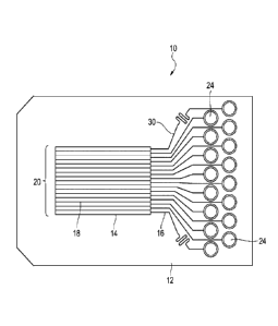
FIG. 1 is a top view of a schematic depicting a biomimetic array device with
microchannels that are configured to transport fluid through microchannels to
microchambers.
FIG. 2 is a top elevational view of the biomimetic array device of FIG. 1
showing
wells with ports for conveying fluid into inlet channels in the inlet region
of biomimetic
array device.
CA 03124645 2021-06-21
WO 2020/132516 PCT/US2019/067969
FIG. 3 is a bottom elevational view of the biomimetic array device of FIG. 1
showing
fluid paths through wells, inlet channels, microchannels, and microchambers.
FIG. 4 is a side perspective view of the biomimetic array device of FIG. 1
showing
the shape of the cassette, with lower sidewalls along microchambers and longer
sidewalls
along microchannels.
FIG. 5 is a perspective enhanced view of the wells of the biomimetic array
device of
FIG. 1 showing embodiments where spacing structures alter fluid height within
wells.
FIG. 6 is a top view of the terminus of microchannels and microchambers of an
embodiment of the biomimetic array device of FIG. 1, where no adjacent
microchannels or
microchambers terminate at the same length.
FIG. 7 is a sectional view of microchannels and microchambers within the
cassette
of the biomimetic array device of FIG. 1, which, in certain embodiments, has
no adjacent
microchannels or microchambers extending to the same depth within the
cassette.
FIG. 8 is a sectional view of microchambers and a biological sample placed on
the
biomimetic array device of FIG. 1, where fluids interact with the biological
sample at the
top interface of each microchamber.
FIG. 9 is a sectional view of microchambers and a biological sample placed on
the
biomimetic array device of FIG. 1, where different fluids interacting with the
biological
sample result in different cell response and viabilities within the biological
sample.
Detailed Description
The present invention is generally directed to a biomimetic array device 10
and
methods of using same. Biomimetic array device has an inlet region 12 for
receiving fluid
and a cassette 14 for transporting fluid through microchannels 16 to
microchambers 18
within a microchamber array 20. A biological sample 22, when placed above a
microchamber array 20, is thus exposed to various fluids present in the
microchambers 18
that make up the microchamber array 20. When biological sample 22 is a tumor
tissue
sample from a patient, the result of exposure to various fluids is assessed by
observing cell
viability within exposed regions of the tumor tissue sample. In these
instances, fluids may
be therapeutic drug candidates. Thus, multiple therapeutic treatments for an
individual
patient may be assessed simultaneously in a biomimetic, in vitro setting, as
opposed to
conventional in vivo therapeutic regimen assessments, where treatments are
conducted on
the patient iteratively until an appropriate regimen is identified.
6
CA 03124645 2021-06-21
WO 2020/132516 PCT/US2019/067969
As used herein, the singular forms "a," "an," and "the" include plural
referents unless
the context clearly indicates otherwise.
As used herein, ranges can be expressed as from "about" one particular value,
and/or
to "about" another particular value. When such a range is expressed, an
embodiment
includes from the one particular value and/or to the other particular value.
Similarly, when
values are expressed as approximations, by the use of "about," it will be
understood that the
particular value forms another embodiment. It will be understood that the
endpoints of each
of the ranges are significant both in relation to the other endpoint and
independently of the
other endpoint. It will also be understood that there are a number of values
disclosed herein,
and that each value is also disclosed herein as "about" that particular value
in addition to the
value itself For example, if the value "50" is disclosed, then "about 50" is
also disclosed.
As used herein, the term "biological sample" refers to biological samples
known in
the art including, but not limited to, tissues, cells, proteins, and lipids.
Biological samples
may be native modified, or engineered, and include non-mammalian and mammalian
samples, including human samples.
As used herein, the terms "patient" or "subject" include any mammal, including
humans.
As used herein, the term "pharmaceutical" refers to articles intended for use
in the
diagnosis, cure, treatment, mitigation, or prevention of disease or biological
disorders.
Referring to FIG. 1, there biomimetic array device 10 is depicted with its
inlet region
12 for fluid application and cassette 14 for sample evaluation. Biomimetic
array device 10
is of unitary construction in the instances depicted, though components or
features are
potentially manufactured separately and attached in embodiments not shown. The
material
used for the manufacture of biomimetic array device 10 is hydrophilic, and
includes
materials such as polylactic acid, acrylonitrile butadiene styrene,
polyethylene terephthalate,
polycarbonate, and nylon, though other materials are contemplated for use. In
some
instances, the material is polypropylene. To manufacture the biomimetic array
device 10
depicted in FIG. 2, additive manufacturing techniques or injection molding are
used.
However, various manufacturing techniques and combinations of manufacturing
techniques
known in the art are suitable for the manufacture of biomimetic array device
10. Similarly,
materials used in the manufacture of biomimetic array device 10 vary based on
the choice
of manufacturing technique. Generally, a 3D model of the intended biomimetic
array device
is first produced and used to direct the accurate manufacture of biomimetic
array device
7
CA 03124645 2021-06-21
WO 2020/132516 PCT/US2019/067969
10, including the desired dimensions and features. Biomimetic array device 10
is, in some
instances, coated in regions with a hydrophobic material. The regions of
coating include
upper edges of cassette 14 of biomimetic array device 10, so that fluids
within cassette 14
are not inadvertently spilled or otherwise inadvertently wet tops of adjacent
walls under
normal operation or agitation of biomimetic array device 10.
The dimensions of biomimetic array device 10 vary based on the number of
fluids
to be introduced in inlet region 12 and the number of microchamber arrays 20
included in
cassette 14. The depth of cassette 14 is variable based on application, and
allows fluid in
microchambers 18 to interact with any biological sample 22 positioned on
cassette 14. In
some instances, the depth of cassette 14 ranges from millimeters to several
centimeters. The
depth of inlet region 12 is about the same as the depth of cassette 14 in some
instances and
is shallower or deeper in other instances. The thickness of cassette 14 is
such that each
microchamber 18 is accommodated with adequately thick walls separating
adjacent
microchannels 16 and microchambers 18 such that the size of biological sample
22 is
minimized. For instance, cassette 14 with microchambers 18 that are about 300
gm thick
and walls between microchambers 18 that are about 300 gm thick would
accommodate these
dimensions in the thickness of cassette 14. The width of inlet region 12 is
greater than the
width of cassette 14 in some instances and is less than or about equal to the
width of cassette
14 in other instances. The length of cassette 14 is such that a desired number
of
microchamber arrays 20 are accommodated, along with microchannels 16 for fluid
transport. In some instances, the length of cassette 14 ranges from several
centimeters to
several decimeters, though other lengths are possible. The length of inlet
region 12 is less
than the length of cassette 14 in some instances and is greater than or about
equal to the
length of cassette 14 in other instances.
As shown in FIG. 2, inlet region 12 of biomimetic array device 10 includes at
least
one well 24 to collect deposited fluid and direct the fluid to other regions
within biomimetic
array device 10. When a plurality of wells 24 are included, biomimetic array
device 10 is
configured to administer various fluids to biological sample 22. In other
instances where a
plurality of wells 24 are included, biomimetic array device 10 is configured
to administer at
least one fluid to biological sample 22 in at least duplicate, allowing for
statistical analysis
of sample-fluid interactions. Wells 24 are built into biomimetic array device
10 and are
positioned in columns, rows, or other configurations. In some embodiments,
wells 24 are
spaced in each row such that commercial multi-channel pipettors are capable of
dispensing
8
CA 03124645 2021-06-21
WO 2020/132516 PCT/US2019/067969
fluid into each well 24 in one row at once. The number and shape of wells 24
is variable,
though approximately equally-shaped wells 24 are used when biomimetic array
device 10
is configured for use with a commercial multi-channel pipettors. The number of
wells 24 in
any row, in instances where a commercial multi-channel pipettors is used, also
depends on
the number of channels of the commercial multi-channel pipettors. For
instance, eight wells
24 are in one row, as depicted in FIG. 2, though other numbers of wells 24 or
rows of wells
24 are contemplated. Further, rows and/or columns have varying numbers of
wells within
one biomimetic array device 10 in some embodiments. The shape of wells 24 is
shown to
be approximately a circle as depicted in FIGS. 1 and 2, though other shapes,
such as an oval,
quadrilateral, or rounded quadrilateral are contemplated. Further, while the
height and
circumference of wells 24 in FIG. 2 are approximately equal, wells 24 within
one inlet
region 12 have varying dimensions in other embodiments not shown.
Referring to FIG. 2, each well 24 has a base 26 with a port 28 that spans base
26 to
allow well 24 to be in fluid communication with an inlet channel 30. As shown
in FIG. 2,
each base 26 has the same cross sectional area as a top opening 32 of each
well, though in
embodiments not depicted the cross sectional areas of each base 26 and each
top opening
32 differs. Fluid is administered to wells 24 though top openings 32 of wells
24 and collects
within the interior of wells 24 defined by well sidewalls and base 26. Due to
the hydrophilic
material used in manufacturing each well 24 and gravitational forces, fluid
flows toward
base 26 after it is administered into well 24. Port 28 is depicted as circular
in cross section
in FIG. 2, though other shapes, such as a quadrilateral, an oval, or a rounded
quadrilateral,
are possible. Port 28 has a largest dimension ranging from several micrometers
to several
millimeters. Fluid within well 24 enters port 28 without the aid of external
pressure sources,
such as a pump, due to hydrophilic material properties, capillary forces,
gravitational forces,
and biomimetic array device 10 geometry.
Referring now to FIG. 3, the fluid paths within inlet region 12 and cassette
14 are
shown from a bottom view. In inlet region 12, each well 24 is in fluid
communication with
an intake region 34 of one inlet channel 30. Similarly, each intake region 34
of one inlet
channel 30 is in fluid communication with one well 24. Thus, fluid that enters
top opening
32 of well 24 is transported to intake region 34 of inlet channel 30 through
port 28. Inlet
channel 30 conveys fluid within inlet region 12 of biomimetic array device 10
and no portion
of inlet channel 30 is open to the external environment in the embodiment
depicted.
However, in embodiments not depicted, inlet channel 30 is potentially open to
the external
9
CA 03124645 2021-06-21
WO 2020/132516 PCT/US2019/067969
environment in at least one portion of inlet channel 30. The cross section of
inlet channel
30 is substantially equal to the cross section of port 28 in the embodiments
depicted, though
in other embodiments the cross section of inlet channel 30 differs from that
of port 28. The
cross sectional dimensions of inlet channel 30 remain substantially consistent
throughout
the length of inlet channel 30 in the embodiments depicted, though in other
embodiments
not shown the cross sectional dimensions of inlet channel 30 increase,
decrease, or otherwise
vary along the length of inlet channel 30. Additionally, cross sectional
dimensions of all
inlet channels 30 are the same in some instances, such as that depicted, or
vary with
individual inlet channels 30 in other instances. After fluid enters inlet
channel 30 in intake
region 34, it flows through a transport region 36 of inlet channel 30. In some
embodiments
that are not shown, inlet channels 30 form branches within their transport
region 36, such
that fluid from one well 24 is divided evenly to each branch. In the
embodiment shown in
FIGS. 2-3, inlet region 12 is wider than cassette 14, and inlet channels 30
transport fluid
from each well 24 to a region where inlet channels 30 meet microchannels 16.
Thus, some
inlet channels 30 are longer than other inlet channels 30 based on the
geometry of
biomimetic array device 10. Adjustments to biomimetic array device 10 that
allow the
transport of equal volumes of fluid and equal exposure of biological sample 22
to the fluid
are discussed below.
Fluid enters microchannels 16 of cassette 14 from transport region 36 of inlet
channels 30. In instances where inlet channels 30 branch, each branch meets
one
microchannel 16. In unbranched inlet regions, each inlet channel 30 transports
fluid to one
microchannel 16 and each microchannel 16 is in fluid communication with one
inlet channel
30.
Referring to FIG. 4, cassette 14 has microchannels 16 that are configured to
transport
fluid to microchambers 18 within a microchamber array 20. While one
microchamber array
20 is depicted in FIG. 4, two or more microchamber arrays 20 are possible and
are connected
either in parallel, in series, or in both parallel and series. When cassette
14 is viewed from
a side as in FIG. 4, microchamber arrays 20 are shown to have shorter
sidewalls than areas
that comprise only microchannels 16. Thus, cassette 14 has a U-shaped or
stepped shape in
profile, where the valley of each U-shape or lower step indicates the location
of one
microchamber array 20. This lower sidewall region allows for biological sample
22 to be
placed at a top interface 38 of microchambers 18 within one microchamber array
20.
Additionally, the U-shape or stepped shape reduces or substantially prevents
fluid from
CA 03124645 2021-06-21
WO 2020/132516 PCT/US2019/067969
spilling from cassette 14 during normal operation or during agitation of
biomimetic array
device 10.
Microchannels 16 branch before reaching a microchamber 18 in instances not
depicted, or do not branch as shown in FIGS. 1-2. Microchannels 16 are
substantially linear
in instances where there is one microchamber array 20 as depicted or where
microchamber
arrays 20 are arranged in series. In other instances, such as when
microchamber arrays 20
are arranged in parallel, microchannels 16 are not linear. As shown in FIG. 4,
microchannels
16 are in fluid communication with microchambers 18, and one microchannel 16
is in fluid
communication with one microchamber 18. Microchannels 16 extend from a
downstream
end of microchambers 18 in embodiments such as those depicted, though
microchannels 16
terminate after a final microchamber array 20 in embodiments not shown. The
combined
length of microchambers 18 and microchannels 16 is equal in some instances or
varies in
other instances, which are discussed below. Within cassette 14, microchannels
16 and
microchambers 18 have substantially equal widths, which may be, for example,
300 gm.
The cross sections of microchannels 16 and microchambers 18 differs from that
of inlet
channels 30 in the instance depicted in FIG. 4, though these cross sections
are substantially
equal in other instances. Cross sections of microchannels 16 and microchambers
18 are, for
example, that of a U-shape or a quadrilateral with its top side removed. The
depth of
microchannels 16 and microchambers 18 is discussed below in greater detail,
though all
depths are equal in some instances and vary in others. Microchannels 16 and
microchambers
18 are open to the external environment at their top interface 38, which is
opposite their
base. Sidewalls dividing adjacent microchannels 16 and microchambers 18 have
the same
thickness as each microchannel 16 or microchamber 16 in FIG. 3, though
sidewall
thicknesses are potentially greater than or less than the thicknesses of
microchannels 16 and
microchambers 18. Microchannels 16 and microchambers 18 are equally spaced
within
cassette 14 in the embodiments depicted, though spacing may vary in other
embodiments.
Referring back to FIG. 4, microchambers 18 include top interface 38 that
provides a
region for the interaction of fluid with any biological sample 22 placed at
top interface 38.
Top interface 38 is depicted as being open to the external environment, though
in
embodiments not depicted top interface 38 includes a permeable or semi-
permeable
interface between microchambers 18 and biological sample 22. In some
instances,
biological sample 22 exposure to and interaction with fluids occurs
immediately when fluid
11
CA 03124645 2021-06-21
WO 2020/132516 PCT/US2019/067969
fills microchamber 18 and in other cases top interface 38 provides a delayed
release of fluids
or components of fluids from microchamber 18 to biological sample 22.
In some embodiments, microchannels 16, inlet channels 30, and/or microchambers
18 include agitation structures 42 to aid in the mixing of fluid components as
then travel
through microchannels 16, inlet channels 30, and/or microchambers 18.
Agitation structures
42 are semi-circular, rod-shaped, branched rods, spherical, or any other
structure that
extends from an interior surface of microchannels 16, inlet channels 30,
and/or
microchambers 18 and mixes fluid as fluid moves past it. Agitation structures
42 are
attached or built into the interior walls and/or base of microchannels 16,
inlet channels 30,
and/or microchambers 18 and extend from these interior surfaces into the fluid
path within
microchannels 16, inlet channels 30, and/or microchambers 18, such that
movement of fluid
past agitation structures 42 induces at least some degree of turbulent flow,
mixing the fluid.
In some instances, agitation structures 42 are aided in their mixing function
by movement
or agitation of biomimetic array device 10. For example, biomimetic agitation
device 10 is
placed on a commercial rocker, shaker or other vibrational or agitation
equipment to provide
agitation or motion.
Referring to FIG. 5, some embodiments include wells 24 with spacing structures
40.
Spacing structures 40 are included within wells 24 to alter the height of
fluid from base 26.
Thus, equal volumes of fluid deposited in multiple wells 24 containing spacing
structures
40 of various sizes results in various fluid heights. As such, in some
embodiments
biomimetic array device 10 is configured such that wells 24 in fluid
communication with
longer inlet channels 30 have larger spacing structures 40 than wells 24 in
fluid
communication with shorter inlet channels 30. In sizing spacing structures 40
according to
inlet channel 30 length, equal volumes of fluid applied to wells 24 result in
equal volumes
of fluid reaching biological sample 22 and equal exposure of fluid from
microchambers 18
to biological sample 22. Spacing structures 40 are positioned such that port
28 is not blocked
or impeded and such that deposited fluid reaches and is transported through
port 28. The
cross sectional shape of spacing structure 40 parallel to base 26 is a circle,
semi-circle,
quadrilateral, oval, rounded quadrilateral, or any other cross sectional shape
that fits within
well 24 without blocking or impeding fluid access to port 28. Spacing
structure 40 is shown
in FIG. 5 to conform to at least some regions of the sidewall of well 24,
though in
embodiments not shown spacing structure 24 does not conform to any sidewall of
well 24.
Extending from base 26, spacing structure 40 reaches a height within well 24
that is less
12
CA 03124645 2021-06-21
WO 2020/132516 PCT/US2019/067969
than or equal to the height of the sidewall of well 24. Sizes of spacing
structures 40 are
varied by altering dimensions of spacing structures 40, such as the height and
cross sectional
area parallel to base 26. In some cases, some wells 24 have spacing structures
40 while other
wells do not. In other cases, all wells 24 or no wells 24 have spacing
structures 40. In some
embodiments, the presence, absence, and size of spacing structures 40 within
wells 24 is
determined by biomimetic array device 10 geometry, so that wells 24 nearest
cassette 14
with shorter inlet channels 30 that reach cassette 14 have no spacing
structure 40 the smallest
spacing structures 40. Similarly, wells 24 farthest from cassette 14 with
longer inlet channels
30 that reach cassette 14 have the largest spacing structures. In embodiments
where wells
24 position relative to cassette 14 does not correlate with inlet channel 30
length, inlet
channel length 30 determines the size and presence or absence of spacing
structures 40, with
wells 24 in fluid communication with longer inlet channels 30 having larger
spacing
structures 40. Larger spacing structures 40 encompass larger volumes of the
interior of wells
24 than smaller spacing structures 40, so that equal volumes of fluid applied
to wells 24 will
have a greater height from base 26 in wells with larger spacing structures 40
than in wells
with no or smaller spacing structures 40. In some embodiments not depicted,
more than one
spacing structure 40 is present in at least one well 24, allowing that port 28
is not blocked
or impeded from fluid transport. Spacing structures 40 are sized and shaped
such that they
do not impede a pipette or other fluid depositing means from providing fluid
to well 24. As
depicted in an embodiment in FIG. 5, spacing structures 40 are built into well
24 and are of
unitary construction with well 24, while in other embodiments that are not
depicted, spacing
structures are formed separately from well 24 and attached by attachment means
known in
the art. In these embodiments, spacing structures 40 are made from the same
material that
well 24 is composed of, or are made from a different material. Spacing
structures 40 are
hollow, partially hollow, or solid, but are substantially impermeable to
fluid. While the use
of spacing structures 40 allows equal volumes to be dispensed and transported
to biological
sample 22 with approximately equal exposure of biological sample 22 to each
fluid, some
embodiments without spacing structures 40 instead use the dispensation of
unequal volumes
of fluid to wells 24 based on inlet channel 30 distance to cassette 14 to
achieve this same
result. These unequal volumes are calculated prior to dispensation, though the
use of spacing
structures 40 simplifies this process for a user by eliminating these
calculations.
Referring now to FIG. 6, in some embodiments, microchannels 16 and
microchambers 18 are produced so that they terminate at various points across
the length of
13
CA 03124645 2021-06-21
WO 2020/132516 PCT/US2019/067969
cassette 14. FIG. 6 depicts microchannels 16 that alternate between longer and
shorter
microchannels 16. However, in other embodiments not depicted microchambers 18
alternate
between longer and shorter microchambers 18 as well. The varying lengths are
produced to
prevent leakage from micron-scale holes that may form at the intersection of
bases and
sidewalls of microchannels 16 and microchambers 18 during manufacture. By
ensuring that
no adjacent microchannels 16 or microchambers 18 share a common terminal
sidewall,
leaks between adjacent microchannels 16 or microchambers 18 are substantially
prevented.
FIG. 6 depicts two alternating microchannel lengths, though more than two
lengths of
microchannels 16 or microchambers 18 are present in other embodiments not
shown.
In FIG. 7, a cross section of cassette 14 is shown with microchannels 16 of
varying
depths within cassette 14. In cross sections similar to that of FIG. 7 that
are not shown,
microchambers 18 also have varying depths within cassette 14. Similar to that
described
above, micron-scale holes may form at the intersection of bases and sidewalls
of
microchannels 16 and microchambers 18 during manufacture. Thus, in some
embodiments,
no adjacent microchannels 16 or microchambers 18 have the same depth to
substantially
prevent leakage between microchannels 16 and microchambers 18 from any micron-
sized
holes. While two depths of microchannels 16 and microchambers 18 are depicted,
more than
two depths of microchannels 16 and microchambers 18 are possible in
embodiments not
shown.
In some embodiments where microchannel and microchamber lengths are varied,
microchannel and microchamber depths are also varied with microchannel and
microchamber widths remaining equal. In these embodiments, equal fluid volume
capacity
is maintained in each microchannel 16 and microchamber 18 by having longer
microchannels 16 and microchambers 18 be relatively shallower and by having
shorter
microchannels 16 and microchambers 18 be relatively deeper. Thus, equal
volumes of fluid
reach microchambers 18 and any biological sample 22 at top interface 38 of
microchambers
18. In other embodiments where microchannel or microchamber length and depths
are
varied, fluid volume capacity within each microchannel 16 or microchamber 18
is not equal.
FIGS. 8 and 9 depict a cross section of a top region of cassette 14, where
fluids
interact with biological sample 22 at top interface 38 of each microchamber
18. Different
fluids are depicted and cells within biological sample 22 are exposed to these
fluids in FIG.
8. Examples of fluids include culture media, wash solutions, labeling
solutions,
pharmaceutical compounds, therapeutic agents, analytes, or other solutions for
interaction
14
CA 03124645 2021-06-21
WO 2020/132516 PCT/US2019/067969
with biological samples 22. When an array of therapeutic agents is utilized,
multiplexed
testing of the efficiency of therapy regimens is possible using a patient's
biological sample
22, allowing fast and low cost identification of promising therapies. When an
array of
pharmaceutical agents is utilized, it is possible to test the effectiveness of
several candidate
compounds or molecules simultaneously, aiding in selection of the most
effective
compounds. When particular fluids are applied in duplicate or greater,
statistical analysis of
results is possible. Similarly, when a portion of fluids in an array are
healthy culture media
and another portion of fluids in the array are analytes, such as therapeutic
agents or
phalmaceutical compounds, the healthy culture media serves as a control for
normal cell
morphology, phenotype, and viability from which comparisons with analytes are
made.
In FIG. 9, fluids from microchambers 18 have been incubated on regions of
biological sample 22 and these regions can be analyzed with respect to cell
viability,
phenotype, response, and morphology to determine the effects of the fluids.
For instance,
healthy media incubation results in a standard, expected cell morphology and
proliferation
profile, with cell counts that may serve as a baseline or control for other
analytes. Thus,
analytes that result in lower cell counts or abnormal cell morphologies,
apoptotic indices or
necrotic indices, or staining patterns are identified relative to controls.
Cell morphologies
for dead or unhealthy cells are bloated or exploded for necrotic cell death or
have blebbing
for apoptotic cell death. When the analyte is taken up by the region of
biological sample 22
at top interface 38 of microchamber 18 that provides the analyte and apoptosis
is observed,
cells are determined to have been induced by analyte to program or initiate
their own death.
Necrosis indicates, in some instances, that cells did not take up the analyte
or that cells
lacked essential nutrients or growth conditions. Staining techniques, such as
those using
fluorescent molecules, are used to indicate live or dead cells in some
instances. For example,
propidium iodide and/or Annexin V provide visible information regarding cell
viability.
In order to observe regions of biological sample 22 and the cells within
biological
sample 22, imaging techniques are used. Generally, microscopy is used to
observe
biological sample 22 at a cellular level, with confocal microscopy providing
images of
biological tissue 22 in one or more z-planes. Thus, confocal microscopy allows
analysis of
cells within biological sample 22 not only on the surfaces of biological
sample 22, but within
biological sample 22. Fluorescence channels are viewed using a microscope to
observe
staining patterns, where different channels are available to view different
fluorophores.
CA 03124645 2021-06-21
WO 2020/132516 PCT/US2019/067969
In some instances, fluid from each microchamber 18 in microchamber array 20 is
analyzed after fluid has been in contact with biological sample 22. Fluid
analysis is
conducted using methods such as enzyme-linked immunosorbent assay (ELISA),
enzyme-
linked immunospot (ELISPOT), and Western blotting, to identify and/or quantify
fluid
components, such as antibodies or proteins, which may vary based on fluid
identity and
biological sample exposure. In other instances, DNA or RNA is detected in
fluid in
microchambers 18 after the fluid has been in contact with biological sample
22. Techniques
such as gel electrophoresis, Northern blotting, Southern blotting, or
polymerase chain
reaction and sequencing may be used to detect and identify nucleic acids.
Other techniques
and methods not specifically described above for the detection,
identification, and/or
quantification of nucleic acids, proteins, antibodies, or other cellular
components are
contemplated for the present disclosure.
Biological sample 22 is includes any tissue, cell, protein, lipid, or other
biological
material. Biological samples 22 are, in some instances, freshly provided from
a patient or
subject or frozen samples that were originally provided by a patient or
subject. Biological
samples 22 further are natural, modified, or at least partially engineered
materials. In some
cases, biological sample 22 includes or is composed of unexpanded cells, while
in other
cases some or all cells of biological sample 22 are expanded. Sample washing
and
preservation techniques aye used in the preparation and storage of biological
sample 22 in
some cases, while in other cases biological sample 22 is not exposed to
preservation
materials, growth factors, or other added solution components. In some
embodiments,
biological sample 22 is a tissue slice culture from a tumor biopsy of a
patient. Fresh or flash-
frozen tumor biopsy specimens are compatible with the present invention. In
other
embodiments, biological sample 22 includes biological components that are
adsorbed,
bonded, or grown on a scaffold, including an elastomer spun scaffold,
according to methods
known in the art. Biological sample 22 is typically less than about 500 gm in
thickness,
though thicknesses ranging from about 100 gm to about 1 mm are contemplated as
compatible with the present disclosure. Biological sample 22 is laid
transversely across one
microchamber array 20 so that each microchamber 18 within microchamber array
20 is
positioned beneath biological sample 22. The positioning of biological sample
22 allows
fluid to be wicked, and often the top surface of biological sample 22 that
faces away from
top interface 38 is less saturated than the surface of biological sample 22
that is in contact
16
CA 03124645 2021-06-21
WO 2020/132516 PCT/US2019/067969
with top interface 38, forming a concentration gradient so that wicking of
fluid from
microchannels is possible.
In one example, biological sample 22 is a fresh tumor tissue sample collected
by
core needle biopsy using a 14-gauge needle. A tissue slice culture that is
about 300 pm thick
is prepared from the core specimen and placed on a porous polymer membrane
culture
insert. Biological sample 22 is then acclimated with pre-warmed medium and
transferred
onto a sterilized biomimetic array device 10 at top interfaces 38 of
microchambers 28 within
one microchamber array 20. Sterilization of biomimetic array device 10 is
undertaken using
methods known in the art, including, but not limited to UV radiation and
application of an
ethanol solution. The position of biological sample 22 is such that the basal
surface of
biological sample 22 is in direct or approximately direct conformal contact
with top
interface 38 of microchambers 18, while the apical surface is facing upwards
to the external
environment. Fluid is provided through microchannels 16 to microchambers 18
and
biomimetic array device 10 is placed in a tissue culture incubator at
approximately 37 C
with 5% CO2. Incubation occurs over time periods ranging from several minutes
to several
days, or, as in this example, from 3 to 14 days. Alternatively, in some
instances fluid is
instead applied and incubated in different temperature and culture conditions.
Returning to
the example, cell viability and morphology within areas of biological sample
22 is
investigated. Viability and proliferation are examined using staining
techniques, such as
staining biological sample 22 with propidium iodide and Annexin V, flowed by
imaging of
cells within biological sample 22 using a microscope, such as a confocal
microscope.
Several z-planes within biological sample 22 are assessed so that viability
and proliferation
can be assessed, such as through visualizing staining patterns, determining
cell numbers
through cell counts, or observing cell morphology. In another example, the
above procedure
is followed using a tissue slice culture prepared from a larger, bulk specimen
instead of a
core needle biopsy.
In yet another example, the tumor biopsy is first flash frozen prior to
analysis. In this
case, either core needle biopsy or larger specimen samples are placed in
sterile cryotubes or
other sterile containers fit for frozen storage. The storage containers also
include 95% tissue
culture medium or fetal bovine serum with 5% dimethyl sulfoxide. Each sample
is treated
according to established bio-banking protocols and samples storage containers
are placed in
secondary freezing containers with isopropanol and then transferred to a -80 C
freezer for
approximately 24 hours. Following this, samples are transferred to liquid
nitrogen storage
17
CA 03124645 2021-06-21
WO 2020/132516 PCT/US2019/067969
for a duration that ranges from several days to years, or more specific to
this example, about
one to four weeks. After this storage duration, samples are thawed according
to established
bio-banking protocols. After thawing and washing of samples to remove storage
agents,
samples are prepared as fresh biological samples 22 as described above.
In some embodiments, following analysis of biological sample 22 and regions
that
were in contact with various analytes and fluids, information is obtained
regarding the
effectiveness of each fluid. When fluids are potential therapeutic agents,
those therapeutic
agents that effectively lead to tumor cell death are identified as candidates
for administration
to the patient from which the tumor biopsy was provided. Thus, instead of the
time
consuming, expensive, and inefficient typical method of administering a
therapeutic
regimen to the patient, evaluating effectiveness, and switching to a different
therapeutic
regimen if the original regimen is not successful, the present disclosure
provides a method
by which multiple therapeutic agents are tested for effectiveness in killing
tumor cells in a
patient's tumor in a low cost, fast, and efficient manner. The patient is able
to avoid
unnecessary and ineffective therapies and initiate those therapeutic regimens
most likely to
be effective, generally initiating effective therapies earlier than would
occur using the
conventional iterative approach.
Biomimetic array device 10 is configured to transport fluids without the aid
of
external mechanical force provided by, for instance, a pump. Device geometry,
capillary
forces, gravitational force, and material properties instead provide the
ability for fluid to
flow within biomimetic array device 10. However, in embodiments not shown,
external
mechanical devices are used in addition to device properties.
The present invention is capable of supporting various configurations of
microchannels 16, microchambers 18, microchamber arrays 20, and wells 24,
including a-
k-d-n configurations where (a) is any number of microchamber arrays 20, (k) is
any number
of wells 24, (d) is any number of microchambers 18 connected to each well 24
by (n) number
of microchannels 16. These components of the a-k-d-n configurations are
capable of being
connected in parallel, series, or any combination thereof. For instance, a 1-1-
1-1
configuration includes one microchamber array 20 with one well 24 connected to
one
microchamber 18 by a single microchannel 16. In an exemplary 3-16-1-1
waterfall
configuration, three microchamber arrays 20 are provided fluid by 16 wells 24,
each well
24 connected to one microchamber 18 within each microchamber array 20 by one
microchannel 16. Thus, in this exemplary waterfall configuration, there are 16
18
CA 03124645 2021-06-21
WO 2020/132516 PCT/US2019/067969
microchambers 18 within each microchamber array 20. When microchamber arrays
20 are
in a series format, one microchannel 16 connects one microchamber 18 within
one
microchamber array 20 to one microchamber 16 within another microchamber array
20.
As will be understood by those familiar with the art, the present invention
may be
embodied in other specific forms without departing from the spirit or
essential
characteristics thereof The present disclosure may be applied to other fields
with
applications not specifically described herein, such as for drug discovery,
chemical energy
storage exploration, biofabrication, and diagnostic imaging. Accordingly, the
disclosures
and descriptions herein are intended to be illustrative, but not limiting, of
the scope of the
invention which is set forth in the following claims.
19