Note: Descriptions are shown in the official language in which they were submitted.
CA 03124709 2021-06-22
WO 2020/163310 PCT/US2020/016555
Device for Tissue Electrotransfer using a Microelectrode
RELATED APPLICATION
[0001] This application claims the benefit of U.S. Provisional Application
No.
62/800,781, filed on February 4, 2019. The entire teachings of the above
application are
incorporated herein by reference.
BACKGROUND
[0002] Over the last 40 years, electroporation has emerged as an attractive
approach for
delivering exogenous biologic materials (i.e., DNA, RNA, or protein factors)
into cells and
tissues. The first reports of electroporation as a means of cellular delivery
were in the 1980's
(1) with many of the initial transdermal efforts emerging in the early 1990's
(2-7).
Electroporation mediated delivery has been explored for transdermal delivery
of small
molecule therapeutics for systemic absorption into the bloodstream via
electroporation of the
cutaneous layers of the skin followed by passive diffusive, iontophoretic
transport of
therapeutics through the skin (4), or other penetration mechanisms (8),
electrochemotherapy
(ECT) (9-17), and gene electrotransfer (GET) (18,19). With the emergence of
DNA based
vaccines, gene editing techniques (i.e., CRISPR Cas9 targeted gene editing),
and FDA
approval of gene therapy products (i.e., CAR T-cell therapy), there has been a
renewed
interest in GET.
[0003] Electroporation has long been explored as a means of cellular and
tissue delivery
with numerous medical applications (34). During electroporation, cells or
tissues are exposed
to a brief, high strength, electric field that induces pore formation in the
cell membrane
facilitating molecular delivery across the membrane barrier. Reversible
electroporation
allows transient permeabilization of the cell membrane to deliver exogenous
materials into
cells followed by cellular recovery. Irreversible electroporation (both
thermal and non-
thermal), on the other hand, causes irrecoverable membrane damage, either
directly killing
cells or promoting cellular apoptosis. Both reversible and irreversible
electroporation have
been explored medically with reversible electroporation being used for
therapeutic and vector
delivery and irreversible electroporation being used as a tissue ablation
technique, especially
to treat cancerous tumors. Transdermal electroporation has typically been
focused on
- 1 -
CA 03124709 2021-06-22
WO 2020/163310 PCT/US2020/016555
reversible electroporation in order to reduce the permeation barrier of the
skin and promoting
uptake of vectors for gene electrotransfer (GET).
[0004] Transdermal electroporation has been seen as a means for
transforming skin cells
through GET or increasing transdermal permeation of therapeutics through the
skin. In
particular, the use of transdermal electroporation has been explored as a
means to deliver
DNA based vaccine products with sufficient expression and immunogenicity to
confer a
protective immunological response. The epidermis, in particular, is targeted
for vaccination
due to the high concentration of dendritic cells (35,36). As a 2016 editorial
on the clinical
potential of electroporation for gene therapy and DNA based vaccine delivery
noted, there are
several commercial electroporation vaccination platforms in development
including the
CELLECTRA, Easy Vax, MedPulser, Trigrid, Dermavax, OncoSec, and Cliniporator,
with
the majority of devices (62%) undergoing Phase I clinical trials (37). The
most advanced
product is the CELLECTRA system by Inovio which is currently in Phase III
trials for IM
delivery of a human papilloma virus (HPV) vaccine (38).
[0005] Transdermal electroporation has traditionally been applied either as
a surface
electrode or as penetrating electrodes which are inserted into skin or muscle.
When a surface
electrode is used, a high intensity electric field is required to permeabilize
the highly
keratinized stratum corneum (SC); the outmost barrier layer of the skin.
However, once the
SC is electroporated the underlying epidermal and dermal tissues are then
exposed to this
high field intensity in an uncontrolled fashion which can lead to skin
irritation, edema and
damage. When penetrating electrode sets are used they are often spaced several
centimeters
apart and inserted deep into the skin or below the skin into muscle. Higher
intensity pulses
are also often used in order to permeabilize the largest tissue volume
surrounding the
electrodes which also leads to tissue damage around the electrodes where the
field intensities
are highest. Further, DNA vectors used during GET are usually delivered
through an
intradermal (ID) or intramuscular (IM) injection of a few hundred 11.1 to ml
volumes. The
DNA injection itself may lead to transfection efficiency variability where the
injected DNA
may not be localized in the targeted tissues under-going electroporation. For
example, clinical
studies utilizing ID injection have used a minimum of 600 ilgs of DNA plasmid;
and
quantities as large as even several milligrams are often injected to ensure
sufficient cellular
uptake. However, a significant fraction of the solution may be delivered to
the subcutaneous
region, as opposed to the dermal or epidermal layers, leading to a higher cost
vaccine due to
the large amount of DNA required for GET.
- 2 -
CA 03124709 2021-06-22
WO 2020/163310 PCT/US2020/016555
[0006] Despite decades of development and multiple commercial efforts,
transdermal
electroporation still faces many challenges to clinical adoption. The major
bottlenecks to be
addressed are 1) low delivery/transfection efficiencies; 2) limited substances
which can
permeate the skin at therapeutic levels; 3) variability in delivery
efficiencies; 4) skin
irritation, edema and scarring following trans-dermal electroporation. These
impediments and
persistent issues plaguing transdermal electroporation have constrained its
clinical use (39)
and there are currently no FDA approved electroporation devices on the market.
[0007] Although there are numerous advantages of electroporation for use in
a variety of
different settings, such as the advantages of being a minimally invasive and
non-viral cell
transfection technique, the inherent tradeoff between cell/tissue damage and
transfection
efficiency has plagued the use of electroporation as a transdermal drug
delivery platform
(26). Amongst the problems encountered during dermal electroporation, low
delivery
efficiencies coupled with skin irritation and scarring (27, 28) are the most
prominent
obstacles to be addressed. There is, therefore, an ongoing need for improved
techniques of
tissue electrotransfer.
SUMMARY
[0008] An embodiment according to the invention is related to a minimally
invasive
penetrating microelectrode array to be used to generate localized electric
field "hotspots" for
delivering biomolecules, such as nucleic acid or protein molecules, into cells
located in the
epidermal or dermal layer of the skin via transient membrane permeabilization.
The
"hotspots" can be controlled by selectively insulating the penetrating
microelectrodes at
specific regions. The portion of microelectrodes that are not covered with
insulation coating
can be coated with nucleic acid or protein vaccine vector, or other
biomolecules to be
delivered. Upon insertion into the skin, an anchor microelectrode region
mechanically
anchors the penetrating microelectrode to position the target tissue
microelectrode region, so
as to selectively align the biomolecule coating with cells located in the
tissue location. The
biomolecule coating will dissolve when in contact with surrounding tissue. By
applying an
electrical pulse, the biomolecules can be delivered into surrounding cells.
[0009] In one embodiment according to the invention, a microelectrode
device for tissue
electrotransfer comprises a penetrating microelectrode. The penetrating
microelectrode
comprises (i) a target tissue microelectrode region comprising an electrically
conductive
surface to selectively deliver, via tissue electrotransfer, a biomolecule to
cells located in a
- 3 -
CA 03124709 2021-06-22
WO 2020/163310 PCT/US2020/016555
tissue location; and (ii) an anchor microelectrode region to mechanically
anchor the
penetrating microelectrode to position the target tissue microelectrode region
to selectively
deliver the biomolecule to cells located in the tissue location. An electrical
connection
connects the penetrating microelectrode to a voltage source.
[0010] In further, related embodiments, the anchor microelectrode region
may be at or
near a distal end of the penetrating microelectrode. The microelectrode device
may comprise
electrical insulation on a surface of the penetrating microelectrode, distinct
from the
electrically conductive surface of the target tissue microelectrode region.
The microelectrode
device may comprise electrical insulation on a surface of the anchor
microelectrode region.
The microelectrode device may comprise a biomolecule coating, comprising the
biomolecule
to be selectively delivered, on at least part of a surface of the target
tissue microelectrode
region. The tissue location may be below a stratum corneum layer of skin and
at least one of
(i) within at least part of an epidermal layer of skin and (ii) within at
least part of the dermal
layer of skin. The tissue location may be within at least a part of only an
epidermal layer of
skin. The anchor microelectrode region may comprise a barb; and may comprise
an adhesion
surface coating. The biomolecule coating may be dissolvable when surrounded by
skin tissue;
and may comprise at least one of a nucleic acid and a protein.
[0011] In other, related embodiments, the electrical insulation may
comprise an insulating
polymer deposited on the penetrating microelectrode. The microelectrode device
may
comprise more than one of the penetrating microelectrode, in which a center-to-
center
spacing of the more than one of the penetrating microelectrode comprises a
spacing between
about 300 micrometers and about 1.5 millimeters. The length of the penetrating
microelectrode may comprise a length between about 225 micrometers and about
1250
micrometers. The penetrating microelectrode may comprise at least one of: a
needle
comprising a tapered tip; and a needle comprising a lateral protrusion. The
penetrating
microelectrode may comprise a diameter between about 100 micrometers and about
500
micrometers. The electrical connection may apply a pulsed voltage from the
voltage source to
the penetrating microelectrode to create a transient permeabilization of a
cell membrane of
tissue in at least one of an epidermal layer of skin and a dermal layer of
skin. The electrical
connection may apply a voltage from the voltage source to the penetrating
microelectrode to
create a maximum electric field strength of between about 0.1 kilovolts (kV)
per centimeter
and about 10 kilovolts (kV) per centimeter in skin tissue surrounding the
penetrating
microelectrode. The electrical connection may comprise a connection defined by
- 4 -
CA 03124709 2021-06-22
WO 2020/163310 PCT/US2020/016555
photolithography, the penetrating microelectrode may comprise an electrode
base defined by
photolithography, and the penetrating microelectrode may comprise
electroplated metal. The
microelectrode device may comprise more than one of the penetrating
microelectrode, and
the electrical connection may comprise an electrically independent connection
to two or more
of the more than one penetrating microelectrodes. The device may comprise more
than one
target tissue microelectrode region each to selectively deliver a different
biomolecule.
[0012] In further, related embodiments, the microelectrode device may
further comprise a
modeling processor, which may comprise a tissue level electric field
prediction module and a
cellular level simulation module. The modeling processor may be configured to
determine
tissue locations to which to selectively deliver, via tissue electrotransfer,
a biomolecule to
cells located in the tissue location. The modeling processor may be configured
to determine a
control voltage delivered by the voltage source to the penetrating
microelectrode.
[0013] In another embodiment according to the invention, a method of
performing tissue
electrotransfer with a penetrating microelectrode comprises anchoring a
penetrating
microelectrode, using an anchor microelectrode region of the penetrating
microelectrode,
such that a target tissue microelectrode region of the penetrating
microelectrode, comprising
an electrically conductive surface, is positioned to selectively deliver a
biomolecule to cells
located in a tissue location; and applying a voltage to the penetrating
microelectrode to
deliver the biomolecule to the cells located in the tissue location. The
penetrating
microelectrode may comprise any of the microelectrode devices taught herein.
[0014] In further, related embodiments, the method may further comprise,
using a
modeling processor, predicting a tissue level electric field and performing a
cellular level
simulation of the tissue location. The modeling processor may determine tissue
locations to
which to selectively deliver, via tissue electrotransfer, a biomolecule to
cells located in the
tissue location. The method may comprise using a modeling processor to control
a voltage
delivered by the voltage source to the penetrating microelectrode.
BRIEF DESCRIPTION OF THE DRAWINGS
[0015] The patent or application file contains at least one drawing
executed in color.
Copies of this patent or patent application publication with color drawings
will be provided
by the Office upon request and payment of the necessary fee.
[0016] The foregoing will be apparent from the following more particular
description of
example embodiments, as illustrated in the accompanying drawings in which like
reference
- 5 -
CA 03124709 2021-06-22
WO 2020/163310 PCT/US2020/016555
characters refer to the same parts throughout the different views. The
drawings are not
necessarily to scale, emphasis instead being placed upon illustrating
embodiments.
[0017] FIG. 1 is a schematic diagram of a microelectrode device for tissue
electrotransfer,
in accordance with an embodiment of the invention.
[0018] FIG. 2 is a schematic diagram of a microelectrode device for tissue
electrotransfer,
including alternatives for an anchor microelectrode region, in accordance with
an
embodiment of the invention.
[0019] FIG. 3 is a schematic diagram of a microelectrode device for tissue
electrotransfer,
including example dimensions of penetrating microelectrodes, in accordance
with an
embodiment of the invention.
[0020] FIG. 4 is a graph illustrating pulsed voltages applied from a
voltage source to a
penetrating microelectrode to create a transient permeabilization of a cell
membrane of tissue
in at least one of an epidermal layer of skin and a dermal layer of skin, in
accordance with an
embodiment of the invention.
[0021] FIG. 5 is a diagram illustrating selective electrical insulation of
penetrating
microelectrodes on a base and a tip of the microelectrode, and showing the
results of
simulations of localized electric field strengths produced by the
microelectrodes for targeted
tissue electrotransfer, in accordance with an embodiment of the invention.
[0022] FIG. 6 is a diagram showing images of Green Fluorescent Protein
(GFP)
expression in porcine skins following intradermal injection and application of
direct current
pulses, in experiments in accordance with an embodiment of the invention.
[0023] FIG. 7 is a diagram showing a photograph and a schematic diagram of
a
penetrating microelectrode array, in accordance with an embodiment of the
invention.
[0024] FIG. 8 is a diagram showing views of the stratum corneum, epidermis
and dermis
layers of skin, and of penetrating microelectrodes inserted in those layers,
in accordance with
an embodiment of the invention.
[0025] FIG. 9 is a diagram showing the results of simulation of localized
electric field
strengths surrounding an array of sixteen penetrating microelectrodes inserted
into a skin
model, in a simulation in accordance with an embodiment of the invention.
[0026] FIG. 10 is a diagram showing, in a simulation in accordance with an
embodiment
of the invention, a packed cell model 1056 of epidermal keratinocytes at an
applied field of
2.0 kV/cm; a 2D projection 1058 of the packed cell model at a field of (Top)
0.83 kV/cm
showing a partially electroporated state and (bottom) 1.0 kV/cm showing a
fully
- 6 -
CA 03124709 2021-06-22
WO 2020/163310 PCT/US2020/016555
electroporated state; and an image 1060 of (Top) transmembrane potential and
(Bottom)
electric field within simulated dermal fibroblasts showing orientation
dependent
electroporation.
[0027] FIG. 11 is a graph showing a change of tissue impedance in
measurements before
and after application of a pulsed electroporation voltage to a penetrating
microelectrode, in an
experiment in accordance with an embodiment of the invention.
[0028] FIG. 12 is a schematic diagram of an electrical system schematic
used with a
penetrating microelectrode array, in an experiment in accordance with an
embodiment of the
invention.
[0029] FIG. 13 is a diagram showing images of the results of
electropermeabilization
testing with rhodamine, in an experiment in accordance with an embodiment of
the invention.
[0030] FIG. 14 is a summary of a protocol used for a permeabilization test
with
propidium iodide, in an experiment in accordance with an embodiment of the
invention.
[0031] FIG. 15 is a diagram showing images of sectioned skin image
examples, in the
experiment of FIG. 14.
[0032] FIG. 16 is a diagram showing image analysis results in the
experiment of FIG. 14.
[0033] FIG. 17 is a schematic diagram of an electrode configuration of a
penetrating
microelectrode array in which multiple electrodes are addressed as a group, in
accordance
with an embodiment of the invention.
[0034] FIG. 18 is a diagram showing a penetrating microelectrode array
device using a
printed circuit board to provide electrical connections, in accordance with an
embodiment of
the invention.
[0035] FIG. 19 is a diagram showing images of the results of testing a
penetrating
microelectrode array device using a printed circuit board, in an experiment in
accordance
with an embodiment of the invention.
[0036] FIG. 20 is a schematic diagram of penetrating microelectrode arrays
fabricated
using photolithography, in accordance with an embodiment of the invention.
[0037] FIG. 21 is an image of dies on a wafer used for fabricating
penetrating
microelectrode arrays using photolithography, in accordance with an embodiment
of the
invention.
[0038] FIG. 22 is a schematic diagram of a microelectrode device for tissue
electrotransfer that uses multiplexing of more than one target tissue
microelectrode region, in
accordance with an embodiment of the invention.
- 7 -
CA 03124709 2021-06-22
WO 2020/163310 PCT/US2020/016555
[0039] FIG. 23 is an image of DNA composites deposited onto penetrating
microelectrode arrays by electrostatic spray, in accordance with an embodiment
of the
invention.
[0040] FIG. 24 is an image of penetrating microelectrodes insulated with a
conformal
layer of an insulating dielectric coating, in accordance with an embodiment of
the invention.
[0041] FIG. 25 is a schematic diagram of a microelectrode device for tissue
electrotransfer, which incorporates a modeling processor, in accordance with
an embodiment
of the invention.
DETAILED DESCRIPTION
[0042] A description of example embodiments follows.
[0043] An embodiment according to the invention provides a minimally
invasive
penetrating microelectrode array with targeted delivery of biomolecules, such
as nucleic acids
or proteins, to distinct layers of the skin. The epidermis contains a higher
cell density than the
dermis as well as a high concentration of dendritic cells and is therefore an
attractive target
for delivery of biomolecules such as nucleic acid or protein vectors. The
penetrating
microelectrode array can achieve efficient, targeted tissue gene
electrotransfer (GET) specific
to the epidermis or dermis. Epidermal transfection may demonstrate the highest
degree of
transfection due to the higher epidermal cell density. Targeted delivery of
biomolecules, such
as vector delivery and tissue transfection, can be achieved by selectively
insulating the
penetrating microelectrodes of the array and coating them with plasmid DNA
(pDNA)
vectors (or other biomolecules, such as nucleic acids or proteins) followed by
efficiently
electroporating the tissue at focused electric field "hotspots" which surround
the electrodes
placed within the epidermal and/or dermal layers of skin. This co-localizes
both the
biomolecule delivery and electric pulses to the same tissue volume, to improve
skin GET
efficiency or other biomolecule delivery. In some embodiments, by monitoring
tissue
impedance prior to, and following, pulse application, the degree of cell
permeabilization and
subsequent tissue GET can be monitored through a drop in the tissue impedance
proportional
to the magnitude of the electric pulse used.
[0044] This approach, in accordance with an embodiment of the invention,
allows lower
voltages and field intensities to be used, limiting tissue damage while
localizing biomolecule
delivery, such as tissue transfection, within the outmost layers of the viable
dermis and
- 8 -
CA 03124709 2021-06-22
WO 2020/163310 PCT/US2020/016555
epidermis. An embodiment can, therefore, substantially improve targeted tissue
transfection
efficiency while avoiding low transfection efficiency and skin irritation.
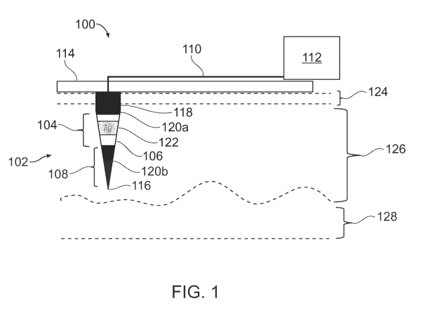
[0045] FIG. 1 is a schematic diagram of a microelectrode device 100 for
tissue
electrotransfer, in accordance with an embodiment of the invention. The
microelectrode
device 100 comprises a penetrating microelectrode 102, such as a needle with a
tapered tip
sufficiently sharp to penetrate skin tissue. The penetrating microelectrode
102 comprises a
target tissue microelectrode region 104 comprising an electrically conductive
surface 106,
such as a conductive metal surface, that selectively delivers a biomolecule to
cells located in
a tissue location, such as the skin tissue surrounding the penetrating
microelectrode 102, via
tissue electrotransfer. The penetrating microelectrode 102 also comprises an
anchor
microelectrode region 108 to mechanically anchor the penetrating
microelectrode 102 to
position the target tissue microelectrode region 104 to selectively deliver
the biomolecule to
the cells located in the tissue location. For example, the anchor
microelectrode region 108 can
have a coating (such as those discussed further herein), that assists in
holding the penetrating
microelectrode 102 within skin tissue into which the penetrating
microelectrode 102 is
inserted, for instance by providing sufficient friction against motion of the
penetrating
microelectrode 102 within the skin tissue. An electrical connection 110
connects the
penetrating microelectrode 102 to a voltage source 112. For example, the
penetrating
microelectrode 102 can be made of (or have a core or other portion made of) a
conductive
metal, and electrical connection 110 can be a conductive trace or other
electrical connection
on an electrical circuit board or other mounting frame 114. The voltage source
112 can, for
example, be a power supply configured to deliver the voltages, including
pulsed voltages, that
are taught further herein.
[0046] Continuing with reference to FIG. 1, the anchor microelectrode
region 108 can be
at or near a distal end 116 of the penetrating microelectrode 102. As used
herein, a "distal
end" 116 of the penetrating microelectrode 102 is the end of the penetrating
microelectrode
102 that is inserted most deeply into skin tissue, while the "proximal end"
118 is the opposite
end, which is nearest to the mounting frame 114. The microelectrode device 100
can
comprise electrical insulation 120a on a surface of the penetrating
microelectrode 102, that is
distinct from the electrically conductive surface 106 of the target tissue
microelectrode region
104. The electrical insulation 120a can comprise an insulating polymer
deposited on the
penetrating microelectrode, such as, for example, of a parylene (poly(p-
xylylene)) film
deposited by chemical vapor deposition (CVD), as discussed below in connection
with the
- 9 -
CA 03124709 2021-06-22
WO 2020/163310 PCT/US2020/016555
example of FIG. 5. For example, more than one distinct regions 120a, 120b of
electrical
insulation 120 can, between them, define the target tissue microelectrode
region 104, which
has the electrically conductive surface 106. The electrical insulation can be
on a surface 120b
of the anchor microelectrode region 108, and can serve as both electrical
insulation and as the
coating of the anchor microelectrode region 108 that assists in holding the
penetrating
microelectrode 102 within the skin tissue into which the penetrating
microelectrode 102 is
inserted. For example, a coating of a parylene (poly(p-xylylene)) film
deposited by chemical
vapor deposition (CVD) can serve both as electrical insulation and to anchor
the
microelectrode by providing sufficient friction against motion of the
penetrating
microelectrode 102 within the skin tissue. The microelectrode device can
comprise a
biomolecule coating 122, comprising the biomolecule to be selectively
delivered, on at least
part of a surface of the target tissue microelectrode region 104, which may be
the entire
surface of the target tissue microelectrode region 104. The biomolecule
coating 122 can, for
example, include a nucleic acid (as defined in more detail below, for example,
DNA or RNA)
or protein. The biomolecule coating 122 can be dissolvable when surrounded by
skin tissue.
If the entire surface of the target tissue microelectrode region 104 is coated
with the
biomolecule coating 122, the target tissue microelectrode region 104 has a
sufficiently
electrically conductive surface 106, after the biomolecule coating 122 is
dissolved when
surrounded by the skin tissue, so that the electroporation can be performed by
the target tissue
microelectrode region 104. In one example, the biomolecule coating 122 can
include a
plasmid DNA (pDNA) vector that is to be delivered by electroporation to cells
located in the
surrounding tissue. The tissue location containing the cells to which the
biomolecule is to be
delivered can be below a stratum corneum layer 124 of skin, and selectively
within either the
epidermal layer 126 of skin, the dermal layer 128 of skin, or both the
epidermal layer 126 and
the dermal layer 128. In one example, the tissue location is selectively
within only the
epidermal layer 126 of skin.
[0047] FIG. 2 is a schematic diagram of a microelectrode device 200 for
tissue
electrotransfer, including alternatives for an anchor microelectrode region
208, in accordance
with an embodiment of the invention. Although it is envisaged that the anchor
microelectrode
region 208 should be minimally harmful to surrounding tissue, it is possible
in some
embodiments that the anchor microelectrode region 208 can comprise a barb 230,
or other
structure to assist with anchoring; and can comprise an additional, adhesion
surface coating
232 to assist with anchoring. The barb 230 can, for example, be a
bioresorbable barb, which
- 10 -
CA 03124709 2021-06-22
WO 2020/163310 PCT/US2020/016555
can have a short length of about 500 micrometers or less. The penetrating
microelectrode can
be a needle comprising a tapered tip (as in FIG. 1); a needle comprising a
lateral protrusion,
such as a barb 230 (of FIG. 2); or both.
[0048] FIG. 3 is a schematic diagram of a microelectrode device 300 for
tissue
electrotransfer, including example dimensions of penetrating microelectrodes,
in accordance
with an embodiment of the invention. The microelectrode device 300 can
comprise more than
one of the penetrating microelectrode, in which a center-to-center spacing 334
of the more
than one of the penetrating microelectrode comprises a spacing between about
300
micrometers and about 1.5 millimeters. The length 336 of the penetrating
microelectrode can
comprise a length between about 225 micrometers and about 1250 micrometers.
The
penetrating microelectrode can comprise a diameter 338 between about 100
micrometers and
about 500 micrometers.
[0049] FIG. 22 is a schematic diagram of a microelectrode device 2200 for
tissue
electrotransfer that uses multiplexing of more than one target tissue
microelectrode region, in
accordance with an embodiment of the invention. In this embodiment, the device
includes
more than one target tissue microelectrode region each to selectively deliver
a different
biomolecule. With reference to the example of FIG. 22, more than one target
tissue
microelectrode regions 2204a-d is used, where different target microelectrode
regions include
more than one different biomolecule coating 2222a-d. For example, more than
one different
target microelectrode regions 2204c and 2204d on the same penetrating
microelectrode 2202c
can be coated with more than one different biomolecule coatings 2222c and
2222d. In
another example, more than one different penetrating microelectrodes 2202a and
2202b in the
same device can have more than one different target tissue microelectrode
regions 2204a and
2204b, which can be coated with more than one different biomolecule coatings
2222a and
2222b. For example, different biomolecules can be delivered to different
layers of skin, such
as one biomolecule within at least part of an epidermal layer of skin and a
different
biomolecule within at least part of the dermal layer of skin, using one or
more of the
arrangements of the type illustrated in FIG. 22.
[0050] FIG. 4 is a graph illustrating pulsed voltages 440 applied from a
voltage source
(see 112 in FIG. 1) to a penetrating microelectrode to create a transient
permeabilization of a
cell membrane of tissue in at least one of an epidermal layer of skin and a
dermal layer of
skin, in accordance with an embodiment of the invention. The electrical
connection (see 110
in FIG. 1) can apply a pulsed voltage from the voltage source (see 112 in FIG.
1) to the
- 11 -
CA 03124709 2021-06-22
WO 2020/163310 PCT/US2020/016555
penetrating microelectrode to create a transient permeabilization of a cell
membrane of tissue
in at least one of an epidermal layer of skin and a dermal layer of skin. With
reference to FIG.
4, the electrical connection can apply a voltage from the voltage source to
the penetrating
microelectrode to create a maximum electric field strength 442 of between
about 0.1 kilovolts
(kV) per centimeter and about 10 kilovolts (kV) per centimeter in skin tissue
surrounding the
penetrating microelectrode.
[0051] FIG. 25 is a schematic diagram of a microelectrode device 2500 for
tissue
electrotransfer, which incorporates a modeling processor 2562, in accordance
with an
embodiment of the invention. Here, the modeling processor 2562 includes a
modeling
module for determining electroporation hotspots by using a multiscale skin
electroporation
model that incorporates tissue level electric field prediction and a cellular
level simulation
accounting for the cell density in the targeted tissue. To implement this
module, the modeling
processor 2562 includes a tissue level electric field prediction module 2564
and a cellular
level simulation module 2566. The tissue level electric field prediction
module 2564 can, for
example, use a model to determine an electric field in the tissue. The
cellular level simulation
module 2566 can, for example, simulate the cell density within the tissue.
Using these
modules, the modeling processor 2562 can determine tissue locations to which
to selectively
deliver, via tissue electrotransfer, a biomolecule to cells located in the
tissue location. In
addition, using these modules, the modeling processor 2562 can determine a
control voltage
2568 delivered by the voltage source 2512 to the penetrating microelectrode
2502. Further,
the output of the modeling processor 2562 can be used to determine the
locations of the
anchor microelectrode region 2508 and target tissue microelectrode region 2504
within the
targeted tissue; and to determine the format, spacing and dimensions of the
penetrating
microelectrodes 2502 used in an array of such penetrating microelectrodes
2502. In addition,
the modeling processor 2562 can be used to determine a control voltage
delivered by the
voltage source to the penetrating microelectrode, regardless of whether the
modeling
processor 2562 includes a tissue level electric field prediction module 2564
and a cellular
level simulation module 2566.
[0052] Various techniques set forth herein can include computer implemented
components, such as modeling processor 2562, tissue level electric field
prediction module
2564 and cellular level simulation module 2566 (see FIG. 25). Such components
can be
implemented using hardware, and can include one or more processors, which can
for example
include one or more Application Specific Integrated Circuits (ASICs),
application software
- 12 -
CA 03124709 2021-06-22
WO 2020/163310 PCT/US2020/016555
running on one or more processors; and sensor and/or control connections
delivering
electronic signals to and from systems set forth herein (such as voltage
source 2512 and
penetrating microelectrode 2502 of FIG. 25), in which the signals can deliver
electronic
signals to and from actuated components within components set forth herein.
The
components can include user input modules, which can include components (such
as a
keyboard, touch pad, and associated electronics in connection with a processor
and a
memory) to receive user input. The components, such as modeling processor 2562
and
voltage source 2512, can also include a memory to store information, and to
implement
procedures under control of computer hardware and software. It will be
appreciated that other
control hardware and software may be used. Techniques can be implemented using
hardware,
software or a combination thereof. When implemented in software, the software
code can be
executed on any suitable processor or collection of processors, whether
provided in a single
computer or distributed among multiple computers.
[0053] FIG. 12 is a schematic diagram of an electrical system schematic
used with a
penetrating microelectrode array, in an experiment in accordance with an
embodiment of the
invention. In this experiment, electrical characterizations were performed in
both conductive
fluid and skin tissue. The schematic includes a function generator, amplifier
and
electroporation chip. Here, the penetrating microelectrodes are needles
mounted to a printed
circuit board, and are of length 2 mm with center-to-center spacing of 1.3 mm.
[0054] FIG. 13 is a diagram showing images of the results of
electropermeabilization
testing with rhodamine, in an experiment in accordance with an embodiment of
the invention.
In this experiment, a 1.5 kV/cm, 10 msec DC pulse was applied. A needle length
of 3 mm
was used. The stratum corneum layer was overcome with pulsation. The image
shows bright
field and red channel images merged. Results with a nonpulsed needle are at
left, and with a
pulsed needle are at right.
[0055] FIG. 14 is a summary of a protocol used for a permeabilization test
with
propidium iodide, in an experiment in accordance with an embodiment of the
invention.
[0056] FIG. 15 is a diagram showing images of sectioned skin image
examples, in the
experiment of FIG. 14.
[0057] FIG. 16 is a diagram showing image analysis results in the
experiment of FIG. 14.
A graph at lower right is from Ge et al., 2010, "The viability change of
pigskin in vitro,"
Burns, 36 (2010).
- 13 -
CA 03124709 2021-06-22
WO 2020/163310 PCT/US2020/016555
[0058] FIG. 17 is a schematic diagram of an electrode configuration of a
penetrating
microelectrode array in which multiple electrodes are addressed as a group, in
accordance
with an embodiment of the invention. In this example, electrodes labeled "1"
are addressed as
a group, as are electrodes labeled "2," "3" and "4." It will be appreciated
that other
arrangements can be used, and that individually addressable electrodes can be
used. As
shown in this figure, the microelectrode device can comprise more than one of
the
penetrating microelectrode, and the electrical connection can comprise an
electrically
independent connection to two or more of the more than one penetrating
microelectrodes.
[0059] FIG. 18 is a diagram showing a penetrating microelectrode array
device using a
printed circuit board to provide electrical connections, in accordance with an
embodiment of
the invention.
[0060] FIG. 19 is a diagram showing images of the results of testing a
penetrating
microelectrode array device using a printed circuit board, in an experiment in
accordance
with an embodiment of the invention. "Hotspots" of propidium iodide can be
seen around the
needle insertion site.
[0061] FIG. 20 is a schematic diagram of penetrating microelectrode arrays
fabricated
using photolithography, in accordance with an embodiment of the invention. As
shown in this
figure, the electrical connection can comprise a connection defined by
photolithography, the
penetrating microelectrode can comprise an electrode base defined by
photolithography, and
the penetrating microelectrode can comprise electroplated metal. In one
example, penetrating
microelectrode arrays can be fabricated using UV LiGA techniques, where "LiGA"
is from
the German acronym signifying Lithography, Electroplating and Molding.
Photolithography
can be used to define traces, penetrating microelectrode bases, mold for
electroplating.
Electroplating of metal can be used to create solid posts. Electrochemical or
wet etch can be
used to define the shape of penetrating microelectrodes. This can be followed
by removal of
molding material, and dicing of a wafer to create separate chips. FIG. 21 is
an image of dies
on a wafer used for fabricating penetrating microelectrode arrays using
photolithography, in
accordance with an embodiment of the invention.
[0062] An embodiment according to the invention uses selective insulation
of a
penetrating microelectrode and coating the penetrating microelectrode with DNA
vector (or
other biomolecule) in order to deliver the vector and low intensity electric
pulses to
coincident "hotspot" areas adjacent to the electrode. Since an embodiment
focuses on only
transfecting the tissue adjacent to the electrodes within the electric field
"hotspots," as
- 14 -
CA 03124709 2021-06-22
WO 2020/163310 PCT/US2020/016555
opposed to attempting to transfect a large tissue volume, a lower pulse field
intensity can be
used for efficient epidermal and dermal gene electrotransfer (GET), or other
biomolecule
delivery. It is believed that the approach can overcome many of the major
bottlenecks
towards clinical translation of transdermal electroporation by addressing the
issues of safety,
tolerability and efficacy in GET by more efficiently delivering vector and
electrical energy to
"hotspots" such that there is derived a lower threshold for skin EP, vector
delivery is targeted
directly to the portion of skin to be permeabilized, and targeted dermal layer
transfection is
obtained. These benefits combined with impedance monitoring of the skin prior
to and
following pulse application will allow the penetrating microelectrode array to
obtain
maximum DNA delivery (or other biomolecule delivery) and GET expression while
minimizing tissue irritation from both electrode insertion as well as pulse
protocols.
[0063] An embodiment according to the invention contrasts with other
penetrating
electroporation platforms where a vector or other biomolecule is injected
under the skin in a
less controlled fashion, and long, deeply penetrating electrodes are used so
that a portion of
the electrodes are below the skin. These approaches also use higher intensity
pulses to
permeabilize the largest volume of tissue around the electrodes. The vector
injection means a
large amount of vector is distributed in the tissue in areas which are not
efficiently
permeabilized and the deep electrode penetration coupled with high intensity
pulses causes
ablative irreversible electroporation tissue damage adjacent to the
electrodes. This leads to
variability in GET transfection efficacy as well as adverse tissue damage.
Clinical protocols
for administration site selection, electrode design, and pulse parameters must
be carefully
evaluated for safety, tolerability and efficacy. (20, 40, 41) In particular,
an embodiment
according to the invention targets the viable epidermis due to the high
concentration of
keratinocytes and dendritic cells which can be activated via GET.
[0064] In other embodiments, selective epidermal and dermal targeting of
vector delivery
and transfection may also be used in other clinical regimes such as
electrochemotherapy
(ECT), non-thermal irreversible electroporation (N-TIRE) or focused
transfection of tissues
other than skin. It will be appreciated that other biomolecule deliveries can
be performed.
[0065] FIG. 5 is a diagram illustrating selective electrical insulation of
penetrating
microelectrodes on a base and a tip of the microelectrode, and showing the
results of
simulations of localized electric field strengths produced by the
microelectrodes for targeted
tissue electrotransfer, in accordance with an embodiment of the invention. In
the embodiment
of FIG. 5, penetrating microelectrodes are selectively insulated, in order to
focus tissue
- 15 -
CA 03124709 2021-06-22
WO 2020/163310 PCT/US2020/016555
"hotspots" to distinct dermal layers. Controlled, low-voltage electroporation
is then targeted
to specific dermal layers using selective insulation over regions of the
penetrating
microelectrodes. In order to accomplish this goal, in one experiment in
accordance with an
embodiment of the invention, penetrating microelectrode arrays are coated via
chemical
vapor deposition (CVD) of a parylene (poly(p-xylylene)) film. In parylene CVD,
a known
mass of solid parylene dimers are sublimed into the gas phase where they are
then pyrolyzed
to cleave the dimer into monomer molecules. The parylene monomers are then
introduced
into the deposition chamber where they polymerize and conformally coat the
exposed
penetrating microelectrode array surface. Parylene is a USP Class VI Polymer
known for its
biological inertness and has been used for decades as an encapsulation
material for medical
devices and medical electronics. A 100-1000 nm thick insulating parylene layer
is deposited
on the penetrating microelectrode array. The parylene is selectively removed
from either the
base or tip of the penetrating microelectrodes via mechanical abrasion or
focused CO2
excimer laser ablation for more accurate removal. These penetrating
microelectrodes are then
coated with a pDNA solution stabilized in a 1% (w/v) carboxy-methylcellulose
and 0.5%
(w/v) Lutrol F-68 NF (BASF, Mt. Olive, NJ, USA) solution as previously
reported for an
expected 0.15-15 [tg DNA coated per needle. It will be appreciated that other
coating
techniques, including alternative coating techniques which have been used for
coating
microneedles, such as inkjet printing or electrospraying, may also be
utilized; and that other
biomolecules can be used.
[0066] FIG. 5 also shows simulation results, in accordance with an
embodiment of the
invention, which predict the effect of selectively insulating the penetrating
microelectrodes
and localizing `hotspots' in either the dermis or epidermis. It can be
accurately predicted
where skin electroporation will occur at varying applied voltages so one can
target DNA
vector gene electrotransfer (GET) (or other biomolecule delivery) at distinct
layers of the skin
while minimizing tissue damage. By focusing the hotspot to either the dermal
or epidermal
skin layers, it is expected that epidermal transfection will demonstrate a
higher degree of
transfection due to the higher epidermal cell density, which will help to
ensure that DNA
based vaccines have sufficient expression for dendritic cell activation to
confer a protective
immunological response. Similar advantages may be able to be achieved with
other
biomolecule deliveries.
[0067] An embodiment according to the invention provides a minimally
invasive
penetrating microelectrode array to localize delivery of DNA (or other
biomolecules, such as
- 16 -
CA 03124709 2021-06-22
WO 2020/163310 PCT/US2020/016555
nucleic acids or proteins) and electric field hotspots around the electrodes.
In experiments in
accordance with an embodiment of the invention, the penetrating electrode
dimensions and
spacing are informed by the development of a skin electroporation model which
can predict
both electric field distribution above an electroporation threshold within
skin as well as a
packed cell model which can predict how electroporation of cells within tissue
then changes
the electric field distribution within the rest of the tissue. By coating high
concentrations of
pDNA vectors (or other biomolecules) directly onto the penetrating
microelectrode array, the
DNA is locally reconstituted in tissue adjacent to the electrode surface
following insertion so
that vectors and electrical energy are delivered to their desired "hotspot"
locations within the
skin to obtain targeted tissue transfection.
[0068] An embodiment according to the invention provides selective
insulation of
penetrating microelectrodes for targeted skin electroporation. By depositing
an insulating
polymer onto the penetrating microelectrode surface and selectively removing
the insulation
over part of the penetrating microelectrode (e.g., tip vs. base) portions of
the penetrating
microelectrodes can be insulated so that vectors and electrical energy are
delivered to their
desired "hotspot" locations within the skin to obtain targeted skin layer
transfection.
[0069] Experiments, constructed in accordance with an embodiment of the
invention and
described herein, employ a multifaceted approach to improving dermal
electroporation
efficiencies through computational modeling of electric field distributions
within a skin
model from different penetrating electrode geometries, computational modeling
of
permeabilization distribution within a packed cell tissue model, development
of a penetrating
microelectrode array, selective insulation of the penetrating microelectrodes,
and DNA vector
coating of the penetrating microelectrode array to co-target vector delivery
and electrical
pulse energy to distinct dermal layers.
[0070] Experimental #1:
[0071] FIG. 7 is a diagram showing a photograph and a schematic diagram of
a
penetrating microelectrode array, in accordance with an embodiment of the
invention. In an
experiment in accordance with an embodiment of the invention, with reference
to FIG. 7,
there was developed a penetrating microelectrode array which consists of
austenitic 316
stainless surgical steel acupuncture needles assembled into a printed circuit
board (PCB)
array. Each penetrating microelectrode is a 160 um diameter needle which
tapers to a fine
point over a 745 um tip length at a 6.129 taper angle. Each needle is placed
into the PCB
through a plated through-hole with the penetration length of the penetrating
microelectrode
- 17 -
CA 03124709 2021-06-22
WO 2020/163310 PCT/US2020/016555
controlled using a plastic silicone rubber spacer of known thickness which the
electrode
pierces. The total penetration depth of the penetrating microelectrode array
is controlled by
the exposed penetrating microelectrode length which can be as short as 1/4 mm.
Following
penetrating microelectrode assembly, the backs of the needles are clipped and
then soldered
to the PCB via dip soldering within a solder bath. The PCB is connected to a
ribbon cable for
electrical excitation. The prototype penetrating microelectrode array supports
16 electrodes
with a 0.75 mm center to center spacing and typically protrude 1 mm as shown
in FIG. 7. The
penetrating microelectrode array design is informed by the development of a
multiscale skin
electroporation model which links a tissue level electric field prediction and
a cellular level
simulation accounting for the cell density in the dermis and epidermis to an
electroporation
circuit model to identify "hotspots" around electrodes where electroporation
is simulated.
This multiscale model helps inform penetrating microelectrode array design and
implementation to determine optimal penetrating microelectrode array geometry
and pulse
parameters. Such a multiscale model can, for example, be implemented by a
modeling
processor described relative to FIG. 25, herein.
[0072] Experimental #2:
[0073] FIG. 8 is a diagram showing views of the stratum corneum, epidermis
and dermis
layers of skin, and of penetrating microelectrodes inserted in those layers,
in accordance with
an embodiment of the invention. In an experimental investigation conducted in
accordance
with an embodiment of the invention, with reference to FIG. 8, there was
developed a
computational skin tissue level model where the skin morphology was extracted
from
histological images to delineate the dermal-epidermal junction (DEJ) with a
realistic surface
topography and epidermal thickness. In FIG. 8, image 848 shows H&E stained
skin
delineating the DEJ; image 850 shows the epidermal thickness extracted into a
2D skin
model; image 852 shows a 2D model extruded into a 3D skin model with periodic
ridges; and
image 854 shows a 3D skin model with wavy surface extracted from skin
histology. A pair of
inserted penetrating microelectrodes are shown in image 854. Physical
properties (thickness,
conductivity, etc.) used in the simulation were found in literature (28, 42).
This global skin
model is used to determine the electric field distribution within tissue to
identify where a
permeabilizing field intensity threshold of 0.5 kV/cm is achieved. Using this
model the
electric field distribution within the skin was simulated using surface
electrodes where all of
the permeabilizing electric field was localized within the stratum corneum at
an applied 20 V
and only penetrated into the dermal layers at voltages of 50 V and above. The
simulation was
- 18 -
CA 03124709 2021-06-22
WO 2020/163310 PCT/US2020/016555
updated to reflect the increase in stratum corneum (SC) conductivity when a
permeabilizing
threshold was achieved. This required a voltage of 100 V and deeper field
penetration into the
skin but is not a controllable process during experiments. Therefore, it was
decided to focus
on a penetrating microelectrode design.
[0074] Experimental #3:
[0075] FIG. 9 is a diagram showing the results of simulation of localized
electric field
strengths surrounding an array of sixteen penetrating microelectrodes inserted
into a skin
model, in a simulation in accordance with an embodiment of the invention. When
using
penetrating microelectrodes the simulation can predict "hotspots" around the
electrodes as
shown in FIG. 9. The simulation can predict the electric field distribution
around simulated
needles, varying the needle spacing, insertion depth and applied voltage. FIG.
9 shows the
results of one such simulation by delineating the volume around the electrodes
where the
electric field exceeds a value of 0.5 kV/cm, the minimum electric field
expected for tissue
electroporation. In particular, the electric field increases at the tips of
the penetrating
microelectrodes due to the sharp tips which focuses electric field lines with
decreasing
electrode area. In this manner, successive iterations of a penetrating
microelectrode array can
be rationally designed as required.
[0076] Experimental #4:
[0077] In addition to a tissue level model, a local packed cell model has
been developed,
in an experiment in accordance with an embodiment of the invention, to
understand how
permeabilization occurs in different layers of the skin. This local model
couples the simulated
electric potential to a local equivalent circuit model of a cell in which the
cell membrane is
treated as a capacitor in parallel with a resistor where the conductance drops
significantly in
the tissue during electroporation when a transmembrane voltage (TMV) of 0.5 V
is achieved.
The field distribution is then updated to reflect this drop in local cell
impedance to indicate
how the influence of one cell undergoing electroporation influences the TMV
(and hence
propensity towards electroporation) of its neighbors. Two models have been
developed. The
first is a packed sphere model 1056 reflecting the high keratinocyte density
found in the
epidermis. FIG. 10 is a diagram showing, in a simulation in accordance with an
embodiment
of the invention, a packed cell model 1056 of epidermal keratinocytes at an
applied field of
2.0 kV/cm. Images 1058 are 2D projections of model 1056 at a field of (Top)
0.83 kV/cm
showing a partially electroporated state and (bottom) 1.0 kV/cm showing a
fully
electroporated state. Image 1060 (Top) shows transmembrane potential and
(Bottom) shows
- 19 -
CA 03124709 2021-06-22
WO 2020/163310 PCT/US2020/016555
electric field within simulated dermal fibroblasts showing orientation
dependent
electroporation. As shown in image 1056 of FIG. 10, when a high intensity
electric field is
applied all cells are fully electroporated. Image 1058 of FIG. 10 shows a 2D
centerline
projection of the packed cells at moderate to high applied electric field
reflecting partially to
fully electroporated condition. In the partially electroporated condition the
permeabilization
of one cell can increase the local potential of its neighbor. Image 1060 of
FIG. 10 shows a
lower cell density simulation of elliptical cells in various orientations
reflecting the cell
density and distribution of dermal fibroblasts (43). Aside from a lower cell
density, the
simulation shows that the degree of electroporation strongly correlates with
cell orientation
with cells aligned with the electric field having a greater degree of
electroporation. These
simulations lead to a hypothesis that targeted electroporation within the
epidermis will lead to
a greater degree of cell electroporation due to the higher cell density and
less orientation
dependent effects. These simulations can also be used to guide penetrating
microelectrode
array design and optimize pulse parameters. It can be accurately predicted
where skin
electroporation will occur at varying applied voltages so that one can target
DNA vector GET
(or other biomolecule delivery) at distinct layers of the skin while
minimizing tissue damage.
Electroporation can be targeted to specific dermal layers using selective
insulation over
regions of the penetrating microelectrodes. Such simulations, and the
consequent control,
design, and optimization can be performed using a modeling processor, such as
that described
in connection with FIG. 25, herein.
[0078] Experiment #5:
[0079] In another experiment in accordance with an embodiment of the
invention, studies
of Green Fluorescent Protein (GFP) expression in porcine skin were performed,
which will be
discussed with reference to FIG. 6. These studies were performed under
protocols approved
by Rutgers IACUC committee (PR0T0201702610 - Porcine skin harvesting). Skin
was
freshly harvested from euthanized 3 to 5-week-old piglets, carefully cleaned
with 70%
ethanol, and then shaved and depilated. The skin tissue was cut into small
square pieces
approximately 1 x 1 cm. The subcutaneous fat and tissue were removed carefully
by a
scalpel. 20 ug/m1 pEGFP-N1 vector (Clontech) in lx PBS solution was injected
by
MicronJet600 microneedles (NanoPass Technologies Ltd., Nes Ziona, Israel)
before the
electroporation treatment. Electric pulses were applied by using the
preliminary penetrating
microelectrode array. The GFP pDNA was injected via shallow ID microneedle
injection
followed by a 5V or 50V 10 ms electroporation pulse. Following electroporation
treatment,
- 20 -
CA 03124709 2021-06-22
WO 2020/163310 PCT/US2020/016555
the skin samples were immediately put into a modified Eagle's medium (MEM) at
37 C on a
rocker in an incubator. At a chosen time point (8, 16, 24, and 48 hours post-
pulsation), the
skin samples were sliced along the transverse plane from the center of
injection site and then
washed with PBS. The slices were 1 ¨ 1.5 mm thick and imaged by using inverted
epi-
fluorescent microscopy. FIG. 6 shows Green Fluorescent Protein (GFP)
expression at 24
hours localized with the tips of penetrating microelectrodes demonstrated in
the freshly
excised porcine skin. GFP expression is shown in the porcine skin following
intradermal ID
injection and, in panel 644, 5V, 10 ms DC pulse and, in panel 646, 50V, 10 ms
DC pulse. In
panel 644, the GFP expression is localized at the needle tips 800 [tm from the
skin surface
and spaced 650 [tm apart consistent with the penetrating microelectrode array
dimensions
(1000 [tm long, 750 [tm apart). The 5V pulse in panel 644) shows localization
at the
penetrating microelectrode tip `hotspof where the field is expected to be
highest due to the
sharp penetrating microelectrode tips whereas the 50V pulse in panel 646 shows
a more
diffuse tissue fluorescence.
[0080] Experiment #6:
[0081] Additionally, the degree of tissue electroporation can be monitored
by a drop in
tissue impedance as cells are permeabilized and, in experiments in accordance
with an
embodiment of the invention, there has been seen evidence of tissue level
electroporation at
voltages as low as 5V (FIG. 6, panel 644). FIG. 11 shows excised porcine skin
impedance
before and following electroporation pulse application, in experiments in
accordance with an
embodiment of the invention. By monitoring the tissue impedance the degree of
tissue
electroporation can be assessed and the impedance change can be correlated
with pDNA
expression (or with expression or other phenomena related to other
biomolecules delivered)
and tissue damage. In FIG. 11, successive tissue impedance measurements prior
to
(measurements 1-3) and following (measurements 4-5) either a 5V or 50V 10 ms
electroporation pulse. The 5V pulse shows a 1% change in tissue impedance
whereas the 50V
pulse shows a 5% drop.
[0082] Experiment #7:
[0083] FIG. 23 is an image of DNA composites deposited onto penetrating
microelectrode arrays by electrostatic spray, in experiments in accordance
with an
embodiment of the invention. This illustrates an example of a biomolecule
coating,
comprising the biomolecule to be selectively delivered, on at least part of a
surface of the
target tissue microelectrode region of a microelectrode device. Electrostatic
spray was
-21 -
CA 03124709 2021-06-22
WO 2020/163310 PCT/US2020/016555
employed for depositing DNA composites onto the penetrating microelectrode
arrays, which
are here implemented as needle microarrays. GFP DNA plasmid was prepared in
solution and
then sprayed out of a needle held at high voltage to the grounded microarray.
The composited
microscope image shows the needles after 20 min of spray at a flow rate of 0.1
mL/hr.
[0084] Experiment #8:
[0085] FIG. 24 is an image of penetrating microelectrodes insulated with a
conformal
layer of an insulating dielectric coating, in experiments in accordance with
an embodiment of
the invention. This illustrates an example of electrical insulation comprising
an insulating
polymer deposited on the penetrating microelectrode of a microelectrode
device. The
microelectrodes are insulated with a conformal layer of poly(para- xylene)
(parylene)
deposited via chemical vapor deposition (CVD), ranging from 100 nm to 2 mm
thick
depending on the mass of the parylene dimer precursor used. Parylene is an USP
Class VI
polymer recognized by the FDA as a biocompatible material. The deposited
parylene acts as
a hydrophobic, insulating dielectric coating suitable for human implantation.
Closeup of
needle tip (top right) shows edge of parylene layer (white arrows).
[0086] Definitions
[0087] As used herein, a "penetrating microelectrode" is a microelectrode
that is capable
of penetrating skin tissue, such as a needle with a tapered tip sufficiently
sharp to penetrate
skin tissue.
[0088] As used herein, a "penetrating microelectrode array" is an array of
more than one
penetrating microelectrode.
[0089] As used herein, a "target tissue microelectrode region" of a
penetrating
microelectrode is a region of a penetrating microelectrode that comprises an
electrically
conductive surface, such as a conductive metal surface, that selectively
delivers a
biomolecule to cells located in a tissue location, such as the skin tissue
surrounding the
penetrating microelectrode, via tissue electrotransfer.
[0090] As used herein, an "anchor microelectrode region" of a penetrating
microelectrode
is a region of a penetrating microelectrode that assists to mechanically
anchor the penetrating
microelectrode within skin tissue into which the penetrating microelectrode is
inserted, such
as by having a coating that assists in holding the penetrating microelectrode
within skin tissue
into which the microelectrode is inserted, for instance by providing
sufficient friction against
motion of the penetrating microelectrode within the skin tissue.
- 22 -
CA 03124709 2021-06-22
WO 2020/163310 PCT/US2020/016555
[0091] As used herein, "tissue electrotransfer" can include any
electroporation mediated
transdermal delivery, including electrochemotherapy (ECT) and gene
electrotransfer (GET).
[0092] As used herein, a "biomolecule" can include a nucleic acid, a
protein or any other
biological molecule to be delivered by tissue transfection in accordance with
techniques
taught herein, or a combination of such nucleic acids, proteins or other
biological molecules.
For example, the biomolecule can include one or more of: a nucleic acid or
protein vaccine
vector, a nucleic acid and protein vaccine vector, another vector, a nucleic
acid biomolecule
(for example, RNA, DNA/plasmid vector, DNA vaccine, DNA/plasmid vector
vaccine) and a
protein (for example, a peptide/protein, peptide/protein vaccine). In
addition, a "biomolecule"
can include (1) an antibody, such as a monoclonal antibody, or another ligand
specific
molecule, and (2) other molecules to be delivered that may have or could
affect biologic
and/or cellular activity.
[0093] As used herein, "nucleic acid" refers to a macromolecule composed of
chains (a
polymer or an oligomer) of monomeric nucleotide. The most common nucleic acids
are
deoxyribonucleic acid (DNA) and ribonucleic acid (RNA). It should be further
understood
that the present invention can be used for biomolecules containing artificial
nucleic acids
such as peptide nucleic acid (PNA), morpholino, locked nucleic acid (LNA),
glycol nucleic
acid (GNA) and threose nucleic acid (TNA), among others. In various
embodiments of the
present invention, nucleic acids can be derived from a variety of sources such
as bacteria,
virus, humans, and animals, as well as sources such as plants and fungi, among
others. The
source can be a pathogen. Alternatively, the source can be a synthetic
organism. Nucleic
acids can be genomic, extrachromosomal or synthetic. Where the term "DNA" is
used
herein, one of ordinary skill in the art will appreciate that the methods and
devices described
herein can be applied to other nucleic acids, for example, RNA or those
mentioned above. In
addition, the terms "nucleic acid," "polynucleotide," and "oligonucleotide"
are used herein to
include a polymeric form of nucleotides of any length, including, but not
limited to,
ribonucleotides or deoxyribonucleotides. There is no intended distinction in
length between
these terms. Further, these terms refer only to the primary structure of the
molecule. Thus, in
certain embodiments these terms can include triple-, double- and single-
stranded DNA, PNA,
as well as triple-, double- and single-stranded RNA. They also include
modifications, such as
by methylation and/or by capping, and unmodified forms of the polynucleotide.
More
particularly, the terms "nucleic acid," "polynucleotide," and
"oligonucleotide," include
polydeoxyribonucleotides (containing 2-deoxy-D-ribose), polyribonucleotides
(containing D-
- 23 -
CA 03124709 2021-06-22
WO 2020/163310 PCT/US2020/016555
ribose), any other type of polynucleotide which is an N- or C-glycoside of a
purine or
pyrimidine base, and other polymers containing nonnucleotidic backbones, for
example,
polyamide (e.g., peptide nucleic acids (PNAs)) and polymorpholino
(commercially available
from Anti-Virals, Inc., Corvallis, Oreg., U.S.A., as Neugene) polymers, and
other synthetic
sequence-specific nucleic acid polymers providing that the polymers contain
nucleobases in a
configuration which allows for base pairing and base stacking, such as is
found in DNA and
RNA. In addition, a "nucleic acid" can include a plasmid DNA (pDNA), such as a
plasmid
DNA vector.
[0094] As used herein, a "protein" is a biological molecule consisting of
one or more
chains of amino acids. Proteins differ from one another primarily in their
sequence of amino
acids, which is dictated by the nucleotide sequence of the encoding gene. A
peptide is a
single linear polymer chain of two or more amino acids bonded together by
peptide bonds
between the carboxyl and amino groups of adjacent amino acid residues;
multiple peptides in
a chain can be referred to as a polypeptide. Proteins can be made of one or
more
polypeptides. Shortly after or even during synthesis, the residues in a
protein are often
chemically modified by posttranslational modification, which alters the
physical and
chemical properties, folding, stability, activity, and ultimately, the
function of the proteins.
Sometimes proteins have non-peptide groups attached, which can be called
prosthetic groups
or cofactors.
[0095] It will be appreciated, in addition, that a biomolecule used herein
can include non-
natural bases and residues, for example, non-natural amino acids inserted into
a biological
sequence.
[0096] References
[0097] (1) Neumann, E., Schaefer-Ridder, M., Wang, Y. & Hofschneider, P. H.
Gene
transfer into mouse lyoma cells by electroporation in high electric fields.
The EMBO Journal
1, 841-845 (1982).
[0098] (2) Jadoul, A. & Preat, V. Electrically enhanced transdermal
delivery of
domperidone. International Journal of Pharmaceutics 154, 229-234,
doi:https://doi.org/10.1016/S0378-5173(97)00139-7 (1997).
[0099] (3) Prausnitz, M. R. A practical assessment of transdermal drug
delivery by skin
electroporation. Advanced Drug Delivery Reviews 35, 61-76,
doi:https://doi.org/10.1016/50169-409X(98)00063-5 (1999).
- 24 -
CA 03124709 2021-06-22
WO 2020/163310 PCT/US2020/016555
[00100] (4) Prausnitz, M. R., Bose, V. G., Langer, R. & Weaver, J. C.
Electroporation of
Mammalian Skin - a Mechanism to Enhance Transdermal Drug-Delivery. P Natl Acad
Sci
USA 90, 10504-10508, doi:DOI 10.1073/pnas.90.22.10504 (1993).
[00101] (5) Vanbever, R., LeBoulenge, E. & Preat, V. Transdermal delivery of
fentanyl by
electroporation. I. Influence of electrical factors. Pharm Res 13, 559-565
(1996).
[00102] (6) Vanbever, R., Morre, N. D. & Preat, V. Transdermal delivery of
fentanyl by
electroporation. II. Mechanisms involved in drug transport. Pharm Res 13, 1360-
1366 (1996).
[00103] (7) Vanbever, R. & Preat, V. Factors affecting transdermal delivery of
metoprolol
by electroporation. Bioelectrochemistry and Bioenergetics 38, 223-228,
doi:https://doi.org/10.1016/0302-4598(95)01830-8 (1995).
[00104] (8) Ita, K. Perspectives on transdermal electroporation. Pharmaceutics
8, 9
(2016).
[00105] (9) Mir, L. M. et al. [Electrochemotherapy, a new antitumor treatment:
first
clinical trial]. C R Acad Sci 111313, 613-618 (1991).
[00106] (10) Groselj, A. et al. Efficiency of electrochemotherapy with
reduced
bleomycin dose in the treatment of nonmelanoma head and neck skin cancer:
Preliminary
results. Head Neck 40, 120-125, doi:10.1002/hed.24991 (2018).
[00107] (11) Wichtowski, M., Murawa, D., Kulcenty, K. & Zaleska, K.
Electrochemotherapy in Breast Cancer - Discussion of the Method and Literature
Review.
Breast Care (Basel) 12, 409-414, doi:10.1159/000479954 (2017).
[00108] (12) Aguado-Romeo, M. J Benot-Lopez, S. & Romero-Tabares, A.
Electrochemotherapy for the Treatment of Unresectable Locoregionally Advanced
Cutaneous
Melanoma: A Systematic Review. Actas Dermosifiliogr 108, 91-97,
doi:10.1016/j.ad.2016.08.008 (2017).
[00109] (13) Plaschke, C. C., Gothelf, A., Gehl, J. & Wessel, I.
Electrochemotherapy of mucosal head and neck tumors: a systematic review. Acta
Oncol 55,
1266-1272, doi:10.1080/0284186X.2016.1207803 (2016).
[00110] (14) Rotunno, R. et al. Electrochemotherapy in non-melanoma head
and
neck skin cancers: a three-center experience and review of the literature. G
Ital Dermatol
Venereol151, 610-618 (2016).
[00111] (15) Schmidt, G., Juhasz-Boss, I., Solomayer, E. F. & Herr, D.
Electrochemotherapy in Breast Cancer: A Review of References. Geburtshilfe
Frauenheilkd
74, 557-562, doi:10.1055/s-0034-1368538 (2014).
- 25 -
CA 03124709 2021-06-22
WO 2020/163310
PCT/US2020/016555
[00112] (16) Queirolo, P., Marincola, F. & Spagnolo, F.
Electrochemotherapy for
the management of melanoma skin metastasis: a review of the literature and
possible
combinations with immunotherapy. Arch Dermatol Res 306, 521-526,
doi:10.1007/s00403-
014-1462-x (2014).
[00113] (17) Jahangeer, S., Forde, P., Soden, D. & Hinchion, J. Review of
current
thermal ablation treatment for lung cancer and the potential of
electrochemotherapy as a
means for treatment of lung tumours. Cancer Treat Rev 39, 862-871,
doi:10.1016/j.ctrv.2013.03.007 (2013).
[00114] (18) Gothelf, A. & Gehl, J. Gene electrotransfer to skin; review
of existing
literature and clinical perspectives. Curr Gene Ther 10, 287-299 (2010).
[00115] (19) Favard, C., Dean, D. S. & Rols, M. P. Electrotransfer as a
non viral
method of gene delivery. Current Gene Therapy 7, 67-77, doi:Doi
10.2174/156652307779940207 (2007).
[00116] (20) Diehl, M. C. et al. Tolerability of intramuscular and
intradermal
delivery by CELLECTRA (R) adaptive constant current electroporation device in
healthy
volunteers. Hum Vacc Immunother 9, 2246-2252, doi:10.4161/hv.24702 (2013).
[00117] (21) El-Kamary, S. S. et al. Safety and Tolerability of the Easy
Vax (TM)
Clinical Epidermal Electroporation System in Healthy Adults. Mot Ther 20, 214-
220,
doi:10.1038/mt.2011.235 (2012).
[00118] (22) McCoy, J. R. et al. A multi-head intradermal electroporation
device
allows for tailored and increased dose DNA vaccine delivery to the skin. Hum
Vacc
Immunother 11,746-754, doi:10.4161/21645515.2014.978223 (2015).
[00119] (23) Littel-van den Hurk, S. V. & Hannaman, D. Electroporation
for DNA
immunization: clinical application. Expert Rev Vaccines 9, 503-517,
doi:10.1586/Erv.10.42
(2010).
[00120] (24) Fakharzadeh, S. S., Zhang, Y., Sarkar, R. & Kazazian, H. H.
Correction of the coagulation defect in hemophilia A mice through factor VIII
expression in
skin. Blood 95, 2799-2805 (2000).
[00121] (25) Fewell, J. G. et al. Gene therapy for the treatment of
hemophilia B
using PINC-formulated plasmid delivered to muscle with electroporation. Mot
Ther 3, 574-
583 (2001).
- 26 -
CA 03124709 2021-06-22
WO 2020/163310 PCT/US2020/016555
[00122] (26) Yarmush, M. L., Golberg, A., Sersa, G., Kotnik, T. &
Miklavcic, D.
Electroporation-Based Technologies for Medicine: Principles, Applications, and
Challenges.
Annu Rev Biomed Eng 16, 295-320, doi:10.1146/annurev-bioeng-071813-104622
(2014).
[00123] (27) Babiuk, S. et al. Needle-free topical electroporation
improves gene
expression from plasmids administered in porcine skin. Mot Ther 8, 992-998,
doi:10.1016/j.ymthe.2003.09.008 (2003).
[00124] (28) Becker, S. M. & Kuznetsov, A. V. Thermal damage reduction
associated with in vivo skin electroporation: A numerical investigation
justifying aggressive
pre-cooling. International Journal of Heat and Mass Transfer 50, 105-116
(2007).
[00125] (29) Li, J., Tan, W., Yu, M. & Lin, H. The effect of
extracellular
conductivity on electroporation-mediated molecular delivery. Bba-Biomembranes
1828, 461-
470, doi:DOI 10.1016/j.bbamem.2012.08.014 (2013).
[00126] (30) Sadik, M. M. et al. Scaling Relationship and Optimization of
Double-
Pulse Electroporation. Biophys J106, 801-812, doi:10.1016/j.bpj.2013.12.045
(2014).
[00127] (31) Demiryurek, Y. et al. Transport, resealing, and re-poration
dynamics of
two-pulse electroporation-mediated molecular delivery. Bba-Biomembranes 1848,
1706-
1714, doi:10.1016/j.bbamem.2015.04.007 (2015).
[00128] (32) Li, J. B. & Lin, H. Numerical simulation of molecular uptake
via
electroporation. Bioelectrochemistry 82, 10-21,
doi:10.1016/j.bioelechem.2011.04.006
(2011).
[00129] (33) Lin, H., Sadik, M., Li, J. B., Shan, J. W. & Shreiber, D.
I. Numerical
Simulation of Molecular Delivery via Electroporation. Biophys J100, 577-577
(2011).
[00130] (34) Dev, S. B., Rabussay, D. P., Widera, G. & Hofmann, G. A.
Medical
applications of electroporation. IEEE Transactions on Plasma Science 28, 206-
223,
doi:10.1109/27.842905 (2000).
[00131] (35) Nicolas, J.-F. & Guy, B. Intradermal, epidermal and
transcutaneous
vaccination: from immunology to clinical practice. Expert Rev Vaccines 7, 1201-
1214,
doi:10.1586/14760584.7.8.1201 (2008).
[00132] (36) Dean, H. J. Epidermal delivery of protein and DNA vaccines.
Expert
Opinion on Drug Delivery 2,227-236, doi:10.1517/17425247.2.2.227 (2005).
[00133] (37) Lambricht, L. et al. Clinical potential of electroporation
for gene
therapy and DNA vaccine delivery. Expert Opinion on Drug Delivery 13, 295-310,
doi:10.1517/17425247.2016.1121990 (2016).
- 27 -
CA 03124709 2021-06-22
WO 2020/163310
PCT/US2020/016555
[00134] (38) REVEAL 1 (Evaluation of VGX-3100 and Electroporation for the
Treatment of Cervical HSIL), <https://ClinicalTrials.gov/show/NCT03185013> (
[00135] (39) Azam, B. Electroporation ¨ Advantages and Drawbacks for
Delivery
of Drug, Gene and Vaccine. doi:10.5772/58376 (2014).
[00136] (40) El-Kamary, S. S. et al. Safety and Tolerability of the Easy
VaxTM
Clinical Epidermal Electroporation System in Healthy Adults. Mot Ther 20, 214-
220,
doi:https://doi.org/10.1038/mt.2011.235 (2012).
[00137] (41) Wallace, M. et al. Tolerability of Two Sequential
Electroporation
Treatments Using MedPulser DNA Delivery System (DDS) in Healthy Adults. Mot
Ther 17,
922-928, doi:https://doi.org/10.1038/mt.2009.27 (2009).
[00138] (42) Wake, K., Sasaki, K. & Watanabe, S. Conductivities of
epidermis,
dermis, and subcutaneous tissue at intermediate frequencies. Phys Med Biol 61,
4376-4389,
doi:10.1088/0031-9155/61/12/4376 (2016).
[00139] (43) Sorrell, J. M. & Caplan, A. I. Fibroblast heterogeneity:
more than skin
deep. Journal of Cell Science 117, 667-675, doi:10.1242/jcs.01005 (2004).
[00140] (44) Gill, H. S. & Prausnitz, M. R. Coated microneedles for
transdermal
delivery. J Control Release 117, 227-237, doi:10.1016/j.jconre1.2006.10.017
(2007).
[00141] (45) Gill, H. S., Soderholm, J., Prausnitz, M. R. & Sallberg, M.
Cutaneous
vaccination using microneedles coated with hepatitis C DNA vaccine. Gene Ther
17, 811-
814, doi:10.1038/gt.2010.22 (2010).
[00142] (46) Haj-Ahmad, R. et al. Microneedle Coating Techniques for
Transdermal Drug Delivery. Pharmaceutics 7, 486-502,
doi:10.3390/pharmaceutics7040486
(2015).
[00143] (47) Kuppusami, S. & Oskouei, R. H. Parylene Coatings in Medical
Devices and Implants: A Review. Universal Journal of Biomedical Engineering 3,
9-14,
doi:10.13189/ujbe.2015.030201 (2015).
[00144] (48) Jung, E. C. & Maibach, H. I. in Topical Drug
Bioavailability,
Bioequivalence, and Penetration (eds Vinod P. Shah, Howard I. Maibach, & John
Jenner)
21-40 (Springer New York, 2014).
[00145] (49) Capt, A., Luzy, A. P., Esdaile, D. & Blanck, 0. Comparison
of the
human skin grafted onto nude mouse model with in vivo and in vitro models in
the prediction
of percutaneous penetration of three lipophilic pesticides. Regulatory
Toxicology and
Pharmacology 47, 274-287, doi:https://doi.org/10.1016/j.yrtph.2006.11.008
(2007).
- 28 -
CA 03124709 2021-06-22
WO 2020/163310 PCT/US2020/016555
[00146] (50) Van Ravenzwaay, B. & Leibold, E. A comparison between in
vitro rat
and human and in vivo rat skin absorption studies. Human & Experimental
Toxicology 23,
421-430, doi:10.1191/0960327104ht47loa (2004).
[00147] (51) Takeuchi, H. etal. Usefulness of Rat Skin as a Substitute
for Human
Skin in the in Vitro Skin Permeation Study. Vol. 60 (2011).
[00148] (52) Dujardin, N., Van der Smissen, P. & Preat, V. Topical gene
transfer
into rat skin using electroporation. Pharmaceutical Research 18, 61-66,
doi:Doi
10.1023/A:1011026726938 (2001).
[00149] (53) Vanbever, R. & Preat, V. In vivo efficacy and safety of skin
electroporation. Advanced Drug Delivery Reviews 35, 77-88, doi:Doi
10.1016/S0169-
409x(98)00064-7 (1999).
[00150] (54) Pearton, M. et al. Microneedle delivery of plasmid DNA to
living
human skin: formulation coating, skin insertion and gene expression. J Control
Release 160,
561-569, doi:10.1016/j.jconre1.2012.04.005 (2012).
[00151] The teachings of all patents, published applications and references
cited herein are
incorporated by reference in their entirety.
[00152] While example embodiments have been particularly shown and described,
it will
be understood by those skilled in the art that various changes in form and
details may be
made therein without departing from the scope of the embodiments encompassed
by the
appended claims.
- 29 -