Note: Descriptions are shown in the official language in which they were submitted.
CA 03124747 2021-06-11
WO 2019/122338 PCT/EP2018/086609
1
DUAL-MODE ENDOSCOPIC CAPSULE WITH IMAGE PROCESSING CAPABILITIES
FIELD OF THE INVENTION
The present invention relates to an endoscopic camera and in particular
relates to
a dual-mode endoscopic capsule with image processing capabilities and a
corresponding method and use.
BACKGROUND OF THE INVENTION
Early detection of colorectal cancer is relevant for enabling early treatment.
Current screening programs, which include immunochemical fecal occult blood
test (iFOBT) and conventional optical colonoscopies, suffer from multiple
shortfalls
in sensitivity, specificity, risk, patient acceptance, availability and cost
effectiveness.
Hence, an improved device and/or method for improving one or more of
sensitivity, specificity, risk, patient acceptance, availability and cost
effectiveness
related to detection, such as early detection, of colorectal cancer would be
advantageous.
SUMMARY OF THE INVENTION
It may be seen as an object of the present invention to provide a device
and/or
method for improving one or more of sensitivity, specificity, risk, patient
acceptance, availability and cost effectiveness related to detection, such as
early
detection, of colorectal cancer. It is a further object of the present
invention to
provide an alternative to the prior art.
Thus, the above described object and several other objects are intended to be
obtained in a first aspect of the invention by providing an endoscopic capsule
comprising:
- a first imaging system,
- a second imaging system, wherein the first imaging system is
arranged for obtaining images at a broader (such as at least 1 %
CA 03124747 2021-06-11
WO 2019/122338 PCT/EP2018/086609
2
broader, such as at least 10 % broader, such as at least 100 %
broader, spectral range than the second imaging system, and
- a processor, such as a processor arranged for image processing,
such as image processing within the endoscopic capsule, such as a
Central Processing Unit (CPU), such as a graphics processing unit
(GPU), such as a Field-Programmable Gate Array (FPGA), such as
an image only processing chip relying on deep learning,
wherein the endoscopic capsule is arranged for operating in any one of:
- a first mode wherein a frame rate of the first imaging system is
higher than a frame rate of the second imaging system, and
- a second mode wherein the frame rate of the second imaging
system is higher than the frame rate of the second imaging system
in the first mode,
wherein the processor in the first mode is arranged for:
- accessing one or more images from the first imaging system,
- detecting if there is a polyp in the one or more images from the
first imaging system, and
- switching from the first mode to the second mode if a polyp is
detected in the one or more images from the first imaging system.
The invention may be particularly, but not exclusively, advantageous for
obtaining
an endoscopic capsule, which enables obtaining images with the second imaging
system at a relatively high frame rate (in the second mode compared to the
frame
rate of the second imaging system in the first mode) when a polyp is detected
(and thus likely in the field of view) and which enables operating the second
imaging with a relatively low frame rate (in the first mode compared to the
frame
rate of the second system in the second mode) when no polyp is present. The
endoscopic capsule may obtain images with the second imaging system when
relevant and otherwise save energy.
The endoscopic capsule may enable increasing a frame rate of the second
imaging
system, with narrower spectral range than the first imaging system, if a polyp
is
detected by the first imaging system. This may be advantageous in that the
power
consuming second imaging system may then be activated only when it is possible
CA 03124747 2021-06-11
WO 2019/122338 PCT/EP2018/086609
3
to obtain images of polyps. This may in turn enable saving energy - which may
be
a limiting factor in an endoscopic capsule. Another possible advantage is that
it
enables that relatively few images are obtained with the second imaging system
(e.g., as compared to an endoscopic capsule where a second imaging system
obtains images at a constant frame rate), which may in turn be advantageous in
that it enables transmitting all the obtained images. Another possible
advantage
may be that although the overall number of images obtained with the second
imaging system may be relatively low (e.g., as compared to an endoscopic
capsule where a second imaging system obtains images at a constant frame
rate),
the frame rate of images obtained when a polyp is present in front of the
camera
may be relatively high (e.g., as compared to an endoscopic capsule where the
second imaging system obtains images at a constant frame rate, which may be
relatively lower in order to ensure battery life time). The images obtained
with the
second imaging system may be obtained when - and only when - they are
relevant.
In the context of the present invention, it is to be understood that when
receiving
a result of the invention, e.g., one or images from the secondary imaging
system
of a polyp, the resulting images may subsequently be the used in a decision
process by a clinician or medically trained person, such as a
gastroenterologist,
resulting in a diagnosis, though the present invention does not necessarily
comprises the intellectual step of making the diagnosis. It is contemplated
that -
at least part of - the decision process may be automated, e.g., as a part of a
decision support system (DSS).
Additionally or alternatively, if one or more images from the secondary
imaging
system are potentially indicative of a disorder, a disease, and/or an abnormal
condition, subsequent clinical actions or remedies may be initiated or
recommended by a decision support system (DSS), e.g., such as if a polyp is
potentially malicious and/or abnormal, such as being indicative of cancer,
abnormal cell growth, a benign tumour, a malignant tumour, etc.
In general, a possible advantage may be that the present invention enables
increasing the frame rate of the second imaging system, when a polyp is
detected
based on images from the first imaging system, which in turn enables that the
CA 03124747 2021-06-11
WO 2019/122338 PCT/EP2018/086609
4
second imaging system can be used for obtaining images at a relatively high
frame rate (in the second mode as compared to the second imaging system in the
first mode) when a polyp is present in the field of view of the endoscopic
capsule
(such as wherein images obtained with the second imaging system may be highly
relevant) and be operated in a relatively low frame rate (in the first mode as
compared to the second imaging system in the second mode) - and therefore
with relatively low power consumption (in the first mode as compared to the
second mode) - when no polyp is present (such as wherein the images may be
irrelevant).
An advantage of saving energy by varying the frame rate of the second imaging
system may be that only relatively few images (e.g., as compared to an
endoscopic capsule where the second imaging system obtains images at a
constant frame rate) are obtained with the second imaging system, and that
there
may be enough energy left for transmitting all these relatively few images,
such
as transmitting to external devices. This may in turn dispense with the need
for
collecting the endoscopic capsule after images have been obtained.
The endoscopic capsule may ensure good temporal resolution of the energy
consuming second imaging system when a polyp is in the field of view of the
capsule, but lower temporal resolution when no polyp is in the field of view
(such
as before a polyp is in the field of view). It might therefore be possible to
overcome the necessity of making a trade-off between battery lifetime and
temporal resolution (because the temporal resolution is sacrificed when it is
irrelevant).
Another possible advantage e.g., with respect to colonoscopy, may be that
embodiments of the endoscopic capsule may enable increasing accuracy, reducing
false negatives and/or preventing a high number of colorectal cancer deaths
that
can be avoided by a proper on-time screening. A clinical trial (250 patients)
has
recently proven that an approach based on endoscopic capsules outperforms
colonoscopy. Another possible advantage may be that embodiments of the
present invention may enable reducing discomfort, risk of complications and/or
adverse events for patients. Another possible advantage may be that
embodiments of the present invention may enable increasing the likelihood that
CA 03124747 2021-06-11
WO 2019/122338 PCT/EP2018/086609
patients accept invitations to screening appointments, and keep the
appointments. Another possible advantage may be that embodiments of the
present invention may enable changing the screening process to reduce the
level
of expertise required at the Point-of-Care, opening-up new locations, safely,
5 where screening can be offered - such as pharmacies and general
practitioners
(GPs) Another possible advantage may be that embodiments of the present
invention may enable automating screening for both video capture and analysis,
such as allowing experienced Gastroenterologists to spend more time on
delivering diagnosis, treatment selection and performing necessary surgical
interventions. Another possible advantage may be that embodiments of the
present invention may enable reducing demand for high cost interventions,
e.g.,
via increasing the early detection of disease because more people can be
offered
screening and a higher percentage attend screening and/or via improving,
through the data and video collected by the endoscopic capsule, the precision
of
measurements, the ability to compare polyps with normalized data, and building
risk associations accurately with known disease biomarkers. Another possible
advantage, such as with respect to known endoscopic capsule without narrowband
imaging capabilities and/or with limited battery life time being insufficient
for
obtaining the required images, may be that embodiments of the present
invention
may enable reducing the number of false-positives from screening, leading to
fewer unnecessary treatments. In embodiments of the present invention, the
battery life may exceed 8 hours, such as the battery life being 8.5 hours or
more,
such as 9 hours or more, such as 10 hours or more, such as 12 hours or more,
such as 15 hours or more, such as 20 hours or more, such as 24 hours or more.
Another possible advantage may be that embodiments of the present invention
may enable storing and analysing images/videos from endoscopic capsule (data
repository) for adjacent use, such as by creating important disease and
surgery
insights from image analysis.
An 'endoscopic capsule' is understood as is common in the art, where it may
also
be referred to as a camera pill or a capsule camera. The term 'endoscopic
capsule'
may be understood as an independent and/or autonomous imaging entity of a
limited volume, suitable for being swallowed by a normal human being. The
capsule may be understood as a compact, particularly sealed, container or
CA 03124747 2021-06-11
WO 2019/122338 PCT/EP2018/086609
6
compartment. A length of the capsule (such as a maximum distance between any
two points in the capsule) may be equal to or smaller than 5 centimetres, such
as
equal to or smaller than 4 centimetres, such as equal to or smaller than 3
centimetres, such as equal to or smaller than 2 centimetres, such as equal to
or
smaller than 1 centimetre, such as equal to or smaller than 0.5 centimetres. A
volume occupied by the capsule (such as the volume enclosed in a fluid-tight
packaging), may be equal to or smaller than 10 cubic centimetres, such as
equal
to or smaller than 5 cubic centimetres, such as equal to or smaller than 3
cubic
centimetres, such as equal to or smaller than 2 cubic centimetres, such as
equal
to or smaller than 1.5 cubic centimetres, such as equal to or smaller than 1
cubic
centimetre, such as equal to or smaller than 0.5 cubic centimetres.
By an 'imaging system' is generally understood a system enabling obtaining an
image, such as a system comprising an optical system (such as an aperture
and/or a lens) for making an image of an object, a media capable of storing an
image, such as storing an image electronically (such as a complementary metal-
oxide semiconductor (CMOS) sensor). The imaging system may furthermore
comprise an illumination source. It might be understood that 'imaging arranged
for obtaining images at a spectral range' may be understood functionally. It
may
in general be understood, that a plurality of imaging systems may share
components, for example, the first and second imaging systems may share one or
more of optical system and media capable of storing an image. It may be
understood that an imaging system may comprise multiple field of views, e.g.,
a
field of view as obtained from each end of the endoscopic capsule. In
embodiments, the first and second imaging system may be given by,
respectively,
a first illumination source and a second illumination source, and then shared
optical system, media capable of storing the image. Furthermore, the first and
second imaging systems may share power sources. One or more of the shared
components, may be shared temporally, such as the first imaging system obtains
first images in a temporally limited period of time, then the second imaging
system obtains first images in a temporally limited period of time, the first
imaging system obtains first images in a temporally limited period of time,
then
the second imaging system obtains first images in a temporally limited period
of
time, and so forth.
CA 03124747 2021-06-11
WO 2019/122338 PCT/EP2018/086609
7
It may be understood, that any imaging system is arranged for obtaining images
at a certain spectral range. The selection of spectral range may be realized
in a
number of ways, e.g.,
- by illuminating with a certain spectral range (and having optical system
and
media capable of storing an image with spectral ranges encompassing said
certain spectral range),
- by inserting a filter with a certain spectral range (and having
illumination,
optical system and media capable of storing an image with spectral ranges
encompassing said certain spectral range), and
- combinations of the above (e.g., having an upper limit of the certain
spectral range set by an upper limit of illumination and a lower limit of the
certain spectral range set by a lower limit of the media capable of storing an
image or vice versa).
By "spectral range" of an imaging system may be understood full-width at half
max (FWHM) of a peak in the spectrum of the imaging system if only one peak is
present or as a distance between the most distant peaks (as measured between
the most distant part of the respective peaks at the half max intensity of the
smallest intensity peak) if multiple peaks are present (such as wherein peaks
having an intensity of less than 1/3, such as 1/5, such as 1/10, of the peak
with
the maximum intensity may be disregarded).
By "arranged for obtaining images at a broader spectral range" may be
understood that a spectral range of the first imaging system is larger than a
spectral range of the second imaging system.
A 'processor' is understood as is common in the art, such as a processing unit
being an electronic circuit which performs operations on some external data
source, usually memory or some other data stream. It may be a processor
arranged for image processing, such as image processing within the endoscopic
capsule. The processor may be a Central Processing Unit (CPU), a graphics
processing unit (GPU), a Field-Programmable Gate Array (FPGA) or an image only
processing chip relying on deep learning. A possible advantage of having the
processor being an FPGA may be that it provides more processing power and/or
higher flexibility while achieving lower power consumption compared to, e.g.,
microcontrollers. This may be advantageous, e.g., for satisfying hardware
CA 03124747 2021-06-11
WO 2019/122338 PCT/EP2018/086609
8
requirements for exploiting advanced machine learning and pattern recognition
algorithms running on the processor within the endoscopic capsule.
The endoscopic capsule may be operated in a plurality of modes, including a
first
mode and a second mode, such as operated in exactly one of a finite number of
states at any given time. It can change (in a transition) from one state to
another
in response to some external inputs.
In a 'first mode' a frame rate of the first imaging system is higher than a
frame
rate of the second imaging system. A possible advantage of this may be that in
the first mode, the first imaging system can obtain images with a relatively
high
temporal resolution (as compared to the temporal resolution of the energy
consuming second imaging system in the first mode), which images can be used
for detection of polyps.
In a 'second mode' the frame rate of the second imaging is higher than the
frame
rate of the second imaging system in the first mode. This may be advantageous
for having a higher frame rate when it is relevant (when a polyp is detected),
and
a lower frame rate when it is not relevant (when a polyp is not detected, such
as
before a polyp is detected in the field of view and optionally after a polyp
is no
longer detected in the field of view).
By 'frame rate' is understood the rate with which images (frame) are obtained,
such as 1 frame per second (fps), such as 2 fps, such as 4 fps, such as 8 fps,
such
as 16 fps, such as 32 fps, such as 64 fps.
The processor is in the first mode is arranged for accessing one or more
images
from the first imaging system and detecting, such as via assessing the one or
more images from the first imaging system, such as via image analysis of the
one
or more images from the first imaging system, if there is a polyp in the one
or
more images from the first imaging system. It may be understood, that the
detecting if there is a polyp in the one or more images from the first imaging
system is carried out within a timescale after obtaining said images, which
allows
obtaining images of substantially the same field of view, such as of a polyp
in the
one or more images, such as of the same field of view, as in said one or more
CA 03124747 2021-06-11
WO 2019/122338 PCT/EP2018/086609
9
images obtained with the first imaging system, such as in a period of time
being
equal to or less than 10 seconds, such as equal to or less than 5 seconds,
such as
equal to or less than 2 seconds, such as equal to or less than 1 seconds, such
as
equal to or less than 0.5 seconds, such as equal to or less than 0.1 seconds,
such
as equal to or less than 1/32 seconds, such as equal to or less than 1/64
seconds.
The processor is in the first mode furthermore arranged for switching from the
first mode to the second mode if a polyp is detected in the one or more images
from the first imaging system. This may correspond to a transition in a state
machine. An advantage may be that the switching (or the transition) is the
result
of detection on a polyp in the field of view, thus entry into the second mode
may
happen exactly when - and only when - it is relevant. This may ensure optimal
usage of the second image system and battery life. Detection may be carried
out
via colour-based segmentation algorithms optionally combined with texture
estimation-based techniques for autonomous pattern recognition of polyps.
In an embodiment there is presented an endoscopic capsule wherein the
endoscopic capsule is further arranged for transmitting (such as wirelessly
transmitting) images outside the endoscopic capsule from the first and/or
second
mode (such as images from the second mode and images from the first mode,
such as images from the second mode and corresponding images from the first
mode where corresponding implies that the images are obtained at substantially
the same time, such as at the same time), such as wherein a polyp is detected,
such as arranged for only transmitting images wherein a polyp is detected. An
advantage of this may be that relevant images, such as all relevant images
and/or
only relevant images are transmitted. This may ensure optimal usage of the
power for transmitting and battery life. It may be understood that images from
the second mode may comprise both images from the first imaging system and
the second imaging system. An advantage of having transmitting images from the
first imaging system and the second imaging system may be that it enables a
person or system receiving these images to perform an improved analysis (with
respect to a situation wherein only images from the first mode or the second
mode were transmitted). Transmission may take place via an RF transmitter and
an RF antenna in the endoscopic capsule. In embodiments, the endoscopic
capsule comprises an RF transmitter and an RF antenna. By '(transmitting)
CA 03124747 2021-06-11
WO 2019/122338 PCT/EP2018/086609
outside the endoscopic capsule' is understood that data comprising the images
may be obtained by an entity, such as a receiver, outside of the capsule.
In an embodiment there is presented an endoscopic capsule wherein the first
5 imaging system is a white light imaging system. The 'first imaging system'
may in
particular embodiments be a white light (WLI) imaging system. White light is
understood as is known in the art. An advantage of this may be that white
light
images may be obtained, which may enable colour-based segmentation of polyps.
Colour-based segmentation may overcome problems related to detection of polyps
10 based on some measure of protrusions from the surrounding mucosal tissue,
use
of principal curvatures and the related quantities, such as shape index and
curvedness and/or the radius of the best-fit ball fit. Polyps are more
vascularized
compared to the inner lining of the gastrointestinal (GI) tract and therefore
colour-based segmentation algorithms optionally combined with texture
estimation-based techniques for autonomous pattern recognition of polyps seems
a better choice as a solution. In particular embodiments, white light may
refer to
a spectral range spanning (such as continuously spanning, such as having a
single
peak spanning) at least 150 nm, such as at least 200 nm, such as at least 300
nm, within a range of electromagnetic radiation with a wavelength between 380
nm and 760 nm (400-790 terahertz), which is detected by the human eye and
perceived as visible light.
In an embodiment there is presented an endoscopic capsule wherein the second
imaging system is a narrowband imaging system. The 'second imaging system'
may be referred to as a narrowband imaging system (where it is understood that
'narrow' is relative to the first imaging system in that the second imaging
system
is arranged for obtaining images at a narrow spectral range with respect to
the
first imaging system). An advantage of this may be that it enables
distinguishing
between non-neoplastic and neoplastic polyps (which may reduce a rate of false
positives, which may in turn reduce a burdening of the endoscopy units). More
particularly: According to the basics of physics, the penetration depth of
light
depends on its wavelength.
CA 03124747 2021-06-11
WO 2019/122338 PCT/EP2018/086609
11
The choice of narrowband imaging (NBI) spectra may be based on the light
absorption property of haemoglobin. Thus, the microvasculature of the mucosal
surface can be clearly seen as dark traces, which enables gastroenterologists
to
distinguish neoplastic polyps from the non-neoplastic ones. This information
combined with the histological properties of the polyps and their estimated
size
and shape are ultimate indicators of polyp malignancy stage.
In particular embodiments, narrowband may refer to a spectral range spanning
at
most 100 nm, such as at most 50 nm, such as at most 25 nm, optionally within a
range of electromagnetic radiation with a wavelength between 380 nm and 760
nm. The 'second imaging system' may in particular embodiments have a spectral
range with a centre wavelength in any one the ranges 415(+/-30) nm, 445 (+/-
30) nm or 500 (+/-30) nm.
In embodiments, the endoscopic capsule may furthermore comprise a third
imaging system (e.g., in addition to an imaging system operating within a
green
narrowband spectral range there may be an imaging system operating within a
blue narrowband imaging range), which is operated with a frame rate similar to
the frame rate of the second imaging system. The third imaging system may be
referred to as narrowband imaging system. The third imaging system may in
particular embodiments have a spectral range with a centre wavelength in any
one the ranges 415(+/-30) nm, 445 (+/-30) nm or 500 (+/-30) nm. The third
imaging system may in particular embodiments have a centre frequency which is
different with respect to a centre wavelength of the second imaging system,
such
as separated by at least 10 nm, such as separated by at least 20 nm, such as
separated by at least 50 nm, such as separated by at least 100 nm.
The first, second and/or the third imaging system may comprise LED
illumination
and/or a colour filter array.
In an embodiment there is presented an endoscopic capsule comprising one or
more illumination systems, such as an illumination system comprising a white
light source and a narrowband imaging source, such as an illumination system
comprising 6 light emitting diodes including 2 white light emitting diodes and
2
green light emitting diodes and 2 blue light emitting diodes. An advantage of
CA 03124747 2021-06-11
WO 2019/122338 PCT/EP2018/086609
12
having one or more illumination systems may be that they enable obtaining
images when little or no light is present, such as within the body. Another
advantage of having one or more illumination systems may be that it enables
setting the spectral range of first and/or second (and/or third) imaging
system,
such as wherein a first and second illumination system, respectively, defines
the
spectral range of the first and second imaging system. A possible advantage of
having green and blue light emitting diodes may be that they spectrally match
light absorption peaks in haemoglobin.
In a specific embodiment, the first- and second imaging systems are as
described
in the reference, such as in Fig. 4 and the accompanying text of the
reference, "A
Wireless Narrowband Imaging Chip for Capsule Endoscope", by Lan-Rong Dung in
IEEE TRANSACTIONS ON BIOMEDICAL CIRCUITS AND SYSTEMS, VOL. 4, NO. 6,
DECEMBER 2010, which reference is hereby incorporated by reference in
entirety,
and in particular Fig. 4 and the accompanying text of the reference is
included by
reference.
In an embodiment there is presented an endoscopic capsule wherein the
processor is arranged for employing a machine learning algorithm for detecting
if
there is a polyp in the one or more images from the first imaging system. An
advantage of this may be that machine learning algorithms may be superior for
detection of polyps in images. The machine learning algorithm may rely on
colour-
based segmentation algorithms. Such algorithms have been in skin detection for
gesture recognition problems. Generally, polyps are more vascularized compared
to the inner lining of the gastrointestinal (GI) tract and therefore colour-
based
segmentation algorithms optionally combined with texture estimation-based
techniques for autonomous pattern recognition of polyps seems an optimal
choice.
The inventors have employed machine learning algorithms trained on a set of
3000 images, which algorithm obtains 94% accuracy, 94 % sensitivity and 98%
specificity. The images may be adapted to have a certain set of properties to
be
fitted to the network, if the original size of the images is totally different
from
what the network accepts. There may be pre-processing to be done:
normalization, centring and/or removing artefacts. The network can have a
CA 03124747 2021-06-11
WO 2019/122338 PCT/EP2018/086609
13
variable size between 16 to 177 layers where the last 3 layers are solely
designed
for the problem in hand (i.e., polyp detection).
'Machine learning algorithm' is understood as is common in the art. 'Machine
learning algorithm' may be understood an algorithm that provides a system the
ability to automatically learn and improve from experience without being
explicitly
programmed and/or an algorithm that has accessed data and used it to learn for
itself and/or an algorithm that can access data and use it learn for itself.
Machine
learning algorithms are known in the art and described in for example the
reference "Deep Learning in Neural Networks: An Overview", by Jurgen
Schmidhuber in Neural Networks, Volume 61, January 2015, Pages 85-117, which
reference is hereby incorporated by reference in entirety. An example of a
specific
algorithm is presented in the reference US 201510065850 Al, which reference is
hereby incorporated by reference in entirety. Another example of a specific
algorithm is presented in the reference "Towards real-time in situ polyp
detection
in WCE images using a boosting-based approach", by Silva J, Histace A, Romain
0, Dray X, Granado B, and Pinna A., in Conf Proc IEEE Eng Med Biol Soc.
2013;2013:5711-4, which reference is hereby incorporated by reference in
entirety. Another example of a specific algorithm is presented in the
reference
"Artificial Intelligence-Assisted Polyp Detection for Colonoscopy: Initial
Experience", by Masashi Misawa, Shin-ei Kudo, Yuichi Mori, Tomonari Cho,
Shinichi Kataoka, Akihiro Yamauchi, Yushi Ogawa, Yasuharu Maeda, Kenichi
Takeda, Katsuro Ichimasa, Hiroki Nakamura, Yusuke Yagawa, Naoya Toyoshima,
Noriyuki Ogata, Toyoki Kudo, Tomokazu Hisayuki, Takemasa Hayashi, Kunihiko
Wakamura, Toshiyuki Baba, Fumio Ishida, Hayato Itoh, Holger Roth, Masahiro
Oda and Kensaku Mori., in Gastroenterology, 2018;154:2027-2029, which
reference is hereby incorporated by reference in entirety.
The group of inventors have conducted a study regarding their machine learning
algorithm, a deep convolutional neural network (CNN) used in the present
invention. The study compares other types of machine learning algorithms for
the
finding and identification of colorectal polyps obtained during wireless colon
capsule endoscopy. The study documents the advantageous effects of using their
machine learning algorithm for identifying polyps from wide-band images before
the present invention automatically switching to the second imaging system in
the
CA 03124747 2021-06-11
WO 2019/122338
PCT/EP2018/086609
14
second mode and hereafter obtaining additional images at an energy-consuming
narrow-band mode for improved diagnosis and investigation of colon pathology -
see Table 1 for details regarding the performance of the inventors CNN
compared
to existing machine learning algorithms.
Network Accuracy % Sensitivity %
Specificity %
Invention CNN 98,0 98,1 96,3
AlexNet 74,1 92,3 82
GoogleNet 51,2 13,2 99,4
ResNet50 69,7 80,7 99,3
VGG16 63,5 42,4 85,6
VGG19 82,7 68,8 90,2
Table 1 - Performance of different networks on training and validation dataseL
In an advantageous embodiment of the present invention, the endoscopic capsule
further comprises a receiver for Over The Air Programming (OTAP), such as a
wireless receiver, for receiving instruction from outside the capsule.
In a more advantageous embodiment of the present invention, the instructions
received may be machine learning algorithms for detecting a specific
pathology,
such as a polyp, in an image obtained in the one or more images from the first
imaging system.
In an embodiment there is presented an endoscopic capsule wherein the
endoscopic capsule is arranged so that a period of time covering (such as
spanning):
- obtaining (in the first mode) with the first imaging system an
image with a polyp,
- detection of a polyp in the image with the polyp,
- switching from the first mode to the second mode due to the
detection of the polyp in the image with the polyp, and
- obtaining (in the second mode) with the second imaging system an
image, such as obtaining with the second imaging system an
image of the polyp,
CA 03124747 2021-06-11
WO 2019/122338
PCT/EP2018/086609
is equal to or less than 10 seconds, such as equal to or less than 5 seconds,
such
as equal to or less than 2 seconds, such as equal to or less than 1 seconds,
such
as equal to or less than 0.5 seconds, such as equal to or less than 0.1
seconds,
such as equal to or less than 1/32 seconds, such as equal to or less than 1/64
5 seconds. An advantage of this may be that said period of time is within a
timescale, which allows obtaining one or more images with the second imaging
system in the second mode of substantially the same field of view and/or of a
polyp in the one or more images, such as of the same field of view, as in said
one
or more images obtained with the first imaging system in the first mode.
In an embodiment there is presented an endoscopic capsule wherein the
processor in the second mode is arranged for:
- accessing one or more images from the first imaging system
and/or the second imaging system,
- detecting and/or confirming if there is a polyp in the one or more
images from the first imaging system and/or the second imaging
system, and
- the processor is furthermore arranged for switching from the
second mode to the first mode, such as switching back from the
second mode to the first mode, if a polyp is not detected in the
one or more images from the first imaging system.
An advantage of this may be that it ensures that after detection of a polyp,
and
after the system has been switched into the second mode (which may be more
energy consuming than the first mode), the endoscopic capsule can return to
operating in the first mode (which may be less energy consuming than the
second
mode).
In an embodiment there is presented an endoscopic capsule wherein the
endoscopic capsule is also arranged for quantifying bowel cleanliness.
An advantage of this may be that it enables determining unambiguously when
bowel cleanliness is insufficient, such as insufficient in terms of detecting
and/or
assessing polyps. In an embodiment, endoscopic capsule is arranged for
quantifying bowel cleanliness via a machine learning-based model to classify
bowel cleansing quality. Bowel cleanliness may be classified as unacceptable,
CA 03124747 2021-06-11
WO 2019/122338 PCT/EP2018/086609
16
poor, fair or good. In an embodiment, endoscopic capsule is arranged for
quantifying bowel cleanliness via support Vector Machines (SVM), which are
based
on machine learning concepts, in which expert's input is used to train the
model
to classify a variable. In a study featuring inventors of the present
application,
SVM was used to determine the cleanliness of a pixel. A medical doctor
classified
pixels to be either clean or dirty in a random selection of endoscopic capsule
images (frames). The model based on this data made a clear distinction between
the dirty and clean pixels. The next step was to determine the cleanliness of
an
image (video frame), based on the number of clean and dirty pixels. Pixels
that
were over or underexposed in the image (frame) were excluded for this
analysis.
The cleanliness was subsequently assessed by the following equation (equation
(3)), in which NA is the number of classified pixels per image (frame) and
f(d)
represents the cleanliness of one pixel using SVM algorithms as described
below.
NA
= r If (di) (3)
ivA
i=1
In this study, a nonlinear classification is applied with a Gaussian radial
basis
function as kernel. Further, the pixel classification function f(d) in Eq. (3)
is
modified to resemble a soft transition between dirty and clean pixels. By
defining d i. as the distance between the jth pixel to the hyperplane in the
RGB
feature space, where the sign represents whether the pixel is clean or dirty,
the
function f(d) determines the cleanliness of the pixel as follows:
¨1 if d ¨ a
L
f(di)= if ¨a < di- < a. (4)
a
if
Applying Eq. (4) to the pixels in the vicinity of the hyperplane with radius
0, the
cleanliness is determined based on the distance of the point to the
hypersphere.
The points outside a have binary cleanliness.
CA 03124747 2021-06-11
WO 2019/122338 PCT/EP2018/086609
17
As Eq. (3) returns a continuous variable, the cleanliness of an image (frame)
is
evaluated by comparing /k to a series of thresholds given by:
01 (Unacceptable) if T < I5_ 1
(Poor) if T2 < Ti
f (5)
2 (Fair) if 71, < I <7,
3 k ¨ 2
3 (Good) if ¨1 T3
As the SVM classifier is trained on domain knowledge, there are no free
parameters in the cleanliness assessment. However, the parameters in Eqs. (4)
and (5), namely a, and Tk, k=1, 2, 3 are estimated through an optimization
process.
In order to define and solve the optimization problem at hand, a gold standard
is
necessary. For this purpose, a variety of colon images (frames) from different
patients and different cleanliness levels are selected. The cleanliness of
images
(frames) is assessed by a medical doctor. Finally, a set of Ng assessed images
(frames) is selected:
Lgj if = 1 = = 2 Ng= (6)
=
The parameters can be estimated by solving
Ng Ng
min 1Eil= min ILL, ¨ Ljl (7)
where E.3 is the estimated error of the image's (frame's) cleanliness.
In an embodiment there is presented a system comprising:
- the endoscopic capsule,
- an optional database of machine learning algorithms suitable for
transmitting one or more machine learning algorithms to the
endoscopic capsule, and
- a database suitable for storing images obtained in the second
mode.
CA 03124747 2021-06-11
WO 2019/122338 PCT/EP2018/086609
18
An advantage of this may be that the database may enable secure storage of the
images obtained with the endoscopic capsule. Another possible advantage may be
that images in the database may enable improving algorithms for polyp
detection
and/or characterization.
In an embodiment there is presented a kit comprising:
- the endoscopic capsule, such as wherein the endoscopic capsule
comprises an RF transmitter and an RF antenna,
- an optional transmitter for transmitting one or more instructions, such
as machine learning algorithms to the endoscopic capsule, and
- a receiver with one or more antennas, such as an RF receiver with an
RF antenna, such as a receiver with an antenna for receiving
information (such as images) sent from the endoscopic capsule via
the RF transmitter and the RF antenna in the endoscopic capsule,
arranged for receiving images from said endoscopic capsule.
In a further embodiment, the kit furthermore comprises a clear liquid, such as
a
bowel preparation liquid.
According to a second aspect there is presented a method of operating an
endoscopic capsule, such as a pre-delivered (such as pre-swallowed) endoscopic
capsule, wherein said method comprises:
- operating the endoscopic capsule in the first mode,
- obtaining one or more images with the first imaging system,
- accessing with the processor the one or more images from the first
imaging system,
- detecting if there is a polyp in the one or more images from the
first imaging system, and
- switching from the first mode to the second mode if a polyp is
detected in the one or more images from the first imaging system.
In an embodiment there is presented a method of operating an endoscopic
capsule wherein the method further comprises, such as wherein the method prior
to obtaining one or more images with the first imaging system further
comprises,
swallowing the endoscopic capsule.
CA 03124747 2021-06-11
WO 2019/122338
PCT/EP2018/086609
19
In an embodiment there is presented a method of operating an endoscopic
capsule wherein the method further comprises:
- operating 238 the endoscopic capsule in the second mode,
- obtaining 240 one or more images 242 with the first imaging
system,
- accessing 244 with the processor the one or more images 242
from the first imaging system, such as accessing and image
analysing so as to obtain a quantification of likelihood 245 that the
image comprises a polyp 104,
- detecting 246 if there is a polyp 104 in the one or more images
from the first imaging system, such as determining if the likelihood
245 is above a predetermined threshold, and
- switching 248 from the second mode to the first mode if a polyp
104 is not detected in the one or more images from the first
imaging system.
In an embodiment there is presented a method of operating an endoscopic
capsule wherein the method further comprises,
- operating the endoscopic capsule in the second mode,
- obtaining one or more images with the second imaging system and
optionally with the first imaging system (such as in the second mode), and
- transmitting one or more images (such as images obtained with the first
and/or second imaging system) obtained in the second mode to a
database.
In an embodiment there is presented a method of operating an endoscopic
capsule according the second aspect, wherein the method further comprises
transmitting one or more instructions, such as machine learning algorithms to
the
endoscopic capsule prior to the any of the preceeding steps.
According to third aspect there is presented a use of an endoscopic capsule
according to the first aspect for obtaining a set of a plurality of images
from any
of the first and second image systems and/or first and second modes and
optionally for transmitting a subset of said set of a plurality of images.
CA 03124747 2021-06-11
WO 2019/122338 PCT/EP2018/086609
The first, second and third aspect of the present invention may each be
combined
with any of the other aspects. These and other aspects of the invention will
be
apparent from and elucidated with reference to the embodiments described
5 hereinafter.
CA 03124747 2021-06-11
WO 2019/122338 PCT/EP2018/086609
21
BRIEF DESCRIPTION OF THE FIGURES
The first, second and third aspect according to the invention will now be
described
in more detail with regard to the accompanying figures. The figures show one
way
of implementing the present invention and is not to be construed as being
limiting
to other possible embodiments falling within the scope of the attached claim
set.
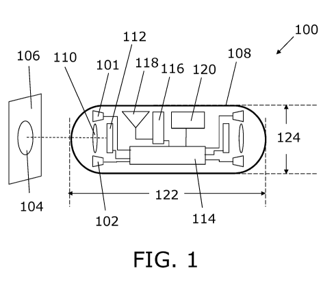
Figure 1 shows a schematic of an endoscopic capsule according to an embodiment
of the invention.
Figure 2 is a flow-chart of a method according to the invention.
CA 03124747 2021-06-11
WO 2019/122338 PCT/EP2018/086609
22
DETAILED DESCRIPTION OF AN EMBODIMENT
Figure 1 shows a schematic of an endoscopic capsule according to an embodiment
of the invention. More particularly, the figure shows an endoscopic capsule
100
comprising:
- a first imaging system, such as a first imaging system comprising
a white light emitting diode (LED) 101, refractive lens 110 and
two-dimensional image sensor 112 with colour filter array, and
- a second imaging system, such as a second imaging system
comprising a blue light emitting diode (LED) 102, refractive lens
110 and two-dimensional image sensor 112 with colour filter array
(note that the refractive lens 110 and two-dimensional image
sensor 112 with colour filter array is shared with the first imaging
system).
The endoscopic capsule 100 according to the present embodiment furthermore
comprises
- a third imaging system, such as a third imaging system comprising
a green LED (not shown), a refractive lens 110 and a two-
dimensional image sensor 112 with a colour filter array.
The first imaging system is arranged for obtaining images at a broader
spectral
range than the second imaging system.
The endoscopic capsule 100 according to the present embodiment furthermore
comprises
- a processor 114 being an FPGA,
wherein the endoscopic capsule is arranged for operating in any one of:
- a first mode wherein a frame rate (such as 1-32 fps) of the first
imaging system is higher than a frame rate (such as 0 fps) of the
second imaging system, and
- a second mode wherein the frame rate (such as 1-32 fps) of the
second imaging system is higher than the frame rate (such as 0
fps) of the second imaging system in the first mode.
CA 03124747 2021-06-11
WO 2019/122338 PCT/EP2018/086609
23
The frame rate of the first imaging system in the second mode may be 1-32 fps.
The processor (114) in the first mode is arranged for:
- accessing one or more images from the first imaging system,
- detecting if there is a polyp 104, such as a polyp on the inner
lining 106 of the colon, in the one or more images from the first
imaging system, and
- switching from the first mode to the second mode if a polyp 104 is
detected in the one or more images from the first imaging system.
The endoscopic capsule 100 according to the present embodiment comprises
imaging systems in both ends. This may decrease a risk that a polyp is missed
(e.g., by rotation of the camera around the polyp). The endoscopic capsule 100
according to the present embodiment comprises a power source 120, such as a
battery, an RF transmitter 116 and an RF antenna 118. The length 122 of the
endoscopic capsule may be within the range (both endpoints included) 10-40 mm,
such as within 20-30 mm. The diameter 124 of the endoscopic capsule may be
within the range (both endpoints included) 1-20 mm, such as within 5-15 mm.
Figure 2 is a flow-chart of a method according to the invention. More
particularly,
the figure shows a flow-chart of a method 230 of operating an endoscopic
capsule
100, such as a pre-delivered (pre-swallowed) endoscopic capsule 100, wherein
said method comprises:
- operating 231 the endoscopic capsule in the first mode,
- obtaining 232 one or more images 233 with the first imaging
system,
- accessing 234 with the processor the one or more images 233
from the first imaging system, such as accessing and image
analysing so as to obtain a quantification of likelihood 235 that the
image comprises a polyp 104,
- detecting 236 if there is a polyp 104 in the one or more images
from the first imaging system, such as determining if the likelihood
235 is above a predetermined threshold, and
- switching 238 from the first mode to the second mode if a polyp
104 is detected in the one or more images from the first imaging
system.
CA 03124747 2021-06-11
WO 2019/122338 PCT/EP2018/086609
24
In the present embodiment, the method further comprises:
- operating 238 the endoscopic capsule in the second mode,
- obtaining 240 one or more images 242 with the first imaging
system,
- accessing 244 with the processor the one or more images 242
from the first imaging system, such as accessing and image
analysing so as to obtain a quantification of likelihood 245 that the
image comprises a polyp 104,
- detecting 246 if there is a polyp 104 in the one or more images
from the first imaging system, such as determining if the likelihood
245 is above a predetermined threshold, and
- switching 248 from the second mode to the first mode if a polyp
104 is not detected in the one or more images from the first
imaging system.
In the present embodiment, the method further comprises:
- operating 238 the endoscopic capsule 100 in the second mode,
- obtaining 240 one or more images with the second imaging
system, and
- transmitting one or more images obtained in the second mode to a
database.
Although the present invention has been described in connection with the
specified embodiments, it should not be construed as being in any way limited
to
the presented examples. The scope of the present invention is set out by the
accompanying claim set. In the context of the claims, the terms "comprising"
or
"comprises" do not exclude other possible elements or steps. Also, the
mentioning
of references such as "a" or "an" etc. should not be construed as excluding a
plurality. The use of reference signs in the claims with respect to elements
indicated in the figures shall also not be construed as limiting the scope of
the
invention. Furthermore, individual features mentioned in different claims, may
possibly be advantageously combined, and the mentioning of these features in
different claims does not exclude that a combination of features is not
possible
and advantageous.