Note: Descriptions are shown in the official language in which they were submitted.
CA 03126499 2021-07-13
WO 2020/147985 1
PCT/EP2019/057647
METHODS FOR PURIFYING AN ALLERGEN EXTRACT
Field of the Invention
The invention relates to processes for producing semi-purified and purified
allergen extracts and
pharmaceutical compositions and vaccines for use in the diagnosis and
treatment of allergy.
Background of the Invention
Allergy is an acquired hypersensitivity disorder of the immune system and is
triggered by exposure
to harmless environment substances known as allergens. A type I
hypersensitivity reaction is
characteristic of allergic reactions and results in the production of
excessive amounts of IgE
antibodies which in turn activate basophils and mast cells causing an
inflammatory reaction. The
effects may be systemic such as vasodilation, mucus secretion, nerve
stimulation or smooth muscle
contraction causing an anaphylaxis reaction, or the effects may be confined to
a particular area of
the body, for example the respiratory system.
Phleum pratense
Grass allergy is one of the most common and prevalent forms of allergy that
affects sensitized
people during certain seasons. Grass pollen is present in the air in late
spring and early summer
months, which can cause allergic rhinitis, allergic conjunctivitis and asthma.
Direct skin contact with
grasses, when sitting on grass or mowing the lawn, can cause itching of the
skin, urticarial and atopic
dermatitis. One of the most representative species of grass is Phleum
pratense. At least nine
different allergens have been identified in the species of Phleum pratense and
include: Phl p 1, a
Beta-expansin of 27 kDa; Phl p 2, a Grass group II/III of 10-12 kDa; Phl p 4,
a protein of 55 kDa, Phl p
5 of 32 kDa, Phl p 6 of 11 kDa, Phl p 7 a calcium binding protein of 6 kDa,
Phl p 11, an Ole e 1-related
protein of 20 kDa, Phl p 12, a profilin of 14 kDa, and Phl p 13, a
polygalacturonase of 55 kDa.
Olea europaea
In the Mediterranean region, olive pollinosis is an important health problem
due to the extensive
cultivation of olive trees. Olive trees release large amounts of pollen into
the atmosphere. Currently,
12 allergens from 0. europaea have been described by the WHO/IUIS Allergen
Nomenclature Sub-
Committee; 11 from pollen and one (thaumatin, also known as Ole e 13) food
allergen from the olive
fruit. Ole e 1 is the major allergen, recognized by more than 70 % of olive
pollen sensitized patients.
Other allergens include a profilin (Ole e 2), polcalcins (Ole e 3 and Ole e
8), glucanases (Ole e 4 and
CA 03126499 2021-07-13
WO 2020/147985 2
PCT/EP2019/057647
Ole e 9), a superoxide dismutase (Ole e 5), a lipid transfer protein (Ole e
7), a glycosyl hydrolase (Ole
e 10), a pectin methylesterase (Ole e 11) and Ole e 6.
Dermatophagoides pteronyssinus
Mites are a major source of sensitization worldwide, especially in regions
where humidity and
temperature favour their development. Currently, house dust mites (HDMs)
belonging to the
Pyroglyphidae family are the most abundant mites in indoor habitats and thus
the main indoor
source of allergens. Nineteen allergens have been described in D.
pteronyssinus by the WHO/IUIS
Allergen Nomenclature Sub-Committee, 9 of them being proteases, having an
activity related with
allergenicity. The most important allergens are Der p 1 (a 25 kDa cysteine
protease) and Der p 2 (a
14 kDa protein of the N PC2 family).
Allergic patients can be treated with drugs to reduce their symptoms and
control the peaks of
symptoms or they can be treated with specific immunotherapy. However, specific
immunotherapy is
the only treatment with capacity to modify the course of the disease. Specific
immunotherapy (SIT)
involves the administration of increasingly larger doses of an allergen
extract with the aim of
inducing immunological tolerance. Allergen immunotherapy modulates the immune
response to the
allergen rather than ameliorating the symptoms induced by an allergic
reaction, and can either
reduce the need for medication, reduce the severity of symptoms or eliminate
hypersensitivity
altogether.
One of the risks of immunotherapy is that injection of an allergen to a
sensitised patient can cause
an allergic reaction or anaphylaxis. Since its first use at the beginning of
the 20th century, many
efforts have been made to further improve the safety and efficacy of allergen
immunotherapy. One
approach is the development of allergoids, which involves employing allergen
vaccines with reduced
allergenicity but with maintenance of immunogenicity.
EP 0 662 080 (CBF LETI SA) disclose a process for removal of substances and
other low molecular
weight material in order to purify the allergen extracts and to increase the
final allergen/protein
content. The process consists of disrupting the electrostatic, hydrophobic or
other physical forces
under such conditions as to detach non-allergenic compounds from the
allergenically active
proteins. The process can consist of a mild acid treatment by lowering the pH
below the pl of the
respective allergen proteins.
CA 03126499 2021-07-13
WO 2020/147985 3
PCT/EP2019/057647
One of the various ways of reducing allergenicity consists of chemically
modifying native allergen
extracts with aldehyde, mainly formaldehyde and glutaraldehyde, to produce
allergoids. This
aldehyde treatment leads to reaction products (mainly polymers), which have
lost part of their
allergenicity (i.e. exhibit a reduction of IgE reactive B-cell epitopes),
reducing allergic side-effects. At
the same time, the native immunogenicity of the allergen is retained. This
route of allergen
modification has been chosen by some manufacturers of allergen vaccines to
develop commercially
available products based on this principle.
However, there is still an interest in finding further methods for obtaining
safe and efficacious
medicaments for use in the immunotherapy of allergic disorders by optimising
the allergen
purification process to ensure that low molecular weight proteins, irritants
and toxic components
are eliminated.
Summary of the Invention
.. The inventors have found that depigmenting an allergen extract using a base
increases protein
content and can also increase the major allergen content and the biological
potency of the extract.
According to a first aspect of the present invention, there is provided a
process for producing a
depigmented allergen extract comprising:
a) basifying a native allergen extract; and
b) removing molecules having a molecular size of less than 3.5 kDa; and
c) adjusting the pH to neutrality;
thereby to produce a depigmented allergen extract.
The process may further comprise a polymerisation step, comprising:
d) contacting a depigmented allergen extract with an aldehyde; and
e) removing molecules having a molecular size of less than 100 kDa;
thereby to produce a depigmented polymerised allergen extract.
According to a second aspect of the invention, there is provided a depigmented
allergen extract
obtainable according to the process of the first aspect of the present
invention.
According to a third aspect of the invention, there is provided a depigmented
polymerised allergen
extract obtainable according to the process of the first aspect of the present
invention.
CA 03126499 2021-07-13
WO 2020/147985 4
PCT/EP2019/057647
According to a further aspect of the invention, there is provided a purified
allergen extract for use as
an active therapeutic substance in the treatment of allergy.
Definitions
"Allergen" can be defined as a molecule capable of inducing an IgE response
and/or a Type I allergic
reaction.
The term "native allergen extract" means an allergen extract which has been
extracted from a
source material and then treated to remove unbound low molecular weight
components.
The term "depigmented allergen extract" referred to herein can be defined as a
semi-purified
allergen extract obtained from a native allergen extract by removal of
allergenically irrelevant
substances bound to the allergen protein including the adsorbed pigments.
The term "depigmented polymerised allergen extract" referred to herein can be
defined as a purified
allergen extract where protein bands <100 kDa are not detectable by non-
reducing SDS-PAGE, and is
obtained by polymerising a depigmented allergen extract.
Detailed Description of the Invention
The allergen extracts of the invention are derived from any source material
comprising natural
allergens known to illicit an IgE mediated immune reaction in an individual.
Such allergens may
include air-borne allergens (e.g. pollen from grass, trees, herbs and weeds,
dust mites, fungi and
moulds), food allergens (e.g. peanuts), insect allergens (eg. from cockroaches
and fleas, and bee and
wasp venom) and epithelial allergens (animal hair and animal dander, for
example cat and dog
dander).
The source material may be any allergen, including food allergens (e.g.
peanuts), air-borne allergens
(e.g. pollen (such as tree pollen, weed pollen, grass pollen, and cereal
pollen), dust mites, fungi, and
moulds), epithelial allergens (animal hair and animal dander, for example cat
hair and dander, and
dog hair and dander) and insect allergens (e.g. from cockroaches and fleas,
and bee and wasp
venom).
CA 03126499 2021-07-13
WO 2020/147985 5
PCT/EP2019/057647
Pollen allergens from trees, grasses and weeds derive from the taxonomic order
group of Fagales
(e.g. Alnus and Betula), Lamiales (e.g. Oleo and Plantago), Poales (e.g.
Phleum pratense), Asterales
(e.g. Ambrosia and Artemisia), Cayophyllales (e.g. Chenopodium and SaIsola),
Rosales (e.g.
Parietaria), Proteales (e.g. Platanus), etc. Dust mites belong to the order
group of Astigmata (e.g.
Dermatophagoides and Euroglyphus). Airborne allergens derived from moulds and
fungi belong to
the order Pleosporales (e.g. Alternaria), Capnodiales (e.g. Cladosporium),
etc.
Air borne allergens may be selected from the groups: tree pollen (Alnus
glutinosa, Betula alba,
Corylus ayellana, Cupressus arizonica, Oleo europaea, Platanus sp), grass
pollen (Cynodon dactylon,
Dactylis glomerata, Festuca elatior, Holcus lanatus, Lolium perenne, Phleum
pratense, Phragmites
communis, Poo pratensis), weed pollen (Ambrosia elatior, Artemisia yulgaris,
Chenopodium album,
Parietaria judaica, Plantago lanceolata, SaIsola kali) and cereal pollen
(Ayena satiya, Hordeum
yulgare, Secale cereale, Triticum aestiyum, Zea mays), dust mites
(Dermatophagoides farinae,
Dermatophagoides microceras, Dermatophagoides pteronyssinus, Euroglyphus
maynei), storage
mites (Acarus siro, Blomia tropicalis, Lepidoglyphus destructor, Tyrophagus
put rescentiae,
Glycyphagus domesticus, Chortoglyphus arcuatus) and fungi and moulds
(Alternaria alternata,
Cladosporium herbarum, Aspergillus fumigatus).
Epithelial allergens may be selected from any animal including cat hair and
dander, dog hair and
dander, horse hair and dander, human hair and dander, rabbit hair and dander,
rat hair and dander,
mouse hair and dander, guinea pig hair and dander and feathers.
Arthropod allergens may be selected from insects, for example, ant, flea,
cockroach, wasp and bee
venom, or mites (Acarus siro, Blomia tropicalis, Dermatophagoides farinae,
Dermatophagoides
microceras, Dermatophagoides pteronyssinus, Euroglyphus maynei, Lepidoglyphus
destructor,
Tyrophagus putrescentiae and Chortoglyphus arcuatus).
Pollen allergens respond particularly well to base treatment. Pollen allergens
include tree pollen,
weed pollen, grass pollen and cereal pollen and are derived from the taxonomic
order group of
Fagales (e.g. Alnus and Betula), Lamiales (e.g. Oleo and Plantago), Poales
(e.g. Phleum pratense),
Asterales (e.g. Ambrosia and Artemisia), Cayophyllales (e.g. Chenopodium and
Sa/so/a), Rosales (e.g.
Parietaria), Proteales (e.g. Platanus) and the like.
CA 03126499 2021-07-13
WO 2020/147985 6
PCT/EP2019/057647
In one embodiment, the allergen extract is derived from a source material
which is a pollen. The
pollen may be selected from Phleum pratense, Olea europaea and Betula alba
(pendula).
More preferably, the source material is selected from peanut (Arachis
hypogaea), pollen (Phleum
pratense, Olea europaea and Betula alba (pendula)), mites (Dermatophagoides
pteronyssinus), and
epithelial (cat dander).
In a preferred embodiment of the invention, the source material is selected
from pollen (Phleum
pratense, Olea europaea and Betula alba (pendula)) and mites (Dermatophagoides
pteronyssinus).
More particularly, the source material is selected from Phleum pratense, Olea
europaea and Betula
alba (pendula).
In a more preferred embodiment of the invention, the source material is Phleum
pratense.
Alternatively, the source material is Olea europaea.
Alternatively, the source material is Dermatophagoides pteronyssinus.
One method for preparing a native allergen extract will now be described,
although it will be
appreciated that other suitable methods for obtaining a native allergen
extract will be known to the
skilled person and could be used as a starting material in the depigmentation
process of the present
invention.
The process for obtaining a native allergen extract from a source material may
comprise:
i) contacting the source material or first source material residue with a
liquid allergen
extraction agent to produce a second mixture of allergens dissolved in a
liquid phase, and a
solid phase comprising a second source material residue;
ii) subjecting the second mixture to a second separation step to isolate
the allergens dissolved
in the liquid phase, to produce a crude allergen extract;
iii) subjecting the crude allergen extract to a low molecular fraction
removal step to remove
molecules having a size of less than 1-10, preferably less than 3.5 kDa at 3-
10 degrees
centigrade;
CA 03126499 2021-07-13
WO 2020/147985 7
PCT/EP2019/057647
iv) carrying out step iii) at 3-10 degrees centigrade until the allergen
extract has a conductivity
below 900 uS/cm, measured at room temperature, to obtain a native allergen
extract;
The source material may first be treated to create a maximum surface area for
contact with the
liquid allergen extraction agent. The source material may be homogenised,
blended, crushed, or
powdered to produce a homogenous slurry for liquid extraction.
In certain instances, preliminary defatting steps are required which may
comprise:
i) contacting the source material comprising an allergen with a liquid
lipid extraction agent to
produce a first mixture containing lipids dissolved in a liquid phase and a
solid phase
consisting of a first source material residue comprising allergens and
proteins; and
ii) subjecting the first mixture to a first separation step to isolate the
first source material
residue.
The preliminary defatting steps may be required in instances where the source
material is pollen,
epithelia or food, such as nuts, e.g. peanut. The lipid extraction, or
"defatting" step, removes
lipophilic compounds such as lipids and fatty acids from the source material.
The liquid lipid extraction agent may be acetone, ether, or a similar solvent,
which may be cold.
Preferably the liquid lipid extraction agent is acetone. The lipid extraction
step may be performed in
a ratio of 1:1 weight ratio of source material to liquid lipid extraction
agent, or any ratio where the
weight of the liquid lipid extraction agent exceeds the weight of the source
material, for example
1:2, 1:3, 1:5, 1:10. The lipid extraction step is preferably performed at a
ratio of 1 kg of source
material to 2 L of liquid lipid extraction agent. The lipid extraction step is
preferably performed for
sufficient time for the lipids in the source material to dissolve in the
liquid lipid extraction agent,
which may be for over 1 minute, preferably over 5 minutes, more preferably
over 30 minutes, and
most preferably for 1 hour or more. The liquid extraction step may be
performed at a temperature
between 2 to 25 degrees centigrade, but is preferably performed cold at
between 2 to 6 degrees
centigrade, and more preferably between 3 to 5 degrees centigrade. During the
lipid extraction step,
the source material is preferably stirred or agitated with the liquid lipid
extraction agent.
The first separation step may be any suitable separation step known to the
skilled person, for
example the first separation step may be filtration or any other suitable
method.
CA 03126499 2021-07-13
WO 2020/147985 8
PCT/EP2019/057647
After the first separation step, the source material residue may be washed
with the liquid lipid
extraction agent. Optionally, the first source material residue may be further
extracted with the
liquid lipid extraction agent, then separated. Preferably, one, two or more
further lipid extraction
steps are performed. Filtration of the liquid extraction agent may be repeated
until it is transparent.
Other suitable separation methods known to the skilled person may also be
used.
After lipid extraction and separation, the source material residue may be
dried. The source material
residue may be dried at between 2 to 25 degrees centigrade, preferably at room
temperature. The
drying step is preferably continued for sufficient time to allow removal of
the liquid lipid extraction
agent from the source material residue, which may be between 1 to 24 hours, 6
to 18 hours,
although drying is preferably for at least 12 hours.
Allergens may be obtained from the ("defatted") first source material residue
by extraction with the
liquid allergen extract agent to produce a crude allergen extract comprising
allergens dissolved in a
liquid phase and a solid phase consisting of "unwanted" non-allergenic
residues. The liquid allergen
extract agent may be an aqueous solution, and preferably comprises a buffering
agent. The liquid
allergen extraction agent may comprise PBS and/or NaCI, for example it may be
an aqueous solution
of 0.01 M PBS/0.15 M NaCI, or an aqueous solution of ammonium bicarbonate
((NH4)HCO3) and/or
NaCI, for example an aqueous solution of 0.125 M (NH4)HCO3/0.15 M NaCI. The
first source material
residue may be extracted in the liquid allergen extract agent in any ratio
where the weight of the
liquid allergen extract agent exceeds the weight of the first source material
residue, for example
weight ratios of 1:2, 1:3, 1:5, 1:10, 1:20, 1:50, 1:80. Preferably, the first
source material residue is
extracted in the liquid allergen extract agent in a ratio of 1:10 by weight
first source material
residue:liquid allergen extract agent. The ratio of the first source material
residue to liquid allergen
extract agent may vary but should be such that the allergens in the first
source material residue can
dissolve in the liquid allergen extract agent. The extraction of the first
source material residue with
the liquid allergen extract agent is preferably performed for sufficient time
for the allergens in the
first source material residue to dissolve in the liquid allergen extraction
agent, which may be for
between 30 minutes to 12 hours, preferably between 1 to 6 hours, more
preferably between 2 to 5
.. hours, and most preferably for around 4 hours. The allergen extraction step
may be performed at
between 2 to 25 C, but is preferably performed cold at between 2 to 6 degrees
centigrade, and
more preferably between 3 to 5 degrees centigrade. During the allergen
extraction step, the first
source material residue is preferably stirred or agitated with the liquid
allergen extraction agent.
CA 03126499 2021-07-13
WO 2020/147985 9
PCT/EP2019/057647
After the allergen extraction step, the allergens dissolved in the liquid
phase may be separated from
the second source material residue, to produce a crude allergen extract. The
separation step is
preferably centrifugation, although many techniques to separate solid from
liquid are applicable,
these being well known to a person skilled in the art. Preferably, the
allergens dissolved in the liquid
phase are centrifuged at between 2 to 6 degrees centigrade, and preferably
between 3 to 5 degrees
centigrade, for sufficient time to sediment the source material residue as a
pellet, for example
between 1 minute to 1 hour, or over 1 hour. The crude allergen extract (i.e.
the supernatant
containing the dissolved allergens) may be stored at between 2 to 6 degrees
centigrade. The second
source material residue pellet may be further extracted with the liquid
allergen extract agent using
the same conditions as the first allergen extraction step, and preferably for
a longer extraction
period such as between 4 to 8 hours, 8 to 12 hours, or over 12 hours. After
the second allergen
extraction step, the allergens dissolved in the liquid phase may be separated
from the second source
material residue to produce a crude allergen extract. The crude allergen
extracts from the first and
second allergen extraction steps are preferably pooled for further treatment.
The crude allergen extract may be filtered, for example using 0.45 um pore
size. The crude allergen
extract may be subjected to a low molecular fraction removal step to remove
molecules having a
low molecular size such as salts and other non-allergenic compounds. In step
iii) molecules having a
molecular size of less than 1-10, preferably less than 3.5 kDa may be removed.
The low molecular
fraction removal step is preferably continued at 3-10, preferably 3-5 C until
the conductivity of the
allergen extract is less than 900 uS/cm, or less than 800 uS/cm, or less than
700 uS/cm, or less than
600 uS/cm, or more preferably less than 500 uS/cm (measured at room
temperature).
The resulting native allergen extract may be filtered, for example using 0.45
and/or 0.22 um pore
size.
The native allergen extract may be used in the preparation of a pharmaceutical
composition or
vaccine for standardisation, diagnosis, synthesis and vaccination purposes.
In the present invention, the native allergen extract is used as the starting
material in the
depigmentation method described herein.
The present invention provides a depigmentation method comprising a basifying
treatment, wherein
non-allergenic compounds adhering to the allergens/proteins are removed using
means which
CA 03126499 2021-07-13
WO 2020/147985 10
PCT/EP2019/057647
disrupt electrostatic, hydrophobic or other physical forces being responsible
for the adherence of
the non-allergenic compounds to the proteins, to produce a depigmented
extract.
The basifying treatment comprises:
a) basifying the native allergen extract;
b) removing molecules having a molecular size of less than 3.5 kDa; and
c) adjusting the pH to neutrality to produce a depigmented allergen
extract.
The basifying treatment comprises either mild base treatment or strong base
treatment. In the base
treatment the pH of the allergens/proteins may be increased to at least pH 7,
for example a pH value
of between 7 and 11. The preferred pH of the allergen proteins is between 7
and 10, more preferred
is a pH between 7 and 8. A pH value of greater than 11 may lead to the protein
profile of the
depigmented allergen extract being incomplete, and a neutral pH, for example
pH 6, leads to
incomplete elimination of the non-allergenic compounds in the resulting
depigmented allergen
extract.
In one embodiment, the base treatment comprises basifying the allergen extract
to pH 7, pH 8, pH 9,
pH 10 or pH 11.
In one embodiment, the base treatment comprises basifying the allergen extract
to pH 7 to 11, or pH
7 to 10, preferably between pH 7 and 8.
The pH of the native allergen extract may be increased using any suitable
base. The base may be a
strong base or a weak base. Strong bases include sodium hydroxide, lithium
hydroxide and
potassium hydroxide. Weak bases include urea, ammonium hydroxide and
methylamine. In one
embodiment the base is selected from the list comprising sodium hydroxide,
lithium hydroxide,
potassium hydroxide, urea, ammonium hydroxide or methylamine. In particular,
the base is sodium
hydroxide.
The basified extract may be maintained at a basic pH for 1 minute to 24 hours,
1 minute to 4 hours,
1 to 60 minutes, preferably 5 to 30 minutes, more preferably 10 to 20 minutes,
and most preferably
around 15 minutes.
CA 03126499 2021-07-13
WO 2020/147985 11
PCT/EP2019/057647
Molecules having a molecular size of less than 3.5 kDa may be removed in a low
molecular fraction
removal step.
After the basifying treatment, the resulting depigmented allergen extract may
be collected, and the
pH of the extract adjusted using a suitable acid, for example HCI. The pH may
be adjusted to a value
where precipitation of the proteins is avoided, for example between pH 7.0 and
7.5, more
particularly between pH 7.3 and 7.4.
In particular, the basifying treatment may comprise:
a) basifying the native allergen extract to pH 7 to 11 and maintaining the
basified extract for 1
minute to 24 hours, for example, 5 to 30 minutes, preferably 15 minutes;
b) subjecting the extract to a low molecular fraction removal step to
remove molecules having
a molecular size of less than 3.5 kDa; and
c) adjusting the pH to between 7.0 and 7.5, in particular 7.3 to 7.4, to
produce a depigmented
allergen extract.
The basifying treatment may comprise basifying the native allergen extract to
pH 7 to 10.
The low molecular fraction removal step may be a dialysis step, where the
extract is dialysed against
a dialysate such as purified water or a buffer. The low molecular fraction
removal step may be
performed at between 2-25 degrees centigrade, but is preferably performed cold
at between 2-6
degrees centigrade, and most preferably between 3-5 degrees centigrade. The
low molecular
fraction removal step may be performed for 12-24 hours, where the solvent, or
in the case of
dialysis, the dialysate, is regularly changed to maintain the reaction.
The resulting depigmented allergen extract may be filtered, for example using
a 0.45 um and/or 0.22
um pore size, and may be frozen or freeze dried for storage.
The extracts produced using the process of the present invention can be
further treated. The process
may further comprise a polymerisation step, comprising:
d) contacting a depigmented allergen extract with an aldehyde, and after
polymerization,
e) removing molecules having a molecular size of less than 100 kDa.
The aldehyde may be any suitable aldehyde, for example glutaraldehyde or
formaldehyde.
CA 03126499 2021-07-13
WO 2020/147985 12
PCT/EP2019/057647
The polymerisation step may comprise:
d) contacting a depigmented allergen extract with glutaraldehyde or
formaldehyde,
e) subjecting the extract to a molecular fraction removal step to remove
molecules having a
molecular size of less than 100 kDa, and
f) carrying out step e) at 3-15, preferably 3-5 degrees centigrade until
the allergen extract has
a conductivity of below 210 uS/cm (measured at room temperature) and/or until
the extract
is absent of glutaraldehyde, to obtain a depigmented polymerised allergen
extract.
Where the extract for polymerisation is freeze-dried, it may be reconstituted
in a buffer, for example
0.01M PBS/0.15M NaCI, to a final concentration of 0.1-500 mg/ml, preferably 1-
100 mg/ml, and
most preferably 10-50 mg/ml.
The polymerisation reaction is preferably performed to completion, such that
protein bands <100
kDa (e.g. 14-25 kDa) are not detectable by non-reducing SDS-PAGE in the
depigmented polymerised
allergen extract.
Increasing concentrations of glutaraldehyde may decrease polymer yield and
increase residue yield
obtained by any centrifugation step before dialysis. In contrast to previously
known polymerisation
conditions employing a glutaraldehyde concentration of around 5 mg/ml (i.e.
0.009 ml
glutaraldehyde per ml of allergen extract), the optimal glutaraldehyde
concentration was
experimentally determined to be approximately double that of the known amount
for some
allergens (peanut, cat epithelia and Ambrosia), i.e. 10 mg/ml (0.02 ml
glutaraldehyde per ml of
allergen extract). The aldehyde may be added in a range of 1-20 mg/ml. Whilst
employing previously
.. known amounts of a final concentration of glutaraldehyde can lead to some
polymerisation of the
allergens, it is preferred that the aldehyde is added at a final concentration
of 5-10 mg/ml or in a
ratio of 0.01-0.02 ml glutaraldehyde per ml of extract to achieve optimal
polymerisation.
Decreasing the addition rate of glutaraldehyde may also decrease polymer yield
and increase
residue yield. The aldehyde may be added to the extract at a constant speed,
for example between
0.001-0.5m1 per minute (1-500 ul/min or 60-3000 ul/hour).
The polymerisation reaction may be maintained for between 1-12 hours,
preferably 7 hours at room
temperature or higher. The polymerisation reaction may be stopped using
glycine in a proportion of
CA 03126499 2021-07-13
WO 2020/147985 13
PCT/EP2019/057647
40 mg per ml of depigmented polymerised allergen extract solution. The stopped
reaction may be
maintained overnight at 3-5 C, preferably under stirring. The depigmented
polymerised allergens in
the liquid phase may be separated from insoluble residue to produce a
depigmented polymerised
allergen extract. The separation step is preferably centrifugation, although
many separation
techniques are applicable, these being well known to a person skilled in the
art. Preferably, the
extract is centrifuged at between 2-6 degrees centigrade, and preferably
between 3-5 degrees
centigrade, for sufficient time to sediment the insoluble residue as a pellet,
for example between 1
minute to 1 hour, or over 1 hour. The supernatant (containing the soluble
depigmented polymerised
allergens) may be collected and subjected to a molecular fraction removal step
e).
In step e) molecules having a molecular size of less than 100 kDa are removed.
Preferably the molecular fraction removal step is a dialysis step, where the
extract is dialysed against
a dialysate such as purified water or a buffer, at 3-15, preferably 3-5
degrees centigrade. The
molecular fraction removal step may be continued at 3-15, preferably 3-5
degrees centigrade until
the conductivity measured at room temperature is less than 300 uS/cm, more
preferably less than
250 uS/cm, most preferably less than 210 uS/cm.
The resulting depigmented polymerised allergen extract may be filtered, for
example using 0.45 um
and/or 0.22 um pore size, and may be frozen or freeze dried for storage.
Any of low molecular fraction removal steps described herein, for example
steps b) or e), may
comprise an ultrafiltration step, a diafiltration step, a dialysis step, or
filtration.
In its simplest form the process of the present invention may comprise
preparing or obtaining a
native allergen extract and basifying the extract, for example, via mild or
strong base treatment, to
remove non-allergenic compounds having a low molecular size. The extract may
then be
polymerised using an aldehyde. The native allergen extract may be peanut,
pollen, grass, epithelial,
mould, fungi, insect or mite allergens, in particular grass, pollen or mite,
more particularly pollen
allergens. The process of the present invention yields an allergen extract
which exhibits reduced IgE
binding capacity but which retains its immunogenic capacity.
The present invention further comprises a treatment for allergy and a
diagnostic drug for allergy,
both comprising allergen extracts produced by the processes of the present
invention, as the active
CA 03126499 2021-07-13
WO 2020/147985 14
PCT/EP2019/057647
ingredient. The allergy may be associated with exposure to various allergens
which illicit an IgE
mediated allergic response as discussed herein.
According to a second aspect of the present invention there is provided a
depigmented allergen
extract obtainable according to the process of the first aspect of the present
invention.
According to a third aspect of the present invention there is provided a
depigmented polymerised
allergen extract obtainable according to the process of the first aspect of
the present invention.
There is provided a purified allergen extract for use as an active therapeutic
substance.
The allergen extract may be selected from peanut (Arachis hypogaea), pollen
(Phleum pratense,
Betula alba Olea europaea, Parietaria judaica, and Cupressus arizonica), mites
(Dermatophagoides
pteronyssinus), and epithelial (cat dander). In particular the allergen
extract is pollen selected from
Olea europaea and Phleum pratense.
The allergen extract may be for use in the treatment of allergy. In a
preferred embodiment the
allergen extract of Olea europaea or Phleum pratense may be for use in the
treatment of pollen
allergy.
The depigmented polymerised allergen extract may be characterised by the
following
physicochemical and biological properties:
i. Soluble in water,
ii. Absence of non-polymerised allergens/proteins with a molecular weight
lower than 100 kDa
(identified as bands by SDS-PAGE in non-reducing conditions)
iii. Absence of IgE recognition bands with a molecular weight lower than
100 kDa (identified by
immunoblot in non-reducing conditions)
iv. Absence of polymerised molecules with a molecular weight lower than 100
kDa (determined
by SDS PAGE)
v. Reduction of the biological potency (95%) with respect to the native
allergen extract
(determined by IgE [LISA inhibition experiments using a specific pool of sera
from sensitized
individuals) and
vi. Absence of abnormal toxicity in mice and guinea pigs.
CA 03126499 2021-07-13
WO 2020/147985 15
PCT/EP2019/057647
In particular, the depigmented polymerised allergen extract is characterised
by a reduction of the
biological potency (95%) with respect to the native allergen extract
(determined by IgE [LISA
competition experiments using a specific pool of sera from sensitized
individuals).
The allergen extracts of the present invention may be for use as an active
component of a
medicament for the treatment of an allergic individual, with the aim of
inducing tolerance to certain
allergens.
There is provided the use of an allergen extract according to the present
invention in diagnostics for
immunological disorders, preferably to detect allergic disease. There is
provided the use of an
allergen extract according to the present invention for the treatment of
allergy or in the
manufacture of a medicament for the treatment of allergy, for example pollen
allergy. The use may
be for immunotherapy. The use may be for standardisation, diagnosis, synthesis
and vaccination
purposes. The use may be in therapeutic treatment of patients, preferably in
immunotherapy. The
use may be in monitoring the patients during immunotherapy.
Alternatively, there is provided a method for treating a person in need
thereof for allergy, such as
pollen allergy, comprising the step of administering to the person in need
thereof the allergen
extracts of the invention.
According to a further aspect of the present invention there is provided a
pharmaceutical
composition comprising an allergen extract according to the present invention.
There is provided a
pharmaceutical composition for the treatment of allergy which comprises as the
active ingredient a
pharmaceutically effective amount of an allergen extract according to the
present invention and at
least one pharmaceutically acceptable carrier or diluent. There is provided a
diagnostic composition
for allergy which comprises as the active ingredient a diagnostically
effective amount of an allergen
extract according to the present invention.
According to a further aspect of the present invention there is provided a
vaccine comprising an
allergen extract according to the present invention. The pharmaceutical
composition and vaccine
may further comprise one or more adjuvants, diluents, preservatives or
mixtures thereof. The
pharmaceutical composition or vaccine may comprise a physiologically
acceptable carrier. As used
herein, the phrase "pharmaceutically acceptable" preferably means approved by
a regulatory agency
CA 03126499 2021-07-13
WO 2020/147985 16
PCT/EP2019/057647
of a government, or listed in the European or US. Pharmacopeia or another
generally recognized
pharmacopeia for use in humans.
Such pharmaceutically acceptable carriers can be sterile liquids, such as
water and oils, including
those of petroleum, animal, vegetable or synthetic origin, such as peanut oil,
soybean oil, mineral oil,
sesame oil and the like. Saline solutions and aqueous dextrose and glycerol
solutions can also be
employed as liquid carriers, particularly for injectable solutions. Suitable
pharmaceutical excipients
include mannitol, human serum albumin (HSA), starch, glucose, lactose,
sucrose, gelatin, malt, rice,
flour, chalk, silica gel, magnesium carbonate, magnesium stearate, sodium
stearate, glycerol
.. monostearate, talc, sodium chloride, dried skim milk, glycerol, propylene,
glycol, water, ethanol and
the like.
There is provided a vaccine obtainable according to the process of the first
aspect of the present
invention. The vaccine may be for sub-cutaneous or sub-lingual use or
epicutaneous.
There is provided the use of a vaccine according to the present invention in
the treatment of allergy,
or in the manufacture of a medicament for the treatment of allergy.
According to a further aspect of the present invention there is provided a
method of preventing an
allergen sensitisation comprising the step of: exposing an individual to an
effective amount of an
allergen extract, the pharmaceutical composition or the vaccine of the present
invention.
According to a further aspect of the present invention there is provided a
method of treating an
allergy in a sensitised individual, comprising administering to the individual
an effective amount of
an allergen extract, the pharmaceutical composition or the vaccine of the
present invention. The
allergen extract, the pharmaceutical composition or the vaccine may be
administered sub-
cutaneously, or sub-lingually, and may be administered as an increasing or
constant dosage.
The individual may be a human or an animal, preferably a human.
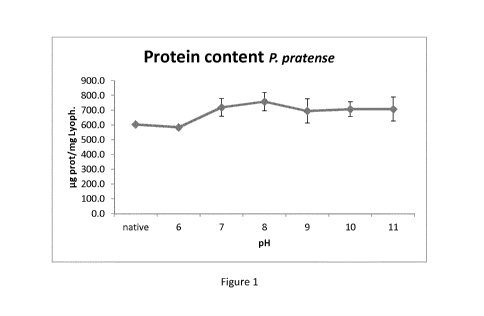
Brief Description of the Figures
Figure 1 shows protein content of P. pratense extracts (determined using Lowry-
Biuret method) of
lyophilized samples obtained after different pH treatments. Error bars refer
to the standard
deviation of different samples' mean value.
CA 03126499 2021-07-13
WO 2020/147985 17
PCT/EP2019/057647
Figure 2 shows protein content of P. pratense extracts (determined using Lowry-
Biuret method) of
lyophilized samples obtained after treatment with different bases. Error bars
refer to the standard
deviation of different samples' mean value.
Figure 3 shows Phl p 5 (Phleum major allergen) content of lyophilized samples
obtained after
different pH treatments. Error bars refer to the standard deviation of
different samples' mean value.
Figure 4 shows Phl p 5 (Phleum major allergen) content of lyophilized samples
obtained after
treatment with different bases. Error bars refer to the standard deviation of
different samples' mean
value.
Figure 5 shows biological potency (ELISA competition) of P. pratense extracts
of lyophilized samples
obtained after different pH treatments. Error bars refer to the standard
deviation of different
samples' mean value.
Figure 6 shows biological potency (ELISA competition) of P. pratense extracts
of lyophilized samples
obtained after treatment with different bases. Error bars refer to the
standard deviation of different
samples' mean value.
Figure 7 shows lig necessary to obtain 50% inhibition of IgE binding to native
extract of P. pratense
extracts of lyophilized samples obtained after different pH treatments. Error
bars refer to the
standard deviation of different samples' mean value.
Figure 8 shows lig necessary to obtain 50% inhibition of IgE binding to native
extract of P. pratense
extracts of lyophilized samples obtained after treatment with different bases.
Error bars refer to the
standard deviation of different samples' mean value.
Figure 9 shows SDS of P. pratense extracts treated with different bases.
Figure 10 shows western-blot of P. pratense extracts treated with different
bases.
CA 03126499 2021-07-13
WO 2020/147985 18
PCT/EP2019/057647
Figure 11 shows protein content of 0. europaea extracts determined using Lowry-
Biuret method of
lyophilized samples obtained after treatment with different pH. Error bars
refer to standard
deviation of different samples mean value.
Figure 12 shows protein content of 0. europaea extracts determined using Lowry-
Biuret method of
lyophilized samples obtained after treatment with different bases. Error bars
refer to the standard
deviation of different samples mean value.
Figure 13 shows biological potency of 0. europaea extracts determined using
ELISA competition
method of lyophilized samples obtained after treatment with different pH.
Error bars refer to the
standard deviation of different samples mean value.
Figure 14 shows biological potency of 0. europaea extracts determined using
ELISA competition
method of lyophilized samples obtained after treatment with different bases.
Error bars refer to the
standard deviation of different samples mean value.
Figure 15 shows lig necessary to obtain 50% inhibition of IgE binding to
native extract of 0. europaea
of lyophilized samples obtained after treatment with different pH. Error bars
refer to the standard
deviation of different samples mean value.
Figure 16 shows lig necessary to obtain 50% inhibition of IgE binding to
native extract of 0. europaea
of lyophilized samples obtained after treatment with different bases. Error
bars refer to the standard
deviation of different samples mean value.
Figure 17 shows SDS of 0. europaea extracts treated with different bases.
Figure 18 shows western-blot of 0. europaea extracts treated with different
bases.
Figure 19 shows thin layer chromatography for 0. europaea. A, samples treated
with sodium and
.. lithium hydroxide; B, treatments with ammonium hydroxide, sodium hydroxide
and urea; C,
treatments with methylamine. All assays were compared with native extract.
Standards are: 1,
chlorogenic acid; 2, quercetin; 3, rutin trihydrate; 4, isoquercitrin; 5,
quercitrin; 6, kaempferol 3-
glucoside; 7, apigenin 7-glucoside.
CA 03126499 2021-07-13
WO 2020/147985 19
PCT/EP2019/057647
Figure 20 shows protein content of D. pteronyssinus extracts determined using
Lowry-Biuret method
of lyophilized samples obtained after treatment with different pH. Error bars
refer to the standard
deviation of different samples mean value.
Figure 21 shows protein content of D. pteronyssinus extracts determined using
Lowry-Biuret method
of lyophilized samples obtained after treatment with different bases. Error
bars refer to the standard
deviation of different samples mean value.
Figure 22 shows major allergen content of D. pteronyssinus extracts determined
using specific ELISA
sandwich kit (Indoor) for Der p 1 and Der p 2of lyophilized samples obtained
after treatment with
different pH. Error bars refer to the standard deviation of different samples
mean value.
Figure 23 shows major allergen content of D. pteronyssinus extracts determined
using using specific
ELISA sandwich kit (Indoor) for Der p 1 and Der p 2 of lyophilized samples
obtained after treatment
with different acid or base. Error bars refer to the standard deviation of
different samples mean
value.
Figure 24 shows biological potency of D. pteronyssinus extracts determined
using ELISA competition
method of lyophilized samples obtained after treatment with different pH.
Error bars refer to the
standard deviation of different samples mean value.
Figure 25 shows biological potency of D. pteronyssinus extracts determined
using ELISA competition
method of lyophilized samples obtained after treatment with different pH.
Error bars refer to the
standard deviation of different samples mean value.
Figure 26 shows biological potency of D. pteronyssinus extracts determined
using ELISA inhibition
method of lyophilized samples obtained after treatment with different pH (rig
necessary to obtain
50% inhibition of IgE binding to native extract). Error bars refer to the
standard deviation of different
samples mean value.
Figure 27 shows biological potency of D. pteronyssinus extracts determined
using ELISA inhibition
method of lyophilized samples obtained after treatment with different bases
(rig necessary to obtain
50% inhibition of IgE binding to native extract). Error bars refer to the
standard deviation of different
samples mean value.
CA 03126499 2021-07-13
WO 2020/147985 20
PCT/EP2019/057647
Figure 28 shows SDS of D. pteronyssinus extracts treated with different bases.
Figure 29 shows western-blot of D. pteronyssinus extracts treated with
different bases.
The present invention is illustrated by the following examples which detail
processes for the
preparation, purification and basification of extracts comprising allergens.
Methods A-C detail the processes used to make the allergen extracts.
Methods
A. Optional defatting process of raw allergen material
Defatted extract was obtained. In general, homogenised material was defatted
in acetone at 3-5 C,
and filtered. This step was repeated until the acetone was transparent. The
defatted material was
recovered and dried at room temperature until all the acetone had been
removed.
B. Preparation of native allergen extract
Dried defatted material was weighed and extracted in 0.01 M PBS/0.15M NaCI in
a proportion 1:10
for 4 hours at 3-5 C under magnetic stirring. Afterwards, the solution was
centrifuged for 30 minutes
at 4 C at 10.000 r.p.m. The resulting supernatant was collected and stored at
3-5 C and the pellet
was reconstituted in 0.01 M/NaCI 0.15M (1:10) and extracted overnight at 3-5 C
under magnetic
stirring. The solution was centrifuged for 30 minutes at 3-5 C at 10.000 r.p.m
and the supernatant
was collected and mixed with the previously obtained fraction. The combined
extract was filtered
using 0.45 um pore size and extensively dialyzed in 3 kDa cut-off dialysis
membranes until the
conductivity was lower than 500 uS/cm. The extract was then filter sterilized
using 0.22 um pore
size.
C. Preparation of depigmented allergen extract
Native extract in aqueous solution and maintained at 3-5 C was further treated
using the following
procedure. Under magnetic stirring, the pH of the solution was adjusted to pH
7-11 by addition of
sodium hydroxide, lithium hydroxide, potassium hydroxide, urea, ammonium
hydroxide or
methylamine and maintained under these conditions for 15 minutes. Afterwards
the extract was
dialyzed in 3.5 kDa cut-off dialysis membranes with purified water for
approximately 17 hours
against 10 volumes of purified water at 3-5 C. Purified water was substituted
4 times during this
CA 03126499 2021-07-13
WO 2020/147985 21
PCT/EP2019/057647
period. After the base treatment, the extract was collected and the pH
adjusted to 7.3-7.4 using
0.1M HCI. Finally the extract was sterile filtered until 0.22 um, frozen and
freeze-dried.
Immunological Characterisation
Protein content
The protein content of native and depigmented extracts was measured by the
Lowry Biuret method
following the manufacturer's instructions.
Sodium Dodecyl Sulfate Polyacrylamide Gel Electrophoresis (SDS-PAGE)
Protein profiles were identified by SDS¨PAGE under reducing conditions
(samples incubated with [3-
mercaptoethanol and heated for 10 minutes at 95 C) in 2.67% C, 15% T
acrylamide -acrylamide gels.
Samples and Low Molecular Weight Standard (BioRad Laboratories, Hercules, CA,
USA) were run in
the same gel. Gels were stained with 0.1% Coomassie Brilliant Blue R-250
(BioRad).
Immuno-blot
Electrophoretically separated proteins (by SDS-PAGE) were transferred to a
PVDF membrane (Trans-
Blot Turbo TM Transfer Pack, BioRad) and incubated overnight with sera from
patients sensitized to
each allergen (Plasmalab International, Everett, WA, USA) diluted in 0.01M
Phosphate Buffer
Solution (PBS)-0.1% Tween. Plasmalab International operates in full compliance
with Food and Drug
Administration regulations. Specific IgE binding to the extract was detected
with peroxidase-
conjugated monoclonal antibodies, antihuman-IgE-P0 (Ingenasa, Madrid, Spain),
developed with
luminol solutions (Western lmmun-StarTM Western CTM Kit, Bio-Rad) and detected
by
chemiluminescence (ChemiDoc XRS, Bio-Rad).
Major allergen quantification
Major allergens were quantified using [LISA sandwich method using enzyme-
linked immunosorbent
assay detection kits (Indoor Biotechnologies, VA, USA). Briefly, Nunc Maxisorp
plates (Thermo
Scientific, Waltham, MA, USA) were coated with a specific monoclonal antibody
diluted in
carbonate/bicarbonate buffer (pH = 9.6) and incubated over night at 4 C.
Afterwards, plates were
blocked with BSA 1 % in PBS 0.01 M - Tween 0.05 %. Then, samples and standard
were added in
serial one half dilutions with BSA 1 % in PBS 0.01 M - Tween 0.05 %. Specific
secondary monoclonal
antibody (biotinylated) was added and streptavidin was finally used. Reaction
with development
solution (chromogen) was measured at OD 450 nm after stopping with sulfuric
acid. Standard curve
CA 03126499 2021-07-13
WO 2020/147985 22
PCT/EP2019/057647
was obtained using a 4-parameters logistic fit by the least-squares method,
where samples
concentrations were interpolated to obtain the results.
ELISA competition (IgE)
Native and depigmented extracts' capacity to inhibit IgE binding to each in-
house reference
preparation (IHRP) biologically standardized was compared. Nunc plates (Thermo
Scientific) were
coated with anti-IgE. A pool of serum from patients sensitized to the allergen
was incubated in the
plate. Dilutions of the sample and IHRP were incubated with the allergen
labelled with peroxidase.
The mixture was added to the coated plate and incubated. Afterwards,
development solution
(chromogen) was added, stopped with sulfuric acid and optical density (OD)
measured at 450 nm.
ELISA inhibition (IgE)
In vitro allergenic activity of the extracts (native and depigmented) was
tested by means of ELISA
inhibition, establishing the 50% inhibition point, using a native extract as
reference. Plastic microtiter
plates (Immulon 4HBX; Thermo Scientific) were coated with the native extract
(10 lig of protein/I'M
overnight. Serial 1:2 dilutions were made from the native and depigmented
extracts in a Nunc F
plate (Thermo Scientific). Each dilution was incubated with a serum pool for 2
hours at room
temperature. Afterward, the dilutions of the extracts were transferred to the
native coated plates
and incubated for 2 hours. After washing, 100 ul of anti-human IgE peroxidase
was added and let to
stand for 30 minutes at room temperature. After washing, the plates were
developed for 30 minutes
(chromogen) and stopped with sulfuric acid (1 N).
Thin layer chromatography (TLC)
Plant flavonoids were used as positive controls. Controls and standards were
applied over a TLC
aluminium sheet silica gel 60F (Merck, Darmstadt, Germany). Ethyl-acetate:
formic acid: acetic acid:
water (100:11:11:27) was used as eluent, and developed using solution 1%
methanolic diphenylboric
acid-P-ethylamino ester followed by 5% ethanolic Poly(ethylene glycol)-4000.
Examples
Example 1: Phleum pratense
Depigmented Phleum protense extract was obtained in accordance with method
steps A to C.
Protein Content
CA 03126499 2021-07-13
WO 2020/147985 23
PCT/EP2019/057647
Maximum protein content was obtained after treatment using methylamonium pH 8
(865 lig
protein/mg lyophilized extract), and minimum content corresponds to potassium
hydroxide pH 11
treatment (579 lig protein/mg lyophilized extract) (Table 1). Mean value
across all pHs was 718 lig
protein/mg lyophilized extract.
The highest protein content corresponded to pH 8 treatments (mean value 758
lig prot/mg lyoph),
and the lowest to pH 9 (694 lig prot/mg lyoph) (Table 2) (Figure 2). The
protein content at all pHs
between 7 to 11 was higher than native extract and the sample at pH 6, which
was the original pH of
the sample (sample treated the same as the depigmented samples, but without
the basifying pH
change).
Regarding different bases used, the highest protein content was achieved using
lithium hydroxide
(739 lig prot/mg lyoph), and the lowest with urea (668 lig prot/mg lyoph)
(Table 3, Figure 2).
Major Allergen Quantification
The lowest level corresponded to ammonium hydroxide treatment at pH 10 (13.0
lig Phl p 5/mg
lyophilized extract) (Table 1). The highest levels corresponded to lithium
hydroxide treatment at pH
8, followed by ammonium hydroxide treatment at pH 11 (41.0 and 38.0 lig Phl p
5/mg lyophilized
extract, respectively). Mean depigmented value was 26.6 lig Phl p 5/ mg
lyophilized extract.
The highest major allergen content was obtained in treatments at pH 8 (31.5
lig Phl p 5/mg
lyophilized extract) and the lowest at pH 10 (23.2 lig Phl p 5/mg lyophilized
extract) (Figure 3, Table
2). However, all treatments with bases yielded higher major allergen content
than native extract and
sample at pH 6 (without treatment).
The lowest major allergen content was obtained in treatments using sodium
hydroxide (24.1 lig Phl
p 5/mg lyophilized extract), and the highest using potassium hydroxide (29.1
lig Phl p 5/mg
lyophilized extract) (Figure 4, Table 3).
[LISA competition (IgE) ¨ Biological Potency
The highest biological potency corresponded to samples treated with methyl
ammonium pH 7 and 9
(3052 and 2909 HEPL/mg lyophilized extract, respectively) (Figures 5 and 6,
Table 1). The lowest
value corresponds to treatments with potassium hydroxide (mean value of 984
HEPL/mg lyophilized
extract), similar to native extract (952 HEPL/mg) (Table 3). Differences could
be detected between
CA 03126499 2021-07-13
WO 2020/147985 24 PCT/EP2019/057647
ammonium hydroxide and other groups (potassium hydroxide P = 0.010, Tukey
Test; urea P = 0.005,
sodium hydroxide P = 0.012, and lithium hydroxide P = 0.049, Mann-Whitney),
except for
methylamine. There were also differences between methylamine and potassium
hydroxide. Mean
value of depigmented samples was 1685 HEPL/mg lyophilized extract, higher than
results obtained
with native extract and sample at pH 6 (without treatment) (952 and 1262
HEPL/mg lyophilized
extract, respectively).
[LISA inhibition (IgE)
Micrograms of lyophilized lig necessary to reach 50% inhibition did not show
correlation with
HEPL/mg values (Pearson Product Moment Correlation, P > 0.050).
50% inhibition values at pH 8 were significantly higher than treatments at pH
11 and 10 (P = 0.030
and P = 0.017, respectively, Mann-Whitney Rank Sum Test) (Figure 7, Table 2).
The lowest value
corresponded to potassium hydroxide at pH 11 (0.007 lig), followed by ammonium
hydroxide at pH
11 (0.020 lig, respectively) (Table 1). Mean value was 0.102 lig, similar to
native extract and sample
at pH 6 (0.105 and 0.097 lig, respectively).
Regarding the base used, the highest 50% inhibition values corresponded to
treatments with urea
(mean of 0.195 lig) (Figure 8, Table 3). The lowest values were observed in
methylamine treatment
(mean value of 0.066 lig). Differences were detected between urea treatments
and methylamine,
ammonium hydroxide, lithium hydroxide, potassium hydroxide and sodium
hydroxide.
Table of individual results
Table n 1: Individual data
Samples p.g prot/ mg lyoph. p.g Phi p 5 /mg
lyoph. HEPL/mg p.g 50% inh.
Native 603.0 19.6 952.4 0.105
W/O treat. pH6 584.0 21.6 1261.9 0.097
pH7 NaOH 795.3 21.2 765.0 0.116
pH8 NaOH 764.3 25.8 1134.0 0.117
pH9 NaOH 587.0 19.0 1055.1 0.144
pH10 NaOH 755.0 27.2 2149.1 0.101
pH11 NaOH 726.3 27.3 1902.0 0.099
pH7 LiOH 724.3 26.6 1988.0 0.089
pH8 LiOH 773.0 40.1 2538.0 0.131
pH9 LiOH 753.0 26.2 1407.9 0.141
pH10 LiOH 749.5 23.7 1441.4 0.040
pH11 LiOH 693.0 24.9 1164.0 0.054
CA 03126499 2021-07-13
WO 2020/147985 25 PCT/EP2019/057647
pH7 KOH 776.0 31.5 1771.4 0.110
pH8 KOH 751.5 34.7 933.5 0.158
pH9 KOH 797.5 33.7 1098.0 0.140
pH10 KOH 711.0 27.3 642.8 0.085
pH11 KOH 578.5 18.2 475.6 0.007
pH7 Urea 706.5 23.7 1355.7 0.235
pH8 urea 689.5 29.2 1744.0 0.168
pH9 urea 608.0 26.0 1053.6 0.182
pH7 NH4OH 645.0 26.4 1897.3 0.081
pH8 NH4OH 706.0 28.0 2378.3 0.129
pH9 NH4OH 702.5 16.5 2666.2 0.109
pH10 NH4OH 681.0 13.0 2487.5 0.068
pH11 NH4OH 794.0 38.0 2336.6 0.020
pH7 CH3NH2 665.0 24.7 3052.1 0.076
pH8 CH3NH2 864.7 31.2 1885.8 0.084
pH9 CH3NH2 718.0 27.0 2909.2 0.068
pH10 CH3NH2 636.0 24.8 736.7 0.048
pH11 CH3NH2 746.5 29.2 2209.1 0.054
Summary of results analysed by groups
Table n 2: Summary of data. Mean values of treatments performed with each pH
standard deviation
p.g prot/ mg p.g Phl p 5 /mg
HEPL/mg p.g 50% inh.
lyoph. lyoph.
Native 603.0 19.6 952 0.105
6 (W/O treat.) 584.0 21.6 1262 0.097
7 718.7 59.4 25.7 3.5 1805 759
0.118 0.060
8 758.2 61.8 31.5 5.2 1769 645
0.131 0.030
9 694.3 82.1 24.7 6.2 1698 858
0.131 0.038
706.5 49.6 23.2 5.9 1491 824 0.068 0.025
11 707.7 80.9 27.5 7.2 1617 784
0.047 0.036
5
Table n 3: Summary of data. Mean values of treatments performed with each
base
standard deviation
p.g prot/ mg p.g Phl p 5 /mg
HEPL/mg p.g 50% inh.
lyoph. lyoph.
Native 603.0 19.6 952 0.105
6 (W/O treat.) 584.0 21.6 1262 0.097
NaOH 725.6 81.3 24.1 3.8 1401 593
0.115 0.018
LiOH 738.6 30.8 28.3 6.7 1708 553
0.091 0.045
KOH 722.9 86.9 29.1 6.7 984 503
0.100 0.059
Urea 668.0 52.7 26.3 2.8 1384 346
0.195 0.036
CA 03126499 2021-07-13
WO 2020/147985 26
PCT/EP2019/057647
NH4OH 705.7 55.0 24.4 9.9 2353 285
0.081 0.042
CH3NH2 726.0 88.8 27.4 2.8 2159 930
0.066 0.015
Native and samples without treatment do not present standard deviation since
only one sample was
analysed.
SDS and Western Blot
SDS and western-blot were performed with all depigmented samples compared with
native extract
(Phleum).
All electrophoresis were performed under reducing conditions, in acrylamide
gels at 15%T. All lanes
were loaded with the same lig of lyophilized samples (25 lig). Gels were
stained with Coomassie R-
250. Membranes were incubated with a pool of sera of patients presenting IgE
to P. pratense
(determined using [LISA) diluted 1/5. Afterwards, membranes were incubated
with a-IgE-P0 and
developed using chemiluminiscence. SDS results are showed in Figure 9. Western
blots are shown in
Figure 10.
The most intense bands for the native extract were observed at 11, 37 and 31
kDa (in intensity
order). The most important difference observed in SDS of depigmented samples
was the decrease in
intensity of high molecular bands as the pH increased, although this effect
only led to less intense
bands, and no bands were completely removed.
Note: Some bands have been sequenced in P. pratense IHRP. Phl p 5 was
identified in the 37 kDa
band, Phl p 1 in the 31 kDa band, and Phl p 2 (or 3) and 6 was identified in
12 kDa band. These
allergens have been reported in the IUIS at slightly different molecular
weights: Phl p 5 at 32 kDa, Phl
p 1 at 27 kDa, Phl p 2 at 10-12 kDa and Phl p 6 at 11 kDa. Other allergens
described in the IUIS are
Phl p 4 and 13, at 55 kDa, Phl p 7 (calcium binding protein), at 6 kDa, Phl p
11 (Ole e 1-related), at 20
kDa and Phl p 12 (profiling), at 14 kDa.
In addition, western-blots were performed (Figure 10). The most intense bands
in native extract
corresponded to 37, 31, 59, 15 and 12 kDa (in intensity order), which may
correspond to Phl p 5, Phl
p 1, Phl p 4, Phl p 12 and Phl p 2 and 6 (the last two were in the same band),
respectively. No
important differences in band intensity were observed with pH change.
Summary
CA 03126499 2021-07-13
WO 2020/147985 27
PCT/EP2019/057647
In most basic treatments (26 out of 28) protein content was higher than native
and untreated
samples, confirming that the basic treatment is responsible for the results.
Regarding major allergen content, Phl p 5 levels were higher in pH 8
treatments.
In relation to [LISA competition (REINA), there was not a clear tendency
depending on the pH or
base treatment used, although treatments with ammonium hydroxide presented
higher potency.
In relation to [LISA inhibition (IgE), the highest values (ug of 50%
inhibition) corresponded to pH 10
and 11.
Protein profiles and allergenic profiles were not significantly affected with
different pH treatments
nor with different bases.
General conclusions
In general, treatment with bases yielded better results in terms of protein
concentration and major
allergen content. Protein and major allergen profiles in SDS PAGE were not
affected by the basic
treatment.
Example 2: Olea europea
Depigmented Olea europaea extract was obtained in accordance with method steps
A to C.
Protein Content
Maximum protein content was obtained after treatment using methylamine pH 9
(862 lig
protein/mg lyophilized extract), and minimum content corresponds to sodium
hydroxide pH 10
treatment (441 lig protein/mg lyophilized extract). Mean value was 696 lig
protein/mg lyophilized
extract (Table 4, Figures 11 and 12).
[LISA competition (IgE) ¨ Biological Potency
Medium value was 302 HEPL/mg lyophilized extract. The highest value
corresponded to sample
treated with sodium hydroxide pH 8 (582 HEPL/mg) and the lowest was treated
with methylamine
pH 7 (128 HEPL/ mg) (Table 4).
CA 03126499 2021-07-13
WO 2020/147985 28
PCT/EP2019/057647
The highest biological potency was observed in treatments at pH 8 (347
HEPL/mg), and the lowest at
pH 9 (236 HEPL/mg) (Table 5, Figure 13).
[LISA Inihibition (IgE)
The amount of lyophilized extract necessary to reach 50% inhibition is
inversely proportional to the
potency of that extract. Micrograms of lyophilized necessary to reach 50%
inhibition did not present
significant correlation with HEPL/mg values (Spearman Rank Order Correlation).
The lowest value
corresponded to urea at pH 9 (0.043 lig), and the maximum was lithium
hydroxide pH 10 (0.132 lig)
(Table 4). Mean value of depigmented samples was 0.088 lig.
Values obtained at different pH were very similar (Table 5, Figure 15).
Greater differences were
obtained using different bases. The highest value was obtained with lithium
hydroxide (mean value
0.108 lig), and the lowest with urea (0.057 lig) (Table 4, Figure 16).
Table of individual results
Table n 4: Individual data
Samples lig prot/ mg lyoph. HEPL/mg pg 50%
inh.
Native 603.0 294.6 0.079
W/O treat. pH6 750.0 380.1 0.059
pH7 NaOH 735.5 467.8 0.083
pH8 NaOH 644.5 582.0 0.105
pH9 NaOH 790.5 157.0 0.105
pH10 NaOH 440.5 219.3 0.112
pH11 NaOH 694.0 233.7 0.106
pH7 LiOH 687.5 208.8 0.120
pH8 LiOH 631.5 270.3 0.073
pH9 LiOH 522.5 245.0 0.117
pH10 LiOH 670.5 368.5 0.132
pH11 LiOH 660.0 529.1 0.101
pH7 KOH 722.5 206.1 0.084
pH8 KOH 701.5 248.6 0.074
pH9 KOH 747.5 182.1 0.072
pH10 KOH 681.5 206.7 0.072
pH11 KOH 648.5 322.0 0.079
pH7 Urea 712.0 469.1 0.076
pH8 urea 617.0 461.0 0.052
pH9 urea 828.0 153.8 0.043
pH7 NH4OH 706.0 168.2 0.083
pH8 NH4OH 856.5 288.7 0.097
CA 03126499 2021-07-13
WO 2020/147985 29
PCT/EP2019/057647
pH9 NH4OH 814.5 439.7 0.077
pH10 NH4OH 713.5 351.8 0.101
pH11 NH4OH 552.0 211.3 0.066
pH7 CH3NH2 750.0 127.9 0.096
pH8 CH3NH2 540.0 233.3 0.095
pH9 CH3NH2 862.0 238.1 0.091
pH10 CH3NH2 795.0 430.1 0.066
pH11 CH3NH2 774.5 429.1 0.092
Summary of results analysed by groups
Table n2 5: Summary of data. Mean values of treatments performed with
each pH standard deviation
lig prot/ mg lyoph. HEPL/mg pg 50% inh.
Native 603.0 294.6 0.079
6 (W/O treat.) 750.0 380.1 0.059
7 718.9 22.2 274.7
153.0 0.090 0.016
8 665.2 107.2 347.3
141.5 0.083 0.020
9 760.8 122.9 235.9
107.2 0.084 0.026
660.2 132.1 315.3 97.9 0.096 0.027
11 665.8 80.5 345.0
133.9 0.089 0.016
Table n26: Summary of data. Mean values of treatments performed with each
base standard deviation.
pg prot/ mg lyoph. HEPL/mg pg 50%
inh.
Native 603.0 294.6 0.079
6 (W/O treat.) 750.0 380.1 0.059
NaOH 661.0 134.4 331.9 183.0
0.102 0.011
LiOH 634.4 65.8 324.3 128.9
0.108 0.023
KOH 700.3 37.9 233.1 55.1
0.076 0.005
Urea 719.0 105.7 361.3 179.7
0.057 0.017
NH4OH 728.5 118.0 291.9 108.7
0.085 0.014
CH3NH2 744.3 121.6 291.7 133.4
0.088 0.012
5
Native and samples without treatment do not present standard deviation since
only one sample was
obtained.
Immunoblot and SDS-PAGE
10 SDS and western-blot were performed with all depigmented samples
compared with native extract.
All electrophoresis were performed under reducing conditions, in acrylamide
gels at 15%T. All lanes
were loaded with the same quantity of lyophilized extract (25 lig). Gels were
stained with Coomassie
CA 03126499 2021-07-13
WO 2020/147985 30
PCT/EP2019/057647
R-250. In addition, western-blot membranes were incubated with a pool of sera
of patients
presenting IgE to 0. europaea (determined using [LISA) diluted 1/5.
Afterwards, membranes were
incubated with a-IgE-P0 and developed using chemiluminiscence.
Most intense bands in SDS of native extract were observed at 20 and 18 kDa (in
intensity order,
Figure 22). Both bands have been identified in the IHRP as Ole e 1, major
allergen of Olea. There
were also bands at 10.5, 42, 48, 73 and 89 kDa. Other allergens reported in
the IUIS are Ole e 2
(profilin, 15 kDa), Ole e 3 (polcalcin, 9 kDa), Ole e 4 (32 kDa), Ole e 5 (16
kDa), Ole e 6 (10 kDa), Ole e
7 (nsLTP, 9-10 kDa), Ole e 8 (21 kDa), Ole e 9 (46 kDa), Ole e 10 (11 kDa) and
Ole e 11 (39.4 kDa).
The most important difference observed in SDS of depigmented samples was the
decrease in high
molecular bands as the pH is more basic, especially at pH 11. However, in the
case of treatments
with urea, it was observed at pH 9.
In addition, western-blots were performed (Figure 18). The most intense bands
in native extract
corresponded to 19 and 17 kDa (Ole e 1). Other observed bands were at 13, 34,
38, 48 and 74 kDa.
There were not clear differences in treatments with bases. However, bands at
34 and 13 kDa were
lost at high pH (pH 9 with urea, pH 10 and 11 with other bases).
Thin Layer Chromatography
Thin layer chromatography was performed with all the samples and results were
compared to the
native. Reference standards (vegetal origin) were also used as technique
control.
Results are shown in Figure 19. Up to five different flavonoids could be
observed in Olea samples.
Intensity of signals was higher in native extract than in depigmented samples.
Summary
Protein content and [LISA competition (REINA): no significant differences were
observed between
groups.
In relation to [LISA inhibition (IgE), no differences were observed between
pHs, but between bases.
The lowest potency (more lig needed to reach 50% inhibition) corresponded to
sodium hydroxide
and lithium hydroxide treated samples.
CA 03126499 2021-07-13
WO 2020/147985 31
PCT/EP2019/057647
Protein profiles and allergenic profiles presented, in general, weaker high
molecular bands as the pH
increased (pH 9-10), especially when treated with urea.
Thin layer chromatography showed a decrease in the amount of pigments during
basic treatment.
General Conclusions
1. In general, treatment of 0. europeoe extracts with bases yielded better
results in terms of
protein content, compared to native.
2. There was a loss of high molecular weight proteins (and allergens) at high
pH treatment,
which implies an enrichment in major allergens (which have lower molecular
weight).
Example 3: D. pteronyssinus
Depigmented D. pteronyssinus extract was obtained in accordance with method
steps A to C.
Protein Content
Maximum protein content was obtained after treatment with ammonium hydroxide
pH 7 (710 lig
protein/mg lyophilized extract), and minimum content corresponds to CH3NH2 pH
8 treatment
(561.5 lig protein/mg lyophilized extract) (Table 7). Mean value of
depigmented samples is 5985 lig
protein/mg lyophilized extract (Table 7).
Protein content was higher in all treatments than in native extract.
In relation to the use of a particular base, the highest protein content
values were obtained with
ammonium hydroxide, whilst NaOH treatments presented the lowest concentrations
(Table 9, Figure
21).
Major Allergen Content
The highest level of Der p 1 corresponded to native extract (20.3 lig Der p 1/
mg lyophilized extract),
followed by pH 7, and 9 (mean of 17.6 and 17.1 lig Der p 1/ mg lyophilized
extract, respectively).
Mean depigmented value was 16.1 lig Der p 1/ mg lyophilized extract (Figure
22, Tables 7 and 8).
Regarding the treatment with different bases, Der p 1 levels of samples
treated with ammonium
hydroxide and methylamine (means of 18.6 and 18.4 lig Der p 1/ mg lyophilized
extract,
respectively) are the highest (Figure 23, Table 9).
CA 03126499 2021-07-13
WO 2020/147985 32
PCT/EP2019/057647
[LISA Competition (IgE)
The highest biological potency corresponded to samples treated at pH 7 with
ammonium hydroxide
(707 HEPL/ mg lyophilized extract). The lowest value corresponds to treatments
with NaOH (185.5
and 168.3 HEPL/ mg lyophilized extract at pH 10 and 11, respectively) (Table
7). Medium value of
depigmented samples was 356 HEPL/ mg lyophilized extract (Figures 29 and 30).
[LISA Inhibition (IgE)
Micrograms of lyophilized necessary to reach 50% inhibition were inversely
proportional to HEPL/mg
values.
The lowest 50% inhibition value corresponded to methylamonium pH 9, followed
by native extract
(0.024 and 0.030 lig, respectively, Table 7).
No clear differences were observed between pH groups (Figure 26, Table 8).
Regarding the base
used, the highest 50% inhibition values corresponded to treatments with LiOH
(mean of 0.043 lig).
Lowest values were observed in native extract, treated with ammonium hydroxide
and sample at pH
6 (without treatment) (0.030, 0.39 and mean of 0.04, respectively) (Figure 27,
Table 9).
Table of individual results
Table n 7: Individual data.
Samples
lig prot/ mg lig Der p 1 /mg lig Der p 2 /mg HEPL/mg
lig 50% lyoph. lyoph. lyoph. inh.
Native 400.5 20.3 14.6 223.9 0.030
W/O treat. pH6 610.0 16.6 24.7 489.4 0.038
pH7 NaOH 602.0 17.6 17.3 502.0 0.033
pH8 NaOH 581.0 12.5 16.2 412.4 0.033
pH9 NaOH 567.0 16.1 16.4 185.5 0.035
pH10 NaOH 587.5 14.0 15.9 168.3 0.048
pH11 NaOH 590.0 11.5 18.0 171.9 0.053
pH7 LiOH 610.5 16.2 19.0 332.0 0.040
pH8 LiOH 562.3 12.2 15.8 288.9 0.041
pH9 LiOH 608.3 15.7 16.6 321.5 0.043
pH10 LiOH 630.5 12.7 16.4 376.0 0.048
pH11 LiOH 661.5 8.9 17.9 375.3 0.043
pH7 KOH 655.0 16.1 19.6 404.2 0.031
pH8 KOH 574.5 18.5 18.9 394.5 0.039
pH9 KOH 611.5 14.7 19.4 360.6 0.046
pH10 KOH 585.5 14.2 15.9 441.8 0.048
CA 03126499 2021-07-13
WO 2020/147985 33
PCT/EP2019/057647
pH11 KOH 581.0 11.0 20.0 331.0
0.047
pH7 NH4OH 710.0 19.3 19.7 707.3
0.038
pH8 NH4OH 612.5 19.5 16.6 468.9
0.037
pH9 NH4OH 699.0 19.5 16.4 511.4
0.037
pH10 NH4OH 701.0 19.2 16.4 671.1
0.034
pH11 NH4OH 645.0 15.3 15.2 481.5
0.048
pH7 CH3NH2 632.0 19.0 16.3 218.2
0.048
pH8 CH3NH2 561.5 20.5 17.8 232.8
0.049
pH9 CH3NH2 600.5 19.5 15.8 279.0
0.024
pH10 CH3NH2 658.5 17.2 14.5 335.2
0.041
pH11 CH3NH2 648.5 15.6 15.3 423.5
0.045
Summary of results analysed by groups
Table n 8: Summary of data. Mean values of treatments performed with each pH
standard
deviation.
1.1.g prot/ mg p.g Der p 1 /mg p.g Der p 2 /mg
HEPL/mg
1.1.g 50% inh.
lyoph. lyoph. lyoph.
Native 400.5 20.26 14.63 223.9 0.030
6 (W/O treat.) 610.0 16.63 24.66 489.4 0.038
7 641.9 44.6 17.6 3.1 18.4 1.5
432.8 144.2 0.038 0.006
8 578.4 46.2 16.6 3.3 17.1 1.5
359.5 145.4 0.040 0.006
9 617.3 45.7 17.1 3.3 16.9 1.5
331.6 146.6 0.037 0.007
632.6 44.9 15.5 3.3 15.8 1.7 398.5 140.8 0.044
0.007
11 625.2 44.4 12.5 3.3 17.3 1.7
356.6 132.9 0.047 0.007
Table n 9: Summary of data. Mean values of treatments performed with each
base standard
deviation.
1.1.g prot/ mg p.g Der p 1 /mg p.g Der p 2 /mg
HEPL/mg
1.1.g 50% inh.
lyoph. lyoph. lyoph.
Native 400.5 20.26 14.63 223.9 0.030
. = =
6 (W/O treat.) 610.0 16.63 24.66 489.4 0.038
NaOH 585.5 12.8 14.35 2.52 16.77 0.85
288.0 157.8 0.040 0.009
,
LiOH 614.6 36.2 13.16 2.95 17.12 1.31
338.7 37.3 0.043 0.003
= = =
KOH 601.5 33.0 14.90 2.74 18.76 1.65
386.4 42.4 0.042 0.007
NH4OH 673.5 42.6 18.55 1.83 16.84 1.67
568.1 112.4 0.039 0.005
CH3NH2 620.2 39.5 18.35 1.92 15.93 1.22
297.7 83.9 0.041 0.010
5
Native and samples without treatment do not present standard deviation since
only one sample was
obtained.
Immunoblot and SDS-PAGE
CA 03126499 2021-07-13
WO 2020/147985 34
PCT/EP2019/057647
SDS and western-blot were performed with all depigmented samples compared with
native extract.
All electrophoresis were performed under reducing conditions, in acrylamide
gels at 15%T. All lanes
were loaded with the same jig of lyophilized (35 ug). Gels were stained with
Coomassie R-250.
Immunoblots were performed transferring proteins to membranes, which were
incubated
afterwards with a pool of sera of patients presenting IgE to D. pteronyssinus
(determined using
[LISA) diluted 1/10. Afterwards, membranes were incubated with a-IgE-P0 and
developed using
chemiluminiscence.
The most intense bands in native extract SDS were observed at 31, 28 and 15
kDa (Figure 35). There
were no important differences in protein profile in SDS of depigmented
samples.
Note: Some bands were sequenced in D. pteronyssinus IHRP. Der p 1 and Der p 3
were identified in
31 kDa band, and Der p 10 and Der p 8 were identified in 28 kDa band. Der p 2
was identified at 15
kDa using monoclonal antibody a-Der p 2.
In addition, western-blot was performed (Figure 29). The most intense bands in
native extract
corresponded to 15, 28, 37, 46 and 60 kDa. 46 kDa band was weaker in
depigmented extracts. In
some cases, the band at 60 kDa disappeared (basic treatments), while band at
80 kDa appeared
more intense compared to native extract.
Summary
Protein content was not affected by the treatments (no significant differences
between groups).
Depigmented samples presented higher protein content than native extract.
However, this
difference was not significant (only one native sample). Even sample at pH 6
(without treatment)
presented higher protein content than native extract, similar value to treated
samples. So the
increase in protein content compared to native extract must be due to the
higher purification of
these samples (they are dialyzed 5 times more).
Regarding major allergen content, Der p 1 and Der p 2 levels were affected by
the use of urea, and
not by the pH change.
In relation to [LISA competition (REINA) and inhibition, the worse results
(lower HEPL/mg and higher
jig of 50% inhibition) corresponded to urea treated samples.
CA 03126499 2021-07-13
WO 2020/147985 35
PCT/EP2019/057647
Protein profiles did not show important differences between depigmentation
treatments, while
allergenic profiles did so. The only common differences refer to urea
treatments, that reduce bands
intensity. Western blot of depigmented with basic pH decrease band recognition
in high molecular
weight.
General conclusions
1. The protein content is higher in "depigmented" extracts than in native
extracts.
2. High pH treatments decreased the recognition of high molecular weight
proteins in an
immunoblot.